US8449561B2 - Graft fixation device combination - Google Patents
Graft fixation device combinationDownload PDFInfo
- Publication number
- US8449561B2 US8449561B2US11/706,537US70653707AUS8449561B2US 8449561 B2US8449561 B2US 8449561B2US 70653707 AUS70653707 AUS 70653707AUS 8449561 B2US8449561 B2US 8449561B2
- Authority
- US
- United States
- Prior art keywords
- section
- implantation
- seen
- members
- bone
- Prior art date
- Legal status (The legal status is an assumption and is not a legal conclusion. Google has not performed a legal analysis and makes no representation as to the accuracy of the status listed.)
- Expired - Fee Related, expires
Links
Images


Classifications
- A—HUMAN NECESSITIES
- A61—MEDICAL OR VETERINARY SCIENCE; HYGIENE
- A61B—DIAGNOSIS; SURGERY; IDENTIFICATION
- A61B17/00—Surgical instruments, devices or methods
- A61B17/04—Surgical instruments, devices or methods for suturing wounds; Holders or packages for needles or suture materials
- A61B17/0401—Suture anchors, buttons or pledgets, i.e. means for attaching sutures to bone, cartilage or soft tissue; Instruments for applying or removing suture anchors
- A—HUMAN NECESSITIES
- A61—MEDICAL OR VETERINARY SCIENCE; HYGIENE
- A61B—DIAGNOSIS; SURGERY; IDENTIFICATION
- A61B17/00—Surgical instruments, devices or methods
- A61B17/064—Surgical staples, i.e. penetrating the tissue
- A61B17/0642—Surgical staples, i.e. penetrating the tissue for bones, e.g. for osteosynthesis or connecting tendon to bone
- A—HUMAN NECESSITIES
- A61—MEDICAL OR VETERINARY SCIENCE; HYGIENE
- A61B—DIAGNOSIS; SURGERY; IDENTIFICATION
- A61B17/00—Surgical instruments, devices or methods
- A61B17/068—Surgical staplers, e.g. containing multiple staples or clamps
- A—HUMAN NECESSITIES
- A61—MEDICAL OR VETERINARY SCIENCE; HYGIENE
- A61F—FILTERS IMPLANTABLE INTO BLOOD VESSELS; PROSTHESES; DEVICES PROVIDING PATENCY TO, OR PREVENTING COLLAPSING OF, TUBULAR STRUCTURES OF THE BODY, e.g. STENTS; ORTHOPAEDIC, NURSING OR CONTRACEPTIVE DEVICES; FOMENTATION; TREATMENT OR PROTECTION OF EYES OR EARS; BANDAGES, DRESSINGS OR ABSORBENT PADS; FIRST-AID KITS
- A61F2/00—Filters implantable into blood vessels; Prostheses, i.e. artificial substitutes or replacements for parts of the body; Appliances for connecting them with the body; Devices providing patency to, or preventing collapsing of, tubular structures of the body, e.g. stents
- A61F2/02—Prostheses implantable into the body
- A61F2/08—Muscles; Tendons; Ligaments
- A61F2/0811—Fixation devices for tendons or ligaments
- A—HUMAN NECESSITIES
- A61—MEDICAL OR VETERINARY SCIENCE; HYGIENE
- A61B—DIAGNOSIS; SURGERY; IDENTIFICATION
- A61B17/00—Surgical instruments, devices or methods
- A61B17/00234—Surgical instruments, devices or methods for minimally invasive surgery
- A—HUMAN NECESSITIES
- A61—MEDICAL OR VETERINARY SCIENCE; HYGIENE
- A61B—DIAGNOSIS; SURGERY; IDENTIFICATION
- A61B17/00—Surgical instruments, devices or methods
- A61B17/04—Surgical instruments, devices or methods for suturing wounds; Holders or packages for needles or suture materials
- A61B17/06—Needles ; Sutures; Needle-suture combinations; Holders or packages for needles or suture materials
- A—HUMAN NECESSITIES
- A61—MEDICAL OR VETERINARY SCIENCE; HYGIENE
- A61B—DIAGNOSIS; SURGERY; IDENTIFICATION
- A61B17/00—Surgical instruments, devices or methods
- A61B17/56—Surgical instruments or methods for treatment of bones or joints; Devices specially adapted therefor
- A61B17/58—Surgical instruments or methods for treatment of bones or joints; Devices specially adapted therefor for osteosynthesis, e.g. bone plates, screws or setting implements
- A61B17/88—Osteosynthesis instruments; Methods or means for implanting or extracting internal or external fixation devices
- A61B17/92—Impactors or extractors, e.g. for removing intramedullary devices
- A—HUMAN NECESSITIES
- A61—MEDICAL OR VETERINARY SCIENCE; HYGIENE
- A61B—DIAGNOSIS; SURGERY; IDENTIFICATION
- A61B17/00—Surgical instruments, devices or methods
- A61B2017/00004—(bio)absorbable, (bio)resorbable or resorptive
- A—HUMAN NECESSITIES
- A61—MEDICAL OR VETERINARY SCIENCE; HYGIENE
- A61B—DIAGNOSIS; SURGERY; IDENTIFICATION
- A61B17/00—Surgical instruments, devices or methods
- A61B17/00234—Surgical instruments, devices or methods for minimally invasive surgery
- A61B2017/00238—Type of minimally invasive operation
- A61B2017/00243—Type of minimally invasive operation cardiac
- A—HUMAN NECESSITIES
- A61—MEDICAL OR VETERINARY SCIENCE; HYGIENE
- A61B—DIAGNOSIS; SURGERY; IDENTIFICATION
- A61B17/00—Surgical instruments, devices or methods
- A61B2017/00743—Type of operation; Specification of treatment sites
- A61B2017/00778—Operations on blood vessels
- A61B2017/00783—Valvuloplasty
- A—HUMAN NECESSITIES
- A61—MEDICAL OR VETERINARY SCIENCE; HYGIENE
- A61B—DIAGNOSIS; SURGERY; IDENTIFICATION
- A61B17/00—Surgical instruments, devices or methods
- A61B17/04—Surgical instruments, devices or methods for suturing wounds; Holders or packages for needles or suture materials
- A61B17/0401—Suture anchors, buttons or pledgets, i.e. means for attaching sutures to bone, cartilage or soft tissue; Instruments for applying or removing suture anchors
- A61B2017/0406—Pledgets
- A—HUMAN NECESSITIES
- A61—MEDICAL OR VETERINARY SCIENCE; HYGIENE
- A61B—DIAGNOSIS; SURGERY; IDENTIFICATION
- A61B17/00—Surgical instruments, devices or methods
- A61B17/04—Surgical instruments, devices or methods for suturing wounds; Holders or packages for needles or suture materials
- A61B17/0401—Suture anchors, buttons or pledgets, i.e. means for attaching sutures to bone, cartilage or soft tissue; Instruments for applying or removing suture anchors
- A61B2017/0409—Instruments for applying suture anchors
- A—HUMAN NECESSITIES
- A61—MEDICAL OR VETERINARY SCIENCE; HYGIENE
- A61B—DIAGNOSIS; SURGERY; IDENTIFICATION
- A61B17/00—Surgical instruments, devices or methods
- A61B17/04—Surgical instruments, devices or methods for suturing wounds; Holders or packages for needles or suture materials
- A61B17/0401—Suture anchors, buttons or pledgets, i.e. means for attaching sutures to bone, cartilage or soft tissue; Instruments for applying or removing suture anchors
- A61B2017/0412—Suture anchors, buttons or pledgets, i.e. means for attaching sutures to bone, cartilage or soft tissue; Instruments for applying or removing suture anchors having anchoring barbs or pins extending outwardly from suture anchor body
- A—HUMAN NECESSITIES
- A61—MEDICAL OR VETERINARY SCIENCE; HYGIENE
- A61B—DIAGNOSIS; SURGERY; IDENTIFICATION
- A61B17/00—Surgical instruments, devices or methods
- A61B17/04—Surgical instruments, devices or methods for suturing wounds; Holders or packages for needles or suture materials
- A61B17/0401—Suture anchors, buttons or pledgets, i.e. means for attaching sutures to bone, cartilage or soft tissue; Instruments for applying or removing suture anchors
- A61B2017/0414—Suture anchors, buttons or pledgets, i.e. means for attaching sutures to bone, cartilage or soft tissue; Instruments for applying or removing suture anchors having a suture-receiving opening, e.g. lateral opening
- A—HUMAN NECESSITIES
- A61—MEDICAL OR VETERINARY SCIENCE; HYGIENE
- A61B—DIAGNOSIS; SURGERY; IDENTIFICATION
- A61B17/00—Surgical instruments, devices or methods
- A61B17/04—Surgical instruments, devices or methods for suturing wounds; Holders or packages for needles or suture materials
- A61B17/0401—Suture anchors, buttons or pledgets, i.e. means for attaching sutures to bone, cartilage or soft tissue; Instruments for applying or removing suture anchors
- A61B2017/0419—H-fasteners
- A—HUMAN NECESSITIES
- A61—MEDICAL OR VETERINARY SCIENCE; HYGIENE
- A61B—DIAGNOSIS; SURGERY; IDENTIFICATION
- A61B17/00—Surgical instruments, devices or methods
- A61B17/04—Surgical instruments, devices or methods for suturing wounds; Holders or packages for needles or suture materials
- A61B17/0401—Suture anchors, buttons or pledgets, i.e. means for attaching sutures to bone, cartilage or soft tissue; Instruments for applying or removing suture anchors
- A61B2017/0427—Suture anchors, buttons or pledgets, i.e. means for attaching sutures to bone, cartilage or soft tissue; Instruments for applying or removing suture anchors having anchoring barbs or pins extending outwardly from the anchor body
- A—HUMAN NECESSITIES
- A61—MEDICAL OR VETERINARY SCIENCE; HYGIENE
- A61B—DIAGNOSIS; SURGERY; IDENTIFICATION
- A61B17/00—Surgical instruments, devices or methods
- A61B17/04—Surgical instruments, devices or methods for suturing wounds; Holders or packages for needles or suture materials
- A61B17/0401—Suture anchors, buttons or pledgets, i.e. means for attaching sutures to bone, cartilage or soft tissue; Instruments for applying or removing suture anchors
- A61B2017/0445—Suture anchors, buttons or pledgets, i.e. means for attaching sutures to bone, cartilage or soft tissue; Instruments for applying or removing suture anchors cannulated, e.g. with a longitudinal through-hole for passage of an instrument
- A—HUMAN NECESSITIES
- A61—MEDICAL OR VETERINARY SCIENCE; HYGIENE
- A61B—DIAGNOSIS; SURGERY; IDENTIFICATION
- A61B17/00—Surgical instruments, devices or methods
- A61B17/04—Surgical instruments, devices or methods for suturing wounds; Holders or packages for needles or suture materials
- A61B17/06—Needles ; Sutures; Needle-suture combinations; Holders or packages for needles or suture materials
- A61B2017/06057—Double-armed sutures, i.e. sutures having a needle attached to each end
- A—HUMAN NECESSITIES
- A61—MEDICAL OR VETERINARY SCIENCE; HYGIENE
- A61B—DIAGNOSIS; SURGERY; IDENTIFICATION
- A61B17/00—Surgical instruments, devices or methods
- A61B17/04—Surgical instruments, devices or methods for suturing wounds; Holders or packages for needles or suture materials
- A61B17/06—Needles ; Sutures; Needle-suture combinations; Holders or packages for needles or suture materials
- A61B17/06066—Needles, e.g. needle tip configurations
- A61B2017/0608—J-shaped
- A—HUMAN NECESSITIES
- A61—MEDICAL OR VETERINARY SCIENCE; HYGIENE
- A61B—DIAGNOSIS; SURGERY; IDENTIFICATION
- A61B17/00—Surgical instruments, devices or methods
- A61B17/064—Surgical staples, i.e. penetrating the tissue
- A61B2017/0646—Surgical staples, i.e. penetrating the tissue for insertion into cartillege, e.g. meniscus
- A—HUMAN NECESSITIES
- A61—MEDICAL OR VETERINARY SCIENCE; HYGIENE
- A61B—DIAGNOSIS; SURGERY; IDENTIFICATION
- A61B17/00—Surgical instruments, devices or methods
- A61B17/064—Surgical staples, i.e. penetrating the tissue
- A61B2017/0647—Surgical staples, i.e. penetrating the tissue having one single leg, e.g. tacks
- A—HUMAN NECESSITIES
- A61—MEDICAL OR VETERINARY SCIENCE; HYGIENE
- A61F—FILTERS IMPLANTABLE INTO BLOOD VESSELS; PROSTHESES; DEVICES PROVIDING PATENCY TO, OR PREVENTING COLLAPSING OF, TUBULAR STRUCTURES OF THE BODY, e.g. STENTS; ORTHOPAEDIC, NURSING OR CONTRACEPTIVE DEVICES; FOMENTATION; TREATMENT OR PROTECTION OF EYES OR EARS; BANDAGES, DRESSINGS OR ABSORBENT PADS; FIRST-AID KITS
- A61F2/00—Filters implantable into blood vessels; Prostheses, i.e. artificial substitutes or replacements for parts of the body; Appliances for connecting them with the body; Devices providing patency to, or preventing collapsing of, tubular structures of the body, e.g. stents
- A61F2/02—Prostheses implantable into the body
- A61F2/08—Muscles; Tendons; Ligaments
- A61F2/0805—Implements for inserting tendons or ligaments
- A—HUMAN NECESSITIES
- A61—MEDICAL OR VETERINARY SCIENCE; HYGIENE
- A61F—FILTERS IMPLANTABLE INTO BLOOD VESSELS; PROSTHESES; DEVICES PROVIDING PATENCY TO, OR PREVENTING COLLAPSING OF, TUBULAR STRUCTURES OF THE BODY, e.g. STENTS; ORTHOPAEDIC, NURSING OR CONTRACEPTIVE DEVICES; FOMENTATION; TREATMENT OR PROTECTION OF EYES OR EARS; BANDAGES, DRESSINGS OR ABSORBENT PADS; FIRST-AID KITS
- A61F2/00—Filters implantable into blood vessels; Prostheses, i.e. artificial substitutes or replacements for parts of the body; Appliances for connecting them with the body; Devices providing patency to, or preventing collapsing of, tubular structures of the body, e.g. stents
- A61F2/02—Prostheses implantable into the body
- A61F2/28—Bones
- A61F2/2846—Support means for bone substitute or for bone graft implants, e.g. membranes or plates for covering bone defects
- A—HUMAN NECESSITIES
- A61—MEDICAL OR VETERINARY SCIENCE; HYGIENE
- A61F—FILTERS IMPLANTABLE INTO BLOOD VESSELS; PROSTHESES; DEVICES PROVIDING PATENCY TO, OR PREVENTING COLLAPSING OF, TUBULAR STRUCTURES OF THE BODY, e.g. STENTS; ORTHOPAEDIC, NURSING OR CONTRACEPTIVE DEVICES; FOMENTATION; TREATMENT OR PROTECTION OF EYES OR EARS; BANDAGES, DRESSINGS OR ABSORBENT PADS; FIRST-AID KITS
- A61F2/00—Filters implantable into blood vessels; Prostheses, i.e. artificial substitutes or replacements for parts of the body; Appliances for connecting them with the body; Devices providing patency to, or preventing collapsing of, tubular structures of the body, e.g. stents
- A61F2/02—Prostheses implantable into the body
- A61F2/08—Muscles; Tendons; Ligaments
- A61F2/0811—Fixation devices for tendons or ligaments
- A61F2002/0847—Mode of fixation of anchor to tendon or ligament
- A61F2002/0858—Fixation of tendon or ligament between anchor and bone, e.g. interference screws, wedges
- A—HUMAN NECESSITIES
- A61—MEDICAL OR VETERINARY SCIENCE; HYGIENE
- A61F—FILTERS IMPLANTABLE INTO BLOOD VESSELS; PROSTHESES; DEVICES PROVIDING PATENCY TO, OR PREVENTING COLLAPSING OF, TUBULAR STRUCTURES OF THE BODY, e.g. STENTS; ORTHOPAEDIC, NURSING OR CONTRACEPTIVE DEVICES; FOMENTATION; TREATMENT OR PROTECTION OF EYES OR EARS; BANDAGES, DRESSINGS OR ABSORBENT PADS; FIRST-AID KITS
- A61F2/00—Filters implantable into blood vessels; Prostheses, i.e. artificial substitutes or replacements for parts of the body; Appliances for connecting them with the body; Devices providing patency to, or preventing collapsing of, tubular structures of the body, e.g. stents
- A61F2/02—Prostheses implantable into the body
- A61F2/08—Muscles; Tendons; Ligaments
- A61F2/0811—Fixation devices for tendons or ligaments
- A61F2002/0876—Position of anchor in respect to the bone
- A61F2002/0888—Anchor in or on a blind hole or on the bone surface without formation of a tunnel
Definitions
- the field of art to which this invention relatesis surgical fastening devices, in particular, surgical fastening devices for fixating tissue grafts to bone.
- tissue engineeringhas advanced at a rapid pace.
- cells from the human bodyfor example, chondrocytes and fibrochrondrocytes from the knee joint.
- These autologous cellsare then cultured in a laboratory environment on a bioabsorbable matrix.
- the matrixwill typically have a shape substantially similar to the tissue section which needs to be replaced.
- the harvested cellswill grow on the matrix to form an implantable section of tissue having substantially the same physical configuration as the section of tissue which needs to be replaced in the patient.
- tissue-engineered constructconsisting of cells on the matrix (or, alternatively, consisting of a matrix alone without cells) is then affixed to the bone site using conventionally known surgical fasteners including sutures, periosteal coverings, or fibrin glue.
- tissue engineeringare many, not the least of which is, for example, that it is now possible to replace cartilage with living cartilage tissue.
- the likelihood of rejection of the tissue implantis minimized since the cartilage tissue which has been grown in-vitro is identical to the autologous cartilage of the patient.
- the graft fixation devicehas first and second implantation members.
- the membersare elongated and preferably have a cylindrical configuration.
- the membersalso have distal ends, proximal ends, and longitudinal axes. There are longitudinal passages extending through the entire length of each implantation member.
- the membershave outer surfaces.
- the implantation membersare connected to each other by a rod member having first and second ends and a central section. The first end of the rod member extends from the proximal end of the first implantation member and the second end of the rod member extends from the proximal end of the second implantation member.
- the rod memberis preferably relatively rigid and may be configured to have a variety of geometric shapes, for example, an inverted “U” shape.
- the rod membermay also be flexible.
- the rod membermaintains the implantation members at a relatively fixed distance from each other.
- the central section of the rod memberis designed to engage a section of a tissue-engineered matrix implant.
- the implantation membershave a series of ridges extending out from the outer surfaces of the implantation members to assist in preventing withdrawal from a bone site or other anchoring site after the implantation members are implanted into previously-created bore holes.
- Yet another aspect of the present inventionis a method of using the graft fixation device of the present invention to affix a matrix containing tissue-engineered tissue to a bone.
- Still yet another aspect of the present inventionis a graft fixation device combination which is the combination of a fixation device and an insertion device.
- the fixation devicehas a first implantation member.
- the implantation memberhas a longitudinal axis, a proximal end, a distal end, an outer surface, and a longitudinal passage therethrough.
- the fixation devicealso has a second implantation member.
- the second implantation memberhas a longitudinal axis, a proximal end, a distal end, an outer surface, and a longitudinal passage therethrough.
- Each implantation memberhas a proximal annular face on its proximal end surrounding the longitudinal passages.
- the connecting memberhas a central section, a first end extending from the first implantation member and a second end extending from the second implantation member.
- Each insertion deviceis a member having a proximal end, a distal tapered end and a longitudinal passage therethrough. The distal end of each implantation member is in engagement with the proximal end of an insertion device. Optionally an insertion device is mounted to the distal end of an implantation member.
- the devicehas a single implantation member.
- the implantation memberhas a longitudinal axis, a proximal end, a distal end, an outer surface, and a longitudinal passage therethrough.
- the devicehas at least one graft retention member.
- the retention memberhas a first section extending from the proximal end of the implantation member, and a second section angulated with respect to first section for engaging a graft.
- an insertion memberis mounted to the distal end of the implantation member.
- FIG. 1is a perspective view of a graft fixation device of the present invention.
- FIG. 2is a cross-sectional view of the graft fixation device of FIG. 1 taken along view line 2 - 2 .
- FIGS. 3-6illustrate a surgical procedure for affixing a matrix to bone using the graft fixation device of the present invention.
- FIG. 7is an illustration of a graft fixation device of the present invention after the implantation members have been implanted in bore holes in bone illustrating the device affixing a matrix securely to the surface of a bone.
- FIG. 8is a cross-sectional view of the graft fixation device of FIG. 7 implanted in bone, and taken along View Line 8 - 8 .
- FIG. 9is an alternative embodiment of a graft fixation device of the present invention having two connecting members.
- FIG. 10is a perspective view of an instrument useful for making bore holes in bone into which the implantable members of the graft fixation devices of the present invention may be emplaced.
- FIG. 11is a perspective view of an instrument useful for implanting the device of the present invention into bore holes made in bone.
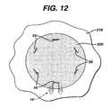
- FIG. 12is a view of a tissue engineered matrix secured to a bone with several graft fixation devices of the present invention.
- FIG. 13is a perspective view of an alternate embodiment of a graft fixation device of the present invention.
- FIG. 14is a side view of the graft fixation device of FIG. 13 .
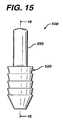
- FIG. 15is an end view of the graft fixation device of FIG. 14 .
- FIG. 16is a cross-sectional view of the graft fixation device of FIG. 14 , taken along View-Line 16 - 16 .
- FIG. 17is a cross-sectional view of the tissue retention member of the graft fixation device of FIG. 14 , taken along View-Line 17 - 17 .
- FIG. 18is a perspective view of an insertion member useful to insert a graft fixation member of the present invention.
- FIG. 19is an exploded perspective view of an insertion instrument, a graft fixation device, and two insertion members.
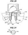
- FIG. 20is a side view of the distal end of the insertion instrument, a graft fixation device, and insertion members engaged in bone, prior to removal of the insertion device.
- FIG. 21is a cross-sectional view taken along View-Line 21 - 21 of FIG. 20 of the prong of the insertion instrument, and a section of the retention member engaged in a longitudinal groove of the prong.
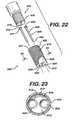
- FIG. 22is an exploded perspective view of the distal end of an insertion instrument of the present invention, illustrating a removable distal end assembly for creating bore holes in bone for receiving the fixation devices of the present invention, wherein the assembly has an end member and pins.
- FIG. 23is a cross-section of the assembly end member of FIG. 22 , taken along View-Line 23 .
- FIG. 24is a perspective view of the assembly end of FIG. 22 , completely assembled and ready for use.
- FIG. 25is a cross-sectional view of the end assembly of FIG. 24 , taken along View-Line 25 - 25 .
- FIG. 26is an exploded perspective view of an insertion instrument of the present invention having a removable distal end assembly useful for inserting the graft retention members of the present invention into bore holes in a bone, having an end assembly member and two pins; when used with insertion members, the instrument can be used to emplace the fixation devices directly into bone without first forming bone bore holes.
- FIG. 27is a cross-sectional view of the end assembly member of FIG. 26 .
- FIG. 28is a perspective view of the distal end of the insertion instrument of FIG. 26 , having the end assembly member and prongs fully assembled and mounted.
- FIG. 29is a cross-sectional view of the distal end of the insertion instrument of FIG. 28 take along View-Line 29 - 29 .
- FIG. 30is a cross-sectional view of the instrument of FIG. 29 taken along View-Line 30 - 30 .
- FIG. 31illustrates a fixation device of the present member having an insertion member molded into the distal end of each implantation member.
- FIG. 32is a cross-sectional view of the fixation device of FIG. 31 .
- FIG. 33is a perspective view of a graft fixation device of the present invention having an single implantation member.
- FIG. 34is a top view of the graft fixation member of FIG. 33 .
- FIG. 35is a cross-sectional view of the graft fixation member of FIG. 33 .
- FIG. 36is a perspective view of an alternate embodiment of the graft fixation member of FIG. 33 having four retention members.
- FIG. 37is a top view of the graft fixation member of FIG. 36 .
- the graft fixation devices of the present inventioncan be made from conventional bio-compatible materials, including absorbable and non-absorbable materials, as well as biodegradable materials.
- the non-absorbable materials which can be utilizedinclude conventional biocompatible materials such as stainless steel, polyethylene, Teflon, Nitinol, non-absorbable polymers, other bio-compatible metals, ceramics, combinations thereof and the like.
- the absorbable materials which can be used to manufacture the graft fixation devices of the present inventionwill typically include those conventional bioabsorbable or bioresorbable materials known in this art which can be effectively molded or machined.
- the bio-absorbable and bio-resorbable materialsinclude polylactic acid, polydioxanone, polycaprolactone, polyglycolic acid, polygalactic acid, other known biocompatible bioabsorbable and bioresorbable polymers, ceramics, composites, combinations thereof and the like and equivalents thereof.
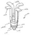
- the graft fixation device 10is seen to have implantation members 20 .
- the implantation members 20are seen to be elongated members, preferably having a substantially cylindrical shape.
- the members 20may have other geometric shapes including conical, pyramidal, polygonal, cubic, spherical, etc.
- the implantation members 20are seen to have distal ends 22 and proximal ends 24 .
- Each implantation member 20is seen to have an outer surface 28 and a longitudinal axis 29 .
- Each member 20is also seen to have longitudinal passage 35 extending therethrough.
- the implantation members 20are also seen to have optional frustoconical ends 30 , and proximal endface surfaces 32 . Although it is preferred that endface surfaces 32 be flat, endface surface 32 may also be angled, concave, convex, etc. Endface surface 32 is seen to have central circular opening 36 in communication with passage 35 . Preferably, central opening 36 will have a circular cross-section, but it may have other geometric cross-sections as well including elliptical, polygonal, square, rectangular, combinations thereof and the like. Members 20 are also seen to have distal end face surfaces 37 having circular openings 38 in communication with passages 35 .
- the annular end face surface 37is of de minimis thickness around opening 38 , however this thickness would increase in the absence of a frustoconical end.
- a series of optional projections 40having tissue engagement edges 44 . Without the projections 40 , the surface 28 of the member 20 will be smooth.
- the device 10is seen to have graft retention member 50 connecting the implantation members 20 .
- Retention member 50is seen to be a rod-like member having first end 52 , second end 54 and central section 55 .
- First end 52is seen to extend from proximal endface surface 32 of the first member 20 while end 54 is seen to extend up from the proximal endface surface 32 of the other member 20 .
- the ends 54 and 52 of retention member 50may also if desired extend from or be mounted to any section of outer surface 28 .
- the connecting member 50is seen to be preferably bent or shaped into three segments including top segment 55 and leg segments 56 .
- the top segment 55is seen to be substantially perpendicular to the leg segments 56 .
- connecting member 50may have other geometric configurations including semicircular, arced, curved, triangular, polygonal, U-shaped, and the like and combinations thereof.
- the ends 52 and 54 of connecting member 50may be permanently affixed to the implantation members 20 , or may be removably attached thereto in a conventional manner.
- Member 50may be rigid or flexible. Member 50 will have a sufficient surface area to effectively retain a tissue-engineered matrix in place on a bone or other body surface.
- connecting member 50will have a circular cross-section, but may have other geometric cross-sections as well including elliptical, polygonal, square, rectangular, combinations thereof and the like.
- Member 50may be rigid or flexible, and may have a single filamentary structure or have multiple interconnected filaments or members.
- the initial stepprior to the installation of a matrix containing a tissue-engineered tissue using a graft fixation device 10 of the present invention, is to drill or ‘tap’ two bore holes 200 into a bone 210 , for example, subchondral bone in the knee joint.
- the bore holes 200are seen to be cylindrical holes having a bottom 208 and an open top 202 and side walls 205 .
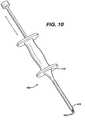
- the bore holesmay be bone tunnels with a continuous passage and no bottom, or an open bottom. It is particularly preferred to tap the holes in the bone by using an instrument 400 as illustrated in FIG.
- tappingor “tap” as used herein is defined to mean a procedure wherein the distal pointed prongs 420 extending from the distal end 415 of the shaft 405 of instrument 400 are located over a bone site, and the proximal end 410 of instrument 400 is tapped or hit with slidable hammer handle 450 (of the “slap hammer”), which slides on shaft 460 between proximal end 410 and proximal stop 470 , to form the bone bore holes 200 .
- the distal end 465 of shaft 460is connected to proximal end 411 .
- Proximal stop 470is mounted to proximal end 467 .
- Hammer handle 450is seen to have grasping section 451 , collars 455 and longitudinal passage 457 .
- a similar pointed instrumentmay be used to “tap” in the bore holes into bone, that is, any instrument having a nail-like distal end.
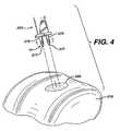
- one bone bore hole at a timemay be “tapped” in. If the surgeon decides to drill the bore holes into bone, any conventional surgical drilling apparatus may be used. After the bore holes 200 are formed into the bone 210 , the matrix 220 containing tissue-engineering tissue is placed upon the bone surface 201 by the surgeon as seen in FIG. 4 .
- Insertion instrument 250is seen to be an elongated rod 260 having a proximal end 262 and a distal end 264 .
- Mounted to the distal end 264 of the rod 260is the depth stop 290 .
- the depth stop 290is seen to be a substantially rectangular member which is mounted perpendicular to the longitudinal axis 251 of the rod 260 .
- Depth stop 290is seen to have bottom 292 . Extending distally from the bottom 292 of plate member 290 is a pair of parallel, spaced-apart, mounting prongs 270 .
- the mounting prongs 270are seen to be substantially rod-like members having distal pointed tips 277 and proximal ends 272 .
- the prongs 270are seen to have first section 273 and distal section 275 .
- Section 273is seen to have a greater cross-sectional dimension than distal section 275 such that the entire section 275 is insertable into passages 35 of members 20 , while proximal section 273 is not insertable therein.
- Instrument 250is also seen to have a “slap hammer section” consisting of proximal shaft 300 extending from proximal end 262 , slidable hammer handle 320 (the “slap hammer”) which is slidable upon shaft 300 between proximal end 262 , and proximal stop 330 .
- Hammer handle member 320is seen to have grasping section 325 , end collars 327 and longitudinal passage 329 .
- the graft fixation device 10is mounted to the insertion instrument 250 by sliding the implantation members 20 onto the prongs 270 such that the distal sections 275 of members 270 are engaged within the longitudinal passages 35 of members 20 and distal points 277 protrude beyond the end of distal endface surfaces 37 .
- the instrument 250is manipulated such that the graft fixation device 10 is inserted through matrix 220 and into bone 210 by moving the implantation members 20 mounted on prongs 270 into the bore holes 200 such that the members 20 are engaged in the bore holes 200 , and such that the tissue engagement section 55 of the retention member 50 engages the matrix 220 such that the matrix 220 is firmly engaged against the surface 201 of the bone 210 .
- holesmay be cut into matrix 220 prior to insertion of device 10 . Then, as seen in FIG.
- the insertion instrument 250is withdrawn proximally causing the prongs 270 to be withdrawn from the passages 35 of the implantation members 20 , thereby leaving the graft fixation device 10 engaged in the bone bore holes, and causing the matrix 220 to be maintained in engagement with the surface 201 of bone 210 .
- the “slap hammer” section of instrument 250may assist in removal of the prongs.
- a cross-sectional view illustrating the device 10 engaged in bone 210 while maintaining the matrix 220 on bone surface 201is seen in FIG. 8 .
- FIG. 12illustrates a matrix 220 mounted to bone surface 201 of bone 210 having multiple fixation devices of the present invention installed to secure the matrix 220 .
- the number, anatomical location and orientation of fixation devices 10 necessary to provide sufficiently effective fixationwill vary with the size and type of implant or matrix, the type of tissue, the age of the patient, the size of the patient's defect, the size of the fixation devices, the material of construction of the fixation devices, the load on the tissue at the repair site, etc.
- the size of the fixation devices of the present inventionwill vary in accordance with a number of variables including the specific design of the device, the materials of construction, the specific application for the devices, the type of surgical procedure, etc.
- the size of the matrices fixated with these deviceswill similarly vary.
- the Figures which are part of this specificationare merely schematic and illustrative of the device and method of the present invention; the actual dimensions of the devices and matrices may vary in practice.
- a punch tool 400was used to create the two requisite bore holes in the subchondral bone to receive one graft fixation device of the present invention. Only one polydioxanone device (4 mm tip-to-tip distance) was used to attach each matrix. To create the bore holes, the punch tool was centered in the defect, oriented in the sagittal plane, and hit or “tapped” with a slap hammer repeatedly until it penetrated several millimeters into the subchondral bone. Next, a 7 mm diameter circular collagen matrix, saturated with saline, was placed in the defect and then blotted dry to remove excess saline.
- the device and inserter toolwere centered above the matrix and oriented in the sagittal plane.
- the surgeonthen located the previously created bore holes by slowly advancing the distal tips of the inserter through the matrix.
- a hammerwas used to fully advance the inserter tool (and implantation members 20 of the fixation device 10 ) through the matrix and into the subchondral bone.
- the inserter toolhad a depth stop to prevent the implantation members 20 from being inserted too deeply, thereby assuring the proper placement of the implantation members through the matrix.
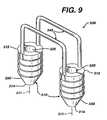
- FIG. 9Another embodiment of the fixation device of the present invention having multiple retention members is seen in FIG. 9 .
- the device 300is seen to have a pair of implantation members 310 .
- the implantation members 310are substantially cylindrical members having longitudinal axis 311 , distal ends 314 and proximal ends 312 .
- Each implantation member 310is seen to have a longitudinal passage 320 .
- the members 310are seen to have a distal frustoconical end 330 , outer surface 350 , and ridges 355 extending outward from surface 350 .
- the members 310are seen to be connected by a pair of retention members 340 , having first and second ends 342 and 344 respectively.
- FIGS. 13-17Yet another embodiment of a fixation device of the present invention is illustrated in FIGS. 13-17 .
- the graft fixation device 500is seen to have implantation members 520 .
- the implantation members 520are seen to be elongated members, preferably having a substantially cylindrical shape.
- the members 520may have other geometric shapes including conical, pyramidal, polygonal, cubic, spherical, etc.
- the implantation members 520are seen to have distal ends 522 and proximal ends 524 .
- Each implantation member 520is seen to have an outer surface 528 and a longitudinal axis 529 .
- Each member 520is also seen to have longitudinal passage 535 extending therethrough.
- the implantation members 520are also seen to have optional frustoconical ends 530 , and proximal end face surfaces 532 . Although it is preferred that endface surfaces 532 be flat; endface surfaces 532 may also be angled, concave, convex, etc. Each endface surface 532 is seen to have central circular opening 536 in communication with passage 535 . Preferably, central opening 536 will have a circular cross-section, but it may have other geometric cross-sections as well including elliptical, polygonal, square, rectangular, combinations thereof and the like. Members 520 are also seen to have distal end face surfaces 537 having circular openings 538 in communication with passages 535 .
- endface surfaces 537have a sharp edge configuration, but may also have various widths with a rounded or flat configuration.
- the annular end face surface 537is of de minimis thickness around opening 538 , however this thickness would typically increase in the absence of a frustoconical end.
- the end surface 537could have various widths as previously mentioned.
- Also seen to extend out from the surface 528 of member 520are a series of optional projections 540 having tissue engagement edges 544 .
- the surface 528 of the member 520will be smooth, however, it will be appreciated that surface 528 could be rough, or could have a variety of conventional projections such as cones, hemispheres, rods, hooks, etc., and the like and equivalents thereof.
- the device 500is seen to have graft retention member 550 connecting the implantation members 520 .
- Retention member 550is seen to be a band-like member preferably having an oval cross-section.
- the retention member 550is seen to have first end 552 , second end 554 and central section 555 .
- First end 552is seen to extend up from proximal endface surface 532 of the first member 520 while end 554 is seen to extend up from the proximal endface surface 532 of the other member 520 .
- a section 557 of end 552is seen to extend out from section 539 of surface 528 , while section 558 of second end 554 is also seen to extend out from a section 539 of surface 528 .
- the ends 554 and 552 of retention member 550may if desired extend from or be mounted to any section of outer surface 528 .
- the connecting member 550is seen to be preferably bent or shaped into three segments including top segment 555 and leg segments 556 .
- the top segment 555is seen to an arc shaped member, and the leg segments 56 are seen to be preferably perpendicular to surfaces 532 .
- connecting member 550may have other geometric configurations including semicircular, arced, curved, triangular, polygonal, V-shaped, and the like and combinations thereof.
- connecting member 550may be permanently affixed to the implantation members 520 , or may be removably attached thereto in a variety of conventional manners, for example, a ball and socket joint, a plug joint, etc.
- Member 550may be rigid or flexible. Member 550 will have a sufficient surface area to effectively retain a tissue-engineered matrix in place on a bone or other body surface.
- connecting member 550will have an oval cross-section, but may have other geometric cross-sections as well including circular, elliptical, polygonal, square, rectangular, combinations thereof and the like. Member 550 may be rigid or flexible, and may have a single filamentary structure or have multiple interconnected filaments or members.
- the insertion device 600is seen to be a substantially cylindrical member having proximal end 610 and distal end 620 .
- Proximal end 610is seen to have a flat end surface 612 .
- Frustoconical end section 630is seen to extend distally from distal end 620 , although device 600 may have other configurations as well.
- distal end 620can have any tapered or curved configuration, but it is preferred that it have a frustoconical end section extending therefrom.
- the frustoconical end section 630is seen to have outer surface 632 and distal tip 640 .
- the member 600is also seen to have exterior surface 650 . Extending through member 600 is the longitudinal passage 660 having first circular opening 665 in communication therewith, and second circular opening 667 in tip 640 in communication therewith.
- the insertion members 600are used in combination with the fixation members of the present invention to engage the fixation member in bone simultaneously with tapping the bore holes into bone, thereby eliminating the need for a separate step to form the bore holes prior to inserting the fixation member.
- a fixation member 500is mounted to prongs 700 extending from the distal end 415 of the shaft 405 of instrument 400 .
- Each prong 700is seen to have first cylindrical section 710 extending from the distal end 415 of the shaft 405 .
- Each cylindrical section 710is seen to have proximal end 711 and distal end 712 , and receiving grooves 715 .
- Extending from the distal end 712 of each first section 710is the central pin section 720 .
- Central pin section 720is seen to have proximal end 722 and distal end 724 .
- Extending distally from distal end 724 of central pin section 720is the distal pin member 730 .
- Distal pin member 730is seen to have proximal end 732 and distal pointed end 734 .
- the insertion member 600may be molded into or affixed to the distal end of an implantation member 520 , thereby forming a unitary structure as seen in FIG. 31 and FIG. 32 .
- the insertion member 600may be mounted to the distal end of an implantation member 520 in a conventional manner by gluing, cementing, mechanical fastening, friction fit and the like and equivalents thereof.
- fixation member 500 of the present inventionare used to affix a matrix to bone in the following manner.
- the implantation members 520 of a fixation device 500are placed upon prongs 700 of an instrument 400 such that the leg members 556 are at least partially engaged in grooves 715 in first section 710 (see FIG. 21 ), and, intermediate sections 720 of pin members 700 are engaged in passages 535 of implantation members 520 , while pin members 730 extend out from the distal ends of the implantation members 520 .
- insertion members 600are placed over the pin members 730 , such that the pin members 730 are engaged in passages 660 , and such that the pointed piercing ends 734 extend beyond the distal ends 640 of the insertion member 660 .
- the tool 400 and the assembly consisting of fixation device 500 and insertion member 600is placed over a tissue matrix 220 placed upon a bone 210 .
- the piercing pointsare then pressed through matrix 220 to contact the surface 211 of bone 210 .
- a slap-hammer section of instrument 400is engaged to drive the piercing points 734 , insertion members 600 and implantation members 520 into the bone 210 as bore holes 200 are formed in the bone.
- the instrument 400is then withdrawn proximately, thereby removing the intermediate sections 720 of prongs 700 from the implantation members 520 and the pin members 730 from the insertion members 600 , leaving the insertion members 600 and the implantation members 520 securely in the bone (as seen in FIG. 20 ).
- fixation devices of the present inventionIt is particularly preferred to use conventional remote visualization surgical procedures when inserting the fixation devices of the present invention. For example, inserting a scope through a trocar cannula into the joint or body cavity, while insufflating the joint or body cavity.
- the insertion members 600will typically be made from a strong, hard, bioabsorbable material such that they can be driven into bone without fracturing or breaking.
- Examples of the types of materials which can be used to make the insertion member 600include polylactic acid, polyglycolic acid, tricalcium phosphate, calcium phosphate, tetracalcium phosphate and hydroxyapatite, and any copolymers, mixtures or blends thereof.
- the insertion member 600assists in forming the bore holes 200 while protecting the implantation members 520 .
- FIGS. 22-23illustrate a disposable distal end assembly 800 for an instrument 400 of the present invention.
- the distal end 415 of the shaft 405 of instrument 400have screw threads 418 , although other conventional detachable mounts may be used, for example a bayonet-type mount, locking levers and tabs, male and female mating sections, etc.
- the assembly 800consists of housing 810 having proximal end 811 and distal end 817 . Housing 810 is seen to have hollow cavity 815 therein. Cavity 815 is seen to be in communication with proximal end opening 812 and distal end openings 820 . Member 810 is seen to have outer surface 822 .
- Outer surface 822is preferably knurled to facilitate the grasping and turning of the housing 810 .
- Housing 810is further seen to have distal end surface 825 .
- the outer surface 822is seen to have a tapered section 823 that tapers toward end face 825 .
- Contained within cavity 815 , on inner surface 818are the screw threads 819 .
- Assembly 800is also seen to have driving pin members 830 .
- Each driving pin member 830is seen to have proximal disk member 832 mounted to proximal end 831 , shaft section 834 and distal pointed end 838 .
- Surrounding each opening 820 on the interior of the member 810are the annular recesses 840 .
- the assembly 800is mounted to the distal end 415 of the instrument 400 in the following manner.
- the pins 830are inserted into cavity 815 and through openings 820 such that the shafts 834 and distal piercing points 838 extend through end face 825 , and the disk members 832 are contained within the annular recesses 840 .
- the housing 810is mounted upon the threads of distal end 415 such that threads 418 engage mating threads 819 , and screwed further such that the proximal end surfaces 833 of the disk members 832 are in contact with the distal end face 416 of distal end 415 .
- the assembly 800is removed and discarded.
- a new sterile assembly 800is utilized with a cleaned and sterilized instrument 400 for each new procedure.
- a disposable end assembly 900 for mounting to an insertion instrument 250is illustrated.
- the insertion member 250is seen to have distal end 264 , having endface 265 and screw threads 266 .
- the assembly 900is seen to have housing 950 .
- Housing 950has proximal end 952 and distal end 956 and exterior surface 954 . Extending from distal end 956 is the plate member 960 .
- Plate member 960is seen to have distal surface 962 .
- the exterior surface 954is seen to have optional knurling and distal tapered section 957 tapering into plate member 960 .
- Housing 950is seen to have internal cavity 955 .
- Housing 950is also seen to have proximal opening 951 in communication with cavity 955 and distal openings 970 also in communication therewith. Housing 950 is seen to have internal screw threads 959 extending from internal surface 958 . Also contained within the interior of housing 950 in the distal end 956 is the recessed groove 980 . Assembly 900 is mounted to the distal end 264 of instrument 250 in the following manner. Pins 910 are inserted through cavity 950 and openings 970 such that proximal members 922 are engaged in groove 980 . Sections 920 and 930 of pins 910 extend through openings 970 . Sections 920 are seen to have grooves 925 .
- the housing 950is screwed on to distal end 264 such that the threads 266 engage the mating internal threads 959 of housing 950 .
- the housingis tightened until the distal end surface 265 of the distal end 264 engages the top surfaces 923 of members 922 .
- the assembly 900is removed from instrument 250 and discarded.
- a new sterile assembly 900is utilized with a cleaned and sterilized instrument 250 for each new procedure.
- FIGS. 33-37An alternate embodiment of the graft fixation devices of the present invention is illustrated in FIGS. 33-37 .
- the graft fixation device 1000is seen to have implantation member 1020 .
- the implantation member 1020is seen to be an elongated member, preferably having a substantially cylindrical shape.
- the member 1020may have other geometric shapes including conical, pyramidal, polygonal, cubic, spherical, etc.
- the implantation member 1020is seen to have distal end 1022 and proximal end 1024 .
- the implantation member 1020is also seen to have an outer surface 1028 and a longitudinal axis 1029 .
- the member 1020is also seen to have longitudinal passage 1035 extending therethrough.
- the implantation member 1020is seen to have truncated frustoconical end 1030 , and proximal end face surface 1032 . Although it is preferred that endface surface 1032 be flat, endface surface 1032 may also be angled, concave, convex, etc.
- the endface surface 1032is seen to have central circular opening 1036 in communication with passage 1035 .
- central opening 1036will have a circular cross-section, but it may have other geometric cross-sections as well including elliptical, polygonal, square, rectangular, combinations thereof and the like.
- Member 1020is also seen to have distal end face surface 1037 having circular opening 1038 in communication with passage 1035 .
- endface surfaces 1037have a sharp edge configuration when used without an insertion member (although not shown), but may also have various widths with a rounded or flat configuration.
- a series of optional projections 1040having tissue engagement edges 1044 . Without the projections 1040 , the surface 1028 of the member 1020 will be smooth, however, it will be appreciated by those skilled in the art that the surface 1028 could be rough, or could have a variety of conventional projections such as cones, hemispheres, rods, hooks, etc., and the like and equivalents thereof.
- the device 1000is seen to have graft retention members 1050 .
- Retention members 1050are seen to be elongated members preferably having an oval cross-section.
- Retention member 1050is seen to have first end 1052 , second free end 1054 , first section 1055 and second section 1057 .
- First end 1052is seen to extend proximally from proximal endface surface 1032 of member 1020 .
- Section 1057is seen to be angulated with respect to section 1055 at angulation 1058 .
- the second section 1057is the graft contact or retention element of the device 1000 .
- the retention member 1050may extend up from any location on the member 1020 preferably perpendicular to surfaces 1032 .
- retention member 1050will have an oval cross-section, but may have other geometric cross-sections as well including circular, elliptical, polygonal, square, rectangular, combinations thereof and the like.
- Member 1050may be rigid or flexible, and may have a single filamentary structure or have multiple interconnected filaments or members. Although it is preferred to have two retention members 1050 mounted diagonally across from each other, any number of retention members 1050 may be utilized sufficient to effectively retain a graft.
- the devicemay have four retention members 1050 as seen in FIGS. 36 and 37 .
- the devicemay have a single retention member 1050 (not shown).
- the device 1000has an insertion device or insertion member 1100 mounted to the distal end of member 1020 .
- the insertion member 1100is seen to be a substantially cylindrical member having proximal end 1110 and distal end 1120 .
- Proximal end 1110is seen to have a flat end surface 1112 .
- Proximal end 1110is seen to have an irregular shape tapering from a first larger diameter to a second smaller diameter to facilitate attachment to the member 1020 , however, proximal end 1110 may have a uniform shape with a constant diameter.
- Frustoconical end section 1130is seen to extend distally from distal end 1120 , although device 1100 may have other configurations as well.
- distal end 1120can have any tapered or curved configuration, but it is preferred that it have a frustoconical end section extending therefrom.
- the frustoconical end section 1130is seen to have outer surface 1132 and distal tip 1140 .
- the member 1100is also seen to have exterior surface 1150 . Extending through member 1100 is the longitudinal passage 1160 having first circular opening 1165 in communication therewith, and second circular opening 1167 in tip 1140 in communication therewith.
- the insertion members 1100are used in combination with the implantation members 1020 to engage the fixation member 1000 in bone simultaneously with tapping the bore holes into bone, thereby eliminating the need for a separate step to form the bore holes prior to inserting the fixation member 1000 .
- the members 1160are preferably mounted to implantation members 1020 by using conventional methods including insert molding, injection molding, press fitting, adhesives, glue and the like and combinations thereof, although the devices 1160 may be used in combination with an implantation member 1020 without mounting it to the implantation member 1020 .
- the devices 1000including implantation member 1050 and insertion devices 1060 are preferably made from the materials listed hereinabove and the like.
- the devices 1000are implanted in bone to retain grafts in a manner similar to that as previously described above for devices having multiple implantation members, using similar insertion instruments suitably modified to insert a device with a single implantation member.
- fixation devices of the present invention and the combination of the fixation devices with insertion members, and methods of using such devices and combinations, of the present inventionhave many advantages.
- the advantagesinclude providing a fast and routine way to fixate a matrix of tissue engineered tissue or other tissue.
- the fixation devices and combinationbecause they eliminate the need for suture knot tying, can be utilized in arthroscopic surgical procedures that require a minimum of surgical incisions and thus greatly reduce patient morbidity.
- the fixation devices and combinationhave been demonstrated to provide excellent matrix fixation without damaging the surrounding normal cartilaginous tissue, unlike the conventional fixation of chondral defect matrices with traditional suture that must be passed through (and thus damage) the surrounding tissue.
Landscapes
- Health & Medical Sciences (AREA)
- Life Sciences & Earth Sciences (AREA)
- Surgery (AREA)
- Heart & Thoracic Surgery (AREA)
- Engineering & Computer Science (AREA)
- Biomedical Technology (AREA)
- Veterinary Medicine (AREA)
- Animal Behavior & Ethology (AREA)
- General Health & Medical Sciences (AREA)
- Public Health (AREA)
- Medical Informatics (AREA)
- Rheumatology (AREA)
- Molecular Biology (AREA)
- Nuclear Medicine, Radiotherapy & Molecular Imaging (AREA)
- Orthopedic Medicine & Surgery (AREA)
- Oral & Maxillofacial Surgery (AREA)
- Vascular Medicine (AREA)
- Transplantation (AREA)
- Cardiology (AREA)
- Rehabilitation Therapy (AREA)
- Prostheses (AREA)
- Materials For Medical Uses (AREA)
- Surgical Instruments (AREA)
Abstract
Description
This is a continuation applicatin of U.S. patent application Ser. No. 10/743,669, filed on Dec. 22, 2003, which is a Continuation application of U.S. patent application Ser. No. 10/056,534, filed on Jan. 24, 2002, which is a Continuation-in-Part application of commonly-assigned, U.S. patent application Ser. No. 09/535,183, filed on Mar. 27, 2000, now U.S. Pat. No. 6,497,707, which is a Continuation-In-Part application of commonly assigned copending U.S. patent application Ser. No. 09/360,367 filed on Jul. 23, 1999, now U.S. Pat. No. 6,179,840, which is incorporated by reference.
The field of art to which this invention relates is surgical fastening devices, in particular, surgical fastening devices for fixating tissue grafts to bone.
The medical technology associated with tissue engineering has advanced at a rapid pace. In particular, it is now known to harvest cells from the human body, for example, chondrocytes and fibrochrondrocytes from the knee joint. These autologous cells are then cultured in a laboratory environment on a bioabsorbable matrix. The matrix will typically have a shape substantially similar to the tissue section which needs to be replaced. After a sufficient period of time in an appropriate culture medium at the proper environmental conditions, the harvested cells will grow on the matrix to form an implantable section of tissue having substantially the same physical configuration as the section of tissue which needs to be replaced in the patient. Such a tissue-engineered construct, consisting of cells on the matrix (or, alternatively, consisting of a matrix alone without cells), is then affixed to the bone site using conventionally known surgical fasteners including sutures, periosteal coverings, or fibrin glue.
The advantages of tissue engineering are many, not the least of which is, for example, that it is now possible to replace cartilage with living cartilage tissue. In addition, the likelihood of rejection of the tissue implant is minimized since the cartilage tissue which has been grown in-vitro is identical to the autologous cartilage of the patient.
Although existing matrix fixation devices are adequate for their intended use, there are also some disadvantages attendant with their use. First of all these fixation devices are generic in the sense that they are not specifically designed for matrix fixation to bone or soft tissue, but can be used for a variety of surgical procedures. Other disadvantages include the difficulty in using many of these devices in a minimally invasive arthroscopic procedure. Additional disadvantages include the difficulty and surgical challenge of harvesting a piece of periosteum for use as a periosteal flap, the significant patient morbidity associated with such harvesting, and the difficulty in suturing such a thin, compliant material to surrounding tissue.
Accordingly, there is a need in this art for novel fixation devices that will effectively affix a matrix of tissue-engineered tissue to a bone or other anchoring site so that the tissue may continue to grow and regenerate in the patient's body.
Therefore, it is an object of the present invention to provide a fixation device that effectively fixates a tissue-engineered matrix to a bone or other anchoring site, thereby enabling the implanted matrix to remain in place while the tissue continues to grow and regenerate.
It is a further object of the present invention to provide such a device for fixating a matrix to a bone site which is easily installed using an arthroscopic procedure or an open procedure.
It is yet a further object of the present invention to provide such a device for fixating a matrix to a bone site which does not require sutures or suture knot tying.
It is still yet a further object of the present invention to provide a surgical method for fixating a matrix utilizing such a device in a location within a patient's body.
Accordingly, a graft fixation device is disclosed. The graft fixation device has first and second implantation members. The members are elongated and preferably have a cylindrical configuration. The members also have distal ends, proximal ends, and longitudinal axes. There are longitudinal passages extending through the entire length of each implantation member. The members have outer surfaces. The implantation members are connected to each other by a rod member having first and second ends and a central section. The first end of the rod member extends from the proximal end of the first implantation member and the second end of the rod member extends from the proximal end of the second implantation member. The rod member is preferably relatively rigid and may be configured to have a variety of geometric shapes, for example, an inverted “U” shape. However, the rod member may also be flexible. The rod member maintains the implantation members at a relatively fixed distance from each other. The central section of the rod member is designed to engage a section of a tissue-engineered matrix implant. In a preferred embodiment, the implantation members have a series of ridges extending out from the outer surfaces of the implantation members to assist in preventing withdrawal from a bone site or other anchoring site after the implantation members are implanted into previously-created bore holes.
Yet another aspect of the present invention is a method of using the graft fixation device of the present invention to affix a matrix containing tissue-engineered tissue to a bone.
Still yet another aspect of the present invention is a graft fixation device combination which is the combination of a fixation device and an insertion device. The fixation device has a first implantation member. The implantation member has a longitudinal axis, a proximal end, a distal end, an outer surface, and a longitudinal passage therethrough. The fixation device also has a second implantation member. The second implantation member has a longitudinal axis, a proximal end, a distal end, an outer surface, and a longitudinal passage therethrough. Each implantation member has a proximal annular face on its proximal end surrounding the longitudinal passages. There is a connecting member connecting the first and second implantation members. The connecting member has a central section, a first end extending from the first implantation member and a second end extending from the second implantation member.
There are a pair of insertion devices. Each insertion device is a member having a proximal end, a distal tapered end and a longitudinal passage therethrough. The distal end of each implantation member is in engagement with the proximal end of an insertion device. Optionally an insertion device is mounted to the distal end of an implantation member.
Another aspect of the present invention is a graft retention device. The device has a single implantation member. The implantation member has a longitudinal axis, a proximal end, a distal end, an outer surface, and a longitudinal passage therethrough. The device has at least one graft retention member. The retention member has a first section extending from the proximal end of the implantation member, and a second section angulated with respect to first section for engaging a graft. Optionally, an insertion member is mounted to the distal end of the implantation member.
These and other features and advantages of the present invention will become more apparent from the following description and accompanying drawings.
The graft fixation devices of the present invention can be made from conventional bio-compatible materials, including absorbable and non-absorbable materials, as well as biodegradable materials. The non-absorbable materials which can be utilized include conventional biocompatible materials such as stainless steel, polyethylene, Teflon, Nitinol, non-absorbable polymers, other bio-compatible metals, ceramics, combinations thereof and the like. The absorbable materials which can be used to manufacture the graft fixation devices of the present invention will typically include those conventional bioabsorbable or bioresorbable materials known in this art which can be effectively molded or machined. The bio-absorbable and bio-resorbable materials include polylactic acid, polydioxanone, polycaprolactone, polyglycolic acid, polygalactic acid, other known biocompatible bioabsorbable and bioresorbable polymers, ceramics, composites, combinations thereof and the like and equivalents thereof.
Referring now toFIGS. 1-2 , a preferred embodiment of agraft fixation device 10 of the present invention is illustrated. Thegraft fixation device 10 is seen to haveimplantation members 20. Theimplantation members 20 are seen to be elongated members, preferably having a substantially cylindrical shape. Themembers 20 may have other geometric shapes including conical, pyramidal, polygonal, cubic, spherical, etc. Theimplantation members 20 are seen to havedistal ends 22 and proximal ends24. Eachimplantation member 20 is seen to have an outer surface28 and alongitudinal axis 29. Eachmember 20 is also seen to havelongitudinal passage 35 extending therethrough. Theimplantation members 20 are also seen to have optional frustoconical ends30, and proximal endface surfaces32. Although it is preferred that endface surfaces32 be flat,endface surface 32 may also be angled, concave, convex, etc.Endface surface 32 is seen to have centralcircular opening 36 in communication withpassage 35. Preferably,central opening 36 will have a circular cross-section, but it may have other geometric cross-sections as well including elliptical, polygonal, square, rectangular, combinations thereof and the like.Members 20 are also seen to have distal end face surfaces37 having circular openings38 in communication withpassages 35. As shown with the optionalfrustoconical end 30, the annular end face surface37 is of de minimis thickness around opening38, however this thickness would increase in the absence of a frustoconical end. Also seen to extend out from the surface28 ofmember 20 are a series ofoptional projections 40 having tissue engagement edges44. Without theprojections 40, the surface28 of themember 20 will be smooth.
Thedevice 10 is seen to havegraft retention member 50 connecting theimplantation members 20.Retention member 50 is seen to be a rod-like member havingfirst end 52,second end 54 andcentral section 55.First end 52 is seen to extend fromproximal endface surface 32 of thefirst member 20 whileend 54 is seen to extend up from theproximal endface surface 32 of theother member 20. The ends54 and52 ofretention member 50 may also if desired extend from or be mounted to any section of outer surface28. The connectingmember 50 is seen to be preferably bent or shaped into three segments includingtop segment 55 andleg segments 56. Thetop segment 55 is seen to be substantially perpendicular to theleg segments 56. Although it is preferred that connectingmember 50 have an inverted “U” configuration, the connectingmember 50 may have other geometric configurations including semicircular, arced, curved, triangular, polygonal, U-shaped, and the like and combinations thereof. The ends52 and54 of connectingmember 50 may be permanently affixed to theimplantation members 20, or may be removably attached thereto in a conventional manner.Member 50 may be rigid or flexible.Member 50 will have a sufficient surface area to effectively retain a tissue-engineered matrix in place on a bone or other body surface. Preferably, connectingmember 50 will have a circular cross-section, but may have other geometric cross-sections as well including elliptical, polygonal, square, rectangular, combinations thereof and the like.Member 50 may be rigid or flexible, and may have a single filamentary structure or have multiple interconnected filaments or members.
Referring now toFIGS. 3-8 , the use of thegraft fixation devices 10 of the present invention in a surgical procedure is illustrated. Referring first toFIG. 3 , the initial step, prior to the installation of a matrix containing a tissue-engineered tissue using agraft fixation device 10 of the present invention, is to drill or ‘tap’ two boreholes 200 into abone 210, for example, subchondral bone in the knee joint. The bore holes200 are seen to be cylindrical holes having a bottom208 and anopen top 202 andside walls 205. Optionally, the bore holes may be bone tunnels with a continuous passage and no bottom, or an open bottom. It is particularly preferred to tap the holes in the bone by using aninstrument 400 as illustrated inFIG. 10 which has a proximal section conventionally referred to in this art as a “slap hammer” section. The term “tapping” or “tap” as used herein is defined to mean a procedure wherein the distal pointedprongs 420 extending from thedistal end 415 of the shaft405 ofinstrument 400 are located over a bone site, and theproximal end 410 ofinstrument 400 is tapped or hit with slidable hammer handle450 (of the “slap hammer”), which slides on shaft460 betweenproximal end 410 and proximal stop470, to form the bone bore holes200. The distal end465 of shaft460 is connected to proximal end411. Proximal stop470 is mounted to proximal end467. Hammer handle450 is seen to have grasping section451, collars455 and longitudinal passage457. Those skilled in the art will appreciate that a similar pointed instrument may be used to “tap” in the bore holes into bone, that is, any instrument having a nail-like distal end. In addition, although not preferred, one bone bore hole at a time may be “tapped” in. If the surgeon decides to drill the bore holes into bone, any conventional surgical drilling apparatus may be used. After the bore holes200 are formed into thebone 210, thematrix 220 containing tissue-engineering tissue is placed upon thebone surface 201 by the surgeon as seen inFIG. 4 . Next, thegraft fixation device 10 is mounted on to theinsertion instrument 250.Insertion instrument 250, as illustrated inFIG. 11 , is seen to be anelongated rod 260 having aproximal end 262 and adistal end 264. Mounted to thedistal end 264 of therod 260 is the depth stop290. The depth stop290 is seen to be a substantially rectangular member which is mounted perpendicular to thelongitudinal axis 251 of therod 260. Depth stop290 is seen to have bottom292. Extending distally from the bottom292 of plate member290 is a pair of parallel, spaced-apart, mountingprongs 270. The mountingprongs 270 are seen to be substantially rod-like members having distal pointedtips 277 and proximal ends272. Theprongs 270 are seen to have first section273 and distal section275. Section273 is seen to have a greater cross-sectional dimension than distal section275 such that the entire section275 is insertable intopassages 35 ofmembers 20, while proximal section273 is not insertable therein.Instrument 250 is also seen to have a “slap hammer section” consisting ofproximal shaft 300 extending fromproximal end 262, slidable hammer handle320 (the “slap hammer”) which is slidable uponshaft 300 betweenproximal end 262, andproximal stop 330.Hammer handle member 320 is seen to have grasping section325, end collars327 and longitudinal passage329. Thegraft fixation device 10 is mounted to theinsertion instrument 250 by sliding theimplantation members 20 onto theprongs 270 such that the distal sections275 ofmembers 270 are engaged within thelongitudinal passages 35 ofmembers 20 anddistal points 277 protrude beyond the end of distal endface surfaces37. Then, as seen inFIGS. 5 and 6 , theinstrument 250 is manipulated such that thegraft fixation device 10 is inserted throughmatrix 220 and intobone 210 by moving theimplantation members 20 mounted onprongs 270 into the bore holes200 such that themembers 20 are engaged in the bore holes200, and such that thetissue engagement section 55 of theretention member 50 engages thematrix 220 such that thematrix 220 is firmly engaged against thesurface 201 of thebone 210. If desired, holes may be cut intomatrix 220 prior to insertion ofdevice 10. Then, as seen inFIG. 7 , theinsertion instrument 250 is withdrawn proximally causing theprongs 270 to be withdrawn from thepassages 35 of theimplantation members 20, thereby leaving thegraft fixation device 10 engaged in the bone bore holes, and causing thematrix 220 to be maintained in engagement with thesurface 201 ofbone 210. The “slap hammer” section ofinstrument 250 may assist in removal of the prongs. A cross-sectional view illustrating thedevice 10 engaged inbone 210 while maintaining thematrix 220 onbone surface 201 is seen inFIG. 8 .
Those skilled in the art will appreciate that the size of the fixation devices of the present invention will vary in accordance with a number of variables including the specific design of the device, the materials of construction, the specific application for the devices, the type of surgical procedure, etc. Similarly, the size of the matrices fixated with these devices will similarly vary. The Figures which are part of this specification are merely schematic and illustrative of the device and method of the present invention; the actual dimensions of the devices and matrices may vary in practice.
The following example is illustrative of the principles and practice of the present invention although not limited thereto.
Six sheep were prepared for a surgical procedure using standard aseptic surgical techniques including the use of fully sterilized instruments and equipment, and conventional anesthesia procedures and protocols. The surgeon then created 7 mm diameter chondral (full thickness cartilage) defects on a weight-bearing area of the medial femoral condyle and in the trochlear groove in the right stifle (knee) in each of the six skeletally mature sheep. Defects were created using a specialized drill with a depth-stop to prevent subchondral bone exposure or penetration. The base surfaces of all the defects were then microfractured with a specialized micropick tool to provide access for cellular migration. The subjects were then separated into three groups of two subjects each:
- Group 1: defect filled with a collagen matrix, fixed with the graft fixation device of the present invention.
- Group 2: defect filled with a collagen matrix, fixed with 9-0 absorbable Vicryl™ suture (interrupted stitch technique, approximately 12 strands per matrix).
- Group 3: unfilled defect (control group).
Both defects in a given stifle received the same treatment or served as controls.
For the two sheep inGroup 1, after a defect had been created and microfractured, apunch tool 400 was used to create the two requisite bore holes in the subchondral bone to receive one graft fixation device of the present invention. Only one polydioxanone device (4 mm tip-to-tip distance) was used to attach each matrix. To create the bore holes, the punch tool was centered in the defect, oriented in the sagittal plane, and hit or “tapped” with a slap hammer repeatedly until it penetrated several millimeters into the subchondral bone. Next, a 7 mm diameter circular collagen matrix, saturated with saline, was placed in the defect and then blotted dry to remove excess saline. When theinserter tool 250 was loaded with thegraft fixation device 10 of the present invention, the device and inserter tool were centered above the matrix and oriented in the sagittal plane. The surgeon then located the previously created bore holes by slowly advancing the distal tips of the inserter through the matrix. Once the surgeon located the holes with the inserter tips, a hammer was used to fully advance the inserter tool (andimplantation members 20 of the fixation device10) through the matrix and into the subchondral bone. The inserter tool had a depth stop to prevent theimplantation members 20 from being inserted too deeply, thereby assuring the proper placement of the implantation members through the matrix. The insertion was completed when the connecting retention member between the two implantation members initially started to compress the collagen matrix, thereby indicating secure fixation with the underlying subchondral bone. After the two defects in a given stifle had each been repaired with a matrix and fixation device, the stifle was dosed and the sheep was allowed to recover. It was noted by the surgeon that it took approximately one minute to attach a matrix with a fixation device of the present invention (Group 1), versus approximately 15 minutes to attach a matrix with suture alone and the requisite suture manipulation and knot tying (Group 2).
Two weeks after the surgeries were completed, the knee joints were surgically opened for examination. Gross macroscopic assessment of the joints demonstrated that all four matrices held by the graft fixation device of the present invention were fully intact. However, all four matrices held by sutures alone were only partially intact with, on average, approximately 30% of the sutures broken on any given matrix.
Another embodiment of the fixation device of the present invention having multiple retention members is seen inFIG. 9 . Thedevice 300 is seen to have a pair ofimplantation members 310. Theimplantation members 310 are substantially cylindrical members havinglongitudinal axis 311, distal ends314 and proximal ends312. Eachimplantation member 310 is seen to have alongitudinal passage 320. Themembers 310 are seen to have a distalfrustoconical end 330, outer surface350, and ridges355 extending outward from surface350. Themembers 310 are seen to be connected by a pair ofretention members 340, having first and second ends342 and344 respectively.
Yet another embodiment of a fixation device of the present invention is illustrated inFIGS. 13-17 . Thegraft fixation device 500 is seen to haveimplantation members 520. Theimplantation members 520 are seen to be elongated members, preferably having a substantially cylindrical shape. Themembers 520 may have other geometric shapes including conical, pyramidal, polygonal, cubic, spherical, etc. Theimplantation members 520 are seen to havedistal ends 522 and proximal ends524. Eachimplantation member 520 is seen to have anouter surface 528 and alongitudinal axis 529. Eachmember 520 is also seen to havelongitudinal passage 535 extending therethrough. Theimplantation members 520 are also seen to have optional frustoconical ends530, and proximal end face surfaces532. Although it is preferred that endface surfaces532 be flat; endface surfaces532 may also be angled, concave, convex, etc. Eachendface surface 532 is seen to have centralcircular opening 536 in communication withpassage 535. Preferably,central opening 536 will have a circular cross-section, but it may have other geometric cross-sections as well including elliptical, polygonal, square, rectangular, combinations thereof and the like.Members 520 are also seen to have distal end face surfaces537 havingcircular openings 538 in communication withpassages 535. Preferably, endface surfaces537 have a sharp edge configuration, but may also have various widths with a rounded or flat configuration. As shown with the optionalfrustoconical end 530, the annularend face surface 537 is of de minimis thickness aroundopening 538, however this thickness would typically increase in the absence of a frustoconical end. However, although not preferred, even with a frustoconical, theend surface 537 could have various widths as previously mentioned. Also seen to extend out from thesurface 528 ofmember 520 are a series ofoptional projections 540 having tissue engagement edges544. Without theprojections 540, thesurface 528 of themember 520 will be smooth, however, it will be appreciated thatsurface 528 could be rough, or could have a variety of conventional projections such as cones, hemispheres, rods, hooks, etc., and the like and equivalents thereof.
Thedevice 500 is seen to havegraft retention member 550 connecting theimplantation members 520.Retention member 550 is seen to be a band-like member preferably having an oval cross-section. Theretention member 550 is seen to havefirst end 552,second end 554 andcentral section 555.First end 552 is seen to extend up fromproximal endface surface 532 of thefirst member 520 whileend 554 is seen to extend up from theproximal endface surface 532 of theother member 520. Asection 557 ofend 552 is seen to extend out fromsection 539 ofsurface 528, whilesection 558 ofsecond end 554 is also seen to extend out from asection 539 ofsurface 528. The ends554 and552 ofretention member 550 may if desired extend from or be mounted to any section ofouter surface 528. The connectingmember 550 is seen to be preferably bent or shaped into three segments includingtop segment 555 andleg segments 556. Thetop segment 555 is seen to an arc shaped member, and theleg segments 56 are seen to be preferably perpendicular tosurfaces 532. Although it is preferred that connectingmember 550 have an inverted “U” configuration, the connectingmember 50 may have other geometric configurations including semicircular, arced, curved, triangular, polygonal, V-shaped, and the like and combinations thereof. The ends552 and554 of connectingmember 550 may be permanently affixed to theimplantation members 520, or may be removably attached thereto in a variety of conventional manners, for example, a ball and socket joint, a plug joint, etc.Member 550 may be rigid or flexible.Member 550 will have a sufficient surface area to effectively retain a tissue-engineered matrix in place on a bone or other body surface. Preferably, connectingmember 550 will have an oval cross-section, but may have other geometric cross-sections as well including circular, elliptical, polygonal, square, rectangular, combinations thereof and the like.Member 550 may be rigid or flexible, and may have a single filamentary structure or have multiple interconnected filaments or members.
Another aspect of the present invention is a distal insertion member (device) useful with the fixation devices of the present invention. As seen inFIG. 18 , theinsertion device 600 is seen to be a substantially cylindrical member havingproximal end 610 anddistal end 620.Proximal end 610 is seen to have aflat end surface 612.Frustoconical end section 630 is seen to extend distally fromdistal end 620, althoughdevice 600 may have other configurations as well. If desired,distal end 620 can have any tapered or curved configuration, but it is preferred that it have a frustoconical end section extending therefrom. Thefrustoconical end section 630 is seen to haveouter surface 632 anddistal tip 640. Themember 600 is also seen to haveexterior surface 650. Extending throughmember 600 is thelongitudinal passage 660 having firstcircular opening 665 in communication therewith, and secondcircular opening 667 intip 640 in communication therewith. Theinsertion members 600 are used in combination with the fixation members of the present invention to engage the fixation member in bone simultaneously with tapping the bore holes into bone, thereby eliminating the need for a separate step to form the bore holes prior to inserting the fixation member.
Referring toFIGS. 19-21 , the previously mentioned combination of aninsertion member 600 and afixation member 500 is illustrated. Initially, afixation member 500 is mounted toprongs 700 extending from thedistal end 415 of the shaft405 ofinstrument 400. Eachprong 700 is seen to have firstcylindrical section 710 extending from thedistal end 415 of the shaft405. Eachcylindrical section 710 is seen to haveproximal end 711 anddistal end 712, and receivinggrooves 715. Extending from thedistal end 712 of eachfirst section 710 is thecentral pin section 720.Central pin section 720 is seen to haveproximal end 722 anddistal end 724. Extending distally fromdistal end 724 ofcentral pin section 720 is thedistal pin member 730.Distal pin member 730 is seen to haveproximal end 732 and distalpointed end 734.
If desired, theinsertion member 600 may be molded into or affixed to the distal end of animplantation member 520, thereby forming a unitary structure as seen inFIG. 31 andFIG. 32 . In addition, theinsertion member 600 may be mounted to the distal end of animplantation member 520 in a conventional manner by gluing, cementing, mechanical fastening, friction fit and the like and equivalents thereof.
The combination of theinsertion members 600 and fixation members, such asfixation member 500 of the present invention, are used to affix a matrix to bone in the following manner. Initially, theimplantation members 520 of afixation device 500 are placed uponprongs 700 of aninstrument 400 such that theleg members 556 are at least partially engaged ingrooves 715 in first section710 (seeFIG. 21 ), and,intermediate sections 720 ofpin members 700 are engaged inpassages 535 ofimplantation members 520, whilepin members 730 extend out from the distal ends of theimplantation members 520. Then,insertion members 600 are placed over thepin members 730, such that thepin members 730 are engaged inpassages 660, and such that the pointed piercing ends734 extend beyond the distal ends640 of theinsertion member 660. Then, thetool 400 and the assembly consisting offixation device 500 andinsertion member 600 is placed over atissue matrix 220 placed upon abone 210. The piercing points are then pressed throughmatrix 220 to contact thesurface 211 ofbone 210. A slap-hammer section ofinstrument 400 is engaged to drive the piercingpoints 734,insertion members 600 andimplantation members 520 into thebone 210 as bore holes200 are formed in the bone. Theinstrument 400 is then withdrawn proximately, thereby removing theintermediate sections 720 ofprongs 700 from theimplantation members 520 and thepin members 730 from theinsertion members 600, leaving theinsertion members 600 and theimplantation members 520 securely in the bone (as seen inFIG. 20 ). This completes the affixation of thematrix 220 to thebone 210 using a single step, wherein the bore holes in the bone are formed simultaneously asinsertion members 600 andfixation device 500 are emplaced in the bone.
It is particularly preferred to use conventional remote visualization surgical procedures when inserting the fixation devices of the present invention. For example, inserting a scope through a trocar cannula into the joint or body cavity, while insufflating the joint or body cavity.
Theinsertion members 600 will typically be made from a strong, hard, bioabsorbable material such that they can be driven into bone without fracturing or breaking. Examples of the types of materials which can be used to make theinsertion member 600 include polylactic acid, polyglycolic acid, tricalcium phosphate, calcium phosphate, tetracalcium phosphate and hydroxyapatite, and any copolymers, mixtures or blends thereof. Although not preferred, it is possible to make the insertion members from a conventional biocompatible material which is not bioabsorbable or biodegradable, such as titanium, stainless steel, ceramics, Nitinol and the like and equivalents thereof. Theinsertion member 600 assists in forming the bore holes200 while protecting theimplantation members 520.
Referring now toFIGS. 26-30 , adisposable end assembly 900 for mounting to aninsertion instrument 250 is illustrated. Theinsertion member 250 is seen to havedistal end 264, havingendface 265 andscrew threads 266. Theassembly 900 is seen to havehousing 950.Housing 950 hasproximal end 952 anddistal end 956 andexterior surface 954. Extending fromdistal end 956 is theplate member 960.Plate member 960 is seen to havedistal surface 962. Theexterior surface 954 is seen to have optional knurling and distaltapered section 957 tapering intoplate member 960.Housing 950 is seen to haveinternal cavity 955.Housing 950 is also seen to haveproximal opening 951 in communication withcavity 955 anddistal openings 970 also in communication therewith.Housing 950 is seen to haveinternal screw threads 959 extending frominternal surface 958. Also contained within the interior ofhousing 950 in thedistal end 956 is the recessedgroove 980.Assembly 900 is mounted to thedistal end 264 ofinstrument 250 in the following manner.Pins 910 are inserted throughcavity 950 andopenings 970 such thatproximal members 922 are engaged ingroove 980.Sections pins 910 extend throughopenings 970.Sections 920 are seen to have grooves925. Then, thehousing 950 is screwed on todistal end 264 such that thethreads 266 engage the matinginternal threads 959 ofhousing 950. The housing is tightened until thedistal end surface 265 of thedistal end 264 engages thetop surfaces 923 ofmembers 922. After a surgical procedure, theassembly 900 is removed frominstrument 250 and discarded. A newsterile assembly 900 is utilized with a cleaned and sterilizedinstrument 250 for each new procedure.
An alternate embodiment of the graft fixation devices of the present invention is illustrated inFIGS. 33-37 . Referring first toFIGS. 33 ,34 and35, thegraft fixation device 1000 is seen to haveimplantation member 1020. Theimplantation member 1020 is seen to be an elongated member, preferably having a substantially cylindrical shape. Themember 1020 may have other geometric shapes including conical, pyramidal, polygonal, cubic, spherical, etc. Theimplantation member 1020 is seen to havedistal end 1022 andproximal end 1024. Theimplantation member 1020 is also seen to have anouter surface 1028 and alongitudinal axis 1029. Themember 1020 is also seen to havelongitudinal passage 1035 extending therethrough. Theimplantation member 1020 is seen to have truncated frustoconical end1030, and proximalend face surface 1032. Although it is preferred thatendface surface 1032 be flat,endface surface 1032 may also be angled, concave, convex, etc. Theendface surface 1032 is seen to have centralcircular opening 1036 in communication withpassage 1035. Preferably,central opening 1036 will have a circular cross-section, but it may have other geometric cross-sections as well including elliptical, polygonal, square, rectangular, combinations thereof and the like.Member 1020 is also seen to have distalend face surface 1037 havingcircular opening 1038 in communication withpassage 1035. Preferably,endface surfaces 1037 have a sharp edge configuration when used without an insertion member (although not shown), but may also have various widths with a rounded or flat configuration. Also seen to extend out from thesurface 1028 ofmember 1020 are a series ofoptional projections 1040 having tissue engagement edges1044. Without theprojections 1040, thesurface 1028 of themember 1020 will be smooth, however, it will be appreciated by those skilled in the art that thesurface 1028 could be rough, or could have a variety of conventional projections such as cones, hemispheres, rods, hooks, etc., and the like and equivalents thereof.
Thedevice 1000 is seen to havegraft retention members 1050.Retention members 1050 are seen to be elongated members preferably having an oval cross-section.Retention member 1050 is seen to havefirst end 1052, secondfree end 1054,first section 1055 andsecond section 1057.First end 1052 is seen to extend proximally fromproximal endface surface 1032 ofmember 1020.Section 1057 is seen to be angulated with respect tosection 1055 at angulation1058. Thesecond section 1057 is the graft contact or retention element of thedevice 1000. If desired, theretention member 1050 may extend up from any location on themember 1020 preferably perpendicular tosurfaces 1032.
Preferably,retention member 1050 will have an oval cross-section, but may have other geometric cross-sections as well including circular, elliptical, polygonal, square, rectangular, combinations thereof and the like.Member 1050 may be rigid or flexible, and may have a single filamentary structure or have multiple interconnected filaments or members. Although it is preferred to have tworetention members 1050 mounted diagonally across from each other, any number ofretention members 1050 may be utilized sufficient to effectively retain a graft. For example, the device may have fourretention members 1050 as seen inFIGS. 36 and 37 . Or, the device may have a single retention member1050 (not shown).
Preferably, thedevice 1000 has an insertion device orinsertion member 1100 mounted to the distal end ofmember 1020. Theinsertion member 1100 is seen to be a substantially cylindrical member havingproximal end 1110 anddistal end 1120.Proximal end 1110 is seen to have aflat end surface 1112.Proximal end 1110 is seen to have an irregular shape tapering from a first larger diameter to a second smaller diameter to facilitate attachment to themember 1020, however,proximal end 1110 may have a uniform shape with a constant diameter.Frustoconical end section 1130 is seen to extend distally fromdistal end 1120, althoughdevice 1100 may have other configurations as well. If desired,distal end 1120 can have any tapered or curved configuration, but it is preferred that it have a frustoconical end section extending therefrom. Thefrustoconical end section 1130 is seen to haveouter surface 1132 anddistal tip 1140. Themember 1100 is also seen to haveexterior surface 1150. Extending throughmember 1100 is the longitudinal passage1160 having firstcircular opening 1165 in communication therewith, and secondcircular opening 1167 intip 1140 in communication therewith. Theinsertion members 1100 are used in combination with theimplantation members 1020 to engage thefixation member 1000 in bone simultaneously with tapping the bore holes into bone, thereby eliminating the need for a separate step to form the bore holes prior to inserting thefixation member 1000.
The members1160 are preferably mounted toimplantation members 1020 by using conventional methods including insert molding, injection molding, press fitting, adhesives, glue and the like and combinations thereof, although the devices1160 may be used in combination with animplantation member 1020 without mounting it to theimplantation member 1020. Thedevices 1000, includingimplantation member 1050 and insertion devices1060 are preferably made from the materials listed hereinabove and the like. Thedevices 1000 are implanted in bone to retain grafts in a manner similar to that as previously described above for devices having multiple implantation members, using similar insertion instruments suitably modified to insert a device with a single implantation member.
The fixation devices of the present invention and the combination of the fixation devices with insertion members, and methods of using such devices and combinations, of the present invention have many advantages. The advantages include providing a fast and routine way to fixate a matrix of tissue engineered tissue or other tissue. The fixation devices and combination, because they eliminate the need for suture knot tying, can be utilized in arthroscopic surgical procedures that require a minimum of surgical incisions and thus greatly reduce patient morbidity. In addition, the fixation devices and combination have been demonstrated to provide excellent matrix fixation without damaging the surrounding normal cartilaginous tissue, unlike the conventional fixation of chondral defect matrices with traditional suture that must be passed through (and thus damage) the surrounding tissue.
Although this invention has been shown and described with respect to detailed embodiments thereof, it will be understood by those skilled in the art that various changes in form and detail may be made without departing from the spirit and scope of the claimed invention.
Claims (12)
1. A graft fixation device comprising:
a bioabsorbable matrix;
an implantation member passing through the bioabsorbable matrix for receipt within the bone, said implantation member having a longitudinal axis, a proximal end, a distal end, an outer surface, and a longitudinal passage therethrough; and
at least one graft retention member, the retention member having a first section extending from the proximal end of the implantation member, and a second section angulated with respect to first section and engaged against the bioabsorbable matrix whereby to trap the bioabsorbable matrix between the second section and the bone.
2. The device ofclaim 1 , wherein the implantation member has a series of ridges extending from the outer surface.
3. The device ofclaim 1 , wherein the retention member comprises a rod member.
4. The device ofclaim 3 , wherein the retention member has an oval cross-section.
5. The device ofclaim 1 , additionally comprising a frustoconical insertion member extending from the distal end of the implantation member, said insertion member having a proximal end, a distal end and an axial passage therethrough.
6. The device ofclaim 1 , wherein the implantation member has a cylindrical configuration.
7. The device ofclaim 5 wherein the insertion member comprises a bioabsorbable material selected from the group consisting of polylactic acid.
8. The device ofclaim 1 wherein the implantation member is implanted in the bone and the matrix is held against the bone via the retention member.
9. The device ofclaim 1 wherein the first section has a substantially smaller cross-section than a cross-section of the implantation member and extends from the proximal end of the implantation member substantially parallel to the longitudinal axis and the second section extends from the first section substantially normal to the longitudinal axis.
10. The device ofclaim 9 wherein the first section and second sections are substantially rigid.
11. The device ofclaim 1 and further comprising a second graft retention member, the second graft retention member having a second retention member first section extending from the proximal end of the implantation member, and a second retention member second section angulated with respect to first section for engaging a graft.
12. The device ofclaim 11 wherein each of the first section and the second retention member first section extend from the proximal end of the implantation member substantially parallel to the longitudinal axis and wherein each of the second section and second retention member second sections extend from and substantially normal to the first section and second retention member first section respectively.
Priority Applications (1)
| Application Number | Priority Date | Filing Date | Title |
|---|---|---|---|
| US11/706,537US8449561B2 (en) | 1999-07-23 | 2007-02-15 | Graft fixation device combination |
Applications Claiming Priority (5)
| Application Number | Priority Date | Filing Date | Title |
|---|---|---|---|
| US09/360,367US6179840B1 (en) | 1999-07-23 | 1999-07-23 | Graft fixation device and method |
| US09/535,183US6497707B1 (en) | 1999-07-23 | 2000-03-27 | Graft fixation device combination |
| US10/056,534US20020095157A1 (en) | 1999-07-23 | 2002-01-24 | Graft fixation device combination |
| US10/743,669US20040138664A1 (en) | 1999-07-23 | 2003-12-22 | Graft fixation device combination |
| US11/706,537US8449561B2 (en) | 1999-07-23 | 2007-02-15 | Graft fixation device combination |
Related Parent Applications (2)
| Application Number | Title | Priority Date | Filing Date |
|---|---|---|---|
| US10/743,669Continuation-In-PartUS20040138664A1 (en) | 1999-07-23 | 2003-12-22 | Graft fixation device combination |
| US10/743,669ContinuationUS20040138664A1 (en) | 1999-07-23 | 2003-12-22 | Graft fixation device combination |
Publications (2)
| Publication Number | Publication Date |
|---|---|
| US20070162055A1 US20070162055A1 (en) | 2007-07-12 |
| US8449561B2true US8449561B2 (en) | 2013-05-28 |
Family
ID=22005043
Family Applications (3)
| Application Number | Title | Priority Date | Filing Date |
|---|---|---|---|
| US10/056,534AbandonedUS20020095157A1 (en) | 1999-07-23 | 2002-01-24 | Graft fixation device combination |
| US10/743,669AbandonedUS20040138664A1 (en) | 1999-07-23 | 2003-12-22 | Graft fixation device combination |
| US11/706,537Expired - Fee RelatedUS8449561B2 (en) | 1999-07-23 | 2007-02-15 | Graft fixation device combination |
Family Applications Before (2)
| Application Number | Title | Priority Date | Filing Date |
|---|---|---|---|
| US10/056,534AbandonedUS20020095157A1 (en) | 1999-07-23 | 2002-01-24 | Graft fixation device combination |
| US10/743,669AbandonedUS20040138664A1 (en) | 1999-07-23 | 2003-12-22 | Graft fixation device combination |
Country Status (5)
| Country | Link |
|---|---|
| US (3) | US20020095157A1 (en) |
| EP (1) | EP1330985A1 (en) |
| JP (1) | JP2003250808A (en) |
| AU (1) | AU2003200043B2 (en) |
| CA (2) | CA2417362C (en) |
Cited By (14)
| Publication number | Priority date | Publication date | Assignee | Title |
|---|---|---|---|---|
| US20160058550A1 (en)* | 2012-02-02 | 2016-03-03 | Smith & Nephew, Inc. | Implantable biologic holder |
| US10299842B2 (en) | 2013-12-20 | 2019-05-28 | Crossroads Extremity Systems, Llc | Bone plates with dynamic elements |
| US10357260B2 (en) | 2015-11-02 | 2019-07-23 | First Ray, LLC | Orthopedic fastener, retainer, and guide methods |
| US10376367B2 (en) | 2015-07-02 | 2019-08-13 | First Ray, LLC | Orthopedic fasteners, instruments and methods |
| US10492841B2 (en) | 2014-07-10 | 2019-12-03 | Crossroads Extremity Systems, Llc | Bone implant and means of insertion |
| US10524775B2 (en) | 2015-07-02 | 2020-01-07 | Arthrex, Inc. | Methods of repairing cartilage defects |
| US10524774B2 (en) | 2015-04-02 | 2020-01-07 | Arthrex, Inc. | Method of repairing cartilage defects |
| US10945725B2 (en) | 2017-02-06 | 2021-03-16 | Crossroads Extremity Systems, Llc | Implant inserter |
| US11179149B2 (en) | 2017-02-07 | 2021-11-23 | Crossroads Extremity Systems, Llc | Counter-torque implant |
| US11202626B2 (en) | 2014-07-10 | 2021-12-21 | Crossroads Extremity Systems, Llc | Bone implant with means for multi directional force and means of insertion |
| US11317951B2 (en) | 2013-12-20 | 2022-05-03 | Crossroads Extremity Systems, Llc | Bone plates with dynamic elements |
| USD961081S1 (en) | 2020-11-18 | 2022-08-16 | Crossroads Extremity Systems, Llc | Orthopedic implant |
| US12059183B2 (en) | 2020-07-31 | 2024-08-13 | Crossroads Extremity Systems, Llc | Bone plates with dynamic elements and screws |
| US12167848B2 (en) | 2020-09-10 | 2024-12-17 | Smith & Nephew, Inc. | Bone anchor delivery system |
Families Citing this family (105)
| Publication number | Priority date | Publication date | Assignee | Title |
|---|---|---|---|---|
| US6179840B1 (en) | 1999-07-23 | 2001-01-30 | Ethicon, Inc. | Graft fixation device and method |
| US20020095157A1 (en) | 1999-07-23 | 2002-07-18 | Bowman Steven M. | Graft fixation device combination |
| US7811286B2 (en)* | 2001-02-12 | 2010-10-12 | Robert J. Medoff | Implant device for applying compression across a fracture site |
| US7037308B2 (en)* | 2001-02-12 | 2006-05-02 | Medoff Robert J | Implant device for applying compression across a fracture site |
| WO2003007784A2 (en) | 2001-07-16 | 2003-01-30 | Depuy Products, Inc. | Meniscus regeneration device and method |
| US20050027307A1 (en) | 2001-07-16 | 2005-02-03 | Schwartz Herbert Eugene | Unitary surgical device and method |
| US8025896B2 (en) | 2001-07-16 | 2011-09-27 | Depuy Products, Inc. | Porous extracellular matrix scaffold and method |
| US8012205B2 (en)* | 2001-07-16 | 2011-09-06 | Depuy Products, Inc. | Cartilage repair and regeneration device |
| US7819918B2 (en) | 2001-07-16 | 2010-10-26 | Depuy Products, Inc. | Implantable tissue repair device |
| AU2002313694B2 (en) | 2001-07-16 | 2007-08-30 | Depuy Products, Inc. | Cartilage repair apparatus and method |
| JP4197158B2 (en)* | 2001-07-16 | 2008-12-17 | デピュイ・プロダクツ・インコーポレイテッド | Devices with naturally occurring biologically derived materials |
| US7794408B2 (en) | 2003-03-28 | 2010-09-14 | Ethicon, Inc. | Tissue collection device and methods |
| US7611473B2 (en) | 2003-09-11 | 2009-11-03 | Ethicon, Inc. | Tissue extraction and maceration device |
| US8034003B2 (en)* | 2003-09-11 | 2011-10-11 | Depuy Mitek, Inc. | Tissue extraction and collection device |
| US20050187565A1 (en)* | 2004-02-20 | 2005-08-25 | Baker Steve G. | Tissue fixation devices and a transoral endoscopic gastroesophageal flap valve restoration device and assembly using same |
| US7608092B1 (en) | 2004-02-20 | 2009-10-27 | Biomet Sports Medicince, LLC | Method and apparatus for performing meniscus repair |
| US8512730B2 (en)* | 2004-07-12 | 2013-08-20 | Isto Technologies, Inc. | Methods of tissue repair and compositions therefor |
| US8192759B2 (en)* | 2004-07-12 | 2012-06-05 | Isto Technologies, Inc. | Matrix made of polyester polymers entangled with hyaluronic polymers useful for supporting tissue repair |
| US8313524B2 (en) | 2004-08-31 | 2012-11-20 | C. R. Bard, Inc. | Self-sealing PTFE graft with kink resistance |
| US8088130B2 (en) | 2006-02-03 | 2012-01-03 | Biomet Sports Medicine, Llc | Method and apparatus for coupling soft tissue to a bone |
| US7857830B2 (en) | 2006-02-03 | 2010-12-28 | Biomet Sports Medicine, Llc | Soft tissue repair and conduit device |
| US8137382B2 (en) | 2004-11-05 | 2012-03-20 | Biomet Sports Medicine, Llc | Method and apparatus for coupling anatomical features |
| US9017381B2 (en) | 2007-04-10 | 2015-04-28 | Biomet Sports Medicine, Llc | Adjustable knotless loops |
| US8361113B2 (en) | 2006-02-03 | 2013-01-29 | Biomet Sports Medicine, Llc | Method and apparatus for coupling soft tissue to a bone |
| US7905903B2 (en) | 2006-02-03 | 2011-03-15 | Biomet Sports Medicine, Llc | Method for tissue fixation |
| US8303604B2 (en) | 2004-11-05 | 2012-11-06 | Biomet Sports Medicine, Llc | Soft tissue repair device and method |
| US8128658B2 (en) | 2004-11-05 | 2012-03-06 | Biomet Sports Medicine, Llc | Method and apparatus for coupling soft tissue to bone |
| US7749250B2 (en) | 2006-02-03 | 2010-07-06 | Biomet Sports Medicine, Llc | Soft tissue repair assembly and associated method |
| US7905904B2 (en) | 2006-02-03 | 2011-03-15 | Biomet Sports Medicine, Llc | Soft tissue repair device and associated methods |
| US8118836B2 (en) | 2004-11-05 | 2012-02-21 | Biomet Sports Medicine, Llc | Method and apparatus for coupling soft tissue to a bone |
| US7909851B2 (en) | 2006-02-03 | 2011-03-22 | Biomet Sports Medicine, Llc | Soft tissue repair device and associated methods |
| US9801708B2 (en) | 2004-11-05 | 2017-10-31 | Biomet Sports Medicine, Llc | Method and apparatus for coupling soft tissue to a bone |
| US20060189993A1 (en) | 2004-11-09 | 2006-08-24 | Arthrotek, Inc. | Soft tissue conduit device |
| US8298262B2 (en) | 2006-02-03 | 2012-10-30 | Biomet Sports Medicine, Llc | Method for tissue fixation |
| US8840645B2 (en) | 2004-11-05 | 2014-09-23 | Biomet Sports Medicine, Llc | Method and apparatus for coupling soft tissue to a bone |
| US7658751B2 (en) | 2006-09-29 | 2010-02-09 | Biomet Sports Medicine, Llc | Method for implanting soft tissue |
| US8034090B2 (en) | 2004-11-09 | 2011-10-11 | Biomet Sports Medicine, Llc | Tissue fixation device |
| US7914539B2 (en) | 2004-11-09 | 2011-03-29 | Biomet Sports Medicine, Llc | Tissue fixation device |
| US8998949B2 (en) | 2004-11-09 | 2015-04-07 | Biomet Sports Medicine, Llc | Soft tissue conduit device |
| US7608098B1 (en) | 2004-11-09 | 2009-10-27 | Biomet Sports Medicine, Llc | Bone fixation device |
| US7354627B2 (en) | 2004-12-22 | 2008-04-08 | Depuy Products, Inc. | Method for organizing the assembly of collagen fibers and compositions formed therefrom |
| US20110076315A1 (en)* | 2005-06-08 | 2011-03-31 | C.R Bard, Inc. | Grafts and Stents Having Inorganic Bio-Compatible Calcium Salt |
| JP2009501027A (en) | 2005-06-17 | 2009-01-15 | シー・アール・バード・インコーポレイテツド | Vascular graft with kinking resistance after tightening |
| US7595062B2 (en)* | 2005-07-28 | 2009-09-29 | Depuy Products, Inc. | Joint resurfacing orthopaedic implant and associated method |
| CA2626598A1 (en) | 2005-11-09 | 2007-05-18 | C.R. Bard Inc. | Grafts and stent grafts having a radiopaque marker |
| CA2631520A1 (en)* | 2005-12-07 | 2007-06-14 | Isto Technologies, Inc. | Cartilage repair methods |
| US20070162030A1 (en)* | 2006-01-06 | 2007-07-12 | Ernest Aranyi | Multi-pronged compressive absorbable tack |
| US8968364B2 (en) | 2006-02-03 | 2015-03-03 | Biomet Sports Medicine, Llc | Method and apparatus for fixation of an ACL graft |
| US8562647B2 (en) | 2006-09-29 | 2013-10-22 | Biomet Sports Medicine, Llc | Method and apparatus for securing soft tissue to bone |
| US8597327B2 (en) | 2006-02-03 | 2013-12-03 | Biomet Manufacturing, Llc | Method and apparatus for sternal closure |
| US8251998B2 (en) | 2006-08-16 | 2012-08-28 | Biomet Sports Medicine, Llc | Chondral defect repair |
| US7959650B2 (en) | 2006-09-29 | 2011-06-14 | Biomet Sports Medicine, Llc | Adjustable knotless loops |
| US9538998B2 (en) | 2006-02-03 | 2017-01-10 | Biomet Sports Medicine, Llc | Method and apparatus for fracture fixation |
| US9271713B2 (en) | 2006-02-03 | 2016-03-01 | Biomet Sports Medicine, Llc | Method and apparatus for tensioning a suture |
| US10517587B2 (en) | 2006-02-03 | 2019-12-31 | Biomet Sports Medicine, Llc | Method and apparatus for forming a self-locking adjustable loop |
| US8652172B2 (en) | 2006-02-03 | 2014-02-18 | Biomet Sports Medicine, Llc | Flexible anchors for tissue fixation |
| US8771352B2 (en) | 2011-05-17 | 2014-07-08 | Biomet Sports Medicine, Llc | Method and apparatus for tibial fixation of an ACL graft |
| US8801783B2 (en) | 2006-09-29 | 2014-08-12 | Biomet Sports Medicine, Llc | Prosthetic ligament system for knee joint |
| US11259792B2 (en) | 2006-02-03 | 2022-03-01 | Biomet Sports Medicine, Llc | Method and apparatus for coupling anatomical features |
| US9078644B2 (en) | 2006-09-29 | 2015-07-14 | Biomet Sports Medicine, Llc | Fracture fixation device |
| US8562645B2 (en) | 2006-09-29 | 2013-10-22 | Biomet Sports Medicine, Llc | Method and apparatus for forming a self-locking adjustable loop |
| US8574235B2 (en) | 2006-02-03 | 2013-11-05 | Biomet Sports Medicine, Llc | Method for trochanteric reattachment |
| US9468433B2 (en) | 2006-02-03 | 2016-10-18 | Biomet Sports Medicine, Llc | Method and apparatus for forming a self-locking adjustable loop |
| US8506597B2 (en) | 2011-10-25 | 2013-08-13 | Biomet Sports Medicine, Llc | Method and apparatus for interosseous membrane reconstruction |
| US9149267B2 (en) | 2006-02-03 | 2015-10-06 | Biomet Sports Medicine, Llc | Method and apparatus for coupling soft tissue to a bone |
| US8652171B2 (en) | 2006-02-03 | 2014-02-18 | Biomet Sports Medicine, Llc | Method and apparatus for soft tissue fixation |
| US11311287B2 (en) | 2006-02-03 | 2022-04-26 | Biomet Sports Medicine, Llc | Method for tissue fixation |
| US8709094B2 (en)* | 2006-06-26 | 2014-04-29 | DePuy Synthes Products, LLC | Anti-adhesion sheet |
| FR2905588B1 (en)* | 2006-09-12 | 2008-11-14 | Small Bone Innovations Interna | SURGICAL CLIP. |
| US9918826B2 (en) | 2006-09-29 | 2018-03-20 | Biomet Sports Medicine, Llc | Scaffold for spring ligament repair |
| US8500818B2 (en) | 2006-09-29 | 2013-08-06 | Biomet Manufacturing, Llc | Knee prosthesis assembly with ligament link |
| US8672969B2 (en) | 2006-09-29 | 2014-03-18 | Biomet Sports Medicine, Llc | Fracture fixation device |
| US11259794B2 (en) | 2006-09-29 | 2022-03-01 | Biomet Sports Medicine, Llc | Method for implanting soft tissue |
| US9198749B2 (en) | 2006-10-12 | 2015-12-01 | C. R. Bard, Inc. | Vascular grafts with multiple channels and methods for making |
| US8114159B2 (en) | 2006-11-20 | 2012-02-14 | Depuy Spine, Inc. | Anterior spinal vessel protector |
| US7871440B2 (en) | 2006-12-11 | 2011-01-18 | Depuy Products, Inc. | Unitary surgical device and method |
| US8652179B2 (en)* | 2008-05-02 | 2014-02-18 | The Cleveland Clinic Foundation | Bone plate extender and extension system for bone restoration and methods of use thereof |
| US8915918B2 (en)* | 2008-05-02 | 2014-12-23 | Thomas James Graham | Bone plate system for bone restoration and methods of use thereof |
| US8628533B2 (en)* | 2008-05-08 | 2014-01-14 | The Cleveland Clinic Foundation | Bone plate with reduction aids and methods of use thereof |
| US8608783B2 (en)* | 2008-05-08 | 2013-12-17 | The Cleveland Clinic Foundation | Bone plate with flange member and methods of use thereof |
| US12245759B2 (en) | 2008-08-22 | 2025-03-11 | Biomet Sports Medicine, Llc | Method and apparatus for coupling soft tissue to bone |
| US12419632B2 (en) | 2008-08-22 | 2025-09-23 | Biomet Sports Medicine, Llc | Method and apparatus for coupling anatomical features |
| US8241298B2 (en) | 2009-03-27 | 2012-08-14 | Depuy Mitek, Inc. | Methods and devices for delivering and affixing tissue scaffolds |
| US8308814B2 (en)* | 2009-03-27 | 2012-11-13 | Depuy Mitek, Inc. | Methods and devices for preparing and implanting tissue scaffolds |
| US8343227B2 (en) | 2009-05-28 | 2013-01-01 | Biomet Manufacturing Corp. | Knee prosthesis assembly with ligament link |
| US12096928B2 (en) | 2009-05-29 | 2024-09-24 | Biomet Sports Medicine, Llc | Method and apparatus for coupling soft tissue to a bone |
| US12329373B2 (en) | 2011-05-02 | 2025-06-17 | Biomet Sports Medicine, Llc | Method and apparatus for soft tissue fixation |
| US9357991B2 (en) | 2011-11-03 | 2016-06-07 | Biomet Sports Medicine, Llc | Method and apparatus for stitching tendons |
| US9381013B2 (en) | 2011-11-10 | 2016-07-05 | Biomet Sports Medicine, Llc | Method for coupling soft tissue to a bone |
| US9314241B2 (en) | 2011-11-10 | 2016-04-19 | Biomet Sports Medicine, Llc | Apparatus for coupling soft tissue to a bone |
| US9370350B2 (en) | 2011-11-10 | 2016-06-21 | Biomet Sports Medicine, Llc | Apparatus for coupling soft tissue to a bone |
| US9259217B2 (en) | 2012-01-03 | 2016-02-16 | Biomet Manufacturing, Llc | Suture Button |
| US9358008B2 (en)* | 2012-04-30 | 2016-06-07 | Zsx Medical, Llc | Surgical device |
| US10245306B2 (en) | 2012-11-16 | 2019-04-02 | Isto Technologies Ii, Llc | Flexible tissue matrix and methods for joint repair |
| US9757119B2 (en) | 2013-03-08 | 2017-09-12 | Biomet Sports Medicine, Llc | Visual aid for identifying suture limbs arthroscopically |
| US9918827B2 (en) | 2013-03-14 | 2018-03-20 | Biomet Sports Medicine, Llc | Scaffold for spring ligament repair |
| US10136886B2 (en) | 2013-12-20 | 2018-11-27 | Biomet Sports Medicine, Llc | Knotless soft tissue devices and techniques |
| US9615822B2 (en) | 2014-05-30 | 2017-04-11 | Biomet Sports Medicine, Llc | Insertion tools and method for soft anchor |
| US9700291B2 (en) | 2014-06-03 | 2017-07-11 | Biomet Sports Medicine, Llc | Capsule retractor |
| US10039543B2 (en) | 2014-08-22 | 2018-08-07 | Biomet Sports Medicine, Llc | Non-sliding soft anchor |
| US10179191B2 (en) | 2014-10-09 | 2019-01-15 | Isto Technologies Ii, Llc | Flexible tissue matrix and methods for joint repair |
| US9955980B2 (en) | 2015-02-24 | 2018-05-01 | Biomet Sports Medicine, Llc | Anatomic soft tissue repair |
| US9974534B2 (en) | 2015-03-31 | 2018-05-22 | Biomet Sports Medicine, Llc | Suture anchor with soft anchor of electrospun fibers |
| CN105147367B (en)* | 2015-09-09 | 2017-07-07 | 北京天钥医疗器械有限公司 | Foreskin staple and the anastomat for circumcision using the staple |
| US11844511B2 (en)* | 2021-07-08 | 2023-12-19 | Arthrex, Inc. | Surgical system and method permitting percutaneous insertion of anchors |
Citations (302)
| Publication number | Priority date | Publication date | Assignee | Title |
|---|---|---|---|---|
| US206200A (en) | 1878-07-23 | Improvement in fountain-pen holders | ||
| US224226A (en) | 1880-02-03 | Frank rhind | ||
| US259260A (en) | 1882-06-06 | Adolf baeyer | ||
| AU717552A (en) | 1952-01-02 | 1952-03-06 | Canadian Patents And Development Limited | Flow distributor valve |
| GB1008193A (en) | 1961-03-01 | 1965-10-27 | Ethicon Inc | Improvements in or relating to surgical implants |
| US3272204A (en) | 1965-09-22 | 1966-09-13 | Ethicon Inc | Absorbable collagen prosthetic implant with non-absorbable reinforcing strands |
| US3739402A (en) | 1970-10-15 | 1973-06-19 | Cutter Lab | Bicuspid fascia lata valve |
| US3812017A (en) | 1972-07-26 | 1974-05-21 | Kennecott Copper Corp | Desulfurized char with phosphoric acid |
| US3857932A (en) | 1970-09-09 | 1974-12-31 | F Gould | Dry hydrophilic acrylate or methacrylate polymer prolonged release drug implants |
| US4045418A (en) | 1975-01-28 | 1977-08-30 | Gulf Oil Corporation | Copolymers of D,L-lactide and epsilon caprolactone |
| US4057537A (en) | 1975-01-28 | 1977-11-08 | Gulf Oil Corporation | Copolymers of L-(-)-lactide and epsilon caprolactone |
| US4060089A (en) | 1975-09-03 | 1977-11-29 | United States Surgical Corporation | Surgical fastening method and device therefor |
| US4105034A (en) | 1977-06-10 | 1978-08-08 | Ethicon, Inc. | Poly(alkylene oxalate) absorbable coating for sutures |
| US4130689A (en) | 1976-06-11 | 1978-12-19 | International Paper Company | Production of high strength hollow rayon fibers |
| US4130639A (en) | 1977-09-28 | 1978-12-19 | Ethicon, Inc. | Absorbable pharmaceutical compositions based on isomorphic copolyoxalates |
| US4140678A (en) | 1977-06-13 | 1979-02-20 | Ethicon, Inc. | Synthetic absorbable surgical devices of poly(alkylene oxalates) |
| US4141087A (en) | 1977-01-19 | 1979-02-27 | Ethicon, Inc. | Isomorphic copolyoxalates and sutures thereof |
| US4205399A (en) | 1977-06-13 | 1980-06-03 | Ethicon, Inc. | Synthetic absorbable surgical devices of poly(alkylene oxalates) |
| US4208511A (en) | 1977-01-19 | 1980-06-17 | Ethicon, Inc. | Isomorphic copolyoxalates and sutures thereof |
| US4344193A (en) | 1980-11-28 | 1982-08-17 | Kenny Charles H | Meniscus prosthesis |
| US4438769A (en) | 1982-04-15 | 1984-03-27 | Pratt Clyde R | Medical staple device |
| US4454875A (en) | 1982-04-15 | 1984-06-19 | Techmedica, Inc. | Osteal medical staple |
| US4520821A (en) | 1982-04-30 | 1985-06-04 | The Regents Of The University Of California | Growing of long-term biological tissue correction structures in vivo |
| US4548202A (en) | 1983-06-20 | 1985-10-22 | Ethicon, Inc. | Mesh tissue fasteners |
| US4553272A (en) | 1981-02-26 | 1985-11-19 | University Of Pittsburgh | Regeneration of living tissues by growth of isolated cells in porous implant and product thereof |
| US4585458A (en) | 1981-06-10 | 1986-04-29 | Kurland Kenneth Z | Means and method of implanting bioprosthetics |
| US4597766A (en) | 1984-10-26 | 1986-07-01 | American Hospital Supply Corporation | Implantable bioprosthetic tendons and ligaments |
| US4609551A (en) | 1984-03-20 | 1986-09-02 | Arnold Caplan | Process of and material for stimulating growth of cartilage and bony tissue at anatomical sites |
| US4635637A (en)* | 1984-03-29 | 1987-01-13 | Schreiber Saul N | Surgical suture |
| US4728329A (en) | 1985-05-03 | 1988-03-01 | Sulzer Brothers Ltd. | Prosthetic band |
| EP0277678A1 (en) | 1987-01-19 | 1988-08-10 | Stichting Science Park Groningen | A graft suitable for treatment by reconstructive surgery and having tissue-specific porosity, and a process for making such graft |
| US4801299A (en) | 1983-06-10 | 1989-01-31 | University Patents, Inc. | Body implants of extracellular matrix and means and methods of making and using such implants |
| US4801229A (en) | 1986-07-31 | 1989-01-31 | Nippon Yusen Kaisha and Nippon Fruehauf Company, Ltd. | Transportation container |
| US4837285A (en) | 1984-03-27 | 1989-06-06 | Medimatrix | Collagen matrix beads for soft tissue repair |
| EP0274898A3 (en) | 1986-12-27 | 1989-10-11 | Ethicon, Inc. | Implant |
| US4884572A (en) | 1986-05-20 | 1989-12-05 | Concept, Inc. | Tack and applicator for treating torn bodily material in vivo |
| US4889119A (en) | 1985-07-17 | 1989-12-26 | Ethicon, Inc. | Surgical fastener made from glycolide-rich polymer blends |
| SU1535542A1 (en) | 1987-11-18 | 1990-01-15 | Всесоюзный Научно-Исследовательский Институт Глазных Болезней | Method of treating secondary graucoma |
| US4902508A (en) | 1988-07-11 | 1990-02-20 | Purdue Research Foundation | Tissue graft composition |
| US4917700A (en) | 1988-08-01 | 1990-04-17 | Zimmer, Inc. | Prosthetic ligament |
| US4946377A (en) | 1989-11-06 | 1990-08-07 | W. L. Gore & Associates, Inc. | Tissue repair device |
| US4994073A (en) | 1989-02-22 | 1991-02-19 | United States Surgical Corp. | Skin fastener |
| US4993410A (en) | 1989-05-01 | 1991-02-19 | Kimsey Timothy P | Prosthetic removal device |
| US5007934A (en) | 1987-07-20 | 1991-04-16 | Regen Corporation | Prosthetic meniscus |
| US5041138A (en) | 1986-11-20 | 1991-08-20 | Massachusetts Institute Of Technology | Neomorphogenesis of cartilage in vivo from cell culture |
| US5053050A (en) | 1988-04-29 | 1991-10-01 | Samuel Itay | Compositions for repair of cartilage and bone |
| US5059206A (en)* | 1989-04-12 | 1991-10-22 | Winters Thomas F | Method and apparatus for repairing a tear in a knee meniscus |
| US5061281A (en) | 1985-12-17 | 1991-10-29 | Allied-Signal Inc. | Bioresorbable polymers and implantation devices thereof |
| US5078744A (en) | 1987-09-04 | 1992-01-07 | Bio-Products, Inc. | Method of using tendon/ligament substitutes composed of long, parallel, non-antigenic tendon/ligament fibers |
| WO1992006179A1 (en) | 1990-10-02 | 1992-04-16 | Imedex S.A. | Biomaterial based on collagen and its applications |
| US5108989A (en) | 1990-04-04 | 1992-04-28 | Genentech, Inc. | Method of predisposing mammals to accelerated tissue repair |
| US5147400A (en) | 1989-05-10 | 1992-09-15 | United States Surgical Corporation | Connective tissue prosthesis |
| DE4110123A1 (en) | 1991-03-27 | 1992-10-01 | Augustin Dr Betz | ELASTIC CLAMP |
| US5176708A (en) | 1990-03-12 | 1993-01-05 | Sulzer Brothers Limited | Prosthetic implant |
| US5206023A (en) | 1991-01-31 | 1993-04-27 | Robert F. Shaw | Method and compositions for the treatment and repair of defects or lesions in cartilage |
| US5258028A (en) | 1988-12-12 | 1993-11-02 | Ersek Robert A | Textured micro implants |
| US5263984A (en) | 1987-07-20 | 1993-11-23 | Regen Biologics, Inc. | Prosthetic ligaments |
| US5269783A (en)* | 1991-05-13 | 1993-12-14 | United States Surgical Corporation | Device and method for repairing torn tissue |
| US5282829A (en) | 1991-08-15 | 1994-02-01 | United States Surgical Corporation | Hollow body implants |
| US5293881A (en) | 1991-04-11 | 1994-03-15 | United States Surgical Corporation | Reduced mass absorbable surgical fastener and retainer |
| US5306311A (en) | 1987-07-20 | 1994-04-26 | Regen Corporation | Prosthetic articular cartilage |
| US5326357A (en) | 1992-03-18 | 1994-07-05 | Mount Sinai Hospital Corporation | Reconstituted cartridge tissue |
| US5352229A (en) | 1993-05-12 | 1994-10-04 | Marlowe Goble E | Arbor press staple and washer and method for its use |
| US5366756A (en) | 1992-06-15 | 1994-11-22 | United States Surgical Corporation | Method for treating bioabsorbable implant material |
| US5425766A (en) | 1987-03-09 | 1995-06-20 | Astra Tech Aktiebolag | Resorbable prosthesis |
| US5443950A (en) | 1986-04-18 | 1995-08-22 | Advanced Tissue Sciences, Inc. | Three-dimensional cell and tissue culture system |
| US5445833A (en) | 1991-09-24 | 1995-08-29 | Purdue Research Foundation | Tendon or ligament graft for promoting autogenous tissue growth |
| US5454814A (en) | 1992-09-02 | 1995-10-03 | Orthomed Sarl | Surgical clamp and clamp driving device |
| US5455041A (en) | 1993-09-13 | 1995-10-03 | Research Foundation Of State University Of New York At Buffalo | Method for inducing periodontal tissue regeneration |
| US5464929A (en) | 1995-03-06 | 1995-11-07 | Ethicon, Inc. | Absorbable polyoxaesters |
| US5468253A (en) | 1993-01-21 | 1995-11-21 | Ethicon, Inc. | Elastomeric medical device |
| US5480827A (en) | 1991-07-19 | 1996-01-02 | Inoteb | Use of porous polycrystalline aragonite as a support material for in vitro culture of cells |
| US5487897A (en) | 1989-07-24 | 1996-01-30 | Atrix Laboratories, Inc. | Biodegradable implant precursor |
| US5500000A (en) | 1993-07-01 | 1996-03-19 | United States Surgical Corporation | Soft tissue repair system and method |
| US5514181A (en) | 1993-09-29 | 1996-05-07 | Johnson & Johnson Medical, Inc. | Absorbable structures for ligament and tendon repair |
| US5514378A (en) | 1993-02-01 | 1996-05-07 | Massachusetts Institute Of Technology | Biocompatible polymer membranes and methods of preparation of three dimensional membrane structures |
| US5520700A (en) | 1992-11-13 | 1996-05-28 | Technion Research & Development Foundation, Ltd. | Stapler device particularly useful in medical suturing |
| US5522817A (en) | 1989-03-31 | 1996-06-04 | United States Surgical Corporation | Absorbable surgical fastener with bone penetrating elements |
| US5534006A (en) | 1993-09-27 | 1996-07-09 | Zsolt Szabo | Knockout tool for joint prostheses |
| US5567806A (en) | 1991-08-02 | 1996-10-22 | Abdul-Malak; Nabil | Collagen crosslinked with a crosslinking agent for the manufacture of a suturable, biocompatible slowresorbing membrane, and such a membrane |
| US5569252A (en) | 1994-09-27 | 1996-10-29 | Justin; Daniel F. | Device for repairing a meniscal tear in a knee and method |
| US5571189A (en) | 1994-05-20 | 1996-11-05 | Kuslich; Stephen D. | Expandable fabric implant for stabilizing the spinal motion segment |
| US5577517A (en) | 1990-06-28 | 1996-11-26 | Bonutti; Peter M. | Method of grafting human tissue particles |
| US5589176A (en) | 1991-10-18 | 1996-12-31 | Seare, Jr.; William J. | Methods of making doubly porous device |
| US5595751A (en) | 1995-03-06 | 1997-01-21 | Ethicon, Inc. | Absorbable polyoxaesters containing amines and/or amido groups |
| US5597579A (en) | 1995-03-06 | 1997-01-28 | Ethicon, Inc. | Blends of absorbable polyoxaamides |
| US5607687A (en) | 1995-03-06 | 1997-03-04 | Ethicon, Inc. | Polymer blends containing absorbable polyoxaesters |
| US5612028A (en) | 1988-02-17 | 1997-03-18 | Genethics Limited | Method of regenerating or replacing cartilage tissue using amniotic cells |
| US5618552A (en) | 1995-03-06 | 1997-04-08 | Ethicon, Inc. | Absorbable polyoxaesters |
| US5620698A (en) | 1995-03-06 | 1997-04-15 | Ethicon, Inc. | Blends of absorbable polyoxaesters containing amines and/or amido groups |
| US5632745A (en) | 1995-02-07 | 1997-05-27 | R&D Biologicals, Inc. | Surgical implantation of cartilage repair unit |
| US5643319A (en)* | 1991-05-13 | 1997-07-01 | United States Surgical Corporation | Device for applying a meniscal staple |
| US5647874A (en) | 1982-05-20 | 1997-07-15 | John O. Hayhurst | Anchoring and manipulating tissue |
| US5654135A (en) | 1990-10-02 | 1997-08-05 | Imedex, Societe Anonyme | Biomaterial based on collagen and its application |
| US5656492A (en) | 1993-02-12 | 1997-08-12 | Brigham And Women's Hospital, Inc. | Cell induction device |
| US5658313A (en) | 1995-06-06 | 1997-08-19 | Thal; Raymond | Knotless suture anchor assembly |
| CA2247158A1 (en) | 1996-02-21 | 1997-08-28 | Advanced Tissue Sciences, Inc. | Method for making and/or repairing cartilage |
| EP0578425B1 (en) | 1992-06-30 | 1997-09-10 | Ethicon, Inc. | Surgical fasteners |
| US5677355A (en) | 1993-08-13 | 1997-10-14 | Smith & Nephew, Inc. | Continuous open-cell polymeric foams containing living cells |
| US5681353A (en) | 1987-07-20 | 1997-10-28 | Regen Biologics, Inc. | Meniscal augmentation device |
| US5688284A (en) | 1996-09-20 | 1997-11-18 | Medicinelodge, Inc. | Variable angle drill guide and ligament fixation method |
| US5690636A (en) | 1995-12-21 | 1997-11-25 | Johnson & Johnson Professional, Inc. | Punch system for tibial prosthesis |
| US5698213A (en) | 1995-03-06 | 1997-12-16 | Ethicon, Inc. | Hydrogels of absorbable polyoxaesters |
| US5700583A (en) | 1995-03-06 | 1997-12-23 | Ethicon, Inc. | Hydrogels of absorbable polyoxaesters containing amines or amido groups |
| US5702462A (en) | 1996-01-24 | 1997-12-30 | Oberlander; Michael | Method of meniscal repair |
| US5709854A (en) | 1993-04-30 | 1998-01-20 | Massachusetts Institute Of Technology | Tissue formation by injecting a cell-polymeric solution that gels in vivo |
| US5711960A (en) | 1993-09-24 | 1998-01-27 | Takiron Co., Ltd. | Biocompatible implant material comprising a tri-axial or more three-dimensional fabric |
| US5720969A (en) | 1993-04-27 | 1998-02-24 | Cytotherapeutics, Inc. | Membrane formed by an acrylonitrile-based polymer |
| US5723331A (en) | 1994-05-05 | 1998-03-03 | Genzyme Corporation | Methods and compositions for the repair of articular cartilage defects in mammals |
| US5736372A (en) | 1986-11-20 | 1998-04-07 | Massachusetts Institute Of Technology | Biodegradable synthetic polymeric fibrous matrix containing chondrocyte for in vivo production of a cartilaginous structure |
| FR2688690B1 (en) | 1992-03-19 | 1998-04-10 | Laboureau Jacques | ARTIFICIAL LIGAMENT. |
| US5755791A (en) | 1996-04-05 | 1998-05-26 | Purdue Research Foundation | Perforated submucosal tissue graft constructs |
| US5759190A (en) | 1996-08-30 | 1998-06-02 | Vts Holdings Limited | Method and kit for autologous transplantation |
| US5766631A (en) | 1993-09-21 | 1998-06-16 | Arnold; Peter Stuart | Wound implant materials |
| US5769899A (en) | 1994-08-12 | 1998-06-23 | Matrix Biotechnologies, Inc. | Cartilage repair unit |
| US5788701A (en) | 1995-12-21 | 1998-08-04 | Johnson & Johnson Professional, Inc. | Instrument system for knee prothesis implantation with universal handle or slap hammer |
| US5830493A (en) | 1994-09-30 | 1998-11-03 | Yamanouchi Pharmaceutical Co., Ltd. | Bone-forming graft |
| US5837235A (en) | 1994-07-08 | 1998-11-17 | Sulzer Medizinaltechnik Ag | Process for regenerating bone and cartilage |
| WO1998053768A1 (en) | 1997-05-30 | 1998-12-03 | Osteobiologics, Inc. | Fiber-reinforced, porous, biodegradable implant device |
| US5855608A (en) | 1994-05-13 | 1999-01-05 | Thm Biomedical, Inc. | Device and methods for in vivo culturing of diverse tissue cells |
| US5859150A (en) | 1995-03-06 | 1999-01-12 | Ethicon, Inc. | Prepolymers of absorbable polyoxaesters |
| US5891558A (en) | 1994-11-22 | 1999-04-06 | Tissue Engineering, Inc. | Biopolymer foams for use in tissue repair and reconstruction |
| US5902741A (en) | 1986-04-18 | 1999-05-11 | Advanced Tissue Sciences, Inc. | Three-dimensional cartilage cultures |
| US5904717A (en) | 1986-01-28 | 1999-05-18 | Thm Biomedical, Inc. | Method and device for reconstruction of articular cartilage |
| US5904716A (en) | 1995-04-26 | 1999-05-18 | Gendler; El | Method for reconstituting cartilage tissue using demineralized bone and product thereof |
| US5914121A (en) | 1997-02-12 | 1999-06-22 | The United States Of America As Represented By The Secretary Of The Department Of Health And Human Services | Formation of human bone in vivo using ceramic powder and human marrow stromal fibroblasts |
| US5922025A (en) | 1992-02-11 | 1999-07-13 | Bristol-Myers Squibb Company | Soft tissue augmentation material |
| US5964805A (en) | 1997-02-12 | 1999-10-12 | Stone; Kevin R. | Method and paste for articular cartilage transplantation |
| CA2328467A1 (en) | 1998-04-16 | 1999-10-21 | Axya Medical, Inc. | Sutureless soft tissue fixation assembly |
| US5980889A (en) | 1993-08-10 | 1999-11-09 | Gore Hybrid Technologies, Inc. | Cell encapsulating device containing a cell displacing core for maintaining cell viability |
| US5984927A (en) | 1998-03-03 | 1999-11-16 | Ethicon, Inc. | Device for sutureless attachment of soft tissue to bone |
| US5989269A (en) | 1996-08-30 | 1999-11-23 | Vts Holdings L.L.C. | Method, instruments and kit for autologous transplantation |
| US5990194A (en) | 1988-10-03 | 1999-11-23 | Atrix Laboratories, Inc. | Biodegradable in-situ forming implants and methods of producing the same |
| US5990378A (en) | 1995-05-25 | 1999-11-23 | Bridport Gundry (Uk) Limited | Textile surgical implants |
| WO1999059647A1 (en) | 1998-05-19 | 1999-11-25 | The American National Red Cross | Hemostatic sandwich bandage |
| US6001394A (en) | 1995-08-07 | 1999-12-14 | Centre National De La Recherche Scientifique (C.N.R.S.) | Biomaterial composition and method for preparing same |
| US6001352A (en) | 1997-03-31 | 1999-12-14 | Osteobiologics, Inc. | Resurfacing cartilage defects with chondrocytes proliferated without differentiation using platelet-derived growth factor |
| US6005161A (en) | 1986-01-28 | 1999-12-21 | Thm Biomedical, Inc. | Method and device for reconstruction of articular cartilage |
| US6022352A (en) | 1997-03-28 | 2000-02-08 | Biomet, Inc. | Bone fixation screw system |
| US6027742A (en) | 1995-05-19 | 2000-02-22 | Etex Corporation | Bioresorbable ceramic composites |
| DE19812195C2 (en) | 1998-03-19 | 2000-03-30 | Uwe Storch | Process for producing a tissue-forming implant and its use |
| US6054122A (en) | 1990-11-27 | 2000-04-25 | The American National Red Cross | Supplemented and unsupplemented tissue sealants, methods of their production and use |
| JP2000116680A (en) | 1998-10-10 | 2000-04-25 | Matsuda Ika Kk | Wide bone contact type staple to be driven into slope |
| US6072742A (en) | 1994-08-04 | 2000-06-06 | Mitsubishi Denki Kabushiki Kaisha | Semiconductor memory device with a voltage down converter stably generating an internal down-converted voltage |
| US6077989A (en) | 1996-05-28 | 2000-06-20 | Kandel; Rita | Resorbable implant biomaterial made of condensed calcium phosphate particles |
| US6080579A (en) | 1997-11-26 | 2000-06-27 | Charlotte-Mecklenburg Hospital Authority | Method for producing human intervertebral disc cells |
| US6083225A (en) | 1996-03-14 | 2000-07-04 | Surgical Dynamics, Inc. | Method and instrumentation for implant insertion |
| US6096532A (en) | 1995-06-07 | 2000-08-01 | Aastrom Biosciences, Inc. | Processor apparatus for use in a system for maintaining and growing biological cells |
| US6103255A (en) | 1999-04-16 | 2000-08-15 | Rutgers, The State University | Porous polymer scaffolds for tissue engineering |
| US6110212A (en) | 1994-11-15 | 2000-08-29 | Kenton W. Gregory | Elastin and elastin-based materials |
| US6110209A (en) | 1997-08-07 | 2000-08-29 | Stone; Kevin R. | Method and paste for articular cartilage transplantation |
| US6117166A (en) | 1997-10-27 | 2000-09-12 | Winston; Thomas R. | Apparatus and methods for grafting blood vessel tissue |
| US6120514A (en) | 1996-08-30 | 2000-09-19 | Vts Holdings, Llc | Method and kit for autologous transplantation |
| US6121042A (en) | 1995-04-27 | 2000-09-19 | Advanced Tissue Sciences, Inc. | Apparatus and method for simulating in vivo conditions while seeding and culturing three-dimensional tissue constructs |
| US6123727A (en) | 1995-05-01 | 2000-09-26 | Massachusetts Institute Of Technology | Tissue engineered tendons and ligaments |
| US6132468A (en) | 1998-09-10 | 2000-10-17 | Mansmann; Kevin A. | Arthroscopic replacement of cartilage using flexible inflatable envelopes |
| US6132463A (en) | 1995-05-19 | 2000-10-17 | Etex Corporation | Cell seeding of ceramic compositions |
| US6140039A (en) | 1986-04-18 | 2000-10-31 | Advanced Tissue Sciences, Inc. | Three-dimensional filamentous tissue having tendon or ligament function |
| US6143293A (en) | 1998-03-26 | 2000-11-07 | Carnegie Mellon | Assembled scaffolds for three dimensional cell culturing and tissue generation |
| US6156068A (en) | 1995-10-11 | 2000-12-05 | Osteobiologics, Inc. | Method of resurfacing a femoral condyle |
| US6165217A (en) | 1997-10-02 | 2000-12-26 | Gore Enterprise Holdings, Inc. | Self-cohering, continuous filament non-woven webs |
| JP2001017535A (en) | 1999-07-05 | 2001-01-23 | Gunze Ltd | In-vivo degradable staple |
| US6176880B1 (en) | 1996-04-05 | 2001-01-23 | Depuy Orthopaedics, Inc. | Tissue graft construct for replacement of cartilaginous structures |
| US6179872B1 (en) | 1998-03-17 | 2001-01-30 | Tissue Engineering | Biopolymer matt for use in tissue repair and reconstruction |
| US6179840B1 (en) | 1999-07-23 | 2001-01-30 | Ethicon, Inc. | Graft fixation device and method |
| US6183737B1 (en) | 1997-10-30 | 2001-02-06 | The General Hospital Corporation | Bonding of cartilage pieces using isolated chondrocytes and a biological gel |
| US6187009B1 (en) | 1997-02-28 | 2001-02-13 | Synthes (U.S.A.) | Osteosynthesis implant |
| US6187053B1 (en) | 1996-11-16 | 2001-02-13 | Will Minuth | Process for producing a natural implant |
| US6187329B1 (en) | 1997-12-23 | 2001-02-13 | Board Of Regents Of The University Of Texas System | Variable permeability bone implants, methods for their preparation and use |
| US6197061B1 (en) | 1999-03-01 | 2001-03-06 | Koichi Masuda | In vitro production of transplantable cartilage tissue cohesive cartilage produced thereby, and method for the surgical repair of cartilage damage |
| US6197325B1 (en) | 1990-11-27 | 2001-03-06 | The American National Red Cross | Supplemented and unsupplemented tissue sealants, methods of their production and use |
| US6200606B1 (en) | 1996-01-16 | 2001-03-13 | Depuy Orthopaedics, Inc. | Isolation of precursor cells from hematopoietic and nonhematopoietic tissues and their use in vivo bone and cartilage regeneration |
| US6214055B1 (en) | 1998-10-30 | 2001-04-10 | Mures Cardiovascular Research, Inc. | Method and kit for rapid preparation of autologous tissue medical devices |
| US6214045B1 (en) | 1997-10-10 | 2001-04-10 | John D. Corbitt, Jr. | Bioabsorbable breast implant |
| JP2001129073A (en) | 1999-11-02 | 2001-05-15 | Olympus Optical Co Ltd | Bone prosthesis material and tool for implanting bone prosthesis material |
| US6242247B1 (en) | 1996-06-04 | 2001-06-05 | Sulzer Orthopedics Ltd. | Method for making cartilage and implants |
| US6251673B1 (en) | 1996-09-10 | 2001-06-26 | Mediphore-Biotechnologie Ag | Method for manufacturing an implant consisting of a carrier material containing medically active agents |
| US6251143B1 (en) | 1999-06-04 | 2001-06-26 | Depuy Orthopaedics, Inc. | Cartilage repair unit |
| US6254606B1 (en) | 1999-10-13 | 2001-07-03 | William P. Carney | Laser aiming device for performing anterior cruciate ligament reconstruction surgery and method for using same |
| US20010014475A1 (en) | 1998-04-08 | 2001-08-16 | Frondoza Carmelita G. | Method for fabricating cell-containing implants |
| US20010016772A1 (en) | 1997-03-07 | 2001-08-23 | Lee David A. | Tissue implant |
| US20010016353A1 (en) | 1999-06-14 | 2001-08-23 | Janas Victor F. | Relic process for producing resorbable ceramic scaffolds |
| JP2001238889A (en) | 2000-01-28 | 2001-09-04 | Depuy Orthopaedics Inc | Device used for fixation of repairing material for soft tissue and assembled body |
| US6287340B1 (en) | 1999-05-14 | 2001-09-11 | Trustees Of Tufts College | Bioengineered anterior cruciate ligament |
| US6291240B1 (en) | 1998-01-29 | 2001-09-18 | Advanced Tissue Sciences, Inc. | Cells or tissues with increased protein factors and methods of making and using same |
| US20010029381A1 (en) | 1999-07-23 | 2001-10-11 | Bowman Steven M. | Graft fixation device combination |
| US20010029382A1 (en) | 1999-07-23 | 2001-10-11 | Bowman Steven M. | Method of securing a graft using a graft fixation device |
| US6306177B1 (en) | 1994-05-06 | 2001-10-23 | Advanced Bio Surfaces, Inc. | Biomaterial system for in situ tissue repair |
| US6306424B1 (en) | 1999-06-30 | 2001-10-23 | Ethicon, Inc. | Foam composite for the repair or regeneration of tissue |
| US20010039453A1 (en) | 1997-08-13 | 2001-11-08 | Gresser Joseph D. | Resorbable interbody spinal fusion devices |
| US20010038848A1 (en) | 2000-02-18 | 2001-11-08 | Donda Russell S. | Implantable tissues infused with growth factors and other additives |
| US6316692B1 (en) | 1997-11-14 | 2001-11-13 | Cedars Sinai Medical Center | Transfection, storage and transfer of male germ cells for generation of transgenic species and genetic therapies |
| JP2001314414A (en) | 2000-03-27 | 2001-11-13 | Ethicon Inc | Method for fixing implant by implant fixing equipment |
| US6319712B1 (en) | 1998-01-30 | 2001-11-20 | Deutsche Institute Fur Textil-Und Faserforschung Stuttgart | Biohybrid articular surface replacement |
| US20010051834A1 (en) | 1999-03-24 | 2001-12-13 | Chondros, Inc. | Method for composite cell-based implants |
| US20010053839A1 (en) | 2000-06-19 | 2001-12-20 | Koken Co. Ltd. | Biomedical material and process for making same |
| US6333029B1 (en) | 1999-06-30 | 2001-12-25 | Ethicon, Inc. | Porous tissue scaffoldings for the repair of regeneration of tissue |
| EP1167517A1 (en) | 2000-06-22 | 2002-01-02 | IsoTis N.V. | Tissue engineering |
| US20020006428A1 (en) | 1999-02-10 | 2002-01-17 | Isotis N.V. | Cartilage tissue engineering |
| US20020009805A1 (en) | 1999-07-06 | 2002-01-24 | Ramot University Authority For Applied Research & Industrial Development Ltd. | Scaffold matrix and tissue maintaining systems |
| US20020009806A1 (en) | 1999-05-27 | 2002-01-24 | Hicks Wesley L. | In vitro cell culture device including cartilage and methods of using the same |
| US20020013627A1 (en) | 1998-10-05 | 2002-01-31 | Ed. Geistlich Soehne Ag Fur Chemische Industrie Switzerland | Method for promoting regeneration of surface cartilage in a damaged joint using multi-layer covering |
| EP1177800A1 (en) | 2000-08-04 | 2002-02-06 | Depuy Orthopaedics, Inc. | Reinforced small intestinal submucosa |
| US20020015719A1 (en) | 2000-04-28 | 2002-02-07 | Karin Kellner | Methods and reagents for tissue engineering of cartilage in vitro |
| US20020022884A1 (en) | 2000-03-27 | 2002-02-21 | Mansmann Kevin A. | Meniscus-type implant with hydrogel surface reinforced by three-dimensional mesh |
| US20020022883A1 (en) | 2000-06-13 | 2002-02-21 | Burg Karen J.L. | Tissue engineering composite |
| US20020028192A1 (en) | 1998-03-24 | 2002-03-07 | S. Dan Dimitrijevich | Non-contracting tissue equivalent |
| US6364884B1 (en) | 1999-07-23 | 2002-04-02 | Ethicon, Inc. | Method of securing a graft using a graft fixation device |
| US6378527B1 (en) | 1998-04-08 | 2002-04-30 | Chondros, Inc. | Cell-culture and polymer constructs |
| US6378572B1 (en) | 2001-03-28 | 2002-04-30 | Siemens Corporate Research, Inc. | Image processing system for inspection of tablets in slab filler packaging machines |
| US20020052628A1 (en) | 1999-07-23 | 2002-05-02 | Bowman Steven M. | Graft fixation device and method |
| US20020095157A1 (en) | 1999-07-23 | 2002-07-18 | Bowman Steven M. | Graft fixation device combination |
| US6423073B2 (en) | 1999-07-23 | 2002-07-23 | Ethicon, Inc. | Instrument for inserting graft fixation device |
| US20020099448A1 (en) | 1996-08-23 | 2002-07-25 | Michael C. Hiles | Multi-formed collagenous biomaterial medical device |
| US20020107570A1 (en) | 2000-12-08 | 2002-08-08 | Sybert Daryl R. | Biocompatible osteogenic band for repair of spinal disorders |
| RU2187261C1 (en) | 2001-05-21 | 2002-08-20 | Новокузнецкий государственный институт усовершенствования врачей | Method for treating nasal septal deformation in sportsmen |
| US20020120348A1 (en) | 2000-12-21 | 2002-08-29 | Melican Mora Carolynne | Reinforced tissue implants and methods of manufacture and use |
| US20020119177A1 (en) | 2000-12-21 | 2002-08-29 | Bowman Steven M. | Reinforced foam implants with enhanced integrity for soft tissue repair and regeneration |
| US20020123750A1 (en) | 2001-02-28 | 2002-09-05 | Lukas Eisermann | Woven orthopedic implants |
| US6447517B1 (en) | 1999-07-23 | 2002-09-10 | Ethicon, Inc. | Instrument for inserting graft fixation device |
| US20020127265A1 (en) | 2000-12-21 | 2002-09-12 | Bowman Steven M. | Use of reinforced foam implants with enhanced integrity for soft tissue repair and regeneration |
| US20020133229A1 (en) | 2000-03-24 | 2002-09-19 | Laurencin Cato T. | Ligament and tendon replacement constructs and methods for production and use thereof |
| US6464729B1 (en) | 1995-11-06 | 2002-10-15 | Mount Sinai Hospital Corporation | Reconstituted mineralized cartilage tissue |
| US20020150604A1 (en) | 2001-04-11 | 2002-10-17 | Chin-Feng Yi | Device and method for tissue engineering |
| US20020151975A1 (en) | 2001-04-17 | 2002-10-17 | Jack Farr | Methods and instruments for improved meniscus transplantation |
| US20020173558A1 (en) | 1999-03-25 | 2002-11-21 | Metabolix, Inc. | Medical devices and applications of polyhydroxyalkanoate polymers |
| US6485723B1 (en) | 1995-02-10 | 2002-11-26 | Purdue Research Foundation | Enhanced submucosal tissue graft constructs |
| US20020177224A1 (en) | 2001-03-15 | 2002-11-28 | Henning Madry | Tissue engineering enhanced by the transfer of a growth factor gene |
| US20020176893A1 (en) | 2001-02-02 | 2002-11-28 | Wironen John F. | Compositions, implants, methods, and kits for closure of lumen openings, repair of ruptured tissue, and for bulking of tissue |
| US6489165B2 (en) | 1997-12-12 | 2002-12-03 | The Regents Of The University Of California | Chondrocyte-like cells useful for tissue engineering and methods |
| US6497707B1 (en)* | 1999-07-23 | 2002-12-24 | Ethicon, Inc. | Graft fixation device combination |
| US20030003153A1 (en) | 2001-05-08 | 2003-01-02 | Samuel Asculai | Reinforced matrices |
| US20030004578A1 (en) | 2001-06-28 | 2003-01-02 | Ethicon, Inc. | Composite scaffold with post anchor for the repair and regeneration of tissue |
| US20030012805A1 (en) | 2001-07-04 | 2003-01-16 | Chen Guoping | Implant for cartilage tissue regeneration |
| US6511958B1 (en) | 1997-08-14 | 2003-01-28 | Sulzer Biologics, Inc. | Compositions for regeneration and repair of cartilage lesions |
| US20030023316A1 (en) | 2000-08-04 | 2003-01-30 | Brown Laura Jean | Hybrid biologic-synthetic bioabsorable scaffolds |
| US20030027332A1 (en) | 2001-07-16 | 2003-02-06 | Edwards Lifesciences Corporation | Tissue engineered heart valve |
| US20030026787A1 (en) | 1999-08-06 | 2003-02-06 | Fearnot Neal E. | Tubular graft construct |
| US20030033022A1 (en) | 2001-07-16 | 2003-02-13 | Plouhar Pamela Lynn | Cartilage repair and regeneration device and method |
| US6521430B1 (en) | 1997-11-06 | 2003-02-18 | Cellectricon Ab | Method for electro-permeabilization of individual cellular and organellar structures and use thereof |
| US20030036797A1 (en) | 2001-07-16 | 2003-02-20 | Prasanna Malaviya | Meniscus regeneration device and method |
| US20030036801A1 (en) | 2001-07-16 | 2003-02-20 | Schwartz Herbert E. | Cartilage repair apparatus and method |
| US6530956B1 (en) | 1998-09-10 | 2003-03-11 | Kevin A. Mansmann | Resorbable scaffolds to promote cartilage regeneration |
| US20030050709A1 (en) | 2001-02-23 | 2003-03-13 | Ulrich Noth | Trabecular bone-derived human mesenchymal stem cells |
| US6541024B1 (en) | 1996-04-19 | 2003-04-01 | Osiris Therapeutics, Inc. | Regeneration and augmentation of bone using mesenchymal stem cells |
| US20030064917A1 (en) | 1998-07-23 | 2003-04-03 | Crawford Susan E. | Methods and compositions for inhibiting angiogenesis |
| EP0770354B1 (en) | 1995-10-20 | 2003-04-09 | United States Surgical Corporation | Meniscal fastener applying device |
| US6551355B1 (en) | 1998-08-14 | 2003-04-22 | Cambridge Scientific, Inc. | Tissue transplant coated with biocompatible biodegradable polymer |
| US20030078617A1 (en) | 2001-07-16 | 2003-04-24 | Schwartz Herbert E. | Unitary surgical device and method |
| US6569172B2 (en) | 1996-08-30 | 2003-05-27 | Verigen Transplantation Service International (Vtsi) | Method, instruments, and kit for autologous transplantation |
| WO2003043674A1 (en) | 2001-11-16 | 2003-05-30 | Children's Medical Center Corporation ¨ | Augmentation of organ function |
| US6592588B1 (en) | 1995-02-16 | 2003-07-15 | Arthrex, Inc. | Apparatus for osteochondral autograft transplantation |
| US6605294B2 (en) | 1998-08-14 | 2003-08-12 | Incept Llc | Methods of using in situ hydration of hydrogel articles for sealing or augmentation of tissue or vessels |
| US6613059B2 (en)* | 1999-03-01 | 2003-09-02 | Coalescent Surgical, Inc. | Tissue connector apparatus and methods |
| JP2003320008A (en) | 2002-04-30 | 2003-11-11 | Olympus Optical Co Ltd | Living tissue filling body and method of manufacturing the same |
| JP2004008437A (en) | 2002-06-06 | 2004-01-15 | Olympus Corp | Cultural bone |
| US20040024457A1 (en) | 2000-12-08 | 2004-02-05 | Boyce Todd M. | Implant for orthopedic applications |
| US20040059416A1 (en) | 1999-06-22 | 2004-03-25 | Murray Martha M. | Biologic replacement for fibrin clot |
| EP1405649A1 (en) | 2002-09-27 | 2004-04-07 | Ethicon, Inc. | Composite scaffolds seeded with mammalian cells |
| US20040078077A1 (en) | 2002-10-18 | 2004-04-22 | Francois Binette | Biocompatible scaffold for ligament or tendon repair |
| US20040078090A1 (en) | 2002-10-18 | 2004-04-22 | Francois Binette | Biocompatible scaffolds with tissue fragments |
| US6727224B1 (en) | 1999-02-01 | 2004-04-27 | Genetics Institute, Llc. | Methods and compositions for healing and repair of articular cartilage |
| JP2004195103A (en) | 2002-12-20 | 2004-07-15 | Japan Tissue Engineering:Kk | Oral implant |
| US6773458B1 (en) | 1991-07-24 | 2004-08-10 | Baxter International Inc. | Angiogenic tissue implant systems and methods |
| EP1216718B1 (en) | 2000-12-21 | 2004-10-06 | Ethicon, Inc. | Reinforced foam implants for soft tissue repair and regeneration |
| US20040219182A1 (en) | 2003-04-29 | 2004-11-04 | Gomes Katherine A. | Novel glue for cartilage repair |
| US20040236424A1 (en) | 2001-05-25 | 2004-11-25 | Imaging Therapeutics, Inc. | Patient selectable joint arthroplasty devices and surgical tools facilitating increased accuracy, speed and simplicity in performing total and partial joint arthroplasty |
| US20040267362A1 (en) | 2003-06-30 | 2004-12-30 | Julia Hwang | Scaffold for connective tissue repair |
| US20050002915A1 (en) | 2003-01-28 | 2005-01-06 | Anthony Atala | Enhancement of angiogenesis to grafts using cells engineered to produce growth factors |
| EP1506790A1 (en) | 2003-08-11 | 2005-02-16 | DePuy Mitek, Inc. | Method and apparatus for resurfacing an articular surface |
| US20050048651A1 (en) | 1997-11-06 | 2005-03-03 | Frida Ryttsen | Method and apparatus for spatially confined electroporation |
| US6866681B2 (en) | 1996-11-20 | 2005-03-15 | Jacques-Phillippe Laboureau | Pre-adjusted prosthetic ligament and method of manufacture |
| EP1027897B1 (en) | 1999-02-10 | 2005-04-13 | IsoTis N.V. | Cartillage tissue engineering |
| US6886569B2 (en) | 1999-07-29 | 2005-05-03 | Ethicon, Inc. | Method for reconstructing a ligament |
| US20050113937A1 (en) | 2003-11-26 | 2005-05-26 | Francois Binette | Conformable tissue repair implant capable of injection delivery |
| EP1537839A1 (en) | 2003-12-02 | 2005-06-08 | Dr. h. c. Robert Mathys Foundation | Prosthetic device for cartilage repair |
| US20050125077A1 (en) | 2003-12-05 | 2005-06-09 | Harmon Alexander M. | Viable tissue repair implants and methods of use |
| US20050147645A1 (en) | 1998-07-24 | 2005-07-07 | Budny John A. | Composition and method for bone regeneration |
| US20050177249A1 (en) | 2004-02-09 | 2005-08-11 | Kladakis Stephanie M. | Scaffolds with viable tissue |
| EP0955024B1 (en) | 1998-05-04 | 2005-09-07 | Adamed SP. Z O.O. | Intravaginal set and method of treatment of prolapse of the urogenital organs |
| JP2005237476A (en) | 2004-02-24 | 2005-09-08 | Gunze Ltd | Neurotization tube |
| US20050234549A1 (en) | 2004-04-20 | 2005-10-20 | Kladakis Stephanie M | Meniscal repair scaffold |
| US20050232967A1 (en) | 2004-04-20 | 2005-10-20 | Kladakis Stephanie M | Nonwoven tissue scaffold |
| EP0562864B1 (en) | 1992-03-25 | 2005-12-28 | Ethicon, Inc. | Heteromorphic sponges containing active agents |
| EP1138263B1 (en) | 2000-03-27 | 2006-03-01 | Ethicon, Inc. | Graft fixation system |
| US20060084930A1 (en) | 2004-10-20 | 2006-04-20 | Sridevi Dhanaraj | Reinforced absorbable multilayered fabric for use in medical devices |
| US20060204439A1 (en) | 2003-07-03 | 2006-09-14 | The Regents Of The University Of California | Methods for comparing relative flux rates of two or more biological molecules in vivo through a single protocol |
| US20060223177A1 (en) | 2003-06-27 | 2006-10-05 | Ethicon Inc. | Postpartum cells derived from umbilical cord tissue, and methods of making and using the same |
| US20060280768A1 (en) | 2005-06-13 | 2006-12-14 | Julia Hwang | Meniscal repair device and method |
| US20060293760A1 (en) | 2005-06-24 | 2006-12-28 | Dedeyne Patrick G | Soft tissue implants with improved interfaces |
| US20070031470A1 (en) | 2004-04-20 | 2007-02-08 | Depuy Mitek, Inc. | Nonwoven tissue scaffold |
| US20070036767A1 (en) | 2003-06-27 | 2007-02-15 | Ethicon, Incorporated | Postpartum cells derived from umbilical cord tissue, and methods of making and using the same |
| US7208177B2 (en) | 1995-02-22 | 2007-04-24 | Ed. Geistlich Soehne Ag Fuer Chemische Industrie | Resorbable extracellular matrix for reconstruction of cartilage |
| US7214232B2 (en) | 1999-07-23 | 2007-05-08 | Ethicon, Inc. | Graft fixation device |
| US20070250177A1 (en) | 2000-09-18 | 2007-10-25 | Organogenesis, Inc. | Methods for treating a patient using a bioengineered flat sheet graft protheses |
| US20080039955A1 (en) | 2001-01-30 | 2008-02-14 | Hunziker Ernst B | Compositions and methods for the treatment and repair of defects or lesions in articular cartilage using synovial-derived tissue or cells |
| US7354764B2 (en) | 2002-07-30 | 2008-04-08 | Bionethos Holding | Method and device for culturing cells |
| US7368124B2 (en) | 2003-03-07 | 2008-05-06 | Depuy Mitek, Inc. | Method of preparation of bioabsorbable porous reinforced tissue implants and implants thereof |
| US20080226870A1 (en) | 2000-05-26 | 2008-09-18 | Sypeck David J | Multifunctional periodic cellular solids and the method of making thereof |
| US20110110958A1 (en) | 2007-09-11 | 2011-05-12 | University Of Florida Research Foundation | Compositions and methods for the treatment of neoplasia |
| EP1348451B1 (en) | 2002-03-29 | 2011-06-15 | Ethicon Inc. | Compositions and medical devices utilizing bioabsorbable liquid polymers |
| US8197837B2 (en) | 2003-03-07 | 2012-06-12 | Depuy Mitek, Inc. | Method of preparation of bioabsorbable porous reinforced tissue implants and implants thereof |
Family Cites Families (3)
| Publication number | Priority date | Publication date | Assignee | Title |
|---|---|---|---|---|
| CA2013539C (en)* | 1989-03-31 | 1999-07-06 | Thomas W. Sander | Absorbable surgical fastener with bone penetrating elements |
| US5129906A (en)* | 1989-09-08 | 1992-07-14 | Linvatec Corporation | Bioabsorbable tack for joining bodily tissue and in vivo method and apparatus for deploying same |
| US6051287A (en)* | 1997-06-20 | 2000-04-18 | Micron Technology, Inc. | Laser desorption of CVD precursor species |
- 2002
- 2002-01-24USUS10/056,534patent/US20020095157A1/ennot_activeAbandoned
- 2003
- 2003-01-08AUAU2003200043Apatent/AU2003200043B2/ennot_activeCeased
- 2003-01-23JPJP2003015085Apatent/JP2003250808A/enactivePending
- 2003-01-23EPEP03250428Apatent/EP1330985A1/ennot_activeWithdrawn
- 2003-01-24CACA2417362Apatent/CA2417362C/ennot_activeExpired - Fee Related
- 2003-01-24CACA2750719Apatent/CA2750719C/ennot_activeExpired - Fee Related
- 2003-12-22USUS10/743,669patent/US20040138664A1/ennot_activeAbandoned
- 2007
- 2007-02-15USUS11/706,537patent/US8449561B2/ennot_activeExpired - Fee Related
Patent Citations (372)
| Publication number | Priority date | Publication date | Assignee | Title |
|---|---|---|---|---|
| US224226A (en) | 1880-02-03 | Frank rhind | ||
| US259260A (en) | 1882-06-06 | Adolf baeyer | ||
| US206200A (en) | 1878-07-23 | Improvement in fountain-pen holders | ||
| AU717552A (en) | 1952-01-02 | 1952-03-06 | Canadian Patents And Development Limited | Flow distributor valve |
| GB1008193A (en) | 1961-03-01 | 1965-10-27 | Ethicon Inc | Improvements in or relating to surgical implants |
| US3272204A (en) | 1965-09-22 | 1966-09-13 | Ethicon Inc | Absorbable collagen prosthetic implant with non-absorbable reinforcing strands |
| US3857932A (en) | 1970-09-09 | 1974-12-31 | F Gould | Dry hydrophilic acrylate or methacrylate polymer prolonged release drug implants |
| US3739402A (en) | 1970-10-15 | 1973-06-19 | Cutter Lab | Bicuspid fascia lata valve |
| US3812017A (en) | 1972-07-26 | 1974-05-21 | Kennecott Copper Corp | Desulfurized char with phosphoric acid |
| US4045418A (en) | 1975-01-28 | 1977-08-30 | Gulf Oil Corporation | Copolymers of D,L-lactide and epsilon caprolactone |
| US4057537A (en) | 1975-01-28 | 1977-11-08 | Gulf Oil Corporation | Copolymers of L-(-)-lactide and epsilon caprolactone |
| US4060089A (en) | 1975-09-03 | 1977-11-29 | United States Surgical Corporation | Surgical fastening method and device therefor |
| US4130689A (en) | 1976-06-11 | 1978-12-19 | International Paper Company | Production of high strength hollow rayon fibers |
| US4208511A (en) | 1977-01-19 | 1980-06-17 | Ethicon, Inc. | Isomorphic copolyoxalates and sutures thereof |
| US4141087A (en) | 1977-01-19 | 1979-02-27 | Ethicon, Inc. | Isomorphic copolyoxalates and sutures thereof |
| US4105034A (en) | 1977-06-10 | 1978-08-08 | Ethicon, Inc. | Poly(alkylene oxalate) absorbable coating for sutures |
| US4140678A (en) | 1977-06-13 | 1979-02-20 | Ethicon, Inc. | Synthetic absorbable surgical devices of poly(alkylene oxalates) |
| US4205399A (en) | 1977-06-13 | 1980-06-03 | Ethicon, Inc. | Synthetic absorbable surgical devices of poly(alkylene oxalates) |
| US4130639A (en) | 1977-09-28 | 1978-12-19 | Ethicon, Inc. | Absorbable pharmaceutical compositions based on isomorphic copolyoxalates |
| US4344193A (en) | 1980-11-28 | 1982-08-17 | Kenny Charles H | Meniscus prosthesis |
| US4553272A (en) | 1981-02-26 | 1985-11-19 | University Of Pittsburgh | Regeneration of living tissues by growth of isolated cells in porous implant and product thereof |
| US4585458A (en) | 1981-06-10 | 1986-04-29 | Kurland Kenneth Z | Means and method of implanting bioprosthetics |
| US4438769A (en) | 1982-04-15 | 1984-03-27 | Pratt Clyde R | Medical staple device |
| US4454875A (en) | 1982-04-15 | 1984-06-19 | Techmedica, Inc. | Osteal medical staple |
| US4520821A (en) | 1982-04-30 | 1985-06-04 | The Regents Of The University Of California | Growing of long-term biological tissue correction structures in vivo |
| US5647874A (en) | 1982-05-20 | 1997-07-15 | John O. Hayhurst | Anchoring and manipulating tissue |
| US4801299A (en) | 1983-06-10 | 1989-01-31 | University Patents, Inc. | Body implants of extracellular matrix and means and methods of making and using such implants |
| US4548202A (en) | 1983-06-20 | 1985-10-22 | Ethicon, Inc. | Mesh tissue fasteners |
| US4609551A (en) | 1984-03-20 | 1986-09-02 | Arnold Caplan | Process of and material for stimulating growth of cartilage and bony tissue at anatomical sites |
| US4837285A (en) | 1984-03-27 | 1989-06-06 | Medimatrix | Collagen matrix beads for soft tissue repair |
| US4635637A (en)* | 1984-03-29 | 1987-01-13 | Schreiber Saul N | Surgical suture |
| US4597766A (en) | 1984-10-26 | 1986-07-01 | American Hospital Supply Corporation | Implantable bioprosthetic tendons and ligaments |
| US4728329A (en) | 1985-05-03 | 1988-03-01 | Sulzer Brothers Ltd. | Prosthetic band |
| US4889119A (en) | 1985-07-17 | 1989-12-26 | Ethicon, Inc. | Surgical fastener made from glycolide-rich polymer blends |
| US5061281A (en) | 1985-12-17 | 1991-10-29 | Allied-Signal Inc. | Bioresorbable polymers and implantation devices thereof |
| US6005161A (en) | 1986-01-28 | 1999-12-21 | Thm Biomedical, Inc. | Method and device for reconstruction of articular cartilage |
| US5904717A (en) | 1986-01-28 | 1999-05-18 | Thm Biomedical, Inc. | Method and device for reconstruction of articular cartilage |
| US5902741A (en) | 1986-04-18 | 1999-05-11 | Advanced Tissue Sciences, Inc. | Three-dimensional cartilage cultures |
| US5443950A (en) | 1986-04-18 | 1995-08-22 | Advanced Tissue Sciences, Inc. | Three-dimensional cell and tissue culture system |
| US6140039A (en) | 1986-04-18 | 2000-10-31 | Advanced Tissue Sciences, Inc. | Three-dimensional filamentous tissue having tendon or ligament function |
| US4884572A (en) | 1986-05-20 | 1989-12-05 | Concept, Inc. | Tack and applicator for treating torn bodily material in vivo |
| US4801229A (en) | 1986-07-31 | 1989-01-31 | Nippon Yusen Kaisha and Nippon Fruehauf Company, Ltd. | Transportation container |
| US5736372A (en) | 1986-11-20 | 1998-04-07 | Massachusetts Institute Of Technology | Biodegradable synthetic polymeric fibrous matrix containing chondrocyte for in vivo production of a cartilaginous structure |
| US5041138A (en) | 1986-11-20 | 1991-08-20 | Massachusetts Institute Of Technology | Neomorphogenesis of cartilage in vivo from cell culture |
| EP0274898A3 (en) | 1986-12-27 | 1989-10-11 | Ethicon, Inc. | Implant |
| EP0277678A1 (en) | 1987-01-19 | 1988-08-10 | Stichting Science Park Groningen | A graft suitable for treatment by reconstructive surgery and having tissue-specific porosity, and a process for making such graft |
| US5425766A (en) | 1987-03-09 | 1995-06-20 | Astra Tech Aktiebolag | Resorbable prosthesis |
| US5263984A (en) | 1987-07-20 | 1993-11-23 | Regen Biologics, Inc. | Prosthetic ligaments |
| US5735903A (en) | 1987-07-20 | 1998-04-07 | Li; Shu-Tung | Meniscal augmentation device |
| US5624463A (en) | 1987-07-20 | 1997-04-29 | Regen Biologics, Inc. | Prosthetic articular cartilage |
| US5681353A (en) | 1987-07-20 | 1997-10-28 | Regen Biologics, Inc. | Meniscal augmentation device |
| US6042610A (en) | 1987-07-20 | 2000-03-28 | Regen Biologics, Inc. | Meniscal augmentation device |
| US5306311A (en) | 1987-07-20 | 1994-04-26 | Regen Corporation | Prosthetic articular cartilage |
| US5007934A (en) | 1987-07-20 | 1991-04-16 | Regen Corporation | Prosthetic meniscus |
| US5078744A (en) | 1987-09-04 | 1992-01-07 | Bio-Products, Inc. | Method of using tendon/ligament substitutes composed of long, parallel, non-antigenic tendon/ligament fibers |
| SU1535542A1 (en) | 1987-11-18 | 1990-01-15 | Всесоюзный Научно-Исследовательский Институт Глазных Болезней | Method of treating secondary graucoma |
| US5612028A (en) | 1988-02-17 | 1997-03-18 | Genethics Limited | Method of regenerating or replacing cartilage tissue using amniotic cells |
| US5053050A (en) | 1988-04-29 | 1991-10-01 | Samuel Itay | Compositions for repair of cartilage and bone |
| US4902508A (en) | 1988-07-11 | 1990-02-20 | Purdue Research Foundation | Tissue graft composition |
| US4917700A (en) | 1988-08-01 | 1990-04-17 | Zimmer, Inc. | Prosthetic ligament |
| US5990194A (en) | 1988-10-03 | 1999-11-23 | Atrix Laboratories, Inc. | Biodegradable in-situ forming implants and methods of producing the same |
| US5258028A (en) | 1988-12-12 | 1993-11-02 | Ersek Robert A | Textured micro implants |
| US4994073A (en) | 1989-02-22 | 1991-02-19 | United States Surgical Corp. | Skin fastener |
| US5522817A (en) | 1989-03-31 | 1996-06-04 | United States Surgical Corporation | Absorbable surgical fastener with bone penetrating elements |
| US5059206A (en)* | 1989-04-12 | 1991-10-22 | Winters Thomas F | Method and apparatus for repairing a tear in a knee meniscus |
| US4993410A (en) | 1989-05-01 | 1991-02-19 | Kimsey Timothy P | Prosthetic removal device |
| US5147400A (en) | 1989-05-10 | 1992-09-15 | United States Surgical Corporation | Connective tissue prosthesis |
| US5487897A (en) | 1989-07-24 | 1996-01-30 | Atrix Laboratories, Inc. | Biodegradable implant precursor |
| US4946377A (en) | 1989-11-06 | 1990-08-07 | W. L. Gore & Associates, Inc. | Tissue repair device |
| US5176708A (en) | 1990-03-12 | 1993-01-05 | Sulzer Brothers Limited | Prosthetic implant |
| US5108989A (en) | 1990-04-04 | 1992-04-28 | Genentech, Inc. | Method of predisposing mammals to accelerated tissue repair |
| US20020091406A1 (en) | 1990-06-28 | 2002-07-11 | Bonutti Peter M. | Apparatus and method for tissue removal |
| US20020091403A1 (en) | 1990-06-28 | 2002-07-11 | Bonutti Peter M. | Cell harvesting method |
| US20020082631A1 (en) | 1990-06-28 | 2002-06-27 | Bonutti Peter M. | Apparatus and method for tissue removal |
| US20020029055A1 (en) | 1990-06-28 | 2002-03-07 | Bonutti Peter M. | Apparatus and method for tissue removal |
| US5577517A (en) | 1990-06-28 | 1996-11-26 | Bonutti; Peter M. | Method of grafting human tissue particles |
| US20020099401A1 (en) | 1990-06-28 | 2002-07-25 | Bonutti Petel M. | Apparatus and method for tissue removal |
| WO1992006179A1 (en) | 1990-10-02 | 1992-04-16 | Imedex S.A. | Biomaterial based on collagen and its applications |
| US5654135A (en) | 1990-10-02 | 1997-08-05 | Imedex, Societe Anonyme | Biomaterial based on collagen and its application |
| US6197325B1 (en) | 1990-11-27 | 2001-03-06 | The American National Red Cross | Supplemented and unsupplemented tissue sealants, methods of their production and use |
| US6054122A (en) | 1990-11-27 | 2000-04-25 | The American National Red Cross | Supplemented and unsupplemented tissue sealants, methods of their production and use |
| US5206023A (en) | 1991-01-31 | 1993-04-27 | Robert F. Shaw | Method and compositions for the treatment and repair of defects or lesions in cartilage |
| DE4110123A1 (en) | 1991-03-27 | 1992-10-01 | Augustin Dr Betz | ELASTIC CLAMP |
| US5293881A (en) | 1991-04-11 | 1994-03-15 | United States Surgical Corporation | Reduced mass absorbable surgical fastener and retainer |
| US5643319A (en)* | 1991-05-13 | 1997-07-01 | United States Surgical Corporation | Device for applying a meniscal staple |
| US5269783A (en)* | 1991-05-13 | 1993-12-14 | United States Surgical Corporation | Device and method for repairing torn tissue |
| US5374268A (en)* | 1991-05-13 | 1994-12-20 | United States Surgical Corporation | Device and method for repairing torn tissue |
| US6190401B1 (en)* | 1991-05-13 | 2001-02-20 | United States Surgical Corporation | Device for applying a meniscal staple |
| US5480827A (en) | 1991-07-19 | 1996-01-02 | Inoteb | Use of porous polycrystalline aragonite as a support material for in vitro culture of cells |
| US6773458B1 (en) | 1991-07-24 | 2004-08-10 | Baxter International Inc. | Angiogenic tissue implant systems and methods |
| US5567806A (en) | 1991-08-02 | 1996-10-22 | Abdul-Malak; Nabil | Collagen crosslinked with a crosslinking agent for the manufacture of a suturable, biocompatible slowresorbing membrane, and such a membrane |
| US5282829A (en) | 1991-08-15 | 1994-02-01 | United States Surgical Corporation | Hollow body implants |
| US5445833A (en) | 1991-09-24 | 1995-08-29 | Purdue Research Foundation | Tendon or ligament graft for promoting autogenous tissue growth |
| US5589176A (en) | 1991-10-18 | 1996-12-31 | Seare, Jr.; William J. | Methods of making doubly porous device |
| US5922025A (en) | 1992-02-11 | 1999-07-13 | Bristol-Myers Squibb Company | Soft tissue augmentation material |
| US5326357A (en) | 1992-03-18 | 1994-07-05 | Mount Sinai Hospital Corporation | Reconstituted cartridge tissue |
| FR2688690B1 (en) | 1992-03-19 | 1998-04-10 | Laboureau Jacques | ARTIFICIAL LIGAMENT. |
| EP0562864B1 (en) | 1992-03-25 | 2005-12-28 | Ethicon, Inc. | Heteromorphic sponges containing active agents |
| US5697976A (en) | 1992-06-15 | 1997-12-16 | United States Surgical Corporation | Bioabsorbable implant material |
| US5366756A (en) | 1992-06-15 | 1994-11-22 | United States Surgical Corporation | Method for treating bioabsorbable implant material |
| EP0578425B1 (en) | 1992-06-30 | 1997-09-10 | Ethicon, Inc. | Surgical fasteners |
| US5454814A (en) | 1992-09-02 | 1995-10-03 | Orthomed Sarl | Surgical clamp and clamp driving device |
| EP0589306B1 (en) | 1992-09-21 | 1999-08-04 | United States Surgical Corporation | Device for applying a meniscal staple |
| US5520700A (en) | 1992-11-13 | 1996-05-28 | Technion Research & Development Foundation, Ltd. | Stapler device particularly useful in medical suturing |
| US5468253A (en) | 1993-01-21 | 1995-11-21 | Ethicon, Inc. | Elastomeric medical device |
| US5514378A (en) | 1993-02-01 | 1996-05-07 | Massachusetts Institute Of Technology | Biocompatible polymer membranes and methods of preparation of three dimensional membrane structures |
| US5656492A (en) | 1993-02-12 | 1997-08-12 | Brigham And Women's Hospital, Inc. | Cell induction device |
| US6180007B1 (en) | 1993-04-27 | 2001-01-30 | Neurotech, Sa | Membrane formed by an acrylonitrile-based polymer |
| US5720969A (en) | 1993-04-27 | 1998-02-24 | Cytotherapeutics, Inc. | Membrane formed by an acrylonitrile-based polymer |
| US5709854A (en) | 1993-04-30 | 1998-01-20 | Massachusetts Institute Of Technology | Tissue formation by injecting a cell-polymeric solution that gels in vivo |
| US20010053353A1 (en) | 1993-04-30 | 2001-12-20 | Griffith Linda G | Injectable polysaccharide-cell compositions |
| US5352229A (en) | 1993-05-12 | 1994-10-04 | Marlowe Goble E | Arbor press staple and washer and method for its use |
| US5500000A (en) | 1993-07-01 | 1996-03-19 | United States Surgical Corporation | Soft tissue repair system and method |
| US5980889A (en) | 1993-08-10 | 1999-11-09 | Gore Hybrid Technologies, Inc. | Cell encapsulating device containing a cell displacing core for maintaining cell viability |
| US5677355A (en) | 1993-08-13 | 1997-10-14 | Smith & Nephew, Inc. | Continuous open-cell polymeric foams containing living cells |
| US5455041A (en) | 1993-09-13 | 1995-10-03 | Research Foundation Of State University Of New York At Buffalo | Method for inducing periodontal tissue regeneration |
| US5766631A (en) | 1993-09-21 | 1998-06-16 | Arnold; Peter Stuart | Wound implant materials |
| US5711960A (en) | 1993-09-24 | 1998-01-27 | Takiron Co., Ltd. | Biocompatible implant material comprising a tri-axial or more three-dimensional fabric |
| US5534006A (en) | 1993-09-27 | 1996-07-09 | Zsolt Szabo | Knockout tool for joint prostheses |
| US5514181A (en) | 1993-09-29 | 1996-05-07 | Johnson & Johnson Medical, Inc. | Absorbable structures for ligament and tendon repair |
| US5786217A (en) | 1994-05-05 | 1998-07-28 | Genzyme Corporation | Methods and compositions for the repair of articular cartilage defects in mammals |
| US5723331A (en) | 1994-05-05 | 1998-03-03 | Genzyme Corporation | Methods and compositions for the repair of articular cartilage defects in mammals |
| US6306177B1 (en) | 1994-05-06 | 2001-10-23 | Advanced Bio Surfaces, Inc. | Biomaterial system for in situ tissue repair |
| US5855608A (en) | 1994-05-13 | 1999-01-05 | Thm Biomedical, Inc. | Device and methods for in vivo culturing of diverse tissue cells |
| US5571189A (en) | 1994-05-20 | 1996-11-05 | Kuslich; Stephen D. | Expandable fabric implant for stabilizing the spinal motion segment |
| US5837235A (en) | 1994-07-08 | 1998-11-17 | Sulzer Medizinaltechnik Ag | Process for regenerating bone and cartilage |
| US6072742A (en) | 1994-08-04 | 2000-06-06 | Mitsubishi Denki Kabushiki Kaisha | Semiconductor memory device with a voltage down converter stably generating an internal down-converted voltage |
| US5769899A (en) | 1994-08-12 | 1998-06-23 | Matrix Biotechnologies, Inc. | Cartilage repair unit |
| US5569252A (en) | 1994-09-27 | 1996-10-29 | Justin; Daniel F. | Device for repairing a meniscal tear in a knee and method |
| US5830493A (en) | 1994-09-30 | 1998-11-03 | Yamanouchi Pharmaceutical Co., Ltd. | Bone-forming graft |
| US6110212A (en) | 1994-11-15 | 2000-08-29 | Kenton W. Gregory | Elastin and elastin-based materials |
| US6153292A (en) | 1994-11-22 | 2000-11-28 | Tissue Engineering, Inc. | Biopolymer foams for use in tissue repair and reconstruction |
| US5891558A (en) | 1994-11-22 | 1999-04-06 | Tissue Engineering, Inc. | Biopolymer foams for use in tissue repair and reconstruction |
| US5632745A (en) | 1995-02-07 | 1997-05-27 | R&D Biologicals, Inc. | Surgical implantation of cartilage repair unit |
| US6485723B1 (en) | 1995-02-10 | 2002-11-26 | Purdue Research Foundation | Enhanced submucosal tissue graft constructs |
| US6592588B1 (en) | 1995-02-16 | 2003-07-15 | Arthrex, Inc. | Apparatus for osteochondral autograft transplantation |
| US7208177B2 (en) | 1995-02-22 | 2007-04-24 | Ed. Geistlich Soehne Ag Fuer Chemische Industrie | Resorbable extracellular matrix for reconstruction of cartilage |
| US5859150A (en) | 1995-03-06 | 1999-01-12 | Ethicon, Inc. | Prepolymers of absorbable polyoxaesters |
| US5645850A (en) | 1995-03-06 | 1997-07-08 | Ethicon, Inc. | Blending containing absorbable polyoxaamides |
| US5700583A (en) | 1995-03-06 | 1997-12-23 | Ethicon, Inc. | Hydrogels of absorbable polyoxaesters containing amines or amido groups |
| US5618552A (en) | 1995-03-06 | 1997-04-08 | Ethicon, Inc. | Absorbable polyoxaesters |
| US5620698A (en) | 1995-03-06 | 1997-04-15 | Ethicon, Inc. | Blends of absorbable polyoxaesters containing amines and/or amido groups |
| US5698213A (en) | 1995-03-06 | 1997-12-16 | Ethicon, Inc. | Hydrogels of absorbable polyoxaesters |
| US5464929A (en) | 1995-03-06 | 1995-11-07 | Ethicon, Inc. | Absorbable polyoxaesters |
| US5595751A (en) | 1995-03-06 | 1997-01-21 | Ethicon, Inc. | Absorbable polyoxaesters containing amines and/or amido groups |
| US5607687A (en) | 1995-03-06 | 1997-03-04 | Ethicon, Inc. | Polymer blends containing absorbable polyoxaesters |
| US5648088A (en) | 1995-03-06 | 1997-07-15 | Ethicon, Inc. | Blends of absorbable polyoxaesters containing amines and/or amide groups |
| US5597579A (en) | 1995-03-06 | 1997-01-28 | Ethicon, Inc. | Blends of absorbable polyoxaamides |
| US5904716A (en) | 1995-04-26 | 1999-05-18 | Gendler; El | Method for reconstituting cartilage tissue using demineralized bone and product thereof |
| US6121042A (en) | 1995-04-27 | 2000-09-19 | Advanced Tissue Sciences, Inc. | Apparatus and method for simulating in vivo conditions while seeding and culturing three-dimensional tissue constructs |
| US6840962B1 (en) | 1995-05-01 | 2005-01-11 | Massachusetts Institute Of Technology | Tissue engineered tendons and ligaments |
| US6123727A (en) | 1995-05-01 | 2000-09-26 | Massachusetts Institute Of Technology | Tissue engineered tendons and ligaments |
| US6027742A (en) | 1995-05-19 | 2000-02-22 | Etex Corporation | Bioresorbable ceramic composites |
| US6132463A (en) | 1995-05-19 | 2000-10-17 | Etex Corporation | Cell seeding of ceramic compositions |
| US6277151B1 (en) | 1995-05-19 | 2001-08-21 | Etex Corporation | Cartilage growth from cell seeded ceramic compositions |
| US6331312B1 (en) | 1995-05-19 | 2001-12-18 | Etex Corporation | Bioresorbable ceramic composites |
| US6139578A (en) | 1995-05-19 | 2000-10-31 | Etex Corporation | Preparation of cell seeded ceramic compositions |
| US5990378A (en) | 1995-05-25 | 1999-11-23 | Bridport Gundry (Uk) Limited | Textile surgical implants |
| US5658313A (en) | 1995-06-06 | 1997-08-19 | Thal; Raymond | Knotless suture anchor assembly |
| US6096532A (en) | 1995-06-07 | 2000-08-01 | Aastrom Biosciences, Inc. | Processor apparatus for use in a system for maintaining and growing biological cells |
| US6001394A (en) | 1995-08-07 | 1999-12-14 | Centre National De La Recherche Scientifique (C.N.R.S.) | Biomaterial composition and method for preparing same |
| US6156068A (en) | 1995-10-11 | 2000-12-05 | Osteobiologics, Inc. | Method of resurfacing a femoral condyle |
| EP0770354B1 (en) | 1995-10-20 | 2003-04-09 | United States Surgical Corporation | Meniscal fastener applying device |
| US6464729B1 (en) | 1995-11-06 | 2002-10-15 | Mount Sinai Hospital Corporation | Reconstituted mineralized cartilage tissue |
| US5690636A (en) | 1995-12-21 | 1997-11-25 | Johnson & Johnson Professional, Inc. | Punch system for tibial prosthesis |
| US5788701A (en) | 1995-12-21 | 1998-08-04 | Johnson & Johnson Professional, Inc. | Instrument system for knee prothesis implantation with universal handle or slap hammer |
| US6200606B1 (en) | 1996-01-16 | 2001-03-13 | Depuy Orthopaedics, Inc. | Isolation of precursor cells from hematopoietic and nonhematopoietic tissues and their use in vivo bone and cartilage regeneration |
| US5702462A (en) | 1996-01-24 | 1997-12-30 | Oberlander; Michael | Method of meniscal repair |
| CA2247158A1 (en) | 1996-02-21 | 1997-08-28 | Advanced Tissue Sciences, Inc. | Method for making and/or repairing cartilage |
| US5842477A (en) | 1996-02-21 | 1998-12-01 | Advanced Tissue Sciences, Inc. | Method for repairing cartilage |
| US6083225A (en) | 1996-03-14 | 2000-07-04 | Surgical Dynamics, Inc. | Method and instrumentation for implant insertion |
| US5968096A (en) | 1996-04-05 | 1999-10-19 | Purdue Research Foundation | Method of repairing perforated submucosal tissue graft constructs |
| US20010023373A1 (en) | 1996-04-05 | 2001-09-20 | Plouhar Pamela L. | Tissue graft construct for replacement of cartilaginous structures |
| US5755791A (en) | 1996-04-05 | 1998-05-26 | Purdue Research Foundation | Perforated submucosal tissue graft constructs |
| US6176880B1 (en) | 1996-04-05 | 2001-01-23 | Depuy Orthopaedics, Inc. | Tissue graft construct for replacement of cartilaginous structures |
| US6541024B1 (en) | 1996-04-19 | 2003-04-01 | Osiris Therapeutics, Inc. | Regeneration and augmentation of bone using mesenchymal stem cells |
| US6077989A (en) | 1996-05-28 | 2000-06-20 | Kandel; Rita | Resorbable implant biomaterial made of condensed calcium phosphate particles |
| US6242247B1 (en) | 1996-06-04 | 2001-06-05 | Sulzer Orthopedics Ltd. | Method for making cartilage and implants |
| US20020099448A1 (en) | 1996-08-23 | 2002-07-25 | Michael C. Hiles | Multi-formed collagenous biomaterial medical device |
| US6379367B1 (en) | 1996-08-30 | 2002-04-30 | Verigen Transplantation Service International (Vtsi) Ag | Method instruments and kit for autologous transplantation |
| US5989269A (en) | 1996-08-30 | 1999-11-23 | Vts Holdings L.L.C. | Method, instruments and kit for autologous transplantation |
| US6283980B1 (en) | 1996-08-30 | 2001-09-04 | Verigen Transplantation Services Internt'l | Method, instruments, and kit for autologous transplantation |
| US6120514A (en) | 1996-08-30 | 2000-09-19 | Vts Holdings, Llc | Method and kit for autologous transplantation |
| US6569172B2 (en) | 1996-08-30 | 2003-05-27 | Verigen Transplantation Service International (Vtsi) | Method, instruments, and kit for autologous transplantation |
| US5759190A (en) | 1996-08-30 | 1998-06-02 | Vts Holdings Limited | Method and kit for autologous transplantation |
| US6251673B1 (en) | 1996-09-10 | 2001-06-26 | Mediphore-Biotechnologie Ag | Method for manufacturing an implant consisting of a carrier material containing medically active agents |
| US5688284A (en) | 1996-09-20 | 1997-11-18 | Medicinelodge, Inc. | Variable angle drill guide and ligament fixation method |
| US6187053B1 (en) | 1996-11-16 | 2001-02-13 | Will Minuth | Process for producing a natural implant |
| US6866681B2 (en) | 1996-11-20 | 2005-03-15 | Jacques-Phillippe Laboureau | Pre-adjusted prosthetic ligament and method of manufacture |
| US5964805A (en) | 1997-02-12 | 1999-10-12 | Stone; Kevin R. | Method and paste for articular cartilage transplantation |
| US5914121A (en) | 1997-02-12 | 1999-06-22 | The United States Of America As Represented By The Secretary Of The Department Of Health And Human Services | Formation of human bone in vivo using ceramic powder and human marrow stromal fibroblasts |
| US6187009B1 (en) | 1997-02-28 | 2001-02-13 | Synthes (U.S.A.) | Osteosynthesis implant |
| US20010016772A1 (en) | 1997-03-07 | 2001-08-23 | Lee David A. | Tissue implant |
| US6022352A (en) | 1997-03-28 | 2000-02-08 | Biomet, Inc. | Bone fixation screw system |
| US6001352A (en) | 1997-03-31 | 1999-12-14 | Osteobiologics, Inc. | Resurfacing cartilage defects with chondrocytes proliferated without differentiation using platelet-derived growth factor |
| US6783712B2 (en) | 1997-05-30 | 2004-08-31 | Osteobiologics, Inc. | Fiber-reinforced, porous, biodegradable implant device |
| WO1998053768A1 (en) | 1997-05-30 | 1998-12-03 | Osteobiologics, Inc. | Fiber-reinforced, porous, biodegradable implant device |
| US20030075822A1 (en) | 1997-05-30 | 2003-04-24 | Michael Slivka | Fiber-reinforced, porous, biodegradable implant device |
| US6110209A (en) | 1997-08-07 | 2000-08-29 | Stone; Kevin R. | Method and paste for articular cartilage transplantation |
| US20010039453A1 (en) | 1997-08-13 | 2001-11-08 | Gresser Joseph D. | Resorbable interbody spinal fusion devices |
| US6511958B1 (en) | 1997-08-14 | 2003-01-28 | Sulzer Biologics, Inc. | Compositions for regeneration and repair of cartilage lesions |
| US6165217A (en) | 1997-10-02 | 2000-12-26 | Gore Enterprise Holdings, Inc. | Self-cohering, continuous filament non-woven webs |
| US6214045B1 (en) | 1997-10-10 | 2001-04-10 | John D. Corbitt, Jr. | Bioabsorbable breast implant |
| US6117166A (en) | 1997-10-27 | 2000-09-12 | Winston; Thomas R. | Apparatus and methods for grafting blood vessel tissue |
| US6183737B1 (en) | 1997-10-30 | 2001-02-06 | The General Hospital Corporation | Bonding of cartilage pieces using isolated chondrocytes and a biological gel |
| US6521430B1 (en) | 1997-11-06 | 2003-02-18 | Cellectricon Ab | Method for electro-permeabilization of individual cellular and organellar structures and use thereof |
| US20050048651A1 (en) | 1997-11-06 | 2005-03-03 | Frida Ryttsen | Method and apparatus for spatially confined electroporation |
| US7109034B2 (en) | 1997-11-06 | 2006-09-19 | Cellectricon Ab | Method for electro-permeabilization of individual cellular and organellar structures and use thereof |
| US7456012B2 (en) | 1997-11-06 | 2008-11-25 | Cellectricon Ab | Method and apparatus for spatially confined electroporation |
| US20020083479A1 (en) | 1997-11-14 | 2002-06-27 | Robert Winston | In vitro transfection, storage and transfer of male germ cells for generation of transgenic species |
| US6316692B1 (en) | 1997-11-14 | 2001-11-13 | Cedars Sinai Medical Center | Transfection, storage and transfer of male germ cells for generation of transgenic species and genetic therapies |
| US6080579A (en) | 1997-11-26 | 2000-06-27 | Charlotte-Mecklenburg Hospital Authority | Method for producing human intervertebral disc cells |
| US6489165B2 (en) | 1997-12-12 | 2002-12-03 | The Regents Of The University Of California | Chondrocyte-like cells useful for tissue engineering and methods |
| US6187329B1 (en) | 1997-12-23 | 2001-02-13 | Board Of Regents Of The University Of Texas System | Variable permeability bone implants, methods for their preparation and use |
| US6291240B1 (en) | 1998-01-29 | 2001-09-18 | Advanced Tissue Sciences, Inc. | Cells or tissues with increased protein factors and methods of making and using same |
| US6319712B1 (en) | 1998-01-30 | 2001-11-20 | Deutsche Institute Fur Textil-Und Faserforschung Stuttgart | Biohybrid articular surface replacement |
| US5984927A (en) | 1998-03-03 | 1999-11-16 | Ethicon, Inc. | Device for sutureless attachment of soft tissue to bone |
| US6179872B1 (en) | 1998-03-17 | 2001-01-30 | Tissue Engineering | Biopolymer matt for use in tissue repair and reconstruction |
| DE19812195C2 (en) | 1998-03-19 | 2000-03-30 | Uwe Storch | Process for producing a tissue-forming implant and its use |
| US20020028192A1 (en) | 1998-03-24 | 2002-03-07 | S. Dan Dimitrijevich | Non-contracting tissue equivalent |
| US6143293A (en) | 1998-03-26 | 2000-11-07 | Carnegie Mellon | Assembled scaffolds for three dimensional cell culturing and tissue generation |
| US6886568B2 (en) | 1998-04-08 | 2005-05-03 | The Johns Hopkins University | Method for fabricating cell-containing implants |
| US20010014475A1 (en) | 1998-04-08 | 2001-08-16 | Frondoza Carmelita G. | Method for fabricating cell-containing implants |
| US20020133235A1 (en) | 1998-04-08 | 2002-09-19 | Hungerford David S. | Cell-culture and polymer constructs |
| US6378527B1 (en) | 1998-04-08 | 2002-04-30 | Chondros, Inc. | Cell-culture and polymer constructs |
| CA2328467A1 (en) | 1998-04-16 | 1999-10-21 | Axya Medical, Inc. | Sutureless soft tissue fixation assembly |
| WO1999052478A1 (en)* | 1998-04-16 | 1999-10-21 | Axya Medical, Inc. | Sutureless soft tissue fixation assembly |
| EP0955024B1 (en) | 1998-05-04 | 2005-09-07 | Adamed SP. Z O.O. | Intravaginal set and method of treatment of prolapse of the urogenital organs |
| WO1999059647A1 (en) | 1998-05-19 | 1999-11-25 | The American National Red Cross | Hemostatic sandwich bandage |
| US20030064917A1 (en) | 1998-07-23 | 2003-04-03 | Crawford Susan E. | Methods and compositions for inhibiting angiogenesis |
| US20050147645A1 (en) | 1998-07-24 | 2005-07-07 | Budny John A. | Composition and method for bone regeneration |
| US6551355B1 (en) | 1998-08-14 | 2003-04-22 | Cambridge Scientific, Inc. | Tissue transplant coated with biocompatible biodegradable polymer |
| US6605294B2 (en) | 1998-08-14 | 2003-08-12 | Incept Llc | Methods of using in situ hydration of hydrogel articles for sealing or augmentation of tissue or vessels |
| US6530956B1 (en) | 1998-09-10 | 2003-03-11 | Kevin A. Mansmann | Resorbable scaffolds to promote cartilage regeneration |
| US6132468A (en) | 1998-09-10 | 2000-10-17 | Mansmann; Kevin A. | Arthroscopic replacement of cartilage using flexible inflatable envelopes |
| US20020013627A1 (en) | 1998-10-05 | 2002-01-31 | Ed. Geistlich Soehne Ag Fur Chemische Industrie Switzerland | Method for promoting regeneration of surface cartilage in a damaged joint using multi-layer covering |
| JP2000116680A (en) | 1998-10-10 | 2000-04-25 | Matsuda Ika Kk | Wide bone contact type staple to be driven into slope |
| US6214055B1 (en) | 1998-10-30 | 2001-04-10 | Mures Cardiovascular Research, Inc. | Method and kit for rapid preparation of autologous tissue medical devices |
| US6727224B1 (en) | 1999-02-01 | 2004-04-27 | Genetics Institute, Llc. | Methods and compositions for healing and repair of articular cartilage |
| EP1027897B1 (en) | 1999-02-10 | 2005-04-13 | IsoTis N.V. | Cartillage tissue engineering |
| US20020009477A1 (en) | 1999-02-10 | 2002-01-24 | Isotis N.V. | Cartilage tissue engineering |
| US20020006428A1 (en) | 1999-02-10 | 2002-01-17 | Isotis N.V. | Cartilage tissue engineering |
| US6197061B1 (en) | 1999-03-01 | 2001-03-06 | Koichi Masuda | In vitro production of transplantable cartilage tissue cohesive cartilage produced thereby, and method for the surgical repair of cartilage damage |
| US6613059B2 (en)* | 1999-03-01 | 2003-09-02 | Coalescent Surgical, Inc. | Tissue connector apparatus and methods |
| US20010051834A1 (en) | 1999-03-24 | 2001-12-13 | Chondros, Inc. | Method for composite cell-based implants |
| US20020173558A1 (en) | 1999-03-25 | 2002-11-21 | Metabolix, Inc. | Medical devices and applications of polyhydroxyalkanoate polymers |
| US6103255A (en) | 1999-04-16 | 2000-08-15 | Rutgers, The State University | Porous polymer scaffolds for tissue engineering |
| US6287340B1 (en) | 1999-05-14 | 2001-09-11 | Trustees Of Tufts College | Bioengineered anterior cruciate ligament |
| US20020062151A1 (en) | 1999-05-14 | 2002-05-23 | Gregory Altman | Bioengineered anterior cruciate ligament |
| US20020009806A1 (en) | 1999-05-27 | 2002-01-24 | Hicks Wesley L. | In vitro cell culture device including cartilage and methods of using the same |
| US6251143B1 (en) | 1999-06-04 | 2001-06-26 | Depuy Orthopaedics, Inc. | Cartilage repair unit |
| US20010016353A1 (en) | 1999-06-14 | 2001-08-23 | Janas Victor F. | Relic process for producing resorbable ceramic scaffolds |
| US20040059416A1 (en) | 1999-06-22 | 2004-03-25 | Murray Martha M. | Biologic replacement for fibrin clot |
| US20030077311A1 (en) | 1999-06-30 | 2003-04-24 | Vyakarnam Murty N. | Foam composite for the repair or regeneration of tissue |
| US6333029B1 (en) | 1999-06-30 | 2001-12-25 | Ethicon, Inc. | Porous tissue scaffoldings for the repair of regeneration of tissue |
| US6365149B2 (en) | 1999-06-30 | 2002-04-02 | Ethicon, Inc. | Porous tissue scaffoldings for the repair or regeneration of tissue |
| US6534084B1 (en) | 1999-06-30 | 2003-03-18 | Ethicon, Inc. | Porous tissue scaffoldings for the repair or regeneration of tissue |
| US6306424B1 (en) | 1999-06-30 | 2001-10-23 | Ethicon, Inc. | Foam composite for the repair or regeneration of tissue |
| EP1064958B1 (en) | 1999-06-30 | 2005-08-17 | Ethicon, Inc. | Porous tissue scaffoldings for the repair or regeneration of tissue |
| JP2001017535A (en) | 1999-07-05 | 2001-01-23 | Gunze Ltd | In-vivo degradable staple |
| US20020009805A1 (en) | 1999-07-06 | 2002-01-24 | Ramot University Authority For Applied Research & Industrial Development Ltd. | Scaffold matrix and tissue maintaining systems |
| US6179840B1 (en) | 1999-07-23 | 2001-01-30 | Ethicon, Inc. | Graft fixation device and method |
| JP2001061851A (en) | 1999-07-23 | 2001-03-13 | Ethicon Inc | Transplanting piece fixing device |
| US6423073B2 (en) | 1999-07-23 | 2002-07-23 | Ethicon, Inc. | Instrument for inserting graft fixation device |
| US6364884B1 (en) | 1999-07-23 | 2002-04-02 | Ethicon, Inc. | Method of securing a graft using a graft fixation device |
| US6497707B1 (en)* | 1999-07-23 | 2002-12-24 | Ethicon, Inc. | Graft fixation device combination |
| US20050059986A1 (en) | 1999-07-23 | 2005-03-17 | Bowman Steven M. | Graft fixation device and method |
| US6402766B2 (en)* | 1999-07-23 | 2002-06-11 | Ethicon, Inc. | Graft fixation device combination |
| US20010029381A1 (en) | 1999-07-23 | 2001-10-11 | Bowman Steven M. | Graft fixation device combination |
| US20010029382A1 (en) | 1999-07-23 | 2001-10-11 | Bowman Steven M. | Method of securing a graft using a graft fixation device |
| US6436110B2 (en) | 1999-07-23 | 2002-08-20 | Ethicon, Inc. | Method of securing a graft using a graft fixation device |
| US20040138664A1 (en) | 1999-07-23 | 2004-07-15 | Bowman Steven M. | Graft fixation device combination |
| US6447517B1 (en) | 1999-07-23 | 2002-09-10 | Ethicon, Inc. | Instrument for inserting graft fixation device |
| US7214232B2 (en) | 1999-07-23 | 2007-05-08 | Ethicon, Inc. | Graft fixation device |
| US20020095157A1 (en) | 1999-07-23 | 2002-07-18 | Bowman Steven M. | Graft fixation device combination |
| US20070162055A1 (en) | 1999-07-23 | 2007-07-12 | Bowman Steven M | Graft fixation device combination |
| US20020052628A1 (en) | 1999-07-23 | 2002-05-02 | Bowman Steven M. | Graft fixation device and method |
| US6886569B2 (en) | 1999-07-29 | 2005-05-03 | Ethicon, Inc. | Method for reconstructing a ligament |
| US20030026787A1 (en) | 1999-08-06 | 2003-02-06 | Fearnot Neal E. | Tubular graft construct |
| US6254606B1 (en) | 1999-10-13 | 2001-07-03 | William P. Carney | Laser aiming device for performing anterior cruciate ligament reconstruction surgery and method for using same |
| JP2001129073A (en) | 1999-11-02 | 2001-05-15 | Olympus Optical Co Ltd | Bone prosthesis material and tool for implanting bone prosthesis material |
| US7172606B2 (en) | 2000-01-28 | 2007-02-06 | Depuy Orthopaedics, Inc. | Soft tissue repair material fixation apparatus and method |
| JP2001238889A (en) | 2000-01-28 | 2001-09-04 | Depuy Orthopaedics Inc | Device used for fixation of repairing material for soft tissue and assembled body |
| US20010038848A1 (en) | 2000-02-18 | 2001-11-08 | Donda Russell S. | Implantable tissues infused with growth factors and other additives |
| US20020133229A1 (en) | 2000-03-24 | 2002-09-19 | Laurencin Cato T. | Ligament and tendon replacement constructs and methods for production and use thereof |
| US20020022884A1 (en) | 2000-03-27 | 2002-02-21 | Mansmann Kevin A. | Meniscus-type implant with hydrogel surface reinforced by three-dimensional mesh |
| JP2001314414A (en) | 2000-03-27 | 2001-11-13 | Ethicon Inc | Method for fixing implant by implant fixing equipment |
| EP1138263B1 (en) | 2000-03-27 | 2006-03-01 | Ethicon, Inc. | Graft fixation system |
| US20020015719A1 (en) | 2000-04-28 | 2002-02-07 | Karin Kellner | Methods and reagents for tissue engineering of cartilage in vitro |
| US20080226870A1 (en) | 2000-05-26 | 2008-09-18 | Sypeck David J | Multifunctional periodic cellular solids and the method of making thereof |
| US20020022883A1 (en) | 2000-06-13 | 2002-02-21 | Burg Karen J.L. | Tissue engineering composite |
| US20010053839A1 (en) | 2000-06-19 | 2001-12-20 | Koken Co. Ltd. | Biomedical material and process for making same |
| EP1167517A1 (en) | 2000-06-22 | 2002-01-02 | IsoTis N.V. | Tissue engineering |
| US20030023316A1 (en) | 2000-08-04 | 2003-01-30 | Brown Laura Jean | Hybrid biologic-synthetic bioabsorable scaffolds |
| EP1177800A1 (en) | 2000-08-04 | 2002-02-06 | Depuy Orthopaedics, Inc. | Reinforced small intestinal submucosa |
| US20070250177A1 (en) | 2000-09-18 | 2007-10-25 | Organogenesis, Inc. | Methods for treating a patient using a bioengineered flat sheet graft protheses |
| US20040024457A1 (en) | 2000-12-08 | 2004-02-05 | Boyce Todd M. | Implant for orthopedic applications |
| US20020107570A1 (en) | 2000-12-08 | 2002-08-08 | Sybert Daryl R. | Biocompatible osteogenic band for repair of spinal disorders |
| US20020120348A1 (en) | 2000-12-21 | 2002-08-29 | Melican Mora Carolynne | Reinforced tissue implants and methods of manufacture and use |
| US6852330B2 (en) | 2000-12-21 | 2005-02-08 | Depuy Mitek, Inc. | Reinforced foam implants with enhanced integrity for soft tissue repair and regeneration |
| US6884428B2 (en) | 2000-12-21 | 2005-04-26 | Depuy Mitek, Inc. | Use of reinforced foam implants with enhanced integrity for soft tissue repair and regeneration |
| US20060067967A1 (en) | 2000-12-21 | 2006-03-30 | Depuy Mitek, Inc. | Reinforced foam implants with enhanced integrity for soft tissue repair and regeneration |
| US20030193104A1 (en) | 2000-12-21 | 2003-10-16 | Melican Mora Carolynne | Reinforced tissue implants and methods of manufacture and use |
| US20020119177A1 (en) | 2000-12-21 | 2002-08-29 | Bowman Steven M. | Reinforced foam implants with enhanced integrity for soft tissue repair and regeneration |
| US20020127265A1 (en) | 2000-12-21 | 2002-09-12 | Bowman Steven M. | Use of reinforced foam implants with enhanced integrity for soft tissue repair and regeneration |
| EP1216718B1 (en) | 2000-12-21 | 2004-10-06 | Ethicon, Inc. | Reinforced foam implants for soft tissue repair and regeneration |
| US6599323B2 (en) | 2000-12-21 | 2003-07-29 | Ethicon, Inc. | Reinforced tissue implants and methods of manufacture and use |
| US20080039955A1 (en) | 2001-01-30 | 2008-02-14 | Hunziker Ernst B | Compositions and methods for the treatment and repair of defects or lesions in articular cartilage using synovial-derived tissue or cells |
| US20020176893A1 (en) | 2001-02-02 | 2002-11-28 | Wironen John F. | Compositions, implants, methods, and kits for closure of lumen openings, repair of ruptured tissue, and for bulking of tissue |
| US20030050709A1 (en) | 2001-02-23 | 2003-03-13 | Ulrich Noth | Trabecular bone-derived human mesenchymal stem cells |
| US20020123750A1 (en) | 2001-02-28 | 2002-09-05 | Lukas Eisermann | Woven orthopedic implants |
| US20020177224A1 (en) | 2001-03-15 | 2002-11-28 | Henning Madry | Tissue engineering enhanced by the transfer of a growth factor gene |
| US6378572B1 (en) | 2001-03-28 | 2002-04-30 | Siemens Corporate Research, Inc. | Image processing system for inspection of tablets in slab filler packaging machines |
| US20020150604A1 (en) | 2001-04-11 | 2002-10-17 | Chin-Feng Yi | Device and method for tissue engineering |
| US20020151975A1 (en) | 2001-04-17 | 2002-10-17 | Jack Farr | Methods and instruments for improved meniscus transplantation |
| US20030003153A1 (en) | 2001-05-08 | 2003-01-02 | Samuel Asculai | Reinforced matrices |
| RU2187261C1 (en) | 2001-05-21 | 2002-08-20 | Новокузнецкий государственный институт усовершенствования врачей | Method for treating nasal septal deformation in sportsmen |
| US20040236424A1 (en) | 2001-05-25 | 2004-11-25 | Imaging Therapeutics, Inc. | Patient selectable joint arthroplasty devices and surgical tools facilitating increased accuracy, speed and simplicity in performing total and partial joint arthroplasty |
| US20030004578A1 (en) | 2001-06-28 | 2003-01-02 | Ethicon, Inc. | Composite scaffold with post anchor for the repair and regeneration of tissue |
| US6626950B2 (en) | 2001-06-28 | 2003-09-30 | Ethicon, Inc. | Composite scaffold with post anchor for the repair and regeneration of tissue |
| US20030012805A1 (en) | 2001-07-04 | 2003-01-16 | Chen Guoping | Implant for cartilage tissue regeneration |
| US20030033021A1 (en) | 2001-07-16 | 2003-02-13 | Plouhar Pamela Lynn | Cartilage repair and regeneration scaffold and method |
| US20030078617A1 (en) | 2001-07-16 | 2003-04-24 | Schwartz Herbert E. | Unitary surgical device and method |
| US20030036801A1 (en) | 2001-07-16 | 2003-02-20 | Schwartz Herbert E. | Cartilage repair apparatus and method |
| US20030027332A1 (en) | 2001-07-16 | 2003-02-06 | Edwards Lifesciences Corporation | Tissue engineered heart valve |
| US20030036797A1 (en) | 2001-07-16 | 2003-02-20 | Prasanna Malaviya | Meniscus regeneration device and method |
| US20030033022A1 (en) | 2001-07-16 | 2003-02-13 | Plouhar Pamela Lynn | Cartilage repair and regeneration device and method |
| WO2003043674A1 (en) | 2001-11-16 | 2003-05-30 | Children's Medical Center Corporation ¨ | Augmentation of organ function |
| EP1348451B1 (en) | 2002-03-29 | 2011-06-15 | Ethicon Inc. | Compositions and medical devices utilizing bioabsorbable liquid polymers |
| JP2003320008A (en) | 2002-04-30 | 2003-11-11 | Olympus Optical Co Ltd | Living tissue filling body and method of manufacturing the same |
| JP2004008437A (en) | 2002-06-06 | 2004-01-15 | Olympus Corp | Cultural bone |
| US7354764B2 (en) | 2002-07-30 | 2008-04-08 | Bionethos Holding | Method and device for culturing cells |
| EP1405649A1 (en) | 2002-09-27 | 2004-04-07 | Ethicon, Inc. | Composite scaffolds seeded with mammalian cells |
| US20040078077A1 (en) | 2002-10-18 | 2004-04-22 | Francois Binette | Biocompatible scaffold for ligament or tendon repair |
| US20040078090A1 (en) | 2002-10-18 | 2004-04-22 | Francois Binette | Biocompatible scaffolds with tissue fragments |
| EP1410811B1 (en) | 2002-10-18 | 2008-10-15 | DePuy Mitek, Inc. | Biocompatible scaffolds with tissue fragments |
| US7824701B2 (en) | 2002-10-18 | 2010-11-02 | Ethicon, Inc. | Biocompatible scaffold for ligament or tendon repair |
| US20110009963A1 (en) | 2002-10-18 | 2011-01-13 | Depuy Mitek, Inc. | Biocompatible scaffold for ligament or tendon repair |
| US20110091517A1 (en) | 2002-10-18 | 2011-04-21 | Depuy Mitek, Inc. | Biocompatible scaffolds with tissue fragments |
| JP2004195103A (en) | 2002-12-20 | 2004-07-15 | Japan Tissue Engineering:Kk | Oral implant |
| US20050002915A1 (en) | 2003-01-28 | 2005-01-06 | Anthony Atala | Enhancement of angiogenesis to grafts using cells engineered to produce growth factors |
| US7368124B2 (en) | 2003-03-07 | 2008-05-06 | Depuy Mitek, Inc. | Method of preparation of bioabsorbable porous reinforced tissue implants and implants thereof |
| US8197837B2 (en) | 2003-03-07 | 2012-06-12 | Depuy Mitek, Inc. | Method of preparation of bioabsorbable porous reinforced tissue implants and implants thereof |
| US20040219182A1 (en) | 2003-04-29 | 2004-11-04 | Gomes Katherine A. | Novel glue for cartilage repair |
| US20070036767A1 (en) | 2003-06-27 | 2007-02-15 | Ethicon, Incorporated | Postpartum cells derived from umbilical cord tissue, and methods of making and using the same |
| US20060223177A1 (en) | 2003-06-27 | 2006-10-05 | Ethicon Inc. | Postpartum cells derived from umbilical cord tissue, and methods of making and using the same |
| US8226715B2 (en) | 2003-06-30 | 2012-07-24 | Depuy Mitek, Inc. | Scaffold for connective tissue repair |
| US20040267362A1 (en) | 2003-06-30 | 2004-12-30 | Julia Hwang | Scaffold for connective tissue repair |
| US7262020B2 (en) | 2003-07-03 | 2007-08-28 | The Regents Of The University Of California | Methods for comparing relative flux rates of two or more biological molecules in vivo through a single protocol |
| US20060204439A1 (en) | 2003-07-03 | 2006-09-14 | The Regents Of The University Of California | Methods for comparing relative flux rates of two or more biological molecules in vivo through a single protocol |
| EP1506790A1 (en) | 2003-08-11 | 2005-02-16 | DePuy Mitek, Inc. | Method and apparatus for resurfacing an articular surface |
| US20050038520A1 (en) | 2003-08-11 | 2005-02-17 | Francois Binette | Method and apparatus for resurfacing an articular surface |
| US7316822B2 (en) | 2003-11-26 | 2008-01-08 | Ethicon, Inc. | Conformable tissue repair implant capable of injection delivery |
| US20050113937A1 (en) | 2003-11-26 | 2005-05-26 | Francois Binette | Conformable tissue repair implant capable of injection delivery |
| US20080071385A1 (en) | 2003-11-26 | 2008-03-20 | Depuy Mitek, Inc. | Conformable tissue repair implant capable of injection delivery |
| US20110097381A1 (en) | 2003-11-26 | 2011-04-28 | Depuy Mitek, Inc. | Conformable tissue repair implant capable of injection delivery |
| US7875296B2 (en) | 2003-11-26 | 2011-01-25 | Depuy Mitek, Inc. | Conformable tissue repair implant capable of injection delivery |
| EP1537839A1 (en) | 2003-12-02 | 2005-06-08 | Dr. h. c. Robert Mathys Foundation | Prosthetic device for cartilage repair |
| US7901461B2 (en) | 2003-12-05 | 2011-03-08 | Ethicon, Inc. | Viable tissue repair implants and methods of use |
| US20110177134A1 (en) | 2003-12-05 | 2011-07-21 | Depuy Mitek, Inc. | Viable Tissue Repair Implants and Methods of Use |
| US20050125077A1 (en) | 2003-12-05 | 2005-06-09 | Harmon Alexander M. | Viable tissue repair implants and methods of use |
| US20050177249A1 (en) | 2004-02-09 | 2005-08-11 | Kladakis Stephanie M. | Scaffolds with viable tissue |
| JP2005237476A (en) | 2004-02-24 | 2005-09-08 | Gunze Ltd | Neurotization tube |
| US20050234549A1 (en) | 2004-04-20 | 2005-10-20 | Kladakis Stephanie M | Meniscal repair scaffold |
| US20050232967A1 (en) | 2004-04-20 | 2005-10-20 | Kladakis Stephanie M | Nonwoven tissue scaffold |
| EP1604622A1 (en) | 2004-04-20 | 2005-12-14 | DePuy Mitek, Inc. | Nonwoven tissue scaffold for meniscal repair |
| US20070031470A1 (en) | 2004-04-20 | 2007-02-08 | Depuy Mitek, Inc. | Nonwoven tissue scaffold |
| US8221780B2 (en) | 2004-04-20 | 2012-07-17 | Depuy Mitek, Inc. | Nonwoven tissue scaffold |
| US20060084930A1 (en) | 2004-10-20 | 2006-04-20 | Sridevi Dhanaraj | Reinforced absorbable multilayered fabric for use in medical devices |
| US20060280768A1 (en) | 2005-06-13 | 2006-12-14 | Julia Hwang | Meniscal repair device and method |
| US20060293760A1 (en) | 2005-06-24 | 2006-12-28 | Dedeyne Patrick G | Soft tissue implants with improved interfaces |
| US20110110958A1 (en) | 2007-09-11 | 2011-05-12 | University Of Florida Research Foundation | Compositions and methods for the treatment of neoplasia |
Non-Patent Citations (81)
| Title |
|---|
| Acta Chirurgica Belgica, 1992, vol. 92, No. 5, p. 217-227. |
| Albrecht et al. "The Closure of Joint Cartilage Defects by Means of Cartilage Fragments and fibrin Adhesive", Fortschr. Med. 101(37): 1650-52 (1983). |
| Albrecht et al., "Closure of Osteochondral Lesions Using Chondral Fragments and Fibrin Adhesive," Arch. Oltho/J. Trauma Surg. 101: 213-217 (1983). |
| Allcock in The encyclopedia of Polymer Science, vol. 13, pp. 31-41, Wiley Intersciences, John Wiley & Sons, 1988. |
| Andreasen, J.O. et al. Evaluation of different types of autotransplanted connective tissues as potential periodontal ligamant substitues: An experimental replantation study in monkeys, International Journal of Oral Surgery, Jun. 1981, vol. 10, Issue 3, pp. 189-201 (Full text). |
| Australian Examination Report for Australian Patent Application No. 28151/01 dated Feb. 18, 2004. |
| Australian Search Report for AU application No. 2006200194, mailed Feb. 4, 2008. |
| Boland et. al., J. Macramol. Sci.-Pure Appl. Chem., 2001, A38(12), p. 1231-1243). |
| Bonding of Cartilage Matrices with Cultured Chondrocytes: An Experimental Model, Peretti et al., Reprinted from The Journal of Orthopaedic Research, vol. 6, No. 1, pp. 89-95, Jan. 1998. |
| Bonisch, M., et al. "Septumredonstrucktion mit PDS-Folie" HNO 47: 1999 pp. 546-550. |
| Buschmann et al., J. Orthop. Res. 1992; 10:745-752. |
| Canadian Examiner's Requisition dated May 4, 2006 for CA Application No. 2314448. |
| Caterson EJ., et al. "Three-Dimensional Cartilage Formation by Bone Marrow-Derived Cells Seeded in Polylactide/Alginate Amalgam," J Biomed Mater Res. 57(3):394-403 (2001) >(Abstract Only). |
| Chen G., Ushida T. and Tateishi T. "A hybrid network of synthetic polymer mesh and collagen sponge," Chem. Commun., 2000, 1505-1506. |
| Cohn and Younes, Journal of Biomaterials Reaserch, vol. 22, pp. 993-1009, 1988. |
| Cohn, Polymer Preprints (ACS Division of Polymer Chemistry), vol. 30(1), p. 498, 1989. |
| De Groot, J.H. et al., "Meniscal tissue regeneration in porous 50/50 copoly(l-lactide/epsilon-caprolactone) implants" Biomaterials, vol. 18, No. 8, 1997, pp. 613-622. |
| De Groot, J.H. et al., "Use of porous polyurethanes for meniscal reconstruction and meniscal prostheses" Biomaterials, vol. 17, No. 2, 1996, pp. 163-173. |
| Defrere et al., "Teflon/polyurethane arthroplasty of the knee: the first 2 years preliminary clinical experience in a new concept of artificial resurfacing of full thickness cartilage legions of the knee," Acta Chir. Belq., 1992, vol. 92, No. 5, pp. 217-227. |
| Deuel, T. et al., "Growth Factors in Principles of Tissue Engineering," Second Edition, Academic Press pp. 129-141 (2000). |
| Eckersberger, M.D., Franz, "Circumferential tracheal replacement with costal cartilage", The Journal of Thoracic and Cardiovascular SurQerv, 1987;94: pp. 175-180. |
| EP Examination for Appl. No. 02251291.7 dated Feb. 27, 2007. |
| EP Examination for Appl.No. 02 251 291.7 dated Feb. 27, 2007. |
| EPO Communication, for EP04257515, dated May 9, 2005. |
| EPO Examination Report for EPO Appl No. 03250428.4 dated Jun. 17, 2004. |
| EPO Examination Report for EPO Appl. No. 01302831.1 dated Apr. 21, 2004. |
| EPO Examination Report for EPO Appl. No. 03250428.4 dated Jun. 17, 2004. |
| EPO Search Report dated Jul. 16, 2004 for EPO App No. 03252877.0. |
| EPO Search Report dated Nov. 22, 2004 for EPO Appl. No. 03252877.0. |
| EPO Search Report dated Oct. 21, 2004 for EPO App No. 02251292.5. |
| EPO Search Report dated Oct. 21, 2004 for EPO Appl. No. 02251292.5. |
| European Search Report for Application No. 01310810.5 dated Mar. 28, 2002. |
| European Search Report for EP 01302831, dated Oct. 22, 2001. |
| European Search Report for EP 03250428, dated Mar. 18, 2003. |
| European Search Report for EP 03252877, dated Nov. 7, 2003. |
| European Search Report for EP 08075114.2. mailed May 12, 2010. |
| European Search Report for EP 10075307 mailed Oct. 6, 2010. |
| European Search Report, for EP 03 25 6522, mailed Feb. 24, 2004. |
| European Search Report, for EP Application No. 07252617.1, mailed Nov. 2, 2007. |
| Examination file history of EP 01310810. |
| Frenkel, S, Ph.D. and Paul E. Di Cesare, M.D., "Degradation and Repair of Articular Cartilage," Frontiers in Bioscience, 4th ed., pp. 671-685, pp. 1-32 (Oct. 15, 1999). |
| Gooch, K. et al., "Mechanical Forces and Growth Factors Utilized in Tissue Engineering" Frontier in Tissue Engineering, Pergamon Chapter 11.3, pp. 61-82 (1998). |
| Grigolo, B., et al. "Transplantation of Chondrocytes Seeded on a Hyaluronan Derivative (hyaff-11) into Cartilage Defects in Rabbits," Biomaterials 22(17):2417-2424 1(2001) >(Abstract Only). |
| Heller, Handbook of Biodegradable Polymers, edited by Domb et al., Hardwood Academic Press, pp. 99-118 (1997). |
| http://onelook.com/?w=lyophilization&ls=a, printed Jan. 16, 2012. |
| Hutmacher DW., "Scaffold Design and Fabrication Technologies for Engineering Tissues-State of the Art and Future Prospectives", J Biomater Sci Polym Ed, 12(1 ):1 07-124 (2001) >(Abstract Only). |
| Hutmacher DW., "Scaffolds in Tissue Engineering Bone and Cartilage", Biomaterials, 21(24):2529-2543 (2000) >(Abstract Only). |
| Ibarra, C. M.D. et al. Tissue-Engineered Meniscus-Cells and Matrix, Tissue Engineering in Orthopedic Suraerv 31 (3):411-418 (Jul. 2000). |
| Ikada, Yoshito, Handbook of Fiber Science and Technology, Edited by Menachem Lewin, Jack Preston, vol. III, Part B, Chapter 8, pp. 253, 289-295, Published by M. Dekker, 1983. |
| Japanese OA from JP 2004-191861, mailed Mar. 1, 2011. |
| Japanese Office Action for Application No. 2004-233655 dated Aug. 28, 2012. |
| Japanese Office Action issued Apr. 24, 2012 for Application No. 2007-171032. |
| Japanese Office Action issued Dec. 6, 2011 for Application No. 2004-233655. |
| Kemnitzer and Kohn, Handbook of Biodegradable Polymers, edited by Domb et al., Hardwood Academic Press, pp. 251-272 (1997). |
| Koski, J. M.D. et al., "Meniscallnjury and Repair", Orthopedic Clinics of North American, 31(3):419-435 (Jul. 2000). |
| Koski, J. M.D. et al., Tissue-Engineered Ligament-Cells, Matrix, and Growth Factors Tissue Engineering in Orthopedic Surgery, 31 (3):437-452 (Jul. 2000). |
| Kurashina, K. et al. "Osteogenesis in muscle with composite graft of hydroxyapatite and autogenous calvarial periosteum: a preliminary report" Biomaterials (1995) vol. 16, No. 2, pp. 119-123. |
| Matsuo, M.D., Kiyoshi et al., "Semiquantitative Correction of Posttraumatic Enophthalmos with Sliced Cartilage Grafts" Plastic and Reconstructive Surgery, vol. 83, No. 3, Postraumatic Enophthalmos, pp. 429-437. |
| Megumi, M.D., Yoshikazu, "Augmentation Rhinoplasty with Soft Tissue and Cartilage" Aesthetic Plastic SurQerv, 1988, pp. 89-933. |
| Murray, M., et al. "The Migration of Cells from the Ruptured Human Anterior Cruciate Ligament into Collagen-Glycosaminoglycan Regeneration Templates in Vitro," Biomaterials 22:2393-2402 (2001). |
| Nioshiki Y., "A new trend in hybrid artificial organs" J. Artificial Organs, 1999, vol. 2: pp. 93-96. |
| Papadopulos, M.D., Angel, "Compound Implant to Projedt the Nasal Tip" Aesthetic Plastic Surqerv, 1987, pp. 181-185. |
| Partial European Search Report, for EP 04 25 7515, mailed May 9, 2005. |
| Powers, Dennis L. et al., "A cartilagenous graft as an adjunct to finger joint implant arthroplasty" Journal of Biomedical Materials Research, vol. 19, 1985 pp. 509-518. |
| Radice, M. "Hyaluronan-Based Biopolymers as delivery vehicles for Bone-Marrow-Derived Mesenchymal Progenitors", J Biomed Mater Res. 50(2): 101-9 (2000) '(Abstract Only). |
| Rohrbach, Jens Martin et al., "Biological Corneal Replacement-Alternative to Keratoplasty and Keratoprosthesis? A Pilot Study with Heterologous Hyaline Cartilage in the Rabbit Model", Klin Monatsbl AUQenheilkd 207,1995; pp. 191-196. |
| Rossi, et al., "Embryonic Purkinje Cells Grafted on the Surface of the Cerebellar Cortex Integrate in the Adult Unlesioned Cerebellum," European Journal of Neuroscience, vol. 4, pp. 589-593 (1992). |
| Sampath, T. et al., "In vitro transformation of mesenchymal cells derived from embryonic muscle intp cartilage in response to extracellular matrix components of bone" Proc. Natl. Acad. Sci USA, 81:3419-3423 (1984). |
| Schreiber RE., et al. "A Method for Tissue Engineering of cartilage by Cell Seeding on Bioresorbable Scaffolds," Ann NYAcad Sci. 875:394-404 (1999hAbstract Only). |
| Solov'ev et al., "Functional Activity of Hepatocy1es in Liver Fragments in Vitro as a Function if Fragment Size and Duration of Culturing" Bull Exp Bioi Med. Jun. 2000;129(6):595-7. |
| Spaans et al. "Solvent-free fabrication of micro-porous polyurethane amide and polyurethane-urea scaffolds for repair and replacement of the knee joint meniscus" Journal of Biomaterials, vol. 21, No. 23, 2000, pp. 2453-2460. |
| Stone, K. et al. "Meniscal Regeneration with Copolymeric Collagen Scaffolds," American Journal of Sports Medicine 20(2):104-111 (1992. |
| Takeuchi et al., Journal of Clinical and Experimental Medicine, 1995, vol. 175(10), p. 748-9. |
| Tienen T. G. et al., "A porous polymer scaffold for meniscallesion repair-A study in dogs" Biomaterials, vol. 24, No. 14, 2003, pp. 2541-2548. |
| Tozum et al., J Canadian Dental Assoc. Nov. 2000 69(10):664-664h. |
| Trenite, M.D., G.J. Nolst et al., "Reimplantation of autologous septal cartilage in the qrowinq nasal septum", RhinoloQY, 25, 1987, pp. 225-236. |
| Van Susante JLC, et al. "Linkage of Chondroitin-Sulfate to Type I Collagen Scaffolds Stimulates the Bioactivity of Seeded Chondrocytes in Vitro", Biomaterials 22(17):2359-2369 (2001) '(Abstract Only). |
| Vandorpe, et al in the Handbook of Biodegradable Polymers, edited by Domb, et al., Hardwood Academic Press, pp. 161-182 (1997). |
| www.bio-medicine.org/medicine-technology-1/New-Study-Shows-Cloning-From-Dried-Cells-Now-Possible-2988-1/, 2 pgs, printed Jan. 11, 2010. |
| www.btc-bti.com/applications/cryogenicstorage.htm, 6 pgs, printed Jan. 11, 2010. |
| Young, A.T., Microcellular Foams Via Phase Seperation, J. Vac. Sci. Technol, vol. 4 (3), May/Jun. 1986. |
Cited By (21)
| Publication number | Priority date | Publication date | Assignee | Title |
|---|---|---|---|---|
| US9913710B2 (en)* | 2012-02-02 | 2018-03-13 | Smith & Nephew, Inc. | Implantable biologic holder |
| US20160058550A1 (en)* | 2012-02-02 | 2016-03-03 | Smith & Nephew, Inc. | Implantable biologic holder |
| US11109902B2 (en) | 2013-12-20 | 2021-09-07 | Crossroads Extremity Systems, Llc | Bone plates with dynamic elements |
| US10299842B2 (en) | 2013-12-20 | 2019-05-28 | Crossroads Extremity Systems, Llc | Bone plates with dynamic elements |
| US11871899B2 (en) | 2013-12-20 | 2024-01-16 | Crossroads Extremity Systems, Llc | Bone plates with dynamic elements |
| US11317951B2 (en) | 2013-12-20 | 2022-05-03 | Crossroads Extremity Systems, Llc | Bone plates with dynamic elements |
| US10492841B2 (en) | 2014-07-10 | 2019-12-03 | Crossroads Extremity Systems, Llc | Bone implant and means of insertion |
| US11202626B2 (en) | 2014-07-10 | 2021-12-21 | Crossroads Extremity Systems, Llc | Bone implant with means for multi directional force and means of insertion |
| US11998191B2 (en) | 2014-07-10 | 2024-06-04 | Crossroads Extremity Systems, Llc | Bone implant with means for multi directional force and means of insertion |
| US11284887B2 (en) | 2014-07-10 | 2022-03-29 | Crossroads Extremity Systems, Llc | Bone implant with means for multi directional force and means of insertion |
| US10524774B2 (en) | 2015-04-02 | 2020-01-07 | Arthrex, Inc. | Method of repairing cartilage defects |
| US10524775B2 (en) | 2015-07-02 | 2020-01-07 | Arthrex, Inc. | Methods of repairing cartilage defects |
| US10376367B2 (en) | 2015-07-02 | 2019-08-13 | First Ray, LLC | Orthopedic fasteners, instruments and methods |
| US10357260B2 (en) | 2015-11-02 | 2019-07-23 | First Ray, LLC | Orthopedic fastener, retainer, and guide methods |
| US10702290B2 (en) | 2015-11-02 | 2020-07-07 | First Ray, LLC | Orthopedic fastener, retainer, and guide |
| US10945725B2 (en) | 2017-02-06 | 2021-03-16 | Crossroads Extremity Systems, Llc | Implant inserter |
| US11864753B2 (en) | 2017-02-06 | 2024-01-09 | Crossroads Extremity Systems, Llc | Implant inserter |
| US11179149B2 (en) | 2017-02-07 | 2021-11-23 | Crossroads Extremity Systems, Llc | Counter-torque implant |
| US12059183B2 (en) | 2020-07-31 | 2024-08-13 | Crossroads Extremity Systems, Llc | Bone plates with dynamic elements and screws |
| US12167848B2 (en) | 2020-09-10 | 2024-12-17 | Smith & Nephew, Inc. | Bone anchor delivery system |
| USD961081S1 (en) | 2020-11-18 | 2022-08-16 | Crossroads Extremity Systems, Llc | Orthopedic implant |
Also Published As
| Publication number | Publication date |
|---|---|
| JP2003250808A (en) | 2003-09-09 |
| US20040138664A1 (en) | 2004-07-15 |
| AU2003200043A1 (en) | 2003-08-14 |
| CA2750719C (en) | 2015-09-29 |
| AU2003200043B2 (en) | 2009-04-23 |
| EP1330985A1 (en) | 2003-07-30 |
| US20020095157A1 (en) | 2002-07-18 |
| CA2750719A1 (en) | 2003-07-24 |
| CA2417362C (en) | 2012-08-14 |
| CA2417362A1 (en) | 2003-07-24 |
| US20070162055A1 (en) | 2007-07-12 |
Similar Documents
| Publication | Publication Date | Title |
|---|---|---|
| US8449561B2 (en) | Graft fixation device combination | |
| US6497707B1 (en) | Graft fixation device combination | |
| US6447517B1 (en) | Instrument for inserting graft fixation device | |
| US6364884B1 (en) | Method of securing a graft using a graft fixation device | |
| US6402766B2 (en) | Graft fixation device combination | |
| US6436110B2 (en) | Method of securing a graft using a graft fixation device | |
| US6423073B2 (en) | Instrument for inserting graft fixation device | |
| US7214232B2 (en) | Graft fixation device | |
| EP1138263B1 (en) | Graft fixation system | |
| CA2342038C (en) | Method of securing a graft using a graft fixation device | |
| CA2342037C (en) | Graft fixation device combination | |
| CA2342041C (en) | Instrument for inserting graft fixation device | |
| AU785000B2 (en) | Method of securing a graft using a graft fixation device |
Legal Events
| Date | Code | Title | Description |
|---|---|---|---|
| STCF | Information on status: patent grant | Free format text:PATENTED CASE | |
| FPAY | Fee payment | Year of fee payment:4 | |
| MAFP | Maintenance fee payment | Free format text:PAYMENT OF MAINTENANCE FEE, 8TH YEAR, LARGE ENTITY (ORIGINAL EVENT CODE: M1552); ENTITY STATUS OF PATENT OWNER: LARGE ENTITY Year of fee payment:8 | |
| FEPP | Fee payment procedure | Free format text:MAINTENANCE FEE REMINDER MAILED (ORIGINAL EVENT CODE: REM.); ENTITY STATUS OF PATENT OWNER: LARGE ENTITY | |
| LAPS | Lapse for failure to pay maintenance fees | Free format text:PATENT EXPIRED FOR FAILURE TO PAY MAINTENANCE FEES (ORIGINAL EVENT CODE: EXP.); ENTITY STATUS OF PATENT OWNER: LARGE ENTITY | |
| STCH | Information on status: patent discontinuation | Free format text:PATENT EXPIRED DUE TO NONPAYMENT OF MAINTENANCE FEES UNDER 37 CFR 1.362 | |
| FP | Lapsed due to failure to pay maintenance fee | Effective date:20250528 |