US8444564B2 - Noninvasive diagnostic system - Google Patents
Noninvasive diagnostic systemDownload PDFInfo
- Publication number
- US8444564B2 US8444564B2US12/364,267US36426709AUS8444564B2US 8444564 B2US8444564 B2US 8444564B2US 36426709 AUS36426709 AUS 36426709AUS 8444564 B2US8444564 B2US 8444564B2
- Authority
- US
- United States
- Prior art keywords
- bone
- patient
- motion
- data
- actual patient
- Prior art date
- Legal status (The legal status is an assumption and is not a legal conclusion. Google has not performed a legal analysis and makes no representation as to the accuracy of the status listed.)
- Active, expires
Links
Images


Classifications
- G—PHYSICS
- G16—INFORMATION AND COMMUNICATION TECHNOLOGY [ICT] SPECIALLY ADAPTED FOR SPECIFIC APPLICATION FIELDS
- G16H—HEALTHCARE INFORMATICS, i.e. INFORMATION AND COMMUNICATION TECHNOLOGY [ICT] SPECIALLY ADAPTED FOR THE HANDLING OR PROCESSING OF MEDICAL OR HEALTHCARE DATA
- G16H40/00—ICT specially adapted for the management or administration of healthcare resources or facilities; ICT specially adapted for the management or operation of medical equipment or devices
- G16H40/60—ICT specially adapted for the management or administration of healthcare resources or facilities; ICT specially adapted for the management or operation of medical equipment or devices for the operation of medical equipment or devices
- G16H40/63—ICT specially adapted for the management or administration of healthcare resources or facilities; ICT specially adapted for the management or operation of medical equipment or devices for the operation of medical equipment or devices for local operation
- A—HUMAN NECESSITIES
- A61—MEDICAL OR VETERINARY SCIENCE; HYGIENE
- A61B—DIAGNOSIS; SURGERY; IDENTIFICATION
- A61B5/00—Measuring for diagnostic purposes; Identification of persons
- A61B5/0002—Remote monitoring of patients using telemetry, e.g. transmission of vital signals via a communication network
- A61B5/0004—Remote monitoring of patients using telemetry, e.g. transmission of vital signals via a communication network characterised by the type of physiological signal transmitted
- A—HUMAN NECESSITIES
- A61—MEDICAL OR VETERINARY SCIENCE; HYGIENE
- A61B—DIAGNOSIS; SURGERY; IDENTIFICATION
- A61B5/00—Measuring for diagnostic purposes; Identification of persons
- A61B5/0002—Remote monitoring of patients using telemetry, e.g. transmission of vital signals via a communication network
- A61B5/0015—Remote monitoring of patients using telemetry, e.g. transmission of vital signals via a communication network characterised by features of the telemetry system
- A61B5/002—Monitoring the patient using a local or closed circuit, e.g. in a room or building
- A—HUMAN NECESSITIES
- A61—MEDICAL OR VETERINARY SCIENCE; HYGIENE
- A61B—DIAGNOSIS; SURGERY; IDENTIFICATION
- A61B5/00—Measuring for diagnostic purposes; Identification of persons
- A61B5/0059—Measuring for diagnostic purposes; Identification of persons using light, e.g. diagnosis by transillumination, diascopy, fluorescence
- A—HUMAN NECESSITIES
- A61—MEDICAL OR VETERINARY SCIENCE; HYGIENE
- A61B—DIAGNOSIS; SURGERY; IDENTIFICATION
- A61B5/00—Measuring for diagnostic purposes; Identification of persons
- A61B5/103—Measuring devices for testing the shape, pattern, colour, size or movement of the body or parts thereof, for diagnostic purposes
- A61B5/1036—Measuring load distribution, e.g. podologic studies
- A61B5/1038—Measuring plantar pressure during gait
- A—HUMAN NECESSITIES
- A61—MEDICAL OR VETERINARY SCIENCE; HYGIENE
- A61B—DIAGNOSIS; SURGERY; IDENTIFICATION
- A61B5/00—Measuring for diagnostic purposes; Identification of persons
- A61B5/103—Measuring devices for testing the shape, pattern, colour, size or movement of the body or parts thereof, for diagnostic purposes
- A61B5/11—Measuring movement of the entire body or parts thereof, e.g. head or hand tremor or mobility of a limb
- A61B5/1113—Local tracking of patients, e.g. in a hospital or private home
- A61B5/1114—Tracking parts of the body
- A—HUMAN NECESSITIES
- A61—MEDICAL OR VETERINARY SCIENCE; HYGIENE
- A61B—DIAGNOSIS; SURGERY; IDENTIFICATION
- A61B5/00—Measuring for diagnostic purposes; Identification of persons
- A61B5/103—Measuring devices for testing the shape, pattern, colour, size or movement of the body or parts thereof, for diagnostic purposes
- A61B5/11—Measuring movement of the entire body or parts thereof, e.g. head or hand tremor or mobility of a limb
- A61B5/1121—Determining geometric values, e.g. centre of rotation or angular range of movement
- A—HUMAN NECESSITIES
- A61—MEDICAL OR VETERINARY SCIENCE; HYGIENE
- A61B—DIAGNOSIS; SURGERY; IDENTIFICATION
- A61B5/00—Measuring for diagnostic purposes; Identification of persons
- A61B5/103—Measuring devices for testing the shape, pattern, colour, size or movement of the body or parts thereof, for diagnostic purposes
- A61B5/11—Measuring movement of the entire body or parts thereof, e.g. head or hand tremor or mobility of a limb
- A61B5/1126—Measuring movement of the entire body or parts thereof, e.g. head or hand tremor or mobility of a limb using a particular sensing technique
- A61B5/1127—Measuring movement of the entire body or parts thereof, e.g. head or hand tremor or mobility of a limb using a particular sensing technique using markers
- A—HUMAN NECESSITIES
- A61—MEDICAL OR VETERINARY SCIENCE; HYGIENE
- A61B—DIAGNOSIS; SURGERY; IDENTIFICATION
- A61B5/00—Measuring for diagnostic purposes; Identification of persons
- A61B5/24—Detecting, measuring or recording bioelectric or biomagnetic signals of the body or parts thereof
- A—HUMAN NECESSITIES
- A61—MEDICAL OR VETERINARY SCIENCE; HYGIENE
- A61B—DIAGNOSIS; SURGERY; IDENTIFICATION
- A61B5/00—Measuring for diagnostic purposes; Identification of persons
- A61B5/45—For evaluating or diagnosing the musculoskeletal system or teeth
- A61B5/4528—Joints
- A—HUMAN NECESSITIES
- A61—MEDICAL OR VETERINARY SCIENCE; HYGIENE
- A61B—DIAGNOSIS; SURGERY; IDENTIFICATION
- A61B5/00—Measuring for diagnostic purposes; Identification of persons
- A61B5/45—For evaluating or diagnosing the musculoskeletal system or teeth
- A61B5/4533—Ligaments
- A—HUMAN NECESSITIES
- A61—MEDICAL OR VETERINARY SCIENCE; HYGIENE
- A61B—DIAGNOSIS; SURGERY; IDENTIFICATION
- A61B5/00—Measuring for diagnostic purposes; Identification of persons
- A61B5/45—For evaluating or diagnosing the musculoskeletal system or teeth
- A61B5/4538—Evaluating a particular part of the muscoloskeletal system or a particular medical condition
- A61B5/4585—Evaluating the knee
- A—HUMAN NECESSITIES
- A61—MEDICAL OR VETERINARY SCIENCE; HYGIENE
- A61B—DIAGNOSIS; SURGERY; IDENTIFICATION
- A61B5/00—Measuring for diagnostic purposes; Identification of persons
- A61B5/68—Arrangements of detecting, measuring or recording means, e.g. sensors, in relation to patient
- A61B5/6801—Arrangements of detecting, measuring or recording means, e.g. sensors, in relation to patient specially adapted to be attached to or worn on the body surface
- A61B5/6802—Sensor mounted on worn items
- A61B5/6804—Garments; Clothes
- A61B5/6807—Footwear
- A—HUMAN NECESSITIES
- A61—MEDICAL OR VETERINARY SCIENCE; HYGIENE
- A61B—DIAGNOSIS; SURGERY; IDENTIFICATION
- A61B5/00—Measuring for diagnostic purposes; Identification of persons
- A61B5/68—Arrangements of detecting, measuring or recording means, e.g. sensors, in relation to patient
- A61B5/6801—Arrangements of detecting, measuring or recording means, e.g. sensors, in relation to patient specially adapted to be attached to or worn on the body surface
- A61B5/6813—Specially adapted to be attached to a specific body part
- A61B5/6828—Leg
- A—HUMAN NECESSITIES
- A61—MEDICAL OR VETERINARY SCIENCE; HYGIENE
- A61B—DIAGNOSIS; SURGERY; IDENTIFICATION
- A61B5/00—Measuring for diagnostic purposes; Identification of persons
- A61B5/68—Arrangements of detecting, measuring or recording means, e.g. sensors, in relation to patient
- A61B5/6801—Arrangements of detecting, measuring or recording means, e.g. sensors, in relation to patient specially adapted to be attached to or worn on the body surface
- A61B5/683—Means for maintaining contact with the body
- A61B5/6831—Straps, bands or harnesses
- A—HUMAN NECESSITIES
- A61—MEDICAL OR VETERINARY SCIENCE; HYGIENE
- A61B—DIAGNOSIS; SURGERY; IDENTIFICATION
- A61B5/00—Measuring for diagnostic purposes; Identification of persons
- A61B5/68—Arrangements of detecting, measuring or recording means, e.g. sensors, in relation to patient
- A61B5/6801—Arrangements of detecting, measuring or recording means, e.g. sensors, in relation to patient specially adapted to be attached to or worn on the body surface
- A61B5/683—Means for maintaining contact with the body
- A61B5/6832—Means for maintaining contact with the body using adhesives
- A61B5/6833—Adhesive patches
- A—HUMAN NECESSITIES
- A61—MEDICAL OR VETERINARY SCIENCE; HYGIENE
- A61B—DIAGNOSIS; SURGERY; IDENTIFICATION
- A61B5/00—Measuring for diagnostic purposes; Identification of persons
- A61B5/72—Signal processing specially adapted for physiological signals or for diagnostic purposes
- A—HUMAN NECESSITIES
- A61—MEDICAL OR VETERINARY SCIENCE; HYGIENE
- A61B—DIAGNOSIS; SURGERY; IDENTIFICATION
- A61B5/00—Measuring for diagnostic purposes; Identification of persons
- A61B5/72—Signal processing specially adapted for physiological signals or for diagnostic purposes
- A61B5/7235—Details of waveform analysis
- A61B5/7264—Classification of physiological signals or data, e.g. using neural networks, statistical classifiers, expert systems or fuzzy systems
- A61B5/7267—Classification of physiological signals or data, e.g. using neural networks, statistical classifiers, expert systems or fuzzy systems involving training the classification device
- A—HUMAN NECESSITIES
- A61—MEDICAL OR VETERINARY SCIENCE; HYGIENE
- A61B—DIAGNOSIS; SURGERY; IDENTIFICATION
- A61B5/00—Measuring for diagnostic purposes; Identification of persons
- A61B5/74—Details of notification to user or communication with user or patient; User input means
- A61B5/742—Details of notification to user or communication with user or patient; User input means using visual displays
- A61B5/743—Displaying an image simultaneously with additional graphical information, e.g. symbols, charts, function plots
- A—HUMAN NECESSITIES
- A61—MEDICAL OR VETERINARY SCIENCE; HYGIENE
- A61B—DIAGNOSIS; SURGERY; IDENTIFICATION
- A61B5/00—Measuring for diagnostic purposes; Identification of persons
- A61B5/74—Details of notification to user or communication with user or patient; User input means
- A61B5/742—Details of notification to user or communication with user or patient; User input means using visual displays
- A61B5/7435—Displaying user selection data, e.g. icons in a graphical user interface
- A—HUMAN NECESSITIES
- A61—MEDICAL OR VETERINARY SCIENCE; HYGIENE
- A61B—DIAGNOSIS; SURGERY; IDENTIFICATION
- A61B8/00—Diagnosis using ultrasonic, sonic or infrasonic waves
- A61B8/08—Clinical applications
- A61B8/0858—Clinical applications involving measuring tissue layers, e.g. skin, interfaces
- A—HUMAN NECESSITIES
- A61—MEDICAL OR VETERINARY SCIENCE; HYGIENE
- A61B—DIAGNOSIS; SURGERY; IDENTIFICATION
- A61B8/00—Diagnosis using ultrasonic, sonic or infrasonic waves
- A61B8/08—Clinical applications
- A61B8/0875—Clinical applications for diagnosis of bone
- A—HUMAN NECESSITIES
- A61—MEDICAL OR VETERINARY SCIENCE; HYGIENE
- A61B—DIAGNOSIS; SURGERY; IDENTIFICATION
- A61B8/00—Diagnosis using ultrasonic, sonic or infrasonic waves
- A61B8/42—Details of probe positioning or probe attachment to the patient
- A61B8/4209—Details of probe positioning or probe attachment to the patient by using holders, e.g. positioning frames
- A61B8/4227—Details of probe positioning or probe attachment to the patient by using holders, e.g. positioning frames characterised by straps, belts, cuffs or braces
- A—HUMAN NECESSITIES
- A61—MEDICAL OR VETERINARY SCIENCE; HYGIENE
- A61B—DIAGNOSIS; SURGERY; IDENTIFICATION
- A61B8/00—Diagnosis using ultrasonic, sonic or infrasonic waves
- A61B8/42—Details of probe positioning or probe attachment to the patient
- A61B8/4245—Details of probe positioning or probe attachment to the patient involving determining the position of the probe, e.g. with respect to an external reference frame or to the patient
- A61B8/4254—Details of probe positioning or probe attachment to the patient involving determining the position of the probe, e.g. with respect to an external reference frame or to the patient using sensors mounted on the probe
- A—HUMAN NECESSITIES
- A61—MEDICAL OR VETERINARY SCIENCE; HYGIENE
- A61B—DIAGNOSIS; SURGERY; IDENTIFICATION
- A61B8/00—Diagnosis using ultrasonic, sonic or infrasonic waves
- A61B8/42—Details of probe positioning or probe attachment to the patient
- A61B8/4245—Details of probe positioning or probe attachment to the patient involving determining the position of the probe, e.g. with respect to an external reference frame or to the patient
- A61B8/4263—Details of probe positioning or probe attachment to the patient involving determining the position of the probe, e.g. with respect to an external reference frame or to the patient using sensors not mounted on the probe, e.g. mounted on an external reference frame
- A—HUMAN NECESSITIES
- A61—MEDICAL OR VETERINARY SCIENCE; HYGIENE
- A61B—DIAGNOSIS; SURGERY; IDENTIFICATION
- A61B8/00—Diagnosis using ultrasonic, sonic or infrasonic waves
- A61B8/52—Devices using data or image processing specially adapted for diagnosis using ultrasonic, sonic or infrasonic waves
- A61B8/5215—Devices using data or image processing specially adapted for diagnosis using ultrasonic, sonic or infrasonic waves involving processing of medical diagnostic data
- A61B8/5223—Devices using data or image processing specially adapted for diagnosis using ultrasonic, sonic or infrasonic waves involving processing of medical diagnostic data for extracting a diagnostic or physiological parameter from medical diagnostic data
- G—PHYSICS
- G06—COMPUTING OR CALCULATING; COUNTING
- G06N—COMPUTING ARRANGEMENTS BASED ON SPECIFIC COMPUTATIONAL MODELS
- G06N3/00—Computing arrangements based on biological models
- G06N3/02—Neural networks
- G06N3/08—Learning methods
- G06N3/084—Backpropagation, e.g. using gradient descent
- G—PHYSICS
- G16—INFORMATION AND COMMUNICATION TECHNOLOGY [ICT] SPECIALLY ADAPTED FOR SPECIFIC APPLICATION FIELDS
- G16H—HEALTHCARE INFORMATICS, i.e. INFORMATION AND COMMUNICATION TECHNOLOGY [ICT] SPECIALLY ADAPTED FOR THE HANDLING OR PROCESSING OF MEDICAL OR HEALTHCARE DATA
- G16H50/00—ICT specially adapted for medical diagnosis, medical simulation or medical data mining; ICT specially adapted for detecting, monitoring or modelling epidemics or pandemics
- G16H50/50—ICT specially adapted for medical diagnosis, medical simulation or medical data mining; ICT specially adapted for detecting, monitoring or modelling epidemics or pandemics for simulation or modelling of medical disorders
- A—HUMAN NECESSITIES
- A61—MEDICAL OR VETERINARY SCIENCE; HYGIENE
- A61B—DIAGNOSIS; SURGERY; IDENTIFICATION
- A61B34/00—Computer-aided surgery; Manipulators or robots specially adapted for use in surgery
- A61B34/10—Computer-aided planning, simulation or modelling of surgical operations
- A61B2034/101—Computer-aided simulation of surgical operations
- A61B2034/105—Modelling of the patient, e.g. for ligaments or bones
- A—HUMAN NECESSITIES
- A61—MEDICAL OR VETERINARY SCIENCE; HYGIENE
- A61B—DIAGNOSIS; SURGERY; IDENTIFICATION
- A61B34/00—Computer-aided surgery; Manipulators or robots specially adapted for use in surgery
- A61B34/20—Surgical navigation systems; Devices for tracking or guiding surgical instruments, e.g. for frameless stereotaxis
- A61B2034/2046—Tracking techniques
- A61B2034/2048—Tracking techniques using an accelerometer or inertia sensor
- A—HUMAN NECESSITIES
- A61—MEDICAL OR VETERINARY SCIENCE; HYGIENE
- A61B—DIAGNOSIS; SURGERY; IDENTIFICATION
- A61B34/00—Computer-aided surgery; Manipulators or robots specially adapted for use in surgery
- A61B34/20—Surgical navigation systems; Devices for tracking or guiding surgical instruments, e.g. for frameless stereotaxis
- A61B2034/2046—Tracking techniques
- A61B2034/2051—Electromagnetic tracking systems
- A—HUMAN NECESSITIES
- A61—MEDICAL OR VETERINARY SCIENCE; HYGIENE
- A61B—DIAGNOSIS; SURGERY; IDENTIFICATION
- A61B2562/00—Details of sensors; Constructional details of sensor housings or probes; Accessories for sensors
- A61B2562/02—Details of sensors specially adapted for in-vivo measurements
- A—HUMAN NECESSITIES
- A61—MEDICAL OR VETERINARY SCIENCE; HYGIENE
- A61B—DIAGNOSIS; SURGERY; IDENTIFICATION
- A61B2562/00—Details of sensors; Constructional details of sensor housings or probes; Accessories for sensors
- A61B2562/02—Details of sensors specially adapted for in-vivo measurements
- A61B2562/0204—Acoustic sensors
- A—HUMAN NECESSITIES
- A61—MEDICAL OR VETERINARY SCIENCE; HYGIENE
- A61B—DIAGNOSIS; SURGERY; IDENTIFICATION
- A61B2562/00—Details of sensors; Constructional details of sensor housings or probes; Accessories for sensors
- A61B2562/02—Details of sensors specially adapted for in-vivo measurements
- A61B2562/0219—Inertial sensors, e.g. accelerometers, gyroscopes, tilt switches
- A—HUMAN NECESSITIES
- A61—MEDICAL OR VETERINARY SCIENCE; HYGIENE
- A61B—DIAGNOSIS; SURGERY; IDENTIFICATION
- A61B2562/00—Details of sensors; Constructional details of sensor housings or probes; Accessories for sensors
- A61B2562/02—Details of sensors specially adapted for in-vivo measurements
- A61B2562/0247—Pressure sensors
- A—HUMAN NECESSITIES
- A61—MEDICAL OR VETERINARY SCIENCE; HYGIENE
- A61B—DIAGNOSIS; SURGERY; IDENTIFICATION
- A61B2562/00—Details of sensors; Constructional details of sensor housings or probes; Accessories for sensors
- A61B2562/04—Arrangements of multiple sensors of the same type
- A61B2562/046—Arrangements of multiple sensors of the same type in a matrix array
- A—HUMAN NECESSITIES
- A61—MEDICAL OR VETERINARY SCIENCE; HYGIENE
- A61B—DIAGNOSIS; SURGERY; IDENTIFICATION
- A61B34/00—Computer-aided surgery; Manipulators or robots specially adapted for use in surgery
- A61B34/20—Surgical navigation systems; Devices for tracking or guiding surgical instruments, e.g. for frameless stereotaxis
- A—HUMAN NECESSITIES
- A61—MEDICAL OR VETERINARY SCIENCE; HYGIENE
- A61B—DIAGNOSIS; SURGERY; IDENTIFICATION
- A61B5/00—Measuring for diagnostic purposes; Identification of persons
- A61B5/0002—Remote monitoring of patients using telemetry, e.g. transmission of vital signals via a communication network
- A—HUMAN NECESSITIES
- A61—MEDICAL OR VETERINARY SCIENCE; HYGIENE
- A61B—DIAGNOSIS; SURGERY; IDENTIFICATION
- A61B5/00—Measuring for diagnostic purposes; Identification of persons
- A61B5/103—Measuring devices for testing the shape, pattern, colour, size or movement of the body or parts thereof, for diagnostic purposes
- A61B5/11—Measuring movement of the entire body or parts thereof, e.g. head or hand tremor or mobility of a limb
- A61B5/112—Gait analysis
- A—HUMAN NECESSITIES
- A61—MEDICAL OR VETERINARY SCIENCE; HYGIENE
- A61B—DIAGNOSIS; SURGERY; IDENTIFICATION
- A61B5/00—Measuring for diagnostic purposes; Identification of persons
- A61B5/24—Detecting, measuring or recording bioelectric or biomagnetic signals of the body or parts thereof
- A61B5/316—Modalities, i.e. specific diagnostic methods
- A61B5/389—Electromyography [EMG]
- A—HUMAN NECESSITIES
- A61—MEDICAL OR VETERINARY SCIENCE; HYGIENE
- A61B—DIAGNOSIS; SURGERY; IDENTIFICATION
- A61B8/00—Diagnosis using ultrasonic, sonic or infrasonic waves
- A61B8/42—Details of probe positioning or probe attachment to the patient
- A61B8/4209—Details of probe positioning or probe attachment to the patient by using holders, e.g. positioning frames
- A61B8/4236—Details of probe positioning or probe attachment to the patient by using holders, e.g. positioning frames characterised by adhesive patches
- A—HUMAN NECESSITIES
- A61—MEDICAL OR VETERINARY SCIENCE; HYGIENE
- A61B—DIAGNOSIS; SURGERY; IDENTIFICATION
- A61B8/00—Diagnosis using ultrasonic, sonic or infrasonic waves
- A61B8/44—Constructional features of the ultrasonic, sonic or infrasonic diagnostic device
- A61B8/4444—Constructional features of the ultrasonic, sonic or infrasonic diagnostic device related to the probe
- A61B8/4472—Wireless probes
- A—HUMAN NECESSITIES
- A61—MEDICAL OR VETERINARY SCIENCE; HYGIENE
- A61B—DIAGNOSIS; SURGERY; IDENTIFICATION
- A61B8/00—Diagnosis using ultrasonic, sonic or infrasonic waves
- A61B8/44—Constructional features of the ultrasonic, sonic or infrasonic diagnostic device
- A61B8/4477—Constructional features of the ultrasonic, sonic or infrasonic diagnostic device using several separate ultrasound transducers or probes
- A—HUMAN NECESSITIES
- A61—MEDICAL OR VETERINARY SCIENCE; HYGIENE
- A61B—DIAGNOSIS; SURGERY; IDENTIFICATION
- A61B8/00—Diagnosis using ultrasonic, sonic or infrasonic waves
- A61B8/46—Ultrasonic, sonic or infrasonic diagnostic devices with special arrangements for interfacing with the operator or the patient
- A61B8/461—Displaying means of special interest
- A61B8/466—Displaying means of special interest adapted to display 3D data
- A—HUMAN NECESSITIES
- A61—MEDICAL OR VETERINARY SCIENCE; HYGIENE
- A61B—DIAGNOSIS; SURGERY; IDENTIFICATION
- A61B8/00—Diagnosis using ultrasonic, sonic or infrasonic waves
- A61B8/46—Ultrasonic, sonic or infrasonic diagnostic devices with special arrangements for interfacing with the operator or the patient
- A61B8/467—Ultrasonic, sonic or infrasonic diagnostic devices with special arrangements for interfacing with the operator or the patient characterised by special input means
- A—HUMAN NECESSITIES
- A61—MEDICAL OR VETERINARY SCIENCE; HYGIENE
- A61B—DIAGNOSIS; SURGERY; IDENTIFICATION
- A61B8/00—Diagnosis using ultrasonic, sonic or infrasonic waves
- A61B8/48—Diagnostic techniques
- A61B8/483—Diagnostic techniques involving the acquisition of a 3D volume of data
- A—HUMAN NECESSITIES
- A61—MEDICAL OR VETERINARY SCIENCE; HYGIENE
- A61B—DIAGNOSIS; SURGERY; IDENTIFICATION
- A61B8/00—Diagnosis using ultrasonic, sonic or infrasonic waves
- A61B8/52—Devices using data or image processing specially adapted for diagnosis using ultrasonic, sonic or infrasonic waves
- A61B8/5215—Devices using data or image processing specially adapted for diagnosis using ultrasonic, sonic or infrasonic waves involving processing of medical diagnostic data
- A61B8/5238—Devices using data or image processing specially adapted for diagnosis using ultrasonic, sonic or infrasonic waves involving processing of medical diagnostic data for combining image data of patient, e.g. merging several images from different acquisition modes into one image
- G—PHYSICS
- G16—INFORMATION AND COMMUNICATION TECHNOLOGY [ICT] SPECIALLY ADAPTED FOR SPECIFIC APPLICATION FIELDS
- G16H—HEALTHCARE INFORMATICS, i.e. INFORMATION AND COMMUNICATION TECHNOLOGY [ICT] SPECIALLY ADAPTED FOR THE HANDLING OR PROCESSING OF MEDICAL OR HEALTHCARE DATA
- G16H20/00—ICT specially adapted for therapies or health-improving plans, e.g. for handling prescriptions, for steering therapy or for monitoring patient compliance
- G16H20/30—ICT specially adapted for therapies or health-improving plans, e.g. for handling prescriptions, for steering therapy or for monitoring patient compliance relating to physical therapies or activities, e.g. physiotherapy, acupressure or exercising
Definitions
- the present inventionrelates to diagnosis of bodily abnormalities, and more particularly, to devices and methods for evaluating the physiological condition of bodily tissue to discern whether abnormalities exist and the extent of any abnormalities. While the exemplary embodiments disclosed herein are utilized and discussed with respect to a human knee joint, it is to be understood that other joints and bodily tissues may be likewise diagnosed.
- the knee jointis functionally controlled by a mechanical system governed by three unique types of forces: (1) active forces resulting in motion, such as those resulting from muscle flexing or relaxing; (2) constraining forces that constrain motion, such as those resulting from ligaments being in tension; and (3) compressive forces that resist motion, such as those acting upon bones.
- active forces resulting in motionsuch as those resulting from muscle flexing or relaxing
- constraining forces that constrain motionsuch as those resulting from ligaments being in tension
- compressive forces that resist motionsuch as those acting upon bones.
- cartilage and meniscusalso produce a dampening effect dissipating the compressive forces propagating to other joints
- Knee joint motionsare stabilized primarily by four ligaments, which restrict and regulate the relative motion between the femur, tibia, and patella.
- These ligamentsare the anterior cruciate ligament (ACL), the posterior cruciate ligament (PCL), the medial collateral ligament (MCL), and the lateral collateral ligament (LCL), as shown in FIGS. 1 and 2 .
- An injury to any of these ligaments or other soft-tissue structurescan cause detectable changes in knee kinematics and the creation of detectable patterns of vibration representative of the type of knee joint injury and the severity of the injury.
- These visual and auditory changesare produced by the bones while moving in a distorted kinematic pattern, and they differ significantly from the look and vibration of a properly balanced knee joint moving through a range of motion.
- the present inventionin one embodiment, provides a patient specific 3D model of a patient's joint, including bone and soft tissue. This model is then registered to the patient's actual bone so that as the joint is taken through a range of motion it can be visualized on a computer screen. A physician can then use the computer generated image to make a diagnosis or compare the motion of the actual bone to a database of clinically relevant information on desirable or undesirable joint motion.
- the exemplary embodiments of the present inventioninclude a diagnostic system for mammalian bodies to determine the type of injury and extent of injury using kinematic data and/or vibration data.
- an exemplary method and embodimentare directed to a knee joint diagnostic system for automatically determining the type of injury and the extent to which ligaments, muscles, bones, meniscus, and cartilage may be affected by an injury through analyzing the kinematics of the knee joint, while also analyzing the pattern and special distribution of the vibration produced knee joint movement.
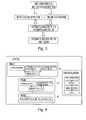
- An exemplary process flow diagram for this exemplary methodis shown in FIG. 3 .
- patient-specific 3D models of the distal femur, proximal tibia, and the patellaare constructed using pulse echo A-mode ultrasound based 3D model reconstruction technology.
- patient-specific kinematic datais obtained for the motions of the femur, tibia, and patella using pulse A-mode ultrasound.
- patient specific vibration datais obtained while the knee joint is taken through a range of motion and loaded in real-world conditions.
- the vibration data and kinematic dataare taken at the same time using the single data acquisition device.
- the datais displayed in real-time on a split screen monitor.
- a recording device and memorymay be utilized to record the data in a time synched manner.
- the patientmay be given an actuator that is operative to note the general time frame within which the patient felt a particular pain or sever pain to allow a correlation between pain felt by the patient and the kinematics and vibration occurring at roughly the same time.
- Patient-specific datais analyzed by a trained neural network in order to provide an automated output as to the existence of an injury, the type of injury, and the severity of the injury.
- This neural networkmay be accessible via the internet or may reside on a physician's local computer.
- patient-specific datamay be analyzed by a physician to make the diagnosis directly without the aid of the neural network.
- a physicianmay diagnose a bodily injury without requiring experimental surgery or requiring exposure of the patient to radiation from still X-rays or fluoroscopy.
- the data taken regarding each patientis continuous through a range of motion, in contrast to X-rays and fluoroscopy which take images at distinct points with significant range of motion gaps.
- data taken in accordance with the exemplary method and devices disclosed hereinalso contrasts data taken by a magnetic resonance imaging machine, not only because the data taken is continuous along the range of motion, but also because the bodily portion evaluated is acting under loaded conditions in a dynamic environment.
- Another object of the present inventionis to provide a method of tracking the motion of a patient's actual bone through space and showing the same on a computer screen.
- Yet another object of the present inventionis to provide method of tracking at lest two bones relative to one another as three dimensional models on a computer screen as the actual bones are taken through a range of motion.
- FIG. 1is a posterior view of a human knee joint in a fixed position
- FIG. 2is a posterior view of a human knee joint in an extended position
- FIG. 3is an exemplary process flow diagram using exemplary methods within the scope of the present invention.
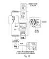
- FIG. 4is a schematic diagram of the modules of an exemplary diagnostic system
- FIG. 5is a screen shot of a software user interface for bone modeling
- FIG. 6is an anterior view of the bones of a human knee joint in an extended position
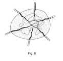
- FIG. 7is a pictorial representation of a human leg having an exemplary brace attached to a distal segment of the femur, and exemplary brace attached to a proximal segment of the tibia, a sensor mounted proximate the patella, and a foot pressure sensing shoe;
- FIG. 8is a is an illustration of a CT slice of the transcutaneous detection of a bone's surface using pulse echo A-mode ultrasound;
- FIG. 9is a schematic of an exemplary inertia-based localizer circuit
- FIG. 10is a schematic of an exemplary brace circuit architecture
- FIG. 11is a circuit schematic of an exemplary high voltage amplifier
- FIG. 12is a circuit layout for the exemplary high voltage amplifier of FIG. 11 ;
- FIG. 13is a block diagram for an exemplary high voltage multiplexer
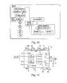
- FIG. 14is a block diagram for an exemplary receiving circuit
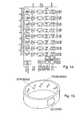
- FIG. 15is a pictorial representation of an exemplary kinematics tracking brace
- FIG. 16is a pictorial representation of an alternative exemplary kinematics tracking brace
- FIG. 17is a pictorial representation of a further alternative kinematics exemplary tracking brace
- FIG. 18is a pictorial representation of a vibration detection module
- FIGS. 19A , 19 B, and 19 Care pictorial representations of exemplary kinematics data, vibration signal, and force data respectively;
- FIG. 20is a graphical representation showing average ACLD medial and lateral condyle contact positions during a deep knee bend activity
- FIGS. 21A , 21 B, and 21 Care a series of views showing contact path tracking in accordance with the exemplary embodiments
- FIG. 22is a is a schematic of the overall classification system flow chart
- FIG. 23is a schematic representation of an exemplary neural network classifier
- FIG. 24is an exemplary process flow for training an exemplary neural network
- FIG. 25is an exemplary process flow for knee deficiency diagnosis using a trained neural network.
- the exemplary embodiments of the present inventionare described and illustrated below to encompass diagnosis of bodily abnormalities and, more particularly, devices and methods for evaluating the physiological condition of bodily tissue to discern whether abnormalities exist and the next of any abnormalities.
- the preferred embodiments discussed beloware exemplary in nature and may be reconfigured without departing from the scope and spirit of the present invention.
- the exemplary embodiments as discussed belowmay include optional steps, methods and features that one of ordinary skill should recognize as not being a requisite to fall within the scope of the present invention.
- the embodiments disclosed hereinare described with respect to diagnosing a knee joint injury. Nevertheless, the embodiments may be utilized to diagnose other joints and bodily tissue injuries, as the knee joint is merely exemplary to facilitate an understanding of the embodiments disclosed.
- a first exemplary diagnostic systemincludes four modules: (1) a pulse echo A-mode ultrasound based 3D model reconstruction (PEAUMR) module for constructing 3D patient specific models of the knee joint bones; (2) a joint kinematics tracking (JKT) module for tracking kinematics of the knee joint using the patient-specific 3D model of the knee joint from the PEAUMR module; (3) a vibration detection (VD) module for capturing sounds emanating from the knee joint while in motion; and (4) an intelligent diagnosis (ID) module for identifying pathological cases of the knee joint using kinematic data and associated vibration data gathered during the joint motion.
- PEAUMRpulse echo A-mode ultrasound based 3D model reconstruction
- JKTjoint kinematics tracking
- VDvibration detection
- IDintelligent diagnosis
- the system described aboveis usable with or without the use of the vibration detection module.
- the databasecould contain mathematical descriptions of healthy or clinically undesirable joint motion.
- the PEAUMR moduleconstructs a 3D model of a subject's (e.g., a patient) bones by transcutaneously acquiring a set of 3D data points that in total are representative of the shape of the bone's surface using a tracked pulse echo A-mode ultrasound probe.
- the probeconsists of a single ultrasound transducer attached to a global localizer.
- the global localizermay be optical, inertial, electromagnetic or ultra wide band radio frequency.
- the probeis battery-powered and connected wirelessly to a computer in order to record the set points and construct a unique or patient-specific bone model using an atlas-based deformable model technique.
- the computerincludes software that interprets data from the tracked pulse echo A-mode ultrasound probe and is operative to construct the 3D models of the patient's bones, which will look very similar to the model shown in FIG. 6 .
- the patient-specific boneis reconstructed using the set of points collected from the bone's surface transcuateously by the tracked ultrasound probe. These points are then used by the atlas-based deformable model software to reconstruct the 3D model of the patient's bone.
- the softwareincludes a plurality of bone models of the femur, tibia, and patella that are classified, for example, based upon ethnicity, gender, skeletal bone to be modeled, and the side of the body the bone is located. Each of these classifications is accounted for by the dropdown menus of the software so that the model initially chosen by the software most closely approximates the bode of the patient.
- the ultrasound transducer probeis repositioned on the exterior of the skin and data points are generated and applied to the model bone (in this case a distal femur), numerically recorded and viewable in a data window, and ultimately utilized by the software to conform the bone model to the patient's actual bone shape.
- model bonein this case a distal femur
- data pointsare generated and applied to the model bone (in this case a distal femur), numerically recorded and viewable in a data window, and ultimately utilized by the software to conform the bone model to the patient's actual bone shape.
- a higher number of data points imposed on the modelwill generally result in a more accurate patient model.
- the JKT moduletracks the kinematics of the knee joint using the patient-specific 3D bone models from the PEAUMR module.
- motion tracking of the patient's knee joint bonesis performed using one or more bone motion tracking braces.
- the bone motion tracking braceincludes pulse echo A-mode ultrasound transducers to transcutaneously localize points on the bones surface.
- the pulse echo A-mode ultrasound transducersmay or may not be identical to the pulse echo A-mode ultrasound transducers used by the PEAUMR module.
- Commercially available transducers for use with the exemplary embodimentsinclude, without limitation, the Olympus immersion unfocused 3.5 MHz transducer.
- the force sensing shoedetects the ground reactive pressures simultaneous with knee joint kinematic data acquisition.
- Each ultrasound transduceris tracked using an accelerometer or a sensor-specific localizer (or any other appropriate inertial sensor).
- the resulting localized bone points generated from the outputs of the ultrasound transducersare used in combination with the patient specific 3D bone models to discern bone movement while the knee joint is taken through a range of motion.
- three braces and a foot force sensing shoeare used to track knee joint kinematics and dynamic forces: (a) a first brace is positioned proximate the distal portion of the femur; (b) a second brace is positioned proximate the distal end of the tibia; and, (c) a third brace is positioned proximate the patella region.
- an exemplary bone motion tacking braceincludes a plurality of pulse echo A-mode ultrasound transducers for transcutaneous detection of the bone's surface and inertia-based localizers to track the motion of the ultrasound transducers, which in turn, track the bones motion.
- Each braceis wirelessly connected to a computer operative to perform computations and visualization in real-time showing movements of the patient-specific 3D bone models paralleling movements of the patient's actual knee joint in a time synchronized manner.
- Each exemplary braceinclude a rigid or semi-rigid body having a plurality (two or more) of complementary metal oxide semiconductor (CMOS) inertia-based sensors attached thereto.
- CMOScomplementary metal oxide semiconductor
- F ris a summation of all the generalized active forces in the system
- F r *is a summation of all the generalized inertia forces in the system.
- the homogenous transformation between the localizer's reference coordinate frame and the world coordinate frameis calculated using the positions of multiple inertia sensors.
- the previous equationsdescribe the dynamic motion and positioning of a point in 3D Euclidean space. Additionally information is needed to describe a 3D body orientation and motion.
- the orientation of the transducercan be described by using a gravity based accelerometer (example: ADXL-330, analog device) by extracting the tilting information from each pair of orthogonal axis.
- the yaw, pitch and rowcan be calculated as shown in the following:
- an accelerometer based localizeris used to track each pulse echo A-mode ultrasound transducer mounted to the brace.
- the localizercomprises a plurality of nodes, with each node comprising a CMOS accelerometer and a temperature sensor for thermal drift comparison. Each node is integrated to minimize noise and distortion.
- the outputs of the accelerometers regarding the X, Y, and Z coordinates and temperature sensorare directed to a multiplexer that multiplexes the signals. Multiplexed outputs are amplified by an amplifier and then directed to an analog-to-digital converter. The digital conversion of the signal can be performed within or outside the CMOS sensors chip. Outputted digital signals are directed to a wireless transmitter by way of a parallel input/serial output device.
- each of the three exemplary design alternatives for the bracehas a similar electronic architecture.
- An exemplary electronic architectureincludes a high voltage amplifier circuit feeding a voltage multiplexer circuit to excite each ultrasound transducer and thereby acting as an analog switch.
- the echo signals from each transducerare multiplexed pursuant to a logic control directing the opening of the switches in the multiplexer circuit at precise intervals.
- An exemplary logic controlis the MSP430 available from Texas Instruments.
- the output from the multiplexer circuitis amplified by an amplifier circuit, signal conditioned using a signal conditioning circuit, and digitized using an analog-to-digital converter. Electric power to the foregoing components is supplied by way of a battery, which also supplies power to a wireless transmitter module.
- the wireless transmitter moduleutilizes the universe asynchronous receiver/transmitter (UART) protocol.
- the moduleincludes a wireless transmitter circuit receiving the output of the first in-first out (FIFO) buffer of the analog-to-digital converter by way of a serial interface. An output from the wireless transmitter circuit is conveyed using a serial link coupled to an antenna. Signals conveyed through the antenna are broadcast for reception by a wireless receiver coupled to a controller computer.
- UARTuniverse asynchronous receiver/transmitter
- an exemplary high voltage circuitis utilized to trigger and generate the excitation energy for the piezoelectric crystal in the transducer.
- Exemplary high voltage circuits for use in this embodimentinclude, without limitation, the pulsar integrated circuit (HV379) available from Supertex.
- an exemplary high voltage multiplexeris utilized to trigger and excite multiple piezoelectric transducers without increasing the number of high voltage circuit mentioned with regard to FIG. 11 .
- Exemplary high voltage multiplexers for use in this embodimentinclude, without limitation, the high voltage multiplexer (HV2221) available from Supertex.
- HV2221available from Supertex.
- the advantage of using a high voltage multiplexeris the ability to use CMOS level control circuitry, thereby making the control logic compatible with virtually any microcontroller or field programmable gate array commercially available.
- an exemplary receiving circuitwhich comprises the multiplexer circuit, the amplifier circuit, the signal conditioning circuit, and the analog-to-digital converter, is utilized to receive the echo signals from each transducer.
- Exemplary receiving circuits for use in this embodimentinclude, without limitation, the AD9271 8-channel ultrasound receiving integrated circuit available from Analog Devices.
- a first exemplary bone tracking braceincludes a plurality of transducers mounted thereto. Each transducer is responsible for determining the location of a point on the surface of the bone for each motion tracking frame. Problems of locating and tracking the bone using ultrasound data are reduced as the motion of the bone relative to the skin is small compared to the gross joint motion.
- the first approachcommonly referred to herein as the ITT (Individual transducer tracking) approach, involves each transducer in the brace having an inertia-based localizer to individually track each transducer.
- the transducersare held together flexible length straps.
- a second approachcommonly referred to herein as the ITML (Inter-transducers Mechanical Links) approach, involves the transducers being connected to each other by movable mechanical links.
- Each mechanical linkincludes length and angle sensors that allow for detection of the movement of the transducers relative to one another and the relative translational motions of the links. Every two links are connected by a pivot pin that allows rotation and translation of the links relative to each other.
- An angle sensoris mounted to at least one link proximate the pivot pin to allow for detection of the angle between the links.
- the ITML approachfeatures less localizers than the individual transducer tracking design.
- a third approachcommonly referred to herein as the RT (Rotating Transducer) approach, involves using a single ultrasound transducer that is mounted to a carriage.
- the carriagetraverses along a track located on the inner circumference of the brace.
- the carriagemay be moved along the tack by a string loop that is wrapped around the drive shaft of a motor.
- the transducerreaches the motor, the rotation direction of the motor is changed and the transducer moves in the opposite direction.
- An inertia-based localizeris mounted to the transducer to track its motion. As the transducer rotates within the inner circumference of the brace, it collects data as to the outer circumferential topography of the bone surface.
- the RT approachincludes the advantage of lower cost than the stationary transducer designs and higher accuracy due to the greater number of localized bone surface points for each tracking step, while maintaining a mechanical flexibility.
- the vibration detection moduleincludes thin film accelerometers that detect the vibration produced by motion of the knee joint.
- Thin film accelerometersare used in lieu of sound sensors, because of better performance and less noise susceptibility.
- the thin film accelerometersmay be the same ones used for the localizer, as well as having the same circuitry for driving the accelerometers.
- the accelerometersare attached to the patients and communicatively connected to the kinematic tracking braces so the outputs from the accelerometers can be amplified, digitized, and sent wirelessly to the controller computer.
- X-ray video fluoroscopy and in-vivo measurements of dynamic knee kinematicsare important for understanding the effects of joint injuries, diseases, and evaluating the outcome of surgical procedures.
- six degrees of freedom (DOF)are determined between the femur and tibia, femur and patella, and tibia and patella that involve the position and orientation of each with respect to the other.
- DOFdegrees of freedom
- the accuracy of this approachis within one degree of rotation and one mm of translation (except for translation parallel to the viewing direction).
- this approachis highly accurate, it constrains the patient to remain within the small working volume of the fluoroscope unit and subjects the patient to ionizing radiation for a prolonged period of time.
- an exemplary systemaccurately measures joint motion during dynamic activities using a portable brace, such as those previously discussed herein.
- a portable bracehaving sensors mounted thereto, X-ray fluoroscopy may be omitted.
- Implementation of joint movement visualizationincludes using the exemplary 3C model reconstruction using pulse-echo A-mode ultrasound system to measure vibrations produced to accurately localize the exact vibration center and causes for its occurrence.
- the interpretation of the vibration and kinematic datais a complicated task involving an in-depth understanding of data acquisition, training data sets and signal analysis, as well as the mechanical system characteristics. Vibrations generated through the implant components, bones, and/or soft tissues interaction result from a forced vibration induced by driving force leading to a dynamic response.
- the driving forcecan be associated with the impact following knee ligament instability, bone properties, and conditions.
- a normal, intact kneewill have a distinct pattern of motion, coupled with distinct vibrational characteristics. Once degeneration or damage occurs to the knee joint, both the kinematic patterns and vibrational characteristics become altered. This altering, for each type of injury or degeneration, leads to distinct changes that can be captured using both kinematic and vibration determination.
- a fourth module of the exemplary diagnostic systemis operative to diagnose ligament, other soft tissue, and bone injuries.
- the diagnostic moduleis a two stage device that includes a first stage involving motion measurement extraction, while a second stage classifies any injury that is detected.
- This first stageincludes acquisition of kinematic feature vectors using multiple physiological measurements taken from the patient while the patient moves the joint in question through a range of motion.
- Exemplary measurementsinclude, without limitation, medical condyle anteroposterior motion (MAP) and lateral condyle anteroposterior (LAP), with the latter pertaining to the anterior-posterior A/P distance of the medial and lateral condyle points relative to the tibia geometric center.
- MAPmedical condyle anteroposterior motion
- LAPlateral condyle anteroposterior
- FIG. 20is an exemplary graphical representation showing average ACLD medial and lateral condyle contact positions during a deep knee bend activity.
- the motion features vectors extracted from the kinematic and vibration analysesare output to a multilayer back propagation neural network for determining the injured ligament.
- an exemplary neural network classifierhas multiple binary outputs. Each output is either a one or zero, with one corresponding to yeas and zero corresponding to no.
- each outputrepresents the response of the neural network to a particular injury type; for example one output will represent the response for anterior cruciate ligament deficiency (ACLD), its state will be one if an ACL injury is detected, and zero otherwise.
- ACLDanterior cruciate ligament deficiency
- the neural networkmay be significantly more sophisticated or less sophisticated, depending upon the underlying model of the joint in question.
- construction of the exemplary neural networkincludes formulating a supervised classifier using a training set of the kinematic and vibration data corresponding to normal and injured knee joist.
- the NNis trained with a set of vectors. Each vector consists of data (kinematics, vibrations and forces) collected from one joint. Fluoroscopy data can be used to calculate the kinematics.
- the NNOnce the NN is trained, it can be used to classify new cases and categorize the injury type using these kinematics, vibration and forces data.
- the types and classifications desired to be accommodated by the neural networknecessarily include training the neural network on these very types and classifications.
- Exemplary types and classifications of injuries to a mammalian knee jointinclude, without limitation, osteoarthritic conditions, soft tissue damage, and abnormal growths.
- the neural networkalso needs to be trained as to indicators of normal knee function. In this manner, once the neural network is trained, it has the capability to differentiate between and output diagnosis data concerning normal and abnormal knee conditions.
- the vibration and kinematics features of a person's knee jointare compiled and fed to the trained neural network.
- the trained neural networkthen diagnoses the condition of the patient's knee joint, identifying and degeneration by type and severity.
- Exemplary embodimentsmay be adapted to collect data outside of a clinical setting.
- an exemplary embodimentmay be worn by a patient for an extended period of time while performing normal activities.
- a patientmay wear vibration sensors and/or a kinematics tracking brace during activities that are not reproducible in the office (for example, weight lifting, racquet ball etc.) that elicit the pain or symptom.
- the patientmay turn the device on immediately prior to the activity and/or the patient may mark the event when it occurs. This enables analysis of the data just a few seconds before the marked time to see what abnormal sounds or joint kinematic were occurring.
- Datamay be stored on a portable hard drive (or any other portable storage device) and then may be downloaded to exemplary systems for analysis.
- the datacan be transmitted and stored in a computer wirelessly. It can also be stored with a miniature memory drive if field data is desired. If the occurrence of the pain was more random, exemplary devices allow continuous gathering of data. In embodiments, the patient may mark the event. Devices capable of continuous monitoring may require a larger data storage capacity.
- embodimentsmay be easily adapted to other joints of a mammalian animal.
- embodimentsmay be adapted for use on hips, ankles, toes, spines, shoulders, elbows, wrists, fingers, and temporomandibular joints.
Landscapes
- Health & Medical Sciences (AREA)
- Life Sciences & Earth Sciences (AREA)
- Engineering & Computer Science (AREA)
- Biomedical Technology (AREA)
- Public Health (AREA)
- Medical Informatics (AREA)
- General Health & Medical Sciences (AREA)
- Physics & Mathematics (AREA)
- Pathology (AREA)
- Biophysics (AREA)
- Molecular Biology (AREA)
- Surgery (AREA)
- Animal Behavior & Ethology (AREA)
- Heart & Thoracic Surgery (AREA)
- Veterinary Medicine (AREA)
- Oral & Maxillofacial Surgery (AREA)
- Dentistry (AREA)
- Physiology (AREA)
- Radiology & Medical Imaging (AREA)
- Nuclear Medicine, Radiotherapy & Molecular Imaging (AREA)
- Rheumatology (AREA)
- Orthopedic Medicine & Surgery (AREA)
- Artificial Intelligence (AREA)
- Computer Vision & Pattern Recognition (AREA)
- Computer Networks & Wireless Communication (AREA)
- Primary Health Care (AREA)
- Epidemiology (AREA)
- Data Mining & Analysis (AREA)
- Signal Processing (AREA)
- Psychiatry (AREA)
- Geometry (AREA)
- Rehabilitation Therapy (AREA)
- General Business, Economics & Management (AREA)
- Databases & Information Systems (AREA)
- Physical Education & Sports Medicine (AREA)
- Business, Economics & Management (AREA)
- Evolutionary Computation (AREA)
- Theoretical Computer Science (AREA)
- Mathematical Physics (AREA)
- General Engineering & Computer Science (AREA)
Abstract
Description
The present invention relates to diagnosis of bodily abnormalities, and more particularly, to devices and methods for evaluating the physiological condition of bodily tissue to discern whether abnormalities exist and the extent of any abnormalities. While the exemplary embodiments disclosed herein are utilized and discussed with respect to a human knee joint, it is to be understood that other joints and bodily tissues may be likewise diagnosed.
In humans, the knee joint is functionally controlled by a mechanical system governed by three unique types of forces: (1) active forces resulting in motion, such as those resulting from muscle flexing or relaxing; (2) constraining forces that constrain motion, such as those resulting from ligaments being in tension; and (3) compressive forces that resist motion, such as those acting upon bones. In addition to the foregoing bodily tissues accounting for these three forces, cartilage and meniscus also produce a dampening effect dissipating the compressive forces propagating to other joints
Knee joint motions are stabilized primarily by four ligaments, which restrict and regulate the relative motion between the femur, tibia, and patella. These ligaments are the anterior cruciate ligament (ACL), the posterior cruciate ligament (PCL), the medial collateral ligament (MCL), and the lateral collateral ligament (LCL), as shown inFIGS. 1 and 2 . An injury to any of these ligaments or other soft-tissue structures can cause detectable changes in knee kinematics and the creation of detectable patterns of vibration representative of the type of knee joint injury and the severity of the injury. These visual and auditory changes are produced by the bones while moving in a distorted kinematic pattern, and they differ significantly from the look and vibration of a properly balanced knee joint moving through a range of motion.
Many research studies have been conducted to assess knee vibration and correlate it with clinical data regarding various joint problems using microphones with or without stethoscope equipment. However, it has been shown that microphones cannot reliably detect joint frequencies, especially those experiencing strong interference from noise, and the signal clearance can substantially influenced by skin friction. It has been hypothesized that the failure associated with the interpretation of sound emissions and possible reasons for occurrence is directly attributable to the complicity of the sound signal, the unknown noise factors, and unknown sound center. It is desirable, therefore, to provide a diagnostic tool that compares patient specific data with kinematic data by providing visual feedback to clinicians.
The present invention, in one embodiment, provides a patient specific 3D model of a patient's joint, including bone and soft tissue. This model is then registered to the patient's actual bone so that as the joint is taken through a range of motion it can be visualized on a computer screen. A physician can then use the computer generated image to make a diagnosis or compare the motion of the actual bone to a database of clinically relevant information on desirable or undesirable joint motion.
The exemplary embodiments of the present invention include a diagnostic system for mammalian bodies to determine the type of injury and extent of injury using kinematic data and/or vibration data. In particular, an exemplary method and embodiment are directed to a knee joint diagnostic system for automatically determining the type of injury and the extent to which ligaments, muscles, bones, meniscus, and cartilage may be affected by an injury through analyzing the kinematics of the knee joint, while also analyzing the pattern and special distribution of the vibration produced knee joint movement. An exemplary process flow diagram for this exemplary method is shown inFIG. 3 .
To evaluate knee kinematics, patient-specific 3D models of the distal femur, proximal tibia, and the patella are constructed using pulse echo A-mode ultrasound based 3D model reconstruction technology. In addition, patient-specific kinematic data is obtained for the motions of the femur, tibia, and patella using pulse A-mode ultrasound. Finally, patient specific vibration data is obtained while the knee joint is taken through a range of motion and loaded in real-world conditions. In exemplary form, the vibration data and kinematic data are taken at the same time using the single data acquisition device. In a further exemplary embodiment, if the data is acquired in a physician's office, the data is displayed in real-time on a split screen monitor. If, however, the data is acquired outside of the doctor's office, a recording device and memory may be utilized to record the data in a time synched manner. In a yet a further exemplary embodiment, the patient may be given an actuator that is operative to note the general time frame within which the patient felt a particular pain or sever pain to allow a correlation between pain felt by the patient and the kinematics and vibration occurring at roughly the same time.
Patient-specific data is analyzed by a trained neural network in order to provide an automated output as to the existence of an injury, the type of injury, and the severity of the injury. This neural network may be accessible via the internet or may reside on a physician's local computer. In addition, or in the alternative, patient-specific data may be analyzed by a physician to make the diagnosis directly without the aid of the neural network.
Using the exemplary methods and devices as disclosed herein, a physician may diagnose a bodily injury without requiring experimental surgery or requiring exposure of the patient to radiation from still X-rays or fluoroscopy. In addition, the data taken regarding each patient is continuous through a range of motion, in contrast to X-rays and fluoroscopy which take images at distinct points with significant range of motion gaps. In addition, data taken in accordance with the exemplary method and devices disclosed herein also contrasts data taken by a magnetic resonance imaging machine, not only because the data taken is continuous along the range of motion, but also because the bodily portion evaluated is acting under loaded conditions in a dynamic environment.
It is an object of the present invention to provide a method of creating a three dimensional model of a patient's bone using tracked pulse-echo A-Mode ultrasound and atlas-based deformable models.
It is another object of the present invention to provide a method of registering a patient's bone with a three dimensional model of the patient's actual bone.
Another object of the present invention is to provide a method of tracking the motion of a patient's actual bone through space and showing the same on a computer screen.
Yet another object of the present invention is to provide method of tracking at lest two bones relative to one another as three dimensional models on a computer screen as the actual bones are taken through a range of motion.
It is also an object of the present invention to provide a method of diagnosis for joint conditions based on a database of kinematic or other information about joint motion.
The exemplary embodiments of the present invention are described and illustrated below to encompass diagnosis of bodily abnormalities and, more particularly, devices and methods for evaluating the physiological condition of bodily tissue to discern whether abnormalities exist and the next of any abnormalities. Of course, it will be apparent to those of ordinary skill in the art that the preferred embodiments discussed below are exemplary in nature and may be reconfigured without departing from the scope and spirit of the present invention. However, for clarity and precision, the exemplary embodiments as discussed below may include optional steps, methods and features that one of ordinary skill should recognize as not being a requisite to fall within the scope of the present invention. In exemplary fashion, the embodiments disclosed herein are described with respect to diagnosing a knee joint injury. Nevertheless, the embodiments may be utilized to diagnose other joints and bodily tissue injuries, as the knee joint is merely exemplary to facilitate an understanding of the embodiments disclosed.
ReferencingFIG. 4 , a first exemplary diagnostic system includes four modules: (1) a pulse echo A-mode ultrasound based 3D model reconstruction (PEAUMR) module for constructing 3D patient specific models of the knee joint bones; (2) a joint kinematics tracking (JKT) module for tracking kinematics of the knee joint using the patient-specific 3D model of the knee joint from the PEAUMR module; (3) a vibration detection (VD) module for capturing sounds emanating from the knee joint while in motion; and (4) an intelligent diagnosis (ID) module for identifying pathological cases of the knee joint using kinematic data and associated vibration data gathered during the joint motion. Each of these four modules is described in further detail in the following sections. The foot sensor interacts in real time with these other modules providing dynamic force data.
It will be understood by those of skill in the art that the system described above is usable with or without the use of the vibration detection module. For example, one may use the present invention by mathematically describing the relative motion of bones in a patient's joint as such motion is tracked in a 3D patient specific bone model and comparing such description with a database of mathematical descriptions of joint motion. The database could contain mathematical descriptions of healthy or clinically undesirable joint motion.
Referring toFIG. 5 , the PEAUMR module constructs a 3D model of a subject's (e.g., a patient) bones by transcutaneously acquiring a set of 3D data points that in total are representative of the shape of the bone's surface using a tracked pulse echo A-mode ultrasound probe. The probe consists of a single ultrasound transducer attached to a global localizer. The global localizer may be optical, inertial, electromagnetic or ultra wide band radio frequency. The probe is battery-powered and connected wirelessly to a computer in order to record the set points and construct a unique or patient-specific bone model using an atlas-based deformable model technique.
The computer includes software that interprets data from the tracked pulse echo A-mode ultrasound probe and is operative to construct the 3D models of the patient's bones, which will look very similar to the model shown inFIG. 6 . The patient-specific bone is reconstructed using the set of points collected from the bone's surface transcuateously by the tracked ultrasound probe. These points are then used by the atlas-based deformable model software to reconstruct the 3D model of the patient's bone.
In exemplary form, the software includes a plurality of bone models of the femur, tibia, and patella that are classified, for example, based upon ethnicity, gender, skeletal bone to be modeled, and the side of the body the bone is located. Each of these classifications is accounted for by the dropdown menus of the software so that the model initially chosen by the software most closely approximates the bode of the patient.
After the software selects the bone model to approximate the bone of the patient, the ultrasound transducer probe is repositioned on the exterior of the skin and data points are generated and applied to the model bone (in this case a distal femur), numerically recorded and viewable in a data window, and ultimately utilized by the software to conform the bone model to the patient's actual bone shape. Obviously, a higher number of data points imposed on the model will generally result in a more accurate patient model. Nevertheless, in view of the model bones already taking into account numerous traits of the patient (ethnicity, gender, bone modeled, and body side of the bone), it is quite possible to construct an accurate patient-specific 3D model with as few as 150 data points, which typically can be taken by repositioning the probe over the bone for 30 seconds for each bone. In this example, it is preferable for the data to be acquired both while the knee is bent and extended to more accurately shape the ends of the bones. This same procedure is repeated for the remaining bones of the joint, in this case the proximal end of the tibia and the patella, in order for the software to combine the bones thereby forming the joint. Ultrasound will not be affected whether the patient has normal or prosthetic implant. The 3D model of the femur can be resected and attached with the implanted CAD model.
Referring toFIG. 7 , the JKT module tracks the kinematics of the knee joint using the patient-specific 3D bone models from the PEAUMR module. In this exemplary embodiment, motion tracking of the patient's knee joint bones is performed using one or more bone motion tracking braces. In exemplary form, the bone motion tracking brace includes pulse echo A-mode ultrasound transducers to transcutaneously localize points on the bones surface. Incidentally, the pulse echo A-mode ultrasound transducers may or may not be identical to the pulse echo A-mode ultrasound transducers used by the PEAUMR module. Commercially available transducers for use with the exemplary embodiments include, without limitation, the Olympus immersion unfocused 3.5 MHz transducer. The force sensing shoe detects the ground reactive pressures simultaneous with knee joint kinematic data acquisition.
Each ultrasound transducer is tracked using an accelerometer or a sensor-specific localizer (or any other appropriate inertial sensor). The resulting localized bone points generated from the outputs of the ultrasound transducers are used in combination with the patient specific 3D bone models to discern bone movement while the knee joint is taken through a range of motion. In exemplary form, three braces and a foot force sensing shoe are used to track knee joint kinematics and dynamic forces: (a) a first brace is positioned proximate the distal portion of the femur; (b) a second brace is positioned proximate the distal end of the tibia; and, (c) a third brace is positioned proximate the patella region.
Referring toFIG. 8 , an exemplary bone motion tacking brace includes a plurality of pulse echo A-mode ultrasound transducers for transcutaneous detection of the bone's surface and inertia-based localizers to track the motion of the ultrasound transducers, which in turn, track the bones motion. Each brace is wirelessly connected to a computer operative to perform computations and visualization in real-time showing movements of the patient-specific 3D bone models paralleling movements of the patient's actual knee joint in a time synchronized manner. Each exemplary brace include a rigid or semi-rigid body having a plurality (two or more) of complementary metal oxide semiconductor (CMOS) inertia-based sensors attached thereto. The position of each sensor and/or transducer is tracked by using the equation of motion: Fr+Fr*=0, where, Fris a summation of all the generalized active forces in the system, and Fr* is a summation of all the generalized inertia forces in the system. The homogenous transformation between the localizer's reference coordinate frame and the world coordinate frame is calculated using the positions of multiple inertia sensors. The following equation calculates the linear movement of the transducer: v(n+1)=v(n)+a(n)dt and s(n+1)=s(n)+v(n)dt−0.5a(n) dt2, where s(n+1) is position at the current state, s(n) is the position from the previous state, v(n+1) is instantaneous velocity of the current state, v(n) is the velocity from previous state, and a(n) is the acceleration from the accelerometer and dt is the sampling time interval. The previous equations describe the dynamic motion and positioning of a point in 3D Euclidean space. Additionally information is needed to describe a 3D body orientation and motion. The orientation of the transducer can be described by using a gravity based accelerometer (example: ADXL-330, analog device) by extracting the tilting information from each pair of orthogonal axis. The acceleration output on x, y, or z due to gravity is equal to the following: Ai=(Voutx−Voff)/S, where Ai is the acceleration at x, y, or z axis, Voutiis the voltage output from the x, y, or z axis, Voff is the offset voltage, and S is the sensitivity of the accelerometer. The yaw, pitch and row can be calculated as shown in the following:
where pitch is ρ, which is x-axis relative to the ground, roll is φ, which is y-axis relative to the ground, and row is θ, which is z-axis relative to the ground. Since the accelerometer is based using gravity, the orientation does not require information from the previous state once the sensor is calibrated. The static calibration requires the resultant sum of accelerations from the 3 axis equal to 1 g. Alternatively, an orientation sensor that gives yaw, pitch, and row information of the body are also commercially available (example: IDG-300, Invensense). The orientation of the transducer can then be resolved by using direction cosine matrix transformation:
where C shorts for cosine and S shorts for sine.
Referring toFIG. 9 , an accelerometer based localizer is used to track each pulse echo A-mode ultrasound transducer mounted to the brace. The localizer comprises a plurality of nodes, with each node comprising a CMOS accelerometer and a temperature sensor for thermal drift comparison. Each node is integrated to minimize noise and distortion. The outputs of the accelerometers regarding the X, Y, and Z coordinates and temperature sensor are directed to a multiplexer that multiplexes the signals. Multiplexed outputs are amplified by an amplifier and then directed to an analog-to-digital converter. The digital conversion of the signal can be performed within or outside the CMOS sensors chip. Outputted digital signals are directed to a wireless transmitter by way of a parallel input/serial output device.
Referring toFIG. 10 , each of the three exemplary design alternatives for the brace has a similar electronic architecture. An exemplary electronic architecture includes a high voltage amplifier circuit feeding a voltage multiplexer circuit to excite each ultrasound transducer and thereby acting as an analog switch. The echo signals from each transducer are multiplexed pursuant to a logic control directing the opening of the switches in the multiplexer circuit at precise intervals. An exemplary logic control is the MSP430 available from Texas Instruments. The output from the multiplexer circuit is amplified by an amplifier circuit, signal conditioned using a signal conditioning circuit, and digitized using an analog-to-digital converter. Electric power to the foregoing components is supplied by way of a battery, which also supplies power to a wireless transmitter module. In exemplary form, the wireless transmitter module utilizes the universe asynchronous receiver/transmitter (UART) protocol. The module includes a wireless transmitter circuit receiving the output of the first in-first out (FIFO) buffer of the analog-to-digital converter by way of a serial interface. An output from the wireless transmitter circuit is conveyed using a serial link coupled to an antenna. Signals conveyed through the antenna are broadcast for reception by a wireless receiver coupled to a controller computer.
Referring toFIGS. 11 and 12 , an exemplary high voltage circuit is utilized to trigger and generate the excitation energy for the piezoelectric crystal in the transducer. Exemplary high voltage circuits for use in this embodiment include, without limitation, the pulsar integrated circuit (HV379) available from Supertex.
ReferencingFIG. 13 , an exemplary high voltage multiplexer is utilized to trigger and excite multiple piezoelectric transducers without increasing the number of high voltage circuit mentioned with regard toFIG. 11 . Exemplary high voltage multiplexers for use in this embodiment include, without limitation, the high voltage multiplexer (HV2221) available from Supertex. The advantage of using a high voltage multiplexer is the ability to use CMOS level control circuitry, thereby making the control logic compatible with virtually any microcontroller or field programmable gate array commercially available.
Referring toFIG. 14 , an exemplary receiving circuit, which comprises the multiplexer circuit, the amplifier circuit, the signal conditioning circuit, and the analog-to-digital converter, is utilized to receive the echo signals from each transducer. Exemplary receiving circuits for use in this embodiment include, without limitation, the AD9271 8-channel ultrasound receiving integrated circuit available from Analog Devices.
Referring toFIG. 15 , a first exemplary bone tracking brace includes a plurality of transducers mounted thereto. Each transducer is responsible for determining the location of a point on the surface of the bone for each motion tracking frame. Problems of locating and tracking the bone using ultrasound data are reduced as the motion of the bone relative to the skin is small compared to the gross joint motion. There are at least three approaches disclosed herein for tracking the motion of the ultrasound transducers themselves. The first approach, commonly referred to herein as the ITT (Individual transducer tracking) approach, involves each transducer in the brace having an inertia-based localizer to individually track each transducer. Using the ITT approach, in exemplary form, the transducers are held together flexible length straps.
ReferencingFIG. 16 , a second approach, commonly referred to herein as the ITML (Inter-transducers Mechanical Links) approach, involves the transducers being connected to each other by movable mechanical links. Each mechanical link includes length and angle sensors that allow for detection of the movement of the transducers relative to one another and the relative translational motions of the links. Every two links are connected by a pivot pin that allows rotation and translation of the links relative to each other. An angle sensor is mounted to at least one link proximate the pivot pin to allow for detection of the angle between the links. The ITML approach features less localizers than the individual transducer tracking design.
Referring toFIG. 17 , a third approach, commonly referred to herein as the RT (Rotating Transducer) approach, involves using a single ultrasound transducer that is mounted to a carriage. The carriage traverses along a track located on the inner circumference of the brace. For example, the carriage may be moved along the tack by a string loop that is wrapped around the drive shaft of a motor. When the transducer reaches the motor, the rotation direction of the motor is changed and the transducer moves in the opposite direction.
An inertia-based localizer is mounted to the transducer to track its motion. As the transducer rotates within the inner circumference of the brace, it collects data as to the outer circumferential topography of the bone surface. By using a single transducer, the RT approach includes the advantage of lower cost than the stationary transducer designs and higher accuracy due to the greater number of localized bone surface points for each tracking step, while maintaining a mechanical flexibility.
Referring toFIG. 18 is the third module of the exemplary diagnostic system, the vibration detection module, includes thin film accelerometers that detect the vibration produced by motion of the knee joint. Thin film accelerometers are used in lieu of sound sensors, because of better performance and less noise susceptibility. In exemplary form, the thin film accelerometers may be the same ones used for the localizer, as well as having the same circuitry for driving the accelerometers. The accelerometers are attached to the patients and communicatively connected to the kinematic tracking braces so the outputs from the accelerometers can be amplified, digitized, and sent wirelessly to the controller computer.
Referring toFIG. 22 , X-ray video fluoroscopy and in-vivo measurements of dynamic knee kinematics are important for understanding the effects of joint injuries, diseases, and evaluating the outcome of surgical procedures. In exemplary form, using the two aforementioned techniques, six degrees of freedom (DOF) are determined between the femur and tibia, femur and patella, and tibia and patella that involve the position and orientation of each with respect to the other. The accuracy of this approach is within one degree of rotation and one mm of translation (except for translation parallel to the viewing direction). Although this approach is highly accurate, it constrains the patient to remain within the small working volume of the fluoroscope unit and subjects the patient to ionizing radiation for a prolonged period of time. For most dynamic activities where the joints are loaded such as running, jumping, or other dynamic activities, fluoroscopy is an unacceptable alternative. To address this deficiency in preexisting approaches, an exemplary system accurately measures joint motion during dynamic activities using a portable brace, such as those previously discussed herein. By using a portable brace having sensors mounted thereto, X-ray fluoroscopy may be omitted.
Implementation of joint movement visualization includes using the exemplary 3C model reconstruction using pulse-echo A-mode ultrasound system to measure vibrations produced to accurately localize the exact vibration center and causes for its occurrence. The interpretation of the vibration and kinematic data is a complicated task involving an in-depth understanding of data acquisition, training data sets and signal analysis, as well as the mechanical system characteristics. Vibrations generated through the implant components, bones, and/or soft tissues interaction result from a forced vibration induced by driving force leading to a dynamic response. The driving force can be associated with the impact following knee ligament instability, bone properties, and conditions. A normal, intact knee will have a distinct pattern of motion, coupled with distinct vibrational characteristics. Once degeneration or damage occurs to the knee joint, both the kinematic patterns and vibrational characteristics become altered. This altering, for each type of injury or degeneration, leads to distinct changes that can be captured using both kinematic and vibration determination.
ReferencingFIG. 25 , a fourth module of the exemplary diagnostic system, the intelligent diagnosis module, (This will be a software module) is operative to diagnose ligament, other soft tissue, and bone injuries. From previous studies, normal and anterior cruciate ligament deficient (ACLD) knee subjects exhibit a similar pattern of posterior femoral translation during progressive knee flexion, but the subjects exhibit different axial rotation patterns of 30 degrees of knee flexion. Accordingly, the diagnostic module is a two stage device that includes a first stage involving motion measurement extraction, while a second stage classifies any injury that is detected.
This first stage includes acquisition of kinematic feature vectors using multiple physiological measurements taken from the patient while the patient moves the joint in question through a range of motion. Exemplary measurements include, without limitation, medical condyle anteroposterior motion (MAP) and lateral condyle anteroposterior (LAP), with the latter pertaining to the anterior-posterior A/P distance of the medial and lateral condyle points relative to the tibia geometric center. Other exemplary measurements include LSI (distance between the lateral femoral condyle and the lateral tibial plateau) and MSI (distance between the medial femoral condyle and the medial tibial plateau) which are S/I (superior/inferior) distance of the lateral and medial condyle points to the tibial plane. Further exemplary measurements include condyle separation, which is the horizontal (x-y plane) distance between the two minimum condyle points to the tibia (SeeFIG. 21 ). Feature vectors also include the femoral position with respect to the tibia which is defined by three Euler angles and three translation components in addition to the vibration signal, and force data (seeFIGS. 19A ,19B, and19C).FIG. 20 is an exemplary graphical representation showing average ACLD medial and lateral condyle contact positions during a deep knee bend activity.
Referring toFIG. 22 , the motion features vectors extracted from the kinematic and vibration analyses are output to a multilayer back propagation neural network for determining the injured ligament.
ReferencingFIG. 23 , an exemplary neural network classifier has multiple binary outputs. Each output is either a one or zero, with one corresponding to yeas and zero corresponding to no. In this exemplary neural network classifier, each output represents the response of the neural network to a particular injury type; for example one output will represent the response for anterior cruciate ligament deficiency (ACLD), its state will be one if an ACL injury is detected, and zero otherwise. Obviously, the neural network may be significantly more sophisticated or less sophisticated, depending upon the underlying model of the joint in question.
Referring toFIG. 24 , construction of the exemplary neural network includes formulating a supervised classifier using a training set of the kinematic and vibration data corresponding to normal and injured knee joist. The NN is trained with a set of vectors. Each vector consists of data (kinematics, vibrations and forces) collected from one joint. Fluoroscopy data can be used to calculate the kinematics. Once the NN is trained, it can be used to classify new cases and categorize the injury type using these kinematics, vibration and forces data. Those skilled in the art will readily understand that the types and classifications desired to be accommodated by the neural network necessarily include training the neural network on these very types and classifications. Exemplary types and classifications of injuries to a mammalian knee joint include, without limitation, osteoarthritic conditions, soft tissue damage, and abnormal growths. Likewise, the neural network also needs to be trained as to indicators of normal knee function. In this manner, once the neural network is trained, it has the capability to differentiate between and output diagnosis data concerning normal and abnormal knee conditions.
ReferencingFIG. 25 , the vibration and kinematics features of a person's knee joint are compiled and fed to the trained neural network. The trained neural network then diagnoses the condition of the patient's knee joint, identifying and degeneration by type and severity.
Exemplary embodiments may be adapted to collect data outside of a clinical setting. For example, an exemplary embodiment may be worn by a patient for an extended period of time while performing normal activities. For example, a patient may wear vibration sensors and/or a kinematics tracking brace during activities that are not reproducible in the office (for example, weight lifting, racquet ball etc.) that elicit the pain or symptom. In some embodiments, the patient may turn the device on immediately prior to the activity and/or the patient may mark the event when it occurs. This enables analysis of the data just a few seconds before the marked time to see what abnormal sounds or joint kinematic were occurring. Data may be stored on a portable hard drive (or any other portable storage device) and then may be downloaded to exemplary systems for analysis. The data can be transmitted and stored in a computer wirelessly. It can also be stored with a miniature memory drive if field data is desired. If the occurrence of the pain was more random, exemplary devices allow continuous gathering of data. In embodiments, the patient may mark the event. Devices capable of continuous monitoring may require a larger data storage capacity.
It is also understood that while the exemplary embodiments have been described herein with respect to a knee joint, those skilled in the art will readily understand that the aforementioned embodiments may be easily adapted to other joints of a mammalian animal. For example, embodiments may be adapted for use on hips, ankles, toes, spines, shoulders, elbows, wrists, fingers, and temporomandibular joints.
Following from the above description and invention summaries, it should be apparent to those of ordinary skill in the art that, while the methods and apparatuses herein described constitute exemplary embodiments of the present invention, the invention contained herein is not limited to this precise embodiment and that changes may be made to such embodiments without departing from the scope of the invention as defined by the claims. Additionally, it is to be understood that the invention is defined by the claims and it is not intended that that any limitations or elements describing the exemplary embodiments set forth herein are to be incorporated into the interpretation of any claim element unless such claim limitation is explicitly stated. Likewise, it is to be understood that it is not necessary to meet any or all of the identified advantages or objects of the invention disclosed herein in order to fall within the scope of any claims. Since the invention is defined by the claims and since inherent and/or unforeseen advantages of the present invention may exist even though they any not have been explicitly discussed herein.
Claims (15)
1. A method of tracking motion of an actual patient bone using creating a patient specific bone model, the method comprising:
acquiring A-mode pulse echo ultrasound signals of the actual patient bone;
utilizing the ultrasound signals to capture a point cloud representative of the actual patient bone;
fitting a deformable 3-D bone model to the captured point cloud to generate a 3-D patient specific bone model;
registering the actual patient bone with the 3-D patient specific bone model;
generating at least one localized bone point from an output of one or more ultrasound transducers positioned proximate the actual patient bone;
tracking the at least one localized bone point by tracking the motion of the one or more ultrasound transducers using one or more inertial sensors; and
tracking motion of the actual patient bone through a range of motion using the at least one tracked localized bone point in combination with the 3-D patient specific bone model.
2. The method ofclaim 1 further comprising:
displaying said motion on the 3-D patient specific bone model.
3. The method ofclaim 1 , further comprising:
selecting from a database a set of empirical mathematical descriptions of the motion of the actual patient bone through the range of motion; and
comparing the motion of the actual patient bone to the selected set of empirical mathematical descriptions.
4. The method ofclaim 1 , further comprising:
collecting vibrational data as the actual patient bone is moved through the range of motion;
selecting from a database a set of empirical vibrational data descriptions of the vibration of the actual patient bone through the range of motion; and
comparing said vibrational data to the selected set of empirical vibration data descriptions.
5. The method ofclaim 1 , further comprising:
selecting one of a plurality of bone models;
measuring the actual patient bone to obtain patient specific measurements; and
applying the patient-specific measurements to the selected one of the plurality of bone models.
6. The method ofclaim 5 , wherein selecting one of the plurality of bone models includes considering at least one demographic characteristic of the patient.
7. The method ofclaim 5 , wherein applying the patient-specific measurements to the bone model includes conforming the model bone to the captured point cloud.
8. The method ofclaim 1 , further comprising:
placing an inertial sensor approximate the actual patient bone; and
gathering vibration data using the inertial sensor while moving the actual patient bone through the range of motion.
9. The method ofclaim 8 , further comprising:
simultaneously, with gathering the vibration data, gathering kinematics data pertaining to the actual patient bone.
10. The method ofclaim 9 , wherein the vibration data is time-synchronized with the kinematics data.
11. The method ofclaim 8 , further comprising:
analyzing the vibration data using a neural network.
12. The method ofclaim 11 , further comprising:
prior to analyzing the vibration data, training the neural network.
13. The method ofclaim 11 , wherein analyzing the vibration data using the neural network further comprises:
diagnosing at least one of a presence and severity of a joint condition.
14. The method ofclaim 13 , wherein the step of analyzing the vibration data using a neural network further comprises:
transmitting the vibration data across a network; and
receiving data pertaining to the joint condition via the network.
15. The method ofclaim 1 wherein the patient is a mammalian animal.
Priority Applications (21)
| Application Number | Priority Date | Filing Date | Title |
|---|---|---|---|
| US12/364,267US8444564B2 (en) | 2009-02-02 | 2009-02-02 | Noninvasive diagnostic system |
| EP10736584.3AEP2391971B1 (en) | 2009-02-02 | 2010-02-02 | Noninvasive diagnostic system |
| PCT/US2010/022939WO2010088696A1 (en) | 2009-02-02 | 2010-02-02 | Noninvasive diagnostic system |
| CA3192190ACA3192190A1 (en) | 2009-02-02 | 2010-02-02 | Noninvasive diagnostic system |
| CA2751422ACA2751422C (en) | 2009-02-02 | 2010-02-02 | Noninvasive diagnostic system |
| CA2977574ACA2977574C (en) | 2009-02-02 | 2010-02-02 | Noninvasive diagnostic system |
| CA3170396ACA3170396C (en) | 2009-02-02 | 2010-02-02 | Noninvasive diagnostic system |
| CA3049975ACA3049975C (en) | 2009-02-02 | 2010-02-02 | Noninvasive diagnostic system |
| EP21199275.5AEP3968220B1 (en) | 2009-02-02 | 2010-02-02 | A method of evaluating a physiological condition of bodily tissue |
| JP2011548412AJP5723788B2 (en) | 2009-02-02 | 2010-02-02 | Non-invasive diagnostic system and device |
| US13/196,701US20120029345A1 (en) | 2009-02-02 | 2011-08-02 | Noninvasive diagnostic system |
| US13/841,632US20130211259A1 (en) | 2009-02-02 | 2013-03-15 | Determination of joint condition based on vibration analysis |
| US13/841,402US9642572B2 (en) | 2009-02-02 | 2013-03-15 | Motion Tracking system with inertial-based sensing units |
| US13/898,092US11342071B2 (en) | 2009-02-02 | 2013-05-20 | Noninvasive diagnostic system |
| JP2014256357AJP6005715B2 (en) | 2009-02-02 | 2014-12-18 | Non-invasive diagnostic method |
| JP2016174550AJP6404286B2 (en) | 2009-02-02 | 2016-09-07 | Non-invasive diagnostic method |
| US15/478,148US11004561B2 (en) | 2009-02-02 | 2017-04-03 | Motion tracking system with inertial-based sensing units |
| US17/181,372US20210193313A1 (en) | 2009-02-02 | 2021-02-22 | Motion Tracking System with Inertial-Based Sensing Units |
| US17/704,376US11776686B2 (en) | 2009-02-02 | 2022-03-25 | Noninvasive diagnostic system |
| US18/238,338US11935648B1 (en) | 2009-02-02 | 2023-08-25 | Noninvasive diagnostic system |
| US18/442,189US20240266043A1 (en) | 2009-02-02 | 2024-02-15 | Noninvasive diagnostic system |
Applications Claiming Priority (1)
| Application Number | Priority Date | Filing Date | Title |
|---|---|---|---|
| US12/364,267US8444564B2 (en) | 2009-02-02 | 2009-02-02 | Noninvasive diagnostic system |
Related Child Applications (3)
| Application Number | Title | Priority Date | Filing Date |
|---|---|---|---|
| PCT/US2010/022939ContinuationWO2010088696A1 (en) | 2009-02-02 | 2010-02-02 | Noninvasive diagnostic system |
| US13/196,701Continuation-In-PartUS20120029345A1 (en) | 2009-02-02 | 2011-08-02 | Noninvasive diagnostic system |
| US13/898,092ContinuationUS11342071B2 (en) | 2009-02-02 | 2013-05-20 | Noninvasive diagnostic system |
Publications (2)
| Publication Number | Publication Date |
|---|---|
| US20100198067A1 US20100198067A1 (en) | 2010-08-05 |
| US8444564B2true US8444564B2 (en) | 2013-05-21 |
Family
ID=42396088
Family Applications (9)
| Application Number | Title | Priority Date | Filing Date |
|---|---|---|---|
| US12/364,267Active2030-11-26US8444564B2 (en) | 2009-02-02 | 2009-02-02 | Noninvasive diagnostic system |
| US13/196,701AbandonedUS20120029345A1 (en) | 2009-02-02 | 2011-08-02 | Noninvasive diagnostic system |
| US13/841,402Active2030-06-06US9642572B2 (en) | 2009-02-02 | 2013-03-15 | Motion Tracking system with inertial-based sensing units |
| US13/898,092Active2032-10-16US11342071B2 (en) | 2009-02-02 | 2013-05-20 | Noninvasive diagnostic system |
| US15/478,148ActiveUS11004561B2 (en) | 2009-02-02 | 2017-04-03 | Motion tracking system with inertial-based sensing units |
| US17/181,372PendingUS20210193313A1 (en) | 2009-02-02 | 2021-02-22 | Motion Tracking System with Inertial-Based Sensing Units |
| US17/704,376ActiveUS11776686B2 (en) | 2009-02-02 | 2022-03-25 | Noninvasive diagnostic system |
| US18/238,338ActiveUS11935648B1 (en) | 2009-02-02 | 2023-08-25 | Noninvasive diagnostic system |
| US18/442,189PendingUS20240266043A1 (en) | 2009-02-02 | 2024-02-15 | Noninvasive diagnostic system |
Family Applications After (8)
| Application Number | Title | Priority Date | Filing Date |
|---|---|---|---|
| US13/196,701AbandonedUS20120029345A1 (en) | 2009-02-02 | 2011-08-02 | Noninvasive diagnostic system |
| US13/841,402Active2030-06-06US9642572B2 (en) | 2009-02-02 | 2013-03-15 | Motion Tracking system with inertial-based sensing units |
| US13/898,092Active2032-10-16US11342071B2 (en) | 2009-02-02 | 2013-05-20 | Noninvasive diagnostic system |
| US15/478,148ActiveUS11004561B2 (en) | 2009-02-02 | 2017-04-03 | Motion tracking system with inertial-based sensing units |
| US17/181,372PendingUS20210193313A1 (en) | 2009-02-02 | 2021-02-22 | Motion Tracking System with Inertial-Based Sensing Units |
| US17/704,376ActiveUS11776686B2 (en) | 2009-02-02 | 2022-03-25 | Noninvasive diagnostic system |
| US18/238,338ActiveUS11935648B1 (en) | 2009-02-02 | 2023-08-25 | Noninvasive diagnostic system |
| US18/442,189PendingUS20240266043A1 (en) | 2009-02-02 | 2024-02-15 | Noninvasive diagnostic system |
Country Status (5)
| Country | Link |
|---|---|
| US (9) | US8444564B2 (en) |
| EP (2) | EP2391971B1 (en) |
| JP (3) | JP5723788B2 (en) |
| CA (5) | CA3170396C (en) |
| WO (1) | WO2010088696A1 (en) |
Cited By (82)
| Publication number | Priority date | Publication date | Assignee | Title |
|---|---|---|---|---|
| US20110092804A1 (en)* | 2006-02-27 | 2011-04-21 | Biomet Manufacturing Corp. | Patient-Specific Pre-Operative Planning |
| US20120123252A1 (en)* | 2010-11-16 | 2012-05-17 | Zebris Medical Gmbh | Imaging apparatus for large area imaging of a body portion |
| US20130185310A1 (en)* | 2012-01-16 | 2013-07-18 | Emovi Inc. | Method and system for human joint treatment plan and personalized surgery planning using 3-d kinematics, fusion imaging and simulation |
| US9060788B2 (en) | 2012-12-11 | 2015-06-23 | Biomet Manufacturing, Llc | Patient-specific acetabular guide for anterior approach |
| WO2015095383A1 (en)* | 2013-12-17 | 2015-06-25 | The Regents Of The University Of California | Diagnostic knee arthrometer for detecting acl structural changes |
| US9066734B2 (en) | 2011-08-31 | 2015-06-30 | Biomet Manufacturing, Llc | Patient-specific sacroiliac guides and associated methods |
| US9084618B2 (en) | 2011-06-13 | 2015-07-21 | Biomet Manufacturing, Llc | Drill guides for confirming alignment of patient-specific alignment guides |
| US9113971B2 (en) | 2006-02-27 | 2015-08-25 | Biomet Manufacturing, Llc | Femoral acetabular impingement guide |
| US9173661B2 (en) | 2006-02-27 | 2015-11-03 | Biomet Manufacturing, Llc | Patient specific alignment guide with cutting surface and laser indicator |
| US9173666B2 (en) | 2011-07-01 | 2015-11-03 | Biomet Manufacturing, Llc | Patient-specific-bone-cutting guidance instruments and methods |
| US9204977B2 (en) | 2012-12-11 | 2015-12-08 | Biomet Manufacturing, Llc | Patient-specific acetabular guide for anterior approach |
| US9237950B2 (en) | 2012-02-02 | 2016-01-19 | Biomet Manufacturing, Llc | Implant with patient-specific porous structure |
| US9241745B2 (en) | 2011-03-07 | 2016-01-26 | Biomet Manufacturing, Llc | Patient-specific femoral version guide |
| US9271744B2 (en) | 2010-09-29 | 2016-03-01 | Biomet Manufacturing, Llc | Patient-specific guide for partial acetabular socket replacement |
| US9289253B2 (en) | 2006-02-27 | 2016-03-22 | Biomet Manufacturing, Llc | Patient-specific shoulder guide |
| US9295497B2 (en) | 2011-08-31 | 2016-03-29 | Biomet Manufacturing, Llc | Patient-specific sacroiliac and pedicle guides |
| US9301812B2 (en) | 2011-10-27 | 2016-04-05 | Biomet Manufacturing, Llc | Methods for patient-specific shoulder arthroplasty |
| US9339278B2 (en) | 2006-02-27 | 2016-05-17 | Biomet Manufacturing, Llc | Patient-specific acetabular guides and associated instruments |
| US9351743B2 (en) | 2011-10-27 | 2016-05-31 | Biomet Manufacturing, Llc | Patient-specific glenoid guides |
| US9386993B2 (en) | 2011-09-29 | 2016-07-12 | Biomet Manufacturing, Llc | Patient-specific femoroacetabular impingement instruments and methods |
| US9393028B2 (en) | 2009-08-13 | 2016-07-19 | Biomet Manufacturing, Llc | Device for the resection of bones, method for producing such a device, endoprosthesis suited for this purpose and method for producing such an endoprosthesis |
| US9408616B2 (en) | 2014-05-12 | 2016-08-09 | Biomet Manufacturing, Llc | Humeral cut guide |
| US9427320B2 (en) | 2011-08-04 | 2016-08-30 | Biomet Manufacturing, Llc | Patient-specific pelvic implants for acetabular reconstruction |
| US9445907B2 (en) | 2011-03-07 | 2016-09-20 | Biomet Manufacturing, Llc | Patient-specific tools and implants |
| US9451973B2 (en) | 2011-10-27 | 2016-09-27 | Biomet Manufacturing, Llc | Patient specific glenoid guide |
| US9456833B2 (en) | 2010-02-26 | 2016-10-04 | Biomet Sports Medicine, Llc | Patient-specific osteotomy devices and methods |
| US9468538B2 (en) | 2009-03-24 | 2016-10-18 | Biomet Manufacturing, Llc | Method and apparatus for aligning and securing an implant relative to a patient |
| US9474539B2 (en) | 2011-04-29 | 2016-10-25 | Biomet Manufacturing, Llc | Patient-specific convertible guides |
| US9480490B2 (en) | 2006-02-27 | 2016-11-01 | Biomet Manufacturing, Llc | Patient-specific guides |
| US9480580B2 (en) | 2006-02-27 | 2016-11-01 | Biomet Manufacturing, Llc | Patient-specific acetabular alignment guides |
| US9498233B2 (en) | 2013-03-13 | 2016-11-22 | Biomet Manufacturing, Llc. | Universal acetabular guide and associated hardware |
| US9517145B2 (en) | 2013-03-15 | 2016-12-13 | Biomet Manufacturing, Llc | Guide alignment system and method |
| US9522010B2 (en) | 2006-02-27 | 2016-12-20 | Biomet Manufacturing, Llc | Patient-specific orthopedic instruments |
| US9539013B2 (en) | 2006-02-27 | 2017-01-10 | Biomet Manufacturing, Llc | Patient-specific elbow guides and associated methods |
| US9554910B2 (en) | 2011-10-27 | 2017-01-31 | Biomet Manufacturing, Llc | Patient-specific glenoid guide and implants |
| US9561040B2 (en) | 2014-06-03 | 2017-02-07 | Biomet Manufacturing, Llc | Patient-specific glenoid depth control |
| US9572590B2 (en) | 2006-10-03 | 2017-02-21 | Biomet Uk Limited | Surgical instrument |
| US9579107B2 (en) | 2013-03-12 | 2017-02-28 | Biomet Manufacturing, Llc | Multi-point fit for patient specific guide |
| US9642572B2 (en) | 2009-02-02 | 2017-05-09 | Joint Vue, LLC | Motion Tracking system with inertial-based sensing units |
| US9662127B2 (en) | 2006-02-27 | 2017-05-30 | Biomet Manufacturing, Llc | Patient-specific acetabular guides and associated instruments |
| US9662216B2 (en) | 2006-02-27 | 2017-05-30 | Biomet Manufacturing, Llc | Patient-specific hip joint devices |
| US9675400B2 (en) | 2011-04-19 | 2017-06-13 | Biomet Manufacturing, Llc | Patient-specific fracture fixation instrumentation and method |
| US9717510B2 (en) | 2011-04-15 | 2017-08-01 | Biomet Manufacturing, Llc | Patient-specific numerically controlled instrument |
| US9743940B2 (en) | 2011-04-29 | 2017-08-29 | Biomet Manufacturing, Llc | Patient-specific partial knee guides and other instruments |
| US9757238B2 (en) | 2011-06-06 | 2017-09-12 | Biomet Manufacturing, Llc | Pre-operative planning and manufacturing method for orthopedic procedure |
| US9795399B2 (en) | 2006-06-09 | 2017-10-24 | Biomet Manufacturing, Llc | Patient-specific knee alignment guide and associated method |
| US9820868B2 (en) | 2015-03-30 | 2017-11-21 | Biomet Manufacturing, Llc | Method and apparatus for a pin apparatus |
| US9826981B2 (en) | 2013-03-13 | 2017-11-28 | Biomet Manufacturing, Llc | Tangential fit of patient-specific guides |
| US9826994B2 (en) | 2014-09-29 | 2017-11-28 | Biomet Manufacturing, Llc | Adjustable glenoid pin insertion guide |
| US9833245B2 (en) | 2014-09-29 | 2017-12-05 | Biomet Sports Medicine, Llc | Tibial tubercule osteotomy |
| US9839438B2 (en) | 2013-03-11 | 2017-12-12 | Biomet Manufacturing, Llc | Patient-specific glenoid guide with a reusable guide holder |
| US9839436B2 (en) | 2014-06-03 | 2017-12-12 | Biomet Manufacturing, Llc | Patient-specific glenoid depth control |
| US9861387B2 (en) | 2006-06-09 | 2018-01-09 | Biomet Manufacturing, Llc | Patient-specific knee alignment guide and associated method |
| US9907659B2 (en) | 2007-04-17 | 2018-03-06 | Biomet Manufacturing, Llc | Method and apparatus for manufacturing an implant |
| US9918740B2 (en) | 2006-02-27 | 2018-03-20 | Biomet Manufacturing, Llc | Backup surgical instrument system and method |
| US9950194B2 (en) | 2014-09-09 | 2018-04-24 | Mevion Medical Systems, Inc. | Patient positioning system |
| US9968376B2 (en) | 2010-11-29 | 2018-05-15 | Biomet Manufacturing, Llc | Patient-specific orthopedic instruments |
| US9993344B2 (en) | 2006-06-09 | 2018-06-12 | Biomet Manufacturing, Llc | Patient-modified implant |
| US10117204B2 (en) | 2008-12-07 | 2018-10-30 | Apdm, Inc | Wireless synchronized apparatus and system |
| US10159498B2 (en) | 2008-04-16 | 2018-12-25 | Biomet Manufacturing, Llc | Method and apparatus for manufacturing an implant |
| US10226262B2 (en) | 2015-06-25 | 2019-03-12 | Biomet Manufacturing, Llc | Patient-specific humeral guide designs |
| US10278711B2 (en) | 2006-02-27 | 2019-05-07 | Biomet Manufacturing, Llc | Patient-specific femoral guide |
| US10282488B2 (en) | 2014-04-25 | 2019-05-07 | Biomet Manufacturing, Llc | HTO guide with optional guided ACL/PCL tunnels |
| US10426429B2 (en) | 2015-10-08 | 2019-10-01 | Decision Sciences Medical Company, LLC | Acoustic orthopedic tracking system and methods |
| US10492798B2 (en) | 2011-07-01 | 2019-12-03 | Biomet Manufacturing, Llc | Backup kit for a patient-specific arthroplasty kit assembly |
| US10512451B2 (en) | 2010-08-02 | 2019-12-24 | Jointvue, Llc | Method and apparatus for three dimensional reconstruction of a joint using ultrasound |
| US10517568B2 (en) | 2011-08-12 | 2019-12-31 | Jointvue, Llc | 3-D ultrasound imaging device and methods |
| US10568647B2 (en) | 2015-06-25 | 2020-02-25 | Biomet Manufacturing, Llc | Patient-specific humeral guide designs |
| US10603179B2 (en) | 2006-02-27 | 2020-03-31 | Biomet Manufacturing, Llc | Patient-specific augments |
| US10722310B2 (en) | 2017-03-13 | 2020-07-28 | Zimmer Biomet CMF and Thoracic, LLC | Virtual surgery planning system and method |
| US10743838B2 (en) | 2015-02-25 | 2020-08-18 | Decision Sciences Medical Company, LLC | Acoustic signal transmission couplants and coupling mediums |
| US10993699B2 (en) | 2011-10-28 | 2021-05-04 | Decision Sciences International Corporation | Spread spectrum coded waveforms in ultrasound diagnostics |
| US11096661B2 (en) | 2013-09-13 | 2021-08-24 | Decision Sciences International Corporation | Coherent spread-spectrum coded waveforms in synthetic aperture image formation |
| US11123040B2 (en) | 2011-10-14 | 2021-09-21 | Jointvue, Llc | Real-time 3-D ultrasound reconstruction of knee and its implications for patient specific implants and 3-D joint injections |
| US11154274B2 (en) | 2019-04-23 | 2021-10-26 | Decision Sciences Medical Company, LLC | Semi-rigid acoustic coupling articles for ultrasound diagnostic and treatment applications |
| US11179165B2 (en) | 2013-10-21 | 2021-11-23 | Biomet Manufacturing, Llc | Ligament guide registration |
| US11419618B2 (en) | 2011-10-27 | 2022-08-23 | Biomet Manufacturing, Llc | Patient-specific glenoid guides |
| US11510035B2 (en) | 2018-11-07 | 2022-11-22 | Kyle Craig | Wearable device for measuring body kinetics |
| US11510737B2 (en) | 2018-06-21 | 2022-11-29 | Mako Surgical Corp. | Patella tracking |
| US11520043B2 (en) | 2020-11-13 | 2022-12-06 | Decision Sciences Medical Company, LLC | Systems and methods for synthetic aperture ultrasound imaging of an object |
| US11583210B2 (en) | 2017-11-07 | 2023-02-21 | Djo, Llc | Brace having integrated remote patient monitoring technology and method of using same |
| US12017389B2 (en) | 2019-03-06 | 2024-06-25 | Decision Sciences Medical Company, LLC | Methods for manufacturing and distributing semi-rigid acoustic coupling articles and packaging for ultrasound imaging |
Families Citing this family (259)
| Publication number | Priority date | Publication date | Assignee | Title |
|---|---|---|---|---|
| RU2142573C1 (en)* | 1997-06-10 | 1999-12-10 | Ковалев Владимир Никитович | Windmill heat and power generating plant |
| US7559931B2 (en) | 2003-06-09 | 2009-07-14 | OrthAlign, Inc. | Surgical orientation system and method |
| EP1720452A1 (en)* | 2004-03-05 | 2006-11-15 | Depuy International Limited | Orthopaedic monitoring system, methods and apparatus |
| PL2023812T3 (en) | 2006-05-19 | 2017-07-31 | The Queen's Medical Center | Motion tracking system for real time adaptive imaging and spectroscopy |
| US20100153081A1 (en)* | 2008-12-11 | 2010-06-17 | Mako Surgical Corp. | Implant planning for multiple implant components using constraints |
| EP2136715B1 (en)* | 2007-04-19 | 2014-06-25 | Mako Surgical Corp. | Implant planning using captured joint motion information |
| JP4378552B1 (en)* | 2008-07-22 | 2009-12-09 | 国立大学法人 東京大学 | Ultrasonic probe support device |
| AU2009273863B2 (en) | 2008-07-24 | 2014-12-18 | OrthAlign, Inc. | Systems and methods for joint replacement |
| AU2009291743B2 (en) | 2008-09-10 | 2015-02-05 | Orthalign, Inc | Hip surgery systems and methods |
| US8826733B2 (en) | 2009-06-30 | 2014-09-09 | Orthosensor Inc | Sensored prosthetic component and method |
| US8421479B2 (en)* | 2009-06-30 | 2013-04-16 | Navisense | Pulsed echo propagation device and method for measuring a parameter |
| US8720270B2 (en) | 2010-06-29 | 2014-05-13 | Ortho Sensor Inc. | Prosthetic component for monitoring joint health |
| US8714009B2 (en) | 2010-06-29 | 2014-05-06 | Orthosensor Inc. | Shielded capacitor sensor system for medical applications and method |
| US8707782B2 (en) | 2009-06-30 | 2014-04-29 | Orthosensor Inc | Prosthetic component for monitoring synovial fluid and method |
| US9462964B2 (en) | 2011-09-23 | 2016-10-11 | Orthosensor Inc | Small form factor muscular-skeletal parameter measurement system |
| US9259179B2 (en) | 2012-02-27 | 2016-02-16 | Orthosensor Inc. | Prosthetic knee joint measurement system including energy harvesting and method therefor |
| US8679186B2 (en) | 2010-06-29 | 2014-03-25 | Ortho Sensor Inc. | Hermetically sealed prosthetic component and method therefor |
| US8701484B2 (en) | 2010-06-29 | 2014-04-22 | Orthosensor Inc. | Small form factor medical sensor structure and method therefor |
| US10869771B2 (en) | 2009-07-24 | 2020-12-22 | OrthAlign, Inc. | Systems and methods for joint replacement |
| US8118815B2 (en) | 2009-07-24 | 2012-02-21 | OrthAlign, Inc. | Systems and methods for joint replacement |
| AU2011341678B2 (en)* | 2010-01-21 | 2014-12-11 | OrthAlign, Inc. | Systems and methods for joint replacement |
| US9161717B2 (en) | 2011-09-23 | 2015-10-20 | Orthosensor Inc. | Orthopedic insert measuring system having a sealed cavity |
| WO2011163367A1 (en)* | 2010-06-22 | 2011-12-29 | Mcgregor Stephen J | Method of monitoring human body movement |
| US20130144135A1 (en)* | 2011-08-02 | 2013-06-06 | Mohamed R. Mahfouz | Method and apparatus for three dimensional reconstruction of a joint using ultrasound |
| US9111305B2 (en)* | 2010-12-17 | 2015-08-18 | Amazon Technologies, Inc. | Personal remote storage for purchased electronic content items |
| US9521966B2 (en)* | 2012-05-17 | 2016-12-20 | Alan N. Schwartz | Localization of the parathyroid |
| US20140128689A1 (en)* | 2011-04-06 | 2014-05-08 | Northeastern University | Joint sensor devices and methods |
| WO2013032933A2 (en) | 2011-08-26 | 2013-03-07 | Kinecticor, Inc. | Methods, systems, and devices for intra-scan motion correction |
| HUP1100471A2 (en)* | 2011-08-30 | 2013-04-29 | Bay Zoltan Alkalmazott Kutatasi Koezhasznu Nonprofit Kft | Method and instrument for detecting equilibrium and intelligent insole suitable for monitoring walking parameters |
| US8911448B2 (en) | 2011-09-23 | 2014-12-16 | Orthosensor, Inc | Device and method for enabling an orthopedic tool for parameter measurement |
| US9414940B2 (en) | 2011-09-23 | 2016-08-16 | Orthosensor Inc. | Sensored head for a measurement tool for the muscular-skeletal system |
| US9839374B2 (en) | 2011-09-23 | 2017-12-12 | Orthosensor Inc. | System and method for vertebral load and location sensing |
| US10335595B2 (en)* | 2011-11-15 | 2019-07-02 | Neurometrix, Inc. | Dynamic control of transcutaneous electrical nerve stimulation therapy using continuous sleep detection |
| US10112040B2 (en) | 2011-11-15 | 2018-10-30 | Neurometrix, Inc. | Transcutaneous electrical nerve stimulation using novel unbalanced biphasic waveform and novel electrode arrangement |
| US9675801B2 (en) | 2011-11-15 | 2017-06-13 | Neurometrix, Inc. | Measuring the “on-skin” time of a transcutaneous electrical nerve stimulator (TENS) device in order to minimize skin irritation due to excessive uninterrupted wearing of the same |
| US10279179B2 (en) | 2013-04-15 | 2019-05-07 | Neurometrix, Inc. | Transcutaneous electrical nerve stimulator with automatic detection of user sleep-wake state |
| PT2780073T (en) | 2011-11-15 | 2017-12-18 | Neurometrix Inc | Apparatus for relieving pain using transcutaneous electrical nerve stimulation |
| US11259744B2 (en) | 2011-11-15 | 2022-03-01 | Neurometrix, Inc. | Transcutaneous electrical nerve stimulator with automatic detection of leg orientation and leg motion for enhanced sleep analysis, including enhanced transcutaneous electrical nerve stimulation (TENS) using the same |
| US11247040B2 (en) | 2011-11-15 | 2022-02-15 | Neurometrix, Inc. | Dynamic control of transcutaneous electrical nerve stimulation therapy using continuous sleep detection |
| NL2008437C2 (en)* | 2012-01-19 | 2013-07-22 | Clinical Graphics B V | Process to generate a computer-accessible medium comprising information on the functioning of a joint. |
| CA3114206A1 (en)* | 2012-02-07 | 2013-08-15 | Jointvue, Llc | Three-dimensional guided injection device and methods |
| US9271675B2 (en) | 2012-02-27 | 2016-03-01 | Orthosensor Inc. | Muscular-skeletal joint stability detection and method therefor |
| US9844335B2 (en) | 2012-02-27 | 2017-12-19 | Orthosensor Inc | Measurement device for the muscular-skeletal system having load distribution plates |
| US9622701B2 (en) | 2012-02-27 | 2017-04-18 | Orthosensor Inc | Muscular-skeletal joint stability detection and method therefor |
| US9549742B2 (en) | 2012-05-18 | 2017-01-24 | OrthAlign, Inc. | Devices and methods for knee arthroplasty |
| US9564682B2 (en)* | 2012-07-11 | 2017-02-07 | Digimarc Corporation | Body-worn phased-array antenna |
| US9649160B2 (en) | 2012-08-14 | 2017-05-16 | OrthAlign, Inc. | Hip replacement navigation system and method |
| US9992729B2 (en)* | 2012-10-22 | 2018-06-05 | The Nielsen Company (Us), Llc | Systems and methods for wirelessly modifying detection characteristics of portable devices |
| US9237885B2 (en) | 2012-11-09 | 2016-01-19 | Orthosensor Inc. | Muscular-skeletal tracking system and method |
| US9526437B2 (en) | 2012-11-21 | 2016-12-27 | i4c Innovations Inc. | Animal health and wellness monitoring using UWB radar |
| US9345609B2 (en) | 2013-01-11 | 2016-05-24 | Elwha Llc | Position sensing active torso support |
| US10314733B2 (en)* | 2012-12-20 | 2019-06-11 | Elwha Llc | Sensor-based control of active wearable system |
| JP6487336B2 (en)* | 2012-12-31 | 2019-03-20 | マコ サージカル コーポレーション | System and method for alignment using an ultrasound probe |
| US9305365B2 (en) | 2013-01-24 | 2016-04-05 | Kineticor, Inc. | Systems, devices, and methods for tracking moving targets |
| US9717461B2 (en) | 2013-01-24 | 2017-08-01 | Kineticor, Inc. | Systems, devices, and methods for tracking and compensating for patient motion during a medical imaging scan |
| US10327708B2 (en) | 2013-01-24 | 2019-06-25 | Kineticor, Inc. | Systems, devices, and methods for tracking and compensating for patient motion during a medical imaging scan |
| CN109008972A (en) | 2013-02-01 | 2018-12-18 | 凯内蒂科尔股份有限公司 | The motion tracking system of real-time adaptive motion compensation in biomedical imaging |
| BR112015018841A2 (en)* | 2013-02-11 | 2017-07-18 | Koninklijke Philips Nv | ultrasound imaging system, method for detecting at least one tissue layer contour of an examination object, and, computer program |
| US20160015319A1 (en) | 2013-03-07 | 2016-01-21 | The Regents Of The University Of California | System for health monitoring on prosthetic and fixation devices |
| US11086970B2 (en)* | 2013-03-13 | 2021-08-10 | Blue Belt Technologies, Inc. | Systems and methods for using generic anatomy models in surgical planning |
| HK1220341A1 (en)* | 2013-03-15 | 2017-05-05 | Jointvue, Llc | Determination of joint condition based on vibration analysis |
| CA2906506C (en)* | 2013-03-15 | 2023-03-28 | Jointvue, Llc | Motion tracking system with inertial-based sensing units |
| US10149617B2 (en) | 2013-03-15 | 2018-12-11 | i4c Innovations Inc. | Multiple sensors for monitoring health and wellness of an animal |
| US11793424B2 (en) | 2013-03-18 | 2023-10-24 | Orthosensor, Inc. | Kinetic assessment and alignment of the muscular-skeletal system and method therefor |
| US9408557B2 (en) | 2013-03-18 | 2016-08-09 | Orthosensor Inc. | System and method to change a contact point of the muscular-skeletal system |
| JP2016515428A (en) | 2013-03-29 | 2016-05-30 | ニューロメトリックス・インコーポレーテッド | Detection of skin electrode peeling using electrode-skin impedance |
| US10940311B2 (en) | 2013-03-29 | 2021-03-09 | Neurometrix, Inc. | Apparatus and method for button-free control of a wearable transcutaneous electrical nerve stimulator using interactive gestures and other means |
| US9439797B2 (en)* | 2013-04-08 | 2016-09-13 | Elwha Llc | Apparatus, system, and method for controlling movement of an orthopedic joint prosthesis in a mammalian subject |
| US10420666B2 (en) | 2013-04-08 | 2019-09-24 | Elwha Llc | Apparatus, system, and method for controlling movement of an orthopedic joint prosthesis in a mammalian subject |
| CN105431196B (en) | 2013-04-15 | 2019-02-01 | Gsk消费者健康有限公司 | Transcutaneous electrical nerve stimulator with automatic detection of user sleep-wake state |
| US9417091B2 (en)* | 2013-05-13 | 2016-08-16 | The Johns Hopkins University | System and method for determining and correcting field sensors errors |
| US11921471B2 (en) | 2013-08-16 | 2024-03-05 | Meta Platforms Technologies, Llc | Systems, articles, and methods for wearable devices having secondary power sources in links of a band for providing secondary power in addition to a primary power source |
| US10042422B2 (en) | 2013-11-12 | 2018-08-07 | Thalmic Labs Inc. | Systems, articles, and methods for capacitive electromyography sensors |
| US20150124566A1 (en) | 2013-10-04 | 2015-05-07 | Thalmic Labs Inc. | Systems, articles and methods for wearable electronic devices employing contact sensors |
| US10188309B2 (en) | 2013-11-27 | 2019-01-29 | North Inc. | Systems, articles, and methods for electromyography sensors |
| WO2015052961A1 (en)* | 2013-10-08 | 2015-04-16 | 株式会社ソニー・コンピュータエンタテインメント | Information processing device |
| FR3012314B1 (en)* | 2013-10-30 | 2018-08-17 | Voice | DEVICE FOR EVALUATING THE MOBILITY OF AN ARTICULATION |
| GB2519987B (en) | 2013-11-04 | 2021-03-03 | Imperial College Innovations Ltd | Biomechanical activity monitoring |
| US10258256B2 (en) | 2014-12-09 | 2019-04-16 | TechMah Medical | Bone reconstruction and orthopedic implants |
| ES2983663T3 (en)* | 2013-12-09 | 2024-10-24 | Lima Usa Inc | Surgical navigation system |
| JP6278577B2 (en)* | 2014-03-12 | 2018-02-14 | 古野電気株式会社 | Ultrasonic diagnostic apparatus and index calculation method |
| WO2015148391A1 (en) | 2014-03-24 | 2015-10-01 | Thomas Michael Ernst | Systems, methods, and devices for removing prospective motion correction from medical imaging scans |
| US10274509B1 (en) | 2014-04-09 | 2019-04-30 | Inertialwave | Inertial motion tracking device |
| US10993639B2 (en)* | 2014-04-25 | 2021-05-04 | Massachusetts Institute Of Technology | Feedback method and wearable device to monitor and modulate knee adduction moment |
| EP3811891B8 (en) | 2014-05-14 | 2025-01-08 | Stryker European Operations Holdings LLC | Navigation system and processor arrangement for tracking the position of a work target |
| US9575560B2 (en) | 2014-06-03 | 2017-02-21 | Google Inc. | Radar-based gesture-recognition through a wearable device |
| US9880632B2 (en) | 2014-06-19 | 2018-01-30 | Thalmic Labs Inc. | Systems, devices, and methods for gesture identification |
| CN107106071B (en)* | 2014-07-10 | 2021-09-07 | 穆罕默德·R·马赫福兹 | Bone Reconstruction and Orthopedic Implants |
| EP3188660A4 (en) | 2014-07-23 | 2018-05-16 | Kineticor, Inc. | Systems, devices, and methods for tracking and compensating for patient motion during a medical imaging scan |
| US9423318B2 (en)* | 2014-07-29 | 2016-08-23 | Honeywell International Inc. | Motion detection devices and systems |
| US9811164B2 (en) | 2014-08-07 | 2017-11-07 | Google Inc. | Radar-based gesture sensing and data transmission |
| US11169988B2 (en) | 2014-08-22 | 2021-11-09 | Google Llc | Radar recognition-aided search |
| US9778749B2 (en) | 2014-08-22 | 2017-10-03 | Google Inc. | Occluded gesture recognition |
| EP3185761B1 (en)* | 2014-08-29 | 2021-03-24 | Bionic Skins LLC | Instrument for determining viscoelastic properties of a body segment of a person |
| WO2016044830A1 (en)* | 2014-09-19 | 2016-03-24 | Think Surgical, Inc. | System and process for ultrasonic determination of long bone orientation |
| US9600080B2 (en) | 2014-10-02 | 2017-03-21 | Google Inc. | Non-line-of-sight radar-based gesture recognition |
| WO2016071773A2 (en)* | 2014-11-04 | 2016-05-12 | Karasahin Deniz | Methods for integrating sensors and effectors in custom three-dimensional orthosis |
| WO2016089442A1 (en)* | 2014-12-06 | 2016-06-09 | Horsemoon Llc | Hand gesture recognition system for controlling electronically controlled devices |
| US9648457B2 (en)* | 2014-12-16 | 2017-05-09 | Intel Corporation | Multi-signal geometric location sensing for access control |
| KR102256676B1 (en)* | 2015-02-06 | 2021-05-26 | 삼성전자주식회사 | Multi-purpose device including mobile terminal and sensing device using radio-wave based sensor |
| US10363149B2 (en) | 2015-02-20 | 2019-07-30 | OrthAlign, Inc. | Hip replacement navigation system and method |
| EP3270781A1 (en)* | 2015-03-19 | 2018-01-24 | Meloq AB | Method and device for anatomical angle measurement |
| CN107613866B (en) | 2015-03-23 | 2021-12-17 | 合意骨科有限公司 | System and method for monitoring orthopedic implants and rehabilitation |
| US11684260B2 (en) | 2015-03-23 | 2023-06-27 | Tracpatch Health, Inc. | System and methods with user interfaces for monitoring physical therapy and rehabilitation |
| US10582891B2 (en) | 2015-03-23 | 2020-03-10 | Consensus Orthopedics, Inc. | System and methods for monitoring physical therapy and rehabilitation of joints |
| US11272879B2 (en) | 2015-03-23 | 2022-03-15 | Consensus Orthopedics, Inc. | Systems and methods using a wearable device for monitoring an orthopedic implant and rehabilitation |
| EP3274912B1 (en)* | 2015-03-26 | 2022-05-11 | Biomet Manufacturing, LLC | System for planning and performing arthroplasty procedures using motion-capture data |
| GB2537644A (en)* | 2015-04-22 | 2016-10-26 | Tintro Ltd | Electronic equipment for the treatment and care of living beings |
| KR102236958B1 (en) | 2015-04-30 | 2021-04-05 | 구글 엘엘씨 | Rf-based micro-motion tracking for gesture tracking and recognition |
| EP3289434A1 (en) | 2015-04-30 | 2018-03-07 | Google LLC | Wide-field radar-based gesture recognition |
| KR102327044B1 (en) | 2015-04-30 | 2021-11-15 | 구글 엘엘씨 | Type-agnostic rf signal representations |
| WO2016186904A1 (en)* | 2015-05-18 | 2016-11-24 | Vayu Technology Corp. | Devices for measuring human gait and related methods of use |
| US10088908B1 (en) | 2015-05-27 | 2018-10-02 | Google Llc | Gesture detection and interactions |
| KR102556074B1 (en) | 2015-05-27 | 2023-07-17 | 조지아 테크 리서치 코오포레이션 | Wearable Technologies for Joint Health Assessment |
| WO2016191813A1 (en)* | 2015-06-01 | 2016-12-08 | Latey Penelope Jane | Foot muscle biofeedback unit |
| US20170225033A1 (en)* | 2015-06-23 | 2017-08-10 | Ipcomm Llc | Method and Apparatus for Analysis of Gait and to Provide Haptic and Visual Corrective Feedback |
| CA2934366A1 (en)* | 2015-06-30 | 2016-12-30 | Ulterra Drilling Technologies, L.P. | Universal joint |
| WO2017007518A1 (en)* | 2015-07-07 | 2017-01-12 | Obma Padraic R | Noninvasive medical monitoring device, system and method |
| US9943247B2 (en) | 2015-07-28 | 2018-04-17 | The University Of Hawai'i | Systems, devices, and methods for detecting false movements for motion correction during a medical imaging scan |
| WO2017055551A1 (en)* | 2015-09-30 | 2017-04-06 | Koninklijke Philips N.V. | Ultrasound apparatus and method for determining a medical condition of a subject |
| US10817065B1 (en) | 2015-10-06 | 2020-10-27 | Google Llc | Gesture recognition using multiple antenna |
| JP6449753B2 (en)* | 2015-11-05 | 2019-01-09 | 国立大学法人佐賀大学 | Joint inflammation detection device |
| CN105468896B (en)* | 2015-11-13 | 2017-06-16 | 上海逸动医学科技有限公司 | Joint motions detecting system and method |
| CN105902274B (en)* | 2016-04-08 | 2017-08-25 | 上海逸动医学科技有限公司 | Knee joint dynamic assessment method and system |
| EP3171286A1 (en)* | 2015-11-17 | 2017-05-24 | Universitat De València, Estudi General | Methods for determining an identifier for use in methods for diagnosing haemophilic arthropathy, methods and apparatus for diagnosing |
| US10716515B2 (en) | 2015-11-23 | 2020-07-21 | Kineticor, Inc. | Systems, devices, and methods for tracking and compensating for patient motion during a medical imaging scan |
| EP3386393B1 (en)* | 2015-12-08 | 2021-05-05 | Kneevoice, Inc. | Assessing joint condition using acoustic sensors |
| US10467534B1 (en)* | 2015-12-09 | 2019-11-05 | Roger Brent | Augmented reality procedural system |
| CA3008479C (en)* | 2015-12-16 | 2022-10-18 | Mohamed R. Mahfouz | Imu calibration |
| US10463279B2 (en) | 2016-02-19 | 2019-11-05 | Trustees Of Dartmouth College | Movement monitoring systems and methods |
| US20190090744A1 (en)* | 2016-02-29 | 2019-03-28 | Mohamed R. Mahfouz | Connected Healthcare Environment |
| WO2017185108A2 (en)* | 2016-04-28 | 2017-11-02 | Medfit Beratungs-Und Beteiligunges.M.B.H | Dynamic ligament balancing system (dlb) |
| WO2017192167A1 (en) | 2016-05-03 | 2017-11-09 | Google Llc | Connecting an electronic component to an interactive textile |
| JP2017202236A (en)* | 2016-05-13 | 2017-11-16 | 花王株式会社 | Gait analysis method and gait analysis device |
| WO2017200949A1 (en) | 2016-05-16 | 2017-11-23 | Google Llc | Interactive fabric |
| US10314514B2 (en)* | 2016-05-29 | 2019-06-11 | Ankon Medical Technologies (Shanghai) Co., Ltd. | System and method for using a capsule device |
| WO2017205983A1 (en)* | 2016-06-02 | 2017-12-07 | Bigmotion Technologies Inc. | Systems and methods for walking speed estimation |
| US10078377B2 (en) | 2016-06-09 | 2018-09-18 | Microsoft Technology Licensing, Llc | Six DOF mixed reality input by fusing inertial handheld controller with hand tracking |
| CN107510466B (en)* | 2016-06-15 | 2022-04-12 | 中慧医学成像有限公司 | Three-dimensional imaging method and system |
| WO2017220173A1 (en)* | 2016-06-24 | 2017-12-28 | Surgical Sensors Bvba | Integrated ligament strain measurement |
| US10849551B2 (en) | 2016-06-24 | 2020-12-01 | Surgical Sensors Bvba | Integrated ligament strain measurement |
| US20170367644A1 (en)* | 2016-06-27 | 2017-12-28 | Claris Healthcare Inc. | Apparatus and Method for Monitoring Rehabilitation from Joint Surgery |
| WO2018013708A1 (en) | 2016-07-13 | 2018-01-18 | Neurometrix, Inc. | Apparatus and method for automated compensation of transcutaneous electrical nerve stimulation for temporal fluctuations such as circadian rhythms |
| WO2018022657A1 (en) | 2016-07-25 | 2018-02-01 | Ctrl-Labs Corporation | System and method for measuring the movements of articulated rigid bodies |
| US10687759B2 (en) | 2018-05-29 | 2020-06-23 | Facebook Technologies, Llc | Shielding techniques for noise reduction in surface electromyography signal measurement and related systems and methods |
| US11179066B2 (en) | 2018-08-13 | 2021-11-23 | Facebook Technologies, Llc | Real-time spike detection and identification |
| US10990174B2 (en) | 2016-07-25 | 2021-04-27 | Facebook Technologies, Llc | Methods and apparatus for predicting musculo-skeletal position information using wearable autonomous sensors |
| EP3487402B1 (en) | 2016-07-25 | 2021-05-05 | Facebook Technologies, LLC | Methods and apparatus for inferring user intent based on neuromuscular signals |
| CN110312471B (en) | 2016-07-25 | 2022-04-29 | 脸谱科技有限责任公司 | Adaptive system for deriving control signals from neuromuscular activity measurements |
| US11216069B2 (en) | 2018-05-08 | 2022-01-04 | Facebook Technologies, Llc | Systems and methods for improved speech recognition using neuromuscular information |
| US11331045B1 (en) | 2018-01-25 | 2022-05-17 | Facebook Technologies, Llc | Systems and methods for mitigating neuromuscular signal artifacts |
| US20180028109A1 (en)* | 2016-07-27 | 2018-02-01 | Andrew TESNOW | System and method for a wearable knee injury prevention |
| WO2018042525A1 (en) | 2016-08-30 | 2018-03-08 | 富士通株式会社 | Information processing device, information processing system, information processing method, and information processing program |
| CN106447713B (en)* | 2016-08-31 | 2019-05-28 | 北京维盛视通科技有限公司 | Method for automatic measurement and device based on cloud manikin |
| JP6738249B2 (en)* | 2016-09-09 | 2020-08-12 | 花王株式会社 | Gait analysis method and gait analysis device |
| JP6738250B2 (en)* | 2016-09-09 | 2020-08-12 | 花王株式会社 | Gait analysis method and gait analysis device |
| JP6757413B2 (en)* | 2016-09-14 | 2020-09-16 | Cyberdyne株式会社 | Knee joint correction tool manufacturing device, knee joint correction tool manufacturing method, and knee joint treatment support device |
| WO2018081795A1 (en) | 2016-10-31 | 2018-05-03 | Zipline Medical, Inc. | Systems and methods for monitoring physical therapy of the knee and other joints |
| WO2018085822A1 (en)* | 2016-11-07 | 2018-05-11 | Synergistic Biosensors, LLC | Systems and methods for monitoring implantable devices for detection of implant failure utilizing wireless in vivo micro sensors |
| FI127689B (en)* | 2016-11-07 | 2018-12-14 | Oulun Yliopisto | Arrangement for knee diagnostics |
| US10579150B2 (en) | 2016-12-05 | 2020-03-03 | Google Llc | Concurrent detection of absolute distance and relative movement for sensing action gestures |
| CN110418659B (en) | 2016-12-23 | 2023-12-19 | 纽诺麦斯股份有限公司 | 'Smart' electrode assembly for transcutaneous electrical nerve stimulation (TENS) |
| CN106725598B (en)* | 2016-12-28 | 2023-09-12 | 苏州科技城医院 | Heart ultrasonic system based on multiple percutaneous ultrasonic transducers and imaging method |
| US10120455B2 (en)* | 2016-12-28 | 2018-11-06 | Industrial Technology Research Institute | Control device and control method |
| WO2018162808A1 (en)* | 2017-01-10 | 2018-09-13 | Braindex S.A.S | Physiological sensor for near-infrared spectroscopy at various depths |
| WO2018165443A1 (en)* | 2017-03-08 | 2018-09-13 | Obma Padraic | Sensors and a method for evaluation of characteristics of human joints and for diagnosis of joint ailments |
| EP3595554A4 (en) | 2017-03-14 | 2021-01-06 | OrthAlign, Inc. | Hip replacement navigation systems and methods |
| CA3056495A1 (en) | 2017-03-14 | 2018-09-20 | OrthAlign, Inc. | Soft tissue measurement & balancing systems and methods |
| GB2560909B (en)* | 2017-03-27 | 2020-12-02 | 270 Vision Ltd | Movement sensor |
| US11446090B2 (en) | 2017-04-07 | 2022-09-20 | Orthosoft Ulc | Non-invasive system and method for tracking bones |
| EP3624736A1 (en)* | 2017-05-18 | 2020-03-25 | Smith & Nephew, Inc. | Systems and methods for determining the position and orientation of an implant for joint replacement surgery |
| US11058877B2 (en) | 2017-05-30 | 2021-07-13 | Neurometrix, Inc. | Apparatus and method for the automated control of transcutaneous electrical nerve stimulation based on current and forecasted weather conditions |
| EP3618715A4 (en) | 2017-06-19 | 2021-02-17 | Mohamed R. Mahfouz | HIP SURGICAL NAVIGATION USING FLUOROSCOPY AND TRACKING SENSORS |
| US11000229B2 (en)* | 2017-08-03 | 2021-05-11 | Orthini, LLC | Systems, methods, and apparatuses for integrating a body joint rehabilitation regimen with a wearable movement capture device operable in conjunction with a cloud based computing environment |
| US10893955B2 (en) | 2017-09-14 | 2021-01-19 | Orthosensor Inc. | Non-symmetrical insert sensing system and method therefor |
| USD865986S1 (en) | 2017-09-21 | 2019-11-05 | Neurometrix, Inc. | Transcutaneous electrical nerve stimulation device strap |
| GB201716123D0 (en)* | 2017-10-03 | 2017-11-15 | Virtualclinic Direct Ltd | Data capture device |
| EP3694408B1 (en)* | 2017-10-10 | 2024-12-04 | Massachusetts Institute Of Technology | Method for neuromechanical and neuroelectromagnetic mitigation of limb pathology |
| WO2019079757A1 (en) | 2017-10-19 | 2019-04-25 | Ctrl-Labs Corporation | Systems and methods for identifying biological structures associated with neuromuscular source signals |
| NZ763381A (en) | 2017-11-30 | 2025-05-30 | Bbi Medical Innovations Llc | Implant evaluation using acoustic emissions |
| CN108030512A (en)* | 2017-12-21 | 2018-05-15 | 福州大学 | A kind of supersonic array measuring method of ankle arthrosis degree of injury |
| CN108245164B (en)* | 2017-12-22 | 2021-03-26 | 北京精密机电控制设备研究所 | Human body gait information acquisition and calculation method for wearable inertial device |
| US11110281B2 (en) | 2018-01-04 | 2021-09-07 | Cardiac Pacemakers, Inc. | Secure transdermal communication with implanted device |
| CN108175381B (en)* | 2018-01-10 | 2024-10-22 | 中山大学附属第一医院 | Knee joint surface injury detection system and application method thereof |
| US10706693B1 (en)* | 2018-01-11 | 2020-07-07 | Facebook Technologies, Llc. | Haptic device for creating vibration-, pressure-, and shear-based haptic cues |
| WO2019147928A1 (en) | 2018-01-25 | 2019-08-01 | Ctrl-Labs Corporation | Handstate reconstruction based on multiple inputs |
| US10504286B2 (en)* | 2018-01-25 | 2019-12-10 | Ctrl-Labs Corporation | Techniques for anonymizing neuromuscular signal data |
| EP3743901A4 (en) | 2018-01-25 | 2021-03-31 | Facebook Technologies, Inc. | REAL-TIME PROCESSING OF HAND REPRESENTATION MODEL ESTIMATES |
| US11567573B2 (en) | 2018-09-20 | 2023-01-31 | Meta Platforms Technologies, Llc | Neuromuscular text entry, writing and drawing in augmented reality systems |
| WO2019147958A1 (en) | 2018-01-25 | 2019-08-01 | Ctrl-Labs Corporation | User-controlled tuning of handstate representation model parameters |
| WO2019147956A1 (en) | 2018-01-25 | 2019-08-01 | Ctrl-Labs Corporation | Visualization of reconstructed handstate information |
| EP3742961A4 (en) | 2018-01-25 | 2021-03-31 | Facebook Technologies, Inc. | CALIBRATION TECHNIQUES FOR HAND REPRESENTATION MODELING USING NEUROMUSCULAR SIGNALS |
| US11493993B2 (en) | 2019-09-04 | 2022-11-08 | Meta Platforms Technologies, Llc | Systems, methods, and interfaces for performing inputs based on neuromuscular control |
| US11961494B1 (en) | 2019-03-29 | 2024-04-16 | Meta Platforms Technologies, Llc | Electromagnetic interference reduction in extended reality environments |
| US11150730B1 (en) | 2019-04-30 | 2021-10-19 | Facebook Technologies, Llc | Devices, systems, and methods for controlling computing devices via neuromuscular signals of users |
| US11481030B2 (en) | 2019-03-29 | 2022-10-25 | Meta Platforms Technologies, Llc | Methods and apparatus for gesture detection and classification |
| US11907423B2 (en) | 2019-11-25 | 2024-02-20 | Meta Platforms Technologies, Llc | Systems and methods for contextualized interactions with an environment |
| US10937414B2 (en) | 2018-05-08 | 2021-03-02 | Facebook Technologies, Llc | Systems and methods for text input using neuromuscular information |
| WO2019152566A1 (en)* | 2018-01-30 | 2019-08-08 | The Regents Of The University Of California | Systems and methods for subject specific kinematic mapping |
| EP3781953A4 (en)* | 2018-04-18 | 2022-05-18 | Georgia Tech Research Corporation | Accelerometer contact microphones and methods thereof |
| US10592001B2 (en) | 2018-05-08 | 2020-03-17 | Facebook Technologies, Llc | Systems and methods for improved speech recognition using neuromuscular information |
| CN112469469B (en) | 2018-05-25 | 2024-11-12 | 元平台技术有限公司 | Method and apparatus for providing submuscular control |
| CN112585600A (en) | 2018-06-14 | 2021-03-30 | 脸谱科技有限责任公司 | User identification and authentication using neuromuscular signatures |
| WO2019245865A1 (en)* | 2018-06-19 | 2019-12-26 | Tornier, Inc. | Mixed reality indication of points at which 3d bone and implant models collide |
| KR20220042478A (en)* | 2018-06-20 | 2022-04-05 | 테크마 메디컬 엘엘씨 | Methods for knee surgery with inertial sensors |
| WO2019243633A1 (en)* | 2018-06-22 | 2019-12-26 | iNDTact GmbH | Sensor arrangement, use of the sensor arrangement, and method for detecting structure-borne noise |
| AU2019295412A1 (en)* | 2018-06-26 | 2021-01-21 | Australian Institute of Robotic Orthopaedics Pty Ltd | Implant fit analysis |
| WO2020018892A1 (en) | 2018-07-19 | 2020-01-23 | Ctrl-Labs Corporation | Methods and apparatus for improved signal robustness for a wearable neuromuscular recording device |
| GB2574074B (en) | 2018-07-27 | 2020-05-20 | Mclaren Applied Tech Ltd | Time synchronisation |
| WO2020047429A1 (en) | 2018-08-31 | 2020-03-05 | Ctrl-Labs Corporation | Camera-guided interpretation of neuromuscular signals |
| US10921764B2 (en) | 2018-09-26 | 2021-02-16 | Facebook Technologies, Llc | Neuromuscular control of physical objects in an environment |
| WO2020072915A1 (en) | 2018-10-05 | 2020-04-09 | Ctrl-Labs Corporation | Use of neuromuscular signals to provide enhanced interactions with physical objects in an augmented reality environment |
| JP7132816B2 (en)* | 2018-10-10 | 2022-09-07 | 大和ハウス工業株式会社 | Joint condition determination system |
| SG10201810156PA (en)* | 2018-11-14 | 2020-06-29 | Prec Medical Pte Ltd | Method and device for measuring anatomical movement of a joint |
| CN109859592B (en)* | 2018-11-14 | 2020-12-08 | 华中科技大学 | A soft tissue injury simulation test device |
| EP3886693A4 (en) | 2018-11-27 | 2022-06-08 | Facebook Technologies, LLC. | Methods and apparatus for autocalibration of a wearable electrode sensor system |
| US11883661B2 (en) | 2018-12-07 | 2024-01-30 | Neurometrix, Inc. | Intelligent determination of therapeutic stimulation intensity for transcutaneous electrical nerve stimulation |
| KR102550854B1 (en)* | 2018-12-13 | 2023-07-04 | 삼성전자주식회사 | Method and device for assisting walking |
| CN111374674B (en)* | 2018-12-29 | 2023-02-10 | 西安思博探声生物科技有限公司 | Knee joint movement information processing equipment |
| CN109801278B (en)* | 2019-01-21 | 2023-05-30 | 燕山大学 | Surface damage classification method for high-speed sliding electric contact movement pair |
| US10905383B2 (en) | 2019-02-28 | 2021-02-02 | Facebook Technologies, Llc | Methods and apparatus for unsupervised one-shot machine learning for classification of human gestures and estimation of applied forces |
| WO2020187753A1 (en)* | 2019-03-20 | 2020-09-24 | Optima Molliter Srl | Orthosis brace with monitoring system |
| US11537702B2 (en)* | 2019-05-13 | 2022-12-27 | Cardiac Pacemakers, Inc. | Implanted medical device authentication based on comparison of internal IMU signal to external IMU signal |
| US11911213B2 (en) | 2019-06-03 | 2024-02-27 | General Electric Company | Techniques for determining ultrasound probe motion |
| US11497452B2 (en)* | 2019-06-20 | 2022-11-15 | The Hong Kong Polytechnic University | Predictive knee joint loading system |
| KR102251925B1 (en)* | 2019-07-18 | 2021-05-13 | 경상국립대학교 산학협력단 | Apparatus and application for predicting musculoskeletal disorders |
| US11877870B2 (en) | 2019-08-05 | 2024-01-23 | Consultation Semperform Inc | Systems, methods and apparatus for prevention of injury |
| US11812978B2 (en) | 2019-10-15 | 2023-11-14 | Orthosensor Inc. | Knee balancing system using patient specific instruments |
| GB2588238B (en)* | 2019-10-18 | 2023-11-22 | Mclaren Applied Ltd | Sensor determination |
| GB2588236B (en) | 2019-10-18 | 2024-03-20 | Mclaren Applied Ltd | Gyroscope bias estimation |
| GB2588237B (en)* | 2019-10-18 | 2023-12-27 | Mclaren Applied Ltd | Joint axis direction estimation |
| US10842415B1 (en)* | 2019-10-25 | 2020-11-24 | Plethy, Inc. | Devices, systems, and methods for monitoring and assessing gait, stability, and/or balance of a user |
| JP7037159B2 (en)* | 2019-11-08 | 2022-03-16 | 国立大学法人大阪大学 | Systems, programs, and methods for measuring a subject's jaw movements |
| US12089953B1 (en) | 2019-12-04 | 2024-09-17 | Meta Platforms Technologies, Llc | Systems and methods for utilizing intrinsic current noise to measure interface impedances |
| CN110772262B (en)* | 2019-12-05 | 2020-12-29 | 广东电网有限责任公司 | Comfort evaluation method for human body tower-climbing posture |
| WO2021119001A1 (en)* | 2019-12-09 | 2021-06-17 | OrthAlign, Inc. | Cup alignment systems and methods |
| GB202000438D0 (en)* | 2020-01-13 | 2020-02-26 | Surepulse Medical Ltd | Improved ppg measurement |
| US10863928B1 (en) | 2020-01-28 | 2020-12-15 | Consensus Orthopedics, Inc. | System and methods for monitoring the spine, balance, gait, or posture of a patient |
| JP7741497B2 (en)* | 2020-04-20 | 2025-09-18 | フォームス ラブス リミティッド | Computer program for surgical system |
| US11832934B1 (en) | 2020-05-04 | 2023-12-05 | Qingbin Zheng | Joint monitoring |
| US12004967B2 (en) | 2020-06-02 | 2024-06-11 | Howmedica Osteonics Corp. | Systems and methods for planning placement of an acetabular implant for a patient based on pelvic tilt |
| US12186223B2 (en) | 2020-06-09 | 2025-01-07 | Aifree Interactive Technology Co., Ltd. | Wearable interface for intelligent health promotion service system |
| US20230270376A1 (en)* | 2020-07-07 | 2023-08-31 | The General Hospital Corporation | Evaluating the stability of a joint in the foot and ankle complex via weight-bearing medical imaging |
| JP7587813B2 (en)* | 2020-09-18 | 2024-11-21 | iMU株式会社 | Detection Equipment |
| US10952775B1 (en)* | 2020-12-14 | 2021-03-23 | Prichard Medical, LLC | Surgical instrument with orientation sensor having a user identified heading |
| EP4016536A1 (en)* | 2020-12-16 | 2022-06-22 | Polar Electro Oy | Biomechanical modelling of motion measurements |
| CN113143256B (en)* | 2021-01-28 | 2023-09-26 | 上海电气集团股份有限公司 | Gait feature extraction method, lower limb evaluation and control method, device and medium |
| CN112754516B (en)* | 2021-02-07 | 2022-04-08 | 河南省肿瘤医院 | Intelligent bowel sound positioning and collecting device |
| JP2024512398A (en)* | 2021-03-07 | 2024-03-19 | リキッド ワイヤ エルエルシー | Apparatus, system, and method for monitoring and characterizing user behavior with flexible circuits |
| US11868531B1 (en) | 2021-04-08 | 2024-01-09 | Meta Platforms Technologies, Llc | Wearable device providing for thumb-to-finger-based input gestures detected based on neuromuscular signals, and systems and methods of use thereof |
| US12350108B2 (en)* | 2021-06-16 | 2025-07-08 | Kinisi Inc. | Wearable imaging system for measuring bone displacement |
| IT202100017267A1 (en)* | 2021-06-30 | 2022-12-30 | Scuola Superiore Santanna | POSITIONING DEVICE FOR ULTRASONIC PROBE |
| JP2024532309A (en)* | 2021-08-23 | 2024-09-05 | オルソセンサー・インコーポレイテッド | Devices, systems and methods for determining tibial coronal alignment - Patents.com |
| EP4395710A4 (en)* | 2021-08-30 | 2025-05-07 | Siemens Industry Software Ltd. | Method and system for determining a connection in a virtual kinematic device |
| USD1053350S1 (en) | 2021-08-30 | 2024-12-03 | Canary Medical Switzerland Ag | Handheld device |
| DE102021124873A1 (en)* | 2021-09-27 | 2023-03-30 | Aesculap Ag | Medical technology system and method |
| WO2023148427A1 (en)* | 2022-02-03 | 2023-08-10 | Aikoa Technologies Oy | Method for training computing arrangement to provide prognosis of progression of tissue condition |
| AU2023281734A1 (en)* | 2022-06-02 | 2024-12-05 | New York Society For The Relief Of The Ruptured And Crippled, Maintaining The Hospital For Special Surgery | Method for mechanical phenotyping of knees |
| US11602443B1 (en)* | 2022-06-07 | 2023-03-14 | Little Engine, LLC | Knee evaluation and arthroplasty method |
Citations (26)
| Publication number | Priority date | Publication date | Assignee | Title |
|---|---|---|---|---|
| US3881164A (en)* | 1973-09-13 | 1975-04-29 | Commw Of Australia | Cross array ultrasonic transducer |
| US4476873A (en) | 1982-09-03 | 1984-10-16 | Medtronic, Inc. | Ultrasound scanning system for skeletal imaging |
| US5394875A (en) | 1993-10-21 | 1995-03-07 | Lewis; Judith T. | Automatic ultrasonic localization of targets implanted in a portion of the anatomy |
| US5413116A (en)* | 1993-06-24 | 1995-05-09 | Bioresearch | Method and apparatus for diagnosing joints |
| US5447154A (en)* | 1992-07-31 | 1995-09-05 | Universite Joseph Fourier | Method for determining the position of an organ |
| US5488952A (en) | 1982-02-24 | 1996-02-06 | Schoolman Scientific Corp. | Stereoscopically display three dimensional ultrasound imaging |
| US5771310A (en) | 1996-12-30 | 1998-06-23 | Shriners Hospitals For Children | Method and apparatus for recording three-dimensional topographies |
| US5806521A (en) | 1996-03-26 | 1998-09-15 | Sandia Corporation | Composite ultrasound imaging apparatus and method |
| US6106464A (en) | 1999-02-22 | 2000-08-22 | Vanderbilt University | Apparatus and method for bone surface-based registration of physical space with tomographic images and for guiding an instrument relative to anatomical sites in the image |
| US6120453A (en) | 1997-11-17 | 2000-09-19 | Sharp; William A. | Three-dimensional ultrasound system based on the coordination of multiple ultrasonic transducers |
| WO2000063719A1 (en) | 1999-04-20 | 2000-10-26 | Synthes Ag Chur | Device for the percutaneous obtainment of 3d-coordinates on the surface of a human or animal organ |
| US6190320B1 (en) | 1998-09-29 | 2001-02-20 | U.S. Philips Corporation | Method for the processing of medical ultrasound images of bony structures, and method and device for computer-assisted surgery |
| US6280387B1 (en) | 1998-05-06 | 2001-08-28 | Siemens Medical Systems, Inc. | Three-dimensional tissue/flow ultrasound imaging system |
| US6537233B1 (en) | 2000-11-06 | 2003-03-25 | University Technologies International Inc. | Auditory display of knee joint vibration signals |
| US6569098B2 (en) | 2001-01-31 | 2003-05-27 | University Technologies International Inc. | Non-invasive diagnostic method and apparatus for musculoskeletal systems |
| US20040152970A1 (en) | 2003-01-30 | 2004-08-05 | Mark Hunter | Six degree of freedom alignment display for medical procedures |
| US20050111718A1 (en) | 2003-11-26 | 2005-05-26 | University Of Chicago | Automated method and system for the evaluation of disease and registration accuracy in the subtraction of temporally sequential medical images |
| US20070249967A1 (en)* | 2006-03-21 | 2007-10-25 | Perception Raisonnement Action En Medecine | Computer-aided osteoplasty surgery system |
| US20070282200A1 (en) | 1992-10-14 | 2007-12-06 | Johnson Steven A | Apparatus and method for imaging objects with wavefields |
| US20070287900A1 (en)* | 2006-04-13 | 2007-12-13 | Alan Breen | Devices, Systems and Methods for Measuring and Evaluating the Motion and Function of Joint Structures and Associated Muscles, Determining Suitability for Orthopedic Intervention, and Evaluating Efficacy of Orthopedic Intervention |
| US20080025463A1 (en) | 2000-08-29 | 2008-01-31 | Imaging Therapeutics, Inc. | Methods and Devices for Quantitative Analysis of X-Ray Images |
| US20080094396A1 (en) | 2004-09-09 | 2008-04-24 | Koninklijke Philips Electronics, N.V. | System For The Three-Dimensional Imaging Of A Moving Joint |
| US20080114270A1 (en) | 2006-09-29 | 2008-05-15 | Disilvestro Mark R | Apparatus and method for monitoring the position of an orthopaedic prosthesis |
| US7454242B2 (en) | 2003-09-17 | 2008-11-18 | Elise Fear | Tissue sensing adaptive radar imaging for breast tumor detection |
| US20090015832A1 (en) | 2007-06-01 | 2009-01-15 | Milica Popovic | Microwave scanning system and miniaturized microwave antenna |
| WO2010088696A1 (en) | 2009-02-02 | 2010-08-05 | Mohamed Rashwan Mahfouz | Noninvasive diagnostic system |
Family Cites Families (66)
| Publication number | Priority date | Publication date | Assignee | Title |
|---|---|---|---|---|
| US4016750B1 (en)* | 1975-11-06 | 1994-04-05 | Stanford Research Inst | Ultrasonic imaging method and apparatus |
| US5826578A (en)* | 1994-05-26 | 1998-10-27 | Curchod; Donald B. | Motion measurement apparatus |
| US5919149A (en)* | 1996-03-19 | 1999-07-06 | Allum; John H. | Method and apparatus for angular position and velocity based determination of body sway for the diagnosis and rehabilitation of balance and gait disorders |
| US6213958B1 (en) | 1996-08-29 | 2001-04-10 | Alan A. Winder | Method and apparatus for the acoustic emission monitoring detection, localization, and classification of metabolic bone disease |
| US7468075B2 (en)* | 2001-05-25 | 2008-12-23 | Conformis, Inc. | Methods and compositions for articular repair |
| US6205411B1 (en)* | 1997-02-21 | 2001-03-20 | Carnegie Mellon University | Computer-assisted surgery planner and intra-operative guidance system |
| JPH10243937A (en)* | 1997-03-07 | 1998-09-14 | Mitsubishi Heavy Ind Ltd | Joint labile measurement device |
| US6231585B1 (en) | 1997-11-20 | 2001-05-15 | Medivas, Llc | Device for stabilizing a treatment site and method of use |
| DE69934215T2 (en)* | 1998-03-20 | 2007-10-25 | Barbara Ann Karmanos Cancer Institute, Detroit | Multi-dimensional identification and characterization of pathological tissues |
| US7239908B1 (en)* | 1998-09-14 | 2007-07-03 | The Board Of Trustees Of The Leland Stanford Junior University | Assessing the condition of a joint and devising treatment |
| JP2000251078A (en)* | 1998-12-22 | 2000-09-14 | Atr Media Integration & Communications Res Lab | Method and device for estimating three-dimensional posture of person, and method and device for estimating position of elbow of person |
| WO2001032114A1 (en) | 1999-11-02 | 2001-05-10 | Wizcare Ltd. | Skin-gripper |
| MXPA02007262A (en)* | 2000-01-29 | 2002-12-09 | Paul E Thomson | Detection and quantification of joint and tissue inflammation. |
| DE60136474D1 (en) | 2000-09-14 | 2008-12-18 | Univ R | ASSESSMENT OF THE CONDITION OF A JOINT AND LOSS OF CARTEL TISSUE |
| US6561991B2 (en)* | 2000-12-19 | 2003-05-13 | The Research Foundation Of The State University Of New York (Suny) | Non-invasive method and system of quantifying human postural stability |
| WO2003079882A2 (en) | 2002-03-25 | 2003-10-02 | Ramot At Tel Aviv University Ltd. | Method and system for determining a risk of ulcer onset |
| US7117026B2 (en)* | 2002-06-12 | 2006-10-03 | Koninklijke Philips Electronics N.V. | Physiological model based non-rigid image registration |
| US7981057B2 (en)* | 2002-10-11 | 2011-07-19 | Northrop Grumman Guidance And Electronics Company, Inc. | Joint motion sensing to make a determination of a positional change of an individual |
| JP2006517433A (en)* | 2003-01-07 | 2006-07-27 | イメージング セラピューティクス,インコーポレーテッド | How to predict musculoskeletal diseases |
| JP2004264060A (en)* | 2003-02-14 | 2004-09-24 | Akebono Brake Ind Co Ltd | Error correction method in attitude detector, and action measuring instrument using the same |
| JP3932360B2 (en)* | 2003-03-04 | 2007-06-20 | 独立行政法人産業技術総合研究所 | Landmark extraction apparatus and landmark extraction method |
| US20050043660A1 (en)* | 2003-03-31 | 2005-02-24 | Izex Technologies, Inc. | Orthoses |
| EP1643905A2 (en)* | 2003-07-10 | 2006-04-12 | Neurocom International, Inc | Apparatus and method for characterizing contributions of forces associated with a body part of a subject |
| US20050093859A1 (en) | 2003-11-04 | 2005-05-05 | Siemens Medical Solutions Usa, Inc. | Viewing direction dependent acquisition or processing for 3D ultrasound imaging |
| US20080269596A1 (en)* | 2004-03-10 | 2008-10-30 | Ian Revie | Orthpaedic Monitoring Systems, Methods, Implants and Instruments |
| JP4455118B2 (en)* | 2004-03-30 | 2010-04-21 | 独立行政法人科学技術振興機構 | Delivery diagnosis support program, recording medium storing the program, and delivery diagnosis support method and apparatus. |
| US7678052B2 (en) | 2004-04-13 | 2010-03-16 | General Electric Company | Method and apparatus for detecting anatomic structures |
| US7483732B2 (en) | 2004-04-15 | 2009-01-27 | Boston Scientific Scimed, Inc. | Magnetic resonance imaging of a medical device and proximate body tissue |
| JP4411384B2 (en)* | 2004-04-15 | 2010-02-10 | 独立行政法人放射線医学総合研究所 | Diagnostic system |
| DE102004026525A1 (en)* | 2004-05-25 | 2005-12-22 | Aesculap Ag & Co. Kg | Method and device for the non-invasive determination of prominent structures of the human or animal body |
| US20060052727A1 (en) | 2004-09-09 | 2006-03-09 | Laurence Palestrant | Activity monitoring device and weight management method utilizing same |
| US20060161052A1 (en)* | 2004-12-08 | 2006-07-20 | Perception Raisonnement Action En Medecine | Computer assisted orthopaedic surgery system for ligament graft reconstruction |
| JPWO2006085387A1 (en)* | 2005-02-08 | 2008-06-26 | 高樹 長宗 | Non-invasive moving body analysis system and method of use thereof |
| GB0504172D0 (en)* | 2005-03-01 | 2005-04-06 | King S College London | Surgical planning |
| JP4304341B2 (en)* | 2005-03-17 | 2009-07-29 | 国立大学法人 新潟大学 | Three-dimensional shape measurement device and socket design device for prosthetic limbs based on the measurement data |
| US20100100011A1 (en) | 2008-10-22 | 2010-04-22 | Martin Roche | System and Method for Orthopedic Alignment and Measurement |
| JP4999012B2 (en) | 2005-06-06 | 2012-08-15 | インチュイティブ サージカル,インコーポレイテッド | Laparoscopic ultrasonic robotic surgical system |
| US8092398B2 (en)* | 2005-08-09 | 2012-01-10 | Massachusetts Eye & Ear Infirmary | Multi-axis tilt estimation and fall remediation |
| WO2007017861A2 (en) | 2005-08-09 | 2007-02-15 | Gil Zwirn | High resolution radio frequency medical imaging and therapy system |
| GB2435614A (en) | 2006-03-01 | 2007-09-05 | Samuel George | Transducer holder for maintaining signal-receiving contact with a patient's body |
| US8165659B2 (en)* | 2006-03-22 | 2012-04-24 | Garrett Sheffer | Modeling method and apparatus for use in surgical navigation |
| US20080009722A1 (en) | 2006-05-11 | 2008-01-10 | Constantine Simopoulos | Multi-planar reconstruction for ultrasound volume data |
| US7578799B2 (en)* | 2006-06-30 | 2009-08-25 | Ossur Hf | Intelligent orthosis |
| WO2008074151A1 (en)* | 2006-12-20 | 2008-06-26 | Mcmaster University | System and method of assessing the condition of a joint |
| US20080194997A1 (en)* | 2007-02-08 | 2008-08-14 | Rehabilitation Institute Of Chicago | System and method for diagnosing and treating patellar maltracking and malalignment |
| US20080221487A1 (en)* | 2007-03-07 | 2008-09-11 | Motek Bv | Method for real time interactive visualization of muscle forces and joint torques in the human body |
| EP1970005B1 (en)* | 2007-03-15 | 2012-10-03 | Xsens Holding B.V. | A system and a method for motion tracking using a calibration unit |
| US7920731B2 (en) | 2007-03-27 | 2011-04-05 | Siemens Medical Solutions Usa, Inc. | Bleeding detection using a blanket ultrasound device |
| CN101742972B (en)* | 2007-05-14 | 2015-01-07 | 金斯顿女王大学 | Patient-specific surgical guidance tool and method of use |
| US8771188B2 (en)* | 2007-06-20 | 2014-07-08 | Perception Raisonnement Action En Medecine | Ultrasonic bone motion tracking system |
| JP5061281B2 (en)* | 2007-08-20 | 2012-10-31 | 国立大学法人広島大学 | Knee joint rotation angle measuring device |
| WO2009042644A2 (en) | 2007-09-25 | 2009-04-02 | Perception Raisonnement Action En Medecine | Methods and apparatus for assisting cartilage diagnostic and therapeutic procedures |
| JP5416900B2 (en) | 2007-11-22 | 2014-02-12 | 株式会社東芝 | Ultrasonic diagnostic apparatus and puncture support control program |
| CA2715898C (en)* | 2008-03-25 | 2018-05-08 | Orthosoft Inc. | Tracking system and method |
| US8377073B2 (en)* | 2008-04-21 | 2013-02-19 | Ray Wasielewski | Method of designing orthopedic implants using in vivo data |
| EP2212994B1 (en) | 2008-05-19 | 2015-10-28 | Hittite Microwave Corporation | Multiple input variable gain amplifier |
| US20100125229A1 (en)* | 2008-07-11 | 2010-05-20 | University Of Delaware | Controllable Joint Brace |
| JP5377166B2 (en) | 2009-09-01 | 2013-12-25 | 古野電気株式会社 | Ultrasound bone analyzer |
| US20110125016A1 (en) | 2009-11-25 | 2011-05-26 | Siemens Medical Solutions Usa, Inc. | Fetal rendering in medical diagnostic ultrasound |
| US8979758B2 (en) | 2010-06-29 | 2015-03-17 | Orthosensor Inc | Sensing module for orthopedic load sensing insert device |
| CA3129784C (en) | 2010-08-02 | 2023-08-22 | Jointvue, Llc | Method and apparatus for three dimensional reconstruction of a joint using ultrasound |
| US20130144135A1 (en) | 2011-08-02 | 2013-06-06 | Mohamed R. Mahfouz | Method and apparatus for three dimensional reconstruction of a joint using ultrasound |
| CA2845044C (en) | 2011-08-12 | 2023-03-28 | Jointvue, Llc | 3-d ultrasound imaging device and methods |
| CA2852233C (en) | 2011-10-14 | 2023-08-08 | Jointvue, Llc | Real-time 3-d ultrasound reconstruction of knee and its implications for patient specific implants and 3-d joint injections |
| CA2906506C (en) | 2013-03-15 | 2023-03-28 | Jointvue, Llc | Motion tracking system with inertial-based sensing units |
| HK1220341A1 (en) | 2013-03-15 | 2017-05-05 | Jointvue, Llc | Determination of joint condition based on vibration analysis |
- 2009
- 2009-02-02USUS12/364,267patent/US8444564B2/enactiveActive
- 2010
- 2010-02-02JPJP2011548412Apatent/JP5723788B2/enactiveActive
- 2010-02-02CACA3170396Apatent/CA3170396C/enactiveActive
- 2010-02-02CACA2751422Apatent/CA2751422C/enactiveActive
- 2010-02-02WOPCT/US2010/022939patent/WO2010088696A1/enactiveApplication Filing
- 2010-02-02CACA3192190Apatent/CA3192190A1/enactivePending
- 2010-02-02CACA3049975Apatent/CA3049975C/enactiveActive
- 2010-02-02EPEP10736584.3Apatent/EP2391971B1/enactiveActive
- 2010-02-02CACA2977574Apatent/CA2977574C/enactiveActive
- 2010-02-02EPEP21199275.5Apatent/EP3968220B1/enactiveActive
- 2011
- 2011-08-02USUS13/196,701patent/US20120029345A1/ennot_activeAbandoned
- 2013
- 2013-03-15USUS13/841,402patent/US9642572B2/enactiveActive
- 2013-05-20USUS13/898,092patent/US11342071B2/enactiveActive
- 2014
- 2014-12-18JPJP2014256357Apatent/JP6005715B2/enactiveActive
- 2016
- 2016-09-07JPJP2016174550Apatent/JP6404286B2/enactiveActive
- 2017
- 2017-04-03USUS15/478,148patent/US11004561B2/enactiveActive
- 2021
- 2021-02-22USUS17/181,372patent/US20210193313A1/enactivePending
- 2022
- 2022-03-25USUS17/704,376patent/US11776686B2/enactiveActive
- 2023
- 2023-08-25USUS18/238,338patent/US11935648B1/enactiveActive
- 2024
- 2024-02-15USUS18/442,189patent/US20240266043A1/enactivePending
Patent Citations (27)
| Publication number | Priority date | Publication date | Assignee | Title |
|---|---|---|---|---|
| US3881164A (en)* | 1973-09-13 | 1975-04-29 | Commw Of Australia | Cross array ultrasonic transducer |
| US5488952A (en) | 1982-02-24 | 1996-02-06 | Schoolman Scientific Corp. | Stereoscopically display three dimensional ultrasound imaging |
| US4476873A (en) | 1982-09-03 | 1984-10-16 | Medtronic, Inc. | Ultrasound scanning system for skeletal imaging |
| US5447154A (en)* | 1992-07-31 | 1995-09-05 | Universite Joseph Fourier | Method for determining the position of an organ |
| US20070282200A1 (en) | 1992-10-14 | 2007-12-06 | Johnson Steven A | Apparatus and method for imaging objects with wavefields |
| US5413116A (en)* | 1993-06-24 | 1995-05-09 | Bioresearch | Method and apparatus for diagnosing joints |
| US5394875A (en) | 1993-10-21 | 1995-03-07 | Lewis; Judith T. | Automatic ultrasonic localization of targets implanted in a portion of the anatomy |
| US5806521A (en) | 1996-03-26 | 1998-09-15 | Sandia Corporation | Composite ultrasound imaging apparatus and method |
| US5771310A (en) | 1996-12-30 | 1998-06-23 | Shriners Hospitals For Children | Method and apparatus for recording three-dimensional topographies |
| US6120453A (en) | 1997-11-17 | 2000-09-19 | Sharp; William A. | Three-dimensional ultrasound system based on the coordination of multiple ultrasonic transducers |
| US6280387B1 (en) | 1998-05-06 | 2001-08-28 | Siemens Medical Systems, Inc. | Three-dimensional tissue/flow ultrasound imaging system |
| US6190320B1 (en) | 1998-09-29 | 2001-02-20 | U.S. Philips Corporation | Method for the processing of medical ultrasound images of bony structures, and method and device for computer-assisted surgery |
| US6106464A (en) | 1999-02-22 | 2000-08-22 | Vanderbilt University | Apparatus and method for bone surface-based registration of physical space with tomographic images and for guiding an instrument relative to anatomical sites in the image |
| US6585651B2 (en) | 1999-04-20 | 2003-07-01 | Synthes Ag Chur | Method and device for percutaneous determination of points associated with the surface of an organ |
| WO2000063719A1 (en) | 1999-04-20 | 2000-10-26 | Synthes Ag Chur | Device for the percutaneous obtainment of 3d-coordinates on the surface of a human or animal organ |
| US20080025463A1 (en) | 2000-08-29 | 2008-01-31 | Imaging Therapeutics, Inc. | Methods and Devices for Quantitative Analysis of X-Ray Images |
| US6537233B1 (en) | 2000-11-06 | 2003-03-25 | University Technologies International Inc. | Auditory display of knee joint vibration signals |
| US6569098B2 (en) | 2001-01-31 | 2003-05-27 | University Technologies International Inc. | Non-invasive diagnostic method and apparatus for musculoskeletal systems |
| US20040152970A1 (en) | 2003-01-30 | 2004-08-05 | Mark Hunter | Six degree of freedom alignment display for medical procedures |
| US7454242B2 (en) | 2003-09-17 | 2008-11-18 | Elise Fear | Tissue sensing adaptive radar imaging for breast tumor detection |
| US20050111718A1 (en) | 2003-11-26 | 2005-05-26 | University Of Chicago | Automated method and system for the evaluation of disease and registration accuracy in the subtraction of temporally sequential medical images |
| US20080094396A1 (en) | 2004-09-09 | 2008-04-24 | Koninklijke Philips Electronics, N.V. | System For The Three-Dimensional Imaging Of A Moving Joint |
| US20070249967A1 (en)* | 2006-03-21 | 2007-10-25 | Perception Raisonnement Action En Medecine | Computer-aided osteoplasty surgery system |
| US20070287900A1 (en)* | 2006-04-13 | 2007-12-13 | Alan Breen | Devices, Systems and Methods for Measuring and Evaluating the Motion and Function of Joint Structures and Associated Muscles, Determining Suitability for Orthopedic Intervention, and Evaluating Efficacy of Orthopedic Intervention |
| US20080114270A1 (en) | 2006-09-29 | 2008-05-15 | Disilvestro Mark R | Apparatus and method for monitoring the position of an orthopaedic prosthesis |
| US20090015832A1 (en) | 2007-06-01 | 2009-01-15 | Milica Popovic | Microwave scanning system and miniaturized microwave antenna |
| WO2010088696A1 (en) | 2009-02-02 | 2010-08-05 | Mohamed Rashwan Mahfouz | Noninvasive diagnostic system |
Non-Patent Citations (3)
| Title |
|---|
| European Patent Office, "International Search Report and Written Opinion," in corresponding International Application No. PCT/US2011/046318, mailed Nov. 11, 2011, 13 pp. |
| United States Patent and Trademark Office, "International Preliminary Report on Patentability," in corresponding International Application No. PCT/US2010/022939, mailed Aug. 2, 2011, 7 pp. |
| United States Patent and Trademark Office, "International Search Report and Written Opinion," in corresponding International Application No. PCT/US20111054952, mailed Jan. 26, 2012, 9 pp. |
Cited By (147)
| Publication number | Priority date | Publication date | Assignee | Title |
|---|---|---|---|---|
| US9339278B2 (en) | 2006-02-27 | 2016-05-17 | Biomet Manufacturing, Llc | Patient-specific acetabular guides and associated instruments |
| US9345548B2 (en)* | 2006-02-27 | 2016-05-24 | Biomet Manufacturing, Llc | Patient-specific pre-operative planning |
| US9539013B2 (en) | 2006-02-27 | 2017-01-10 | Biomet Manufacturing, Llc | Patient-specific elbow guides and associated methods |
| US11534313B2 (en) | 2006-02-27 | 2022-12-27 | Biomet Manufacturing, Llc | Patient-specific pre-operative planning |
| US9522010B2 (en) | 2006-02-27 | 2016-12-20 | Biomet Manufacturing, Llc | Patient-specific orthopedic instruments |
| US9662127B2 (en) | 2006-02-27 | 2017-05-30 | Biomet Manufacturing, Llc | Patient-specific acetabular guides and associated instruments |
| US9662216B2 (en) | 2006-02-27 | 2017-05-30 | Biomet Manufacturing, Llc | Patient-specific hip joint devices |
| US9113971B2 (en) | 2006-02-27 | 2015-08-25 | Biomet Manufacturing, Llc | Femoral acetabular impingement guide |
| US9173661B2 (en) | 2006-02-27 | 2015-11-03 | Biomet Manufacturing, Llc | Patient specific alignment guide with cutting surface and laser indicator |
| US9480580B2 (en) | 2006-02-27 | 2016-11-01 | Biomet Manufacturing, Llc | Patient-specific acetabular alignment guides |
| US10603179B2 (en) | 2006-02-27 | 2020-03-31 | Biomet Manufacturing, Llc | Patient-specific augments |
| US10507029B2 (en) | 2006-02-27 | 2019-12-17 | Biomet Manufacturing, Llc | Patient-specific acetabular guides and associated instruments |
| US10426492B2 (en) | 2006-02-27 | 2019-10-01 | Biomet Manufacturing, Llc | Patient specific alignment guide with cutting surface and laser indicator |
| US10390845B2 (en) | 2006-02-27 | 2019-08-27 | Biomet Manufacturing, Llc | Patient-specific shoulder guide |
| US10278711B2 (en) | 2006-02-27 | 2019-05-07 | Biomet Manufacturing, Llc | Patient-specific femoral guide |
| US9289253B2 (en) | 2006-02-27 | 2016-03-22 | Biomet Manufacturing, Llc | Patient-specific shoulder guide |
| US9480490B2 (en) | 2006-02-27 | 2016-11-01 | Biomet Manufacturing, Llc | Patient-specific guides |
| US10206695B2 (en) | 2006-02-27 | 2019-02-19 | Biomet Manufacturing, Llc | Femoral acetabular impingement guide |
| US9700329B2 (en) | 2006-02-27 | 2017-07-11 | Biomet Manufacturing, Llc | Patient-specific orthopedic instruments |
| US9913734B2 (en) | 2006-02-27 | 2018-03-13 | Biomet Manufacturing, Llc | Patient-specific acetabular alignment guides |
| US10743937B2 (en) | 2006-02-27 | 2020-08-18 | Biomet Manufacturing, Llc | Backup surgical instrument system and method |
| US9918740B2 (en) | 2006-02-27 | 2018-03-20 | Biomet Manufacturing, Llc | Backup surgical instrument system and method |
| US20110092804A1 (en)* | 2006-02-27 | 2011-04-21 | Biomet Manufacturing Corp. | Patient-Specific Pre-Operative Planning |
| US11576689B2 (en) | 2006-06-09 | 2023-02-14 | Biomet Manufacturing, Llc | Patient-specific knee alignment guide and associated method |
| US10893879B2 (en) | 2006-06-09 | 2021-01-19 | Biomet Manufacturing, Llc | Patient-specific knee alignment guide and associated method |
| US9795399B2 (en) | 2006-06-09 | 2017-10-24 | Biomet Manufacturing, Llc | Patient-specific knee alignment guide and associated method |
| US9861387B2 (en) | 2006-06-09 | 2018-01-09 | Biomet Manufacturing, Llc | Patient-specific knee alignment guide and associated method |
| US9993344B2 (en) | 2006-06-09 | 2018-06-12 | Biomet Manufacturing, Llc | Patient-modified implant |
| US10206697B2 (en) | 2006-06-09 | 2019-02-19 | Biomet Manufacturing, Llc | Patient-specific knee alignment guide and associated method |
| US9572590B2 (en) | 2006-10-03 | 2017-02-21 | Biomet Uk Limited | Surgical instrument |
| US9907659B2 (en) | 2007-04-17 | 2018-03-06 | Biomet Manufacturing, Llc | Method and apparatus for manufacturing an implant |
| US11554019B2 (en) | 2007-04-17 | 2023-01-17 | Biomet Manufacturing, Llc | Method and apparatus for manufacturing an implant |
| US10159498B2 (en) | 2008-04-16 | 2018-12-25 | Biomet Manufacturing, Llc | Method and apparatus for manufacturing an implant |
| US10117204B2 (en) | 2008-12-07 | 2018-10-30 | Apdm, Inc | Wireless synchronized apparatus and system |
| US11004561B2 (en) | 2009-02-02 | 2021-05-11 | Jointvue Llc | Motion tracking system with inertial-based sensing units |
| US11342071B2 (en) | 2009-02-02 | 2022-05-24 | Jointvue, Llc | Noninvasive diagnostic system |
| US9642572B2 (en) | 2009-02-02 | 2017-05-09 | Joint Vue, LLC | Motion Tracking system with inertial-based sensing units |
| US9468538B2 (en) | 2009-03-24 | 2016-10-18 | Biomet Manufacturing, Llc | Method and apparatus for aligning and securing an implant relative to a patient |
| US10052110B2 (en) | 2009-08-13 | 2018-08-21 | Biomet Manufacturing, Llc | Device for the resection of bones, method for producing such a device, endoprosthesis suited for this purpose and method for producing such an endoprosthesis |
| US9393028B2 (en) | 2009-08-13 | 2016-07-19 | Biomet Manufacturing, Llc | Device for the resection of bones, method for producing such a device, endoprosthesis suited for this purpose and method for producing such an endoprosthesis |
| US9839433B2 (en) | 2009-08-13 | 2017-12-12 | Biomet Manufacturing, Llc | Device for the resection of bones, method for producing such a device, endoprosthesis suited for this purpose and method for producing such an endoprosthesis |
| US11324522B2 (en) | 2009-10-01 | 2022-05-10 | Biomet Manufacturing, Llc | Patient specific alignment guide with cutting surface and laser indicator |
| US9456833B2 (en) | 2010-02-26 | 2016-10-04 | Biomet Sports Medicine, Llc | Patient-specific osteotomy devices and methods |
| US10893876B2 (en) | 2010-03-05 | 2021-01-19 | Biomet Manufacturing, Llc | Method and apparatus for manufacturing an implant |
| US10512451B2 (en) | 2010-08-02 | 2019-12-24 | Jointvue, Llc | Method and apparatus for three dimensional reconstruction of a joint using ultrasound |
| US9271744B2 (en) | 2010-09-29 | 2016-03-01 | Biomet Manufacturing, Llc | Patient-specific guide for partial acetabular socket replacement |
| US10098648B2 (en) | 2010-09-29 | 2018-10-16 | Biomet Manufacturing, Llc | Patient-specific guide for partial acetabular socket replacement |
| US11234719B2 (en) | 2010-11-03 | 2022-02-01 | Biomet Manufacturing, Llc | Patient-specific shoulder guide |
| US20120123252A1 (en)* | 2010-11-16 | 2012-05-17 | Zebris Medical Gmbh | Imaging apparatus for large area imaging of a body portion |
| US9968376B2 (en) | 2010-11-29 | 2018-05-15 | Biomet Manufacturing, Llc | Patient-specific orthopedic instruments |
| US9445907B2 (en) | 2011-03-07 | 2016-09-20 | Biomet Manufacturing, Llc | Patient-specific tools and implants |
| US9743935B2 (en) | 2011-03-07 | 2017-08-29 | Biomet Manufacturing, Llc | Patient-specific femoral version guide |
| US9241745B2 (en) | 2011-03-07 | 2016-01-26 | Biomet Manufacturing, Llc | Patient-specific femoral version guide |
| US9717510B2 (en) | 2011-04-15 | 2017-08-01 | Biomet Manufacturing, Llc | Patient-specific numerically controlled instrument |
| US10251690B2 (en) | 2011-04-19 | 2019-04-09 | Biomet Manufacturing, Llc | Patient-specific fracture fixation instrumentation and method |
| US9675400B2 (en) | 2011-04-19 | 2017-06-13 | Biomet Manufacturing, Llc | Patient-specific fracture fixation instrumentation and method |
| US9743940B2 (en) | 2011-04-29 | 2017-08-29 | Biomet Manufacturing, Llc | Patient-specific partial knee guides and other instruments |
| US9474539B2 (en) | 2011-04-29 | 2016-10-25 | Biomet Manufacturing, Llc | Patient-specific convertible guides |
| US9757238B2 (en) | 2011-06-06 | 2017-09-12 | Biomet Manufacturing, Llc | Pre-operative planning and manufacturing method for orthopedic procedure |
| US9084618B2 (en) | 2011-06-13 | 2015-07-21 | Biomet Manufacturing, Llc | Drill guides for confirming alignment of patient-specific alignment guides |
| US9687261B2 (en) | 2011-06-13 | 2017-06-27 | Biomet Manufacturing, Llc | Drill guides for confirming alignment of patient-specific alignment guides |
| US11253269B2 (en) | 2011-07-01 | 2022-02-22 | Biomet Manufacturing, Llc | Backup kit for a patient-specific arthroplasty kit assembly |
| US10492798B2 (en) | 2011-07-01 | 2019-12-03 | Biomet Manufacturing, Llc | Backup kit for a patient-specific arthroplasty kit assembly |
| US9668747B2 (en) | 2011-07-01 | 2017-06-06 | Biomet Manufacturing, Llc | Patient-specific-bone-cutting guidance instruments and methods |
| US9173666B2 (en) | 2011-07-01 | 2015-11-03 | Biomet Manufacturing, Llc | Patient-specific-bone-cutting guidance instruments and methods |
| US9427320B2 (en) | 2011-08-04 | 2016-08-30 | Biomet Manufacturing, Llc | Patient-specific pelvic implants for acetabular reconstruction |
| US10517568B2 (en) | 2011-08-12 | 2019-12-31 | Jointvue, Llc | 3-D ultrasound imaging device and methods |
| US9603613B2 (en) | 2011-08-31 | 2017-03-28 | Biomet Manufacturing, Llc | Patient-specific sacroiliac guides and associated methods |
| US9066734B2 (en) | 2011-08-31 | 2015-06-30 | Biomet Manufacturing, Llc | Patient-specific sacroiliac guides and associated methods |
| US9439659B2 (en) | 2011-08-31 | 2016-09-13 | Biomet Manufacturing, Llc | Patient-specific sacroiliac guides and associated methods |
| US9295497B2 (en) | 2011-08-31 | 2016-03-29 | Biomet Manufacturing, Llc | Patient-specific sacroiliac and pedicle guides |
| US10456205B2 (en) | 2011-09-29 | 2019-10-29 | Biomet Manufacturing, Llc | Patient-specific femoroacetabular impingement instruments and methods |
| US9386993B2 (en) | 2011-09-29 | 2016-07-12 | Biomet Manufacturing, Llc | Patient-specific femoroacetabular impingement instruments and methods |
| US11406398B2 (en) | 2011-09-29 | 2022-08-09 | Biomet Manufacturing, Llc | Patient-specific femoroacetabular impingement instruments and methods |
| US11529119B2 (en) | 2011-10-14 | 2022-12-20 | Jointvue, Llc | Real-time 3-D ultrasound reconstruction of knee and its implications for patient specific implants and 3-D joint injections |
| US11123040B2 (en) | 2011-10-14 | 2021-09-21 | Jointvue, Llc | Real-time 3-D ultrasound reconstruction of knee and its implications for patient specific implants and 3-D joint injections |
| US10426493B2 (en) | 2011-10-27 | 2019-10-01 | Biomet Manufacturing, Llc | Patient-specific glenoid guides |
| US9301812B2 (en) | 2011-10-27 | 2016-04-05 | Biomet Manufacturing, Llc | Methods for patient-specific shoulder arthroplasty |
| US12089898B2 (en) | 2011-10-27 | 2024-09-17 | Biomet Manufacturing, Llc | Methods for patient-specific shoulder arthroplasty |
| US9451973B2 (en) | 2011-10-27 | 2016-09-27 | Biomet Manufacturing, Llc | Patient specific glenoid guide |
| US9554910B2 (en) | 2011-10-27 | 2017-01-31 | Biomet Manufacturing, Llc | Patient-specific glenoid guide and implants |
| US11602360B2 (en) | 2011-10-27 | 2023-03-14 | Biomet Manufacturing, Llc | Patient specific glenoid guide |
| US11298188B2 (en) | 2011-10-27 | 2022-04-12 | Biomet Manufacturing, Llc | Methods for patient-specific shoulder arthroplasty |
| US9351743B2 (en) | 2011-10-27 | 2016-05-31 | Biomet Manufacturing, Llc | Patient-specific glenoid guides |
| US10842510B2 (en) | 2011-10-27 | 2020-11-24 | Biomet Manufacturing, Llc | Patient specific glenoid guide |
| US9936962B2 (en) | 2011-10-27 | 2018-04-10 | Biomet Manufacturing, Llc | Patient specific glenoid guide |
| US10426549B2 (en) | 2011-10-27 | 2019-10-01 | Biomet Manufacturing, Llc | Methods for patient-specific shoulder arthroplasty |
| US11419618B2 (en) | 2011-10-27 | 2022-08-23 | Biomet Manufacturing, Llc | Patient-specific glenoid guides |
| US10993699B2 (en) | 2011-10-28 | 2021-05-04 | Decision Sciences International Corporation | Spread spectrum coded waveforms in ultrasound diagnostics |
| US11596388B2 (en) | 2011-10-28 | 2023-03-07 | Decision Sciences International Corporation | Spread spectrum coded waveforms in ultrasound diagnostics |
| US11957516B2 (en) | 2011-10-28 | 2024-04-16 | Decision Sciences International Corporation | Spread spectrum coded waveforms in ultrasound diagnostics |
| US20130185310A1 (en)* | 2012-01-16 | 2013-07-18 | Emovi Inc. | Method and system for human joint treatment plan and personalized surgery planning using 3-d kinematics, fusion imaging and simulation |
| US9286355B2 (en)* | 2012-01-16 | 2016-03-15 | Emovi Inc. | Method and system for human joint treatment plan and personalized surgery planning using 3-D kinematics, fusion imaging and simulation |
| US9827106B2 (en) | 2012-02-02 | 2017-11-28 | Biomet Manufacturing, Llc | Implant with patient-specific porous structure |
| US9237950B2 (en) | 2012-02-02 | 2016-01-19 | Biomet Manufacturing, Llc | Implant with patient-specific porous structure |
| US9060788B2 (en) | 2012-12-11 | 2015-06-23 | Biomet Manufacturing, Llc | Patient-specific acetabular guide for anterior approach |
| US9597201B2 (en) | 2012-12-11 | 2017-03-21 | Biomet Manufacturing, Llc | Patient-specific acetabular guide for anterior approach |
| US9204977B2 (en) | 2012-12-11 | 2015-12-08 | Biomet Manufacturing, Llc | Patient-specific acetabular guide for anterior approach |
| US10441298B2 (en) | 2013-03-11 | 2019-10-15 | Biomet Manufacturing, Llc | Patient-specific glenoid guide with a reusable guide holder |
| US11617591B2 (en) | 2013-03-11 | 2023-04-04 | Biomet Manufacturing, Llc | Patient-specific glenoid guide with a reusable guide holder |
| US9839438B2 (en) | 2013-03-11 | 2017-12-12 | Biomet Manufacturing, Llc | Patient-specific glenoid guide with a reusable guide holder |
| US9700325B2 (en) | 2013-03-12 | 2017-07-11 | Biomet Manufacturing, Llc | Multi-point fit for patient specific guide |
| US9579107B2 (en) | 2013-03-12 | 2017-02-28 | Biomet Manufacturing, Llc | Multi-point fit for patient specific guide |
| US10376270B2 (en) | 2013-03-13 | 2019-08-13 | Biomet Manufacturing, Llc | Universal acetabular guide and associated hardware |
| US9826981B2 (en) | 2013-03-13 | 2017-11-28 | Biomet Manufacturing, Llc | Tangential fit of patient-specific guides |
| US10426491B2 (en) | 2013-03-13 | 2019-10-01 | Biomet Manufacturing, Llc | Tangential fit of patient-specific guides |
| US11191549B2 (en) | 2013-03-13 | 2021-12-07 | Biomet Manufacturing, Llc | Tangential fit of patient-specific guides |
| US9498233B2 (en) | 2013-03-13 | 2016-11-22 | Biomet Manufacturing, Llc. | Universal acetabular guide and associated hardware |
| US9517145B2 (en) | 2013-03-15 | 2016-12-13 | Biomet Manufacturing, Llc | Guide alignment system and method |
| US12121394B2 (en) | 2013-09-13 | 2024-10-22 | Decision Sciences International Corporation | Coherent spread-spectrum coded waveforms in synthetic aperture image formation |
| US11096661B2 (en) | 2013-09-13 | 2021-08-24 | Decision Sciences International Corporation | Coherent spread-spectrum coded waveforms in synthetic aperture image formation |
| US11607192B2 (en) | 2013-09-13 | 2023-03-21 | Decision Sciences International Corporation | Coherent spread-spectrum coded waveforms in synthetic aperture image formation |
| US11179165B2 (en) | 2013-10-21 | 2021-11-23 | Biomet Manufacturing, Llc | Ligament guide registration |
| US11583225B2 (en) | 2013-12-17 | 2023-02-21 | The Regents Of The University Of California | Diagnostic knee arthrometer for detecting ACL structural changes |
| US10327694B2 (en) | 2013-12-17 | 2019-06-25 | The Regents Of The University Of California | Diagnostic knee arthrometer for detecting ACL structural changes |
| WO2015095383A1 (en)* | 2013-12-17 | 2015-06-25 | The Regents Of The University Of California | Diagnostic knee arthrometer for detecting acl structural changes |
| US10282488B2 (en) | 2014-04-25 | 2019-05-07 | Biomet Manufacturing, Llc | HTO guide with optional guided ACL/PCL tunnels |
| US9408616B2 (en) | 2014-05-12 | 2016-08-09 | Biomet Manufacturing, Llc | Humeral cut guide |
| US9561040B2 (en) | 2014-06-03 | 2017-02-07 | Biomet Manufacturing, Llc | Patient-specific glenoid depth control |
| US9839436B2 (en) | 2014-06-03 | 2017-12-12 | Biomet Manufacturing, Llc | Patient-specific glenoid depth control |
| US9950194B2 (en) | 2014-09-09 | 2018-04-24 | Mevion Medical Systems, Inc. | Patient positioning system |
| US9826994B2 (en) | 2014-09-29 | 2017-11-28 | Biomet Manufacturing, Llc | Adjustable glenoid pin insertion guide |
| US9833245B2 (en) | 2014-09-29 | 2017-12-05 | Biomet Sports Medicine, Llc | Tibial tubercule osteotomy |
| US10335162B2 (en) | 2014-09-29 | 2019-07-02 | Biomet Sports Medicine, Llc | Tibial tubercle osteotomy |
| US11026699B2 (en) | 2014-09-29 | 2021-06-08 | Biomet Manufacturing, Llc | Tibial tubercule osteotomy |
| US11839512B2 (en) | 2015-02-25 | 2023-12-12 | Decision Sciences Medical Company, LLC | Acoustic signal transmission couplants and coupling mediums |
| US11191521B2 (en) | 2015-02-25 | 2021-12-07 | Decision Sciences Medical Company, LLC | Acoustic signal transmission couplants and coupling mediums |
| US10743838B2 (en) | 2015-02-25 | 2020-08-18 | Decision Sciences Medical Company, LLC | Acoustic signal transmission couplants and coupling mediums |
| US9820868B2 (en) | 2015-03-30 | 2017-11-21 | Biomet Manufacturing, Llc | Method and apparatus for a pin apparatus |
| US10568647B2 (en) | 2015-06-25 | 2020-02-25 | Biomet Manufacturing, Llc | Patient-specific humeral guide designs |
| US10925622B2 (en) | 2015-06-25 | 2021-02-23 | Biomet Manufacturing, Llc | Patient-specific humeral guide designs |
| US10226262B2 (en) | 2015-06-25 | 2019-03-12 | Biomet Manufacturing, Llc | Patient-specific humeral guide designs |
| US11801064B2 (en) | 2015-06-25 | 2023-10-31 | Biomet Manufacturing, Llc | Patient-specific humeral guide designs |
| US12251259B2 (en) | 2015-10-08 | 2025-03-18 | Decision Sciences Medical Company, LLC | Acoustic orthopedic tracking system and methods |
| US11737726B2 (en) | 2015-10-08 | 2023-08-29 | Decision Sciences Medical Company, LLC | Acoustic orthopedic tracking system and methods |
| US10426429B2 (en) | 2015-10-08 | 2019-10-01 | Decision Sciences Medical Company, LLC | Acoustic orthopedic tracking system and methods |
| US10722310B2 (en) | 2017-03-13 | 2020-07-28 | Zimmer Biomet CMF and Thoracic, LLC | Virtual surgery planning system and method |
| US11583210B2 (en) | 2017-11-07 | 2023-02-21 | Djo, Llc | Brace having integrated remote patient monitoring technology and method of using same |
| US11510737B2 (en) | 2018-06-21 | 2022-11-29 | Mako Surgical Corp. | Patella tracking |
| US11944393B2 (en) | 2018-06-21 | 2024-04-02 | Mako Surgical Corp. | Patella tracking |
| US12402958B2 (en) | 2018-06-21 | 2025-09-02 | Mako Surgical Corp. | Patella tracking |
| US11510035B2 (en) | 2018-11-07 | 2022-11-22 | Kyle Craig | Wearable device for measuring body kinetics |
| US12017389B2 (en) | 2019-03-06 | 2024-06-25 | Decision Sciences Medical Company, LLC | Methods for manufacturing and distributing semi-rigid acoustic coupling articles and packaging for ultrasound imaging |
| US20250144853A1 (en)* | 2019-03-06 | 2025-05-08 | Decision Sciences Medical Company, LLC | Methods for manufacturing and distributing semi-rigid acoustic coupling articles and packaging for ultrasound imaging |
| US11154274B2 (en) | 2019-04-23 | 2021-10-26 | Decision Sciences Medical Company, LLC | Semi-rigid acoustic coupling articles for ultrasound diagnostic and treatment applications |
| US11520043B2 (en) | 2020-11-13 | 2022-12-06 | Decision Sciences Medical Company, LLC | Systems and methods for synthetic aperture ultrasound imaging of an object |
| US12320893B2 (en) | 2020-11-13 | 2025-06-03 | Decision Sciences Medical Company, LLC | Systems and methods for synthetic aperture ultrasound imaging of an object |
Also Published As
| Publication number | Publication date |
|---|---|
| US20130217998A1 (en) | 2013-08-22 |
| US20120029345A1 (en) | 2012-02-02 |
| JP6005715B2 (en) | 2016-10-12 |
| CA2751422A1 (en) | 2010-08-05 |
| JP5723788B2 (en) | 2015-05-27 |
| EP3968220B1 (en) | 2025-04-09 |
| CA2977574A1 (en) | 2010-08-05 |
| US20100198067A1 (en) | 2010-08-05 |
| US9642572B2 (en) | 2017-05-09 |
| US20170296115A1 (en) | 2017-10-19 |
| US11342071B2 (en) | 2022-05-24 |
| US20210193313A1 (en) | 2021-06-24 |
| US11004561B2 (en) | 2021-05-11 |
| EP2391971B1 (en) | 2021-11-10 |
| JP2012516719A (en) | 2012-07-26 |
| CA3049975C (en) | 2022-10-11 |
| CA3170396C (en) | 2023-04-18 |
| JP6404286B2 (en) | 2018-10-10 |
| JP2016202974A (en) | 2016-12-08 |
| EP3968220A1 (en) | 2022-03-16 |
| EP2391971A4 (en) | 2015-10-21 |
| US20130253379A1 (en) | 2013-09-26 |
| US11935648B1 (en) | 2024-03-19 |
| CA3049975A1 (en) | 2010-08-05 |
| US11776686B2 (en) | 2023-10-03 |
| CA2751422C (en) | 2017-10-17 |
| JP2015109972A (en) | 2015-06-18 |
| CA3192190A1 (en) | 2010-08-05 |
| US20240079125A1 (en) | 2024-03-07 |
| US20220215947A1 (en) | 2022-07-07 |
| CA2977574C (en) | 2019-07-23 |
| CA3170396A1 (en) | 2010-08-05 |
| WO2010088696A1 (en) | 2010-08-05 |
| US20240266043A1 (en) | 2024-08-08 |
| EP2391971A1 (en) | 2011-12-07 |
Similar Documents
| Publication | Publication Date | Title |
|---|---|---|
| US11935648B1 (en) | Noninvasive diagnostic system | |
| Zhang et al. | Techniques for in vivo measurement of ligament and tendon strain: a review | |
| US20130211259A1 (en) | Determination of joint condition based on vibration analysis | |
| US20020147415A1 (en) | Method for simultaneous anatomical and functional mapping of a joint | |
| WO2014150780A2 (en) | Determination of joint condition based on vibration analysis | |
| CN106572821A (en) | Systems and methods for measuring performance parameters related to artificial orthopedic joints | |
| WO2013106918A1 (en) | Method and system for human joint treatment plan and personalized surgery planning using 3-d kinematics, fusion imaging and simulation | |
| JPWO2006085387A1 (en) | Non-invasive moving body analysis system and method of use thereof | |
| Smith et al. | Cross-Platform Comparison of Imaging Technologies for Measuring Musculoskeletal Motion | |
| US20250099038A1 (en) | Devices, systems, and methods for controlling implantable sensors | |
| Blokker | Development and assessment of a micro-CT based system for quantifying loaded knee joint kinematics and tissue mechanics | |
| Zingde | In vivo mechanics of cam-post engagement in fixed and mobile bearing tka and vibroarthrography of the knee joint | |
| Klets | Subject-specific musculoskeletal modeling of the lower extremities in persons with unilateral cerebral palsy | |
| Charbonnier | Extreme hip movements based on optical motion capture | |
| Okamoto et al. | Study of anatomical landmark sampling error effect on motion measurement reproducibility for orthopaedic physical examination assisting system | |
| ENIM | A Multi-sensors System for Human Motion Measurement: Preliminary Setup | |
| Sheehan | Cross-platform Comparison of Imaging Technologies for Measuring Musculoskeletal Motion Richard M. Smith, BA1 and Frances T. Sheehan, PhD1 |
Legal Events
| Date | Code | Title | Description |
|---|---|---|---|
| AS | Assignment | Owner name:JOINTVUE, LLC, OHIO Free format text:ASSIGNMENT OF ASSIGNORS INTEREST;ASSIGNORS:MAHFOUZ, MOHAMED M.;WASIELEWSKI, RAY C.;KOMISTEK, RICK;SIGNING DATES FROM 20101110 TO 20110726;REEL/FRAME:026651/0708 | |
| STCF | Information on status: patent grant | Free format text:PATENTED CASE | |
| FPAY | Fee payment | Year of fee payment:4 | |
| MAFP | Maintenance fee payment | Free format text:PAYMENT OF MAINTENANCE FEE, 8TH YR, SMALL ENTITY (ORIGINAL EVENT CODE: M2552); ENTITY STATUS OF PATENT OWNER: SMALL ENTITY Year of fee payment:8 | |
| MAFP | Maintenance fee payment | Free format text:PAYMENT OF MAINTENANCE FEE, 12TH YR, SMALL ENTITY (ORIGINAL EVENT CODE: M2553); ENTITY STATUS OF PATENT OWNER: SMALL ENTITY Year of fee payment:12 |