US8430808B2 - Endoscopic method and device for avoiding cutting of rectal tissue in the treatment of hemorrhoids - Google Patents
Endoscopic method and device for avoiding cutting of rectal tissue in the treatment of hemorrhoidsDownload PDFInfo
- Publication number
- US8430808B2 US8430808B2US12/547,322US54732209AUS8430808B2US 8430808 B2US8430808 B2US 8430808B2US 54732209 AUS54732209 AUS 54732209AUS 8430808 B2US8430808 B2US 8430808B2
- Authority
- US
- United States
- Prior art keywords
- rectal tissue
- tissue
- rectal
- patient
- hemorrhoid
- Prior art date
- Legal status (The legal status is an assumption and is not a legal conclusion. Google has not performed a legal analysis and makes no representation as to the accuracy of the status listed.)
- Expired - Fee Related, expires
Links
Images


Classifications
- A—HUMAN NECESSITIES
- A61—MEDICAL OR VETERINARY SCIENCE; HYGIENE
- A61B—DIAGNOSIS; SURGERY; IDENTIFICATION
- A61B17/00—Surgical instruments, devices or methods
- A61B17/32—Surgical cutting instruments
- A61B17/320016—Endoscopic cutting instruments, e.g. arthroscopes, resectoscopes
- A—HUMAN NECESSITIES
- A61—MEDICAL OR VETERINARY SCIENCE; HYGIENE
- A61B—DIAGNOSIS; SURGERY; IDENTIFICATION
- A61B1/00—Instruments for performing medical examinations of the interior of cavities or tubes of the body by visual or photographical inspection, e.g. endoscopes; Illuminating arrangements therefor
- A61B1/00064—Constructional details of the endoscope body
- A61B1/00071—Insertion part of the endoscope body
- A61B1/00078—Insertion part of the endoscope body with stiffening means
- A—HUMAN NECESSITIES
- A61—MEDICAL OR VETERINARY SCIENCE; HYGIENE
- A61B—DIAGNOSIS; SURGERY; IDENTIFICATION
- A61B1/00—Instruments for performing medical examinations of the interior of cavities or tubes of the body by visual or photographical inspection, e.g. endoscopes; Illuminating arrangements therefor
- A61B1/00064—Constructional details of the endoscope body
- A61B1/00071—Insertion part of the endoscope body
- A61B1/0008—Insertion part of the endoscope body characterised by distal tip features
- A61B1/00082—Balloons
- A—HUMAN NECESSITIES
- A61—MEDICAL OR VETERINARY SCIENCE; HYGIENE
- A61B—DIAGNOSIS; SURGERY; IDENTIFICATION
- A61B1/00—Instruments for performing medical examinations of the interior of cavities or tubes of the body by visual or photographical inspection, e.g. endoscopes; Illuminating arrangements therefor
- A61B1/012—Instruments for performing medical examinations of the interior of cavities or tubes of the body by visual or photographical inspection, e.g. endoscopes; Illuminating arrangements therefor characterised by internal passages or accessories therefor
- A61B1/018—Instruments for performing medical examinations of the interior of cavities or tubes of the body by visual or photographical inspection, e.g. endoscopes; Illuminating arrangements therefor characterised by internal passages or accessories therefor for receiving instruments
- A—HUMAN NECESSITIES
- A61—MEDICAL OR VETERINARY SCIENCE; HYGIENE
- A61B—DIAGNOSIS; SURGERY; IDENTIFICATION
- A61B1/00—Instruments for performing medical examinations of the interior of cavities or tubes of the body by visual or photographical inspection, e.g. endoscopes; Illuminating arrangements therefor
- A61B1/04—Instruments for performing medical examinations of the interior of cavities or tubes of the body by visual or photographical inspection, e.g. endoscopes; Illuminating arrangements therefor combined with photographic or television appliances
- A61B1/045—Control thereof
- A—HUMAN NECESSITIES
- A61—MEDICAL OR VETERINARY SCIENCE; HYGIENE
- A61B—DIAGNOSIS; SURGERY; IDENTIFICATION
- A61B1/00—Instruments for performing medical examinations of the interior of cavities or tubes of the body by visual or photographical inspection, e.g. endoscopes; Illuminating arrangements therefor
- A61B1/04—Instruments for performing medical examinations of the interior of cavities or tubes of the body by visual or photographical inspection, e.g. endoscopes; Illuminating arrangements therefor combined with photographic or television appliances
- A61B1/05—Instruments for performing medical examinations of the interior of cavities or tubes of the body by visual or photographical inspection, e.g. endoscopes; Illuminating arrangements therefor combined with photographic or television appliances characterised by the image sensor, e.g. camera, being in the distal end portion
- A61B1/051—Details of CCD assembly
- A—HUMAN NECESSITIES
- A61—MEDICAL OR VETERINARY SCIENCE; HYGIENE
- A61B—DIAGNOSIS; SURGERY; IDENTIFICATION
- A61B1/00—Instruments for performing medical examinations of the interior of cavities or tubes of the body by visual or photographical inspection, e.g. endoscopes; Illuminating arrangements therefor
- A61B1/06—Instruments for performing medical examinations of the interior of cavities or tubes of the body by visual or photographical inspection, e.g. endoscopes; Illuminating arrangements therefor with illuminating arrangements
- A61B1/0661—Endoscope light sources
- A61B1/0676—Endoscope light sources at distal tip of an endoscope
- A—HUMAN NECESSITIES
- A61—MEDICAL OR VETERINARY SCIENCE; HYGIENE
- A61B—DIAGNOSIS; SURGERY; IDENTIFICATION
- A61B1/00—Instruments for performing medical examinations of the interior of cavities or tubes of the body by visual or photographical inspection, e.g. endoscopes; Illuminating arrangements therefor
- A61B1/31—Instruments for performing medical examinations of the interior of cavities or tubes of the body by visual or photographical inspection, e.g. endoscopes; Illuminating arrangements therefor for the rectum, e.g. proctoscopes, sigmoidoscopes, colonoscopes
- A—HUMAN NECESSITIES
- A61—MEDICAL OR VETERINARY SCIENCE; HYGIENE
- A61B—DIAGNOSIS; SURGERY; IDENTIFICATION
- A61B17/00—Surgical instruments, devices or methods
- A61B17/068—Surgical staplers, e.g. containing multiple staples or clamps
- A61B17/072—Surgical staplers, e.g. containing multiple staples or clamps for applying a row of staples in a single action, e.g. the staples being applied simultaneously
- A—HUMAN NECESSITIES
- A61—MEDICAL OR VETERINARY SCIENCE; HYGIENE
- A61B—DIAGNOSIS; SURGERY; IDENTIFICATION
- A61B17/00—Surgical instruments, devices or methods
- A61B17/22—Implements for squeezing-off ulcers or the like on inner organs of the body; Implements for scraping-out cavities of body organs, e.g. bones; for invasive removal or destruction of calculus using mechanical vibrations; for removing obstructions in blood vessels, not otherwise provided for
- A—HUMAN NECESSITIES
- A61—MEDICAL OR VETERINARY SCIENCE; HYGIENE
- A61B—DIAGNOSIS; SURGERY; IDENTIFICATION
- A61B17/00—Surgical instruments, devices or methods
- A61B17/32—Surgical cutting instruments
- A61B17/3205—Excision instruments
- A—HUMAN NECESSITIES
- A61—MEDICAL OR VETERINARY SCIENCE; HYGIENE
- A61B—DIAGNOSIS; SURGERY; IDENTIFICATION
- A61B18/00—Surgical instruments, devices or methods for transferring non-mechanical forms of energy to or from the body
- A61B18/04—Surgical instruments, devices or methods for transferring non-mechanical forms of energy to or from the body by heating
- A61B18/12—Surgical instruments, devices or methods for transferring non-mechanical forms of energy to or from the body by heating by passing a current through the tissue to be heated, e.g. high-frequency current
- A61B18/14—Probes or electrodes therefor
- A61B18/1442—Probes having pivoting end effectors, e.g. forceps
- A61B18/1445—Probes having pivoting end effectors, e.g. forceps at the distal end of a shaft, e.g. forceps or scissors at the end of a rigid rod
- A—HUMAN NECESSITIES
- A61—MEDICAL OR VETERINARY SCIENCE; HYGIENE
- A61B—DIAGNOSIS; SURGERY; IDENTIFICATION
- A61B18/00—Surgical instruments, devices or methods for transferring non-mechanical forms of energy to or from the body
- A61B18/18—Surgical instruments, devices or methods for transferring non-mechanical forms of energy to or from the body by applying electromagnetic radiation, e.g. microwaves
- A61B18/20—Surgical instruments, devices or methods for transferring non-mechanical forms of energy to or from the body by applying electromagnetic radiation, e.g. microwaves using laser
- A61B18/22—Surgical instruments, devices or methods for transferring non-mechanical forms of energy to or from the body by applying electromagnetic radiation, e.g. microwaves using laser the beam being directed along or through a flexible conduit, e.g. an optical fibre; Couplings or hand-pieces therefor
- A—HUMAN NECESSITIES
- A61—MEDICAL OR VETERINARY SCIENCE; HYGIENE
- A61B—DIAGNOSIS; SURGERY; IDENTIFICATION
- A61B17/00—Surgical instruments, devices or methods
- A61B17/00234—Surgical instruments, devices or methods for minimally invasive surgery
- A61B2017/00238—Type of minimally invasive operation
- A61B2017/00269—Type of minimally invasive operation endoscopic mucosal resection EMR
- A—HUMAN NECESSITIES
- A61—MEDICAL OR VETERINARY SCIENCE; HYGIENE
- A61B—DIAGNOSIS; SURGERY; IDENTIFICATION
- A61B17/00—Surgical instruments, devices or methods
- A61B17/00234—Surgical instruments, devices or methods for minimally invasive surgery
- A61B2017/00292—Surgical instruments, devices or methods for minimally invasive surgery mounted on or guided by flexible, e.g. catheter-like, means
- A61B2017/0034—Surgical instruments, devices or methods for minimally invasive surgery mounted on or guided by flexible, e.g. catheter-like, means adapted to be inserted through a working channel of an endoscope
- A—HUMAN NECESSITIES
- A61—MEDICAL OR VETERINARY SCIENCE; HYGIENE
- A61B—DIAGNOSIS; SURGERY; IDENTIFICATION
- A61B17/00—Surgical instruments, devices or methods
- A61B2017/00831—Material properties
- A61B2017/00893—Material properties pharmaceutically effective
- A—HUMAN NECESSITIES
- A61—MEDICAL OR VETERINARY SCIENCE; HYGIENE
- A61B—DIAGNOSIS; SURGERY; IDENTIFICATION
- A61B17/00—Surgical instruments, devices or methods
- A61B17/068—Surgical staplers, e.g. containing multiple staples or clamps
- A61B17/072—Surgical staplers, e.g. containing multiple staples or clamps for applying a row of staples in a single action, e.g. the staples being applied simultaneously
- A61B2017/07214—Stapler heads
- A61B2017/07221—Stapler heads curved
- A—HUMAN NECESSITIES
- A61—MEDICAL OR VETERINARY SCIENCE; HYGIENE
- A61B—DIAGNOSIS; SURGERY; IDENTIFICATION
- A61B17/00—Surgical instruments, devices or methods
- A61B17/28—Surgical forceps
- A61B17/29—Forceps for use in minimally invasive surgery
- A61B2017/2926—Details of heads or jaws
- A61B2017/2932—Transmission of forces to jaw members
- A61B2017/2944—Translation of jaw members
- A—HUMAN NECESSITIES
- A61—MEDICAL OR VETERINARY SCIENCE; HYGIENE
- A61B—DIAGNOSIS; SURGERY; IDENTIFICATION
- A61B17/00—Surgical instruments, devices or methods
- A61B17/32—Surgical cutting instruments
- A61B17/320016—Endoscopic cutting instruments, e.g. arthroscopes, resectoscopes
- A61B2017/32004—Endoscopic cutting instruments, e.g. arthroscopes, resectoscopes having a laterally movable cutting member at its most distal end which remains within the contours of said end
- A—HUMAN NECESSITIES
- A61—MEDICAL OR VETERINARY SCIENCE; HYGIENE
- A61B—DIAGNOSIS; SURGERY; IDENTIFICATION
- A61B17/00—Surgical instruments, devices or methods
- A61B17/34—Trocars; Puncturing needles
- A61B17/3417—Details of tips or shafts, e.g. grooves, expandable, bendable; Multiple coaxial sliding cannulas, e.g. for dilating
- A61B17/3421—Cannulas
- A61B2017/345—Cannulas for introduction into a natural body opening
- A61B2017/3452—Cannulas for introduction into a natural body opening for the rectum, e.g. for hemorrhoid surgery
- A—HUMAN NECESSITIES
- A61—MEDICAL OR VETERINARY SCIENCE; HYGIENE
- A61B—DIAGNOSIS; SURGERY; IDENTIFICATION
- A61B18/00—Surgical instruments, devices or methods for transferring non-mechanical forms of energy to or from the body
- A61B2018/00315—Surgical instruments, devices or methods for transferring non-mechanical forms of energy to or from the body for treatment of particular body parts
- A61B2018/00345—Vascular system
- A61B2018/00404—Blood vessels other than those in or around the heart
- A61B2018/00428—Severing
- A—HUMAN NECESSITIES
- A61—MEDICAL OR VETERINARY SCIENCE; HYGIENE
- A61B—DIAGNOSIS; SURGERY; IDENTIFICATION
- A61B18/00—Surgical instruments, devices or methods for transferring non-mechanical forms of energy to or from the body
- A61B2018/00315—Surgical instruments, devices or methods for transferring non-mechanical forms of energy to or from the body for treatment of particular body parts
- A61B2018/00482—Digestive system
- A61B2018/005—Rectum
- A—HUMAN NECESSITIES
- A61—MEDICAL OR VETERINARY SCIENCE; HYGIENE
- A61B—DIAGNOSIS; SURGERY; IDENTIFICATION
- A61B18/00—Surgical instruments, devices or methods for transferring non-mechanical forms of energy to or from the body
- A61B2018/00571—Surgical instruments, devices or methods for transferring non-mechanical forms of energy to or from the body for achieving a particular surgical effect
- A61B2018/00589—Coagulation
- A—HUMAN NECESSITIES
- A61—MEDICAL OR VETERINARY SCIENCE; HYGIENE
- A61B—DIAGNOSIS; SURGERY; IDENTIFICATION
- A61B18/00—Surgical instruments, devices or methods for transferring non-mechanical forms of energy to or from the body
- A61B2018/00571—Surgical instruments, devices or methods for transferring non-mechanical forms of energy to or from the body for achieving a particular surgical effect
- A61B2018/0063—Sealing
Definitions
- This inventionrelates to the surgical treatment of tissue masses located inside the human body and particularly along the walls of hollow internal organs such as the colon.
- the inventionis particularly but not exclusively suitable for use in the treatment of hemorrhoids.
- This inventionalso relates, more specifically, to a hemorrhoid treatment method wherein the base of a hemorrhoid is compressed by jaws of a clamping instrument, and then the vascular supply of the hemorrhoid is occluded by application of an impact or energy.
- This inventionalso relates to an instrument assembly designed to accomplish this task.
- Hemorrhoidal diseaseis a very common condition, affecting more than half of people at age 50. Approximately 500,000 patients receive one or another type of interventional treatment annually in the United States for symptomatic hemorrhoids. Approximately 160,000 patients a year in the U.S. undergo surgical excision of hemorrhoids.
- hemorhoidis generally used to refer to the disturbing perianal symptoms related to vascular complexes in the lower rectum and anus. This is usually associated with enlargement of this naturally occurring vascular tissue, which is responsible for its subsequent bleeding, prolapsing, thrombosis, itching, burning, etcetera.
- the word “hemorrhoids”originates from Greek “haimorrhoos” (haimo ⁇ hemo+rhein ⁇ to flow), which means “flowing with blood.”
- pileis a synonym for hemorrhoid, which originates from Latin “pila”—“ball.”
- Rectal mucosais free of pain receptors. The procedures limited to the rectal mucosa, therefore, generally are not associated with pain. In contrast, anal mucosa contains many pain receptors and is, therefore, very sensitive to painful stimuli. Hemorrhoids, located in the rectum, are called internal. Internal hemorrhoids are located within the submucosal layer. External hemorrhoids are located in the anus. Internal and external hemorrhoids have generally different clinical presentation and complications. Internal hemorrhoids are prone to bleeding and prolapsing outside of the anal ring.
- a prolapsed internal hemorrhoidcan easily become traumatized and strangulated by a spastic anal sphincter. External hemorrhoids may rupture, causing painful subcutaneous lumps in the perianal area, which is frequently referred to as “thrombosed external hemorrhoids”. Thrombosis of external hemorrhoid may lead to ulceration of the overlying tissues and bleeding. Both types of hemorrhoids may be responsible for perianal discomfort, itching, irritation, impeding of perianal hygiene, loss of work time and measurable decrease of quality of life.
- Treatmentis tailored to the type and severity of hemorrhoids.
- Pharmacological treatmentwhich is aimed at the regulation of defecation and symptomatic relief, is notorious for having only temporary and frequently incomplete effect.
- Current interventional, non-excisional, therapiesare designed to obliterate blood supply to part of or to the entire hemorrhoid (rubber band ligation, infrared coagulation, injection sclerotherapy, ultrasound guided hemorrhoidal artery ligation). These have modest, inconsistent clinical success with frequent recurrences.
- Rubber band ligationis the most popular method of treatment of hemorrhoids in the United States. The technique was described by Blaisdell in 1963. It is quick and not expensive. In this procedure, some hemorrhoidal tissue is pulled into the ligator and a rubber band is placed around the base of the pulled tissue. This causes essentially a strangulation of the blood supply to a portion of the internal hemorrhoid and its overlying rectal mucosa. An ischemic necrosis and autoamputation of the hemorrhoid follows in a few days, leaving an open rectal wound, which heals over several days.
- Sclerotherapyis another method to treat first- and second-degree internal hemorrhoids.
- the delivery of a sclerosing agentis accomplished through a single fine needle, attached to the syringe, and is intended to be within the vascular lumen. Since a hemorrhoid is essentially a ball of multiple twisted vascular lumens, it is virtually impossible to deliver sclerosing agent with the desired precision. The rates of complications and recurrence are high.
- Ultrasound guided hemorrhoidal artery ligationinvolves manual suturing of the rectal tissues containing the hemorrhoial artery.
- the arteryis located by the ultrasound.
- a resulting regression of the corresponding internal hemorrhoidis expected.
- suture-ligationis performed above the internal hemorrhoid in the pain-insensitive zone, the procedure should be painless.
- the techniqueis demanding and is highly dependent on the operator's experience and dexterity. Inexperience or lack of skill is responsible for both “missing” the hemorrhoidal artery and inadvertent rectal and vascular injuries. Hemorrhoidal artery injuries with resulting severe bleeding, rectal wall injury, etc. have been reported. Recurrences are frequent.
- Infrared coagulation of a hemorrhoidal arteryinvolves delivery of the infrared coagulation energy to the hemorrhoidal artery and causes subsequent regression of the corresponding internal hemorrhoid. Since the exact location of the artery is not known and is only presumed to be just proximal to the internal hemorrhoid, several blind infrared firings are required to improve the chance of reaching the hidden target. Several sessions of treatments in a time span of several weeks is recommended. The proper application of the infrared probe is more difficult with larger hemorrhoids due to obscurity of the interface between the probe and mucosa. Recurrences are frequent.
- PPHProlapse and Hemorrhoids
- the so-called Procedure for Prolapse and Hemorrhoidsinvolves circumferential excision of the rectal mucosa and submucosal layer with a circular stapler, proximal to the internal hemorrhoids.
- the procedureis essentially directed towards a radical devascularization of the hemorrhoids while the hemorrhoidal tissue itself is left to ischemically regress. Since excision is done in the pain insensitive area (above the dentate line), a decreased postoperative pain and faster recovery when compared to traditional hemorrhoidectomy are observed.
- the internal hemorrhoidspurportedly shrink within four to six weeks after the procedure. Advocates of PPH claim less pain and faster recovery, but the technique requires anesthesia and a demanding technical and instrumental set-up.
- this techniquecreates substantial circumferential rectal trauma, which is clearly excessive in the majority of cases when only 1 or 2 hemorrhoids are enlarged. Serious complications have been reported.
- a substantial circumferential injury of the anal canal and subsequent scarringmay cause rectal stricture (narrowing), which is debilitating and difficult to treat.
- the techniquerequires massive anal dilation in order to accommodate a large head assembly of the circular stapler, which by itself presents an additional source of postoperative anal discomfort and potential anal trauma (anal fissures, bleeding, etc.).
- the main achievement of PPH technique over traditional hemorrhoidectomyis the placement of the surgical injury line in transverse fashion above the dentate line.
- a further relatively specific object of the present inventionis to provide a surgical method for the treatment of hemorrhoids, that may appropriately be carried out in an office, rather than requiring an operating room.
- Another related object of the present inventionis to provide an instrument assembly including a tissue occlusion device that may be used in carrying out the method of the invention.
- a further object of the present inventionis to provide a surgical instrument assembly for treating one or more hemorrhoids with any severity of enlargement and prolapse.
- Yet another object of the present inventionis to provide a method and/or an associated instrument assembly that may be used with an endoscope to access surgical sites in natural body lumens where the surgical sites are far removed from natural body openings.
- the present inventionis directed in part to providing a device and an associate method for the treatment of hemorrhoids.
- the devicecan also be utilized for the treatment of other pathologies in locations remote that are from natural body openings.
- the inventionis directed in part to an endoluminal intervention assembly that includes an accessory system for the delivery and support (optically and mechanically) of instrumentation to surgical sites remote from the natural openings.
- a surgical instrumentcomprises, in accordance with the present invention, a hollow member having a sidewall provided with a window, and a closure member movably connected to the hollow member for alternately covering and uncovering the window.
- the hollow memberhas a first clamping surface along an edge of the window, while the closure member has a second clamping surface opposing the first clamping surface and disposable substantially adjacent thereto in a clamping or closure configuration of the instrument.
- the instrumentadditionally comprises a tissue occlusion component mounted to at least one of the hollow member and the closure member for acting on tissues gripped between the first clamping surface and the second clamping surface, to couple the tissues to each other.
- the hollow memberhas a channel for receiving an insertion member of an endoscope, and the hollow member includes a chamber located laterally relative to the channel, the window communicating with the chamber.
- the closure membermay be slidably inserted in another channel in the hollow member.
- the hollow membermay be provided with a plurality of light access openings for permitting visual inspection of the chamber from outside the hollow member.
- the hollow member or the endoscopeis in this case provided with light guide components such as optical fiber bundles for conveying illumination to the chamber and for transmitting images from the chamber to a viewer such as a video monitor or eyepiece.
- the hollow member of the endoscopemay be further provided with one or more working channels that communicate at the distal ends with the chamber for enabling the insertion of endoscopic instrument tips into the chamber.
- the sidewall of the hollow memberis curved and at least one of the first clamping surface and the second clamping surface has a curved form, e.g., a C shape or U shape.
- the first clamping surface and the second clamping surfacemay stay substantially parallel to one another during opening and closing strokes of the closure member.
- the first clamping surface and the second clamping surfacemay extend in planes oriented substantially perpendicularly to the axis, while the closure member is movable parallel to the axis.
- the hollow memberis provided with a channel
- the closure memberis disposed in part in the channel
- the windowcommunicates with the channel
- the hollow memberis closed at one end, and is provided with a handle at an opposite end.
- the closure membermay also be provided, at an end opposite the second clamping surface, with a handgrip extending parallel to the handle.
- the tissue occlusion componentmay be a stapling mechanism, an injection mechanism connectable to a reservoir of a sclerosing composition, or optical fibers connectable to a source of laser radiation.
- Other kinds of tissue occlusion devicewill be apparent to those skilled in the art.
- a surgical instrumentcomprises, in accordance with the present invention, a hollow body defining a longitudinal channel, the hollow body being at least partially open at a proximal end, the hollow body having a sidewall provided with a window spaced from the proximal end.
- the surgical instrumentfurther comprises a shutter or closure member movably mounted to the hollow body to cover the window during a positioning of the hollow body in a body lumen, the shutter or closure member being removable from the window to permit organic tissues to protrude through the window.
- the windowmay be located in a bulging portion of a sidewall of the hollow body of the instrument.
- the hollow bodyhas a chamber disposed in the bulging portion, (b) the window communicates with the chamber, (c) the hollow member is formed with a partition separating the channel from the chamber, and (d) the channel is dimensioned for receiving an insertion member of an endoscope.
- the instrumentis designed for coupling to an endoscope for insertion into a patient together with a distal end portion of the endoscope.
- the shutter or closure membermay be slidably disposed in the hollow body.
- a surgical method in accordance with the present inventionutilizes an instrument assembly including a hollow body member having a sidewall formed with a window and further including a tissue occlusion component, the occlusion component defining a pair of jaws, at least one of the jaws including an arcuate clamping surface.
- the methodcomprises (i) inserting the hollow body member into a body lumen of a patient, (ii) manipulating the hollow body member so that organic tissues protrude through the window into the hollow body member, (iii) after the protruding of the tissues through the window, manipulating the occlusion component so that the jaws are located on opposite sides of the protruding tissues, (iv) thereafter closing the jaws to clamp the protruding tissues, and (v) subsequently operating the occlusion component to permanently constrict a portion of the protruding tissues.
- the inserting of the hollow body member into the body lumentypically includes inserting the hollow body member with the shutter or closure member covering the window, while the method further comprises moving the shutter or closure member to uncover the window to permit the organic tissues to protrude through the window.
- the moving of the shutter or closure membermay include sliding the shutter or closure member relative to the hollow body member.
- the methodmay additionally comprise attaching the hollow body member to an insertion member of an endoscope, so that the inserting of the hollow body member into the body lumen includes inserting the endoscope with the hollow body member attached thereto into the body lumen.
- the attaching of the hollow body member to the insertion memberincludes inserting the endoscope insertion member into the channel of the hollow boy member, and the method further comprises visualizing the protruding tissues in the chamber via an optical system having access to the chamber.
- the present inventionoffers to provide minimally invasive treatment of one or more hemorrhoids through an anal cannula having a normal size or any degree of enlargement and protrusion.
- the approach of the present inventionrecommends the application of a staple line in a transverse direction (in relation to the anal axis) above the so-called dentate line (the dentate line is an anatomical line in the anal canal, above which the mucosa is pain-insensitive). Since the C-curve of the tissue-occluding jaws in a closure device of the present invention is essentially a circular section, all the advantages of circular stapling can be attained in the present methodology without the disadvantages.
- a smaller stapling cartridge or jaws with a different C-curvecan be used for smaller hemorrhoids or different rectums as needed without the potential of rectal narrowing or substantial collateral ano-rectal trauma, which accompany the method of U.S. Pat. No. 6,142,933.
- the particular anal port or anoscope design of the present invention, together with the C-curved stapler clamp,allows treatment of the chosen number of hemorrhoids without incurring unnecessary surgical trauma and expense.
- the anoscope and tissue-occluding device of the present inventioncan be used in the office without the need for trained medical assistance. Less surgical trauma, particularly in the treatment of hemorrhoids, translates into a reduced loss of work and interruption of normal life.
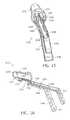
- FIG. 1is a schematic perspective view, partially broken away, of an anoscope in accordance with the present invention, for use in a method in accordance with the present invention, showing a pair of jaws.
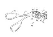
- FIG. 2is a schematic perspective view, partially broken away, of a tissue occlusion device in accordance with the present invention, for use in a method in accordance with the present invention.
- FIG. 3is a schematic perspective view of a proximal one of the jaws depicted in FIG. 1 , showing details of a tissue occlusion mechanism.
- FIG. 4is a schematic perspective view of the proximal jaw of FIG. 1 , showing details of another tissue occlusion mechanism.
- FIG. 5is a schematic perspective view of the proximal jaw of FIG. 1 , showing details of a further tissue occlusion mechanism.
- FIG. 6is a schematic perspective view of the proximal jaw of FIG. 1 , showing details of yet another tissue occlusion mechanism.
- FIGS. 7A-7Fare schematic cross-sectional views of the anoscope of FIG. 1 inserted into an anal canal, showing successive steps of a method in accordance with the present invention.
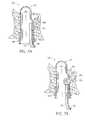
- FIG. 8is a schematic transverse cross-sectional view taken along line IX-IX in FIG. 7D .
- FIG. 9is a schematic transverse cross-sectional view taken along line VIII-VIII in FIG. 7C .
- FIG. 10is a diagrammatic transverse cross-section of the anoscope of FIGS. 1 and 7 A- 7 F.
- FIG. 11is a diagrammatic transverse cross-section similar to FIG. 10 , showing an alternative design of the anoscope of FIGS. 1 and 7 A- 7 F.
- FIG. 12is a diagrammatic transverse cross-section similar to FIG. 10 , showing another alternative design of the anoscope of FIGS. 1 and 7 A- 7 F.
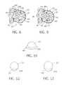
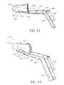
- FIG. 13is a side elevational view of an anoscope with an included tissue occlusion component, in accordance with the present invention.
- FIG. 14is a bottom, front and left side perspective view of the tissue occluding anoscope of FIG. 13 .
- FIG. 15is a top, rear, and left side perspective view of the tissue occluding anoscope of FIGS. 13 and 14 .
- FIG. 16is a top, rear, and left side perspective view of the tissue occluding anoscope of FIGS. 13-15 , in longitudinal or axial section.
- FIG. 17is a longitudinal cross-sectional view of the tissue occluding anodsocpe of FIGS. 13-16 , showing a shutter or closure member in an open or tissue receiving position.
- FIG. 18is a view similar to FIG. 17 , showing the shutter or closure member in a closed or unclamping position.
- FIG. 19is a schematic top plan view of an embodiment of an endoscopic tissue occlusion assembly in accordance with the present invention.
- FIG. 20is a schematic bottom view of the endoscopic tissue occlusion assembly of FIG. 19 .
- FIG. 21is a schematic longitudinal cross-sectional view of the tissue occlusion assembly of FIGS. 19 and 20 , taken along line XXI-XXI in FIG. 19 .
- FIG. 22is a schematic cross-sectional view of the tissue occlusion assembly of FIGS. 19-21 , taken along line XXII-XXII in FIG. 20 .
- FIG. 23is a schematic bottom view of another embodiment of an endoscopic tissue occlusion assembly in accordance with the present invention.
- FIG. 24is a schematic longitudinal cross-sectional view of the tissue occlusion assembly of FIG. 23 , taken along line XXIV-XXIV in FIG. 23 .
- an anoscope 20 for hemorrhoidal surgerycomprises a hollow body 22 and a shutter member 24 .
- Hollow body 22defines a longitudinal channel or lumen 26 that is closed at a distal end 28 and formed with an opening 30 at a proximal end 32 . Opening 30 enables visual inspection of a surgical site and the insertion of instrumentation.
- Hollow body 22has a sidewall 34 provided with a rectangular window 36 spaced from distal end 28 and preferably also from proximal end 28 of hollow body 22 .
- Shutter member 24is movably mounted to hollow body 22 to cover window 36 during a positioning of anoscope 20 in an anal canal. Shutter member 24 is removable from window 36 to permit hemorrhoidal tissues to protrude through window 36 into anoscope channel 26 . More specifically, shutter member 24 is slidably mounted to hollow body 22 , is disposed in hollow body 22 , and has a shape conforming to sidewall 34 in a region thereof about window 36 .
- Shutter member 24is located in a track 37 in the hollow body.
- Track 37takes the form of a shallow depression or recess with longitudinal edges or shoulders 39 serving as guides for the sliding shutter member 24 .
- a transverse edge or shoulder 41serves as an abutment to continued distal motion of shutter member 24 during an insertion stroke thereof.
- Shutter member 24may be locked into track 37 , for example, by grooves (not illustrated) in longitudinal edges or shoulders 39 .
- Hollow body 22generally has a longitudinal axis 38 , and sidewall 34 is formed with a bulging portion or protrusion 40 located on one side of the axis and extending from proximal end 32 of the hollow anoscope body partially along a length of sidewall 34 towards distal end 28 .
- Window 36is located in bulging portion 40 , and shutter member 24 is slidable along and in engagement with bulging portion 40 .
- shutter member 24 and bulging portion 40may be cooperatively formed so that the bulging portion serves as a track that slidably retains the shutter member.
- Window 36may generally take any shape suitable for the admission of protruding hemorrhoidal tissues HT ( FIGS. 7B-7F , 8 , and 9 ). Rectangular and circular are possible shapes.
- Hollow body 22 of anoscope 20has a rim 42 surrounding opening 30 at proximal end 32 .
- Hollow body 22is preferably provided along rim 42 with a flange 44 serving as a stop for preventing anoscope 20 from slipping entirely into the anal canal.
- Hollow body 22is further provided along rim 42 with a cutout 46 disposed on a side of axis 38 opposite bulging portion 40 .
- Cutout 46facilitates manipulation of any instrument that is inserted into anoscope 20 for operating on hemorrhoidal tissues.
- cutout 46facilitates observation of window 36 and of hemorrhoidal tissues HT protruding into longitudinal channel 26 through window 36 .
- window 36may extend in a proximal direction all the way to flange 44 .
- window 36is large enough for the admission of hemorrhoids into channel or lumen 26 of anoscope 20 .
- the placement of window 36 in bulging portion or protrusion 40is conducive to providing window 36 with properly large dimensions.
- Anoscope 20may be provided as part of a surgical instrument assembly than also includes a hemorrhoid treatment device 48 depicted in FIG. 2 .
- Device 48comprises an instrument shaft 50 , a handle or actuator 52 connected to the shaft at a proximal end thereof, and a pair of jaws 54 and 56 (proximal and distal) mounted to the shaft at a distal end thereof.
- Handle 52is operatively connected to jaws 54 and 56 for alternatively opening and closing the jaws.
- Jaws 54 and 56each takes the form of a C- or U-shaped clamping member movable alternately away from and towards the other jaw.
- Jaws 54 and 56define respective gaps 55 and 57 .
- a distal end portion of instrument shaft 50is U- or C-shaped in cross-section and defines a recess 59 aligned and communicating with gap 55 . This asymmetrical shape of the distal end of instrument shaft 50 facilitates a visualization of a surgical site while a distal end portion of hemorrhoid treatment device 48 is inserted into anoscope 20 .
- a hemorrhoid occlusion componentis mounted to jaws 54 and 56 for acting on tissues gripped between the jaws, to couple the tissues to each other.
- the hemorrhoid occlusion componentmay take any form capable of bonding organic tissues, particularly hemorrhoidal tissues, to one another.
- the hemorrhoid occlusion componentmay take the form of a stapling mechanism 58 including a plurality of staples 60 disposed in an arcuate configuration inside proximal jaw 54 .
- Staples 60are longitudinally aligned on a distal side with respective ejection apertures 62 in jaw 54 and on a proximal side with respective pusher elements 64 .
- Pusher elements 64may be disposed on a proximal side in contact with a pressure application ring (not shown) or other force-transmission structure operatively connected at a proximal end with handle 52 .
- Distal jaw 56is provided with a series of anvil elements or areas (not shown) that are aligned with respective slots or ejection apertures 62 , for causing staple closure upon firing.
- Staples 60may be housed in a disposable cartridge element that may be a portion or the entirely of proximal jaw 54 .
- This variationpermits a surgeon, proctologist or other medical practitioner to clamp plural hemorrhoids in the course of a single procedure. After the stapling of one hemorrhoid, as discussed below with reference to FIGS. 7A-7E , the empty cartridge (e.g., jaw 54 ) is removed and replaced with a similar loaded staple cartridge.
- an alternative hemorrhoid occlusion componenttakes the form of an injection mechanism 66 including a plurality of hollow needles 68 fixed to proximal jaw 54 . Needles 68 are longitudinally oriented and circumferentially spaced about jaw 54 . Needles 68 are connectable via a distribution manifold 70 to a reservoir 72 of a sclerosing composition such as a concentrated sugar solution or a biocompatible adhesive.
- a sclerosing compositionsuch as a concentrated sugar solution or a biocompatible adhesive.
- FIG. 5shows another alternative occlusion component in the form of a radiant-energy applicator 74 , for instance, in the infrared or optical portions of the electromagnetic spectrum. More specifically, radiant-energy applicator 74 includes optical fibers 76 connectable via a distribution manifold 78 to a source 80 of laser radiation.
- FIG. 6depicts yet another alternative occlusion component in the form of an electrode 82 mounted to proximal jaw 54 and facing in the distal direction towards distal jaw 56 .
- Distal jaw 56may also be provided with an electrode (not shown), in the case of a bipolar delivery of electrical energy.
- Electrode 82is connectable to a source 84 of radio-frequency current for delivering RF cauterizing current to hemorrhoidal tissues.
- Jaws 54 and 56together with rods 86 and 88 , may form a disposable occlusion cartridge that is removable from shaft 50 .
- the cartridgeis removed and replaced with a new cartridge for use on another patient.
- radiant-energy applicator 74or electrode 82
- handle 52may be provided with a port or connector 85 for enabling the coupling of the hand-held hemorrhoid treatment device 48 to reservoir 72 , laser source 80 , or RF electric source 84 , respectively.
- jaws 54 and 56are mounted to a pair of parallel rods 86 and 88 each connected at a proximal end to instrument shaft 50 .
- Jaws 54 and 56are connected to one another and to shaft 50 via rods 86 and 88 so that the jaws remain parallel to one another and perpendicular to rods 86 and 88 during opening and closing strokes of the jaws.
- Any reciprocatable drive mechanism known in the art or hereafter developedmay be operatively coupled to jaws 54 and 56 and handle 52 for enabling opening and closing of jaws 54 and 56 by manipulation of handle 52 .
- distal jaw 56is slidably coupled to rods 86 and 88
- proximal jaw 54is fixed with respect to the rods
- the rodsare coupled to distal jaw 56 on opposite sides thereof.
- Jaws 54 and 56 and rods 86 and 88may be manufactured as a disposable cartridge assembly detachable from instrument shaft 50 .
- the operative components, such as staples 60 and apertures 62may be formed as parts of a disposable cartridge separate from the jaws 54 and 56 .
- FIGS. 7A-7Fillustrate steps in a method for the treatment of hemorrhoids utilizing anoscope 20 and hemorrhoid treatment device 48 .
- anoscope 20 with shutter member 24 closing window 36is inserted through a transparent anal port member 89 into an anal canal AC and is manipulated so that hemorrhoidal tissues HT are disposed adjacent to window 36 .
- This proceduremay involve longitudinally shifting and/or rotating the anoscope 20 inside the anal canal AC until the anoscope is in the desired position relative to the hemorrhoidal tissues HT.
- shutter member 24 and optionally sidewall 34 of hollow body 22are made of a transparent polymeric material.
- anal tissuescan be visualized through sidewall 34 and shutter member 24 during the manipulation of anoscope.
- shutter member 24Upon an appropriate positioning of anoscope 20 , shutter member 24 is grasped at an external flange or finger grip 90 and pulled in a proximal direction, as indicated by an arrow 92 in FIG. 7B . This action uncovers window 36 and enables hemorrhoidal tissues HT to protrude through the window into channel 26 of anoscope 20 . Subsequently, a distal end portion of hemorrhoid treatment device 48 particularly including jaws 54 and 56 is inserted into anoscope 20 . As depicted in FIG. 7C , this insertion may be performed with jaw 54 and 56 located in channel 26 on a side of longitudinal axis 38 opposite bulging sidewall portion 40 (see FIGS.
- the distal end portion of hemorrhoid treatment device 48is inserted into anoscope 20 in such a manner that jaws 54 and 56 are located in channel 26 on the same side of longitudinal axis 38 as bulging sidewall portion 40 (see FIGS. 7C and 8 ). Because the protruding hemorrhoidal tissues HT are malleable, distal jaw 56 of hemorrhoid treatment device 48 may be slipped past the protruding tissues.
- hemorrhoid treatment device 48it may be necessary or expedient to wiggle hemorrhoid treatment device 48 during the insertion (and removal) phase of a deployment operation, depending on the relative sizes of anoscope 20 , hemorrhoid treatment device 48 , and the protruding hemorrhoidal tissues HT.
- this alternative deployment procedurethere is no need to rotate device 48 about a longitudinal axis to align jaws 54 and 56 with a neck or base region 96 of the protruding hemorrhoidal tissues HT as shown in FIGS. 7D and 9 .
- jaws 54 and 56located on opposite sides of hemorrhoidal tissues HT, the are approximated, as depicted in FIG. 7E , to clamp the hemorrhoidal tissues HT.
- jaws 54 and 56are maintained in parallel to one another during their closing and opening strokes.
- tissue occlusion componentFIGS. 3-6
- the tissue occlusion componentFIGS. 3-6
- the tissue occlusion componentFIGS. 3-6
- the tissue occlusion componentFIGS. 3-6
- the tissue occlusion componentFIGS. 3-6
- the tissue occlusion componentFIGS. 3-6
- the tissue occlusion componentFIGS. 3-6
- the tissue occlusion componentFIGS. 3-6
- the tissue occlusion componentFIGS. 3-6
- the hemorrhoid treatment device 48is operated to permanently constrict hemorrhoidal tissues HT in or about neck region 96 .
- stapling mechanism 58FIG. 3
- staples 60are fired through ejection apertures 62 in jaw 54 by a distal motion of pusher elements 64 , the staples being closed upon meeting respective anvil elements (not illustrated) in distal jaw 56 .
- injection mechanism 66FIG.
- hollow needles 68 fixed to proximal jaw 54are naturally or automatically inserted into hemorrhoidal tissues during the approximation of jaws 54 and 56 .
- Sclerosing compositionis then guided from reservoir 72 into the hemorrhoidal tissues HT.
- radiant-energy applicator 74FIG. 5
- the applicatoris operated to generate electromagnetic radiation of a predetermined spectral range, which is then directed into hemorrhoidal tissues HT via optical fibers 76 .
- radio-frequency currentis conducted from source 84 through electrode 82 into hemorrhoidal tissues HT.
- distal electrode 56is also provided with an electrode, the current passes from electrode 82 through neck or base region 96 to jaw 56 .
- a monopolar cauterization currentthe current spread out from tissues HT into the patient's body.
- handle 52is operated to separate jaws 54 and 56 from one another and the treatment device 48 is manipulated to separate the jaws from the treated hemorrhoidal tissues HT ( FIG. 7F ).
- Treatment device 48is then further manipulated to withdraw it from anoscope 20 . Again, because of the deformability of the clamped hemorrhoidal tissues HT, in many cases it will be possible to simply withdraw the hemorrhoid treatment device 48 without rotation, but perhaps with some wiggling.
- the hemorrhoidal tissues HT distal to the occluded neck region 96may be transected with a scalpel or allowed to ischemically regress or self amputate. Self-amputation occurs within a few days of the occlusion procedure. Ischemic regression takes place within several weeks. Ischemic regression and self-amputation are the result of occlusion of bloods vessels in neck or base region 96 .
- Bulging portion or protrusion 40 of anoscope 20serves as a retractor of collateral anal or rectal tissues.
- bulging portion or protrusion 40creates more work space in the area of hemorrhoidal tissues HT. This design allows for better access to the neck or base 96 of tissues HT, which is located in the submucosal layer close to the rectal muscle.
- FIGS. 8-10show one configuration of bulging portion or protrusion 40 , where the protrusion has a radius of curvature that is greater than a radius of curvature of the remaining part of hollow body 22 .
- FIG. 11depicts a configuration where a bulging portion or protrusion 98 has a radius of curvature that is smaller than the radius of curvature of the main part of hollow body member 22 .
- FIG. 12illustrates a configuration where a bulging portion or protrusion 100 has a radius of curvature that is essentially equal to the radius of curvature of the main part of hollow body member 22 .
- the dashed lines 102 , 104 , 106represent the respective occluding jaws of hemorrhoid treatment device 48 .
- anoscope 20 and port member 89are preferably made of a transparent polymeric material that facilitates visual inspection and locating of the hemorrhoids.
- Jaws 54 and 56 of the occlusion deviceare inserted into anoscope 20 after the inserting of anoscope 20 into the anal canal AC, after the manipulating of anoscope 20 to align window 36 with hemorrhoidal tissues HT, and after the protruding of the hemorrhoidal tissues HT through window 36 .
- a hemorrhoid treatment instrument or device as disclosed hereinabovemay be partially or completely disposable. Where both jaws 54 and 56 are parts of a disposable cartridge removably attached to shaft 50 , the proximal portion of the instrument may be utilizable in treating different patients at different times. Alternatively or additionally, where proximal jaw 54 contains a staple magazine, jaw 54 may be replaceable to permit multiple hemorrhoid occlusion procedures on the same patient.
- FIGS. 13-18depict a surgical instrument assembly and more particularly a tissue-occluding anoscope assembly 120 for the treatment of hemorrhoids.
- the instrument assembly 120comprises a hollow member 122 having a sidewall 124 provided with a window 126 and further comprises a closure member 128 slidably connected to the hollow member for alternately covering and uncovering the window.
- Hollow member 122has a first clamping surface 130 along an edge of window 126 .
- Closure member 128has a second clamping surface 132 opposing clamping surface 130 and disposable substantially adjacent thereto in a clamping or closure configuration of the instrument shown in FIGS. 13-15 and 18 .
- Closure member 128is slidable in a proximal direction, away from clamping surface 130 to open window 126 , as shown in FIGS. 16 and 17 .
- Tissue occluding instrument assembly 120additionally comprises a tissue occlusion component 134 mounted to at least one of the hollow member 122 and the closure member 128 for acting on organic tissues gripped between clamping surfaces 130 and 132 , to couple the tissues to each other.
- Tissue occlusion component 134may be a stapling mechanism, an injection mechanism connectable to a reservoir of a sclerosing composition, or optical fibers connectable to a source of laser radiation.
- Sidewall 124 of hollow member 122is generally conically curved, so that clamping surfaces 130 and 132 have a curved form, e.g., a C shape or U shape.
- Clamping surfaces 130 and 132lie in parallel planes that extend perpendicularly to a longitudinal axis 136 of instrument 120 and remain parallel to one another during opening and closing strokes of closure member 128 .
- Closure member 128moves parallel to axis 136 .
- Hollow member 122is closed at a distal end 138 and defines a longitudinal channel 140 in which closure member 128 is disposed in part. Window 126 communicates with channel 140 .
- hollow body 122is provided with a handle 142 including a extending longitudinally stem 144 (that is oriented at a small angle relative to axis 136 ) and a substantially transversely extending handgrip 146 .
- Closure member 128includes a main part 148 formed as a channel member and, at an end of main part 148 opposite clamping surface 132 , a handgrip 150 extending parallel to handgrip 146 of handle 142 .
- Stem portion 144 of handle 142is formed as a channel member that slidingly receiving main part 148 of closure or shutter member 128 .
- FIGS. 19-22depict an endoscopic version of a tissue occluding instrument assembly 152 including a hollow body member 154 that has a channel 156 for receiving an insertion member 158 of an endoscope.
- Hollow member 154incorporates a chamber 160 that is located laterally relative to channel 156 .
- Chamber 160is optionally separated from channel 156 by a partition or wall 162 .
- Endoscope insertion member 158extends in an arc about chamber 160 , which is accordingly located in a bulging portion 164 of hollow body member 154 .
- Hollow body member 154 and particularly bulging wall 164 thereofis provided with a window or aperture 166 through which organic tissues such as a polyp 168 may protrude during an endoscopic tissue occluding procedure.
- endoscope insertion member 158with hollow body member 154 attached thereto as illustrated, is inserted through a natural body opening such as the anal orifice into an internal lumen such as the colon.
- Optical components (not illustrated) in the distal end face 170 of endoscope insertion member 158are used to visually inspect the walls of the body lumen and to detect a surgical site containing polyp 168 or other undesirable tissue mass.
- Tissue occluding instrument assembly 152further includes a closure or shutter member 172 that includes a tissue clamping surface 174 at a distal end.
- Surface 174generally has an arcuate shape and lies in a plane transverse to a longitudinal axis 176 of the instrument assembly.
- Surface 174is opposable to another arcuate tissue clamping surface 178 that is attached to hollow body member 154 along a distal edge (not separately labeled) of window 166 .
- Surface 178also lies in a plane transverse to a longitudinal axis 176 and is accordingly parallel to surface 174 .
- Closure or shutter member 172is attached to a distal end of a rod 180 that is slidably disposed in a channel 182 of hollow body member 154 that extends parallel to axis 176 .
- rod 180is pushed in a distal direction so that closure or shutter member 172 covers or closes opening 166 .
- rod 180is pulled in a proximal direction to remove closure or shutter member 172 from window 166 and allow polyp 168 to protrude through window 166 into chamber 160 .
- Body member 154is provided in chamber 160 with an opening 184 via which a visual inspection of chamber 160 may be undertaken. Opening 184 may provide visual access to chamber 160 via optical components of endoscope insertion member 158 (exemplarily including an illumination source, a lens, and an optical fiber bundle—none illustrated). Alternatively, as discussed hereinafter with reference to FIGS. 23 and 24 , hollow body member 154 may be provided with its own dedicated optical components for establishing visual access to chamber 160 .
- Hollow body member 154may additionally be provided along chamber 160 with openings 186 for enabling access to chamber 160 by the working tips of endoscopic instruments such as a suction device 188 .
- Suction device 188includes a conical head 190 that engages polyp 168 .
- device 188is pulled in proximal direction through a working channel 192 of endoscope insertion member or hollow body member 154 .
- polyp 168is stretched out to facilitate an occlusion operation in which closure or shutter member 172 is moved in the distal direction so that pedicle or neck tissues 194 of polyp 168 are sandwiched between clamping surfaces 174 and 178 .
- Occlusion componentry 196then operates through clamping surface 178 and/or surface 176 to effectuate an occlusion of the pedicle or neck tissues 194 .
- Occlusion componentry 196exemplarily takes the form of a stapling mechanism, an injection mechanism connectable to a reservoir of a sclerosing composition, or optical fibers connectable to a source of laser radiation.
- the tissue occluding componentrymay effectuate a heating of the tissues via resistive heat producing elements or electrical current transmission components.
- closure or shutter member 172is opened after the application of the occlusion energy.
- the occluded tissue masse.g., polyp 168 , is then allowed to slip out of chamber 160 back into the natural body lumen.
- FIGS. 23 and 24depict a tissue occluding instrument assembly 202 similar to instrument assembly 152 of FIGS. 19-22 .
- a hollow body member 204 having a channel 206 receiving an endoscope insertion member 208incorporates a fiber-optic illumination guide 210 , a fiber-optic image guide 214 , at least one working channel 216 for the deployment of endoscopic instrument, cables 218 and 220 for assisting in curving endoscope insertion member 208 to form a chamber 222 adjacent channel 206 .
- a stiffening rod 224may be insertable through a channel 226 of hollow body member 204 also for purposes of assisting the formation of chamber 222 .
- hollow body member 204may take the form of an endoscope sheath that is deformable at a distal end to expand chamber 222 from a collapsed insertion configuration to an expanded use configuration as shown particularly in FIG. 24 .
- Illumination guide 210 and image guide 214terminate distally at light access openings 212 and 228 along the wall of chamber 222 .
- a first arcuate clamping surface 230is located on hollow body member 204 , along a distal edge of a window 232 that communicates with chamber 222 .
- a second arcuate clamping surface 234is attached to the distal end of a closure or shutter member 236 .
- Closure or shutter member 236is connected to a rod 238 that moves the closure member alternately in a distal and proximal direction for effectuating a closure of window 232 during an insertion operation, an opening of window 232 to enable a protruding of a tissue mass into chamber 222 , and a clamping of the protruding tissues during an application of energy to the tissues to effectuate an occlusion thereof.
- Tissue occluding componentry 240 provided on closure member 236 at surface 234may take the form of any of the instrumentalities discussed above.
- a suction head 242 of a suction device 244 inserted through working channel 216may be used to draw a polyp 246 away from a wall 248 of a body lumen to facilitate a tissue occluding operation.
- Tissue occluding instrument assemblies 152 and 202may be used to treat a variety of pathologies (polyp, wall perforation, bleeding point at a previously placed staple line, etc.) but are generally not useful for treating hemorrhoids.
- Endoluminal tissue occluding instrument assemblies 152 and 202may be used for treatment of lesions in natural and artificial lumens other than the colon, including the trachea, the bronchi, blood vessels (arteries and veins), etc.
- tissue occluding instrument assemblies 152 and 202can be used to perform various known types of surgical maneuvers/operations, such as operating on an intimal/endothelial lesion in a vessel (arterial plaque, etc.) or operating on diseased venous or arterial valves.
- tissue occluding instrument assemblies 152 and 202can be used to carry out such additional procedures.
- endoscopic surgical instrumentscan be introduced via endoscopic sheath channels 192 , 216 to the targeted tissues.
- the extractionmay be implemented either via working channels 192 , 216 or upon the withdrawal of the entire instrument. In the latter case, the severed specimen is carried in the chamber 160 , 222 until outside of the patient.
- the vesselcan be coagulated with RF or injected with sclerosing or hemostatic agent.
- any diagnostic and surgical maneuvers described herecan be performed in conjunction with external maneuvers, for example, laparoscopic maneuvers.
- This laparo-endoluminal approachis generally known in the field of surgery and may facilitate the performance and safety of the operation while preserving the benefits of minimally-invasive approach.
- rods 86 and 88may be fixed to distal jaw 56 and slidably connected to shaft 50 .
- rods 86 and 88may be fixed to both distal jaw 56 and shaft 50 , in which case proximal jaw 54 is slidable along rods 86 and 88 alternately towards and away from jaw 56 . Also, more than two rods 86 and 88 may be provided for coupling distal jaw 56 to instrument shaft 50 .
- both jaws 54 and 56are movable along rods 86 and 88 during a clamping or closure stroke. Such a design facilitates hemorrhoid occlusion without tearing of the tissues below the occluded tissue base. If only one jaw 54 or 56 is movable along rods 86 and 88 , then the entire instrument could be moved relative to the patient during closure of the jaws to ensure against undesired tissue tears. Where the distal jaw 56 is slidable along rods 86 and 88 , the entire instrument is pushed into the patient while the distal jaw is moving in a proximal direction.
Landscapes
- Health & Medical Sciences (AREA)
- Life Sciences & Earth Sciences (AREA)
- Surgery (AREA)
- Engineering & Computer Science (AREA)
- Veterinary Medicine (AREA)
- Public Health (AREA)
- Nuclear Medicine, Radiotherapy & Molecular Imaging (AREA)
- General Health & Medical Sciences (AREA)
- Biomedical Technology (AREA)
- Heart & Thoracic Surgery (AREA)
- Medical Informatics (AREA)
- Molecular Biology (AREA)
- Animal Behavior & Ethology (AREA)
- Physics & Mathematics (AREA)
- Optics & Photonics (AREA)
- Pathology (AREA)
- Radiology & Medical Imaging (AREA)
- Biophysics (AREA)
- Otolaryngology (AREA)
- Plasma & Fusion (AREA)
- Orthopedic Medicine & Surgery (AREA)
- Electromagnetism (AREA)
- Vascular Medicine (AREA)
- Surgical Instruments (AREA)
- Endoscopes (AREA)
Abstract
Description
Claims (31)
Priority Applications (1)
| Application Number | Priority Date | Filing Date | Title |
|---|---|---|---|
| US12/547,322US8430808B2 (en) | 2004-03-16 | 2009-08-25 | Endoscopic method and device for avoiding cutting of rectal tissue in the treatment of hemorrhoids |
Applications Claiming Priority (3)
| Application Number | Priority Date | Filing Date | Title |
|---|---|---|---|
| US10/801,283US7118528B1 (en) | 2004-03-16 | 2004-03-16 | Hemorrhoids treatment method and associated instrument assembly including anoscope and cofunctioning tissue occlusion device |
| US11/197,965US8100822B2 (en) | 2004-03-16 | 2005-08-05 | Anoscope for treating hemorrhoids without the trauma of cutting or the use of an endoscope |
| US12/547,322US8430808B2 (en) | 2004-03-16 | 2009-08-25 | Endoscopic method and device for avoiding cutting of rectal tissue in the treatment of hemorrhoids |
Related Parent Applications (1)
| Application Number | Title | Priority Date | Filing Date |
|---|---|---|---|
| US11/197,965DivisionUS8100822B2 (en) | 2004-03-16 | 2005-08-05 | Anoscope for treating hemorrhoids without the trauma of cutting or the use of an endoscope |
Publications (2)
| Publication Number | Publication Date |
|---|---|
| US20100010297A1 US20100010297A1 (en) | 2010-01-14 |
| US8430808B2true US8430808B2 (en) | 2013-04-30 |
Family
ID=37727929
Family Applications (11)
| Application Number | Title | Priority Date | Filing Date |
|---|---|---|---|
| US11/197,965Active2027-05-09US8100822B2 (en) | 2004-03-16 | 2005-08-05 | Anoscope for treating hemorrhoids without the trauma of cutting or the use of an endoscope |
| US12/547,296Active2028-04-07US9039601B2 (en) | 2004-03-16 | 2009-08-25 | Endoluminal treatment method and associated surgical assembly including tissue occlusion device |
| US12/547,258Active2029-04-17US9867633B2 (en) | 2004-03-16 | 2009-08-25 | Endoluminal treatment method and associated surgical assembly including tissue occlusion device |
| US12/547,322Expired - Fee RelatedUS8430808B2 (en) | 2004-03-16 | 2009-08-25 | Endoscopic method and device for avoiding cutting of rectal tissue in the treatment of hemorrhoids |
| US12/547,279Active2027-09-26US8715166B2 (en) | 2004-03-16 | 2009-08-25 | Gentle method of treating a hemorrhoid |
| US14/214,898Expired - LifetimeUS10245061B2 (en) | 2004-03-16 | 2014-03-15 | Treatment method including tissue occlusion device |
| US14/511,694Expired - Fee RelatedUS9661984B2 (en) | 2004-03-16 | 2014-10-10 | Endoluminal treatment method and associated surgical assembly |
| US15/168,083Expired - LifetimeUS10492815B2 (en) | 2004-03-16 | 2016-05-29 | Endoluminal treatment method and associated surgical assembly |
| US15/361,457Expired - LifetimeUS10485567B2 (en) | 2004-03-16 | 2016-11-27 | Endoluminal treatment method and associated surgical assembly |
| US16/601,666Expired - Fee RelatedUS11109879B2 (en) | 2004-03-16 | 2019-10-15 | Endoluminal treatment method and associated surgical assembly including tissue occlusion device |
| US17/393,719Expired - LifetimeUS12178463B2 (en) | 2005-08-05 | 2021-08-04 | Endoluminal treatment method and associated surgical assembly including tissue occlusion device |
Family Applications Before (3)
| Application Number | Title | Priority Date | Filing Date |
|---|---|---|---|
| US11/197,965Active2027-05-09US8100822B2 (en) | 2004-03-16 | 2005-08-05 | Anoscope for treating hemorrhoids without the trauma of cutting or the use of an endoscope |
| US12/547,296Active2028-04-07US9039601B2 (en) | 2004-03-16 | 2009-08-25 | Endoluminal treatment method and associated surgical assembly including tissue occlusion device |
| US12/547,258Active2029-04-17US9867633B2 (en) | 2004-03-16 | 2009-08-25 | Endoluminal treatment method and associated surgical assembly including tissue occlusion device |
Family Applications After (7)
| Application Number | Title | Priority Date | Filing Date |
|---|---|---|---|
| US12/547,279Active2027-09-26US8715166B2 (en) | 2004-03-16 | 2009-08-25 | Gentle method of treating a hemorrhoid |
| US14/214,898Expired - LifetimeUS10245061B2 (en) | 2004-03-16 | 2014-03-15 | Treatment method including tissue occlusion device |
| US14/511,694Expired - Fee RelatedUS9661984B2 (en) | 2004-03-16 | 2014-10-10 | Endoluminal treatment method and associated surgical assembly |
| US15/168,083Expired - LifetimeUS10492815B2 (en) | 2004-03-16 | 2016-05-29 | Endoluminal treatment method and associated surgical assembly |
| US15/361,457Expired - LifetimeUS10485567B2 (en) | 2004-03-16 | 2016-11-27 | Endoluminal treatment method and associated surgical assembly |
| US16/601,666Expired - Fee RelatedUS11109879B2 (en) | 2004-03-16 | 2019-10-15 | Endoluminal treatment method and associated surgical assembly including tissue occlusion device |
| US17/393,719Expired - LifetimeUS12178463B2 (en) | 2005-08-05 | 2021-08-04 | Endoluminal treatment method and associated surgical assembly including tissue occlusion device |
Country Status (7)
| Country | Link |
|---|---|
| US (11) | US8100822B2 (en) |
| EP (1) | EP1928291B1 (en) |
| JP (1) | JP4918549B2 (en) |
| CN (1) | CN101237808B (en) |
| AU (1) | AU2006278480B2 (en) |
| CA (1) | CA2617844C (en) |
| WO (1) | WO2007019321A2 (en) |
Cited By (5)
| Publication number | Priority date | Publication date | Assignee | Title |
|---|---|---|---|---|
| US20100030019A1 (en)* | 2008-07-31 | 2010-02-04 | Kuroda Noriko | Endoscopic surgical operation method |
| US20170112371A1 (en)* | 2015-10-27 | 2017-04-27 | George Percy McGown | Anoscope |
| US10245061B2 (en) | 2004-03-16 | 2019-04-02 | Covidien Lp | Treatment method including tissue occlusion device |
| US10660666B2 (en) | 2018-07-12 | 2020-05-26 | Steven William Walton | Cutting tool |
| US11497507B2 (en) | 2017-02-19 | 2022-11-15 | Orpheus Ventures, Llc | Systems and methods for closing portions of body tissue |
Families Citing this family (136)
| Publication number | Priority date | Publication date | Assignee | Title |
|---|---|---|---|---|
| US7537564B2 (en) | 1998-12-01 | 2009-05-26 | Atropos Limited | Wound retractor device |
| US7559893B2 (en) | 1998-12-01 | 2009-07-14 | Atropos Limited | Wound retractor device |
| US7998068B2 (en) | 1998-12-01 | 2011-08-16 | Atropos Limited | Instrument access device |
| US6254534B1 (en) | 1999-10-14 | 2001-07-03 | Atropos Limited | Retractor |
| JP5190169B2 (en) | 2000-10-19 | 2013-04-24 | アプライド メディカル リソーシーズ コーポレイション | Surgical access instruments and methods |
| EP2422829B1 (en) | 2001-08-14 | 2013-03-06 | Applied Medical Resources Corporation | Surgical access sealing apparatus |
| US6958037B2 (en) | 2001-10-20 | 2005-10-25 | Applied Medical Resources Corporation | Wound retraction apparatus and method |
| EP2343032B1 (en) | 2002-06-05 | 2012-05-09 | Applied Medical Resources Corporation | Wound retractor |
| US9271753B2 (en) | 2002-08-08 | 2016-03-01 | Atropos Limited | Surgical device |
| DE60314464T2 (en) | 2002-09-19 | 2008-02-14 | Atropos Ltd., Bray | SURGICAL WOUND RETRACTOR |
| US20050020884A1 (en) | 2003-02-25 | 2005-01-27 | Hart Charles C. | Surgical access system |
| CA2533204A1 (en) | 2003-08-06 | 2005-02-17 | Applied Medical Resources Corporation | Surgical device with tack-free gel and method of manufacture |
| US7163510B2 (en) | 2003-09-17 | 2007-01-16 | Applied Medical Resources Corporation | Surgical instrument access device |
| JP2009501045A (en) | 2005-07-15 | 2009-01-15 | アトロポス・リミテッド | Wound retractor |
| AU2006304141B2 (en) | 2005-10-14 | 2012-07-05 | Applied Medical Resources Corporation | Gel cap for wound retractor |
| AU2006338436B2 (en)* | 2006-02-14 | 2012-07-26 | Thd S.P.A. | A process and a device for surgical treatment of rectal and haemorrhoidal prolapse |
| US8357085B2 (en) | 2009-03-31 | 2013-01-22 | Ethicon Endo-Surgery, Inc. | Devices and methods for providing access into a body cavity |
| US8821391B2 (en) | 2009-03-06 | 2014-09-02 | Ethicon Endo-Surgery, Inc. | Methods and devices for providing access into a body cavity |
| US8485970B2 (en) | 2008-09-30 | 2013-07-16 | Ethicon Endo-Surgery, Inc. | Surgical access device |
| US8926506B2 (en) | 2009-03-06 | 2015-01-06 | Ethicon Endo-Surgery, Inc. | Methods and devices for providing access into a body cavity |
| US8206294B2 (en) | 2008-09-30 | 2012-06-26 | Ethicon Endo-Surgery, Inc. | Surgical access device with flexible seal channel |
| US8430811B2 (en) | 2008-09-30 | 2013-04-30 | Ethicon Endo-Surgery, Inc. | Multiple port surgical access device |
| US8251900B2 (en) | 2009-03-06 | 2012-08-28 | Ethicon Endo-Surgery, Inc. | Surgical access devices and methods providing seal movement in predefined paths |
| US8961406B2 (en) | 2009-03-06 | 2015-02-24 | Ethicon Endo-Surgery, Inc. | Surgical access devices and methods providing seal movement in predefined movement regions |
| US8425410B2 (en) | 2008-09-30 | 2013-04-23 | Ethicon Endo-Surgery, Inc. | Surgical access device with protective element |
| EP2015681B1 (en) | 2006-05-03 | 2018-03-28 | Datascope Corp. | Tissue closure device |
| US9591965B2 (en)* | 2007-04-10 | 2017-03-14 | Boston Scientific Scimed, Inc. | Endoscopes including distal chamber and related methods of use |
| WO2008130675A1 (en)* | 2007-04-18 | 2008-10-30 | Delaney Conor P | Anoscope for inspection and/or surgery |
| EP2146644A4 (en) | 2007-05-11 | 2012-07-18 | Applied Med Resources | Surgical retractor |
| WO2008141291A1 (en) | 2007-05-11 | 2008-11-20 | Applied Medical Resources Corporation | Surgical retractor with gel pad |
| ITTO20080032U1 (en)* | 2007-05-11 | 2008-09-12 | Ceramoptec Gmbh | SYSTEM AND METHOD FOR HEMORROID TREATMENT |
| US8657740B2 (en) | 2007-06-05 | 2014-02-25 | Atropos Limited | Instrument access device |
| WO2008149332A1 (en) | 2007-06-05 | 2008-12-11 | Atropos Limited | An instrument access device |
| US20110105848A1 (en)* | 2007-10-07 | 2011-05-05 | Niv Sadovsky | Laparoscopic tissue retractor |
| CA2711116C (en) | 2008-01-22 | 2017-08-29 | Applied Medical Resources Corporation | Surgical instrument access device |
| US8328761B2 (en) | 2008-09-30 | 2012-12-11 | Ethicon Endo-Surgery, Inc. | Variable surgical access device |
| AU2008362711B2 (en)* | 2008-10-06 | 2015-02-12 | Thd S.P.A. | A device for surgical operations on a prolapse |
| CA2739910C (en) | 2008-10-13 | 2017-06-06 | Applied Medical Resources Corporation | Single port access system |
| US8348837B2 (en)* | 2008-12-09 | 2013-01-08 | Covidien Lp | Anoscope |
| US8375955B2 (en) | 2009-02-06 | 2013-02-19 | Atropos Limited | Surgical procedure |
| EP2398548B1 (en)* | 2009-02-17 | 2017-04-19 | The Board Of Trustees Of The Leland | Closure device |
| US9737334B2 (en) | 2009-03-06 | 2017-08-22 | Ethicon Llc | Methods and devices for accessing a body cavity |
| US8353824B2 (en) | 2009-03-31 | 2013-01-15 | Ethicon Endo-Surgery, Inc. | Access method with insert |
| US20100249520A1 (en) | 2009-03-31 | 2010-09-30 | Shelton Iv Frederick E | Method Of Surgical Access |
| US8945163B2 (en) | 2009-04-01 | 2015-02-03 | Ethicon Endo-Surgery, Inc. | Methods and devices for cutting and fastening tissue |
| US8257251B2 (en) | 2009-04-08 | 2012-09-04 | Ethicon Endo-Surgery, Inc. | Methods and devices for providing access into a body cavity |
| US8137267B2 (en) | 2009-04-08 | 2012-03-20 | Ethicon Endo-Surgery, Inc. | Retractor with flexible sleeve |
| US8419635B2 (en) | 2009-04-08 | 2013-04-16 | Ethicon Endo-Surgery, Inc. | Surgical access device having removable and replaceable components |
| US8475490B2 (en) | 2009-06-05 | 2013-07-02 | Ethicon Endo-Surgery, Inc. | Methods and devices for providing access through tissue to a surgical site |
| US8465422B2 (en) | 2009-06-05 | 2013-06-18 | Ethicon Endo-Surgery, Inc. | Retractor with integrated wound closure |
| US8361109B2 (en) | 2009-06-05 | 2013-01-29 | Ethicon Endo-Surgery, Inc. | Multi-planar obturator with foldable retractor |
| US8795163B2 (en) | 2009-06-05 | 2014-08-05 | Ethicon Endo-Surgery, Inc. | Interlocking seal components |
| US8033995B2 (en) | 2009-06-05 | 2011-10-11 | Ethicon Endo-Surgery, Inc. | Inflatable retractor with insufflation and method |
| US8241209B2 (en) | 2009-06-05 | 2012-08-14 | Ethicon Endo-Surgery, Inc. | Active seal components |
| US9078695B2 (en) | 2009-06-05 | 2015-07-14 | Ethicon Endo-Surgery, Inc. | Methods and devices for accessing a body cavity using a surgical access device with modular seal components |
| EP2475421B1 (en) | 2009-09-10 | 2017-08-09 | University College Cork-National University of Ireland, Cork | A device for treating tissue |
| WO2011033495A1 (en) | 2009-09-17 | 2011-03-24 | Atropos Limited | An instrument access device |
| WO2011041578A2 (en)* | 2009-10-01 | 2011-04-07 | Macroplata, Inc. | Detachable balloon catheter |
| US9474540B2 (en) | 2009-10-08 | 2016-10-25 | Ethicon-Endo-Surgery, Inc. | Laparoscopic device with compound angulation |
| US10966701B2 (en) | 2009-12-16 | 2021-04-06 | Boston Scientific Scimed, Inc. | Tissue retractor for minimally invasive surgery |
| US12376737B1 (en) | 2009-12-16 | 2025-08-05 | Boston Scientific Scimed, Inc. | Tissue retractor for minimally invasive surgery |
| US10531869B2 (en) | 2009-12-16 | 2020-01-14 | Boston Scientific Scimed, Inc. | Tissue retractor for minimally invasive surgery |
| US10758116B2 (en) | 2009-12-16 | 2020-09-01 | Boston Scientific Scimed, Inc. | System for a minimally-invasive, operative gastrointestinal treatment |
| US8932211B2 (en) | 2012-06-22 | 2015-01-13 | Macroplata, Inc. | Floating, multi-lumen-catheter retractor system for a minimally-invasive, operative gastrointestinal treatment |
| USRE48850E1 (en) | 2009-12-16 | 2021-12-14 | Boston Scientific Scimed, Inc. | Multi-lumen-catheter retractor system for a minimally-invasive, operative gastrointestinal treatment |
| US10595711B2 (en) | 2009-12-16 | 2020-03-24 | Boston Scientific Scimed, Inc. | System for a minimally-invasive, operative gastrointestinal treatment |
| US11344285B2 (en) | 2009-12-16 | 2022-05-31 | Boston Scientific Scimed, Inc. | Multi-lumen-catheter retractor system for a minimally-invasive, operative gastrointestinal treatment |
| ES2874194T3 (en) | 2009-12-16 | 2021-11-04 | Boston Scient Scimed Inc | Arrangements for making an endoluminal anatomical structure |
| US9565998B2 (en) | 2009-12-16 | 2017-02-14 | Boston Scientific Scimed, Inc. | Multi-lumen-catheter retractor system for a minimally-invasive, operative gastrointestinal treatment |
| US9186131B2 (en) | 2009-12-16 | 2015-11-17 | Macroplata, Inc. | Multi-lumen-catheter retractor system for a minimally-invasive, operative gastrointestinal treatment |
| US8632458B2 (en) | 2011-10-26 | 2014-01-21 | Macroplata Inc. | Gentle hemorrhoid treatment offering a substantially painless healing |
| KR101605934B1 (en) | 2010-04-26 | 2016-03-23 | 코비디엔 엘피 | Apparatus and method for effecting at least one anatomical structure |
| AU2014262200B2 (en)* | 2010-04-26 | 2016-03-10 | Covidien Lp | Apparatus and method for effecting at least one anatomical structure |
| US9226760B2 (en) | 2010-05-07 | 2016-01-05 | Ethicon Endo-Surgery, Inc. | Laparoscopic devices with flexible actuation mechanisms |
| US8562592B2 (en) | 2010-05-07 | 2013-10-22 | Ethicon Endo-Surgery, Inc. | Compound angle laparoscopic methods and devices |
| US8460337B2 (en) | 2010-06-09 | 2013-06-11 | Ethicon Endo-Surgery, Inc. | Selectable handle biasing |
| US8617062B2 (en)* | 2010-07-08 | 2013-12-31 | Warsaw Orthopedic, Inc. | Over dilation |
| US9289115B2 (en) | 2010-10-01 | 2016-03-22 | Applied Medical Resources Corporation | Natural orifice surgery system |
| JP6396657B2 (en) | 2010-10-01 | 2018-09-26 | アプライド メディカル リソーシーズ コーポレイション | Natural orifice surgery system |
| US9149271B2 (en) | 2010-11-22 | 2015-10-06 | The Board Of Trustees Of The Leland Stanford, Jr. University | Device and method for treatment of hemorrhoids |
| US8758236B2 (en) | 2011-05-10 | 2014-06-24 | Applied Medical Resources Corporation | Wound retractor |
| WO2013101845A2 (en)* | 2011-12-27 | 2013-07-04 | Boston Scientific Scimed, Inc. | Adjustable resection device and related methods of use |
| CN102641546A (en)* | 2012-05-18 | 2012-08-22 | 普瑞斯星(常州)医疗器械有限公司 | Adjustable anal dilation importer |
| US20140128677A1 (en)* | 2012-07-30 | 2014-05-08 | Janet Lenart | Enema and Coloscopy Aide |
| CN102940479B (en)* | 2012-10-16 | 2015-03-25 | 上海中医药大学附属岳阳中西医结合医院 | Injection anoscope for internal hemorrhoid |
| US8915947B2 (en)* | 2012-10-18 | 2014-12-23 | Spectrum Spine Ip Holdings, Llc | Surgical access system |
| US20140257036A1 (en)* | 2013-03-11 | 2014-09-11 | Ohio University | Method And Device For Detecting Thermal Effects Of Tissue Ablation On A Tissue Of A Patient |
| JP2016512725A (en) | 2013-03-15 | 2016-05-09 | アプライド メディカル リソーシーズ コーポレイション | Mechanical gel surgical access instrument |
| WO2015077356A1 (en) | 2013-11-19 | 2015-05-28 | Wheeler William K | Fastener applicator with interlock |
| EP3169510B1 (en) | 2014-07-18 | 2018-10-03 | Applied Medical Resources Corporation | Method for manufacturing gels having permanent tack free coatings |
| KR20240172766A (en) | 2014-08-15 | 2024-12-10 | 어플라이드 메디컬 리소시스 코포레이션 | Natural orifice surgery system |
| WO2016085930A2 (en) | 2014-11-25 | 2016-06-02 | Applied Medical Resources Corporation | Circumferential wound retraction with support and guidance structures |
| CN104758030B (en)* | 2015-03-13 | 2017-02-01 | 上海朝举信息科技有限公司 | Transverse arc-shaped clamp treatment device for haemorrhoids |
| US10368908B2 (en) | 2015-09-15 | 2019-08-06 | Applied Medical Resources Corporation | Surgical robotic access system |
| WO2017062850A2 (en) | 2015-10-07 | 2017-04-13 | Applied Medical Resources Corporation | Wound retractor with multi-segment outer ring |
| WO2017106738A1 (en)* | 2015-12-18 | 2017-06-22 | Boston Scientific Scimed, Inc. | Resection tools and related methods of use |
| AU2017209092B2 (en)* | 2016-01-21 | 2019-12-05 | The Cleveland Clinic Foundation | System and apparatus for assisting with submucosal dissections |
| WO2017200555A1 (en)* | 2016-05-20 | 2017-11-23 | Choicespine, Lp | Access instruments to extend a surgical working channel |
| AU2017324450B2 (en)* | 2016-09-12 | 2022-09-29 | Applied Medical Resources Corporation | Surgical robotic access system for irregularly shaped robotic actuators and associated robotic surgical instruments |
| CN110087526B (en) | 2016-12-30 | 2022-01-14 | 波士顿科学国际有限公司 | System for minimally invasive treatment inside body cavity |
| CN110913773B (en) | 2017-03-18 | 2023-03-28 | 波士顿科学国际有限公司 | System for minimally invasive treatment of a body cavity |
| US11065048B2 (en) | 2017-06-28 | 2021-07-20 | Cilag Gmbh International | Flexible circuit arrangement for surgical fastening instruments |
| US11013552B2 (en) | 2017-06-28 | 2021-05-25 | Cilag Gmbh International | Electrosurgical cartridge for use in thin profile surgical cutting and stapling instrument |
| US10813640B2 (en) | 2017-06-28 | 2020-10-27 | Ethicon Llc | Method of coating slip rings |
| US10888369B2 (en) | 2017-06-28 | 2021-01-12 | Ethicon Llc | Systems and methods for controlling control circuits for independent energy delivery over segmented sections |
| US11103301B2 (en) | 2017-06-28 | 2021-08-31 | Cilag Gmbh International | Surgical system coupleable with staple cartridge and radio frequency cartridge, and having a plurality of radio-frequency energy return paths |
| US11272976B2 (en) | 2017-06-28 | 2022-03-15 | Cilag Gmbh International | Surgical end effector for applying electrosurgical energy to different electrodes on different time periods |
| US11298128B2 (en)* | 2017-06-28 | 2022-04-12 | Cilag Gmbh International | Surgical system couplable with staple cartridge and radio frequency cartridge, and method of using same |
| US11160604B2 (en) | 2017-06-28 | 2021-11-02 | Cilag Gmbh International | Surgical end effector to adjust jaw compression |
| US11058477B2 (en) | 2017-06-28 | 2021-07-13 | Cilag Gmbh International | Surgical cutting and fastening instruments with dual power sources |
| US11278346B2 (en) | 2017-06-28 | 2022-03-22 | Cilag Gmbh International | Systems and methods of displaying surgical instrument status |
| US11129666B2 (en) | 2017-06-28 | 2021-09-28 | Cilag Gmbh International | Shaft module circuitry arrangements |
| EP3687372B1 (en) | 2017-09-29 | 2022-11-30 | Gregory Piskun | Rubber band ligation system for treatment of hemorrhoids |
| SE541467C2 (en)* | 2017-10-13 | 2019-10-08 | Dev Ab | Device for use in the treatment of hemorrhoids |
| CN108175372B (en)* | 2018-01-10 | 2023-05-30 | 苏州贝诺医疗器械有限公司 | Rectal prolapse measuring instrument |
| CN108095680A (en)* | 2018-02-02 | 2018-06-01 | 无锡市惠山区中医医院 | A kind of operation anoscope |
| JP7348199B2 (en) | 2018-03-28 | 2023-09-20 | データスコープ コーポレイション | Device for atrial appendage exclusion |
| US11065021B2 (en) | 2018-10-03 | 2021-07-20 | Daniel Ezra Walzman | Osteotomy device |
| FR3095349B1 (en)* | 2019-04-26 | 2021-11-26 | Emmanuel Odet | DEVICE FOR THE TREATMENT OF HEMORROIDAL DISEASE |
| US20200405378A1 (en)* | 2019-06-27 | 2020-12-31 | Covidien Lp | Multi-function surgical instruments |
| WO2021011170A1 (en) | 2019-07-16 | 2021-01-21 | Boston Scientific Scimed, Inc. | Device and methods for inspection and treatment of hemorrhoids |
| WO2021016390A1 (en)* | 2019-07-24 | 2021-01-28 | Boston Scientific Scimed, Inc. | Device for treatment of hemorrhoids |
| WO2021021396A1 (en)* | 2019-07-29 | 2021-02-04 | Boston Scientific Limited | Devices for treatment of hemorrhoids |
| US11832789B2 (en) | 2019-12-13 | 2023-12-05 | Boston Scientific Scimed, Inc. | Devices, systems, and methods for minimally invasive surgery in a body lumen |
| USD1051379S1 (en)* | 2020-01-21 | 2024-11-12 | Pirum Solmed Ltd | Height-adjustable anoscope |
| CN111588444A (en)* | 2020-06-01 | 2020-08-28 | 江西樟乡天然冰片股份有限公司 | Hemorrhoid ointment prepared from natural borneol hydrolat and preparation method thereof |
| US11602342B2 (en) | 2020-08-27 | 2023-03-14 | Covidien Lp | Surgical stapling device with laser probe |
| US20240050147A1 (en)* | 2021-01-06 | 2024-02-15 | Minimax Medical (Nanjing) Co., Limited | Method and system for treating hemorrhoids |
| US11918275B2 (en) | 2021-04-30 | 2024-03-05 | Cilag Gmbh International | Electrosurgical adaptation techniques of energy modality for combination electrosurgical instruments based on shorting or tissue impedance irregularity |
| US11944295B2 (en) | 2021-04-30 | 2024-04-02 | Cilag Gmbh International | Surgical instrument comprising end effector with longitudinal sealing step |
| US11931035B2 (en) | 2021-04-30 | 2024-03-19 | Cilag Gmbh International | Articulation system for surgical instrument |
| US11857184B2 (en) | 2021-04-30 | 2024-01-02 | Cilag Gmbh International | Surgical instrument comprising a rotation-driven and translation-driven tissue cutting knife |
| US11826043B2 (en) | 2021-04-30 | 2023-11-28 | Cilag Gmbh International | Staple cartridge comprising formation support features |
| US12232796B2 (en) | 2021-04-30 | 2025-02-25 | Cilag Gmbh International | Electrosurgical techniques for sealing, short circuit detection, and system determination of power level |
| US20230012728A1 (en)* | 2021-07-13 | 2023-01-19 | Boston Scientific Scimed, Inc. | Tissue traction devices, systems, and methods |
| CN116983070B (en)* | 2023-09-06 | 2023-12-29 | 浙江伽奈维医疗科技有限公司 | Tissue clamping device |
Citations (49)
| Publication number | Priority date | Publication date | Assignee | Title |
|---|---|---|---|---|
| US457787A (en) | 1891-08-18 | Rectal | ||
| US2482971A (en) | 1947-07-11 | 1949-09-27 | Golson Kelly Kendall | Self-illuminated transparent proctoscope |
| US5402772A (en) | 1991-05-29 | 1995-04-04 | Origin Medsystems, Inc. | Endoscopic expandable retraction device |
| US5411508A (en) | 1991-10-29 | 1995-05-02 | The Trustees Of Columbia University In The City Of New York | Gastrointestinal approximating and tissue attaching device |
| US5570692A (en) | 1995-05-19 | 1996-11-05 | Hayashi Denki Co. Ltd. | Ultrasonic doppler blood flow detector for hemorrhoid artery ligation |
| WO1997013451A1 (en) | 1995-10-10 | 1997-04-17 | Conceptus, Inc. | Access catheter and method for maintaining separation between a falloposcope and a tubal wall |
| US5655698A (en) | 1994-07-13 | 1997-08-12 | Yoon; Inbae | Surgical stapler with curved jaws |
| US5665062A (en) | 1995-01-23 | 1997-09-09 | Houser; Russell A. | Atherectomy catheter and RF cutting method |
| US5947983A (en) | 1998-03-16 | 1999-09-07 | Boston Scientific Corporation | Tissue cutting and stitching device and method |
| US6019733A (en) | 1997-09-19 | 2000-02-01 | United States Surgical Corporation | Biopsy apparatus and method |
| US6102271A (en) | 1998-11-23 | 2000-08-15 | Ethicon Endo-Surgery, Inc. | Circular stapler for hemorrhoidal surgery |
| US6119913A (en) | 1996-06-14 | 2000-09-19 | Boston Scientific Corporation | Endoscopic stapler |
| US6126594A (en) | 1998-07-21 | 2000-10-03 | Bayer; Izhack | Anoscope for internal hemorrhoidectomy |
| US6142933A (en) | 1998-11-23 | 2000-11-07 | Ethicon Endo-Surgery, Inc. | Anoscope for hemorrhoidal surgery |
| US6142931A (en) | 1997-10-06 | 2000-11-07 | Olympus Optical Co., Ltd. | Guide tube unit for endoscope and method for resecting a tissue |
| US6264086B1 (en) | 1994-12-07 | 2001-07-24 | Mcguckin, Jr. James F. | Surgical apparatus and method |
| US6277066B1 (en)* | 1999-04-30 | 2001-08-21 | Civco Medical Instruments Inc. | Endocavity imaging sensor positioning apparatus and method |
| US6277065B1 (en)* | 1998-03-20 | 2001-08-21 | Boston Scientific Corporation | Anchoring and positioning device and method for an endoscope |
| US6343731B1 (en) | 1998-06-19 | 2002-02-05 | Scimed Life Systems, Inc. | Method and device for full thickness resectioning of an organ |
| GB2365340A (en) | 2000-05-23 | 2002-02-20 | Queen Mary & Westfield College | Medical tissue resection device for removing tissue comprising a cavity of variable volume for receiving tissue & means for removing the tissue thus received |
| US6398795B1 (en) | 2000-11-30 | 2002-06-04 | Scimed Life Systems, Inc. | Stapling and cutting in resectioning for full thickness resection devices |
| US6428473B1 (en) | 2000-02-18 | 2002-08-06 | Genzyme Corporation | Illuminated rectal retractor |
| US6494881B1 (en) | 1997-09-30 | 2002-12-17 | Scimed Life Systems, Inc. | Apparatus and method for electrode-surgical tissue removal having a selectively insulated electrode |
| US6595917B2 (en) | 2001-02-27 | 2003-07-22 | German Nieto | Disposable speculum with included light and mechanisms for examination and gynecological surgery |
| US6616603B1 (en) | 2000-09-01 | 2003-09-09 | Sergio Bicocchi | Anoscope |
| WO2004004555A1 (en) | 2002-07-09 | 2004-01-15 | Dwl Elektronische Systeme Gmbh | Proctoscope |
| US20040033202A1 (en) | 2002-06-10 | 2004-02-19 | Elan Pharma International, Ltd. | Nanoparticulate sterol formulations and novel sterol combinations |
| US6695198B2 (en) | 1998-06-19 | 2004-02-24 | Scimed Life Systems, Inc. | Integrated surgical staple retainer for a full thickness resectioning device |
| US6805273B2 (en) | 2002-11-04 | 2004-10-19 | Federico Bilotti | Surgical stapling instrument |
| US6808491B2 (en) | 2001-05-21 | 2004-10-26 | Syntheon, Llc | Methods and apparatus for on-endoscope instruments having end effectors and combinations of on-endoscope and through-endoscope instruments |
| US20050119523A1 (en)* | 2003-09-03 | 2005-06-02 | Guided Delivery Systems, Inc. | Cardiac visualization devices and methods |
| US6923806B2 (en) | 2000-04-27 | 2005-08-02 | Atricure Inc. | Transmural ablation device with spring loaded jaws |
| US6938814B2 (en) | 2002-05-24 | 2005-09-06 | Boston Scientific Scimed, Inc. | Full thickness resectioning device |
| US20050277975A1 (en) | 2004-06-09 | 2005-12-15 | Usgi Medical Corp. | Methods and apparatus for creating a working space within a body lumen or cavity |
| US6979290B2 (en)* | 2002-05-30 | 2005-12-27 | The Board Of Trustees Of The Leland Stanford Junior University | Apparatus and methods for coronary sinus access |
| US7014646B2 (en) | 2001-08-24 | 2006-03-21 | Boston Scientific Scimed, Inc. | Endoscopic resection devices and related methods of use |
| US7029438B2 (en) | 2001-11-20 | 2006-04-18 | Medsurge Medical Products, Corp. | Anoscope |
| US7059331B2 (en) | 1998-06-19 | 2006-06-13 | Boston Scientific Scimed, Inc. | Method and device for full thickness resectioning of an organ |
| US7118528B1 (en) | 2004-03-16 | 2006-10-10 | Gregory Piskun | Hemorrhoids treatment method and associated instrument assembly including anoscope and cofunctioning tissue occlusion device |
| US7169115B2 (en) | 2003-09-29 | 2007-01-30 | Ethicon Endo-Surgery, Inc. | Endoscopic mucosal resection device with overtube and method of use |
| WO2007017854A2 (en) | 2005-08-08 | 2007-02-15 | Smart Medical Systems Ltd. | Balloon guided endoscopy |
| WO2007019321A2 (en) | 2005-08-05 | 2007-02-15 | Gregory Piskun | Endoluminal treatment method and associated surgical assembly including tissue occlusion device |
| US20070255207A1 (en) | 2003-12-11 | 2007-11-01 | Masanori Hangai | Balloon Catheter |
| US20080132835A1 (en) | 2005-06-02 | 2008-06-05 | Olympus Corporation | Balloon catheter |
| US7452329B2 (en) | 2003-01-21 | 2008-11-18 | Thd S.P.A. | Retractor for operations on the arteria haemorroidalis |
| US20090187074A1 (en) | 2008-01-17 | 2009-07-23 | Nidus Medical, Llc | Epicardial access and treatment systems |
| WO2011041578A2 (en) | 2009-10-01 | 2011-04-07 | Macroplata, Inc. | Detachable balloon catheter |
| US7972299B2 (en) | 2007-07-09 | 2011-07-05 | Cook Medical Technologies Llc | Balloon catheter with deflation mechanism |
| WO2011084616A2 (en) | 2009-12-16 | 2011-07-14 | Macroplata, Inc. | Arrangements and methods for effecting an endoluminal anatomical structure |
Family Cites Families (197)
| Publication number | Priority date | Publication date | Assignee | Title |
|---|---|---|---|---|
| US6840A (en)* | 1849-10-30 | Improvement in the mode of making toothed cylinders | ||
| US614231A (en)* | 1898-11-15 | Box-nailing machine | ||
| US357216A (en)* | 1887-02-08 | Speculum | ||
| US1621159A (en) | 1925-11-27 | 1927-03-15 | Robert T Evans | Abdominoscope |
| US2434167A (en) | 1945-05-23 | 1948-01-06 | Ernest O Knoblauch | Valved coupling |
| US3382873A (en) | 1965-06-30 | 1968-05-14 | Banich Francis Edward | Angulated hemorrhoid ligator |
| US3517128A (en) | 1968-02-08 | 1970-06-23 | James R Hines | Surgical expanding arm dilator |
| DE2157911C2 (en) | 1970-12-11 | 1982-02-04 | Marc Edmond Jean van Bruxelles Hoorn | Surgical device for ligating internal structures |
| US4040413A (en) | 1974-07-18 | 1977-08-09 | Fuji Photo Optical Co. Ltd. | Endoscope |
| US4083369A (en) | 1976-07-02 | 1978-04-11 | Manfred Sinnreich | Surgical instruments |
| US4224929A (en) | 1977-11-08 | 1980-09-30 | Olympus Optical Co., Ltd. | Endoscope with expansible cuff member and operation section |
| US4257419A (en) | 1978-12-14 | 1981-03-24 | Mo Och Domsjo Aktiebolag | Suction-assisted hemorrhoid ligator |
| US4295464A (en) | 1980-03-21 | 1981-10-20 | Shihata Alfred A | Ureteric stone extractor with two ballooned catheters |
| US4718406A (en) | 1982-06-28 | 1988-01-12 | The United States Of America As Represented By The Secretary Of The Navy | Fiber optics image scope (micro-endoscope), ureteroscope |
| US4519403A (en) | 1983-04-29 | 1985-05-28 | Medtronic, Inc. | Balloon lead and inflator |
| US4611594A (en) | 1984-04-11 | 1986-09-16 | Northwestern University | Medical instrument for containment and removal of calculi |
| US4577787A (en) | 1984-05-10 | 1986-03-25 | Hersey Fredrick B | Invertible and reversible holster |
| DE3578694D1 (en) | 1984-05-30 | 1990-08-23 | Devices Vascular Intervention | DEVICE FOR ATHEREECTOMY. |
| US4635634A (en)* | 1985-07-12 | 1987-01-13 | Santos Manuel V | Surgical clip applicator system |
| US4710171A (en) | 1986-06-09 | 1987-12-01 | The Kendall Company | Needle depth setting sheath assembly and needle stop |
| JPS63292935A (en) | 1987-05-26 | 1988-11-30 | Olympus Optical Co Ltd | Treatment jig guide apparatus for endoscope |
| US4836204A (en) | 1987-07-06 | 1989-06-06 | Landymore Roderick W | Method for effecting closure of a perforation in the septum of the heart |
| US5211638A (en) | 1988-01-25 | 1993-05-18 | Baxter International Inc. | Pre-slit injection site |
| JPH01272139A (en) | 1988-04-23 | 1989-10-31 | Ricoh Co Ltd | Forming method of multilayer interconnection |
| US4966596A (en) | 1988-08-08 | 1990-10-30 | The Beth Israel Hospital Association | Laser atherectomy catheter |
| US5087265A (en)* | 1989-02-17 | 1992-02-11 | American Biomed, Inc. | Distal atherectomy catheter |
| US5059199A (en)* | 1989-04-12 | 1991-10-22 | Olympus Optical Co., Ltd. | Treating device for endoscopes |
| WO1991000752A1 (en) | 1989-06-28 | 1991-01-24 | Zimmon David S | Balloon tamponade devices and methods for their placement |
| AU6042690A (en) | 1989-08-01 | 1991-03-11 | Claudio D'orazi | Percutaneous mechanical dilating catheter for cardiac valves and blood vessels |
| US5197971A (en)* | 1990-03-02 | 1993-03-30 | Bonutti Peter M | Arthroscopic retractor and method of using the same |
| US5454365A (en) | 1990-11-05 | 1995-10-03 | Bonutti; Peter M. | Mechanically expandable arthroscopic retractors |
| US5345927A (en) | 1990-03-02 | 1994-09-13 | Bonutti Peter M | Arthroscopic retractors |
| US5025778A (en)* | 1990-03-26 | 1991-06-25 | Opielab, Inc. | Endoscope with potential channels and method of using the same |
| JP2533732Y2 (en) | 1990-11-29 | 1997-04-23 | 株式会社トップ | Endoscope mounting balloon |
| US5112310A (en) | 1991-02-06 | 1992-05-12 | Grobe James L | Apparatus and methods for percutaneous endoscopic gastrostomy |
| US5167239A (en) | 1991-05-30 | 1992-12-01 | Endomedix Corporation | Anchorable guidewire |
| US5386817A (en) | 1991-06-10 | 1995-02-07 | Endomedical Technologies, Inc. | Endoscope sheath and valve system |
| US5766151A (en) | 1991-07-16 | 1998-06-16 | Heartport, Inc. | Endovascular system for arresting the heart |
| US5579978A (en)* | 1991-10-18 | 1996-12-03 | United States Surgical Corporation | Apparatus for applying surgical fasteners |
| US5304123A (en) | 1991-10-24 | 1994-04-19 | Children's Medical Center Corporation | Detachable balloon catheter for endoscopic treatment of vesicoureteral reflux |
| US5336227A (en) | 1991-11-05 | 1994-08-09 | Wilk & Nakao Medical Technology Incorporated | Surgical cauterization snare with polyp capturing web net |
| US5997547A (en) | 1991-11-05 | 1999-12-07 | Nakao; Naomi L. | Surgical retrieval assembly and associated method |
| US5211624A (en) | 1991-12-09 | 1993-05-18 | Cinberg James Z | Surgical closure device method |
| US5318589A (en) | 1992-04-15 | 1994-06-07 | Microsurge, Inc. | Surgical instrument for endoscopic surgery |
| US5259366A (en) | 1992-11-03 | 1993-11-09 | Boris Reydel | Method of using a catheter-sleeve assembly for an endoscope |
| JPH06296617A (en) | 1993-04-16 | 1994-10-25 | Olympus Optical Co Ltd | Basket forceps |
| US5423830A (en) | 1993-07-07 | 1995-06-13 | Schneebaum; Cary W. | Polyp retrieval method and associated instrument assembly |
| JP2533732B2 (en) | 1993-07-19 | 1996-09-11 | 株式会社日立製作所 | Refrigeration equipment using scroll fluid machinery |
| US5443472A (en) | 1993-10-08 | 1995-08-22 | Li Medical Technologies, Inc. | Morcellator system |
| US6405732B1 (en) | 1994-06-24 | 2002-06-18 | Curon Medical, Inc. | Method to treat gastric reflux via the detection and ablation of gastro-esophageal nerves and receptors |
| US6009877A (en) | 1994-06-24 | 2000-01-04 | Edwards; Stuart D. | Method for treating a sphincter |
| US6032673A (en) | 1994-10-13 | 2000-03-07 | Femrx, Inc. | Methods and devices for tissue removal |
| US5879365A (en) | 1995-04-04 | 1999-03-09 | United States Surgical Corporation | Surgical cutting apparatus |
| WO1996035469A1 (en) | 1995-05-10 | 1996-11-14 | Cardiogenesis Corporation | System for treating or diagnosing heart tissue |
| JPH08317928A (en) | 1995-05-26 | 1996-12-03 | Olympus Optical Co Ltd | Medical treatment apparatus |
| JPH08336538A (en) | 1995-06-12 | 1996-12-24 | Olympus Optical Co Ltd | Vital operating appliance |
| US5722103A (en) | 1996-02-01 | 1998-03-03 | International Shoe Machine Corporation | Toe and side and heel lasting machine and method of lasting |
| JPH1028691A (en) | 1996-07-18 | 1998-02-03 | Olympus Optical Co Ltd | Pressing extracting piece |
| US5776097A (en) | 1996-12-19 | 1998-07-07 | University Of California At Los Angeles | Method and device for treating intracranial vascular aneurysms |
| US6626903B2 (en) | 1997-07-24 | 2003-09-30 | Rex Medical, L.P. | Surgical biopsy device |
| US5954731A (en) | 1997-07-29 | 1999-09-21 | Yoon; Inbae | Surgical instrument with multiple rotatably mounted spreadable end effectors |
| US6179832B1 (en) | 1997-09-11 | 2001-01-30 | Vnus Medical Technologies, Inc. | Expandable catheter having two sets of electrodes |
| US5968033A (en)* | 1997-11-03 | 1999-10-19 | Fuller Research Corporation | Optical delivery system and method for subsurface tissue irradiation |
| IL122111A (en)* | 1997-11-04 | 2004-06-01 | Sightline Techn Ltd | Video rectoscope |
| US6159178A (en) | 1998-01-23 | 2000-12-12 | Heartport, Inc. | Methods and devices for occluding the ascending aorta and maintaining circulation of oxygenated blood in the patient when the patient's heart is arrested |
| US6423058B1 (en) | 1998-02-19 | 2002-07-23 | Curon Medical, Inc. | Assemblies to visualize and treat sphincters and adjoining tissue regions |
| US7329254B2 (en) | 1998-02-19 | 2008-02-12 | Curon Medical, Inc. | Systems and methods for treating dysfunctions in the intestines and rectum that adapt to the anatomic form and structure of different individuals |
| JP2002505138A (en) | 1998-03-06 | 2002-02-19 | キューロン メディカル,インコーポレイテッド | Instrument for electrosurgically treating the esophageal sphincter |
| US6203552B1 (en) | 1998-03-20 | 2001-03-20 | Cook Urological Incorporated | Minimally invasive medical retrieval device |
| US7674259B2 (en)* | 2000-12-09 | 2010-03-09 | Tsunami Medtech | Medical instruments and techniques for thermally-mediated therapies |
| US6247547B1 (en) | 1998-04-21 | 2001-06-19 | A.S.V., Inc. | Suspension and drive mechanism for a multi-surface vehicle |
| US6406425B1 (en) | 1998-06-22 | 2002-06-18 | Origin Medasystems | Cannula-based irrigation system and method |
| US6346105B1 (en)* | 1998-07-27 | 2002-02-12 | Quantum Cor Incorporated | Device for treating tissue and methods thereof |
| JP4242491B2 (en) | 1998-12-09 | 2009-03-25 | オリンパス株式会社 | Endoscopic treatment device |
| JP4503725B2 (en) | 1999-05-17 | 2010-07-14 | オリンパス株式会社 | Endoscopic treatment device |
| WO2001074260A1 (en) | 2000-03-24 | 2001-10-11 | Johns Hopkins University | Peritoneal cavity device and method |
| JP3758962B2 (en) | 2000-10-11 | 2006-03-22 | ペンタックス株式会社 | Endoscope basket-type collection tool |
| DE10056862C1 (en)* | 2000-11-16 | 2002-05-08 | Siemens Ag | Method for injecting fuel during the starting phase of an internal combustion engine |
| US20060009797A1 (en) | 2001-01-09 | 2006-01-12 | Armstrong David N | Anoscope |
| US7037314B2 (en) | 2001-01-09 | 2006-05-02 | Armstrong David N | Multiple band ligator and anoscope system and method for using same |
| US6575978B2 (en) | 2001-04-05 | 2003-06-10 | Spineology, Inc. | Circumferential resecting reamer tool |
| JP2002345724A (en) | 2001-05-22 | 2002-12-03 | Asahi Optical Co Ltd | Endoscope |
| US6659941B2 (en) | 2001-06-19 | 2003-12-09 | Mallinckrodt, Inc. | Balloon assisted endoscope for viewing a fetus during delivery |
| CA2455385C (en)* | 2001-06-26 | 2011-01-25 | Tyco Healthcare Group, Lp | Conduit harvesting instrument and method |
| ATE372729T1 (en) | 2001-06-28 | 2007-09-15 | Lithotech Medical Ltd | DEVICE FOR CATCHING FOREIGN BODY |
| US20070060952A1 (en) | 2005-09-02 | 2007-03-15 | Roby Mark S | Surgical stapling device with coated knife blade |
| US6913610B2 (en)* | 2001-10-16 | 2005-07-05 | Granit Medical Innovations, Inc. | Endoscopic retractor instrument and associated method |
| US6605078B2 (en)* | 2001-11-26 | 2003-08-12 | Scimed Life Systems, Inc. | Full thickness resection device |
| US7306574B2 (en) | 2002-01-17 | 2007-12-11 | Optivia Medical, Llc | Steerable dilatation system, dilator, and related methods for stepped dilatation |
| US7749193B2 (en) | 2002-03-26 | 2010-07-06 | Activein Ltd. | Vascular coupling device |
| AU2003232111B2 (en) | 2002-05-09 | 2008-10-02 | Covidien Lp | Endoscopic organ retractor and method of using the same |
| US20030225432A1 (en) | 2002-05-31 | 2003-12-04 | Baptiste Reginald C. | Soft tissue retraction device for an endoscopic instrument |
| JP4488280B2 (en) | 2002-06-07 | 2010-06-23 | オリンパス株式会社 | Endoscopic treatment tool and endoscope apparatus |
| US20040082859A1 (en) | 2002-07-01 | 2004-04-29 | Alan Schaer | Method and apparatus employing ultrasound energy to treat body sphincters |
| JP3960904B2 (en) | 2002-11-08 | 2007-08-15 | オリンパスメディカルシステムズ株式会社 | Transendoscopic medical device |
| US20040186349A1 (en) | 2002-12-24 | 2004-09-23 | Usgi Medical Corp. | Apparatus and methods for achieving endoluminal access |
| US20040249367A1 (en) | 2003-01-15 | 2004-12-09 | Usgi Medical Corp. | Endoluminal tool deployment system |
| US7122003B2 (en) | 2003-04-16 | 2006-10-17 | Granit Medical Innovations, Llc | Endoscopic retractor instrument and associated method |
| JP2007511247A (en) | 2003-05-19 | 2007-05-10 | ユーエスジーアイ メディカル, インコーポレイテッド | Lumen tool deployment system |
| US8753262B2 (en) | 2003-07-29 | 2014-06-17 | Hoya Corporation | Internal treatment apparatus having circumferential side holes |
| JP4500016B2 (en) | 2003-07-31 | 2010-07-14 | オリンパス株式会社 | Endoscope insertion aid |
| US7276066B2 (en) | 2003-10-29 | 2007-10-02 | Pentax Corporation | Medical instrument for endoscope |
| ITMI20032278A1 (en) | 2003-11-21 | 2005-05-22 | Ethicon Endo Surgery Inc | DIAGNOSIS DEVICE |
| US20080228209A1 (en) | 2004-03-08 | 2008-09-18 | Demello Richard M | System and method for removal of material from a blood vessel using a small diameter catheter |
| US20050272977A1 (en) | 2004-04-14 | 2005-12-08 | Usgi Medical Inc. | Methods and apparatus for performing endoluminal procedures |
| US8277373B2 (en) | 2004-04-14 | 2012-10-02 | Usgi Medical, Inc. | Methods and apparaus for off-axis visualization |
| WO2006033671A2 (en) | 2004-04-15 | 2006-03-30 | Wilson-Cook Medical Inc. | Endoscopic surgical access devices and methods of articulating an external accessory channel |
| US7654997B2 (en) | 2004-04-21 | 2010-02-02 | Acclarent, Inc. | Devices, systems and methods for diagnosing and treating sinusitus and other disorders of the ears, nose and/or throat |
| US8444657B2 (en) | 2004-05-07 | 2013-05-21 | Usgi Medical, Inc. | Apparatus and methods for rapid deployment of tissue anchors |
| US7678117B2 (en) | 2004-06-07 | 2010-03-16 | Novare Surgical Systems, Inc. | Articulating mechanism with flex-hinged links |
| US7210609B2 (en) | 2004-07-30 | 2007-05-01 | Tools For Surgery, Llc | Stapling apparatus having a curved anvil and driver |
| JP3768222B1 (en) | 2004-10-06 | 2006-04-19 | 有限会社エムテック ジャパン | Retractor |
| US7918787B2 (en) | 2005-02-02 | 2011-04-05 | Voyage Medical, Inc. | Tissue visualization and manipulation systems |
| US7860555B2 (en) | 2005-02-02 | 2010-12-28 | Voyage Medical, Inc. | Tissue visualization and manipulation system |
| US10064540B2 (en) | 2005-02-02 | 2018-09-04 | Intuitive Surgical Operations, Inc. | Visualization apparatus for transseptal access |
| JP2008537493A (en) | 2005-02-10 | 2008-09-18 | ジー.アイ. ビュー リミティド | Advancement technique of gastrointestinal device with guide element |
| JP2006244071A (en) | 2005-03-02 | 2006-09-14 | Toshiba Corp | Information providing method, portal site system, and program |
| US7517322B2 (en) | 2005-03-04 | 2009-04-14 | Ethicon Endo-Surgery, Inc. | Biopsy device with variable side aperture |
| JP2006288431A (en) | 2005-04-05 | 2006-10-26 | Olympus Medical Systems Corp | Ultrasonic surgical device |
| JP2008536552A (en) | 2005-04-11 | 2008-09-11 | ユーエスジーアイ メディカル インク. | Method and apparatus for off-axis visualization |
| US7914540B2 (en) | 2005-04-27 | 2011-03-29 | Board Of Trustees Of Southern Illinois University | Material retrieval device and method of using |
| US7717312B2 (en) | 2005-06-03 | 2010-05-18 | Tyco Healthcare Group Lp | Surgical instruments employing sensors |
| US8926633B2 (en) | 2005-06-24 | 2015-01-06 | Abbott Laboratories | Apparatus and method for delivering a closure element |
| US8007508B2 (en) | 2005-07-01 | 2011-08-30 | Cox John A | System for tissue dissection and retraction |
| WO2007052354A1 (en) | 2005-11-04 | 2007-05-10 | Olympus Medical Systems Corp. | Endoscope system, endoscope, supporting member and method of using endoscope system |
| EP1971279A4 (en) | 2006-01-10 | 2009-05-13 | Univ Southern Illinois | MATERIAL RECOVERY DEVICE AND METHOD OF USE |
| AU2006338436B2 (en)* | 2006-02-14 | 2012-07-26 | Thd S.P.A. | A process and a device for surgical treatment of rectal and haemorrhoidal prolapse |
| WO2007093198A1 (en) | 2006-02-15 | 2007-08-23 | Ethicon Endo-Surgery, Inc. | A device, clip, endoscope and method for the intraluminal treatment of tissue, e.g. hemorrhoids |
| US7959559B2 (en) | 2006-03-29 | 2011-06-14 | Olympus Medical Systems Corp. | Endoscope insertion assisting device, endoscope apparatus, medical treatment device and endoscope insertion method |
| US8517933B2 (en) | 2006-06-13 | 2013-08-27 | Intuitive Surgical Operations, Inc. | Retraction of tissue for single port entry, robotically assisted medical procedures |
| WO2008011163A2 (en) | 2006-07-21 | 2008-01-24 | Prescient Medical, Inc. | Conformable tissue contact catheter |
| US9138137B2 (en) | 2006-08-31 | 2015-09-22 | Dignity Health | Inflatable surgical retractor |
| WO2008028022A2 (en) | 2006-09-01 | 2008-03-06 | Nidus Medical, Llc | Tissue visualization device having multi-segmented frame |
| US8708210B2 (en) | 2006-10-05 | 2014-04-29 | Covidien Lp | Method and force-limiting handle mechanism for a surgical instrument |
| US9456877B2 (en) | 2006-12-01 | 2016-10-04 | Boston Scientific Scimed, Inc. | Direct drive instruments and methods of use |
| US9259233B2 (en) | 2007-04-06 | 2016-02-16 | Hologic, Inc. | Method and device for distending a gynecological cavity |
| US20080262492A1 (en) | 2007-04-11 | 2008-10-23 | Cambridge Endoscopic Devices, Inc. | Surgical Instrument |
| WO2008130675A1 (en) | 2007-04-18 | 2008-10-30 | Delaney Conor P | Anoscope for inspection and/or surgery |
| US8591399B2 (en) | 2007-04-25 | 2013-11-26 | Karl Storz Endovision, Inc. | Surgical method utilizing transluminal endoscope and instruments |
| EP2148608A4 (en) | 2007-04-27 | 2010-04-28 | Voyage Medical Inc | Complex shape steerable tissue visualization and manipulation catheter |
| JP2008295728A (en) | 2007-05-31 | 2008-12-11 | Olympus Medical Systems Corp | Treatment tool |
| US8460184B2 (en) | 2009-12-11 | 2013-06-11 | University Hospitals Of Cleveland | Airway management |
| US20090025910A1 (en) | 2007-07-27 | 2009-01-29 | Paul Hoffman | Vapor chamber structure with improved wick and method for manufacturing the same |
| US8430890B2 (en) | 2007-11-02 | 2013-04-30 | Jeffrey Milsom | Method and apparatus for endoscopically treating rectal prolapse |
| WO2009076176A1 (en) | 2007-12-07 | 2009-06-18 | Edward Diao | Endoscopic system for accessing constrained surgical spaces |
| US20090182332A1 (en) | 2008-01-15 | 2009-07-16 | Ethicon Endo-Surgery, Inc. | In-line electrosurgical forceps |
| US20090182350A1 (en) | 2008-01-16 | 2009-07-16 | Mcgown George Percy | Instrucment and method for treatment of hemorrhoids |
| WO2009104182A2 (en) | 2008-02-18 | 2009-08-27 | Polytouch Medical Ltd | A device and method for deploying and attaching a patch to a biological tissue |
| US20090259141A1 (en) | 2008-03-21 | 2009-10-15 | Usgi Medical, Inc. | Steerable tool guide for use with flexible endoscopic medical devices |
| CN201200436Y (en) | 2008-04-17 | 2009-03-04 | 丁国平 | Rotary cutter of celioscope for sample |
| US8764630B2 (en) | 2008-05-19 | 2014-07-01 | Olympus Medical Systems Corp. | Endoscopic surgical procedure and surgical apparatus |
| EP2306930A4 (en) | 2008-06-10 | 2015-07-29 | Univ Cornell | METHOD AND APPARATUS FOR REPAIRING VASCULAR ANOMALIES AND / OR OTHER ANOMALIES OF BODILY LIGHTS USING ENDOLUMINAL APPROACH AND DOUBLE FLOW FORMING MATERIAL |
| US20090312645A1 (en) | 2008-06-16 | 2009-12-17 | Boston Scientific Scimed, Inc. | Methods and Devices for Accessing Anatomic Structures |
| CA2732313A1 (en) | 2008-07-30 | 2010-02-04 | Cornell University | Method and apparatus for straightening and flattening the side wall of a body lumen or body cavity so as to provide three dimensional exposure of a lesion or abnormality within the body lumen or body cavity, and/or for stabilizing an instrument relative to the same |
| US20100049137A1 (en) | 2008-08-20 | 2010-02-25 | Cook Incorporated | Device for Crossing Occlusions and Method of Use Thereof |
| AU2008362711B2 (en) | 2008-10-06 | 2015-02-12 | Thd S.P.A. | A device for surgical operations on a prolapse |
| EP2344680A2 (en) | 2008-10-12 | 2011-07-20 | Utilight Ltd. | Solar cells and method of manufacturing thereof |
| US8470013B2 (en) | 2008-10-20 | 2013-06-25 | Imds Corporation | Systems and methods for aneurysm treatment and vessel occlusion |
| EP2358278B1 (en) | 2008-12-08 | 2021-05-12 | Acist Medical Systems, Inc. | System and catheter for image guidance and methods thereof |
| EP2398548B1 (en) | 2009-02-17 | 2017-04-19 | The Board Of Trustees Of The Leland | Closure device |
| WO2010122823A1 (en) | 2009-04-20 | 2010-10-28 | オリンパスメディカルシステムズ株式会社 | Subject internal examination system |
| DE102009041510A1 (en) | 2009-09-14 | 2011-03-31 | Richard Wolf Gmbh | Endoscopic instrument |
| US9101733B2 (en) | 2009-09-29 | 2015-08-11 | Biosense Webster, Inc. | Catheter with biased planar deflection |
| WO2011044084A1 (en) | 2009-10-05 | 2011-04-14 | Osman Said G | Endoscopic soft tissue working space creation |
| US8608652B2 (en) | 2009-11-05 | 2013-12-17 | Ethicon Endo-Surgery, Inc. | Vaginal entry surgical devices, kit, system, and method |
| US9986893B2 (en) | 2009-12-15 | 2018-06-05 | Cornell University | Method and apparatus for manipulating the side wall of a body lumen or body cavity so as to provide increased visualization of the same and/or increased access to the same, and/or for stabilizing instruments relative to the same |
| WO2011084490A1 (en) | 2009-12-15 | 2011-07-14 | Cornell University | Method and apparatus for stabilizing, straightening, or expanding the wall of a lumen or cavity |
| US10758116B2 (en) | 2009-12-16 | 2020-09-01 | Boston Scientific Scimed, Inc. | System for a minimally-invasive, operative gastrointestinal treatment |
| US10531869B2 (en) | 2009-12-16 | 2020-01-14 | Boston Scientific Scimed, Inc. | Tissue retractor for minimally invasive surgery |
| US10595711B2 (en) | 2009-12-16 | 2020-03-24 | Boston Scientific Scimed, Inc. | System for a minimally-invasive, operative gastrointestinal treatment |
| US9186131B2 (en) | 2009-12-16 | 2015-11-17 | Macroplata, Inc. | Multi-lumen-catheter retractor system for a minimally-invasive, operative gastrointestinal treatment |
| US9326757B2 (en) | 2009-12-31 | 2016-05-03 | Teleflex Medical Incorporated | Surgical instruments for laparoscopic aspiration and retraction |
| JP2013517899A (en)* | 2010-01-27 | 2013-05-20 | ケアフュージョン・2200・インコーポレイテッド | Overforce mechanism |
| US8632458B2 (en) | 2011-10-26 | 2014-01-21 | Macroplata Inc. | Gentle hemorrhoid treatment offering a substantially painless healing |
| KR101605934B1 (en) | 2010-04-26 | 2016-03-23 | 코비디엔 엘피 | Apparatus and method for effecting at least one anatomical structure |
| HRP20160090T4 (en) | 2010-05-25 | 2019-06-28 | Arc Medical Design Limited | SCOPE MEDICAL DEVICE COVER |
| CA2743187A1 (en) | 2010-06-11 | 2011-12-11 | The Hospital For Sick Children | Folding endoscope and method of using the same |
| NL2005359C2 (en) | 2010-08-22 | 2012-02-27 | Anton Jansen | DEVICE FOR REMOVING TISSUE. |
| US20120053485A1 (en) | 2010-09-01 | 2012-03-01 | Salient Surgical Technologies, Inc. | Catheter Having Needle And Expandable Support Member And Methods Of Use |
| US8777961B2 (en) | 2010-10-04 | 2014-07-15 | Covidien Lp | Surgical retrieval apparatus |
| US20120095498A1 (en) | 2010-10-13 | 2012-04-19 | Ethicon Endo-Surgery, Inc. | Methods and devices for mechanical space creation at a surgical site |
| US9545323B2 (en) | 2010-11-16 | 2017-01-17 | W. L. Gore & Associates, Inc. | Fenestration devices, systems, and methods |
| US20120165604A1 (en) | 2010-12-22 | 2012-06-28 | Ethicon Endo-Surgery, Inc. | Endoluminal fold creation |
| EP2679169A4 (en) | 2011-02-25 | 2016-12-28 | Tokusen Kogyo Kk | THREE-DIMENSIONAL RETRACTOR |
| US10149697B2 (en) | 2011-10-04 | 2018-12-11 | Angioworks Medical, B.V. | Devices and methods for percutaneous tissue removal |
| ES2666386T3 (en) | 2011-12-07 | 2018-05-04 | Becton, Dickinson And Company | Needle protection sets and infusion devices for use with them |
| US9345574B2 (en) | 2011-12-09 | 2016-05-24 | Edwards Lifesciences Corporation | Force-based heart valve sizer |
| US20130172828A1 (en) | 2011-12-28 | 2013-07-04 | Gary S. Kappel | Endoscopic guide wire track |
| US8881442B2 (en) | 2012-01-28 | 2014-11-11 | Arthur Joseph Elftmann, JR. | Dual trigger for semi-automatic rifle |
| KR20150048706A (en) | 2012-06-22 | 2015-05-07 | 매크로프라타, 아이엔씨. | Multi-lumen-catheter retractor system for a mininally-invasive, operative gastrointestinal treatment |
| US20160038018A1 (en) | 2013-03-13 | 2016-02-11 | Vantage Surgical Systems, Inc. | Merged trocar-obturator device for optical-entry in minimally invasive surgery |
| KR20160088849A (en) | 2013-06-09 | 2016-07-26 | 매크로프라타, 아이엔씨. | Multi-lumen-catheter retractor system for a minimally-invasive, operative gastrointestinal treatment |
| CN103340679B (en) | 2013-07-16 | 2015-07-08 | 东南大学 | A new auxiliary system for gynecological surgery |
| WO2015026968A1 (en) | 2013-08-20 | 2015-02-26 | Brigham Young University | Surgical retractor |
| US9440043B2 (en) | 2014-06-13 | 2016-09-13 | Leading Age Supplies LLC | Catheter having a tapered structure and balloon formed above a lower drainage hole |
| EP4427687A3 (en) | 2014-09-19 | 2024-11-20 | Boston Scientific Scimed Inc. | Method of attaching a mesh to a coated loop member of a surgical snare device |
| GB201421588D0 (en) | 2014-12-04 | 2015-01-21 | Gyrus Medical Ltd | Tissue bag and method of morcellating tissue |
- 2005
- 2005-08-05USUS11/197,965patent/US8100822B2/enactiveActive
- 2006
- 2006-08-04CNCN2006800287062Apatent/CN101237808B/ennot_activeExpired - Fee Related
- 2006-08-04WOPCT/US2006/030464patent/WO2007019321A2/enactiveApplication Filing
- 2006-08-04JPJP2008525229Apatent/JP4918549B2/enactiveActive
- 2006-08-04EPEP06789411Apatent/EP1928291B1/ennot_activeNot-in-force
- 2006-08-04AUAU2006278480Apatent/AU2006278480B2/ennot_activeCeased
- 2006-08-04CACA2617844Apatent/CA2617844C/ennot_activeExpired - Fee Related
- 2009
- 2009-08-25USUS12/547,296patent/US9039601B2/enactiveActive
- 2009-08-25USUS12/547,258patent/US9867633B2/enactiveActive
- 2009-08-25USUS12/547,322patent/US8430808B2/ennot_activeExpired - Fee Related
- 2009-08-25USUS12/547,279patent/US8715166B2/enactiveActive
- 2014
- 2014-03-15USUS14/214,898patent/US10245061B2/ennot_activeExpired - Lifetime
- 2014-10-10USUS14/511,694patent/US9661984B2/ennot_activeExpired - Fee Related
- 2016
- 2016-05-29USUS15/168,083patent/US10492815B2/ennot_activeExpired - Lifetime
- 2016-11-27USUS15/361,457patent/US10485567B2/ennot_activeExpired - Lifetime
- 2019
- 2019-10-15USUS16/601,666patent/US11109879B2/ennot_activeExpired - Fee Related
- 2021
- 2021-08-04USUS17/393,719patent/US12178463B2/ennot_activeExpired - Lifetime
Patent Citations (71)
| Publication number | Priority date | Publication date | Assignee | Title |
|---|---|---|---|---|
| US457787A (en) | 1891-08-18 | Rectal | ||
| US2482971A (en) | 1947-07-11 | 1949-09-27 | Golson Kelly Kendall | Self-illuminated transparent proctoscope |
| US5402772A (en) | 1991-05-29 | 1995-04-04 | Origin Medsystems, Inc. | Endoscopic expandable retraction device |
| US5411508A (en) | 1991-10-29 | 1995-05-02 | The Trustees Of Columbia University In The City Of New York | Gastrointestinal approximating and tissue attaching device |
| US5655698A (en) | 1994-07-13 | 1997-08-12 | Yoon; Inbae | Surgical stapler with curved jaws |
| US6264086B1 (en) | 1994-12-07 | 2001-07-24 | Mcguckin, Jr. James F. | Surgical apparatus and method |
| US6872204B2 (en)* | 1995-01-23 | 2005-03-29 | Russell A. | Tissue cutting catheter and RF cutting method |
| US5665062A (en) | 1995-01-23 | 1997-09-09 | Houser; Russell A. | Atherectomy catheter and RF cutting method |
| US6214024B1 (en) | 1995-01-23 | 2001-04-10 | Russell U. Houser | Tissue cutting catheter and RF cutting method |
| US6540761B2 (en) | 1995-01-23 | 2003-04-01 | Russell A. Houser | Tissue cutting/tissue removing device with vacuum feature |
| US5876369A (en) | 1995-01-23 | 1999-03-02 | Advanced Catheter Engineering | Tissue removing catheter and RF cutting method |
| US20040002706A1 (en)* | 1995-01-23 | 2004-01-01 | Houser Russell A. | Tissue cutting catheter and RF cutting method |
| US5570692A (en) | 1995-05-19 | 1996-11-05 | Hayashi Denki Co. Ltd. | Ultrasonic doppler blood flow detector for hemorrhoid artery ligation |
| WO1997013451A1 (en) | 1995-10-10 | 1997-04-17 | Conceptus, Inc. | Access catheter and method for maintaining separation between a falloposcope and a tubal wall |
| US5935056A (en) | 1995-10-10 | 1999-08-10 | Conceptus, Inc. | Access catheter and method for maintaining separation between a falloposcope and a tubal wall |
| US5873815A (en) | 1995-10-10 | 1999-02-23 | Conceptus, Inc. | Access catheter and method for maintaining separation between a falloposcope and a tubal wall |
| US6196966B1 (en) | 1995-10-10 | 2001-03-06 | Conceptus, Inc. | Access catheter and method for maintaining separation between a falloposcope and a tubal wall |
| US5716321A (en) | 1995-10-10 | 1998-02-10 | Conceptus, Inc. | Method for maintaining separation between a falloposcope and a tubal wall |
| US6119913A (en) | 1996-06-14 | 2000-09-19 | Boston Scientific Corporation | Endoscopic stapler |
| US6302311B1 (en) | 1996-06-14 | 2001-10-16 | Boston Scientific Corporation | Endoscopic stapler |
| US6019733A (en) | 1997-09-19 | 2000-02-01 | United States Surgical Corporation | Biopsy apparatus and method |
| US6494881B1 (en) | 1997-09-30 | 2002-12-17 | Scimed Life Systems, Inc. | Apparatus and method for electrode-surgical tissue removal having a selectively insulated electrode |
| US6142931A (en) | 1997-10-06 | 2000-11-07 | Olympus Optical Co., Ltd. | Guide tube unit for endoscope and method for resecting a tissue |
| US5947983A (en) | 1998-03-16 | 1999-09-07 | Boston Scientific Corporation | Tissue cutting and stitching device and method |
| US6277065B1 (en)* | 1998-03-20 | 2001-08-21 | Boston Scientific Corporation | Anchoring and positioning device and method for an endoscope |
| US6866178B2 (en) | 1998-06-19 | 2005-03-15 | Boston Scientific Scimed, Inc. | Integrated surgical staple retainer for a full thickness resectioning device |
| US6695198B2 (en) | 1998-06-19 | 2004-02-24 | Scimed Life Systems, Inc. | Integrated surgical staple retainer for a full thickness resectioning device |
| US7059331B2 (en) | 1998-06-19 | 2006-06-13 | Boston Scientific Scimed, Inc. | Method and device for full thickness resectioning of an organ |
| US20060191975A1 (en) | 1998-06-19 | 2006-08-31 | Boston Scientific Scimed, Inc. | Method and device for full thickness resectioning of an organ |
| US6874669B2 (en) | 1998-06-19 | 2005-04-05 | Boston Scientific Scimed, Inc. | Integrated surgical staple retainer for a full thickness resectioning device |
| US6343731B1 (en) | 1998-06-19 | 2002-02-05 | Scimed Life Systems, Inc. | Method and device for full thickness resectioning of an organ |
| US6840423B2 (en) | 1998-06-19 | 2005-01-11 | Scimed Life Systems, Inc. | Integrated surgical staple retainer for a full thickness resectioning device |
| US7546939B2 (en) | 1998-06-19 | 2009-06-16 | Boston Scientific Scimed, Inc. | Method and device for full thickness resectioning of an organ |
| US6126594A (en) | 1998-07-21 | 2000-10-03 | Bayer; Izhack | Anoscope for internal hemorrhoidectomy |
| US6142933A (en) | 1998-11-23 | 2000-11-07 | Ethicon Endo-Surgery, Inc. | Anoscope for hemorrhoidal surgery |
| US6102271A (en) | 1998-11-23 | 2000-08-15 | Ethicon Endo-Surgery, Inc. | Circular stapler for hemorrhoidal surgery |
| US6277066B1 (en)* | 1999-04-30 | 2001-08-21 | Civco Medical Instruments Inc. | Endocavity imaging sensor positioning apparatus and method |
| US6428473B1 (en) | 2000-02-18 | 2002-08-06 | Genzyme Corporation | Illuminated rectal retractor |
| US6923806B2 (en) | 2000-04-27 | 2005-08-02 | Atricure Inc. | Transmural ablation device with spring loaded jaws |
| GB2365340A (en) | 2000-05-23 | 2002-02-20 | Queen Mary & Westfield College | Medical tissue resection device for removing tissue comprising a cavity of variable volume for receiving tissue & means for removing the tissue thus received |
| US6616603B1 (en) | 2000-09-01 | 2003-09-09 | Sergio Bicocchi | Anoscope |
| US7334718B2 (en) | 2000-11-30 | 2008-02-26 | Boston Scientific Scimed, Inc. | Stapling and cutting in resectioning for full thickness resection devices |
| US20040158263A1 (en) | 2000-11-30 | 2004-08-12 | Mcalister Gary | Stapling and cutting in resectioning for full thickness resection devices |
| US7882995B2 (en) | 2000-11-30 | 2011-02-08 | Boston Scientific Scimed, Inc. | Stapling and cutting in resectioning for full thickness resection devices |
| US6716222B2 (en) | 2000-11-30 | 2004-04-06 | Scimed Life Systems, Inc. | Stapling and cutting in resectioning for full thickness resection devices |
| US6398795B1 (en) | 2000-11-30 | 2002-06-04 | Scimed Life Systems, Inc. | Stapling and cutting in resectioning for full thickness resection devices |
| US6595917B2 (en) | 2001-02-27 | 2003-07-22 | German Nieto | Disposable speculum with included light and mechanisms for examination and gynecological surgery |
| US6808491B2 (en) | 2001-05-21 | 2004-10-26 | Syntheon, Llc | Methods and apparatus for on-endoscope instruments having end effectors and combinations of on-endoscope and through-endoscope instruments |
| US7014646B2 (en) | 2001-08-24 | 2006-03-21 | Boston Scientific Scimed, Inc. | Endoscopic resection devices and related methods of use |
| US7029438B2 (en) | 2001-11-20 | 2006-04-18 | Medsurge Medical Products, Corp. | Anoscope |
| US6938814B2 (en) | 2002-05-24 | 2005-09-06 | Boston Scientific Scimed, Inc. | Full thickness resectioning device |
| US6979290B2 (en)* | 2002-05-30 | 2005-12-27 | The Board Of Trustees Of The Leland Stanford Junior University | Apparatus and methods for coronary sinus access |
| US8016748B2 (en)* | 2002-05-30 | 2011-09-13 | The Board Of Trustees Of The Leland Stanford Jr. University | Apparatus and methods for coronary sinus access |
| US20040033202A1 (en) | 2002-06-10 | 2004-02-19 | Elan Pharma International, Ltd. | Nanoparticulate sterol formulations and novel sterol combinations |
| WO2004004555A1 (en) | 2002-07-09 | 2004-01-15 | Dwl Elektronische Systeme Gmbh | Proctoscope |
| US6805273B2 (en) | 2002-11-04 | 2004-10-19 | Federico Bilotti | Surgical stapling instrument |
| US7452329B2 (en) | 2003-01-21 | 2008-11-18 | Thd S.P.A. | Retractor for operations on the arteria haemorroidalis |
| US7534204B2 (en) | 2003-09-03 | 2009-05-19 | Guided Delivery Systems, Inc. | Cardiac visualization devices and methods |
| US20050119523A1 (en)* | 2003-09-03 | 2005-06-02 | Guided Delivery Systems, Inc. | Cardiac visualization devices and methods |
| US7169115B2 (en) | 2003-09-29 | 2007-01-30 | Ethicon Endo-Surgery, Inc. | Endoscopic mucosal resection device with overtube and method of use |
| US20070255207A1 (en) | 2003-12-11 | 2007-11-01 | Masanori Hangai | Balloon Catheter |
| US7118528B1 (en) | 2004-03-16 | 2006-10-10 | Gregory Piskun | Hemorrhoids treatment method and associated instrument assembly including anoscope and cofunctioning tissue occlusion device |
| US20050277975A1 (en) | 2004-06-09 | 2005-12-15 | Usgi Medical Corp. | Methods and apparatus for creating a working space within a body lumen or cavity |
| US20090030369A1 (en) | 2005-06-02 | 2009-01-29 | Olympus Corporation | Balloon catheter |
| US20080132835A1 (en) | 2005-06-02 | 2008-06-05 | Olympus Corporation | Balloon catheter |
| WO2007019321A2 (en) | 2005-08-05 | 2007-02-15 | Gregory Piskun | Endoluminal treatment method and associated surgical assembly including tissue occlusion device |
| WO2007017854A2 (en) | 2005-08-08 | 2007-02-15 | Smart Medical Systems Ltd. | Balloon guided endoscopy |
| US7972299B2 (en) | 2007-07-09 | 2011-07-05 | Cook Medical Technologies Llc | Balloon catheter with deflation mechanism |
| US20090187074A1 (en) | 2008-01-17 | 2009-07-23 | Nidus Medical, Llc | Epicardial access and treatment systems |
| WO2011041578A2 (en) | 2009-10-01 | 2011-04-07 | Macroplata, Inc. | Detachable balloon catheter |
| WO2011084616A2 (en) | 2009-12-16 | 2011-07-14 | Macroplata, Inc. | Arrangements and methods for effecting an endoluminal anatomical structure |
Non-Patent Citations (19)
| Title |
|---|
| Chinese Office Action dated May 12, 2009 for Chinese Application No. 200680028706.2. |
| European Search Report dated May 3, 2011 for European Patent Application No. 06789411.3. |
| First Office Action for Chinese App No. 200680028706.2, Jun. 20, 2007, Macroplata Inc. |
| Notice of Reasons for Rejection for Japan App No. 2008-525229, Aug. 10, 2011, Macroplata, Inc. |
| PCT/US2006/030464 International Preliminary Report on Patentability, Feb. 5, 2008, Macroplata, Inc. |
| PCT/US2006/030464 International Search Report, Jun. 20, 2007, Macroplata, Inc. |
| PCT/US2010/060802 International Preliminary Report on Patentability, Jun. 19, 2012, Macroplata, Inc. |
| PCT/US2010/060802 International Search Report, Nov. 17, 2011, Macroplata, Inc. |
| Second Office Action for Chinese App No. 200680028706.2, Feb. 5, 2010, Macroplata Inc. |
| Supplemental European Search Report for EP App No. 06789411.3, May 3, 2011, Macroplata, Inc. |
| Third Office Action for Chinese App No. 200680028706.2, Nov. 4, 2010, Macroplata Inc. |
| U.S. Appl. No. 10/801,283, filed Jun. 16, 2004, Piskun, et al.-related case. |
| U.S. Appl. No. 11/197,965, filed Aug. 5, 2005, Piskun, et al.-related case. |
| U.S. Appl. No. 12/547,258, filed Aug. 25, 2009, Piskun, et al.-related case. |
| U.S. Appl. No. 12/547,279, filed Aug. 25, 2009, Piskun, et al.-related case. |
| U.S. Appl. No. 12/547,296, filed Aug. 25, 2009, Piskun, et al.-related case. |
| U.S. Appl. No. 12/547,322, filed Aug. 25, 2009, Piskun, et al.-related case. |
| U.S. Appl. No. 12/970,604, filed Dec. 16, 2010, Piskun, et al.-related case. |
| Written Opinion dated Jun. 20, 2007 for International Application No. PCT/US06/30464. |
Cited By (6)
| Publication number | Priority date | Publication date | Assignee | Title |
|---|---|---|---|---|
| US10245061B2 (en) | 2004-03-16 | 2019-04-02 | Covidien Lp | Treatment method including tissue occlusion device |
| US20100030019A1 (en)* | 2008-07-31 | 2010-02-04 | Kuroda Noriko | Endoscopic surgical operation method |
| US8863748B2 (en)* | 2008-07-31 | 2014-10-21 | Olympus Medical Systems Corp. | Endoscopic surgical operation method |
| US20170112371A1 (en)* | 2015-10-27 | 2017-04-27 | George Percy McGown | Anoscope |
| US11497507B2 (en) | 2017-02-19 | 2022-11-15 | Orpheus Ventures, Llc | Systems and methods for closing portions of body tissue |
| US10660666B2 (en) | 2018-07-12 | 2020-05-26 | Steven William Walton | Cutting tool |
Also Published As
| Publication number | Publication date |
|---|---|
| US20170071620A1 (en) | 2017-03-16 |
| US20150045616A1 (en) | 2015-02-12 |
| US20140200404A1 (en) | 2014-07-17 |
| CA2617844A1 (en) | 2007-02-15 |
| US20200113594A1 (en) | 2020-04-16 |
| US9661984B2 (en) | 2017-05-30 |
| US20090318940A1 (en) | 2009-12-24 |
| US8715166B2 (en) | 2014-05-06 |
| CN101237808B (en) | 2011-08-31 |
| CN101237808A (en) | 2008-08-06 |
| US9867633B2 (en) | 2018-01-16 |
| US20100056870A1 (en) | 2010-03-04 |
| US11109879B2 (en) | 2021-09-07 |
| AU2006278480B2 (en) | 2012-08-02 |
| WO2007019321A3 (en) | 2007-08-16 |
| JP2009502440A (en) | 2009-01-29 |
| AU2006278480A1 (en) | 2007-02-15 |
| US20210361313A1 (en) | 2021-11-25 |
| EP1928291B1 (en) | 2013-04-03 |
| JP4918549B2 (en) | 2012-04-18 |
| US8100822B2 (en) | 2012-01-24 |
| WO2007019321A2 (en) | 2007-02-15 |
| EP1928291A2 (en) | 2008-06-11 |
| US20100010296A1 (en) | 2010-01-14 |
| US20160324533A1 (en) | 2016-11-10 |
| US10485567B2 (en) | 2019-11-26 |
| EP1928291A4 (en) | 2011-05-18 |
| US10245061B2 (en) | 2019-04-02 |
| US12178463B2 (en) | 2024-12-31 |
| CA2617844C (en) | 2018-06-19 |
| US9039601B2 (en) | 2015-05-26 |
| US10492815B2 (en) | 2019-12-03 |
| US20060264706A1 (en) | 2006-11-23 |
| US20100010297A1 (en) | 2010-01-14 |
Similar Documents
| Publication | Publication Date | Title |
|---|---|---|
| US12178463B2 (en) | Endoluminal treatment method and associated surgical assembly including tissue occlusion device | |
| US7118528B1 (en) | Hemorrhoids treatment method and associated instrument assembly including anoscope and cofunctioning tissue occlusion device | |
| Gossot et al. | Thoracoscopic esophagectomy: technique and initial results | |
| JP4566500B2 (en) | Surgical stapling instrument | |
| US7210609B2 (en) | Stapling apparatus having a curved anvil and driver | |
| US7235089B1 (en) | Surgical apparatus and method | |
| US5868760A (en) | Method and apparatus for endolumenally resectioning tissue | |
| JP2000166936A (en) | Endoscopic treatment instrument | |
| US5769848A (en) | Endoscopic instrument | |
| AU2012244280B2 (en) | An endoscopic method and device for treatment of hemorrhoids without cutting of rectal tissue | |
| CN210301146U (en) | Medical multifunctional tissue snare | |
| Song | Devices and Accessories for Endoscopic Mucosal Resection | |
| Conlan et al. | Thoracoscopic lobectomy with endoarterial vascular control: an experimental study in swine |
Legal Events
| Date | Code | Title | Description |
|---|---|---|---|
| AS | Assignment | Owner name:MACROPLATA, INC., NEW JERSEY Free format text:ASSIGNMENT OF ASSIGNORS INTEREST;ASSIGNOR:PISKUN, GREGORY;REEL/FRAME:029659/0342 Effective date:20130115 | |
| STCF | Information on status: patent grant | Free format text:PATENTED CASE | |
| FEPP | Fee payment procedure | Free format text:PAT HOLDER NO LONGER CLAIMS SMALL ENTITY STATUS, ENTITY STATUS SET TO UNDISCOUNTED (ORIGINAL EVENT CODE: STOL); ENTITY STATUS OF PATENT OWNER: LARGE ENTITY | |
| AS | Assignment | Owner name:HET SYSTEM, LLC, NEW JERSEY Free format text:ASSIGNMENT OF ASSIGNORS INTEREST;ASSIGNOR:MACROPLATA, INC.;REEL/FRAME:032765/0019 Effective date:20140409 Owner name:MACROPLATA, INC., NEW JERSEY Free format text:ASSIGNMENT OF ASSIGNORS INTEREST;ASSIGNOR:PISKUN, GREGORY;REEL/FRAME:032764/0992 Effective date:20140302 Owner name:MACROPLATA, INC., NEW JERSEY Free format text:ASSIGNMENT OF ASSIGNORS INTEREST;ASSIGNOR:MACROPLATA SYSTEMS, LLC;REEL/FRAME:032764/0962 Effective date:20140304 Owner name:COVIDIEN LP, MASSACHUSETTS Free format text:ASSIGNMENT OF ASSIGNORS INTEREST;ASSIGNOR:HET SYSTEMS, LLC;REEL/FRAME:032765/0028 Effective date:20140414 | |
| AS | Assignment | Owner name:HET SYSTEMS, LLC, NEW JERSEY Free format text:CORRECTIVE ASSIGNMENT TO CORRECT THE RECEIVING PARTY DATA FROM HET SYSTEM, LLC TO HET SYSTEMS, LLC PREVIOUSLY RECORDED ON REEL 032765 FRAME 0019. ASSIGNOR(S) HEREBY CONFIRMS THE INFORMATION CAN BE FOUND IN THE SECOND PARAGRAPH ON PAGE ONE OF THE FILED ASSIGNMENT DOCUMENT;ASSIGNOR:MACROPLATA, INC.;REEL/FRAME:032790/0731 Effective date:20140409 | |
| FPAY | Fee payment | Year of fee payment:4 | |
| MAFP | Maintenance fee payment | Free format text:PAYMENT OF MAINTENANCE FEE, 8TH YEAR, LARGE ENTITY (ORIGINAL EVENT CODE: M1552); ENTITY STATUS OF PATENT OWNER: LARGE ENTITY Year of fee payment:8 | |
| FEPP | Fee payment procedure | Free format text:MAINTENANCE FEE REMINDER MAILED (ORIGINAL EVENT CODE: REM.); ENTITY STATUS OF PATENT OWNER: LARGE ENTITY | |
| LAPS | Lapse for failure to pay maintenance fees | Free format text:PATENT EXPIRED FOR FAILURE TO PAY MAINTENANCE FEES (ORIGINAL EVENT CODE: EXP.); ENTITY STATUS OF PATENT OWNER: LARGE ENTITY | |
| STCH | Information on status: patent discontinuation | Free format text:PATENT EXPIRED DUE TO NONPAYMENT OF MAINTENANCE FEES UNDER 37 CFR 1.362 | |
| FP | Lapsed due to failure to pay maintenance fee | Effective date:20250430 |