US8406860B2 - Method for evaluating blush in myocardial tissue - Google Patents
Method for evaluating blush in myocardial tissueDownload PDFInfo
- Publication number
- US8406860B2 US8406860B2US12/841,659US84165910AUS8406860B2US 8406860 B2US8406860 B2US 8406860B2US 84165910 AUS84165910 AUS 84165910AUS 8406860 B2US8406860 B2US 8406860B2
- Authority
- US
- United States
- Prior art keywords
- roi
- intensity
- image
- blush
- rate
- Prior art date
- Legal status (The legal status is an assumption and is not a legal conclusion. Google has not performed a legal analysis and makes no representation as to the accuracy of the status listed.)
- Active, expires
Links
Images


Classifications
- A—HUMAN NECESSITIES
- A61—MEDICAL OR VETERINARY SCIENCE; HYGIENE
- A61B—DIAGNOSIS; SURGERY; IDENTIFICATION
- A61B5/00—Measuring for diagnostic purposes; Identification of persons
- A61B5/0059—Measuring for diagnostic purposes; Identification of persons using light, e.g. diagnosis by transillumination, diascopy, fluorescence
- A—HUMAN NECESSITIES
- A61—MEDICAL OR VETERINARY SCIENCE; HYGIENE
- A61B—DIAGNOSIS; SURGERY; IDENTIFICATION
- A61B5/00—Measuring for diagnostic purposes; Identification of persons
- A61B5/02—Detecting, measuring or recording for evaluating the cardiovascular system, e.g. pulse, heart rate, blood pressure or blood flow
- A61B5/026—Measuring blood flow
- A61B5/0275—Measuring blood flow using tracers, e.g. dye dilution
- A—HUMAN NECESSITIES
- A61—MEDICAL OR VETERINARY SCIENCE; HYGIENE
- A61B—DIAGNOSIS; SURGERY; IDENTIFICATION
- A61B5/00—Measuring for diagnostic purposes; Identification of persons
- A61B5/02—Detecting, measuring or recording for evaluating the cardiovascular system, e.g. pulse, heart rate, blood pressure or blood flow
- A61B5/026—Measuring blood flow
- A61B5/0261—Measuring blood flow using optical means, e.g. infrared light
- A—HUMAN NECESSITIES
- A61—MEDICAL OR VETERINARY SCIENCE; HYGIENE
- A61B—DIAGNOSIS; SURGERY; IDENTIFICATION
- A61B5/00—Measuring for diagnostic purposes; Identification of persons
- A61B5/48—Other medical applications
- A61B5/4887—Locating particular structures in or on the body
- A61B5/489—Blood vessels
- A—HUMAN NECESSITIES
- A61—MEDICAL OR VETERINARY SCIENCE; HYGIENE
- A61B—DIAGNOSIS; SURGERY; IDENTIFICATION
- A61B5/00—Measuring for diagnostic purposes; Identification of persons
- A61B5/72—Signal processing specially adapted for physiological signals or for diagnostic purposes
- A61B5/7225—Details of analogue processing, e.g. isolation amplifier, gain or sensitivity adjustment, filtering, baseline or drift compensation
- A—HUMAN NECESSITIES
- A61—MEDICAL OR VETERINARY SCIENCE; HYGIENE
- A61B—DIAGNOSIS; SURGERY; IDENTIFICATION
- A61B6/00—Apparatus or devices for radiation diagnosis; Apparatus or devices for radiation diagnosis combined with radiation therapy equipment
- A61B6/50—Apparatus or devices for radiation diagnosis; Apparatus or devices for radiation diagnosis combined with radiation therapy equipment specially adapted for specific body parts; specially adapted for specific clinical applications
- A61B6/503—Apparatus or devices for radiation diagnosis; Apparatus or devices for radiation diagnosis combined with radiation therapy equipment specially adapted for specific body parts; specially adapted for specific clinical applications for diagnosis of the heart
- A—HUMAN NECESSITIES
- A61—MEDICAL OR VETERINARY SCIENCE; HYGIENE
- A61M—DEVICES FOR INTRODUCING MEDIA INTO, OR ONTO, THE BODY; DEVICES FOR TRANSDUCING BODY MEDIA OR FOR TAKING MEDIA FROM THE BODY; DEVICES FOR PRODUCING OR ENDING SLEEP OR STUPOR
- A61M5/00—Devices for bringing media into the body in a subcutaneous, intra-vascular or intramuscular way; Accessories therefor, e.g. filling or cleaning devices, arm-rests
- A61M5/007—Devices for bringing media into the body in a subcutaneous, intra-vascular or intramuscular way; Accessories therefor, e.g. filling or cleaning devices, arm-rests for contrast media
- G—PHYSICS
- G06—COMPUTING OR CALCULATING; COUNTING
- G06T—IMAGE DATA PROCESSING OR GENERATION, IN GENERAL
- G06T7/00—Image analysis
- G06T7/0002—Inspection of images, e.g. flaw detection
- G06T7/0012—Biomedical image inspection
- G—PHYSICS
- G06—COMPUTING OR CALCULATING; COUNTING
- G06T—IMAGE DATA PROCESSING OR GENERATION, IN GENERAL
- G06T7/00—Image analysis
- G06T7/10—Segmentation; Edge detection
- G06T7/12—Edge-based segmentation
- G—PHYSICS
- G06—COMPUTING OR CALCULATING; COUNTING
- G06T—IMAGE DATA PROCESSING OR GENERATION, IN GENERAL
- G06T7/00—Image analysis
- G06T7/20—Analysis of motion
- G06T7/246—Analysis of motion using feature-based methods, e.g. the tracking of corners or segments
- G06T7/248—Analysis of motion using feature-based methods, e.g. the tracking of corners or segments involving reference images or patches
- G—PHYSICS
- G06—COMPUTING OR CALCULATING; COUNTING
- G06T—IMAGE DATA PROCESSING OR GENERATION, IN GENERAL
- G06T7/00—Image analysis
- G06T7/90—Determination of colour characteristics
- A—HUMAN NECESSITIES
- A61—MEDICAL OR VETERINARY SCIENCE; HYGIENE
- A61B—DIAGNOSIS; SURGERY; IDENTIFICATION
- A61B2576/00—Medical imaging apparatus involving image processing or analysis
- A61B2576/02—Medical imaging apparatus involving image processing or analysis specially adapted for a particular organ or body part
- A—HUMAN NECESSITIES
- A61—MEDICAL OR VETERINARY SCIENCE; HYGIENE
- A61B—DIAGNOSIS; SURGERY; IDENTIFICATION
- A61B6/00—Apparatus or devices for radiation diagnosis; Apparatus or devices for radiation diagnosis combined with radiation therapy equipment
- A61B6/50—Apparatus or devices for radiation diagnosis; Apparatus or devices for radiation diagnosis combined with radiation therapy equipment specially adapted for specific body parts; specially adapted for specific clinical applications
- A61B6/504—Apparatus or devices for radiation diagnosis; Apparatus or devices for radiation diagnosis combined with radiation therapy equipment specially adapted for specific body parts; specially adapted for specific clinical applications for diagnosis of blood vessels, e.g. by angiography
- A—HUMAN NECESSITIES
- A61—MEDICAL OR VETERINARY SCIENCE; HYGIENE
- A61B—DIAGNOSIS; SURGERY; IDENTIFICATION
- A61B6/00—Apparatus or devices for radiation diagnosis; Apparatus or devices for radiation diagnosis combined with radiation therapy equipment
- A61B6/50—Apparatus or devices for radiation diagnosis; Apparatus or devices for radiation diagnosis combined with radiation therapy equipment specially adapted for specific body parts; specially adapted for specific clinical applications
- A61B6/507—Apparatus or devices for radiation diagnosis; Apparatus or devices for radiation diagnosis combined with radiation therapy equipment specially adapted for specific body parts; specially adapted for specific clinical applications for determination of haemodynamic parameters, e.g. perfusion CT
- G—PHYSICS
- G06—COMPUTING OR CALCULATING; COUNTING
- G06T—IMAGE DATA PROCESSING OR GENERATION, IN GENERAL
- G06T2207/00—Indexing scheme for image analysis or image enhancement
- G06T2207/10—Image acquisition modality
- G06T2207/10064—Fluorescence image
- G—PHYSICS
- G06—COMPUTING OR CALCULATING; COUNTING
- G06T—IMAGE DATA PROCESSING OR GENERATION, IN GENERAL
- G06T2207/00—Indexing scheme for image analysis or image enhancement
- G06T2207/30—Subject of image; Context of image processing
- G06T2207/30004—Biomedical image processing
- G06T2207/30101—Blood vessel; Artery; Vein; Vascular
- G06T2207/30104—Vascular flow; Blood flow; Perfusion
Definitions
- the inventionrelates to a method for evaluating myocardial blush in tissue from images recorded following injection of fluorescent dyes.
- TIMIThrombolysis In Myocardial Infarction
- myocardial blush gradecorrelates significantly with ST segment resolution on ECGs, enzymatic infarct size, LVEF, and is an independent predictor of long-term mortality.
- Myocardial blush grademay be the best invasive predictor of follow-up left ventricular function. Determining the myocardial blush has emerged as a valuable tool for assessing coronary microvasculature and myocardial perfusion in patients undergoing coronary angiography and angioplasty.
- DSAdigital subtraction angiography
- a representative region of myocardiumis sampled that is free of overlap by epicardial arterial branches to determine the increase in the grayscale brightness of the myocardium at peak intensity.
- the circumference of the myocardial blushis then measured using a handheld planimeter.
- the number of frames required for the myocardium to reach peak brightnessis converted into time by dividing the frame count by the frame rate. This approach is quite time-consuming and is difficult to perform on a beating heart and to conclude within a reasonable time.
- the present inventionis directed to a method for evaluating myocardial blush in tissue from images recorded following injection of fluorescent dyes using a static ROI (Region-of-Interest) that is fixed in position on the image while the heart (or other tissue of interest) moves under it in the image sequence.
- the static ROIuses a statistical technique to eliminate intensity outliers and to evaluate only those pixels that have less inter-pixel intensity variance. The technique is highly robust, and the results depend only insignificantly on changes to the ROI size and position, providing the ROI is placed in the same general region of the anatomy.
- a method for determining perfusion in myocardial tissue using fluorescence imagingincludes the steps of defining a static region of interest (ROI) in an image of the myocardial tissue, measuring fluorescence intensity values of image elements (pixels) located within the ROI, and determining a blush value from an average of the intensity values of image elements located within a smallest contiguous range of image intensity values containing a first predefined fraction of a total measured image intensity of all image elements within the ROI.
- ROIstatic region of interest
- the smallest range of contiguous image intensity valuesmay be determined from a histogram of a frequency of occurrence of the measured image intensity values, wherein the first predefined fraction may be between 70% and 30%, preferably between 60% and 40%, and most preferably at about 50%.
- Blush valuesare determined, optionally continuously, over a predefined period of time. At least one of the blush rate and the washout rate may be determined from the slope of the time-dependent blush values.
- the blush and associated perfusionmay be determined by defining a second static ROI in the image of the myocardial tissue, with the second ROI including an arterial blood vessel, and determining a measure of the peak intensity of the arterial blood vessel from a total intensity of the intensity values of image elements located within a smallest contiguous range of high image intensity values containing a second predefined fraction, for example 20%, of a total measured image intensity of brightest image elements within the ROI.
- This measurementcan then be used to determine an outcome of a procedure by comparing an elapsed time between a maximum blush value and maximum measure of perfusion before the procedure and an elapsed time between a maximum blush value and maximum measure of perfusion after the procedure.
- a method for tracking a blood vessel in an imageincludes the steps of (a) acquiring a fluorescence image of tissue containing a blood vessel, (b) delimiting a segment of the blood vessel with boundaries oriented substantially perpendicular to a longitudinal direction of the blood vessel, (c) constructing at least one curve extending between the delimiting boundaries and located within lateral vessel walls of the blood vessel, wherein the at least one curve terminates at the delimiting boundaries substantially perpendicular to the boundaries, and (d) determining a fluorescence signal intensity in the fluorescence image along the at least one curve, with the signal intensity being representative of vessel perfusion.
- the at least one curvemay be defined by a spline function.
- more than one curvemay be constructed and the fluorescence signal intensity may be determined by averaging the signal intensity from points on the curves having a substantially identical distance from one of the delimiting boundaries.
- the position of the lateral vessel walls in the fluorescence imagemay be determined using an edge-detection algorithm, such as a Laplacian-of-a-Gaussian operator.
- time-sequential fluorescence images of the tissue containing the blood vesselmay be acquired. Characteristic dimensions of the delimited segment may then be determined from the location of the lateral vessel walls in the first image, and positions of lateral vessel walls may be determined in at least one second image. The characteristic dimensions from the first image may then be matched to the positions of lateral vessel walls in the second image to find a location of the lateral vessel walls of the first image in the at least one second image. The steps (c) and (d) above are then repeated for the second image or images.
- an average fluorescence signal intensity of all pointsmay be computed along the curve and a change in perfusion of the blood vessel may be determined from a change in the average fluorescence signal intensity between the time-sequential images.
- FIG. 1shows schematically a camera system for observing ICG fluorescence
- FIG. 2shows an ICG fluorescent cardiac image, with the rectangle delineating a static ROI on the imaged area;
- FIG. 3shows a histogram of the number of pixels (vertical axis) as a function of the measured brightness value (horizontal axis);
- FIG. 4shows the location of pixels within the static ROI that contain at least 50% of the intensity counts over the smallest set of adjacent histogram bins in FIG. 3 ;
- FIG. 5shows the static ROI of FIG. 2 (top image) and a smaller static ROI (bottom image) located within the ROI of the top image;
- FIG. 6shows the time dependence of the computed average intensity for the pixels highlighted in FIG. 4 (top image) and for the smaller static ROI of FIG. 5 (bottom image) taken over a 28 second time period;
- FIG. 7shows an ICG fluorescent cardiac image with a static ROI before a surgical procedure (top image), and after the procedure (bottom image);
- FIG. 8shows the time evolution of the average blush intensity for the pixels within the ROI of FIG. 7 before the procedure (top image) and after the procedure (bottom image) taken over a 28 second time period;
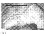
- FIG. 9shows delineation of a segment of a blood vessel for analysis with the method of the invention.
- FIG. 10shows the delineated segment of FIG. 9 with lines terminating at the vessel walls and line normals at the longitudinal end points;
- FIG. 11shows the vessel walls and line normals at the longitudinal end points of FIG. 10 with proper orientation
- FIG. 12shows splines connecting the longitudinal end points of FIG. 11 and a longitudinal intensity profile (upper left corner) taken before a procedure;
- FIG. 13shows splines connecting the longitudinal end points together with a longitudinal intensity profile (upper left corner) and the time dependence of the intensity profile (upper right corner) taken after a procedure.
- FIG. 1shows schematically a device for non-invasively determining blush of myocardial tissue by ICG fluorescence imaging.
- An infrared light sourcefor example, one or more diode lasers or LEOs, with a peak emission of about 780-800 nm for exciting fluorescence in ICG is located inside housing 1 .
- the fluorescence signalis detected by a CCD camera 2 having adequate near-IR sensitivity; such cameras are commercially available from several vendors (Hitachi, Hamamatsu, etc.).
- the CCD camera 2may have a viewfinder 8 , but the image may also be viewed during the operation on an external monitor which may be part of an electronic image processing and evaluation system 11 .
- a light beam 3which may be a divergent or a scanned beam, emerges from the housing 1 to illuminate an area of interest 4 , i.e. the area where the blush of myocardial tissue is to be measured.
- the area of interestmay be about 10 cm ⁇ 10 cm, but may vary based on surgical requirements and the available illumination intensity and camera sensitivity.
- a filter 6is typically placed in front of the camera lens 7 to block excitation light from reaching the camera sensor, while allowing fluorescence light to pass through.
- the filter 6may be an NIR long-wave pass filter (cut filter), which is only transparent to wavelengths greater than about 815 nm, or preferably a bandpass filter transmitting at peak wavelengths of between about 830 and about 845 nm and having a full width at half maximum (FWHM) transmission window of between about 10 nm and 25 nm in order to block the excitation wavelength band.
- the camera 2may also be designed to acquire a color image of the area of interest to allow real-time correlation between the fluorescence image and the color image.
- Blood vessels visible in the imagetypically include major blood vessels, e.g., arteries; however, arterial blood flow may not be of interest to the surgeon when considering perfusion of the surrounding myocardial tissue. Because these blood vessels may have either a higher or a lower brightness in the image, depending on the phase of the cardiac cycle, contributions from blood vessels to the measured image brightness may alter the myocardial blush grade by skewing the average image brightness upward or downward. In order to obtain a correct value for the myocardial blush, the contributions from the blood vessels must be eliminated before the blush grade is computed.
- FIG. 2shows a typical ICG fluorescent image of a heart showing blood vessels and myocardial tissue, with a rectangle delineating a static ROI on the imaged area.
- the ROIis static, meaning that it does not track tissue movement when the heart is beating. This simplifies the computation, while the results computed with the method of the invention are robust and largely insensitive to tissue movement.
- a histogram of the grayscale intensity values in the ROI of FIG. 2is generated.
- a sliding window Wis applied across the abscissa, and the smallest set of adjacent histogram bins containing in excess of a predetermined percentage of the total intensity is determined.
- a percentage value of 50%is selected as criterion for the bins to be included, although other values can be selected as long as these selected values exclude outliers and provide a reliable assessment of the blush.
- the smallest set of adjacent histogram bins containing at least 50% of the intensity countsresults in a window W which is 12 bins wide and includes the intensity values between 120 and
- the average intensity for the static ROIis then computed using only the values inside the window determined above, i.e., the number of pixels in a bin multiplied with the intensity in that bin and summed over all bins within the window W.
- This approachexcludes the intensity outliers (both low and high intensity values) from the computation of the average intensity representing the myocardial blush value in the ROI. In other words, only intensity values between 120 and 131 within the ROI are included in the subsequent calculation.
- FIG. 4shows the location of pixels within the static ROI with intensity values within the window W (according to the selection criterion that about 50% of the intensity values are located within the window W).
- the bright areasindicate the pixels included. As can be seen, the area with the included pixels need not be contiguous.
- FIG. 5shows the static ROI of FIG. 2 (top image) and a smaller static ROI (bottom image) located within the ROI of the top image.
- the smaller ROIincludes less arterial blood vessels.
- FIG. 6shows schematically the computed average intensity for both the static ROIs of FIG. 5 taken over a 28 second time interval.
- the elapsed time(from the point an increase in the intensity was detected, in seconds) is plotted on the abscissa, and the average intensity for the static ROI (in arbitrary units) is plotted on the ordinate.
- the two curvesmatch within about 1-3 percent.
- the maximum blushis approximately 112 [arb. units]
- the blush rate measured over about 6.1 sec from about zero blush to about the maximum valueis in linear approximation about 16.2 [arb. units]/sec
- the washout rate measured over about 6.1 sec from about the maximum blush value to about 15-20% blushis in linear approximation about 10.5 [arb. units]/sec.
- Blushappears to increase and decrease (washout) exponentially, so the linear curve fitting described above should be considered only as an approximation.
- Other characteristic values of the curves of FIG. 6such as a maximum slope or a curve fit with an exponential rise and decay time may also be used.
- the static ROI algorithm described abovedoes not rely on image tracking and is generally insensitive to the motion artifacts because of the exclusion of outliers. It is computationally fast and works well with both low and high contrast images.
- FIG. 7shows pictures of the heart before and after a surgical procedure has been performed on the heart. A comparison of the blush determined with the aforedescribed method of the invention before and after the procedure can be used to determine whether perfusion has improved as a result of the procedure.
- the ICG dosage, illumination level and camera sensitivity settingsshould be adjusted so that the detector in the camera does not saturate when areas in the image, such as arteries, reach their maximum intensity. If the camera nevertheless does saturate, the user needs to decide whether the computed blush rate and washout rate are likely to represent the actual rates, had the detector not saturated.
- Two approachesare proposed for comparing image data obtained before and after the procedure: (1) comparing the blush and washout rates before and after the procedure; and (2) comparing the elapsed time from blood vessel peak intensity to maximum blush on images taken before and after the procedure.
- a time series of fluorescence images of the anatomyis acquired before (top image of FIG. 7 ) and after the surgical procedure (bottom image of FIG. 7 ) by, for example, injecting a bolus of ICG dye. Only one of the time series of images is shown. A ROI is delineated in each of the images in approximately the same area of the anatomy. The average intensity of the blush is then determined in each of, or in a subset of, the fluorescence images in the time series with the method of the invention described above with reference to the histogram of FIG. 3 , which excludes outliers, such as arteries. The average ROI intensity from each image in the time series is normalized to the baseline average intensity of the ROI in the first frame to correct for residual ICG that may have remained in the system.
- FIG. 8shows schematically the computed average intensities (about 50% of the intensity values are located within the window W of a histogram corresponding to the histogram of FIG. 3 ) for the static ROIs of FIG. 7 taken over a 28 second time interval.
- the top graphrepresents values before the procedure and the bottom graph values after the procedure.
- the elapsed time(from the point an increase in the intensity was detected, in seconds) is plotted on the abscissa, and the average intensity for the static ROI (in arbitrary units) is plotted on the ordinate.
- the broken line through the datarepresents a smoothed curve of the raw data. This helps to mask variation in the measurement due to motion caused by the cardiac cycle or respiration and serves as a visual guide for assessing the blush rate and washout rate. As mentioned above, saturation of the sensor should be avoided, because saturation would make an absolute determination of the slope impractical.
- the blush and washout ratesare determined from the corresponding slopes of straight lines connecting the 5% and 95% points in the average intensity curves, i.e., the start of blush is taken as the time at which the intensity rises above the baseline by 5% of its maximum value, and the 95% point is the time at which the intensity reaches 95% of its maximum value.
- the 5% and 95% thresholdsare heuristic thresholds used to discount for any noise that may appear in the image both before the blush appears, and as it nears its maximum value.
- the slope of the straight linesrepresents an average rate, and that the rate can also be determined from a least-square curve fit or by selecting points other than 5% and 95%, as described in the illustrated example.
- the blush rate following the procedureis about 43 units/sec, compared to about 18 units/sec before the procedure, representing an improvement of about 140%.
- the washout rate following the procedureis about 21 units/sec, compared to about 10 units/sec before the procedure, representing an improvement of more than 100%. Greater perfusion (blush) and washout rates suggest faster movement of blood and greater maximum blush suggests a greater volume of ICG-bound blood in the tissue and are hence clear indicators of improved perfusion through the tissue.
- perfusionis determined from the time of maximum blood vessel (artery) intensity to maximum myocardial blush.
- arteryblood vessel
- maximum myocardial blushis determined from the histogram of the first region, as described above ( FIG. 8 ).
- Peak intensity of the blood vesselmay advantageously be determined from an area in the first region showing pixel intensity greater than that of the surrounding tissue.
- a histogram of the grayscale intensity valuesmay be constructed for the first region and a sliding window W applied across the abscissa, wherein the smallest set of adjacent histogram bins containing a predetermined percentage, for example about 20%, of the pixels with the highest intensity.
- a predetermined percentagefor example about 20%
- first and second regionsneed not be separate, but may 20 overlap or even be identical, as long as the fluorescence signals from the blood vessels and the myocardial tissue can be clearly separated in the histogram.
- the myocardial areamay reach maximum blush two seconds after the coronary artery reaches maximum fluorescence intensity. After the procedure, it may only take one second for the myocardial blush to reach maximum blush after the coronary artery reaches maximum fluorescence intensity following the vessel reaching maximum. This finding would lead to the conclusion that cardiac function has improved.
- a blood vesselmay move laterally during image acquisition which may make it more difficult to reliably determine the fluorescence intensity, for example during ICG imaging, of a coronary artery.
- the proposed methodprovides a means for tracking the movement of the vessel by determining several, typically three, lines which follow the contour of a segment of interest of the blood vessel and approximately span the width of the vessel.
- features or edges in the imageare determined by filtering using a convolution with the Laplacian-of-a-Gaussian kernel.
- the detected edgesmay be enhanced (thickened) by defining the edge by a width of at least two pixels. Both the original and the edge-enhanced images are stored.
- an operatordelimits the segment of the vessel of interest by drawing two lines across the vessel, for example with a computer mouse ( FIG. 9 ).
- the systemuses the previously determined edge information to detect the segment of each line located between the vessel edges and the mid-point of that segment, which is necessarily also the mid-point of the vessel, and constructs a line normal to each line segment ( FIG. 10 ). Thereafter, the system aligns two line normals with the major longitudinal axis of the vessel ( FIG. 11 ).
- the systemconstructs a series of 3 parallel lines, for example cubic spline, of approximately equal length joining the two ends of the segment of interest.
- the lineshave at their respective end points the same slope as the respective line normals.
- Three exemplary lines which approximately span the width of the vesselare shown in FIG. 12 .
- the pixel intensityis sampled at points of each line along the longitudinal axis of the vessel.
- intensitiesare averaged across the three lines at each location along the longitudinal axis to produce an average vessel intensity at each location in the vessel.
- the average intensity in the vessel segmentis approximately 55, substantially independent of the longitudinal location in the vessel.
- the processis then repeated for the time series of images frame-by-frame, while making sure that the positions match from one frame to the next.
- FIG. 13illustrates a final frame in the image sequence processed in this manner.
- the insert at the top left corner of FIG. 13shows, as in FIG. 12 , the averaged pixel intensity along the three lines.
- the insert at the top right corner of FIG. 13shows the change in the average intensity for all of the processed time-ordered frame sequence of images.
- the “fill time” of the blood vesselcan be calculated from the slope of the latter curve (pixel intensity vs. time).
- the preceding conceptscan be extended to develop quantitative indices useful for intraoperative assessment of blood flow in surgical flaps and for identifying vascular compromise.
- I′ Inis a measure for the rate of change of increasing perfusion with time as evidenced by the rate of ICG ingress or wash-in.
- I′ Outis a measure for the rate of change of decreasing perfusion with time after reaching maximum fluorescence intensity as evidenced by the rate of ICG egress or wash-out.
- Each of the measuresmay be taken on a flap either pre- and post-operatively or, once the flap is in place, the measures may be taken from the flap and from adjacent native tissue.
- I′ in-Prebeing the rate of ICG ingress measured on either adjacent native tissue or on the flap pre-operatively
- I′ in-Postbeing the rate of ICG ingress measured on the flap post-operatively
- I′ Out-Prebeing the rate of ICG egress measured on either adjacent native tissue or on the flap pre-operatively
- I′ Out-Postbeing the rate of ICG egress measured on the flap post-operatively
- WR InI′ in-Post /I′ in-Pre
- WR OutI′ Out-Post /I′ Out-Pre .
- WR In and WR Outwill be close to 1.0 in cases with normal vascular conditions.
- WR Inwill be significantly less than 1.0 in cases of arterial spasm or partial arterial occlusion.
- This metricwill vary inversely to the degree of arterial spasm or partial arterial occlusion; the amount by which this metric is less than 1.0 will correlate with increased arterial spasm or arterial occlusion.
- WR Outwill be significantly less than 1.0 in cases of venous congestion. This metric will vary inversely to the degree of venous congestion; the amount by which this metric is less than 1.0 will correlate with increased venous congestion.
Landscapes
- Health & Medical Sciences (AREA)
- Engineering & Computer Science (AREA)
- Life Sciences & Earth Sciences (AREA)
- Physics & Mathematics (AREA)
- Medical Informatics (AREA)
- General Health & Medical Sciences (AREA)
- Animal Behavior & Ethology (AREA)
- Public Health (AREA)
- Veterinary Medicine (AREA)
- Biomedical Technology (AREA)
- Heart & Thoracic Surgery (AREA)
- Surgery (AREA)
- Molecular Biology (AREA)
- Pathology (AREA)
- Biophysics (AREA)
- Computer Vision & Pattern Recognition (AREA)
- General Physics & Mathematics (AREA)
- Theoretical Computer Science (AREA)
- Cardiology (AREA)
- Physiology (AREA)
- Hematology (AREA)
- Nuclear Medicine, Radiotherapy & Molecular Imaging (AREA)
- Radiology & Medical Imaging (AREA)
- Vascular Medicine (AREA)
- Signal Processing (AREA)
- Oral & Maxillofacial Surgery (AREA)
- High Energy & Nuclear Physics (AREA)
- Dentistry (AREA)
- Optics & Photonics (AREA)
- Multimedia (AREA)
- Power Engineering (AREA)
- Anesthesiology (AREA)
- Artificial Intelligence (AREA)
- Psychiatry (AREA)
- Quality & Reliability (AREA)
- Investigating, Analyzing Materials By Fluorescence Or Luminescence (AREA)
- Apparatus For Radiation Diagnosis (AREA)
Abstract
Description
- 1 The selected area of the anatomy within the ROI should consist primarily of myocardial tissue, while minimizing the effects from blood vessels, clips, etc. that appear in the ROI and may move in and out of the ROI when the heart is beating.
- 2 The measured myocardial blush value should be substantially independent of the size of the ROI in the selected area of the anatomy.
WRIn=I′in-Post/I′in-Pre
WROut=I′Out-Post/I′Out-Pre.
Claims (1)
Priority Applications (6)
| Application Number | Priority Date | Filing Date | Title |
|---|---|---|---|
| US12/841,659US8406860B2 (en) | 2008-01-25 | 2010-07-22 | Method for evaluating blush in myocardial tissue |
| US13/850,063US8965488B2 (en) | 2008-01-25 | 2013-03-25 | Method for evaluating blush in myocardial tissue |
| US14/598,832US9610021B2 (en) | 2008-01-25 | 2015-01-16 | Method for evaluating blush in myocardial tissue |
| US15/476,290US9936887B2 (en) | 2008-01-25 | 2017-03-31 | Method for evaluating blush in myocardial tissue |
| US15/947,221US10835138B2 (en) | 2008-01-25 | 2018-04-06 | Method for evaluating blush in myocardial tissue |
| US17/099,675US11564583B2 (en) | 2008-01-25 | 2020-11-16 | Method for evaluating blush in myocardial tissue |
Applications Claiming Priority (4)
| Application Number | Priority Date | Filing Date | Title |
|---|---|---|---|
| US2381808P | 2008-01-25 | 2008-01-25 | |
| PCT/CA2009/000073WO2009092162A1 (en) | 2008-01-25 | 2009-01-23 | Method for evaluating blush in myocardial tissue |
| US24368809P | 2009-09-18 | 2009-09-18 | |
| US12/841,659US8406860B2 (en) | 2008-01-25 | 2010-07-22 | Method for evaluating blush in myocardial tissue |
Related Parent Applications (2)
| Application Number | Title | Priority Date | Filing Date |
|---|---|---|---|
| PCT/CA2006/000073Continuation-In-PartWO2006079195A1 (en) | 2005-01-26 | 2006-01-20 | Golf putt measuring device |
| PCT/CA2009/000073Continuation-In-PartWO2009092162A1 (en) | 2008-01-25 | 2009-01-23 | Method for evaluating blush in myocardial tissue |
Related Child Applications (1)
| Application Number | Title | Priority Date | Filing Date |
|---|---|---|---|
| US13/850,063DivisionUS8965488B2 (en) | 2008-01-25 | 2013-03-25 | Method for evaluating blush in myocardial tissue |
Publications (2)
| Publication Number | Publication Date |
|---|---|
| US20100305454A1 US20100305454A1 (en) | 2010-12-02 |
| US8406860B2true US8406860B2 (en) | 2013-03-26 |
Family
ID=43221020
Family Applications (6)
| Application Number | Title | Priority Date | Filing Date |
|---|---|---|---|
| US12/841,659Active2029-04-09US8406860B2 (en) | 2008-01-25 | 2010-07-22 | Method for evaluating blush in myocardial tissue |
| US13/850,063ActiveUS8965488B2 (en) | 2008-01-25 | 2013-03-25 | Method for evaluating blush in myocardial tissue |
| US14/598,832ActiveUS9610021B2 (en) | 2008-01-25 | 2015-01-16 | Method for evaluating blush in myocardial tissue |
| US15/476,290ActiveUS9936887B2 (en) | 2008-01-25 | 2017-03-31 | Method for evaluating blush in myocardial tissue |
| US15/947,221Active2030-02-27US10835138B2 (en) | 2008-01-25 | 2018-04-06 | Method for evaluating blush in myocardial tissue |
| US17/099,675Active2029-09-30US11564583B2 (en) | 2008-01-25 | 2020-11-16 | Method for evaluating blush in myocardial tissue |
Family Applications After (5)
| Application Number | Title | Priority Date | Filing Date |
|---|---|---|---|
| US13/850,063ActiveUS8965488B2 (en) | 2008-01-25 | 2013-03-25 | Method for evaluating blush in myocardial tissue |
| US14/598,832ActiveUS9610021B2 (en) | 2008-01-25 | 2015-01-16 | Method for evaluating blush in myocardial tissue |
| US15/476,290ActiveUS9936887B2 (en) | 2008-01-25 | 2017-03-31 | Method for evaluating blush in myocardial tissue |
| US15/947,221Active2030-02-27US10835138B2 (en) | 2008-01-25 | 2018-04-06 | Method for evaluating blush in myocardial tissue |
| US17/099,675Active2029-09-30US11564583B2 (en) | 2008-01-25 | 2020-11-16 | Method for evaluating blush in myocardial tissue |
Country Status (1)
| Country | Link |
|---|---|
| US (6) | US8406860B2 (en) |
Cited By (11)
| Publication number | Priority date | Publication date | Assignee | Title |
|---|---|---|---|---|
| US9610021B2 (en) | 2008-01-25 | 2017-04-04 | Novadaq Technologies Inc. | Method for evaluating blush in myocardial tissue |
| US9816930B2 (en) | 2014-09-29 | 2017-11-14 | Novadaq Technologies Inc. | Imaging a target fluorophore in a biological material in the presence of autofluorescence |
| US10041042B2 (en) | 2008-05-02 | 2018-08-07 | Novadaq Technologies ULC | Methods for production and use of substance-loaded erythrocytes (S-IEs) for observation and treatment of microvascular hemodynamics |
| US10219742B2 (en) | 2008-04-14 | 2019-03-05 | Novadaq Technologies ULC | Locating and analyzing perforator flaps for plastic and reconstructive surgery |
| US10265419B2 (en) | 2005-09-02 | 2019-04-23 | Novadaq Technologies ULC | Intraoperative determination of nerve location |
| US10278585B2 (en) | 2012-06-21 | 2019-05-07 | Novadaq Technologies ULC | Quantification and analysis of angiography and perfusion |
| US10434190B2 (en) | 2006-09-07 | 2019-10-08 | Novadaq Technologies ULC | Pre-and-intra-operative localization of penile sentinel nodes |
| US10492671B2 (en) | 2009-05-08 | 2019-12-03 | Novadaq Technologies ULC | Near infra red fluorescence imaging for visualization of blood vessels during endoscopic harvest |
| US10631746B2 (en) | 2014-10-09 | 2020-04-28 | Novadaq Technologies ULC | Quantification of absolute blood flow in tissue using fluorescence-mediated photoplethysmography |
| US10992848B2 (en) | 2017-02-10 | 2021-04-27 | Novadaq Technologies ULC | Open-field handheld fluorescence imaging systems and methods |
| US12239409B2 (en) | 2022-02-28 | 2025-03-04 | Visionsense Ltd. | Fluorescence imaging camera assembly for open surgery |
Families Citing this family (30)
| Publication number | Priority date | Publication date | Assignee | Title |
|---|---|---|---|---|
| CA3194784A1 (en) | 2008-05-20 | 2009-11-26 | University Health Network | Device and method for fluorescence-based imaging and monitoring |
| US9921156B2 (en) | 2013-02-04 | 2018-03-20 | The General Hospital Corporation | System and method for fluorescence detection |
| GB2512876A (en)* | 2013-04-09 | 2014-10-15 | Image Analysis Ltd | Methods and apparatus for quantifying inflammation |
| JP6030035B2 (en)* | 2013-09-27 | 2016-11-24 | 富士フイルム株式会社 | Fluorescence observation apparatus, endoscope system, processor apparatus, and operation method |
| ITMO20130326A1 (en)* | 2013-11-29 | 2015-05-30 | Istituto Naz Tumori Fondazi One G Pascale | ANALYSIS METHOD |
| US9730662B2 (en)* | 2014-01-17 | 2017-08-15 | Siemens Medical Solutions Usa, Inc. | System and method for tracking blood flow |
| JP6769949B2 (en) | 2014-07-24 | 2020-10-14 | ユニバーシティー ヘルス ネットワーク | Data collection and analysis for diagnostic purposes |
| WO2016061041A1 (en) | 2014-10-14 | 2016-04-21 | East Carolina University | Methods, systems and computer program products for determining hemodynamic status parameters using signals derived from multispectral blood flow and perfusion imaging |
| JP6813245B2 (en) | 2014-10-14 | 2021-01-13 | イースト カロライナ ユニバーシティ | How to operate a system to combine anatomical and physiological data on a single image, a computer system, and a program to combine anatomical and physiological data on a single image. Recording medium on which |
| US11553844B2 (en)* | 2014-10-14 | 2023-01-17 | East Carolina University | Methods, systems and computer program products for calculating MetaKG signals for regions having multiple sets of optical characteristics |
| US20160213303A1 (en)* | 2015-01-22 | 2016-07-28 | Elwha LLC, a limited liability company of the State of Delaware | Devices and methods for remote hydration measurement |
| US10159439B2 (en) | 2015-01-22 | 2018-12-25 | Elwha Llc | Devices and methods for remote hydration measurement |
| US10130301B2 (en) | 2015-01-22 | 2018-11-20 | Elwha Llc | Devices and methods for remote hydration measurement |
| CN107530452A (en) | 2015-02-02 | 2018-01-02 | 诺瓦达克技术公司 | Methods and systems for characterizing tissue of a subject |
| US10390718B2 (en) | 2015-03-20 | 2019-08-27 | East Carolina University | Multi-spectral physiologic visualization (MSPV) using laser imaging methods and systems for blood flow and perfusion imaging and quantification in an endoscopic design |
| US10058256B2 (en) | 2015-03-20 | 2018-08-28 | East Carolina University | Multi-spectral laser imaging (MSLI) methods and systems for blood flow and perfusion imaging and quantification |
| EP3319515B1 (en)* | 2015-07-06 | 2020-03-18 | Scinovia Corp. | Fluorescence based flow imaging and measurements |
| JP6640334B2 (en)* | 2015-09-23 | 2020-02-05 | ノバダック テクノロジーズ ユーエルシー | Methods and systems for management of data derived from medical imaging |
| USD916294S1 (en) | 2016-04-28 | 2021-04-13 | Stryker European Operations Limited | Illumination and imaging device |
| WO2018018160A1 (en) | 2016-07-29 | 2018-02-01 | Novadaq Technologies ULC | Methods and systems for characterizing tissue of a subject utilizing machine learning |
| US10271750B2 (en)* | 2016-12-09 | 2019-04-30 | Perfusion Tech IVS | System and method for assessing perfusion in an anatomical structure |
| USD844005S1 (en)* | 2016-12-23 | 2019-03-26 | Novadaq Technologies ULC | Device for illumination and imaging of a target |
| US20220313092A1 (en)* | 2017-10-26 | 2022-10-06 | Fluoptics | Method and device for monitoring the fluorescence emitted at the surface of a biological tissue |
| US11911139B2 (en) | 2018-06-14 | 2024-02-27 | Perfusion Tech Aps | System and method for automatic perfusion measurement |
| AU2019285086B2 (en) | 2018-06-14 | 2025-03-27 | Perfusion Tech Aps | System and method for automatic perfusion measurement |
| US11571128B2 (en) | 2018-10-24 | 2023-02-07 | The George Washington University | Fast label-free method for mapping cardiac physiology |
| US11744643B2 (en) | 2019-02-04 | 2023-09-05 | Covidien Lp | Systems and methods facilitating pre-operative prediction of post-operative tissue function |
| CN111862046B (en)* | 2020-07-21 | 2023-11-17 | 江苏省人民医院(南京医科大学第一附属医院) | A system and method for identifying catheter position in cardiac coronary artery silhouette |
| WO2022058966A1 (en)* | 2020-09-18 | 2022-03-24 | Stryker European Operations Limited | Systems and methods for determining timing for fluorescence based blood flow assessment |
| US20230076477A1 (en)* | 2021-09-05 | 2023-03-09 | Shimadzu Corporation | Imaging device and imaging method |
Citations (1)
| Publication number | Priority date | Publication date | Assignee | Title |
|---|---|---|---|---|
| US20100036217A1 (en)* | 2006-10-11 | 2010-02-11 | Korea Advanced Institute Of Science And Technology | System for analyzing tissue perfusion using concentration of inocyanine green in blood |
Family Cites Families (406)
| Publication number | Priority date | Publication date | Assignee | Title |
|---|---|---|---|---|
| JPS5854822B2 (en) | 1975-09-11 | 1983-12-06 | ミノルタ株式会社 | Kodenshikisei Taikeisokuki |
| US4109647A (en) | 1977-03-16 | 1978-08-29 | The United States Of America As Represented By The Secretary Of The Department Of Health, Education And Welfare | Method of and apparatus for measurement of blood flow using coherent light |
| JPS5918532B2 (en) | 1977-06-15 | 1984-04-27 | 石川島播磨重工業株式会社 | supercharger |
| US4263916A (en) | 1978-03-27 | 1981-04-28 | University Of Southern California | Image averaging for angiography by registration and combination of serial images |
| US4162405A (en) | 1978-05-23 | 1979-07-24 | Britton Chance | Flying spot fluoro-meter for oxidized flavoprotein and reduced pyridine nucleotide |
| US4200801A (en) | 1979-03-28 | 1980-04-29 | The United States Of America As Represented By The United States Department Of Energy | Portable spotter for fluorescent contaminants on surfaces |
| US4394199A (en) | 1981-09-08 | 1983-07-19 | Agnus Chemical Company | Explosive emulsion composition |
| JPS58141135A (en) | 1981-10-20 | 1983-08-22 | 富士写真フイルム株式会社 | Image transmitting system of endoscope using solid image sensor |
| EP0091805B1 (en) | 1982-04-08 | 1989-07-26 | Olympus Optical Co., Ltd. | Endoscope focus state detectors |
| JPS58222331A (en) | 1982-06-21 | 1983-12-24 | Sony Corp | Reproducer of character information |
| JPS5940830A (en) | 1982-08-31 | 1984-03-06 | 浜松ホトニクス株式会社 | Apparatus for diagnosis of cancer using laser beam pulse |
| JPS5969721A (en) | 1982-10-15 | 1984-04-20 | Olympus Optical Co Ltd | Endoscope measuring device |
| JPS5970903A (en) | 1982-10-15 | 1984-04-21 | Olympus Optical Co Ltd | Automatic measuring apparatus of endoscope |
| US4541438A (en) | 1983-06-02 | 1985-09-17 | The Johns Hopkins University | Localization of cancerous tissue by monitoring infrared fluorescence emitted by intravenously injected porphyrin tumor-specific markers excited by long wavelength light |
| US4532918A (en) | 1983-10-07 | 1985-08-06 | Welch Allyn Inc. | Endoscope signal level control |
| JPS60256443A (en) | 1984-05-31 | 1985-12-18 | オムロン株式会社 | Image measuring apparatus |
| US4559557A (en) | 1984-06-01 | 1985-12-17 | General Electric Company | Region-of-interest digital subtraction angiography |
| SE455646B (en) | 1984-10-22 | 1988-07-25 | Radians Innova Ab | FLUORESCENT DEVICE |
| US5125404A (en) | 1985-03-22 | 1992-06-30 | Massachusetts Institute Of Technology | Apparatus and method for obtaining spectrally resolved spatial images of tissue |
| US5318024A (en) | 1985-03-22 | 1994-06-07 | Massachusetts Institute Of Technology | Laser endoscope for spectroscopic imaging |
| US4718417A (en) | 1985-03-22 | 1988-01-12 | Massachusetts Institute Of Technology | Visible fluorescence spectral diagnostic for laser angiosurgery |
| DE3511255A1 (en) | 1985-03-28 | 1986-10-02 | Grün Optik Wetzlar GmbH, 6330 Wetzlar | ARRANGEMENT FOR THE INDIVIDUAL CONTROL OF THE INTENSITY OF SEVERAL SPECTRAL LAMPS |
| CN85100424B (en) | 1985-04-01 | 1986-10-29 | 上海医疗器械研究所 | Inherent fluorescence diagnostic instrument for malignant tumor |
| US4619249A (en) | 1985-07-24 | 1986-10-28 | Kim Landry | Transcutaneous intravenous illuminator |
| AT387860B (en) | 1985-09-16 | 1989-03-28 | Optical Sensing Technology | METHOD AND DEVICE FOR TUMOR DIAGNOSIS BY MEANS OF SERA |
| US4719508A (en) | 1985-10-02 | 1988-01-12 | Olympus Optical Co., Ltd. | Endoscopic photographing apparatus |
| US5134662A (en) | 1985-11-04 | 1992-07-28 | Cell Analysis Systems, Inc. | Dual color camera microscope and methodology for cell staining and analysis |
| US5042494A (en) | 1985-11-13 | 1991-08-27 | Alfano Robert R | Method and apparatus for detecting cancerous tissue using luminescence excitation spectra |
| US4930516B1 (en) | 1985-11-13 | 1998-08-04 | Laser Diagnostic Instr Inc | Method for detecting cancerous tissue using visible native luminescence |
| US4774568A (en) | 1986-01-27 | 1988-09-27 | Kabushiki Kaisha Toshiba | Endoscopic apparatus |
| JPS62247232A (en) | 1986-04-21 | 1987-10-28 | Agency Of Ind Science & Technol | Fluorescence measuring apparatus |
| GB2203831B (en) | 1986-07-07 | 1991-02-06 | Academy Of Applied Sciences | Apparatus and method for the diagnosis of malignant tumours |
| JPH0763443B2 (en) | 1986-09-30 | 1995-07-12 | 株式会社東芝 | Electronic endoscope |
| JPS63122421A (en) | 1986-11-12 | 1988-05-26 | 株式会社東芝 | endoscope equipment |
| US5255087A (en) | 1986-11-29 | 1993-10-19 | Olympus Optical Co., Ltd. | Imaging apparatus and endoscope apparatus using the same |
| JP2572394B2 (en) | 1987-03-19 | 1997-01-16 | オリンパス光学工業株式会社 | Electronic endoscope |
| JPH0783B2 (en) | 1987-03-30 | 1995-01-11 | 株式会社東芝 | Electronic endoscopic device |
| US4986262A (en) | 1987-03-31 | 1991-01-22 | Kabushiki Kaisha Toshiba | Measuring endoscope |
| US4852579A (en) | 1987-04-20 | 1989-08-01 | Karl Storz Endoscopy Gmbh And Company | Photocharacterization and treatment of normal abnormal and ectopic endometrium |
| US4900934A (en) | 1987-07-15 | 1990-02-13 | University Of Utah | Apparatus for simultaneous visualization and measurement of fluorescence from fluorescent dye-treated cell preparations and solutions |
| JPH0824668B2 (en) | 1987-09-14 | 1996-03-13 | オリンパス光学工業株式会社 | Electronic endoscopic device |
| US4858001A (en) | 1987-10-08 | 1989-08-15 | High-Tech Medical Instrumentation, Inc. | Modular endoscopic apparatus with image rotation |
| JPH01160525A (en) | 1987-12-17 | 1989-06-23 | Olympus Optical Co Ltd | Endoscope |
| DE3906860A1 (en) | 1988-03-08 | 1989-09-28 | Fraunhofer Ges Forschung | DEVICE FOR PRODUCING ANGIOGRAPHY |
| JPH01236879A (en) | 1988-03-17 | 1989-09-21 | Canon Inc | Picture encoder |
| JPH06105190B2 (en) | 1988-03-31 | 1994-12-21 | 工業技術院長 | Signal analyzer |
| US4998972A (en) | 1988-04-28 | 1991-03-12 | Thomas J. Fogarty | Real time angioscopy imaging system |
| US5078150A (en) | 1988-05-02 | 1992-01-07 | Olympus Optical Co., Ltd. | Spectral diagnosing apparatus with endoscope |
| IL90188A0 (en) | 1988-05-18 | 1989-12-15 | Cryopharm Corp | Process and medium for the lyophilization of erythrocytes |
| US4938205A (en) | 1988-05-27 | 1990-07-03 | The University Of Connecticut | Endoscope with traced raster and elemental photodetectors |
| US5178616A (en) | 1988-06-06 | 1993-01-12 | Sumitomo Electric Industries, Ltd. | Method and apparatus for intravascular laser surgery |
| US4995396A (en) | 1988-12-08 | 1991-02-26 | Olympus Optical Co., Ltd. | Radioactive ray detecting endoscope |
| US5353799A (en) | 1991-01-22 | 1994-10-11 | Non Invasive Technology, Inc. | Examination of subjects using photon migration with high directionality techniques |
| DE68925586T2 (en) | 1988-12-21 | 1996-10-24 | Massachusetts Inst Technology | METHOD FOR LASER-INDUCED FLUORESCENCE OF TISSUE |
| US5009233A (en) | 1989-01-10 | 1991-04-23 | Lasermike, Inc. | Miniaturized hand-held laser scanning micrometer |
| JP2987816B2 (en) | 1989-01-30 | 1999-12-06 | オリンパス光学工業株式会社 | Fluorescence observation device |
| DE3903019A1 (en) | 1989-02-02 | 1990-08-09 | Hell Rudolf Dr Ing Gmbh | OPTICAL COLOR DIVIDER ARRANGEMENT |
| SE8900612D0 (en) | 1989-02-22 | 1989-02-22 | Jonas Johansson | TISSUE CHARACTERIZATION USING A BLOOD-FREE FLUORESCENCE CRITERIA |
| EP0466828A1 (en) | 1989-04-14 | 1992-01-22 | Massachusetts Institute Of Technology | Spectral diagnosis of diseased tissue |
| US5421337A (en) | 1989-04-14 | 1995-06-06 | Massachusetts Institute Of Technology | Spectral diagnosis of diseased tissue |
| KR100190423B1 (en) | 1989-06-06 | 1999-06-01 | 기타지마 요시도시 | Method and device for correcting defects such as emulsion mask |
| US4993404A (en) | 1989-06-26 | 1991-02-19 | Lane Timothy G | Fluoroscopy switching device |
| CN1049781A (en) | 1989-09-02 | 1991-03-13 | 住友电气工业株式会社 | Laser surgery instruments for vascular surgery |
| JPH0614921B2 (en) | 1989-09-29 | 1994-03-02 | 浜松ホトニクス株式会社 | Fluorescence observation device for biological tissue |
| US5150292A (en) | 1989-10-27 | 1992-09-22 | Arch Development Corporation | Method and system for determination of instantaneous and average blood flow rates from digital angiograms |
| DE69114314T2 (en) | 1990-01-08 | 1996-04-18 | Ernest M D Feiler | Diagnostic procedure for measuring blood flow. |
| US5091652A (en) | 1990-01-12 | 1992-02-25 | The Regents Of The University Of California | Laser excited confocal microscope fluorescence scanner and method |
| US5420628A (en) | 1990-01-16 | 1995-05-30 | Research Development Foundation | Video densitometer with determination of color composition |
| US5131398A (en) | 1990-01-22 | 1992-07-21 | Mediscience Technology Corp. | Method and apparatus for distinguishing cancerous tissue from benign tumor tissue, benign tissue or normal tissue using native fluorescence |
| US4995398A (en) | 1990-04-30 | 1991-02-26 | Turnidge Patrick A | Coronary angiography imaging system |
| US5071417A (en) | 1990-06-15 | 1991-12-10 | Rare Earth Medical Lasers, Inc. | Laser fusion of biological materials |
| US5699798A (en) | 1990-08-10 | 1997-12-23 | University Of Washington | Method for optically imaging solid tumor tissue |
| US6196226B1 (en) | 1990-08-10 | 2001-03-06 | University Of Washington | Methods and apparatus for optically imaging neuronal tissue and activity |
| US5845639A (en) | 1990-08-10 | 1998-12-08 | Board Of Regents Of The University Of Washington | Optical imaging methods |
| US5465718A (en) | 1990-08-10 | 1995-11-14 | Hochman; Daryl | Solid tumor, cortical function, and nerve tissue imaging methods and device |
| US5438989A (en) | 1990-08-10 | 1995-08-08 | Hochman; Darryl | Solid tumor, cortical function, and nerve tissue imaging methods and device |
| US6671540B1 (en) | 1990-08-10 | 2003-12-30 | Daryl W. Hochman | Methods and systems for detecting abnormal tissue using spectroscopic techniques |
| US5997844A (en) | 1991-02-08 | 1999-12-07 | Diatide, Inc. | Technetium-99m labeled peptides for imaging |
| JPH04297236A (en) | 1991-03-26 | 1992-10-21 | Toshiba Corp | Digital fluorography system |
| JPH04307024A (en) | 1991-04-02 | 1992-10-29 | Olympus Optical Co Ltd | Electronic endoscope apparatus |
| US5377676A (en) | 1991-04-03 | 1995-01-03 | Cedars-Sinai Medical Center | Method for determining the biodistribution of substances using fluorescence spectroscopy |
| US5318023A (en) | 1991-04-03 | 1994-06-07 | Cedars-Sinai Medical Center | Apparatus and method of use for a photosensitizer enhanced fluorescence based biopsy needle |
| US6485413B1 (en) | 1991-04-29 | 2002-11-26 | The General Hospital Corporation | Methods and apparatus for forward-directed optical scanning instruments |
| US5117466A (en) | 1991-04-30 | 1992-05-26 | The United States Of America As Represented By The United States Department Of Energy | Integrated fluorescence analysis system |
| CA2042075C (en) | 1991-05-08 | 2001-01-23 | Branko Palcic | Endoscopic imaging system |
| US5225883A (en) | 1991-06-05 | 1993-07-06 | The Babcock & Wilcox Company | Video temperature monitor |
| SE468925B (en) | 1991-08-22 | 1993-04-19 | Gert Nilsson | A METHOD AND APPARATUS SHOULD REDUCE THE DISTANCE-BASED FACTOR IN Saturation of STRAIGHT MOVEMENTS WITH AN IMAGING LASER-DOUBLE TECHNIQUE, SPECIFICALLY IN SEATING BLOOD PERFUSION THROUGH |
| US5377686A (en) | 1991-10-11 | 1995-01-03 | The University Of Connecticut | Apparatus for detecting leakage from vascular tissue |
| JP3297725B2 (en) | 1991-12-09 | 2002-07-02 | 国立循環器病センター総長 | Contrast-enhanced blood vessel high-precision pipe diameter measuring device |
| US5214503A (en) | 1992-01-31 | 1993-05-25 | The United States Of America As Represented By The Secretary Of The Army | Color night vision camera system |
| US5235984A (en) | 1992-03-30 | 1993-08-17 | Hewlett-Packard Company | On-line acoustic densitometry tool for use with an ultrasonic imaging system |
| US6096289A (en) | 1992-05-06 | 2000-08-01 | Immunomedics, Inc. | Intraoperative, intravascular, and endoscopic tumor and lesion detection, biopsy and therapy |
| DE4220633C1 (en) | 1992-06-24 | 1994-02-03 | Wolf Gmbh Richard | Device for supplying light to endoscopes |
| US5279298A (en) | 1992-11-20 | 1994-01-18 | The Johns Hopkins University | Method and apparatus to identify and treat neovascular membranes in the eye |
| US5733721A (en) | 1992-11-20 | 1998-03-31 | The Board Of Regents Of The University Of Oklahoma | Cell analysis method using quantitative fluorescence image analysis |
| GB2275198B (en) | 1993-02-18 | 1997-03-26 | Central Research Lab Ltd | Apparatus for irradiating an area with a controllable pattern of light |
| US5437274A (en) | 1993-02-25 | 1995-08-01 | Gholam A. Peyman | Method of visualizing submicron-size vesicles and particles in blood circulation |
| JP3228627B2 (en) | 1993-03-19 | 2001-11-12 | オリンパス光学工業株式会社 | Endoscope image processing device |
| US5431161A (en) | 1993-04-15 | 1995-07-11 | Adac Laboratories | Method and apparatus for information acquistion, processing, and display within a medical camera system |
| US5421339A (en) | 1993-05-12 | 1995-06-06 | Board Of Regents, The University Of Texas System | Diagnosis of dysplasia using laser induced fluoroescence |
| WO1996009792A1 (en) | 1993-05-17 | 1996-04-04 | The Johns Hopkins University | Improved visualization of choroidal blood flow and aberrant vascular structures in the eye |
| US5394199A (en) | 1993-05-17 | 1995-02-28 | The Johns Hopkins University | Methods and apparatus for improved visualization of choroidal blood flow and aberrant vascular structures in the eye using fluorescent dye angiography |
| US5424841A (en) | 1993-05-28 | 1995-06-13 | Molecular Dynamics | Apparatus for measuring spatial distribution of fluorescence on a substrate |
| US5673701A (en) | 1994-10-07 | 1997-10-07 | Non Invasive Technology, Inc. | Optical techniques for examination of biological tissue |
| DK75593D0 (en) | 1993-06-25 | 1993-06-25 | Novo Nordisk As | |
| US5365057A (en) | 1993-07-02 | 1994-11-15 | Litton Systems, Inc. | Light-weight night vision device |
| US5371355A (en) | 1993-07-30 | 1994-12-06 | Litton Systems, Inc. | Night vision device with separable modular image intensifier assembly |
| JP3224640B2 (en) | 1993-07-30 | 2001-11-05 | 三菱重工業株式会社 | Apparatus and method for measuring concentration by LIF |
| JPH0765154A (en) | 1993-08-31 | 1995-03-10 | Toshiba Corp | Quantitative analysis apparatus for blood vessel image and quantitative analysis method thereof |
| JPH0779955A (en) | 1993-09-14 | 1995-03-28 | Toshiba Corp | X-ray equipment |
| JP3487933B2 (en) | 1993-12-03 | 2004-01-19 | オリンパス株式会社 | Fluorescence observation device |
| JP3283128B2 (en) | 1993-12-03 | 2002-05-20 | オリンパス光学工業株式会社 | Fluorescence observation endoscope device |
| JP3194660B2 (en) | 1993-12-03 | 2001-07-30 | オリンパス光学工業株式会社 | Fluorescence observation device |
| JPH07155291A (en) | 1993-12-03 | 1995-06-20 | Olympus Optical Co Ltd | Fluorescence observation apparatus |
| JP3285265B2 (en) | 1993-12-03 | 2002-05-27 | オリンパス光学工業株式会社 | Fluorescence observation device |
| JPH07222712A (en) | 1994-02-10 | 1995-08-22 | Olympus Optical Co Ltd | Fluorescent endoscope system |
| JPH07155290A (en) | 1993-12-03 | 1995-06-20 | Olympus Optical Co Ltd | Endoscope apparatus |
| US5453448A (en) | 1993-12-09 | 1995-09-26 | Pdt Cardiovascular, Inc. | In vivo method for estimating the lipid contant of an atheromatous lesion |
| JP3275159B2 (en) | 1993-12-17 | 2002-04-15 | 日本光電工業株式会社 | Circulating blood volume measurement device |
| US5496369A (en) | 1994-02-09 | 1996-03-05 | University Of Iowa Research Foundation | Human cerebral cortex neural prosthetic |
| DE69534151T2 (en) | 1994-02-22 | 2006-01-12 | Nippon Telegraph And Telephone Corp. | Freeze-dried blood cells, stem cells and platelets and process for their preparation |
| US5707986A (en) | 1994-03-14 | 1998-01-13 | Miller; Joan W. | Angiographic method using green porphyrins in primate eyes |
| JPH07250812A (en) | 1994-03-15 | 1995-10-03 | Olympus Optical Co Ltd | Fluorescence diagnosing apparatus |
| JPH07250804A (en) | 1994-03-15 | 1995-10-03 | Olympus Optical Co Ltd | Fluorescence observer |
| US5491343A (en) | 1994-03-25 | 1996-02-13 | Brooker; Gary | High-speed multiple wavelength illumination source, apparatus containing the same, and applications thereof to methods of irradiating luminescent samples and of quantitative luminescence ratio microscopy |
| US5590660A (en) | 1994-03-28 | 1997-01-07 | Xillix Technologies Corp. | Apparatus and method for imaging diseased tissue using integrated autofluorescence |
| JPH10503480A (en) | 1994-05-03 | 1998-03-31 | モレキュラー バイオシステムズ, インコーポレイテッド | Composition for ultrasonically quantifying myocardial perfusion |
| US5519534A (en) | 1994-05-25 | 1996-05-21 | The Government Of The United States Of America As Represented By The Secretary Of The Department Of Health And Human Services | Irradiance attachment for an optical fiber to provide a uniform level of illumination across a plane |
| JP3641495B2 (en) | 1994-07-19 | 2005-04-20 | 株式会社日立メディコ | Medical diagnostic imaging equipment |
| CA2141181A1 (en) | 1994-09-21 | 1996-03-22 | Kimberly-Clark Worldwide, Inc. | Wet-resilient webs |
| EP0801534B1 (en) | 1994-09-26 | 2003-12-10 | The Johns Hopkins University | Improved visualization of choroidal blood flow and aberrant vascular structures in the eye |
| US5627907A (en) | 1994-12-01 | 1997-05-06 | University Of Pittsburgh | Computerized detection of masses and microcalcifications in digital mammograms |
| US5935942A (en) | 1994-12-14 | 1999-08-10 | Zeimer; Ran | Selective and non-invasive visualization or treatment of vasculature |
| US5951980A (en) | 1995-01-06 | 1999-09-14 | Leuven Research & Development Vzw | Identification, production and use of new staphylokinase derivatives with reduced immunogenicity |
| GB9502065D0 (en) | 1995-02-02 | 1995-03-22 | Nycomed Imaging As | Contrast media |
| JPH08224240A (en) | 1995-02-22 | 1996-09-03 | Olympus Optical Co Ltd | Fluorescent diagnosing device |
| JPH08224208A (en) | 1995-02-22 | 1996-09-03 | Olympus Optical Co Ltd | Fluorescence observing endoscope device |
| JP3560671B2 (en) | 1995-02-23 | 2004-09-02 | オリンパス株式会社 | Fluorescence observation device |
| JP3411737B2 (en) | 1995-03-03 | 2003-06-03 | ペンタックス株式会社 | Biological fluorescence diagnostic equipment |
| US7236815B2 (en) | 1995-03-14 | 2007-06-26 | The Board Of Regents Of The University Of Texas System | Method for probabilistically classifying tissue in vitro and in vivo using fluorescence spectroscopy |
| US5576013A (en) | 1995-03-21 | 1996-11-19 | Eastern Virginia Medical School | Treating vascular and neoplastic tissues |
| CA2215978A1 (en) | 1995-04-04 | 1996-10-10 | Wound Healing Of Oklahoma | Cancer treatment by photodynamic therapy, in combination with an immunoadjuvant |
| US5689241A (en) | 1995-04-24 | 1997-11-18 | Clarke, Sr.; James Russell | Sleep detection and driver alert apparatus |
| US5743266A (en) | 1995-04-25 | 1998-04-28 | Molecular Biosystems, Inc. | Method for processing real-time contrast enhanced ultrasonic images |
| US5623930A (en) | 1995-05-02 | 1997-04-29 | Acuson Corporation | Ultrasound system for flow measurement |
| US6032070A (en) | 1995-06-07 | 2000-02-29 | University Of Arkansas | Method and apparatus for detecting electro-magnetic reflection from biological tissue |
| AU1130797A (en) | 1995-08-24 | 1997-03-19 | Purdue Research Foundation | Fluorescence lifetime-based imaging and spectroscopy in tissues and other random media |
| US5836311A (en) | 1995-09-20 | 1998-11-17 | Medtronic, Inc. | Method and apparatus for temporarily immobilizing a local area of tissue |
| US5647368A (en) | 1996-02-28 | 1997-07-15 | Xillix Technologies Corp. | Imaging system for detecting diseased tissue using native fluorsecence in the gastrointestinal and respiratory tract |
| US5756541A (en) | 1996-03-11 | 1998-05-26 | Qlt Phototherapeutics Inc | Vision through photodynamic therapy of the eye |
| DE19613342A1 (en) | 1996-04-03 | 1997-10-09 | Philips Patentverwaltung | Automatic image evaluation process |
| US5766127A (en) | 1996-04-15 | 1998-06-16 | Ohmeda Inc. | Method and apparatus for improved photoplethysmographic perfusion-index monitoring |
| JPH09305845A (en) | 1996-05-13 | 1997-11-28 | Shibaura Eng Works Co Ltd | Onsen vending machine |
| US5662644A (en) | 1996-05-14 | 1997-09-02 | Mdlt, Inc. | Dermatological laser apparatus and method |
| US5785965A (en) | 1996-05-15 | 1998-07-28 | The Board Of Trustees Of The Leland Stanford Junior Univ. | VEGF gene transfer into endothelial cells for vascular prosthesis |
| JP3896176B2 (en) | 1996-05-21 | 2007-03-22 | 浜松ホトニクス株式会社 | Near-infrared fluorescent tracer and fluorescent imaging method |
| GB9610700D0 (en) | 1996-05-22 | 1996-07-31 | Moor Instr Ltd | Apparatus for imaging microvascular blood flow |
| JPH09308609A (en) | 1996-05-24 | 1997-12-02 | Canon Inc | Ophthalmic image processing device |
| DE19635038A1 (en) | 1996-08-29 | 1998-03-12 | Pulsion Verwaltungs Gmbh & Co | Method for the non-invasive determination of cerebral blood flow by means of near infrared spectroscopy |
| US5851181A (en) | 1996-08-30 | 1998-12-22 | Esc Medical Systems Ltd. | Apparatus for simultaneously viewing and spectrally analyzing a portion of skin |
| US6544193B2 (en) | 1996-09-04 | 2003-04-08 | Marcio Marc Abreu | Noninvasive measurement of chemical substances |
| JP3177635B2 (en) | 1996-09-30 | 2001-06-18 | 株式会社応用光電研究室 | Frequency standard and selection standard frequency generation method |
| JP2793989B2 (en) | 1996-09-30 | 1998-09-03 | オリンパス光学工業株式会社 | Rotating filter of light source device for endoscope |
| US6013265A (en) | 1996-10-22 | 2000-01-11 | University Of Maryland, Baltimore | Vaccine composition for herpes simplex virus and methods of using |
| JP3962122B2 (en) | 1996-11-20 | 2007-08-22 | オリンパス株式会社 | Endoscope device |
| US6293911B1 (en) | 1996-11-20 | 2001-09-25 | Olympus Optical Co., Ltd. | Fluorescent endoscope system enabling simultaneous normal light observation and fluorescence observation in infrared spectrum |
| JP3713347B2 (en) | 1996-11-25 | 2005-11-09 | オリンパス株式会社 | Fluorescence endoscope device |
| DE19648935B4 (en) | 1996-11-26 | 2008-05-15 | IMEDOS Intelligente Optische Systeme der Medizin- und Messtechnik GmbH | Device and method for the examination of vessels |
| CA2192036A1 (en) | 1996-12-04 | 1998-06-04 | Harvey Lui | Fluorescence scope system for dermatologic diagnosis |
| US6086539A (en) | 1996-12-04 | 2000-07-11 | Acuson Corporation | Methods and apparatus for ultrasound image quantification |
| US6200310B1 (en) | 1997-01-08 | 2001-03-13 | Biosense, Inc. | Monitoring of myocardial revascularization |
| JPH10210367A (en) | 1997-01-20 | 1998-08-07 | Olympus Optical Co Ltd | Electronic imaging device |
| JP3771985B2 (en) | 1997-01-20 | 2006-05-10 | オリンパス株式会社 | Fluorescence observation endoscope device |
| US5965356A (en) | 1997-01-31 | 1999-10-12 | University Of Maryland, Baltimore | Herpes simplex virus type specific seroassay |
| US6122042A (en) | 1997-02-07 | 2000-09-19 | Wunderman; Irwin | Devices and methods for optically identifying characteristics of material objects |
| US6466687B1 (en) | 1997-02-12 | 2002-10-15 | The University Of Iowa Research Foundation | Method and apparatus for analyzing CT images to determine the presence of pulmonary tissue pathology |
| US6081612A (en) | 1997-02-28 | 2000-06-27 | Electro Optical Sciences Inc. | Systems and methods for the multispectral imaging and characterization of skin tissue |
| US6008889A (en) | 1997-04-16 | 1999-12-28 | Zeng; Haishan | Spectrometer system for diagnosis of skin disease |
| JP2001521422A (en) | 1997-04-17 | 2001-11-06 | アビモ グループ リミテッド | Eye microcirculation inspection and processing equipment |
| GB9710049D0 (en) | 1997-05-19 | 1997-07-09 | Nycomed Imaging As | Method |
| US6280386B1 (en) | 1997-06-16 | 2001-08-28 | The Research Foundation Of The City University Of New York | Apparatus for enhancing the visibility of a luminous object inside tissue and methods for same |
| US6353750B1 (en) | 1997-06-27 | 2002-03-05 | Sysmex Corporation | Living body inspecting apparatus and noninvasive blood analyzer using the same |
| US6270772B1 (en) | 1997-09-16 | 2001-08-07 | Oregon Health Sciences University | Recombinant MHC molecules useful for manipulation of antigen-specific T-cells |
| US6982431B2 (en) | 1998-08-31 | 2006-01-03 | Molecular Devices Corporation | Sample analysis systems |
| US6342611B1 (en) | 1997-10-10 | 2002-01-29 | Cytovia, Inc. | Fluorogenic or fluorescent reporter molecules and their applications for whole-cell fluorescence screening assays for capsases and other enzymes and the use thereof |
| DE19747172C2 (en) | 1997-10-24 | 2000-04-13 | Pulsion Verwaltungs Gmbh & Co | Device for determining a pericardial effusion |
| JP3370912B2 (en) | 1997-11-14 | 2003-01-27 | 松下電器産業株式会社 | Imaging device |
| US6306642B1 (en) | 1997-11-24 | 2001-10-23 | Quidel Corporation | Enzyme substrate delivery and product registration in one step enzyme immunoassays |
| JPH11155812A (en) | 1997-12-02 | 1999-06-15 | Olympus Optical Co Ltd | Fluorescent observation device |
| US5919616A (en) | 1997-12-12 | 1999-07-06 | Aurx, Inc. | Serological assay for herpes |
| JPH11183358A (en) | 1997-12-25 | 1999-07-09 | Kowa Co | Fluorescent particle imaging container |
| US6277064B1 (en) | 1997-12-30 | 2001-08-21 | Inbae Yoon | Surgical instrument with rotatably mounted offset endoscope |
| DE19800312A1 (en) | 1998-01-07 | 1999-07-08 | Wolf Gmbh Richard | Diagnostic device for imaging of fluorescent biological tissue areas |
| US6054131A (en) | 1998-01-16 | 2000-04-25 | University Of Maryland Baltimore | Vaccine composition for herpes simplex virus and method of using |
| EP1051104B1 (en) | 1998-01-26 | 2008-04-09 | Massachusetts Institute Of Technology | Fluorescence imaging endoscope |
| EP1054618B1 (en) | 1998-02-11 | 2006-12-20 | Non-Invasive Technology, Inc. | Detection, imaging and characterization of breast tumors |
| US6113588A (en) | 1998-03-13 | 2000-09-05 | Corvascular, Inc. | Transillumination catheter and method |
| EP1066537A1 (en) | 1998-03-18 | 2001-01-10 | Magnetic Imaging Technologies Inc. | MR METHODS FOR IMAGING PULMONARY AND CARDIAC VASCULATURE AND EVALUATING BLOOD FLOW USING DISSOLVED POLARIZED ?129 Xe |
| US6462770B1 (en) | 1998-04-20 | 2002-10-08 | Xillix Technologies Corp. | Imaging system with automatic gain control for reflectance and fluorescence endoscopy |
| US6399354B1 (en) | 1998-07-31 | 2002-06-04 | President And Fellows Of Harvard College | Replication-competent virus expressing a detectable fusion protein |
| US6178340B1 (en) | 1998-08-24 | 2001-01-23 | Eduardo Svetliza | Three-dimensional infrared imager for subcutaneous puncture and study of vascular network |
| CA2413033A1 (en) | 1998-09-18 | 2000-03-30 | Schering Aktiengesellschaft | Near infrared fluorescent contrast agent and fluorescence imaging |
| US6162242A (en) | 1999-01-21 | 2000-12-19 | Peyman; Gholam A. | Selective photodynamic treatment |
| KR20010110420A (en) | 1999-01-26 | 2001-12-13 | 추후제출 | Autofluorescence imaging system for endoscopy |
| GB9903394D0 (en) | 1999-02-15 | 1999-04-07 | Avimo Group Limited | Treatment of neovascularization and other eye diseases |
| US6331703B1 (en) | 1999-03-12 | 2001-12-18 | Ethicon Endo-Surgery, Inc. | Guidance method for radiation detection |
| US6167297A (en) | 1999-05-05 | 2000-12-26 | Benaron; David A. | Detecting, localizing, and targeting internal sites in vivo using optical contrast agents |
| US6217848B1 (en) | 1999-05-20 | 2001-04-17 | Mallinckrodt Inc. | Cyanine and indocyanine dye bioconjugates for biomedical applications |
| US6186628B1 (en) | 1999-05-23 | 2001-02-13 | Jozek F. Van de Velde | Scanning laser ophthalmoscope for selective therapeutic laser |
| WO2001008552A1 (en) | 1999-08-03 | 2001-02-08 | Biophysica, Llc | Spectroscopic systems and methods for detecting tissue properties |
| AU7106400A (en) | 1999-09-10 | 2001-04-10 | Akorn, Inc. | Fluorescent dye angiography and dye-enhanced photocoagulation |
| US6944493B2 (en) | 1999-09-10 | 2005-09-13 | Akora, Inc. | Indocyanine green (ICG) compositions and related methods of use |
| US6351663B1 (en) | 1999-09-10 | 2002-02-26 | Akorn, Inc. | Methods for diagnosing and treating conditions associated with abnormal vasculature using fluorescent dye angiography and dye-enhanced photocoagulation |
| CN101406392B (en) | 1999-09-24 | 2011-05-18 | 加拿大国家研究委员会 | Apparatus for performing intra-operative angiography |
| US20050182434A1 (en) | 2000-08-11 | 2005-08-18 | National Research Council Of Canada | Method and apparatus for performing intra-operative angiography |
| US6915154B1 (en) | 1999-09-24 | 2005-07-05 | National Research Council Of Canada | Method and apparatus for performing intra-operative angiography |
| US7581191B2 (en) | 1999-11-15 | 2009-08-25 | Xenogen Corporation | Graphical user interface for 3-D in-vivo imaging |
| JP2001147387A (en) | 1999-11-22 | 2001-05-29 | Asahi Optical Co Ltd | Scanning optical device |
| WO2001037717A2 (en) | 1999-11-26 | 2001-05-31 | Applied Spectral Imaging Ltd. | System and method for functional brain mapping |
| US6443976B1 (en) | 1999-11-30 | 2002-09-03 | Akorn, Inc. | Methods for treating conditions and illnesses associated with abnormal vasculature |
| AT409451B (en) | 1999-12-14 | 2002-08-26 | Hoffmann La Roche | DEVICE FOR DETERMINING THE LOCAL DISTRIBUTION OF A MEASURED VALUE |
| US6319273B1 (en) | 1999-12-16 | 2001-11-20 | Light Sciences Corporation | Illuminating device for treating eye disease |
| US6603552B1 (en) | 1999-12-22 | 2003-08-05 | Xillix Technologies Corp. | Portable system for detecting skin abnormalities based on characteristic autofluorescence |
| AUPQ514600A0 (en) | 2000-01-18 | 2000-02-10 | James Cook University | Brain injury treatment |
| US6447443B1 (en) | 2001-01-13 | 2002-09-10 | Medtronic, Inc. | Method for organ positioning and stabilization |
| JP2001198079A (en) | 2000-01-19 | 2001-07-24 | Fuji Photo Film Co Ltd | Fluorescent diagnostic device |
| US6577884B1 (en) | 2000-06-19 | 2003-06-10 | The General Hospital Corporation | Detection of stroke events using diffuse optical tomagraphy |
| US7570982B2 (en) | 2000-01-27 | 2009-08-04 | Biosense Webster, Inc. | Catheter having mapping assembly |
| US8215314B2 (en) | 2000-02-11 | 2012-07-10 | The General Hospital Corporation | Photochemical tissue bonding |
| WO2001069244A2 (en) | 2000-03-10 | 2001-09-20 | Washington University | Method for labeling individual cells |
| GR1004180B (en) | 2000-03-28 | 2003-03-11 | ����������� ����� ��������� (����) | Method and system for characterization and mapping of tissue lesions |
| CA2401234A1 (en) | 2000-04-13 | 2001-10-25 | Mark D. Hewko | Tissue viability/health monitor utilizing near infrared spectroscopy |
| JP2001299676A (en) | 2000-04-25 | 2001-10-30 | Fuji Photo Film Co Ltd | Method and system for detecting sentinel lymph node |
| GB0010123D0 (en) | 2000-04-27 | 2000-06-14 | Univ Nottingham | Planar light sheet anemometers |
| AU2001259435A1 (en) | 2000-05-03 | 2001-11-12 | Stephen T Flock | Optical imaging of subsurface anatomical structures and biomolecules |
| GB0011278D0 (en) | 2000-05-10 | 2000-06-28 | Univ London | Repair of nerve damage |
| DE10028233A1 (en) | 2000-06-07 | 2002-01-24 | Cobra Electronic Gmbh | Colour camera has slide with infrared or optical filters provides day and night modes |
| AU2001282986A1 (en) | 2000-07-26 | 2002-02-05 | University Of Maryland, Baltimore | The protein kinase domain of the large subunit of herpes simplex type 2 ribonucleotide reductase (icp10pk) has anti-apoptopic activity |
| US6669926B1 (en) | 2000-10-16 | 2003-12-30 | Mallinckrodt, Inc. | Hydrophilic light absorbing indole compounds for determination of physiological function in critically ill patients |
| US6869437B1 (en) | 2000-11-13 | 2005-03-22 | Cardica, Inc. | Method and system for performing closed-chest bypass |
| DE10059070C1 (en) | 2000-11-28 | 2002-02-14 | Pulsion Medical Sys Ag | Device for determining tissue perfusion has source and expansion optics arranged in safety housing so only expanded beam of intensity within safety limits for persons near device emanates |
| US20030054015A1 (en) | 2000-12-25 | 2003-03-20 | Shinichiro Haze | Sympathetic-activating perfume composition |
| ES2344594T3 (en) | 2001-01-31 | 2010-09-01 | Mayo Foundation For Medical Education And Research | DETECTION OF THE SIMPLEX HERPES VIRUS. |
| US20020181752A1 (en) | 2001-03-14 | 2002-12-05 | Warren Wallo | Method for measuring changes in portions of a human body |
| JP4347571B2 (en) | 2001-04-02 | 2009-10-21 | コーニンクレッカ フィリップス エレクトロニクス エヌ ヴィ | Heart model with template |
| WO2002080756A2 (en) | 2001-04-05 | 2002-10-17 | The Johns Hopkins University | Imaging systems for in vivo protocols |
| DE10120980B4 (en) | 2001-05-01 | 2009-12-03 | Pulsion Medical Systems Ag | A method, apparatus and computer program for determining blood flow in a tissue or organ region |
| US6757554B2 (en) | 2001-05-22 | 2004-06-29 | Alfred E. Mann Institute For Biomedical Engineering At The University Of Southern California | Measurement of cardiac output and blood volume by non-invasive detection of indicator dilution |
| US7474906B2 (en) | 2001-05-22 | 2009-01-06 | Alfred E. Mann Institute For Biomedical Engineering At The University Of Southern California | Method for dye injection for the transcutaneous measurement of cardiac output |
| US6609794B2 (en) | 2001-06-05 | 2003-08-26 | Adaptive Optics Associates, Inc. | Method of treating the human eye with a wavefront sensor-based ophthalmic instrument |
| WO2003005005A1 (en) | 2001-07-03 | 2003-01-16 | Hitachi, Ltd. | Biological sample optical measuring method and biological sample optical measuring apparatus |
| WO2003006658A1 (en) | 2001-07-13 | 2003-01-23 | The General Hospital Corporation | Mutant herpes simplex virus that expresses yeast cytosine deaminase |
| US6544183B2 (en) | 2001-08-02 | 2003-04-08 | Unilever Home & Personal Care Usa, Division Of Conopco, Inc. | Method for imaging skin surface intercellular and intracellular structure using a compound to enhance contrast |
| US7018396B2 (en) | 2001-08-07 | 2006-03-28 | New England Medical Center Hospitals, Inc. | Method of treating acne |
| DE10143995A1 (en) | 2001-09-07 | 2003-04-03 | Pulsion Medical Sys Ag | System and computer program for determining a patient's circulatory parameters |
| JP3655860B2 (en) | 2001-09-27 | 2005-06-02 | アロカ株式会社 | Ultrasonic probe |
| US7113817B1 (en) | 2001-10-04 | 2006-09-26 | Wintec, Llc | Optical imaging of blood circulation velocities |
| AU2002363315A1 (en) | 2001-10-19 | 2003-05-19 | Innovative Retinal Products Llc | Macula cover and method |
| US10595710B2 (en) | 2001-10-19 | 2020-03-24 | Visionscope Technologies Llc | Portable imaging system employing a miniature endoscope |
| US6942655B2 (en) | 2001-11-13 | 2005-09-13 | Minu, Llc | Method to treat age-related macular degeneration |
| US6936043B2 (en) | 2001-11-13 | 2005-08-30 | Minu, Llc | Method to treat age-related macular degeneration |
| JP3753650B2 (en) | 2001-11-14 | 2006-03-08 | 株式会社島津製作所 | Blood flow measuring device |
| SE0103827D0 (en) | 2001-11-16 | 2001-11-16 | Mikael Wiberg | Nerve repair unit and method of producing it |
| JP3820979B2 (en) | 2001-12-17 | 2006-09-13 | ブラザー工業株式会社 | Patch forming apparatus and program |
| US6899675B2 (en) | 2002-01-15 | 2005-05-31 | Xillix Technologies Corp. | Fluorescence endoscopy video systems with no moving parts in the camera |
| EP1332718A1 (en) | 2002-02-01 | 2003-08-06 | Stichting Voor De Technische Wetenschappen | Laser doppler perfusion imaging using a CMOS image sensor |
| EP1479768B1 (en) | 2002-02-14 | 2010-04-28 | Takeda Pharmaceutical Company Limited | Novel screening method |
| AU2003218116A1 (en) | 2002-03-12 | 2003-09-29 | Beth Israel Deaconess Medical Center | Medical imaging systems |
| US20030187349A1 (en) | 2002-03-29 | 2003-10-02 | Olympus Optical Co., Ltd. | Sentinel lymph node detecting method |
| US20030232016A1 (en) | 2002-04-17 | 2003-12-18 | Russell Heinrich | Nerve identification and sparing method |
| US7404640B2 (en) | 2002-06-14 | 2008-07-29 | Physical Sciences, Inc. | Monitoring blood flow in the retina using a line-scanning laser ophthalmoscope |
| JP4515721B2 (en) | 2002-06-21 | 2010-08-04 | コーニンクレッカ フィリップス エレクトロニクス エヌ ヴィ | Method, apparatus and software for analyzing perfusion images |
| US20060134001A1 (en) | 2002-07-12 | 2006-06-22 | Frangioni John V | Conjugated infrared fluorescent substances for detection of cell death |
| WO2004006816A2 (en) | 2002-07-17 | 2004-01-22 | Novadaq Technologies Inc. | Combined photocoagulation and photodynamic therapy |
| EP1536731A2 (en) | 2002-08-01 | 2005-06-08 | The Johns Hopkins University | Techniques for identifying molecular structures and treating cell types lining a body lumen using fluorescence |
| EP1560924A4 (en) | 2002-08-16 | 2006-05-31 | Wayne John Cancer Inst | MOLECULAR LYMPHATIC MAPPING OF SENTINEL LYMPHATIC GANGLIONS |
| DE10339784B4 (en) | 2002-08-28 | 2021-09-16 | Carl Zeiss Meditec Ag | Microscopy system and microscopy method |
| US20040077952A1 (en) | 2002-10-21 | 2004-04-22 | Rafter Patrick G. | System and method for improved diagnostic image displays |
| US7005518B2 (en) | 2002-10-25 | 2006-02-28 | Li-Cor, Inc. | Phthalocyanine dyes |
| AU2003302577A1 (en) | 2002-12-02 | 2004-06-23 | Yeda Research And Development Co. Ltd. | Characterization of arteriosclerosis by optical imaging |
| DE10257743B4 (en) | 2002-12-10 | 2006-11-23 | Irmgard Zerrle | Device for determining perfusion in a tissue region and blood flow through individual blood vessels |
| US20040156782A1 (en) | 2003-02-12 | 2004-08-12 | Akorn, Inc. | Methods of using indocyanine green (ICG) dye |
| US8712118B2 (en) | 2003-04-10 | 2014-04-29 | Carl Zeiss Microimaging Gmbh | Automated measurement of concentration and/or amount in a biological sample |
| JP2004325200A (en) | 2003-04-24 | 2004-11-18 | Hitachi Ltd | Tissue concentration measurement device |
| WO2005002425A2 (en) | 2003-07-02 | 2005-01-13 | U.S. Government As Represented By The Secretary Of The Army | Wearable tissue viability diagnostic unit |
| US20050019744A1 (en) | 2003-07-25 | 2005-01-27 | La Jolla Bioengineering Institute | Ultrasound-assisted ischemic reperfusion |
| US20070105220A1 (en) | 2003-08-06 | 2007-05-10 | The Regents Of The University Of California | Erythrocytic cells and method for loading solutes |
| AU2003272531A1 (en) | 2003-09-15 | 2005-04-27 | Beth Israel Deaconess Medical Center | Medical imaging systems |
| JP4537681B2 (en) | 2003-09-24 | 2010-09-01 | 株式会社東芝 | Blood flow analyzer |
| US7426026B2 (en) | 2003-10-10 | 2008-09-16 | Hamamatsu Photonics K.K. | Method and system for measuring the concentrations of fluorescent dyes |
| EP2301428B1 (en) | 2003-12-09 | 2016-11-30 | Dexcom, Inc. | Signal processing for continuous analyte sensor |
| US7512436B2 (en) | 2004-02-12 | 2009-03-31 | The Regents Of The University Of Michigan | Method of evaluating metabolism of the eye |
| WO2006014334A2 (en) | 2004-07-06 | 2006-02-09 | Advanced Biophotonics, Inc. | Systems and methods for localizing vascular architecture, and evaluation and monitoring of functional behavior of same |
| US20060011853A1 (en) | 2004-07-06 | 2006-01-19 | Konstantinos Spartiotis | High energy, real time capable, direct radiation conversion X-ray imaging system for Cd-Te and Cd-Zn-Te based cameras |
| US7381400B2 (en) | 2004-07-13 | 2008-06-03 | Board Of Supervisors Of Louisiana State University And Agricultural And Mechanical College | Injection of a radioactive dye for sentinel lymph node identification |
| US7394053B2 (en) | 2004-09-09 | 2008-07-01 | Beth Israel Deaconess Medical Center, Inc. | Systems and methods for multi-modal imaging having a spatial relationship in three dimensions between first and second image data |
| US7899271B1 (en) | 2004-09-15 | 2011-03-01 | Raytheon Company | System and method of moving target based calibration of non-uniformity compensation for optical imagers |
| EP1827238A4 (en) | 2004-12-06 | 2009-04-22 | Cambridge Res & Instrmnt Inc | Systems and methods for in-vivo optical imaging and measurement |
| JP5028008B2 (en) | 2004-12-08 | 2012-09-19 | オリンパス株式会社 | Fluorescence endoscope device |
| JP4937991B2 (en) | 2004-12-08 | 2012-05-23 | オリンパス株式会社 | Fluorescence endoscope apparatus and imaging unit used therefor |
| US20090005693A1 (en) | 2004-12-22 | 2009-01-01 | Biotree Systems, Inc. | Medical Imaging Methods and Apparatus for Diagnosis and Monitoring of Diseases and Uses Therefor |
| US7729750B2 (en) | 2005-01-20 | 2010-06-01 | The Regents Of The University Of California | Method and apparatus for high resolution spatially modulated fluorescence imaging and tomography |
| WO2006086442A2 (en) | 2005-02-08 | 2006-08-17 | Case Western Reserve University | Method of detecting abnormal tissue |
| FR2882147B1 (en) | 2005-02-14 | 2007-10-05 | Commissariat Energie Atomique | FLUORESCENCE IMAGING DEVICE BY TWO WAVE LENGTH REFLECTION |
| DE202005003411U1 (en) | 2005-02-24 | 2006-07-06 | Karl Storz Gmbh & Co. Kg | Multifunctional fluorescence diagnostic system |
| DE602006010831D1 (en) | 2005-03-14 | 2010-01-14 | Philips Intellectual Property | METHOD AND DEVICE FOR DETERMINING THE BLOOD IN A BODY ELEMENT |
| CN100584277C (en) | 2005-04-14 | 2010-01-27 | 伯拉考开发股份有限公司 | Perfusion assessment based on animated perfusion imaging |
| US20130296715A1 (en) | 2005-04-20 | 2013-11-07 | Ecole Polytechnique Federale De Lausanne (Epfl) | Instrument and method for high-speed perfusion imaging |
| WO2006111909A1 (en) | 2005-04-20 | 2006-10-26 | Cvl Cosmetics S.A. | Instrument and method for high-speed perfusion imaging |
| US20060239921A1 (en) | 2005-04-26 | 2006-10-26 | Novadaq Technologies Inc. | Real time vascular imaging during solid organ transplant |
| US8185176B2 (en) | 2005-04-26 | 2012-05-22 | Novadaq Technologies, Inc. | Method and apparatus for vasculature visualization with applications in neurosurgery and neurology |
| WO2006119349A2 (en) | 2005-04-29 | 2006-11-09 | Novadaq Technologies, Inc. | Choroid and retinal imaging and treatment system |
| RU2288633C1 (en) | 2005-04-29 | 2006-12-10 | ГОУ ВПО "Красноярская государственная медицинская академия Федерального агентства по здравоохранению и социальному развитию" | Method for detecting the borders of resection in case of close trauma of pancreas along with the rupture of the main pancreatic duct |
| JP4842933B2 (en) | 2005-05-20 | 2011-12-21 | 株式会社日立メディコ | Diagnostic imaging equipment |
| US7400755B2 (en) | 2005-06-02 | 2008-07-15 | Accuray Incorporated | Inverse planning using optimization constraints derived from image intensity |
| JP2007021006A (en) | 2005-07-20 | 2007-02-01 | Hitachi Medical Corp | X-ray ct apparatus |
| CA2618443A1 (en) | 2005-08-10 | 2007-02-15 | Novadaq Technologies, Inc. | Intra-operative head & neck nerve mapping |
| US20080194970A1 (en) | 2005-08-19 | 2008-08-14 | Steers William D | Methods for Intraoperative Organotypic Nerve Mapping |
| US20070122344A1 (en) | 2005-09-02 | 2007-05-31 | University Of Rochester Medical Center Office Of Technology Transfer | Intraoperative determination of nerve location |
| DE102005044531A1 (en) | 2005-09-16 | 2007-03-22 | Myrenne Gmbh | Indicator`s e.g. indocyanin green, perfusion distribution determining method, involves measuring intensity of light radiation or field by measuring device directed on tissue, and determining gradient of intensity as measure of perfusion |
| WO2007069166A2 (en) | 2005-12-15 | 2007-06-21 | Koninklijke Philips Electronics, N.V. | A method for determining a degree of pulsatility in angiography |
| JP2007230877A (en) | 2006-02-27 | 2007-09-13 | Keio Gijuku | Luminescent labeling reagent |
| WO2007122602A1 (en) | 2006-04-26 | 2007-11-01 | Seng Enterprises Ltd. | A method and system for measuring membrane potential based on fluorescence polarization |
| US20130035569A1 (en) | 2006-05-03 | 2013-02-07 | Nellcor Puritan Bennett Llc | Method and apparatus for hemometry |
| DE102006025423A1 (en) | 2006-05-31 | 2007-12-06 | Siemens Ag | X-ray arrangement operating method, involves storing projection images that correspond with energy spectra, which are different from one another, where one spectra lies above and below energy barrier that amounts to specific volts |
| EP2034890A4 (en) | 2006-06-01 | 2011-02-16 | Gen Hospital Corp | IN VIVO OPTICAL IMAGING METHOD COMPRISING THE ANALYSIS OF DYNAMIC IMAGES |
| CA2692923A1 (en) | 2006-07-03 | 2008-06-26 | Beth Israel Deaconess Medical Center, Inc. | Intraoperative imaging methods |
| US20080025918A1 (en) | 2006-07-03 | 2008-01-31 | Beth Israel Deaconess Medical Center, Inc. | Invisible light fluorescent platelets for intraoperative detection of vascular thrombosis |
| US9089601B2 (en) | 2006-07-10 | 2015-07-28 | University Of Rochester | Pre- and intra-operative imaging of bladder cancer |
| US7450243B2 (en) | 2006-07-10 | 2008-11-11 | The Board Of Trustees Of The University Of Illinois | Volumetric endoscopic coherence microscopy using a coherent fiber bundle |
| US8078265B2 (en) | 2006-07-11 | 2011-12-13 | The General Hospital Corporation | Systems and methods for generating fluorescent light images |
| JP4936812B2 (en) | 2006-07-21 | 2012-05-23 | 株式会社日本色材工業研究所 | Cosmetic molding apparatus and molding method |
| US8725225B2 (en) | 2006-08-10 | 2014-05-13 | University Of Rochester | Intraoperative imaging of renal cortical tumors and cysts |
| US20080161744A1 (en) | 2006-09-07 | 2008-07-03 | University Of Rochester Medical Center | Pre-And Intra-Operative Localization of Penile Sentinel Nodes |
| WO2008039968A2 (en) | 2006-09-28 | 2008-04-03 | The Research Foundation Of State University Of New York | Apparatus, system, kit and method for heart mapping |
| WO2008070269A2 (en) | 2006-10-06 | 2008-06-12 | Novadaq Technologies, Inc. | Methods, software and systems for imaging |
| US8498695B2 (en) | 2006-12-22 | 2013-07-30 | Novadaq Technologies Inc. | Imaging system with a single color image sensor for simultaneous fluorescence and color video endoscopy |
| JP4954699B2 (en) | 2006-12-28 | 2012-06-20 | オリンパス株式会社 | Fluorescence endoscope system |
| WO2008087869A1 (en) | 2007-01-16 | 2008-07-24 | Olympus Corporation | Fluorescent signal analyzing apparatus and fluorescent signal analyzing method |
| KR100818669B1 (en) | 2007-03-09 | 2008-04-02 | 한국과학기술원 | Unit perfusion degree measuring device |
| DE102007014133B4 (en) | 2007-03-23 | 2015-10-29 | Siemens Aktiengesellschaft | A method of visualizing a sequence of tomographic volume data sets of medical imaging |
| EP2129283A1 (en) | 2007-03-23 | 2009-12-09 | Enverdis GmbH | Method for determining microvascular lesions |
| JP2008245612A (en) | 2007-03-30 | 2008-10-16 | Hitachi Ltd | Sample preparation method and apparatus |
| DE102007063626A1 (en) | 2007-04-19 | 2009-09-10 | Carl Zeiss Surgical Gmbh | Method and apparatus for displaying a field of a brain of a patient and navigation system for brain operations |
| EP2166934A4 (en) | 2007-07-06 | 2012-10-17 | Ind Res Ltd | SYSTEMS AND METHOD FOR SHAPING LASER SPECKLES |
| US8073224B2 (en) | 2007-07-09 | 2011-12-06 | Siemens Aktiengesellschaft | System and method for two-dimensional visualization of temporal phenomena and three dimensional vessel reconstruction |
| US20090068132A1 (en) | 2007-09-08 | 2009-03-12 | Daniela Bratescu | Resveratrol Ferulate Compounds, Compositions Containing The Compounds, And Methods Of Using The Same |
| DE102007048362B9 (en) | 2007-10-09 | 2017-08-17 | Carl Zeiss Meditec Ag | System and method for examining an object |
| JP2009100971A (en) | 2007-10-24 | 2009-05-14 | Ge Medical Systems Global Technology Co Llc | Ultrasonic imaging apparatus |
| WO2009089056A1 (en) | 2008-01-10 | 2009-07-16 | Pacific Biosciences Of California, Inc. | Methods and systems for analysis of fluorescent reactions with modulated excitation |
| JP5476317B2 (en) | 2008-01-25 | 2014-04-23 | ノバダック テクノロジーズ インコーポレイテッド | Method and system for assessing brush in myocardial tissue |
| US8406860B2 (en) | 2008-01-25 | 2013-03-26 | Novadaq Technologies Inc. | Method for evaluating blush in myocardial tissue |
| US20140316262A1 (en) | 2008-02-26 | 2014-10-23 | Novadaq Technologies Inc. | Preoperative identification of perforator vessels in flaps to be used in reconstructive surgery |
| KR101517264B1 (en) | 2008-03-18 | 2015-05-04 | 노바다크 테크놀러지즈 인코포레이티드 | Imaging system for combined full-color reflectance and near-infrared imaging |
| JP5074256B2 (en) | 2008-03-24 | 2012-11-14 | オリンパス株式会社 | Fluorescence observation equipment |
| US10219742B2 (en) | 2008-04-14 | 2019-03-05 | Novadaq Technologies ULC | Locating and analyzing perforator flaps for plastic and reconstructive surgery |
| MX2010011249A (en) | 2008-04-14 | 2011-03-02 | Novadaq Technologies Inc | Locating and analyzing perforator flaps for plastic and reconstructive surgery. |
| EP2687235A3 (en) | 2008-05-02 | 2014-11-05 | Novadaq Technologies Inc. | Methods for production and use of substance-loaded erythrocytes (S-LES) for observation and treatment of microvascular hemodynamics |
| KR101406506B1 (en) | 2008-05-14 | 2014-06-19 | 노바다크 테크놀러지즈 인코포레이티드 | Compositions comprising fluorescent dyes associated with viral components for nerve imaging |
| US8150127B2 (en) | 2008-05-28 | 2012-04-03 | Siemens Medical Solutions Usa, Inc. | Method for automatically synchronizing the review of two DSA scenes |
| JP5389382B2 (en) | 2008-06-09 | 2014-01-15 | オリンパス株式会社 | Fluorescence endoscope device |
| DE102008040804B4 (en) | 2008-07-28 | 2021-07-29 | Carl Zeiss Meditec Ag | Method, surgical microscope and analysis system for the quantitative representation of blood flow |
| DE102008040803A1 (en) | 2008-07-28 | 2010-02-04 | Carl Zeiss Surgical Gmbh | Method for the quantitative representation of the blood flow |
| US8144958B2 (en) | 2008-09-11 | 2012-03-27 | Carl Zeiss Meditec Ag | Medical systems and methods |
| CN101451953B (en) | 2008-12-31 | 2011-03-30 | 聚光科技(杭州)股份有限公司 | Biotoxin detecting method |
| US10316000B2 (en) | 2009-02-18 | 2019-06-11 | Cornell University | Coupled recognition/detection system for in vivo and in vitro use |
| US10492671B2 (en) | 2009-05-08 | 2019-12-03 | Novadaq Technologies ULC | Near infra red fluorescence imaging for visualization of blood vessels during endoscopic harvest |
| FR2944104A1 (en) | 2009-08-25 | 2010-10-08 | Commissariat Energie Atomique | Method for detection of non-toxic indocyanine green fluorescent marker in e.g. solid medium of biological cancer tissues of patient in neurology field, involves detecting time-resolved fluorescence signal of fluorescent marker |
| US20110071403A1 (en) | 2009-09-21 | 2011-03-24 | Board Of Regents Of The University Of Texas System | Functional near-infrared fluorescence lymphatic mapping for diagnosing, accessing, monitoring and directing therapy of lymphatic disorders |
| WO2011100396A2 (en) | 2010-02-10 | 2011-08-18 | Trustees Of Boston University | Serum clusterin levels in systemic amyloidosis featuring cardiomyopathy |
| US8718747B2 (en) | 2010-04-16 | 2014-05-06 | Oslo Universitetssykehus Hf | Estimating and correcting for contrast agent extravasation in tissue perfusion imaging |
| US20110312078A1 (en) | 2010-06-17 | 2011-12-22 | Geneasys Pty Ltd | Microfluidic device for detecting target nucleic acid sequences in mitochondrial dna |
| US9091903B2 (en) | 2010-07-29 | 2015-07-28 | Logitech Europe S.A. | Optimized movable IR filter in cameras |
| EP2613151B1 (en) | 2010-09-02 | 2016-06-29 | Tohoku University | Method for determining cancer onset or cancer onset risk |
| CN103154741B (en) | 2010-09-17 | 2016-02-03 | 国立大学法人东北大学 | Containing the decision method of antibody as the validity of the pharmaceuticals of composition |
| US9451903B2 (en) | 2010-10-20 | 2016-09-27 | Advanced Mri Technologies, Llc | Simultaneous multislice perfusion imaging in MRI |
| JP5371946B2 (en) | 2010-12-24 | 2013-12-18 | 富士フイルム株式会社 | Endoscopic diagnosis device |
| US9226673B2 (en) | 2011-01-10 | 2016-01-05 | East Carolina University | Methods, systems and computer program products for non-invasive determination of blood flow distribution using speckle imaging techniques and hemodynamic modeling |
| EP2663222B1 (en) | 2011-01-10 | 2021-10-27 | East Carolina University | Methods and systems for non-invasive determination of blood flow distribution using speckle imaging techniques and hemodynamic modeling |
| US9179843B2 (en) | 2011-04-21 | 2015-11-10 | Hassan Ghaderi MOGHADDAM | Method and system for optically evaluating proximity to the inferior alveolar nerve in situ |
| EP2679137B1 (en) | 2011-06-07 | 2016-03-23 | Olympus Corporation | Endoscope apparatus for controlling light quantity of fluorescence imaging |
| WO2012174495A2 (en) | 2011-06-17 | 2012-12-20 | Carnegie Mellon University | Physics based image processing and evaluation process of perfusion images from radiology imaging |
| CN102288589B (en) | 2011-06-23 | 2013-05-08 | 深圳大学 | Method and device for positioning single fluorescent probe, and super-resolution imaging method and system |
| KR20140039030A (en) | 2011-06-29 | 2014-03-31 | 교토후고리츠다이가쿠호진 | Tumor site identification device and method |
| CN102436648B (en) | 2011-08-11 | 2014-01-01 | 上海交通大学 | Target Fluorescence Spectrum Unmixing Method Based on Background Fluorescence Elimination |
| US9299142B2 (en) | 2011-10-24 | 2016-03-29 | Koninklijke Philips N.V. | Perfusion imaging |
| IN2014DN08703A (en) | 2012-03-29 | 2015-05-22 | Univ Johns Hopkins | |
| WO2013190391A2 (en) | 2012-06-21 | 2013-12-27 | Novadaq Technologies Inc. | Quantification and analysis of angiography and perfusion |
| US9575304B2 (en) | 2012-06-25 | 2017-02-21 | Huron Technologies International Inc. | Pathology slide scanners for fluorescence and brightfield imaging and method of operation |
| WO2014006612A1 (en) | 2012-07-06 | 2014-01-09 | Orsense Ltd. | System and method for measuring blood parameters |
| FR2996667B1 (en) | 2012-10-05 | 2015-12-11 | Olea Medical | SYSTEM AND METHOD FOR ESTIMATING A QUANTITY OF INTEREST IN A CINEMATIC SYSTEM BY CONTRAST AGENT TOMOGRAPHY |
| US9449377B2 (en) | 2012-10-09 | 2016-09-20 | The United States Of America As Represented By The Secretary Of The Department Of Health And Human Services | Imaging methods and computer-readable media |
| JP6198426B2 (en) | 2013-03-29 | 2017-09-20 | 浜松ホトニクス株式会社 | Fluorescence observation apparatus and fluorescence observation method |
| US9407838B2 (en) | 2013-04-23 | 2016-08-02 | Cedars-Sinai Medical Center | Systems and methods for recording simultaneously visible light image and infrared light image from fluorophores |
| CN105658138B (en) | 2013-06-14 | 2020-06-16 | 诺瓦达克技术公司 | Quantification of absolute blood flow in tissue using fluorescence-mediated photoplethysmography |
| US10426361B2 (en) | 2013-06-14 | 2019-10-01 | Novadaq Technologies ULC | Quantification of absolute blood flow in tissue using fluorescence-mediated photoplethysmography |
| WO2015002082A1 (en) | 2013-07-03 | 2015-01-08 | コニカミノルタ株式会社 | Image processing device, pathological diagnosis support system, image processing program, and pathological diagnosis support method |
| CN106455942B (en) | 2014-06-05 | 2018-07-06 | 奥林巴斯株式会社 | Processing device, endoscope device and system, working method of image processing device |
| WO2016054079A1 (en) | 2014-09-29 | 2016-04-07 | Zyomed Corp. | Systems and methods for blood glucose and other analyte detection and measurement using collision computing |
| HK1244873A1 (en) | 2014-09-29 | 2018-08-17 | Novadaq Technologies Inc. | Imaging a target fluorophore in a biological material in the presence of autofluorescence |
| CA2963450A1 (en) | 2014-10-09 | 2016-04-14 | Novadaq Technologies Inc. | Quantification of absolute blood flow in tissue using fluorescence-mediated photoplethysmography |
| CN107530452A (en) | 2015-02-02 | 2018-01-02 | 诺瓦达克技术公司 | Methods and systems for characterizing tissue of a subject |
| WO2017051230A1 (en) | 2015-09-23 | 2017-03-30 | Novadaq Technologies Inc. | Methods and systems for assessing healing of tissue |
| EP3416547A4 (en) | 2016-02-16 | 2019-08-14 | Novadaq Technologies ULC | FACILITATION OF EVALUATION OF BLOOD FLOW AND TISSUE PERFUSION BY FLUORESCENCE-MEDIATED PHOTOPLETHYSMOGRAPHY |
| US9554738B1 (en) | 2016-03-30 | 2017-01-31 | Zyomed Corp. | Spectroscopic tomography systems and methods for noninvasive detection and measurement of analytes using collision computing |
| EP3580609B1 (en) | 2017-02-10 | 2023-05-24 | Stryker European Operations Limited | Open-field handheld fluorescence imaging systems and methods |
- 2010
- 2010-07-22USUS12/841,659patent/US8406860B2/enactiveActive
- 2013
- 2013-03-25USUS13/850,063patent/US8965488B2/enactiveActive
- 2015
- 2015-01-16USUS14/598,832patent/US9610021B2/enactiveActive
- 2017
- 2017-03-31USUS15/476,290patent/US9936887B2/enactiveActive
- 2018
- 2018-04-06USUS15/947,221patent/US10835138B2/enactiveActive
- 2020
- 2020-11-16USUS17/099,675patent/US11564583B2/enactiveActive
Patent Citations (1)
| Publication number | Priority date | Publication date | Assignee | Title |
|---|---|---|---|---|
| US20100036217A1 (en)* | 2006-10-11 | 2010-02-11 | Korea Advanced Institute Of Science And Technology | System for analyzing tissue perfusion using concentration of inocyanine green in blood |
Cited By (19)
| Publication number | Priority date | Publication date | Assignee | Title |
|---|---|---|---|---|
| US10265419B2 (en) | 2005-09-02 | 2019-04-23 | Novadaq Technologies ULC | Intraoperative determination of nerve location |
| US10434190B2 (en) | 2006-09-07 | 2019-10-08 | Novadaq Technologies ULC | Pre-and-intra-operative localization of penile sentinel nodes |
| US9936887B2 (en) | 2008-01-25 | 2018-04-10 | Novadaq Technologies ULC | Method for evaluating blush in myocardial tissue |
| US9610021B2 (en) | 2008-01-25 | 2017-04-04 | Novadaq Technologies Inc. | Method for evaluating blush in myocardial tissue |
| US11564583B2 (en) | 2008-01-25 | 2023-01-31 | Stryker European Operations Limited | Method for evaluating blush in myocardial tissue |
| US10835138B2 (en) | 2008-01-25 | 2020-11-17 | Stryker European Operations Limited | Method for evaluating blush in myocardial tissue |
| US10219742B2 (en) | 2008-04-14 | 2019-03-05 | Novadaq Technologies ULC | Locating and analyzing perforator flaps for plastic and reconstructive surgery |
| US10041042B2 (en) | 2008-05-02 | 2018-08-07 | Novadaq Technologies ULC | Methods for production and use of substance-loaded erythrocytes (S-IEs) for observation and treatment of microvascular hemodynamics |
| US10492671B2 (en) | 2009-05-08 | 2019-12-03 | Novadaq Technologies ULC | Near infra red fluorescence imaging for visualization of blood vessels during endoscopic harvest |
| US11284801B2 (en) | 2012-06-21 | 2022-03-29 | Stryker European Operations Limited | Quantification and analysis of angiography and perfusion |
| US12186055B2 (en) | 2012-06-21 | 2025-01-07 | Stryker Corporation | Quantification and analysis of angiography and perfusion |
| US10278585B2 (en) | 2012-06-21 | 2019-05-07 | Novadaq Technologies ULC | Quantification and analysis of angiography and perfusion |
| US10488340B2 (en) | 2014-09-29 | 2019-11-26 | Novadaq Technologies ULC | Imaging a target fluorophore in a biological material in the presence of autofluorescence |
| US9816930B2 (en) | 2014-09-29 | 2017-11-14 | Novadaq Technologies Inc. | Imaging a target fluorophore in a biological material in the presence of autofluorescence |
| US10631746B2 (en) | 2014-10-09 | 2020-04-28 | Novadaq Technologies ULC | Quantification of absolute blood flow in tissue using fluorescence-mediated photoplethysmography |
| US11140305B2 (en) | 2017-02-10 | 2021-10-05 | Stryker European Operations Limited | Open-field handheld fluorescence imaging systems and methods |
| US10992848B2 (en) | 2017-02-10 | 2021-04-27 | Novadaq Technologies ULC | Open-field handheld fluorescence imaging systems and methods |
| US12028600B2 (en) | 2017-02-10 | 2024-07-02 | Stryker Corporation | Open-field handheld fluorescence imaging systems and methods |
| US12239409B2 (en) | 2022-02-28 | 2025-03-04 | Visionsense Ltd. | Fluorescence imaging camera assembly for open surgery |
Also Published As
| Publication number | Publication date |
|---|---|
| US20180220907A1 (en) | 2018-08-09 |
| US10835138B2 (en) | 2020-11-17 |
| US9936887B2 (en) | 2018-04-10 |
| US20180020933A1 (en) | 2018-01-25 |
| US20130217985A1 (en) | 2013-08-22 |
| US11564583B2 (en) | 2023-01-31 |
| US20100305454A1 (en) | 2010-12-02 |
| US9610021B2 (en) | 2017-04-04 |
| US8965488B2 (en) | 2015-02-24 |
| US20210169354A1 (en) | 2021-06-10 |
| US20150196208A1 (en) | 2015-07-16 |
Similar Documents
| Publication | Publication Date | Title |
|---|---|---|
| US11564583B2 (en) | Method for evaluating blush in myocardial tissue | |
| CA2750760C (en) | Method for evaluating blush in myocardial tissue | |
| US11701016B2 (en) | Facilitating assessment of blood flow and tissue perfusion using fluorescence-mediated photoplethysmography | |
| EP3243068B1 (en) | Multi-spectral laser imaging (msli) methods and systems for blood flow and perfusion imaging and quantification | |
| EP3203902B1 (en) | Quantification of absolute blood flow in tissue using fluorescence-mediated photoplethysmography | |
| JP6234226B2 (en) | Localization and analysis of penetrating flaps for plastic surgery | |
| US7558618B1 (en) | Method for extracting images of vascular structure and blood flow from image sequences | |
| US8942451B2 (en) | Method and system for mapping tissue status of acute stroke | |
| US11553844B2 (en) | Methods, systems and computer program products for calculating MetaKG signals for regions having multiple sets of optical characteristics | |
| BRPI0815175B1 (en) | device for diagnosing and / or monitoring therapy for inflammatory diseases such as rheumatoid arthritis | |
| JP2017534378A (en) | Method, system, and computer program product for determining hemodynamic parameters using signals obtained by multispectral imaging of blood flow and perfusion | |
| US12350011B2 (en) | Method of measuring a fluorescence signal, determining a peak frequency map and providing a risk prediction value, image capturing and processing device | |
| US11361430B2 (en) | System and method for medical imaging | |
| CA3028084A1 (en) | Fluorescence based flow imaging and measurements | |
| JP4951758B2 (en) | Program for removing anisotropic noise and anisotropic noise removing method | |
| Rubins et al. | The analysis of blood flow changes under local anesthetic input using non-contact technique | |
| US11832924B2 (en) | Fluorescence imaging processing and computation for surgery | |
| JP6834040B2 (en) | Quantification of absolute blood flow in tissues using fluorescence-mediated photoelectric volumetric pulse wave recording | |
| US20200367753A1 (en) | Method and device for monitoring the fluorescence emitted at the surface of a biological tissue |
Legal Events
| Date | Code | Title | Description |
|---|---|---|---|
| AS | Assignment | Owner name:NOVADAQ TECHNOLOGIES INC., CANADA Free format text:ASSIGNMENT OF ASSIGNORS INTEREST;ASSIGNORS:DVORSKY, PETER;GOYETTE, DAVID MARK HENRI;REEL/FRAME:024729/0821 Effective date:20100722 | |
| STCF | Information on status: patent grant | Free format text:PATENTED CASE | |
| AS | Assignment | Owner name:NOVADAQ TECHNOLOGIES INC., CANADA Free format text:ASSIGNMENT OF ASSIGNORS INTEREST;ASSIGNORS:FERGUSON, T. BRUCE, JR.;CHEN, CHENG;REEL/FRAME:030696/0396 Effective date:20130116 | |
| SULP | Surcharge for late payment | ||
| FEPP | Fee payment procedure | Free format text:PAT HOLDER NO LONGER CLAIMS SMALL ENTITY STATUS, ENTITY STATUS SET TO UNDISCOUNTED (ORIGINAL EVENT CODE: STOL); ENTITY STATUS OF PATENT OWNER: LARGE ENTITY | |
| FPAY | Fee payment | Year of fee payment:4 | |
| AS | Assignment | Owner name:MIDCAP FINANCIAL TRUST, AS AGENT, MARYLAND Free format text:SECURITY AGREEMENT (TERM);ASSIGNORS:NOVADAQ TECHNOLOGIES;NOVADAQ CORP.;REEL/FRAME:041306/0189 Effective date:20170106 Owner name:MIDCAP FINANCIAL TRUST, AS AGENT, MARYLAND Free format text:SECURITY AGREEMENT (REVOLVING);ASSIGNORS:NOVADAQ TECHNOLOGIES;NOVADAQ CORP.;REEL/FRAME:041306/0172 Effective date:20170106 | |
| AS | Assignment | Owner name:NOVADAQ TECHNOLOGIES INC., CANADA Free format text:RELEASE BY SECURED PARTY;ASSIGNOR:MIDCAP FUNDING IV TRUST (AS SUCCESSOR AGENT TO MIDCAP FINANCIAL TRUST);REEL/FRAME:043786/0344 Effective date:20170901 Owner name:NOVADAQ CORP., CANADA Free format text:RELEASE BY SECURED PARTY;ASSIGNOR:MIDCAP FUNDING IV TRUST (AS SUCCESSOR AGENT TO MIDCAP FINANCIAL TRUST);REEL/FRAME:043786/0344 Effective date:20170901 Owner name:NOVADAQ CORP., CANADA Free format text:RELEASE BY SECURED PARTY;ASSIGNOR:MIDCAP FINANCIAL TRUST;REEL/FRAME:043788/0799 Effective date:20170901 Owner name:NOVADAQ TECHNOLOGIES INC., CANADA Free format text:RELEASE BY SECURED PARTY;ASSIGNOR:MIDCAP FINANCIAL TRUST;REEL/FRAME:043788/0799 Effective date:20170901 | |
| AS | Assignment | Owner name:NOVADAQ TECHNOLOGIES ULC, CANADA Free format text:MERGER AND CHANGE OF NAME;ASSIGNORS:NOVADAQ TECHNOLOGIES INC.;STRYKER CANADA OPERATIONS ULC;REEL/FRAME:044910/0507 Effective date:20170927 | |
| AS | Assignment | Owner name:STRYKER EUROPEAN HOLDINGS I, LLC, MICHIGAN Free format text:NUNC PRO TUNC ASSIGNMENT;ASSIGNOR:NOVADAQ TECHNOLOGIES ULC;REEL/FRAME:052873/0548 Effective date:20200520 Owner name:STRYKER EUROPEAN HOLDINGS III, LLC, DELAWARE Free format text:NUNC PRO TUNC ASSIGNMENT;ASSIGNOR:STRYKER EUROPEAN HOLDINGS IV, LLC;REEL/FRAME:052873/0597 Effective date:20200519 Owner name:STRYKER EUROPEAN OPERATIONS LIMITED, IRELAND Free format text:NUNC PRO TUNC ASSIGNMENT;ASSIGNOR:STRYKER EUROPEAN HOLDINGS LLC;REEL/FRAME:052860/0900 Effective date:20200604 Owner name:STRYKER EUROPEAN HOLDINGS IV, LLC, DELAWARE Free format text:NUNC PRO TUNC ASSIGNMENT;ASSIGNOR:STRYKER EUROPEAN HOLDINGS I, LLC;REEL/FRAME:053318/0612 Effective date:20200519 Owner name:STRYKER EUROPEAN HOLDINGS LLC, DELAWARE Free format text:NUNC PRO TUNC ASSIGNMENT;ASSIGNOR:STRYKER EUROPEAN HOLDINGS III, LLC;REEL/FRAME:053823/0445 Effective date:20200604 | |
| MAFP | Maintenance fee payment | Free format text:PAYMENT OF MAINTENANCE FEE, 8TH YEAR, LARGE ENTITY (ORIGINAL EVENT CODE: M1552); ENTITY STATUS OF PATENT OWNER: LARGE ENTITY Year of fee payment:8 | |
| AS | Assignment | Owner name:NOVADAQ TECHNOLOGIES ULC, CANADA Free format text:CORRECTIVE ASSIGNMENT TO CORRECT THE ERRONEOUSLY FILED AGAINST APPLICATION NO. 15/570,072 PREVIOUSLY RECORDED AT REEL: 044910 FRAME: 0507. ASSIGNOR(S) HEREBY CONFIRMS THE MERGER AND CHANGE OF NAME;ASSIGNOR:NOVADAQ TECHNOLOGIES INC.;REEL/FRAME:056425/0530 Effective date:20170927 Owner name:STRYKER EUROPEAN HOLDINGS I, LLC, MICHIGAN Free format text:CORRECTIVE ASSIGNMENT TO CORRECT THE ERRONEOUSLY FILED AGAINST APPLICATION NO. 15/570,072 PREVIOUSLY RECORDED AT REEL: 052873 FRAME: 0548. ASSIGNOR(S) HEREBY CONFIRMS THE NUNC PRO TUNC ASSIGNMENT EFFECTIVE 11/30/2017;ASSIGNOR:NOVADAQ TECHNOLOGIES ULC;REEL/FRAME:056425/0616 Effective date:20200520 Owner name:STRYKER EUROPEAN HOLDINGS IV, LLC, DELAWARE Free format text:CORRECTIVE ASSIGNMENT TO CORRECT THE ERRONEOUSLY FILED AGAINST APPLICAITON NO. 15/570,072 PREVIOUSLY RECORDED AT REEL: 053318 FRAME: 0612. ASSIGNOR(S) HEREBY CONFIRMS THE NUNC PRO TUNC ASSIGNMENT EFFECTIVE 09/05/2018;ASSIGNOR:STRYKER EUROPEAN HOLDINGS I, LLC;REEL/FRAME:056425/0731 Effective date:20200519 Owner name:STRYKER EUROPEAN HOLDINGS III, LLC, DELAWARE Free format text:CORRECTIVE ASSIGNMENT TO CORRECT THE ERRONEOUSLY FILED AGAINST APPLICATION NO. 15/570,072 PREVIOUSLY RECORDED AT REEL: 052873 FRAME: 0597. ASSIGNOR(S) HEREBY CONFIRMS THE NUNC PRO TUNC ASSIGNMENT EFFECTIVE 09/05/2018;ASSIGNOR:STRYKER EUROPEAN HOLDINGS IV, LLC;REEL/FRAME:056426/0352 Effective date:20200519 Owner name:STRYKER EUROPEAN HOLDINGS LLC, DELAWARE Free format text:CORRECTIVE ASSIGNMENT TO CORRECT THE ERRONEOUSLY FILED AGAINST APPLICATION NO. 15/570,072 PREVIOUSLY RECORDED AT REEL: 053823 FRAME: 0445. ASSIGNOR(S) HEREBY CONFIRMS THE NUNC PRO TUNC ASSIGNMENT EFFECTIVE 11/29/2018;ASSIGNOR:STRYKER EUROPEAN HOLDINGS III, LLC;REEL/FRAME:056426/0496 Effective date:20200604 Owner name:STRYKER EUROPEAN OPERATIONS LIMITED, IRELAND Free format text:CORRECTIVE ASSIGNMENT TO CORRECT THE ERRONEOUSLY FILED AGAINST APPLICATION NO. 15/570,072 PREVIOUSLY RECORDED AT REEL: 052860 FRAME: 0900. ASSIGNOR(S) HEREBY CONFIRMS THE NUNC PRO TUNC ASSIGNMENT EFFECTIVE 12/31/2018;ASSIGNOR:STRYKER EUROPEAN HOLDINGS LLC;REEL/FRAME:056426/0585 Effective date:20200604 | |
| AS | Assignment | Owner name:STRYKER CORPORATION, MICHIGAN Free format text:ASSIGNMENT OF ASSIGNORS INTEREST;ASSIGNOR:STRYKER EUROPEAN OPERATIONS LIMITED;REEL/FRAME:066140/0647 Effective date:20231213 | |
| MAFP | Maintenance fee payment | Free format text:PAYMENT OF MAINTENANCE FEE, 12TH YEAR, LARGE ENTITY (ORIGINAL EVENT CODE: M1553); ENTITY STATUS OF PATENT OWNER: LARGE ENTITY Year of fee payment:12 |