US8403676B2 - Anatomical model - Google Patents
Anatomical modelDownload PDFInfo
- Publication number
- US8403676B2 US8403676B2US12/259,066US25906608AUS8403676B2US 8403676 B2US8403676 B2US 8403676B2US 25906608 AUS25906608 AUS 25906608AUS 8403676 B2US8403676 B2US 8403676B2
- Authority
- US
- United States
- Prior art keywords
- anatomical model
- model according
- lumen
- frame
- lumen structure
- Prior art date
- Legal status (The legal status is an assumption and is not a legal conclusion. Google has not performed a legal analysis and makes no representation as to the accuracy of the status listed.)
- Active, expires
Links
- 210000000056organAnatomy0.000claimsdescription18
- 239000007788liquidSubstances0.000claimsdescription6
- 241000270728AlligatorSpecies0.000claimsdescription3
- 210000002784stomachAnatomy0.000claimsdescription2
- 210000001035gastrointestinal tractAnatomy0.000claims1
- 230000001105regulatory effectEffects0.000claims1
- 239000012530fluidSubstances0.000description14
- 230000002496gastric effectEffects0.000description14
- 210000004400mucous membraneAnatomy0.000description9
- 239000000463materialSubstances0.000description8
- 210000003484anatomyAnatomy0.000description7
- 238000003780insertionMethods0.000description5
- 230000037431insertionEffects0.000description5
- 239000000314lubricantSubstances0.000description5
- 210000000713mesenteryAnatomy0.000description5
- 238000000034methodMethods0.000description4
- 238000002560therapeutic procedureMethods0.000description4
- 239000007789gasSubstances0.000description3
- 230000037361pathwayEffects0.000description3
- 210000001519tissueAnatomy0.000description3
- IJGRMHOSHXDMSA-UHFFFAOYSA-NAtomic nitrogenChemical compoundN#NIJGRMHOSHXDMSA-UHFFFAOYSA-N0.000description2
- 239000004698PolyethyleneSubstances0.000description2
- 230000003187abdominal effectEffects0.000description2
- 238000010276constructionMethods0.000description2
- 230000007812deficiencyEffects0.000description2
- 238000012986modificationMethods0.000description2
- -1polyethylenePolymers0.000description2
- 229920000573polyethylenePolymers0.000description2
- 229920002725thermoplastic elastomerPolymers0.000description2
- 238000012549trainingMethods0.000description2
- XLYOFNOQVPJJNP-UHFFFAOYSA-NwaterSubstancesOXLYOFNOQVPJJNP-UHFFFAOYSA-N0.000description2
- 241000124008MammaliaSpecies0.000description1
- 241001465754MetazoaSpecies0.000description1
- 210000001015abdomenAnatomy0.000description1
- 238000007792additionMethods0.000description1
- 238000013459approachMethods0.000description1
- 238000004891communicationMethods0.000description1
- 230000006835compressionEffects0.000description1
- 238000007906compressionMethods0.000description1
- 238000012217deletionMethods0.000description1
- 230000037430deletionEffects0.000description1
- 239000003599detergentSubstances0.000description1
- 230000000694effectsEffects0.000description1
- 229920001971elastomerPolymers0.000description1
- 239000000806elastomerSubstances0.000description1
- 230000006870functionEffects0.000description1
- 238000001727in vivoMethods0.000description1
- 239000000203mixtureSubstances0.000description1
- 230000004048modificationEffects0.000description1
- 229910052757nitrogenInorganic materials0.000description1
- 230000007170pathologyEffects0.000description1
- 229920001296polysiloxanePolymers0.000description1
- 230000028327secretionEffects0.000description1
- 238000004088simulationMethods0.000description1
- 239000000126substanceSubstances0.000description1
- 210000003708urethraAnatomy0.000description1
- 230000002792vascularEffects0.000description1
- 210000001835visceraAnatomy0.000description1
- 238000012800visualizationMethods0.000description1
Images


Classifications
- G—PHYSICS
- G09—EDUCATION; CRYPTOGRAPHY; DISPLAY; ADVERTISING; SEALS
- G09B—EDUCATIONAL OR DEMONSTRATION APPLIANCES; APPLIANCES FOR TEACHING, OR COMMUNICATING WITH, THE BLIND, DEAF OR MUTE; MODELS; PLANETARIA; GLOBES; MAPS; DIAGRAMS
- G09B23/00—Models for scientific, medical, or mathematical purposes, e.g. full-sized devices for demonstration purposes
- G09B23/28—Models for scientific, medical, or mathematical purposes, e.g. full-sized devices for demonstration purposes for medicine
- G09B23/285—Models for scientific, medical, or mathematical purposes, e.g. full-sized devices for demonstration purposes for medicine for injections, endoscopy, bronchoscopy, sigmoidscopy, insertion of contraceptive devices or enemas
- G—PHYSICS
- G09—EDUCATION; CRYPTOGRAPHY; DISPLAY; ADVERTISING; SEALS
- G09B—EDUCATIONAL OR DEMONSTRATION APPLIANCES; APPLIANCES FOR TEACHING, OR COMMUNICATING WITH, THE BLIND, DEAF OR MUTE; MODELS; PLANETARIA; GLOBES; MAPS; DIAGRAMS
- G09B23/00—Models for scientific, medical, or mathematical purposes, e.g. full-sized devices for demonstration purposes
- G09B23/28—Models for scientific, medical, or mathematical purposes, e.g. full-sized devices for demonstration purposes for medicine
- G09B23/30—Anatomical models
- G09B23/32—Anatomical models with moving parts
Definitions
- This inventionrelates to anatomical models in general, and more particularly to anatomical models of a mammalian tract for use in teaching endoscopic insertion techniques and therapeutic procedures to physicians and other medical personnel.
- mucous membranesline the passages by which internal organs communicate with the exterior environment.
- the two primary mammalian tractsi.e., the gastrointestinal and genitourinary tracts
- mucous membranesare lined with mucous membranes.
- These mucous membranesare generally soft and velvety, and very vascular, and their surfaces are coated over by their mucous secretion, which is typically of a viscous consistency.
- the mucousserves to protect tissue from foreign substances which may be introduced into the body.
- elastomeric materialstend to have a high coefficient of friction, which is the opposite of the slippery mucous-lined anatomy of the gastrointestinal and genitourinary tracts.
- forming the anatomical model out of an elastomermakes it difficult to pass the instrumentation (e.g., an endoscope) through the anatomical model in a realistic manner.
- One solution to this problemis to add a lubricant to the anatomical model and/or the instrumentation.
- this approachis not completely satisfactory, since the lubricant can dry out, even in a relatively short time period, which can then make it even more difficult to pass instrumentation through the model.
- Prior art anatomical modelsfail to realistically incorporate the external compression (e.g., abdominal pressure from adjacent organs) which acts on the gastrointestinal and/or genitourinary tract.
- abdominal pressuree.g., abdominal pressure from adjacent organs
- the bowel and the urethraare both tubular organs which typically lay in a flat condition when these organs are not distended.
- Prior art anatomical modelsare generally constructed with self-supporting walls which do not simulate the lay-flat anatomy which is being compressed by abdominal pressure from adjacent organs.
- the gastrointestinal and/or genitourinary tractsare typically supported by the adjacent anatomy via movable connections.
- the small bowelis typically movably supported by the mesentery.
- Prior art anatomical modelsare generally constructed with fixed supports which do not properly simulate the movable connections which can be characteristic of the gastrointestinal and/or genitourinary tracts.
- an object of the present inventionto provide an accurate and realistic anatomical model which can be used by physicians and other medical personnel to learn endoscopic insertion techniques and therapeutic procedures.
- an anatomical modelcomprising an inner lumen and an outer lumen, wherein the inner lumen is disposed inside of the outer lumen so as to create a space therebetween, and further wherein a fluid is disposed within the space, interior to the outer lumen and exterior to the inner lumen, whereby the inner lumen can accurately simulate the mucous membrane lining a mammalian tract (e.g., the gastrointestinal or genitourinary tract).
- a mammalian tracte.g., the gastrointestinal or genitourinary tract
- an anatomical modelwhich generally comprises a lumen structure supported on a frame using movable connections.
- an anatomical modelcomprising:
- movable connectionsselectively supporting the lumen structure to the frame.
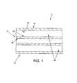
- FIG. 1is a schematic view showing a preferred embodiment of the present invention
- FIG. 2is a schematic view showing another preferred embodiment of the present invention.
- FIGS. 3-6show an endoscope being advanced through the anatomical model shown in FIG. 2 .
- Anatomical ModelComprising an Inner Lumen and an Outer Lumen, and a Fluid Disposed in the Space Interior to the Outer Lumen and Exterior to the Inner Lumen
- the present inventiongenerally comprises an anatomical model comprising an inner lumen and an outer lumen, wherein the inner lumen is disposed inside of the outer lumen so as to create a space therebetween, and further wherein a fluid is disposed within the space, interior to the outer lumen and exterior to the inner lumen, whereby the inner lumen can accurately simulate the mucous membrane lining a mammalian tract (e.g., the gastrointestinal or genitourinary tract).
- a mammalian tracte.g., the gastrointestinal or genitourinary tract
- an anatomical model 5which comprises two lumens, an inner lumen 10 and an outer lumen 15 , wherein inner lumen 10 is disposed inside of outer lumen 15 .
- Inner lumen 10generally comprises an interior surface 20 and an exterior surface 25 .
- Outer lumen 15generally comprises an interior surface 30 and an exterior surface 35 .
- a space 40is formed in between exterior surface 25 of inner lumen 10 and interior surface 30 of outer lumen 15 .
- Inner and outer lumens 10 , 15are sized so as to approximate different anatomical locations or pathology.
- space 40is filled with a fluid.
- This fluidcreates a radially compressive force on exterior surface 25 of inner lumen 10 which simulates the forces acting on the mammalian tract within the body (e.g., the gastrointestinal or genitourinary tract within the abdomen).
- inner lumen 10 and outer lumen 15are sealed so as to prevent fluid communication between the inner and outer lumens.
- Inner lumen 10 and outer lumen 15are formed out of one or more materials which have physical characteristics which, when combined with the effect of a fluid filled space 40 , provide properties simulating those of natural tissue. These materials may have inherently low surface friction so as to approximate the coefficient of friction of natural mucosal tissue, or the materials may incorporate a lubricant so as to simulate the low friction of natural mucosal tissue.
- the material of inner lumen 10may comprise polyethylene
- the material of outer lumen 15may also comprise polyethylene.
- inner lumen 10 and/or outer lumen 15may be constructed from a clear material so as to provide for external visualization.
- space 40is filled with a fluid in order to simulate the actual force on a mammalian tract (e.g., the gastrointestinal or genitourinary tract). More particularly, this fluid is selected, and pressurized, so as to provide the desired compressive force on exterior surface 25 of inner lumen 10 .
- this fluidmay be a gas pressurized to a desired level.
- the fluidmay be a liquid chosen from a wide range of weights or viscosities so as to affect (i.e., determine) the feel of the simulator.
- the liquidmay be water.
- outer lumen 15may comprise a vessel which holds inner lumen 10 .
- mechanical supportmay be used to create a 2-dimensional or 3-dimensional shape so as to simulate human or animal anatomy.
- inner lumen 10 and outer lumen 15may comprise “lay-flat” tubing so that anatomical model 5 comprises a long “poly-bag”.
- the structure shown in FIG. 1is provided with the fluid positioned in space 40 so as to provide the desired anatomical characteristics for anatomical model 5 .
- instrumentatione.g., an endoscope
- inner and outer lumens 10 , 15with the fluid-filled space 40 therebetween, model the natural tissue in a more realistic manner than the prior art.
- a lubricantmay be added to aid in the insertion of the instrumentation into inner lumen 10 .
- the lubricantmay comprise a mixture of water and liquid detergent.
- the present inventionprovides an anatomical model comprising an inner lumen and an outer lumen, wherein the inner lumen is disposed inside of the outer lumen so as to create a space therebetween, and further wherein a fluid is disposed within the space, interior to the outer lumen and exterior to the inner lumen, whereby the inner lumen can accurately simulate the mucous membrane lining a mammalian tract (e.g., the gastrointestinal or genitourinary tract).
- a mammalian tracte.g., the gastrointestinal or genitourinary tract
- the anatomy of interestmay comprise a lumen structure and, furthermore, the lumen structure may be supported on adjacent anatomy via movable connections.
- the small bowelcomprises a lumen structure which is movably supported by the mesentery along its length. Accordingly, an anatomical model intended to simulate such a small bowel structure should be capable of reproducing not only the lumen passageway of the small bowel, but also the nature and character of the movable connections which attach the lumen passageway to the mesentery.
- anatomical lumen structuresare also movably supported to adjacent structures at various locations along their length.
- an anatomical model 100which generally comprises a lumen structure 105 , a supporting frame 110 and a plurality of movable connections 115 selectively securing lumen structure 105 to supporting frame 110 .
- Lumen structure 105preferably comprises an excised specimen of the organ which is to be simulated.
- lumen structure 105preferably comprises an excised small bowel.
- lumen structure 105need not necessarily comprise an excised organ—thus, lumen structure 105 may also comprise an artificial structure which simulates the organ in geometric form (e.g., shape and size), mechanical structure (e.g., pliability and resiliency), surface characteristics (e.g., texture and coefficient of friction), etc.
- Frame 110preferably comprises an elongated structure capable of supporting a length of lumen structure 105 .
- frame 110may comprise a base 120 , one or more vertical risers 125 extending upward from base 120 , and a rod 130 supported above base 120 by the one or more vertical risers 125 .
- Rod 130is preferably configured so that it extends in a manner which is generally representative of the disposition of the organ which is to be simulated (e.g., where the small bowel is to be simulated, rod 130 preferably extends along a curving pathway as shown in FIG. 2 .
- Movable connections 115serve to selectively secure lumen structure 105 to supporting frame 110 .
- movable connections 115are configured so as to secure lumen structure 105 to supporting frame 110 in a manner which is generally representative of the manner in which the natural organ is secured to its own supporting structure.
- movable connections 115may comprise an organ end 135 comprising an alligator clip 140 for engaging the organ, and a frame end 145 comprising a rod hanger 150 for slidably mounting on rod 130 .
- movable connections 115may comprise various other clamps, hooks, springs and/or combined elements so as to attach lumen structure 105 to frame 110 in an anatomically realistic manner.
- movable connections 115are adjustable in location, pathway and function so as to permit accurate simulation of the manner in which the natural organ is secured to its natural supporting structure.
- anatomical model 100may include an entry port 155 so as to provide access to the internal structure of the lumen structure 105 .
- the lumen structure 105may be supported within an enclosure which can incorporate appropriate liquids and/or temperature control so as to simulate in vivo conditions.
- anatomical model 100may be used with external support elements to create a 2-dimensional path or a 3-dimensional path.
- anatomical model 100comprises a lumen structure 105 in the form of a porcine stomach and small bowel, a frame 110 in the form of a rod following a curved pathway, and movable connections 115 in the form of alligator clips 140 /rod hangers 150 . See FIG. 2 .
- This constructionpermits the porcine bowel to be pleated during endoscopic examination on the simulated mesentery.
- FIGS. 3-6show an endoscope 200 being advanced through the aforementioned anatomical model.
- the present inventionprovides an anatomical model which generally comprises a lumen structure supported on a frame using movable connections, in order to simulate both (i) a natural lumen passageway, and (ii) the nature and character of movable connections which attach the lumen passageway to adjacent anatomy.
Landscapes
- Engineering & Computer Science (AREA)
- Physics & Mathematics (AREA)
- General Physics & Mathematics (AREA)
- Health & Medical Sciences (AREA)
- Mathematical Analysis (AREA)
- Pure & Applied Mathematics (AREA)
- Medical Informatics (AREA)
- Algebra (AREA)
- Computational Mathematics (AREA)
- General Health & Medical Sciences (AREA)
- Chemical & Material Sciences (AREA)
- Mathematical Optimization (AREA)
- Mathematical Physics (AREA)
- Medicinal Chemistry (AREA)
- Business, Economics & Management (AREA)
- Educational Administration (AREA)
- Educational Technology (AREA)
- Theoretical Computer Science (AREA)
- Pulmonology (AREA)
- Radiology & Medical Imaging (AREA)
- Instructional Devices (AREA)
- Endoscopes (AREA)
Abstract
Description
Claims (19)
Priority Applications (1)
| Application Number | Priority Date | Filing Date | Title |
|---|---|---|---|
| US12/259,066US8403676B2 (en) | 2006-05-19 | 2008-10-27 | Anatomical model |
Applications Claiming Priority (4)
| Application Number | Priority Date | Filing Date | Title |
|---|---|---|---|
| US80171906P | 2006-05-19 | 2006-05-19 | |
| US11/804,873US7854612B2 (en) | 2006-05-19 | 2007-05-21 | Anatomical model |
| US53907P | 2007-10-26 | 2007-10-26 | |
| US12/259,066US8403676B2 (en) | 2006-05-19 | 2008-10-27 | Anatomical model |
Related Parent Applications (1)
| Application Number | Title | Priority Date | Filing Date |
|---|---|---|---|
| US11/804,873Continuation-In-PartUS7854612B2 (en) | 2006-05-19 | 2007-05-21 | Anatomical model |
Publications (2)
| Publication Number | Publication Date |
|---|---|
| US20090226868A1 US20090226868A1 (en) | 2009-09-10 |
| US8403676B2true US8403676B2 (en) | 2013-03-26 |
Family
ID=41053979
Family Applications (1)
| Application Number | Title | Priority Date | Filing Date |
|---|---|---|---|
| US12/259,066Active2028-12-17US8403676B2 (en) | 2006-05-19 | 2008-10-27 | Anatomical model |
Country Status (1)
| Country | Link |
|---|---|
| US (1) | US8403676B2 (en) |
Cited By (37)
| Publication number | Priority date | Publication date | Assignee | Title |
|---|---|---|---|---|
| US20120164616A1 (en)* | 2009-09-07 | 2012-06-28 | Koken Co., Ltd. | Exercise Mode For Small Intestine Endoscope |
| US20160140876A1 (en)* | 2014-11-18 | 2016-05-19 | Ibrahim Ihsan Jabbour | Collapsible Surgical Training Apparatus and Method for Laparoscopic Procedures |
| US20160140879A1 (en)* | 2014-11-19 | 2016-05-19 | David Hananel | Anatomically correct movement or deformation of simulated bodily structures |
| US20160217710A1 (en)* | 2015-01-26 | 2016-07-28 | Ethicon, Inc. | Ex-Vivo Anatomic Tissue Specimen Wound Closure Simulation Model |
| US9472121B2 (en) | 2010-10-01 | 2016-10-18 | Applied Medical Resources Corporation | Portable laparoscopic trainer |
| US9548002B2 (en) | 2013-07-24 | 2017-01-17 | Applied Medical Resources Corporation | First entry model |
| US9842515B1 (en) | 2013-10-03 | 2017-12-12 | Encoris Group Corporation, Inc. | Apparatus for surgical training |
| US9898937B2 (en) | 2012-09-28 | 2018-02-20 | Applied Medical Resources Corporation | Surgical training model for laparoscopic procedures |
| US9922579B2 (en) | 2013-06-18 | 2018-03-20 | Applied Medical Resources Corporation | Gallbladder model |
| US9940849B2 (en) | 2013-03-01 | 2018-04-10 | Applied Medical Resources Corporation | Advanced surgical simulation constructions and methods |
| US9959786B2 (en) | 2012-09-27 | 2018-05-01 | Applied Medical Resources Corporation | Surgical training model for laparoscopic procedures |
| US10081727B2 (en) | 2015-05-14 | 2018-09-25 | Applied Medical Resources Corporation | Synthetic tissue structures for electrosurgical training and simulation |
| US10121391B2 (en) | 2012-09-27 | 2018-11-06 | Applied Medical Resources Corporation | Surgical training model for laparoscopic procedures |
| US10127838B2 (en) | 2016-11-22 | 2018-11-13 | PraxiCut, LLC | Surgical simulation systems, methods, and compositions |
| US10140889B2 (en) | 2013-05-15 | 2018-11-27 | Applied Medical Resources Corporation | Hernia model |
| US10198965B2 (en) | 2012-08-03 | 2019-02-05 | Applied Medical Resources Corporation | Simulated stapling and energy based ligation for surgical training |
| US10198966B2 (en) | 2013-07-24 | 2019-02-05 | Applied Medical Resources Corporation | Advanced first entry model for surgical simulation |
| US10223936B2 (en) | 2015-06-09 | 2019-03-05 | Applied Medical Resources Corporation | Hysterectomy model |
| US10332425B2 (en) | 2015-07-16 | 2019-06-25 | Applied Medical Resources Corporation | Simulated dissectible tissue |
| US10354556B2 (en) | 2015-02-19 | 2019-07-16 | Applied Medical Resources Corporation | Simulated tissue structures and methods |
| US10395559B2 (en) | 2012-09-28 | 2019-08-27 | Applied Medical Resources Corporation | Surgical training model for transluminal laparoscopic procedures |
| US10410542B1 (en) | 2018-07-18 | 2019-09-10 | Simulated Inanimate Models, LLC | Surgical training apparatus, methods and systems |
| US10490105B2 (en) | 2015-07-22 | 2019-11-26 | Applied Medical Resources Corporation | Appendectomy model |
| US10535281B2 (en) | 2012-09-26 | 2020-01-14 | Applied Medical Resources Corporation | Surgical training model for laparoscopic procedures |
| US10679520B2 (en) | 2012-09-27 | 2020-06-09 | Applied Medical Resources Corporation | Surgical training model for laparoscopic procedures |
| US10706743B2 (en) | 2015-11-20 | 2020-07-07 | Applied Medical Resources Corporation | Simulated dissectible tissue |
| US10720084B2 (en) | 2015-10-02 | 2020-07-21 | Applied Medical Resources Corporation | Hysterectomy model |
| US20200294422A1 (en)* | 2017-11-30 | 2020-09-17 | Queensland University Of Technology | Surgical training device |
| US10796606B2 (en) | 2014-03-26 | 2020-10-06 | Applied Medical Resources Corporation | Simulated dissectible tissue |
| US10818201B2 (en) | 2014-11-13 | 2020-10-27 | Applied Medical Resources Corporation | Simulated tissue models and methods |
| US10847057B2 (en) | 2017-02-23 | 2020-11-24 | Applied Medical Resources Corporation | Synthetic tissue structures for electrosurgical training and simulation |
| US11030922B2 (en) | 2017-02-14 | 2021-06-08 | Applied Medical Resources Corporation | Laparoscopic training system |
| US20210183267A1 (en)* | 2017-07-27 | 2021-06-17 | Mochtech, Llc | Self-contained multipurpose medical training system and components |
| US11120708B2 (en) | 2016-06-27 | 2021-09-14 | Applied Medical Resources Corporation | Simulated abdominal wall |
| US11158212B2 (en) | 2011-10-21 | 2021-10-26 | Applied Medical Resources Corporation | Simulated tissue structure for surgical training |
| US11403968B2 (en) | 2011-12-20 | 2022-08-02 | Applied Medical Resources Corporation | Advanced surgical simulation |
| US12106678B2 (en) | 2021-10-23 | 2024-10-01 | Simulated Inanimate Models, LLC | Procedure guidance and training apparatus, methods and systems |
Families Citing this family (4)
| Publication number | Priority date | Publication date | Assignee | Title |
|---|---|---|---|---|
| WO2011035088A2 (en)* | 2009-09-18 | 2011-03-24 | University Of Tennessee Research Foundation | Flexible and rigid endoscopic training device (fred) |
| JP5550050B2 (en)* | 2010-12-14 | 2014-07-16 | 株式会社ティー・エム・シー | Partial model of human body |
| ES2526245B1 (en)* | 2013-07-02 | 2015-11-24 | Fundación Centro De Cirugia Jesús Usón | Vivid simulator for learning, practice and training of endoscopic techniques |
| WO2016109879A1 (en) | 2015-01-06 | 2016-07-14 | The Hospital For Sick Children | Simulator for practicing trans-oral surgery and method of use thereof |
Citations (32)
| Publication number | Priority date | Publication date | Assignee | Title |
|---|---|---|---|---|
| US4001952A (en)* | 1975-10-24 | 1977-01-11 | Kleppinger Trygve M | Hysteroscopy teaching aid |
| US4055148A (en)* | 1974-10-28 | 1977-10-25 | Brockman's Service Ltd. | Animal holding clamp apparatus |
| US4355631A (en)* | 1981-03-19 | 1982-10-26 | Minnesota Scientific, Inc. | Surgical retractor apparatus with improved clamping device |
| US5061187A (en)* | 1990-04-12 | 1991-10-29 | Ravinder Jerath | Ultrasound training apparatus |
| US5149270A (en)* | 1990-10-29 | 1992-09-22 | Mckeown M J | Apparatus for practicing surgical procedures |
| US5231974A (en)* | 1991-05-31 | 1993-08-03 | Giglio Steven R | Self retaining retractor |
| US5320537A (en) | 1993-03-16 | 1994-06-14 | Triangle Research And Development Corporation | Microsurgical training apparatus |
| US5403191A (en)* | 1991-10-21 | 1995-04-04 | Tuason; Leo B. | Laparoscopic surgery simulator and method of use |
| US5425644A (en)* | 1993-05-13 | 1995-06-20 | Gerhard Szinicz | Surgical training apparatus and method |
| US5947743A (en)* | 1997-09-26 | 1999-09-07 | Hasson; Harrith M. | Apparatus for training for the performance of a medical procedure |
| US5947744A (en)* | 1998-01-21 | 1999-09-07 | The Chinese University Of Hong Kong | Training apparatus and method for coronary artery anastomoses featuring simulated pulsating heart |
| US6052932A (en)* | 1998-06-05 | 2000-04-25 | Haworth, Inc. | Presentation unit extendible side panels |
| US6062866A (en) | 1998-03-27 | 2000-05-16 | Prom; James M. | Medical angioplasty model |
| US6077221A (en)* | 1999-09-01 | 2000-06-20 | Lone Star Medical Products, Inc. | Surgical restraint system |
| USD435062S (en)* | 2000-04-13 | 2000-12-12 | Hewllett-Packard Company | Pull-out extension tray |
| US20010019818A1 (en) | 1999-03-02 | 2001-09-06 | Peter Yong | Method of endoscopic cardiac surgery training |
| US6336812B1 (en)* | 1997-06-19 | 2002-01-08 | Limbs & Things Limited | Clinical and/or surgical training apparatus |
| US6511325B1 (en) | 1998-05-04 | 2003-01-28 | Advanced Research & Technology Institute | Aortic stent-graft calibration and training model |
| US6543657B2 (en) | 2001-03-20 | 2003-04-08 | Hong Kong Polytechnic University | Thermal manikin |
| US20040025253A1 (en)* | 1999-01-22 | 2004-02-12 | Heimbrock Richard H. | Convertible stretcher |
| US6773263B2 (en) | 2001-10-09 | 2004-08-10 | Robert J. Nicholls | Medical simulator |
| US20050074732A1 (en) | 2003-10-02 | 2005-04-07 | Morris Gary Jay | Blood pressure simulation apparatus with tactile interface |
| US6887092B2 (en)* | 2002-11-19 | 2005-05-03 | Nec Corporation | Optical module locking mechanism for locking optical module case and cage for housing case to each other |
| US6908309B2 (en)* | 2001-12-03 | 2005-06-21 | Sdgi Holdings, Inc. | Demonstration devices for medical procedures |
| US6997719B2 (en) | 2002-06-26 | 2006-02-14 | Ethicon, Inc. | Training model for endoscopic vessel harvesting |
| US7008232B2 (en) | 2001-09-29 | 2006-03-07 | Friedhelm Brassel | Method for producing a modeling system for vessel deformations |
| US7059168B2 (en) | 2002-10-01 | 2006-06-13 | Olympus Corporation | Ultrasound phantom |
| US20070020598A1 (en) | 2003-03-26 | 2007-01-25 | National Institute Of Advanced Industrial Science And Technology | Manikin and method of manufacturing the same |
| US20080076101A1 (en) | 2006-05-12 | 2008-03-27 | Abbott Laboratories | Forming vascular diseases within anatomical models |
| US20080187895A1 (en) | 2005-02-03 | 2008-08-07 | Christopher Sakezles | Models And Methods Of Using Same For Testing Medical Devices |
| US7568247B2 (en)* | 2002-12-26 | 2009-08-04 | Gendron, Inc. | Bariatric patient management system |
| US8113847B2 (en)* | 2007-10-23 | 2012-02-14 | K2M, Inc. | Spinal surgery modeling system |
- 2008
- 2008-10-27USUS12/259,066patent/US8403676B2/enactiveActive
Patent Citations (32)
| Publication number | Priority date | Publication date | Assignee | Title |
|---|---|---|---|---|
| US4055148A (en)* | 1974-10-28 | 1977-10-25 | Brockman's Service Ltd. | Animal holding clamp apparatus |
| US4001952A (en)* | 1975-10-24 | 1977-01-11 | Kleppinger Trygve M | Hysteroscopy teaching aid |
| US4355631A (en)* | 1981-03-19 | 1982-10-26 | Minnesota Scientific, Inc. | Surgical retractor apparatus with improved clamping device |
| US5061187A (en)* | 1990-04-12 | 1991-10-29 | Ravinder Jerath | Ultrasound training apparatus |
| US5149270A (en)* | 1990-10-29 | 1992-09-22 | Mckeown M J | Apparatus for practicing surgical procedures |
| US5231974A (en)* | 1991-05-31 | 1993-08-03 | Giglio Steven R | Self retaining retractor |
| US5403191A (en)* | 1991-10-21 | 1995-04-04 | Tuason; Leo B. | Laparoscopic surgery simulator and method of use |
| US5320537A (en) | 1993-03-16 | 1994-06-14 | Triangle Research And Development Corporation | Microsurgical training apparatus |
| US5425644A (en)* | 1993-05-13 | 1995-06-20 | Gerhard Szinicz | Surgical training apparatus and method |
| US6336812B1 (en)* | 1997-06-19 | 2002-01-08 | Limbs & Things Limited | Clinical and/or surgical training apparatus |
| US5947743A (en)* | 1997-09-26 | 1999-09-07 | Hasson; Harrith M. | Apparatus for training for the performance of a medical procedure |
| US5947744A (en)* | 1998-01-21 | 1999-09-07 | The Chinese University Of Hong Kong | Training apparatus and method for coronary artery anastomoses featuring simulated pulsating heart |
| US6062866A (en) | 1998-03-27 | 2000-05-16 | Prom; James M. | Medical angioplasty model |
| US6511325B1 (en) | 1998-05-04 | 2003-01-28 | Advanced Research & Technology Institute | Aortic stent-graft calibration and training model |
| US6052932A (en)* | 1998-06-05 | 2000-04-25 | Haworth, Inc. | Presentation unit extendible side panels |
| US20040025253A1 (en)* | 1999-01-22 | 2004-02-12 | Heimbrock Richard H. | Convertible stretcher |
| US20010019818A1 (en) | 1999-03-02 | 2001-09-06 | Peter Yong | Method of endoscopic cardiac surgery training |
| US6077221A (en)* | 1999-09-01 | 2000-06-20 | Lone Star Medical Products, Inc. | Surgical restraint system |
| USD435062S (en)* | 2000-04-13 | 2000-12-12 | Hewllett-Packard Company | Pull-out extension tray |
| US6543657B2 (en) | 2001-03-20 | 2003-04-08 | Hong Kong Polytechnic University | Thermal manikin |
| US7008232B2 (en) | 2001-09-29 | 2006-03-07 | Friedhelm Brassel | Method for producing a modeling system for vessel deformations |
| US6773263B2 (en) | 2001-10-09 | 2004-08-10 | Robert J. Nicholls | Medical simulator |
| US6908309B2 (en)* | 2001-12-03 | 2005-06-21 | Sdgi Holdings, Inc. | Demonstration devices for medical procedures |
| US6997719B2 (en) | 2002-06-26 | 2006-02-14 | Ethicon, Inc. | Training model for endoscopic vessel harvesting |
| US7059168B2 (en) | 2002-10-01 | 2006-06-13 | Olympus Corporation | Ultrasound phantom |
| US6887092B2 (en)* | 2002-11-19 | 2005-05-03 | Nec Corporation | Optical module locking mechanism for locking optical module case and cage for housing case to each other |
| US7568247B2 (en)* | 2002-12-26 | 2009-08-04 | Gendron, Inc. | Bariatric patient management system |
| US20070020598A1 (en) | 2003-03-26 | 2007-01-25 | National Institute Of Advanced Industrial Science And Technology | Manikin and method of manufacturing the same |
| US20050074732A1 (en) | 2003-10-02 | 2005-04-07 | Morris Gary Jay | Blood pressure simulation apparatus with tactile interface |
| US20080187895A1 (en) | 2005-02-03 | 2008-08-07 | Christopher Sakezles | Models And Methods Of Using Same For Testing Medical Devices |
| US20080076101A1 (en) | 2006-05-12 | 2008-03-27 | Abbott Laboratories | Forming vascular diseases within anatomical models |
| US8113847B2 (en)* | 2007-10-23 | 2012-02-14 | K2M, Inc. | Spinal surgery modeling system |
Cited By (73)
| Publication number | Priority date | Publication date | Assignee | Title |
|---|---|---|---|---|
| US9257055B2 (en)* | 2009-09-07 | 2016-02-09 | Showa University | Small intestine endoscope training simulator |
| US20120164616A1 (en)* | 2009-09-07 | 2012-06-28 | Koken Co., Ltd. | Exercise Mode For Small Intestine Endoscope |
| US12154454B2 (en) | 2010-10-01 | 2024-11-26 | Applied Medical Resources Corporation | Portable laparoscopic trainer |
| US9472121B2 (en) | 2010-10-01 | 2016-10-18 | Applied Medical Resources Corporation | Portable laparoscopic trainer |
| US10854112B2 (en) | 2010-10-01 | 2020-12-01 | Applied Medical Resources Corporation | Portable laparoscopic trainer |
| US12014652B2 (en) | 2011-10-21 | 2024-06-18 | Applied Medical Resources Corporation | Simulated tissue structure for surgical training |
| US11158212B2 (en) | 2011-10-21 | 2021-10-26 | Applied Medical Resources Corporation | Simulated tissue structure for surgical training |
| US11403968B2 (en) | 2011-12-20 | 2022-08-02 | Applied Medical Resources Corporation | Advanced surgical simulation |
| US10198965B2 (en) | 2012-08-03 | 2019-02-05 | Applied Medical Resources Corporation | Simulated stapling and energy based ligation for surgical training |
| US10535281B2 (en) | 2012-09-26 | 2020-01-14 | Applied Medical Resources Corporation | Surgical training model for laparoscopic procedures |
| US11514819B2 (en) | 2012-09-26 | 2022-11-29 | Applied Medical Resources Corporation | Surgical training model for laparoscopic procedures |
| US10121391B2 (en) | 2012-09-27 | 2018-11-06 | Applied Medical Resources Corporation | Surgical training model for laparoscopic procedures |
| US9959786B2 (en) | 2012-09-27 | 2018-05-01 | Applied Medical Resources Corporation | Surgical training model for laparoscopic procedures |
| US11990055B2 (en) | 2012-09-27 | 2024-05-21 | Applied Medical Resources Corporation | Surgical training model for laparoscopic procedures |
| US10679520B2 (en) | 2012-09-27 | 2020-06-09 | Applied Medical Resources Corporation | Surgical training model for laparoscopic procedures |
| US11869378B2 (en) | 2012-09-27 | 2024-01-09 | Applied Medical Resources Corporation | Surgical training model for laparoscopic procedures |
| US11361679B2 (en) | 2012-09-27 | 2022-06-14 | Applied Medical Resources Corporation | Surgical training model for laparoscopic procedures |
| US10395559B2 (en) | 2012-09-28 | 2019-08-27 | Applied Medical Resources Corporation | Surgical training model for transluminal laparoscopic procedures |
| US9898937B2 (en) | 2012-09-28 | 2018-02-20 | Applied Medical Resources Corporation | Surgical training model for laparoscopic procedures |
| US9940849B2 (en) | 2013-03-01 | 2018-04-10 | Applied Medical Resources Corporation | Advanced surgical simulation constructions and methods |
| US10140889B2 (en) | 2013-05-15 | 2018-11-27 | Applied Medical Resources Corporation | Hernia model |
| US11049418B2 (en) | 2013-06-18 | 2021-06-29 | Applied Medical Resources Corporation | Gallbladder model |
| US11735068B2 (en) | 2013-06-18 | 2023-08-22 | Applied Medical Resources Corporation | Gallbladder model |
| US9922579B2 (en) | 2013-06-18 | 2018-03-20 | Applied Medical Resources Corporation | Gallbladder model |
| US11450236B2 (en) | 2013-07-24 | 2022-09-20 | Applied Medical Resources Corporation | Advanced first entry model for surgical simulation |
| US10198966B2 (en) | 2013-07-24 | 2019-02-05 | Applied Medical Resources Corporation | Advanced first entry model for surgical simulation |
| US9548002B2 (en) | 2013-07-24 | 2017-01-17 | Applied Medical Resources Corporation | First entry model |
| US10657845B2 (en) | 2013-07-24 | 2020-05-19 | Applied Medical Resources Corporation | First entry model |
| US11854425B2 (en) | 2013-07-24 | 2023-12-26 | Applied Medical Resources Corporation | First entry model |
| US10026337B2 (en) | 2013-07-24 | 2018-07-17 | Applied Medical Resources Corporation | First entry model |
| US12288476B2 (en) | 2013-07-24 | 2025-04-29 | Applied Medical Resources Corporation | Advanced first entry model for surgical simulation |
| US9842515B1 (en) | 2013-10-03 | 2017-12-12 | Encoris Group Corporation, Inc. | Apparatus for surgical training |
| US10796606B2 (en) | 2014-03-26 | 2020-10-06 | Applied Medical Resources Corporation | Simulated dissectible tissue |
| US10818201B2 (en) | 2014-11-13 | 2020-10-27 | Applied Medical Resources Corporation | Simulated tissue models and methods |
| US12211394B2 (en) | 2014-11-13 | 2025-01-28 | Applied Medical Resources Corporation | Simulated tissue models and methods |
| US11887504B2 (en) | 2014-11-13 | 2024-01-30 | Applied Medical Resources Corporation | Simulated tissue models and methods |
| US20160140876A1 (en)* | 2014-11-18 | 2016-05-19 | Ibrahim Ihsan Jabbour | Collapsible Surgical Training Apparatus and Method for Laparoscopic Procedures |
| US9734732B2 (en)* | 2014-11-18 | 2017-08-15 | Ibrahim Ihsan Jabbour | Collapsible surgical training apparatus and method for laparoscopic procedures |
| US20160140879A1 (en)* | 2014-11-19 | 2016-05-19 | David Hananel | Anatomically correct movement or deformation of simulated bodily structures |
| US20160217710A1 (en)* | 2015-01-26 | 2016-07-28 | Ethicon, Inc. | Ex-Vivo Anatomic Tissue Specimen Wound Closure Simulation Model |
| US9520073B2 (en)* | 2015-01-26 | 2016-12-13 | Ethicon, Inc. | Ex-vivo anatomic tissue specimen wound closure simulation model |
| US10354556B2 (en) | 2015-02-19 | 2019-07-16 | Applied Medical Resources Corporation | Simulated tissue structures and methods |
| US12131664B2 (en) | 2015-02-19 | 2024-10-29 | Applied Medical Resources Corporation | Simulated tissue structures and methods |
| US11100815B2 (en) | 2015-02-19 | 2021-08-24 | Applied Medical Resources Corporation | Simulated tissue structures and methods |
| US11034831B2 (en) | 2015-05-14 | 2021-06-15 | Applied Medical Resources Corporation | Synthetic tissue structures for electrosurgical training and simulation |
| US10081727B2 (en) | 2015-05-14 | 2018-09-25 | Applied Medical Resources Corporation | Synthetic tissue structures for electrosurgical training and simulation |
| US11721240B2 (en) | 2015-06-09 | 2023-08-08 | Applied Medical Resources Corporation | Hysterectomy model |
| US10733908B2 (en) | 2015-06-09 | 2020-08-04 | Applied Medical Resources Corporation | Hysterectomy model |
| US10223936B2 (en) | 2015-06-09 | 2019-03-05 | Applied Medical Resources Corporation | Hysterectomy model |
| US12175883B2 (en) | 2015-06-09 | 2024-12-24 | Applied Medical Resources Corporation | Hysterectomy model |
| US10332425B2 (en) | 2015-07-16 | 2019-06-25 | Applied Medical Resources Corporation | Simulated dissectible tissue |
| US11587466B2 (en) | 2015-07-16 | 2023-02-21 | Applied Medical Resources Corporation | Simulated dissectible tissue |
| US10755602B2 (en) | 2015-07-16 | 2020-08-25 | Applied Medical Resources Corporation | Simulated dissectible tissue |
| US12087179B2 (en) | 2015-07-16 | 2024-09-10 | Applied Medical Resources Corporation | Simulated dissectible tissue |
| US10490105B2 (en) | 2015-07-22 | 2019-11-26 | Applied Medical Resources Corporation | Appendectomy model |
| US12243441B2 (en) | 2015-10-02 | 2025-03-04 | Applied Medical Resources Corporation | Hysterectomy model |
| US10720084B2 (en) | 2015-10-02 | 2020-07-21 | Applied Medical Resources Corporation | Hysterectomy model |
| US11721242B2 (en) | 2015-10-02 | 2023-08-08 | Applied Medical Resources Corporation | Hysterectomy model |
| US12217625B2 (en) | 2015-11-20 | 2025-02-04 | Applied Medical Resources Corporation | Simulated dissectible tissue |
| US10706743B2 (en) | 2015-11-20 | 2020-07-07 | Applied Medical Resources Corporation | Simulated dissectible tissue |
| US11830378B2 (en) | 2016-06-27 | 2023-11-28 | Applied Medical Resources Corporation | Simulated abdominal wall |
| US11120708B2 (en) | 2016-06-27 | 2021-09-14 | Applied Medical Resources Corporation | Simulated abdominal wall |
| US10127838B2 (en) | 2016-11-22 | 2018-11-13 | PraxiCut, LLC | Surgical simulation systems, methods, and compositions |
| US12243439B2 (en) | 2017-02-14 | 2025-03-04 | Applied Medical Resources Corporation | Laparoscopic training system |
| US11030922B2 (en) | 2017-02-14 | 2021-06-08 | Applied Medical Resources Corporation | Laparoscopic training system |
| US10847057B2 (en) | 2017-02-23 | 2020-11-24 | Applied Medical Resources Corporation | Synthetic tissue structures for electrosurgical training and simulation |
| US20210183267A1 (en)* | 2017-07-27 | 2021-06-17 | Mochtech, Llc | Self-contained multipurpose medical training system and components |
| US11749136B2 (en)* | 2017-07-27 | 2023-09-05 | Mochtech, Llc | Self-contained multipurpose medical training system and components |
| US11830377B2 (en)* | 2017-11-30 | 2023-11-28 | Queensland University Of Technology | Surgical training device |
| US20200294422A1 (en)* | 2017-11-30 | 2020-09-17 | Queensland University Of Technology | Surgical training device |
| US10410542B1 (en) | 2018-07-18 | 2019-09-10 | Simulated Inanimate Models, LLC | Surgical training apparatus, methods and systems |
| US10665134B2 (en) | 2018-07-18 | 2020-05-26 | Simulated Inanimate Models, LLC | Surgical training apparatus, methods and systems |
| US12106678B2 (en) | 2021-10-23 | 2024-10-01 | Simulated Inanimate Models, LLC | Procedure guidance and training apparatus, methods and systems |
Also Published As
| Publication number | Publication date |
|---|---|
| US20090226868A1 (en) | 2009-09-10 |
Similar Documents
| Publication | Publication Date | Title |
|---|---|---|
| US8403676B2 (en) | Anatomical model | |
| US7854612B2 (en) | Anatomical model | |
| US10013896B2 (en) | Modular staged reality simulator | |
| CA2293585C (en) | Clinical and/or surgical training apparatus | |
| US7866983B2 (en) | Surgical simulator system | |
| US6062866A (en) | Medical angioplasty model | |
| US10553131B2 (en) | Central line simulation and training device | |
| US20070003917A1 (en) | Medical training system for diagnostic examinations performed by palpation | |
| US11417241B2 (en) | Artificial canine model | |
| WO2018236607A1 (en) | OBSTETRIC TRAINING SIMULATOR | |
| AU2016297579B2 (en) | Appendectomy model | |
| JP2021503095A (en) | Hysterectomy model | |
| EP3288009A1 (en) | Low-resistance anatomical tissue model | |
| US20230097495A1 (en) | Simulated tissue structure composition and use for surgical training | |
| US20210074184A1 (en) | Retroperitoneal surgical simulation model | |
| US20220406221A1 (en) | Medical Training Device and Method to Use it in Teaching Laparoscopic and Robotic Partial Nephrectomy | |
| BR102020008843A2 (en) | BIOMODEL FOR THE DIGESTIVE ENDOSCOPY AREA | |
| Gomes et al. | How Intra-Abdominal Pressure Increment Could Affect Cardiovascular Monitoring: A Simulation Assay | |
| BR202020015410U2 (en) | SYNTHETIC REALISTIC SIMULATOR FOR HUMAN DIAGNOSIS AND THERAPEUTIC HIGH DIGESTIVE ENDOSCOPY | |
| Donelan | Creative anatomy teaching | |
| HK1023832B (en) | Clinical and/or surgical training apparatus |
Legal Events
| Date | Code | Title | Description |
|---|---|---|---|
| AS | Assignment | Owner name:SPIRUS MEDICAL, INC, MASSACHUSETTS Free format text:ASSIGNMENT OF ASSIGNORS INTEREST;ASSIGNORS:FRASSICA, JAMES J;AILINGER, ROBERT E.;BOOKWALTER, BILL;SIGNING DATES FROM 20110420 TO 20110422;REEL/FRAME:026305/0985 | |
| AS | Assignment | Owner name:SPIRUS MEDICAL, INC., MASSACHUSETTS Free format text:ASSIGNMENT OF ASSIGNORS INTEREST;ASSIGNOR:AKERMAN, PAUL;REEL/FRAME:026374/0394 Effective date:20110429 | |
| AS | Assignment | Owner name:OLYMPUS ENDO TECHNOLOGY AMERICA INC., MASSACHUSETT Free format text:CHANGE OF NAME;ASSIGNOR:SPIRUS MEDICAL, INC.;REEL/FRAME:029928/0978 Effective date:20110804 | |
| STCF | Information on status: patent grant | Free format text:PATENTED CASE | |
| FPAY | Fee payment | Year of fee payment:4 | |
| MAFP | Maintenance fee payment | Free format text:PAYMENT OF MAINTENANCE FEE, 8TH YEAR, LARGE ENTITY (ORIGINAL EVENT CODE: M1552); ENTITY STATUS OF PATENT OWNER: LARGE ENTITY Year of fee payment:8 | |
| MAFP | Maintenance fee payment | Free format text:PAYMENT OF MAINTENANCE FEE, 12TH YEAR, LARGE ENTITY (ORIGINAL EVENT CODE: M1553); ENTITY STATUS OF PATENT OWNER: LARGE ENTITY Year of fee payment:12 |