US8398720B2 - Craniofacial implant - Google Patents
Craniofacial implantDownload PDFInfo
- Publication number
- US8398720B2 US8398720B2US12/652,896US65289610AUS8398720B2US 8398720 B2US8398720 B2US 8398720B2US 65289610 AUS65289610 AUS 65289610AUS 8398720 B2US8398720 B2US 8398720B2
- Authority
- US
- United States
- Prior art keywords
- implant
- bone
- mesh
- porous
- resin matrix
- Prior art date
- Legal status (The legal status is an assumption and is not a legal conclusion. Google has not performed a legal analysis and makes no representation as to the accuracy of the status listed.)
- Expired - Lifetime, expires
Links
Images


Classifications
- A—HUMAN NECESSITIES
- A61—MEDICAL OR VETERINARY SCIENCE; HYGIENE
- A61F—FILTERS IMPLANTABLE INTO BLOOD VESSELS; PROSTHESES; DEVICES PROVIDING PATENCY TO, OR PREVENTING COLLAPSING OF, TUBULAR STRUCTURES OF THE BODY, e.g. STENTS; ORTHOPAEDIC, NURSING OR CONTRACEPTIVE DEVICES; FOMENTATION; TREATMENT OR PROTECTION OF EYES OR EARS; BANDAGES, DRESSINGS OR ABSORBENT PADS; FIRST-AID KITS
- A61F2/00—Filters implantable into blood vessels; Prostheses, i.e. artificial substitutes or replacements for parts of the body; Appliances for connecting them with the body; Devices providing patency to, or preventing collapsing of, tubular structures of the body, e.g. stents
- A61F2/02—Prostheses implantable into the body
- A61F2/30—Joints
- A61F2/3094—Designing or manufacturing processes
- A61F2/30965—Reinforcing the prosthesis by embedding particles or fibres during moulding or dipping
- A—HUMAN NECESSITIES
- A61—MEDICAL OR VETERINARY SCIENCE; HYGIENE
- A61B—DIAGNOSIS; SURGERY; IDENTIFICATION
- A61B17/00—Surgical instruments, devices or methods
- A61B17/56—Surgical instruments or methods for treatment of bones or joints; Devices specially adapted therefor
- A61B17/58—Surgical instruments or methods for treatment of bones or joints; Devices specially adapted therefor for osteosynthesis, e.g. bone plates, screws or setting implements
- A61B17/68—Internal fixation devices, including fasteners and spinal fixators, even if a part thereof projects from the skin
- A61B17/80—Cortical plates, i.e. bone plates; Instruments for holding or positioning cortical plates, or for compressing bones attached to cortical plates
- A61B17/8085—Cortical plates, i.e. bone plates; Instruments for holding or positioning cortical plates, or for compressing bones attached to cortical plates with pliable or malleable elements or having a mesh-like structure, e.g. small strips
- A—HUMAN NECESSITIES
- A61—MEDICAL OR VETERINARY SCIENCE; HYGIENE
- A61F—FILTERS IMPLANTABLE INTO BLOOD VESSELS; PROSTHESES; DEVICES PROVIDING PATENCY TO, OR PREVENTING COLLAPSING OF, TUBULAR STRUCTURES OF THE BODY, e.g. STENTS; ORTHOPAEDIC, NURSING OR CONTRACEPTIVE DEVICES; FOMENTATION; TREATMENT OR PROTECTION OF EYES OR EARS; BANDAGES, DRESSINGS OR ABSORBENT PADS; FIRST-AID KITS
- A61F2/00—Filters implantable into blood vessels; Prostheses, i.e. artificial substitutes or replacements for parts of the body; Appliances for connecting them with the body; Devices providing patency to, or preventing collapsing of, tubular structures of the body, e.g. stents
- A61F2/02—Prostheses implantable into the body
- A61F2/28—Bones
- A61F2/2875—Skull or cranium
- A—HUMAN NECESSITIES
- A61—MEDICAL OR VETERINARY SCIENCE; HYGIENE
- A61L—METHODS OR APPARATUS FOR STERILISING MATERIALS OR OBJECTS IN GENERAL; DISINFECTION, STERILISATION OR DEODORISATION OF AIR; CHEMICAL ASPECTS OF BANDAGES, DRESSINGS, ABSORBENT PADS OR SURGICAL ARTICLES; MATERIALS FOR BANDAGES, DRESSINGS, ABSORBENT PADS OR SURGICAL ARTICLES
- A61L27/00—Materials for grafts or prostheses or for coating grafts or prostheses
- A61L27/40—Composite materials, i.e. containing one material dispersed in a matrix of the same or different material
- A61L27/44—Composite materials, i.e. containing one material dispersed in a matrix of the same or different material having a macromolecular matrix
- A61L27/446—Composite materials, i.e. containing one material dispersed in a matrix of the same or different material having a macromolecular matrix with other specific inorganic fillers other than those covered by A61L27/443 or A61L27/46
- A—HUMAN NECESSITIES
- A61—MEDICAL OR VETERINARY SCIENCE; HYGIENE
- A61F—FILTERS IMPLANTABLE INTO BLOOD VESSELS; PROSTHESES; DEVICES PROVIDING PATENCY TO, OR PREVENTING COLLAPSING OF, TUBULAR STRUCTURES OF THE BODY, e.g. STENTS; ORTHOPAEDIC, NURSING OR CONTRACEPTIVE DEVICES; FOMENTATION; TREATMENT OR PROTECTION OF EYES OR EARS; BANDAGES, DRESSINGS OR ABSORBENT PADS; FIRST-AID KITS
- A61F2/00—Filters implantable into blood vessels; Prostheses, i.e. artificial substitutes or replacements for parts of the body; Appliances for connecting them with the body; Devices providing patency to, or preventing collapsing of, tubular structures of the body, e.g. stents
- A61F2/02—Prostheses implantable into the body
- A61F2/28—Bones
- A61F2/2875—Skull or cranium
- A61F2002/2878—Skull or cranium for orbital repair
- A—HUMAN NECESSITIES
- A61—MEDICAL OR VETERINARY SCIENCE; HYGIENE
- A61F—FILTERS IMPLANTABLE INTO BLOOD VESSELS; PROSTHESES; DEVICES PROVIDING PATENCY TO, OR PREVENTING COLLAPSING OF, TUBULAR STRUCTURES OF THE BODY, e.g. STENTS; ORTHOPAEDIC, NURSING OR CONTRACEPTIVE DEVICES; FOMENTATION; TREATMENT OR PROTECTION OF EYES OR EARS; BANDAGES, DRESSINGS OR ABSORBENT PADS; FIRST-AID KITS
- A61F2/00—Filters implantable into blood vessels; Prostheses, i.e. artificial substitutes or replacements for parts of the body; Appliances for connecting them with the body; Devices providing patency to, or preventing collapsing of, tubular structures of the body, e.g. stents
- A61F2/02—Prostheses implantable into the body
- A61F2/30—Joints
- A61F2002/30001—Additional features of subject-matter classified in A61F2/28, A61F2/30 and subgroups thereof
- A61F2002/30316—The prosthesis having different structural features at different locations within the same prosthesis; Connections between prosthetic parts; Special structural features of bone or joint prostheses not otherwise provided for
- A61F2002/30535—Special structural features of bone or joint prostheses not otherwise provided for
- A—HUMAN NECESSITIES
- A61—MEDICAL OR VETERINARY SCIENCE; HYGIENE
- A61F—FILTERS IMPLANTABLE INTO BLOOD VESSELS; PROSTHESES; DEVICES PROVIDING PATENCY TO, OR PREVENTING COLLAPSING OF, TUBULAR STRUCTURES OF THE BODY, e.g. STENTS; ORTHOPAEDIC, NURSING OR CONTRACEPTIVE DEVICES; FOMENTATION; TREATMENT OR PROTECTION OF EYES OR EARS; BANDAGES, DRESSINGS OR ABSORBENT PADS; FIRST-AID KITS
- A61F2250/00—Special features of prostheses classified in groups A61F2/00 - A61F2/26 or A61F2/82 or A61F9/00 or A61F11/00 or subgroups thereof
- A61F2250/0058—Additional features; Implant or prostheses properties not otherwise provided for
Definitions
- Craniofacial and especially orbital wall and floor defectsmay result from trauma, cancer, resection, or congenital defects. Such defects are typically treated surgically using bone grafts or synthetic implants. Congenital defects or fractures of the complex and relatively thin bone structures surrounding and supporting the human eye present difficult internal bone repair and fixation problems. In instances when the eye is subject to trauma, the margin or rim of the orbit may diffuse the force of the impact. However, compression of the orbital contents sometimes may occur and fracture the relatively fragile orbit floor and/or the lateral and medial orbital walls. Also injury at the lateral orbital rim may produce a fracture within the orbit. When the orbit is fractured standard bone-grafting techniques for orbital reconstruction may not result in predictable eye function and positioning.
- the support of the globeis deficient as a result of under correction of the defect, over correction, or inadequate reconstruction of the orbital volume.
- the bone graphmay be subject to resorption that may result in result in a less than optimal support.
- the accurate anatomical reconstruction of the bony orbitis essential to maintain normal function and appearance of the eye following orbital fractures. Because most of the bone of the internal orbit surfaces is thin, it is difficult to adequately stabilize the fractured bone fragments without the use of autogenous or alloplastic materials.
- Autologous bone graftshave been considered an optimal treatment method for orbital floor and wall reconstruction. However, this material is sometimes difficult to obtain and difficult to shape the bone graft material to properly fit within the orbit. There are problems relating to the tissue donor site morbidity. As discussed above, autogenous bone grafts have frequently been used by craniomaxillofacial surgeons for the reconstruction of the internal orbit. Bone may be harvested from the calvarium and other autogenous materials including iliac bone, split rib bone. Cartilage has also been used as a bone graft material. However, autogenous bones sometimes result in an unacceptable amount of resorption.
- a variety of alloplastic materialshave been used for orbital reconstruction and craniofacial applications including, silicone rubber, Teflon, Supramid, tantalum mesh, Vitallium mesh, titanium mesh, polyethylene, and methyl methacrylate
- Perforated biocompatible metallic strips and metallic panelsmay be used for rigid internal fixation of fractures in trauma surgery and as a plate material for bone immobilization and stabilization.
- Metal implantscan be used for bone graft support material in reconstructive surgery.
- Synthetic implant materialshave the advantage of no donor site morbidity, ease of use, relative low cost and ready availability. While there are advantages of synthetic implants, some characteristics may be regarded as disadvantages. Silicone rubber has a smooth surface, but does not allow fibrovascular ingrowth into the implant. Further, although it is flexible, it does not readily conform to the profile of the region where it is required or maintain a new shape when shaped to fit a particular location. For example, in connection with the reconstruction of the orbit, a silicone rubber implant is not an attractive option because upon shaping it to the desired profile, it will tend to be biased back to its original shape.
- an implant with a smooth superior surfaceis desirable to prevent attachment of the tissues to the implant upon healing. Attachment of these tissues to the wall of the implant may result in restriction of movement of the eye, causing diplopia, dizziness, and headaches, as well as a cosmetic anomaly on upgaze, downgaze or lateral gaze.
- Implants having a porous structure such as porous polyethylene with predetermined pore sizesallow for fibrovascular ingrowth.
- fibrovascular ingrowthis desirable because it integrates the implant within the tissues, and reduces the possibility that that the synthetic material will be rejected.
- fibrovascular ingrowth on the inferior or sinus side of the implantallows for mucosalization of the implant surface, and, since the opposite side of the implant may be a barrier, the sinus is effectively isolated from the soft tissues of the orbit. This arrangement is considered desirable because it increases the ability of the implant to ward off infection and minimizes the chance of a sinus infection from entering through the orbit. Fibrovascular ingrowth is also thought to minimize the chance of implant migration or displacement.
- Porous polyethyleneis somewhat flexible and thin sheets appropriate for orbital floor and wall reconstruction can be bent to an appropriate shape. However, this material tends to return to its original shape. Further, porous polyethylene does not have a smooth superior surface, so it may result in restriction of the orbital tissues due to fibrous ingrowth when used for orbital reconstruction.
- Pure titaniumis the material of choice in craniofacial reconstructive surgery, especially when the implant is intended to be permanent.
- pure titaniumis preferred because its low density and elastic modules are less than some of the stainless steel or cobalt-chromium alloys that have been used as implant materials.
- Titaniumis corrosion resistant and, when provided in thin sheets, is pliable. Titanium implants many be cut and shaped to the appropriate configuration at the time of surgery. Titanium mesh is easily moldable in situ and easily fixed to bone, but does not have smooth surfaces, nor does it allow for fibrovascular ingrowth. An easily molded material is desirable so that the surgeon can create the correct shape to properly reconstruct the orbital walls or orbital floor. Titanium mesh can be molded to the desired shape by hand and it will retain the shape due to the malleability and strength of the titanium material.
- an implant material for orbital reconstructionWhile there are a number of options for an implant material for orbital reconstruction, there remains a need for a material that is easily moldable by hand and will retain its shape after molding, has a smooth impenetrable surface on one side, and a porous surface on the opposite side, and is made from highly biocompatible materials.
- the present inventionis directed to an improved implant and method of reconstruction of craniofacial defects, and in particular for orbital defects.
- the implantis a composite structure comprised of a surgical grade metal provided in a planar sheet form that is encased within a thermoplastic resin.
- one surface of the implantis smooth and impervious so that when the implant is placed within the body, it may form a bather.
- the opposite side of the implantis comprised of porous polyethylene that allows for fibrous tissue ingrowth.
- the implant that is described hereinis cut and then shaped to conform to the profile of a defect to be treated. The implant is then secured to bony tissue using surgical screws or an alternative mechanical fastener. Because the implant contains a mesh it will maintain its shape.
- FIG. 1is a top plan view of a first embodiment of an implant according to the invention wherein top side of the implant is a barrier surface.
- FIG. 2is a side view in elevation of the first embodiment of the invention showing the barrier surface and the bottom porous surface.
- FIG. 3is a bottom view of the first embodiment of the invention.
- FIG. 4is a perspective view of the first embodiment of the invention.
- FIG. 5is a side sectional view of an implant within a mold used to assemble the invention.
- FIG. 6is a top view of a mold depicted in FIG. 5 with the top cover removed.
- FIG. 7is a top view of an alternative mold that can be used to create the invention with the top cover removed.
- FIG. 8is a side sectional view of the mold depicted in FIG. 7
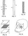
- FIG. 9is a top view of titanium mesh that may be employed with any of the embodiments of the invention.
- FIG. 10is an enlarged view of a section of the titanium mesh depicted in FIG. 9 .
- FIG. 11is a side sectional view of an implant having opposite barrier surfaces that a center section.
- FIG. 12is a side view in elevation of the implant depicted in FIG. 11 .
- FIG. 13is a side sectional view of the implant depicted in FIGS. 1-3 .
- FIG. 14depicts a sectional view of a cranial defect.
- FIG. 15is a side sectional view of the implant shown in FIGS. 1-3 within a cranial defect.
- FIG. 16is yet another embodiment of the invention wherein the implant has opposite barrier surfaces.
- FIG. 17is a side view in elevation of the implant depicted in FIG. 16 .
- FIG. 18is a side sectional view of a further embodiment of the invention wherein the metal mesh is formed with an implant with opposite porous surfaces.
- FIG. 19is an exploded view of an implant having three layers.
- FIG. 20is a perspective illustration of an implant according to the invention shown in an orbital reconstruction application.
- the present inventionis directed to novel implants for craniofacial surgery, methods for making said implant and a method of reconstructing orbital and cranial defects with the implants described.
- a preferred application for the implantis for the reconstruction of orbital defects that may have resulted from trauma or disease or birth defects.
- Other craniofacial applicationsare also contemplated.
- a first embodiment of the inventioncomprises a sheet of titanium mesh 20 , with porous polyethylene formed in the interstices of the mesh and completely covering the bottom surface 27 of the implant.
- a solid sheet of polyethylene film 23covers the top side of the implant.
- the mesh 20provides for strength and serves to retain the shape of the implant in a rigid and fixed position.
- a mesh as used hereinencompass any flat sheet of surgical grade metal that has perforations or passages formed through the sheet. The passages in the sheet help enable the sheet to be shaped or bent in more than one dimension and then retain the desired shape.
- the meshcould be formed in a variety of manners including woven screens, or be etched from plates, or be formed from sold plates that are cut and then expanded to form a substrate having passages.
- FIG. 1The first specific embodiment of the invention is illustrated in FIG. 1 where a smooth bather material 23 lies on top of the titanium mesh material 20 with porous polyethylene 25 formed in the interstices and under the titanium mesh 20 .
- the top surface 23 of the implanthas some transparency so that the mesh 20 may be seen through the polyethylene film layer 23 .
- FIG. 1shows the mesh 105 extended to the periphery of the implant, it is contemplated that in some embodiments the mesh may not extend to the edge of the implant structure. In yet other embodiments, the mesh may extend from the implant structure. In this later regard, it may be advantageous to extend the mesh from the implant structure to provide for a metal projection to be employed for the attachment of the implant during the surgical procedure. While in the embodiments depicted herein, the mesh is depicted in the center of the implant structure, it is contemplated that the mesh may be positioned adjacent to the top thin sheet layer or other locations within the implant depending on the respective application.
- a mesh 40is selected and positioned on tabs 50 that project form the sidewalls 45 and 48 of the bottom of the mold section 42 .
- polyethylene finesare introduced into the mold so that they fill the void below the mesh 40 , the spaces between the titanium mesh 40 and cover the top surface of mesh 40 .
- a thin sheet or continuous film of solid polyethylene 55is placed on the top of a suitable mold. The solid barrier sheet 55 extends beyond the edges of the cavity section of the mold and extends to the mold surface 63 thereby maintaining the sheet on one side of the mold.
- FIG. 7depicts an alternative arrangement for a mold wherein the mesh may be received on a shelf 70 that is suspended over the cavity using a shelf 70 around the mold cavity that holds the mesh sheet in position.
- shelf region 70that extend into the void area 78 of mold 75 supports the edges of the mesh.
- a polyethylene sheet 90is positioned above polyethylene fines 92 that fill the cavity 78 .
- the passages through the meshare identified by reference number 52 . It should be understood that the dimensions, including the depth of the cavity from top surface 85 of bottom mold section 75 , and the length and width of the mold may be altered depending on the particular application intended for the implant.
- the fines 92come into contact with both the smooth polyethylene sheet 90 and the mesh 80 .
- the top section 98is placed over the components and the materials are subjected to heat and pressure, as is known in the current art, to form a porous polyethylene material.
- the heat and pressurecauses the fines to be sintered together and to be affix the polyethylene sheet and titanium mesh.
- the resulting structurehas titanium mesh embedded within a porous matrix and a solid smooth polyethylene film that is attached both to the titanium mesh and/or to the porous polyethylene structure.
- the sheet or film of polyethyleneis impervious to water and serves as a barrier.
- the polyethylene filmis approximately 0.1 mm thick
- the titanium meshis approximately 0.35 mm thick
- the porous polyethyleneis approximately 0.9 mm thick, inclusive of the imbedded titanium mesh.
- the overall thickness of the materialis approximately 1 mm.
- the titanium meshconsists of a series of annular rings 107 that are attached to adjacent annular rings by bridges 110 also made of titanium.
- the annular ringshave countersunk holes 115 that will receive the head of surgical screw.
- the titaniumis of sufficient strength in relation to the thickness of the polyethylene components (the solid sheet and the porous matrix) so that the implant will hold its shape after being bent by the surgeon.
- surgeonmay bend the implant to conform to the shape of the defect that is being treated.
- surgeoncan bend the implant by hand during the procedure.
- the implant as described abovecan also be cut with conventional plate cutters that are routinely used for cutting titanium surgical plates or mesh.
- titanium mesh productsthat can be used in connection with the invention are commercially available from sources that include Stryker Instruments, Synthes Maxillofacial, Leibinger, K L S-Martin, L. P. and Walter-Lorenz Surgical.
- FIGS. 11depicts yet another embodiment of the invention in which the titanium 150 is placed between two opposite polyethylene barrier sheets 153 and 155 .
- a porous matrix 160is sandwiched between the barrier sheets 153 and 155 use.
- the configuration of this implantprovides a bendable sheet that has a smooth polyethylene surface on both the top and bottom surface.
- the implantwill retain its shape after it has been bent to conform to the contours of defect to be treated.
- the implanthas strength properties that are inherent to titanium, and it has a non-porous barrier surface that is not amenable to tissue attachment to the implant.
- the thickness of the sheets of polyethylenemay be selected to result in an implant having the desired thickness. In the alternative, the thickness of the implant may be adjusted by variation of the porous matrix layer 160 .
- the implantmay be bent by the surgeon and it will maintain its shape.
- FIG. 13a side sectional view of the implant depicted in FIGS. 1-4 shows the mesh 20 formed along the interface 175 between the porous layer and the sold polyethylene layer 23 .
- a defect in the cranium 178has a floor 180 and a wall 182 .
- the implantis bent to conform to the contour of the defect and cut to the shape of the defect.
- the implantis placed within the defect and the bottom porous layer is brought into contact with the bone on the floor and sidewalls.
- the implantmay be secured into place with screw or sutures.
- the bottom surface and the sidewalls of the implantare porous, fibrovascular ingrowth into the implant is encouraged and this ingrowth serves to further stabilize the implant and diminish the possibility of rejection.
- the smooth barrier surfaceprevents the dermis from attachment and thereby allows the skin to slide over the implant area.
- the structureinvolves the providing of a titanium mesh plate within a porous polyethylene matrix wherein all sides have porous surfaces.
- FIG. 18depicts a sectional view wherein the mesh 300 is formed with a porous polyethylene matrix.
- This implantmay be suitable for those applications where a smooth barrier surface is not indicated.
- an implant having porous surfaces that allow for fibrovascular ingrowth on opposite sidesmay be indicated in cranial applications and for temporal implants for soft tissue replacement.
- the pore size of the porous polyethyleneis sized large enough to allow for fibrovascular ingrowth.
- This pore size rangewould preferably be in the range of 100-250 microns, but could vary in the range of 20-500 microns.
- polyethylene sheets and high density porous polyethylene matrixare preferred, it is also contemplated that other synthetic resins and combinations can be used in connection with the invention.
- PETE, PTFE and/or nylonmay be selected as the thermoplastic resin.
- the Figures depicted hereinare not necessarily drawn to scale.
- the barrier in FIGS. 1-4may be formed with a sheet having a much smaller width than the drawings may suggest.
- the invention as depicted in FIGS. 1-4is approximately 5 mm wide by 10 mm in length and has a thickness of approximately 1 mm.
- other dimensionsare contemplated.
- FIG. 5is a sectional view of the implant according to the invention located within a mold. As depicted therein, the mesh is located adjacent to the barrier layer on the top of the mold.
- the barrier layeris formed of a solid sheet of polyethylene and the porous section is made by sintering together polyethylene fines under heat and pressure.
- the sold sheetmay be made by introducing polyethylene fines to a press having opposite smooth metal sheets and heating the surfaces causing the fines to completely fuse together.
- the structuremay be removed from the mold because both the tabs 50 and the implant material have some flexibility.
- FIG. 6a contemplated arrangement depicting a plurality of tabs 50 provided on the lower section of mold 61 is shown.
- the titanium sheetwill rest on or is supported by the tabs 50 provided around the periphery of the mold.
- the tabsare placed a distance from the top surface of the mold that is slightly less than the width of the mesh, so that when the top of the mold that retains the barrier sheet is placed over the mold bottom, the thin barrier sheet may come into contact with the mesh.
- FIG. 7depicts an alternative arrangement wherein the mold is provided with a shelf to retain the titanium mesh in position near the top of the mold.
- FIG. 16depicts yet a further embodiment of the implant wherein the top surface 214 and bottom surface 126 are polyethylene sheets.
- the mesh 220is contiguous with the internal surfaces of both the top sheet 214 and the lower sheet 216 .
- This implanthas a top barrier surface 221 and bottom barrier surface 223 and is indicated in those applications where fibrovascular ingrowth is not desired.
- FIG. 19shows an exploded perspective schematic view of the embodiment according to the invention.
- Top layer 400may comprise a barrier surface or porous surface.
- the mesh 405may be any metallic material suitable for surgical applications that and that is malleable and will retain its shape.
- Bottom layer 410may be a barrier surface or a porous surface. This embodiment depicts mesh 405 at the interface between the layers 400 and 410 .
- FIG. 20depicts an implant 500 made according to the invention in position on the orbit floor of an orbit 507 .
- the implants according to the inventionmay be advantageously employed with other surgery such as the repair of lost bone flaps resulting from neurological procedures, repair of the mastoid area after a mastoidectomy, fixation for LeFort procedures, fixation for sliding genioplasty.
- the planar sheetsmay be bent into tubular shapes and used for orthopedic applications. A planar sheet bent in a U shaped configuration may be useful in connection with spinal fixation procedures or the repair of herniated disks.
Landscapes
- Health & Medical Sciences (AREA)
- Life Sciences & Earth Sciences (AREA)
- Orthopedic Medicine & Surgery (AREA)
- Engineering & Computer Science (AREA)
- Animal Behavior & Ethology (AREA)
- Veterinary Medicine (AREA)
- Public Health (AREA)
- General Health & Medical Sciences (AREA)
- Oral & Maxillofacial Surgery (AREA)
- Transplantation (AREA)
- Chemical & Material Sciences (AREA)
- Biomedical Technology (AREA)
- Heart & Thoracic Surgery (AREA)
- Vascular Medicine (AREA)
- Cardiology (AREA)
- Surgery (AREA)
- Epidemiology (AREA)
- Dermatology (AREA)
- Medicinal Chemistry (AREA)
- Composite Materials (AREA)
- Materials Engineering (AREA)
- Neurology (AREA)
- Inorganic Chemistry (AREA)
- Nuclear Medicine, Radiotherapy & Molecular Imaging (AREA)
- Neurosurgery (AREA)
- Medical Informatics (AREA)
- Molecular Biology (AREA)
- Manufacturing & Machinery (AREA)
- Prostheses (AREA)
- Materials For Medical Uses (AREA)
Abstract
Description
Claims (16)
Priority Applications (1)
| Application Number | Priority Date | Filing Date | Title |
|---|---|---|---|
| US12/652,896US8398720B2 (en) | 2003-04-16 | 2010-01-06 | Craniofacial implant |
Applications Claiming Priority (5)
| Application Number | Priority Date | Filing Date | Title |
|---|---|---|---|
| US46303603P | 2003-04-16 | 2003-04-16 | |
| US49668403P | 2003-08-21 | 2003-08-21 | |
| US10/517,843US7655047B2 (en) | 2003-04-16 | 2004-04-16 | Craniofacial implant |
| PCT/US2004/011903WO2004093743A1 (en) | 2003-04-16 | 2004-04-16 | Craniofacial implant |
| US12/652,896US8398720B2 (en) | 2003-04-16 | 2010-01-06 | Craniofacial implant |
Related Parent Applications (3)
| Application Number | Title | Priority Date | Filing Date |
|---|---|---|---|
| US10/517,843ContinuationUS7655047B2 (en) | 2003-04-16 | 2004-04-16 | Craniofacial implant |
| US10517843Continuation | 2004-04-16 | ||
| PCT/US2004/011903ContinuationWO2004093743A1 (en) | 2003-04-16 | 2004-04-16 | Craniofacial implant |
Publications (2)
| Publication Number | Publication Date |
|---|---|
| US20100114316A1 US20100114316A1 (en) | 2010-05-06 |
| US8398720B2true US8398720B2 (en) | 2013-03-19 |
Family
ID=33313431
Family Applications (2)
| Application Number | Title | Priority Date | Filing Date |
|---|---|---|---|
| US10/517,843Expired - LifetimeUS7655047B2 (en) | 2003-04-16 | 2004-04-16 | Craniofacial implant |
| US12/652,896Expired - LifetimeUS8398720B2 (en) | 2003-04-16 | 2010-01-06 | Craniofacial implant |
Family Applications Before (1)
| Application Number | Title | Priority Date | Filing Date |
|---|---|---|---|
| US10/517,843Expired - LifetimeUS7655047B2 (en) | 2003-04-16 | 2004-04-16 | Craniofacial implant |
Country Status (6)
| Country | Link |
|---|---|
| US (2) | US7655047B2 (en) |
| EP (2) | EP2308423A1 (en) |
| KR (2) | KR20100102753A (en) |
| CN (1) | CN100586401C (en) |
| BR (1) | BRPI0409487A (en) |
| WO (1) | WO2004093743A1 (en) |
Cited By (12)
| Publication number | Priority date | Publication date | Assignee | Title |
|---|---|---|---|---|
| US20120265313A1 (en)* | 2011-04-01 | 2012-10-18 | Shawn Burke | Method of Cranial Repair and Cranial Repair Implant Molding Device |
| WO2014019083A1 (en)* | 2012-07-30 | 2014-02-06 | Sunnybrook Health Sciences Centre | Bone stabilization device and method of production |
| US20140228969A1 (en)* | 2013-02-12 | 2014-08-14 | Ossdsign Ab | Mosaic Implants, Kits and Methods for Correcting Bone Defects |
| US8864826B2 (en)* | 2010-02-26 | 2014-10-21 | Limacorporate Spa | Integrated prosthetic element |
| USD723162S1 (en)* | 2011-09-30 | 2015-02-24 | Osteosymbionics, Llc | Soft tissue implant |
| US9724198B2 (en) | 2014-07-17 | 2017-08-08 | Poriferous, LLC | Orbital floor sheet |
| US10076416B2 (en) | 2013-02-12 | 2018-09-18 | Ossdsign Ab | Mosaic implants, kits and methods for correcting bone defects |
| USD884898S1 (en) | 2017-09-29 | 2020-05-19 | Matrix Surgical Holdings, LLC | Orbital implant for human ocular support |
| US10881519B2 (en) | 2014-08-14 | 2021-01-05 | Ossdsign Ab | Bone implants for correcting bone defects |
| US10898332B2 (en) | 2015-11-24 | 2021-01-26 | Ossdsign Ab | Bone implants and methods for correcting bone defects |
| USD909580S1 (en) | 2019-04-05 | 2021-02-02 | Sunnybrook Research Institute | Surgical mesh implant |
| US11384260B1 (en) | 2021-05-28 | 2022-07-12 | Cohesys Inc. | Adhesive devices and uses thereof |
Families Citing this family (97)
| Publication number | Priority date | Publication date | Assignee | Title |
|---|---|---|---|---|
| AU2003261497B2 (en) | 2002-11-08 | 2009-02-26 | Howmedica Osteonics Corp. | Laser-produced porous surface |
| US8298292B2 (en) | 2003-04-16 | 2012-10-30 | Howmedica Osteonics Corp. | Craniofacial implant |
| KR20100102753A (en) | 2003-04-16 | 2010-09-24 | 포렉스 서지칼, 인크. | Craniofacial implant |
| WO2005041812A2 (en)* | 2003-10-22 | 2005-05-12 | Implant Brace, Inc. | Implantable brace for a fracture and methods |
| US7887587B2 (en)* | 2004-06-04 | 2011-02-15 | Synthes Usa, Llc | Soft tissue spacer |
| US20060116682A1 (en)* | 2004-11-18 | 2006-06-01 | Longo Marc N | Surgical implant and methods of making and using the same |
| WO2006102470A2 (en)* | 2005-03-22 | 2006-09-28 | Posnick, Jeffrey, C. | Facial implant |
| US8728387B2 (en) | 2005-12-06 | 2014-05-20 | Howmedica Osteonics Corp. | Laser-produced porous surface |
| US9119677B2 (en) | 2005-12-09 | 2015-09-01 | DePuy Synthes Products, Inc. | Spinal plate and drill guide |
| RU2308909C1 (en)* | 2005-12-28 | 2007-10-27 | Федеральное государственное лечебно-профилактическое учреждение "Научно-клинический центр охраны здоровья шахтеров" Федерального агентства по энергетике РФ | Device for closing cranial vault bones defects |
| AU2006341485A1 (en)* | 2006-04-05 | 2007-10-11 | Synthes Gmbh | Method and device for producing a planar implant for a human or animal body, which planar implant is preformed corresponding to a desired anatomical shape |
| CN100384383C (en)* | 2006-07-17 | 2008-04-30 | 中国人民解放军第二炮兵总医院 | A medical device for reduction and fixation of inferior orbital wall fracture |
| US20060287654A1 (en)* | 2006-08-11 | 2006-12-21 | Jeffrey Posnick | Implant securing device and method |
| US8114080B2 (en)* | 2006-09-27 | 2012-02-14 | Depuy Products, Inc. | Flexible bone fixation device |
| RU2336054C1 (en)* | 2007-02-09 | 2008-10-20 | МУЗ городская клиническая больница №3 им. М.А. Подгорбунского г. Кемерово | Calvarium bone prosthesis |
| RU2340310C1 (en)* | 2007-04-16 | 2008-12-10 | МУЗ городская клиническая больница №3 им. М.А. Подгорбунского г. Кемерово | Attachment to skull prosthesis |
| CA2692376A1 (en)* | 2007-06-29 | 2009-01-08 | Synthes (U.S.A.) | Improved orthopedic implants for use with precision bone resurfacing instrumentation |
| US9993337B1 (en)* | 2007-07-19 | 2018-06-12 | Osteosymbionics, Llc | Orthopaedic implant and method of making same |
| US8114156B2 (en)* | 2008-05-30 | 2012-02-14 | Edwin Burton Hatch | Flexibly compliant ceramic prosthetic meniscus for the replacement of damaged cartilage in orthopedic surgical repair or reconstruction of hip, knee, ankle, shoulder, elbow, wrist and other anatomical joints |
| US9107712B2 (en) | 2008-09-15 | 2015-08-18 | Biomet C.V. | Bone plate system for hand fractures and other small bones |
| RU2387411C1 (en)* | 2008-11-05 | 2010-04-27 | Общество с ограниченной ответственностью "КОНМЕТ" | Shape-changing mesh implant for fixation and immobilisation of bone fragments and covering bone defects or spaces on one or several patient's spots |
| EP2401000A2 (en)* | 2009-02-25 | 2012-01-04 | Howmedica Osteonics Corp. | Bone graft material containment structures |
| BR112012022686B1 (en) | 2010-03-10 | 2021-04-20 | Oss-Q Ab | mosaic implant, method for preparing an implant and using an implant |
| WO2011153645A2 (en)* | 2010-06-11 | 2011-12-15 | Sunnybrook Health Sciences Center | Method of forming patient-specific implant |
| WO2012016200A1 (en)* | 2010-07-30 | 2012-02-02 | The Henry M. Jackson Foundation For The Advancement Of Military Medicine, Inc. | Systems and methds for cranial implant assembly adapted for insertion during craniectomy procedure |
| US9023085B2 (en) | 2010-12-22 | 2015-05-05 | Walter E. Strippgen | Dynamic surgical implant |
| US8231624B1 (en) | 2010-12-22 | 2012-07-31 | Strippgen Walter E | Dynamic surgical implant |
| US9034048B2 (en)* | 2011-01-26 | 2015-05-19 | John A. Choren | Orthopaedic implants and methods of forming implant structures |
| US20120203227A1 (en)* | 2011-02-08 | 2012-08-09 | Christopher Harris Martin | Low profile dorsal plate |
| US9510940B2 (en) | 2011-02-17 | 2016-12-06 | Ethicon, Inc. | Bioabsorbable multilayer nasal valve spreader graft |
| WO2012116401A1 (en) | 2011-02-28 | 2012-09-07 | Anatomics Pty Ltd | Surgical implant and method |
| US8579990B2 (en)* | 2011-03-30 | 2013-11-12 | Ethicon, Inc. | Tissue repair devices of rapid therapeutic absorbency |
| US9463046B2 (en) | 2011-08-22 | 2016-10-11 | Ossdsign Ab | Implants and methods for using such implants to fill holes in bone tissue |
| US9381112B1 (en) | 2011-10-06 | 2016-07-05 | William Eric Sponsell | Bleb drainage device, ophthalmological product and methods |
| US8632489B1 (en) | 2011-12-22 | 2014-01-21 | A. Mateen Ahmed | Implantable medical assembly and methods |
| US9414873B2 (en) | 2012-01-05 | 2016-08-16 | The Cleveland Clinic Foundation | Modular bone fixation system |
| WO2013106323A1 (en) | 2012-01-09 | 2013-07-18 | Zimmer, Inc. | Porous metal implants with bone cement |
| BR112014019797B1 (en) | 2012-02-10 | 2019-11-19 | Synthes Gmbh | porous implant materials and related methods |
| US9180010B2 (en) | 2012-04-06 | 2015-11-10 | Howmedica Osteonics Corp. | Surface modified unit cell lattice structures for optimized secure freeform fabrication |
| US9135374B2 (en) | 2012-04-06 | 2015-09-15 | Howmedica Osteonics Corp. | Surface modified unit cell lattice structures for optimized secure freeform fabrication |
| WO2013155043A1 (en)* | 2012-04-09 | 2013-10-17 | The Johns Hopkins University | Universal cranioplasty mesh |
| US20130317540A1 (en) | 2012-05-22 | 2013-11-28 | Krasimira Hristov | Universal bioabsorbable nasal implant kit |
| AU2013267381B2 (en)* | 2012-05-30 | 2016-03-31 | New York University | Tissue repair devices and scaffolds |
| US9579133B2 (en) | 2013-02-01 | 2017-02-28 | James Guthlein | Internal fixation device |
| US9517097B2 (en)* | 2013-04-17 | 2016-12-13 | Stc.Unm | Low-profile, high tension mesh plate for subcutaneous fracture fixation |
| US9044195B2 (en) | 2013-05-02 | 2015-06-02 | University Of South Florida | Implantable sonic windows |
| KR101355598B1 (en) | 2013-10-11 | 2014-02-04 | (주)이트리온홀딩스 | 3d implant for orbital wall and floor |
| AT515384B1 (en)* | 2014-02-05 | 2016-04-15 | Dietmar Dr Sonnleitner | Preconnected multilayer film for covering a bone defect site |
| US9549819B1 (en) | 2014-06-23 | 2017-01-24 | DePuy Synthes Products, Inc. | Preformed cranial implant |
| RU2579744C1 (en)* | 2014-11-14 | 2016-04-10 | Александр Ливиевич Ураков | Cranial implant-heat insulation material |
| CN105105872A (en)* | 2015-09-08 | 2015-12-02 | 哈尔滨工业大学 | Skull replacing apparatus of 3D print and manufacturing method thereof |
| US10130402B2 (en) | 2015-09-25 | 2018-11-20 | Globus Medical, Inc. | Bone fixation devices having a locking feature |
| US9974581B2 (en) | 2015-11-20 | 2018-05-22 | Globus Medical, Inc. | Expandable intramedullary systems and methods of using the same |
| US10932834B2 (en)* | 2015-12-03 | 2021-03-02 | Howard D. Stupak | Oblique three-dimensional plate |
| US20170202586A1 (en)* | 2015-12-11 | 2017-07-20 | DePuy Synthes Products, Inc. | Composite implant trial |
| US10596660B2 (en) | 2015-12-15 | 2020-03-24 | Howmedica Osteonics Corp. | Porous structures produced by additive layer manufacturing |
| AU2016369593B2 (en) | 2015-12-16 | 2021-04-01 | Nuvasive, Inc. | Porous spinal fusion implant |
| US9795411B2 (en) | 2016-03-02 | 2017-10-24 | Globus Medical, Inc. | Fixators for bone stabilization and associated systems and methods |
| US10531905B2 (en) | 2016-04-19 | 2020-01-14 | Globus Medical, Inc. | Implantable compression screws |
| CA3025434A1 (en)* | 2016-06-03 | 2017-12-07 | DePuy Synthes Products, Inc. | Surgical templates with radio-opaque markings |
| KR101671150B1 (en)* | 2016-07-15 | 2016-10-31 | 가톨릭관동대학교산학협력단 | Implant for orbital wall |
| US11432857B2 (en) | 2016-08-17 | 2022-09-06 | Globus Medical, Inc. | Stabilization systems |
| US11197701B2 (en) | 2016-08-17 | 2021-12-14 | Globus Medical, Inc. | Stabilization systems |
| US10299847B2 (en) | 2016-09-22 | 2019-05-28 | Globus Medical, Inc. | Systems and methods for intramedullary nail implantation |
| WO2018076003A1 (en)* | 2016-10-21 | 2018-04-26 | University Of Pittsburgh-Of The Commonwealth System Of Higher Education | Degradable bulk metallic magnesium/polymer composite barrier membranes for dental, craniomaxillofacial and orthopedic applications and manufacturing methods |
| WO2018140706A1 (en)* | 2017-01-26 | 2018-08-02 | Poriferous, LLC | Channel implant |
| KR101922966B1 (en)* | 2017-02-22 | 2018-11-28 | 가톨릭관동대학교산학협력단 | Manufacturing method of implant for orbital wall and implant for orbital wall |
| US11298747B2 (en) | 2017-05-18 | 2022-04-12 | Howmedica Osteonics Corp. | High fatigue strength porous structure |
| US11628517B2 (en) | 2017-06-15 | 2023-04-18 | Howmedica Osteonics Corp. | Porous structures produced by additive layer manufacturing |
| US10603180B2 (en)* | 2017-07-17 | 2020-03-31 | Aaron MARLOW | Tapered fixation device for a knee replacement |
| EP3479798B1 (en) | 2017-11-03 | 2023-06-21 | Howmedica Osteonics Corp. | Flexible construct for femoral reconstruction |
| KR102024598B1 (en) | 2017-11-03 | 2019-09-24 | 울산대학교 산학협력단 | Method and apparatus for generating 3d model data for manufacturing of implant |
| US11224468B2 (en) | 2018-03-02 | 2022-01-18 | Globus Medical, Inc. | Distal tibial plating system |
| US11071570B2 (en) | 2018-03-02 | 2021-07-27 | Globus Medical, Inc. | Distal tibial plating system |
| US11141172B2 (en) | 2018-04-11 | 2021-10-12 | Globus Medical, Inc. | Method and apparatus for locking a drill guide in a polyaxial hole |
| EP3784176B1 (en)* | 2018-04-23 | 2025-08-27 | ECA Medical Instruments | Flexible adjustable radiopaque trial plate |
| KR102183079B1 (en) | 2018-06-12 | 2020-11-26 | 경북대학교 산학협력단 | Method and device for modelling and producing implant for orbital wall |
| DE102018121553A1 (en)* | 2018-09-04 | 2020-03-05 | Karl Leibinger Medizintechnik Gmbh & Co. Kg | Bone implant for the reconstruction of a bony defect and for guiding a marking and / or processing tool for transferring the necessary osteotomy situations |
| US11090149B2 (en) | 2018-09-28 | 2021-08-17 | DePuy Synthes Products, Inc. | Inflatable orbital implant for repositioning an eyeball, and related methods |
| FR3089128B1 (en) | 2018-11-30 | 2020-12-18 | Carthera | ACOUSTIC WINDOW FOR IMAGING AND / OR TREATMENT OF CEREBRAL TISSUE |
| US11173057B2 (en)* | 2018-11-30 | 2021-11-16 | Arizona Board Of Regents On Behalf Of Arizona State University | Volume adjustable transtibial socket |
| US11771563B2 (en)* | 2019-01-03 | 2023-10-03 | University of Alaska Anchorage | Artificial tessellated implants, and systems and methods of making and using same |
| US11202663B2 (en) | 2019-02-13 | 2021-12-21 | Globus Medical, Inc. | Proximal humeral stabilization systems and methods thereof |
| AU2020240099B2 (en)* | 2019-03-19 | 2024-06-27 | Poriferous, LLC | Orbital floor template |
| KR102250250B1 (en) | 2019-03-29 | 2021-05-12 | 경북대학교 산학협력단 | Method and device for modelling and producing implant for orbital wall of human body customication with patterns for implanting a screw |
| BR112021021916A8 (en)* | 2019-05-02 | 2023-02-28 | Poriferous Llc | ORBITAL FLOOR IMPLANT |
| US11730582B2 (en)* | 2019-09-25 | 2023-08-22 | Washington University | Barbed mesh for incision closure and hernia repair |
| US12185995B2 (en) | 2019-10-09 | 2025-01-07 | Globus Medical, Inc. | Bone stabilization systems |
| US11129627B2 (en) | 2019-10-30 | 2021-09-28 | Globus Medical, Inc. | Method and apparatus for inserting a bone plate |
| KR102287890B1 (en) | 2019-11-20 | 2021-08-09 | 윤설아 | Manufacturing method of implants for reconstruction of CMF and implants manufactured by the same |
| US11723647B2 (en) | 2019-12-17 | 2023-08-15 | Globus Medical, Inc. | Syndesmosis fixation assembly |
| RU2743108C1 (en)* | 2019-12-25 | 2021-02-15 | Федеральное государственное автономное образовательное учреждение высшего образования "Национальный исследовательский технологический университет "МИСиС" | Hybrid plate for cranioplasty |
| KR102608168B1 (en)* | 2021-06-01 | 2023-12-04 | 전북대학교병원 | Carbon implant and Manufacturing method of the same |
| EP4104799B1 (en)* | 2021-06-16 | 2025-07-30 | Discoseal BV | Patch for covering a defect in the annulus fibrosus of an intervertebral disc, and in particular also in the posterior longitudinal ligament, of a spine |
| US12064150B2 (en) | 2022-01-19 | 2024-08-20 | Globus Medical Inc. | System and method for treating bone fractures |
| CN115446547B (en)* | 2022-09-16 | 2024-06-04 | 景德镇陶瓷大学 | Titanium mesh plate incremental forming method and preparation method of brain skull prosthesis |
| US20240415555A1 (en)* | 2023-06-19 | 2024-12-19 | Karl Leibinger Asset Management Gmbh & Co. Kg | Implant and Method for Covering Large-Scale Bone Defects for Thorax |
Citations (119)
| Publication number | Priority date | Publication date | Assignee | Title |
|---|---|---|---|---|
| US2671444A (en) | 1951-12-08 | 1954-03-09 | Jr Benjamin F Pease | Nonmetallic mesh surgical insert for hernia repair |
| US3048537A (en)* | 1958-01-06 | 1962-08-07 | Pall Corp | Porous articles of polyethylene polymers and process of making the same |
| US3178728A (en) | 1962-10-22 | 1965-04-20 | Robert W Christensen | Surgical prosthesis for the temporomandibular joint |
| US3579643A (en) | 1968-12-12 | 1971-05-25 | Douglas H Morgan | Artificial articular eminence for the mandibular joint |
| US4089071A (en) | 1976-09-08 | 1978-05-16 | Kalnberz Viktor Konstantinovic | Material for making bone endoprosthesis and endoprosthesis made of said material |
| DE2404214C3 (en) | 1973-01-31 | 1978-08-24 | Comptoir Lyon-Alemand - Louyot, Paris | Bone prosthesis and process for the manufacture thereof |
| US4164794A (en) | 1977-04-14 | 1979-08-21 | Union Carbide Corporation | Prosthetic devices having coatings of selected porous bioengineering thermoplastics |
| GB2059267B (en) | 1979-09-28 | 1983-05-11 | Leuven Res & Dev Vzw | Compound material for prosthetic devices |
| US4479271A (en) | 1981-10-26 | 1984-10-30 | Zimmer, Inc. | Prosthetic device adapted to promote bone/tissue ingrowth |
| US4502161A (en) | 1981-09-21 | 1985-03-05 | Wall W H | Prosthetic meniscus for the repair of joints |
| US4531916A (en) | 1983-07-08 | 1985-07-30 | W. L. Gore & Associates, Inc. | Dental implant with expanded PTFE gingival interface |
| US4535485A (en) | 1982-03-12 | 1985-08-20 | Medical Biological Sciences, Inc. | Polymeric acrylic prothesis |
| US4542539A (en) | 1982-03-12 | 1985-09-24 | Artech Corp. | Surgical implant having a graded porous coating |
| US4550448A (en) | 1982-02-18 | 1985-11-05 | Pfizer Hospital Products Group, Inc. | Bone prosthesis with porous coating |
| US4636215A (en) | 1984-01-11 | 1987-01-13 | Rei, Inc. | Combination tray and condylar prosthesis for mandibular reconstruction and the like |
| US4693721A (en) | 1984-10-17 | 1987-09-15 | Paul Ducheyne | Porous flexible metal fiber material for surgical implantation |
| US4756862A (en) | 1977-04-14 | 1988-07-12 | Amoco Corporation | Prosthetic devices having coatings of selected porous bioengineering thermoplastics |
| US4778472A (en) | 1985-04-30 | 1988-10-18 | Vitek, Inc. | Implant for reconstruction of temporomanibular joint |
| US4790849A (en) | 1985-08-23 | 1988-12-13 | Edward Terino | Malar implant and method of inserting the prothesis |
| US4863474A (en) | 1983-07-08 | 1989-09-05 | Zimmer Limited | Skeletal implants |
| US4917701A (en) | 1988-09-12 | 1990-04-17 | Morgan Douglas H | Temporomandibular joint prostheses |
| US4936852A (en) | 1985-04-30 | 1990-06-26 | Vitek, Inc. | Temporomandibular mini condyle prosthesis |
| US4969901A (en) | 1988-06-28 | 1990-11-13 | Binder William J | Plastic surgery implant |
| US4976738A (en) | 1985-01-09 | 1990-12-11 | Sulzer Brothers Limited | Porous metal overlay for an implant surface |
| US4976737A (en) | 1988-01-19 | 1990-12-11 | Research And Education Institute, Inc. | Bone reconstruction |
| US4978355A (en) | 1985-01-25 | 1990-12-18 | Sulzer Brothers Limited | Plastic bone implant having a reinforced contact surface |
| US5030233A (en) | 1984-10-17 | 1991-07-09 | Paul Ducheyne | Porous flexible metal fiber material for surgical implantation |
| US5139497A (en) | 1991-11-25 | 1992-08-18 | Timesh, Inc. | Orbital repair implant |
| US5201737A (en) | 1991-04-11 | 1993-04-13 | Oswald Leibinger Gmbh | Plate for covering a drill hole in a skull cap and for fixing a cranial bone cover |
| US5218975A (en) | 1991-10-25 | 1993-06-15 | Prostkoff Melvin E | Cranial prosthesis |
| US5250048A (en) | 1991-01-28 | 1993-10-05 | Ferdinand Gundolf | Stabilizing element for osteosynthesis of bone fragments, especially for the fixation of bone fractures |
| US5282861A (en) | 1992-03-11 | 1994-02-01 | Ultramet | Open cell tantalum structures for cancellous bone implants and cell and tissue receptors |
| US5290315A (en) | 1991-03-07 | 1994-03-01 | Joint Medical Products Corporation | Oblong acetabular cup |
| US5346492A (en) | 1992-03-30 | 1994-09-13 | Timesh, Inc. | Perforated metallic panels and strips for internal fixation of bone fractures and for reconstructive surgery |
| US5372598A (en) | 1987-05-14 | 1994-12-13 | Howmedica Gmbh | Small bone plate for cranial or facial fractures or the like |
| US5380328A (en)* | 1993-08-09 | 1995-01-10 | Timesh, Inc. | Composite perforated implant structures |
| US5383931A (en) | 1992-01-03 | 1995-01-24 | Synthes (U.S.A.) | Resorbable implantable device for the reconstruction of the orbit of the human skull |
| US5397361A (en) | 1993-06-23 | 1995-03-14 | Surgical Prosthetics Resource, Inc. | Cranioplasty surgical procedure and kit |
| US5421831A (en) | 1991-04-19 | 1995-06-06 | Giampapa; Vincent C. | Sub-malar facial implant |
| US5433996A (en) | 1993-02-18 | 1995-07-18 | W. L. Gore & Associates, Inc. | Laminated patch tissue repair sheet material |
| US5443519A (en) | 1993-04-22 | 1995-08-22 | Implex Corporation | Prosthetic ellipsoidal acetabular cup |
| US5443512A (en) | 1990-10-30 | 1995-08-22 | Zimmer, Inc. | Orthopaedic implant device |
| US5445650A (en) | 1991-03-21 | 1995-08-29 | Nealis; Michael F. | Temporomandibular joint prosthesis |
| US5456723A (en) | 1989-03-23 | 1995-10-10 | Institut Straumann Ag | Metallic implant anchorable to bone tissue for replacing a broken or diseased bone |
| US5468242A (en) | 1993-11-19 | 1995-11-21 | Leibinger Gmbh | Form-fitting mesh implant |
| US5489305A (en) | 1994-10-03 | 1996-02-06 | Timesh, Inc. | Mandibular prostheses |
| US5496372A (en) | 1992-04-17 | 1996-03-05 | Kyocera Corporation | Hard tissue prosthesis including porous thin metal sheets |
| US5496371A (en) | 1991-06-10 | 1996-03-05 | United States Surgical Corporation | Prosthetic implant |
| EP0433852B1 (en) | 1989-12-22 | 1996-03-13 | Leibinger GmbH | Grid for osteosynthesis or for attaching of artificial members of the body |
| US5545226A (en) | 1992-05-29 | 1996-08-13 | Porex Technologies Corp. | Implants for cranioplasty |
| US5554194A (en) | 1995-06-07 | 1996-09-10 | United States Surgical Corporation | Modular surgical implant |
| US5578086A (en) | 1989-02-15 | 1996-11-26 | Xomed, Inc. | Prosthesis using biocompatible composite material |
| EP0423420B1 (en) | 1989-10-17 | 1996-11-27 | Timesh, Inc. | Bone fracture reduction and fixation devices with identity tags |
| US5669909A (en) | 1995-03-27 | 1997-09-23 | Danek Medical, Inc. | Interbody fusion device and method for restoration of normal spinal anatomy |
| US5690631A (en) | 1996-09-11 | 1997-11-25 | Walter Lorenz Surgical, Inc. | Multi-configurable plating system |
| US5743913A (en)* | 1997-04-02 | 1998-04-28 | Wellisz; Tadeusz Z. | Readily expansible bone fixation plate |
| US5755809A (en) | 1995-06-07 | 1998-05-26 | Implex Corporation | Femoral head core channel filling prothesis |
| US5766176A (en) | 1996-09-11 | 1998-06-16 | Walter Lorenz Surgical, Inc. | Formable mesh |
| US5769637A (en) | 1996-05-22 | 1998-06-23 | Sofamor Danek Properties, Inc. | Dental implant and alveolar process augmentation structures and method of installation |
| US5814048A (en) | 1996-05-03 | 1998-09-29 | Sofamor Danek Properties, Inc. | Cranioplasty plates and method of installation |
| US5824088A (en) | 1994-04-27 | 1998-10-20 | Kirsch; Axel | Cover device for bone voids and method for the manufacture thereof |
| US5863297A (en) | 1995-10-11 | 1999-01-26 | Osteobiologics, Inc. | Moldable, hand-shapable biodegradable implant material |
| US5876447A (en) | 1996-02-14 | 1999-03-02 | Implantech Associates | Silicone implant for facial plastic surgery |
| US5980540A (en) | 1997-04-11 | 1999-11-09 | Kinamed, Inc. | Perforated cover for covering spaces in the cranium and conforming to the shape of the cranium |
| US5989427A (en) | 1997-07-17 | 1999-11-23 | Tetra Technologies, Inc. | Method of degassing biological filters |
| US5989472A (en) | 1994-10-05 | 1999-11-23 | Howmedica International, Inc. | Method for making a reinforced orthopedic implant |
| US6008430A (en) | 1991-01-30 | 1999-12-28 | Interpore Orthopaedics, Inc. | Three-dimensional prosthetic articles and methods for producing same |
| US6010336A (en) | 1994-12-26 | 2000-01-04 | Kyocera Corporation | Living body-supporting member and preparation process thereof |
| US6031148A (en) | 1990-12-06 | 2000-02-29 | W. L. Gore & Associates, Inc. | Implantable bioabsorbable article |
| US6065197A (en) | 1998-04-06 | 2000-05-23 | Aichi Co., Ltd. | Method of spreading a sheet on a frame member and method of manufacturing a chair by the sheet spreading method |
| US6071291A (en) | 1997-10-21 | 2000-06-06 | Howmedica Leibinger Gmbh & Co. Kg (Leibinger) | Micro dynamic mesh |
| US6087553A (en) | 1996-02-26 | 2000-07-11 | Implex Corporation | Implantable metallic open-celled lattice/polyethylene composite material and devices |
| US6093188A (en) | 1997-11-10 | 2000-07-25 | Murray; William M. | Adjustable bone fixation plate |
| US6120539A (en) | 1997-05-01 | 2000-09-19 | C. R. Bard Inc. | Prosthetic repair fabric |
| US6129728A (en) | 1998-02-18 | 2000-10-10 | Walter Lorenz Surgical, Inc. | Method and apparatus for mandibular osteosynthesis |
| US6143036A (en) | 1997-12-18 | 2000-11-07 | Comfort Biomedical, Inc. | Bone augmentation for prosthetic implants and the like |
| US6187041B1 (en) | 1998-12-31 | 2001-02-13 | Scott N. Garonzik | Ocular replacement apparatus and method of coupling a prosthesis to an implant |
| US6221075B1 (en) | 1998-03-06 | 2001-04-24 | Bionx Implants Oy | Bioabsorbable, deformable fixation plate |
| US6238214B1 (en) | 1996-03-01 | 2001-05-29 | Dane Q. Robinson | Guided tissue regeneration plate for use in a process for growing jaw bone in anticipation of performing dental implants |
| US6267772B1 (en) | 1992-05-20 | 2001-07-31 | C. R. Bard, Inc. | Implantable prosthesis |
| US6325803B1 (en) | 1998-02-18 | 2001-12-04 | Walter Lorenz Surgical, Inc. | Method and apparatus for mandibular osteosynthesis |
| US20020022883A1 (en) | 2000-06-13 | 2002-02-21 | Burg Karen J.L. | Tissue engineering composite |
| US6350284B1 (en) | 1998-09-14 | 2002-02-26 | Bionx Implants, Oy | Bioabsorbable, layered composite material for guided bone tissue regeneration |
| US20020050463A1 (en) | 2000-09-22 | 2002-05-02 | Mcdowell Christopher | Tray for surgical fastners |
| US6391059B1 (en) | 1998-04-07 | 2002-05-21 | Macropore, Inc. | Membrane with tissue-guiding surface corrugations |
| US20020120348A1 (en) | 2000-12-21 | 2002-08-29 | Melican Mora Carolynne | Reinforced tissue implants and methods of manufacture and use |
| US20020123750A1 (en) | 2001-02-28 | 2002-09-05 | Lukas Eisermann | Woven orthopedic implants |
| WO2002069817A1 (en) | 2001-03-02 | 2002-09-12 | Woodwelding Ag | Implants, device and method for joining tissue parts |
| US6475094B1 (en) | 1998-12-28 | 2002-11-05 | Mark W. Bruns | Method for making product and product having ultra high molecular weight plastic parts |
| US6530953B2 (en) | 2000-05-25 | 2003-03-11 | Scott N. Garonzik | Method of magnetically coupling a prosthesis with an ocular implant |
| US6620332B2 (en) | 2001-01-25 | 2003-09-16 | Tecomet, Inc. | Method for making a mesh-and-plate surgical implant |
| WO2003086495A1 (en) | 2002-04-09 | 2003-10-23 | Astra Tech Ab | Medical prosthetic devices having improved biocompatibility |
| US20030208205A1 (en) | 2002-05-01 | 2003-11-06 | Jordan Medical Llc | Implantable device for covering and opening in a cranium |
| US6645250B2 (en) | 2001-10-30 | 2003-11-11 | Carl W. Schulter | Biocompatible form and method of fabrication |
| US6652585B2 (en) | 2001-02-28 | 2003-11-25 | Sdgi Holdings, Inc. | Flexible spine stabilization system |
| US20030220696A1 (en) | 2002-05-23 | 2003-11-27 | Levine David Jerome | Implantable porous metal |
| US20040019389A1 (en) | 2002-07-23 | 2004-01-29 | Greg Swords | Composite surgical implant made from macroporous synthetic resin and bioglass particles |
| US6692498B1 (en) | 2000-11-27 | 2004-02-17 | Linvatec Corporation | Bioabsorbable, osteopromoting fixation plate |
| US20040054372A1 (en) | 1997-08-19 | 2004-03-18 | Btg International Limited | Biodegradable composites |
| US20040059356A1 (en) | 2002-07-17 | 2004-03-25 | Peter Gingras | Soft tissue implants and methods for making same |
| US20040059422A1 (en) | 2000-03-24 | 2004-03-25 | Karl Koschatzky | Implant for cranioplastics |
| WO2004093743A1 (en) | 2003-04-16 | 2004-11-04 | Porex Surgical, Inc. | Craniofacial implant |
| US20040267349A1 (en) | 2003-06-27 | 2004-12-30 | Kobi Richter | Amorphous metal alloy medical devices |
| US6852330B2 (en) | 2000-12-21 | 2005-02-08 | Depuy Mitek, Inc. | Reinforced foam implants with enhanced integrity for soft tissue repair and regeneration |
| US20050146070A1 (en) | 2002-06-21 | 2005-07-07 | Massachusetts General Hospital | Meta lback or mesh crosslinking |
| US20050192675A1 (en) | 2004-03-01 | 2005-09-01 | Robinson Dane Q. | Device for use in stimulating bone growth |
| US20050208095A1 (en) | 2003-11-20 | 2005-09-22 | Angiotech International Ag | Polymer compositions and methods for their use |
| CN1225373C (en) | 2000-07-21 | 2005-11-02 | 本田技研工业株式会社 | Support structure of radiator in two-wheel and three-wheel motorcycle |
| US7050877B2 (en) | 2001-08-30 | 2006-05-23 | Pentax Corporation | Method for modeling an implant and an implant manufactured by the method |
| US20060116682A1 (en) | 2004-11-18 | 2006-06-01 | Longo Marc N | Surgical implant and methods of making and using the same |
| US20060217813A1 (en) | 2005-03-22 | 2006-09-28 | Posnick Jeffrey C | Facial implant |
| US20060224242A1 (en) | 2003-04-16 | 2006-10-05 | Porex Surgical, Inc. | Craniofacial implant |
| US7208222B2 (en) | 2003-07-24 | 2007-04-24 | Viasys Healthcare Inc. | Assembled non-random foams |
| US20070156146A1 (en) | 2005-12-29 | 2007-07-05 | Metzger Marc C | Implant for use as replacement of an orbita bottom |
| US20090138067A1 (en) | 1993-10-21 | 2009-05-28 | Leonard Pinchuk | Expandable supportive branched endoluminal grafts |
| US20090216338A1 (en) | 2005-09-12 | 2009-08-27 | Peter Gingras | Soft tissue implants and methods for making same |
| US7614258B2 (en) | 2006-10-19 | 2009-11-10 | C.R. Bard, Inc. | Prosthetic repair fabric |
| US20100023130A1 (en) | 2008-01-18 | 2010-01-28 | Porex Surgical, Inc. | Composite Implants and Methods of Making and Using the Same |
| US20100076555A1 (en) | 2008-09-19 | 2010-03-25 | Marten Lewis H | Coated devices comprising a fiber mesh imbedded in the device walls |
Family Cites Families (5)
| Publication number | Priority date | Publication date | Assignee | Title |
|---|---|---|---|---|
| US4327014A (en) | 1979-04-11 | 1982-04-27 | Kanebo Ltd. | Resin-forming material, implant material and compositions for restorative material suitable for medical or dental use |
| JPH02237559A (en) | 1989-03-10 | 1990-09-20 | Kobe Steel Ltd | Implant member for living body and preparation thereof |
| US5919234A (en) | 1996-08-19 | 1999-07-06 | Macropore, Inc. | Resorbable, macro-porous, non-collapsing and flexible membrane barrier for skeletal repair and regeneration |
| CN1148407C (en) | 1999-02-11 | 2004-05-05 | 上海超高工程塑料有限公司 | Polyethylene formation implant and method for making same |
| WO2003084410A1 (en) | 2002-04-01 | 2003-10-16 | Board Of Regents, The University Of Texas System | Composite material for wound repair |
- 2004
- 2004-04-16KRKR1020107020396Apatent/KR20100102753A/ennot_activeWithdrawn
- 2004-04-16CNCN200480009959Apatent/CN100586401C/ennot_activeExpired - Lifetime
- 2004-04-16WOPCT/US2004/011903patent/WO2004093743A1/enactiveApplication Filing
- 2004-04-16USUS10/517,843patent/US7655047B2/ennot_activeExpired - Lifetime
- 2004-04-16EPEP10183427Apatent/EP2308423A1/ennot_activeWithdrawn
- 2004-04-16BRBRPI0409487-5Apatent/BRPI0409487A/ennot_activeApplication Discontinuation
- 2004-04-16KRKR1020057019487Apatent/KR101027252B1/ennot_activeExpired - Fee Related
- 2004-04-16EPEP04759969.1Apatent/EP1613240B1/ennot_activeExpired - Lifetime
- 2010
- 2010-01-06USUS12/652,896patent/US8398720B2/ennot_activeExpired - Lifetime
Patent Citations (142)
| Publication number | Priority date | Publication date | Assignee | Title |
|---|---|---|---|---|
| US2671444A (en) | 1951-12-08 | 1954-03-09 | Jr Benjamin F Pease | Nonmetallic mesh surgical insert for hernia repair |
| US3048537A (en)* | 1958-01-06 | 1962-08-07 | Pall Corp | Porous articles of polyethylene polymers and process of making the same |
| US3178728A (en) | 1962-10-22 | 1965-04-20 | Robert W Christensen | Surgical prosthesis for the temporomandibular joint |
| US3579643A (en) | 1968-12-12 | 1971-05-25 | Douglas H Morgan | Artificial articular eminence for the mandibular joint |
| DE2404214C3 (en) | 1973-01-31 | 1978-08-24 | Comptoir Lyon-Alemand - Louyot, Paris | Bone prosthesis and process for the manufacture thereof |
| US4089071A (en) | 1976-09-08 | 1978-05-16 | Kalnberz Viktor Konstantinovic | Material for making bone endoprosthesis and endoprosthesis made of said material |
| US4164794A (en) | 1977-04-14 | 1979-08-21 | Union Carbide Corporation | Prosthetic devices having coatings of selected porous bioengineering thermoplastics |
| US4756862A (en) | 1977-04-14 | 1988-07-12 | Amoco Corporation | Prosthetic devices having coatings of selected porous bioengineering thermoplastics |
| GB2059267B (en) | 1979-09-28 | 1983-05-11 | Leuven Res & Dev Vzw | Compound material for prosthetic devices |
| US4502161A (en) | 1981-09-21 | 1985-03-05 | Wall W H | Prosthetic meniscus for the repair of joints |
| US4502161B1 (en) | 1981-09-21 | 1989-07-25 | ||
| US4479271A (en) | 1981-10-26 | 1984-10-30 | Zimmer, Inc. | Prosthetic device adapted to promote bone/tissue ingrowth |
| US4550448A (en) | 1982-02-18 | 1985-11-05 | Pfizer Hospital Products Group, Inc. | Bone prosthesis with porous coating |
| US4535485A (en) | 1982-03-12 | 1985-08-20 | Medical Biological Sciences, Inc. | Polymeric acrylic prothesis |
| US4542539A (en) | 1982-03-12 | 1985-09-24 | Artech Corp. | Surgical implant having a graded porous coating |
| US4863474A (en) | 1983-07-08 | 1989-09-05 | Zimmer Limited | Skeletal implants |
| US4531916A (en) | 1983-07-08 | 1985-07-30 | W. L. Gore & Associates, Inc. | Dental implant with expanded PTFE gingival interface |
| US4636215A (en) | 1984-01-11 | 1987-01-13 | Rei, Inc. | Combination tray and condylar prosthesis for mandibular reconstruction and the like |
| EP0178650B1 (en) | 1984-10-17 | 1993-04-07 | Paul Ducheyne | Porous flexible metal fiber material for surgical implantation |
| US5030233A (en) | 1984-10-17 | 1991-07-09 | Paul Ducheyne | Porous flexible metal fiber material for surgical implantation |
| US4693721A (en) | 1984-10-17 | 1987-09-15 | Paul Ducheyne | Porous flexible metal fiber material for surgical implantation |
| US4976738A (en) | 1985-01-09 | 1990-12-11 | Sulzer Brothers Limited | Porous metal overlay for an implant surface |
| US4978355A (en) | 1985-01-25 | 1990-12-18 | Sulzer Brothers Limited | Plastic bone implant having a reinforced contact surface |
| US4936852A (en) | 1985-04-30 | 1990-06-26 | Vitek, Inc. | Temporomandibular mini condyle prosthesis |
| US4778472A (en) | 1985-04-30 | 1988-10-18 | Vitek, Inc. | Implant for reconstruction of temporomanibular joint |
| US4790849A (en) | 1985-08-23 | 1988-12-13 | Edward Terino | Malar implant and method of inserting the prothesis |
| US5372598A (en) | 1987-05-14 | 1994-12-13 | Howmedica Gmbh | Small bone plate for cranial or facial fractures or the like |
| US4976737A (en) | 1988-01-19 | 1990-12-11 | Research And Education Institute, Inc. | Bone reconstruction |
| US4969901A (en) | 1988-06-28 | 1990-11-13 | Binder William J | Plastic surgery implant |
| US4917701A (en) | 1988-09-12 | 1990-04-17 | Morgan Douglas H | Temporomandibular joint prostheses |
| US5728157A (en) | 1989-02-15 | 1998-03-17 | Xomed Surgical Products, Inc. | Biocompatible composite prostheses |
| US5578086A (en) | 1989-02-15 | 1996-11-26 | Xomed, Inc. | Prosthesis using biocompatible composite material |
| US5456723A (en) | 1989-03-23 | 1995-10-10 | Institut Straumann Ag | Metallic implant anchorable to bone tissue for replacing a broken or diseased bone |
| EP0423420B1 (en) | 1989-10-17 | 1996-11-27 | Timesh, Inc. | Bone fracture reduction and fixation devices with identity tags |
| EP0433852B1 (en) | 1989-12-22 | 1996-03-13 | Leibinger GmbH | Grid for osteosynthesis or for attaching of artificial members of the body |
| US5443512A (en) | 1990-10-30 | 1995-08-22 | Zimmer, Inc. | Orthopaedic implant device |
| US6031148A (en) | 1990-12-06 | 2000-02-29 | W. L. Gore & Associates, Inc. | Implantable bioabsorbable article |
| US5250048A (en) | 1991-01-28 | 1993-10-05 | Ferdinand Gundolf | Stabilizing element for osteosynthesis of bone fragments, especially for the fixation of bone fractures |
| US6008430A (en) | 1991-01-30 | 1999-12-28 | Interpore Orthopaedics, Inc. | Three-dimensional prosthetic articles and methods for producing same |
| US5290315A (en) | 1991-03-07 | 1994-03-01 | Joint Medical Products Corporation | Oblong acetabular cup |
| US5445650A (en) | 1991-03-21 | 1995-08-29 | Nealis; Michael F. | Temporomandibular joint prosthesis |
| US5201737A (en) | 1991-04-11 | 1993-04-13 | Oswald Leibinger Gmbh | Plate for covering a drill hole in a skull cap and for fixing a cranial bone cover |
| US5421831A (en) | 1991-04-19 | 1995-06-06 | Giampapa; Vincent C. | Sub-malar facial implant |
| US5496371A (en) | 1991-06-10 | 1996-03-05 | United States Surgical Corporation | Prosthetic implant |
| US5218975A (en) | 1991-10-25 | 1993-06-15 | Prostkoff Melvin E | Cranial prosthesis |
| US5139497A (en) | 1991-11-25 | 1992-08-18 | Timesh, Inc. | Orbital repair implant |
| EP0544384B1 (en) | 1991-11-25 | 1996-01-17 | TiMesh, Inc. | Orbital repair implant |
| US5383931A (en) | 1992-01-03 | 1995-01-24 | Synthes (U.S.A.) | Resorbable implantable device for the reconstruction of the orbit of the human skull |
| US5282861A (en) | 1992-03-11 | 1994-02-01 | Ultramet | Open cell tantalum structures for cancellous bone implants and cell and tissue receptors |
| US5346492A (en) | 1992-03-30 | 1994-09-13 | Timesh, Inc. | Perforated metallic panels and strips for internal fixation of bone fractures and for reconstructive surgery |
| EP0566255B1 (en) | 1992-03-30 | 1997-11-19 | Sofamor Danek Properties Inc | Perforated metallic panels and strips for internal fixation of bone fractures and for reconstructive surgery |
| US5496372A (en) | 1992-04-17 | 1996-03-05 | Kyocera Corporation | Hard tissue prosthesis including porous thin metal sheets |
| US6267772B1 (en) | 1992-05-20 | 2001-07-31 | C. R. Bard, Inc. | Implantable prosthesis |
| US5545226A (en) | 1992-05-29 | 1996-08-13 | Porex Technologies Corp. | Implants for cranioplasty |
| US5433996A (en) | 1993-02-18 | 1995-07-18 | W. L. Gore & Associates, Inc. | Laminated patch tissue repair sheet material |
| US5443519A (en) | 1993-04-22 | 1995-08-22 | Implex Corporation | Prosthetic ellipsoidal acetabular cup |
| US5397361A (en) | 1993-06-23 | 1995-03-14 | Surgical Prosthetics Resource, Inc. | Cranioplasty surgical procedure and kit |
| US5380328A (en)* | 1993-08-09 | 1995-01-10 | Timesh, Inc. | Composite perforated implant structures |
| US20090138067A1 (en) | 1993-10-21 | 2009-05-28 | Leonard Pinchuk | Expandable supportive branched endoluminal grafts |
| US5468242A (en) | 1993-11-19 | 1995-11-21 | Leibinger Gmbh | Form-fitting mesh implant |
| EP0654250B1 (en) | 1993-11-19 | 1999-12-22 | Howmedica Leibinger GmbH & Co KG | Form-fitting mesh implant |
| US5824088A (en) | 1994-04-27 | 1998-10-20 | Kirsch; Axel | Cover device for bone voids and method for the manufacture thereof |
| US5489305A (en) | 1994-10-03 | 1996-02-06 | Timesh, Inc. | Mandibular prostheses |
| US5989472A (en) | 1994-10-05 | 1999-11-23 | Howmedica International, Inc. | Method for making a reinforced orthopedic implant |
| US6315798B1 (en) | 1994-10-05 | 2001-11-13 | Howmedica International S. De R.L. | Prosthetic implant attachment surface |
| US6010336A (en) | 1994-12-26 | 2000-01-04 | Kyocera Corporation | Living body-supporting member and preparation process thereof |
| US5669909A (en) | 1995-03-27 | 1997-09-23 | Danek Medical, Inc. | Interbody fusion device and method for restoration of normal spinal anatomy |
| US5755809A (en) | 1995-06-07 | 1998-05-26 | Implex Corporation | Femoral head core channel filling prothesis |
| US5554194A (en) | 1995-06-07 | 1996-09-10 | United States Surgical Corporation | Modular surgical implant |
| US5863297A (en) | 1995-10-11 | 1999-01-26 | Osteobiologics, Inc. | Moldable, hand-shapable biodegradable implant material |
| US5876447A (en) | 1996-02-14 | 1999-03-02 | Implantech Associates | Silicone implant for facial plastic surgery |
| US6087553A (en) | 1996-02-26 | 2000-07-11 | Implex Corporation | Implantable metallic open-celled lattice/polyethylene composite material and devices |
| US6238214B1 (en) | 1996-03-01 | 2001-05-29 | Dane Q. Robinson | Guided tissue regeneration plate for use in a process for growing jaw bone in anticipation of performing dental implants |
| US6394807B2 (en) | 1996-03-01 | 2002-05-28 | Dane O. Robinson | Guided tissue regeneration plate for use in a process for growing jaw bone in anticipation of performing dental implants |
| US20010012607A1 (en) | 1996-03-01 | 2001-08-09 | Robinson Dane O. | Guided tissue regeneration plate for use in a process for growing jaw bone in anticipation of performing dental implants |
| US5814048A (en) | 1996-05-03 | 1998-09-29 | Sofamor Danek Properties, Inc. | Cranioplasty plates and method of installation |
| US5769637A (en) | 1996-05-22 | 1998-06-23 | Sofamor Danek Properties, Inc. | Dental implant and alveolar process augmentation structures and method of installation |
| US5766176A (en) | 1996-09-11 | 1998-06-16 | Walter Lorenz Surgical, Inc. | Formable mesh |
| US5690631A (en) | 1996-09-11 | 1997-11-25 | Walter Lorenz Surgical, Inc. | Multi-configurable plating system |
| US5743913A (en)* | 1997-04-02 | 1998-04-28 | Wellisz; Tadeusz Z. | Readily expansible bone fixation plate |
| US5980540A (en) | 1997-04-11 | 1999-11-09 | Kinamed, Inc. | Perforated cover for covering spaces in the cranium and conforming to the shape of the cranium |
| US6120539A (en) | 1997-05-01 | 2000-09-19 | C. R. Bard Inc. | Prosthetic repair fabric |
| US6270530B1 (en) | 1997-05-01 | 2001-08-07 | C.R. Bard, Inc. | Prosthetic repair fabric |
| US5989427A (en) | 1997-07-17 | 1999-11-23 | Tetra Technologies, Inc. | Method of degassing biological filters |
| US20040054372A1 (en) | 1997-08-19 | 2004-03-18 | Btg International Limited | Biodegradable composites |
| US6071291A (en) | 1997-10-21 | 2000-06-06 | Howmedica Leibinger Gmbh & Co. Kg (Leibinger) | Micro dynamic mesh |
| EP0910993B1 (en) | 1997-10-21 | 2002-07-17 | Stryker Leibinger GmbH & Co. KG | Mesh for fixing bone fragments or for bridging bone defects |
| US6093188A (en) | 1997-11-10 | 2000-07-25 | Murray; William M. | Adjustable bone fixation plate |
| US6143036A (en) | 1997-12-18 | 2000-11-07 | Comfort Biomedical, Inc. | Bone augmentation for prosthetic implants and the like |
| US6129728A (en) | 1998-02-18 | 2000-10-10 | Walter Lorenz Surgical, Inc. | Method and apparatus for mandibular osteosynthesis |
| US6325803B1 (en) | 1998-02-18 | 2001-12-04 | Walter Lorenz Surgical, Inc. | Method and apparatus for mandibular osteosynthesis |
| US6221075B1 (en) | 1998-03-06 | 2001-04-24 | Bionx Implants Oy | Bioabsorbable, deformable fixation plate |
| US6692497B1 (en) | 1998-03-06 | 2004-02-17 | Toermaelae Pertti | Bioabsorbable, deformable fixation plate |
| US6065197A (en) | 1998-04-06 | 2000-05-23 | Aichi Co., Ltd. | Method of spreading a sheet on a frame member and method of manufacturing a chair by the sheet spreading method |
| US6391059B1 (en) | 1998-04-07 | 2002-05-21 | Macropore, Inc. | Membrane with tissue-guiding surface corrugations |
| US6350284B1 (en) | 1998-09-14 | 2002-02-26 | Bionx Implants, Oy | Bioabsorbable, layered composite material for guided bone tissue regeneration |
| US6475094B1 (en) | 1998-12-28 | 2002-11-05 | Mark W. Bruns | Method for making product and product having ultra high molecular weight plastic parts |
| US6187041B1 (en) | 1998-12-31 | 2001-02-13 | Scott N. Garonzik | Ocular replacement apparatus and method of coupling a prosthesis to an implant |
| US20040059422A1 (en) | 2000-03-24 | 2004-03-25 | Karl Koschatzky | Implant for cranioplastics |
| US6530953B2 (en) | 2000-05-25 | 2003-03-11 | Scott N. Garonzik | Method of magnetically coupling a prosthesis with an ocular implant |
| US20020022883A1 (en) | 2000-06-13 | 2002-02-21 | Burg Karen J.L. | Tissue engineering composite |
| CN1225373C (en) | 2000-07-21 | 2005-11-02 | 本田技研工业株式会社 | Support structure of radiator in two-wheel and three-wheel motorcycle |
| US20020050463A1 (en) | 2000-09-22 | 2002-05-02 | Mcdowell Christopher | Tray for surgical fastners |
| US6692498B1 (en) | 2000-11-27 | 2004-02-17 | Linvatec Corporation | Bioabsorbable, osteopromoting fixation plate |
| US6852330B2 (en) | 2000-12-21 | 2005-02-08 | Depuy Mitek, Inc. | Reinforced foam implants with enhanced integrity for soft tissue repair and regeneration |
| US20020120348A1 (en) | 2000-12-21 | 2002-08-29 | Melican Mora Carolynne | Reinforced tissue implants and methods of manufacture and use |
| US6620332B2 (en) | 2001-01-25 | 2003-09-16 | Tecomet, Inc. | Method for making a mesh-and-plate surgical implant |
| US20020123750A1 (en) | 2001-02-28 | 2002-09-05 | Lukas Eisermann | Woven orthopedic implants |
| US7326249B2 (en) | 2001-02-28 | 2008-02-05 | Warsaw Orthopedic, Inc. | Flexible spine stabilization systems |
| US7341601B2 (en) | 2001-02-28 | 2008-03-11 | Warsaw Orthopedic, Inc. | Woven orthopedic implants |
| US6652585B2 (en) | 2001-02-28 | 2003-11-25 | Sdgi Holdings, Inc. | Flexible spine stabilization system |
| US6827743B2 (en) | 2001-02-28 | 2004-12-07 | Sdgi Holdings, Inc. | Woven orthopedic implants |
| US7041138B2 (en) | 2001-02-28 | 2006-05-09 | Sdgi Holdings, Inc. | Flexible spine stabilization systems |
| US6852128B2 (en) | 2001-02-28 | 2005-02-08 | Sdgi Holdings, Inc. | Flexible spine stabilization systems |
| US20050043733A1 (en) | 2001-02-28 | 2005-02-24 | Lukas Eisermann | Woven orthopedic implants |
| WO2002069817A1 (en) | 2001-03-02 | 2002-09-12 | Woodwelding Ag | Implants, device and method for joining tissue parts |
| US7113841B2 (en) | 2001-08-30 | 2006-09-26 | Pentax Corporation | Implant forming method |
| US7050877B2 (en) | 2001-08-30 | 2006-05-23 | Pentax Corporation | Method for modeling an implant and an implant manufactured by the method |
| US6645250B2 (en) | 2001-10-30 | 2003-11-11 | Carl W. Schulter | Biocompatible form and method of fabrication |
| WO2003086495A1 (en) | 2002-04-09 | 2003-10-23 | Astra Tech Ab | Medical prosthetic devices having improved biocompatibility |
| US20030208205A1 (en) | 2002-05-01 | 2003-11-06 | Jordan Medical Llc | Implantable device for covering and opening in a cranium |
| US20030220696A1 (en) | 2002-05-23 | 2003-11-27 | Levine David Jerome | Implantable porous metal |
| US20050146070A1 (en) | 2002-06-21 | 2005-07-07 | Massachusetts General Hospital | Meta lback or mesh crosslinking |
| US20040059356A1 (en) | 2002-07-17 | 2004-03-25 | Peter Gingras | Soft tissue implants and methods for making same |
| US7066962B2 (en) | 2002-07-23 | 2006-06-27 | Porex Surgical, Inc. | Composite surgical implant made from macroporous synthetic resin and bioglass particles |
| US20040019389A1 (en) | 2002-07-23 | 2004-01-29 | Greg Swords | Composite surgical implant made from macroporous synthetic resin and bioglass particles |
| WO2004093743A1 (en) | 2003-04-16 | 2004-11-04 | Porex Surgical, Inc. | Craniofacial implant |
| US20060224242A1 (en) | 2003-04-16 | 2006-10-05 | Porex Surgical, Inc. | Craniofacial implant |
| US7655047B2 (en) | 2003-04-16 | 2010-02-02 | Porex Surgical, Inc. | Craniofacial implant |
| US20050288790A1 (en) | 2003-04-16 | 2005-12-29 | Porex Surgical, Inc | Craniofacial implant |
| US20040267349A1 (en) | 2003-06-27 | 2004-12-30 | Kobi Richter | Amorphous metal alloy medical devices |
| US7208222B2 (en) | 2003-07-24 | 2007-04-24 | Viasys Healthcare Inc. | Assembled non-random foams |
| US20050208095A1 (en) | 2003-11-20 | 2005-09-22 | Angiotech International Ag | Polymer compositions and methods for their use |
| US20050192675A1 (en) | 2004-03-01 | 2005-09-01 | Robinson Dane Q. | Device for use in stimulating bone growth |
| US20060116682A1 (en) | 2004-11-18 | 2006-06-01 | Longo Marc N | Surgical implant and methods of making and using the same |
| US20060217813A1 (en) | 2005-03-22 | 2006-09-28 | Posnick Jeffrey C | Facial implant |
| US20090216338A1 (en) | 2005-09-12 | 2009-08-27 | Peter Gingras | Soft tissue implants and methods for making same |
| US20070156146A1 (en) | 2005-12-29 | 2007-07-05 | Metzger Marc C | Implant for use as replacement of an orbita bottom |
| US7662155B2 (en) | 2005-12-29 | 2010-02-16 | Synthes Usa, Llc | Implant for use as replacement of an orbita bottom |
| US7614258B2 (en) | 2006-10-19 | 2009-11-10 | C.R. Bard, Inc. | Prosthetic repair fabric |
| US20100023130A1 (en) | 2008-01-18 | 2010-01-28 | Porex Surgical, Inc. | Composite Implants and Methods of Making and Using the Same |
| US20100076555A1 (en) | 2008-09-19 | 2010-03-25 | Marten Lewis H | Coated devices comprising a fiber mesh imbedded in the device walls |
Non-Patent Citations (65)
| Title |
|---|
| "Final Office Action dated Nov. 12, 2009 issued in related U.S. Appl. No. 11/445,560". |
| "First Examination Report for EP04759969.1 issued by the European Patent Office on Sep. 29, 2008". |
| "International Search Report for PCT/US07/009471 dated Aug. 8, 2008". |
| "Korean Application No. 10-2005-7019487, Office Action", Mar. 30, 2010 , 6 pages (2 pages of translation). |
| "Supplementary Partial European Search Report from EP Appl. No. EP 04759969.1 issued May 2, 2008". |
| "The Ahmed Glaucoma Valve", Ahmed website 2004. |
| "U.S. Appl. No. 10/517,843 Final Office Action dated Jun. 25, 2008". |
| "U.S. Appl. No. 10/517,843 Non-Final Office Action dated Mar. 17, 2009". |
| "U.S. Appl. No. 10/517,843 Office Action dated Dec. 14, 2007". |
| "U.S. Appl. No. 10/517,843 Office Action dated Jul. 3, 2007". |
| "U.S. Appl. No. 10/517,843 Response to Dec. 14, 2007 Office Action dated Mar. 14, 2008". |
| "U.S. Appl. No. 10/517,843 Response to Jul. 3, 2007 Office Action dated Sep. 4, 2007". |
| "U.S. Appl. No. 11/445,560 Non-Final Office Action dated Mar. 17, 2009". |
| "U.S. Appl. No. 11/445,560 Non-Final Office Action mailed Oct. 29, 2008". |
| "U.S. Appl. No. 11/445,560 Office Action dated May 9, 2008". |
| "U.S. Appl. No. 11/445,560 Response to May 9, 2008 OA dated Jun. 2, 2008". |
| "U.S. Appl. No. 11/445,560 Response to Office Action dated Dec. 23, 2008". |
| "U.S. Appl. No. 11/445,560, Examiner Interview Summary", Dec. 18, 2008. |
| "U.S. Appl. No. 11/445,560, Examiner Interview Summary", Jun. 29, 2009. |
| "U.S. Appl. No. 11/445,560, Request for Continued Examination and Response to Office Action", Jan. 21, 2010. |
| "U.S. Appl. No. 11/445,560, Response to Office Action", Jul. 8, 2009. |
| 2006 ASOPRS Fall Scientific Symposium-Holck. |
| Choi et al., Ophthalmic Plastic and Reconstructive Surgery, vol. 15, No. 1, pp. 56-66, 1999. |
| Civil Docket for Case #: 3:10-cv-00023-JTC (U.S. District Court, Northern District of GU.S. District Court, Northern District of Georgia (Newnan)). |
| Civil Docket for Case #: 3:10-cv-00023-JTC Answer to Amended Complaint ( Discovery ends on Jan. 3, 2011.), Counterclaim against all plaintiffs with Jury Demand by Synthes USA Sales, LLC, Synthes North America, Inc., Synthes, Inc. |
| Civil Docket for Case #: 3:10-cv-00023-JTC Claim Construction Brief filed by Porex Corporation, Porex Surgical, Inc . . . (Attachments: # Exhibit 1, # Exhibit 2, # Exhibit 3, # Exhibit 4, # Exhibit 5). |
| Civil Docket for Case #: 3:10-cv-00023-JTC Claim Construction Brief filed by Porex Corporation, Porex Surgical, Inc. (Attachments: # Exhibit 1, # Exhibit 2, # Exhibit 3, # Exhibit 4, # Exhibit 5). |
| Civil Docket for Case #: 3:10-cv-00023-JTC Complaint with Jury Demand filed by Porex Surgical, Inc., Porex Corporation and summon(s) issued. (Filing fee $350 receipt No. 300000475.) (Attachments: # Exhibit A, # Summons, # Civil Cover Sheet, # AO 120). |
| Civil Docket for Case #: 3:10-cv-00023-JTC First Amended Complaint against Synthes USA Sales, LLC, Synthes North America, Inc., Synthes, Inc., filed by Porex Surgical, Inc., Porex Corporation. (Attachments: Exhibit A). |
| Civil Docket for Case #: 3:10-cv-00023-JTC First Claim Construction Brief filed by Synthes North America, Inc., Synthes North America, Inc., Synthes USA Sales, LLC, Synthes USA Sales, LLC. (Attachments: # Appendix, # Exhibit 1, # Exhibit 2, # Exhibit 3, # Exhibit 4, # Exhibit 5, # Exhibit 6, # Exhibit 7. |
| Civil Docket for Case #: 3:10-cv-00023-JTC First Claim Construction Brief filed by Synthes North America, Inc., Synthes North America, Inc., Synthes USA Sales, LLC, Synthes USA Sales, LLC.. (Attachments: # Appendix, # Exhibit 1, # Exhibit 2, # Exhibit 3, # Exhibit 4, # Exhibit 5, # Exhibit 6, # Exhibit 7. |
| Civil Docket for Case #: 3:10-cv-00023-JTC Joint Claim Construction Statement filed by Synthes USA Sales, LLC, Synthes, Inc., Porex Surgical, Inc., Porex Corporation. |
| Civil Docket for Case #: 3:10-cv-00023-JTC Motion for Leave to File a Sur-Reply in Opposition to Plaintiff's Motion for Preliminary Injunction with Brief in Support by Synthes USA Sales, LLC, Synthes North America, Inc., Synthes, Inc., Synthes USA Sales, LLC, Synthes North America, Inc., Synthes, Inc.. (Attachments: # Exhibit A, # Exhibit B, # Exhibit 21, # Exhibit 22, # Exhibit 23). |
| Civil Docket for Case #: 3:10-cv-00023-JTC Motion for Preliminary Injunction with Brief in Support by Porex Surgical, Inc., Porex Corporation. (Attachments: # Brief, # Exhibit A, # Exhibit B, # Exhibit C, # Exhibit D, # Exhibit E, # Text of Proposed Order. |
| Civil Docket for Case #: 3:10-cv-00023-JTC Plaintiffs' Answer to Counterclaim of Defendants by Porex Surgical, Inc., Porex Corporation. |
| Civil Docket for Case #: 3:10-cv-00023-JTC Reply Brief re Motion for Preliminary Injunction filed by Porex Surgical, Inc., Porex Corporation. (Attachments: # Exhibit A, # Exhibit B). |
| Civil Docket for Case #: 3:10-cv-00023-JTC Response in Opposition re Motion for Preliminary Injunction filed by Synthes USA Sales, LLC, Synthes North America, Inc., Synthes, Inc.. (Attachments: # Index to Appendix vol. 2 with Certificate of Service, # Exhibits 3-7, # Exhibits 8-12, # Exhibits 13-17, # Exhibits 18-20) Appendix Sealed Document, # Exhibit 1 Sealed Document, # Exhibit 2 Sealed Document. |
| Civil Docket for Case #: 3:10-cv-00023-JTC Response in Opposition re Motion for Preliminary Injunction filed by Synthes USA Sales, LLC, Synthes North America, Inc., Synthes, Inc.. (Attachments: # Index to Appendix vol. 2 with Certificate of Service, # Exhibits 3-7, # Exhibits 8-12, # Exhibits 13-17, # Exhibits 18-20)-Exhibit 1 Sealed Document, # Exhibit 2 Sealed Document. |
| European Application No. 07755661.1, Summons to Attend Oral Proceedings Pursuant to Rule 115 (1) EPC, Feb. 12, 2010. |
| European Application No. 07755661.1, Summons to Attend Oral Proceeings Rusuant to Rule 115 (1) EPC, Feb. 12, 2010. |
| Guarda-Nardini, "Use of porous polyethylene (Medpor) in maxillofacial surgery", Minerva Stomatol. 1995 , 44(12): 559-82. |
| Helfen, et al., "Zementfreie Pfanne und zementierter Schaft-Konzept einger . . . Hybrid Losung" sowie Ergebnisse einer drei-bis sechsjahrigen klinischen Erfahrung, Z Ortho 1993 , 131:578-584. |
| Janecka, "New Reconstructive Technologies in Skull Base Surgery: Role of Titanium Mesh and Porous Polyethylene," Arch Otolaryngol Head Neck Surg, vol. 126, pp. 396-401, Mar. 2000. |
| Janecka, I. P. "New Reconstructive Technologies in Skull Base Surgery", Archieves of Otolaryngology Head and Neck Surgery Mar. 2000 , 126(3): 396-401. |
| Jones, et al., "Combined use of titanium mesh and biocompatible osteoconductive polymer in the treatment of full thickness calvarial defects", Br. J. Oral Maxillofac. Surg. 1998 , 36(2): 143-5. |
| Korean Application No. 10-2005-7019487, Office Action, 6 pages (2 pages of translation), Mar. 3, 2010. |
| Lara, et al., "Technical considerations in the use of polymethylmethacrylate in cranioplasty", J. Long Term Eff. Med. Implants 1998 , 8(1):43-53. |
| Liu, M.D., James K. "Porous Polyethylene Implant for Crabioplasty and Skull Base Reconstruction", Medscape Apr. 12, 2004. |
| Marbacher, S. et al., "Primary Reconstruciton of Open Depressed Skull Fractures with Titanium Mesh", The Journal of Craniofacial Surgery Mar. 2008 , vol. 19, No. 2, pp. 490-495. |
| MEDPOR Biomaterials brochure, 05 pages, Porex Surgical, Inc. (2004). |
| Moe, et al., "Resorbably fixation in facial plastic and head and neck reconstructive surgery: an initial report on polyactic acid implants", Laryngoscope 2001 , 111(10): 1697-701. |
| Mouatt, "Acrylic cranioplasty and axial pattern flap following calvarial and cerebral mass excision in a dog", Aust. Vet J. 2002 , 80(4): 211-5. |
| Murakami, N. et al., "Repair of a proximal femoral bone defect in dogs using a porous surfaced prosthesis in combination with recombinant BMP-2 and a synthetic polymer carrier", 2003. |
| Murakami, N. et al., "Repair of segmental defects in rabbit humeri with titanium fiber mesh cyliders containing recombinant human bone morphogenetic protein-2 (rhBMP-2) and a synthetic polymer", J Biomed Mat Res Nov. 2002 , 62(2):169-174. |
| Nguyen, Clinics in Plastic Surgery, vol. 19, No. 1, pp. 87-98, Jan. 1992. |
| Office Action dated Nov. 12, 2009 issued in related U.S. Appl. No. 11/445,560. |
| Office Action from Russian Application No. 2008152069/15(0684840 dated Dec. 6, 2010. |
| Oka, M. et al., "Artificial Articular Cartilage", Proc Inst Mech Eng [H] 2000 , 214(1): 59-68. |
| Park, Hun K. "Biomechanical properties of high-density polyethylene for pterional prosthesis", http://findarticlescom, Neurological Research Oct. 2002. |
| POREX Surgical Products Group, website printout, www.porexsurgical.com, printed May 13, 2010. |
| Roberson, et al., "Traumatic cranial defects reconstructed with the HTR-PMI cranioplastic implant", J. Craniomaxillofac. Trauma 1997 , 3(2):8-13. |
| Rubin, Ophthalmology, vol. 101, No. 10, pp. 1697-1708, 1994. |
| Temerkhanov, et al., "The use of titanium mesh plates for graft fixation in mandibular osteoplasty", Stomatoligiia (Mosk) 1998 , 77(6): 31-3. |
| W.L. Gore & Associates, GORE-TEX Regenerative Membrane, Instructional broshure, Feb. 2003. |
| Wong, "Rigid mesh fixation for alloplastic cranioplasty", J. Craniofac. Surg. 1994 , 5(4):265-9. |
Cited By (18)
| Publication number | Priority date | Publication date | Assignee | Title |
|---|---|---|---|---|
| US8864826B2 (en)* | 2010-02-26 | 2014-10-21 | Limacorporate Spa | Integrated prosthetic element |
| US20120265313A1 (en)* | 2011-04-01 | 2012-10-18 | Shawn Burke | Method of Cranial Repair and Cranial Repair Implant Molding Device |
| US8673014B2 (en)* | 2011-04-01 | 2014-03-18 | Kls-Martin, L.P. | Method of cranial repair and cranial repair implant molding device |
| USD723162S1 (en)* | 2011-09-30 | 2015-02-24 | Osteosymbionics, Llc | Soft tissue implant |
| US10045801B2 (en) | 2012-07-30 | 2018-08-14 | The Governing Council Of The University Of Toronto | Bone stabilization device and method of production |
| WO2014019083A1 (en)* | 2012-07-30 | 2014-02-06 | Sunnybrook Health Sciences Centre | Bone stabilization device and method of production |
| US10076416B2 (en) | 2013-02-12 | 2018-09-18 | Ossdsign Ab | Mosaic implants, kits and methods for correcting bone defects |
| US9220597B2 (en)* | 2013-02-12 | 2015-12-29 | Ossdsign Ab | Mosaic implants, kits and methods for correcting bone defects |
| US20140228969A1 (en)* | 2013-02-12 | 2014-08-14 | Ossdsign Ab | Mosaic Implants, Kits and Methods for Correcting Bone Defects |
| US10758355B2 (en) | 2014-07-17 | 2020-09-01 | Poriferous, LLC | Orbital floor sheet |
| US9724198B2 (en) | 2014-07-17 | 2017-08-08 | Poriferous, LLC | Orbital floor sheet |
| US10881519B2 (en) | 2014-08-14 | 2021-01-05 | Ossdsign Ab | Bone implants for correcting bone defects |
| US10898332B2 (en) | 2015-11-24 | 2021-01-26 | Ossdsign Ab | Bone implants and methods for correcting bone defects |
| US11865005B2 (en) | 2015-11-24 | 2024-01-09 | Ossdsign Ab | Bone implants and methods for correcting bone defects |
| USD884898S1 (en) | 2017-09-29 | 2020-05-19 | Matrix Surgical Holdings, LLC | Orbital implant for human ocular support |
| USD909580S1 (en) | 2019-04-05 | 2021-02-02 | Sunnybrook Research Institute | Surgical mesh implant |
| US11384260B1 (en) | 2021-05-28 | 2022-07-12 | Cohesys Inc. | Adhesive devices and uses thereof |
| US11643574B2 (en) | 2021-05-28 | 2023-05-09 | Cohesys Inc. | Adhesive devices and uses thereof |
Also Published As
| Publication number | Publication date |
|---|---|
| KR101027252B1 (en) | 2011-04-06 |
| CN100586401C (en) | 2010-02-03 |
| BRPI0409487A (en) | 2006-05-02 |
| US7655047B2 (en) | 2010-02-02 |
| EP1613240A1 (en) | 2006-01-11 |
| CN1777400A (en) | 2006-05-24 |
| WO2004093743A1 (en) | 2004-11-04 |
| KR20100102753A (en) | 2010-09-24 |
| EP2308423A1 (en) | 2011-04-13 |
| KR20060033858A (en) | 2006-04-20 |
| EP1613240A4 (en) | 2008-05-28 |
| US20100114316A1 (en) | 2010-05-06 |
| EP1613240B1 (en) | 2016-05-25 |
| US20050288790A1 (en) | 2005-12-29 |
| HK1090278A1 (en) | 2006-12-22 |
Similar Documents
| Publication | Publication Date | Title |
|---|---|---|
| US8398720B2 (en) | Craniofacial implant | |
| US8298292B2 (en) | Craniofacial implant | |
| US9433707B2 (en) | Bone graft material containment structures | |
| KR100974781B1 (en) | Complex surgical implants made from macroporous synthetic resin and bioactive glass particles | |
| US6277150B1 (en) | Facial implant having one porous surface | |
| US5545226A (en) | Implants for cranioplasty | |
| US8945220B2 (en) | Soft tissue spacer | |
| JP2002524199A (en) | Bioabsorbable layered composites for inductive bone tissue regeneration | |
| EP3962383B1 (en) | Orbital floor implant | |
| HK1090278B (en) | Craniofacial implant | |
| RU2281732C2 (en) | Vertebra's body implant | |
| HK40064166B (en) | Orbital floor implant | |
| HK40064166A (en) | Orbital floor implant |
Legal Events
| Date | Code | Title | Description |
|---|---|---|---|
| AS | Assignment | Owner name:GENERAL ELECTRIC CAPITAL CORPORATION, AS US AGENT, Free format text:SECURITY AGREEMENT;ASSIGNOR:POREX SURGICAL, INC.;REEL/FRAME:024185/0236 Effective date:20100401 | |
| AS | Assignment | Owner name:POREX SURGICAL, INC., GEORGIA Free format text:ASSIGNMENT OF ASSIGNORS INTEREST;ASSIGNOR:SWORDS, GREG;REEL/FRAME:024730/0571 Effective date:20050315 | |
| AS | Assignment | Owner name:POREX SURGICAL, INC., GEORGIA Free format text:RELEASE OF PATENT SECURITY AGREEMENT;ASSIGNOR:GENERAL ELECTRIC CAPITAL CORPORATION, AS US AGENT;REEL/FRAME:025217/0801 Effective date:20101029 | |
| AS | Assignment | Owner name:HOWMEDICA OSTEONICS CORP., NEW JERSEY Free format text:ASSIGNMENT OF ASSIGNORS INTEREST;ASSIGNOR:POREX SURGICAL, INC.;REEL/FRAME:025227/0255 Effective date:20101029 | |
| AS | Assignment | Owner name:ORTHOVITA, INC., PENNSYLVANIA Free format text:ASSIGNMENT OF ASSIGNORS INTEREST;ASSIGNOR:HOWMEDICA OSTEONICS CORP.;REEL/FRAME:029582/0418 Effective date:20121217 | |
| STCF | Information on status: patent grant | Free format text:PATENTED CASE | |
| FPAY | Fee payment | Year of fee payment:4 | |
| MAFP | Maintenance fee payment | Free format text:PAYMENT OF MAINTENANCE FEE, 8TH YEAR, LARGE ENTITY (ORIGINAL EVENT CODE: M1552); ENTITY STATUS OF PATENT OWNER: LARGE ENTITY Year of fee payment:8 | |
| AS | Assignment | Owner name:HOWMEDICA OSTEONICS CORP., NEW JERSEY Free format text:ASSIGNMENT OF ASSIGNORS INTEREST;ASSIGNOR:ORTHOVITA, INC.;REEL/FRAME:065734/0863 Effective date:20201108 | |
| AS | Assignment | Owner name:HOWMEDICA OSTEONICS CORP., NEW JERSEY Free format text:CORRECTIVE ASSIGNMENT TO CORRECT THE ASSIGNMENT EXECUTION DATE FROM 11/8/2020 TO 11/8/2023 PREVIOUSLY RECORDED ON REEL 065734 FRAME 0863. ASSIGNOR(S) HEREBY CONFIRMS THE ASSIGNMENT;ASSIGNOR:ORTHOVITA, INC.;REEL/FRAME:065819/0377 Effective date:20231108 | |
| MAFP | Maintenance fee payment | Free format text:PAYMENT OF MAINTENANCE FEE, 12TH YEAR, LARGE ENTITY (ORIGINAL EVENT CODE: M1553); ENTITY STATUS OF PATENT OWNER: LARGE ENTITY Year of fee payment:12 |