US8388535B2 - Methods and apparatus for focused ultrasound application - Google Patents
Methods and apparatus for focused ultrasound applicationDownload PDFInfo
- Publication number
- US8388535B2 US8388535B2US13/011,533US201113011533AUS8388535B2US 8388535 B2US8388535 B2US 8388535B2US 201113011533 AUS201113011533 AUS 201113011533AUS 8388535 B2US8388535 B2US 8388535B2
- Authority
- US
- United States
- Prior art keywords
- high intensity
- blood vessel
- focused ultrasound
- transducer
- intensity focused
- Prior art date
- Legal status (The legal status is an assumption and is not a legal conclusion. Google has not performed a legal analysis and makes no representation as to the accuracy of the status listed.)
- Expired - Fee Related
Links
- 238000000034methodMethods0.000titleclaimsabstractdescription89
- 238000002604ultrasonographyMethods0.000titleclaimsabstractdescription76
- 230000001225therapeutic effectEffects0.000claimsabstractdescription25
- 238000003384imaging methodMethods0.000claimsabstractdescription23
- 210000004204blood vesselAnatomy0.000claimsdescription33
- 230000002792vascularEffects0.000claimsdescription30
- 102000008186CollagenHuman genes0.000claimsdescription11
- 108010035532CollagenProteins0.000claimsdescription11
- 229920001436collagenPolymers0.000claimsdescription11
- 230000008685targetingEffects0.000claimsdescription11
- 238000005259measurementMethods0.000claimsdescription7
- 230000006378damageEffects0.000claimsdescription3
- 230000002427irreversible effectEffects0.000claimsdescription3
- 230000000451tissue damageEffects0.000claimsdescription3
- 231100000827tissue damageToxicity0.000claimsdescription3
- 230000000644propagated effectEffects0.000claims2
- 238000002560therapeutic procedureMethods0.000claims2
- 206010052428WoundDiseases0.000abstractdescription55
- 238000007789sealingMethods0.000abstractdescription29
- 210000001367arteryAnatomy0.000abstractdescription15
- 230000001681protective effectEffects0.000abstractdescription9
- 208000027418Wounds and injuryDiseases0.000description45
- 210000001519tissueAnatomy0.000description23
- 230000008878couplingEffects0.000description11
- 238000010168coupling processMethods0.000description11
- 238000005859coupling reactionMethods0.000description11
- 230000023597hemostasisEffects0.000description11
- 230000008569processEffects0.000description11
- 238000012545processingMethods0.000description11
- 238000013459approachMethods0.000description8
- 230000000007visual effectEffects0.000description8
- 208000032843HemorrhageDiseases0.000description7
- 230000000740bleeding effectEffects0.000description7
- 239000000463materialSubstances0.000description7
- 239000008280bloodSubstances0.000description6
- 210000004369bloodAnatomy0.000description6
- 239000012530fluidSubstances0.000description5
- 210000005036nerveAnatomy0.000description5
- 238000010586diagramMethods0.000description4
- 230000006870functionEffects0.000description4
- 239000000523sampleSubstances0.000description4
- 241001465754MetazoaSpecies0.000description3
- 208000007536ThrombosisDiseases0.000description3
- 230000008901benefitEffects0.000description3
- 230000005540biological transmissionEffects0.000description3
- 230000017531blood circulationEffects0.000description3
- 238000006243chemical reactionMethods0.000description3
- 238000002592echocardiographyMethods0.000description3
- 230000002500effect on skinEffects0.000description3
- 210000001105femoral arteryAnatomy0.000description3
- 238000013152interventional procedureMethods0.000description3
- 230000010363phase shiftEffects0.000description3
- 230000001960triggered effectEffects0.000description3
- 102000009123FibrinHuman genes0.000description2
- 108010073385FibrinProteins0.000description2
- BWGVNKXGVNDBDI-UHFFFAOYSA-NFibrin monomerChemical compoundCNC(=O)CNC(=O)CNBWGVNKXGVNDBDI-UHFFFAOYSA-N0.000description2
- 208000031481Pathologic ConstrictionDiseases0.000description2
- 230000015572biosynthetic processEffects0.000description2
- 230000023555blood coagulationEffects0.000description2
- 230000035602clottingEffects0.000description2
- 230000003750conditioning effectEffects0.000description2
- 238000013461designMethods0.000description2
- 238000001514detection methodMethods0.000description2
- 229950003499fibrinDrugs0.000description2
- 230000035876healingEffects0.000description2
- 230000000977initiatory effectEffects0.000description2
- 229920003023plasticPolymers0.000description2
- 230000036262stenosisEffects0.000description2
- 208000037804stenosisDiseases0.000description2
- 238000001356surgical procedureMethods0.000description2
- 238000012549trainingMethods0.000description2
- 210000003462veinAnatomy0.000description2
- 206010003226Arteriovenous fistulaDiseases0.000description1
- 238000012935AveragingMethods0.000description1
- 206010053567CoagulopathiesDiseases0.000description1
- 206010018852HaematomaDiseases0.000description1
- HTTJABKRGRZYRN-UHFFFAOYSA-NHeparinChemical compoundOC1C(NC(=O)C)C(O)OC(COS(O)(=O)=O)C1OC1C(OS(O)(=O)=O)C(O)C(OC2C(C(OS(O)(=O)=O)C(OC3C(C(O)C(O)C(O3)C(O)=O)OS(O)(=O)=O)C(CO)O2)NS(O)(=O)=O)C(C(O)=O)O1HTTJABKRGRZYRN-UHFFFAOYSA-N0.000description1
- 208000034693LacerationDiseases0.000description1
- FAPWRFPIFSIZLT-UHFFFAOYSA-MSodium chlorideChemical compound[Na+].[Cl-]FAPWRFPIFSIZLT-UHFFFAOYSA-M0.000description1
- 238000010521absorption reactionMethods0.000description1
- 230000009471actionEffects0.000description1
- 230000004913activationEffects0.000description1
- 239000000853adhesiveSubstances0.000description1
- 230000001070adhesive effectEffects0.000description1
- 230000003321amplificationEffects0.000description1
- 229940035676analgesicsDrugs0.000description1
- 238000002583angiographyMethods0.000description1
- 238000002399angioplastyMethods0.000description1
- 239000000730antalgic agentSubstances0.000description1
- 210000004191axillary arteryAnatomy0.000description1
- 230000004888barrier functionEffects0.000description1
- 230000000903blocking effectEffects0.000description1
- 238000013131cardiovascular procedureMethods0.000description1
- 210000001715carotid arteryAnatomy0.000description1
- 239000003795chemical substances by applicationSubstances0.000description1
- 230000015271coagulationEffects0.000description1
- 238000005345coagulationMethods0.000description1
- 238000000576coating methodMethods0.000description1
- 239000013065commercial productSubstances0.000description1
- 230000000295complement effectEffects0.000description1
- 230000001143conditioned effectEffects0.000description1
- 238000001816coolingMethods0.000description1
- 238000002059diagnostic imagingMethods0.000description1
- 238000002405diagnostic procedureMethods0.000description1
- 229940079593drugDrugs0.000description1
- 239000003814drugSubstances0.000description1
- 230000000694effectsEffects0.000description1
- 230000010102embolizationEffects0.000description1
- 230000002708enhancing effectEffects0.000description1
- 210000003191femoral veinAnatomy0.000description1
- 229920002457flexible plasticPolymers0.000description1
- 238000010438heat treatmentMethods0.000description1
- 229960002897heparinDrugs0.000description1
- 229920000669heparinPolymers0.000description1
- 238000005286illuminationMethods0.000description1
- 238000001727in vivoMethods0.000description1
- 238000002347injectionMethods0.000description1
- 239000007924injectionSubstances0.000description1
- 208000014674injuryDiseases0.000description1
- 238000011835investigationMethods0.000description1
- 230000007794irritationEffects0.000description1
- 208000028867ischemiaDiseases0.000description1
- 210000004731jugular veinAnatomy0.000description1
- 230000004807localizationEffects0.000description1
- 238000013507mappingMethods0.000description1
- 239000003550markerSubstances0.000description1
- 230000007246mechanismEffects0.000description1
- 239000000155meltSubstances0.000description1
- 238000012986modificationMethods0.000description1
- 230000004048modificationEffects0.000description1
- 238000003199nucleic acid amplification methodMethods0.000description1
- 230000003287optical effectEffects0.000description1
- 238000010248power generationMethods0.000description1
- 238000002360preparation methodMethods0.000description1
- 238000003825pressingMethods0.000description1
- 239000003805procoagulantSubstances0.000description1
- 210000000664rectumAnatomy0.000description1
- 238000002310reflectometryMethods0.000description1
- 230000004044responseEffects0.000description1
- 238000000926separation methodMethods0.000description1
- 239000011780sodium chlorideSubstances0.000description1
- 210000004872soft tissueAnatomy0.000description1
- 239000011343solid materialSubstances0.000description1
- 239000000243solutionSubstances0.000description1
- 238000011477surgical interventionMethods0.000description1
- 239000003356suture materialSubstances0.000description1
- 238000012360testing methodMethods0.000description1
- 238000007669thermal treatmentMethods0.000description1
- 230000002885thrombogenetic effectEffects0.000description1
- 239000012780transparent materialSubstances0.000description1
- 230000008733traumaEffects0.000description1
- 238000012285ultrasound imagingMethods0.000description1
- 238000011144upstream manufacturingMethods0.000description1
- 210000001215vaginaAnatomy0.000description1
Images


Classifications
- A—HUMAN NECESSITIES
- A61—MEDICAL OR VETERINARY SCIENCE; HYGIENE
- A61B—DIAGNOSIS; SURGERY; IDENTIFICATION
- A61B8/00—Diagnosis using ultrasonic, sonic or infrasonic waves
- A61B8/44—Constructional features of the ultrasonic, sonic or infrasonic diagnostic device
- A61B8/4422—Constructional features of the ultrasonic, sonic or infrasonic diagnostic device related to hygiene or sterilisation
- A—HUMAN NECESSITIES
- A61—MEDICAL OR VETERINARY SCIENCE; HYGIENE
- A61B—DIAGNOSIS; SURGERY; IDENTIFICATION
- A61B17/00—Surgical instruments, devices or methods
- A61B17/0057—Implements for plugging an opening in the wall of a hollow or tubular organ, e.g. for sealing a vessel puncture or closing a cardiac septal defect
- A—HUMAN NECESSITIES
- A61—MEDICAL OR VETERINARY SCIENCE; HYGIENE
- A61N—ELECTROTHERAPY; MAGNETOTHERAPY; RADIATION THERAPY; ULTRASOUND THERAPY
- A61N7/00—Ultrasound therapy
- A61N7/02—Localised ultrasound hyperthermia
- G—PHYSICS
- G10—MUSICAL INSTRUMENTS; ACOUSTICS
- G10K—SOUND-PRODUCING DEVICES; METHODS OR DEVICES FOR PROTECTING AGAINST, OR FOR DAMPING, NOISE OR OTHER ACOUSTIC WAVES IN GENERAL; ACOUSTICS NOT OTHERWISE PROVIDED FOR
- G10K11/00—Methods or devices for transmitting, conducting or directing sound in general; Methods or devices for protecting against, or for damping, noise or other acoustic waves in general
- G10K11/18—Methods or devices for transmitting, conducting or directing sound
- G10K11/26—Sound-focusing or directing, e.g. scanning
- G10K11/32—Sound-focusing or directing, e.g. scanning characterised by the shape of the source
- G—PHYSICS
- G10—MUSICAL INSTRUMENTS; ACOUSTICS
- G10K—SOUND-PRODUCING DEVICES; METHODS OR DEVICES FOR PROTECTING AGAINST, OR FOR DAMPING, NOISE OR OTHER ACOUSTIC WAVES IN GENERAL; ACOUSTICS NOT OTHERWISE PROVIDED FOR
- G10K11/00—Methods or devices for transmitting, conducting or directing sound in general; Methods or devices for protecting against, or for damping, noise or other acoustic waves in general
- G10K11/18—Methods or devices for transmitting, conducting or directing sound
- G10K11/26—Sound-focusing or directing, e.g. scanning
- G10K11/34—Sound-focusing or directing, e.g. scanning using electrical steering of transducer arrays, e.g. beam steering
- G10K11/341—Circuits therefor
- G10K11/346—Circuits therefor using phase variation
- A—HUMAN NECESSITIES
- A61—MEDICAL OR VETERINARY SCIENCE; HYGIENE
- A61B—DIAGNOSIS; SURGERY; IDENTIFICATION
- A61B17/00—Surgical instruments, devices or methods
- A61B17/00491—Surgical glue applicators
- A61B2017/00504—Tissue welding
- A—HUMAN NECESSITIES
- A61—MEDICAL OR VETERINARY SCIENCE; HYGIENE
- A61B—DIAGNOSIS; SURGERY; IDENTIFICATION
- A61B17/00—Surgical instruments, devices or methods
- A61B17/0057—Implements for plugging an opening in the wall of a hollow or tubular organ, e.g. for sealing a vessel puncture or closing a cardiac septal defect
- A61B2017/00641—Implements for plugging an opening in the wall of a hollow or tubular organ, e.g. for sealing a vessel puncture or closing a cardiac septal defect for closing fistulae, e.g. anorectal fistulae
- A—HUMAN NECESSITIES
- A61—MEDICAL OR VETERINARY SCIENCE; HYGIENE
- A61B—DIAGNOSIS; SURGERY; IDENTIFICATION
- A61B17/00—Surgical instruments, devices or methods
- A61B2017/00831—Material properties
- A61B2017/00902—Material properties transparent or translucent
- A61B2017/00907—Material properties transparent or translucent for light
- A—HUMAN NECESSITIES
- A61—MEDICAL OR VETERINARY SCIENCE; HYGIENE
- A61B—DIAGNOSIS; SURGERY; IDENTIFICATION
- A61B90/00—Instruments, implements or accessories specially adapted for surgery or diagnosis and not covered by any of the groups A61B1/00 - A61B50/00, e.g. for luxation treatment or for protecting wound edges
- A61B90/06—Measuring instruments not otherwise provided for
- A61B2090/062—Measuring instruments not otherwise provided for penetration depth
- A—HUMAN NECESSITIES
- A61—MEDICAL OR VETERINARY SCIENCE; HYGIENE
- A61B—DIAGNOSIS; SURGERY; IDENTIFICATION
- A61B90/00—Instruments, implements or accessories specially adapted for surgery or diagnosis and not covered by any of the groups A61B1/00 - A61B50/00, e.g. for luxation treatment or for protecting wound edges
- A61B90/36—Image-producing devices or illumination devices not otherwise provided for
- A61B90/37—Surgical systems with images on a monitor during operation
- A61B2090/378—Surgical systems with images on a monitor during operation using ultrasound
- A—HUMAN NECESSITIES
- A61—MEDICAL OR VETERINARY SCIENCE; HYGIENE
- A61B—DIAGNOSIS; SURGERY; IDENTIFICATION
- A61B90/00—Instruments, implements or accessories specially adapted for surgery or diagnosis and not covered by any of the groups A61B1/00 - A61B50/00, e.g. for luxation treatment or for protecting wound edges
- A61B90/39—Markers, e.g. radio-opaque or breast lesions markers
- A61B2090/3904—Markers, e.g. radio-opaque or breast lesions markers specially adapted for marking specified tissue
- A—HUMAN NECESSITIES
- A61—MEDICAL OR VETERINARY SCIENCE; HYGIENE
- A61B—DIAGNOSIS; SURGERY; IDENTIFICATION
- A61B90/00—Instruments, implements or accessories specially adapted for surgery or diagnosis and not covered by any of the groups A61B1/00 - A61B50/00, e.g. for luxation treatment or for protecting wound edges
- A61B90/39—Markers, e.g. radio-opaque or breast lesions markers
- A61B2090/3937—Visible markers
- A—HUMAN NECESSITIES
- A61—MEDICAL OR VETERINARY SCIENCE; HYGIENE
- A61B—DIAGNOSIS; SURGERY; IDENTIFICATION
- A61B2562/00—Details of sensors; Constructional details of sensor housings or probes; Accessories for sensors
- A61B2562/16—Details of sensor housings or probes; Details of structural supports for sensors
- A61B2562/164—Details of sensor housings or probes; Details of structural supports for sensors the sensor is mounted in or on a conformable substrate or carrier
Definitions
- the present inventiongenerally relates to methods and apparatus for sealing vascular punctures and wounds, and more particularly, to a device that may be used to deliver ultrasound energy to a vascular puncture site to arrest bleeding.
- a small gauge needleis introduced through the skin and into a target blood vessel, often the femoral artery.
- the needleforms a puncture through the blood vessel wall at the distal end of a tract that extends through the overlying tissue.
- a guide wireis then introduced through the bore of the needle, and the needle is withdrawn over the guide wire.
- An introducer sheathis next advanced over the guide wire; the sheath and guide wire are left in place to provide access during subsequent procedure(s). The sheath facilitates passage of a variety of diagnostic and therapeutic instruments and devices into the vessel and its tributaries.
- Illustrative diagnostic proceduresinclude angiography, intravascular ultrasonic imaging, and the like; exemplary interventional procedures include angioplasty, atherectomy, stent and graft placement, embolization, and the like. After this procedure is completed, the catheters, guide wire, and introducer sheath are removed, and it is necessary to close the vascular puncture to provide hemostasis and allow healing.
- the most common technique for achieving hemostasisis to apply hard pressure on the patient's skin in the region of the tissue tract and vascular puncture to form a blood clot.
- pressureis applied manually and subsequently is maintained through the use of mechanical clamps and other pressure-applying devices.
- the application of external pressure to the patient's skinpresents a number of disadvantages.
- the procedureis time-consuming and requires the presence of a medical professional for thirty minutes or more.
- the procedureis uncomfortable for the patient and frequently requires the administration of analgesics to be tolerable.
- the application of excessive pressurecan occlude the underlying artery, resulting in ischemia and/or thrombosis.
- the method and apparatusshould cause rapid cessation of bleeding, not rely on blood clot formation, and should be independent of the patient's coagulation status.
- the patientwill be more comfortable as a result of shortened hemostasis and ambulation times, and physician and hospital resources will thereby be minimized.
- the method and apparatusshould not leave any foreign object in the patient's body, to reduce the risk of stenosis at or distal to the puncture wound.
- An ideal devicewill be noninvasive and should not include any component that must be inserted in the catheter tract and which might further damage the wound and impede the sealing process.
- a method and apparatusare defined that provide advantageous solutions to the problem of expeditiously and safely sealing vascular catheter entry wounds made in connection with medical procedures.
- the methodincludes the steps of determining a site of the puncture in the vascular vessel and positioning an ultrasonic transducer applicator at a position adjacent to the site.
- the ultrasonic transducer applicatoris coupled to a control that includes a processor programmed to administer ultrasonic energy in a manner that efficaciously seals a puncture.
- a useris enabled to initiate a process that is controlled by the control, so that very little operator training is required.
- the controlautomatically controls the ultrasonic transducer applicator so that the ultrasonic energy produced by the ultrasonic transducer applicator is focused at the site and is administered at a sufficient intensity and duration to denature tissue at the puncture, closing and sealing the puncture.
- an imaging ultrasonic beamis generated with the ultrasonic transducer applicator and is transmitted into the patient, proximate an expected location for the site.
- a reflection of the imaging ultrasonic beamis then received from within the patient using the ultrasonic transducer applicator, producing a corresponding output signal.
- the output signalis processed with the processor included in the control to facilitate determining the site of the puncture.
- a visual indication of a location of the site of the puncturecan be provided to enable an operator to position the ultrasonic transducer applicator so that the ultrasonic energy produced by the ultrasonic transducer applicator is directed at the site.
- a visual indicationmay be in the form of, for example, lighted display indicators.
- the visual indicationincludes an image of the site in which an axis of the vascular vessel is visually evident, enabling the operator to position the ultrasonic transducer applicator longitudinally along the axis of the vascular vessel so that the ultrasonic energy is directed at the site of the puncture.
- an objectis provided that extends into the puncture from outside the patient.
- the operatorcan then estimate the location of the puncture along the longitudinal axis of the vessel based upon a disposition of the object extending outside the patient.
- the visual indicationincludes an image of the site in which the object extending into the puncture is evident. An estimate is made of the location of the puncture based upon a disposition of the object in the image.
- the output signalcan be processed with the processor to determine the site of the puncture.
- An indicator disposed on the ultrasonic transducer applicatorcan be controlled by the processor to provide an indication of a direction in which the ultrasonic transducer applicator should be moved to position it adjacent to the site of the puncture.
- the processoris preferably used for automatically controlling at least one of a direction, an intensity, and a focus of the ultrasonic energy, to ensure that the ultrasonic energy is administered to the site of the puncture.
- the ultrasonic energyis directed so as to overscan the site of the puncture, ensuring that the puncture is closed and sealed.
- the processorcan move the focus of the ultrasonic energy while it is being administered, to overscan the site of the puncture.
- an ultrasound emitter of the ultrasonic transducer applicatorhas an aspheric shape so that the ultrasonic energy that is directed at the site of the puncture covers a larger area that overscans the site.
- Other transducer configurationsthat provide a laterally broadened focal region may also be employed.
- the ultrasonic transducer applicatoruses a common array of transducers for generating both the imaging ultrasound beam and the ultrasound energy that closes and seals the puncture.
- the administration of the ultrasonic energybe interrupted, to again generate the imaging ultrasound beam, thereby confirming whether the ultrasonic energy is being directed at the site of the puncture.
- the processoris preferably employed to control a force applied against a surface of the patient using a force generator included in the ultrasonic transducer applicator. This force is controlled so that a pressure developed by the force is sufficient to substantially stop fluid leakage from the vascular vessel, but not so great as to substantially occlude fluid flow through the vascular vessel.
- Another aspect of the inventionis directed to enclosing the unit applicator within a protective, acoustic coupling shell.
- the protective, acoustic coupling shellis adapted to contact an external dermal portion of the patient in order to convey the ultrasonic energy transdermally to the site of the puncture, while isolating an ultrasonic emitter surface on the ultrasonic transducer from direct, contacting exposure to the patient.
- the protective, acoustic coupling shellis preferably pre-sterilized and preferably includes a gel patch on an outer surface that is protected by a tab. The tab is removed prior to contacting the external dermal surface of the patient.
- Still another aspect of the present inventionis directed to apparatus.
- the apparatusinclude elements that carry out functions generally consistent with the steps of the method discussed above.
- FIG. 1is a schematic block diagram of the primary components employed in a preferred embodiment of the present invention
- FIG. 2is a schematic diagram illustrating how the present invention is employed for sealing a puncture in a vessel
- FIG. 3schematically illustrates a collagen seal produced by the present invention to close the puncture in the vessel of FIG. 2 ;
- FIG. 4is a flow chart illustrating the logical steps followed during sealing of a vascular wound in accord with the present invention
- FIG. 4Ais a flow chart illustrating the optional steps employed for detecting nerves during the method of FIG. 4 ;
- FIG. 5is a cross-sectional view of a portion of a patient's body, illustrating an applicator unit in accord with the present invention disposed adjacent to a puncture that extends transdermally into an artery;
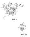
- FIG. 6is a cutaway isometric view of the applicator shown in FIG. 5 ;
- FIG. 6Ais an isometric view showing the force sensing transducer, force generator, and ultrasonic array of the applicator
- FIG. 7is an elevational view of a locator rod adapted to be inserted into a puncture wound over a guide wire;
- FIG. 7Ais a cross-sectional view of a portion of a patient's body like that in FIG. 5 , illustrating the locator rod of FIG. 7 being used to determine a location of a puncture in the artery relative to the applicator unit;
- FIG. 7Bis similar to FIG. 7 A, but illustrates the use of ultrasonic pulse-echo techniques to determine the spatial location of the locator rod;
- FIG. 7Cdepicts a view of a framed two-dimensional image a target region generally orthogonal to that shown in FIG. 7 A and FIG. 7B , for use in locating the puncture site;
- FIG. 8is a schematic isometric view of an embodiment of the applicator that uses a disposable shell
- FIG. 8Ais an side view of the disposable shell of FIG. 8 ;
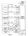
- FIG. 9is a schematic system block diagram depicting modules included in the applicator and control unit.
- FIGS. 10 and 10Arespectively illustrate the ultrasound beam orientation relative to the vessel from the side of the vessel and as viewed along the vessel;
- FIG. 11is a plan view of an embodiment of the applicator illustrating the controls and indicators that it includes;
- FIGS. 12A and 12Brespectively illustrate the side and longitudinal geometry of the therapeutic ultrasound beam
- FIG. 13is an isometric view illustrating the ultrasound beam geometry produced by an aspheric transducer.
- ultrasoundcan be brought to a tight focus at a distance from its source. If sufficient energy is radiated within the ultrasound beam, cells located in the focal volume can be rapidly heated, while intervening and surrounding tissues are spared. Surrounding tissues are unaffected in the unfocused portion of the ultrasound beam because the energy is spread over a correspondingly larger area and associated heating is minimized.
- HIFUcan rapidly seal blood vessel punctures and lacerations over a wide range of sizes.
- ultrasoundhas been shown to induce complete hemostasis in less than one minute in the femoral, carotid, and axillary arteries, and in the femoral and jugular veins of large animals, while blood flow through the treated vessels remained unaffected.
- FIGS. 1 and 2show the main components of an ultrasonic system suitable for use in implementing the present invention.
- a hand-held applicator unit 2is positioned over an arterial wound 8 in the patient.
- a generally single-use, pre-sterilized cover and acoustic coupling shell 4that slips over applicator 2 .
- a control unit 6implements algorithms to facilitate the method and is coupled to applicator 2 .
- the userenters various conventional scan and control parameters into an input unit 70 , which typically includes user input devices 72 .
- user input devices 72include a keyboard, knobs, and buttons.
- the input unitis connected to a processing system 74 , which will typically comprise a plurality of microprocessors and/or digital signal processors. Processing system 74 may, however, also be implemented using a single processor of sufficient speed to handle the various tasks described below.
- a conventional memory 75will normally be included in the system to store, among other data, transmission control parameters and imaging data generated in any given implementation of the present invention.
- Control circuit 76forms a transmit ultrasonic waveform by generating and applying electrical control and driving signals to an ultrasound transducer 78 , which preferably comprises an array of individually controllable piezoelectric elements.
- an ultrasound transducer 78which preferably comprises an array of individually controllable piezoelectric elements.
- the piezoelectric elementsgenerate ultrasonic waves when electric, signals of a proper frequency are applied to them; conversely, when receiving reflected ultrasonic waves, they generate electrical signals corresponding to the mechanical vibrations caused by the returning ultrasonic waves.
- Transducer 78is positioned against it portion 82 of the body of a patient, and by varying the phasing, amplitude, and timing of the driving signals for the transducer array elements, ultrasonic waves are focused to form a transmit beam 314 of high-intensity ultrasound.
- open arrowsindicate the direction of a flow of blood within a blood vessel 312 , which has a puncture site 316 that may have been caused by introduction of a catheter or, in the case of unintended punctures that have produced wounds, by some other object.
- the tissue forming a layer 318 of collagen found on the surface of blood vesselsis also shown in FIG. 2 surrounding blood vessel 312 .
- an imaging capabilityincluding pulse-echo lines of interrogation that are not displayed as images
- the provision of an imaging capability, including pulse-echo lines of interrogation that are not displayed as images, in the present inventionshould assist a user to more accurately locate a vascular puncture site. It is recognized that a full display of the insonified vascular target site is not required.
- FIG. 2also illustrates a reception controller 88 , which will include conventional amplification and signal conditioning circuitry as needed.
- Reception controller 88all or part of which is normally integrated into processing system 74 , converts the ultrasonic echo signals (which are typically at radio frequencies, on the order of a few to tens of megahertz) into lower frequency ranges for processing and may also include analog-to-digital conversion circuitry.
- the processingincludes, as needed, such known signal conditioning as time-gating, gain compensation, Doppler frequency shift processing, and diffraction compensation, in order to identify echo signals from any selected focal region.
- the type of conventional signal processing needed(if any) will in general depend on the particular implementation of the present invention employed and can be implemented using known design methods.
- the transducer 78be used externally, relative to the patient's body. It is also contemplated that the transducer may be maneuvered inside a patient's body, and the beam focused on a puncture from inside the body.
- a transesophageal probe, laparoscopic, or other probe inserted into a body cavity, such as the vagina or rectumcan be used to practice the present invention.
- a suitably designed probe inserted into an open body cavity or via minimally invasive meanscould be used to arrest bleeding in surgical or trauma care situations.
- most of the following discussionis directed to a preferred embodiment of the present invention in which the transducer is intended to be used externally, since an initial commercial product in accord with the present invention will be designed for such use.
- a conventional display system 92may also be included in order to display information concerning transmission power, time, focus data, etc.
- the display systemwill include known circuitry for scan conversion and for driving the display, as needed. These circuits are well known and therefore need not be specifically illustrated or described further to provide an enabling disclosure.
- FIG. 3illustrates the result of an insonification of puncture site 316 using the present invention.
- the focal point of transmit beam 314(see FIG. 2 ) is moved around (as illustrated by the arrows pointing in either direction from the focus of the beam) the area of puncture site 316 using conventional beam-steering techniques, the tissue adjacent the puncture site 316 will denature.
- the collagen in the tissue“melts” and flows over and into the puncture opening.
- the collagencools, it forms a “patch” that not only covers the puncture, but also flows partially into the puncture opening in the wall of the blood vessel.
- the denatured tissuetends to contract, it also tends to pull the edges of the puncture together and thus further close the wound.
- the present inventioncan be employed to seal vascular wounds of various types and is not limited to the type of wound created as a result of interventional procedures in which a catheter has been introduced into a vascular vessel.
- application of thermal treatment to the tissue overlying a woundhas been demonstrated to seal the wound.
- a practical methodhas been developed for repeatedly and reliably achieving sealing of the puncture in a vascular vessel.
- Steps carried out in this methodare shown in FIG. 4 , which is discussed below. These steps represent one preferred embodiment of the present invention, but do not represent all alternatives that might be employed to achieve acceptable sealing.
- a clinically acceptable device for sealing a puncture wound in accord with the present inventionmust meet a number of requirements, including:
- a preferred embodiment of the deviceincludes the components shown in FIGS. 1 and 2 , which were described above.
- FIG. 6 and FIG. 6Ashow one preferred embodiment of applicator unit 2 .
- the applicator unitincludes an outer housing 10 having an ergonomically considered shape so that it can be conveniently hand held.
- the outer housingis best fabricated from an injection moldable plastic material such as ABS or the like.
- the operatorgrasps the outer housing of the applicator unit so as to enable a control push-button 14 to be accessible and indicators 12 , 16 and 30 , 32 , 34 , and 36 to be visible to the operator. Positioning the applicator unit at the appropriate location over the wound area and activation of an essentially automated treatment cycle are readily accomplished. The operator simply refers to the indicators to determine when the applicator unit is properly positioned and ready for use.
- Indicators 12 and 16are used to indicate when alignment of the applicator unit with the longitudinal axis of the vessel to be sealed has been achieved.
- Indicators 30 , 32 , 34 , and 36display the state of operation and instruct the operator with respect to holding the applicator in place as described in detail later in this description.
- Control 14when pressed, activates the treatment cycle, thus initiating a sequence of operations that determine ultrasonic scan parameters (exposure time, scan pattern, intensity, focal range, etc.).
- ultrasonic array assembly 20is held within outer housing 10 on a shaft 40 , in a bearing assembly within a force transducer 18 , so as to permit movement of the ultrasonic array assembly to and away from patient 8 .
- Shaft 40passes through the bearing assembly provided within a force generator 18 and terminates at a contacting force sensing transducer 42 .
- Force generator 18comprises an electromagnetic solenoid that is rigidly supported and mounted within housing 10 by structural members 46 .
- the face of ultrasonic array assembly 20is in contact with the appropriate location on the body of the patient (overlying the site of the puncture) and is thus capable of applying a substantially compressive, controllable force on the tissue structures with which it is in contact.
- the force applied by the ultrasonic array assemblyis produced at least in part by controllably energizing force generator 18 .
- Ultrasonic array assembly 20preferably operates in a multiplicity of modes; however, separate ultrasonic transducers can instead be provided for some or all of the functions performed in a different design within the scope of the present invention.
- electrical connections comprising wires 26are routed within the outer housing 10 and out in a sealed bushing 44 that mounts a cable 28 to the control unit 6 .
- Cable 28is sufficiently long, on the order of 10 feet in length, so that the control unit may be conveniently located at a distance from the patient and operator location.
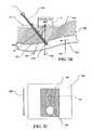
- Applicator unit housing 10is shaped to be used with a slip-on, generally single-use protective applicator shell 4 (illustrated schematically in FIG. 1 ).
- the shell employed in the preferred embodimentis shown in greater detail in FIGS. 8 and 8A .
- Shell 4has side walls 54 that are fabricated from a generally optically transparent, semi-rigid plastic material.
- a skirt 52extends from the rear of the shell and is pleated so that in preparing for use of the applicator unit, an operator can grasp the skirt and extend it sufficiently to protect a sterile area of the patient from coming into contact with cord.
- the protective shellis packaged in a sterile condition.
- the shellis fabricated from a flexible plastic material having low acoustic absorption characteristics.
- a fiducial mark 56is provided on a side of the applicator unit and visible through the optically transparent material of the protective shell. This fiducial mark is employed to visually align the applicator unit with a position on the patient at which the applicator unit will be used to seal a puncture.
- Sterile, generally gas free acoustic coupling gel 62is deposited in a patch on the bottom of flexible bottom 58 . Prior to use, the gel is held in place and sealed by semi-sticky adhesive coated tab 60 . Tab 60 is removed by the operator just prior to use, thereby exposing the gel so that it provides a good acoustic coupling with the surface of the patient's body.
- Protective applicator shell 4thus provides a sterile barrier over the multi-use applicator unit and conveniently provides a pre-determined amount of a specific appropriate acoustic coupling medium.
- the bottom of the interior cavity of the shellmay also include a layer of acoustic coupling gel to ensure good acoustic coupling between the applicator unit through the protective, applicator coupling shell.
- FIG. 9is a system block diagram illustrating the modules disposed, in the preferred embodiment, within the applicator unit 2 (i.e., the component shown within the dotted line portion of this Figure) as well as the modules (all other modules that are not in the applicator unit) disposed in the control unit 6 .
- control unit 6is packaged in a small, self-contained pole- or cart-mounted enclosure that derives its input power from a standard AC line power service (not shown). Power supplies with the unit are designed to assure low leakage currents for patient safe operation.
- control unit 6is based on a programmable processing unit which processes various signals and controls a number of functional modules.
- a microprocessoris well suited to perform the computation and control functions required.
- Applicator unit 2is coupled to control unit 6 by a plurality of signal paths 212 , 214 , 216 , and 218 .
- Signal path 212couples display drivers 200 , which are controlled by a computer/controller 236 , with indicators 30 , 32 , 34 , and 36 on the applicator unit.
- Control button 14 on the applicator unitis coupled through signal line 214 to an interface 202 and thus to the computer/controller.
- Force sensing transducer 42produces an output signal indicative of the force (i.e., the pressure) applied against the surface of the patient's tissues by the applicator unit, and this signal is conveyed by signal lines 216 to an interface 204 , which provides the signal to the computer/controller.
- the computer/controllerIn response to the magnitude of the monitored force, the computer/controller produces a control signal applied to a driver 206 , which provides the current signal used to energize force transducer 16 , to determine any additional force that it generates to achieve a desired pressure on the site of the puncture that is sufficient to prevent loss of fluid from the vessel, but not so great as to occlude the flow of fluid through the vessel.
- Signal lines 240couple ultrasonic array assembly 20 to a transmit/receive switch 224 .
- the transmit/receive switchdetermines the operational mode of the ultrasonic array assembly under the control of the computer/controller.
- the signal corresponding to the echo reflected received by ultrasonic array assembly 20 from tissue and other structuresis conveyed through transmit/receive switch 224 and through signal lines 222 to an amplifier digitizer array 220 .
- the output signals from the amplifier digitizer arrayare conveyed to computer/controller 236 through signal lines 228 .
- the ultrasonic array assemblyWhen the ultrasonic array assembly is generating either the imaging beam or the HIFU beam, it is responding to signals received from an RF generator 232 that is coupled to a phase shift/amplifier array 234 by signal lines 236 , and to a control signal provided by the computer/controller and conveyed to the phase shift/amplifier on a signal line 230 .
- the output of the phase shift/amplifieris conveyed on signal lines 226 to transmit/receive switch 224 , and thus, to ultrasonic array assembly 20 through signal lines 240 .
- Manual control inputs 241are coupled to computer/controller 236 through signal lines 242 .
- Possibilitiesinclude:
- An aspheric ultrasonic transducer configurationhas the advantage of covering a large treatment area on the surface of the vessel without the complication of electronic or mechanical beam steering. Covering a large area (i.e., overscanning) is desired in order to ensure that the actual site of the puncture wound is treated, given its positional ambiguity.
- FIG. 13depicts the geometry of such a configuration.
- an ultrasonic transducer 404excited by an appropriate RF source via connections 402 , is generally aspheric in shape and does not bring the ultrasound beam to a sharp focus.
- the ultrasonic energy that it producescovers area 412 on a vessel 408 that includes puncture wound 410 .
- Fluid or solid material acoustic coupling(not shown) is used between the ultrasonic transducer and the tissues of the patient.
- the method describedincludes a series of manual steps (operator actions) and automated steps.
- the automated stepsare carried out as control processes or algorithms executed by one or more processors and other hardware in accord with machine instructions executed by the one or more processors. It is understood that variations in the order of these steps, and in the total complement of steps implemented is possible in alternative embodiments. Steps as shown in FIG. 4 are described as follows.
- a step 100 labeled Patient Preparationthe operator positions the patient and the apparatus so that the applicator unit is conveniently positioned over the puncture wound area, e.g., over the puncture made by a catheter in the femoral artery.
- Shell 4is removed from its sterile package and fitted onto applicator unit 2 , and gel sealing tab 60 (shown in FIGS. 8 and 8A ) is removed, exposing the gel 62 .
- a step 102 labeled Manually Alignis then carried out.
- the operatorpalpates the area locating the point at which the introducer just enters the artery.
- the operatormarks this location on the patient's skin with a suitable marking device (e.g. a surgical marker), drawing a line substantially perpendicular to the perceived direction of the artery, extending approximately 3 cm from the entry wound location. It is the purpose of this marking to estimate the longitudinal location of the wound; the operation of the HIFU sealing process provides for an overscan of the wound area so that practical variations in the operator's ability to make the longitudinal position estimate are permissible. Other techniques for locating the site of the puncture are discussed below.
- lateral and range locations of the woundare more precisely located by the automated capability of the processor(s) used in the apparatus.
- step 102the operator places the device over the wound location, aligning fiducial mark “ 56 ” (shown in FIG. 8 ) with the line that was drawn on the patient's skin.
- the applicator unitis rotated in place until both alignment indicators 12 and 16 ( FIGS. 6 and 8 ) illuminate, indicating that the artery is axially aligned under the applicator unit.
- the axial alignment indicationsare, in this preferred embodiment, derived from two ultrasonic pulsed Doppler interrogations.
- FIGS. 10 and 10Ashow the geometry of the Doppler alignment beams. Use of a ultrasonic transducer 20 enables the same ultrasonic transducer to be employed to produce an imaging beam and the HIFU beam for both a pulse-echo targeting mode and a therapeutic mode.
- phased array ultrasonic transducer 20sends and receives a downstream pulsed Doppler line 304 and an upstream line 308 sequentially.
- Lines 304 and 308are in plane with the axial centerline of the applicator unit and a line perpendicular to the bottom surface of the applicator unit.
- Lines 304 and 308are transmitted at angles A and B with respect to a line 306 , which is perpendicular to the applicator unit.
- Angles A and Bare chosen to provide a axial separation as well as an appreciable vector flow component in the direction of the interrogating line—an angle of approximately 45 degrees.
- Direction of flow in an interior 310 of the vesselis sensed and tested to assure that an artery 300 (not a vein) is being interrogated.
- Doppler signalsfrom lines 304 and 308 , integrated over an appropriate range, above a pre-determined threshold value, are used to cause the illumination of alignment indicators 12 and 16 respectively. Averaging multiple lines is, in this preferred embodiment, employed to improve the performance of the alignment detection scheme.
- the conditioned signalsalso set logical flags so that the system may interlock the initiation of a therapeutic treatment sequence with assurance of alignment. Thus in a decision step 106 ( FIG. 4 ), alignment is tested by interrogating the logical presence of both of the flags.
- FIG. 11illustrates a top view of the applicator unit.
- three parallel linesare transmitted and received in the two directions indicated by lines 304 and 308 in FIG. 10 .
- Additional transducersare appropriately positioned in housing 10 ( FIG. 6 ).
- Pulsed Doppler signalsare processed in a manner similar to that described above.
- indicator lights 336 and 334would be illuminated, indicating the misalignment and suggesting the appropriate direction to move the applicator unit to achieve alignment (denoted when indicators 338 and 330 are illuminated).
- continuous wave Dopplermay be employed to interrogate the flow position of the target artery, with operation essentially similar to that described above.
- step 108labeled Set Pressure, wherein the pressure over the artery is set and controlled within a predetermined range using force generator 18 ( FIG. 6 ) and force sensing transducer 42 .
- the weight of the applicator unitis purposefully made to be in a range where additional pressure applied by the operator to hold the unit firmly in place is reduced. This useful weight is about 1 lb (0.45 kg) or more.
- Force generator 18is activated and controlled such that applied pressure to the artery partially restricts flow, but maintains sufficient flow so that thermal cooling due to blood flow within the artery protects the intimal lining of the vessel from irreversible damage. Presence of a Doppler flow signal on down-stream line 304 ( FIG. 10 ) is employed to assure vessel patency.
- indicator 32( FIG. 6 ), which is marked “READY” on the applicator unit is illuminated (see block 118 FIG. 4 ), indicating to the operator that a treatment cycle may be manually initiated (triggered) by pushing control button 14 .
- the systemis in a wait state as indicated in a decision block 112 in FIG. 4 , until a manually triggered treatment cycle is detected.
- axial alignmentis verified in a step 114 by generally repeating the logical test described at step 102 .
- a step 116then makes a ranging measurement to estimate the acoustic path length between ultrasonic transducer assembly 20 and the vessel boundary. i.e. the distance between points F and C along line 306 in FIG. 10 .
- Pulsed Doppleris, in this preferred embodiment, employed to make this measurement, wherein lines 304 and 308 measure distances F-D and F-E, respectively. Points D and E are recognized by the fact that these correspond to the first instance of flow detected along each line as range increases.
- the range estimate of FCis therefore: FC ⁇ (FE COS A+FD COS B)/2 Equation 1
- a step 120estimates the acoustic attenuation at the therapeutic frequency between ultrasonic transducer assembly 20 and the target collagen layer overlaying the vascular wound, path F-C in FIG. 10 .
- estimated dimension FCis used to access data in a look-up table of attenuation values. Attenuation values in the table are predetermined by empirical measurement. Alternatively, more sophisticated A-Mode attenuation measurements may be employed to assess to F-C path.
- a step 122determines the therapeutic ultrasound exposure parameters to be employed.
- Dimension F-C, the attenuation estimate, and optionally, patient parameters (e.g., size and weight), input at module 240 in FIG. 9are used to access predetermined data and scan protocols in resident look-up tables Ultrasound scan geometry, intensity and epochal exposure intervals are thus determined.
- Ultrasound scan geometry, intensity and time parametersare determined to accomplish three key objectives: (1) provide a sufficient overscan of a longitudinal and lateral surface of the target vessel so as to include the site of the wound; (2) ensure delivery of an appropriate energy dose to the collagen layer in the region of the target site to raise its temperature to a range of between 60 and 100 degrees Celsius for a predetermined period of time; and, (3) assure that the skin and interpath tissue is not exposed to a temperature-time exposure that will result in pain and or irreversible tissue damage.
- Therapeutic scan geometryis shown in FIGS. 12A and 12B .
- a therapeutic level of approximately 50 watts total transmitted acoustic power, generally weakly focused,is transmitted along a centerline 314 through a dermal layer 302 .
- the desired scan patternis achieved by directing the beam over varying angles of the beam with respect to a line 320 that extends perpendicular to the center of the face of the application unit.
- a raster scan patternmay cover a therapeutic area, over collagen layer 318 , of dimensions 1 cm wide by 1.5 cm long (in the arterial axial direction).
- Such an overscan of the wound siteprovides for variations in the operator's ability to predetermine and locate the precise lateral position of the wound, as well as the variation in wound location that results from the possible variation of wound lateral entry points.
- scan geometryis selected such that ultrasonic exposure is generally confined to the vessel, minimizing the possibility that an adjacent vein or nerve structure will be insonified.
- a step 126carries out the therapeutic exposure cycle. It is generally desirable to hold the applicator unit in place, providing the established orientation and pressure for a period of time after the therapeutic exposure cycle.
- a hold interval 130( FIG. 4 ) is selected to enable exposed tissue structures to cool, a time period of approximately 1 minute. Following this time period, indicator 36 on the application unit ( FIG. 6 ) marked “COMPLETE” is illuminated and the “HOLD” indicator is turned off, instructing the operator that the therapeutic treatment is completed and the device may be removed from the patient.
- an additional sequence depicted in FIG. 4Amay optionally be employed.
- the sequenceis inserted into the process flow of FIG. 4 , in this preferred embodiment, at a location marked “A.”
- a weak, sub-therapeutic energy level ultrasonic pulseis transmitted in a step 136 ( FIG. 4A ), to cover the determined target area.
- the operatorobserves in a decision step 138 whether a reaction of pain and or uncommanded movement from the patient has occurred, indicating that a nerve structure has been stimulated.
- System logicthen waits for an addition manually initiated trigger input in steps 140 and 142 , prior to proceeding with therapeutic exposure at step 126 in FIG. 4 .
- Step 102which facilitates alignment of the applicator unit over the wound area and targeting of the therapeutic exposure, may be accomplished using several alternative approaches compared to that described above. It is desired to employ an approach for targeting and aligning the applicator unit that is easy to implement and requires minimum operator instruction. The approach further should be consistent with minimizing bleeding during the process of achieving alignment. Additionally, the approach should be robust and provide targeting of the wound site with sufficient accuracy such that the wound will reliably be included within the area of therapeutic exposure.
- a principle employed in the present inventionis the concept that the overscan of the target location during therapeutic exposure is sufficient to accommodate targeting ambiguity and possible patient or operator movement during the procedure. Nevertheless, accurate targeting is needed to ensure efficacy of the wound sealing process.
- pulsed Doppler rangingwas employed to locate the axis of the vessel, and the operator was advised of the longitudinal location of the wound on the vessel by reference to visual landmarks on the skin surface.
- the need for the operator to locate the longitudinal positionis eliminated, thereby substantially simplifying the targeting procedure for the operator.
- FIG. 5illustrates the geometry that relates the location of applicator unit 2 to the locations of an entry channel 518 and a vessel wound 516 .
- applicator unit 2is positioned on a patient's skin surface 510 over a vessel 506 in which a blood flow 508 is contained.
- a location “C”is indicated at wound 516 ;
- a line 512passes from “C” through entry channel 518 , intersecting a plane 514 defined by reference locations on applicator unit 2 at a point “H.”
- a point “F”is a reference point in plane 514 .
- the location of wound 516 relative to applicator unit 2is determinable by basic geometry. Knowledge of this geometry permits automated system function to be employed to provide guidance instructions (e.g. visual indicators) to the operator during the manual alignment portion of the procedure.
- guidance instructionse.g. visual indicators
- acoustic pulsed Dopplermay be employed to determine the distance “C”-“F” (reference FIG. 5 ).
- the vesselis an artery having a substantial high velocity flow and is thus readily localized using well known pulsed Doppler techniques. Knowing this distance, localization of wound 516 on the vessel is made possible by determining the spatial position of line 512 .
- the spatial location of line 512may be determined by employing a substantially rigid, straight locator rod 554 as depicted in FIG. 7 , which is placed in entry channel 518 .
- Locator rodmay have a longitudinal center bore 556 through which a guide wire 552 may pass.
- Locator rod 554would be introduced into the wound over the guide wire.
- Sealing assembly 550is disposed at one end of the locator rod to prevent the loss of blood through center bore 556 .
Landscapes
- Health & Medical Sciences (AREA)
- Life Sciences & Earth Sciences (AREA)
- Engineering & Computer Science (AREA)
- Animal Behavior & Ethology (AREA)
- Veterinary Medicine (AREA)
- Public Health (AREA)
- General Health & Medical Sciences (AREA)
- Nuclear Medicine, Radiotherapy & Molecular Imaging (AREA)
- Surgery (AREA)
- Biomedical Technology (AREA)
- Physics & Mathematics (AREA)
- Heart & Thoracic Surgery (AREA)
- Molecular Biology (AREA)
- Medical Informatics (AREA)
- Multimedia (AREA)
- Acoustics & Sound (AREA)
- Radiology & Medical Imaging (AREA)
- Cardiology (AREA)
- Biophysics (AREA)
- Pathology (AREA)
- Ultra Sonic Daignosis Equipment (AREA)
- Surgical Instruments (AREA)
Abstract
Description
- 1. Positioning an ultrasound generating source on a patient such that the source is targeted at an area including the wound to be sealed;
- 2. Applying a pressure against the patient in an area overlying the wound and directed substantially toward the vessel to be treated;
- 3. While the pressure and the positioning of the ultrasound source are maintained, carrying out an insonification of a volume of tissue that includes the wound, using an ultrasound exposure that delivers an acoustic energy density (measured at the approximate location of the wound), in excess of 100 joules/sq. cm, but generally less than several thousand joules/sq. cm.
Additional steps of the method described below employ the apparatus in an automated manner that facilitates ease of use and ensures the safety and consistency of the results obtained.
- 1. The device and associated procedure must be safe to use in that they avoid undesirable bioeffects, so that the patient is not injured directly or indirectly as a result of the procedure; also, in the event that effective vessel sealing does not occur as desired, traditional methods of applying pressure to the wound are sufficient to accomplish hemostasis;
- 2. The device and associated procedure must be easy to use in an efficient manner, to facilitate proper, repeatable execution of the sealing process; requirements for operator training should be minimized;
- 3. The sealing process must be sufficiently fast to enable the entire procedure to be rapidly completed—preferably, in less than 5 minutes;
- 4. The cost per sealing procedure should be minimized; and,
- 5. The efficacy of the device should be very high, generally achieving a success rate in excess of about 95%; acceptability of the present invention in routine clinical practice does not permit an unpredictable outcome.
- Configurations wherein therapeutic and, pulse-echo Doppler functionality are accomplished by the same ultrasonic transducer or by separate ultrasonic transducers; and,
- Configurations wherein the ultrasonic transducer is either of a fixed focus type, or a segmented electrically selectable macro element array, or a phased array, or a linear array, or an annular array; and,
- Configurations where a large focal spot412 (see
FIG. 13 ) (e.g. a focal spot produced by a transducer having an aspheric shape), or those in which a tightly focused spot is produced; and, - Configurations wherein the ultrasonic transducer is mechanically positioned (or scanned), or those in which it is fixed in one position.
FC˜(FE COS A+FD COS B)/2 Equation 1
- 1. As shown in
FIG. 7 A, a spatialposition resolving element 558 may be included onlocator rod 554. Spatialposition resolving element 558 may be an acoustic position sensor, an optical position sensor, a magnetic position sensor, an electromechanical positioning or resolving arm, or an inertial position sensor. The spatial relationship between the position oflocator rod 554 andapplicator unit 2 is accomplished by either a direct mechanical linkage or by way of an intermediate electronic or computational circuit. - 2. As shown in
FIG. 7B , an ultrasonic pulse-echo technique may be employed to determine the spatial location oflocator rod 554. A transducer inapplicator unit 2 transmits directed acoustic pulses in more than one direction, e.g.,lines Locator rod 554 provides reflections that permit making time of flight measurements of, for example, distances “F”-“J,” “F”-“K,” and “F”-“L.”Locator rod 554 may be coated or constructed of materials chosen to enhance acoustic reflectivity, thus providing echoes that are readily distinguished from background clutter. Common guide wires may alternatively be used aslocator rod 554, as these wire structures are typically highly reflective of ultrasound energy. Alternatively,locator rod 554 may be constructed from materials that enhance reflection at a harmonic of the interrogating ultrasound pulse, providing another advantageous method for clearly distinguishing the echoes fromlocator rod 554 from those received from surrounding tissue. Materials or coatings that entrap gas bubbles are, for example, effective in providing higher harmonic reflection. Echo enhancing properties may also be incorporated into an introducer that is then used as the locator rod. Pulsed Doppler may also be employed to identify and locate the locator rod. In this latter alternative, the locator rod may be a common introducer. A strong Doppler shifted reflection will return from the lumen of the introducer even when blood is not permitted to flow out of the introducer. - 3. As indicated in
FIG. 7C , two dimensional, or three dimensional, imaging may also be employed to locatelocator rod 554, the guide wire, and ‘the introducer, as well as the vessel.FIG. 7C depicts a view of a framed two-dimensional image 580 of the target region generally orthogonal to that shown inFIGS. 7A and 7B . Distance from the ultrasound source increases toward the bottom of this depiction. This image is generally representative of a cross-sectional plane of interrogation located at aline 562 inFIG. 7B . Automated image recognition provided by the processor may be employed to identify structures includinglocator rod 554, imaged as alocator rod structure 586 andvessel 506, imaged as avessel structure 584 in this image. Doppler imaging and color flow mapping may be employed to increase the recognizablility of relevant features. The interrogating image plane may be scanned over the region containing the target. Whenlocator rod structure 586 is recognized at a location just touching the top surface ofvessel structure 584. the wound target. site has been identified.
- 1. As shown in
Claims (24)
Priority Applications (1)
| Application Number | Priority Date | Filing Date | Title |
|---|---|---|---|
| US13/011,533US8388535B2 (en) | 1999-10-25 | 2011-01-21 | Methods and apparatus for focused ultrasound application |
Applications Claiming Priority (7)
| Application Number | Priority Date | Filing Date | Title |
|---|---|---|---|
| US16346699P | 1999-10-25 | 1999-10-25 | |
| US17170399P | 1999-12-23 | 1999-12-23 | |
| US09/696,076US6656136B1 (en) | 1999-10-25 | 2000-10-25 | Use of focused ultrasound for vascular sealing |
| US10/616,831US20040106880A1 (en) | 1999-10-25 | 2003-07-10 | Use of focused ultrasound for vascular sealing |
| US11/619,996US20070179379A1 (en) | 1999-10-25 | 2007-01-04 | Use of focused ultrasound for vascular sealing |
| US12/896,740US20110021913A1 (en) | 1999-10-25 | 2010-10-01 | Use of focused ultrasound for vascular sealing |
| US13/011,533US8388535B2 (en) | 1999-10-25 | 2011-01-21 | Methods and apparatus for focused ultrasound application |
Related Parent Applications (1)
| Application Number | Title | Priority Date | Filing Date |
|---|---|---|---|
| US12/896,740ContinuationUS20110021913A1 (en) | 1999-10-25 | 2010-10-01 | Use of focused ultrasound for vascular sealing |
Publications (2)
| Publication Number | Publication Date |
|---|---|
| US20110118602A1 US20110118602A1 (en) | 2011-05-19 |
| US8388535B2true US8388535B2 (en) | 2013-03-05 |
Family
ID=26859662
Family Applications (10)
| Application Number | Title | Priority Date | Filing Date |
|---|---|---|---|
| US09/696,076Expired - LifetimeUS6656136B1 (en) | 1999-10-25 | 2000-10-25 | Use of focused ultrasound for vascular sealing |
| US10/616,831AbandonedUS20040106880A1 (en) | 1999-10-25 | 2003-07-10 | Use of focused ultrasound for vascular sealing |
| US11/619,996AbandonedUS20070179379A1 (en) | 1999-10-25 | 2007-01-04 | Use of focused ultrasound for vascular sealing |
| US12/896,740AbandonedUS20110021913A1 (en) | 1999-10-25 | 2010-10-01 | Use of focused ultrasound for vascular sealing |
| US13/011,533Expired - Fee RelatedUS8388535B2 (en) | 1999-10-25 | 2011-01-21 | Methods and apparatus for focused ultrasound application |
| US13/025,959Expired - Fee RelatedUS8137274B2 (en) | 1999-10-25 | 2011-02-11 | Methods to deliver high intensity focused ultrasound to target regions proximate blood vessels |
| US13/026,108Expired - Fee RelatedUS8277398B2 (en) | 1999-10-25 | 2011-02-11 | Methods and devices to target vascular targets with high intensity focused ultrasound |
| US13/245,689AbandonedUS20120059258A1 (en) | 1999-10-25 | 2011-09-26 | Use of focused ultrasound for vascular sealing |
| US13/344,418AbandonedUS20120108966A1 (en) | 1999-10-25 | 2012-01-05 | Use of focused ultrasound for vascular sealing |
| US13/346,466AbandonedUS20120108967A1 (en) | 1999-10-25 | 2012-01-09 | Use of focused ultrasound for vascular sealing |
Family Applications Before (4)
| Application Number | Title | Priority Date | Filing Date |
|---|---|---|---|
| US09/696,076Expired - LifetimeUS6656136B1 (en) | 1999-10-25 | 2000-10-25 | Use of focused ultrasound for vascular sealing |
| US10/616,831AbandonedUS20040106880A1 (en) | 1999-10-25 | 2003-07-10 | Use of focused ultrasound for vascular sealing |
| US11/619,996AbandonedUS20070179379A1 (en) | 1999-10-25 | 2007-01-04 | Use of focused ultrasound for vascular sealing |
| US12/896,740AbandonedUS20110021913A1 (en) | 1999-10-25 | 2010-10-01 | Use of focused ultrasound for vascular sealing |
Family Applications After (5)
| Application Number | Title | Priority Date | Filing Date |
|---|---|---|---|
| US13/025,959Expired - Fee RelatedUS8137274B2 (en) | 1999-10-25 | 2011-02-11 | Methods to deliver high intensity focused ultrasound to target regions proximate blood vessels |
| US13/026,108Expired - Fee RelatedUS8277398B2 (en) | 1999-10-25 | 2011-02-11 | Methods and devices to target vascular targets with high intensity focused ultrasound |
| US13/245,689AbandonedUS20120059258A1 (en) | 1999-10-25 | 2011-09-26 | Use of focused ultrasound for vascular sealing |
| US13/344,418AbandonedUS20120108966A1 (en) | 1999-10-25 | 2012-01-05 | Use of focused ultrasound for vascular sealing |
| US13/346,466AbandonedUS20120108967A1 (en) | 1999-10-25 | 2012-01-09 | Use of focused ultrasound for vascular sealing |
Country Status (6)
| Country | Link |
|---|---|
| US (10) | US6656136B1 (en) |
| EP (1) | EP1229839A4 (en) |
| JP (1) | JP2003513691A (en) |
| AU (1) | AU2619301A (en) |
| CA (1) | CA2387127A1 (en) |
| WO (1) | WO2001034018A2 (en) |
Cited By (27)
| Publication number | Priority date | Publication date | Assignee | Title |
|---|---|---|---|---|
| US9283409B2 (en) | 2004-10-06 | 2016-03-15 | Guided Therapy Systems, Llc | Energy based fat reduction |
| US9283410B2 (en) | 2004-10-06 | 2016-03-15 | Guided Therapy Systems, L.L.C. | System and method for fat and cellulite reduction |
| US9320537B2 (en) | 2004-10-06 | 2016-04-26 | Guided Therapy Systems, Llc | Methods for noninvasive skin tightening |
| US9421029B2 (en) | 2004-10-06 | 2016-08-23 | Guided Therapy Systems, Llc | Energy based hyperhidrosis treatment |
| US9427600B2 (en) | 2004-10-06 | 2016-08-30 | Guided Therapy Systems, L.L.C. | Systems for treating skin laxity |
| US9427601B2 (en) | 2004-10-06 | 2016-08-30 | Guided Therapy Systems, Llc | Methods for face and neck lifts |
| US9440096B2 (en) | 2004-10-06 | 2016-09-13 | Guided Therapy Systems, Llc | Method and system for treating stretch marks |
| US9510802B2 (en) | 2012-09-21 | 2016-12-06 | Guided Therapy Systems, Llc | Reflective ultrasound technology for dermatological treatments |
| US9694212B2 (en) | 2004-10-06 | 2017-07-04 | Guided Therapy Systems, Llc | Method and system for ultrasound treatment of skin |
| US9827449B2 (en) | 2004-10-06 | 2017-11-28 | Guided Therapy Systems, L.L.C. | Systems for treating skin laxity |
| US10420960B2 (en) | 2013-03-08 | 2019-09-24 | Ulthera, Inc. | Devices and methods for multi-focus ultrasound therapy |
| US10537304B2 (en) | 2008-06-06 | 2020-01-21 | Ulthera, Inc. | Hand wand for ultrasonic cosmetic treatment and imaging |
| US10603521B2 (en) | 2014-04-18 | 2020-03-31 | Ulthera, Inc. | Band transducer ultrasound therapy |
| US10864385B2 (en) | 2004-09-24 | 2020-12-15 | Guided Therapy Systems, Llc | Rejuvenating skin by heating tissue for cosmetic treatment of the face and body |
| US11207548B2 (en) | 2004-10-07 | 2021-12-28 | Guided Therapy Systems, L.L.C. | Ultrasound probe for treating skin laxity |
| US11224895B2 (en) | 2016-01-18 | 2022-01-18 | Ulthera, Inc. | Compact ultrasound device having annular ultrasound array peripherally electrically connected to flexible printed circuit board and method of assembly thereof |
| US11235179B2 (en) | 2004-10-06 | 2022-02-01 | Guided Therapy Systems, Llc | Energy based skin gland treatment |
| US11241218B2 (en) | 2016-08-16 | 2022-02-08 | Ulthera, Inc. | Systems and methods for cosmetic ultrasound treatment of skin |
| US11338156B2 (en) | 2004-10-06 | 2022-05-24 | Guided Therapy Systems, Llc | Noninvasive tissue tightening system |
| US11382515B2 (en) | 2014-08-07 | 2022-07-12 | Verve Medical, Inc. | Renal denervation using nerve fluorescing dye |
| US11724133B2 (en) | 2004-10-07 | 2023-08-15 | Guided Therapy Systems, Llc | Ultrasound probe for treatment of skin |
| US11883688B2 (en) | 2004-10-06 | 2024-01-30 | Guided Therapy Systems, Llc | Energy based fat reduction |
| US11944849B2 (en) | 2018-02-20 | 2024-04-02 | Ulthera, Inc. | Systems and methods for combined cosmetic treatment of cellulite with ultrasound |
| US12076591B2 (en) | 2018-01-26 | 2024-09-03 | Ulthera, Inc. | Systems and methods for simultaneous multi-focus ultrasound therapy in multiple dimensions |
| US12102473B2 (en) | 2008-06-06 | 2024-10-01 | Ulthera, Inc. | Systems for ultrasound treatment |
| US12161379B2 (en) | 2011-07-12 | 2024-12-10 | Verve Medical, Inc. | Treatment of kidney disease using renal nerve denervation via the renal pelvis |
| US12377293B2 (en) | 2019-07-15 | 2025-08-05 | Ulthera, Inc. | Systems and methods for measuring elasticity with imaging of ultrasound multi-focus shearwaves in multiple dimensions |
Families Citing this family (249)
| Publication number | Priority date | Publication date | Assignee | Title |
|---|---|---|---|---|
| US9023031B2 (en) | 1997-08-13 | 2015-05-05 | Verathon Inc. | Noninvasive devices, methods, and systems for modifying tissues |
| US7686763B2 (en)* | 1998-09-18 | 2010-03-30 | University Of Washington | Use of contrast agents to increase the effectiveness of high intensity focused ultrasound therapy |
| US7722539B2 (en)* | 1998-09-18 | 2010-05-25 | University Of Washington | Treatment of unwanted tissue by the selective destruction of vasculature providing nutrients to the tissue |
| US7510536B2 (en) | 1999-09-17 | 2009-03-31 | University Of Washington | Ultrasound guided high intensity focused ultrasound treatment of nerves |
| US7520856B2 (en) | 1999-09-17 | 2009-04-21 | University Of Washington | Image guided high intensity focused ultrasound device for therapy in obstetrics and gynecology |
| JP2003513691A (en)* | 1999-10-25 | 2003-04-15 | シーラス、コーポレイション | Use of focused ultrasound to seal blood vessels |
| US6626855B1 (en)* | 1999-11-26 | 2003-09-30 | Therus Corpoation | Controlled high efficiency lesion formation using high intensity ultrasound |
| WO2001045550A2 (en)* | 1999-12-23 | 2001-06-28 | Therus Corporation | Ultrasound transducers for imaging and therapy |
| US8241274B2 (en) | 2000-01-19 | 2012-08-14 | Medtronic, Inc. | Method for guiding a medical device |
| US9522217B2 (en) | 2000-03-15 | 2016-12-20 | Orbusneich Medical, Inc. | Medical device with coating for capturing genetically-altered cells and methods for using same |
| US8088060B2 (en) | 2000-03-15 | 2012-01-03 | Orbusneich Medical, Inc. | Progenitor endothelial cell capturing with a drug eluting implantable medical device |
| US7306591B2 (en)* | 2000-10-02 | 2007-12-11 | Novasys Medical, Inc. | Apparatus and methods for treating female urinary incontinence |
| US6743195B2 (en)* | 2001-03-14 | 2004-06-01 | Cardiodex | Balloon method and apparatus for vascular closure following arterial catheterization |
| US20030069502A1 (en)* | 2001-05-29 | 2003-04-10 | Makin Inder Raj. S. | Ultrasound feedback in medically-treated patients |
| US7211044B2 (en) | 2001-05-29 | 2007-05-01 | Ethicon Endo-Surgery, Inc. | Method for mapping temperature rise using pulse-echo ultrasound |
| US6951542B2 (en) | 2002-06-26 | 2005-10-04 | Esaote S.P.A. | Method and apparatus for ultrasound imaging of a biopsy needle or the like during an ultrasound imaging examination |
| US7511704B2 (en)* | 2001-11-20 | 2009-03-31 | Illinois Tool Works Inc. | Acoustic wave touch bar system and method of use |
| US7617005B2 (en) | 2002-04-08 | 2009-11-10 | Ardian, Inc. | Methods and apparatus for thermally-induced renal neuromodulation |
| US8150519B2 (en) | 2002-04-08 | 2012-04-03 | Ardian, Inc. | Methods and apparatus for bilateral renal neuromodulation |
| US20040082859A1 (en) | 2002-07-01 | 2004-04-29 | Alan Schaer | Method and apparatus employing ultrasound energy to treat body sphincters |
| US20060267255A1 (en)* | 2003-01-31 | 2006-11-30 | Daniela Tomova | Process for producing a performance enhanced single-layer blow-moulded container |
| US7223266B2 (en) | 2003-02-04 | 2007-05-29 | Cardiodex Ltd. | Methods and apparatus for hemostasis following arterial catheterization |
| US7115127B2 (en) | 2003-02-04 | 2006-10-03 | Cardiodex, Ltd. | Methods and apparatus for hemostasis following arterial catheterization |
| US20050267368A1 (en)* | 2003-07-21 | 2005-12-01 | The Johns Hopkins University | Ultrasound strain imaging in tissue therapies |
| DE202004021953U1 (en) | 2003-09-12 | 2013-06-19 | Vessix Vascular, Inc. | Selectable eccentric remodeling and / or ablation of atherosclerotic material |
| US20110040171A1 (en) | 2003-12-16 | 2011-02-17 | University Of Washington | Image guided high intensity focused ultrasound treatment of nerves |
| WO2005072616A2 (en) | 2004-01-20 | 2005-08-11 | Therus Corporation | Interface for use between medical instrumentation and a patient |
| US7662114B2 (en) | 2004-03-02 | 2010-02-16 | Focus Surgery, Inc. | Ultrasound phased arrays |
| WO2005107601A2 (en)* | 2004-05-06 | 2005-11-17 | Focus Surgery, Inc. | Method and apparatus for the selective treatment of tissue |
| US20050267520A1 (en) | 2004-05-12 | 2005-12-01 | Modesitt D B | Access and closure device and method |
| US7473250B2 (en) | 2004-05-21 | 2009-01-06 | Ethicon Endo-Surgery, Inc. | Ultrasound medical system and method |
| US7678133B2 (en)* | 2004-07-10 | 2010-03-16 | Arstasis, Inc. | Biological tissue closure device and method |
| WO2006021651A1 (en) | 2004-07-23 | 2006-03-02 | Inserm | Ultrasound treating device and method |
| US9066679B2 (en) | 2004-08-31 | 2015-06-30 | University Of Washington | Ultrasonic technique for assessing wall vibrations in stenosed blood vessels |
| US9713730B2 (en) | 2004-09-10 | 2017-07-25 | Boston Scientific Scimed, Inc. | Apparatus and method for treatment of in-stent restenosis |
| US8396548B2 (en) | 2008-11-14 | 2013-03-12 | Vessix Vascular, Inc. | Selective drug delivery in a lumen |
| JP4690832B2 (en)* | 2004-09-14 | 2011-06-01 | 株式会社東芝 | Ultrasonic probe diagnostic apparatus, ultrasonic diagnostic apparatus, and ultrasonic probe diagnostic method |
| EP1788950A4 (en) | 2004-09-16 | 2009-12-23 | Univ Washington | ACOUSTIC COUPLER WITH AN INDEPENDENT WATER CUSHION WITH CIRCULATION FOR COOLING A CONVERTER |
| JP4755186B2 (en)* | 2004-09-16 | 2011-08-24 | ユニヴァーシティ オブ ワシントン | Incoherent ultrasound imaging during HIFU treatment using software tools |
| EP1814478A4 (en) | 2004-11-22 | 2011-05-18 | Cardiodex Ltd | Techniques for heat-treating varicose veins |
| US7553284B2 (en)* | 2005-02-02 | 2009-06-30 | Vaitekunas Jeffrey J | Focused ultrasound for pain reduction |
| US20060241527A1 (en)* | 2005-03-03 | 2006-10-26 | Robert Muratore | System and method for inducing controlled cardiac damage |
| EP2438877B1 (en) | 2005-03-28 | 2016-02-17 | Vessix Vascular, Inc. | Intraluminal electrical tissue characterization and tuned RF energy for selective treatment of atheroma and other target tissues |
| CN103190942A (en) | 2005-05-12 | 2013-07-10 | 阿尔斯塔西斯公司 | Access and closure device and method |
| JP2008539908A (en)* | 2005-05-12 | 2008-11-20 | コンピュメディクス メディカル イノベーション ピーティーワイ リミテッド | Ultrasound diagnostic and treatment equipment |
| US8038631B1 (en) | 2005-06-01 | 2011-10-18 | Sanghvi Narendra T | Laparoscopic HIFU probe |
| JP4686269B2 (en)* | 2005-06-22 | 2011-05-25 | 株式会社日立メディコ | Ultrasonic therapy device |
| US20070038096A1 (en)* | 2005-07-06 | 2007-02-15 | Ralf Seip | Method of optimizing an ultrasound transducer |
| US20070010805A1 (en)* | 2005-07-08 | 2007-01-11 | Fedewa Russell J | Method and apparatus for the treatment of tissue |
| EP1921976A2 (en) | 2005-08-12 | 2008-05-21 | University of Washington | Method and apparatus for preparing organs and tissues for laparoscopic surgery |
| US7591996B2 (en)* | 2005-08-17 | 2009-09-22 | University Of Washington | Ultrasound target vessel occlusion using microbubbles |
| US7621873B2 (en)* | 2005-08-17 | 2009-11-24 | University Of Washington | Method and system to synchronize acoustic therapy with ultrasound imaging |
| US7824397B2 (en)* | 2005-08-19 | 2010-11-02 | Boston Scientific Scimed, Inc. | Occlusion apparatus |
| WO2007035529A2 (en) | 2005-09-16 | 2007-03-29 | University Of Washington | Thin-profile therapeutic ultrasound applicators |
| US20070083120A1 (en)* | 2005-09-22 | 2007-04-12 | Cain Charles A | Pulsed cavitational ultrasound therapy |
| US8057408B2 (en) | 2005-09-22 | 2011-11-15 | The Regents Of The University Of Michigan | Pulsed cavitational ultrasound therapy |
| US10219815B2 (en) | 2005-09-22 | 2019-03-05 | The Regents Of The University Of Michigan | Histotripsy for thrombolysis |
| US8016757B2 (en) | 2005-09-30 | 2011-09-13 | University Of Washington | Non-invasive temperature estimation technique for HIFU therapy monitoring using backscattered ultrasound |
| US20070088346A1 (en)* | 2005-10-14 | 2007-04-19 | Mirizzi Michael S | Method and apparatus for varicose vein treatment using acoustic hemostasis |
| US20070213616A1 (en) | 2005-10-20 | 2007-09-13 | Thomas Anderson | Systems and methods for arteriotomy localization |
| KR100875208B1 (en)* | 2005-12-09 | 2008-12-19 | 주식회사 메디슨 | High intensity focus ultrasound system |
| US8137276B2 (en) | 2005-12-14 | 2012-03-20 | Koninklijke Philips Electronics N.V. | Method and apparatus for guidance and application of high intensity focused ultrasound for control of bleeding due to severed limbs |
| EP1962692B1 (en)* | 2005-12-14 | 2011-08-17 | Koninklijke Philips Electronics N.V. | Transducer cuff for guidance and application of high intensity focused ultrasound for control of bleeding due to severed limbs |
| US20070149880A1 (en)* | 2005-12-22 | 2007-06-28 | Boston Scientific Scimed, Inc. | Device and method for determining the location of a vascular opening prior to application of HIFU energy to seal the opening |
| US8019435B2 (en) | 2006-05-02 | 2011-09-13 | Boston Scientific Scimed, Inc. | Control of arterial smooth muscle tone |
| WO2007132449A2 (en)* | 2006-05-11 | 2007-11-22 | Yossi Gross | Implantable respiration therapy device |
| US10499937B2 (en) | 2006-05-19 | 2019-12-10 | Recor Medical, Inc. | Ablation device with optimized input power profile and method of using the same |
| US20080039746A1 (en) | 2006-05-25 | 2008-02-14 | Medtronic, Inc. | Methods of using high intensity focused ultrasound to form an ablated tissue area containing a plurality of lesions |
| US8118743B2 (en)* | 2006-05-26 | 2012-02-21 | Ultrasound Ventures, Llc | Sterile cover |
| US20080039724A1 (en)* | 2006-08-10 | 2008-02-14 | Ralf Seip | Ultrasound transducer with improved imaging |
| US20080097218A1 (en)* | 2006-08-24 | 2008-04-24 | Boston Scientific Scimed, Inc. | Blood vessel puncture locating apparatus and method |
| US7789893B2 (en)* | 2006-09-12 | 2010-09-07 | Boston Scientific Scimed, Inc. | Method and apparatus for promoting hemostasis of a blood vessel puncture |
| WO2008033528A2 (en)* | 2006-09-13 | 2008-03-20 | Acoustx Corporation | Therapeutic and diagnostic electrostrictor ultrasonic arrays |
| US7559905B2 (en) | 2006-09-21 | 2009-07-14 | Focus Surgery, Inc. | HIFU probe for treating tissue with in-line degassing of fluid |
| EP2076198A4 (en) | 2006-10-18 | 2009-12-09 | Minnow Medical Inc | Inducing desirable temperature effects on body tissue |
| EP2455036B1 (en) | 2006-10-18 | 2015-07-15 | Vessix Vascular, Inc. | Tuned RF energy and electrical tissue characterization for selective treatment of target tissues |
| JP5559539B2 (en) | 2006-10-18 | 2014-07-23 | べシックス・バスキュラー・インコーポレイテッド | System that induces desirable temperature effects on body tissue |
| US7985254B2 (en) | 2007-01-08 | 2011-07-26 | David Tolkowsky | Endobronchial fluid exhaler devices and methods for use thereof |
| US8496653B2 (en) | 2007-04-23 | 2013-07-30 | Boston Scientific Scimed, Inc. | Thrombus removal |
| US8003157B2 (en) | 2007-06-15 | 2011-08-23 | Abbott Cardiovascular Systems Inc. | System and method for coating a stent |
| US8677650B2 (en)* | 2007-06-15 | 2014-03-25 | Abbott Cardiovascular Systems Inc. | Methods and devices for drying coated stents |
| CN101754784B (en) | 2007-06-25 | 2013-11-20 | 国际心脏公司 | Image guided plaque ablation |
| EP2182875A4 (en) | 2007-08-15 | 2011-08-24 | Cardiodex Ltd | Systems and methods for puncture closure |
| JP2009050372A (en)* | 2007-08-24 | 2009-03-12 | Ge Medical Systems Global Technology Co Llc | Ultrasonic diagnostic apparatus |
| US8235902B2 (en) | 2007-09-11 | 2012-08-07 | Focus Surgery, Inc. | System and method for tissue change monitoring during HIFU treatment |
| US8241324B2 (en)* | 2008-03-03 | 2012-08-14 | Eilaz Babaev | Ultrasonic vascular closure device |
| ES2358248B1 (en)* | 2008-04-02 | 2012-03-01 | S.O.R. Internacional, S.A. | PROCEDURE FOR THE MANUFACTURE OF A RADIANT SURFACE OF A PIEZOELECTRIC CERAMIC. |
| US20090254005A1 (en)* | 2008-04-03 | 2009-10-08 | Eilaz Babaev | Ultrasound assisted tissue welding device |
| US8149093B2 (en)* | 2008-06-06 | 2012-04-03 | Lyngsoe Systems | System and method for wireless communications |
| JP2011528605A (en) | 2008-07-21 | 2011-11-24 | アルスタシス,インコーポレイテッド | Device, method, and kit for forming a tube in tissue |
| WO2010019481A1 (en) | 2008-08-11 | 2010-02-18 | Conceptx Medical, Inc. | Systems and methods for treating dyspnea, including via electrical afferent signal blocking |
| EP2355737B1 (en) | 2008-11-17 | 2021-08-11 | Boston Scientific Scimed, Inc. | Selective accumulation of energy without knowledge of tissue topography |
| WO2010080886A1 (en) | 2009-01-09 | 2010-07-15 | Recor Medical, Inc. | Methods and apparatus for treatment of mitral valve in insufficiency |
| US8206306B2 (en)* | 2009-05-07 | 2012-06-26 | Hitachi Aloka Medical, Ltd. | Ultrasound systems and methods for orthopedic applications |
| JP5702365B2 (en) | 2009-05-07 | 2015-04-15 | 日立アロカメディカル株式会社 | Ultrasound system for orthopedic applications |
| US8551096B2 (en) | 2009-05-13 | 2013-10-08 | Boston Scientific Scimed, Inc. | Directional delivery of energy and bioactives |
| WO2011022411A2 (en)* | 2009-08-17 | 2011-02-24 | Histosonics, Inc. | Disposable acoustic coupling medium container |
| JP5726191B2 (en) | 2009-08-26 | 2015-05-27 | リージェンツ オブ ザ ユニバーシティー オブ ミシガン | Apparatus and method using control of bubble turbidity cavitation phenomenon during fracture of ureteral stones |
| JP5863654B2 (en)* | 2009-08-26 | 2016-02-16 | リージェンツ オブ ザ ユニバーシティー オブ ミシガン | Micromanipulator control arm for therapeutic and image processing ultrasonic transducers |
| WO2011033454A1 (en) | 2009-09-17 | 2011-03-24 | Koninklijke Philips Electronics N.V. | Medical ultrasound device with temperature detection at distal end |
| US8932238B2 (en)* | 2009-09-29 | 2015-01-13 | Liposonix, Inc. | Medical ultrasound device with liquid dispensing device coupled to a therapy head |
| US9119951B2 (en) | 2009-10-12 | 2015-09-01 | Kona Medical, Inc. | Energetic modulation of nerves |
| US11998266B2 (en) | 2009-10-12 | 2024-06-04 | Otsuka Medical Devices Co., Ltd | Intravascular energy delivery |
| US20110092880A1 (en) | 2009-10-12 | 2011-04-21 | Michael Gertner | Energetic modulation of nerves |
| US9174065B2 (en) | 2009-10-12 | 2015-11-03 | Kona Medical, Inc. | Energetic modulation of nerves |
| US20160059044A1 (en) | 2009-10-12 | 2016-03-03 | Kona Medical, Inc. | Energy delivery to intraparenchymal regions of the kidney to treat hypertension |
| US8986231B2 (en) | 2009-10-12 | 2015-03-24 | Kona Medical, Inc. | Energetic modulation of nerves |
| US8295912B2 (en) | 2009-10-12 | 2012-10-23 | Kona Medical, Inc. | Method and system to inhibit a function of a nerve traveling with an artery |
| US8517962B2 (en) | 2009-10-12 | 2013-08-27 | Kona Medical, Inc. | Energetic modulation of nerves |
| US8469904B2 (en) | 2009-10-12 | 2013-06-25 | Kona Medical, Inc. | Energetic modulation of nerves |
| US20110118600A1 (en) | 2009-11-16 | 2011-05-19 | Michael Gertner | External Autonomic Modulation |
| US8986211B2 (en) | 2009-10-12 | 2015-03-24 | Kona Medical, Inc. | Energetic modulation of nerves |
| KR101673574B1 (en) | 2009-10-30 | 2016-11-07 | 레코 메디컬, 인코포레이티드 | Method and apparatus for treatment of hypertension through percutaneous ultrasound renal denervation |
| EP2528509B1 (en) | 2010-01-29 | 2021-10-13 | University Of Virginia Patent Foundation | Ultrasound for locating anatomy or probe guidance |
| WO2011126580A2 (en) | 2010-04-09 | 2011-10-13 | Minnow Medical, Inc. | Power generating and control apparatus for the treatment of tissue |
| US9192790B2 (en) | 2010-04-14 | 2015-11-24 | Boston Scientific Scimed, Inc. | Focused ultrasonic renal denervation |
| US10117564B2 (en) | 2010-04-16 | 2018-11-06 | Hitachi Healthcare Americas Corporation | Ultrasound and detachable instrument for procedures |
| WO2011156624A2 (en)* | 2010-06-09 | 2011-12-15 | Regents Of The University Of Minnesota | Dual mode ultrasound transducer (dmut) system and method for controlling delivery of ultrasound therapy |
| US8473067B2 (en) | 2010-06-11 | 2013-06-25 | Boston Scientific Scimed, Inc. | Renal denervation and stimulation employing wireless vascular energy transfer arrangement |
| US9358365B2 (en) | 2010-07-30 | 2016-06-07 | Boston Scientific Scimed, Inc. | Precision electrode movement control for renal nerve ablation |
| US9463062B2 (en) | 2010-07-30 | 2016-10-11 | Boston Scientific Scimed, Inc. | Cooled conductive balloon RF catheter for renal nerve ablation |
| US9155589B2 (en) | 2010-07-30 | 2015-10-13 | Boston Scientific Scimed, Inc. | Sequential activation RF electrode set for renal nerve ablation |
| US9084609B2 (en) | 2010-07-30 | 2015-07-21 | Boston Scientific Scime, Inc. | Spiral balloon catheter for renal nerve ablation |
| US9408661B2 (en) | 2010-07-30 | 2016-08-09 | Patrick A. Haverkost | RF electrodes on multiple flexible wires for renal nerve ablation |
| US8702592B2 (en) | 2010-09-30 | 2014-04-22 | David Allan Langlois | System and method for inhibiting injury to a patient during laparoscopic surgery |
| US8974451B2 (en) | 2010-10-25 | 2015-03-10 | Boston Scientific Scimed, Inc. | Renal nerve ablation using conductive fluid jet and RF energy |
| US9220558B2 (en) | 2010-10-27 | 2015-12-29 | Boston Scientific Scimed, Inc. | RF renal denervation catheter with multiple independent electrodes |
| US20120109021A1 (en)* | 2010-10-27 | 2012-05-03 | Roger Hastings | Renal denervation catheter employing acoustic wave generator arrangement |
| US9028485B2 (en) | 2010-11-15 | 2015-05-12 | Boston Scientific Scimed, Inc. | Self-expanding cooling electrode for renal nerve ablation |
| US9668811B2 (en) | 2010-11-16 | 2017-06-06 | Boston Scientific Scimed, Inc. | Minimally invasive access for renal nerve ablation |
| US9089350B2 (en) | 2010-11-16 | 2015-07-28 | Boston Scientific Scimed, Inc. | Renal denervation catheter with RF electrode and integral contrast dye injection arrangement |
| US9326751B2 (en)* | 2010-11-17 | 2016-05-03 | Boston Scientific Scimed, Inc. | Catheter guidance of external energy for renal denervation |
| US9060761B2 (en) | 2010-11-18 | 2015-06-23 | Boston Scientific Scime, Inc. | Catheter-focused magnetic field induced renal nerve ablation |
| JP6046631B2 (en)* | 2010-11-19 | 2016-12-21 | ボストン サイエンティフィック サイムド,インコーポレイテッドBoston Scientific Scimed,Inc. | Renal nerve detection and excision device |
| US9192435B2 (en) | 2010-11-22 | 2015-11-24 | Boston Scientific Scimed, Inc. | Renal denervation catheter with cooled RF electrode |
| US9023034B2 (en) | 2010-11-22 | 2015-05-05 | Boston Scientific Scimed, Inc. | Renal ablation electrode with force-activatable conduction apparatus |
| US20120157993A1 (en) | 2010-12-15 | 2012-06-21 | Jenson Mark L | Bipolar Off-Wall Electrode Device for Renal Nerve Ablation |
| WO2012094426A2 (en) | 2011-01-04 | 2012-07-12 | Schwartz Alan N | Gel-based seals and fixation devices and associated systems and methods |
| US11045246B1 (en) | 2011-01-04 | 2021-06-29 | Alan N. Schwartz | Apparatus for effecting feedback of vaginal cavity physiology |
| US9521966B2 (en) | 2012-05-17 | 2016-12-20 | Alan N. Schwartz | Localization of the parathyroid |
| US9220561B2 (en) | 2011-01-19 | 2015-12-29 | Boston Scientific Scimed, Inc. | Guide-compatible large-electrode catheter for renal nerve ablation with reduced arterial injury |
| AU2012242639B2 (en) | 2011-04-14 | 2016-09-01 | Regents Of The University Of Minnesota | Vascular characterization using ultrasound imaging |
| WO2012148985A1 (en)* | 2011-04-26 | 2012-11-01 | University Of Virginia Patent Foundation | Bone surface image reconstruction using ultrasound |
| US10117705B2 (en) | 2011-05-16 | 2018-11-06 | Covidien Lp | Optical recognition of tissue and vessels |
| US9028470B2 (en)* | 2011-06-17 | 2015-05-12 | University Of Utah Research Foundation | Image-guided renal nerve ablation |
| CN103813745B (en) | 2011-07-20 | 2016-06-29 | 波士顿科学西美德公司 | In order to visualize, be directed at and to melt transcutaneous device and the method for nerve |
| EP2734264B1 (en) | 2011-07-22 | 2018-11-21 | Boston Scientific Scimed, Inc. | Nerve modulation system with a nerve modulation element positionable in a helical guide |
| US9144694B2 (en) | 2011-08-10 | 2015-09-29 | The Regents Of The University Of Michigan | Lesion generation through bone using histotripsy therapy without aberration correction |
| WO2013044166A1 (en) | 2011-09-23 | 2013-03-28 | Schwartz Alan N | Non-invasive and minimally invasive and tightly targeted minimally invasive therapy methods and devices for parathyroid treatment |
| WO2013055826A1 (en) | 2011-10-10 | 2013-04-18 | Boston Scientific Scimed, Inc. | Medical devices including ablation electrodes |
| EP2765940B1 (en) | 2011-10-11 | 2015-08-26 | Boston Scientific Scimed, Inc. | Off-wall electrode device for nerve modulation |
| US9420955B2 (en) | 2011-10-11 | 2016-08-23 | Boston Scientific Scimed, Inc. | Intravascular temperature monitoring system and method |
| US9364284B2 (en) | 2011-10-12 | 2016-06-14 | Boston Scientific Scimed, Inc. | Method of making an off-wall spacer cage |
| EP2768568B1 (en) | 2011-10-18 | 2020-05-06 | Boston Scientific Scimed, Inc. | Integrated crossing balloon catheter |
| US9162046B2 (en) | 2011-10-18 | 2015-10-20 | Boston Scientific Scimed, Inc. | Deflectable medical devices |
| US8951251B2 (en) | 2011-11-08 | 2015-02-10 | Boston Scientific Scimed, Inc. | Ostial renal nerve ablation |
| WO2013074813A1 (en) | 2011-11-15 | 2013-05-23 | Boston Scientific Scimed, Inc. | Device and methods for renal nerve modulation monitoring |
| US9107737B2 (en) | 2011-11-21 | 2015-08-18 | Alan Schwartz | Goggles with facial conforming eyepieces |
| US9119632B2 (en) | 2011-11-21 | 2015-09-01 | Boston Scientific Scimed, Inc. | Deflectable renal nerve ablation catheter |
| JP6441679B2 (en) | 2011-12-09 | 2018-12-19 | メタベンション インコーポレイテッド | Therapeutic neuromodulation of the liver system |
| US9265969B2 (en) | 2011-12-21 | 2016-02-23 | Cardiac Pacemakers, Inc. | Methods for modulating cell function |
| US9028472B2 (en) | 2011-12-23 | 2015-05-12 | Vessix Vascular, Inc. | Methods and apparatuses for remodeling tissue of or adjacent to a body passage |
| EP2797534A1 (en) | 2011-12-28 | 2014-11-05 | Boston Scientific Scimed, Inc. | Device and methods for nerve modulation using a novel ablation catheter with polymeric ablative elements |
| US9050106B2 (en) | 2011-12-29 | 2015-06-09 | Boston Scientific Scimed, Inc. | Off-wall electrode device and methods for nerve modulation |
| JP5962018B2 (en) | 2012-01-11 | 2016-08-03 | セイコーエプソン株式会社 | Ultrasonic transducers, ultrasonic probes, diagnostic equipment and electronic equipment |
| US9049783B2 (en) | 2012-04-13 | 2015-06-02 | Histosonics, Inc. | Systems and methods for obtaining large creepage isolation on printed circuit boards |
| KR101356431B1 (en)* | 2012-04-17 | 2014-01-29 | 주식회사 하이로닉 | High intensity focused ultrasound generating device for deduction of fat tissue |
| WO2013163322A1 (en) | 2012-04-24 | 2013-10-31 | Cibiem, Inc. | Endovascular catheters and methods for carotid body ablation |
| KR101356437B1 (en)* | 2012-04-27 | 2014-02-11 | 주식회사 하이로닉 | High intensity focused ultrasound generating device for deduction of fat tissue |
| WO2013166019A1 (en) | 2012-04-30 | 2013-11-07 | The Regents Of The University Of Michigan | Ultrasound transducer manufacturing using rapid-prototyping method |
| US10660703B2 (en) | 2012-05-08 | 2020-05-26 | Boston Scientific Scimed, Inc. | Renal nerve modulation devices |
| TWI775096B (en) | 2012-05-15 | 2022-08-21 | 澳大利亞商艾佛蘭屈澳洲私營有限公司 | Treatment of amd using aav sflt-1 |
| US20130317481A1 (en) | 2012-05-25 | 2013-11-28 | Arstasis, Inc. | Vascular access configuration |
| US20130317438A1 (en) | 2012-05-25 | 2013-11-28 | Arstasis, Inc. | Vascular access configuration |
| WO2013181660A1 (en) | 2012-06-01 | 2013-12-05 | Cibiem, Inc. | Methods and devices for cryogenic carotid body ablation |
| EP2854681A4 (en) | 2012-06-01 | 2016-02-17 | Cibiem Inc | Percutaneous methods and devices for carotid body ablation |
| WO2014005155A1 (en) | 2012-06-30 | 2014-01-03 | Cibiem, Inc. | Carotid body ablation via directed energy |
| US10321946B2 (en) | 2012-08-24 | 2019-06-18 | Boston Scientific Scimed, Inc. | Renal nerve modulation devices with weeping RF ablation balloons |
| CN104780859B (en) | 2012-09-17 | 2017-07-25 | 波士顿科学西美德公司 | Self-positioning electrode systems and methods for renal neuromodulation |
| US10398464B2 (en) | 2012-09-21 | 2019-09-03 | Boston Scientific Scimed, Inc. | System for nerve modulation and innocuous thermal gradient nerve block |
| US10549127B2 (en) | 2012-09-21 | 2020-02-04 | Boston Scientific Scimed, Inc. | Self-cooling ultrasound ablation catheter |
| US20140100459A1 (en) | 2012-10-05 | 2014-04-10 | The Regents Of The University Of Michigan | Bubble-induced color doppler feedback during histotripsy |
| CN104869930B (en) | 2012-10-10 | 2020-12-25 | 波士顿科学国际有限公司 | Renal neuromodulation apparatus and methods |
| CN104936517B (en)* | 2013-03-09 | 2020-06-05 | 科纳医药股份有限公司 | Transducers, systems, and fabrication techniques for focused ultrasound therapy |
| WO2014143571A1 (en) | 2013-03-11 | 2014-09-18 | Boston Scientific Scimed, Inc. | Medical devices for modulating nerves |
| WO2014163987A1 (en) | 2013-03-11 | 2014-10-09 | Boston Scientific Scimed, Inc. | Medical devices for modulating nerves |
| US9808311B2 (en) | 2013-03-13 | 2017-11-07 | Boston Scientific Scimed, Inc. | Deflectable medical devices |
| WO2014159276A1 (en) | 2013-03-14 | 2014-10-02 | Recor Medical, Inc. | Ultrasound-based neuromodulation system |
| EP2968919B1 (en) | 2013-03-15 | 2021-08-25 | Medtronic Ardian Luxembourg S.à.r.l. | Controlled neuromodulation systems |
| US10265122B2 (en) | 2013-03-15 | 2019-04-23 | Boston Scientific Scimed, Inc. | Nerve ablation devices and related methods of use |
| CN105228546B (en) | 2013-03-15 | 2017-11-14 | 波士顿科学国际有限公司 | Medical devices and methods for treating hypertension utilizing impedance compensation |
| EP2967734B1 (en) | 2013-03-15 | 2019-05-15 | Boston Scientific Scimed, Inc. | Methods and apparatuses for remodeling tissue of or adjacent to a body passage |
| US10350439B2 (en)* | 2013-03-28 | 2019-07-16 | University Of Washington Through Its Center For Commercialization | Focused ultrasound apparatus and methods of use |
| AU2014274903B2 (en) | 2013-06-05 | 2019-03-07 | Medtronic Ireland Manufacturing Unlimited Company | Modulation of targeted nerve fibers |
| CN105473091B (en) | 2013-06-21 | 2020-01-21 | 波士顿科学国际有限公司 | Renal denervation balloon catheter with co-movable electrode supports |
| CN105473092B (en) | 2013-06-21 | 2019-05-17 | 波士顿科学国际有限公司 | The medical instrument for renal nerve ablation with rotatable shaft |
| US9707036B2 (en) | 2013-06-25 | 2017-07-18 | Boston Scientific Scimed, Inc. | Devices and methods for nerve modulation using localized indifferent electrodes |
| CN105358084B (en) | 2013-07-01 | 2018-11-09 | 波士顿科学国际有限公司 | Medical instrument for renal nerve ablation |
| WO2015003154A1 (en) | 2013-07-03 | 2015-01-08 | Histosonics, Inc. | Articulating arm limiter for cavitational ultrasound therapy system |
| EP3016594B1 (en) | 2013-07-03 | 2023-01-25 | Histosonics, Inc. | Histotripsy excitation sequences optimized for bubble cloud formation using shock scattering |
| CN105377169B (en) | 2013-07-11 | 2019-04-19 | 波士顿科学国际有限公司 | Devices and methods for neuromodulation |
| US10413357B2 (en) | 2013-07-11 | 2019-09-17 | Boston Scientific Scimed, Inc. | Medical device with stretchable electrode assemblies |
| US9925001B2 (en) | 2013-07-19 | 2018-03-27 | Boston Scientific Scimed, Inc. | Spiral bipolar electrode renal denervation balloon |
| US10342609B2 (en) | 2013-07-22 | 2019-07-09 | Boston Scientific Scimed, Inc. | Medical devices for renal nerve ablation |
| US10695124B2 (en) | 2013-07-22 | 2020-06-30 | Boston Scientific Scimed, Inc. | Renal nerve ablation catheter having twist balloon |
| CN105793729B (en) | 2013-07-23 | 2018-07-31 | 明尼苏达大学评议会 | Ultrasound image formation and/or reconstruction using multi-frequency waveforms |
| EP3036563A4 (en) | 2013-08-19 | 2017-03-29 | Ultrasonix Medical Corporation | Ultrasound imaging instrument visualization |
| US10780298B2 (en) | 2013-08-22 | 2020-09-22 | The Regents Of The University Of Michigan | Histotripsy using very short monopolar ultrasound pulses |
| CN105473093B (en) | 2013-08-22 | 2019-02-05 | 波士顿科学国际有限公司 | Flexible circuit with improved adhesion to renal neuromodulation balloon |
| US9895194B2 (en) | 2013-09-04 | 2018-02-20 | Boston Scientific Scimed, Inc. | Radio frequency (RF) balloon catheter having flushing and cooling capability |
| EP3043733A1 (en) | 2013-09-13 | 2016-07-20 | Boston Scientific Scimed, Inc. | Ablation balloon with vapor deposited cover layer |
| EP3057488B1 (en) | 2013-10-14 | 2018-05-16 | Boston Scientific Scimed, Inc. | High resolution cardiac mapping electrode array catheter |
| US11246654B2 (en) | 2013-10-14 | 2022-02-15 | Boston Scientific Scimed, Inc. | Flexible renal nerve ablation devices and related methods of use and manufacture |
| US9962223B2 (en) | 2013-10-15 | 2018-05-08 | Boston Scientific Scimed, Inc. | Medical device balloon |
| US9770606B2 (en) | 2013-10-15 | 2017-09-26 | Boston Scientific Scimed, Inc. | Ultrasound ablation catheter with cooling infusion and centering basket |
| EP3057521B1 (en) | 2013-10-18 | 2020-03-25 | Boston Scientific Scimed, Inc. | Balloon catheters with flexible conducting wires |
| CN105658163B (en) | 2013-10-25 | 2020-08-18 | 波士顿科学国际有限公司 | Embedded thermocouple in denervation flexible circuit |
| EP3091922B1 (en) | 2014-01-06 | 2018-10-17 | Boston Scientific Scimed, Inc. | Tear resistant flex circuit assembly |
| CN106572881B (en) | 2014-02-04 | 2019-07-26 | 波士顿科学国际有限公司 | Alternative placement of thermal sensors on bipolar electrodes |
| US11000679B2 (en) | 2014-02-04 | 2021-05-11 | Boston Scientific Scimed, Inc. | Balloon protection and rewrapping devices and related methods of use |
| EP3116408B1 (en) | 2014-03-12 | 2018-12-19 | Cibiem, Inc. | Ultrasound ablation catheter |
| EP3800191B1 (en) | 2014-03-17 | 2025-08-20 | Adverum Biotechnologies, Inc. | Compositions and methods for enhanced gene expression in cone cells |
| US10918304B2 (en)* | 2014-03-26 | 2021-02-16 | Koninklijke Philips N.V. | Rheology system and MR rheology system with rheology sensor feedback control |
| US20170027460A1 (en) | 2015-07-29 | 2017-02-02 | NeuroMedic, Inc. | Intraluminal microneurography probe |
| US12350050B2 (en) | 2014-04-14 | 2025-07-08 | Recor Medical, Inc. | Intraluminal microneurography probes and related systems and methods |
| US9999463B2 (en) | 2014-04-14 | 2018-06-19 | NeuroMedic, Inc. | Monitoring nerve activity |
| US10925579B2 (en) | 2014-11-05 | 2021-02-23 | Otsuka Medical Devices Co., Ltd. | Systems and methods for real-time tracking of a target tissue using imaging before and during therapy delivery |
| CA2969129A1 (en) | 2014-12-03 | 2016-06-09 | Metavention, Inc. | Systems and methods for modulating nerves or other tissue |
| US11021519B2 (en) | 2015-03-02 | 2021-06-01 | Adverum Biotechnologies, Inc. | Compositions and methods for intravitreal delivery of polynucleotides to retinal cones |
| CN107635472A (en) | 2015-06-19 | 2018-01-26 | 神经系统分析公司 | Transcranial doppler detector |
| JP6979882B2 (en) | 2015-06-24 | 2021-12-15 | ザ リージェンツ オブ ザ ユニヴァシティ オブ ミシガン | Tissue disruption therapy systems and methods for the treatment of brain tissue |
| GB2545763A (en) | 2015-12-23 | 2017-06-28 | Adverum Biotechnologies Inc | Mutant viral capsid libraries and related systems and methods |
| US11589836B2 (en) | 2016-01-05 | 2023-02-28 | Novasignal Corp. | Systems and methods for detecting neurological conditions |
| CN108778140A (en) | 2016-01-05 | 2018-11-09 | 神经系统分析公司 | Systems and methods for determining clinical indications |
| WO2017120361A1 (en) | 2016-01-05 | 2017-07-13 | Neural Analytics, Inc. | Integrated probe structure |
| US10524859B2 (en) | 2016-06-07 | 2020-01-07 | Metavention, Inc. | Therapeutic tissue modulation devices and methods |
| FR3072577B1 (en)* | 2017-10-23 | 2019-09-27 | Cardiawave Sa | APPARATUS FOR TREATING VASCULAR THROMBOSIS BY ULTRASOUND |
| US11458337B2 (en) | 2017-11-28 | 2022-10-04 | Regents Of The University Of Minnesota | Adaptive refocusing of ultrasound transducer arrays using image data |
| US11596812B2 (en) | 2018-04-06 | 2023-03-07 | Regents Of The University Of Minnesota | Wearable transcranial dual-mode ultrasound transducers for neuromodulation |
| US11540856B2 (en)* | 2018-05-31 | 2023-01-03 | Covidien Lp | Methods and systems for ultrasonic vessel sealing |
| AU2019389001B2 (en) | 2018-11-28 | 2025-08-14 | Histosonics, Inc. | Histotripsy systems and methods |
| US11813485B2 (en) | 2020-01-28 | 2023-11-14 | The Regents Of The University Of Michigan | Systems and methods for histotripsy immunosensitization |
| KR20210120716A (en) | 2020-03-27 | 2021-10-07 | 삼성메디슨 주식회사 | Ultrasound diagnosis apparatus and operating method for the same |
| JP2023540482A (en) | 2020-08-27 | 2023-09-25 | ザ リージェンツ オブ ザ ユニバーシティー オブ ミシガン | Ultrasonic transducer with transmitting and receiving functions for histotripsy |
| JP7242621B2 (en)* | 2020-10-27 | 2023-03-20 | ジーイー・プレシジョン・ヘルスケア・エルエルシー | Ultrasound image display system and its control program |
| US12419662B2 (en) | 2021-02-19 | 2025-09-23 | Otsuka Medical Devices Co., Ltd. | Selectively insulated ultrasound transducers |
| US12193681B2 (en) | 2021-07-06 | 2025-01-14 | Terumo Medical Corporation | Monitoring system for a hemostasis band |
| CN114112305B (en)* | 2021-12-06 | 2024-02-06 | 西南石油大学 | Device and method for testing fluid-solid coupling effect of flexible riser by internal and external flow clamping |
| EP4608504A1 (en) | 2022-10-28 | 2025-09-03 | Histosonics, Inc. | Histotripsy systems and methods |
Citations (357)
| Publication number | Priority date | Publication date | Assignee | Title |
|---|---|---|---|---|
| US385256A (en) | 1888-06-26 | eggers | ||
| US3274437A (en) | 1963-11-20 | 1966-09-20 | Thompson Ramo Wooldridge Inc | High intensity radiant energy source |
| US3499437A (en) | 1967-03-10 | 1970-03-10 | Ultrasonic Systems | Method and apparatus for treatment of organic structures and systems thereof with ultrasonic energy |
| US3552382A (en) | 1968-01-11 | 1971-01-05 | Hoffmann La Roche | Ultrasonic transducer assembly for biological inspections |
| US3847016A (en) | 1971-12-08 | 1974-11-12 | Hoffmann La Roche | Ultrasonic transducer assembly |
| US3927662A (en) | 1971-12-08 | 1975-12-23 | Hoffmann La Roche | Ultrasonic transducer assembly |
| US4059098A (en) | 1975-07-21 | 1977-11-22 | Stanford Research Institute | Flexible ultrasound coupling system |
| US4167180A (en) | 1975-05-01 | 1979-09-11 | The Commonwealth Of Australia, Care Of The Department Of Health | Method and apparatus for ultrasonic examination |
| US4197856A (en) | 1978-04-10 | 1980-04-15 | Northrop Robert B | Ultrasonic respiration/convulsion monitoring apparatus and method for its use |
| US4206763A (en) | 1978-08-01 | 1980-06-10 | Drexel University | Ultrasonic scanner for breast cancer examination |
| US4237901A (en) | 1978-08-30 | 1980-12-09 | Picker Corporation | Low and constant pressure transducer probe for ultrasonic diagnostic system |
| US4273127A (en) | 1978-10-12 | 1981-06-16 | Research Corporation | Method for cutting and coagulating tissue |
| US4315514A (en)* | 1980-05-08 | 1982-02-16 | William Drewes | Method and apparatus for selective cell destruction |
| US4469099A (en) | 1980-10-02 | 1984-09-04 | Western Clinical Engineering Ltd. | Pneumatic torniquet |
| US4479494A (en) | 1982-01-05 | 1984-10-30 | Western Clinical Engineering Ltd. | Adaptive pneumatic tourniquet |
| US4484569A (en) | 1981-03-13 | 1984-11-27 | Riverside Research Institute | Ultrasonic diagnostic and therapeutic transducer assembly and method for using |
| US4545386A (en) | 1982-05-21 | 1985-10-08 | Siemens Ag | Manually operated ultrasound application |
| US4594895A (en)* | 1983-10-06 | 1986-06-17 | Terumo Kabushiki Kaisha | Ultrasonic measurement method, and apparatus therefor |
| US4601296A (en) | 1983-10-07 | 1986-07-22 | Yeda Research And Development Co., Ltd. | Hyperthermia apparatus |
| US4605010A (en) | 1984-05-17 | 1986-08-12 | Western Clinical Engineering Ltd. | Pressurizing cuff |
| US4688578A (en) | 1983-03-25 | 1987-08-25 | Tokyo Shibaura Denki Kabushiki Kaisha | Ultrasonic probe device |
| US4708836A (en) | 1985-03-22 | 1987-11-24 | Commissariat A L'energie Atomique | Process for producing an artificial cranium and a prosthetic head |
| US4748985A (en) | 1985-05-10 | 1988-06-07 | Olympus Optical Co., Ltd. | Ultrasonic imaging apparatus having circulating cooling liquid for cooling ultrasonic transducers thereof |
| US4757820A (en) | 1985-03-15 | 1988-07-19 | Kabushiki Kaisha Toshiba | Ultrasound therapy system |
| US4770175A (en) | 1986-10-22 | 1988-09-13 | Western Clinical Engineering Ltd. | Occlusive cuff |
| US4773865A (en) | 1987-06-26 | 1988-09-27 | Baldwin Jere F | Training mannequin |
| US4784148A (en) | 1986-02-21 | 1988-11-15 | Johnson & Johnson | Ultrasonic transducer probe expansion chamber |
| US4841979A (en) | 1988-01-25 | 1989-06-27 | Capistrano Labs, Inc. | Ultrasonic prostate probe assembly |
| US4850363A (en) | 1986-10-16 | 1989-07-25 | Olympus Optical Co., Ltd. | Ultrasonic diagnostic apparatus with multiple focal lengths |
| US4858613A (en) | 1988-03-02 | 1989-08-22 | Laboratory Equipment, Corp. | Localization and therapy system for treatment of spatially oriented focal disease |
| US4905672A (en) | 1985-12-14 | 1990-03-06 | Dornier Medizintechnik Gmbh | Thromboses formation by means of shock waves |
| US4913155A (en) | 1987-05-11 | 1990-04-03 | Capistrano Labs, Inc. | Ultrasonic transducer probe assembly |
| US4929246A (en) | 1988-10-27 | 1990-05-29 | C. R. Bard, Inc. | Method for closing and sealing an artery after removing a catheter |
| US4931047A (en) | 1987-09-30 | 1990-06-05 | Cavitron, Inc. | Method and apparatus for providing enhanced tissue fragmentation and/or hemostasis |
| US4938217A (en) | 1988-06-21 | 1990-07-03 | Massachusetts Institute Of Technology | Electronically-controlled variable focus ultrasound hyperthermia system |
| US4938216A (en) | 1988-06-21 | 1990-07-03 | Massachusetts Institute Of Technology | Mechanically scanned line-focus ultrasound hyperthermia system |
| US4957099A (en) | 1988-02-10 | 1990-09-18 | Siemens Aktiengesellschaft | Shock wave source for extracorporeal lithotripsy |
| US5005579A (en) | 1987-02-17 | 1991-04-09 | Richard Wolf Gmbh | Apparatus for spatial location and destruction of objects inside the body by means of ultrasound |
| USRE33590E (en) | 1983-12-14 | 1991-05-21 | Edap International, S.A. | Method for examining, localizing and treating with ultrasound |
| US5026387A (en) | 1990-03-12 | 1991-06-25 | Ultracision Inc. | Method and apparatus for ultrasonic surgical cutting and hemostatis |
| US5036855A (en) | 1988-03-02 | 1991-08-06 | Laboratory Equipment, Corp. | Localization and therapy system for treatment of spatially oriented focal disease |
| US5039774A (en) | 1988-06-02 | 1991-08-13 | Takiron Co., Ltd. | Liquid segment polyurethane gel and couplers for ultrasonic diagnostic probe comprising the same |
| US5042486A (en) | 1989-09-29 | 1991-08-27 | Siemens Aktiengesellschaft | Catheter locatable with non-ionizing field and method for locating same |
| US5065742A (en) | 1989-07-10 | 1991-11-19 | Richard Wolf Gmbh | Ultrasonic wave coupler for locating transducer in a lithotriptic apparatus |
| US5080101A (en) | 1983-12-14 | 1992-01-14 | Edap International, S.A. | Method for examining and aiming treatment with untrasound |
| EP0225120B1 (en) | 1985-11-29 | 1992-01-15 | University Of Aberdeen | Divergent ultrasound arrays |
| FR2672486A1 (en) | 1991-02-11 | 1992-08-14 | Technomed Int Sa | Ultrasound apparatus for extracorporeal therapeutic treatment of superficial varicose veins |
| US5150712A (en) | 1983-12-14 | 1992-09-29 | Edap International, S.A. | Apparatus for examining and localizing tumors using ultra sounds, comprising a device for localized hyperthermia treatment |
| DE4110308A1 (en) | 1989-10-10 | 1992-10-01 | Schubert Werner | Hyperbaric medical breathing mask - is used for treating disorders which result in excess lipid in body tissue, and completely surrounds patients head |
| US5170790A (en) | 1990-04-06 | 1992-12-15 | Technomed International | Arm having an end movable in translation, and therapeutic treatment apparatus constituting an application thereof |
| US5178148A (en) | 1990-04-06 | 1993-01-12 | Technomed International | Method of automatically measuring the volume of a tumor or of a gland, in particular the prostate, a measuring device, and a method and apparatus constituting and application thereof |
| US5178135A (en) | 1987-04-16 | 1993-01-12 | Olympus Optical Co., Ltd. | Therapeutical apparatus of extracorporeal type |
| US5181522A (en) | 1987-04-03 | 1993-01-26 | Abatis Medical Technologies Limited | Tourniquet for sensing and regulation of applied pressure |
| US5194291A (en) | 1991-04-22 | 1993-03-16 | General Atomics | Corona discharge treatment |
| US5211160A (en) | 1988-09-14 | 1993-05-18 | Interpore Orthopaedics, Inc. | Ultrasonic orthopedic treatment head and body-mounting means therefor |
| US5215680A (en) | 1990-07-10 | 1993-06-01 | Cavitation-Control Technology, Inc. | Method for the production of medical-grade lipid-coated microbubbles, paramagnetic labeling of such microbubbles and therapeutic uses of microbubbles |
| US5219401A (en) | 1989-02-21 | 1993-06-15 | Technomed Int'l | Apparatus for selective destruction of cells by implosion of gas bubbles |
| US5230334A (en) | 1992-01-22 | 1993-07-27 | Summit Technology, Inc. | Method and apparatus for generating localized hyperthermia |
| US5230921A (en) | 1992-08-04 | 1993-07-27 | Blacktoe Medical, Inc. | Flexible piezo-electric membrane |
| US5233994A (en) | 1991-05-13 | 1993-08-10 | Advanced Technology Laboratories, Inc. | Detection of tissue abnormality through blood perfusion differentiation |
| US5243988A (en) | 1991-03-13 | 1993-09-14 | Scimed Life Systems, Inc. | Intravascular imaging apparatus and methods for use and manufacture |
| EP0239999B1 (en) | 1986-04-02 | 1993-10-13 | Matsushita Electric Industrial Co., Ltd. | Ultrasonic probe having an ultrasonic propagation medium |
| US5254087A (en) | 1990-01-29 | 1993-10-19 | Ivra Systems, Inc. | Tourniquet apparatus for intravenous regional anesthesia |
| US5263957A (en) | 1990-03-12 | 1993-11-23 | Ultracision Inc. | Ultrasonic scalpel blade and methods of application |
| US5290278A (en) | 1992-10-20 | 1994-03-01 | Proclosure Inc. | Method and apparatus for applying thermal energy to luminal tissue |
| DE4230415A1 (en) | 1992-09-11 | 1994-03-17 | Csb Syst Software Entwicklung | Ultrasonic thickness measurement system e.g. for fat and muscle - includes piezoelectric crystals and ultrasonic processing circuits for imaging of organs and different layers of tissue |
| US5307816A (en) | 1991-08-21 | 1994-05-03 | Kabushiki Kaisha Toshiba | Thrombus resolving treatment apparatus |
| US5311869A (en) | 1990-03-24 | 1994-05-17 | Kabushiki Kaisha Toshiba | Method and apparatus for ultrasonic wave treatment in which medical progress may be evaluated |
| US5312431A (en) | 1991-09-30 | 1994-05-17 | Abatis Medical Technologies Limited | Occlusive cuff |
| US5318035A (en) | 1991-12-02 | 1994-06-07 | Nitto Denko Corporation | Crosslinked molding, sound medium using it and ultrasonic coupler |
| US5364389A (en) | 1992-11-25 | 1994-11-15 | Premier Laser Systems, Inc. | Method and apparatus for sealing and/or grasping luminal tissue |
| EP0383270B1 (en) | 1989-02-16 | 1994-11-30 | Hitachi, Ltd. | Ultrasonic imaging system |
| US5383896A (en) | 1993-05-25 | 1995-01-24 | Gershony; Gary | Vascular sealing device |
| US5391140A (en) | 1993-01-29 | 1995-02-21 | Siemens Aktiengesellschaft | Therapy apparatus for locating and treating a zone in the body of a life form with acoustic waves |
| US5391197A (en) | 1992-11-13 | 1995-02-21 | Dornier Medical Systems, Inc. | Ultrasound thermotherapy probe |
| US5394877A (en) | 1993-04-01 | 1995-03-07 | Axon Medical, Inc. | Ultrasound medical diagnostic device having a coupling medium providing self-adherence to a patient |
| US5415657A (en) | 1992-10-13 | 1995-05-16 | Taymor-Luria; Howard | Percutaneous vascular sealing method |
| EP0420758B1 (en) | 1989-09-29 | 1995-07-26 | Terumo Kabushiki Kaisha | Ultrasonic coupler and method for production thereof |
| US5453576A (en) | 1994-10-24 | 1995-09-26 | Transonic Systems Inc. | Cardiovascular measurements by sound velocity dilution |
| US5454373A (en) | 1994-07-20 | 1995-10-03 | Boston Scientific Corporation | Medical acoustic imaging |
| US5471988A (en) | 1993-12-24 | 1995-12-05 | Olympus Optical Co., Ltd. | Ultrasonic diagnosis and therapy system in which focusing point of therapeutic ultrasonic wave is locked at predetermined position within observation ultrasonic scanning range |
| US5474071A (en)* | 1991-03-05 | 1995-12-12 | Technomed Medical Systems | Therapeutic endo-rectal probe and apparatus constituting an application thereof for destroying cancer tissue, in particular of the prostate, and preferably in combination with an imaging endo-cavitary-probe |
| US5492126A (en) | 1994-05-02 | 1996-02-20 | Focal Surgery | Probe for medical imaging and therapy using ultrasound |
| US5503152A (en) | 1994-09-28 | 1996-04-02 | Tetrad Corporation | Ultrasonic transducer assembly and method for three-dimensional imaging |
| US5507790A (en) | 1994-03-21 | 1996-04-16 | Weiss; William V. | Method of non-invasive reduction of human site-specific subcutaneous fat tissue deposits by accelerated lipolysis metabolism |
| US5507744A (en) | 1992-04-23 | 1996-04-16 | Scimed Life Systems, Inc. | Apparatus and method for sealing vascular punctures |
| US5515853A (en) | 1995-03-28 | 1996-05-14 | Sonometrics Corporation | Three-dimensional digital ultrasound tracking system |
| US5520188A (en) | 1994-11-02 | 1996-05-28 | Focus Surgery Inc. | Annular array transducer |
| US5522878A (en) | 1988-03-25 | 1996-06-04 | Lectec Corporation | Solid multipurpose ultrasonic biomedical couplant gel in sheet form and method |
| US5524620A (en) | 1991-11-12 | 1996-06-11 | November Technologies Ltd. | Ablation of blood thrombi by means of acoustic energy |
| US5526815A (en) | 1993-01-29 | 1996-06-18 | Siemens Aktiengesellschat | Therapy apparatus for locating and treating a zone located in the body of a life form with acoustic waves |
| US5534232A (en) | 1994-08-11 | 1996-07-09 | Wisconsin Alumini Research Foundation | Apparatus for reactions in dense-medium plasmas |
| US5536489A (en) | 1993-06-04 | 1996-07-16 | Molecular Biosystems, Inc. | Emulsions as contrast agents and method of use |
| US5553618A (en) | 1993-03-12 | 1996-09-10 | Kabushiki Kaisha Toshiba | Method and apparatus for ultrasound medical treatment |
| US5556415A (en) | 1990-01-29 | 1996-09-17 | Mcewen; James A. | Physiologic tourniquet for intravenous regional anesthesia |
| US5558092A (en) | 1995-06-06 | 1996-09-24 | Imarx Pharmaceutical Corp. | Methods and apparatus for performing diagnostic and therapeutic ultrasound simultaneously |
| US5573497A (en) | 1994-11-30 | 1996-11-12 | Technomed Medical Systems And Institut National | High-intensity ultrasound therapy method and apparatus with controlled cavitation effect and reduced side lobes |
| US5584853A (en) | 1990-01-29 | 1996-12-17 | Mcewen; James A. | Tourniquet cuff apparatus |
| US5590657A (en) | 1995-11-06 | 1997-01-07 | The Regents Of The University Of Michigan | Phased array ultrasound system and method for cardiac ablation |
| US5601526A (en) | 1991-12-20 | 1997-02-11 | Technomed Medical Systems | Ultrasound therapy apparatus delivering ultrasound waves having thermal and cavitation effects |
| US5607447A (en) | 1993-09-28 | 1997-03-04 | Mcewen; James A. | Physiologic tourniquet |
| US5609485A (en) | 1994-10-03 | 1997-03-11 | Medsim, Ltd. | Medical reproduction system |
| US5626601A (en) | 1995-10-27 | 1997-05-06 | Gary Gershony | Vascular sealing apparatus and method |
| US5628730A (en) | 1990-06-15 | 1997-05-13 | Cortrak Medical, Inc. | Phoretic balloon catheter with hydrogel coating |
| US5630837A (en) | 1993-07-01 | 1997-05-20 | Boston Scientific Corporation | Acoustic ablation |
| US5638823A (en) | 1995-08-28 | 1997-06-17 | Rutgers University | System and method for noninvasive detection of arterial stenosis |
| US5643179A (en) | 1993-12-28 | 1997-07-01 | Kabushiki Kaisha Toshiba | Method and apparatus for ultrasonic medical treatment with optimum ultrasonic irradiation control |
| US5649954A (en) | 1991-09-30 | 1997-07-22 | Mcewen; James A. | Tourniquet cuff system |
| US5655538A (en) | 1995-06-19 | 1997-08-12 | General Electric Company | Ultrasonic phased array transducer with an ultralow impedance backfill and a method for making |
| US5655539A (en) | 1996-02-26 | 1997-08-12 | Abbott Laboratories | Method for conducting an ultrasound procedure using an ultrasound transmissive pad |
| US5657760A (en) | 1994-05-03 | 1997-08-19 | Board Of Regents, The University Of Texas System | Apparatus and method for noninvasive doppler ultrasound-guided real-time control of tissue damage in thermal therapy |
| US5665073A (en) | 1995-02-07 | 1997-09-09 | Bulow; Christi | Protective sheath and securement apparatus and method for surgical conduits |
| US5681339A (en) | 1996-08-12 | 1997-10-28 | Mcewen; James A. | Apparatus and method for monitoring the patency of tubing in a pneumatic medical device |
| US5685307A (en) | 1995-02-28 | 1997-11-11 | Iowa State University Research Foundation, Inc. | Method and apparatus for tissue characterization of animals using ultrasound |
| US5695493A (en) | 1991-08-30 | 1997-12-09 | Hoya Corporation | Laser surgical unit |
| US5697897A (en) | 1994-01-14 | 1997-12-16 | Siemens Aktiengesellschaft | Endoscope carrying a source of therapeutic ultrasound |
| US5704361A (en) | 1991-11-08 | 1998-01-06 | Mayo Foundation For Medical Education And Research | Volumetric image ultrasound transducer underfluid catheter system |
| USD389574S (en) | 1996-11-27 | 1998-01-20 | Eclipse Surgical Technologies, Inc. | Finger grip device for a laser fiber optic delivery system |
| US5711058A (en) | 1994-11-21 | 1998-01-27 | General Electric Company | Method for manufacturing transducer assembly with curved transducer array |
| US5713363A (en) | 1991-11-08 | 1998-02-03 | Mayo Foundation For Medical Education And Research | Ultrasound catheter and method for imaging and hemodynamic monitoring |
| US5716374A (en) | 1995-10-10 | 1998-02-10 | Symbiosis Corporation | Stamped clevis for endoscopic instruments and method of making the same |
| US5720287A (en) | 1993-07-26 | 1998-02-24 | Technomed Medical Systems | Therapy and imaging probe and therapeutic treatment apparatus utilizing it |
| US5720286A (en) | 1994-05-30 | 1998-02-24 | Technomed Medical Systems | Use of A-mode echography for monitoring the position of a patient during ultrasound therapy |
| US5726066A (en) | 1994-03-10 | 1998-03-10 | Lg Electronics Inc. | Method for manufacturing an infrared sensor array |
| US5735796A (en) | 1995-11-23 | 1998-04-07 | Siemens Aktiengesellschaft | Therapy apparatus with a source of acoustic waves |
| US5738635A (en) | 1993-01-22 | 1998-04-14 | Technomed Medical Systems | Adjustable focusing therapeutic apparatus with no secondary focusing |
| US5741295A (en) | 1991-09-30 | 1998-04-21 | James A. McEwen | Overlapping tourniquet cuff system |
| US5755228A (en) | 1995-06-07 | 1998-05-26 | Hologic, Inc. | Equipment and method for calibration and quality assurance of an ultrasonic bone anaylsis apparatus |
| US5762066A (en) | 1992-02-21 | 1998-06-09 | Ths International, Inc. | Multifaceted ultrasound transducer probe system and methods for its use |
| US5769790A (en) | 1996-10-25 | 1998-06-23 | General Electric Company | Focused ultrasound surgery system guided by ultrasound imaging |
| US5788636A (en) | 1997-02-25 | 1998-08-04 | Acuson Corporation | Method and system for forming an ultrasound image of a tissue while simultaneously ablating the tissue |
| US5807285A (en) | 1994-08-18 | 1998-09-15 | Ethicon-Endo Surgery, Inc. | Medical applications of ultrasonic energy |
| US5810810A (en) | 1992-04-23 | 1998-09-22 | Scimed Life Systems, Inc. | Apparatus and method for sealing vascular punctures |
| US5810007A (en) | 1995-07-26 | 1998-09-22 | Associates Of The Joint Center For Radiation Therapy, Inc. | Ultrasound localization and image fusion for the treatment of prostate cancer |
| US5817021A (en) | 1993-04-15 | 1998-10-06 | Siemens Aktiengesellschaft | Therapy apparatus for treating conditions of the heart and heart-proximate vessels |
| US5824277A (en) | 1995-12-06 | 1998-10-20 | E. I.Du Pont De Nemours And Company | Plasma oxidation of an exhaust gas stream from chlorinating titanium-containing material |
| US5824015A (en) | 1991-02-13 | 1998-10-20 | Fusion Medical Technologies, Inc. | Method for welding biological tissue |
| US5823962A (en) | 1996-09-02 | 1998-10-20 | Siemens Aktiengesellschaft | Ultrasound transducer for diagnostic and therapeutic use |
| US5827204A (en) | 1996-11-26 | 1998-10-27 | Grandia; Willem | Medical noninvasive operations using focused modulated high power ultrasound |
| US5827268A (en) | 1996-10-30 | 1998-10-27 | Hearten Medical, Inc. | Device for the treatment of patent ductus arteriosus and method of using the device |
| US5833647A (en) | 1995-10-10 | 1998-11-10 | The Penn State Research Foundation | Hydrogels or lipogels with enhanced mass transfer for transdermal drug delivery |
| US5840028A (en) | 1996-06-24 | 1998-11-24 | Japan Science And Technology Corporation | Ultrasonic diagnostic equipment |
| US5846517A (en) | 1996-09-11 | 1998-12-08 | Imarx Pharmaceutical Corp. | Methods for diagnostic imaging using a renal contrast agent and a vasodilator |
| US5853752A (en) | 1989-12-22 | 1998-12-29 | Imarx Pharmaceutical Corp. | Methods of preparing gas and gaseous precursor-filled microspheres |
| US5855589A (en) | 1995-08-25 | 1999-01-05 | Mcewen; James A. | Physiologic tourniquet for intravenous regional anesthesia |
| US5873828A (en) | 1994-02-18 | 1999-02-23 | Olympus Optical Co., Ltd. | Ultrasonic diagnosis and treatment system |
| US5879314A (en) | 1997-06-30 | 1999-03-09 | Cybersonics, Inc. | Transducer assembly and method for coupling ultrasonic energy to a body for thrombolysis of vascular thrombi |
| US5882302A (en)* | 1992-02-21 | 1999-03-16 | Ths International, Inc. | Methods and devices for providing acoustic hemostasis |
| US5895356A (en) | 1995-11-15 | 1999-04-20 | American Medical Systems, Inc. | Apparatus and method for transurethral focussed ultrasound therapy |
| US5904659A (en) | 1997-02-14 | 1999-05-18 | Exogen, Inc. | Ultrasonic treatment for wounds |
| US5906580A (en) | 1997-05-05 | 1999-05-25 | Creare Inc. | Ultrasound system and method of administering ultrasound including a plurality of multi-layer transducer elements |
| US5919139A (en) | 1997-12-19 | 1999-07-06 | Diasonics Ultrasound | Vibrational doppler ultrasonic imaging |
| US5922945A (en) | 1997-04-16 | 1999-07-13 | Abbott Laboratories | Method and apparatus for noninvasively analyzing flowable products |
| US5921994A (en) | 1995-06-15 | 1999-07-13 | Perclose, Inc. | Low profile intraluminal suturing device and method |
| US5931853A (en) | 1995-08-25 | 1999-08-03 | Mcewen; James A. | Physiologic tourniquet with safety circuit |
| US5931786A (en) | 1997-06-13 | 1999-08-03 | Barzell Whitmore Maroon Bells, Inc. | Ultrasound probe support and stepping device |
| US5935144A (en) | 1998-04-09 | 1999-08-10 | Ethicon Endo-Surgery, Inc. | Double sealed acoustic isolation members for ultrasonic |
| US5935339A (en) | 1995-12-14 | 1999-08-10 | Iowa State University | Decontamination device and method thereof |
| US5951476A (en) | 1997-11-14 | 1999-09-14 | Beach; Kirk Watson | Method for detecting brain microhemorrhage |
| US5957849A (en) | 1997-06-30 | 1999-09-28 | The Regents Of The University Of California | Endoluminal ultrasound-guided resectoscope |
| US5964782A (en) | 1997-09-18 | 1999-10-12 | Scimed Life Systems, Inc. | Closure device and method |
| US5976092A (en) | 1998-06-15 | 1999-11-02 | Chinn; Douglas O. | Combination stereotactic surgical guide and ultrasonic probe |
| US5979453A (en) | 1995-11-09 | 1999-11-09 | Femrx, Inc. | Needle myolysis system for uterine fibriods |
| US5997481A (en) | 1998-02-17 | 1999-12-07 | Ultra Sound Probe Covers, Llc | Probe cover with deformable membrane gel reservoir |
| US6007499A (en)* | 1997-10-31 | 1999-12-28 | University Of Washington | Method and apparatus for medical procedures using high-intensity focused ultrasound |
| US6014473A (en) | 1996-02-29 | 2000-01-11 | Acuson Corporation | Multiple ultrasound image registration system, method and transducer |
| US6013031A (en)* | 1998-03-09 | 2000-01-11 | Mendlein; John D. | Methods and devices for improving ultrasonic measurements using anatomic landmarks and soft tissue correction |
| US6033506A (en) | 1997-09-02 | 2000-03-07 | Lockheed Martin Engery Research Corporation | Process for making carbon foam |
| US6037032A (en) | 1997-09-02 | 2000-03-14 | Lockheed Martin Energy Research Corp. | Pitch-based carbon foam heat sink with phase change material |
| US6036650A (en) | 1998-09-15 | 2000-03-14 | Endosonics Corporation | Ultrasonic imaging system and method with ringdown reduction |
| US6039694A (en) | 1998-06-25 | 2000-03-21 | Sonotech, Inc. | Coupling sheath for ultrasound transducers |
| US6042556A (en) | 1998-09-04 | 2000-03-28 | University Of Washington | Method for determining phase advancement of transducer elements in high intensity focused ultrasound |
| US6050943A (en) | 1997-10-14 | 2000-04-18 | Guided Therapy Systems, Inc. | Imaging, therapy, and temperature monitoring ultrasonic system |
| US6067371A (en) | 1995-11-28 | 2000-05-23 | Dornier Medical Systems, Inc. | Method and system for non-invasive temperature mapping of tissue |
| US6068596A (en) | 1993-02-10 | 2000-05-30 | Weth; Gosbert | Method for administering a pulse-like wave to a patient for pain therapy and/or for influencing the autonomic nervous system |
| US6071239A (en) | 1997-10-27 | 2000-06-06 | Cribbs; Robert W. | Method and apparatus for lipolytic therapy using ultrasound energy |
| US6071277A (en) | 1996-03-05 | 2000-06-06 | Vnus Medical Technologies, Inc. | Method and apparatus for reducing the size of a hollow anatomical structure |
| US6078831A (en) | 1997-09-29 | 2000-06-20 | Scimed Life Systems, Inc. | Intravascular imaging guidewire |
| US6102860A (en) | 1998-12-24 | 2000-08-15 | Agilent Technologies, Inc. | Ultrasound transducer for three-dimensional imaging |
| US6106463A (en) | 1998-04-20 | 2000-08-22 | Wilk; Peter J. | Medical imaging device and associated method including flexible display |
| US6120453A (en) | 1997-11-17 | 2000-09-19 | Sharp; William A. | Three-dimensional ultrasound system based on the coordination of multiple ultrasonic transducers |
| US6128522A (en) | 1997-05-23 | 2000-10-03 | Transurgical, Inc. | MRI-guided therapeutic unit and methods |
| US6179831B1 (en) | 1999-04-29 | 2001-01-30 | Galil Medical Ltd. | Method of cryoablating benign prostate hyperplasia |
| US6182341B1 (en) | 1995-06-07 | 2001-02-06 | Acuson Corporation | Method of manufacturing an improved coupling of acoustic window and lens for medical ultrasound transducers |
| US6193660B1 (en) | 1999-03-31 | 2001-02-27 | Acuson Corporation | Medical diagnostic ultrasound system and method for region of interest determination |
| US6200539B1 (en) | 1998-01-08 | 2001-03-13 | The University Of Tennessee Research Corporation | Paraelectric gas flow accelerator |
| US6200268B1 (en) | 1999-09-10 | 2001-03-13 | The Cleveland Clinic Foundation | Vascular plaque characterization |
| US6206843B1 (en) | 1999-01-28 | 2001-03-27 | Ultra Cure Ltd. | Ultrasound system and methods utilizing same |
| US6213939B1 (en) | 1998-12-10 | 2001-04-10 | Mcewen James Allen | Hazard monitor for surgical tourniquet systems |
| US6217530B1 (en) | 1999-05-14 | 2001-04-17 | University Of Washington | Ultrasonic applicator for medical applications |
| US6221015B1 (en) | 1986-02-28 | 2001-04-24 | Cardiovascular Imaging Systems, Inc. | Method and apparatus for intravascular two-dimensional ultrasonography |
| US6231507B1 (en) | 1997-06-02 | 2001-05-15 | Vnus Medical Technologies, Inc. | Pressure tourniquet with ultrasound window and method of use |
| US6233477B1 (en) | 1997-10-20 | 2001-05-15 | Irvine Biomedical, Inc. | Catheter system having controllable ultrasound locating means |
| US6246156B1 (en) | 1998-03-27 | 2001-06-12 | Ngk Insulators, Ltd. | Piezoelectric/electrostrictive element |
| US6254601B1 (en) | 1998-12-08 | 2001-07-03 | Hysterx, Inc. | Methods for occlusion of the uterine arteries |
| US6259945B1 (en) | 1999-04-30 | 2001-07-10 | Uromed Corporation | Method and device for locating a nerve |
| US6261233B1 (en) | 1996-01-05 | 2001-07-17 | Sunlight Medical Ltd. | Method and device for a blood velocity determination |
| US6267734B1 (en) | 1995-03-31 | 2001-07-31 | Kabushiki Kaisha Toshiba | Ultrasound therapeutic apparatus |
| US6270458B1 (en) | 1999-03-05 | 2001-08-07 | Barnev Inc. | Cervix dilation and labor progression monitor |
| US20010014775A1 (en) | 1997-11-19 | 2001-08-16 | Koger James D. | Aerogel backed ultrasound transducer |
| US6277077B1 (en) | 1998-11-16 | 2001-08-21 | Cardiac Pathways Corporation | Catheter including ultrasound transducer with emissions attenuation |
| US6311692B1 (en)* | 1996-10-22 | 2001-11-06 | Epicor, Inc. | Apparatus and method for diagnosis and therapy of electrophysiological disease |
| US6315441B2 (en) | 1995-12-05 | 2001-11-13 | Ronnald B. King | Mixing device with vanes having sloping edges and method of mixing viscous fluids |
| US20010041910A1 (en) | 1999-08-20 | 2001-11-15 | Mcewen James Allen | Matching limb protection sleeve for tourniquet cuff |
| US20010044636A1 (en) | 1999-02-22 | 2001-11-22 | Roberto Pedros | Arterial hole closure apparatus |
| US6332089B1 (en) | 1996-02-15 | 2001-12-18 | Biosense, Inc. | Medical procedures and apparatus using intrabody probes |
| US20020032394A1 (en) | 2000-03-08 | 2002-03-14 | Axel Brisken | Methods, systems, and kits for plaque stabilization |
| US20020055736A1 (en) | 2000-11-07 | 2002-05-09 | Gerald Horn | Method and apparatus for the correction of presbyopia using high intensity focused ultrasound |
| US6398792B1 (en) | 1999-06-21 | 2002-06-04 | O'connor Lawrence | Angioplasty catheter with transducer using balloon for focusing of ultrasonic energy and method for use |
| US6406759B1 (en) | 1998-01-08 | 2002-06-18 | The University Of Tennessee Research Corporation | Remote exposure of workpieces using a recirculated plasma |
| US6409720B1 (en) | 2000-01-19 | 2002-06-25 | Medtronic Xomed, Inc. | Methods of tongue reduction using high intensity focused ultrasound to form an ablated tissue area containing a plurality of lesions |
| US6419669B1 (en) | 1999-09-20 | 2002-07-16 | Appriva Medical, Inc. | Method and apparatus for patching a tissue opening |
| US20020095164A1 (en) | 1997-06-26 | 2002-07-18 | Andreas Bernard H. | Device and method for suturing tissue |
| EP0679371B1 (en) | 1994-04-30 | 2002-07-24 | Orthosonics Limited | Ultrasonic therapeutic system |
| US6425867B1 (en) | 1998-09-18 | 2002-07-30 | University Of Washington | Noise-free real time ultrasonic imaging of a treatment site undergoing high intensity focused ultrasound therapy |
| US6425876B1 (en) | 1997-03-18 | 2002-07-30 | Pavis Varese S.R.L. | Tightening device |
| US6443894B1 (en) | 1999-09-29 | 2002-09-03 | Acuson Corporation | Medical diagnostic ultrasound system and method for mapping surface data for three dimensional imaging |
| US6488639B1 (en) | 1998-05-13 | 2002-12-03 | Technomed Medical Systems, S.A | Frequency adjustment in high intensity focused ultrasound treatment apparatus |
| US6491672B2 (en) | 2000-02-10 | 2002-12-10 | Harmonia Medical Technologies, Inc. | Transurethral volume reduction of the prostate (TUVOR) |
| US6494848B1 (en) | 1996-12-19 | 2002-12-17 | St. Jude Medical Puerto Rico B.V. | Measuring device for use with a hemostatic puncture closure device |
| US20020193831A1 (en) | 2001-04-26 | 2002-12-19 | Smith Edward Dewey | Method and apparatus for the treatment of cosmetic skin conditions |
| US20030009194A1 (en) | 2000-12-07 | 2003-01-09 | Saker Mark B. | Tissue tract sealing device |
| US6520915B1 (en) | 2000-01-28 | 2003-02-18 | U-Systems, Inc. | Ultrasound imaging system with intrinsic doppler capability |
| US6522926B1 (en) | 2000-09-27 | 2003-02-18 | Cvrx, Inc. | Devices and methods for cardiovascular reflex control |
| US20030036771A1 (en) | 2001-08-14 | 2003-02-20 | Mcewen James Allen | Low cost tourniquet cuff with identification apparatus |
| US20030050665A1 (en) | 2001-09-07 | 2003-03-13 | Integrated Vascular Systems, Inc. | Needle apparatus for closing septal defects and methods for using such apparatus |
| US20030069569A1 (en) | 2001-08-29 | 2003-04-10 | Burdette Everette C. | Ultrasound device for treatment of intervertebral disc tissue |
| US6548047B1 (en) | 1997-09-15 | 2003-04-15 | Bristol-Myers Squibb Medical Imaging, Inc. | Thermal preactivation of gaseous precursor filled compositions |
| US6551576B1 (en) | 1989-12-22 | 2003-04-22 | Bristol-Myers Squibb Medical Imaging, Inc. | Container with multi-phase composition for use in diagnostic and therapeutic applications |
| US6559644B2 (en) | 2001-05-30 | 2003-05-06 | Insightec - Txsonics Ltd. | MRI-based temperature mapping with error compensation |
| US6562037B2 (en) | 1998-02-12 | 2003-05-13 | Boris E. Paton | Bonding of soft biological tissues by passing high frequency electric current therethrough |
| US6565557B1 (en) | 1997-06-16 | 2003-05-20 | Board Of Regents, The University Of Texas System | Apparatus and methods for fallopian tube occlusion |
| US6576168B2 (en) | 2001-05-22 | 2003-06-10 | Poco Graphite, Inc. | Process for making carbon foam induced by process depressurization |
| US20030114756A1 (en) | 2001-12-18 | 2003-06-19 | Xiang-Ning Li | Method and system for ultrasound blood flow imaging and volume flow calculations |
| US6584360B2 (en) | 2000-04-27 | 2003-06-24 | Medtronic Inc. | System and method for assessing transmurality of ablation lesions |
| US20030120204A1 (en) | 1997-06-18 | 2003-06-26 | Unger Evan C. | Oxygen delivery agents and uses for the same |
| US6595934B1 (en) | 2000-01-19 | 2003-07-22 | Medtronic Xomed, Inc. | Methods of skin rejuvenation using high intensity focused ultrasound to form an ablated tissue area containing a plurality of lesions |
| US6599256B1 (en) | 1999-09-10 | 2003-07-29 | Transurgical, Inc. | Occlusion of tubular anatomical structures by energy application |
| US6599288B2 (en) | 2000-05-16 | 2003-07-29 | Atrionix, Inc. | Apparatus and method incorporating an ultrasound transducer onto a delivery member |
| US20030153849A1 (en) | 1997-02-06 | 2003-08-14 | Huckle James William | Method and apparatus for connective tissue treatment |
| US6612988B2 (en) | 2000-08-29 | 2003-09-02 | Brigham And Women's Hospital, Inc. | Ultrasound therapy |
| US6616624B1 (en) | 2000-10-30 | 2003-09-09 | Cvrx, Inc. | Systems and method for controlling renovascular perfusion |
| US6626855B1 (en) | 1999-11-26 | 2003-09-30 | Therus Corpoation | Controlled high efficiency lesion formation using high intensity ultrasound |
| US6633658B1 (en) | 2000-03-17 | 2003-10-14 | Senorx, Inc. | System and method for managing intermittent interference on imaging systems |
| US20030195420A1 (en) | 1997-08-19 | 2003-10-16 | Mendlein John D. | Ultrasonic transmission films and devices, particularly for hygienic transducer surfaces |
| US20030208101A1 (en) | 2002-05-03 | 2003-11-06 | Cecchi Michael D. | Embryo-implanting catheter control system and method of the same |
| US20030216792A1 (en) | 2002-04-08 | 2003-11-20 | Levin Howard R. | Renal nerve stimulation method and apparatus for treatment of patients |
| US6652461B1 (en) | 1999-04-15 | 2003-11-25 | F.R.A.Y Project Ltd. | Ultrasound device for three-dimensional imaging of internal structure of a body part |
| US6656131B2 (en) | 2000-10-06 | 2003-12-02 | Notal Vision Inc. | Method and system for detecting eye disease |
| US6656136B1 (en) | 1999-10-25 | 2003-12-02 | Therus Corporation | Use of focused ultrasound for vascular sealing |
| US20040002654A1 (en) | 2000-09-29 | 2004-01-01 | New Health Sciences, Inc. | Precision brain blood flow assessment remotely in real time using nanotechnology ultrasound |
| US6676601B1 (en) | 1999-05-26 | 2004-01-13 | Technomed Medical Systems, S.A. | Apparatus and method for location and treatment using ultrasound |
| US6682483B1 (en) | 1999-05-28 | 2004-01-27 | Vuesonix Sensors, Inc. | Device and method for mapping and tracking blood flow and determining parameters of blood flow |
| US6685639B1 (en) | 1998-01-25 | 2004-02-03 | Chongqing Hifu | High intensity focused ultrasound system for scanning and curing tumor |
| US20040030227A1 (en) | 2002-05-16 | 2004-02-12 | Barbara Ann Karmanos Cancer Institute | Method and apparatus for combined diagnostic and therapeutic ultrasound system incorporating noninvasive thermometry, ablation control and automation |
| US6706892B1 (en) | 1999-09-07 | 2004-03-16 | Conjuchem, Inc. | Pulmonary delivery for bioconjugation |
| US20040054289A1 (en) | 1997-01-08 | 2004-03-18 | Volcano Therapeutics, Inc. | Method for manufacturing an intravascular ultrasound transducer assembly having a flexible substrate |
| US20040054287A1 (en) | 2002-08-29 | 2004-03-18 | Stephens Douglas Neil | Ultrasonic imaging devices and methods of fabrication |
| US6709392B1 (en) | 2002-10-10 | 2004-03-23 | Koninklijke Philips Electronics N.V. | Imaging ultrasound transducer temperature control system and method using feedback |
| US6709407B2 (en) | 2001-10-30 | 2004-03-23 | Mayo Foundation For Medical Education And Research | Method and apparatus for fetal audio stimulation |
| US6719694B2 (en) | 1999-12-23 | 2004-04-13 | Therus Corporation | Ultrasound transducers for imaging and therapy |
| US6719699B2 (en) | 2002-02-07 | 2004-04-13 | Sonotech, Inc. | Adhesive hydrophilic membranes as couplants in ultrasound imaging applications |
| US20040078034A1 (en) | 2001-07-16 | 2004-04-22 | Acker David E | Coagulator and spinal disk surgery |
| US20040078219A1 (en) | 2001-12-04 | 2004-04-22 | Kimberly-Clark Worldwide, Inc. | Healthcare networks with biosensors |
| US6726627B1 (en) | 1999-08-09 | 2004-04-27 | Riverside Research Institute | System and method for ultrasonic harmonic imaging for therapy guidance and monitoring |
| US20040082978A1 (en) | 2000-09-28 | 2004-04-29 | Harrison William Vanbrooks | Systems and methods for modulation of circulatory perfusion by electrical and/or drug stimulation |
| US6735461B2 (en) | 2001-06-19 | 2004-05-11 | Insightec-Txsonics Ltd | Focused ultrasound system with MRI synchronization |
| US20040097840A1 (en) | 2001-01-22 | 2004-05-20 | Nils-Gunnar Holmer | Method and apparatus for high energetic ultrasonic tissue treatment |
| US20040113524A1 (en) | 2002-12-11 | 2004-06-17 | Baumgartner Charles E | Backing material for micromachined ultrasonic transducer devices |
| US20040122493A1 (en) | 2002-09-09 | 2004-06-24 | Kabushiki Kaisha Toshiba | Ultrasonic irradiation apparatus |
| US6755789B2 (en) | 2002-02-05 | 2004-06-29 | Inceptio Medical Technologies, Llc | Ultrasonic vascular imaging system and method of blood vessel cannulation |
| US20040127798A1 (en) | 2002-07-22 | 2004-07-01 | Ep Medsystems, Inc. | Method and system for using ultrasound in cardiac diagnosis and therapy |
| US20040153126A1 (en) | 2001-06-07 | 2004-08-05 | Takashi Okai | Method and apparatus for treating uterine myoma |
| US20040158154A1 (en) | 2003-02-06 | 2004-08-12 | Siemens Medical Solutions Usa, Inc. | Portable three dimensional diagnostic ultrasound imaging methods and systems |
| EP1449583A1 (en) | 2001-11-29 | 2004-08-25 | Shibaura Mechatronics Corporation | Method and apparatus for producing photocatalyst element |
| US20040234453A1 (en) | 2003-05-19 | 2004-11-25 | Smith Larry L. | Geometrically shaped coupling hydrogel standoffs for high intensity focused ultrasound |
| US20040254620A1 (en) | 2001-07-13 | 2004-12-16 | Francois Lacoste | Treatment probe for focussed ultrasound |
| US20040267252A1 (en) | 2003-06-30 | 2004-12-30 | Ethicon, Inc. | Multi-modality ablation device |
| US6846291B2 (en) | 2002-11-20 | 2005-01-25 | Sonotech, Inc. | Production of lubricious coating on adhesive hydrogels |
| US20050043625A1 (en) | 2003-08-22 | 2005-02-24 | Siemens Medical Solutions Usa, Inc. | Composite acoustic absorber for ultrasound transducer backing material and method of manufacture |
| US20050054955A1 (en) | 2002-01-15 | 2005-03-10 | Lars Lidgren | Device for non-invasive ultrasound treatment of an object |
| US6868739B1 (en) | 1999-10-19 | 2005-03-22 | Transonic Systems, Inc. | Method and apparatus to measure blood flow by an introduced volume change |
| US20050065436A1 (en) | 2003-09-23 | 2005-03-24 | Ho Winston Zonh | Rapid and non-invasive optical detection of internal bleeding |
| US20050070790A1 (en) | 2003-09-30 | 2005-03-31 | Olympus Corporation | Inserting shape detecting probe |
| US6875420B1 (en) | 1991-09-17 | 2005-04-05 | Amersham Health As | Method of ultrasound imaging |
| US6875176B2 (en) | 2000-11-28 | 2005-04-05 | Aller Physionix Limited | Systems and methods for making noninvasive physiological assessments |
| US20050085793A1 (en) | 2001-03-30 | 2005-04-21 | Glossop Neil D. | Device and method for registering a position sensor in an anatomical body |
| US20050090104A1 (en) | 2003-10-27 | 2005-04-28 | Kai Yang | Slurry compositions for chemical mechanical polishing of copper and barrier films |
| EP1219245B1 (en) | 1996-01-29 | 2005-05-11 | Aloka Co. Ltd. | Bone assessment apparatus |
| US20050124884A1 (en) | 2003-12-05 | 2005-06-09 | Mirsaid Bolorforosh | Multidimensional transducer systems and methods for intra patient probes |
| US6905498B2 (en) | 2000-04-27 | 2005-06-14 | Atricure Inc. | Transmural ablation device with EKG sensor and pacing electrode |
| US20050154299A1 (en) | 2003-12-30 | 2005-07-14 | Hoctor Ralph T. | Method and apparatus for ultrasonic continuous, non-invasive blood pressure monitoring |
| US20050165298A1 (en) | 2003-09-04 | 2005-07-28 | Crum, Kaminski & Larson, Llc | Treatment of cardiac tissue following myocardial infarction utilizing high intensity focused ultrasound |
| EP1265223B1 (en) | 1995-11-16 | 2005-08-10 | Anthony P. Chandraratna | Ultrasound transducer device for continuous imaging of the heart and other body parts |
| US20050182297A1 (en) | 1996-10-04 | 2005-08-18 | Dietrich Gravenstein | Imaging scope |
| US20050182319A1 (en) | 2004-02-17 | 2005-08-18 | Glossop Neil D. | Method and apparatus for registration, verification, and referencing of internal organs |
| US6932771B2 (en) | 2001-07-09 | 2005-08-23 | Civco Medical Instruments Co., Inc. | Tissue warming device and method |
| US20050240170A1 (en) | 1999-10-25 | 2005-10-27 | Therus Corporation | Insertable ultrasound probes, systems, and methods for thermal therapy |
| US20050240102A1 (en) | 2002-07-12 | 2005-10-27 | Daniel Rachlin | Ultrasound interfacing device for tissue imaging |
| US20050240103A1 (en) | 2004-04-20 | 2005-10-27 | Ep Medsystems, Inc. | Method and apparatus for ultrasound imaging with autofrequency selection |
| US20050240126A1 (en) | 1999-09-17 | 2005-10-27 | University Of Washington | Ultrasound guided high intensity focused ultrasound treatment of nerves |
| US6978174B2 (en) | 2002-04-08 | 2005-12-20 | Ardian, Inc. | Methods and devices for renal nerve blocking |
| US20060025756A1 (en) | 2000-01-19 | 2006-02-02 | Francischelli David E | Methods of using high intensity focused ultrasound to form an ablated tissue area |
| US20060058678A1 (en) | 2004-08-26 | 2006-03-16 | Insightec - Image Guided Treatment Ltd. | Focused ultrasound system for surrounding a body tissue mass |
| US7022077B2 (en) | 2000-11-28 | 2006-04-04 | Allez Physionix Ltd. | Systems and methods for making noninvasive assessments of cardiac tissue and parameters |
| US7052463B2 (en) | 2002-09-25 | 2006-05-30 | Koninklijke Philips Electronics, N.V. | Method and apparatus for cooling a contacting surface of an ultrasound probe |
| US20060122514A1 (en) | 2004-11-23 | 2006-06-08 | Ep Medsystems, Inc. | Method and apparatus for localizing an ultrasound catheter |
| US20060184069A1 (en) | 2005-02-02 | 2006-08-17 | Vaitekunas Jeffrey J | Focused ultrasound for pain reduction |
| US7128711B2 (en) | 2002-03-25 | 2006-10-31 | Insightec, Ltd. | Positioning systems and methods for guided ultrasound therapy systems |
| US7149564B2 (en) | 1994-10-27 | 2006-12-12 | Wake Forest University Health Sciences | Automatic analysis in virtual endoscopy |
| US20070004984A1 (en) | 1997-10-31 | 2007-01-04 | University Of Washington | Method and apparatus for preparing organs and tissues for laparoscopic surgery |
| US20070055155A1 (en) | 2005-08-17 | 2007-03-08 | Neil Owen | Method and system to synchronize acoustic therapy with ultrasound imaging |
| US7211060B1 (en) | 1998-05-06 | 2007-05-01 | Exogen, Inc. | Ultrasound bandages |
| US20070106339A1 (en) | 2005-11-10 | 2007-05-10 | Electrocore, Inc. | Electrical stimulation treatment of bronchial constriction |
| US20070129720A1 (en) | 2002-04-08 | 2007-06-07 | Ardian, Inc. | Methods and apparatus for performing a non-continuous circumferential treatment of a body lumen |
| US20070142879A1 (en) | 2005-12-20 | 2007-06-21 | The Cleveland Clinic Foundation | Apparatus and method for modulating the baroreflex system |
| US20070149880A1 (en) | 2005-12-22 | 2007-06-28 | Boston Scientific Scimed, Inc. | Device and method for determining the location of a vascular opening prior to application of HIFU energy to seal the opening |
| US20070167806A1 (en) | 2005-11-28 | 2007-07-19 | Koninklijke Philips Electronics N.V. | Multi-modality imaging and treatment |
| US7260250B2 (en) | 2002-09-30 | 2007-08-21 | The United States Of America As Represented By The Secretary Of The Department Of Health And Human Services | Computer-aided classification of anomalies in anatomical structures |
| US20070213616A1 (en) | 2005-10-20 | 2007-09-13 | Thomas Anderson | Systems and methods for arteriotomy localization |
| US7285093B2 (en) | 2003-10-10 | 2007-10-23 | Imadent Ltd. | systems for ultrasonic imaging of a jaw, methods of use thereof and coupling cushions suited for use in the mouth |
| US20070265687A1 (en) | 2002-04-08 | 2007-11-15 | Ardian, Inc. | Apparatuses for renal neuromodulation |
| EP1874192A1 (en) | 2005-04-14 | 2008-01-09 | Verasonics, Inc. | Ultrasound imaging system with pixel oriented processing |
| US20080033292A1 (en) | 2006-08-02 | 2008-02-07 | Insightec Ltd | Ultrasound patient interface device |
| US20080039746A1 (en) | 2006-05-25 | 2008-02-14 | Medtronic, Inc. | Methods of using high intensity focused ultrasound to form an ablated tissue area containing a plurality of lesions |
| US20080045865A1 (en) | 2004-11-12 | 2008-02-21 | Hanoch Kislev | Nanoparticle Mediated Ultrasound Therapy and Diagnostic Imaging |
| US20080045864A1 (en) | 2002-09-12 | 2008-02-21 | The Regents Of The University Of California. | Dynamic acoustic focusing utilizing time reversal |
| US20080047325A1 (en) | 2006-07-17 | 2008-02-28 | Kci Licensing, Inc., Legal Department, Intellectual Property | Measurement of moisture vapor transfer rate |
| US20080200815A1 (en) | 2004-08-13 | 2008-08-21 | Stichting Voor De Technische Wetenschappen | Intravascular Ultrasound Techniques |
| US20080234569A1 (en) | 2004-01-20 | 2008-09-25 | Topspin Medical (Israel) Ltd. | Mri Probe for Prostate Imaging |
| US20080255498A1 (en) | 2005-08-25 | 2008-10-16 | Houle Philip R | Sensitizer Solutions, Systems, and Methods of Use |
| US20080255642A1 (en) | 2006-06-28 | 2008-10-16 | Ardian, Inc. | Methods and systems for thermally-induced renal neuromodulation |
| US7445599B2 (en) | 2000-10-13 | 2008-11-04 | Sonocine, Inc. | Ultrasonic cellular tissue screening tool |
| US20080312561A1 (en) | 2004-05-06 | 2008-12-18 | Nanyang Technological University | Mechanical Manipulator for Hifu Transducers |
| US20080319375A1 (en) | 2007-06-06 | 2008-12-25 | Biovaluation & Analysis, Inc. | Materials, Methods, and Systems for Cavitation-mediated Ultrasonic Drug Delivery in vivo |
| US20080317204A1 (en) | 2007-03-16 | 2008-12-25 | Cyberheart, Inc. | Radiation treatment planning and delivery for moving targets in the heart |
| US20090012098A1 (en) | 2001-01-29 | 2009-01-08 | Otsuka Pharmaceutical Co., | 5-ht1a receptor subtype agonist |
| DE10209380B4 (en) | 2002-03-02 | 2009-01-15 | Richard Wolf Gmbh | Shock wave converter |
| US20090054770A1 (en) | 2007-08-23 | 2009-02-26 | Ronald Elvin Daigle | Adaptive ultrasound image reconstruction based on sensing of local media motion |
| US7499748B2 (en) | 2005-04-11 | 2009-03-03 | Cardiac Pacemakers, Inc. | Transvascular neural stimulation device |
| US20090088623A1 (en) | 2007-10-01 | 2009-04-02 | Insightec, Ltd. | Motion compensated image-guided focused ultrasound therapy system |
| US20090112133A1 (en) | 2007-10-31 | 2009-04-30 | Karl Deisseroth | Device and method for non-invasive neuromodulation |
| US7530958B2 (en) | 2004-09-24 | 2009-05-12 | Guided Therapy Systems, Inc. | Method and system for combined ultrasound treatment |
| US7534209B2 (en) | 2000-05-26 | 2009-05-19 | Physiosonics, Inc. | Device and method for mapping and tracking blood flow and determining parameters of blood flow |
| US20090163982A1 (en) | 2004-11-15 | 2009-06-25 | Decharms R Christopher | Applications of the stimulation of neural tissue using light |
| US20090221939A1 (en) | 2002-04-08 | 2009-09-03 | Ardian, Inc. | Methods and apparatus for thermally-induced renal neuromodulation |
| US20090247911A1 (en) | 2008-03-25 | 2009-10-01 | Petr Novak | Multiple-angle switched high intensity focused ultrasound |
| US20090264755A1 (en) | 2008-04-22 | 2009-10-22 | National Taiwan University | High-Intensity Ultrasonic Vessel Ablator Using Blood Flow Signal for Precise Positioning |
| US7620451B2 (en) | 2005-12-29 | 2009-11-17 | Ardian, Inc. | Methods and apparatus for pulsed electric field neuromodulation via an intra-to-extravascular approach |
| US20090306644A1 (en) | 2008-05-09 | 2009-12-10 | Innovative Pulmonary Solutions, Inc. | Systems, assemblies, and methods for treating a bronchial tree |
| US20090326379A1 (en) | 2008-06-26 | 2009-12-31 | Ronald Elvin Daigle | High frame rate quantitative doppler flow imaging using unfocused transmit beams |
| US7684865B2 (en) | 2003-03-14 | 2010-03-23 | Endovx, Inc. | Methods and apparatus for treatment of obesity |
| US7697972B2 (en) | 2002-11-19 | 2010-04-13 | Medtronic Navigation, Inc. | Navigation system for cardiac therapies |
| US20100092424A1 (en) | 2007-11-21 | 2010-04-15 | Sanghvi Narendra T | Method of diagnosis and treatment of tumors using high intensity focused ultrasound |
| US20100125269A1 (en) | 2008-10-21 | 2010-05-20 | Microcube, Limited Liability Corporation | Microwave treatment devices and methods |
| US20100174188A1 (en) | 2006-08-24 | 2010-07-08 | Chongqing Ronghai Medical Ultrasound Industry Ltd. | High Intensity Focused Ultrasound Therapeutic System Guided by an Imaging Device |
| US20110028867A1 (en) | 2009-07-29 | 2011-02-03 | Seh-Eun Choo | Apparatus and method for non-invasive delivery and tracking of focused ultrasound generated from transducer |
Family Cites Families (7)
| Publication number | Priority date | Publication date | Assignee | Title |
|---|---|---|---|---|
| US558092A (en)* | 1896-04-14 | Automatic railway-gate | ||
| DE4439860C2 (en)* | 1994-11-08 | 1997-04-30 | Rwe Dea Ag | Device for bringing solids into contact in the form of pourable pieces with liquids and possibly gases |
| US5736796A (en)* | 1995-05-01 | 1998-04-07 | Apple Computer, Inc. | Printed circuit board having split voltage planes |
| EP0989822A4 (en)* | 1997-06-23 | 2004-07-28 | Focus Surgery Inc | Methods and devices for providing acoustic hemostasis |
| JPH1147144A (en)* | 1997-08-06 | 1999-02-23 | Yasuto Takeuchi | Method and device for ultrasonically suturing fenestration |
| US6964782B1 (en)* | 2002-07-23 | 2005-11-15 | Tec Labs, Inc. | Stable hydrogen peroxide compositions, products and methods of use |
| US6957849B2 (en)* | 2004-01-02 | 2005-10-25 | Ford Global Technologies, Llc | Windshield stop for cowl vent grille attachment |
- 2000
- 2000-10-25JPJP2001536034Apatent/JP2003513691A/enactivePending
- 2000-10-25USUS09/696,076patent/US6656136B1/ennot_activeExpired - Lifetime
- 2000-10-25EPEP00989717Apatent/EP1229839A4/ennot_activeWithdrawn
- 2000-10-25WOPCT/US2000/041606patent/WO2001034018A2/enactiveApplication Filing
- 2000-10-25CACA002387127Apatent/CA2387127A1/ennot_activeAbandoned
- 2000-10-25AUAU26193/01Apatent/AU2619301A/ennot_activeAbandoned
- 2003
- 2003-07-10USUS10/616,831patent/US20040106880A1/ennot_activeAbandoned
- 2007
- 2007-01-04USUS11/619,996patent/US20070179379A1/ennot_activeAbandoned
- 2010
- 2010-10-01USUS12/896,740patent/US20110021913A1/ennot_activeAbandoned
- 2011
- 2011-01-21USUS13/011,533patent/US8388535B2/ennot_activeExpired - Fee Related
- 2011-02-11USUS13/025,959patent/US8137274B2/ennot_activeExpired - Fee Related
- 2011-02-11USUS13/026,108patent/US8277398B2/ennot_activeExpired - Fee Related
- 2011-09-26USUS13/245,689patent/US20120059258A1/ennot_activeAbandoned
- 2012
- 2012-01-05USUS13/344,418patent/US20120108966A1/ennot_activeAbandoned
- 2012-01-09USUS13/346,466patent/US20120108967A1/ennot_activeAbandoned
Patent Citations (411)
| Publication number | Priority date | Publication date | Assignee | Title |
|---|---|---|---|---|
| US385256A (en) | 1888-06-26 | eggers | ||
| US3274437A (en) | 1963-11-20 | 1966-09-20 | Thompson Ramo Wooldridge Inc | High intensity radiant energy source |
| US3499437A (en) | 1967-03-10 | 1970-03-10 | Ultrasonic Systems | Method and apparatus for treatment of organic structures and systems thereof with ultrasonic energy |
| US3552382A (en) | 1968-01-11 | 1971-01-05 | Hoffmann La Roche | Ultrasonic transducer assembly for biological inspections |
| US3847016A (en) | 1971-12-08 | 1974-11-12 | Hoffmann La Roche | Ultrasonic transducer assembly |
| US3927662A (en) | 1971-12-08 | 1975-12-23 | Hoffmann La Roche | Ultrasonic transducer assembly |
| US4167180A (en) | 1975-05-01 | 1979-09-11 | The Commonwealth Of Australia, Care Of The Department Of Health | Method and apparatus for ultrasonic examination |
| US4059098A (en) | 1975-07-21 | 1977-11-22 | Stanford Research Institute | Flexible ultrasound coupling system |
| US4197856A (en) | 1978-04-10 | 1980-04-15 | Northrop Robert B | Ultrasonic respiration/convulsion monitoring apparatus and method for its use |
| US4206763A (en) | 1978-08-01 | 1980-06-10 | Drexel University | Ultrasonic scanner for breast cancer examination |
| US4237901A (en) | 1978-08-30 | 1980-12-09 | Picker Corporation | Low and constant pressure transducer probe for ultrasonic diagnostic system |
| US4273127A (en) | 1978-10-12 | 1981-06-16 | Research Corporation | Method for cutting and coagulating tissue |
| US4315514A (en)* | 1980-05-08 | 1982-02-16 | William Drewes | Method and apparatus for selective cell destruction |
| US4469099A (en) | 1980-10-02 | 1984-09-04 | Western Clinical Engineering Ltd. | Pneumatic torniquet |
| US4469099B1 (en) | 1980-10-02 | 1992-11-17 | Western Clinical Eng | |
| US4484569A (en) | 1981-03-13 | 1984-11-27 | Riverside Research Institute | Ultrasonic diagnostic and therapeutic transducer assembly and method for using |
| US4479494A (en) | 1982-01-05 | 1984-10-30 | Western Clinical Engineering Ltd. | Adaptive pneumatic tourniquet |
| US4545386A (en) | 1982-05-21 | 1985-10-08 | Siemens Ag | Manually operated ultrasound application |
| US4688578A (en) | 1983-03-25 | 1987-08-25 | Tokyo Shibaura Denki Kabushiki Kaisha | Ultrasonic probe device |
| US4594895A (en)* | 1983-10-06 | 1986-06-17 | Terumo Kabushiki Kaisha | Ultrasonic measurement method, and apparatus therefor |
| US4601296A (en) | 1983-10-07 | 1986-07-22 | Yeda Research And Development Co., Ltd. | Hyperthermia apparatus |
| US5080102A (en) | 1983-12-14 | 1992-01-14 | Edap International, S.A. | Examining, localizing and treatment with ultrasound |
| US5150712A (en) | 1983-12-14 | 1992-09-29 | Edap International, S.A. | Apparatus for examining and localizing tumors using ultra sounds, comprising a device for localized hyperthermia treatment |
| US5080101A (en) | 1983-12-14 | 1992-01-14 | Edap International, S.A. | Method for examining and aiming treatment with untrasound |
| USRE33590E (en) | 1983-12-14 | 1991-05-21 | Edap International, S.A. | Method for examining, localizing and treating with ultrasound |
| US4605010A (en) | 1984-05-17 | 1986-08-12 | Western Clinical Engineering Ltd. | Pressurizing cuff |
| US4757820A (en) | 1985-03-15 | 1988-07-19 | Kabushiki Kaisha Toshiba | Ultrasound therapy system |
| US4708836A (en) | 1985-03-22 | 1987-11-24 | Commissariat A L'energie Atomique | Process for producing an artificial cranium and a prosthetic head |
| US4748985A (en) | 1985-05-10 | 1988-06-07 | Olympus Optical Co., Ltd. | Ultrasonic imaging apparatus having circulating cooling liquid for cooling ultrasonic transducers thereof |
| EP0225120B1 (en) | 1985-11-29 | 1992-01-15 | University Of Aberdeen | Divergent ultrasound arrays |
| US4905672A (en) | 1985-12-14 | 1990-03-06 | Dornier Medizintechnik Gmbh | Thromboses formation by means of shock waves |
| US4784148A (en) | 1986-02-21 | 1988-11-15 | Johnson & Johnson | Ultrasonic transducer probe expansion chamber |
| US6221015B1 (en) | 1986-02-28 | 2001-04-24 | Cardiovascular Imaging Systems, Inc. | Method and apparatus for intravascular two-dimensional ultrasonography |
| EP0239999B1 (en) | 1986-04-02 | 1993-10-13 | Matsushita Electric Industrial Co., Ltd. | Ultrasonic probe having an ultrasonic propagation medium |
| US4850363A (en) | 1986-10-16 | 1989-07-25 | Olympus Optical Co., Ltd. | Ultrasonic diagnostic apparatus with multiple focal lengths |
| US4770175A (en) | 1986-10-22 | 1988-09-13 | Western Clinical Engineering Ltd. | Occlusive cuff |
| US5005579A (en) | 1987-02-17 | 1991-04-09 | Richard Wolf Gmbh | Apparatus for spatial location and destruction of objects inside the body by means of ultrasound |
| US5181522A (en) | 1987-04-03 | 1993-01-26 | Abatis Medical Technologies Limited | Tourniquet for sensing and regulation of applied pressure |
| US5178135A (en) | 1987-04-16 | 1993-01-12 | Olympus Optical Co., Ltd. | Therapeutical apparatus of extracorporeal type |
| US4913155A (en) | 1987-05-11 | 1990-04-03 | Capistrano Labs, Inc. | Ultrasonic transducer probe assembly |
| US4773865A (en) | 1987-06-26 | 1988-09-27 | Baldwin Jere F | Training mannequin |
| US4931047A (en) | 1987-09-30 | 1990-06-05 | Cavitron, Inc. | Method and apparatus for providing enhanced tissue fragmentation and/or hemostasis |
| US4841979A (en) | 1988-01-25 | 1989-06-27 | Capistrano Labs, Inc. | Ultrasonic prostate probe assembly |
| US4957099A (en) | 1988-02-10 | 1990-09-18 | Siemens Aktiengesellschaft | Shock wave source for extracorporeal lithotripsy |
| US5036855A (en) | 1988-03-02 | 1991-08-06 | Laboratory Equipment, Corp. | Localization and therapy system for treatment of spatially oriented focal disease |
| US4858613A (en) | 1988-03-02 | 1989-08-22 | Laboratory Equipment, Corp. | Localization and therapy system for treatment of spatially oriented focal disease |
| US5522878A (en) | 1988-03-25 | 1996-06-04 | Lectec Corporation | Solid multipurpose ultrasonic biomedical couplant gel in sheet form and method |
| US5039774A (en) | 1988-06-02 | 1991-08-13 | Takiron Co., Ltd. | Liquid segment polyurethane gel and couplers for ultrasonic diagnostic probe comprising the same |
| US4938216A (en) | 1988-06-21 | 1990-07-03 | Massachusetts Institute Of Technology | Mechanically scanned line-focus ultrasound hyperthermia system |
| US4938217A (en) | 1988-06-21 | 1990-07-03 | Massachusetts Institute Of Technology | Electronically-controlled variable focus ultrasound hyperthermia system |
| US5211160A (en) | 1988-09-14 | 1993-05-18 | Interpore Orthopaedics, Inc. | Ultrasonic orthopedic treatment head and body-mounting means therefor |
| US4929246A (en) | 1988-10-27 | 1990-05-29 | C. R. Bard, Inc. | Method for closing and sealing an artery after removing a catheter |
| EP0383270B1 (en) | 1989-02-16 | 1994-11-30 | Hitachi, Ltd. | Ultrasonic imaging system |
| US5219401A (en) | 1989-02-21 | 1993-06-15 | Technomed Int'l | Apparatus for selective destruction of cells by implosion of gas bubbles |
| US5065742A (en) | 1989-07-10 | 1991-11-19 | Richard Wolf Gmbh | Ultrasonic wave coupler for locating transducer in a lithotriptic apparatus |
| US5042486A (en) | 1989-09-29 | 1991-08-27 | Siemens Aktiengesellschaft | Catheter locatable with non-ionizing field and method for locating same |
| EP0420758B1 (en) | 1989-09-29 | 1995-07-26 | Terumo Kabushiki Kaisha | Ultrasonic coupler and method for production thereof |
| DE4110308A1 (en) | 1989-10-10 | 1992-10-01 | Schubert Werner | Hyperbaric medical breathing mask - is used for treating disorders which result in excess lipid in body tissue, and completely surrounds patients head |
| US6551576B1 (en) | 1989-12-22 | 2003-04-22 | Bristol-Myers Squibb Medical Imaging, Inc. | Container with multi-phase composition for use in diagnostic and therapeutic applications |
| US5853752A (en) | 1989-12-22 | 1998-12-29 | Imarx Pharmaceutical Corp. | Methods of preparing gas and gaseous precursor-filled microspheres |
| US5584853A (en) | 1990-01-29 | 1996-12-17 | Mcewen; James A. | Tourniquet cuff apparatus |
| US5911735A (en) | 1990-01-29 | 1999-06-15 | Mcewen; James A. | Time-limited physiologic tourniquet |
| US5556415A (en) | 1990-01-29 | 1996-09-17 | Mcewen; James A. | Physiologic tourniquet for intravenous regional anesthesia |
| US5254087A (en) | 1990-01-29 | 1993-10-19 | Ivra Systems, Inc. | Tourniquet apparatus for intravenous regional anesthesia |
| US5352195A (en) | 1990-01-29 | 1994-10-04 | Ivra Systems, Inc. | Apparatus for intravenous regional anesthesia |
| US5439477A (en) | 1990-01-29 | 1995-08-08 | Abatis Medical Technologies, Ltd. | Tourniquet apparatus for applying minimum effective pressure |
| US5026387A (en) | 1990-03-12 | 1991-06-25 | Ultracision Inc. | Method and apparatus for ultrasonic surgical cutting and hemostatis |
| US5263957A (en) | 1990-03-12 | 1993-11-23 | Ultracision Inc. | Ultrasonic scalpel blade and methods of application |
| US5311869A (en) | 1990-03-24 | 1994-05-17 | Kabushiki Kaisha Toshiba | Method and apparatus for ultrasonic wave treatment in which medical progress may be evaluated |
| US5178148A (en) | 1990-04-06 | 1993-01-12 | Technomed International | Method of automatically measuring the volume of a tumor or of a gland, in particular the prostate, a measuring device, and a method and apparatus constituting and application thereof |
| US5170790A (en) | 1990-04-06 | 1992-12-15 | Technomed International | Arm having an end movable in translation, and therapeutic treatment apparatus constituting an application thereof |
| US5628730A (en) | 1990-06-15 | 1997-05-13 | Cortrak Medical, Inc. | Phoretic balloon catheter with hydrogel coating |
| US5215680A (en) | 1990-07-10 | 1993-06-01 | Cavitation-Control Technology, Inc. | Method for the production of medical-grade lipid-coated microbubbles, paramagnetic labeling of such microbubbles and therapeutic uses of microbubbles |
| FR2672486A1 (en) | 1991-02-11 | 1992-08-14 | Technomed Int Sa | Ultrasound apparatus for extracorporeal therapeutic treatment of superficial varicose veins |
| US5824015A (en) | 1991-02-13 | 1998-10-20 | Fusion Medical Technologies, Inc. | Method for welding biological tissue |
| US5666954A (en) | 1991-03-05 | 1997-09-16 | Technomed Medical Systems Inserm-Institut National De La Sante Et De La Recherche Medicale | Therapeutic endo-rectal probe, and apparatus constituting an application thereof for destroying cancer tissue, in particular of the prostate, and preferably in combination with an imaging endo-cavitary-probe |
| US5474071A (en)* | 1991-03-05 | 1995-12-12 | Technomed Medical Systems | Therapeutic endo-rectal probe and apparatus constituting an application thereof for destroying cancer tissue, in particular of the prostate, and preferably in combination with an imaging endo-cavitary-probe |
| US5243988A (en) | 1991-03-13 | 1993-09-14 | Scimed Life Systems, Inc. | Intravascular imaging apparatus and methods for use and manufacture |
| US5194291A (en) | 1991-04-22 | 1993-03-16 | General Atomics | Corona discharge treatment |
| US5233994A (en) | 1991-05-13 | 1993-08-10 | Advanced Technology Laboratories, Inc. | Detection of tissue abnormality through blood perfusion differentiation |
| US5307816A (en) | 1991-08-21 | 1994-05-03 | Kabushiki Kaisha Toshiba | Thrombus resolving treatment apparatus |
| US5695493A (en) | 1991-08-30 | 1997-12-09 | Hoya Corporation | Laser surgical unit |
| US6875420B1 (en) | 1991-09-17 | 2005-04-05 | Amersham Health As | Method of ultrasound imaging |
| US5741295A (en) | 1991-09-30 | 1998-04-21 | James A. McEwen | Overlapping tourniquet cuff system |
| US5454831A (en) | 1991-09-30 | 1995-10-03 | Abatis Medical Technologies Ltd. | Occlusive cuff system |
| US5312431A (en) | 1991-09-30 | 1994-05-17 | Abatis Medical Technologies Limited | Occlusive cuff |
| US5578055A (en) | 1991-09-30 | 1996-11-26 | Mcewen; James A. | Occlusive cuff |
| US5649954A (en) | 1991-09-30 | 1997-07-22 | Mcewen; James A. | Tourniquet cuff system |
| US5713363A (en) | 1991-11-08 | 1998-02-03 | Mayo Foundation For Medical Education And Research | Ultrasound catheter and method for imaging and hemodynamic monitoring |
| US5704361A (en) | 1991-11-08 | 1998-01-06 | Mayo Foundation For Medical Education And Research | Volumetric image ultrasound transducer underfluid catheter system |
| US5524620A (en) | 1991-11-12 | 1996-06-11 | November Technologies Ltd. | Ablation of blood thrombi by means of acoustic energy |
| US5318035A (en) | 1991-12-02 | 1994-06-07 | Nitto Denko Corporation | Crosslinked molding, sound medium using it and ultrasonic coupler |
| US5601526A (en) | 1991-12-20 | 1997-02-11 | Technomed Medical Systems | Ultrasound therapy apparatus delivering ultrasound waves having thermal and cavitation effects |
| US5230334A (en) | 1992-01-22 | 1993-07-27 | Summit Technology, Inc. | Method and apparatus for generating localized hyperthermia |
| US5882302A (en)* | 1992-02-21 | 1999-03-16 | Ths International, Inc. | Methods and devices for providing acoustic hemostasis |
| US5762066A (en) | 1992-02-21 | 1998-06-09 | Ths International, Inc. | Multifaceted ultrasound transducer probe system and methods for its use |
| US5810810A (en) | 1992-04-23 | 1998-09-22 | Scimed Life Systems, Inc. | Apparatus and method for sealing vascular punctures |
| US5507744A (en) | 1992-04-23 | 1996-04-16 | Scimed Life Systems, Inc. | Apparatus and method for sealing vascular punctures |
| US5230921A (en) | 1992-08-04 | 1993-07-27 | Blacktoe Medical, Inc. | Flexible piezo-electric membrane |
| DE4230415A1 (en) | 1992-09-11 | 1994-03-17 | Csb Syst Software Entwicklung | Ultrasonic thickness measurement system e.g. for fat and muscle - includes piezoelectric crystals and ultrasonic processing circuits for imaging of organs and different layers of tissue |
| US5415657A (en) | 1992-10-13 | 1995-05-16 | Taymor-Luria; Howard | Percutaneous vascular sealing method |
| US5290278A (en) | 1992-10-20 | 1994-03-01 | Proclosure Inc. | Method and apparatus for applying thermal energy to luminal tissue |
| US5391197A (en) | 1992-11-13 | 1995-02-21 | Dornier Medical Systems, Inc. | Ultrasound thermotherapy probe |
| US5364389A (en) | 1992-11-25 | 1994-11-15 | Premier Laser Systems, Inc. | Method and apparatus for sealing and/or grasping luminal tissue |
| US5738635A (en) | 1993-01-22 | 1998-04-14 | Technomed Medical Systems | Adjustable focusing therapeutic apparatus with no secondary focusing |
| US5526815A (en) | 1993-01-29 | 1996-06-18 | Siemens Aktiengesellschat | Therapy apparatus for locating and treating a zone located in the body of a life form with acoustic waves |
| US5391140A (en) | 1993-01-29 | 1995-02-21 | Siemens Aktiengesellschaft | Therapy apparatus for locating and treating a zone in the body of a life form with acoustic waves |
| US6068596A (en) | 1993-02-10 | 2000-05-30 | Weth; Gosbert | Method for administering a pulse-like wave to a patient for pain therapy and/or for influencing the autonomic nervous system |
| US5553618A (en) | 1993-03-12 | 1996-09-10 | Kabushiki Kaisha Toshiba | Method and apparatus for ultrasound medical treatment |
| US5394877A (en) | 1993-04-01 | 1995-03-07 | Axon Medical, Inc. | Ultrasound medical diagnostic device having a coupling medium providing self-adherence to a patient |
| US5817021A (en) | 1993-04-15 | 1998-10-06 | Siemens Aktiengesellschaft | Therapy apparatus for treating conditions of the heart and heart-proximate vessels |
| US5383896A (en) | 1993-05-25 | 1995-01-24 | Gershony; Gary | Vascular sealing device |
| US5536489A (en) | 1993-06-04 | 1996-07-16 | Molecular Biosystems, Inc. | Emulsions as contrast agents and method of use |
| US5630837A (en) | 1993-07-01 | 1997-05-20 | Boston Scientific Corporation | Acoustic ablation |
| US5720287A (en) | 1993-07-26 | 1998-02-24 | Technomed Medical Systems | Therapy and imaging probe and therapeutic treatment apparatus utilizing it |
| US5607447A (en) | 1993-09-28 | 1997-03-04 | Mcewen; James A. | Physiologic tourniquet |
| US5471988A (en) | 1993-12-24 | 1995-12-05 | Olympus Optical Co., Ltd. | Ultrasonic diagnosis and therapy system in which focusing point of therapeutic ultrasonic wave is locked at predetermined position within observation ultrasonic scanning range |
| US5643179A (en) | 1993-12-28 | 1997-07-01 | Kabushiki Kaisha Toshiba | Method and apparatus for ultrasonic medical treatment with optimum ultrasonic irradiation control |
| US5697897A (en) | 1994-01-14 | 1997-12-16 | Siemens Aktiengesellschaft | Endoscope carrying a source of therapeutic ultrasound |
| US5873828A (en) | 1994-02-18 | 1999-02-23 | Olympus Optical Co., Ltd. | Ultrasonic diagnosis and treatment system |
| US5726066A (en) | 1994-03-10 | 1998-03-10 | Lg Electronics Inc. | Method for manufacturing an infrared sensor array |
| US5507790A (en) | 1994-03-21 | 1996-04-16 | Weiss; William V. | Method of non-invasive reduction of human site-specific subcutaneous fat tissue deposits by accelerated lipolysis metabolism |
| EP0679371B1 (en) | 1994-04-30 | 2002-07-24 | Orthosonics Limited | Ultrasonic therapeutic system |
| US5492126A (en) | 1994-05-02 | 1996-02-20 | Focal Surgery | Probe for medical imaging and therapy using ultrasound |
| US5657760A (en) | 1994-05-03 | 1997-08-19 | Board Of Regents, The University Of Texas System | Apparatus and method for noninvasive doppler ultrasound-guided real-time control of tissue damage in thermal therapy |
| US5720286A (en) | 1994-05-30 | 1998-02-24 | Technomed Medical Systems | Use of A-mode echography for monitoring the position of a patient during ultrasound therapy |
| US5454373A (en) | 1994-07-20 | 1995-10-03 | Boston Scientific Corporation | Medical acoustic imaging |
| US5534232A (en) | 1994-08-11 | 1996-07-09 | Wisconsin Alumini Research Foundation | Apparatus for reactions in dense-medium plasmas |
| US5807285A (en) | 1994-08-18 | 1998-09-15 | Ethicon-Endo Surgery, Inc. | Medical applications of ultrasonic energy |
| US5503152A (en) | 1994-09-28 | 1996-04-02 | Tetrad Corporation | Ultrasonic transducer assembly and method for three-dimensional imaging |
| US5609485A (en) | 1994-10-03 | 1997-03-11 | Medsim, Ltd. | Medical reproduction system |
| US5453576A (en) | 1994-10-24 | 1995-09-26 | Transonic Systems Inc. | Cardiovascular measurements by sound velocity dilution |
| US7149564B2 (en) | 1994-10-27 | 2006-12-12 | Wake Forest University Health Sciences | Automatic analysis in virtual endoscopy |
| US5520188A (en) | 1994-11-02 | 1996-05-28 | Focus Surgery Inc. | Annular array transducer |
| US5711058A (en) | 1994-11-21 | 1998-01-27 | General Electric Company | Method for manufacturing transducer assembly with curved transducer array |
| US5573497A (en) | 1994-11-30 | 1996-11-12 | Technomed Medical Systems And Institut National | High-intensity ultrasound therapy method and apparatus with controlled cavitation effect and reduced side lobes |
| US5665073A (en) | 1995-02-07 | 1997-09-09 | Bulow; Christi | Protective sheath and securement apparatus and method for surgical conduits |
| US5685307A (en) | 1995-02-28 | 1997-11-11 | Iowa State University Research Foundation, Inc. | Method and apparatus for tissue characterization of animals using ultrasound |
| US5515853A (en) | 1995-03-28 | 1996-05-14 | Sonometrics Corporation | Three-dimensional digital ultrasound tracking system |
| US6267734B1 (en) | 1995-03-31 | 2001-07-31 | Kabushiki Kaisha Toshiba | Ultrasound therapeutic apparatus |
| US5993389A (en) | 1995-05-22 | 1999-11-30 | Ths International, Inc. | Devices for providing acoustic hemostasis |
| US6083159A (en) | 1995-05-22 | 2000-07-04 | Ths International, Inc. | Methods and devices for providing acoustic hemostasis |
| US5558092A (en) | 1995-06-06 | 1996-09-24 | Imarx Pharmaceutical Corp. | Methods and apparatus for performing diagnostic and therapeutic ultrasound simultaneously |
| US5755228A (en) | 1995-06-07 | 1998-05-26 | Hologic, Inc. | Equipment and method for calibration and quality assurance of an ultrasonic bone anaylsis apparatus |
| US6182341B1 (en) | 1995-06-07 | 2001-02-06 | Acuson Corporation | Method of manufacturing an improved coupling of acoustic window and lens for medical ultrasound transducers |
| US5921994A (en) | 1995-06-15 | 1999-07-13 | Perclose, Inc. | Low profile intraluminal suturing device and method |
| US5852860A (en) | 1995-06-19 | 1998-12-29 | General Electric Company | Ultrasonic phased array transducer with an ultralow impedance backfill and a method for making |
| US6263551B1 (en) | 1995-06-19 | 2001-07-24 | General Electric Company | Method for forming an ultrasonic phased array transducer with an ultralow impedance backing |
| US20010032382A1 (en) | 1995-06-19 | 2001-10-25 | Lorraine Peter William | Ultrasonic phased array transducer with an ultralow impedance backfill and a method for making |
| US6087761A (en) | 1995-06-19 | 2000-07-11 | General Electric Company | Ultrasonic phased array transducer with an ultralow impedance backfill and a method for making |
| US5655538A (en) | 1995-06-19 | 1997-08-12 | General Electric Company | Ultrasonic phased array transducer with an ultralow impedance backfill and a method for making |
| US6453526B2 (en) | 1995-06-19 | 2002-09-24 | General Electric Company | Method for making an ultrasonic phased array transducer with an ultralow impedance backing |
| US5810007A (en) | 1995-07-26 | 1998-09-22 | Associates Of The Joint Center For Radiation Therapy, Inc. | Ultrasound localization and image fusion for the treatment of prostate cancer |
| US5931853A (en) | 1995-08-25 | 1999-08-03 | Mcewen; James A. | Physiologic tourniquet with safety circuit |
| US5855589A (en) | 1995-08-25 | 1999-01-05 | Mcewen; James A. | Physiologic tourniquet for intravenous regional anesthesia |
| US5638823A (en) | 1995-08-28 | 1997-06-17 | Rutgers University | System and method for noninvasive detection of arterial stenosis |
| US5833647A (en) | 1995-10-10 | 1998-11-10 | The Penn State Research Foundation | Hydrogels or lipogels with enhanced mass transfer for transdermal drug delivery |
| US5716374A (en) | 1995-10-10 | 1998-02-10 | Symbiosis Corporation | Stamped clevis for endoscopic instruments and method of making the same |
| US5626601A (en) | 1995-10-27 | 1997-05-06 | Gary Gershony | Vascular sealing apparatus and method |
| US5590657A (en) | 1995-11-06 | 1997-01-07 | The Regents Of The University Of Michigan | Phased array ultrasound system and method for cardiac ablation |
| US5979453A (en) | 1995-11-09 | 1999-11-09 | Femrx, Inc. | Needle myolysis system for uterine fibriods |
| US5895356A (en) | 1995-11-15 | 1999-04-20 | American Medical Systems, Inc. | Apparatus and method for transurethral focussed ultrasound therapy |
| EP1265223B1 (en) | 1995-11-16 | 2005-08-10 | Anthony P. Chandraratna | Ultrasound transducer device for continuous imaging of the heart and other body parts |
| US5735796A (en) | 1995-11-23 | 1998-04-07 | Siemens Aktiengesellschaft | Therapy apparatus with a source of acoustic waves |
| US6067371A (en) | 1995-11-28 | 2000-05-23 | Dornier Medical Systems, Inc. | Method and system for non-invasive temperature mapping of tissue |
| US6315441B2 (en) | 1995-12-05 | 2001-11-13 | Ronnald B. King | Mixing device with vanes having sloping edges and method of mixing viscous fluids |
| US5824277A (en) | 1995-12-06 | 1998-10-20 | E. I.Du Pont De Nemours And Company | Plasma oxidation of an exhaust gas stream from chlorinating titanium-containing material |
| US5935339A (en) | 1995-12-14 | 1999-08-10 | Iowa State University | Decontamination device and method thereof |
| US6261233B1 (en) | 1996-01-05 | 2001-07-17 | Sunlight Medical Ltd. | Method and device for a blood velocity determination |
| EP1219245B1 (en) | 1996-01-29 | 2005-05-11 | Aloka Co. Ltd. | Bone assessment apparatus |
| US6332089B1 (en) | 1996-02-15 | 2001-12-18 | Biosense, Inc. | Medical procedures and apparatus using intrabody probes |
| US5655539A (en) | 1996-02-26 | 1997-08-12 | Abbott Laboratories | Method for conducting an ultrasound procedure using an ultrasound transmissive pad |
| US6014473A (en) | 1996-02-29 | 2000-01-11 | Acuson Corporation | Multiple ultrasound image registration system, method and transducer |
| US6071277A (en) | 1996-03-05 | 2000-06-06 | Vnus Medical Technologies, Inc. | Method and apparatus for reducing the size of a hollow anatomical structure |
| US5840028A (en) | 1996-06-24 | 1998-11-24 | Japan Science And Technology Corporation | Ultrasonic diagnostic equipment |
| US5681339A (en) | 1996-08-12 | 1997-10-28 | Mcewen; James A. | Apparatus and method for monitoring the patency of tubing in a pneumatic medical device |
| US5935146A (en) | 1996-08-12 | 1999-08-10 | Mcewen; James A. | Method for monitoring the patency of pneumatic tubing |
| US5823962A (en) | 1996-09-02 | 1998-10-20 | Siemens Aktiengesellschaft | Ultrasound transducer for diagnostic and therapeutic use |
| US5846517A (en) | 1996-09-11 | 1998-12-08 | Imarx Pharmaceutical Corp. | Methods for diagnostic imaging using a renal contrast agent and a vasodilator |
| US20050182297A1 (en) | 1996-10-04 | 2005-08-18 | Dietrich Gravenstein | Imaging scope |
| US6311692B1 (en)* | 1996-10-22 | 2001-11-06 | Epicor, Inc. | Apparatus and method for diagnosis and therapy of electrophysiological disease |
| US5769790A (en) | 1996-10-25 | 1998-06-23 | General Electric Company | Focused ultrasound surgery system guided by ultrasound imaging |
| US5827268A (en) | 1996-10-30 | 1998-10-27 | Hearten Medical, Inc. | Device for the treatment of patent ductus arteriosus and method of using the device |
| US5827204A (en) | 1996-11-26 | 1998-10-27 | Grandia; Willem | Medical noninvasive operations using focused modulated high power ultrasound |
| USD389574S (en) | 1996-11-27 | 1998-01-20 | Eclipse Surgical Technologies, Inc. | Finger grip device for a laser fiber optic delivery system |
| US6494848B1 (en) | 1996-12-19 | 2002-12-17 | St. Jude Medical Puerto Rico B.V. | Measuring device for use with a hemostatic puncture closure device |
| US20040054289A1 (en) | 1997-01-08 | 2004-03-18 | Volcano Therapeutics, Inc. | Method for manufacturing an intravascular ultrasound transducer assembly having a flexible substrate |
| US20030153849A1 (en) | 1997-02-06 | 2003-08-14 | Huckle James William | Method and apparatus for connective tissue treatment |
| US5904659A (en) | 1997-02-14 | 1999-05-18 | Exogen, Inc. | Ultrasonic treatment for wounds |
| US7628764B2 (en) | 1997-02-14 | 2009-12-08 | Exogen, Inc. | Ultrasonic treatment for wounds |
| US5788636A (en) | 1997-02-25 | 1998-08-04 | Acuson Corporation | Method and system for forming an ultrasound image of a tissue while simultaneously ablating the tissue |
| US6425876B1 (en) | 1997-03-18 | 2002-07-30 | Pavis Varese S.R.L. | Tightening device |
| US5922945A (en) | 1997-04-16 | 1999-07-13 | Abbott Laboratories | Method and apparatus for noninvasively analyzing flowable products |
| US5906580A (en) | 1997-05-05 | 1999-05-25 | Creare Inc. | Ultrasound system and method of administering ultrasound including a plurality of multi-layer transducer elements |
| US6128522A (en) | 1997-05-23 | 2000-10-03 | Transurgical, Inc. | MRI-guided therapeutic unit and methods |
| US6231507B1 (en) | 1997-06-02 | 2001-05-15 | Vnus Medical Technologies, Inc. | Pressure tourniquet with ultrasound window and method of use |
| US6361496B1 (en) | 1997-06-02 | 2002-03-26 | Vnus Medical Technologies, Inc. | Pressure tourniquet with ultrasound window and method of use |
| US5931786A (en) | 1997-06-13 | 1999-08-03 | Barzell Whitmore Maroon Bells, Inc. | Ultrasound probe support and stepping device |
| US6565557B1 (en) | 1997-06-16 | 2003-05-20 | Board Of Regents, The University Of Texas System | Apparatus and methods for fallopian tube occlusion |
| US20030120204A1 (en) | 1997-06-18 | 2003-06-26 | Unger Evan C. | Oxygen delivery agents and uses for the same |
| US20020095164A1 (en) | 1997-06-26 | 2002-07-18 | Andreas Bernard H. | Device and method for suturing tissue |
| US5957849A (en) | 1997-06-30 | 1999-09-28 | The Regents Of The University Of California | Endoluminal ultrasound-guided resectoscope |
| US5879314A (en) | 1997-06-30 | 1999-03-09 | Cybersonics, Inc. | Transducer assembly and method for coupling ultrasonic energy to a body for thrombolysis of vascular thrombi |
| US20030195420A1 (en) | 1997-08-19 | 2003-10-16 | Mendlein John D. | Ultrasonic transmission films and devices, particularly for hygienic transducer surfaces |
| US6037032A (en) | 1997-09-02 | 2000-03-14 | Lockheed Martin Energy Research Corp. | Pitch-based carbon foam heat sink with phase change material |
| US6399149B1 (en) | 1997-09-02 | 2002-06-04 | Ut-Battelle, Llc | Pitch-based carbon foam heat sink with phase change material |
| US6033506A (en) | 1997-09-02 | 2000-03-07 | Lockheed Martin Engery Research Corporation | Process for making carbon foam |
| US6548047B1 (en) | 1997-09-15 | 2003-04-15 | Bristol-Myers Squibb Medical Imaging, Inc. | Thermal preactivation of gaseous precursor filled compositions |
| US5964782A (en) | 1997-09-18 | 1999-10-12 | Scimed Life Systems, Inc. | Closure device and method |
| US6078831A (en) | 1997-09-29 | 2000-06-20 | Scimed Life Systems, Inc. | Intravascular imaging guidewire |
| US6050943A (en) | 1997-10-14 | 2000-04-18 | Guided Therapy Systems, Inc. | Imaging, therapy, and temperature monitoring ultrasonic system |
| US6233477B1 (en) | 1997-10-20 | 2001-05-15 | Irvine Biomedical, Inc. | Catheter system having controllable ultrasound locating means |
| US6071239A (en) | 1997-10-27 | 2000-06-06 | Cribbs; Robert W. | Method and apparatus for lipolytic therapy using ultrasound energy |
| US6432067B1 (en) | 1997-10-31 | 2002-08-13 | University Of Washington | Method and apparatus for medical procedures using high-intensity focused ultrasound |
| US6007499A (en)* | 1997-10-31 | 1999-12-28 | University Of Washington | Method and apparatus for medical procedures using high-intensity focused ultrasound |
| US20030018255A1 (en) | 1997-10-31 | 2003-01-23 | Martin Roy W. | Method and apparatus for medical procedures using high-intensity focused ultrasound |
| US20070004984A1 (en) | 1997-10-31 | 2007-01-04 | University Of Washington | Method and apparatus for preparing organs and tissues for laparoscopic surgery |
| US5951476A (en) | 1997-11-14 | 1999-09-14 | Beach; Kirk Watson | Method for detecting brain microhemorrhage |
| US6120453A (en) | 1997-11-17 | 2000-09-19 | Sharp; William A. | Three-dimensional ultrasound system based on the coordination of multiple ultrasonic transducers |
| US20010014775A1 (en) | 1997-11-19 | 2001-08-16 | Koger James D. | Aerogel backed ultrasound transducer |
| US5919139A (en) | 1997-12-19 | 1999-07-06 | Diasonics Ultrasound | Vibrational doppler ultrasonic imaging |
| US6406759B1 (en) | 1998-01-08 | 2002-06-18 | The University Of Tennessee Research Corporation | Remote exposure of workpieces using a recirculated plasma |
| US6200539B1 (en) | 1998-01-08 | 2001-03-13 | The University Of Tennessee Research Corporation | Paraelectric gas flow accelerator |
| US6685639B1 (en) | 1998-01-25 | 2004-02-03 | Chongqing Hifu | High intensity focused ultrasound system for scanning and curing tumor |
| US6562037B2 (en) | 1998-02-12 | 2003-05-13 | Boris E. Paton | Bonding of soft biological tissues by passing high frequency electric current therethrough |
| US5997481A (en) | 1998-02-17 | 1999-12-07 | Ultra Sound Probe Covers, Llc | Probe cover with deformable membrane gel reservoir |
| US6013031A (en)* | 1998-03-09 | 2000-01-11 | Mendlein; John D. | Methods and devices for improving ultrasonic measurements using anatomic landmarks and soft tissue correction |
| US6246156B1 (en) | 1998-03-27 | 2001-06-12 | Ngk Insulators, Ltd. | Piezoelectric/electrostrictive element |
| US5935144A (en) | 1998-04-09 | 1999-08-10 | Ethicon Endo-Surgery, Inc. | Double sealed acoustic isolation members for ultrasonic |
| US6106463A (en) | 1998-04-20 | 2000-08-22 | Wilk; Peter J. | Medical imaging device and associated method including flexible display |
| US7211060B1 (en) | 1998-05-06 | 2007-05-01 | Exogen, Inc. | Ultrasound bandages |
| US6488639B1 (en) | 1998-05-13 | 2002-12-03 | Technomed Medical Systems, S.A | Frequency adjustment in high intensity focused ultrasound treatment apparatus |
| US5976092A (en) | 1998-06-15 | 1999-11-02 | Chinn; Douglas O. | Combination stereotactic surgical guide and ultrasonic probe |
| US6039694A (en) | 1998-06-25 | 2000-03-21 | Sonotech, Inc. | Coupling sheath for ultrasound transducers |
| US6042556A (en) | 1998-09-04 | 2000-03-28 | University Of Washington | Method for determining phase advancement of transducer elements in high intensity focused ultrasound |
| US6036650A (en) | 1998-09-15 | 2000-03-14 | Endosonics Corporation | Ultrasonic imaging system and method with ringdown reduction |
| US6716184B2 (en) | 1998-09-18 | 2004-04-06 | University Of Washington | Ultrasound therapy head configured to couple to an ultrasound imaging probe to facilitate contemporaneous imaging using low intensity ultrasound and treatment using high intensity focused ultrasound |
| US6425867B1 (en) | 1998-09-18 | 2002-07-30 | University Of Washington | Noise-free real time ultrasonic imaging of a treatment site undergoing high intensity focused ultrasound therapy |
| US6277077B1 (en) | 1998-11-16 | 2001-08-21 | Cardiac Pathways Corporation | Catheter including ultrasound transducer with emissions attenuation |
| US6764488B1 (en) | 1998-12-08 | 2004-07-20 | Vascular Control Systems, Inc. | Devices and methods for occlusion of the uterine arteries |
| US20010014805A1 (en) | 1998-12-08 | 2001-08-16 | Fred Burbank | Devices for occlusion of the uterine arteries |
| US6602251B2 (en) | 1998-12-08 | 2003-08-05 | Vascular Control Systems, Inc. | Device and methods for occlusion of the uterine artieries |
| US6254601B1 (en) | 1998-12-08 | 2001-07-03 | Hysterx, Inc. | Methods for occlusion of the uterine arteries |
| US6213939B1 (en) | 1998-12-10 | 2001-04-10 | Mcewen James Allen | Hazard monitor for surgical tourniquet systems |
| US6102860A (en) | 1998-12-24 | 2000-08-15 | Agilent Technologies, Inc. | Ultrasound transducer for three-dimensional imaging |
| US6206843B1 (en) | 1999-01-28 | 2001-03-27 | Ultra Cure Ltd. | Ultrasound system and methods utilizing same |
| US20010044636A1 (en) | 1999-02-22 | 2001-11-22 | Roberto Pedros | Arterial hole closure apparatus |
| US6270458B1 (en) | 1999-03-05 | 2001-08-07 | Barnev Inc. | Cervix dilation and labor progression monitor |
| US6193660B1 (en) | 1999-03-31 | 2001-02-27 | Acuson Corporation | Medical diagnostic ultrasound system and method for region of interest determination |
| US6652461B1 (en) | 1999-04-15 | 2003-11-25 | F.R.A.Y Project Ltd. | Ultrasound device for three-dimensional imaging of internal structure of a body part |
| US6179831B1 (en) | 1999-04-29 | 2001-01-30 | Galil Medical Ltd. | Method of cryoablating benign prostate hyperplasia |
| US6259945B1 (en) | 1999-04-30 | 2001-07-10 | Uromed Corporation | Method and device for locating a nerve |
| US6217530B1 (en) | 1999-05-14 | 2001-04-17 | University Of Washington | Ultrasonic applicator for medical applications |
| US6500133B2 (en) | 1999-05-14 | 2002-12-31 | University Of Washington | Apparatus and method for producing high intensity focused ultrasonic energy for medical applications |
| US6676601B1 (en) | 1999-05-26 | 2004-01-13 | Technomed Medical Systems, S.A. | Apparatus and method for location and treatment using ultrasound |
| US6682483B1 (en) | 1999-05-28 | 2004-01-27 | Vuesonix Sensors, Inc. | Device and method for mapping and tracking blood flow and determining parameters of blood flow |
| US6398792B1 (en) | 1999-06-21 | 2002-06-04 | O'connor Lawrence | Angioplasty catheter with transducer using balloon for focusing of ultrasonic energy and method for use |
| US6726627B1 (en) | 1999-08-09 | 2004-04-27 | Riverside Research Institute | System and method for ultrasonic harmonic imaging for therapy guidance and monitoring |
| US20010041910A1 (en) | 1999-08-20 | 2001-11-15 | Mcewen James Allen | Matching limb protection sleeve for tourniquet cuff |
| US6361548B1 (en) | 1999-08-20 | 2002-03-26 | Mcewen James Allen | Limb protection sleeve for matching tourniquet cuff |
| US6706892B1 (en) | 1999-09-07 | 2004-03-16 | Conjuchem, Inc. | Pulmonary delivery for bioconjugation |
| US6200268B1 (en) | 1999-09-10 | 2001-03-13 | The Cleveland Clinic Foundation | Vascular plaque characterization |
| US6599256B1 (en) | 1999-09-10 | 2003-07-29 | Transurgical, Inc. | Occlusion of tubular anatomical structures by energy application |
| US7510536B2 (en) | 1999-09-17 | 2009-03-31 | University Of Washington | Ultrasound guided high intensity focused ultrasound treatment of nerves |
| US20050240126A1 (en) | 1999-09-17 | 2005-10-27 | University Of Washington | Ultrasound guided high intensity focused ultrasound treatment of nerves |
| US6419669B1 (en) | 1999-09-20 | 2002-07-16 | Appriva Medical, Inc. | Method and apparatus for patching a tissue opening |
| US6443894B1 (en) | 1999-09-29 | 2002-09-03 | Acuson Corporation | Medical diagnostic ultrasound system and method for mapping surface data for three dimensional imaging |
| US6868739B1 (en) | 1999-10-19 | 2005-03-22 | Transonic Systems, Inc. | Method and apparatus to measure blood flow by an introduced volume change |
| US6656136B1 (en) | 1999-10-25 | 2003-12-02 | Therus Corporation | Use of focused ultrasound for vascular sealing |
| US20040106880A1 (en) | 1999-10-25 | 2004-06-03 | Therus Corporation (Legal) | Use of focused ultrasound for vascular sealing |
| US20090062697A1 (en) | 1999-10-25 | 2009-03-05 | Therus Corporation | Insertable ultrasound probes, systems, and methods for thermal therapy |
| US20050240170A1 (en) | 1999-10-25 | 2005-10-27 | Therus Corporation | Insertable ultrasound probes, systems, and methods for thermal therapy |
| US20110118602A1 (en) | 1999-10-25 | 2011-05-19 | Kona Medical, Inc. | Methods and apparatus for focused ultrasound application |
| US20070179379A1 (en) | 1999-10-25 | 2007-08-02 | Therus Corporation | Use of focused ultrasound for vascular sealing |
| US20110178403A1 (en) | 1999-10-25 | 2011-07-21 | Kona Medical, Inc. | Methods and devices to target vascular targets with high intensity focused ultrasound |
| US20110178445A1 (en) | 1999-10-25 | 2011-07-21 | Kona Medical, Inc. | Methods and devices to target vascular targets with high intensity focused ultrasound |
| US7470241B2 (en) | 1999-11-26 | 2008-12-30 | Therus Corporation | Controlled high efficiency lesion formation using high intensity ultrasound |
| US20040030268A1 (en) | 1999-11-26 | 2004-02-12 | Therus Corporation (Legal) | Controlled high efficiency lesion formation using high intensity ultrasound |
| US6626855B1 (en) | 1999-11-26 | 2003-09-30 | Therus Corpoation | Controlled high efficiency lesion formation using high intensity ultrasound |
| US20060235300A1 (en) | 1999-12-23 | 2006-10-19 | Lee Weng | Ultrasound transducers for imaging and therapy |
| US7063666B2 (en) | 1999-12-23 | 2006-06-20 | Therus Corporation | Ultrasound transducers for imaging and therapy |
| US6719694B2 (en) | 1999-12-23 | 2004-04-13 | Therus Corporation | Ultrasound transducers for imaging and therapy |
| US20050096542A1 (en) | 1999-12-23 | 2005-05-05 | Lee Weng | Ultrasound transducers for imaging and therapy |
| US6595934B1 (en) | 2000-01-19 | 2003-07-22 | Medtronic Xomed, Inc. | Methods of skin rejuvenation using high intensity focused ultrasound to form an ablated tissue area containing a plurality of lesions |
| US20060025756A1 (en) | 2000-01-19 | 2006-02-02 | Francischelli David E | Methods of using high intensity focused ultrasound to form an ablated tissue area |
| US6409720B1 (en) | 2000-01-19 | 2002-06-25 | Medtronic Xomed, Inc. | Methods of tongue reduction using high intensity focused ultrasound to form an ablated tissue area containing a plurality of lesions |
| US6520915B1 (en) | 2000-01-28 | 2003-02-18 | U-Systems, Inc. | Ultrasound imaging system with intrinsic doppler capability |
| US6491672B2 (en) | 2000-02-10 | 2002-12-10 | Harmonia Medical Technologies, Inc. | Transurethral volume reduction of the prostate (TUVOR) |
| US20020032394A1 (en) | 2000-03-08 | 2002-03-14 | Axel Brisken | Methods, systems, and kits for plaque stabilization |
| US6633658B1 (en) | 2000-03-17 | 2003-10-14 | Senorx, Inc. | System and method for managing intermittent interference on imaging systems |
| US6584360B2 (en) | 2000-04-27 | 2003-06-24 | Medtronic Inc. | System and method for assessing transmurality of ablation lesions |
| US6905498B2 (en) | 2000-04-27 | 2005-06-14 | Atricure Inc. | Transmural ablation device with EKG sensor and pacing electrode |
| US6599288B2 (en) | 2000-05-16 | 2003-07-29 | Atrionix, Inc. | Apparatus and method incorporating an ultrasound transducer onto a delivery member |
| US7534209B2 (en) | 2000-05-26 | 2009-05-19 | Physiosonics, Inc. | Device and method for mapping and tracking blood flow and determining parameters of blood flow |
| US6612988B2 (en) | 2000-08-29 | 2003-09-02 | Brigham And Women's Hospital, Inc. | Ultrasound therapy |
| US6522926B1 (en) | 2000-09-27 | 2003-02-18 | Cvrx, Inc. | Devices and methods for cardiovascular reflex control |
| US20040082978A1 (en) | 2000-09-28 | 2004-04-29 | Harrison William Vanbrooks | Systems and methods for modulation of circulatory perfusion by electrical and/or drug stimulation |
| US6955648B2 (en) | 2000-09-29 | 2005-10-18 | New Health Sciences, Inc. | Precision brain blood flow assessment remotely in real time using nanotechnology ultrasound |
| US20040002654A1 (en) | 2000-09-29 | 2004-01-01 | New Health Sciences, Inc. | Precision brain blood flow assessment remotely in real time using nanotechnology ultrasound |
| US6656131B2 (en) | 2000-10-06 | 2003-12-02 | Notal Vision Inc. | Method and system for detecting eye disease |
| US7445599B2 (en) | 2000-10-13 | 2008-11-04 | Sonocine, Inc. | Ultrasonic cellular tissue screening tool |
| US6616624B1 (en) | 2000-10-30 | 2003-09-09 | Cvrx, Inc. | Systems and method for controlling renovascular perfusion |
| US20020055736A1 (en) | 2000-11-07 | 2002-05-09 | Gerald Horn | Method and apparatus for the correction of presbyopia using high intensity focused ultrasound |
| US7022077B2 (en) | 2000-11-28 | 2006-04-04 | Allez Physionix Ltd. | Systems and methods for making noninvasive assessments of cardiac tissue and parameters |
| US6875176B2 (en) | 2000-11-28 | 2005-04-05 | Aller Physionix Limited | Systems and methods for making noninvasive physiological assessments |
| US20030009194A1 (en) | 2000-12-07 | 2003-01-09 | Saker Mark B. | Tissue tract sealing device |
| US20040097840A1 (en) | 2001-01-22 | 2004-05-20 | Nils-Gunnar Holmer | Method and apparatus for high energetic ultrasonic tissue treatment |
| US20090012098A1 (en) | 2001-01-29 | 2009-01-08 | Otsuka Pharmaceutical Co., | 5-ht1a receptor subtype agonist |
| US20050085793A1 (en) | 2001-03-30 | 2005-04-21 | Glossop Neil D. | Device and method for registering a position sensor in an anatomical body |
| US20020193831A1 (en) | 2001-04-26 | 2002-12-19 | Smith Edward Dewey | Method and apparatus for the treatment of cosmetic skin conditions |
| US6576168B2 (en) | 2001-05-22 | 2003-06-10 | Poco Graphite, Inc. | Process for making carbon foam induced by process depressurization |
| US6559644B2 (en) | 2001-05-30 | 2003-05-06 | Insightec - Txsonics Ltd. | MRI-based temperature mapping with error compensation |
| US20040153126A1 (en) | 2001-06-07 | 2004-08-05 | Takashi Okai | Method and apparatus for treating uterine myoma |
| US6735461B2 (en) | 2001-06-19 | 2004-05-11 | Insightec-Txsonics Ltd | Focused ultrasound system with MRI synchronization |
| US6932771B2 (en) | 2001-07-09 | 2005-08-23 | Civco Medical Instruments Co., Inc. | Tissue warming device and method |
| US20040254620A1 (en) | 2001-07-13 | 2004-12-16 | Francois Lacoste | Treatment probe for focussed ultrasound |
| US20040078034A1 (en) | 2001-07-16 | 2004-04-22 | Acker David E | Coagulator and spinal disk surgery |
| US20030036771A1 (en) | 2001-08-14 | 2003-02-20 | Mcewen James Allen | Low cost tourniquet cuff with identification apparatus |
| US20030069569A1 (en) | 2001-08-29 | 2003-04-10 | Burdette Everette C. | Ultrasound device for treatment of intervertebral disc tissue |
| US20030050665A1 (en) | 2001-09-07 | 2003-03-13 | Integrated Vascular Systems, Inc. | Needle apparatus for closing septal defects and methods for using such apparatus |
| US6709407B2 (en) | 2001-10-30 | 2004-03-23 | Mayo Foundation For Medical Education And Research | Method and apparatus for fetal audio stimulation |
| EP1449583A1 (en) | 2001-11-29 | 2004-08-25 | Shibaura Mechatronics Corporation | Method and apparatus for producing photocatalyst element |
| US20040078219A1 (en) | 2001-12-04 | 2004-04-22 | Kimberly-Clark Worldwide, Inc. | Healthcare networks with biosensors |
| US20030114756A1 (en) | 2001-12-18 | 2003-06-19 | Xiang-Ning Li | Method and system for ultrasound blood flow imaging and volume flow calculations |
| US20050054955A1 (en) | 2002-01-15 | 2005-03-10 | Lars Lidgren | Device for non-invasive ultrasound treatment of an object |
| US6755789B2 (en) | 2002-02-05 | 2004-06-29 | Inceptio Medical Technologies, Llc | Ultrasonic vascular imaging system and method of blood vessel cannulation |
| US6719699B2 (en) | 2002-02-07 | 2004-04-13 | Sonotech, Inc. | Adhesive hydrophilic membranes as couplants in ultrasound imaging applications |
| DE10209380B4 (en) | 2002-03-02 | 2009-01-15 | Richard Wolf Gmbh | Shock wave converter |
| US7128711B2 (en) | 2002-03-25 | 2006-10-31 | Insightec, Ltd. | Positioning systems and methods for guided ultrasound therapy systems |
| US20030216792A1 (en) | 2002-04-08 | 2003-11-20 | Levin Howard R. | Renal nerve stimulation method and apparatus for treatment of patients |
| US20090036948A1 (en) | 2002-04-08 | 2009-02-05 | Ardian, Inc. | Renal nerve stimulation methods for treatment of patients |
| US20070129720A1 (en) | 2002-04-08 | 2007-06-07 | Ardian, Inc. | Methods and apparatus for performing a non-continuous circumferential treatment of a body lumen |
| US6978174B2 (en) | 2002-04-08 | 2005-12-20 | Ardian, Inc. | Methods and devices for renal nerve blocking |
| US20070265687A1 (en) | 2002-04-08 | 2007-11-15 | Ardian, Inc. | Apparatuses for renal neuromodulation |
| US7162303B2 (en) | 2002-04-08 | 2007-01-09 | Ardian, Inc. | Renal nerve stimulation method and apparatus for treatment of patients |
| US20090221939A1 (en) | 2002-04-08 | 2009-09-03 | Ardian, Inc. | Methods and apparatus for thermally-induced renal neuromodulation |
| US7617005B2 (en) | 2002-04-08 | 2009-11-10 | Ardian, Inc. | Methods and apparatus for thermally-induced renal neuromodulation |
| US20030208101A1 (en) | 2002-05-03 | 2003-11-06 | Cecchi Michael D. | Embryo-implanting catheter control system and method of the same |
| US20040030227A1 (en) | 2002-05-16 | 2004-02-12 | Barbara Ann Karmanos Cancer Institute | Method and apparatus for combined diagnostic and therapeutic ultrasound system incorporating noninvasive thermometry, ablation control and automation |
| US20050240102A1 (en) | 2002-07-12 | 2005-10-27 | Daniel Rachlin | Ultrasound interfacing device for tissue imaging |
| US20040127798A1 (en) | 2002-07-22 | 2004-07-01 | Ep Medsystems, Inc. | Method and system for using ultrasound in cardiac diagnosis and therapy |
| US20040054287A1 (en) | 2002-08-29 | 2004-03-18 | Stephens Douglas Neil | Ultrasonic imaging devices and methods of fabrication |
| US20040122493A1 (en) | 2002-09-09 | 2004-06-24 | Kabushiki Kaisha Toshiba | Ultrasonic irradiation apparatus |
| US20080045864A1 (en) | 2002-09-12 | 2008-02-21 | The Regents Of The University Of California. | Dynamic acoustic focusing utilizing time reversal |
| US7052463B2 (en) | 2002-09-25 | 2006-05-30 | Koninklijke Philips Electronics, N.V. | Method and apparatus for cooling a contacting surface of an ultrasound probe |
| US7260250B2 (en) | 2002-09-30 | 2007-08-21 | The United States Of America As Represented By The Secretary Of The Department Of Health And Human Services | Computer-aided classification of anomalies in anatomical structures |
| US6709392B1 (en) | 2002-10-10 | 2004-03-23 | Koninklijke Philips Electronics N.V. | Imaging ultrasound transducer temperature control system and method using feedback |
| US7697972B2 (en) | 2002-11-19 | 2010-04-13 | Medtronic Navigation, Inc. | Navigation system for cardiac therapies |
| US6846291B2 (en) | 2002-11-20 | 2005-01-25 | Sonotech, Inc. | Production of lubricious coating on adhesive hydrogels |
| US20040113524A1 (en) | 2002-12-11 | 2004-06-17 | Baumgartner Charles E | Backing material for micromachined ultrasonic transducer devices |
| US20050046311A1 (en) | 2002-12-11 | 2005-03-03 | Baumgartner Charles E. | Method of manufacturing ultrasound transducer device having acoustic backing |
| US20040158154A1 (en) | 2003-02-06 | 2004-08-12 | Siemens Medical Solutions Usa, Inc. | Portable three dimensional diagnostic ultrasound imaging methods and systems |
| US7684865B2 (en) | 2003-03-14 | 2010-03-23 | Endovx, Inc. | Methods and apparatus for treatment of obesity |
| US20040234453A1 (en) | 2003-05-19 | 2004-11-25 | Smith Larry L. | Geometrically shaped coupling hydrogel standoffs for high intensity focused ultrasound |
| US20040267252A1 (en) | 2003-06-30 | 2004-12-30 | Ethicon, Inc. | Multi-modality ablation device |
| US20050043625A1 (en) | 2003-08-22 | 2005-02-24 | Siemens Medical Solutions Usa, Inc. | Composite acoustic absorber for ultrasound transducer backing material and method of manufacture |
| US20050165298A1 (en) | 2003-09-04 | 2005-07-28 | Crum, Kaminski & Larson, Llc | Treatment of cardiac tissue following myocardial infarction utilizing high intensity focused ultrasound |
| US20050065436A1 (en) | 2003-09-23 | 2005-03-24 | Ho Winston Zonh | Rapid and non-invasive optical detection of internal bleeding |
| US20050070790A1 (en) | 2003-09-30 | 2005-03-31 | Olympus Corporation | Inserting shape detecting probe |
| US7285093B2 (en) | 2003-10-10 | 2007-10-23 | Imadent Ltd. | systems for ultrasonic imaging of a jaw, methods of use thereof and coupling cushions suited for use in the mouth |
| US20050090104A1 (en) | 2003-10-27 | 2005-04-28 | Kai Yang | Slurry compositions for chemical mechanical polishing of copper and barrier films |
| US20050124884A1 (en) | 2003-12-05 | 2005-06-09 | Mirsaid Bolorforosh | Multidimensional transducer systems and methods for intra patient probes |
| US20050154299A1 (en) | 2003-12-30 | 2005-07-14 | Hoctor Ralph T. | Method and apparatus for ultrasonic continuous, non-invasive blood pressure monitoring |
| US20080234569A1 (en) | 2004-01-20 | 2008-09-25 | Topspin Medical (Israel) Ltd. | Mri Probe for Prostate Imaging |
| US20050182319A1 (en) | 2004-02-17 | 2005-08-18 | Glossop Neil D. | Method and apparatus for registration, verification, and referencing of internal organs |
| US20050240103A1 (en) | 2004-04-20 | 2005-10-27 | Ep Medsystems, Inc. | Method and apparatus for ultrasound imaging with autofrequency selection |
| US20080312561A1 (en) | 2004-05-06 | 2008-12-18 | Nanyang Technological University | Mechanical Manipulator for Hifu Transducers |
| US20080200815A1 (en) | 2004-08-13 | 2008-08-21 | Stichting Voor De Technische Wetenschappen | Intravascular Ultrasound Techniques |
| US20060058678A1 (en) | 2004-08-26 | 2006-03-16 | Insightec - Image Guided Treatment Ltd. | Focused ultrasound system for surrounding a body tissue mass |
| US7530958B2 (en) | 2004-09-24 | 2009-05-12 | Guided Therapy Systems, Inc. | Method and system for combined ultrasound treatment |
| US20080045865A1 (en) | 2004-11-12 | 2008-02-21 | Hanoch Kislev | Nanoparticle Mediated Ultrasound Therapy and Diagnostic Imaging |
| US20090163982A1 (en) | 2004-11-15 | 2009-06-25 | Decharms R Christopher | Applications of the stimulation of neural tissue using light |
| US20060122514A1 (en) | 2004-11-23 | 2006-06-08 | Ep Medsystems, Inc. | Method and apparatus for localizing an ultrasound catheter |
| US7553284B2 (en) | 2005-02-02 | 2009-06-30 | Vaitekunas Jeffrey J | Focused ultrasound for pain reduction |
| US20060184069A1 (en) | 2005-02-02 | 2006-08-17 | Vaitekunas Jeffrey J | Focused ultrasound for pain reduction |
| US7499748B2 (en) | 2005-04-11 | 2009-03-03 | Cardiac Pacemakers, Inc. | Transvascular neural stimulation device |
| EP1874192A1 (en) | 2005-04-14 | 2008-01-09 | Verasonics, Inc. | Ultrasound imaging system with pixel oriented processing |
| US20090112095A1 (en) | 2005-04-14 | 2009-04-30 | Verasonics, Inc. | Ultrasound imaging system with pixel oriented processing |
| US20070055155A1 (en) | 2005-08-17 | 2007-03-08 | Neil Owen | Method and system to synchronize acoustic therapy with ultrasound imaging |
| US20080255498A1 (en) | 2005-08-25 | 2008-10-16 | Houle Philip R | Sensitizer Solutions, Systems, and Methods of Use |
| US20070233185A1 (en) | 2005-10-20 | 2007-10-04 | Thomas Anderson | Systems and methods for sealing a vascular opening |
| US20070239000A1 (en) | 2005-10-20 | 2007-10-11 | Charles Emery | Systems and methods for ultrasound applicator station keeping |
| US20070213616A1 (en) | 2005-10-20 | 2007-09-13 | Thomas Anderson | Systems and methods for arteriotomy localization |
| US20110230796A1 (en) | 2005-10-20 | 2011-09-22 | Kona Medical, Inc. | System and method for treating a therapeutic site |
| US20110230763A1 (en) | 2005-10-20 | 2011-09-22 | Kona Medical, Inc. | System and method for treating a therapeutic site |
| US20070106339A1 (en) | 2005-11-10 | 2007-05-10 | Electrocore, Inc. | Electrical stimulation treatment of bronchial constriction |
| US20070167806A1 (en) | 2005-11-28 | 2007-07-19 | Koninklijke Philips Electronics N.V. | Multi-modality imaging and treatment |
| US20070142879A1 (en) | 2005-12-20 | 2007-06-21 | The Cleveland Clinic Foundation | Apparatus and method for modulating the baroreflex system |
| US20070149880A1 (en) | 2005-12-22 | 2007-06-28 | Boston Scientific Scimed, Inc. | Device and method for determining the location of a vascular opening prior to application of HIFU energy to seal the opening |
| US7620451B2 (en) | 2005-12-29 | 2009-11-17 | Ardian, Inc. | Methods and apparatus for pulsed electric field neuromodulation via an intra-to-extravascular approach |
| US20080039746A1 (en) | 2006-05-25 | 2008-02-14 | Medtronic, Inc. | Methods of using high intensity focused ultrasound to form an ablated tissue area containing a plurality of lesions |
| US20080255642A1 (en) | 2006-06-28 | 2008-10-16 | Ardian, Inc. | Methods and systems for thermally-induced renal neuromodulation |
| US20090076409A1 (en) | 2006-06-28 | 2009-03-19 | Ardian, Inc. | Methods and systems for thermally-induced renal neuromodulation |
| US20090062873A1 (en) | 2006-06-28 | 2009-03-05 | Ardian, Inc. | Methods and systems for thermally-induced renal neuromodulation |
| US20080047325A1 (en) | 2006-07-17 | 2008-02-28 | Kci Licensing, Inc., Legal Department, Intellectual Property | Measurement of moisture vapor transfer rate |
| US20080033292A1 (en) | 2006-08-02 | 2008-02-07 | Insightec Ltd | Ultrasound patient interface device |
| US20100174188A1 (en) | 2006-08-24 | 2010-07-08 | Chongqing Ronghai Medical Ultrasound Industry Ltd. | High Intensity Focused Ultrasound Therapeutic System Guided by an Imaging Device |
| US20080317204A1 (en) | 2007-03-16 | 2008-12-25 | Cyberheart, Inc. | Radiation treatment planning and delivery for moving targets in the heart |
| US20080319375A1 (en) | 2007-06-06 | 2008-12-25 | Biovaluation & Analysis, Inc. | Materials, Methods, and Systems for Cavitation-mediated Ultrasonic Drug Delivery in vivo |
| EP2181342A1 (en) | 2007-08-23 | 2010-05-05 | Verasonics, Inc. | Adaptive ultrasound image reconstruction based on sensing of local media motion |
| US20090054770A1 (en) | 2007-08-23 | 2009-02-26 | Ronald Elvin Daigle | Adaptive ultrasound image reconstruction based on sensing of local media motion |
| US20090088623A1 (en) | 2007-10-01 | 2009-04-02 | Insightec, Ltd. | Motion compensated image-guided focused ultrasound therapy system |
| US20090112133A1 (en) | 2007-10-31 | 2009-04-30 | Karl Deisseroth | Device and method for non-invasive neuromodulation |
| US20100092424A1 (en) | 2007-11-21 | 2010-04-15 | Sanghvi Narendra T | Method of diagnosis and treatment of tumors using high intensity focused ultrasound |
| US20090247911A1 (en) | 2008-03-25 | 2009-10-01 | Petr Novak | Multiple-angle switched high intensity focused ultrasound |
| US20090264755A1 (en) | 2008-04-22 | 2009-10-22 | National Taiwan University | High-Intensity Ultrasonic Vessel Ablator Using Blood Flow Signal for Precise Positioning |
| US20090306644A1 (en) | 2008-05-09 | 2009-12-10 | Innovative Pulmonary Solutions, Inc. | Systems, assemblies, and methods for treating a bronchial tree |
| EP2303131A1 (en) | 2008-06-26 | 2011-04-06 | Verasonics, Inc. | High frame rate quantitative doppler flow imaging using unfocused transmit beams |
| US20090326379A1 (en) | 2008-06-26 | 2009-12-31 | Ronald Elvin Daigle | High frame rate quantitative doppler flow imaging using unfocused transmit beams |
| US20100125269A1 (en) | 2008-10-21 | 2010-05-20 | Microcube, Limited Liability Corporation | Microwave treatment devices and methods |
| US20110028867A1 (en) | 2009-07-29 | 2011-02-03 | Seh-Eun Choo | Apparatus and method for non-invasive delivery and tracking of focused ultrasound generated from transducer |
Non-Patent Citations (166)
| Title |
|---|
| Accord et al., "The Issue of Transmurality in Surgical Ablation for Atrial Fibrillation." Cardiothoracic Surgery Network: 3pp, Feb. 8, 2007. |
| Amenta et al., "A New Voronoi-Based Surface Reconstruction Algorithm." Computer Graphics: 7pp, 1998. |
| American Red Cross., "Blood 101." 4pp., Dec. 11, 2007. |
| Anand et al., "Monitoring formation of high intensity focused ultrasound (HIFU) induced lesions using backscattered ultrasound." Acoustical Society of America; Mar. 10, 2004. |
| Anand et al., "Using the ATL 1000 to Collect Domodulated RF Data for Monitoring HIFU Lesion Formation." Presented at SPIE Medical Imaging 2003. 11pp, 2003. |
| Aurenhammer, F. "Voronoi diagrams-A Survey of a Fundamental Geometric Data Structure." ACM Computing Surveys, vol. 23, No. 3: 345-405, Sep. 1991. |
| Bachmann et al., "Targeting Mucosal Addressin Cellular Adhesion Molecule (MAdCAM)-1 to Noninvasively Image Experimental Crohn's Disease." Gastroenterology; vol. 130: 8-16, 2006. |
| Barthe et al. "Efficient Wideband Linear Arrays for Imaging and Therapy" IEEE Ultrasonics Symposium. pp. 1249-1252 (1999). |
| Bauer et al., "Ultrasound Imaging with SonoVue: Low Mechanical Index Real-Time Imaging." Acad. Radiol.; vol. 9, Suppl. 2: S282-S284, 2002. |
| Beard et al., "An Annular Focus Ultrasonic Lens for Local Hyperthermia Treatment of Small Tumors." Ultrasound in Medicine & Biology; vol. 8, No. 2: 177-184, 1982. |
| Bokarewa et al., "Tissue factor as a proinflammatory agent." Arthritis Research, vol. 4: 190-195, Jan. 10, 2002. |
| Bots et al., "Intima Media Thickness as a Surrogate Marker for Generalised Atherosclerosis." Cardiovascular Drugs and Therapy, ProQuest Medical Library; vol. 16, No. 4: 341-351, Jul. 2002. |
| Brayman et al., "Erosion of Artificial Endothelia In Vitro by Pulsed Ultrasound: Acoustic Pressure, Frequency, Membrane Orientation and Microbubble Contrast Agent Dependence." Ultrasound in Medicine & Biology; vol. 25, No. 8: 1305-1320, 1999. |
| Buller et al., "Accurate Three-dimensional Wall Thickness Measurement From Multi-Slice Short-Axis MR Imaging." Computers in Cardiology, 245-248, 1995. |
| Byram et al., "3-D Phantom and In Vivo Cardiac Speckle Tracking Using a Matrix Array and Raw Echo Data." IEEE Transactions on Ultrasonics, Ferroelectrics, and Frequency Control, vol. 57, No. 4; 839-854, Apr. 2010. |
| Campese, V., Krol, E. Neurogenic Factors in Renal Hypertension. Current Hypertension Reports 2002, 4:256-260. |
| Canadian Examination Report dated Nov. 14, 2007 in CA Patent Application 2,387,127, filed Oct. 25, 2000. |
| Chao et al., "Aspheric lens design." Ultrasonics Symposium, 2000 IEEE, vol. 2: Abstract Only, Oct. 2000. |
| Chelule et al., "Fabrication of Medical Models From Scan Data via Rapid Prototyping Techniques." 9 pp., Feb. 7, 2007. |
| Chen et al., "A comparison of the fragmentation thresholds and inertial cavitation doses of different ultrasound contrast agents." Journal of the Acoustical Society of America, vol. 113, No. 1: 643-665, Jan. 2003. |
| Chen et al., "Inertial Cavitation Dose and Hemolysis Produced In Vitro With or Without Optison." Ultrasound in Medicine & Biology, vol. 29, No. 5: 725-737, 2003. |
| Chen et al., DC-Biased Electrostrictive Materials and Transducers for Medical Imaging, 1997 IEEE Ultrasonics Symposium, IEEE, Aug. 1997. |
| Chong et al., "Tissue Factor and Thrombin Mediate Myocardial Ischemia-Reperfusion Injury." The Society of Thoracic Surgeons, vol. 75: S649-655, 2003. |
| Damianou, et al., "Application of the Thermal Dose Concept for Predicting the Necrosed Tissue Volume During Ultrasound Surgery", IEEE Ultrasonic Symposium, (1993) 1199-1202. |
| Dayton et al., "The magnitude of radiation force on ultrasound contrast agents." Journal of the Acoustical Society of America, vol. 112, No. 5, Part 1: 2183-2192, Nov. 2002. |
| Dempsey et al., "Thickness of Carotid Artery Atherosclerotic Plaque and Ischemic Risk." Neurosurgery, vol. 27, No. 3: 343-348, 1990. |
| Dewhirst, et al., "Basic principles of thermal dosimetry and thermal thresholds for tissue damage from hyperthermia", Int. J. Hyperthermia, (2003) 19(3):267-294. |
| Dibona, G. F., et al., Chaotic behavior of renal sympathetic nerve activity: effect of baroreceptor denervation and cardiac failure, Am J Physiol Renal Physiol, 279:F491-501, 2000. |
| Dibona, G.F.: "Neural control of the kidney: functionally specific renal sympathetic nerve fibers." Am J. Physiol Regulatory Integrative Comp Physiol 279: R1517-1524, 2000. |
| Dibona, GF. Functionally Specific Renal Sympathetic Nerve Fibers: Role in Cardiovascular Regulation. American Journal of Hypertension. 2001 vol. 14(6) 163S-170S. |
| Doumas, M., et al., Renal Sympathetic Denervation: The Jury is Still Out, The Lancet, Nov. 2010, vol. 376, Issue 9756, pp. 1878-1880. |
| Ebbini et al., "Image-guided noninvasive surgery with ultrasound phased arrays." SPIE, vol. 3249: 230-239, Apr. 2, 1998. |
| Edelsbrunner, Herbert. "Geometry and Topology for Mesh Generation." Cambridge University Press: 68pp, 2001. |
| Esler, Murray D., et al., Renal sympathetic denervation in patients with treatmentresistant hypertension (The Symplicity HTN-2 Trial): a randomised controlled trial, Nov. 2010, The Lancet, vol. 376, Issue 9756, pp. 1903-1909. |
| European Examination Report dated Mar. 7, 2008 in EP Patent Application 989717.4, filed Oct. 25, 2000. |
| Everbach et al., "Cavitational Mechanisms in Ultrasound-Accelerated Thrombolysis at 1 MHz." Ultrasound in Medicine & Biology, vol. 26, No. 7: 1153-1160, 2000. |
| Ewert et al., "Anti-myeloperoxidase antibodies stimulate neutrophils to damage human endothelial cells." Kidney International, vol. 41: 375-383, 1992. |
| Final Office Action dated Jun. 13, 2012 for U.S. Appl. No. 13/245,689. |
| Final Office Action dated Jun. 5, 2012 for U.S. Appl. No. 12/896,740. |
| Final Office Action dated May 10, 2012 for U.S. Appl. No. 11/583,656. |
| Final Office Action dated May 14, 2012 for U.S. Appl. No. 12/026,108. |
| First Action Interview Office Action Summary dated May 30, 2012 for U.S. Appl. No. 13/245,703. |
| Fjield et al.; "A parametric study of the concentric-ring transducer design for MRI guided ultrasound surgery." J. Acoust. Soc. Am 100 (2) Pt. 1, Aug. 1996. |
| Ganapathy et al., "A New General Triangulation Method for Planar Contours." Computer Graphics vol. 16, No. 3:69-75, 1982. |
| Grassi, G. Role of the Sympathetic Nervous System in Human Hypertension. Journal of Hypertension. 1998, 16: 1979-1987. |
| Gray, Henry. "The Skull." Anatomy of the Human Body: 7pp., 1918. |
| Guzman et al., "Ultrasound-Mediated Disruption of Cell Membranes. I. Quantification of Molecular uptake and Cell Viability. / II. Heterogeneous effects on cells." Journal of the Acoustical Society of America, vol. 110, No. 1: 588-606, Jul. 2001. |
| Hachimine, K. et. al. Sonodynamic Therapy of Cancer Using a Novel Porphyrin Derivative, DCPH-P-Na(I),which is Devoid of Photosensitivity. Cancer Science 2007; 98: 916-920. |
| Hadimioglu et al., "High-Efficiency Fresnel Acoustic Lenses." Ultrasonics Symposium 1993 IEEE: 579-582, 1993. |
| Han et al., "A Fast Minimal Path Active Contour Model." IEEE Transactions on Image Processing, vol. 10, No. 6: 865-873, Jun. 2001. |
| Hatangadi, Ram. "A Novel Dual Axis Multiplanar Transesophageal Ultrasound Probe for Three-Dimensional Echocardiograph." University of Washington, Department of Sciences and Engineering, vol. 55-11B: Abstract 1pg, 1994. |
| Holt et al., "Bubbles and Hifu: the Good, the Bad and the Ugly." Boston University, Department of Aerospace and Mechanical Engineering: 120-131, 2002. |
| Hubka et al., "Three-dimensional echocardiographic measurement of left ventricular wall thickness: In vitro and in vivo validation." Journal of the American Society of Echocardiography, vol. 15, No. 2: 129-135, 2002. |
| Hutchinson et al. "Intracavitary Ultrasound Phased Arrays for Noninvasive Prostate Surgery." IEEE Transactions on Ultrasonics. Ferroelectrics, and Frequency Control. 43(6):1032-1042 (1996). |
| Hwang et al., "Vascular Effects Induced by Combined 1-MHz Ultrasound and Microbubble Contrast Agent Treatments In Vivo." Ultrasound in Medicine & Biology, vol. 31, No. 4: 553-564, 2005. |
| Hynynen et al., "Potential Adverse Effects of High-Intensity Focused Ultrasound Exposure on Blood Vessels In Vivo." Ultrasound in Medicine & Biology, vol. 22, No. 2: 193-201, 1996. |
| Iannuzzi et al., "Ultrasonographic Correlates of Carotid Atherosclerosis in Transient Ischemic Attack and Stroke." Stroke, ProQuest Medical Library, vol. 26, No. 4: 614-619, 1995. |
| Idell et al., "Fibrin Turnover in Lung Inflammation and Neoplasia." American Journal of Respiratory and Critical Care Medicine, vol. 163: 578-584, 2001. |
| Indman, Paul. "Alternatives in Gynecology." Hysteroscopy, OBGYN.net, Oct. 14, 2004. http://www.gynalternatives.com/hsc.html. |
| International Preliminary Report on Patentability dated Jun. 5, 2003 for PCT Application No. PCT/US00/35262. |
| International Search Report and Written Opinion dated Apr. 23, 2001 for PCT Application No. PCT/US00/35262. |
| International Search Report and Written Opinion dated Aug. 4, 2005 for PCT Application No. PCT/US2005/001893. |
| International Search Report and Written Opinion dated Dec. 6, 2010 for PCT Application No. PCT/US2010/052193. |
| International Search Report and Written Opinion dated Jul. 11, 2007 for PCT Application No. PCT/US2006/041163. |
| International Search Report and Written Opinion dated Jul. 27, 2011 for PCT Application No. PCT/US2011/033337. |
| International Search Report and Written Opinion dated Jun. 30, 2008 for PCT Application No. PCT/US2007/087310. |
| International Search Report and Written Opinion dated Jun. 6, 2011 for PCT Application No. PCT/US2010/052197. |
| International Search Report and Written Opinion dated Mar. 30, 2007 for Application No. PCT/US2006/027688 filed on Jul. 13, 2006. |
| International Search Report and Written Opinion dated May 18, 2001 for PCT Application No. PCT/US00/41606. |
| International Search Report and Written Opinion dated May 29, 2007 for PCT Application No. PCT/US04/31506. |
| Invitation to Pay Additional Fees and Partial International Search Report dated Nov. 29, 2006 for PCT Application No. PCT/US2006/027688. |
| Janssen, BJ and Smits, J. Renal Nerves in Hypertension. Mineral and Electrolyte Metabolism. 1090; 15:74-82, 1989. |
| Jolesz, F. MRI-Guided Focused Ultrasound Surgery. Annual Review of Medicine. 2009 60: 417-30. |
| Kaczkowski et al., "Development of a High Intensity Focused Ultrasound System for Image-Guided Ultrasonic Surgery." Ultrasound for Surgery, Oct. 14, 2004. (http://cimu.apl.washington.edu/hifusurgerysystem.html). |
| Kang et al., "Analysis of the Measurement Precision of Arterial Lumen and Wall Areas Using High-Resolution MRI." Magnetic Resonance in Medicine, vol. 44: 968-972, 2000. |
| Klibanov et al., "Detection of Individual Microbubbles of an Ultrasound contrast Agent: Fundamental and Pulse Inversion Imaging." Academy of Radiology, vol. 9, Suppl. 2: S279-S281, 2002. |
| Kojima, T., Matrix Array Transducer and Flexible Matrix ARRY Transducer,Proceedings of the Ultrasonics Symposium, vol. 2:649-653 (1986). |
| Krum, H et. al. Catheter-Based Renal Sympathetic Denervation for Resistant Hypertension: a Multicentre Safety and Proof-of-Principle Cohort Study. Lancet 2009 373; 1275-81. |
| Krum, H. et. al. Pharmacologic Management of the Cardiorenal Syndrome in Heart FAilure. Current Heart Failure Reports 2009, 6: 105-111. |
| Kudo et al., "Study on Mechanism of Cell Damage Caused by Microbubbles Exposed to Ultrasound." Ultrasound in Medicine & Biology, vol. 29, Supplement: 4pp, 2003. |
| Lalonde et al., "Field conjugate acoustic lenses for ultrasound hyperthermia." Ultrasonics, Ferroelectrics and Frequency Control, IEEE Transactions, vol. 40, Issue 5: Abstract 1 pg., Sep. 1993. |
| Martin et al., Hemostasis of Punctured Vessels Using Doppler-Guided High Intensity Ultrasound, Ultrasound in Med.& Biol., vol. 25, pp. 985-990, 1999, USA. |
| Meyers, D. "Multiresolution tiling." Computer Graphics, No. 5: 325-340, 1994. |
| Miller et al., "A Review of In Vitro Bioeffects of Inertial Ultrasonic Cavitation From a Mechanistic Perspective." Ultrasound in Medicine & Biology, vol. 22, No. 9: 1131-1154, 1996. |
| Miller et al., "Diagnostic ultrasound activation of contrast agent gas bodies induces capillary rupture in mice." PNAS, vol. 97, No. 18: 10179-10184, 2000. |
| Moss, Nicholas G. Renal Function and Renal Afferent and Efferent Nerve Activity. American Journal Physiology. 243 (Renal Fluid Electrolyte Physiology) 12: F425-F433, 1982. |
| Mughal et al., Assessment of Bone Status Using the Contact Ultrasound Bone Analyser, Archives of Disease in Childhood, 1997, p. 535-536.* |
| n. a., "Breast Cancer-Insightec: focused ultrasound for non invasive treatment." FAQ, Oct. 14, 2004. (http://www.exablate2000.com/physicians-faq.html). |
| n. a., "Cavitation." Ultrasound TIP-U.S. Database: Dec. 12, 2007. |
| n. a., "Mechanical Bioeffects in the Presence of Gas-Carrier Ultrasound Contrast Agents." Journal of Ultrasound & Medicine, vol. 19: 120-142, 2000. |
| Ng et al., "Therapeutic Ultrasound: Its Application in Drug Delivery." Medicinal Research Reviews, vol. 22, No. 2: 204-233, 2002. |
| Notice of Allowance dated Mar. 25, 2003 from U.S. Appl. No. 09/696,076, filed Oct. 25, 2000. |
| Office Action dated Apr. 10, 2012 for U.S. Appl. No. 12/725,450. |
| Office Action dated Apr. 29, 2011 for U.S. Appl. No. 12/202,195. |
| Office Action dated Apr. 6, 2010 for U.S. Appl. No. 11/619,996. |
| Office Action dated Apr. 6, 2012 for U.S. Appl. No. 12/685,655. |
| Office Action dated Aug. 17, 2006 from U.S. Appl. No. 10/671,417, filed Sep. 24, 2003. |
| Office Action dated Dec. 10, 2012 for U.S. Appl. No. 13/344,418. |
| Office Action dated Dec. 15, 2011 for U.S. Appl. No. 13/026,108. |
| Office Action dated Dec. 30, 2011 for U.S. Appl. No. 12/896,740. |
| Office Action dated Dec. 8, 2011 for U.S. Appl. No. 11/955,310. |
| Office Action dated Feb. 18, 2011 for U.S. Appl. No. 11/583,656. |
| Office Action dated Feb. 3, 2012 for U.S. Appl. No. 13/245,689. |
| Office Action dated Jan. 7, 2011 for U.S. Appl. No. 12/762,938. |
| Office Action dated Jul. 10, 2012 for U.S. Appl. No. 12/951,850. |
| Office Action dated Jul. 14, 2009 for U.S. Appl. No. 11/619,996. |
| Office Action dated Jul. 31, 2007 from U.S. Appl. No. 10/671,417, filed Sep. 24, 2003. |
| Office Action dated Jul. 5, 2006 for U.S. Appl. No. 10/616,831. |
| Office Action dated Jul. 9, 2008 for U.S. Appl. No. 11/486,528. |
| Office Action dated Jun. 11, 2012 for U.S. Appl. No. 13/346,466. |
| Office Action dated Jun. 28, 2010 for U.S. Appl. No. 12/247,969. |
| Office Action dated Jun. 7, 2012 for U.S. Appl. No. 13/344,418. |
| Office Action dated Mar. 18, 2011 for U.S. Appl. No. 12/247,969. |
| Office Action dated Mar. 20, 2012 for U.S. Appl. No. 13/246,775. |
| Office Action dated Mar. 4, 2011 for U.S. Appl. No. 11/583,569. |
| Office Action dated May 24, 2012 for U.S. Appl. No. 13/118,144. |
| Office Action dated Nov. 16, 2010 for U.S. Appl. No. 12/202,195. |
| Office Action dated Nov. 2, 2012 for U.S. Appl. No. 12/896,740. |
| Office Action dated Nov. 28, 2011 for U.S. Appl. No. 13/246,763. |
| Office Action dated Nov. 29, 2002 from U.S. Appl. No. 09/696,076, filed Oct. 25, 2000. |
| Office Action dated Nov. 30, 2011 for U.S. Appl. No. 13/011,533. |
| Office Action dated Oct. 19, 2009 for U.S. Appl. No. 11/486,526. |
| Office Action dated Oct. 19, 2009 for U.S. Appl. No. 11/583,256. |
| Office Action dated Oct. 25, 2011 for U.S. Appl. No. 13/025,959. |
| Office Action dated Oct. 25, 2012 for U.S. Appl. No. 13/245,689. |
| Office Action dated Sep. 16, 2010 for U.S. Appl. No. 11/583,656. |
| O'Leary et al., "Carotid-artery Intima and Media Thickness as a Risk Factor for Myocardial Infarction and Stroke in Older Adults." Cardiovascular Health Study Collaborative Research Group. New England Journal of Medicine, vol. 340, No. 1: 14-22, Jan. 7, 1999. |
| Ostensen et al., "Characterization and Use of Ultrasound Contrast Agents." Academy of Radiology, vol. 9, Suppl. 2: S276-S278, 2002. |
| Owaki et al., "The Ultrasonic Coagulating and Cutting System Injuries Nerve Function." Endoscopy, vol. 34, No. 7: 575-579, 2002. |
| Pernot, et al., "Temperature Estimation Using Ultrasonic Spatial Compound Imaging", IEEE Transactions on Ultrasonics, Ferroelectrics, and Frequency Control, (May 2004) 51(5):606-615. |
| Pignoli et al., "Intimal plus medial thickness of the arterial wall: a direct measurement with ultrasound imaging." Circulation, vol. 74, No. 6:1399-1406, Dec. 1986. |
| Poliachik et al., "Activation, Aggregation and Adhesion of Platelets Exposed to High-Intensity Focused Ultrasound." Ultrasound in Medicine & Biology, vol. 27, No. 11: 1567-1576, 2001. |
| Poliachik et al., "Effect of High —Intensity Focused Ultrasound on Whole Blood With or Without Microbubble Contrast Agent." Ultrasound in Medicine & Biology, vol. 25, No. 6: 991-998, 1999. |
| Porter et al., "Ultrasound, Microbubbles and Thrombolysis." Progress in Cardiovascular Diseases, vol. 44, No. 2: 101-110, Oct. 2001. |
| Recchia et al., Ultrasonic Tissue Characterization of Blood during Stasis and Thrombosis with a Real-Time Linear-Array Backscatter Imaging System., Coronary Artery Disease, 1993, 4:987-994. |
| Rivens et al., "Vascular Occlusion Using Focused Ultrasound Surgery for Use in Fetal Medicine." European Journal of Ultrasound, vol. 9: 89-97, 1999. |
| Rose, Joseph, "Source Influence" Ultrasonic Waves in Solid Media, pp. 200-227, Cambridge University Press, 1999, USA. |
| Rosen et al., "Vascular Occlusive Diseases." 37pp., revised 2002. |
| Rosenschein et al., "Shock-Wave Thrombus Ablation, A New Method for Noninvasive Mechanical Thrombolysis." The American Journal of Cardiology, vol. 70, Issue 15: Abstract, Nov. 15, 1992. |
| Rosenschein et al., "Ultrasound Imaging-Guided Nonivasive Ultrasound Thrombolysis-Preclinical Results." Circulation, vol. 102: 238-245, 2000. (http://www.circulationaha.com.org). |
| Sanghvi et al. "High-Intensity Focused Ultrasounds." Experimental and Investigational Endoscopy. 4(2):383-395 (1994). |
| Schlaich, MP. Sympathetic Activation in Chronic Renal Failure. Journal American Society Nephrology 20: 933-939, 2009. |
| Schulte-Altedorneburg et al., "Accuracy of In Vivo Carotid B-Mode Ultrasound Compared with Pathological Analysis: Intima-Media Thickening, Lumen Diameter, and Cross-Sectional Area." Stroke, vol. 32, No. 7: 1520-1524, 2001. |
| Sheahan et al., Observing the Bracial Artery through a Pressure Cuff, Physiol. Meas. 14 (1993) 1-6. |
| Sherrit et al., The Characterisation and Modelling of Electrostrictive Ceramics for Transducers, Ferroelectrics, 228:(1-4), pp. 167-196, 1999. |
| Shrout et al., Classification of Electrostrictive-Based Materials for Transducers, 1993. |
| Shung, et al., "Ultrasonic Characterization of Blood During Coagulation", J. Clin. Ultrasound, (1984) 12:147-153. |
| Simon, et al, "Two-Dimensional Temperature Estimation Using Diagnostic Ultrasound", IEEE Transactions on Ultrasonics, Ferroelectrics, and Frequency Control, (Jul. 1998) 45(4):1088-1099. |
| Tachibana et al., "Albumin Microbubble Echo-Contrast Material as an Enhancer for Ultrasound Accelerated Thrombolysis." Circulation, vol. 92: 1148-1150, 1995. |
| Tachibana et al., "The Use of Ultrasound for Drug Delivery." Echocardiography, vol. 18, No. 4: 323-328, May 2001. |
| Takeuchi et al., Dec. 4, 1990, Relaxor ferroelectric transducers, IEEE Ultrasonics Symposium, pp. 697-705. |
| Tardy et al., "In Vivo Ultrasound Imaging of Thrombi Using a Target-specific Contrast Agent." Academy of Radiology, vol. 9, Suppl. 2: S294-S296, 2002. |
| ter Haar. G. "Ultrasound Focal Beam Surgery." Ultrasound in Medicine and Biology. 21(9):1089-1100 (1995). |
| Vaezy et al., "Acoustic surgery." Physics World: 35-39, Aug. 2001. |
| Vaezy et al., "Hemostasis and Tumor Treatment using High Intensity Focused Ultrasound: Experimental Investigations and Device Development." First International Workshop on the Application of HIFU in Medicine: 46-49, 2001. |
| Vaezy et al., "Hemostasis of Punctured Blood Vessels Using High Intensity Focused Ultrasound," Ultrasound in Med.& Biol., vol. 24, No. 6, pp. 903-910,1998, USA. |
| Vaezy et al., "Hemostasis using high intensity focused ultrasound." European Journal of Ultrasound, vol. 9: 79-87, 1999. |
| Vaezy et al., "Intra-operative acoustic hemostasis of liver: production of a homogenate for effective treatment." Ultrasonics, vol. 43: 265-269, 2005. |
| Vaezy et al., Use of High-Intensity Focused Ultrasound to Control Bleeding, 1999, The Society for Vascular Surgery, pp. 533-542.* |
| Vaezy et al., Use of High-Intensity Focused Ultrasound to Control Bleeding, Mar. 1999, J Vasc Surg, vol. 29, pp. 533-542. |
| Valente, JF et. al. Laparoscopic Renal Denervation for Intractable ADPKD Related Pain. Nephrology Dialysis and Transplantation. 2001 16:160. |
| Von Land et al., "Development of an Improved Centerline Wall Motion Model." IEEE: 687-690, 1991. |
| Watkin et al., "Multi-Modal Contrast Agents: A First Step." Academy of Radiology, vol. 9, Suppl. 2: S285-S287, 2002. |
| Wickline et al., "Blood Contrast Enhancement with a Novel, Non-Gaseous Nanoparticle Contrast Agent." Academy of Radiology, vol. 9, Suppl. 2: S290-S293, 2002. |
| Williamson et al., "Color Doppler Ultrasound Imaging of the Eye and Orbit." Survey of Ophthamology, vol. 40, No. 4: 255-267, 1996. |
| Yu et al., "A microbubble agent improves the therapeutic efficiency of high intensity focused ultrasound: a rabbit kidney study." Urological Research, PubMed: Abstract, 2004. |
Cited By (72)
| Publication number | Priority date | Publication date | Assignee | Title |
|---|---|---|---|---|
| US9895560B2 (en) | 2004-09-24 | 2018-02-20 | Guided Therapy Systems, Llc | Methods for rejuvenating skin by heating tissue for cosmetic treatment of the face and body |
| US11590370B2 (en) | 2004-09-24 | 2023-02-28 | Guided Therapy Systems, Llc | Rejuvenating skin by heating tissue for cosmetic treatment of the face and body |
| US10864385B2 (en) | 2004-09-24 | 2020-12-15 | Guided Therapy Systems, Llc | Rejuvenating skin by heating tissue for cosmetic treatment of the face and body |
| US10328289B2 (en) | 2004-09-24 | 2019-06-25 | Guided Therapy Systems, Llc | Rejuvenating skin by heating tissue for cosmetic treatment of the face and body |
| US11338156B2 (en) | 2004-10-06 | 2022-05-24 | Guided Therapy Systems, Llc | Noninvasive tissue tightening system |
| US10610706B2 (en) | 2004-10-06 | 2020-04-07 | Guided Therapy Systems, Llc | Ultrasound probe for treatment of skin |
| US9440096B2 (en) | 2004-10-06 | 2016-09-13 | Guided Therapy Systems, Llc | Method and system for treating stretch marks |
| US11883688B2 (en) | 2004-10-06 | 2024-01-30 | Guided Therapy Systems, Llc | Energy based fat reduction |
| US11717707B2 (en) | 2004-10-06 | 2023-08-08 | Guided Therapy Systems, Llc | System and method for noninvasive skin tightening |
| US9533175B2 (en) | 2004-10-06 | 2017-01-03 | Guided Therapy Systems, Llc | Energy based fat reduction |
| US9694211B2 (en) | 2004-10-06 | 2017-07-04 | Guided Therapy Systems, L.L.C. | Systems for treating skin laxity |
| US9694212B2 (en) | 2004-10-06 | 2017-07-04 | Guided Therapy Systems, Llc | Method and system for ultrasound treatment of skin |
| US9707412B2 (en) | 2004-10-06 | 2017-07-18 | Guided Therapy Systems, Llc | System and method for fat and cellulite reduction |
| US9713731B2 (en) | 2004-10-06 | 2017-07-25 | Guided Therapy Systems, Llc | Energy based fat reduction |
| US10603523B2 (en) | 2004-10-06 | 2020-03-31 | Guided Therapy Systems, Llc | Ultrasound probe for tissue treatment |
| US9827449B2 (en) | 2004-10-06 | 2017-11-28 | Guided Therapy Systems, L.L.C. | Systems for treating skin laxity |
| US9827450B2 (en) | 2004-10-06 | 2017-11-28 | Guided Therapy Systems, L.L.C. | System and method for fat and cellulite reduction |
| US9833639B2 (en) | 2004-10-06 | 2017-12-05 | Guided Therapy Systems, L.L.C. | Energy based fat reduction |
| US9833640B2 (en) | 2004-10-06 | 2017-12-05 | Guided Therapy Systems, L.L.C. | Method and system for ultrasound treatment of skin |
| US9427600B2 (en) | 2004-10-06 | 2016-08-30 | Guided Therapy Systems, L.L.C. | Systems for treating skin laxity |
| US9974982B2 (en) | 2004-10-06 | 2018-05-22 | Guided Therapy Systems, Llc | System and method for noninvasive skin tightening |
| US10010724B2 (en) | 2004-10-06 | 2018-07-03 | Guided Therapy Systems, L.L.C. | Ultrasound probe for treating skin laxity |
| US10010725B2 (en) | 2004-10-06 | 2018-07-03 | Guided Therapy Systems, Llc | Ultrasound probe for fat and cellulite reduction |
| US10010721B2 (en) | 2004-10-06 | 2018-07-03 | Guided Therapy Systems, L.L.C. | Energy based fat reduction |
| US10010726B2 (en) | 2004-10-06 | 2018-07-03 | Guided Therapy Systems, Llc | Ultrasound probe for treatment of skin |
| US10046181B2 (en) | 2004-10-06 | 2018-08-14 | Guided Therapy Systems, Llc | Energy based hyperhidrosis treatment |
| US10046182B2 (en) | 2004-10-06 | 2018-08-14 | Guided Therapy Systems, Llc | Methods for face and neck lifts |
| US10238894B2 (en) | 2004-10-06 | 2019-03-26 | Guided Therapy Systems, L.L.C. | Energy based fat reduction |
| US10245450B2 (en) | 2004-10-06 | 2019-04-02 | Guided Therapy Systems, Llc | Ultrasound probe for fat and cellulite reduction |
| US10252086B2 (en) | 2004-10-06 | 2019-04-09 | Guided Therapy Systems, Llc | Ultrasound probe for treatment of skin |
| US10265550B2 (en) | 2004-10-06 | 2019-04-23 | Guided Therapy Systems, L.L.C. | Ultrasound probe for treating skin laxity |
| US9421029B2 (en) | 2004-10-06 | 2016-08-23 | Guided Therapy Systems, Llc | Energy based hyperhidrosis treatment |
| US9283410B2 (en) | 2004-10-06 | 2016-03-15 | Guided Therapy Systems, L.L.C. | System and method for fat and cellulite reduction |
| US10525288B2 (en) | 2004-10-06 | 2020-01-07 | Guided Therapy Systems, Llc | System and method for noninvasive skin tightening |
| US10532230B2 (en) | 2004-10-06 | 2020-01-14 | Guided Therapy Systems, Llc | Methods for face and neck lifts |
| US11400319B2 (en) | 2004-10-06 | 2022-08-02 | Guided Therapy Systems, Llc | Methods for lifting skin tissue |
| US9522290B2 (en) | 2004-10-06 | 2016-12-20 | Guided Therapy Systems, Llc | System and method for fat and cellulite reduction |
| US9427601B2 (en) | 2004-10-06 | 2016-08-30 | Guided Therapy Systems, Llc | Methods for face and neck lifts |
| US11697033B2 (en) | 2004-10-06 | 2023-07-11 | Guided Therapy Systems, Llc | Methods for lifting skin tissue |
| US10603519B2 (en) | 2004-10-06 | 2020-03-31 | Guided Therapy Systems, Llc | Energy based fat reduction |
| US10610705B2 (en) | 2004-10-06 | 2020-04-07 | Guided Therapy Systems, L.L.C. | Ultrasound probe for treating skin laxity |
| US9320537B2 (en) | 2004-10-06 | 2016-04-26 | Guided Therapy Systems, Llc | Methods for noninvasive skin tightening |
| US10888717B2 (en) | 2004-10-06 | 2021-01-12 | Guided Therapy Systems, Llc | Probe for ultrasound tissue treatment |
| US10888718B2 (en) | 2004-10-06 | 2021-01-12 | Guided Therapy Systems, L.L.C. | Ultrasound probe for treating skin laxity |
| US10888716B2 (en) | 2004-10-06 | 2021-01-12 | Guided Therapy Systems, Llc | Energy based fat reduction |
| US10960236B2 (en) | 2004-10-06 | 2021-03-30 | Guided Therapy Systems, Llc | System and method for noninvasive skin tightening |
| US9283409B2 (en) | 2004-10-06 | 2016-03-15 | Guided Therapy Systems, Llc | Energy based fat reduction |
| US11167155B2 (en) | 2004-10-06 | 2021-11-09 | Guided Therapy Systems, Llc | Ultrasound probe for treatment of skin |
| US11179580B2 (en) | 2004-10-06 | 2021-11-23 | Guided Therapy Systems, Llc | Energy based fat reduction |
| US11235180B2 (en) | 2004-10-06 | 2022-02-01 | Guided Therapy Systems, Llc | System and method for noninvasive skin tightening |
| US11207547B2 (en) | 2004-10-06 | 2021-12-28 | Guided Therapy Systems, Llc | Probe for ultrasound tissue treatment |
| US11235179B2 (en) | 2004-10-06 | 2022-02-01 | Guided Therapy Systems, Llc | Energy based skin gland treatment |
| US11724133B2 (en) | 2004-10-07 | 2023-08-15 | Guided Therapy Systems, Llc | Ultrasound probe for treatment of skin |
| US11207548B2 (en) | 2004-10-07 | 2021-12-28 | Guided Therapy Systems, L.L.C. | Ultrasound probe for treating skin laxity |
| US11123039B2 (en) | 2008-06-06 | 2021-09-21 | Ulthera, Inc. | System and method for ultrasound treatment |
| US12102473B2 (en) | 2008-06-06 | 2024-10-01 | Ulthera, Inc. | Systems for ultrasound treatment |
| US10537304B2 (en) | 2008-06-06 | 2020-01-21 | Ulthera, Inc. | Hand wand for ultrasonic cosmetic treatment and imaging |
| US11723622B2 (en) | 2008-06-06 | 2023-08-15 | Ulthera, Inc. | Systems for ultrasound treatment |
| US12161379B2 (en) | 2011-07-12 | 2024-12-10 | Verve Medical, Inc. | Treatment of kidney disease using renal nerve denervation via the renal pelvis |
| US9510802B2 (en) | 2012-09-21 | 2016-12-06 | Guided Therapy Systems, Llc | Reflective ultrasound technology for dermatological treatments |
| US9802063B2 (en) | 2012-09-21 | 2017-10-31 | Guided Therapy Systems, Llc | Reflective ultrasound technology for dermatological treatments |
| US11969609B2 (en) | 2013-03-08 | 2024-04-30 | Ulthera, Inc. | Devices and methods for multi-focus ultrasound therapy |
| US11517772B2 (en) | 2013-03-08 | 2022-12-06 | Ulthera, Inc. | Devices and methods for multi-focus ultrasound therapy |
| US10420960B2 (en) | 2013-03-08 | 2019-09-24 | Ulthera, Inc. | Devices and methods for multi-focus ultrasound therapy |
| US10603521B2 (en) | 2014-04-18 | 2020-03-31 | Ulthera, Inc. | Band transducer ultrasound therapy |
| US11351401B2 (en) | 2014-04-18 | 2022-06-07 | Ulthera, Inc. | Band transducer ultrasound therapy |
| US11382515B2 (en) | 2014-08-07 | 2022-07-12 | Verve Medical, Inc. | Renal denervation using nerve fluorescing dye |
| US11224895B2 (en) | 2016-01-18 | 2022-01-18 | Ulthera, Inc. | Compact ultrasound device having annular ultrasound array peripherally electrically connected to flexible printed circuit board and method of assembly thereof |
| US11241218B2 (en) | 2016-08-16 | 2022-02-08 | Ulthera, Inc. | Systems and methods for cosmetic ultrasound treatment of skin |
| US12076591B2 (en) | 2018-01-26 | 2024-09-03 | Ulthera, Inc. | Systems and methods for simultaneous multi-focus ultrasound therapy in multiple dimensions |
| US11944849B2 (en) | 2018-02-20 | 2024-04-02 | Ulthera, Inc. | Systems and methods for combined cosmetic treatment of cellulite with ultrasound |
| US12377293B2 (en) | 2019-07-15 | 2025-08-05 | Ulthera, Inc. | Systems and methods for measuring elasticity with imaging of ultrasound multi-focus shearwaves in multiple dimensions |
Also Published As
| Publication number | Publication date |
|---|---|
| US20110178445A1 (en) | 2011-07-21 |
| US8277398B2 (en) | 2012-10-02 |
| AU2619301A (en) | 2001-06-06 |
| EP1229839A4 (en) | 2005-12-07 |
| US20120108967A1 (en) | 2012-05-03 |
| US20110118602A1 (en) | 2011-05-19 |
| US20120059258A1 (en) | 2012-03-08 |
| CA2387127A1 (en) | 2001-05-17 |
| US8137274B2 (en) | 2012-03-20 |
| EP1229839A2 (en) | 2002-08-14 |
| US20040106880A1 (en) | 2004-06-03 |
| JP2003513691A (en) | 2003-04-15 |
| US20070179379A1 (en) | 2007-08-02 |
| US20110021913A1 (en) | 2011-01-27 |
| US6656136B1 (en) | 2003-12-02 |
| US20120108966A1 (en) | 2012-05-03 |
| WO2001034018A3 (en) | 2002-01-10 |
| WO2001034018A2 (en) | 2001-05-17 |
| US20110178403A1 (en) | 2011-07-21 |
Similar Documents
| Publication | Publication Date | Title |
|---|---|---|
| US8388535B2 (en) | Methods and apparatus for focused ultrasound application | |
| US7553284B2 (en) | Focused ultrasound for pain reduction | |
| US6106517A (en) | Surgical instrument with ultrasound pulse generator | |
| EP1962694B1 (en) | Device for determining the location of a vascular opening prior to application of hifu energy to seal the opening | |
| US8372009B2 (en) | System and method for treating a therapeutic site | |
| CA2253664C (en) | Methods and devices for providing acoustic hemostasis | |
| US5993389A (en) | Devices for providing acoustic hemostasis | |
| US9198635B2 (en) | Method and apparatus for preparing organs and tissues for laparoscopic surgery | |
| US20090062697A1 (en) | Insertable ultrasound probes, systems, and methods for thermal therapy | |
| WO1996000037A9 (en) | Surgical instrument with ultrasound pulse generator |
Legal Events
| Date | Code | Title | Description |
|---|---|---|---|
| STCF | Information on status: patent grant | Free format text:PATENTED CASE | |
| FPAY | Fee payment | Year of fee payment:4 | |
| AS | Assignment | Owner name:THERUS CORPORATION, WASHINGTON Free format text:ASSIGNMENT OF ASSIGNORS INTEREST;ASSIGNORS:WENG, LEE;PEROZEK, DAVID M.;ZHANG, JIMIN;REEL/FRAME:052299/0238 Effective date:20010223 Owner name:KONA MEDICAL, INC., CALIFORNIA Free format text:ASSIGNMENT OF ASSIGNORS INTEREST;ASSIGNOR:THERUS CORPORATION;REEL/FRAME:052299/0369 Effective date:20101230 Owner name:OTSUKA MEDICAL DEVICES CO., LTD., JAPAN Free format text:ASSIGNMENT OF ASSIGNORS INTEREST;ASSIGNOR:KONA MEDICAL, INC.;REEL/FRAME:052309/0492 Effective date:20200318 | |
| FEPP | Fee payment procedure | Free format text:ENTITY STATUS SET TO UNDISCOUNTED (ORIGINAL EVENT CODE: BIG.); ENTITY STATUS OF PATENT OWNER: LARGE ENTITY | |
| FEPP | Fee payment procedure | Free format text:MAINTENANCE FEE REMINDER MAILED (ORIGINAL EVENT CODE: REM.); ENTITY STATUS OF PATENT OWNER: LARGE ENTITY | |
| LAPS | Lapse for failure to pay maintenance fees | Free format text:PATENT EXPIRED FOR FAILURE TO PAY MAINTENANCE FEES (ORIGINAL EVENT CODE: EXP.); ENTITY STATUS OF PATENT OWNER: LARGE ENTITY | |
| STCH | Information on status: patent discontinuation | Free format text:PATENT EXPIRED DUE TO NONPAYMENT OF MAINTENANCE FEES UNDER 37 CFR 1.362 | |
| FP | Lapsed due to failure to pay maintenance fee | Effective date:20210305 |