US8384907B2 - Method and apparatus for optical imaging via spectral encoding - Google Patents
Method and apparatus for optical imaging via spectral encodingDownload PDFInfo
- Publication number
- US8384907B2 US8384907B2US12/946,635US94663510AUS8384907B2US 8384907 B2US8384907 B2US 8384907B2US 94663510 AUS94663510 AUS 94663510AUS 8384907 B2US8384907 B2US 8384907B2
- Authority
- US
- United States
- Prior art keywords
- arrangement
- signal
- anatomical structure
- electromagnetic radiation
- exemplary
- Prior art date
- Legal status (The legal status is an assumption and is not a legal conclusion. Google has not performed a legal analysis and makes no representation as to the accuracy of the status listed.)
- Active
Links
Images


Classifications
- G—PHYSICS
- G01—MEASURING; TESTING
- G01N—INVESTIGATING OR ANALYSING MATERIALS BY DETERMINING THEIR CHEMICAL OR PHYSICAL PROPERTIES
- G01N33/00—Investigating or analysing materials by specific methods not covered by groups G01N1/00 - G01N31/00
- G01N33/48—Biological material, e.g. blood, urine; Haemocytometers
- G01N33/483—Physical analysis of biological material
- G01N33/4833—Physical analysis of biological material of solid biological material, e.g. tissue samples, cell cultures
- A—HUMAN NECESSITIES
- A61—MEDICAL OR VETERINARY SCIENCE; HYGIENE
- A61B—DIAGNOSIS; SURGERY; IDENTIFICATION
- A61B5/00—Measuring for diagnostic purposes; Identification of persons
- A61B5/0059—Measuring for diagnostic purposes; Identification of persons using light, e.g. diagnosis by transillumination, diascopy, fluorescence
- A61B5/0062—Arrangements for scanning
- A61B5/0066—Optical coherence imaging
- A—HUMAN NECESSITIES
- A61—MEDICAL OR VETERINARY SCIENCE; HYGIENE
- A61B—DIAGNOSIS; SURGERY; IDENTIFICATION
- A61B5/00—Measuring for diagnostic purposes; Identification of persons
- A61B5/0059—Measuring for diagnostic purposes; Identification of persons using light, e.g. diagnosis by transillumination, diascopy, fluorescence
- A61B5/0062—Arrangements for scanning
- A61B5/0068—Confocal scanning
- A—HUMAN NECESSITIES
- A61—MEDICAL OR VETERINARY SCIENCE; HYGIENE
- A61B—DIAGNOSIS; SURGERY; IDENTIFICATION
- A61B5/00—Measuring for diagnostic purposes; Identification of persons
- A61B5/0059—Measuring for diagnostic purposes; Identification of persons using light, e.g. diagnosis by transillumination, diascopy, fluorescence
- A61B5/0073—Measuring for diagnostic purposes; Identification of persons using light, e.g. diagnosis by transillumination, diascopy, fluorescence by tomography, i.e. reconstruction of 3D images from 2D projections
- A—HUMAN NECESSITIES
- A61—MEDICAL OR VETERINARY SCIENCE; HYGIENE
- A61B—DIAGNOSIS; SURGERY; IDENTIFICATION
- A61B5/00—Measuring for diagnostic purposes; Identification of persons
- A61B5/0059—Measuring for diagnostic purposes; Identification of persons using light, e.g. diagnosis by transillumination, diascopy, fluorescence
- A61B5/0082—Measuring for diagnostic purposes; Identification of persons using light, e.g. diagnosis by transillumination, diascopy, fluorescence adapted for particular medical purposes
- A61B5/0084—Measuring for diagnostic purposes; Identification of persons using light, e.g. diagnosis by transillumination, diascopy, fluorescence adapted for particular medical purposes for introduction into the body, e.g. by catheters
- A—HUMAN NECESSITIES
- A61—MEDICAL OR VETERINARY SCIENCE; HYGIENE
- A61B—DIAGNOSIS; SURGERY; IDENTIFICATION
- A61B5/00—Measuring for diagnostic purposes; Identification of persons
- A61B5/68—Arrangements of detecting, measuring or recording means, e.g. sensors, in relation to patient
- A61B5/6846—Arrangements of detecting, measuring or recording means, e.g. sensors, in relation to patient specially adapted to be brought in contact with an internal body part, i.e. invasive
- A61B5/6847—Arrangements of detecting, measuring or recording means, e.g. sensors, in relation to patient specially adapted to be brought in contact with an internal body part, i.e. invasive mounted on an invasive device
- A61B5/6852—Catheters
- G—PHYSICS
- G01—MEASURING; TESTING
- G01B—MEASURING LENGTH, THICKNESS OR SIMILAR LINEAR DIMENSIONS; MEASURING ANGLES; MEASURING AREAS; MEASURING IRREGULARITIES OF SURFACES OR CONTOURS
- G01B9/00—Measuring instruments characterised by the use of optical techniques
- G01B9/02—Interferometers
- G—PHYSICS
- G01—MEASURING; TESTING
- G01B—MEASURING LENGTH, THICKNESS OR SIMILAR LINEAR DIMENSIONS; MEASURING ANGLES; MEASURING AREAS; MEASURING IRREGULARITIES OF SURFACES OR CONTOURS
- G01B9/00—Measuring instruments characterised by the use of optical techniques
- G01B9/02—Interferometers
- G01B9/02015—Interferometers characterised by the beam path configuration
- G01B9/02027—Two or more interferometric channels or interferometers
- G—PHYSICS
- G01—MEASURING; TESTING
- G01B—MEASURING LENGTH, THICKNESS OR SIMILAR LINEAR DIMENSIONS; MEASURING ANGLES; MEASURING AREAS; MEASURING IRREGULARITIES OF SURFACES OR CONTOURS
- G01B9/00—Measuring instruments characterised by the use of optical techniques
- G01B9/02—Interferometers
- G01B9/02049—Interferometers characterised by particular mechanical design details
- G—PHYSICS
- G01—MEASURING; TESTING
- G01B—MEASURING LENGTH, THICKNESS OR SIMILAR LINEAR DIMENSIONS; MEASURING ANGLES; MEASURING AREAS; MEASURING IRREGULARITIES OF SURFACES OR CONTOURS
- G01B9/00—Measuring instruments characterised by the use of optical techniques
- G01B9/02—Interferometers
- G01B9/02055—Reduction or prevention of errors; Testing; Calibration
- G01B9/02062—Active error reduction, i.e. varying with time
- G01B9/02064—Active error reduction, i.e. varying with time by particular adjustment of coherence gate, i.e. adjusting position of zero path difference in low coherence interferometry
- G—PHYSICS
- G01—MEASURING; TESTING
- G01B—MEASURING LENGTH, THICKNESS OR SIMILAR LINEAR DIMENSIONS; MEASURING ANGLES; MEASURING AREAS; MEASURING IRREGULARITIES OF SURFACES OR CONTOURS
- G01B9/00—Measuring instruments characterised by the use of optical techniques
- G01B9/02—Interferometers
- G01B9/02083—Interferometers characterised by particular signal processing and presentation
- G01B9/02087—Combining two or more images of the same region
- G—PHYSICS
- G01—MEASURING; TESTING
- G01B—MEASURING LENGTH, THICKNESS OR SIMILAR LINEAR DIMENSIONS; MEASURING ANGLES; MEASURING AREAS; MEASURING IRREGULARITIES OF SURFACES OR CONTOURS
- G01B9/00—Measuring instruments characterised by the use of optical techniques
- G01B9/02—Interferometers
- G01B9/0209—Low-coherence interferometers
- G01B9/02091—Tomographic interferometers, e.g. based on optical coherence
- G—PHYSICS
- G01—MEASURING; TESTING
- G01B—MEASURING LENGTH, THICKNESS OR SIMILAR LINEAR DIMENSIONS; MEASURING ANGLES; MEASURING AREAS; MEASURING IRREGULARITIES OF SURFACES OR CONTOURS
- G01B9/00—Measuring instruments characterised by the use of optical techniques
- G01B9/04—Measuring microscopes
- G—PHYSICS
- G01—MEASURING; TESTING
- G01N—INVESTIGATING OR ANALYSING MATERIALS BY DETERMINING THEIR CHEMICAL OR PHYSICAL PROPERTIES
- G01N21/00—Investigating or analysing materials by the use of optical means, i.e. using sub-millimetre waves, infrared, visible or ultraviolet light
- G01N21/17—Systems in which incident light is modified in accordance with the properties of the material investigated
- G01N21/25—Colour; Spectral properties, i.e. comparison of effect of material on the light at two or more different wavelengths or wavelength bands
- G01N21/27—Colour; Spectral properties, i.e. comparison of effect of material on the light at two or more different wavelengths or wavelength bands using photo-electric detection ; circuits for computing concentration
- G—PHYSICS
- G01—MEASURING; TESTING
- G01N—INVESTIGATING OR ANALYSING MATERIALS BY DETERMINING THEIR CHEMICAL OR PHYSICAL PROPERTIES
- G01N21/00—Investigating or analysing materials by the use of optical means, i.e. using sub-millimetre waves, infrared, visible or ultraviolet light
- G01N21/17—Systems in which incident light is modified in accordance with the properties of the material investigated
- G01N21/47—Scattering, i.e. diffuse reflection
- G01N21/4795—Scattering, i.e. diffuse reflection spatially resolved investigating of object in scattering medium
- G—PHYSICS
- G01—MEASURING; TESTING
- G01N—INVESTIGATING OR ANALYSING MATERIALS BY DETERMINING THEIR CHEMICAL OR PHYSICAL PROPERTIES
- G01N21/00—Investigating or analysing materials by the use of optical means, i.e. using sub-millimetre waves, infrared, visible or ultraviolet light
- G01N21/62—Systems in which the material investigated is excited whereby it emits light or causes a change in wavelength of the incident light
- G01N21/63—Systems in which the material investigated is excited whereby it emits light or causes a change in wavelength of the incident light optically excited
- G01N21/64—Fluorescence; Phosphorescence
- G01N21/645—Specially adapted constructive features of fluorimeters
- G01N21/6456—Spatial resolved fluorescence measurements; Imaging
- G01N21/6458—Fluorescence microscopy
- G—PHYSICS
- G01—MEASURING; TESTING
- G01N—INVESTIGATING OR ANALYSING MATERIALS BY DETERMINING THEIR CHEMICAL OR PHYSICAL PROPERTIES
- G01N21/00—Investigating or analysing materials by the use of optical means, i.e. using sub-millimetre waves, infrared, visible or ultraviolet light
- G01N21/62—Systems in which the material investigated is excited whereby it emits light or causes a change in wavelength of the incident light
- G01N21/63—Systems in which the material investigated is excited whereby it emits light or causes a change in wavelength of the incident light optically excited
- G01N21/64—Fluorescence; Phosphorescence
- G01N21/6486—Measuring fluorescence of biological material, e.g. DNA, RNA, cells
- G—PHYSICS
- G01—MEASURING; TESTING
- G01N—INVESTIGATING OR ANALYSING MATERIALS BY DETERMINING THEIR CHEMICAL OR PHYSICAL PROPERTIES
- G01N23/00—Investigating or analysing materials by the use of wave or particle radiation, e.g. X-rays or neutrons, not covered by groups G01N3/00 – G01N17/00, G01N21/00 or G01N22/00
- G01N23/02—Investigating or analysing materials by the use of wave or particle radiation, e.g. X-rays or neutrons, not covered by groups G01N3/00 – G01N17/00, G01N21/00 or G01N22/00 by transmitting the radiation through the material
- G01N23/04—Investigating or analysing materials by the use of wave or particle radiation, e.g. X-rays or neutrons, not covered by groups G01N3/00 – G01N17/00, G01N21/00 or G01N22/00 by transmitting the radiation through the material and forming images of the material
- G01N23/046—Investigating or analysing materials by the use of wave or particle radiation, e.g. X-rays or neutrons, not covered by groups G01N3/00 – G01N17/00, G01N21/00 or G01N22/00 by transmitting the radiation through the material and forming images of the material using tomography, e.g. computed tomography [CT]
- G—PHYSICS
- G02—OPTICS
- G02B—OPTICAL ELEMENTS, SYSTEMS OR APPARATUS
- G02B23/00—Telescopes, e.g. binoculars; Periscopes; Instruments for viewing the inside of hollow bodies; Viewfinders; Optical aiming or sighting devices
- G02B23/24—Instruments or systems for viewing the inside of hollow bodies, e.g. fibrescopes
- G02B23/2407—Optical details
- G02B23/2423—Optical details of the distal end
- G02B23/243—Objectives for endoscopes
- G—PHYSICS
- G02—OPTICS
- G02B—OPTICAL ELEMENTS, SYSTEMS OR APPARATUS
- G02B23/00—Telescopes, e.g. binoculars; Periscopes; Instruments for viewing the inside of hollow bodies; Viewfinders; Optical aiming or sighting devices
- G02B23/24—Instruments or systems for viewing the inside of hollow bodies, e.g. fibrescopes
- G02B23/2407—Optical details
- G02B23/2461—Illumination
- G—PHYSICS
- G02—OPTICS
- G02B—OPTICAL ELEMENTS, SYSTEMS OR APPARATUS
- G02B23/00—Telescopes, e.g. binoculars; Periscopes; Instruments for viewing the inside of hollow bodies; Viewfinders; Optical aiming or sighting devices
- G02B23/24—Instruments or systems for viewing the inside of hollow bodies, e.g. fibrescopes
- G02B23/2476—Non-optical details, e.g. housings, mountings, supports
- A—HUMAN NECESSITIES
- A61—MEDICAL OR VETERINARY SCIENCE; HYGIENE
- A61B—DIAGNOSIS; SURGERY; IDENTIFICATION
- A61B5/00—Measuring for diagnostic purposes; Identification of persons
- A61B5/0059—Measuring for diagnostic purposes; Identification of persons using light, e.g. diagnosis by transillumination, diascopy, fluorescence
- A61B5/0075—Measuring for diagnostic purposes; Identification of persons using light, e.g. diagnosis by transillumination, diascopy, fluorescence by spectroscopy, i.e. measuring spectra, e.g. Raman spectroscopy, infrared absorption spectroscopy
- G—PHYSICS
- G01—MEASURING; TESTING
- G01N—INVESTIGATING OR ANALYSING MATERIALS BY DETERMINING THEIR CHEMICAL OR PHYSICAL PROPERTIES
- G01N21/00—Investigating or analysing materials by the use of optical means, i.e. using sub-millimetre waves, infrared, visible or ultraviolet light
- G01N21/17—Systems in which incident light is modified in accordance with the properties of the material investigated
- G01N2021/1765—Method using an image detector and processing of image signal
- G—PHYSICS
- G01—MEASURING; TESTING
- G01N—INVESTIGATING OR ANALYSING MATERIALS BY DETERMINING THEIR CHEMICAL OR PHYSICAL PROPERTIES
- G01N2223/00—Investigating materials by wave or particle radiation
- G01N2223/40—Imaging
- G01N2223/419—Imaging computed tomograph
- G—PHYSICS
- G02—OPTICS
- G02B—OPTICAL ELEMENTS, SYSTEMS OR APPARATUS
- G02B21/00—Microscopes
- G02B21/0004—Microscopes specially adapted for specific applications
- G02B21/002—Scanning microscopes
- G02B21/0024—Confocal scanning microscopes (CSOMs) or confocal "macroscopes"; Accessories which are not restricted to use with CSOMs, e.g. sample holders
- G02B21/0028—Confocal scanning microscopes (CSOMs) or confocal "macroscopes"; Accessories which are not restricted to use with CSOMs, e.g. sample holders specially adapted for specific applications, e.g. for endoscopes, ophthalmoscopes, attachments to conventional microscopes
Definitions
- the present inventionrelates to devices and methods for comprehensive optical imaging of epithelial organs and other biological structures via spectral encoding.
- Radiological techniquessuch as X-ray computed tomography (“CT”), magnetic resonance imaging (“MRI”), and ultrasound can enable noninvasive visualization of human pathology at the organ level.
- CTcomputed tomography
- MRImagnetic resonance imaging
- ultrasoundcan enable noninvasive visualization of human pathology at the organ level.
- these modalitiesmay be capable of identifying large-scale pathology
- the diagnosis of cancercan require the evaluation of microscopic structures that is beyond the resolution of conventional imaging techniques. Consequently, biopsy and histopathologic examination may be required for diagnosis.
- precancerous growth and early stage cancersoften arise on a microscopic scale, they can present significant challenges for identification and diagnosis.
- Conventional screening and surveillance of these pathologiesrelies on unguided biopsy and morphological analysis of Hematoxylin and Eosin (“H&E”) stained slides.
- H&EHematoxylin and Eosin
- RCMcan be implemented, e.g., by rapidly scanning a focused beam of electromagnetic radiation in a plane parallel to a tissue surface, yielding transverse or en face images of tissue.
- the large numerical aperture (NA) that may be used in RCMcan yield a very high spatial resolution (1-2 ⁇ m), enabling visualization of subcellular structures.
- High NA imagingcan be particularly sensitive to aberrations that arise as light propagates through inhomogeneous tissue.
- high-resolution imaging with RCMis typically limited to a depth of about 100-400 ⁇ m.
- RCMhas been extensively demonstrated as a viable imaging technique for skin tissue. Development of endoscopic confocal microscopy systems has been more difficult, owing at least in part to the substantial technical challenges involved in miniaturizing a scanning microscope.
- One major obstacle to direct application of the concepts of confocal microscopy to endoscopyis the engineering of a mechanism for rapidly rastering a focused beam at the distal end of a small-diameter, flexible probe.
- MEMSmicro-electromechanical systems
- RCMmay provide microscopic images only at discrete locations—a “point sampling” technique. As currently implemented, point sampling can be inherent to RCM because it has a limited field of view, which may be comparable to or less than that of an excisional biopsy, and the imaging rate can be too slow for comprehensive large field microscopy.
- miniaturization of high NA objectivescan be achieved by providing, e.g., a gradient-index lens system, dual-axis objectives, or custom designs of miniature objectives.
- a gradient-index lens systeme.g., a gradient-index lens system
- dual-axis objectivese.g., dual-axis objectives
- custom designs of miniature objectivese.g., detailed images of the morphology of cervical epithelium may be obtained in vivo using a fiber optic bundle coupled to a miniature objective lens, and fluorescence-based images of colorectal lesions may be achieved using commercial instruments such as those which may be obtained, e.g., from Olympus Corp. and Pentax/Optiscan.
- One of the objects of the present inventionis to overcome certain deficiencies and shortcomings of the prior art systems and methods (including those described herein above), and provide an exemplary embodiment of a method and an apparatus which are capable of providing comprehensive microscopic optical imaging of anatomical structures such as, e.g., epithelial organs or other bodily tissues.
- an apparatus in accordance with exemplary embodiments of the present inventionmay be in the form of a probe or an assembly, which may be disposable.
- the probe or assemblymay include, e.g., one or more optical waveguides capable of forwarding an electromagnetic radiation to the probe or assembly and forming an optical beam, one or more focusing arrangements provided at a distal end which may be configured to focus the optical beam, and a scanning arrangement configured to scan the beam across a portion of the anatomical structure.
- the electromagnetic radiationmay include a plurality of wavelengths, and the wavelengths may vary with time.
- the probemay also include one or more diffraction arrangements which may be configured to diffract or spectrally disperse the beam, one or more correcting arrangements which may be configured to correct for optical aberrations, a mechanism capable of centering or positioning the probe or assembly within the anatomical structure being imaged, and/or a guidewire arrangement which can be capable of translating and/or rotating the probe or assembly.
- the waveguidemay be, e.g., an optical fiber or a bundle of optical fibers or other waveguides.
- the probe or assemblymay further include a spectral encoding arrangement and/or a corrective optical arrangement such as, e.g., a curved transparent surface, which can be used to correct aberrations such as an astigmatism in the optical beam path.
- the probe or assemblycan be configured to scan a region of the anatomical structure which can have an area greater than about 1 mm 2 , and where the region may include a surface, a volume, or a location below a surface of the anatomical structure.
- the probe or assemblymay be configured to obtain data which can be used to generate an image of the region with a resolution that is below approximately 10 ⁇ m.
- a probe or assemblycan be provided which is capable of positioning and/or focus the optical beam relative to the anatomical structure.
- the positioning and/or focusingcan be based on, e.g., an interferometric signal, a time-of-flight signal, or an intensity of the electromagnetic radiation.
- the probe or assemblycan include a confocal optical arrangement that can
- the probe or assemblycan include a locating arrangement that is capable of determining a location of the probe or assembly relative to a location within the anatomical structure, and an optional positioning arrangement that can control the motion and/or position of the probe based on the location.
- a method for obtaining comprehensive microscopic optical imaging of anatomical structurescan be provided, which can include scanning a region of the anatomical structure to be imaged that is larger than about 1 mm 2 using an electromagnetic radiation such as, e.g., an optical beam, receiving a signal based on the radiation, and generating an image based on the signal, where the image can have a transverse resolution that is below about 10 ⁇ m.
- an electromagnetic radiationsuch as, e.g., an optical beam
- a method for positioning or directing an electromagnetic radiation within an anatomical structurecan include scanning at least a portion of the anatomical structure using the electromagnetic beam, and using a signal which may be based on the electromagnetic radiation to control the position and/or focus of the radiation.
- a methodcan also be provided to control the position or focus of a confocal beam within the anatomical structure based on a signal obtained from scanning the electromagnetic radiation over a region of the anatomical structure.
- FIG. 1is a schematic illustration of an exemplary spectrally encoded confocal microscopy (SECM) system
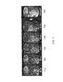
- FIG. 2Ais an exemplary SECM image of a swine intestinal epithelium, obtained ex vivo, 100 ⁇ m from the tissue surface using a single mode source and single-mode detection (SM-MM) configuration;
- SM-MMsingle mode source and single-mode detection
- FIG. 2Bis another exemplary SECM image of a swine intestinal epithelium, obtained using a single-mode source and multi-mode detection (SM-MM) configuration;
- SM-MMsingle-mode source and multi-mode detection
- FIG. 2Cis a magnified view of an SECM image of a swine intestinal epithelium
- FIG. 3Ais an exemplary SECM image of a swine intestinal epithelium, obtained ex vivo, after compression of the bowel wall at an imaging depth of 50 ⁇ m;
- FIG. 3Bis an exemplary SECM image of a swine intestinal epithelium, obtained ex vivo, after compression of the bowel wall at an imaging depth of 100 ⁇ m;
- FIG. 4is a schematic illustration of an exemplary SECM apparatus
- FIG. 5is an exemplary SECM image of a USAF chart
- FIG. 6Ais an exemplary SECM image based on data taken from a lens paper sample, displayed at a magnification of 1 ⁇ ;
- FIG. 6Bis an exemplary SECM image based on data taken from a lens paper sample, displayed at a magnification of 4.5 ⁇ ;
- FIG. 6Cis an exemplary SECM image based on data taken from a lens paper sample, displayed at a magnification of 16.7 ⁇ ;
- FIG. 6Dis an exemplary SECM image based on data taken from a lens paper sample, displayed at a magnification of 50 ⁇ ;
- FIG. 6Eis an exemplary SECM image based on data taken from a lens paper sample, displayed at a magnification of 125 ⁇ ;
- FIG. 7is a series of exemplary SECM data obtained from a lens paper sample at five different focal positions, together with a combine image that was generated by combining the data in the five individual images;
- FIG. 8Ais an exemplary SECM image based on data taken from a swine intestinal tissue fragment, displayed at a magnification of 1 ⁇ ;
- FIG. 8Bis an exemplary SECM image based on data taken from a swine intestinal tissue fragment, displayed at a magnification of 4 ⁇ ;
- FIG. 8Cis an exemplary SECM image based on data taken from a swine intestinal tissue fragment, displayed at a magnification of 20 ⁇ ;
- FIG. 8Dis an exemplary SECM image based on data taken from a swine intestinal tissue fragment, displayed at a magnification of 40 ⁇ ;
- FIG. 9is a schematic illustration of an exemplary SECM system capable of imaging large tissue volumes
- FIG. 10is a schematic illustration of a distal end of an exemplary catheter that may be used for imaging in accordance with exemplary embodiments of the present invention.
- FIG. 11is a schematic illustration of an exemplary catheter that may be used for imaging in accordance with exemplary embodiments of the present invention that includes an external rotational scanning arrangement;
- FIG. 12 Ais a schematic illustration of optical effects of a curved window and a negative cylindrical lens
- FIG. 12Bis a schematic illustration of an astigmatic aberration correction using a curved window
- FIG. 13Ais an illustration of an exemplary technique which may be used to acquire the desired depth range by stepping through a range of focal depths
- FIG. 13Bis an illustration of an exemplary technique which may be used for imaging tissue at a particular depth by actively adjusting a focal plane;
- FIG. 14Ais a schematic illustration of a dual bimorph piezoelectric bender
- FIG. 14Bis a schematic illustration of an exemplary arrangement whereby a motor may be moved within a transparent outer sheath using bending actuators;
- FIG. 15is a schematic illustration of an exemplary balloon catheter design that is configured to control a focus by translating a collimating lens
- FIG. 16is a photograph of a particular variable-focus lens
- FIG. 17Ais a schematic illustration of a cylindrical inner housing design which has a form of a transparent cylinder
- FIG. 17Bis a schematic illustration of a cylindrical inner housing design which includes a transparent window
- FIG. 17Cis a schematic illustration of a cylindrical inner housing design which includes several openings in the housing wall;
- FIG. 17Dis a schematic illustration of a cylindrical inner housing design which includes openings in a connection between the housing and a motor;
- FIG. 18is a schematic illustration of electrical and data connections between components of an exemplary imaging system
- FIG. 19Ais an illustration of an exemplary probe scanning pattern in which a beam is rotated quickly and simultaneously displaced slowly in an axial direction to provide a spiral imaging pattern;
- FIG. 19Bis an illustration of an exemplary probe scanning pattern in which the beam is rotated quickly and then repositioned axially;
- FIG. 19Cis an illustration of an exemplary probe scanning pattern in which the beam is rapidly scanned in the axial direction and then repositioned in the rotational direction;
- FIG. 19Dis an illustration of an exemplary probe scanning pattern in which the beam is scanned over concentric circular paths cover a circular tissue area
- FIG. 20Ais a schematic illustration of a rapid exchange balloon catheter design which includes a guidewire arrangement located at a distal tip of a housing;
- FIG. 20Bis a schematic illustration of a rapid exchange balloon catheter design which includes the guidewire arrangement located at the distal tip of the housing and having a form of a secondary channel;
- FIG. 20Cis a schematic illustration of a rapid exchange balloon catheter design which includes the guidewire arrangement located at a proximal tip of a housing and having a form of a secondary channel;
- FIG. 21Ais a schematic illustration of a first step in an exemplary technique for positioning a wire balloon catheter that includes insertion of a guidewire;
- FIG. 21Bis a schematic illustration of a second step in an exemplary technique for positioning a wire balloon catheter that includes placing a balloon catheter over the guidewire;
- FIG. 21Cis a schematic illustration of a third step in an exemplary technique for positioning a wire balloon catheter that includes placing an optical arrangement in the balloon catheter;
- FIG. 22Ais a schematic illustration of an exemplary balloon catheter which includes a single channel configured to deliver an inflation material from a remote location to the balloon;
- FIG. 22Bis a schematic illustration of an exemplary balloon catheter which includes two sheaths, where the inflation material can be provided between the sheaths;
- FIG. 23Ais a schematic illustration of a centering arrangement having a form of a wire cage, where the arrangement is contained within an outer sheath;
- FIG. 23Bis a schematic illustration of the centering arrangement having the form of a wire cage, where the arrangement is partially protruding from the outer sheath;
- FIG. 23Cis a schematic illustration of the centering arrangement having the form of a wire cage, where the arrangement is fully extended from outer sheath;
- FIG. 24Ais a schematic illustration of an exemplary SECM/SD-OCT system which includes a wavelength division multiplexer and a dispersion compensator;
- FIG. 24Bis a schematic illustration of an exemplary spectrum which may be provided by an SECM/SD-OCT system using a linear CCD array;
- FIG. 25is a schematic illustration of an exemplary SECM/SD-OCT probe
- FIG. 26is a schematic illustration of an exemplary SECM/SD-OCT probe which includes a single optical fiber for both the SECM and the SD-OCT arrangements;
- FIG. 27is an exemplary flow diagram of a technique which may be used to adjust a focus for an SECM image using SD-OCT data
- FIG. 28is a schematic illustration of a cross section of an exemplary catheter cable
- FIG. 29is a schematic illustration of an exemplary probe which includes a beam deflection optical arrangement that may provide a more compact probe configuration
- FIG. 30Ais a schematic illustration of a translational scanning technique showing a compact configuration of a probe during delivery of the probe to the site to be imaged;
- FIG. 30Bis a schematic illustration of the translational scanning technique showing an inner housing of the probe positioned at a distal limit of a translational range;
- FIG. 30Cis a schematic illustration of the translational scanning technique showing the inner housing of the probe positioned at a proximal limit of the translational range;
- FIG. 31is a schematic illustration of an outer housing which includes transparent openings
- FIG. 32is a schematic illustration of an exemplary compact probe which includes an off-center collimation optical arrangement and which is configured to provide external rotational scanning;
- FIG. 33Ais a schematic illustration of a probe which includes a forward inflatable balloon and an inner housing that is configured to scan while in contact with an inner wall of the balloon;
- FIG. 33Bis a schematic illustration of the probe shown in FIG. 33A which is in contact with an inner wall of the inflated balloon;
- FIG. 34Ais a schematic illustration of an exemplary probe that includes an outer inflatable balloon and an inner inflatable balloon which may be configured to maintain contact between the probe and a wall of the outer balloon when inflated;
- FIG. 34Bis a schematic illustration of the probe shown in FIG. 34A , where the inflated inner balloon is provided around the probe and is configured to maintain contact between the probe and the wall of the inflated outer balloon;
- FIG. 35Ais a schematic illustration of a further exemplary probe that includes an outer inflatable balloon and an inner inflatable balloon which may be configured to maintain contact between the probe and a wall of the outer balloon when inflated;
- FIG. 35Bis a schematic illustration of the probe shown in FIG. 35A , where the inflated inner balloon is provided between the probe and the outer balloon and is configured to maintain contact between the probe and the wall of the inflated outer balloon;
- FIG. 36Ais a schematic illustration of a bottom view of a probe that is configured to scan along a pullback axis while in contact with an inner wall of an inflatable balloon;
- FIG. 36Bis a schematic illustration of a side view of the probe shown in FIG. 36A ;
- FIG. 36Cis a schematic illustration of a side view of the probe shown in FIG. 36A , where the probe is in contact with the inner wall of the inflated balloon;
- FIG. 36Dis a front view of the probe shown in FIG. 36C .
- SECMSpectrally encoded confocal microscopy
- SECMutilizes a broad bandwidth light source and can encode one dimension of spatial information in the optical spectrum.
- FIG. 1An exemplary SECM technique is shown in FIG. 1 .
- the output from a single-mode optical fiber 100which may be located at a distal end of a probe, can be collimated by a collimating lens 110 , and then illuminate a dispersive optical element (such as, e.g., a transmission diffraction grating 120 ).
- An objective lens 130can then focus each diffracted wavelength to a distinct spatial location within the specimen, resulting in a transverse line focus 140 where each point on the line may be characterized by a distinct wavelength.
- the optical signalAfter reflection from the specimen, which may be, e.g., biological tissue, the optical signal can be recombined by the diffraction element 120 and collected by the single-mode fiber 100 .
- the core aperture of the single-mode fiber 100can provide a spatial filtering mechanism that is capable of rejecting out-of-focus light.
- the spectrum of the returned lightcan be measured and converted into confocal reflectance as a function of transverse displacement within the specimen.
- the spectral decodingcan be performed rapidly. Thus an image created by scanning the beam in a direction orthogonal to the line focus can be accomplished by relatively slow and straightforward mechanical actuation.
- SECM techniquesmay allow the use of endoscopic RCM, and it can be capable of providing image data at extremely high rates using high-speed linear CCD cameras.
- Commercially available linear CCD arrayscan obtain data at a rate greater than about 60 million pixels per second. When incorporated into an SECM spectrometer, these arrays can produce confocal images at speeds that are about 10 times faster than a typical video rate and up to 100 times faster than some endoscopic RCM techniques.
- the rapid imaging rate and fiber-optic design of typical SECM systemscan permit comprehensive, large area microscopy through an endoscopic probe.
- OCToptical coherence tomography
- TD-OCTtime-domain OCT
- exemplary SD-OCT and SECM systemscan be complementary, and a hybrid platform utilizing both techniques can provide information on the architectural and cellular structure of tissue that may be essential to accurate diagnosis.
- SECM and SD-OCT systemscan share key components, and a high-performance multi-modality system can be provided without substantially increasing complexity or cost of the individual systems.
- An SECM system in accordance with certain exemplary embodiments of the present inventioncan utilize a wavelength-swept 1300 nm source and a single-element photodetector to obtain spectrally encoded information as a function of time.
- imagescan be acquired at rates of up to about 30 frames/second having high lateral (1.4 ⁇ m) and axial (6 ⁇ m) resolutions, over a 400 ⁇ m field of view (“FOV”).
- FOVfield of view
- FIGS. 2A-2Cdepict exemplary SECM images of a swine intestinal epithelium obtained ex vivo using two imaging modes and corresponding fiber configurations: a single-mode illumination with single-mode detection (“SM-SM”), and a single-mode illumination with multi-mode detection (“SM-MM”).
- the SM-SM image in FIG. 2Ashows the epithelium structure located 100 ⁇ m from the tissue surface using a single mode source and single-mode detection.
- the image of the same tissue region shown in FIG. 2Bobtained using a using a single mode source and multi-mode detection (SM-MM) with a core:aperture ratio of 1:4, may have a smoother appearance and may be more easily interpreted because of a reduction in speckle noise.
- FIG. 2Cis a magnified view of the image shown in FIG. 2B that indicates a presence of villi containing a poorly reflecting core (e.g., lamina intestinal or “lp”) and a more highly scattering columnar epithelium. Bright image densities visible at the base of the columnar cells, consistent with nuclei (indicated by arrows) are shown in FIG. 2C .
- a poorly reflecting coree.g., lamina basement or “lp”
- the thickness of an esophageal wall being imaged in vivo using OCT techniquescan be decreased, e.g., by about a factor of two using an inflated balloon.
- the swine intestinal sample thickness shown in FIGS. 2A-2Cwas decreased by the same amount, and the subcellular features observed using SECM techniques were well preserved.
- FIGS. 3A and 3Bshow images of this thinned sample obtained at a depth of 50 ⁇ m and 100 ⁇ m, respectively.
- the penetration depth of a commercial 800 nm laser scanning confocal microscopewas observed to be reduced by about 20% as compared to that obtained with a 1300 nm SECM system. This reduced penetration may be a result of increased scattering of the shorter wavelength source.
- an SECM system using an 840 nm sourcemay provide sufficient penetration to identify subcellular structure of, e.g., an intestinal epithelium.
- FIG. 4An apparatus in accordance with certain exemplary embodiments of the present invention that is configured to provide comprehensive SECM images is illustrated schematically in FIG. 4 .
- This exemplary apparatuscan be configured to obtain images from a cylindrical sample having a length of 2.5 cm and a diameter of 2.0 cm, which are approximately the dimensions of the distal esophagus.
- a fiber-coupled 2.0 mW superluminescent diode 200having a wavelength centered at 800 nm and a bandwidth of 45 nm (QSSL-790-2, qPhotonics, Chesapeake, Va.) can be configured to illuminate a 50/50 single-mode fiber optic beam splitter 405 .
- Light transmitted through one port of the splittercan be collimated by a collimator 410 and transmitted through a fiber 412 to a focusing apparatus 415 and to a grating-lens pair that includes a grating 420 (1780 lpmm, Holographix, LLC, Hudson, Mass.) and a 350230-B asphere lens 425 (Thor Labs, Inc., Newton, N.J.) having a focal length, f, of 4.5 mm, a clear aperture of 5.0 mm, and a NA of 0.55.
- This arrangementcan be capable of producing a 500 ⁇ m longitudinal linear array, or line, of focused, spectrally-encoded spots 430 on an interior surface of the cylindrical sample.
- the grating-lens pairmay be affixed to a shaft of a motor 435 (e.g., a 1516SR, 15 mm diameter motor obtained from MicroMo Electronics, Inc., Clearwater, Fla.) by a housing 440 .
- a motor 435e.g., a 1516SR, 15 mm diameter motor obtained from MicroMo Electronics, Inc., Clearwater, Fla.
- the motor 435 , housing 440 , and grating-lens pairmay be translated along a longitudinal axis of the cylindrical sample during rotation of the motor 435 using, e.g., a computer-controlled linear stage 445 (such as, e.g., a Nanomotion II, 2.5 cm range, obtained from Melles Griot, Rochester, N.Y.). This procedure produced a helical scan of the entire interior surface of the cylindrical sample.
- a computer-controlled linear stage 445such as, e.g., a Nanomotion II, 2.5 cm range, obtained from Melles
- a spectrometer 450 and linear CCD 455can include, e.g., 2048 pixels and has a 30 kHz line rate (such as, e.g., a Basler L104K, obtained from Basler Vision Technologies, Exton, Pa.).
- a computer 460can be used to store, analyze and display image data provided by the spectrometer 450 and CCD 455 .
- Approximately 60,000 points per motor rotation (at 0.5 Hz, or 30 rpm)may be digitized. to provide a circumferential sampling density of approximately 1.0 ⁇ m.
- the longitudinal velocity of the motorcan be approximately 0.25 mm/s and the time required for one complete scan of the cylindrical sample may be about 100 seconds.
- the 1/e 2 diameter of the collimated beam on the grating-lens paircan be about 4.0 mm.
- the effective NA of this exemplary apparatuscan be approximately 0.4, which corresponds to a theoretical spot diameter of approximately 1.2 ⁇ m and a confocal parameter of approximately 2.5 ⁇ m.
- a theoretical spectral resolution on the samplemay be 0.8 ⁇ , which can yield up to approximately 630 resolvable points across the spectrally encoded line 430 .
- the spectrometer 450 in the detection armcan be designed to exceed the predicted spectral resolution of the probe.
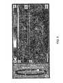
- FIG. 5An SECM scan of a 1951 USAF resolution chart obtained using this apparatus is shown in FIG. 5 .
- Exemplary SECM image data for a complete pullback image of a 2.5 cm phantom specimenare shown in FIG. 6 .
- Polar coordinateswere converted to rectangular coordinates prior to generating these displayed images.
- the phantom specimenwas made using lens paper affixed to the inner surface of a 2.1 cm inner diameter Teflon tube.
- FIG. 6AIn a low magnification image shown in FIG. 6A , macroscopic structure of the paper, including folds and voids, can be observed. Circumferential stripes that are visible may have resulted from the lower spectral power and lens aberrations that may be present at or near the ends of the spectrally-encoded line. Individual fibers and fiber microstructure can be clearly resolved in regions of this data set that are presented at higher magnifications, as shown in FIGS. 6B-6E .
- cylindrical two-dimensional (“2D”) images of the phantom samplewere acquired at five discrete focal depths over a range of 120 ⁇ m. These five images 710 - 750 shown in FIG. 7 were then summed to create an integrated image 760 , which demonstrates a nearly complete coverage of the surface of the phantom sample.
- FIG. 8AA 360° scan of this sample, which was acquired in 1 second, is shown in FIG. 8A .
- Imaged tissuelikely appears in only one sector of the cylindrical scan because the probe was not centered and the sample did not wrap completely around the cylinder.
- FIGS. 8B-8Dshow a sequence of exemplary magnified regions of this tissue sample.
- the image shown in FIG. 8Bis an expansion of a 1.5 cm sector outlined by a dotted rectangle in FIG. 8A .
- FIG. 8Crepresents an expansion of the rectangle outlined in FIG. 8B
- FIG. 8Drepresents an expansion of the rectangle outlined in FIG. 8C
- Magnified images of the tissue in the image FIG. 8Bare suggestive of a glandular structure.
- the magnified images in FIGS. 8C-8Dexhibit villi and nuclear features that are similar to those observed using a 1300 nm SECM system, as shown in FIGS. 2 and 3 .
- Other areas of the SECM scan in FIG. 8Ashow artifacts, including specular reflectance from the transparent cylinder and complete signal dropout, both of which may result from improper positioning of a focused SECM beam.
- Such challengesmay include, e.g., increasing the imaging rate, miniaturizing the probe optical components and mechanical components, incorporating a centering mechanism, and implementing a technique for dynamically changing the focal plane.
- the image acquisition speed of an SECM systemcan be improved by, e.g., a factor of about 2-4 as compared with the exemplary system described hereinabove. Such an improvement can be realized by providing certain modifications.
- a higher power semiconductor light sourcesuch as, e.g., a Superlum Diode, T-840 HP: 25 mW, 840 nm, 100 nm spectral bandwidth
- T-840 HP25 mW
- 840 nm100 nm spectral bandwidth
- Such an increase in optical powercan improve sensitivity and a larger bandwidth may widen the field of view, making it possible to scan the SECM beam approximately two times faster.
- an optical circulatorsuch as, e.g., an OC-3-850 (Optics for Research, Caldwell, N.J.) can increase the efficiency of light delivered to the probe and collected from the probe.
- Using a faster, more sensitive linear CCDsuch as, for example, an AVIIVA M4-2048 having 2048 pixels and a 60 kHz readout rate (Atmel Corporation) can provide a twofold increase in data acquisition speed and an improved spectral response over the wavelength range used to generate image data. Performance may also be improved by using, e.g., a Camera Link interface that can be capable of transferring data at a rate of approximately 120 MB/s from a camera to a hard-drive array for storage.
- Sensitivitywhich can be understood to refer to a minimum detectable reflectance, is a system parameter that can affect confocal image quality and penetration depth.
- a fraction of the incident lightwhich may be approximately 10 ⁇ 4 to 10 ⁇ 7 , can be reflected from skin at depths up to approximately 300 ⁇ m when using a near-infrared RCM technique.
- the exemplary SECM probe objective described hereinmay collect approximately 3 ⁇ 10 ⁇ 4 to 3 ⁇ 10 ⁇ 7 of the illuminating light reflected from deep within tissue.
- a 25 mW light sourcemay be separated into, e.g., approximately 1000 independent beams.
- a maximum double pass insertion losscan be estimated to be approximately 10 dB (which can include a 6 dB loss from the probe, and a 4 dB loss from the fiber optics and spectrometer).
- Each pixel in an arraymay thus be illuminated by approximately 50 to 50,000 photons/pixel for each line integration period based on these estimated parameters.
- a factor of 10 signal gainmay be achieved, resulting in approximately 500 to 500,000 photons/pixel per scan for such a configuration.
- a single pixel on an Atmel AVIIVA M4 camerae.g., can reliably detect light if a signal is above the dark current fluctuation that occurs at approximately 240 photons. If this device has approximately a 50% quantum efficiency at these wavelengths, a minimum detectable signal can be produced at approximately 480 photons/pixel per scan. Based on these approximations, an Atmel camera may have sufficient sensitivity to allow SECM imaging at deeper tissue depths. Quantum noise-limited detection of a predicted minimum reflectance can be achieved by using a multi-mode fiber for collection or by increasing the source power.
- a schematic diagram of an apparatus capable of performing large-area microscopic imaging of epithelial organs in accordance with certain exemplary embodiments of the present inventionis shown in FIG. 9 .
- a light source 900which may be a broadband source or a wavelength swept source, can provide light which may be conveyed through a circulator 910 or, alternatively, through a fiber splitter. The light can then be transmitted to an imaging catheter 930 through a scanning mechanism 920 . Scanning can be performed either externally to the catheter or within the catheter. In certain preferred exemplary embodiments, pullback scanning may be performed outside the catheter, and rotational scanning may be performed inside the catheter.
- Reflected light that is collectedmay then be detected with a detector 940 which may be, e.g., a spectrometer if a broadband light is used.
- the detector 940may also be, e.g., a single detector if a wavelength swept source is used.
- Data provided by the detector 940may be processed, displayed and/or saved by a computer 950 which may also be configured to control and synchronize the scanning procedure.
- Screening large luminal organsmay preferably utilize a centering of a distal portion of a catheter within the lumen to provide a consistent focus distance and/or depth relative to the tissue, and rapid acquisition of circumferential images over lengths of several centimeters.
- These criteriacan be satisfied by incorporating a circumferentially scanning imaging probe within a centering device.
- Provided an imaging optical arrangement located at or near the middle of the centering devicecan provide several additional advantages, including, e.g., elimination of surface height fluctuations, which may simplify focusing requirements, and physical coupling of the imaging system to a patient, which can greatly reduce motion artifacts that may otherwise occur.
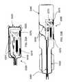
- FIG. 10A schematic diagram of the distal end of an SECM catheter in accordance with certain exemplary embodiments of the present invention is shown in FIG. 10 .
- Lightcan be provided through an optical fiber 1000 , which may be fixed by a fiber chuck 1005 , and then collimated using a collimating lens 1010 .
- This lightmay then pass through a variable focusing mechanism 1015 and a cylindrical lens 1020 that can be configured to pre-compensate the optical path to correct for astigmatism effects.
- the lightmay then be diffracted through a diffraction grating 1025 , which can be configured to diffract a center wavelength of the light by, for example, approximately 90 degrees, and focused by an imaging lens 1030 onto a spectral encoded line 1035 .
- Speckle artifactmay be reduced using multi-mode detection by increasing the diameter of a pinhole aperture associated with the optical fiber 1000 .
- This techniquecan provide an increased signal throughput and a reduction in speckle artifacts, together with only a slight decrease in spatial resolution.
- a double clad optical fibermay be used to implement this technique for spectral encoding, in which a single-mode core can illuminate a tissue and a multi-mode inner cladding can detect reflected light.
- the imaging lens 1030may preferably have a relatively large working distance that can be, e.g., approximately 2-7 mm, and maintain a large NA of approximately 0.25 to 0.5.
- the imaging lens 1030can be thin, preferably not more than about 5 mm thick.
- Conventional lenses, such as aspheres or achromats,may be used as imaging lenses.
- the inner housing 1040may surround some or all of the various optical components and the motor 1045 , and it may allow for longitudinal positioning of these components within the outer housing 1060 .
- the inner housing 1040can include portions thereof that have good optical transmission characteristics and low wavefront distortion to allow high quality imaging, while still maintaining structural rigidity to maintain a motor shaft 1050 centered within the probe.
- Materials that may be used to form transparent windows as part or all of the inner housing 1040may include, for example, glass or plastic materials such as, e.g., Pebax and high-density polyethylene (HDPE).
- HDPEhigh-density polyethylene
- the outer housing 1060can surround the inner housing 1040 , and can be configured to remain in a fixed position relative to the imaged tissue 1080 using the centering mechanism 1065 .
- An opening in a wall of the outer housing 1060can allow a pullback cable 1065 to move the inner housing 1040 .
- Linear scanningcan be conducted by affixing the inner housing 1040 to a computer-controlled translator (such as a translator that may be provided, e.g., by Newport Corp., Irvine, Calif.), while maintaining the outer housing 1060 in a fixed position relative to the tissue 1080 being imaged.
- a pullback techniquemay be used, e.g., to obtain longitudinal esophageal OCT images.
- All or a portion of the outer housing 1060may be transparent to allow a transmission of light therethrough. Optical characteristics of the transparent portions of the outer housing 1060 can be similar to those of the inner optical window 1055 .
- the cylindrical lens 1020 , the diffraction grating 1025 , and the imaging lens 1030may be housed in a rotational housing 1070 , which may be attached to the motor shaft 1050 .
- a conventional motor 1045may be used, which can have a diameter as small as about 1.5 mm or less. Using an encoder may improve image quality and registration, and may also increase the diameter of the motor 1045 to approximately 6-10 mm.
- Such a motorcan be provided, e.g., by (MicroMo Electronics, Inc. (Clearwater, Fla.). Dimensions of motor wires can be minimized to limit obstruction of a field of view of the apparatus. Circumferential scanning may be performed by rotating the rotational housing 1070 within the inner housing 1040 using the motor 1045 via the motor shaft 1050 .
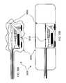
- a catheter configured to provide rotation of the inner housing 1040 relative to the external housing 1060 from a location external to a distal end of the catheter, in accordance with certain exemplary embodiments of the present invention,is illustrated schematically in FIG. 11 .
- a rotary motioncan be transmitted through an optical rotary junction 1100 , and light may be coupled into a rotation optical fiber 1110 .
- the rotary junctionmay also maintain electrical contact via one or more electrical wires 1120 and mechanical contacts via a rotatable pullback cable 1030 that can be configured to control pullback and focusing mechanisms.
- the inner housing 1140does not surround a motor and thus it can be smaller and lighter.
- a cylindrical lensmay be used to correct for astigmatism effects that can be created by a wall of a balloon or another centering device and/or by a transparent window or a transparent section of the inner and/or outer housing.
- a curved glasscan induce astigmatism in a manner similar to that of a negative cylindrical lens.
- the astigmatism induced by the two curved transparent walls shown in FIG. 12Aare optically similar to the negative cylindrical lens shown towards the right side of this Figure.
- Light passing through the central dashed line of any of the objects shown in FIG. 12Amay have a shorter path than light passing through the upper or lower dashed lines, which leads to induced astigmatism.
- Efficient and accurate correction of this optical distortioncan be achieved, e.g., by placing a curved window, similar to the window that induces the astigmatism, in the optical path, as shown in FIG. 12B .
- the curvature axis of the correcting curved windowshould be perpendicular to the axis of the curved housing windows to provide optical correction of the astigmatism.
- an endoscopic SECM systemcan be provided that is capable of comprehensively imaging an organ without user intervention during the acquisition of image data.
- the systemcan be capable of accounting for motion due to, e.g., heartbeat, respiration, and/or peristalsis movements.
- Utilization of a centering mechanismcan greatly reduces artifacts caused by motion of the tissue being imaged. For example, variations in distance between an imaging arrangement and the tissue being imaged can vary, for example, by as much as approximately ⁇ 250 ⁇ m during one comprehensive scan. This distance variation can occur on a slow time scale (e.g., over several seconds) relative to a circumferential scanning speed, but it may be significant relative to a time required to scan the length of a tissue region being imaged during longitudinal pullback of the imaging arrangement.
- An exemplary techniquecan be used in accordance with certain exemplary embodiments of the present invention to reduce or eliminate the effects of tissue motion during sampling.
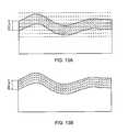
- This techniqueillustrated in FIG. 13A , can include a procedure for obtaining image data over a wider range of focal depths. If a desired total imaging depth is, for example, 200 ⁇ m, and a variation in tissue distance from the imaging arrangement is, e.g., ⁇ 250 ⁇ m, then image data can be acquired over a focal range of about 700 ⁇ m. This procedure can ensure that image data is obtained throughout the desired tissue volume. Although many portions of the volumetric image may not contain tissue when imaged, it is likely that at least one good image would be obtained from most regions of the tissue volume of interest.
- FIG. 13BA second exemplary technique that may be used to compensate for motion of tissue during imaging is illustrated in FIG. 13B .
- This techniquecan include a procedure for determining a distance between the imaging lens and a surface of the tissue being imaged. This distance can be tracked, and a focus of the lens can be adaptively controlled to provide a known focal distance relative to the tissue surface throughout the acquisition of image data in the tissue volume of interest. Adaptive focusing can decrease the number of focal scans required, and therefore may also decrease the time needed to obtain comprehensive coverage of the tissue volume of interest. Focus of the beam can be controlled, e.g., using an interferometric signal, a time-of-flight signal, an intensity of the electromagnetic radiation, etc.
- the above-described exemplary techniques for addressing motion of the tissue being imagedcan utilize a mechanism for adjusting the focal distance of the imaging arrangement.
- an inner housing of the imaging arrangement that includes a focus lenscan be moved relative to an exterior housing.
- multi-layered bimorph piezoelectric actuators 1410e.g., D220-A4-103YB, Piezo Systems, Inc., Cambridge, Mass.
- FIG. 14Acan be attached to, e.g., a metal sheet 1420 at both ends, which may provide a buckling of the ceramic material.
- These actuatorscan be placed back-to-back, as shown in FIG.
- actuators 1430can be arranged between an outer sheath 1440 and an assembly 1450 that can include a motor and focal optical components surround the motor, as shown in FIG. 14B .
- These actuators 1430can be utilized to change the focal position over the required range by controllably displacing the assembly 1450 relative to the outer housing 1440 .
- This techniquecan require the presence of a high voltage within the probe, additional electrical wires that may traverse and interrupt the field of view, and/or an increase of the overall diameter of a probe containing the imaging arrangement by, e.g., several mm.
- FIG. 15An alternate exemplary technique that may be used to adjust the focal distance of the imaging arrangement is shown in FIG. 15 .
- a cable housing 1510can be provided that surrounds a cable 1530 .
- the cable 1530can be attached at one end to a collimating lens 1540 , which may be configured to be movable in a longitudinal direction relative to a housing 1550 .
- the collimating lens 1540can be moved relative to the housing 1550 and other optical components to vary the focal distance. This translation can be controlled, e.g., externally to the imaging catheter, using the cable 1530 as is illustrated in FIG. 15 .
- motion of the collimating lens 1540can be controlled, e.g., by an electric or piezoelectric motor that can be provided inside the catheter.
- the focal distancecan also be varied by moving an optical fiber 1520 , which can provide the light used to image tissue, relative to the collimating lens 1540 .
- both the optical fiber 1520 and the collimating lens 1540may be moved relative to each other to vary the focal distance.
- the focal lengthcan be shifted by a distance ⁇ z by changing the separation between the optical fiber 1520 and the collimating lens 1540 by a distance of approximately M 2 ⁇ z, where M is a magnification factor of the imaging apparatus.
- Mis a magnification factor of the imaging apparatus.
- an exemplary imaging apparatuscan have a magnification factor that is approximately 3.
- the distance between the optical fiber 1520 and the collimating lens 1540would need to move approximately ⁇ 4.0 mm, which is a distance that can be achieved using any of the techniques described above for changing the focal distance.
- a further exemplary technique that can be used to vary the focal distancecan be to utilize an electronically tunable variable lens.
- a commercially available lens 1600(Varioptic AMS-1000, Lyon, France) shown in FIG. 16 , which may be used in cell phone cameras, may be utilized to vary the focal length in an imaging apparatus in accordance with an exemplary embodiment of the present invention.
- This lens 1600uses an electrowetting principle, and can provide a variable focal length between about ⁇ 200 mm and 40 mm, with optical quality that may only be limited by diffraction effects.
- the current effective clear aperture (CA) of this exemplary lens 1600is 3.0 mm and the total outer diameter (OD) is 10 mm.
- a similar lens having a 4.0 mm CA and a 6.0 mm ODmay be possible to produce.
- the full-range response time of this exemplary lens 1600is about 150 ms, which can be sufficiently fast to be used to track the distance between the optical components and the tissue surface and adjust the focal distance accordingly. It may be possible to produce this type of lens having a response time of about 10 ms.
- Utilizing a variable lens such as the one described above between the collimator and the SECM gratingcan provide, e.g., a focal distance that can vary by about ⁇ 300 ⁇ m or greater.
- a housing formed from transparent material 1700can be used, as shown in FIG. 17A .
- a housingcan be provided that includes a transparent window 1710 , as shown in FIG. 17B .
- a housingmay also be provided that includes an opening 1720 between two walls, such as that as shown in FIG. 17C , or an opening adjacent to a motor 1730 that may be attached to the housing as shown, e.g., in FIG. 17D .
- FIG. 18An exemplary schematic diagram of a control and data recording arrangement which can be used with the exemplary system shown in FIG. 9 is provided in FIG. 18 .
- the arrangement shown in FIG. 18can be configured to record a beam position while acquiring imaging data 1800 , which can provide a more precise spatial registration of the imaging data 1800 .
- the imaging data 1800can be acquired by a data acquisition and control unit 1810 .
- a catheter scanner arrangementmay scan a beam, e.g., using a rotary motor 1820 to provide angular motion of the beam and a pullback motor 1830 to move the beam longitudinally.
- the rotary motor 1820can be controlled by a rotary motor controller 1840
- the pullback motor 1830can be controlled by a pullback motor controller 1850 .
- the data acquisition and control unit 1810can direct the motor controller units 1840 , 1850 to provide specified motor velocities and/or positions.
- Encoder signals generated by the motors 1820 , 1830can be provided to both the motor controller units 1840 , 1850 and the data acquisition and control unit 1810 . In this manner, the encoder signals associated with each motor 1820 , 1830 can be recorded when a line of imaging data 1800 is acquired, thereby allowing a precise beam position to be associated with each line of data 1800 .
- FIG. 19Various scanning priorities that may be used in the imaging catheter in accordance with an exemplary embodiment of the present invention are shown in FIG. 19 .
- an exemplary scanning technique in which rotational scanning is performed as a first priority and axial (pullback) scanning is performed as a second priorityis shown in FIG. 19A .
- This techniquecan provide a set of data having a helical geometry.
- the axial scanningcan be performed in small increments, with each axial increment following a full revolution, as shown in FIG. 19B .
- axial (pullback) scanningcan be performed as a first priority and rotational scanning can be performed as a second priority, which may generate the scanning pattern shown in FIG. 19C .
- a greater imaging qualitycan be achieved along a direction of the first scan priority.
- a choice of scan prioritymay depend on whether transverse (rotational) images or axial images are preferred. Imaging of other organs or tissues that may have different symmetries can be performed in several ways. For example, a circular scanning pattern that may be used to image certain organs is shown in FIG. 19D .
- a balloon cathetersuch as, e.g., the one shown in FIG. 10
- a balloon cathetercan be configured to allow for a rapid-exchange placement procedure using a guidewire.
- a guidewirecan first be placed in an organ to be imaged, and the catheter can then be threaded down the guidewire. This procedure can allow easier and more precise placement of the catheter in many applications.
- FIG. 20Ashows an exemplary guidewire 2000 that passes through a hole 2010 in a distal end of the outer housing 2040 .
- FIG. 20Ashows an exemplary guidewire 2000 that passes through a hole 2010 in a distal end of the outer housing 2040 .
- a guidewire 2000passes through a tube 2020 that is attached to the distal end of the outer housing 2040 .
- the guidewire 2000can be configured to pass through the tube 2020 which may be attached to a proximal end of the outer housing 2040 , as shown in FIG. 20C .
- FIGS. 21A-CAn exemplary procedure that may be used to position a catheter that employs a guidewire in a center lumen of the catheter is illustrated in FIGS. 21A-C .
- the guidewire 2100can be placed within the organ 2150 , as shown in FIG. 21A .
- an outer housing 2110 of the catheter, together with a balloon 2120can be threaded over the guidewire 2100 , as shown in FIG. 21B .
- the inner housing 2130which may contain an optical arrangement, can be threaded down the catheter center lumen as shown in FIG. 21C , and an imaging procedure using the optical arrangement can be performed.
- FIG. 22ATwo exemplary configurations of a balloon catheter are shown in FIG. 22 .
- a device 2200that may include a source of pressurized air or gas can be used to inflate a balloon 2210 .
- a tube or other small passageway 2230can be provided that is connected to the balloon 2210 surrounding the catheter and which allows transfer of the pressurized air or gas to the balloon 2210 .
- Pressure within the balloon 2210 being inflatedcan be monitored using a manometer 2220 . This pressure can be used to optimize the balloon inflation as well as to assess placement of the catheter by monitoring pressure within a surrounding organ which may be contacted by the inflated balloon 2210 .
- a passageway 2240can be provided along an outer sheath of the catheter, which can allow transfer of the pressurized air or gas to the balloon 2210 , as shown in FIG. 22B .
- a balloon that is capable of changing its diameter in response to pressure changesmay be used, where focus depth can be controlled by varying the balloon diameter and thus moving the surrounding tissue to be allows transfer of the pressurized air or gas to the balloon 2210 . with respect to the imaging lens.
- FIGS. 23A-23CAn exemplary catheter design that may be used in accordance with another exemplary embodiment of the present invention is shown in FIGS. 23A-23C .
- This catheter designcan be configured to use one or more expandable wire strands 2300 to center an inner optical core of an imaging device within a luminal organ.
- the cathetermay include an additional sheath 2310 and a set of expandable wire strands 2300 located within the sheath 2310 that may be provided around the outer housing 2320 , as shown in FIG. 23A .
- the wire strands 2300can be pushed through the sheath 2310 to protrude from the end thereof as shown in FIG. 23B Alternatively, the sheath 2310 can be retracted from the outer housing 2320 .
- a sufficient length of the wire strands 2300can be exposed around the outer housing 2320 to allow the wire strands 2300 to expand the surrounding organ or tissue as shown in FIG. 23C , and to center the housing 2320 . After the imaging procedure is performed, the wire strands 2300 may be pulled back into the sheath 2310 and the catheter can be removed.
- Exemplary OCT and RCM techniquescan reject or ignore multiply scattered light received from a tissue sample being imaged, and thereby detect singly backscattered photons that may contain structural information. Each of these techniques, however, can reject multiply scattered light in a different way.
- the RCM techniquesmay employ confocal selection of light reflected by tissue being imaged from a tightly focused incident beam.
- RCM techniquescan be implemented by rapidly scanning the focused beam in a plane parallel to the tissue surface, which may provide transverse or en face images of the tissue.
- a large numerical aperture (NA)which can be used with conventional RCM techniques, may yield a very high spatial resolution (e.g., approximately 1-2 ⁇ m that can allow visualization of subcellular structure. Imaging procedures using a high NA, however, can be particularly sensitive to aberrations that can arise as light propagates through inhomogeneous tissue. Therefore, high-resolution imaging using RCM techniques may be limited to a depth of about 100-400 ⁇ m.
- the OCT techniquescan utilize coherence gating principles for optical sectioning and may not rely on the use of a high NA lens. OCT techniques may thus be performed using an imaging lens having a relatively large confocal parameter. This can provide a greater penetration depth into the tissue being imaged (e.g., approximately 1-3 mm) and a cross-sectional image format. These advantages may come at the expense of a reduced transverse resolution, which can be typically on the order of about 10-30 ⁇ m.
- the exemplary OCT and RCM techniquescan offer different imaging information which may be complementary.
- RCM techniquescan provide subcellular detail
- OCT techniquescan provide, e.g., architectural morphology. Imaging information from these two size regimes can be critical for histopathologic diagnosis, and in many cases, it may be difficult if not impossible to make an accurate diagnosis without using both.
- SECM and SD-OCT techniquescan share certain components. Therefore, a high-performance multi-modality system employing both of these imaging techniques can be provided that does not include a substantial increase in complexity or cost relative to a system that may use either technique alone.
- FIG. 24AAn overview of an exemplary system that is capable of performing both SECM techniques and SD-OCT techniques in accordance with an exemplary embodiment of the present invention is shown in FIG. 24A .
- a portion of a broadband light source bandwidthcan be used for obtaining SECM image data, and a further portion of the bandwidth data can be used, e.g., to obtain SD-OCT data.
- a light source 2400can be used to provide electromagnetic energy having a bandwidth greater than, e.g., about 100 nm.
- Devices that may be used as a light source 2400can include, e.g., a diode-pumped ultrafast laser (such as that available from, e.g., IntegralOCT, Femtolasers Obers GmbH, Vienna, Germany), or an array of super luminescent diodes (which may be obtained, e.g., from Superlum, Russia).
- a diode-pumped ultrafast lasersuch as that available from, e.g., IntegralOCT, Femtolasers architectures GmbH, Vienna, Germany
- super luminescent diodeswhich may be obtained, e.g., from Superlum, Russia.
- a portion of the light source spectrum that may be used for SD-OCT data(e.g., light having a wavelength between about 810-900 nm) can be separated from a portion of the spectrum that may be used for SECM data using a wavelength division multiplexer (WDM) 2410 and transmitted to a catheter 2420 and to a reference arm 2445 .
- WDMwavelength division multiplexer
- Light returning from the catheter 2420 through an SECM optical fiber 2430 and an SD-OCT optical fiber 2440can be provided to a spectrometer 2450 .
- the spectrometer 2450may be configured so that approximately half of the elements of the exemplary CCD array 2460 shown in FIG.
- the SD-OCT datacan be converted into axial structural data, e.g., by performing a Fourier transformation following interpolation of the SD-OCT data from wavelength space to k-space. For example, if the spectrometer 2450 has a resolution of approximately 0.1 nm, a total SD-OCT ranging depth may be greater than about 2.0 mm. Axial image resolution using the SD-OCT technique may be approximately 5 ⁇ m.
- FIG. 25A schematic overview of an exemplary SECM/SD-OCT probe is shown in FIG. 25 .
- This probeis similar to the probe shown, e.g., in FIG. 15 , and it further includes an arrangement configured to provide an SD-OCT beam path.
- an OCT optical fiber 2500can be inserted into the inner housing, together with an SECM optical fiber 2510 .
- the OCT optical fiber 2500can be configured to illuminate a small lens 2520 .
- a confocal parameter and a spot size for the SD-OCT beamcan be selected to achieve cross-sectional imaging over a range of depths. Exemplary values of the confocal parameter spot size can be, e.g., be approximately 1.1 mm and 25 ⁇ m, respectively.
- the NA of the SD-OCT lens 2520can be selected to be, e.g., approximately 0.02, and a collimated beam diameter of the SD-OCT beam can be selected to be, e.g., approximately 200 ⁇ m.
- a dichroic mirror 2530can be placed before the SECM grating to reflect the SD-OCT light beam 2540 and transmit the SECM light beam 2550 .
- the dichroic mirror 2530 shown in FIG. 25is arranged at an angle of approximately 45 degrees with respect to the SD-OCT light beam 2540 . This angle can be increased by using an appropriate coating on the mirror 2530 , which can allow the SD-OCT beam 2540 to overlap the SECM beam 2550 for a more precise spatial registration of the two images.
- Optical aberrations of the SD-OCT beam 2540which may be produced, e.g., by a curved window or balloon can be corrected by using a cylindrical element to pre-compensate for astigmatism as shown in FIG. 12B .
- FIG. 26A further exemplary embodiment of a catheter probe which may be used for both SECM imaging and SD-OCT imaging is shown in FIG. 26 .
- Broadband lightmay be provided through a single optical fiber 2600 , instead of through two separate fibers 2500 , 2510 as shown in FIG. 25 .
- a portion of the light which may be used to form an SD-OCT beam 2640may be reflected out of the optical path of the SECM beam 2650 using a dichroic mirror 2610 .
- the diameter of the SD-OCT beam 2640may be reduced by an aperture 2620 and/or by focusing the SD-OCT beam 2640 using a lens 2630 .
- the SD-OCT arrangementmay also be used to locate a surface of a tissue being imaged using an SECM technique, even with SD-OCT depth resolutions between about 20-100 ⁇ m. This can be performed even if the bandwidth of the SD-OCT beam 2640 is not sufficient to obtain a high quality SD-OCT image.
- SD-OCT image datamay be obtained from a depth scan (step 2700 ) and subsequently processed (step 2710 ). The image data may be analyzed and displayed as an SD-OCT image (step 2720 ). This image data may also be used to determine the location of a tissue surface (step 2730 ) using, for example, edge detection algorithms. Once the surface location of the tissue has been determined, a variable focus mechanism can be used to adjust a location of a focal plane of the SECM arrangement (step 2740 ).
- This focus control techniquecan be performed rapidly (e.g., in less than about 100 ms), which may allow for real-time tracking and focusing of a tissue surface.
- a location of a tissue edgecan be calibrated using an angle that is formed with respect to the SECM beam.
- the cable 2800may include, e.g., a pullback cable 2810 , a plurality of wires 2820 configured to supply electric power to a motor, a focus control cable 2830 , a channel 2840 configured to provide a gas or other fluid to an inflatable balloon or membrane, an SECM optical fiber 2850 , and/or an SD-OCT optical fiber 2860 .
- FIG. 29A schematic illustration of an exemplary SECM probe 2900 is shown in FIG. 29 .
- the probe 2900includes two prisms 2910 which may be configured to deflect a beam 2920 before it passes through a grating 2930 and an imaging lens 2940 .
- This exemplary configurationcan provide more space within the probe 2900 for the objective lens 2940 , which can result in a higher NA and/or a size reduction of the probe 2900 .
- the probe 3000can include an inner housing 3010 which may be provided within an outer housing 3020 while the probe 3000 is delivered to the imaging location, as shown in FIG. 3A .
- the inner housing 3010can slide through the outer housing 3020 to provide an extended pullback range, as shown in FIGS. 30B and 30C .
- providing an imaging lens 3020 near a center of the inner housing 3010can provide increased positional stability at the extreme scanning locations shown in FIGS. 30B and 30C .
- the outer housing 3100can be made of rigid materials such as, e.g., stainless steel or plastic. It may include one or more gaps 3110 which can allow light to pass therethrough to generate image data without introducing optical aberrations. Optionally, the gaps 3110 may include transparent windows.
- FIG. 32shows an exemplary probe in accordance with certain exemplary embodiments of the present invention.
- the probe 3200can provide a compact configuration of components and a small overall probe size.
- a cylindrical inner housing 3210can be configured to rotate and move freely within a cylindrical outer housing 3220 , allowing a collimating lens 3230 and an optical fiber 3240 to be placed away from a center axis of the inner housing 3210 . Scanning of a region of tissue to be imaged can be performed externally, where motion of the inner housing 3210 can be controlled using a pullback cable 3250 .
- a liquidsuch as, e.g., water or an index-matching oil can be provided in a space between an imaging lens and a surface of the tissue to be imaged.
- Providing such a liquidcan, e.g., improve optical parameters such as a NA and/or reduce back reflections of a light beam used to obtain image data.
- FIGS. 33A and 33BAn exemplary probe configuration 3300 which can provide a high NA for obtaining image data is shown in FIGS. 33A and 33B .
- an inner housing 3310can be provided in an outer housing 3320 , which may also include an uninflated balloon 3330 .
- the uninflated balloon 3330may be inflated such that it can expand forward of the outer housing 3320 .
- the inner housing 3310may then be deployed outside of the outer housing 3310 and within the inflated balloon 3340 .
- An elastic arrangement 3350can be provided in a compressed configuration between the inner housing 3310 and the outer housing 3320 , as shown in FIG. 33A .
- the elastic arrangement 3350can be configured to position the inner housing 3310 against an inside wall of the inflated balloon 3340 when the inner housing 3310 is deployed, as shown in FIG.
- the inner housing 3310can be configured to scan a region of tissue outside of the inflated balloon 3340 the balloon area using a pullback cable 3360 .
- the cable 3360can be capable of controlling both rotation and longitudinal translation (e.g., pullback) of the inner housing 3310 within the inflated balloon 3340 .
- Spacers 3370may be used to improve contact between the imaging optical arrangement and the wall of the inflated balloon 3340 or the adjacent tissue surface.
- FIGS. 34A and 34BA further exemplary probe configuration 3400 is shown in FIGS. 34A and 34B , which can be capable of maintaining an inner probe housing 3410 against an inside wall of an outer balloon 3420 , in accordance with certain exemplary embodiments of the present invention.
- an outer balloon 3420 and an inner balloon 3430shown uninflated in FIG. 34A , can be provided such that they surround the inner housing 3410 .
- Each balloonmay be inflated, as shown in FIG. 34B .
- the inner housing 3410may be attached to one face of the inner balloon 3430 .
- Rotational and translational scanning within the outer balloon 3420may be performed by moving the inner housing 3410 together with the inner balloon 3430 relative to the outer balloon 3420 .
- FIGS. 35A and 35BA still further exemplary probe configuration 3500 is shown in FIGS. 35A and 35B , which can be capable of maintaining an inner probe housing 3510 against an inside wall of an outer balloon 3520 , in accordance with certain exemplary embodiments of the present invention.
- the outer balloon 3520shown uninflated in FIG. 35A , may be inflated within an organ or region of tissue to be imaged.
- An inner balloon 3530shown uninflated in FIG. 35A , may be provided between the inner housing 3510 and the outer balloon 3520 .
- the inner balloon 3530may be inflated, as shown in FIG. 35B , and pressure provided by the inner balloon 3530 can be used to maintain contact between the inner housing 3510 and an inner wall of the outer balloon 3520 , as shown in FIG. 35B .
- the exemplary probe configurations 3400 and 3500 shown in FIGS. 34 and 35may be used without an outer housing.
- the uninflated balloons 3420 , 3430 , 3520 , 3530may be packed inside an external enclosure that can be used to deliver the probe 3400 , 3500 to a desired location.
- Such an external enclosurecan optionally be formed, e.g., from a dissolvable material.
- FIGS. 36A-36DAn exemplary configuration of an SECM probe 3600 is shown in FIGS. 36A-36D , which is capable of providing a spectrally encoded line 3610 that lies perpendicular to an axis of an organ or a balloon cylinder.
- a bottom view of this probe configurationis provided in FIG. 36A
- a corresponding side viewis shown in FIG. 36B .
- FIG. 36Cshows a further side view in which the probe housing 3640 is deployed within an inflated balloon 3650 , similar to that shown in FIG. 33B .
- a longitudinal (e.g., pullback) directioncan be a primary scanning direction, such that the probe housing 3640 is moved in this longitudinal direction at a relatively fast rate of speed.
- the probe 3600can be provided with positioning arrangements such as those shown, e.g., in any of FIGS. 33-35 .
- the probe housing 3640can include a mirror 3620 which may be configured to deflect a light beam towards a suitably positioned grating to provide a spectrally-encoded line 3610 configured as shown in FIGS. 36A and 36D .
- Combination of SD-OCT and SECM imaging arrangements within a probecan provide a useful apparatus for obtaining structural information on different scales using different image formats.
- Data obtained for both imaging techniquescan be acquired simultaneously, because the resolutions of the two techniques are different.
- useful scan rates for the two techniquesmay not be compatible with each other.
- a typical SECM scan ratecan be provided using a rotation rate, e.g., of about 1 Hz and a longitudinal pullback speed, e.g., of approximately 1 mm/s.
- Typical scan rates for obtaining SD-OCT image datacan be, e.g., approximately 50-100 Hz in a rotational direction and, e.g., approximately 0.2-0.5 mm/s in a longitudinal direction.
- One technique which may be used to obtain comprehensive image data that is properly sampled for both techniquesis to conduct an additional comprehensive SD-OCT scan, sampled appropriately, following acquisition of the SECM data set.
- This techniquemay increase the data acquisition time for a tissue region by, e.g., approximately 1-2 minutes.
- Encoder signals obtained for both the rotating and linearly translating motorscan be digitized throughout each scan.
- the encoder signalscan be corrected for shifts in position of a balloon by quantitatively correlating SD-OCT images to determine angular and rotational offsets for each scan.
- This techniquecan provide accurate spatial registration of the SD-OCT and SECM data sets within about 500 ⁇ m.
- an imaging arrangement provided, e.g., in a probemay be operated in an abbreviated imaging mode (e.g., ‘scout imaging’) to determine if a catheter which may be used to deliver the probe is properly positioned within the organ or tissue region to be imaged.
- an abbreviated imaging modee.g., ‘scout imaging’
- a comprehensive set of image datacan be obtained after proper catheter placement is confirmed.
- a balloon centering cathetermay be inflated using a material that is optically transparent other than air such as, e.g., water, heavy water (D2O), oil, etc.
- a lubricating agentmay also be used to aid insertion of the catheter.
- a mucousal removal agentmay be applied prior to obtaining image data to reduce the amount of mucous present in the organ to be imaged, where presence of such mucous may reduce image quality.
Landscapes
- Health & Medical Sciences (AREA)
- Physics & Mathematics (AREA)
- Life Sciences & Earth Sciences (AREA)
- General Physics & Mathematics (AREA)
- General Health & Medical Sciences (AREA)
- Engineering & Computer Science (AREA)
- Pathology (AREA)
- Biomedical Technology (AREA)
- Nuclear Medicine, Radiotherapy & Molecular Imaging (AREA)
- Molecular Biology (AREA)
- Chemical & Material Sciences (AREA)
- Biophysics (AREA)
- Radiology & Medical Imaging (AREA)
- Animal Behavior & Ethology (AREA)
- Veterinary Medicine (AREA)
- Public Health (AREA)
- Surgery (AREA)
- Medical Informatics (AREA)
- Heart & Thoracic Surgery (AREA)
- Analytical Chemistry (AREA)
- Biochemistry (AREA)
- Immunology (AREA)
- Optics & Photonics (AREA)
- Astronomy & Astrophysics (AREA)
- Theoretical Computer Science (AREA)
- Spectroscopy & Molecular Physics (AREA)
- Medicinal Chemistry (AREA)
- Urology & Nephrology (AREA)
- Hematology (AREA)
- Signal Processing (AREA)
- Pulmonology (AREA)
- Food Science & Technology (AREA)
- Mathematical Physics (AREA)
- Investigating Or Analysing Materials By Optical Means (AREA)
- Investigating, Analyzing Materials By Fluorescence Or Luminescence (AREA)
- Endoscopes (AREA)
- Microscoopes, Condenser (AREA)
- Instruments For Viewing The Inside Of Hollow Bodies (AREA)
- Automatic Focus Adjustment (AREA)
- Transforming Light Signals Into Electric Signals (AREA)
Abstract
Description
This application is a divisional application of, and therefore claims priority from, U.S. application Ser. No. 11/537,048, filed on Sep. 29, 2006 now U.S. Pat. No. 7,847,949, which is based upon and claims the benefit of priority from U.S. Patent Application Ser. No. 60/721,802, filed Sep. 29, 2005. The entire disclosures of both applications are incorporated herein by reference.
The present invention relates to devices and methods for comprehensive optical imaging of epithelial organs and other biological structures via spectral encoding.
Radiological techniques such as X-ray computed tomography (“CT”), magnetic resonance imaging (“MRI”), and ultrasound can enable noninvasive visualization of human pathology at the organ level. Although these modalities may be capable of identifying large-scale pathology, the diagnosis of cancer can require the evaluation of microscopic structures that is beyond the resolution of conventional imaging techniques. Consequently, biopsy and histopathologic examination may be required for diagnosis. Because precancerous growth and early stage cancers often arise on a microscopic scale, they can present significant challenges for identification and diagnosis. Conventional screening and surveillance of these pathologies relies on unguided biopsy and morphological analysis of Hematoxylin and Eosin (“H&E”) stained slides. Although this approach may be regarded as a current standard for microscopic diagnosis, it requires the removal of tissue from the patient and significant processing time to generate slides. More importantly, histopathology is inherently a point sampling technique; frequently only a very small fraction of the diseased tissue can be excised and often less than 1% of a biopsy sample may be examined by a pathologist.
It may be preferable to obtain microscopic diagnoses from an entire organ or biological system in a living human patient. However, the lack of an appropriate imaging technology can greatly limits options for screening for pre-neoplastic conditions (e.g. metaplasia) and dysplasia. In addition, an inability to identify areas of dysplasia and carcinoma in situ has led to screening procedures such as, e.g., random biopsy of the prostate, colon, esophagus, and bladder, etc., which can be highly undesirable and indiscriminate. Many diagnostic tasks presently referred to a frozen section laboratory, such as the delineation of surgical tumor margins, could be improved by a diagnostic modality capable of rapidly imaging large tissue volumes on a microscopic scale. A technology that could fill this gap between pathology and radiology would be of great benefit to patient management and health care.
Technical advances have been made to increase the resolution of non-invasive imaging techniques such as, e.g., micro-CT, micro-PET, and magnetic resonance imaging (“MRI”) microscopy. Resolutions approaching 20 μm have been achieved by these technologies, but fundamental physical limitations can still prevent their application in patients. Microscopic optical biopsy techniques, performed in situ, have recently been advanced for non-excisional histopathologic diagnosis. Reflectance confocal microscopy (“RCM”) may be particularly well-suited for non-invasive microscopy in patients, as it is capable of measuring microscopic structure without tissue contact and does not require the administration of extrinsic contrast agents. RCM can reject out of focus light and detects backscattered photons selectively originating from a single plane within the tissue. RCM can be implemented, e.g., by rapidly scanning a focused beam of electromagnetic radiation in a plane parallel to a tissue surface, yielding transverse or en face images of tissue. The large numerical aperture (NA) that may be used in RCM can yield a very high spatial resolution (1-2 μm), enabling visualization of subcellular structures. High NA imaging, however, can be particularly sensitive to aberrations that arise as light propagates through inhomogeneous tissue. Also, high-resolution imaging with RCM is typically limited to a depth of about 100-400 μm.
RCM has been extensively demonstrated as a viable imaging technique for skin tissue. Development of endoscopic confocal microscopy systems has been more difficult, owing at least in part to the substantial technical challenges involved in miniaturizing a scanning microscope. One major obstacle to direct application of the concepts of confocal microscopy to endoscopy is the engineering of a mechanism for rapidly rastering a focused beam at the distal end of a small-diameter, flexible probe. A variety of approaches have been proposed to address this problem, including the use of distal micro-electromechanical systems (“MEMS”) beam scanning devices and proximal scanning of single-mode fiber bundles. Also, RCM may provide microscopic images only at discrete locations—a “point sampling” technique. As currently implemented, point sampling can be inherent to RCM because it has a limited field of view, which may be comparable to or less than that of an excisional biopsy, and the imaging rate can be too slow for comprehensive large field microscopy.
Another challenge in adapting confocal microscopy to endoscopic applications can include miniaturization of high NA objectives that may be used for optical sectioning. Such miniaturization may be achieved by providing, e.g., a gradient-index lens system, dual-axis objectives, or custom designs of miniature objectives. For example, detailed images of the morphology of cervical epithelium may be obtained in vivo using a fiber optic bundle coupled to a miniature objective lens, and fluorescence-based images of colorectal lesions may be achieved using commercial instruments such as those which may be obtained, e.g., from Olympus Corp. and Pentax/Optiscan.
Despite these advances, there may be a need for improved imaging techniques that can provide microscopic resolution of biological structures in situ over large regions.
One of the objects of the present invention is to overcome certain deficiencies and shortcomings of the prior art systems and methods (including those described herein above), and provide an exemplary embodiment of a method and an apparatus which are capable of providing comprehensive microscopic optical imaging of anatomical structures such as, e.g., epithelial organs or other bodily tissues.
For example, an apparatus in accordance with exemplary embodiments of the present invention may be in the form of a probe or an assembly, which may be disposable. The probe or assembly may include, e.g., one or more optical waveguides capable of forwarding an electromagnetic radiation to the probe or assembly and forming an optical beam, one or more focusing arrangements provided at a distal end which may be configured to focus the optical beam, and a scanning arrangement configured to scan the beam across a portion of the anatomical structure. The electromagnetic radiation may include a plurality of wavelengths, and the wavelengths may vary with time. The probe may also include one or more diffraction arrangements which may be configured to diffract or spectrally disperse the beam, one or more correcting arrangements which may be configured to correct for optical aberrations, a mechanism capable of centering or positioning the probe or assembly within the anatomical structure being imaged, and/or a guidewire arrangement which can be capable of translating and/or rotating the probe or assembly. The waveguide may be, e.g., an optical fiber or a bundle of optical fibers or other waveguides. The probe or assembly may further include a spectral encoding arrangement and/or a corrective optical arrangement such as, e.g., a curved transparent surface, which can be used to correct aberrations such as an astigmatism in the optical beam path.
In certain exemplary embodiments of the present invention, the probe or assembly can be configured to scan a region of the anatomical structure which can have an area greater than about 1 mm2, and where the region may include a surface, a volume, or a location below a surface of the anatomical structure. The probe or assembly may be configured to obtain data which can be used to generate an image of the region with a resolution that is below approximately 10 μm.
In further exemplary embodiments of the present invention, a probe or assembly can be provided which is capable of positioning and/or focus the optical beam relative to the anatomical structure. The positioning and/or focusing can be based on, e.g., an interferometric signal, a time-of-flight signal, or an intensity of the electromagnetic radiation. The probe or assembly can include a confocal optical arrangement that can
In still further exemplary embodiments of the present invention, the probe or assembly can include a locating arrangement that is capable of determining a location of the probe or assembly relative to a location within the anatomical structure, and an optional positioning arrangement that can control the motion and/or position of the probe based on the location.
In other exemplary embodiments of the present invention, a method for obtaining comprehensive microscopic optical imaging of anatomical structures can be provided, which can include scanning a region of the anatomical structure to be imaged that is larger than about 1 mm2using an electromagnetic radiation such as, e.g., an optical beam, receiving a signal based on the radiation, and generating an image based on the signal, where the image can have a transverse resolution that is below about 10 μm.
In yet further exemplary embodiments of the present invention, a method for positioning or directing an electromagnetic radiation within an anatomical structure is provided, which can include scanning at least a portion of the anatomical structure using the electromagnetic beam, and using a signal which may be based on the electromagnetic radiation to control the position and/or focus of the radiation. A method can also be provided to control the position or focus of a confocal beam within the anatomical structure based on a signal obtained from scanning the electromagnetic radiation over a region of the anatomical structure.
Other features and advantages of the present invention will become apparent upon reading the following detailed description of embodiments of the invention, when taken in conjunction with the appended claims.
Further objects, features and advantages of the present invention will become apparent from the following detailed description taken in conjunction with the accompanying figures showing illustrative embodiments of the present invention, in which:
Throughout the figures, the same reference numerals and characters, unless otherwise stated, are used to denote like features, elements, components or portions of the illustrated embodiments. Moreover, while the subject invention will now be described in detail with reference to the figures, it is done so in connection with the illustrative embodiments. It is intended that changes and modifications can be made to the described embodiments without departing from the true scope and spirit of the subject invention as defined by the appended claims.
In accordance with exemplary embodiments of the present invention, a method and apparatus for endoscopic confocal microscopy is provided which circumvents the need for miniature, high-speed scanning mechanisms within a probe. Spectrally encoded confocal microscopy (“SECM”) is a wavelength-division multiplexed confocal approach that may be used. SECM utilizes a broad bandwidth light source and can encode one dimension of spatial information in the optical spectrum.
An exemplary SECM technique is shown inFIG. 1 . The output from a single-modeoptical fiber 100, which may be located at a distal end of a probe, can be collimated by acollimating lens 110, and then illuminate a dispersive optical element (such as, e.g., a transmission diffraction grating120). Anobjective lens 130 can then focus each diffracted wavelength to a distinct spatial location within the specimen, resulting in atransverse line focus 140 where each point on the line may be characterized by a distinct wavelength. After reflection from the specimen, which may be, e.g., biological tissue, the optical signal can be recombined by the diffraction element120 and collected by the single-mode fiber 100. The core aperture of the single-mode fiber 100 can provide a spatial filtering mechanism that is capable of rejecting out-of-focus light. Outside the probe (and optionally within a system console) the spectrum of the returned light can be measured and converted into confocal reflectance as a function of transverse displacement within the specimen. The spectral decoding can be performed rapidly. Thus an image created by scanning the beam in a direction orthogonal to the line focus can be accomplished by relatively slow and straightforward mechanical actuation.
SECM techniques may allow the use of endoscopic RCM, and it can be capable of providing image data at extremely high rates using high-speed linear CCD cameras. Commercially available linear CCD arrays can obtain data at a rate greater than about 60 million pixels per second. When incorporated into an SECM spectrometer, these arrays can produce confocal images at speeds that are about 10 times faster than a typical video rate and up to 100 times faster than some endoscopic RCM techniques. The rapid imaging rate and fiber-optic design of typical SECM systems can permit comprehensive, large area microscopy through an endoscopic probe.
Techniques using optical coherence tomography (“OCT”) and variations thereof may be used for comprehensive architectural screening. Acquiring an OCT signal in the wavelength domain, rather than in the time domain, can provide orders of magnitude improvement in imaging speed while maintaining excellent image quality. Using spectral domain OCT (“SD-OCT”) techniques, high-resolution ranging can be conducted in biological tissue by detecting spectrally resolved interference between a tissue sample and a reference. Because SD-OCT systems can utilize the same high-speed linear CCD's as SECM systems, they can also be capable of capturing images at 60 million pixels/s, which is approximately two orders of magnitude faster than conventional time-domain OCT (“TD-OCT”) systems. With this acquisition rate and resolution, SD-OCT systems can provide comprehensive volumetric microscopy at the architectural level in a clinical environment.
The information provided by exemplary SD-OCT and SECM systems can be complementary, and a hybrid platform utilizing both techniques can provide information on the architectural and cellular structure of tissue that may be essential to accurate diagnosis. Although a combination of disparate technologies typically requires extensive engineering and may compromises performance, SECM and SD-OCT systems can share key components, and a high-performance multi-modality system can be provided without substantially increasing complexity or cost of the individual systems.
An SECM system in accordance with certain exemplary embodiments of the present invention can utilize a wavelength-swept 1300 nm source and a single-element photodetector to obtain spectrally encoded information as a function of time. With this system, images can be acquired at rates of up to about 30 frames/second having high lateral (1.4 μm) and axial (6 μm) resolutions, over a 400 μm field of view (“FOV”). Images of freshly excised swine duodenum segments were imaged ex vivo with a high speed system to illustrate the capability of an SECM system to identify subcellular structures that may be found in, e.g., specialized intestinal metaplasia (“SIM”) or the metaplastic change of Barretts esophagus.
The thickness of an esophageal wall being imaged in vivo using OCT techniques can be decreased, e.g., by about a factor of two using an inflated balloon. The swine intestinal sample thickness shown inFIGS. 2A-2C was decreased by the same amount, and the subcellular features observed using SECM techniques were well preserved.FIGS. 3A and 3B show images of this thinned sample obtained at a depth of 50 μm and 100 μm, respectively.
The penetration depth of a commercial 800 nm laser scanning confocal microscope was observed to be reduced by about 20% as compared to that obtained with a 1300 nm SECM system. This reduced penetration may be a result of increased scattering of the shorter wavelength source. Thus an SECM system using an 840 nm source may provide sufficient penetration to identify subcellular structure of, e.g., an intestinal epithelium.
An apparatus in accordance with certain exemplary embodiments of the present invention that is configured to provide comprehensive SECM images is illustrated schematically inFIG. 4 . This exemplary apparatus can be configured to obtain images from a cylindrical sample having a length of 2.5 cm and a diameter of 2.0 cm, which are approximately the dimensions of the distal esophagus. A fiber-coupled 2.0 mW superluminescent diode200, having a wavelength centered at 800 nm and a bandwidth of 45 nm (QSSL-790-2, qPhotonics, Chesapeake, Va.) can be configured to illuminate a 50/50 single-mode fiberoptic beam splitter 405. Light transmitted through one port of the splitter can be collimated by acollimator 410 and transmitted through a fiber412 to a focusingapparatus 415 and to a grating-lens pair that includes a grating420 (1780 lpmm, Holographix, LLC, Hudson, Mass.) and a 350230-B asphere lens425 (Thor Labs, Inc., Newton, N.J.) having a focal length, f, of 4.5 mm, a clear aperture of 5.0 mm, and a NA of 0.55. This arrangement can be capable of producing a 500 μm longitudinal linear array, or line, of focused, spectrally-encodedspots 430 on an interior surface of the cylindrical sample. The grating-lens pair may be affixed to a shaft of a motor435 (e.g., a 1516SR, 15 mm diameter motor obtained from MicroMo Electronics, Inc., Clearwater, Fla.) by ahousing 440. As themotor 435 rotates, the spectrally encoded line can be scanned across the inner circumference of the cylindrical sample. Themotor 435,housing 440, and grating-lens pair may be translated along a longitudinal axis of the cylindrical sample during rotation of themotor 435 using, e.g., a computer-controlled linear stage445 (such as, e.g., a Nanomotion II, 2.5 cm range, obtained from Melles Griot, Rochester, N.Y.). This procedure produced a helical scan of the entire interior surface of the cylindrical sample.
Light reflected from the sample can be transmitted back through the optical system into the single-mode fiber412 and provided by the fiber412 to aspectrometer 450 andlinear CCD 455 that can include, e.g., 2048 pixels and has a 30 kHz line rate (such as, e.g., a Basler L104K, obtained from Basler Vision Technologies, Exton, Pa.). Acomputer 460 can be used to store, analyze and display image data provided by thespectrometer 450 andCCD 455. Approximately 60,000 points per motor rotation (at 0.5 Hz, or 30 rpm) may be digitized. to provide a circumferential sampling density of approximately 1.0 μm. The longitudinal velocity of the motor can be approximately 0.25 mm/s and the time required for one complete scan of the cylindrical sample may be about 100 seconds.
The 1/e2diameter of the collimated beam on the grating-lens pair can be about 4.0 mm. As a result, the effective NA of this exemplary apparatus can be approximately 0.4, which corresponds to a theoretical spot diameter of approximately 1.2 μm and a confocal parameter of approximately 2.5 μm. In a system that is free of optical aberrations, a theoretical spectral resolution on the sample may be 0.8 Å, which can yield up to approximately 630 resolvable points across the spectrally encodedline 430. Thespectrometer 450 in the detection arm can be designed to exceed the predicted spectral resolution of the probe.
An SECM scan of a 1951 USAF resolution chart obtained using this apparatus is shown inFIG. 5 . The smallest bars in this Figure, which are separated by 2.2 μm, were resolved. A transverse line spread function full-width-half-maximum (“FWHM”) and an axial FWHM function obtained using a mirror scanned through the focus were measured as 2.1 μm and 5.5 μm, respectively. The field of view was observed to be about 500 μm. These measurements were slightly lower than corresponding theoretical values, which may be attributed to aberrations in the optical path. These parameters indicate that the exemplary apparatus described herein can be capable of providing sufficient resolution to be used for confocal microscopy in biological tissue.
Exemplary SECM image data for a complete pullback image of a 2.5 cm phantom specimen are shown inFIG. 6 . Polar coordinates were converted to rectangular coordinates prior to generating these displayed images. The phantom specimen was made using lens paper affixed to the inner surface of a 2.1 cm inner diameter Teflon tube. In a low magnification image shown inFIG. 6A , macroscopic structure of the paper, including folds and voids, can be observed. Circumferential stripes that are visible may have resulted from the lower spectral power and lens aberrations that may be present at or near the ends of the spectrally-encoded line. Individual fibers and fiber microstructure can be clearly resolved in regions of this data set that are presented at higher magnifications, as shown inFIGS. 6B-6E .
By adjusting the focusingapparatus 415 inFIG. 4A , cylindrical two-dimensional (“2D”) images of the phantom sample were acquired at five discrete focal depths over a range of 120 μm. These five images710-750 shown inFIG. 7 were then summed to create anintegrated image 760, which demonstrates a nearly complete coverage of the surface of the phantom sample.
Imaging biological samples using an SECM apparatus such as that described herein can be complicated by the lack of a centering apparatus for the optical scan head. In order to provide further improvements for generating wide-field microscopy images and data, a sample of swine intestine was placed on top of a 2.0 cm diameter transparent cylinder. A 360° scan of this sample, which was acquired in 1 second, is shown inFIG. 8A . Imaged tissue likely appears in only one sector of the cylindrical scan because the probe was not centered and the sample did not wrap completely around the cylinder.FIGS. 8B-8D show a sequence of exemplary magnified regions of this tissue sample. The image shown inFIG. 8B is an expansion of a 1.5 cm sector outlined by a dotted rectangle inFIG. 8A . Similarly, the image inFIG. 8C represents an expansion of the rectangle outlined inFIG. 8B , and the image inFIG. 8D represents an expansion of the rectangle outlined inFIG. 8C . Magnified images of the tissue in the imageFIG. 8B are suggestive of a glandular structure. The magnified images inFIGS. 8C-8D exhibit villi and nuclear features that are similar to those observed using a 1300 nm SECM system, as shown inFIGS. 2 and 3 . Other areas of the SECM scan inFIG. 8A show artifacts, including specular reflectance from the transparent cylinder and complete signal dropout, both of which may result from improper positioning of a focused SECM beam.
Conducting comprehensive confocal microscopy in patients can present a variety of technical challenges. Such challenges may include, e.g., increasing the imaging rate, miniaturizing the probe optical components and mechanical components, incorporating a centering mechanism, and implementing a technique for dynamically changing the focal plane.
The image acquisition speed of an SECM system can be improved by, e.g., a factor of about 2-4 as compared with the exemplary system described hereinabove. Such an improvement can be realized by providing certain modifications. For example, a higher power semiconductor light source (such as, e.g., a Superlum Diode, T-840 HP: 25 mW, 840 nm, 100 nm spectral bandwidth) can provide, e.g., approximately 1000 spectrally resolvable points. Such an increase in optical power can improve sensitivity and a larger bandwidth may widen the field of view, making it possible to scan the SECM beam approximately two times faster. Also, using an optical circulator such as, e.g., an OC-3-850 (Optics for Research, Caldwell, N.J.) can increase the efficiency of light delivered to the probe and collected from the probe. Using a faster, more sensitive linear CCD such as, for example, an AVIIVA M4-2048 having 2048 pixels and a 60 kHz readout rate (Atmel Corporation) can provide a twofold increase in data acquisition speed and an improved spectral response over the wavelength range used to generate image data. Performance may also be improved by using, e.g., a Camera Link interface that can be capable of transferring data at a rate of approximately 120 MB/s from a camera to a hard-drive array for storage.
Sensitivity, which can be understood to refer to a minimum detectable reflectance, is a system parameter that can affect confocal image quality and penetration depth. A fraction of the incident light, which may be approximately 10−4to 10−7, can be reflected from skin at depths up to approximately 300 μm when using a near-infrared RCM technique. Based on the NA of the objective lens used in the exemplary system in accordance with certain exemplary embodiments of the present invention described herein, and the observation that skin may attenuate light more significantly than non-keratinized epithelial mucosa, the exemplary SECM probe objective described herein may collect approximately 3×10⁻⁴to 3×10⁻⁷of the illuminating light reflected from deep within tissue. A 25 mW light source may be separated into, e.g., approximately 1000 independent beams. A maximum double pass insertion loss can be estimated to be approximately 10 dB (which can include a 6 dB loss from the probe, and a 4 dB loss from the fiber optics and spectrometer). Each pixel in an array may thus be illuminated by approximately 50 to 50,000 photons/pixel for each line integration period based on these estimated parameters.
Using a multi-mode detection technique, a factor of 10 signal gain may be achieved, resulting in approximately 500 to 500,000 photons/pixel per scan for such a configuration. A single pixel on an Atmel AVIIVA M4 camera, e.g., can reliably detect light if a signal is above the dark current fluctuation that occurs at approximately 240 photons. If this device has approximately a 50% quantum efficiency at these wavelengths, a minimum detectable signal can be produced at approximately 480 photons/pixel per scan. Based on these approximations, an Atmel camera may have sufficient sensitivity to allow SECM imaging at deeper tissue depths. Quantum noise-limited detection of a predicted minimum reflectance can be achieved by using a multi-mode fiber for collection or by increasing the source power.
A schematic diagram of an apparatus capable of performing large-area microscopic imaging of epithelial organs in accordance with certain exemplary embodiments of the present invention is shown inFIG. 9 . Alight source 900, which may be a broadband source or a wavelength swept source, can provide light which may be conveyed through acirculator 910 or, alternatively, through a fiber splitter. The light can then be transmitted to animaging catheter 930 through ascanning mechanism 920. Scanning can be performed either externally to the catheter or within the catheter. In certain preferred exemplary embodiments, pullback scanning may be performed outside the catheter, and rotational scanning may be performed inside the catheter. Reflected light that is collected may then be detected with adetector 940 which may be, e.g., a spectrometer if a broadband light is used. Thedetector 940 may also be, e.g., a single detector if a wavelength swept source is used. Data provided by thedetector 940 may be processed, displayed and/or saved by acomputer 950 which may also be configured to control and synchronize the scanning procedure.
Screening large luminal organs may preferably utilize a centering of a distal portion of a catheter within the lumen to provide a consistent focus distance and/or depth relative to the tissue, and rapid acquisition of circumferential images over lengths of several centimeters. These criteria can be satisfied by incorporating a circumferentially scanning imaging probe within a centering device. Provided an imaging optical arrangement located at or near the middle of the centering device can provide several additional advantages, including, e.g., elimination of surface height fluctuations, which may simplify focusing requirements, and physical coupling of the imaging system to a patient, which can greatly reduce motion artifacts that may otherwise occur.
A schematic diagram of the distal end of an SECM catheter in accordance with certain exemplary embodiments of the present invention is shown inFIG. 10 . Light can be provided through anoptical fiber 1000, which may be fixed by afiber chuck 1005, and then collimated using acollimating lens 1010. This light may then pass through a variable focusingmechanism 1015 and acylindrical lens 1020 that can be configured to pre-compensate the optical path to correct for astigmatism effects. The light may then be diffracted through adiffraction grating 1025, which can be configured to diffract a center wavelength of the light by, for example, approximately 90 degrees, and focused by animaging lens 1030 onto a spectral encodedline 1035.
Speckle artifact may be reduced using multi-mode detection by increasing the diameter of a pinhole aperture associated with theoptical fiber 1000. This technique can provide an increased signal throughput and a reduction in speckle artifacts, together with only a slight decrease in spatial resolution. A double clad optical fiber may be used to implement this technique for spectral encoding, in which a single-mode core can illuminate a tissue and a multi-mode inner cladding can detect reflected light.
Theimaging lens 1030 may preferably have a relatively large working distance that can be, e.g., approximately 2-7 mm, and maintain a large NA of approximately 0.25 to 0.5. In addition, theimaging lens 1030 can be thin, preferably not more than about 5 mm thick. Conventional lenses, such as aspheres or achromats, may be used as imaging lenses.
Theinner housing 1040 may surround some or all of the various optical components and themotor 1045, and it may allow for longitudinal positioning of these components within theouter housing 1060. Theinner housing 1040 can include portions thereof that have good optical transmission characteristics and low wavefront distortion to allow high quality imaging, while still maintaining structural rigidity to maintain amotor shaft 1050 centered within the probe. Materials that may be used to form transparent windows as part or all of theinner housing 1040 may include, for example, glass or plastic materials such as, e.g., Pebax and high-density polyethylene (HDPE).
Theouter housing 1060 can surround theinner housing 1040, and can be configured to remain in a fixed position relative to the imagedtissue 1080 using the centeringmechanism 1065. An opening in a wall of theouter housing 1060 can allow apullback cable 1065 to move theinner housing 1040. Linear scanning can be conducted by affixing theinner housing 1040 to a computer-controlled translator (such as a translator that may be provided, e.g., by Newport Corp., Irvine, Calif.), while maintaining theouter housing 1060 in a fixed position relative to thetissue 1080 being imaged. Such a pullback technique may be used, e.g., to obtain longitudinal esophageal OCT images. All or a portion of theouter housing 1060 may be transparent to allow a transmission of light therethrough. Optical characteristics of the transparent portions of theouter housing 1060 can be similar to those of the inneroptical window 1055.
Thecylindrical lens 1020, thediffraction grating 1025, and theimaging lens 1030 may be housed in arotational housing 1070, which may be attached to themotor shaft 1050. Aconventional motor 1045 may be used, which can have a diameter as small as about 1.5 mm or less. Using an encoder may improve image quality and registration, and may also increase the diameter of themotor 1045 to approximately 6-10 mm. Such a motor can be provided, e.g., by (MicroMo Electronics, Inc. (Clearwater, Fla.). Dimensions of motor wires can be minimized to limit obstruction of a field of view of the apparatus. Circumferential scanning may be performed by rotating therotational housing 1070 within theinner housing 1040 using themotor 1045 via themotor shaft 1050.
A catheter configured to provide rotation of theinner housing 1040 relative to theexternal housing 1060 from a location external to a distal end of the catheter, in accordance with certain exemplary embodiments of the present invention, is illustrated schematically inFIG. 11 . A rotary motion can be transmitted through anoptical rotary junction 1100, and light may be coupled into a rotationoptical fiber 1110. The rotary junction may also maintain electrical contact via one or moreelectrical wires 1120 and mechanical contacts via arotatable pullback cable 1030 that can be configured to control pullback and focusing mechanisms. In the exemplary apparatus configuration shown inFIG. 11 , the inner housing1140 does not surround a motor and thus it can be smaller and lighter.
A cylindrical lens may be used to correct for astigmatism effects that can be created by a wall of a balloon or another centering device and/or by a transparent window or a transparent section of the inner and/or outer housing. A curved glass can induce astigmatism in a manner similar to that of a negative cylindrical lens. For example, the astigmatism induced by the two curved transparent walls shown inFIG. 12A are optically similar to the negative cylindrical lens shown towards the right side of this Figure. Light passing through the central dashed line of any of the objects shown inFIG. 12A may have a shorter path than light passing through the upper or lower dashed lines, which leads to induced astigmatism. Efficient and accurate correction of this optical distortion can be achieved, e.g., by placing a curved window, similar to the window that induces the astigmatism, in the optical path, as shown inFIG. 12B . The curvature axis of the correcting curved window should be perpendicular to the axis of the curved housing windows to provide optical correction of the astigmatism.
In another exemplary embodiment of the present invention, an endoscopic SECM system can be provided that is capable of comprehensively imaging an organ without user intervention during the acquisition of image data. The system can be capable of accounting for motion due to, e.g., heartbeat, respiration, and/or peristalsis movements. Utilization of a centering mechanism can greatly reduces artifacts caused by motion of the tissue being imaged. For example, variations in distance between an imaging arrangement and the tissue being imaged can vary, for example, by as much as approximately ±250 μm during one comprehensive scan. This distance variation can occur on a slow time scale (e.g., over several seconds) relative to a circumferential scanning speed, but it may be significant relative to a time required to scan the length of a tissue region being imaged during longitudinal pullback of the imaging arrangement.
An exemplary technique can be used in accordance with certain exemplary embodiments of the present invention to reduce or eliminate the effects of tissue motion during sampling. This technique, illustrated inFIG. 13A , can include a procedure for obtaining image data over a wider range of focal depths. If a desired total imaging depth is, for example, 200 μm, and a variation in tissue distance from the imaging arrangement is, e.g., ±250 μm, then image data can be acquired over a focal range of about 700 μm. This procedure can ensure that image data is obtained throughout the desired tissue volume. Although many portions of the volumetric image may not contain tissue when imaged, it is likely that at least one good image would be obtained from most regions of the tissue volume of interest.
A second exemplary technique that may be used to compensate for motion of tissue during imaging is illustrated inFIG. 13B . This technique can include a procedure for determining a distance between the imaging lens and a surface of the tissue being imaged. This distance can be tracked, and a focus of the lens can be adaptively controlled to provide a known focal distance relative to the tissue surface throughout the acquisition of image data in the tissue volume of interest. Adaptive focusing can decrease the number of focal scans required, and therefore may also decrease the time needed to obtain comprehensive coverage of the tissue volume of interest. Focus of the beam can be controlled, e.g., using an interferometric signal, a time-of-flight signal, an intensity of the electromagnetic radiation, etc.
The above-described exemplary techniques for addressing motion of the tissue being imaged can utilize a mechanism for adjusting the focal distance of the imaging arrangement. There are several exemplary techniques that may be used for adjusting the focal depth within the tissue volume being imaged. For example, an inner housing of the imaging arrangement that includes a focus lens can be moved relative to an exterior housing. To achieve this motion, for example, multi-layered bimorph piezoelectric actuators1410 (e.g., D220-A4-103YB, Piezo Systems, Inc., Cambridge, Mass.) shown inFIG. 14A can be attached to, e.g., ametal sheet 1420 at both ends, which may provide a buckling of the ceramic material. These actuators can be placed back-to-back, as shown inFIG. 14A , which can effectively double the range of their free motion. Foursuch actuators 1430 can be arranged between anouter sheath 1440 and anassembly 1450 that can include a motor and focal optical components surround the motor, as shown inFIG. 14B . Theseactuators 1430 can be utilized to change the focal position over the required range by controllably displacing theassembly 1450 relative to theouter housing 1440. This technique can require the presence of a high voltage within the probe, additional electrical wires that may traverse and interrupt the field of view, and/or an increase of the overall diameter of a probe containing the imaging arrangement by, e.g., several mm.
An alternate exemplary technique that may be used to adjust the focal distance of the imaging arrangement is shown inFIG. 15 . Acable housing 1510 can be provided that surrounds acable 1530. Thecable 1530 can be attached at one end to acollimating lens 1540, which may be configured to be movable in a longitudinal direction relative to ahousing 1550. Thecollimating lens 1540 can be moved relative to thehousing 1550 and other optical components to vary the focal distance. This translation can be controlled, e.g., externally to the imaging catheter, using thecable 1530 as is illustrated inFIG. 15 . Alternatively, motion of thecollimating lens 1540 can be controlled, e.g., by an electric or piezoelectric motor that can be provided inside the catheter. The focal distance can also be varied by moving anoptical fiber 1520, which can provide the light used to image tissue, relative to thecollimating lens 1540. Alternatively, both theoptical fiber 1520 and thecollimating lens 1540 may be moved relative to each other to vary the focal distance.
The focal length can be shifted by a distance Δz by changing the separation between theoptical fiber 1520 and thecollimating lens 1540 by a distance of approximately M2Δz, where M is a magnification factor of the imaging apparatus. For example, an exemplary imaging apparatus can have a magnification factor that is approximately 3. To obtain a change in the focal distance of approximately ±450 μm, the distance between theoptical fiber 1520 and thecollimating lens 1540 would need to move approximately ±4.0 mm, which is a distance that can be achieved using any of the techniques described above for changing the focal distance.
A further exemplary technique that can be used to vary the focal distance can be to utilize an electronically tunable variable lens. For example, a commercially available lens1600 (Varioptic AMS-1000, Lyon, France) shown inFIG. 16 , which may be used in cell phone cameras, may be utilized to vary the focal length in an imaging apparatus in accordance with an exemplary embodiment of the present invention. Thislens 1600 uses an electrowetting principle, and can provide a variable focal length between about −200 mm and 40 mm, with optical quality that may only be limited by diffraction effects. The current effective clear aperture (CA) of thisexemplary lens 1600 is 3.0 mm and the total outer diameter (OD) is 10 mm. A similar lens having a 4.0 mm CA and a 6.0 mm OD may be possible to produce. The full-range response time of thisexemplary lens 1600 is about 150 ms, which can be sufficiently fast to be used to track the distance between the optical components and the tissue surface and adjust the focal distance accordingly. It may be possible to produce this type of lens having a response time of about 10 ms. Utilizing a variable lens such as the one described above between the collimator and the SECM grating can provide, e.g., a focal distance that can vary by about ±300 μm or greater.
Various configurations can be provided for the inner housing in accordance with certain exemplary embodiments of the present invention. For example, a housing formed fromtransparent material 1700 can be used, as shown inFIG. 17A . Alternatively, a housing can be provided that includes atransparent window 1710, as shown inFIG. 17B . A housing may also be provided that includes anopening 1720 between two walls, such as that as shown inFIG. 17C , or an opening adjacent to amotor 1730 that may be attached to the housing as shown, e.g., inFIG. 17D .
An exemplary schematic diagram of a control and data recording arrangement which can be used with the exemplary system shown inFIG. 9 is provided inFIG. 18 . The arrangement shown inFIG. 18 can be configured to record a beam position while acquiringimaging data 1800, which can provide a more precise spatial registration of theimaging data 1800. As shown inFIG. 18 , theimaging data 1800 can be acquired by a data acquisition andcontrol unit 1810. A catheter scanner arrangement may scan a beam, e.g., using arotary motor 1820 to provide angular motion of the beam and apullback motor 1830 to move the beam longitudinally. Therotary motor 1820 can be controlled by arotary motor controller 1840, and thepullback motor 1830 can be controlled by apullback motor controller 1850. Each of these control techniques may be performed using a closed loop operation. The data acquisition andcontrol unit 1810 can direct themotor controller units motors motor controller units control unit 1810. In this manner, the encoder signals associated with eachmotor imaging data 1800 is acquired, thereby allowing a precise beam position to be associated with each line ofdata 1800.
Various scanning priorities that may be used in the imaging catheter in accordance with an exemplary embodiment of the present invention are shown inFIG. 19 . For example, an exemplary scanning technique in which rotational scanning is performed as a first priority and axial (pullback) scanning is performed as a second priority is shown inFIG. 19A . This technique can provide a set of data having a helical geometry. In a further scanning technique, the axial scanning can be performed in small increments, with each axial increment following a full revolution, as shown inFIG. 19B . Alternatively, axial (pullback) scanning can be performed as a first priority and rotational scanning can be performed as a second priority, which may generate the scanning pattern shown inFIG. 19C . A greater imaging quality can be achieved along a direction of the first scan priority. Thus, a choice of scan priority may depend on whether transverse (rotational) images or axial images are preferred. Imaging of other organs or tissues that may have different symmetries can be performed in several ways. For example, a circular scanning pattern that may be used to image certain organs is shown inFIG. 19D .
In a further exemplary embodiment of the present invention, a balloon catheter such as, e.g., the one shown inFIG. 10 , can be configured to allow for a rapid-exchange placement procedure using a guidewire. In a rapid-exchange placement procedure, a guidewire can first be placed in an organ to be imaged, and the catheter can then be threaded down the guidewire. This procedure can allow easier and more precise placement of the catheter in many applications. Various configurations may be used to guide a catheter using a rapid-exchange procedure. For example,FIG. 20A shows anexemplary guidewire 2000 that passes through ahole 2010 in a distal end of theouter housing 2040. In a second exemplary configuration shown inFIG. 20B , aguidewire 2000 passes through atube 2020 that is attached to the distal end of theouter housing 2040. Alternatively, theguidewire 2000 can be configured to pass through thetube 2020 which may be attached to a proximal end of theouter housing 2040, as shown inFIG. 20C .
An exemplary procedure that may be used to position a catheter that employs a guidewire in a center lumen of the catheter is illustrated inFIGS. 21A-C . First, theguidewire 2100 can be placed within theorgan 2150, as shown inFIG. 21A . Next, anouter housing 2110 of the catheter, together with aballoon 2120, can be threaded over theguidewire 2100, as shown inFIG. 21B . Finally, theinner housing 2130, which may contain an optical arrangement, can be threaded down the catheter center lumen as shown inFIG. 21C , and an imaging procedure using the optical arrangement can be performed.
Two exemplary configurations of a balloon catheter are shown inFIG. 22 . InFIG. 22A , adevice 2200 that may include a source of pressurized air or gas can be used to inflate aballoon 2210. A tube or othersmall passageway 2230 can be provided that is connected to theballoon 2210 surrounding the catheter and which allows transfer of the pressurized air or gas to theballoon 2210. Pressure within theballoon 2210 being inflated can be monitored using amanometer 2220. This pressure can be used to optimize the balloon inflation as well as to assess placement of the catheter by monitoring pressure within a surrounding organ which may be contacted by theinflated balloon 2210. Alternatively, apassageway 2240 can be provided along an outer sheath of the catheter, which can allow transfer of the pressurized air or gas to theballoon 2210, as shown inFIG. 22B . A balloon that is capable of changing its diameter in response to pressure changes may be used, where focus depth can be controlled by varying the balloon diameter and thus moving the surrounding tissue to be allows transfer of the pressurized air or gas to theballoon 2210. with respect to the imaging lens.
An exemplary catheter design that may be used in accordance with another exemplary embodiment of the present invention is shown inFIGS. 23A-23C . This catheter design can be configured to use one or moreexpandable wire strands 2300 to center an inner optical core of an imaging device within a luminal organ. The catheter may include anadditional sheath 2310 and a set ofexpandable wire strands 2300 located within thesheath 2310 that may be provided around theouter housing 2320, as shown inFIG. 23A . After placement of the catheter, thewire strands 2300 can be pushed through thesheath 2310 to protrude from the end thereof as shown inFIG. 23B Alternatively, thesheath 2310 can be retracted from theouter housing 2320. A sufficient length of thewire strands 2300 can be exposed around theouter housing 2320 to allow thewire strands 2300 to expand the surrounding organ or tissue as shown inFIG. 23C , and to center thehousing 2320. After the imaging procedure is performed, thewire strands 2300 may be pulled back into thesheath 2310 and the catheter can be removed.
Exemplary OCT and RCM techniques can reject or ignore multiply scattered light received from a tissue sample being imaged, and thereby detect singly backscattered photons that may contain structural information. Each of these techniques, however, can reject multiply scattered light in a different way.
For example, the RCM techniques may employ confocal selection of light reflected by tissue being imaged from a tightly focused incident beam. RCM techniques can be implemented by rapidly scanning the focused beam in a plane parallel to the tissue surface, which may provide transverse or en face images of the tissue. A large numerical aperture (NA), which can be used with conventional RCM techniques, may yield a very high spatial resolution (e.g., approximately 1-2 μm that can allow visualization of subcellular structure. Imaging procedures using a high NA, however, can be particularly sensitive to aberrations that can arise as light propagates through inhomogeneous tissue. Therefore, high-resolution imaging using RCM techniques may be limited to a depth of about 100-400 μm.
The OCT techniques can utilize coherence gating principles for optical sectioning and may not rely on the use of a high NA lens. OCT techniques may thus be performed using an imaging lens having a relatively large confocal parameter. This can provide a greater penetration depth into the tissue being imaged (e.g., approximately 1-3 mm) and a cross-sectional image format. These advantages may come at the expense of a reduced transverse resolution, which can be typically on the order of about 10-30 μm.
Thus, in view of the distinctions described above, the exemplary OCT and RCM techniques can offer different imaging information which may be complementary. For example, RCM techniques can provide subcellular detail, whereas OCT techniques can provide, e.g., architectural morphology. Imaging information from these two size regimes can be critical for histopathologic diagnosis, and in many cases, it may be difficult if not impossible to make an accurate diagnosis without using both. Although a combination of these disparate imaging techniques may conventionally utilize extensive engineering efforts which can compromise performance, SECM and SD-OCT techniques can share certain components. Therefore, a high-performance multi-modality system employing both of these imaging techniques can be provided that does not include a substantial increase in complexity or cost relative to a system that may use either technique alone.
An overview of an exemplary system that is capable of performing both SECM techniques and SD-OCT techniques in accordance with an exemplary embodiment of the present invention is shown inFIG. 24A . In this exemplary system, a portion of a broadband light source bandwidth can be used for obtaining SECM image data, and a further portion of the bandwidth data can be used, e.g., to obtain SD-OCT data. For example, alight source 2400 can be used to provide electromagnetic energy having a bandwidth greater than, e.g., about 100 nm. Devices that may be used as alight source 2400 can include, e.g., a diode-pumped ultrafast laser (such as that available from, e.g., IntegralOCT, Femtolasers Produktions GmbH, Vienna, Germany), or an array of super luminescent diodes (which may be obtained, e.g., from Superlum, Russia).
A portion of the light source spectrum that may be used for SD-OCT data (e.g., light having a wavelength between about 810-900 nm) can be separated from a portion of the spectrum that may be used for SECM data using a wavelength division multiplexer (WDM)2410 and transmitted to acatheter 2420 and to areference arm 2445. Light returning from thecatheter 2420 through an SECMoptical fiber 2430 and an SD-OCToptical fiber 2440 can be provided to aspectrometer 2450. Thespectrometer 2450 may be configured so that approximately half of the elements of theexemplary CCD array 2460 shown inFIG. 24B can detect a signal associated with the SECM data, and approximately half of the CCD elements can detect a signal associated with the SD-OCT data. The SD-OCT data can be converted into axial structural data, e.g., by performing a Fourier transformation following interpolation of the SD-OCT data from wavelength space to k-space. For example, if thespectrometer 2450 has a resolution of approximately 0.1 nm, a total SD-OCT ranging depth may be greater than about 2.0 mm. Axial image resolution using the SD-OCT technique may be approximately 5 μm.
A schematic overview of an exemplary SECM/SD-OCT probe is shown inFIG. 25 . This probe is similar to the probe shown, e.g., inFIG. 15 , and it further includes an arrangement configured to provide an SD-OCT beam path. In order to obtain an SD-OCT beam, an OCToptical fiber 2500 can be inserted into the inner housing, together with an SECMoptical fiber 2510. The OCToptical fiber 2500 can be configured to illuminate asmall lens 2520. A confocal parameter and a spot size for the SD-OCT beam can be selected to achieve cross-sectional imaging over a range of depths. Exemplary values of the confocal parameter spot size can be, e.g., be approximately 1.1 mm and 25 μm, respectively. The NA of the SD-OCT lens 2520 can be selected to be, e.g., approximately 0.02, and a collimated beam diameter of the SD-OCT beam can be selected to be, e.g., approximately 200 μm. Adichroic mirror 2530 can be placed before the SECM grating to reflect the SD-OCT light beam 2540 and transmit theSECM light beam 2550. Thedichroic mirror 2530 shown inFIG. 25 is arranged at an angle of approximately 45 degrees with respect to the SD-OCT light beam 2540. This angle can be increased by using an appropriate coating on themirror 2530, which can allow the SD-OCT beam 2540 to overlap theSECM beam 2550 for a more precise spatial registration of the two images. Optical aberrations of the SD-OCT beam 2540 which may be produced, e.g., by a curved window or balloon can be corrected by using a cylindrical element to pre-compensate for astigmatism as shown inFIG. 12B .
A further exemplary embodiment of a catheter probe which may be used for both SECM imaging and SD-OCT imaging is shown inFIG. 26 . Broadband light may be provided through a singleoptical fiber 2600, instead of through twoseparate fibers FIG. 25 . A portion of the light which may be used to form an SD-OCT beam 2640 may be reflected out of the optical path of theSECM beam 2650 using adichroic mirror 2610. The diameter of the SD-OCT beam 2640 may be reduced by anaperture 2620 and/or by focusing the SD-OCT beam 2640 using alens 2630. The SD-OCT arrangement may also be used to locate a surface of a tissue being imaged using an SECM technique, even with SD-OCT depth resolutions between about 20-100 μm. This can be performed even if the bandwidth of the SD-OCT beam 2640 is not sufficient to obtain a high quality SD-OCT image.
Data obtained from an exemplary SD-OCT image can be used to adjust a focal plane of an SECM beam. An exemplary flow diagram illustrating this technique is shown inFIG. 27 . For example, SD-OCT image data may be obtained from a depth scan (step2700) and subsequently processed (step2710). The image data may be analyzed and displayed as an SD-OCT image (step2720). This image data may also be used to determine the location of a tissue surface (step2730) using, for example, edge detection algorithms. Once the surface location of the tissue has been determined, a variable focus mechanism can be used to adjust a location of a focal plane of the SECM arrangement (step2740). This focus control technique can be performed rapidly (e.g., in less than about 100 ms), which may allow for real-time tracking and focusing of a tissue surface. A location of a tissue edge can be calibrated using an angle that is formed with respect to the SECM beam.
A cross section of anexemplary catheter cable 2800 which may be used with certain exemplary embodiments of the present invention is shown inFIG. 28 . Thecable 2800 may include, e.g., apullback cable 2810, a plurality ofwires 2820 configured to supply electric power to a motor, afocus control cable 2830, achannel 2840 configured to provide a gas or other fluid to an inflatable balloon or membrane, an SECMoptical fiber 2850, and/or an SD-OCToptical fiber 2860.
A schematic illustration of anexemplary SECM probe 2900 is shown inFIG. 29 . Theprobe 2900 includes twoprisms 2910 which may be configured to deflect abeam 2920 before it passes through agrating 2930 and animaging lens 2940. This exemplary configuration can provide more space within theprobe 2900 for theobjective lens 2940, which can result in a higher NA and/or a size reduction of theprobe 2900.
A further reduction in probe length can be achieved using theexemplary probe configuration 3000 shown inFIGS. 30A-30C . Theprobe 3000 can include aninner housing 3010 which may be provided within anouter housing 3020 while theprobe 3000 is delivered to the imaging location, as shown inFIG. 3A . After theprobe 3000 is placed and centered within the tissue or organ to be imaged, theinner housing 3010 can slide through theouter housing 3020 to provide an extended pullback range, as shown inFIGS. 30B and 30C . For example, providing animaging lens 3020 near a center of theinner housing 3010 can provide increased positional stability at the extreme scanning locations shown inFIGS. 30B and 30C .
An exemplaryouter housing 3100 is shown inFIG. 31 . Theouter housing 3100 can be made of rigid materials such as, e.g., stainless steel or plastic. It may include one ormore gaps 3110 which can allow light to pass therethrough to generate image data without introducing optical aberrations. Optionally, thegaps 3110 may include transparent windows.
In certain exemplary embodiments of the present invention, a liquid such as, e.g., water or an index-matching oil can be provided in a space between an imaging lens and a surface of the tissue to be imaged. Providing such a liquid can, e.g., improve optical parameters such as a NA and/or reduce back reflections of a light beam used to obtain image data.
Anexemplary probe configuration 3300 which can provide a high NA for obtaining image data is shown inFIGS. 33A and 33B . For example, aninner housing 3310 can be provided in anouter housing 3320, which may also include anuninflated balloon 3330. Theuninflated balloon 3330 may be inflated such that it can expand forward of theouter housing 3320. Theinner housing 3310 may then be deployed outside of theouter housing 3310 and within theinflated balloon 3340. Anelastic arrangement 3350 can be provided in a compressed configuration between theinner housing 3310 and theouter housing 3320, as shown inFIG. 33A . Theelastic arrangement 3350 can be configured to position theinner housing 3310 against an inside wall of theinflated balloon 3340 when theinner housing 3310 is deployed, as shown inFIG. 33B . Theinner housing 3310 can be configured to scan a region of tissue outside of theinflated balloon 3340 the balloon area using apullback cable 3360. Thecable 3360 can be capable of controlling both rotation and longitudinal translation (e.g., pullback) of theinner housing 3310 within theinflated balloon 3340.Spacers 3370 may be used to improve contact between the imaging optical arrangement and the wall of theinflated balloon 3340 or the adjacent tissue surface.
A furtherexemplary probe configuration 3400 is shown inFIGS. 34A and 34B , which can be capable of maintaining aninner probe housing 3410 against an inside wall of anouter balloon 3420, in accordance with certain exemplary embodiments of the present invention. For example, anouter balloon 3420 and aninner balloon 3430, shown uninflated inFIG. 34A , can be provided such that they surround theinner housing 3410. Each balloon may be inflated, as shown inFIG. 34B . In this exemplary configuration, theinner housing 3410 may be attached to one face of theinner balloon 3430. Rotational and translational scanning within theouter balloon 3420 may be performed by moving theinner housing 3410 together with theinner balloon 3430 relative to theouter balloon 3420.
A still furtherexemplary probe configuration 3500 is shown inFIGS. 35A and 35B , which can be capable of maintaining aninner probe housing 3510 against an inside wall of anouter balloon 3520, in accordance with certain exemplary embodiments of the present invention. Theouter balloon 3520, shown uninflated inFIG. 35A , may be inflated within an organ or region of tissue to be imaged. Aninner balloon 3530, shown uninflated inFIG. 35A , may be provided between theinner housing 3510 and theouter balloon 3520. Theinner balloon 3530 may be inflated, as shown inFIG. 35B , and pressure provided by theinner balloon 3530 can be used to maintain contact between theinner housing 3510 and an inner wall of theouter balloon 3520, as shown inFIG. 35B . Theexemplary probe configurations FIGS. 34 and 35 , respectively, may be used without an outer housing. Theuninflated balloons probe
An exemplary configuration of anSECM probe 3600 is shown inFIGS. 36A-36D , which is capable of providing a spectrally encodedline 3610 that lies perpendicular to an axis of an organ or a balloon cylinder. A bottom view of this probe configuration is provided inFIG. 36A , and a corresponding side view is shown inFIG. 36B .FIG. 36C shows a further side view in which theprobe housing 3640 is deployed within aninflated balloon 3650, similar to that shown inFIG. 33B . In this exemplary configuration, a longitudinal (e.g., pullback) direction can be a primary scanning direction, such that theprobe housing 3640 is moved in this longitudinal direction at a relatively fast rate of speed. Scanning in a rotational direction around a longitudinal axis can be performed at a relatively low rate compared to the longitudinal speed. Theprobe 3600 can be provided with positioning arrangements such as those shown, e.g., in any ofFIGS. 33-35 . Theprobe housing 3640 can include amirror 3620 which may be configured to deflect a light beam towards a suitably positioned grating to provide a spectrally-encodedline 3610 configured as shown inFIGS. 36A and 36D .
Combination of SD-OCT and SECM imaging arrangements within a probe can provide a useful apparatus for obtaining structural information on different scales using different image formats. Data obtained for both imaging techniques can be acquired simultaneously, because the resolutions of the two techniques are different. However, useful scan rates for the two techniques may not be compatible with each other. For example, a typical SECM scan rate can be provided using a rotation rate, e.g., of about 1 Hz and a longitudinal pullback speed, e.g., of approximately 1 mm/s. Typical scan rates for obtaining SD-OCT image data can be, e.g., approximately 50-100 Hz in a rotational direction and, e.g., approximately 0.2-0.5 mm/s in a longitudinal direction.
One technique which may be used to obtain comprehensive image data that is properly sampled for both techniques is to conduct an additional comprehensive SD-OCT scan, sampled appropriately, following acquisition of the SECM data set. This technique may increase the data acquisition time for a tissue region by, e.g., approximately 1-2 minutes. Encoder signals obtained for both the rotating and linearly translating motors can be digitized throughout each scan. The encoder signals can be corrected for shifts in position of a balloon by quantitatively correlating SD-OCT images to determine angular and rotational offsets for each scan. This technique can provide accurate spatial registration of the SD-OCT and SECM data sets within about 500 μm.
In a further exemplary embodiment of the present invention, an imaging arrangement provided, e.g., in a probe may be operated in an abbreviated imaging mode (e.g., ‘scout imaging’) to determine if a catheter which may be used to deliver the probe is properly positioned within the organ or tissue region to be imaged. A comprehensive set of image data can be obtained after proper catheter placement is confirmed.
In a still further exemplary embodiment of the present invention, a balloon centering catheter may be inflated using a material that is optically transparent other than air such as, e.g., water, heavy water (D2O), oil, etc. A lubricating agent may also be used to aid insertion of the catheter. In certain exemplary embodiments of the present invention, a mucousal removal agent may be applied prior to obtaining image data to reduce the amount of mucous present in the organ to be imaged, where presence of such mucous may reduce image quality.
The foregoing merely illustrates the principles of the invention. Various modifications and alterations to the described embodiments will be apparent to those skilled in the art in view of the teachings herein. Indeed, the arrangements, systems and methods according to the exemplary embodiments of the present invention can be used with any OCT system, OFDI system, SD-OCT system or other imaging systems, and for example with those described in International Patent Application PCT/US2004/029148, filed Sep. 8, 2004, U.S. patent application Ser. No. 11/266,779, filed Nov. 2, 2005, and U.S. patent application Ser. No. 10/501,276, filed Jul. 9, 2004, the disclosures of which are incorporated by reference herein in their entireties. It will thus be appreciated that those skilled in the art will be able to devise numerous systems, arrangements and methods which, although not explicitly shown or described herein, embody the principles of the invention and are thus within the spirit and scope of the present invention. In addition, to the extent that the prior art knowledge has not been explicitly incorporated by reference herein above, it is explicitly being incorporated herein in its entirety. All publications referenced herein above are incorporated herein by reference in their entireties.
Claims (29)
1. An apparatus comprising:
at least one first arrangement configured to forward an electromagnetic radiation to an anatomical structure, and continuously scan an entire region of at least one portion of the anatomical structure using the electromagnetic radiation to generate at least one signal, wherein the entire region has an area that is greater than 1 mm2; and
at least one second arrangement configured to receive the signal and to generate at least one image based on the signal that has a transverse resolution that is better than 10 μm.
2. The apparatus according toclaim 1 , wherein the entire region is a volume of the structure.
3. The apparatus according toclaim 1 , wherein the at least one image is contiguous without substantial gaps therein.
4. The apparatus according toclaim 1 , wherein the at least one portion is provided on a surface of the anatomical structure.
5. The apparatus according toclaim 1 , wherein the at least one portion is provided below a surface of the anatomical structure.
6. The apparatus according toclaim 1 , wherein the electromagnetic radiation comprises a plurality of wavelengths.
7. The apparatus according toclaim 1 , wherein the electromagnetic radiation comprises one or more wavelengths that vary over time.
8. The apparatus according toclaim 1 , wherein at least one of the at least one first arrangement or the at least one second arrangement includes a microscope arrangement, and where the microscope arrangement is at least one of a multiphoton microscope arrangement or a confocal microscope arrangement.
9. The apparatus according toclaim 8 , wherein the microscope arrangement includes a spectral encoding arrangement.
10. The apparatus according toclaim 1 , wherein the at least one anatomical structure is an internal organ.
11. The apparatus according toclaim 1 , wherein at least one of the at least one first arrangement forwards the electromagnetic radiation via an optical fiber arrangement, or at least one second arrangement receives the signal via an optical fiber arrangement.
12. The apparatus according toclaim 11 , wherein the optical fiber arrangement includes a plurality of electromagnetic radiation guiding arrangements.
13. The apparatus according toclaim 1 , wherein the signal is associated with at least one portion of an intensity of the electromagnetic radiation received from the anatomical structure.
14. The apparatus according toclaim 1 , wherein the at least one first arrangement is provided in a probe.
15. The apparatus according toclaim 1 , wherein the first arrangement includes at least one optical component which is configured to compensate for at least one optical aberration.
16. The apparatus according toclaim 15 , wherein the at least one optical component comprises a curved surface.
17. The apparatus according toclaim 16 , wherein the at least one optical aberration is an astigmatism.
18. The apparatus ofclaim 1 , further comprising a positioning arrangement configured to position at least one of the at least one first arrangement or the at least one second arrangement at a particular location relative to the anatomical structure.
19. The apparatus according toclaim 1 , wherein the at least one first arrangement is further configured to position a focus of the electromagnetic radiation at a plurality of depths within the anatomical structure.
20. The apparatus according toclaim 1 , further comprising at least one third arrangement configured to:
generate a further signal;
determine at least one location of a particular section associated with the anatomical structure based on the further signal; and
control at least one of a motion or a position of a focus of the at least one electromagnetic radiation to a further location within the anatomical structure based on the further signal.
21. The apparatus according toclaim 20 , wherein the further signal is the signal.
22. The apparatus according toclaim 21 , wherein the further signal is at least one of an interferometric signal, a time-of-flight signal, or an intensity of the electromagnetic radiation.
23. The apparatus according toclaim 20 , wherein the further signal is associated with at least one distance between at least one location within the region of the anatomical structure and at least one component of the at least one first arrangement.
24. The apparatus according toclaim 1 , further comprising:
at least one third arrangement which is different from the first and second arrangements and configured to receive the signal and an additional signal from a reference to generate an interferometric signal.
25. The apparatus according toclaim 1 , further comprising at least one third arrangement configured to automatically control a position of a focus of the at least one first arrangement as a function of the signal to a predetermined location within the anatomical structure.
26. The apparatus according toclaim 25 , wherein the automatic control of the position of the focus by the at least one third arrangement is performed approximately parallel to an optical axis of the electromagnetic radiation that impinges on the anatomical structure.
27. The apparatus according toclaim 25 , wherein the predetermined location is determined based on an interferometric signal.
28. The apparatus according toclaim 1 , wherein the signal is at least one signal provided from the anatomical structure.
29. A method for imaging a region of an anatomical structure, comprising:
forwarding at least one electromagnetic radiation to an anatomical structure;
continuously scanning an entire region of at least one portion of the anatomical structure using the electromagnetic radiation, wherein the entire region has an area that is greater than 1 mm2;
obtaining at least one signal based on the electromagnetic radiation; and
generating at least one image based on the signal, wherein the image has a transverse resolution that is better than 10 μm.
Priority Applications (2)
| Application Number | Priority Date | Filing Date | Title |
|---|---|---|---|
| US12/946,635US8384907B2 (en) | 2005-09-29 | 2010-11-15 | Method and apparatus for optical imaging via spectral encoding |
| US13/774,715US9304121B2 (en) | 2005-09-29 | 2013-02-22 | Method and apparatus for optical imaging via spectral encoding |
Applications Claiming Priority (3)
| Application Number | Priority Date | Filing Date | Title |
|---|---|---|---|
| US72180205P | 2005-09-29 | 2005-09-29 | |
| US11/537,048US7847949B2 (en) | 2005-09-29 | 2006-09-29 | Method and apparatus for optical imaging via spectral encoding |
| US12/946,635US8384907B2 (en) | 2005-09-29 | 2010-11-15 | Method and apparatus for optical imaging via spectral encoding |
Related Parent Applications (1)
| Application Number | Title | Priority Date | Filing Date |
|---|---|---|---|
| US11/537,048DivisionUS7847949B2 (en) | 2005-09-29 | 2006-09-29 | Method and apparatus for optical imaging via spectral encoding |
Related Child Applications (1)
| Application Number | Title | Priority Date | Filing Date |
|---|---|---|---|
| US13/774,715ContinuationUS9304121B2 (en) | 2005-09-29 | 2013-02-22 | Method and apparatus for optical imaging via spectral encoding |
Publications (2)
| Publication Number | Publication Date |
|---|---|
| US20110144504A1 US20110144504A1 (en) | 2011-06-16 |
| US8384907B2true US8384907B2 (en) | 2013-02-26 |
Family
ID=37533302
Family Applications (11)
| Application Number | Title | Priority Date | Filing Date |
|---|---|---|---|
| US11/537,170Expired - Fee RelatedUS7843572B2 (en) | 2005-09-29 | 2006-09-29 | Method and apparatus for optical imaging via spectral encoding |
| US11/537,048Active2027-05-27US7847949B2 (en) | 2005-09-29 | 2006-09-29 | Method and apparatus for optical imaging via spectral encoding |
| US11/537,343Active2029-01-01US7872759B2 (en) | 2005-09-29 | 2006-09-29 | Arrangements and methods for providing multimodality microscopic imaging of one or more biological structures |
| US11/537,123AbandonedUS20070121196A1 (en) | 2005-09-29 | 2006-09-29 | Method and apparatus for method for viewing and analyzing of one or more biological samples with progressively increasing resolutions |
| US12/910,533Expired - Fee RelatedUS8149418B2 (en) | 2005-09-29 | 2010-10-22 | Method and apparatus for optical imaging via spectral encoding |
| US12/943,762ActiveUS8289522B2 (en) | 2005-09-29 | 2010-11-10 | Arrangements and methods for providing multimodality microscopic imaging of one or more biological structures |
| US12/946,635ActiveUS8384907B2 (en) | 2005-09-29 | 2010-11-15 | Method and apparatus for optical imaging via spectral encoding |
| US13/437,697Expired - Fee RelatedUS8760663B2 (en) | 2005-09-29 | 2012-04-02 | Method and apparatus for optical imaging via spectral encoding |
| US13/646,834ActiveUS8928889B2 (en) | 2005-09-29 | 2012-10-08 | Arrangements and methods for providing multimodality microscopic imaging of one or more biological structures |
| US13/774,715ActiveUS9304121B2 (en) | 2005-09-29 | 2013-02-22 | Method and apparatus for optical imaging via spectral encoding |
| US14/311,916Active2027-03-18US9513276B2 (en) | 2005-09-29 | 2014-06-23 | Method and apparatus for optical imaging via spectral encoding |
Family Applications Before (6)
| Application Number | Title | Priority Date | Filing Date |
|---|---|---|---|
| US11/537,170Expired - Fee RelatedUS7843572B2 (en) | 2005-09-29 | 2006-09-29 | Method and apparatus for optical imaging via spectral encoding |
| US11/537,048Active2027-05-27US7847949B2 (en) | 2005-09-29 | 2006-09-29 | Method and apparatus for optical imaging via spectral encoding |
| US11/537,343Active2029-01-01US7872759B2 (en) | 2005-09-29 | 2006-09-29 | Arrangements and methods for providing multimodality microscopic imaging of one or more biological structures |
| US11/537,123AbandonedUS20070121196A1 (en) | 2005-09-29 | 2006-09-29 | Method and apparatus for method for viewing and analyzing of one or more biological samples with progressively increasing resolutions |
| US12/910,533Expired - Fee RelatedUS8149418B2 (en) | 2005-09-29 | 2010-10-22 | Method and apparatus for optical imaging via spectral encoding |
| US12/943,762ActiveUS8289522B2 (en) | 2005-09-29 | 2010-11-10 | Arrangements and methods for providing multimodality microscopic imaging of one or more biological structures |
Family Applications After (4)
| Application Number | Title | Priority Date | Filing Date |
|---|---|---|---|
| US13/437,697Expired - Fee RelatedUS8760663B2 (en) | 2005-09-29 | 2012-04-02 | Method and apparatus for optical imaging via spectral encoding |
| US13/646,834ActiveUS8928889B2 (en) | 2005-09-29 | 2012-10-08 | Arrangements and methods for providing multimodality microscopic imaging of one or more biological structures |
| US13/774,715ActiveUS9304121B2 (en) | 2005-09-29 | 2013-02-22 | Method and apparatus for optical imaging via spectral encoding |
| US14/311,916Active2027-03-18US9513276B2 (en) | 2005-09-29 | 2014-06-23 | Method and apparatus for optical imaging via spectral encoding |
Country Status (11)
| Country | Link |
|---|---|
| US (11) | US7843572B2 (en) |
| EP (6) | EP1928306B1 (en) |
| JP (8) | JP5275804B2 (en) |
| KR (1) | KR20080066705A (en) |
| CN (5) | CN103479331A (en) |
| AU (1) | AU2006299659A1 (en) |
| CA (1) | CA2624109A1 (en) |
| DK (1) | DK1937137T3 (en) |
| ES (1) | ES2925725T3 (en) |
| PL (1) | PL1937137T3 (en) |
| WO (4) | WO2007041376A1 (en) |
Cited By (23)
| Publication number | Priority date | Publication date | Assignee | Title |
|---|---|---|---|---|
| US20120025099A1 (en)* | 2010-07-28 | 2012-02-02 | Technion Research & Development Foundation Ltd. | Systems and methods for spectrally encoded imaging |
| US20120136343A1 (en)* | 2009-06-01 | 2012-05-31 | Channel Medsystems, Inc. | Methods and apparatus for treatment of a body cavity or lumen |
| US20130176571A1 (en)* | 2005-09-29 | 2013-07-11 | The General Hospital Corporation | Method and apparatus for optical imaging via spectral encoding |
| US20130286403A1 (en)* | 2012-04-25 | 2013-10-31 | The Arizona Board Of Regents On Behalf Of The University Of Arizona | Interferometric method and digital holographic microscope |
| US20150055089A1 (en)* | 2013-07-02 | 2015-02-26 | Nidek Co., Ltd. | Ophthalmic photographing apparatus |
| US9677869B2 (en) | 2012-12-05 | 2017-06-13 | Perimeter Medical Imaging, Inc. | System and method for generating a wide-field OCT image of a portion of a sample |
| US9684076B1 (en) | 2013-03-15 | 2017-06-20 | Daniel Feldkhun | Frequency multiplexed ranging |
| US20170343791A1 (en)* | 2016-05-30 | 2017-11-30 | Eric Swanson | Few-mode fiber endoscope |
| US9983134B2 (en) | 2010-11-15 | 2018-05-29 | Timothy Ragan | Systems and methods for imaging and processing tissue |
| US10577573B2 (en) | 2017-07-18 | 2020-03-03 | Perimeter Medical Imaging, Inc. | Sample container for stabilizing and aligning excised biological tissue samples for ex vivo analysis |
| US10631718B2 (en) | 2015-08-31 | 2020-04-28 | Gentuity, Llc | Imaging system includes imaging probe and delivery devices |
| US10788403B2 (en) | 2015-03-11 | 2020-09-29 | Tissuevision, Inc. | Systems and methods for serial staining and imaging |
| US10908084B2 (en) | 2008-10-14 | 2021-02-02 | Timothy M. Ragan | Devices and methods for direct-sampling analog time-resolved detection |
| US10952659B2 (en) | 2011-03-07 | 2021-03-23 | Potrero Medical, Inc. | Sensing Foley catheter |
| US10959879B2 (en) | 2011-02-01 | 2021-03-30 | Channel Medsystems, Inc. | Methods and apparatus for cryogenic treatment of a body cavity or lumen |
| US11026824B2 (en) | 2012-12-17 | 2021-06-08 | Theranova, Llc | Wearable apparatus for the treatment or prevention of osteopenia and osteoporosis, stimulating bone growth, preserving or improving bone mineral density, and inhibiting adipogenesis |
| US11278206B2 (en) | 2015-04-16 | 2022-03-22 | Gentuity, Llc | Micro-optic probes for neurology |
| US11684242B2 (en) | 2017-11-28 | 2023-06-27 | Gentuity, Llc | Imaging system |
| US12076118B2 (en) | 2021-10-01 | 2024-09-03 | Canon U.S.A., Inc. | Devices, systems, and methods for detecting external elastic lamina (EEL) from intravascular OCT images |
| US12135261B2 (en) | 2016-11-18 | 2024-11-05 | Tissuevision, Inc. | Automated tissue section capture, indexing and storage system and methods |
| US12239412B2 (en) | 2019-05-21 | 2025-03-04 | Spryte Medical, Inc. | Systems and methods for OCT-guided treatment of a patient |
| US12262872B2 (en) | 2018-09-17 | 2025-04-01 | Gentuity, Llc | Imaging system with optical pathway |
| US12364385B2 (en) | 2019-04-30 | 2025-07-22 | Gentuity, Llc | Imaging probe with fluid pressurization element |
Families Citing this family (470)
| Publication number | Priority date | Publication date | Assignee | Title |
|---|---|---|---|---|
| US7231243B2 (en) | 2000-10-30 | 2007-06-12 | The General Hospital Corporation | Optical methods for tissue analysis |
| US9295391B1 (en) | 2000-11-10 | 2016-03-29 | The General Hospital Corporation | Spectrally encoded miniature endoscopic imaging probe |
| AT503309B1 (en) | 2001-05-01 | 2011-08-15 | Gen Hospital Corp | DEVICE FOR DETERMINING ATHEROSCLEROTIC BEARING BY MEASURING OPTICAL TISSUE PROPERTIES |
| US7355716B2 (en) | 2002-01-24 | 2008-04-08 | The General Hospital Corporation | Apparatus and method for ranging and noise reduction of low coherence interferometry LCI and optical coherence tomography OCT signals by parallel detection of spectral bands |
| US8054468B2 (en) | 2003-01-24 | 2011-11-08 | The General Hospital Corporation | Apparatus and method for ranging and noise reduction of low coherence interferometry LCI and optical coherence tomography OCT signals by parallel detection of spectral bands |
| EP2436307B1 (en) | 2003-03-31 | 2015-10-21 | The General Hospital Corporation | Speckle reduction in optical coherence tomography by path length encoded angular compounding |
| KR101386971B1 (en) | 2003-06-06 | 2014-04-18 | 더 제너럴 하스피탈 코포레이션 | Process and apparatus for a wavelength tunning source |
| EP2280256B1 (en) | 2003-10-27 | 2016-11-16 | The General Hospital Corporation | Method and apparatus for performing optical imaging using frequency-domain interferometry |
| JP5214883B2 (en) | 2003-11-28 | 2013-06-19 | ザ ジェネラル ホスピタル コーポレイション | Method and apparatus for three-dimensional spectrally encoded imaging |
| KR101239250B1 (en) | 2004-05-29 | 2013-03-05 | 더 제너럴 하스피탈 코포레이션 | Process, system and software arrangement for a chromatic dispersion compensation using reflective layers in optical coherence tomography (oct) imaging |
| AU2005270037B2 (en) | 2004-07-02 | 2012-02-09 | The General Hospital Corporation | Endoscopic imaging probe comprising dual clad fibre |
| EP1782020B1 (en) | 2004-08-06 | 2012-10-03 | The General Hospital Corporation | Process, system and software arrangement for determining at least one location in a sample using an optical coherence tomography |
| EP2272421A1 (en) | 2004-08-24 | 2011-01-12 | The General Hospital Corporation | Method and apparatus for imaging of vessel segments |
| WO2006024014A2 (en) | 2004-08-24 | 2006-03-02 | The General Hospital Corporation | Process, system and software arrangement for measuring a mechanical strain and elastic properties of a sample |
| KR101257100B1 (en) | 2004-09-29 | 2013-04-22 | 더 제너럴 하스피탈 코포레이션 | System and Method for Optical Coherence Imaging |
| WO2006058049A1 (en) | 2004-11-24 | 2006-06-01 | The General Hospital Corporation | Common-path interferometer for endoscopic oct |
| WO2006058346A1 (en) | 2004-11-29 | 2006-06-01 | The General Hospital Corporation | Arrangements, devices, endoscopes, catheters and methods for performing optical imaging by simultaneously illuminating and detecting multiple points on a sample |
| US7336366B2 (en)* | 2005-01-20 | 2008-02-26 | Duke University | Methods and systems for reducing complex conjugate ambiguity in interferometric data |
| ES2337497T3 (en) | 2005-04-28 | 2010-04-26 | The General Hospital Corporation | EVALUATION OF CHARACTERISTICS OF THE IMAGE OF AN ANATOMICAL STRUCTURE IN IMAGES OF TOMOGRAPHY OF OPTICAL COHERENCE. |
| US7859679B2 (en) | 2005-05-31 | 2010-12-28 | The General Hospital Corporation | System, method and arrangement which can use spectral encoding heterodyne interferometry techniques for imaging |
| US9060689B2 (en) | 2005-06-01 | 2015-06-23 | The General Hospital Corporation | Apparatus, method and system for performing phase-resolved optical frequency domain imaging |
| EP2267404B1 (en) | 2005-08-09 | 2016-10-05 | The General Hospital Corporation | Apparatus and method for performing polarization-based quadrature demodulation in optical coherence tomography |
| US7889348B2 (en) | 2005-10-14 | 2011-02-15 | The General Hospital Corporation | Arrangements and methods for facilitating photoluminescence imaging |
| FI20051329A0 (en)* | 2005-12-27 | 2005-12-27 | Wallac Oy | Apparatus and method for optical measurement of samples |
| EP1971848B1 (en) | 2006-01-10 | 2019-12-04 | The General Hospital Corporation | Systems and methods for generating data based on one or more spectrally-encoded endoscopy techniques |
| DK1973466T3 (en)* | 2006-01-19 | 2021-02-01 | Massachusetts Gen Hospital | BALLOON IMAGING CATHETER |
| US8145018B2 (en) | 2006-01-19 | 2012-03-27 | The General Hospital Corporation | Apparatus for obtaining information for a structure using spectrally-encoded endoscopy techniques and methods for producing one or more optical arrangements |
| JP2009537024A (en) | 2006-02-01 | 2009-10-22 | ザ ジェネラル ホスピタル コーポレイション | Apparatus for controlling at least one of at least two sites of at least one fiber |
| JP5680829B2 (en) | 2006-02-01 | 2015-03-04 | ザ ジェネラル ホスピタル コーポレイション | A device that irradiates a sample with multiple electromagnetic radiations |
| WO2007149602A2 (en) | 2006-02-01 | 2007-12-27 | The General Hospital Corporation | Methods and systems for providing electromagnetic radiation to at least one portion of a sample using conformal laser therapy procedures |
| US9777053B2 (en) | 2006-02-08 | 2017-10-03 | The General Hospital Corporation | Methods, arrangements and systems for obtaining information associated with an anatomical sample using optical microscopy |
| EP2982929A1 (en) | 2006-02-24 | 2016-02-10 | The General Hospital Corporation | Methods and systems for performing angle-resolved fourier-domain optical coherence tomography |
| JP5135324B2 (en) | 2006-04-05 | 2013-02-06 | ザ ジェネラル ホスピタル コーポレイション | Method, arrangement and system for polarization sensitive optical frequency domain imaging of samples |
| US7439483B2 (en)* | 2006-04-17 | 2008-10-21 | Korea Advanced Institute Of Science And Technology | Real-time confocal microscope using the dispersion optics |
| WO2007133961A2 (en) | 2006-05-10 | 2007-11-22 | The General Hospital Corporation | Processes, arrangements and systems for providing frequency domain imaging of a sample |
| US7782464B2 (en) | 2006-05-12 | 2010-08-24 | The General Hospital Corporation | Processes, arrangements and systems for providing a fiber layer thickness map based on optical coherence tomography images |
| US9867530B2 (en) | 2006-08-14 | 2018-01-16 | Volcano Corporation | Telescopic side port catheter device with imaging system and method for accessing side branch occlusions |
| US7920271B2 (en) | 2006-08-25 | 2011-04-05 | The General Hospital Corporation | Apparatus and methods for enhancing optical coherence tomography imaging using volumetric filtering techniques |
| DE102006046925A1 (en)* | 2006-09-28 | 2008-04-03 | Jenlab Gmbh | Method for laser endoscopy e.g. for medical work and for semiconductor processing, requires laser pulse for producing multi-photon processes as target ionization |
| US8838213B2 (en)* | 2006-10-19 | 2014-09-16 | The General Hospital Corporation | Apparatus and method for obtaining and providing imaging information associated with at least one portion of a sample, and effecting such portion(s) |
| US7935060B2 (en) | 2006-11-08 | 2011-05-03 | Lightlab Imaging, Inc. | Opto-acoustic imaging devices and methods |
| EP2104968A1 (en) | 2007-01-19 | 2009-09-30 | The General Hospital Corporation | Rotating disk reflection for fast wavelength scanning of dispersed broadband light |
| US8190244B2 (en)* | 2007-01-23 | 2012-05-29 | Case Western Reserve University | Gated optical coherence tomography (OCT) environmental chamber |
| US9176319B2 (en) | 2007-03-23 | 2015-11-03 | The General Hospital Corporation | Methods, arrangements and apparatus for utilizing a wavelength-swept laser using angular scanning and dispersion procedures |
| US10534129B2 (en) | 2007-03-30 | 2020-01-14 | The General Hospital Corporation | System and method providing intracoronary laser speckle imaging for the detection of vulnerable plaque |
| US8045177B2 (en) | 2007-04-17 | 2011-10-25 | The General Hospital Corporation | Apparatus and methods for measuring vibrations using spectrally-encoded endoscopy |
| US7571707B2 (en)* | 2007-04-19 | 2009-08-11 | Ford Global Technologies, Llc | Engine mode transition utilizing dynamic torque control |
| JP5037215B2 (en)* | 2007-05-02 | 2012-09-26 | 富士フイルム株式会社 | Compensation table generation method, apparatus, program, and tomographic image processing apparatus using the same |
| WO2008137637A2 (en) | 2007-05-04 | 2008-11-13 | The General Hospital Corporation | Methods, arrangements and systems for obtaining information associated with a sample using brillouin microscopy |
| WO2008144766A1 (en)* | 2007-05-21 | 2008-11-27 | Board Of Regents, The University Of Texas System | Porcine biliary tract imaging |
| JP5291304B2 (en)* | 2007-06-29 | 2013-09-18 | テルモ株式会社 | Optical cable and optical interference diagnostic imaging apparatus using the same |
| WO2009009802A1 (en) | 2007-07-12 | 2009-01-15 | Volcano Corporation | Oct-ivus catheter for concurrent luminal imaging |
| EP2178442B1 (en) | 2007-07-12 | 2017-09-06 | Volcano Corporation | Catheter for in vivo imaging |
| US9596993B2 (en) | 2007-07-12 | 2017-03-21 | Volcano Corporation | Automatic calibration systems and methods of use |
| US9375158B2 (en) | 2007-07-31 | 2016-06-28 | The General Hospital Corporation | Systems and methods for providing beam scan patterns for high speed doppler optical frequency domain imaging |
| US7995828B2 (en)* | 2007-12-19 | 2011-08-09 | Carestream Health, Inc. | Speckle reporting in digital radiographic imaging |
| WO2009023635A1 (en)* | 2007-08-10 | 2009-02-19 | Board Of Regents, The University Of Texas System | Forward-imaging optical coherence tomography (oct) systems and probe |
| WO2009029843A1 (en) | 2007-08-31 | 2009-03-05 | The General Hospital Corporation | System and method for self-interference fluoresence microscopy, and computer-accessible medium associated therewith |
| US20090076396A1 (en)* | 2007-09-17 | 2009-03-19 | The General Hospital Corporation | Optical wavelength range for high contrast imaging of cancer |
| WO2009039303A1 (en)* | 2007-09-19 | 2009-03-26 | State University Of New York At Stony Brook | Optical coherence tomography systems and methods |
| US9347765B2 (en)* | 2007-10-05 | 2016-05-24 | Volcano Corporation | Real time SD-OCT with distributed acquisition and processing |
| US8184365B2 (en)* | 2007-10-19 | 2012-05-22 | University Of Central Florida Research Foundation, Inc. | Optical instruments having dynamic focus |
| US7933021B2 (en) | 2007-10-30 | 2011-04-26 | The General Hospital Corporation | System and method for cladding mode detection |
| WO2009079460A1 (en) | 2007-12-14 | 2009-06-25 | University Of Florida Research Foundation, Inc. | Electrothermal microactuator for large vertical displacement without tilt or lateral shift |
| EP2070468A1 (en)* | 2007-12-14 | 2009-06-17 | Koninklijke Philips Electronics N.V. | An optical image probe |
| EP2240068A1 (en)* | 2008-01-04 | 2010-10-20 | Koninklijke Philips Electronics N.V. | An optical probe |
| WO2009087522A1 (en)* | 2008-01-04 | 2009-07-16 | Koninklijke Philips Electronics N.V. | An optical probe |
| US9332942B2 (en) | 2008-01-28 | 2016-05-10 | The General Hospital Corporation | Systems, processes and computer-accessible medium for providing hybrid flourescence and optical coherence tomography imaging |
| US11123047B2 (en) | 2008-01-28 | 2021-09-21 | The General Hospital Corporation | Hybrid systems and methods for multi-modal acquisition of intravascular imaging data and counteracting the effects of signal absorption in blood |
| US20090227875A1 (en)* | 2008-03-04 | 2009-09-10 | Cao Group, Inc. | Three-dimensional Imaging System |
| US8718743B2 (en)* | 2008-04-24 | 2014-05-06 | Duke University | Methods for single-pass volumetric bidirectional blood flow imaging spectral domain optical coherence tomography using a modified hilbert transform |
| EP2274572A4 (en) | 2008-05-07 | 2013-08-28 | Gen Hospital Corp | SYSTEM, METHOD AND COMPUTER MEDIUM FOR MONITORING THE MOVEMENT OF VESSELS DURING A THREE-DIMENSIONAL MICROSCOPY EXAMINATION OF CORONARY ARTERIES |
| JP2009276327A (en)* | 2008-05-19 | 2009-11-26 | Topcon Corp | Optical image measuring device |
| CA3194784A1 (en) | 2008-05-20 | 2009-11-26 | University Health Network | Device and method for fluorescence-based imaging and monitoring |
| CN102046071B (en) | 2008-06-02 | 2013-11-06 | 光学实验室成像公司 | Quantitative methods for obtaining tissue characteristics from optical coherence tomography images |
| US8861910B2 (en) | 2008-06-20 | 2014-10-14 | The General Hospital Corporation | Fused fiber optic coupler arrangement and method for use thereof |
| WO2010009136A2 (en) | 2008-07-14 | 2010-01-21 | The General Hospital Corporation | Apparatus and methods for color endoscopy |
| US8133127B1 (en)* | 2008-07-21 | 2012-03-13 | Synder Terrance W | Sports training device and methods of use |
| ES2537333T3 (en)* | 2008-07-24 | 2015-06-05 | The Regents Of The University Of California | Apparatus and procedure for imaging using the dispersive Fourier transform |
| JPWO2010044322A1 (en)* | 2008-10-17 | 2012-03-15 | コニカミノルタオプト株式会社 | Optical tomograph |
| US8386015B2 (en)* | 2008-10-27 | 2013-02-26 | Siemens Aktiengesellschaft | Integration of micro and macro information for biomedical imaging |
| US8440952B2 (en) | 2008-11-18 | 2013-05-14 | The Regents Of The University Of California | Methods for optical amplified imaging using a two-dimensional spectral brush |
| FR2938917B1 (en)* | 2008-11-26 | 2018-05-18 | Formulaction | DEVICE FOR ANALYZING A POLYPHASIC MIXTURE VIA A LIGHT BEAM RETRODIFFUSED THEREBY |
| JP2010127902A (en)* | 2008-12-01 | 2010-06-10 | Fujifilm Corp | Optical three-dimensional structure image device and optical signal processing method therefor |
| US8275226B2 (en)* | 2008-12-09 | 2012-09-25 | Spectral Applied Research Ltd. | Multi-mode fiber optically coupling a radiation source module to a multi-focal confocal microscope |
| JP5731394B2 (en) | 2008-12-10 | 2015-06-10 | ザ ジェネラル ホスピタル コーポレイション | System, apparatus and method for extending imaging depth range of optical coherence tomography through optical subsampling |
| TWI414818B (en)* | 2008-12-22 | 2013-11-11 | Academia Sinica | Wide-field super-resolution optical sectioning microscopy using a spatial light modulator |
| US20100188739A1 (en)* | 2009-01-19 | 2010-07-29 | Visiongate, Inc. | Tomographic Light Field Microscope |
| JP2012515576A (en)* | 2009-01-20 | 2012-07-12 | ザ ジェネラル ホスピタル コーポレイション | Endoscopic biopsy device, system, and method |
| US8097864B2 (en) | 2009-01-26 | 2012-01-17 | The General Hospital Corporation | System, method and computer-accessible medium for providing wide-field superresolution microscopy |
| JP2010175448A (en)* | 2009-01-30 | 2010-08-12 | Kowa Co | Optical imaging device |
| JP5404078B2 (en)* | 2009-02-03 | 2014-01-29 | 株式会社トプコン | Optical image measuring device |
| CA2749670A1 (en) | 2009-02-04 | 2010-08-12 | The General Hospital Corporation | Apparatus and method for utilization of a high-speed optical wavelength tuning source |
| US10485422B2 (en)* | 2009-02-19 | 2019-11-26 | Manish Dinkarrao Kulkarni | System and method for imaging subsurface of specimen |
| JP5321145B2 (en)* | 2009-03-04 | 2013-10-23 | 日本電気株式会社 | Image diagnosis support apparatus, image diagnosis support method, image diagnosis support program, and storage medium thereof |
| WO2010105197A2 (en) | 2009-03-12 | 2010-09-16 | The General Hospital Corporation | Non-contact optical system, computer-accessible medium and method for measuring at least one mechanical property of tissue using coherent speckle techniques(s) |
| US8693745B2 (en)* | 2009-05-04 | 2014-04-08 | Duke University | Methods and computer program products for quantitative three-dimensional image correction and clinical parameter computation in optical coherence tomography |
| JP5498728B2 (en)* | 2009-06-09 | 2014-05-21 | Hoya株式会社 | Medical observation system |
| US8812087B2 (en)* | 2009-06-16 | 2014-08-19 | Technion Research & Development Foundation Limited | Method and system of spectrally encoded imaging |
| DE102009029831A1 (en) | 2009-06-17 | 2011-01-13 | W.O.M. World Of Medicine Ag | Apparatus and method for multi-photon fluorescence microscopy for obtaining information from biological tissue |
| JP5819823B2 (en)* | 2009-07-14 | 2015-11-24 | ザ ジェネラル ホスピタル コーポレイション | Device for measuring the flow and pressure inside a blood vessel and method of operating the device |
| JP2011045525A (en)* | 2009-08-27 | 2011-03-10 | Fujifilm Corp | Endoscope |
| JP5298352B2 (en)* | 2009-09-16 | 2013-09-25 | 株式会社トプコン | Optical structure image observation device and endoscope device |
| US8348427B2 (en) | 2009-09-22 | 2013-01-08 | Bioptigen, Inc. | Systems for extended depth fourier domain optical coherence tomography (FDOCT) and related methods |
| US12426789B2 (en) | 2009-09-23 | 2025-09-30 | Lightlab Imaging, Inc. | Blood vessel lumen morphology and minimum lumen area measurements data collection by intravascular imaging systems for stenosis or stent planning |
| EP2742858B1 (en) | 2009-09-23 | 2024-06-05 | Light-Lab Imaging Inc. | Lumen morphology and vascular resistance measurements data collection systems, apparatus and methods |
| WO2011044301A2 (en)* | 2009-10-06 | 2011-04-14 | The General Hospital Corporation | Apparatus and methods for imaging particular cells including eosinophils |
| US8625104B2 (en)* | 2009-10-23 | 2014-01-07 | Bioptigen, Inc. | Systems for comprehensive fourier domain optical coherence tomography (FDOCT) and related methods |
| US9210288B2 (en) | 2009-11-20 | 2015-12-08 | Faro Technologies, Inc. | Three-dimensional scanner with dichroic beam splitters to capture a variety of signals |
| US9113023B2 (en) | 2009-11-20 | 2015-08-18 | Faro Technologies, Inc. | Three-dimensional scanner with spectroscopic energy detector |
| US9529083B2 (en) | 2009-11-20 | 2016-12-27 | Faro Technologies, Inc. | Three-dimensional scanner with enhanced spectroscopic energy detector |
| DE102009057101A1 (en) | 2009-11-20 | 2011-05-26 | Faro Technologies, Inc., Lake Mary | Device for optically scanning and measuring an environment |
| US9879976B2 (en) | 2010-01-20 | 2018-01-30 | Faro Technologies, Inc. | Articulated arm coordinate measurement machine that uses a 2D camera to determine 3D coordinates of smoothly continuous edge features |
| US9607239B2 (en) | 2010-01-20 | 2017-03-28 | Faro Technologies, Inc. | Articulated arm coordinate measurement machine having a 2D camera and method of obtaining 3D representations |
| US9628775B2 (en) | 2010-01-20 | 2017-04-18 | Faro Technologies, Inc. | Articulated arm coordinate measurement machine having a 2D camera and method of obtaining 3D representations |
| EP2538833A4 (en)* | 2010-02-08 | 2014-03-12 | Univ Oregon Health & Science | METHOD AND APPARATUS FOR ULTRASENSITIVE OPTICAL MICROANGIOGRAPHY |
| KR20130004326A (en) | 2010-03-05 | 2013-01-09 | 더 제너럴 하스피탈 코포레이션 | Systems, methods and computer-accessible medium which provide microscopic images of at least one anatomical structure at a particular resolution |
| US9044142B2 (en)* | 2010-03-12 | 2015-06-02 | Carl Zeiss Meditec Ag | Surgical optical systems for detecting brain tumors |
| CN101852982B (en)* | 2010-04-15 | 2012-05-23 | 万事利集团有限公司 | Manufacturing method of four-dimensional vivid photograph |
| US9069130B2 (en) | 2010-05-03 | 2015-06-30 | The General Hospital Corporation | Apparatus, method and system for generating optical radiation from biological gain media |
| DE102010020925B4 (en) | 2010-05-10 | 2014-02-27 | Faro Technologies, Inc. | Method for optically scanning and measuring an environment |
| US11105686B2 (en) | 2010-05-10 | 2021-08-31 | University of Pittshurgh-Of the Commonwealth System of Higher Education | Spatial-domain low-coherence quantitative phase microscopy |
| EP2575598A2 (en) | 2010-05-25 | 2013-04-10 | The General Hospital Corporation | Apparatus, systems, methods and computer-accessible medium for spectral analysis of optical coherence tomography images |
| EP2575597B1 (en) | 2010-05-25 | 2022-05-04 | The General Hospital Corporation | Apparatus for providing optical imaging of structures and compositions |
| WO2011152523A1 (en)* | 2010-06-03 | 2011-12-08 | 株式会社ニコン | Microscope device |
| JP6066901B2 (en) | 2010-06-03 | 2017-01-25 | ザ ジェネラル ホスピタル コーポレイション | Method for apparatus and device for imaging structures in or in one or more luminal organs |
| US20110301414A1 (en)* | 2010-06-04 | 2011-12-08 | Robert Hotto | Intelligent endoscopy systems and methods |
| JP5541972B2 (en)* | 2010-06-09 | 2014-07-09 | オリンパス株式会社 | Scanning confocal microscope |
| FR2962531B1 (en)* | 2010-07-08 | 2014-01-17 | Lltech Inc | METHOD AND DEVICE FOR THREE-DIMENSIONAL IMAGING BY FULL FIELD INTERFERENTIAL MICROSCOPY |
| DE102010032724A1 (en)* | 2010-07-26 | 2012-01-26 | Faro Technologies, Inc. | Device for optically scanning and measuring an environment |
| WO2012037058A1 (en)* | 2010-09-14 | 2012-03-22 | Albert Einstein College Of Medicine Of Yeshiva University | Methods and apparatus for imaging molecules in living systems |
| WO2012035148A1 (en)* | 2010-09-17 | 2012-03-22 | Lltech Management | Full-field optical coherence tomography system for imaging an object |
| CN101975769A (en)* | 2010-09-17 | 2011-02-16 | 福建师范大学 | Human tissue autofluorescence detection system based on excitation of light sources with different wavelength |
| EP2615967B1 (en)* | 2010-09-17 | 2017-08-16 | LLtech Management | Optical tissue sectioning using full field optical coherence tomography |
| US9482659B2 (en)* | 2010-09-27 | 2016-11-01 | Progyny, Inc. | Apparatus, method, and system for the automated imaging and evaluation of embryos, oocytes and stem cells |
| US9329308B2 (en)* | 2010-10-26 | 2016-05-03 | The Board Of Trustees Of The Leland Stanford Junior University | Longitudinally graded index lens |
| WO2012058381A2 (en) | 2010-10-27 | 2012-05-03 | The General Hospital Corporation | Apparatus, systems and methods for measuring blood pressure within at least one vessel |
| US8792102B2 (en)* | 2010-10-28 | 2014-07-29 | General Electric Company | Interferometric spectral imaging of a two-dimensional array of samples using surface plasmon resonance |
| WO2012055047A1 (en)* | 2010-10-29 | 2012-05-03 | Nir Science Corporation | Method and apparatus for analyte detection |
| US9168654B2 (en) | 2010-11-16 | 2015-10-27 | Faro Technologies, Inc. | Coordinate measuring machines with dual layer arm |
| US11141063B2 (en) | 2010-12-23 | 2021-10-12 | Philips Image Guided Therapy Corporation | Integrated system architectures and methods of use |
| US11040140B2 (en) | 2010-12-31 | 2021-06-22 | Philips Image Guided Therapy Corporation | Deep vein thrombosis therapeutic methods |
| US9279659B2 (en) | 2011-01-21 | 2016-03-08 | Duke University | Systems and methods for complex conjugate artifact resolved optical coherence tomography |
| US8851675B2 (en)* | 2011-01-26 | 2014-10-07 | Josh N. Hogan | Hybrid OCT scanning device |
| US9446189B2 (en)* | 2011-02-11 | 2016-09-20 | Lifecell Corporation | Tissue transfer systems |
| US9864190B2 (en)* | 2011-02-24 | 2018-01-09 | The Board Of Trustees Of The Leland Stanford Junior University | Confocal microscope, system and method therefor |
| US9057706B2 (en) | 2011-02-25 | 2015-06-16 | University Of Western Australia | Optical cantilever based analyte detection |
| US9835591B2 (en) | 2011-02-25 | 2017-12-05 | Panorama Synergy Ltd | Optical cantilever based analysis |
| US20130205455A1 (en)* | 2012-02-07 | 2013-08-08 | University Of Western Australia | System and method of performing atomic force measurements |
| WO2012128367A1 (en)* | 2011-03-24 | 2012-09-27 | 株式会社ニコン | Optical coherence tomography observation device, method for determining relative position between images, and program for determining relative position between images |
| ES2527004T3 (en)* | 2011-04-06 | 2015-01-19 | Agfa Healthcare N.V. | System and procedure for optical coherence tomography |
| CA2832162C (en)* | 2011-04-08 | 2019-05-14 | British Columbia Cancer Agency Branch | Apparatus and methods for multiphoton microscopy |
| JP6240064B2 (en) | 2011-04-29 | 2017-11-29 | ザ ジェネラル ホスピタル コーポレイション | Method for determining depth-resolved physical and / or optical properties of a scattering medium |
| US9330092B2 (en) | 2011-07-19 | 2016-05-03 | The General Hospital Corporation | Systems, methods, apparatus and computer-accessible-medium for providing polarization-mode dispersion compensation in optical coherence tomography |
| WO2013066447A1 (en)* | 2011-08-01 | 2013-05-10 | The Trustees Of Columbia University In The City Of New York | Lens-free planar imager and wireless transmitter |
| EP3835718B1 (en) | 2011-08-25 | 2023-07-26 | The General Hospital Corporation | Apparatus for providing micro-optical coherence tomography inside a respiratory system |
| US9360630B2 (en) | 2011-08-31 | 2016-06-07 | Volcano Corporation | Optical-electrical rotary joint and methods of use |
| KR101849974B1 (en)* | 2011-09-16 | 2018-04-19 | 삼성전자주식회사 | Numerical aperture controlling unit, variable optic probe and depth scanning method using the same |
| US9678007B2 (en)* | 2011-10-14 | 2017-06-13 | Northwestern University | Biological tissue analysis by inverse spectroscopic optical coherence tomography |
| JP2015502562A (en) | 2011-10-18 | 2015-01-22 | ザ ジェネラル ホスピタル コーポレイション | Apparatus and method for generating and / or providing recirculating optical delay |
| WO2013059665A1 (en) | 2011-10-19 | 2013-04-25 | The Trustees Of Columbia University In The City Of New York | Ultracompact fabry-perot array for ultracompact hyperspectral imaging |
| US8953911B1 (en) | 2011-10-28 | 2015-02-10 | Lightlab Imaging, Inc. | Spectroscopic imaging probes, devices, and methods |
| GB201118773D0 (en) | 2011-10-31 | 2011-12-14 | Univ Bristol | Probe |
| US8831321B1 (en) | 2011-11-07 | 2014-09-09 | Lightlab Imaging, Inc. | Side branch detection methods, systems and devices |
| EP3597100B1 (en) | 2011-12-05 | 2024-10-23 | Leica Microsystems NC, Inc. | Optical imaging systems having input beam shape control and path length control |
| CN104011508B (en)* | 2011-12-20 | 2016-11-02 | 洛桑联邦理工学院 | Optical Fiber Sensing System Based on Bragg Grating and Optical Time Domain Reflectometer |
| DE102012100609A1 (en) | 2012-01-25 | 2013-07-25 | Faro Technologies, Inc. | Device for optically scanning and measuring an environment |
| ES2878548T3 (en) | 2012-02-26 | 2021-11-19 | Caliber Imaging & Diagnostics Inc | Tissue specimen stage for an optical sectioning microscope |
| US20130250088A1 (en)* | 2012-03-22 | 2013-09-26 | Molecular Devices, Llc | Multi-color confocal microscope and imaging methods |
| WO2013148349A1 (en) | 2012-03-30 | 2013-10-03 | The Trustees Of Columbia University In The City Of New York | Graphene photonics for resonator-enhanced electro-optic devices and all-optical interactions |
| WO2013148306A1 (en) | 2012-03-30 | 2013-10-03 | The General Hospital Corporation | Imaging system, method and distal attachment for multidirectional field of view endoscopy |
| WO2013155537A1 (en)* | 2012-04-13 | 2013-10-17 | Tessonics Corp. | A method to obtain 3d images of a flowing region beneath an object using speckle reflections |
| US10835127B2 (en) | 2012-04-13 | 2020-11-17 | Baker Idi Heart & Diabetes Institute Holdings Limited | Atherosclerotic plaque detection |
| JP2015517387A (en)* | 2012-05-21 | 2015-06-22 | ザ ジェネラル ホスピタル コーポレイション | Apparatus, device and method for capsule microscopy |
| DE102012104874B4 (en)* | 2012-06-05 | 2016-05-19 | Technische Universität München | Optical measuring system with polarization compensation and corresponding method |
| WO2013188520A2 (en)* | 2012-06-12 | 2013-12-19 | Yale University | Multimode optical fiber spectrometer |
| US9541375B2 (en) | 2012-07-20 | 2017-01-10 | Samsung Electronics Co., Ltd. | Method and apparatus for generating tomography images |
| EP2690396A1 (en)* | 2012-07-24 | 2014-01-29 | Hexagon Technology Center GmbH | Interferometric distance measuring assembly and method |
| CN102818768A (en)* | 2012-07-31 | 2012-12-12 | 苏州微清医疗器械有限公司 | Multifunctional biomedical microscope |
| JP6227652B2 (en) | 2012-08-22 | 2017-11-08 | ザ ジェネラル ホスピタル コーポレイション | System, method, and computer-accessible medium for fabricating a miniature endoscope using soft lithography |
| US9307926B2 (en) | 2012-10-05 | 2016-04-12 | Volcano Corporation | Automatic stent detection |
| US11272845B2 (en) | 2012-10-05 | 2022-03-15 | Philips Image Guided Therapy Corporation | System and method for instant and automatic border detection |
| US9513107B2 (en) | 2012-10-05 | 2016-12-06 | Faro Technologies, Inc. | Registration calculation between three-dimensional (3D) scans based on two-dimensional (2D) scan data from a 3D scanner |
| US10070827B2 (en) | 2012-10-05 | 2018-09-11 | Volcano Corporation | Automatic image playback |
| US9292918B2 (en) | 2012-10-05 | 2016-03-22 | Volcano Corporation | Methods and systems for transforming luminal images |
| DE102012109481A1 (en) | 2012-10-05 | 2014-04-10 | Faro Technologies, Inc. | Device for optically scanning and measuring an environment |
| US10067231B2 (en) | 2012-10-05 | 2018-09-04 | Faro Technologies, Inc. | Registration calculation of three-dimensional scanner data performed between scans based on measurements by two-dimensional scanner |
| CA2887421A1 (en) | 2012-10-05 | 2014-04-10 | David Welford | Systems and methods for amplifying light |
| US9286673B2 (en) | 2012-10-05 | 2016-03-15 | Volcano Corporation | Systems for correcting distortions in a medical image and methods of use thereof |
| US9367965B2 (en) | 2012-10-05 | 2016-06-14 | Volcano Corporation | Systems and methods for generating images of tissue |
| US9858668B2 (en) | 2012-10-05 | 2018-01-02 | Volcano Corporation | Guidewire artifact removal in images |
| US10568586B2 (en) | 2012-10-05 | 2020-02-25 | Volcano Corporation | Systems for indicating parameters in an imaging data set and methods of use |
| US20140100454A1 (en) | 2012-10-05 | 2014-04-10 | Volcano Corporation | Methods and systems for establishing parameters for three-dimensional imaging |
| US9324141B2 (en) | 2012-10-05 | 2016-04-26 | Volcano Corporation | Removal of A-scan streaking artifact |
| US9840734B2 (en) | 2012-10-22 | 2017-12-12 | Raindance Technologies, Inc. | Methods for analyzing DNA |
| US9417127B2 (en)* | 2012-10-23 | 2016-08-16 | Eric Udd | Fiber grating sensor system for measuring key parameters during high speed |
| US9212948B2 (en) | 2012-11-07 | 2015-12-15 | The Trustees Of Columbia University In The City Of New York | Lossless hyperspectral imaging |
| DE102012111008B4 (en) | 2012-11-15 | 2014-05-22 | Precitec Optronik Gmbh | Optical measuring method and optical measuring device for detecting a surface topography |
| US10829816B2 (en) | 2012-11-19 | 2020-11-10 | Apton Biosystems, Inc. | Methods of analyte detection |
| EP2919658B1 (en) | 2012-11-19 | 2024-03-20 | Lightlab Imaging, Inc. | Interface devices, systems and methods for multimodal probes |
| CN105264532B (en) | 2012-11-19 | 2019-02-26 | 雅普顿生物系统公司 | Methods and kits for detection of multiple single-molecule target analytes |
| EP2931115B1 (en) | 2012-12-12 | 2017-07-26 | Lightlab Imaging, Inc. | Apparatus for automated determination of a lumen contour of a blood vessel |
| EP2931132B1 (en) | 2012-12-13 | 2023-07-05 | Philips Image Guided Therapy Corporation | System for targeted cannulation |
| US10942022B2 (en) | 2012-12-20 | 2021-03-09 | Philips Image Guided Therapy Corporation | Manual calibration of imaging system |
| EP2934310A4 (en) | 2012-12-20 | 2016-10-12 | Nathaniel J Kemp | Optical coherence tomography system that is reconfigurable between different imaging modes |
| US10939826B2 (en) | 2012-12-20 | 2021-03-09 | Philips Image Guided Therapy Corporation | Aspirating and removing biological material |
| EP2934311B1 (en) | 2012-12-20 | 2020-04-15 | Volcano Corporation | Smooth transition catheters |
| US11406498B2 (en) | 2012-12-20 | 2022-08-09 | Philips Image Guided Therapy Corporation | Implant delivery system and implants |
| WO2014113188A2 (en) | 2012-12-20 | 2014-07-24 | Jeremy Stigall | Locating intravascular images |
| US9486143B2 (en) | 2012-12-21 | 2016-11-08 | Volcano Corporation | Intravascular forward imaging device |
| EP2936241B1 (en) | 2012-12-21 | 2020-10-21 | Nathaniel J. Kemp | Power-efficient optical buffering using a polarisation-maintaining active optical switch |
| CA2895769A1 (en) | 2012-12-21 | 2014-06-26 | Douglas Meyer | Rotational ultrasound imaging catheter with extended catheter body telescope |
| US10413317B2 (en) | 2012-12-21 | 2019-09-17 | Volcano Corporation | System and method for catheter steering and operation |
| US10332228B2 (en) | 2012-12-21 | 2019-06-25 | Volcano Corporation | System and method for graphical processing of medical data |
| JP2016501625A (en) | 2012-12-21 | 2016-01-21 | ジェローム マイ, | Ultrasound imaging with variable line density |
| US10058284B2 (en) | 2012-12-21 | 2018-08-28 | Volcano Corporation | Simultaneous imaging, monitoring, and therapy |
| EP2934323A4 (en) | 2012-12-21 | 2016-08-17 | Andrew Hancock | SYSTEM AND METHOD FOR MULTIPLE PROCESSING OF IMAGE SIGNALS |
| JP2016507892A (en) | 2012-12-21 | 2016-03-10 | デイビッド ウェルフォード, | System and method for narrowing the wavelength emission of light |
| US9612105B2 (en) | 2012-12-21 | 2017-04-04 | Volcano Corporation | Polarization sensitive optical coherence tomography system |
| JP6338597B2 (en)* | 2013-01-09 | 2018-06-06 | ザ リージェンツ オブ ザ ユニバーシティ オブ カリフォルニアThe Regents Of The University Of California | Apparatus and method for fluorescence imaging using radio frequency multiplex excitation |
| US10580128B2 (en)* | 2013-01-10 | 2020-03-03 | Akoya Biosciences, Inc. | Whole slide multispectral imaging systems and methods |
| US9968261B2 (en) | 2013-01-28 | 2018-05-15 | The General Hospital Corporation | Apparatus and method for providing diffuse spectroscopy co-registered with optical frequency domain imaging |
| WO2014120791A1 (en)* | 2013-01-29 | 2014-08-07 | The General Hospital Corporation | Apparatus, systems and methods for providing information regarding the aortic valve |
| TW201447282A (en)* | 2013-01-31 | 2014-12-16 | Univ California | Inspecting method and inspecting apparatus |
| CN105073186B (en) | 2013-01-31 | 2018-07-10 | 迪格玛医疗有限公司 | Methods and systems for reducing neural activity in an organ of a subject |
| US9655521B2 (en)* | 2013-01-31 | 2017-05-23 | Physical Sciences, Inc. | Combined reflectance confocal microscopy-optical coherence tomography system for imaging of biological tissue |
| US11179028B2 (en) | 2013-02-01 | 2021-11-23 | The General Hospital Corporation | Objective lens arrangement for confocal endomicroscopy |
| CN103134784B (en)* | 2013-02-05 | 2015-04-29 | 华中科技大学 | Optical fiber living body fluorescence excitation spectral imaging device |
| WO2014138555A1 (en) | 2013-03-07 | 2014-09-12 | Bernhard Sturm | Multimodal segmentation in intravascular images |
| SG10201707238QA (en)* | 2013-03-07 | 2017-10-30 | Univ Nanyang Tech | Optical imaging device and method for imaging a sample |
| US10226597B2 (en) | 2013-03-07 | 2019-03-12 | Volcano Corporation | Guidewire with centering mechanism |
| US20140276923A1 (en) | 2013-03-12 | 2014-09-18 | Volcano Corporation | Vibrating catheter and methods of use |
| EP2967391A4 (en) | 2013-03-12 | 2016-11-02 | Donna Collins | SYSTEMS AND METHODS FOR DIAGNOSING CORONARY MICROVASCULAR DISEASE |
| US9351698B2 (en) | 2013-03-12 | 2016-05-31 | Lightlab Imaging, Inc. | Vascular data processing and image registration systems, methods, and apparatuses |
| US11026591B2 (en) | 2013-03-13 | 2021-06-08 | Philips Image Guided Therapy Corporation | Intravascular pressure sensor calibration |
| WO2014159819A1 (en) | 2013-03-13 | 2014-10-02 | Jinhyoung Park | System and methods for producing an image from a rotational intravascular ultrasound device |
| US9301687B2 (en) | 2013-03-13 | 2016-04-05 | Volcano Corporation | System and method for OCT depth calibration |
| US20160030151A1 (en) | 2013-03-14 | 2016-02-04 | Volcano Corporation | Filters with echogenic characteristics |
| US10292677B2 (en) | 2013-03-14 | 2019-05-21 | Volcano Corporation | Endoluminal filter having enhanced echogenic properties |
| US10219887B2 (en) | 2013-03-14 | 2019-03-05 | Volcano Corporation | Filters with echogenic characteristics |
| US12343198B2 (en) | 2013-03-14 | 2025-07-01 | Philips Image Guided Therapy Corporation | Delivery catheter having imaging capabilities |
| US10478072B2 (en) | 2013-03-15 | 2019-11-19 | The General Hospital Corporation | Methods and system for characterizing an object |
| US9833221B2 (en) | 2013-03-15 | 2017-12-05 | Lightlab Imaging, Inc. | Apparatus and method of image registration |
| JP6120624B2 (en)* | 2013-03-18 | 2017-04-26 | オリンパス株式会社 | Optical fiber scanner, illumination device and observation device |
| CN105050475B (en)* | 2013-03-29 | 2017-10-13 | 索尼公司 | Device and Laser Scanning are observed in laser scanning |
| GB201307116D0 (en)* | 2013-04-18 | 2013-05-29 | Mbda Uk Ltd | Imaging apparatus and method |
| EP2997354A4 (en) | 2013-05-13 | 2017-01-18 | The General Hospital Corporation | Detecting self-interefering fluorescence phase and amplitude |
| US10092189B2 (en) | 2013-05-17 | 2018-10-09 | Ninepoint Medical, Inc. | Determining angular orientation for imaging |
| US9464883B2 (en) | 2013-06-23 | 2016-10-11 | Eric Swanson | Integrated optical coherence tomography systems and methods |
| US9683928B2 (en) | 2013-06-23 | 2017-06-20 | Eric Swanson | Integrated optical system and components utilizing tunable optical sources and coherent detection and phased array for imaging, ranging, sensing, communications and other applications |
| EP3021735A4 (en) | 2013-07-19 | 2017-04-19 | The General Hospital Corporation | Determining eye motion by imaging retina. with feedback |
| WO2015009932A1 (en) | 2013-07-19 | 2015-01-22 | The General Hospital Corporation | Imaging apparatus and method which utilizes multidirectional field of view endoscopy |
| WO2015013651A2 (en) | 2013-07-26 | 2015-01-29 | The General Hospital Corporation | System, apparatus and method utilizing optical dispersion for fourier-domain optical coherence tomography |
| EP4306650A3 (en) | 2013-08-22 | 2024-03-27 | Pacific Biosciences of California, Inc. | Digital analysis of molecular analytes using electrical methods |
| WO2015089308A1 (en)* | 2013-12-11 | 2015-06-18 | The General Hospital Corporation | Apparatus and method for high-speed full field optical coherence microscopy |
| WO2015094411A1 (en) | 2013-12-18 | 2015-06-25 | Massachusetts Institute Of Technology | Methods and apparatus for spectrometry |
| WO2015105870A1 (en)* | 2014-01-08 | 2015-07-16 | The General Hospital Corporation | Method and apparatus for microscopic imaging |
| CN103776474A (en)* | 2014-01-10 | 2014-05-07 | 江苏昂德光电科技有限公司 | 3D matrix-type multi-channel optical fiber sensing demodulation system |
| WO2015116951A2 (en) | 2014-01-31 | 2015-08-06 | Canon U.S.A., Inc. | Optical probe, light intensity detection, imaging method and system |
| US10261223B2 (en) | 2014-01-31 | 2019-04-16 | Canon Usa, Inc. | System and method for fabrication of miniature endoscope using nanoimprint lithography |
| US10095020B2 (en) | 2014-01-31 | 2018-10-09 | Canon U.S.A., Inc. | Apparatus and methods for color endoscopy |
| US10736494B2 (en) | 2014-01-31 | 2020-08-11 | The General Hospital Corporation | System and method for facilitating manual and/or automatic volumetric imaging with real-time tension or force feedback using a tethered imaging device |
| US9482796B2 (en)* | 2014-02-04 | 2016-11-01 | California Institute Of Technology | Controllable planar optical focusing system |
| EP3120130B1 (en) | 2014-03-18 | 2023-07-26 | The Regents of the University of California | Parallel flow cytometer using radiofrequency mulitplexing, and method |
| WO2015153982A1 (en) | 2014-04-04 | 2015-10-08 | The General Hospital Corporation | Apparatus and method for controlling propagation and/or transmission of electromagnetic radiation in flexible waveguide(s) |
| EP3131490B1 (en)* | 2014-04-17 | 2019-08-07 | Digma Medical Ltd. | Systems for blocking neural activity in the duodenum |
| KR101609365B1 (en)* | 2014-05-27 | 2016-04-21 | 주식회사 고영테크놀러지 | Removable optical coherence tomography apparatus |
| JP6512756B2 (en)* | 2014-06-06 | 2019-05-15 | キヤノン株式会社 | Light source device and information acquisition device using the same |
| US9636011B2 (en) | 2014-07-03 | 2017-05-02 | Carl Zeiss Meditec, Inc. | Systems and methods for spectrally dispersed illumination optical coherence tomography |
| US9907507B2 (en)* | 2014-07-11 | 2018-03-06 | Lancaster University Business Enterprises Limited | Diagnostic methods and device |
| JP6769949B2 (en) | 2014-07-24 | 2020-10-14 | ユニバーシティー ヘルス ネットワーク | Data collection and analysis for diagnostic purposes |
| US10912462B2 (en) | 2014-07-25 | 2021-02-09 | The General Hospital Corporation | Apparatus, devices and methods for in vivo imaging and diagnosis |
| CN104224117B (en)* | 2014-09-10 | 2017-06-30 | 南京航空航天大学 | It is a kind of optical spectrum encoded confocal imaging method and system to be cooperateed with optical coherence tomography |
| US10499813B2 (en) | 2014-09-12 | 2019-12-10 | Lightlab Imaging, Inc. | Methods, systems and apparatus for temporal calibration of an intravascular imaging system |
| US10664705B2 (en) | 2014-09-26 | 2020-05-26 | Nec Corporation | Object tracking apparatus, object tracking system, object tracking method, display control device, object detection device, and computer-readable medium |
| JP6378602B2 (en)* | 2014-10-03 | 2018-08-22 | 日本電信電話株式会社 | Optical measuring device |
| US10184892B2 (en)* | 2014-10-29 | 2019-01-22 | Horiba Instruments Incorporated | Determination of water treatment parameters based on absorbance and fluorescence |
| TWI553294B (en)* | 2014-11-05 | 2016-10-11 | Univ Nat Taiwan | Optical interference imaging apparatus, system and method of the application of the same |
| US20160143589A1 (en)* | 2014-11-26 | 2016-05-26 | Panasonic Intellectual Property Management Co., Ltd. | Biological measurement device and biological measurement method |
| EP3229663B1 (en) | 2014-12-12 | 2021-02-24 | Lightlab Imaging, Inc. | Method to detect and display endovascular features |
| AU2015367283B2 (en)* | 2014-12-14 | 2021-05-20 | Alcon Inc. | Multichannel optical receivers |
| US10105107B2 (en) | 2015-01-08 | 2018-10-23 | St. Jude Medical International Holding S.À R.L. | Medical system having combined and synergized data output from multiple independent inputs |
| CN104568777A (en)* | 2015-01-12 | 2015-04-29 | 南京理工大学 | Spectrum-coding-based confocal microscopy imaging device and method |
| US10070055B2 (en) | 2015-03-25 | 2018-09-04 | Massachusetts Institute Of Technology | Devices and methods for optically multiplexed imaging |
| FR3034858B1 (en)* | 2015-04-10 | 2017-05-26 | Lltech Man | METHOD AND SYSTEM FOR FULL FIELD INTERFERENTIAL MICROSCOPY IMAGING |
| WO2016167204A1 (en)* | 2015-04-16 | 2016-10-20 | 住友電気工業株式会社 | Optical probe |
| JP2016202866A (en)* | 2015-04-16 | 2016-12-08 | 住友電気工業株式会社 | Optical probe |
| DE102015207328A1 (en)* | 2015-04-22 | 2016-10-27 | Siemens Aktiengesellschaft | Method for depth determination |
| US20160357007A1 (en) | 2015-05-05 | 2016-12-08 | Eric Swanson | Fixed distal optics endoscope employing multicore fiber |
| US9996921B2 (en) | 2015-05-17 | 2018-06-12 | LIGHTLAB IMAGING, lNC. | Detection of metal stent struts |
| US10109058B2 (en) | 2015-05-17 | 2018-10-23 | Lightlab Imaging, Inc. | Intravascular imaging system interfaces and stent detection methods |
| US10222956B2 (en) | 2015-05-17 | 2019-03-05 | Lightlab Imaging, Inc. | Intravascular imaging user interface systems and methods |
| US10646198B2 (en) | 2015-05-17 | 2020-05-12 | Lightlab Imaging, Inc. | Intravascular imaging and guide catheter detection methods and systems |
| WO2016187456A1 (en) | 2015-05-20 | 2016-11-24 | Gravitas Medical, Inc. | Methods and apparatus for guiding medical care based on sensor data from the gastrointestinal tract |
| WO2016191367A1 (en)* | 2015-05-22 | 2016-12-01 | Massachusetts Institute Of Technology | Rapid and precise optically multiplexed imaging |
| IL239113A (en)* | 2015-06-01 | 2016-12-29 | Elbit Systems Land & C4I Ltd | System and method for determining audio characteristics from within a body |
| US20180217051A1 (en)* | 2015-06-02 | 2018-08-02 | Centre National De La Recherche Scientifique - Cnrs | Acoustic-optical imaging methods and systems |
| DE102015108759A1 (en)* | 2015-06-03 | 2016-12-08 | Valeo Schalter Und Sensoren Gmbh | Method for classifying a longitudinally extended stationary object in a lateral surrounding area of a motor vehicle, driver assistance system and motor vehicle |
| US10542961B2 (en) | 2015-06-15 | 2020-01-28 | The Research Foundation For The State University Of New York | System and method for infrasonic cardiac monitoring |
| DE102015111426B3 (en)* | 2015-07-14 | 2016-10-20 | Fraunhofer-Gesellschaft zur Förderung der angewandten Forschung e.V. | Method for phase contrast microscopy and phase contrast microscope |
| EP3324830B1 (en) | 2015-07-25 | 2023-01-04 | Lightlab Imaging, Inc. | Intravascular data visualization method and device |
| KR101898220B1 (en)* | 2015-08-04 | 2018-09-12 | 한국과학기술원 | Confocal microscopy and method of processing image using the same |
| WO2017024234A1 (en) | 2015-08-05 | 2017-02-09 | Canon U.S.A., Inc. | Endoscope probes and systems, and methods for use therewith |
| WO2017024145A1 (en) | 2015-08-05 | 2017-02-09 | Canon U.S.A., Inc. | Forward and angle view endoscope |
| KR102711298B1 (en) | 2015-10-13 | 2024-09-30 | 벡톤 디킨슨 앤드 컴퍼니 | Multi-modal fluorescence imaging flow cytometry system |
| WO2020206362A1 (en)* | 2019-04-04 | 2020-10-08 | Inscopix, Inc. | Multi-modal microscopic imaging |
| US9772282B2 (en)* | 2015-11-12 | 2017-09-26 | Massachusetts Institute Of Technology | System for wide field-of-view, highly oblique illumination microscopy for scatter-based discrimination of cells |
| JP6894896B2 (en) | 2015-11-18 | 2021-06-30 | ライトラボ・イメージング・インコーポレーテッド | X-ray image feature detection and alignment systems and methods |
| ES2851548T3 (en) | 2015-11-23 | 2021-09-07 | Lightlab Imaging Inc | Shadow detection and validation in intravascular images |
| US9869820B2 (en) | 2015-12-09 | 2018-01-16 | Canon U.S.A, Inc. | Optical probe, light intensity detection, imaging method and system |
| US9869854B2 (en) | 2015-12-16 | 2018-01-16 | Canon U.S.A, Inc. | Endoscopic system |
| WO2017108129A1 (en)* | 2015-12-23 | 2017-06-29 | Siemens Healthcare Gmbh | Flow cell for analyzing particles in a liquid to be examined |
| WO2017117203A1 (en) | 2015-12-28 | 2017-07-06 | Canon U.S.A., Inc. | Optical probe, light intensity detection, imaging method and system |
| CN105534606B (en)* | 2016-02-04 | 2018-11-09 | 清华大学 | Intelligent imaging system for surgical operation |
| US10551245B2 (en)* | 2016-02-12 | 2020-02-04 | Canon U.S.A., Inc. | Simple monolithic optical element for forward-viewing spectrally encoded endoscopy |
| CN105675615B (en)* | 2016-02-26 | 2018-04-06 | 苏州塞罗尔医学影像科技有限公司 | A kind of high speed large range high resolution rate imaging system |
| BR112018068866B1 (en) | 2016-03-17 | 2022-11-29 | Bd Biosciences | CELL SORTING USING A HIGH-THROUGH FLUORESCENCE FLOW CYTOMETER |
| CN105699401A (en)* | 2016-03-22 | 2016-06-22 | 杭州惠威无损探伤设备有限公司 | X-ray flaw detection machine with bidirectional circuit switch |
| US10996402B2 (en) | 2016-03-24 | 2021-05-04 | Canon U.S.A., Inc. | Multi-channel optical fiber rotary junction |
| EP3222964B1 (en)* | 2016-03-25 | 2020-01-15 | Fogale Nanotech | Chromatic confocal device and method for 2d/3d inspection of an object such as a wafer |
| JP7027331B2 (en) | 2016-04-14 | 2022-03-01 | ライトラボ・イメージング・インコーポレーテッド | Identification of blood vessel branches |
| EP3455608A1 (en) | 2016-05-12 | 2019-03-20 | BD Biosciences | Fluorescence imaging flow cytometry with enhanced image resolution |
| US10631754B2 (en) | 2016-05-16 | 2020-04-28 | Lightlab Imaging, Inc. | Intravascular absorbable stent detection and diagnostic methods and systems |
| WO2017197527A1 (en) | 2016-05-20 | 2017-11-23 | Perimeter Medical Imaging, Inc. | Method and system for combining microscopic imaging with x-ray |
| WO2017203369A2 (en)* | 2016-05-25 | 2017-11-30 | Carson Benjamin | Method, system, software, and device for remote, miniaturized, and three-dimensional imaging and analysis of human lesions. research and clinical applications thereof |
| EP3252446B1 (en)* | 2016-05-30 | 2019-07-24 | Hitachi High-Tech Analytical Science Limited | A detector assembly for analysis of elemental composition of a sample using optical emission spectroscopy |
| US10578422B2 (en) | 2016-06-08 | 2020-03-03 | Canon U.S.A., Inc. | Devices, systems, methods and storage mediums using full range optical coherence tomography |
| US10321810B2 (en) | 2016-06-13 | 2019-06-18 | Canon U.S.A., Inc. | Spectrally encoded endoscopic probe having a fixed fiber |
| JP6895463B6 (en)* | 2016-06-20 | 2021-09-08 | プレアー ソシエテ・アノニム | Devices and methods for detecting and / or characterization suspended particles in fluids |
| US10952702B2 (en) | 2016-06-21 | 2021-03-23 | Canon U.S.A., Inc. | Non-uniform rotational distortion detection catheter system |
| US10234694B2 (en) | 2016-07-15 | 2019-03-19 | Canon U.S.A., Inc. | Spectrally encoded probes |
| US10401610B2 (en) | 2016-07-15 | 2019-09-03 | Canon Usa, Inc. | Spectrally encoded probe with multiple diffraction orders |
| WO2018031462A1 (en) | 2016-08-12 | 2018-02-15 | Canon U.S.A. Inc. | Coherence range imaging using common path interference |
| JP7048606B2 (en) | 2016-08-14 | 2022-04-05 | ジグマ メディカル リミテッド | Devices and methods for nerve ablation in the walls of the gastrointestinal tract |
| US10575904B1 (en) | 2016-08-14 | 2020-03-03 | Digma Medical Ltd. | Apparatus and method for selective submucosal ablation |
| KR101903074B1 (en) | 2016-08-24 | 2018-10-01 | 울산과학기술원 | Photoacoustic and ultrasonic endoscopy system including a coaxially-configured optical and electromagnetic rotary waveguide assembly and embodiment method thereof |
| US10602989B2 (en) | 2016-09-02 | 2020-03-31 | Canon U.S.A., Inc. | Capacitive sensing and encoding for imaging probes |
| CN109477785B (en) | 2016-09-13 | 2022-09-27 | 贝克顿·迪金森公司 | Flow cytometer with optical equalization |
| JP2019534069A (en) | 2016-09-23 | 2019-11-28 | キヤノン ユーエスエイ, インコーポレイテッドCanon U.S.A., Inc | Spectral-coded endoscopy apparatus and method |
| US10898068B2 (en) | 2016-11-01 | 2021-01-26 | Canon U.S.A., Inc. | Multi-bandwidth spectrally encoded endoscope |
| JP2018094395A (en) | 2016-11-03 | 2018-06-21 | キヤノン ユーエスエイ, インコーポレイテッドCanon U.S.A., Inc | Diagnostic spectrally encoded endoscopy apparatuses and systems, and methods for use with the same |
| EP3538941B1 (en) | 2016-11-10 | 2025-04-23 | The Trustees of Columbia University in the City of New York | Rapid high-resolution imaging methods for large samples |
| CN106419853B (en)* | 2016-11-30 | 2018-03-30 | 苏州阿格斯医疗技术有限公司 | Closed loop OCT conduit automatic returning methods and its device |
| US10234265B2 (en) | 2016-12-12 | 2019-03-19 | Precitec Optronik Gmbh | Distance measuring device and method for measuring distances |
| US10222607B2 (en) | 2016-12-14 | 2019-03-05 | Canon U.S.A., Inc. | Three-dimensional endoscope |
| JP7019700B2 (en) | 2016-12-21 | 2022-02-15 | アキュセラ インコーポレイテッド | Optical coherence tomography (OCT) system for measuring retina thickness |
| US10682044B2 (en) | 2017-01-12 | 2020-06-16 | Canon U.S.A., Inc. | Spectrally encoded forward view and spectrally encoded multi-view endoscope using back-reflected light between reflective surfaces |
| WO2018140788A1 (en) | 2017-01-27 | 2018-08-02 | Canon U.S.A. Inc. | Apparatus, system and method for dynamic in-line spectrum compensation of an image |
| DE102017104617A1 (en)* | 2017-03-06 | 2018-09-06 | Grintech Gmbh | Optical probe and method for operating the optical probe |
| CA3056765C (en) | 2017-03-17 | 2024-04-02 | Apton Biosystems, Inc. | Sequencing and high resolution imaging |
| US12064183B2 (en) | 2017-03-21 | 2024-08-20 | Canon U.S.A., Inc. | Methods, apparatuses and storage mediums for ablation planning and performance |
| WO2018204674A1 (en) | 2017-05-04 | 2018-11-08 | Massachusetts Institute Of Technology | Scanning optical imaging device |
| US10274312B2 (en)* | 2017-05-11 | 2019-04-30 | Nanovea, Inc. | 3d surface scanning white light axial chromatism device |
| US10895692B2 (en) | 2017-06-01 | 2021-01-19 | Canon U.S.A., Inc. | Fiber optic rotary joints and methods of using and manufacturing same |
| US10337987B2 (en)* | 2017-06-16 | 2019-07-02 | Canon U.S.A. , Inc. | Radial-line scanning spectrometer with two-dimensional sensor |
| US10323926B2 (en)* | 2017-06-21 | 2019-06-18 | Canon U.S.A., Inc. | Crosstalk elimination or mitigation in optical coherence tomography |
| JP2020525055A (en)* | 2017-06-29 | 2020-08-27 | ソニー株式会社 | Medical imaging system, method and computer program |
| DE102017115922C5 (en)* | 2017-07-14 | 2023-03-23 | Precitec Gmbh & Co. Kg | Method and device for measuring and setting a distance between a machining head and a workpiece and associated method for regulation |
| US10678044B2 (en) | 2017-08-23 | 2020-06-09 | Canon U.S.A., Inc. | Beam-steering devices employing electrowetting prisms |
| US10825152B2 (en) | 2017-09-14 | 2020-11-03 | Canon U.S.A., Inc. | Distortion measurement and correction for spectrally encoded endoscopy |
| JP2019054223A (en)* | 2017-09-14 | 2019-04-04 | 日本電信電話株式会社 | Wavelength swept light source |
| CN107561059A (en)* | 2017-09-28 | 2018-01-09 | 中国科学院苏州生物医学工程技术研究所 | Multi-wavelength line scans Raman spectrometer |
| CN107462336B (en)* | 2017-09-30 | 2019-01-22 | 飞秒激光研究中心(广州)有限公司 | A kind of multi-modal molecule image system of femtosecond laser |
| US11147453B2 (en) | 2017-10-03 | 2021-10-19 | Canon U.S.A., Inc. | Calibration for OCT-NIRAF multimodality probe |
| US10621748B2 (en) | 2017-10-03 | 2020-04-14 | Canon U.S.A., Inc. | Detecting and displaying stent expansion |
| US11571129B2 (en) | 2017-10-03 | 2023-02-07 | Canon U.S.A., Inc. | Detecting and displaying stent expansion |
| US10357160B2 (en) | 2017-10-05 | 2019-07-23 | Canon U.S.A., Inc. | Image acquiring apparatus, systems, and methods |
| WO2019089531A1 (en) | 2017-10-30 | 2019-05-09 | University Of Maryland, College Park | Brillouin imaging devices, and systems and methods employing such devices |
| US10502546B2 (en) | 2017-11-07 | 2019-12-10 | Santec Corporation | Systems and methods for variable-range fourier domain imaging |
| DE102017126310A1 (en) | 2017-11-09 | 2019-05-09 | Precitec Optronik Gmbh | Distance measuring device |
| US11224336B2 (en) | 2017-11-17 | 2022-01-18 | Canon U.S.A., Inc. | Rotational extender and/or repeater for rotating fiber based optical imaging systems, and methods and storage mediums for use therewith |
| US10809538B2 (en) | 2017-11-27 | 2020-10-20 | Canon U.S.A., Inc. | Image acquisition apparatus, spectral apparatus, methods, and storage medium for use with same |
| US10401883B2 (en) | 2018-01-11 | 2019-09-03 | Eric Swanson | Optical probe using multimode optical waveguide and proximal processing |
| US10561303B2 (en) | 2018-01-24 | 2020-02-18 | Canon U.S.A., Inc. | Optical probes with correction components for astigmatism correction |
| US10234676B1 (en) | 2018-01-24 | 2019-03-19 | Canon U.S.A., Inc. | Optical probes with reflecting components for astigmatism correction |
| US10606064B2 (en) | 2018-01-24 | 2020-03-31 | Canon U.S.A., Inc. | Optical probes with astigmatism correction |
| US10816789B2 (en) | 2018-01-24 | 2020-10-27 | Canon U.S.A., Inc. | Optical probes that include optical-correction components for astigmatism correction |
| US10806329B2 (en) | 2018-01-24 | 2020-10-20 | Canon U.S.A., Inc. | Optical probes with optical-correction components |
| JP7381087B2 (en)* | 2018-01-26 | 2023-11-15 | コーリー、ジェッド | Micro broadband spectrometer |
| US11650296B2 (en)* | 2018-02-16 | 2023-05-16 | Xiaotian Steve Yao | Optical sensing based on wavelength division multiplexed (WDM) light at different wavelengths in light detection and ranging LiDAR systems |
| JP6969459B2 (en)* | 2018-03-15 | 2021-11-24 | オムロン株式会社 | The sensor head |
| EP3542710A1 (en)* | 2018-03-23 | 2019-09-25 | JenLab GmbH | Multimodal imaging system and method for non-invasive examination of an object |
| CN108519329B (en)* | 2018-03-26 | 2021-01-15 | 华中科技大学 | Multi-channel scanning and detecting line confocal imaging device |
| US10952616B2 (en) | 2018-03-30 | 2021-03-23 | Canon U.S.A., Inc. | Fluorescence imaging apparatus |
| US10506922B2 (en) | 2018-04-06 | 2019-12-17 | Canon U.S.A., Inc. | Spectrometer for color spectrally-encoded endoscopy |
| CN108732133B (en)* | 2018-04-12 | 2020-10-16 | 杭州电子科技大学 | Plant disease in-vivo nondestructive testing system based on optical imaging technology |
| US11406327B2 (en) | 2018-04-17 | 2022-08-09 | Canon U.S.A., Inc. | Imaging catheter assembly |
| US10838047B2 (en)* | 2018-04-17 | 2020-11-17 | Santec Corporation | Systems and methods for LIDAR scanning of an environment over a sweep of wavelengths |
| US11067671B2 (en) | 2018-04-17 | 2021-07-20 | Santec Corporation | LIDAR sensing arrangements |
| US10314469B1 (en) | 2018-05-02 | 2019-06-11 | Canon U.S.A., Inc. | Spectrally encoded probes |
| JP7075371B2 (en) | 2018-05-03 | 2022-05-25 | キヤノン ユーエスエイ,インコーポレイテッド | Devices, systems, and methods for highlighting areas of interest across multiple imaging modality |
| TWI818020B (en) | 2018-05-15 | 2023-10-11 | 美商萊特美特股份有限公司 | Photonic processing systems and methods |
| TW202032187A (en)* | 2018-06-04 | 2020-09-01 | 美商萊特美特股份有限公司 | Real-number photonic encoding |
| US11382516B2 (en) | 2018-06-08 | 2022-07-12 | Canon U.S.A., Inc. | Apparatuses, methods, and storage mediums for lumen and artifacts detection in one or more images, such as in optical coherence tomography images |
| CN112638233B (en) | 2018-06-20 | 2024-06-14 | 奥克塞拉有限公司 | Miniature mobile low cost optical coherence tomography system for home-based ophthalmic applications |
| US12144491B2 (en)* | 2018-07-31 | 2024-11-19 | The Penn State Research Foundation | Spectrally encoded imaging using band-shifting imaging probes |
| JP7239285B2 (en)* | 2018-08-27 | 2023-03-14 | セメス株式会社 | X-ray inspection device |
| CN109171650A (en)* | 2018-08-31 | 2019-01-11 | 秦少平 | A kind of biological tissue's three-dimensional image forming apparatus |
| CN109061863B (en)* | 2018-09-11 | 2021-02-26 | 深圳立仪科技有限公司 | A side-illuminated spectral confocal lens |
| US10743749B2 (en) | 2018-09-14 | 2020-08-18 | Canon U.S.A., Inc. | System and method for detecting optical probe connection |
| KR20210047364A (en) | 2018-09-19 | 2021-04-29 | 앱톤 바이오시스템즈, 인코포레이티드 | Densely packed analyte layer and detection method |
| US10791923B2 (en) | 2018-09-24 | 2020-10-06 | Canon U.S.A., Inc. | Ball lens for optical probe and methods therefor |
| KR102670423B1 (en)* | 2018-10-22 | 2024-05-28 | 캘리포니아 인스티튜트 오브 테크놀로지 | Color and multi-spectral image sensors based on 3D engineered materials |
| US10794732B2 (en) | 2018-11-08 | 2020-10-06 | Canon U.S.A., Inc. | Apparatus, system and method for correcting nonuniform rotational distortion in an image comprising at least two stationary light transmitted fibers with predetermined position relative to an axis of rotation of at least one rotating fiber |
| RU2722066C2 (en)* | 2018-11-19 | 2020-05-26 | Самсунг Электроникс Ко., Лтд. | Multichannel dust sensor |
| WO2020106036A1 (en)* | 2018-11-19 | 2020-05-28 | Samsung Electronics Co., Ltd. | Multimodal dust sensor |
| KR102135593B1 (en)* | 2018-11-29 | 2020-07-20 | 재단법인대구경북과학기술원 | Multimode microscope |
| DE102018130901A1 (en) | 2018-12-04 | 2020-06-04 | Precitec Optronik Gmbh | Optical measuring device |
| CN109758114A (en)* | 2019-01-21 | 2019-05-17 | 中国科学院宁波工业技术研究院慈溪生物医学工程研究所 | Light Sheet Microscopy for Corneal Neuroimaging |
| WO2020159984A1 (en) | 2019-01-30 | 2020-08-06 | Canon U.S.A., Inc. | Apparatuses, systems, methods and storage mediums for performance of co-registration |
| US20220125633A1 (en)* | 2019-02-06 | 2022-04-28 | The Johns Hopkins University | External and internal intraoperative optical coherence tomography imaging for subretinal material delivery |
| WO2020176393A1 (en) | 2019-02-25 | 2020-09-03 | Lightmatter, Inc. | Path-number-balanced universal photonic network |
| US10803259B2 (en) | 2019-02-26 | 2020-10-13 | Lightmatter, Inc. | Hybrid analog-digital matrix processors |
| US11284786B2 (en)* | 2019-03-07 | 2022-03-29 | ZSquare Ltd. | Spatial encoding system decoding system imaging system and methods thereof |
| US11175126B2 (en) | 2019-04-08 | 2021-11-16 | Canon U.S.A., Inc. | Automated polarization control |
| WO2020223157A1 (en)* | 2019-04-29 | 2020-11-05 | Animated Dynamics, Inc. | Interferometric method and apparatus for non-invasive assessment of oocyte maturity and competency |
| US11661845B2 (en) | 2019-05-10 | 2023-05-30 | Baker Hughes Oilfield Operations Llc | Attenuated total internal reflection optical sensor for obtaining downhole fluid properties |
| US11408282B2 (en) | 2019-05-10 | 2022-08-09 | Baker Hughes Oilfield Operations Llc | Bi-conical optical sensor for obtaining downhole fluid properties |
| US11707186B2 (en) | 2019-06-14 | 2023-07-25 | Canon U.S.A., Inc. | Fluorescence or auto-fluorescence trigger or triggers |
| US11704918B2 (en) | 2019-07-10 | 2023-07-18 | Becton, Dickinson And Company | Reconfigurable integrated circuits for adjusting cell sorting classification |
| US11950881B2 (en) | 2019-07-28 | 2024-04-09 | Holovsions LLC | Smart bra for optical scanning of breast tissue to detect abnormal tissue with selectively-expandable components to reduce air gaps |
| US11304456B2 (en) | 2019-07-28 | 2022-04-19 | Holovisions LLC | Smart bra with optical sensors to detect abnormal breast tissue |
| US12201402B2 (en) | 2019-07-28 | 2025-01-21 | Holovisions LLC | Wearable device (smart bra) with compressive chambers and optical sensors for analyzing breast tissue |
| CN114514490A (en) | 2019-07-29 | 2022-05-17 | 轻物质公司 | System and method for analog computation using linear optical processors |
| US12032181B2 (en) | 2019-08-27 | 2024-07-09 | The General Hospital Corporation | System and method for high-resolution, high-speed capsule endomicroscopy |
| CN110530609A (en)* | 2019-08-28 | 2019-12-03 | 中国科学院合肥物质科学研究院 | The device and method for surveying FP transmittance curve using Whispering-gallery-mode laser light source |
| US12109056B2 (en) | 2019-09-17 | 2024-10-08 | Canon U.S.A., Inc. | Constructing or reconstructing 3D structure(s) |
| JP7514300B2 (en) | 2019-09-20 | 2024-07-10 | キヤノン ユーエスエイ,インコーポレイテッド | Artificial Intelligence Coregistration and Marker Detection Including Machine Learning and Its Use |
| US12320988B2 (en) | 2019-10-18 | 2025-06-03 | California Institute Of Technology | Broadband polarization splitting based on volumetric meta-optics |
| US11239276B2 (en) | 2019-10-18 | 2022-02-01 | California Institute Of Technology | CMOS color image sensors with metamaterial color splitting |
| WO2021102349A1 (en) | 2019-11-22 | 2021-05-27 | Lightmatter Inc. | Linear photonic processors and related methods |
| US12121211B2 (en)* | 2019-11-22 | 2024-10-22 | Lake Region Manufacturing, Inc. | Guidewire and catheter system for in-vivo forward viewing of the vasculature |
| US11963740B2 (en) | 2019-12-05 | 2024-04-23 | Canon U.S.A., Inc. | Lumen, stent, and/or artifact detection in one or more images, such as in optical coherence tomography images |
| EP4081096A4 (en) | 2019-12-26 | 2024-01-10 | Acucela Inc. | Optical coherence tomography patient alignment system for home based ophthalmic applications |
| US11513228B2 (en) | 2020-03-05 | 2022-11-29 | Santec Corporation | Lidar sensing arrangements |
| US12334224B2 (en)* | 2020-04-14 | 2025-06-17 | Siemens Healthineers Ag | Cardiovascular assessment of patients suspected of having COVID-19 |
| US11681093B2 (en) | 2020-05-04 | 2023-06-20 | Eric Swanson | Multicore fiber with distal motor |
| US11802759B2 (en) | 2020-05-13 | 2023-10-31 | Eric Swanson | Integrated photonic chip with coherent receiver and variable optical delay for imaging, sensing, and ranging applications |
| JP2023527306A (en) | 2020-05-19 | 2023-06-28 | ベクトン・ディキンソン・アンド・カンパニー | Method and system for modulating the intensity profile of a laser beam |
| JP2023532895A (en) | 2020-06-26 | 2023-08-01 | ベクトン・ディキンソン・アンド・カンパニー | Dual excitation beam for irradiating a sample in a flowstream, and method for using same |
| US12038777B2 (en) | 2020-06-29 | 2024-07-16 | Lightmatter, Inc. | Fast prediction processor |
| US11922633B2 (en) | 2020-06-30 | 2024-03-05 | Canon U.S.A., Inc. | Real-time lumen distance calculation based on three-dimensional (3D) A-line signal data |
| JP2023536703A (en) | 2020-07-24 | 2023-08-29 | ライトマター インコーポレイテッド | Systems and methods for exploiting photon degrees of freedom in photonic processors |
| US10959613B1 (en) | 2020-08-04 | 2021-03-30 | Acucela Inc. | Scan pattern and signal processing for optical coherence tomography |
| US12112488B2 (en) | 2020-08-06 | 2024-10-08 | Canon U.S.A., Inc. | Methods and systems for image synchronization |
| US12366441B2 (en) | 2020-08-06 | 2025-07-22 | Canon U.S.A., Inc. | Detector or photomultiplier tube (PMT) gain control over time |
| US11972561B2 (en) | 2020-08-06 | 2024-04-30 | Canon U.S.A., Inc. | Auto-pullback triggering method for intracoronary imaging apparatuses or systems using blood clearing |
| US11920929B2 (en) | 2020-08-06 | 2024-03-05 | Canon U.S.A., Inc. | Detecting and guiding optical connection(s) for one or more imaging modalities, such as in optical coherence tomography |
| US11944778B2 (en) | 2020-08-06 | 2024-04-02 | Canon U.S.A., Inc. | Methods and systems for automatic pullback trigger |
| JP2023538542A (en) | 2020-08-14 | 2023-09-08 | アキュセラ インコーポレイテッド | Systems and methods for optical coherence tomography A-scan decarving |
| KR102488298B1 (en)* | 2020-08-21 | 2023-01-12 | 동아대학교 산학협력단 | Wavelength selection filter |
| US11393094B2 (en) | 2020-09-11 | 2022-07-19 | Acucela Inc. | Artificial intelligence for evaluation of optical coherence tomography images |
| JP2023544704A (en) | 2020-09-30 | 2023-10-25 | アキュセラ インコーポレイテッド | Myopia prediction, diagnosis, planning, and monitoring devices |
| WO2022079721A1 (en)* | 2020-10-18 | 2022-04-21 | Technion Research And Development Foundation Ltd. | System for in vivo measurements of tympanic membrane vibration |
| US12429746B2 (en) | 2020-10-20 | 2025-09-30 | Xiaotian Steve Yao | Optical beam scanning based on waveguide switching and position-to-angle conversion of a lens and applications |
| US12373687B2 (en) | 2020-11-30 | 2025-07-29 | Lightmatter, Inc. | Machine learning model training using an analog processor |
| WO2022132667A1 (en)* | 2020-12-14 | 2022-06-23 | Life Technologies Corporation | Systems and methods for improved imaging and fluorescence in flow cytometry and other applications |
| JP2024511975A (en)* | 2021-03-18 | 2024-03-18 | ワシントン・ユニバーシティ | hemodilution detector |
| US11497396B2 (en) | 2021-03-24 | 2022-11-15 | Acucela Inc. | Axial length measurement monitor |
| WO2023034300A1 (en) | 2021-08-31 | 2023-03-09 | Lightmatter, Inc. | Fiber-coupled laser light source |
| USD1083949S1 (en) | 2021-10-15 | 2025-07-15 | Lightlab Imaging, Inc. | Display screen or portion thereof with graphical user interface |
| TWI800046B (en) | 2021-10-21 | 2023-04-21 | 財團法人工業技術研究院 | Optical probe |
| US12320694B2 (en)* | 2021-12-30 | 2025-06-03 | Technion Research Development Foundation Limited | Homodyne time-domain acousto-optic imaging for low-gain photodetectors |
| CN115077375B (en)* | 2022-05-23 | 2025-09-05 | 广州大学 | A self-witnessed coherent imaging system for artifact removal and noise filtering |
| US12277731B2 (en) | 2022-07-21 | 2025-04-15 | Canon U.S.A., Inc. | Methods and systems for system self-diagnosis |
| US12313883B2 (en) | 2023-03-03 | 2025-05-27 | santec Holdings Corporation | Photonic beam steering device with wavelength sweep |
| WO2025048720A1 (en)* | 2023-08-29 | 2025-03-06 | Agency For Science, Technology And Research | Parallel scanning confocal microscope and parallel scanning confocal microscopy method |
| US12085387B1 (en) | 2023-09-23 | 2024-09-10 | Hamamatsu Photonics K.K. | Optical coherence tomography system for subsurface inspection |
| WO2025147567A1 (en)* | 2024-01-03 | 2025-07-10 | The General Hospital Corporation | Bubble-encapsulated exosomes and their uses |
| US20250302312A1 (en) | 2024-03-27 | 2025-10-02 | Canon U.S.A., Inc. | Photobleached imaging apparatus or catheter, and methods for using same or performing photo-bleaching for same |
Citations (365)
| Publication number | Priority date | Publication date | Assignee | Title |
|---|---|---|---|---|
| US2339754A (en) | 1941-03-04 | 1944-01-25 | Westinghouse Electric & Mfg Co | Supervisory apparatus |
| US3090753A (en) | 1960-08-02 | 1963-05-21 | Exxon Research Engineering Co | Ester oil compositions containing acid anhydride |
| US3601480A (en) | 1968-07-10 | 1971-08-24 | Physics Int Co | Optical tunnel high-speed camera system |
| GB1257778A (en) | 1967-12-07 | 1971-12-22 | ||
| US3856000A (en) | 1972-06-19 | 1974-12-24 | Machido Seisakusho Kk | Endoscope |
| US3872407A (en) | 1972-09-01 | 1975-03-18 | Us Navy | Rapidly tunable laser |
| US3941121A (en) | 1974-12-20 | 1976-03-02 | The University Of Cincinnati | Focusing fiber-optic needle endoscope |
| US3973219A (en) | 1975-04-24 | 1976-08-03 | Cornell Research Foundation, Inc. | Very rapidly tuned cw dye laser |
| US3983507A (en) | 1975-01-06 | 1976-09-28 | Research Corporation | Tunable laser systems and method |
| US4030831A (en) | 1976-03-22 | 1977-06-21 | The United States Of America As Represented By The Secretary Of The Navy | Phase detector for optical figure sensing |
| US4030827A (en) | 1973-12-03 | 1977-06-21 | Institut National De La Sante Et De La Recherche Medicale (Inserm) | Apparatus for the non-destructive examination of heterogeneous samples |
| US4140364A (en) | 1973-06-23 | 1979-02-20 | Olympus Optical Co., Ltd. | Variable field optical system for endoscopes |
| US4141362A (en) | 1977-05-23 | 1979-02-27 | Richard Wolf Gmbh | Laser endoscope |
| GB2030313A (en) | 1978-06-29 | 1980-04-02 | Wolf Gmbh Richard | Endoscopes |
| US4224929A (en) | 1977-11-08 | 1980-09-30 | Olympus Optical Co., Ltd. | Endoscope with expansible cuff member and operation section |
| US4295738A (en) | 1979-08-30 | 1981-10-20 | United Technologies Corporation | Fiber optic strain sensor |
| US4300816A (en) | 1979-08-30 | 1981-11-17 | United Technologies Corporation | Wide band multicore optical fiber |
| US4303300A (en) | 1979-02-07 | 1981-12-01 | Thomson-Csf | Rotary-joint device providing for an optical waveguide transmission |
| US4428643A (en) | 1981-04-08 | 1984-01-31 | Xerox Corporation | Optical scanning system with wavelength shift correction |
| EP0110201A2 (en) | 1982-11-25 | 1984-06-13 | Központi Elelmiszeripari Kutato Intezet | Apparatus for providing radiation of controlled spectral composition |
| US4479499A (en) | 1982-01-29 | 1984-10-30 | Alfano Robert R | Method and apparatus for detecting the presence of caries in teeth using visible light |
| US4533247A (en) | 1981-09-03 | 1985-08-06 | International Standard Electric Corporation | Optical transmission system |
| US4585349A (en) | 1983-09-12 | 1986-04-29 | Battelle Memorial Institute | Method of and apparatus for determining the position of a device relative to a reference |
| US4601036A (en) | 1982-09-30 | 1986-07-15 | Honeywell Inc. | Rapidly tunable laser |
| US4607622A (en) | 1985-04-11 | 1986-08-26 | Charles D. Fritch | Fiber optic ocular endoscope |
| US4631498A (en) | 1985-04-26 | 1986-12-23 | Hewlett-Packard Company | CW Laser wavemeter/frequency locking technique |
| US4650327A (en) | 1985-10-28 | 1987-03-17 | Oximetrix, Inc. | Optical catheter calibrating assembly |
| US4744656A (en) | 1986-12-08 | 1988-05-17 | Spectramed, Inc. | Disposable calibration boot for optical-type cardiovascular catheter |
| US4751706A (en) | 1986-12-31 | 1988-06-14 | The United States Of America As Represented By The Secretary Of The Army | Laser for providing rapid sequence of different wavelengths |
| US4770492A (en) | 1986-10-28 | 1988-09-13 | Spectran Corporation | Pressure or strain sensitive optical fiber |
| US4827907A (en) | 1986-11-28 | 1989-05-09 | Teac Optical Co., Ltd. | Intra-observation apparatus |
| US4834111A (en) | 1987-01-12 | 1989-05-30 | The Trustees Of Columbia University In The City Of New York | Heterodyne interferometer |
| US4868834A (en) | 1988-09-14 | 1989-09-19 | The United States Of America As Represented By The Secretary Of The Army | System for rapidly tuning a low pressure pulsed laser |
| US4890901A (en) | 1987-12-22 | 1990-01-02 | Hughes Aircraft Company | Color corrector for embedded prisms |
| US4892406A (en) | 1988-01-11 | 1990-01-09 | United Technologies Corporation | Method of and arrangement for measuring vibrations |
| US4905169A (en) | 1988-06-02 | 1990-02-27 | The United States Of America As Represented By The United States Department Of Energy | Method and apparatus for simultaneously measuring a plurality of spectral wavelengths present in electromagnetic radiation |
| US4909631A (en) | 1987-12-18 | 1990-03-20 | Tan Raul Y | Method for film thickness and refractive index determination |
| US4925302A (en) | 1988-04-13 | 1990-05-15 | Hewlett-Packard Company | Frequency locking device |
| US4928005A (en) | 1988-01-25 | 1990-05-22 | Thomson-Csf | Multiple-point temperature sensor using optic fibers |
| US4965441A (en) | 1988-01-27 | 1990-10-23 | Commissariat A L'energie Atomique | Method for the scanning confocal light-optical microscopic and indepth examination of an extended field and devices for implementing said method |
| US4965599A (en) | 1989-11-13 | 1990-10-23 | Eastman Kodak Company | Scanning apparatus for halftone image screen writing |
| US4993834A (en) | 1988-10-03 | 1991-02-19 | Fried. Krupp Gmbh | Spectrometer for the simultaneous measurement of intensity in various spectral regions |
| US4998972A (en) | 1988-04-28 | 1991-03-12 | Thomas J. Fogarty | Real time angioscopy imaging system |
| US5039193A (en) | 1990-04-03 | 1991-08-13 | Focal Technologies Incorporated | Fibre optic single mode rotary joint |
| US5040889A (en) | 1986-05-30 | 1991-08-20 | Pacific Scientific Company | Spectrometer with combined visible and ultraviolet sample illumination |
| US5045936A (en) | 1988-07-25 | 1991-09-03 | Keymed (Medical And Industrial Equipment) Limited | Laser scanning imaging apparatus and method of ranging |
| US5046501A (en) | 1989-01-18 | 1991-09-10 | Wayne State University | Atherosclerotic identification |
| DE4105221A1 (en) | 1990-03-05 | 1991-09-12 | Jenoptik Jena Gmbh | LIGHT GUIDE FOR A MEDICAL OBSERVATION DEVICE |
| GB2209221B (en) | 1987-09-01 | 1991-10-23 | Litton Systems Inc | Hydrophone demodulator circuit and method |
| US5065331A (en) | 1981-05-18 | 1991-11-12 | Vachon Reginald I | Apparatus and method for determining the stress and strain in pipes, pressure vessels, structural members and other deformable bodies |
| US5085496A (en) | 1989-03-31 | 1992-02-04 | Sharp Kabushiki Kaisha | Optical element and optical pickup device comprising it |
| US5120953A (en) | 1988-07-13 | 1992-06-09 | Harris Martin R | Scanning confocal microscope including a single fibre for transmitting light to and receiving light from an object |
| US5121983A (en) | 1989-12-14 | 1992-06-16 | Goldstar Co., Ltd. | Stereoscopic projector |
| US5127730A (en) | 1990-08-10 | 1992-07-07 | Regents Of The University Of Minnesota | Multi-color laser scanning confocal imaging system |
| US5197470A (en) | 1990-07-16 | 1993-03-30 | Eastman Kodak Company | Near infrared diagnostic method and instrument |
| US5202745A (en) | 1990-11-07 | 1993-04-13 | Hewlett-Packard Company | Polarization independent optical coherence-domain reflectometry |
| US5208651A (en) | 1991-07-16 | 1993-05-04 | The Regents Of The University Of California | Apparatus and method for measuring fluorescence intensities at a plurality of wavelengths and lifetimes |
| US5212667A (en) | 1992-02-03 | 1993-05-18 | General Electric Company | Light imaging in a scattering medium, using ultrasonic probing and speckle image differencing |
| US5214538A (en) | 1988-07-25 | 1993-05-25 | Keymed (Medical And Industrial Equipment) Limited | Optical apparatus |
| US5228001A (en) | 1991-01-23 | 1993-07-13 | Syracuse University | Optical random access memory |
| US5241364A (en) | 1990-10-19 | 1993-08-31 | Fuji Photo Film Co., Ltd. | Confocal scanning type of phase contrast microscope and scanning microscope |
| US5248876A (en) | 1992-04-21 | 1993-09-28 | International Business Machines Corporation | Tandem linear scanning confocal imaging system with focal volumes at different heights |
| US5250186A (en) | 1990-10-23 | 1993-10-05 | Cetus Corporation | HPLC light scattering detector for biopolymers |
| US5262644A (en) | 1990-06-29 | 1993-11-16 | Southwest Research Institute | Remote spectroscopy for raman and brillouin scattering |
| EP0251062B1 (en) | 1986-06-20 | 1993-12-22 | Fujitsu Limited | Dual balanced optical signal receiver |
| US5275594A (en) | 1990-11-09 | 1994-01-04 | C. R. Bard, Inc. | Angioplasty system having means for identification of atherosclerotic plaque |
| US5291885A (en) | 1990-11-27 | 1994-03-08 | Kowa Company Ltd. | Apparatus for measuring blood flow |
| US5293873A (en) | 1991-08-29 | 1994-03-15 | Siemens Aktiengesellschaft | Measuring arrangement for tissue-optical examination of a subject with visible, NIR or IR light |
| US5293872A (en) | 1991-04-03 | 1994-03-15 | Alfano Robert R | Method for distinguishing between calcified atherosclerotic tissue and fibrous atherosclerotic tissue or normal cardiovascular tissue using Raman spectroscopy |
| US5304810A (en) | 1990-07-18 | 1994-04-19 | Medical Research Council | Confocal scanning optical microscope |
| US5304173A (en) | 1985-03-22 | 1994-04-19 | Massachusetts Institute Of Technology | Spectral diagonostic and treatment system |
| US5305759A (en) | 1990-09-26 | 1994-04-26 | Olympus Optical Co., Ltd. | Examined body interior information observing apparatus by using photo-pulses controlling gains for depths |
| US5317389A (en) | 1989-06-12 | 1994-05-31 | California Institute Of Technology | Method and apparatus for white-light dispersed-fringe interferometric measurement of corneal topography |
| US5321501A (en) | 1991-04-29 | 1994-06-14 | Massachusetts Institute Of Technology | Method and apparatus for optical imaging with means for controlling the longitudinal range of the sample |
| US5348003A (en) | 1992-09-03 | 1994-09-20 | Sirraya, Inc. | Method and apparatus for chemical analysis |
| US5353790A (en) | 1992-01-17 | 1994-10-11 | Board Of Regents, The University Of Texas System | Method and apparatus for optical measurement of bilirubin in tissue |
| US5383467A (en) | 1992-11-18 | 1995-01-24 | Spectrascience, Inc. | Guidewire catheter and apparatus for diagnostic imaging |
| US5394235A (en) | 1993-03-17 | 1995-02-28 | Ando Electric Co., Ltd. | Apparatus for measuring distortion position of optical fiber |
| US5411016A (en) | 1994-02-22 | 1995-05-02 | Scimed Life Systems, Inc. | Intravascular balloon catheter for use in combination with an angioscope |
| US5419323A (en) | 1988-12-21 | 1995-05-30 | Massachusetts Institute Of Technology | Method for laser induced fluorescence of tissue |
| US5439000A (en) | 1992-11-18 | 1995-08-08 | Spectrascience, Inc. | Method of diagnosing tissue with guidewire |
| US5441053A (en) | 1991-05-03 | 1995-08-15 | University Of Kentucky Research Foundation | Apparatus and method for multiple wavelength of tissue |
| US5450203A (en) | 1993-12-22 | 1995-09-12 | Electroglas, Inc. | Method and apparatus for determining an objects position, topography and for imaging |
| US5454807A (en) | 1993-05-14 | 1995-10-03 | Boston Scientific Corporation | Medical treatment of deeply seated tissue using optical radiation |
| US5459325A (en) | 1994-07-19 | 1995-10-17 | Molecular Dynamics, Inc. | High-speed fluorescence scanner |
| US5465147A (en) | 1991-04-29 | 1995-11-07 | Massachusetts Institute Of Technology | Method and apparatus for acquiring images using a ccd detector array and no transverse scanner |
| US5486701A (en) | 1992-06-16 | 1996-01-23 | Prometrix Corporation | Method and apparatus for measuring reflectance in two wavelength bands to enable determination of thin film thickness |
| US5491524A (en) | 1994-10-05 | 1996-02-13 | Carl Zeiss, Inc. | Optical coherence tomography corneal mapping apparatus |
| US5491552A (en) | 1993-03-29 | 1996-02-13 | Bruker Medizintechnik | Optical interferometer employing mutually coherent light source and an array detector for imaging in strongly scattered media |
| US5526338A (en) | 1995-03-10 | 1996-06-11 | Yeda Research & Development Co. Ltd. | Method and apparatus for storage and retrieval with multilayer optical disks |
| US5555087A (en) | 1993-06-15 | 1996-09-10 | Fuji Photo Film Co., Ltd. | Method and apparatus for employing a light source and heterodyne interferometer for obtaining information representing the microstructure of a medium at various depths therein |
| US5566267A (en) | 1994-12-15 | 1996-10-15 | Ceram Optec Industries Inc. | Flat surfaced optical fibers and diode laser medical delivery devices |
| US5565986A (en) | 1994-03-30 | 1996-10-15 | Kn+E,Uml U+Ee Ttel; Alexander | Stationary optical spectroscopic imaging in turbid objects by special light focusing and signal detection of light with various optical wavelengths |
| US5583342A (en) | 1993-06-03 | 1996-12-10 | Hamamatsu Photonics K.K. | Laser scanning optical system and laser scanning optical apparatus |
| US5590660A (en) | 1994-03-28 | 1997-01-07 | Xillix Technologies Corp. | Apparatus and method for imaging diseased tissue using integrated autofluorescence |
| US5600486A (en) | 1995-01-30 | 1997-02-04 | Lockheed Missiles And Space Company, Inc. | Color separation microlens |
| US5621830A (en) | 1995-06-07 | 1997-04-15 | Smith & Nephew Dyonics Inc. | Rotatable fiber optic joint |
| GB2298054B (en) | 1995-02-01 | 1997-04-16 | Nikon Precision Inc | Apparatus and method for delivering orthogonally polarised light beam(s) for interferometery |
| US5623336A (en) | 1993-04-30 | 1997-04-22 | Raab; Michael | Method and apparatus for analyzing optical fibers by inducing Brillouin spectroscopy |
| US5635830A (en) | 1993-03-29 | 1997-06-03 | Matsushita Electric Industrial Co., Ltd. | Optical magnetic field sensor employing differently sized transmission lines |
| US5649924A (en) | 1988-06-10 | 1997-07-22 | Trimedyne, Inc. | Medical device for irradiation of tissue |
| FR2738343B1 (en) | 1995-08-30 | 1997-10-24 | Cohen Sabban Joseph | OPTICAL MICROSTRATIGRAPHY DEVICE |
| US5698397A (en) | 1995-06-07 | 1997-12-16 | Sri International | Up-converting reporters for biological and other assays using laser excitation techniques |
| US5697373A (en) | 1995-03-14 | 1997-12-16 | Board Of Regents, The University Of Texas System | Optical method and apparatus for the diagnosis of cervical precancers using raman and fluorescence spectroscopies |
| US5710630A (en) | 1994-05-05 | 1998-01-20 | Boehringer Mannheim Gmbh | Method and apparatus for determining glucose concentration in a biological sample |
| US5716324A (en) | 1992-08-25 | 1998-02-10 | Fuji Photo Film Co., Ltd. | Endoscope with surface and deep portion imaging systems |
| US5719399A (en) | 1995-12-18 | 1998-02-17 | The Research Foundation Of City College Of New York | Imaging and characterization of tissue based upon the preservation of polarized light transmitted therethrough |
| US5730731A (en) | 1988-04-28 | 1998-03-24 | Thomas J. Fogarty | Pressure-based irrigation accumulator |
| US5735276A (en) | 1995-03-21 | 1998-04-07 | Lemelson; Jerome | Method and apparatus for scanning and evaluating matter |
| US5740808A (en) | 1996-10-28 | 1998-04-21 | Ep Technologies, Inc | Systems and methods for guilding diagnostic or therapeutic devices in interior tissue regions |
| US5748318A (en) | 1996-01-23 | 1998-05-05 | Brown University Research Foundation | Optical stress generator and detector |
| US5748598A (en) | 1995-12-22 | 1998-05-05 | Massachusetts Institute Of Technology | Apparatus and methods for reading multilayer storage media using short coherence length sources |
| EP0590268B1 (en) | 1985-03-22 | 1998-07-01 | Massachusetts Institute Of Technology | Fiber Optic Probe System for Spectrally Diagnosing Tissue |
| US5784352A (en) | 1995-07-21 | 1998-07-21 | Massachusetts Institute Of Technology | Apparatus and method for accessing data on multilayered optical media |
| US5785651A (en) | 1995-06-07 | 1998-07-28 | Keravision, Inc. | Distance measuring confocal microscope |
| US5795295A (en) | 1996-06-25 | 1998-08-18 | Carl Zeiss, Inc. | OCT-assisted surgical microscope with multi-coordinate manipulator |
| US5801826A (en) | 1997-02-18 | 1998-09-01 | Williams Family Trust B | Spectrometric device and method for recognizing atomic and molecular signatures |
| US5803082A (en) | 1993-11-09 | 1998-09-08 | Staplevision Inc. | Omnispectramammography |
| US5807261A (en) | 1992-09-14 | 1998-09-15 | Sextant Medical Corporation | Noninvasive system for characterizing tissue in vivo |
| US5817144A (en) | 1994-10-25 | 1998-10-06 | Latis, Inc. | Method for contemporaneous application OF laser energy and localized pharmacologic therapy |
| US5840023A (en) | 1996-01-31 | 1998-11-24 | Oraevsky; Alexander A. | Optoacoustic imaging for medical diagnosis |
| US5840075A (en) | 1996-08-23 | 1998-11-24 | Eclipse Surgical Technologies, Inc. | Dual laser device for transmyocardial revascularization procedures |
| US5843000A (en) | 1996-05-07 | 1998-12-01 | The General Hospital Corporation | Optical biopsy forceps and method of diagnosing tissue |
| US5842995A (en) | 1996-06-28 | 1998-12-01 | Board Of Regents, The Univerisity Of Texas System | Spectroscopic probe for in vivo measurement of raman signals |
| US5843052A (en) | 1996-10-04 | 1998-12-01 | Benja-Athon; Anuthep | Irrigation kit for application of fluids and chemicals for cleansing and sterilizing wounds |
| US5847827A (en) | 1995-06-23 | 1998-12-08 | Carl Zeiss Jena Gmbh | Coherence biometry and coherence tomography with dynamic coherent |
| US5862273A (en) | 1996-02-23 | 1999-01-19 | Kaiser Optical Systems, Inc. | Fiber optic probe with integral optical filtering |
| US5865754A (en) | 1995-08-24 | 1999-02-02 | Purdue Research Foundation Office Of Technology Transfer | Fluorescence imaging system and method |
| US5867268A (en) | 1995-03-01 | 1999-02-02 | Optical Coherence Technologies, Inc. | Optical fiber interferometer with PZT scanning of interferometer arm optical length |
| US5872879A (en) | 1996-11-25 | 1999-02-16 | Boston Scientific Corporation | Rotatable connecting optical fibers |
| US5871449A (en) | 1996-12-27 | 1999-02-16 | Brown; David Lloyd | Device and method for locating inflamed plaque in an artery |
| DE19542955C2 (en) | 1995-11-17 | 1999-02-18 | Schwind Gmbh & Co Kg Herbert | endoscope |
| US5877856A (en) | 1996-05-14 | 1999-03-02 | Carl Zeiss Jena Gmbh | Methods and arrangement for increasing contrast in optical coherence tomography by means of scanning an object with a dual beam |
| US5887009A (en) | 1997-05-22 | 1999-03-23 | Optical Biopsy Technologies, Inc. | Confocal optical scanning system employing a fiber laser |
| US5892583A (en) | 1997-08-21 | 1999-04-06 | Li; Ming-Chiang | High speed inspection of a sample using superbroad radiation coherent interferometer |
| US5910839A (en) | 1996-02-05 | 1999-06-08 | The Regents Of The University Of California | White light velocity interferometer |
| US5912764A (en) | 1996-09-18 | 1999-06-15 | Olympus Optical Co., Ltd. | Endoscope optical system and image pickup apparatus |
| US5920390A (en) | 1997-06-26 | 1999-07-06 | University Of North Carolina | Fiberoptic interferometer and associated method for analyzing tissue |
| US5920373A (en) | 1997-09-24 | 1999-07-06 | Heidelberg Engineering Optische Messysteme Gmbh | Method and apparatus for determining optical characteristics of a cornea |
| US5921926A (en) | 1997-07-28 | 1999-07-13 | University Of Central Florida | Three dimensional optical imaging colposcopy |
| US5926592A (en) | 1995-03-24 | 1999-07-20 | Optiscan Pty Ltd | Optical fibre confocal imager with variable near-confocal control |
| US5951482A (en) | 1997-10-03 | 1999-09-14 | Intraluminal Therapeutics, Inc. | Assemblies and methods for advancing a guide wire through body tissue |
| US5955737A (en) | 1997-10-27 | 1999-09-21 | Systems & Processes Engineering Corporation | Chemometric analysis for extraction of individual fluorescence spectrum and lifetimes from a target mixture |
| US5956355A (en) | 1991-04-29 | 1999-09-21 | Massachusetts Institute Of Technology | Method and apparatus for performing optical measurements using a rapidly frequency-tuned laser |
| US5968064A (en) | 1997-02-28 | 1999-10-19 | Lumend, Inc. | Catheter system for treating a vascular occlusion |
| US5975697A (en) | 1998-11-25 | 1999-11-02 | Oti Ophthalmic Technologies, Inc. | Optical mapping apparatus with adjustable depth resolution |
| US5983125A (en) | 1993-12-13 | 1999-11-09 | The Research Foundation Of City College Of New York | Method and apparatus for in vivo examination of subcutaneous tissues inside an organ of a body using optical spectroscopy |
| US5987346A (en) | 1993-02-26 | 1999-11-16 | Benaron; David A. | Device and method for classification of tissue |
| US5991697A (en) | 1996-12-31 | 1999-11-23 | The Regents Of The University Of California | Method and apparatus for optical Doppler tomographic imaging of fluid flow velocity in highly scattering media |
| US5994690A (en) | 1997-03-17 | 1999-11-30 | Kulkarni; Manish D. | Image enhancement in optical coherence tomography using deconvolution |
| US5995223A (en) | 1998-06-01 | 1999-11-30 | Power; Joan Fleurette | Apparatus for rapid phase imaging interferometry and method therefor |
| US6002480A (en) | 1997-06-02 | 1999-12-14 | Izatt; Joseph A. | Depth-resolved spectroscopic optical coherence tomography |
| US6006128A (en) | 1997-06-02 | 1999-12-21 | Izatt; Joseph A. | Doppler flow imaging using optical coherence tomography |
| US6004314A (en) | 1994-08-18 | 1999-12-21 | Carl Zeiss, Inc. | Optical coherence tomography assisted surgical apparatus |
| EP0617286B1 (en) | 1993-03-18 | 1999-12-22 | Wallac Oy | Biospecific solid phase carrier |
| US6010449A (en) | 1997-02-28 | 2000-01-04 | Lumend, Inc. | Intravascular catheter system for treating a vascular occlusion |
| US6014214A (en) | 1997-08-21 | 2000-01-11 | Li; Ming-Chiang | High speed inspection of a sample using coherence processing of scattered superbroad radiation |
| US6016197A (en) | 1995-08-25 | 2000-01-18 | Ceramoptec Industries Inc. | Compact, all-optical spectrum analyzer for chemical and biological fiber optic sensors |
| US6020963A (en) | 1996-06-04 | 2000-02-01 | Northeastern University | Optical quadrature Interferometer |
| US6033721A (en) | 1994-10-26 | 2000-03-07 | Revise, Inc. | Image-based three-axis positioner for laser direct write microchemical reaction |
| US6044288A (en) | 1996-11-08 | 2000-03-28 | Imaging Diagnostics Systems, Inc. | Apparatus and method for determining the perimeter of the surface of an object being scanned |
| US6045511A (en) | 1995-02-24 | 2000-04-04 | Dipl-Ing. Lutz Ott | Device and evaluation procedure for the depth-selective, noninvasive detection of the blood flow and/or intra and/or extra-corporeally flowing liquids in biological tissue |
| US6048742A (en) | 1998-02-26 | 2000-04-11 | The United States Of America As Represented By The Secretary Of The Air Force | Process for measuring the thickness and composition of thin semiconductor films deposited on semiconductor wafers |
| US6053613A (en) | 1998-05-15 | 2000-04-25 | Carl Zeiss, Inc. | Optical coherence tomography with new interferometer |
| US6069698A (en) | 1997-08-28 | 2000-05-30 | Olympus Optical Co., Ltd. | Optical imaging apparatus which radiates a low coherence light beam onto a test object, receives optical information from light scattered by the object, and constructs therefrom a cross-sectional image of the object |
| US6091984A (en) | 1997-10-10 | 2000-07-18 | Massachusetts Institute Of Technology | Measuring tissue morphology |
| US6091496A (en) | 1997-01-28 | 2000-07-18 | Zetetic Institute | Multiple layer, multiple track optical disk access by confocal interference microscopy using wavenumber domain reflectometry and background amplitude reduction and compensation |
| US6111645A (en) | 1991-04-29 | 2000-08-29 | Massachusetts Institute Of Technology | Grating based phase control optical delay line |
| US6117128A (en) | 1997-04-30 | 2000-09-12 | Kenton W. Gregory | Energy delivery catheter and method for the use thereof |
| US6120516A (en) | 1997-02-28 | 2000-09-19 | Lumend, Inc. | Method for treating vascular occlusion |
| US6134033A (en) | 1998-02-26 | 2000-10-17 | Tyco Submarine Systems Ltd. | Method and apparatus for improving spectral efficiency in wavelength division multiplexed transmission systems |
| US6134003A (en) | 1991-04-29 | 2000-10-17 | Massachusetts Institute Of Technology | Method and apparatus for performing optical measurements using a fiber optic imaging guidewire, catheter or endoscope |
| US6134010A (en) | 1997-11-07 | 2000-10-17 | Lucid, Inc. | Imaging system using polarization effects to enhance image quality |
| US6151522A (en) | 1998-03-16 | 2000-11-21 | The Research Foundation Of Cuny | Method and system for examining biological materials using low power CW excitation raman spectroscopy |
| US6161031A (en) | 1990-08-10 | 2000-12-12 | Board Of Regents Of The University Of Washington | Optical imaging methods |
| US6159445A (en) | 1994-07-20 | 2000-12-12 | Nycomed Imaging As | Light imaging contrast agents |
| US6166373A (en) | 1998-07-21 | 2000-12-26 | The Institute For Technology Development | Focal plane scanner with reciprocating spatial window |
| US6174291B1 (en) | 1998-03-09 | 2001-01-16 | Spectrascience, Inc. | Optical biopsy system and methods for tissue diagnosis |
| US6175669B1 (en) | 1998-03-30 | 2001-01-16 | The Regents Of The Universtiy Of California | Optical coherence domain reflectometry guidewire |
| US6185271B1 (en) | 1999-02-16 | 2001-02-06 | Richard Estyn Kinsinger | Helical computed tomography with feedback scan control |
| US6191862B1 (en) | 1999-01-20 | 2001-02-20 | Lightlab Imaging, Llc | Methods and apparatus for high speed longitudinal scanning in imaging systems |
| US6193676B1 (en) | 1997-10-03 | 2001-02-27 | Intraluminal Therapeutics, Inc. | Guide wire assembly |
| US6198956B1 (en) | 1999-09-30 | 2001-03-06 | Oti Ophthalmic Technologies Inc. | High speed sector scanning apparatus having digital electronic control |
| US6201989B1 (en) | 1997-03-13 | 2001-03-13 | Biomax Technologies Inc. | Methods and apparatus for detecting the rejection of transplanted tissue |
| US6208887B1 (en) | 1999-06-24 | 2001-03-27 | Richard H. Clarke | Catheter-delivered low resolution Raman scattering analyzing system for detecting lesions |
| US6208415B1 (en) | 1997-06-12 | 2001-03-27 | The Regents Of The University Of California | Birefringence imaging in biological tissue using polarization sensitive optical coherent tomography |
| US6245026B1 (en) | 1996-07-29 | 2001-06-12 | Farallon Medsystems, Inc. | Thermography catheter |
| US6249349B1 (en) | 1996-09-27 | 2001-06-19 | Vincent Lauer | Microscope generating a three-dimensional representation of an object |
| US6249381B1 (en) | 1998-05-13 | 2001-06-19 | Sony Corporation | Illuminating method and illuminating device |
| US6263234B1 (en) | 1996-10-01 | 2001-07-17 | Leica Microsystems Heidelberg Gmbh | Confocal surface-measuring device |
| US6264610B1 (en) | 1999-05-05 | 2001-07-24 | The University Of Connecticut | Combined ultrasound and near infrared diffused light imaging system |
| US6272376B1 (en) | 1999-01-22 | 2001-08-07 | Cedars-Sinai Medical Center | Time-resolved, laser-induced fluorescence for the characterization of organic material |
| US6274871B1 (en) | 1998-10-22 | 2001-08-14 | Vysis, Inc. | Method and system for performing infrared study on a biological sample |
| US6297018B1 (en) | 1998-04-17 | 2001-10-02 | Ljl Biosystems, Inc. | Methods and apparatus for detecting nucleic acid polymorphisms |
| US6308092B1 (en) | 1999-10-13 | 2001-10-23 | C. R. Bard Inc. | Optical fiber tissue localization device |
| US6324419B1 (en) | 1998-10-27 | 2001-11-27 | Nejat Guzelsu | Apparatus and method for non-invasive measurement of stretch |
| US20010047137A1 (en) | 1998-10-08 | 2001-11-29 | University Of Kentucky Research Foundation, Kentucky Corporation | Methods and apparatus for in vivo identification and characterization of vulnerable atherosclerotic plaques |
| US6341036B1 (en) | 1998-02-26 | 2002-01-22 | The General Hospital Corporation | Confocal microscopy with multi-spectral encoding |
| US20020016533A1 (en) | 2000-05-03 | 2002-02-07 | Marchitto Kevin S. | Optical imaging of subsurface anatomical structures and biomolecules |
| US6353693B1 (en) | 1999-05-31 | 2002-03-05 | Sanyo Electric Co., Ltd. | Optical communication device and slip ring unit for an electronic component-mounting apparatus |
| US6359692B1 (en) | 1999-07-09 | 2002-03-19 | Zygo Corporation | Method and system for profiling objects having multiple reflective surfaces using wavelength-tuning phase-shifting interferometry |
| US6374128B1 (en) | 1998-11-20 | 2002-04-16 | Fuji Photo Film Co., Ltd. | Blood vessel imaging system |
| US6377349B1 (en) | 1998-03-30 | 2002-04-23 | Carl Zeiss Jena Gmbh | Arrangement for spectral interferometric optical tomography and surface profile measurement |
| US20020048025A1 (en) | 2000-08-12 | 2002-04-25 | Hideyuki Takaoka | Optical system and optical apparatus |
| US20020048026A1 (en) | 2000-03-28 | 2002-04-25 | Fumio Isshiki | Laser interferometer displacement measuring system, exposure apparatus, and elecron beam lithography apparatus |
| US20020052547A1 (en) | 2000-10-31 | 2002-05-02 | Fuji Photo Film Co., Ltd. | Endoscope apparatus |
| US6384915B1 (en) | 1998-03-30 | 2002-05-07 | The Regents Of The University Of California | Catheter guided by optical coherence domain reflectometry |
| US20020057431A1 (en) | 1999-04-09 | 2002-05-16 | Fateley William G. | System and method for encoded spatio-spectral information processing |
| US6393312B1 (en) | 1999-10-13 | 2002-05-21 | C. R. Bard, Inc. | Connector for coupling an optical fiber tissue localization device to a light source |
| US6394964B1 (en) | 1998-03-09 | 2002-05-28 | Spectrascience, Inc. | Optical forceps system and method of diagnosing and treating tissue |
| US6396941B1 (en) | 1996-08-23 | 2002-05-28 | Bacus Research Laboratories, Inc. | Method and apparatus for internet, intranet, and local viewing of virtual microscope slides |
| US20020064341A1 (en) | 2000-11-27 | 2002-05-30 | Fauver Mark E. | Micro-fabricated optical waveguide for use in scanning fiber displays and scanned fiber image acquisition |
| US20020076152A1 (en) | 2000-12-14 | 2002-06-20 | Hughes Richard P. | Optical fiber termination |
| US20020085209A1 (en) | 2000-11-14 | 2002-07-04 | Mittleman Daniel M. | Interferometric imaging system and method |
| US20020091322A1 (en) | 1998-12-03 | 2002-07-11 | Joseph Chaiken | Method and apparatus for noninvasive assessment of skin condition and diagnosis of skin abnormalities |
| US20020093662A1 (en) | 2000-09-05 | 2002-07-18 | Chen Gang Paul | System and method for fabricating components of precise optical path length |
| US20020109851A1 (en) | 2000-08-08 | 2002-08-15 | Deck Leslie L. | Frequency transform phase shifting interferometry |
| US6445485B1 (en) | 2000-01-21 | 2002-09-03 | At&T Corp. | Micro-machine polarization-state controller |
| US6445944B1 (en) | 1999-02-01 | 2002-09-03 | Scimed Life Systems | Medical scanning system and related method of scanning |
| US20020122246A1 (en) | 1998-02-26 | 2002-09-05 | Tearney Guillermo J. | Confocal microscopy with multi-spectral encoding and system and apparatus for spectroscopically encoded confocal microscopy |
| US20020140942A1 (en) | 2001-02-17 | 2002-10-03 | Fee Michale Sean | Acousto-optic monitoring and imaging in a depth sensitive manner |
| US6463313B1 (en) | 1997-07-09 | 2002-10-08 | Thomas R. Winston | Systems for guiding a medical instrument through a body |
| US6469846B2 (en) | 2000-06-29 | 2002-10-22 | Riken | Grism |
| US20020158211A1 (en) | 2001-04-16 | 2002-10-31 | Dakota Technologies, Inc. | Multi-dimensional fluorescence apparatus and method for rapid and highly sensitive quantitative analysis of mixtures |
| US20020161357A1 (en) | 2000-12-28 | 2002-10-31 | Anderson R. Rox | Method and apparatus for EMR treatment |
| US6475210B1 (en) | 2000-02-11 | 2002-11-05 | Medventure Technology Corp | Light treatment of vulnerable atherosclerosis plaque |
| US6475159B1 (en) | 1995-09-20 | 2002-11-05 | S. Ward Casscells | Method of detecting vulnerable atherosclerotic plaque |
| US6477403B1 (en) | 1999-08-09 | 2002-11-05 | Asahi Kogaku Kogyo Kabushiki Kaisha | Endoscope system |
| US20020163622A1 (en) | 2001-04-06 | 2002-11-07 | Paul Magnin | Apparatus and method for selective data collection and signal to noise ratio enhancement using optical coherence tomography |
| US20020168158A1 (en) | 1999-09-02 | 2002-11-14 | Asahi Kogaku Kogyo Kabushiki Kaisha | Fiber bundle and endoscope apparatus |
| US20020172485A1 (en) | 2001-04-02 | 2002-11-21 | Keaton Gregory L. | Optical wavelength filtering apparatus with depressed-index claddings |
| US6485413B1 (en) | 1991-04-29 | 2002-11-26 | The General Hospital Corporation | Methods and apparatus for forward-directed optical scanning instruments |
| US6485482B1 (en) | 1999-07-30 | 2002-11-26 | Scimed Life Systems, Inc. | Rotational and translational drive coupling for catheter assembly |
| US20020183623A1 (en) | 2001-05-31 | 2002-12-05 | Jing Tang | Multi-path optical catheter |
| US20020188204A1 (en) | 2001-06-07 | 2002-12-12 | Mcnamara Edward I. | Fiber optic endoscopic gastrointestinal probe |
| US20020196446A1 (en) | 2001-01-22 | 2002-12-26 | Roth Jonathan E. | Method and apparatus for polarization-sensitive optical coherence tomography |
| US20020198457A1 (en) | 2001-04-30 | 2002-12-26 | Tearney Guillermo J. | Method and apparatus for improving image clarity and sensitivity in optical coherence tomography using dynamic feedback to control focal properties and coherence gating |
| US6501551B1 (en) | 1991-04-29 | 2002-12-31 | Massachusetts Institute Of Technology | Fiber optic imaging endoscope interferometer with at least one faraday rotator |
| US20030028114A1 (en) | 1995-09-20 | 2003-02-06 | Texas Heart Institute | Method and apparatus for detecting vulnerable atherosclerotic plaque |
| US20030026735A1 (en) | 2001-06-22 | 2003-02-06 | Nolte David D. | Bio-optical compact disk system |
| US6517532B1 (en) | 1997-05-15 | 2003-02-11 | Palomar Medical Technologies, Inc. | Light energy delivery head |
| US20030030816A1 (en) | 2001-08-11 | 2003-02-13 | Eom Tae Bong | Nonlinearity error correcting method and phase angle measuring method for displacement measurement in two-freqency laser interferometer and displacement measurement system using the same |
| US6538817B1 (en) | 1999-10-25 | 2003-03-25 | Aculight Corporation | Method and apparatus for optical coherence tomography with a multispectral laser source |
| US6549801B1 (en) | 1998-06-11 | 2003-04-15 | The Regents Of The University Of California | Phase-resolved optical coherence tomography and optical doppler tomography for imaging fluid flow in tissue with fast scanning speed and high velocity sensitivity |
| US6556305B1 (en) | 2000-02-17 | 2003-04-29 | Veeco Instruments, Inc. | Pulsed source scanning interferometer |
| US6556853B1 (en) | 1995-12-12 | 2003-04-29 | Applied Spectral Imaging Ltd. | Spectral bio-imaging of the eye |
| US20030082105A1 (en) | 2001-06-04 | 2003-05-01 | Alan Fischman | Methods and devices for detection and therapy of atheromatous plaque |
| US6558324B1 (en) | 2000-11-22 | 2003-05-06 | Siemens Medical Solutions, Inc., Usa | System and method for strain image display |
| US6564089B2 (en) | 1999-02-04 | 2003-05-13 | University Hospital Of Cleveland | Optical imaging device |
| US6564087B1 (en) | 1991-04-29 | 2003-05-13 | Massachusetts Institute Of Technology | Fiber optic needle probes for optical coherence tomography imaging |
| US6567585B2 (en) | 2000-04-04 | 2003-05-20 | Optiscan Pty Ltd | Z sharpening for fibre confocal microscopes |
| US20030097048A1 (en) | 2001-05-31 | 2003-05-22 | Ryan S. Eric | Referencing optical catheters |
| US20030108911A1 (en) | 2001-08-01 | 2003-06-12 | Chromeon Gmbh | Arrangement and method for multiple-fluorescence measurement |
| US20030120137A1 (en) | 2001-12-21 | 2003-06-26 | Romuald Pawluczyk | Raman spectroscopic system with integrating cavity |
| EP1324051A1 (en) | 2001-12-26 | 2003-07-02 | Kevin R. Forrester | Motion measuring device |
| US20030135101A1 (en) | 2001-12-21 | 2003-07-17 | Advanced Cardiovascular Systems, Inc. | System and methods for imaging within a body lumen |
| US20030137669A1 (en) | 2001-08-03 | 2003-07-24 | Rollins Andrew M. | Aspects of basic OCT engine technologies for high speed optical coherence tomography and light source and other improvements in optical coherence tomography |
| US6615071B1 (en) | 1995-09-20 | 2003-09-02 | Board Of Regents, The University Of Texas System | Method and apparatus for detecting vulnerable atherosclerotic plaque |
| US20030164952A1 (en) | 2000-08-25 | 2003-09-04 | Nikolaj Deichmann | Method and apparatus for three-dimensional optical scanning of interior surfaces |
| US20030171691A1 (en) | 1999-06-25 | 2003-09-11 | Casscells S. Ward | Method and apparatus for detecting vulnerable atherosclerotic plaque |
| US20030174339A1 (en) | 2002-02-14 | 2003-09-18 | Feldchtein Felix I. | Method for studying a sample and optical interferometer for doing the same |
| US6622732B2 (en) | 1998-07-15 | 2003-09-23 | Corazon Technologies, Inc. | Methods and devices for reducing the mineral content of vascular calcified lesions |
| US20030199769A1 (en) | 2002-04-08 | 2003-10-23 | Adrian Podoleanu | Apparatus for high resolution imaging of moving organs |
| US20030216719A1 (en) | 2001-12-12 | 2003-11-20 | Len Debenedictis | Method and apparatus for treating skin using patterns of optical energy |
| US20030220749A1 (en) | 2002-04-09 | 2003-11-27 | Zhongping Chen | Phase-resolved functional optical coherence tomography: simultaneous imaging of the stokes vectors, structure, blood flow velocity, standard deviation and birefringence in biological samples |
| US20030218756A1 (en)* | 2002-01-16 | 2003-11-27 | Zhongping Chen | High resolution optical coherence tomography with an improved depth range using an axicon lens |
| US20030236443A1 (en) | 2002-04-19 | 2003-12-25 | Cespedes Eduardo Ignacio | Methods and apparatus for the identification and stabilization of vulnerable plaque |
| US20040002650A1 (en) | 2001-06-20 | 2004-01-01 | Evgenia Mandrusov | Agents that stimulate therapeutic angiogenesis and techniques and devices that enable their delivery |
| US6680780B1 (en) | 1999-12-23 | 2004-01-20 | Agere Systems, Inc. | Interferometric probe stabilization relative to subject movement |
| US6687036B2 (en) | 2000-11-03 | 2004-02-03 | Nuonics, Inc. | Multiplexed optical scanner technology |
| US6687007B1 (en) | 2000-12-14 | 2004-02-03 | Kestrel Corporation | Common path interferometer for spectral image generation |
| US6687010B1 (en) | 1999-09-09 | 2004-02-03 | Olympus Corporation | Rapid depth scanning optical imaging device |
| US20040054268A1 (en) | 2000-03-01 | 2004-03-18 | Rinat Esenaliev | Continuous optoacoustic monitoring of hemoglobin concentration and hematocrit |
| US6721094B1 (en) | 2001-03-05 | 2004-04-13 | Sandia Corporation | Long working distance interference microscope |
| US20040072200A1 (en) | 2000-11-13 | 2004-04-15 | Rudolf Rigler | Detection of nucleic acid polymorphisms |
| US20040075841A1 (en) | 2002-10-16 | 2004-04-22 | Fiso Technologies, Inc. | System and method for measuring an optical path difference in a sensing interferometer |
| US20040077949A1 (en) | 2001-01-11 | 2004-04-22 | Blofgett David W. | Assessment of tooth structure using laser based ultrasonics |
| US20040086245A1 (en) | 2002-03-19 | 2004-05-06 | Farroni Julia A. | Optical fiber |
| US6738144B1 (en) | 1999-12-17 | 2004-05-18 | University Of Central Florida | Non-invasive method and low-coherence apparatus system analysis and process control |
| US6741355B2 (en) | 2000-11-20 | 2004-05-25 | Robert Bosch Gmbh | Short coherence fiber probe interferometric measuring device |
| US20040100631A1 (en) | 2002-11-27 | 2004-05-27 | Mark Bashkansky | Method and apparatus for reducing speckle in optical coherence tomography images |
| US20040100681A1 (en) | 2000-08-11 | 2004-05-27 | Anders Bjarklev | Optical wavelength converter |
| US6757467B1 (en) | 2000-07-25 | 2004-06-29 | Optical Air Data Systems, Lp | Optical fiber system |
| US20040126048A1 (en) | 2001-01-12 | 2004-07-01 | Dave Digant P. | Fiber-based optical low coherence tomography |
| WO2004034869A3 (en) | 2002-10-18 | 2004-07-08 | Arieh Sher | Atherectomy system with imaging guidewire |
| US20040133191A1 (en) | 2002-12-27 | 2004-07-08 | Masayuki Momiuchi | Laser device for medical treatment system |
| US20040150829A1 (en) | 2001-04-17 | 2004-08-05 | Peter Koch | Interferometric arrangement for determining the transit time of light in a sample |
| US20040150830A1 (en) | 2003-01-29 | 2004-08-05 | Chan Winston Kong | Interferometer having a scanning mirror |
| US20040152989A1 (en) | 2003-01-03 | 2004-08-05 | Jayanth Puttappa | Speckle pattern analysis method and system |
| US20040165184A1 (en) | 2003-02-24 | 2004-08-26 | Pentax Corporation | Confocal probe |
| US20040166593A1 (en) | 2001-06-22 | 2004-08-26 | Nolte David D. | Adaptive interferometric multi-analyte high-speed biosensor |
| US6790175B1 (en) | 1999-10-28 | 2004-09-14 | Pentax Corporation | Endoscope system |
| US6806963B1 (en) | 1999-11-24 | 2004-10-19 | Haag-Streit Ag | Method and device for measuring the optical properties of at least two regions located at a distance from one another in a transparent and/or diffuse object |
| US20040212808A1 (en) | 2002-09-25 | 2004-10-28 | Olympus Optical Co., Ltd. | Optical probe system |
| US20040239938A1 (en) | 2003-05-28 | 2004-12-02 | Duke University | System for fourier domain optical coherence tomography |
| WO2004105598A1 (en) | 2003-05-22 | 2004-12-09 | Boston Scientific Limited | Systems and methods for dynamic optical imaging |
| US20040254474A1 (en) | 2001-05-07 | 2004-12-16 | Eric Seibel | Optical fiber scanner for performing multimodal optical imaging |
| WO2004066824A3 (en) | 2003-01-24 | 2004-12-29 | Gen Hospital Corp | System and method for identifying tissue using low-coherence interferometry |
| US20040263843A1 (en) | 2003-04-18 | 2004-12-30 | Knopp Kevin J. | Raman spectroscopy system and method and specimen holder therefor |
| US6839496B1 (en) | 1999-06-28 | 2005-01-04 | University College Of London | Optical fibre probe for photoacoustic material analysis |
| WO2005000115A1 (en) | 2003-06-23 | 2005-01-06 | Infraredx, Inc. | Intraluminal spectroscope with wall-contacting probe |
| WO2004057266A8 (en) | 2002-12-20 | 2005-01-20 | Zeiss Carl | Interferometer system and measuring device |
| US20050018201A1 (en) | 2002-01-24 | 2005-01-27 | De Boer Johannes F | Apparatus and method for ranging and noise reduction of low coherence interferometry lci and optical coherence tomography oct signals by parallel detection of spectral bands |
| US20050018133A1 (en) | 2003-05-01 | 2005-01-27 | The Cleveland Clinic Foundation | Method and apparatus for measuring a retinal sublayer characteristic |
| US20050035295A1 (en) | 2003-06-06 | 2005-02-17 | Brett Bouma | Process and apparatus for a wavelength tuning source |
| US20050046837A1 (en) | 2003-09-03 | 2005-03-03 | Fujitsu Limited | Spectroscopic apparatus |
| US20050057680A1 (en) | 2003-09-16 | 2005-03-17 | Agan Martin J. | Method and apparatus for controlling integration time in imagers |
| US20050057756A1 (en) | 2001-12-18 | 2005-03-17 | Massachusetts Institute Of Technology | Systems and methods for phase measurements |
| WO2004088361A3 (en) | 2003-03-31 | 2005-04-07 | Gen Hospital Corp | Speckle reduction in optical coherence tomography by path length encoded angular compounding |
| US20050075547A1 (en) | 2003-06-04 | 2005-04-07 | Feiling Wang | Coherence-gated optical glucose monitor |
| US20050083534A1 (en) | 2003-08-28 | 2005-04-21 | Riza Nabeel A. | Agile high sensitivity optical sensor |
| EP1426799A3 (en) | 2002-11-29 | 2005-05-18 | Matsushita Electric Industrial Co., Ltd. | Optical demultiplexer, optical multi-/demultiplexer, and optical device |
| WO2005047813A1 (en) | 2003-10-27 | 2005-05-26 | The General Hospital Corporation | Method and apparatus for performing optical imaging using frequency-domain interferometry |
| US20050119567A1 (en) | 2002-04-25 | 2005-06-02 | Cardiac Pacemakers, Inc. | Methods using a dual balloon telescoping guiding catheter |
| US6903820B2 (en) | 2003-06-04 | 2005-06-07 | Tomophase Corporation | Measurements of substances using two different propagation modes of light through a common optical path |
| WO2005054780A1 (en) | 2003-11-28 | 2005-06-16 | The General Hospital Corporation | Method and apparatus for three-dimensional spectrally encoded imaging |
| US6909105B1 (en) | 1999-03-02 | 2005-06-21 | Max-Planck-Gesellschaft Zur Forderung Der Wissenschaften E.V. | Method and device for representing an object |
| US20050165303A1 (en) | 2003-12-15 | 2005-07-28 | Martin Kleen | Catheter device |
| US20050171438A1 (en) | 2003-12-09 | 2005-08-04 | Zhongping Chen | High speed spectral domain functional optical coherence tomography and optical doppler tomography for in vivo blood flow dynamics and tissue structure |
| WO2005082225A1 (en) | 2004-02-27 | 2005-09-09 | Optiscan Pty Ltd | Optical element |
| US6949072B2 (en) | 2003-09-22 | 2005-09-27 | Infraredx, Inc. | Devices for vulnerable plaque detection |
| DE10351319B4 (en) | 2003-10-31 | 2005-10-20 | Med Laserzentrum Luebeck Gmbh | Interferometer for optical coherence tomography |
| US6961123B1 (en) | 2001-09-28 | 2005-11-01 | The Texas A&M University System | Method and apparatus for obtaining information from polarization-sensitive optical coherence tomography |
| US6980299B1 (en) | 2001-10-16 | 2005-12-27 | General Hospital Corporation | Systems and methods for imaging a sample |
| WO2006014392A1 (en) | 2004-07-02 | 2006-02-09 | The General Hospital Corporation | Endoscopic imaging probe comprising dual clad fibre |
| US7006231B2 (en) | 2001-10-18 | 2006-02-28 | Scimed Life Systems, Inc. | Diffraction grating based interferometric systems and methods |
| US7019838B2 (en) | 2003-05-30 | 2006-03-28 | Duke University | System and method for low coherence broadband quadrature interferometry |
| US20060103850A1 (en) | 2004-11-12 | 2006-05-18 | Alphonse Gerard A | Single trace multi-channel low coherence interferometric sensor |
| WO2006059109A1 (en) | 2004-12-03 | 2006-06-08 | Ic Innovations Limited | Method for the analysis of cells |
| US20060146339A1 (en) | 2004-12-06 | 2006-07-06 | Fujinon Corporation | Optical tomographic apparatus |
| US20060155193A1 (en) | 2000-01-27 | 2006-07-13 | National Research Center Of Canada | Visible-near infrared spectroscopy in burn injury assessment |
| WO2006039091A3 (en) | 2004-09-10 | 2006-07-27 | Gen Hospital Corp | System and method for optical coherence imaging |
| US20060184048A1 (en) | 2005-02-02 | 2006-08-17 | Vahid Saadat | Tissue visualization and manipulation system |
| US7099358B1 (en) | 2005-08-05 | 2006-08-29 | Santec Corporation | Tunable laser light source |
| US20060193352A1 (en) | 2005-02-25 | 2006-08-31 | Changho Chong | Tunable fiber laser light source |
| US7130320B2 (en) | 2003-11-13 | 2006-10-31 | Mitutoyo Corporation | External cavity laser with rotary tuning element |
| WO2006124860A1 (en) | 2005-05-13 | 2006-11-23 | The General Hospital Corporation | Arrangements, systems and methods capable of providing spectral-domain optical coherence reflectometry for a sensitive detection of chemical and biological sample |
| US7142835B2 (en) | 2003-09-29 | 2006-11-28 | Silicon Laboratories, Inc. | Apparatus and method for digital image correction in a receiver |
| US7145661B2 (en)* | 2003-12-31 | 2006-12-05 | Carl Zeiss Meditec, Inc. | Efficient optical coherence tomography (OCT) system and method for rapid imaging in three dimensions |
| WO2006130797A3 (en) | 2005-05-31 | 2007-01-18 | Gen Hospital Corp | Spectral encoding heterodyne interferometry techniques for imaging |
| US20070019208A1 (en) | 2004-12-10 | 2007-01-25 | Fuji Photo Film Co., Ltd. | Optical tomography apparatus |
| US7190464B2 (en) | 2004-05-14 | 2007-03-13 | Medeikon Corporation | Low coherence interferometry for detecting and characterizing plaques |
| WO2007028531A1 (en) | 2005-09-09 | 2007-03-15 | Carl Zeiss Meditec Ag | Method of bioimage data processing for revealing more meaningful anatomic features of diseased tissues |
| US20070070496A1 (en) | 2005-09-23 | 2007-03-29 | Gweon Dae G | Confocal self-interference microscopy from which side lobe has been removed |
| WO2007038787A1 (en) | 2005-09-29 | 2007-04-05 | General Hospital Corporation | Method and apparatus for optical imaging via spectral encoding |
| US20070086013A1 (en) | 2005-10-11 | 2007-04-19 | Zygo Corporation | Interferometry method and system including spectral decomposition |
| US7231243B2 (en) | 2000-10-30 | 2007-06-12 | The General Hospital Corporation | Optical methods for tissue analysis |
| US20070133002A1 (en) | 2005-10-11 | 2007-06-14 | Duke University | Systems and methods for endoscopic angle-resolved low coherence interferometry |
| US7236637B2 (en) | 1999-11-24 | 2007-06-26 | Ge Medical Systems Information Technologies, Inc. | Method and apparatus for transmission and display of a compressed digitized image |
| US7242480B2 (en) | 2004-05-14 | 2007-07-10 | Medeikon Corporation | Low coherence interferometry for detecting and characterizing plaques |
| WO2007083138A1 (en) | 2006-01-20 | 2007-07-26 | Perkinelmer Singapore Pte Ltd. | Improvements in and relating to imaging of biological samples |
| US7267494B2 (en) | 2005-02-01 | 2007-09-11 | Finisar Corporation | Fiber stub for cladding mode coupling reduction |
| US20070223006A1 (en) | 2006-01-19 | 2007-09-27 | The General Hospital Corporation | Systems and methods for performing rapid fluorescence lifetime, excitation and emission spectral measurements |
| US20070291277A1 (en) | 2006-06-20 | 2007-12-20 | Everett Matthew J | Spectral domain optical coherence tomography system |
| EP0933096B1 (en) | 1998-01-29 | 2008-01-02 | International Business Machines Corporation | Laser for dermal ablation |
| US20080002197A1 (en) | 2006-06-19 | 2008-01-03 | Ke-Xun Sun | Grating angle magnification enhanced angular sensor and scanner |
| US7336366B2 (en) | 2005-01-20 | 2008-02-26 | Duke University | Methods and systems for reducing complex conjugate ambiguity in interferometric data |
| US20080049220A1 (en) | 2006-08-28 | 2008-02-28 | Federico Izzia | Spectroscopic microscopy with image-driven analysis |
| US7355721B2 (en) | 2003-05-05 | 2008-04-08 | D4D Technologies, Llc | Optical coherence tomography imaging |
| US7366376B2 (en) | 2004-09-29 | 2008-04-29 | The General Hospital Corporation | System and method for optical coherence imaging |
| US7391520B2 (en) | 2005-07-01 | 2008-06-24 | Carl Zeiss Meditec, Inc. | Fourier domain optical coherence tomography employing a swept multi-wavelength laser and a multi-channel receiver |
| WO2007084995A3 (en) | 2006-01-19 | 2008-07-03 | Gen Hospital Corp | Methods and systems for optical imaging of epithelial luminal organs by beam scanning thereof |
| JP4135550B2 (en) | 2003-04-18 | 2008-08-20 | 日立電線株式会社 | Semiconductor light emitting device |
| JP4135551B2 (en) | 2002-05-07 | 2008-08-20 | 松下電工株式会社 | Position sensor |
| US7609391B2 (en) | 2004-11-23 | 2009-10-27 | Robert Eric Betzig | Optical lattice microscopy |
Family Cites Families (299)
| Publication number | Priority date | Publication date | Assignee | Title |
|---|---|---|---|---|
| US126048A (en)* | 1872-04-23 | Improvement in printing-telegraphs | ||
| US53673A (en)* | 1866-04-03 | Improved railway-frog | ||
| US53613A (en)* | 1866-04-03 | Improvement in steam-engines | ||
| US86347A (en)* | 1869-02-02 | Improvement in the manufacture of tin-lined lead pipes | ||
| US2002503A (en)* | 1930-11-19 | 1935-05-28 | John Speranza | Direction signal for automobiles |
| US2002309A (en)* | 1932-03-28 | 1935-05-21 | Mccabe Maier Corp | Refrigerating apparatus and method |
| US4077949A (en)* | 1973-12-28 | 1978-03-07 | Sloan-Kettering Institute For Cancer Research | Polypeptide hormones of the thymus |
| DE2601226C3 (en)* | 1976-01-14 | 1982-01-14 | Zahnradfabrik Friedrichshafen Ag, 7990 Friedrichshafen | Control device for the automotive control of the hydraulic variable displacement pump of a hydrostat |
| GB2047894B (en) | 1978-03-09 | 1982-11-03 | Nat Res Dev | Speckle interferometric measurement of small oscillatory movements |
| US5302025A (en) | 1982-08-06 | 1994-04-12 | Kleinerman Marcos Y | Optical systems for sensing temperature and other physical parameters |
| US4451706A (en)* | 1982-12-20 | 1984-05-29 | Gte Automatic Electric Inc. | Telephone substation transmitter muting circuit |
| US4639999A (en)* | 1984-11-02 | 1987-02-03 | Xerox Corporation | High resolution, high efficiency I.R. LED printing array fabrication method |
| US4763977A (en) | 1985-01-09 | 1988-08-16 | Canadian Patents And Development Limited-Societe | Optical fiber coupler with tunable coupling ratio and method of making |
| DE3610165A1 (en)* | 1985-03-27 | 1986-10-02 | Olympus Optical Co., Ltd., Tokio/Tokyo | OPTICAL SCAN MICROSCOPE |
| JPS62188001U (en) | 1986-05-20 | 1987-11-30 | ||
| JPS63158363A (en) | 1986-12-22 | 1988-07-01 | Daikin Mfg Co Ltd | Seal device for air rotary joint |
| US5202931A (en)* | 1987-10-06 | 1993-04-13 | Cell Analysis Systems, Inc. | Methods and apparatus for the quantitation of nuclear protein |
| US5372138A (en)* | 1988-03-21 | 1994-12-13 | Boston Scientific Corporation | Acousting imaging catheters and the like |
| US4940328A (en)* | 1988-11-04 | 1990-07-10 | Georgia Tech Research Corporation | Optical sensing apparatus and method |
| US4966589A (en) | 1988-11-14 | 1990-10-30 | Hemedix International, Inc. | Intravenous catheter placement device |
| US5205186A (en)* | 1989-05-23 | 1993-04-27 | Eldra-Kunststofftechnik Gmbh | Steering wheel |
| US5133035A (en) | 1989-11-14 | 1992-07-21 | Hicks John W | Multifiber endoscope with multiple scanning modes to produce an image free of fixed pattern noise |
| US4984888A (en)* | 1989-12-13 | 1991-01-15 | Imo Industries, Inc. | Two-dimensional spectrometer |
| US5251009A (en) | 1990-01-22 | 1993-10-05 | Ciba-Geigy Corporation | Interferometric measuring arrangement for refractive index measurements in capillary tubes |
| JP2543516Y2 (en) | 1990-09-25 | 1997-08-06 | 因幡電機産業株式会社 | Connector |
| US5784162A (en) | 1993-08-18 | 1998-07-21 | Applied Spectral Imaging Ltd. | Spectral bio-imaging methods for biological research, medical diagnostics and therapy |
| US5281811A (en)* | 1991-06-17 | 1994-01-25 | Litton Systems, Inc. | Digital wavelength division multiplex optical transducer having an improved decoder |
| WO1993003672A1 (en) | 1991-08-20 | 1993-03-04 | Redd Douglas C B | Optical histochemical analysis, in vivo detection and real-time guidance for ablation of abnormal tissues using a raman spectroscopic detection system |
| US5177488A (en)* | 1991-10-08 | 1993-01-05 | Hughes Aircraft Company | Programmable fiber optic delay line, and radar target simulation system incorporating the same |
| ATE150573T1 (en) | 1991-12-30 | 1997-04-15 | Philips Electronics Nv | OPTICAL DEVICE AND DEVICE PROVIDED WITH SUCH AN OPTICAL DEVICE FOR SCANNING AN INFORMATION PLANE |
| US5217456A (en) | 1992-02-24 | 1993-06-08 | Pdt Cardiovascular, Inc. | Device and method for intra-vascular optical radial imaging |
| US5283795A (en)* | 1992-04-21 | 1994-02-01 | Hughes Aircraft Company | Diffraction grating driven linear frequency chirped laser |
| EP0587514A1 (en) | 1992-09-11 | 1994-03-16 | Welch Allyn, Inc. | Processor module for video inspection probe |
| US5785663A (en) | 1992-12-21 | 1998-07-28 | Artann Corporation | Method and device for mechanical imaging of prostate |
| JPH06209953A (en)* | 1993-01-21 | 1994-08-02 | Olympus Optical Co Ltd | Microscope device for surgery |
| US5400771A (en) | 1993-01-21 | 1995-03-28 | Pirak; Leon | Endotracheal intubation assembly and related method |
| JPH06222242A (en)* | 1993-01-27 | 1994-08-12 | Shin Etsu Chem Co Ltd | Optical fiber coupler and manufacturing method thereof |
| US5414509A (en) | 1993-03-08 | 1995-05-09 | Associated Universities, Inc. | Optical pressure/density measuring means |
| DE4309056B4 (en) | 1993-03-20 | 2006-05-24 | Häusler, Gerd, Prof. Dr. | Method and device for determining the distance and scattering intensity of scattering points |
| SE501932C2 (en)* | 1993-04-30 | 1995-06-26 | Ericsson Telefon Ab L M | Apparatus and method for dispersion compensation in a fiber optic transmission system |
| US5424827A (en) | 1993-04-30 | 1995-06-13 | Litton Systems, Inc. | Optical system and method for eliminating overlap of diffraction spectra |
| US5840031A (en) | 1993-07-01 | 1998-11-24 | Boston Scientific Corporation | Catheters for imaging, sensing electrical potentials and ablating tissue |
| US5995645A (en) | 1993-08-18 | 1999-11-30 | Applied Spectral Imaging Ltd. | Method of cancer cell detection |
| WO1996002184A1 (en) | 1994-07-14 | 1996-02-01 | Washington Research Foundation | Method and apparatus for detecting barrett's metaplasia of the esophagus |
| US5623940A (en) | 1994-08-02 | 1997-04-29 | S.L.T. Japan Co., Ltd. | Catheter apparatus with a sensor |
| WO1996004839A1 (en) | 1994-08-08 | 1996-02-22 | Computed Anatomy, Incorporated | Processing of keratoscopic images using local spatial phase |
| JPH08136345A (en) | 1994-11-10 | 1996-05-31 | Anritsu Corp | Double monochromator |
| JPH08160129A (en) | 1994-12-05 | 1996-06-21 | Uniden Corp | Speed detector |
| WO1996028212A1 (en) | 1995-03-09 | 1996-09-19 | Innotech Usa, Inc. | Laser surgical device and method of its use |
| US5868731A (en) | 1996-03-04 | 1999-02-09 | Innotech Usa, Inc. | Laser surgical device and method of its use |
| US5565983A (en) | 1995-05-26 | 1996-10-15 | The Perkin-Elmer Corporation | Optical spectrometer for detecting spectra in separate ranges |
| JP2738343B2 (en) | 1995-05-29 | 1998-04-08 | 株式会社日立製作所 | Method and apparatus for controlling shape of rolling mill |
| US5745496A (en)* | 1995-06-02 | 1998-04-28 | Dsc Communications Corporation | Apparatus and method of establishing a downlink communication path in a wireless telecommunications system |
| US6104945A (en) | 1995-08-01 | 2000-08-15 | Medispectra, Inc. | Spectral volume microprobe arrays |
| US5638178A (en)* | 1995-09-01 | 1997-06-10 | Phase Metrics | Imaging polarimeter detector for measurement of small spacings |
| US5742419A (en) | 1995-11-07 | 1998-04-21 | The Board Of Trustees Of The Leland Stanford Junior Universtiy | Miniature scanning confocal microscope |
| JP3699761B2 (en)* | 1995-12-26 | 2005-09-28 | オリンパス株式会社 | Epifluorescence microscope |
| US6296608B1 (en) | 1996-07-08 | 2001-10-02 | Boston Scientific Corporation | Diagnosing and performing interventional procedures on tissue in vivo |
| JP3517535B2 (en)* | 1996-07-10 | 2004-04-12 | 日本碍子株式会社 | Display device |
| US6544193B2 (en)* | 1996-09-04 | 2003-04-08 | Marcio Marc Abreu | Noninvasive measurement of chemical substances |
| US5801831A (en) | 1996-09-20 | 1998-09-01 | Institute For Space And Terrestrial Science | Fabry-Perot spectrometer for detecting a spatially varying spectral signature of an extended source |
| US5904651A (en) | 1996-10-28 | 1999-05-18 | Ep Technologies, Inc. | Systems and methods for visualizing tissue during diagnostic or therapeutic procedures |
| US5752518A (en) | 1996-10-28 | 1998-05-19 | Ep Technologies, Inc. | Systems and methods for visualizing interior regions of the body |
| US6437867B2 (en) | 1996-12-04 | 2002-08-20 | The Research Foundation Of The City University Of New York | Performing selected optical measurements with optical coherence domain reflectometry |
| US6249630B1 (en) | 1996-12-13 | 2001-06-19 | Imra America, Inc. | Apparatus and method for delivery of dispersion-compensated ultrashort optical pulses with high peak power |
| WO1998029768A1 (en) | 1996-12-31 | 1998-07-09 | Corning Incorporated | Optical couplers with multilayer fibers |
| JP3213250B2 (en) | 1997-01-29 | 2001-10-02 | 株式会社生体光情報研究所 | Optical measurement device |
| US5836877A (en) | 1997-02-24 | 1998-11-17 | Lucid Inc | System for facilitating pathological examination of a lesion in tissue |
| JP2001515382A (en) | 1997-03-06 | 2001-09-18 | マサチューセッツ インスティチュート オブ テクノロジー | Equipment for optical scanning of living tissue |
| US6078047A (en) | 1997-03-14 | 2000-06-20 | Lucent Technologies Inc. | Method and apparatus for terahertz tomographic imaging |
| JPH10267830A (en) | 1997-03-26 | 1998-10-09 | Kowa Co | Optical measuring device |
| JPH10267631A (en) | 1997-03-26 | 1998-10-09 | Kowa Co | Optical measuring device |
| GB9707414D0 (en) | 1997-04-11 | 1997-05-28 | Imperial College | Anatomical probe |
| JP2001526650A (en) | 1997-04-29 | 2001-12-18 | ニユコメド・イメージング・アクシエセルカペト | Optical contrast agent |
| DE69820867T2 (en) | 1997-04-29 | 2004-11-18 | Amersham Health As | LIGHT IMAGING CONTRAST AGENTS |
| EP0989822A4 (en) | 1997-06-23 | 2004-07-28 | Focus Surgery Inc | Methods and devices for providing acoustic hemostasis |
| US6058352A (en) | 1997-07-25 | 2000-05-02 | Physical Optics Corporation | Accurate tissue injury assessment using hybrid neural network analysis |
| JPH11119106A (en) | 1997-10-16 | 1999-04-30 | Olympus Optical Co Ltd | Laser scanning microscope |
| US6052186A (en)* | 1997-11-05 | 2000-04-18 | Excel Precision, Inc. | Dual laser system for extended heterodyne interferometry |
| US6037579A (en)* | 1997-11-13 | 2000-03-14 | Biophotonics Information Laboratories, Ltd. | Optical interferometer employing multiple detectors to detect spatially distorted wavefront in imaging of scattering media |
| US6107048A (en) | 1997-11-20 | 2000-08-22 | Medical College Of Georgia Research Institute, Inc. | Method of detecting and grading dysplasia in epithelial tissue |
| GB2349730B (en)* | 1998-01-28 | 2003-04-09 | Ht Medical Systems Inc | Interface device and method for interfacing instruments to medical procedure simulation system |
| RU2148378C1 (en) | 1998-03-06 | 2000-05-10 | Геликонов Валентин Михайлович | Device for performing optic coherent tomography, optic fiber scanning device and method for diagnosing biological tissue in vivo |
| US6201608B1 (en)* | 1998-03-13 | 2001-03-13 | Optical Biopsy Technologies, Inc. | Method and apparatus for measuring optical reflectivity and imaging through a scattering medium |
| US5975699A (en) | 1998-04-29 | 1999-11-02 | Carl Zeiss, Inc. | Method and apparatus for simultaneously measuring the length and refractive error of an eye |
| WO1999057507A1 (en) | 1998-05-01 | 1999-11-11 | Board Of Regents, The University Of Texas System | Method and apparatus for subsurface imaging |
| US6996549B2 (en)* | 1998-05-01 | 2006-02-07 | Health Discovery Corporation | Computer-aided image analysis |
| FR2778838A1 (en) | 1998-05-19 | 1999-11-26 | Koninkl Philips Electronics Nv | METHOD FOR DETECTING VARIATIONS IN ELASTICITY AND ECHOGRAPHIC APPARATUS FOR CARRYING OUT THIS METHOD |
| JPH11352409A (en) | 1998-06-05 | 1999-12-24 | Olympus Optical Co Ltd | Fluorescence detector |
| JP2000046729A (en) | 1998-07-31 | 2000-02-18 | Takahisa Mitsui | Apparatus and method for high-speed measurement of optical topographic image by using wavelength dispersion |
| US6741884B1 (en) | 1998-09-03 | 2004-05-25 | Hypermed, Inc. | Infrared endoscopic balloon probes |
| US8024027B2 (en) | 1998-09-03 | 2011-09-20 | Hyperspectral Imaging, Inc. | Infrared endoscopic balloon probes |
| IL141864A0 (en) | 1998-09-11 | 2002-03-10 | Spectrx Inc | Multi-modal optical tissue diagnostic system |
| JP4163301B2 (en)* | 1998-09-21 | 2008-10-08 | オリンパス株式会社 | Scanning cytometer |
| JP2000121961A (en) | 1998-10-13 | 2000-04-28 | Olympus Optical Co Ltd | Confocal optical scanning probe system |
| JP2000126116A (en) | 1998-10-28 | 2000-05-09 | Olympus Optical Co Ltd | Photo-diagnosis system |
| US6524249B2 (en) | 1998-11-11 | 2003-02-25 | Spentech, Inc. | Doppler ultrasound method and apparatus for monitoring blood flow and detecting emboli |
| US6516014B1 (en)* | 1998-11-13 | 2003-02-04 | The Research And Development Institute, Inc. | Programmable frequency reference for laser frequency stabilization, and arbitrary optical clock generator, using persistent spectral hole burning |
| RU2149464C1 (en) | 1999-01-19 | 2000-05-20 | Таганрогский государственный радиотехнический университет | Dynamic memory unit for storage of radio signals |
| US20070048818A1 (en)* | 1999-03-12 | 2007-03-01 | Human Genome Sciences, Inc. | Human secreted proteins |
| CA2367804A1 (en) | 1999-03-29 | 2000-10-05 | Mark A. Hamm | Single mode optical fiber coupling systems |
| US6541614B1 (en)* | 1999-06-01 | 2003-04-01 | Kao Corporation | Polysaccharide derivative |
| US6611833B1 (en) | 1999-06-23 | 2003-08-26 | Tissueinformatics, Inc. | Methods for profiling and classifying tissue using a database that includes indices representative of a tissue population |
| JP2001004447A (en) | 1999-06-23 | 2001-01-12 | Yokogawa Electric Corp | Spectroscope |
| US6993170B2 (en) | 1999-06-23 | 2006-01-31 | Icoria, Inc. | Method for quantitative analysis of blood vessel structure |
| AU6093400A (en) | 1999-07-13 | 2001-01-30 | Chromavision Medical Systems, Inc. | Automated detection of objects in a biological sample |
| CA2381223C (en) | 1999-07-30 | 2009-11-24 | Ceramoptec Industries, Inc. | Dual wavelength medical diode laser system |
| US6445939B1 (en) | 1999-08-09 | 2002-09-03 | Lightlab Imaging, Llc | Ultra-small optical probes, imaging optics, and methods for using same |
| US6725073B1 (en)* | 1999-08-17 | 2004-04-20 | Board Of Regents, The University Of Texas System | Methods for noninvasive analyte sensing |
| JP4464519B2 (en)* | 2000-03-21 | 2010-05-19 | オリンパス株式会社 | Optical imaging device |
| JP2001174744A (en) | 1999-10-06 | 2001-06-29 | Olympus Optical Co Ltd | Optical scanning probe device |
| WO2001027679A1 (en)* | 1999-10-15 | 2001-04-19 | Cellavision Ab | Microscope and method for manufacturing a composite image with a high resolution |
| IL132687A0 (en) | 1999-11-01 | 2001-03-19 | Keren Mechkarim Ichilov Pnimit | System and method for evaluating body fluid samples |
| CA2392228A1 (en) | 1999-11-19 | 2001-05-25 | Ming Xiao | Compact spectrofluorometer |
| EP1240476A1 (en) | 1999-12-09 | 2002-09-18 | Oti Ophthalmic Technologies Inc. | Optical mapping apparatus with adjustable depth resolution |
| JP2001174404A (en) | 1999-12-15 | 2001-06-29 | Takahisa Mitsui | Apparatus and method for measuring optical tomographic image |
| JP3660185B2 (en)* | 2000-02-07 | 2005-06-15 | 独立行政法人科学技術振興機構 | Tomographic image forming method and apparatus therefor |
| US6618143B2 (en) | 2000-02-18 | 2003-09-09 | Idexx Laboratories, Inc. | High numerical aperture flow cytometer and method of using same |
| AU2001251114A1 (en) | 2000-03-28 | 2001-10-08 | Board Of Regents, The University Of Texas System | Enhancing contrast in biological imaging |
| US6692430B2 (en)* | 2000-04-10 | 2004-02-17 | C2Cure Inc. | Intra vascular imaging apparatus |
| AU2001259188A1 (en) | 2000-04-27 | 2001-11-07 | Iridex Corporation | Method and apparatus for real-time detection, control and recording of sub-clinical therapeutic laser lesions during ocular laser photocoagulation |
| US6711283B1 (en)* | 2000-05-03 | 2004-03-23 | Aperio Technologies, Inc. | Fully automatic rapid microscope slide scanner |
| US6441959B1 (en) | 2000-05-19 | 2002-08-27 | Avanex Corporation | Method and system for testing a tunable chromatic dispersion, dispersion slope, and polarization mode dispersion compensator utilizing a virtually imaged phased array |
| US6301048B1 (en) | 2000-05-19 | 2001-10-09 | Avanex Corporation | Tunable chromatic dispersion and dispersion slope compensator utilizing a virtually imaged phased array |
| JP4172898B2 (en) | 2000-05-31 | 2008-10-29 | Hoya株式会社 | Electronic endoscope device |
| US6560259B1 (en) | 2000-05-31 | 2003-05-06 | Applied Optoelectronics, Inc. | Spatially coherent surface-emitting, grating coupled quantum cascade laser with unstable resonance cavity |
| EP1164582A3 (en)* | 2000-06-13 | 2002-03-20 | Toda Kogyo Corporation | Secondary agglomerates of magnetic metal particles for magnetic recording and process for producing the same |
| US6975898B2 (en) | 2000-06-19 | 2005-12-13 | University Of Washington | Medical imaging, diagnosis, and therapy using a scanning single optical fiber system |
| US6450949B1 (en) | 2000-06-30 | 2002-09-17 | Inner Vision Imaging, Inc. | Endoscope |
| US7242833B2 (en)* | 2000-07-10 | 2007-07-10 | University Health Network | Method and apparatus for high resolution coherent optical imaging |
| JP2002035005A (en) | 2000-07-21 | 2002-02-05 | Olympus Optical Co Ltd | Therapeutic device |
| US6441356B1 (en)* | 2000-07-28 | 2002-08-27 | Optical Biopsy Technologies | Fiber-coupled, high-speed, angled-dual-axis optical coherence scanning microscopes |
| DE10042840A1 (en)* | 2000-08-30 | 2002-03-14 | Leica Microsystems | Device and method for exciting fluorescence microscope markers in multiphoton scanning microscopy |
| JP2002095663A (en)* | 2000-09-26 | 2002-04-02 | Fuji Photo Film Co Ltd | Method of acquiring optical tomographic image of sentinel lymph node and its device |
| JP2002113017A (en) | 2000-10-05 | 2002-04-16 | Fuji Photo Film Co Ltd | Laser treatment device |
| AU1210502A (en) | 2000-10-31 | 2002-05-15 | Forskningsct Riso | Optical amplification in coherent optical frequency modulated continuous wave reflectometry |
| JP2002148185A (en) | 2000-11-08 | 2002-05-22 | Fuji Photo Film Co Ltd | Oct apparatus |
| US9295391B1 (en)* | 2000-11-10 | 2016-03-29 | The General Hospital Corporation | Spectrally encoded miniature endoscopic imaging probe |
| US7027633B2 (en)* | 2000-11-30 | 2006-04-11 | Foran David J | Collaborative diagnostic systems |
| WO2002054046A1 (en) | 2000-12-28 | 2002-07-11 | Dmitri Olegovich Lapotko | Method and device for photothermal examination of microinhomogeneities |
| US6515752B2 (en) | 2000-12-28 | 2003-02-04 | Coretek, Inc. | Wavelength monitoring system |
| EP1221581A1 (en) | 2001-01-04 | 2002-07-10 | Universität Stuttgart | Interferometer |
| JP2002205434A (en) | 2001-01-10 | 2002-07-23 | Seiko Epson Corp | Image output device and printing system |
| JP3628615B2 (en) | 2001-01-16 | 2005-03-16 | 独立行政法人科学技術振興機構 | Heterodyne beat image synchronous measurement device |
| US6697652B2 (en)* | 2001-01-19 | 2004-02-24 | Massachusetts Institute Of Technology | Fluorescence, reflectance and light scattering spectroscopy for measuring tissue |
| US7973936B2 (en) | 2001-01-30 | 2011-07-05 | Board Of Trustees Of Michigan State University | Control system and apparatus for use with ultra-fast laser |
| US6989441B2 (en) | 2001-02-15 | 2006-01-24 | Millennium Pharmaceuticals, Inc. | 25466, a human transporter family member and uses therefor |
| GB0104378D0 (en) | 2001-02-22 | 2001-04-11 | Expro North Sea Ltd | Improved tubing coupling |
| US6654127B2 (en) | 2001-03-01 | 2003-11-25 | Carl Zeiss Ophthalmic Systems, Inc. | Optical delay line |
| US7244232B2 (en) | 2001-03-07 | 2007-07-17 | Biomed Solutions, Llc | Process for identifying cancerous and/or metastatic cells of a living organism |
| IL142773A (en) | 2001-03-08 | 2007-10-31 | Xtellus Inc | Fiber optical attenuator |
| JP2002263055A (en) | 2001-03-12 | 2002-09-17 | Olympus Optical Co Ltd | Tip hood for endoscope |
| US8046057B2 (en) | 2001-04-11 | 2011-10-25 | Clarke Dana S | Tissue structure identification in advance of instrument |
| US7139598B2 (en) | 2002-04-04 | 2006-11-21 | Veralight, Inc. | Determination of a measure of a glycation end-product or disease state using tissue fluorescence |
| EP1191321B1 (en) | 2001-06-07 | 2002-12-11 | Agilent Technologies, Inc. (a Delaware corporation) | Determination of properties of an optical device |
| DE10129651B4 (en) | 2001-06-15 | 2010-07-08 | Carl Zeiss Jena Gmbh | Method for compensation of the dispersion in signals of short-coherence and / or OCT interferometers |
| US6723090B2 (en) | 2001-07-02 | 2004-04-20 | Palomar Medical Technologies, Inc. | Fiber laser device for medical/cosmetic procedures |
| US6847454B2 (en)* | 2001-07-16 | 2005-01-25 | Scimed Life Systems, Inc. | Systems and methods for processing signals from an interferometer by an ultrasound console |
| US6795199B2 (en)* | 2001-07-18 | 2004-09-21 | Avraham Suhami | Method and apparatus for dispersion compensated reflected time-of-flight tomography |
| JP2003344268A (en)* | 2002-05-29 | 2003-12-03 | Olympus Optical Co Ltd | Optical imaging apparatus |
| US6900899B2 (en)* | 2001-08-20 | 2005-05-31 | Agilent Technologies, Inc. | Interferometers with coated polarizing beam splitters that are rotated to optimize extinction ratios |
| US20030045798A1 (en) | 2001-09-04 | 2003-03-06 | Richard Hular | Multisensor probe for tissue identification |
| EP1293925A1 (en) | 2001-09-18 | 2003-03-19 | Agfa-Gevaert | Radiographic scoring method |
| JP2003102672A (en) | 2001-10-01 | 2003-04-08 | Japan Science & Technology Corp | Method and apparatus for automatically detecting and treating or collecting a target site such as a lesion |
| DE10150934A1 (en)* | 2001-10-09 | 2003-04-10 | Zeiss Carl Jena Gmbh | Depth resolved measurement and imaging of biological samples using laser scanning microscopy, whereby heterodyne detection and optical modulation is used to allow imaging of deep sample regions |
| US7822470B2 (en) | 2001-10-11 | 2010-10-26 | Osypka Medical Gmbh | Method for determining the left-ventricular ejection time TLVE of a heart of a subject |
| US6658278B2 (en) | 2001-10-17 | 2003-12-02 | Terumo Cardiovascular Systems Corporation | Steerable infrared imaging catheter having steering fins |
| US6749344B2 (en) | 2001-10-24 | 2004-06-15 | Scimed Life Systems, Inc. | Connection apparatus for optical coherence tomography catheters |
| US6661513B1 (en) | 2001-11-21 | 2003-12-09 | Roygbiv, Llc | Refractive-diffractive spectrometer |
| EP1463441A4 (en) | 2001-12-11 | 2009-01-21 | C2Cure Inc | Apparatus, method and system for intravascular photographic imaging |
| US6856384B1 (en)* | 2001-12-13 | 2005-02-15 | Nanometrics Incorporated | Optical metrology system with combined interferometer and ellipsometer |
| US20040246583A1 (en) | 2001-12-14 | 2004-12-09 | Emmerich Mueller | Retro-reflecting device in particular for tunable lasers |
| US7736301B1 (en) | 2001-12-18 | 2010-06-15 | Advanced Cardiovascular Systems, Inc. | Rotatable ferrules and interfaces for use with an optical guidewire |
| JP3943927B2 (en)* | 2001-12-25 | 2007-07-11 | オリンパス株式会社 | Optical scanning observation system |
| US20080154090A1 (en) | 2005-01-04 | 2008-06-26 | Dune Medical Devices Ltd. | Endoscopic System for In-Vivo Procedures |
| EP1468245B1 (en)* | 2002-01-11 | 2011-03-30 | The General Hospital Corporation | Apparatus for OCT imaging with axial line focus for improved resolution and depth of field |
| JP2005516187A (en) | 2002-01-24 | 2005-06-02 | ザ ジェネラル ホスピタル コーポレーション | Apparatus and method for ranging with parallel detection of spectral bands and noise reduction of low coherence interferometry (LCI) and optical coherence tomography (OCT) signals |
| US20030165263A1 (en) | 2002-02-19 | 2003-09-04 | Hamer Michael J. | Histological assessment |
| US7006232B2 (en)* | 2002-04-05 | 2006-02-28 | Case Western Reserve University | Phase-referenced doppler optical coherence tomography |
| JP3834789B2 (en)* | 2002-05-17 | 2006-10-18 | 独立行政法人科学技術振興機構 | Autonomous ultra-short optical pulse compression, phase compensation, waveform shaping device |
| WO2003105678A2 (en) | 2002-06-12 | 2003-12-24 | Advanced Research And Technology Institute, Inc. | Method and apparatus for improving both lateral and axial resolution in ophthalmoscopy |
| US7272252B2 (en) | 2002-06-12 | 2007-09-18 | Clarient, Inc. | Automated system for combining bright field and fluorescent microscopy |
| RU2213421C1 (en) | 2002-06-21 | 2003-09-27 | Южно-Российский государственный университет экономики и сервиса | Dynamic radio-signal memory device |
| JP4045140B2 (en) | 2002-06-21 | 2008-02-13 | 国立大学法人 筑波大学 | Polarization-sensitive optical spectral interference coherence tomography apparatus and method for measuring polarization information inside a sample using the apparatus |
| US20040039252A1 (en) | 2002-06-27 | 2004-02-26 | Koch Kenneth Elmon | Self-navigating endotracheal tube |
| US20040002625A1 (en) | 2002-06-27 | 2004-01-01 | Timothy Dietz | Apparatus and methods for cardiac surgery |
| CA2390072C (en)* | 2002-06-28 | 2018-02-27 | Adrian Gh Podoleanu | Optical mapping apparatus with adjustable depth resolution and multiple functionality |
| JP3621693B2 (en) | 2002-07-01 | 2005-02-16 | フジノン株式会社 | Interferometer device |
| WO2004006751A2 (en) | 2002-07-12 | 2004-01-22 | Volker Westphal | Method and device for quantitative image correction for optical coherence tomography |
| JP3950378B2 (en) | 2002-07-19 | 2007-08-01 | 新日本製鐵株式会社 | Synchronous machine |
| JP4258015B2 (en) | 2002-07-31 | 2009-04-30 | 毅 椎名 | Ultrasonic diagnostic system, strain distribution display method, and elastic modulus distribution display method |
| JP2004145302A (en)* | 2002-10-02 | 2004-05-20 | Olympus Corp | Optical scanning type observation device |
| US20040110206A1 (en) | 2002-09-26 | 2004-06-10 | Bio Techplex Corporation | Waveform modulated light emitting diode (LED) light source for use in a method of and apparatus for screening to identify drug candidates |
| US20040092829A1 (en) | 2002-11-07 | 2004-05-13 | Simon Furnish | Spectroscope with modified field-of-view |
| JP2004159924A (en)* | 2002-11-13 | 2004-06-10 | Olympus Corp | Endoscope |
| JP4246986B2 (en) | 2002-11-18 | 2009-04-02 | 株式会社町田製作所 | Vibration object observation system and vocal cord observation processing apparatus |
| GB0229734D0 (en)* | 2002-12-23 | 2003-01-29 | Qinetiq Ltd | Grading oestrogen and progesterone receptors expression |
| US7075658B2 (en)* | 2003-01-24 | 2006-07-11 | Duke University | Method for optical coherence tomography imaging with molecular contrast |
| US8054468B2 (en) | 2003-01-24 | 2011-11-08 | The General Hospital Corporation | Apparatus and method for ranging and noise reduction of low coherence interferometry LCI and optical coherence tomography OCT signals by parallel detection of spectral bands |
| US7474407B2 (en) | 2003-02-20 | 2009-01-06 | Applied Science Innovations | Optical coherence tomography with 3d coherence scanning |
| US7271918B2 (en) | 2003-03-06 | 2007-09-18 | Zygo Corporation | Profiling complex surface structures using scanning interferometry |
| JP2004317437A (en) | 2003-04-18 | 2004-11-11 | Olympus Corp | Optical imaging apparatus |
| JP2004321696A (en) | 2003-04-28 | 2004-11-18 | Olympus Corp | Optical imaging apparatus |
| CN100522043C (en) | 2003-05-12 | 2009-08-05 | 富士能株式会社 | Air bag type endoscope |
| SE527164C2 (en) | 2003-05-14 | 2006-01-10 | Spectracure Ab | Interactive therapy/diagnosis system for tumor, has operation mode selector to optically direct non-ionizing electromagnetic therapeutic and/or diagnostic radiation to tumor site, through radiation conductor |
| US7362449B2 (en)* | 2003-05-16 | 2008-04-22 | Universite Libre De Bruxelles | Digital holographic microscope for 3D imaging and process using it |
| US7362500B2 (en)* | 2003-05-29 | 2008-04-22 | The Regents Of The University Of Michigan | Double-clad fiber scanning microscope |
| US7458683B2 (en) | 2003-06-16 | 2008-12-02 | Amo Manufacturing Usa, Llc | Methods and devices for registering optical measurement datasets of an optical system |
| US7170913B2 (en) | 2003-06-19 | 2007-01-30 | Multiwave Photonics, Sa | Laser source with configurable output beam characteristics |
| JP4677208B2 (en) | 2003-07-29 | 2011-04-27 | オリンパス株式会社 | Confocal microscope |
| US7307734B2 (en) | 2003-08-14 | 2007-12-11 | University Of Central Florida | Interferometric sensor for characterizing materials |
| US7539530B2 (en) | 2003-08-22 | 2009-05-26 | Infraredx, Inc. | Method and system for spectral examination of vascular walls through blood during cardiac motion |
| US20050059894A1 (en)* | 2003-09-16 | 2005-03-17 | Haishan Zeng | Automated endoscopy device, diagnostic method, and uses |
| US7935055B2 (en)* | 2003-09-19 | 2011-05-03 | Siemens Medical Solutions Usa, Inc. | System and method of measuring disease severity of a patient before, during and after treatment |
| US8172747B2 (en) | 2003-09-25 | 2012-05-08 | Hansen Medical, Inc. | Balloon visualization for traversing a tissue wall |
| JP3796550B2 (en)* | 2003-09-26 | 2006-07-12 | 日本電信電話株式会社 | Optical interference tomography device |
| US7292792B2 (en) | 2003-09-30 | 2007-11-06 | Lucent Technologies Inc. | High speed modulation of optical subcarriers |
| DE10349230A1 (en) | 2003-10-23 | 2005-07-07 | Carl Zeiss Meditec Ag | Apparatus for interferometric eye length measurement with increased sensitivity |
| JP4414771B2 (en) | 2004-01-08 | 2010-02-10 | オリンパス株式会社 | Confocal microspectroscope |
| RU2255426C1 (en) | 2004-02-19 | 2005-06-27 | Южно-Российский государственный университет экономики и сервиса | Radio-signal dynamic memory device having series binary fiber- optic system |
| JP2005230202A (en)* | 2004-02-19 | 2005-09-02 | Fujinon Corp | Tomographic apparatus |
| JP4462959B2 (en)* | 2004-02-25 | 2010-05-12 | 富士通株式会社 | Microscope image photographing system and method |
| US20050254059A1 (en) | 2004-05-14 | 2005-11-17 | Alphonse Gerard A | Low coherence interferometric system for optical metrology |
| KR101239250B1 (en) | 2004-05-29 | 2013-03-05 | 더 제너럴 하스피탈 코포레이션 | Process, system and software arrangement for a chromatic dispersion compensation using reflective layers in optical coherence tomography (oct) imaging |
| DE102004035269A1 (en)* | 2004-07-21 | 2006-02-16 | Rowiak Gmbh | Laryngoscope with OCT |
| EP1782020B1 (en)* | 2004-08-06 | 2012-10-03 | The General Hospital Corporation | Process, system and software arrangement for determining at least one location in a sample using an optical coherence tomography |
| WO2006020605A2 (en) | 2004-08-10 | 2006-02-23 | The Regents Of The University Of California | Device and method for the delivery and/or elimination of compounds in tissue |
| US7113625B2 (en) | 2004-10-01 | 2006-09-26 | U.S. Pathology Labs, Inc. | System and method for image analysis of slides |
| SE0402435L (en) | 2004-10-08 | 2006-04-09 | Trajan Badju | Process and system for generating three-dimensional images |
| JP2008517281A (en) | 2004-10-22 | 2008-05-22 | ファーミスカン・オーストラリア・ピーティーワイ・リミテッド | Analysis method and apparatus |
| JP2006122195A (en)* | 2004-10-27 | 2006-05-18 | Olympus Corp | Endoscope optical system |
| WO2006050320A2 (en) | 2004-10-29 | 2006-05-11 | The General Hospital Corporation | Polarization-sensitive optical coherence tomography |
| WO2006050453A1 (en) | 2004-11-02 | 2006-05-11 | The General Hospital Corporation | Fiber-optic rotational device, optical system and method for imaging a sample |
| US8617152B2 (en) | 2004-11-15 | 2013-12-31 | Medtronic Ablation Frontiers Llc | Ablation system with feedback |
| GB0425419D0 (en) | 2004-11-18 | 2004-12-22 | Sira Ltd | Interference apparatus and method and probe |
| GB2420881B (en) | 2004-12-01 | 2008-01-16 | Westerngeco Ltd | Processing seismic data |
| US7301644B2 (en)* | 2004-12-02 | 2007-11-27 | University Of Miami | Enhanced optical coherence tomography for anatomical mapping |
| US7342659B2 (en) | 2005-01-21 | 2008-03-11 | Carl Zeiss Meditec, Inc. | Cross-dispersed spectrometer in a spectral domain optical coherence tomography system |
| US7330270B2 (en)* | 2005-01-21 | 2008-02-12 | Carl Zeiss Meditec, Inc. | Method to suppress artifacts in frequency-domain optical coherence tomography |
| HU227859B1 (en) | 2005-01-27 | 2012-05-02 | E Szilveszter Vizi | Real-time 3d nonlinear microscope measuring system and its application |
| US7664300B2 (en)* | 2005-02-03 | 2010-02-16 | Sti Medical Systems, Llc | Uterine cervical cancer computer-aided-diagnosis (CAD) |
| DE102005007574B3 (en) | 2005-02-18 | 2006-08-31 | Siemens Ag | catheter device |
| WO2006090320A1 (en)* | 2005-02-23 | 2006-08-31 | Lyncee Tec S.A. | Wave front sensing method and apparatus |
| US7530948B2 (en) | 2005-02-28 | 2009-05-12 | University Of Washington | Tethered capsule endoscope for Barrett's Esophagus screening |
| DE102005010790A1 (en) | 2005-03-09 | 2006-09-14 | Basf Ag | Photovoltaic cell with a photovoltaically active semiconductor material contained therein |
| US20060224053A1 (en) | 2005-03-30 | 2006-10-05 | Skyline Biomedical, Inc. | Apparatus and method for non-invasive and minimally-invasive sensing of venous oxygen saturation and pH levels |
| KR20080013919A (en)* | 2005-04-22 | 2008-02-13 | 더 제너럴 하스피탈 코포레이션 | Apparatus, Systems and Methods for Providing Spectral Domain Polarization Sensitive Optical Coherence Tomography |
| US9599611B2 (en)* | 2005-04-25 | 2017-03-21 | Trustees Of Boston University | Structured substrates for optical surface profiling |
| EP2453241B1 (en)* | 2005-05-23 | 2017-01-11 | Harald F. Hess | Optical microscopy with phototransformable optical labels |
| US9060689B2 (en) | 2005-06-01 | 2015-06-23 | The General Hospital Corporation | Apparatus, method and system for performing phase-resolved optical frequency domain imaging |
| WO2006131859A2 (en) | 2005-06-07 | 2006-12-14 | Philips Intellectual Property & Standards Gmbh | Laser optical feedback tomography sensor and method |
| US20080218696A1 (en) | 2005-07-01 | 2008-09-11 | Jose Mir | Non-Invasive Monitoring System |
| DE102005034443A1 (en) | 2005-07-22 | 2007-02-22 | Carl Zeiss Jena Gmbh | Sample e.g. cell particle, luminescence microscopy method, involves prevailing one of sample regions for image of sample, so that image has local resolution which is enhanced in relation to excitation radiation distribution |
| US7292347B2 (en)* | 2005-08-01 | 2007-11-06 | Mitutoyo Corporation | Dual laser high precision interferometer |
| EP2267404B1 (en)* | 2005-08-09 | 2016-10-05 | The General Hospital Corporation | Apparatus and method for performing polarization-based quadrature demodulation in optical coherence tomography |
| WO2007030835A2 (en) | 2005-09-10 | 2007-03-15 | Baer Stephen C | High resolution microscopy using an optically switchable fluorophore |
| WO2007035553A2 (en) | 2005-09-15 | 2007-03-29 | The Regents Of The University Of California | Methods and compositions for detecting neoplastic cells |
| JP4708937B2 (en) | 2005-09-15 | 2011-06-22 | Hoya株式会社 | OCT observation instrument, fixing instrument, and OCT system |
| US7450241B2 (en)* | 2005-09-30 | 2008-11-11 | Infraredx, Inc. | Detecting vulnerable plaque |
| US7400410B2 (en) | 2005-10-05 | 2008-07-15 | Carl Zeiss Meditec, Inc. | Optical coherence tomography for eye-length measurement |
| US7545504B2 (en)* | 2005-10-07 | 2009-06-09 | Biotigen, Inc. | Imaging systems using unpolarized light and related methods and controllers |
| US7408649B2 (en) | 2005-10-26 | 2008-08-05 | Kla-Tencor Technologies Corporation | Method and apparatus for optically analyzing a surface |
| US8145018B2 (en)* | 2006-01-19 | 2012-03-27 | The General Hospital Corporation | Apparatus for obtaining information for a structure using spectrally-encoded endoscopy techniques and methods for producing one or more optical arrangements |
| US7787129B2 (en) | 2006-01-31 | 2010-08-31 | The Board Of Trustees Of The University Of Illinois | Method and apparatus for measurement of optical properties in tissue |
| US9777053B2 (en) | 2006-02-08 | 2017-10-03 | The General Hospital Corporation | Methods, arrangements and systems for obtaining information associated with an anatomical sample using optical microscopy |
| US8184367B2 (en) | 2006-02-15 | 2012-05-22 | University Of Central Florida Research Foundation | Dynamically focused optical instrument |
| DE102006008990B4 (en) | 2006-02-23 | 2008-05-21 | Atmos Medizintechnik Gmbh & Co. Kg | Method and arrangement for generating a signal corresponding to the opening state of the vocal folds of the larynx |
| JP2007271761A (en) | 2006-03-30 | 2007-10-18 | Fujitsu Ltd | Spectroscopic device and wavelength dispersion control device |
| JP5135324B2 (en) | 2006-04-05 | 2013-02-06 | ザ ジェネラル ホスピタル コーポレイション | Method, arrangement and system for polarization sensitive optical frequency domain imaging of samples |
| US20070253901A1 (en) | 2006-04-27 | 2007-11-01 | David Deng | Atherosclerosis genes and related reagents and methods of use thereof |
| WO2007127395A2 (en) | 2006-04-28 | 2007-11-08 | Bioptigen, Inc. | Methods, systems and computer program products for optical coherence tomography (oct) using automatic dispersion compensation |
| US7782464B2 (en) | 2006-05-12 | 2010-08-24 | The General Hospital Corporation | Processes, arrangements and systems for providing a fiber layer thickness map based on optical coherence tomography images |
| EP1859727A1 (en) | 2006-05-26 | 2007-11-28 | Stichting voor de Technische Wetenschappen | optical triggering system for stroboscopy and a stroboscopic system |
| US8838213B2 (en) | 2006-10-19 | 2014-09-16 | The General Hospital Corporation | Apparatus and method for obtaining and providing imaging information associated with at least one portion of a sample, and effecting such portion(s) |
| US8554035B2 (en)* | 2006-10-26 | 2013-10-08 | Cornell Research Foundation, Inc. | Production of optical pulses at a desired wavelength using soliton self-frequency shift in higher-order-mode fiber |
| WO2008053474A2 (en) | 2006-10-30 | 2008-05-08 | Elfi-Tech Ltd. | System and method for in vivo measurement of biological parameters |
| DE102006054556A1 (en) | 2006-11-20 | 2008-05-21 | Zimmer Medizinsysteme Gmbh | Apparatus and method for non-invasive, optical detection of chemical and physical blood values and body constituents |
| US20080204762A1 (en) | 2007-01-17 | 2008-08-28 | Duke University | Methods, systems, and computer program products for removing undesired artifacts in fourier domain optical coherence tomography (FDOCT) systems using integrating buckets |
| WO2008089406A2 (en)* | 2007-01-19 | 2008-07-24 | The General Hospital Corporation | Apparatus and method for simultaneous inspection at different depths based on the principle of frequency domain optical coherence tomography |
| JP5227525B2 (en) | 2007-03-23 | 2013-07-03 | 株式会社日立製作所 | Biological light measurement device |
| EP2133423B1 (en) | 2007-03-26 | 2013-01-09 | National University Corporation Tokyo University of Marine Science And Technology | Germ cell marker using fish vasa gene |
| JP5683946B2 (en)* | 2007-04-10 | 2015-03-11 | ユニヴァーシティー オブ サザン カリフォルニア | Method and system for blood flow measurement using Doppler optical coherence tomography |
| WO2008137637A2 (en) | 2007-05-04 | 2008-11-13 | The General Hospital Corporation | Methods, arrangements and systems for obtaining information associated with a sample using brillouin microscopy |
| US7799558B1 (en) | 2007-05-22 | 2010-09-21 | Dultz Shane C | Ligand binding assays on microarrays in closed multiwell plates |
| US8166967B2 (en) | 2007-08-15 | 2012-05-01 | Chunyuan Qiu | Systems and methods for intubation |
| WO2009033064A2 (en) | 2007-09-05 | 2009-03-12 | The General Hospital Corporation | Systems, methods and computer-accessible medium for providing spectral-domain optical coherence phase microscopy for cell and deep tissue imaging |
| WO2009049296A2 (en) | 2007-10-12 | 2009-04-16 | The General Hospital Corporation | Systems and processes for optical imaging of luminal anatomic structures |
| US9332942B2 (en) | 2008-01-28 | 2016-05-10 | The General Hospital Corporation | Systems, processes and computer-accessible medium for providing hybrid flourescence and optical coherence tomography imaging |
| JP5192247B2 (en) | 2008-01-29 | 2013-05-08 | 並木精密宝石株式会社 | OCT probe |
| US7898656B2 (en) | 2008-04-30 | 2011-03-01 | The General Hospital Corporation | Apparatus and method for cross axis parallel spectroscopy |
| US8184298B2 (en) | 2008-05-21 | 2012-05-22 | The Board Of Trustees Of The University Of Illinois | Spatial light interference microscopy and fourier transform light scattering for cell and tissue characterization |
| JP5324839B2 (en) | 2008-06-19 | 2013-10-23 | 株式会社トプコン | Optical image measuring device |
| JP5546112B2 (en)* | 2008-07-07 | 2014-07-09 | キヤノン株式会社 | Ophthalmic imaging apparatus and ophthalmic imaging method |
| US8133127B1 (en)* | 2008-07-21 | 2012-03-13 | Synder Terrance W | Sports training device and methods of use |
| US20110160681A1 (en) | 2008-12-04 | 2011-06-30 | Searete Llc, A Limited Liability Corporation Of The State Of Delaware | Systems, devices, and methods including catheters having light removable coatings based on a sensed condition |
| US8457715B2 (en) | 2009-04-08 | 2013-06-04 | Covidien Lp | System and method for determining placement of a tracheal tube |
| US9089331B2 (en)* | 2009-07-31 | 2015-07-28 | Case Western Reserve University | Characterizing ablation lesions using optical coherence tomography (OCT) |
| US20120228523A1 (en) | 2009-11-09 | 2012-09-13 | Tata Institute Of Fundamental Research | Biological laser plasma x-ray point source |
| KR20130004326A (en) | 2010-03-05 | 2013-01-09 | 더 제너럴 하스피탈 코포레이션 | Systems, methods and computer-accessible medium which provide microscopic images of at least one anatomical structure at a particular resolution |
- 2006
- 2006-09-29USUS11/537,170patent/US7843572B2/ennot_activeExpired - Fee Related
- 2006-09-29KRKR1020087009988Apatent/KR20080066705A/ennot_activeWithdrawn
- 2006-09-29CNCN201310343981.5Apatent/CN103479331A/enactivePending
- 2006-09-29PLPL06816077.9Tpatent/PL1937137T3/enunknown
- 2006-09-29CNCN200680040695XApatent/CN101304683B/ennot_activeExpired - Fee Related
- 2006-09-29USUS11/537,048patent/US7847949B2/enactiveActive
- 2006-09-29CNCN2006800405482Apatent/CN101304682B/enactiveActive
- 2006-09-29JPJP2008533714Apatent/JP5275804B2/enactiveActive
- 2006-09-29EPEP06815893.0Apatent/EP1928306B1/enactiveActive
- 2006-09-29EPEP10186060.9Apatent/EP2279691B1/enactiveActive
- 2006-09-29ESES06816077Tpatent/ES2925725T3/enactiveActive
- 2006-09-29EPEP06816077.9Apatent/EP1937137B1/enactiveActive
- 2006-09-29WOPCT/US2006/038213patent/WO2007041376A1/enactiveApplication Filing
- 2006-09-29DKDK06816077.9Tpatent/DK1937137T3/enactive
- 2006-09-29AUAU2006299659Apatent/AU2006299659A1/ennot_activeAbandoned
- 2006-09-29WOPCT/US2006/038277patent/WO2007041412A1/enactiveApplication Filing
- 2006-09-29WOPCT/US2006/038541patent/WO2007038787A1/enactiveApplication Filing
- 2006-09-29CACA002624109Apatent/CA2624109A1/ennot_activeAbandoned
- 2006-09-29JPJP2008533712Apatent/JP5371433B2/ennot_activeExpired - Fee Related
- 2006-09-29JPJP2008533727Apatent/JP6046325B2/ennot_activeExpired - Fee Related
- 2006-09-29EPEP10186044Apatent/EP2275026A1/ennot_activeWithdrawn
- 2006-09-29EPEP06825290Apatent/EP1940286A1/ennot_activeWithdrawn
- 2006-09-29USUS11/537,343patent/US7872759B2/enactiveActive
- 2006-09-29JPJP2008533764Apatent/JP2009509689A/enactivePending
- 2006-09-29CNCN2006800405035Apatent/CN101365375B/enactiveActive
- 2006-09-29WOPCT/US2006/038223patent/WO2007041382A1/enactiveApplication Filing
- 2006-09-29USUS11/537,123patent/US20070121196A1/ennot_activeAbandoned
- 2006-09-29EPEP06815885Apatent/EP1928305A1/ennot_activeWithdrawn
- 2006-09-29CNCN2006800409801Apatent/CN101360447B/ennot_activeExpired - Fee Related
- 2010
- 2010-10-22USUS12/910,533patent/US8149418B2/ennot_activeExpired - Fee Related
- 2010-11-10USUS12/943,762patent/US8289522B2/enactiveActive
- 2010-11-15USUS12/946,635patent/US8384907B2/enactiveActive
- 2012
- 2012-04-02USUS13/437,697patent/US8760663B2/ennot_activeExpired - Fee Related
- 2012-10-08USUS13/646,834patent/US8928889B2/enactiveActive
- 2012-11-08JPJP2012246293Apatent/JP2013064743A/enactivePending
- 2012-11-08JPJP2012246622Apatent/JP5678024B2/ennot_activeExpired - Fee Related
- 2013
- 2013-02-12JPJP2013025021Apatent/JP5988383B2/enactiveActive
- 2013-02-22USUS13/774,715patent/US9304121B2/enactiveActive
- 2014
- 2014-06-23USUS14/311,916patent/US9513276B2/enactiveActive
- 2015
- 2015-01-26JPJP2015012474Apatent/JP6174609B2/enactiveActive
Patent Citations (391)
| Publication number | Priority date | Publication date | Assignee | Title |
|---|---|---|---|---|
| US2339754A (en) | 1941-03-04 | 1944-01-25 | Westinghouse Electric & Mfg Co | Supervisory apparatus |
| US3090753A (en) | 1960-08-02 | 1963-05-21 | Exxon Research Engineering Co | Ester oil compositions containing acid anhydride |
| GB1257778A (en) | 1967-12-07 | 1971-12-22 | ||
| US3601480A (en) | 1968-07-10 | 1971-08-24 | Physics Int Co | Optical tunnel high-speed camera system |
| US3856000A (en) | 1972-06-19 | 1974-12-24 | Machido Seisakusho Kk | Endoscope |
| US3872407A (en) | 1972-09-01 | 1975-03-18 | Us Navy | Rapidly tunable laser |
| US4140364A (en) | 1973-06-23 | 1979-02-20 | Olympus Optical Co., Ltd. | Variable field optical system for endoscopes |
| US4030827A (en) | 1973-12-03 | 1977-06-21 | Institut National De La Sante Et De La Recherche Medicale (Inserm) | Apparatus for the non-destructive examination of heterogeneous samples |
| US3941121A (en) | 1974-12-20 | 1976-03-02 | The University Of Cincinnati | Focusing fiber-optic needle endoscope |
| US3983507A (en) | 1975-01-06 | 1976-09-28 | Research Corporation | Tunable laser systems and method |
| US3973219A (en) | 1975-04-24 | 1976-08-03 | Cornell Research Foundation, Inc. | Very rapidly tuned cw dye laser |
| US4030831A (en) | 1976-03-22 | 1977-06-21 | The United States Of America As Represented By The Secretary Of The Navy | Phase detector for optical figure sensing |
| US4141362A (en) | 1977-05-23 | 1979-02-27 | Richard Wolf Gmbh | Laser endoscope |
| US4224929A (en) | 1977-11-08 | 1980-09-30 | Olympus Optical Co., Ltd. | Endoscope with expansible cuff member and operation section |
| GB2030313A (en) | 1978-06-29 | 1980-04-02 | Wolf Gmbh Richard | Endoscopes |
| US4303300A (en) | 1979-02-07 | 1981-12-01 | Thomson-Csf | Rotary-joint device providing for an optical waveguide transmission |
| US4295738A (en) | 1979-08-30 | 1981-10-20 | United Technologies Corporation | Fiber optic strain sensor |
| US4300816A (en) | 1979-08-30 | 1981-11-17 | United Technologies Corporation | Wide band multicore optical fiber |
| US4428643A (en) | 1981-04-08 | 1984-01-31 | Xerox Corporation | Optical scanning system with wavelength shift correction |
| US5065331A (en) | 1981-05-18 | 1991-11-12 | Vachon Reginald I | Apparatus and method for determining the stress and strain in pipes, pressure vessels, structural members and other deformable bodies |
| US4533247A (en) | 1981-09-03 | 1985-08-06 | International Standard Electric Corporation | Optical transmission system |
| US4479499A (en) | 1982-01-29 | 1984-10-30 | Alfano Robert R | Method and apparatus for detecting the presence of caries in teeth using visible light |
| US4601036A (en) | 1982-09-30 | 1986-07-15 | Honeywell Inc. | Rapidly tunable laser |
| EP0110201A2 (en) | 1982-11-25 | 1984-06-13 | Központi Elelmiszeripari Kutato Intezet | Apparatus for providing radiation of controlled spectral composition |
| US4585349A (en) | 1983-09-12 | 1986-04-29 | Battelle Memorial Institute | Method of and apparatus for determining the position of a device relative to a reference |
| US5304173A (en) | 1985-03-22 | 1994-04-19 | Massachusetts Institute Of Technology | Spectral diagonostic and treatment system |
| EP0590268B1 (en) | 1985-03-22 | 1998-07-01 | Massachusetts Institute Of Technology | Fiber Optic Probe System for Spectrally Diagnosing Tissue |
| US5318024A (en) | 1985-03-22 | 1994-06-07 | Massachusetts Institute Of Technology | Laser endoscope for spectroscopic imaging |
| US4607622A (en) | 1985-04-11 | 1986-08-26 | Charles D. Fritch | Fiber optic ocular endoscope |
| US4631498A (en) | 1985-04-26 | 1986-12-23 | Hewlett-Packard Company | CW Laser wavemeter/frequency locking technique |
| US4650327A (en) | 1985-10-28 | 1987-03-17 | Oximetrix, Inc. | Optical catheter calibrating assembly |
| US5040889A (en) | 1986-05-30 | 1991-08-20 | Pacific Scientific Company | Spectrometer with combined visible and ultraviolet sample illumination |
| EP0251062B1 (en) | 1986-06-20 | 1993-12-22 | Fujitsu Limited | Dual balanced optical signal receiver |
| US4770492A (en) | 1986-10-28 | 1988-09-13 | Spectran Corporation | Pressure or strain sensitive optical fiber |
| US4827907A (en) | 1986-11-28 | 1989-05-09 | Teac Optical Co., Ltd. | Intra-observation apparatus |
| US4744656A (en) | 1986-12-08 | 1988-05-17 | Spectramed, Inc. | Disposable calibration boot for optical-type cardiovascular catheter |
| US4751706A (en) | 1986-12-31 | 1988-06-14 | The United States Of America As Represented By The Secretary Of The Army | Laser for providing rapid sequence of different wavelengths |
| US4834111A (en) | 1987-01-12 | 1989-05-30 | The Trustees Of Columbia University In The City Of New York | Heterodyne interferometer |
| GB2209221B (en) | 1987-09-01 | 1991-10-23 | Litton Systems Inc | Hydrophone demodulator circuit and method |
| US4909631A (en) | 1987-12-18 | 1990-03-20 | Tan Raul Y | Method for film thickness and refractive index determination |
| US4890901A (en) | 1987-12-22 | 1990-01-02 | Hughes Aircraft Company | Color corrector for embedded prisms |
| US4892406A (en) | 1988-01-11 | 1990-01-09 | United Technologies Corporation | Method of and arrangement for measuring vibrations |
| US4928005A (en) | 1988-01-25 | 1990-05-22 | Thomson-Csf | Multiple-point temperature sensor using optic fibers |
| US4965441A (en) | 1988-01-27 | 1990-10-23 | Commissariat A L'energie Atomique | Method for the scanning confocal light-optical microscopic and indepth examination of an extended field and devices for implementing said method |
| US4925302A (en) | 1988-04-13 | 1990-05-15 | Hewlett-Packard Company | Frequency locking device |
| US4998972A (en) | 1988-04-28 | 1991-03-12 | Thomas J. Fogarty | Real time angioscopy imaging system |
| US5730731A (en) | 1988-04-28 | 1998-03-24 | Thomas J. Fogarty | Pressure-based irrigation accumulator |
| US4905169A (en) | 1988-06-02 | 1990-02-27 | The United States Of America As Represented By The United States Department Of Energy | Method and apparatus for simultaneously measuring a plurality of spectral wavelengths present in electromagnetic radiation |
| US5649924A (en) | 1988-06-10 | 1997-07-22 | Trimedyne, Inc. | Medical device for irradiation of tissue |
| US5120953A (en) | 1988-07-13 | 1992-06-09 | Harris Martin R | Scanning confocal microscope including a single fibre for transmitting light to and receiving light from an object |
| US5045936A (en) | 1988-07-25 | 1991-09-03 | Keymed (Medical And Industrial Equipment) Limited | Laser scanning imaging apparatus and method of ranging |
| US5214538A (en) | 1988-07-25 | 1993-05-25 | Keymed (Medical And Industrial Equipment) Limited | Optical apparatus |
| US4868834A (en) | 1988-09-14 | 1989-09-19 | The United States Of America As Represented By The Secretary Of The Army | System for rapidly tuning a low pressure pulsed laser |
| US4993834A (en) | 1988-10-03 | 1991-02-19 | Fried. Krupp Gmbh | Spectrometer for the simultaneous measurement of intensity in various spectral regions |
| US5419323A (en) | 1988-12-21 | 1995-05-30 | Massachusetts Institute Of Technology | Method for laser induced fluorescence of tissue |
| US5562100A (en) | 1988-12-21 | 1996-10-08 | Massachusetts Institute Of Technology | Method for laser induced fluorescence of tissue |
| US5046501A (en) | 1989-01-18 | 1991-09-10 | Wayne State University | Atherosclerotic identification |
| US5085496A (en) | 1989-03-31 | 1992-02-04 | Sharp Kabushiki Kaisha | Optical element and optical pickup device comprising it |
| US5317389A (en) | 1989-06-12 | 1994-05-31 | California Institute Of Technology | Method and apparatus for white-light dispersed-fringe interferometric measurement of corneal topography |
| US4965599A (en) | 1989-11-13 | 1990-10-23 | Eastman Kodak Company | Scanning apparatus for halftone image screen writing |
| US5121983A (en) | 1989-12-14 | 1992-06-16 | Goldstar Co., Ltd. | Stereoscopic projector |
| DE4105221A1 (en) | 1990-03-05 | 1991-09-12 | Jenoptik Jena Gmbh | LIGHT GUIDE FOR A MEDICAL OBSERVATION DEVICE |
| US5039193A (en) | 1990-04-03 | 1991-08-13 | Focal Technologies Incorporated | Fibre optic single mode rotary joint |
| US5262644A (en) | 1990-06-29 | 1993-11-16 | Southwest Research Institute | Remote spectroscopy for raman and brillouin scattering |
| US5197470A (en) | 1990-07-16 | 1993-03-30 | Eastman Kodak Company | Near infrared diagnostic method and instrument |
| US5304810A (en) | 1990-07-18 | 1994-04-19 | Medical Research Council | Confocal scanning optical microscope |
| US5127730A (en) | 1990-08-10 | 1992-07-07 | Regents Of The University Of Minnesota | Multi-color laser scanning confocal imaging system |
| US6161031A (en) | 1990-08-10 | 2000-12-12 | Board Of Regents Of The University Of Washington | Optical imaging methods |
| US5305759A (en) | 1990-09-26 | 1994-04-26 | Olympus Optical Co., Ltd. | Examined body interior information observing apparatus by using photo-pulses controlling gains for depths |
| US5241364A (en) | 1990-10-19 | 1993-08-31 | Fuji Photo Film Co., Ltd. | Confocal scanning type of phase contrast microscope and scanning microscope |
| US5250186A (en) | 1990-10-23 | 1993-10-05 | Cetus Corporation | HPLC light scattering detector for biopolymers |
| US5202745A (en) | 1990-11-07 | 1993-04-13 | Hewlett-Packard Company | Polarization independent optical coherence-domain reflectometry |
| US5275594A (en) | 1990-11-09 | 1994-01-04 | C. R. Bard, Inc. | Angioplasty system having means for identification of atherosclerotic plaque |
| US5291885A (en) | 1990-11-27 | 1994-03-08 | Kowa Company Ltd. | Apparatus for measuring blood flow |
| US5228001A (en) | 1991-01-23 | 1993-07-13 | Syracuse University | Optical random access memory |
| US5293872A (en) | 1991-04-03 | 1994-03-15 | Alfano Robert R | Method for distinguishing between calcified atherosclerotic tissue and fibrous atherosclerotic tissue or normal cardiovascular tissue using Raman spectroscopy |
| US6501551B1 (en) | 1991-04-29 | 2002-12-31 | Massachusetts Institute Of Technology | Fiber optic imaging endoscope interferometer with at least one faraday rotator |
| US6421164B2 (en) | 1991-04-29 | 2002-07-16 | Massachusetts Institute Of Technology | Interferometeric imaging with a grating based phase control optical delay line |
| US6134003A (en) | 1991-04-29 | 2000-10-17 | Massachusetts Institute Of Technology | Method and apparatus for performing optical measurements using a fiber optic imaging guidewire, catheter or endoscope |
| US6564087B1 (en) | 1991-04-29 | 2003-05-13 | Massachusetts Institute Of Technology | Fiber optic needle probes for optical coherence tomography imaging |
| US6485413B1 (en) | 1991-04-29 | 2002-11-26 | The General Hospital Corporation | Methods and apparatus for forward-directed optical scanning instruments |
| US6160826A (en) | 1991-04-29 | 2000-12-12 | Massachusetts Institute Of Technology | Method and apparatus for performing optical frequency domain reflectometry |
| US6111645A (en) | 1991-04-29 | 2000-08-29 | Massachusetts Institute Of Technology | Grating based phase control optical delay line |
| US5956355A (en) | 1991-04-29 | 1999-09-21 | Massachusetts Institute Of Technology | Method and apparatus for performing optical measurements using a rapidly frequency-tuned laser |
| US5321501A (en) | 1991-04-29 | 1994-06-14 | Massachusetts Institute Of Technology | Method and apparatus for optical imaging with means for controlling the longitudinal range of the sample |
| US5459570A (en) | 1991-04-29 | 1995-10-17 | Massachusetts Institute Of Technology | Method and apparatus for performing optical measurements |
| US6282011B1 (en) | 1991-04-29 | 2001-08-28 | Massachusetts Institute Of Technology | Grating based phase control optical delay line |
| US5465147A (en) | 1991-04-29 | 1995-11-07 | Massachusetts Institute Of Technology | Method and apparatus for acquiring images using a ccd detector array and no transverse scanner |
| US5441053A (en) | 1991-05-03 | 1995-08-15 | University Of Kentucky Research Foundation | Apparatus and method for multiple wavelength of tissue |
| US5208651A (en) | 1991-07-16 | 1993-05-04 | The Regents Of The University Of California | Apparatus and method for measuring fluorescence intensities at a plurality of wavelengths and lifetimes |
| US5293873A (en) | 1991-08-29 | 1994-03-15 | Siemens Aktiengesellschaft | Measuring arrangement for tissue-optical examination of a subject with visible, NIR or IR light |
| US5353790A (en) | 1992-01-17 | 1994-10-11 | Board Of Regents, The University Of Texas System | Method and apparatus for optical measurement of bilirubin in tissue |
| US5212667A (en) | 1992-02-03 | 1993-05-18 | General Electric Company | Light imaging in a scattering medium, using ultrasonic probing and speckle image differencing |
| US5248876A (en) | 1992-04-21 | 1993-09-28 | International Business Machines Corporation | Tandem linear scanning confocal imaging system with focal volumes at different heights |
| US5486701A (en) | 1992-06-16 | 1996-01-23 | Prometrix Corporation | Method and apparatus for measuring reflectance in two wavelength bands to enable determination of thin film thickness |
| US5810719A (en) | 1992-08-25 | 1998-09-22 | Fuji Photo Film Co., Ltd. | Endoscope |
| US5716324A (en) | 1992-08-25 | 1998-02-10 | Fuji Photo Film Co., Ltd. | Endoscope with surface and deep portion imaging systems |
| US5348003A (en) | 1992-09-03 | 1994-09-20 | Sirraya, Inc. | Method and apparatus for chemical analysis |
| US5807261A (en) | 1992-09-14 | 1998-09-15 | Sextant Medical Corporation | Noninvasive system for characterizing tissue in vivo |
| US5439000A (en) | 1992-11-18 | 1995-08-08 | Spectrascience, Inc. | Method of diagnosing tissue with guidewire |
| US5601087A (en) | 1992-11-18 | 1997-02-11 | Spectrascience, Inc. | System for diagnosing tissue with guidewire |
| US5383467A (en) | 1992-11-18 | 1995-01-24 | Spectrascience, Inc. | Guidewire catheter and apparatus for diagnostic imaging |
| US5987346A (en) | 1993-02-26 | 1999-11-16 | Benaron; David A. | Device and method for classification of tissue |
| US5394235A (en) | 1993-03-17 | 1995-02-28 | Ando Electric Co., Ltd. | Apparatus for measuring distortion position of optical fiber |
| EP0617286B1 (en) | 1993-03-18 | 1999-12-22 | Wallac Oy | Biospecific solid phase carrier |
| US5635830A (en) | 1993-03-29 | 1997-06-03 | Matsushita Electric Industrial Co., Ltd. | Optical magnetic field sensor employing differently sized transmission lines |
| US5491552A (en) | 1993-03-29 | 1996-02-13 | Bruker Medizintechnik | Optical interferometer employing mutually coherent light source and an array detector for imaging in strongly scattered media |
| US5623336A (en) | 1993-04-30 | 1997-04-22 | Raab; Michael | Method and apparatus for analyzing optical fibers by inducing Brillouin spectroscopy |
| US5454807A (en) | 1993-05-14 | 1995-10-03 | Boston Scientific Corporation | Medical treatment of deeply seated tissue using optical radiation |
| US5583342A (en) | 1993-06-03 | 1996-12-10 | Hamamatsu Photonics K.K. | Laser scanning optical system and laser scanning optical apparatus |
| US5555087A (en) | 1993-06-15 | 1996-09-10 | Fuji Photo Film Co., Ltd. | Method and apparatus for employing a light source and heterodyne interferometer for obtaining information representing the microstructure of a medium at various depths therein |
| US5803082A (en) | 1993-11-09 | 1998-09-08 | Staplevision Inc. | Omnispectramammography |
| US5983125A (en) | 1993-12-13 | 1999-11-09 | The Research Foundation Of City College Of New York | Method and apparatus for in vivo examination of subcutaneous tissues inside an organ of a body using optical spectroscopy |
| US5450203A (en) | 1993-12-22 | 1995-09-12 | Electroglas, Inc. | Method and apparatus for determining an objects position, topography and for imaging |
| US5411016A (en) | 1994-02-22 | 1995-05-02 | Scimed Life Systems, Inc. | Intravascular balloon catheter for use in combination with an angioscope |
| US5590660A (en) | 1994-03-28 | 1997-01-07 | Xillix Technologies Corp. | Apparatus and method for imaging diseased tissue using integrated autofluorescence |
| US5565986A (en) | 1994-03-30 | 1996-10-15 | Kn+E,Uml U+Ee Ttel; Alexander | Stationary optical spectroscopic imaging in turbid objects by special light focusing and signal detection of light with various optical wavelengths |
| US5710630A (en) | 1994-05-05 | 1998-01-20 | Boehringer Mannheim Gmbh | Method and apparatus for determining glucose concentration in a biological sample |
| US5459325A (en) | 1994-07-19 | 1995-10-17 | Molecular Dynamics, Inc. | High-speed fluorescence scanner |
| US6159445A (en) | 1994-07-20 | 2000-12-12 | Nycomed Imaging As | Light imaging contrast agents |
| US6004314A (en) | 1994-08-18 | 1999-12-21 | Carl Zeiss, Inc. | Optical coherence tomography assisted surgical apparatus |
| US5491524A (en) | 1994-10-05 | 1996-02-13 | Carl Zeiss, Inc. | Optical coherence tomography corneal mapping apparatus |
| US5817144A (en) | 1994-10-25 | 1998-10-06 | Latis, Inc. | Method for contemporaneous application OF laser energy and localized pharmacologic therapy |
| US6033721A (en) | 1994-10-26 | 2000-03-07 | Revise, Inc. | Image-based three-axis positioner for laser direct write microchemical reaction |
| US5566267A (en) | 1994-12-15 | 1996-10-15 | Ceram Optec Industries Inc. | Flat surfaced optical fibers and diode laser medical delivery devices |
| US5600486A (en) | 1995-01-30 | 1997-02-04 | Lockheed Missiles And Space Company, Inc. | Color separation microlens |
| GB2298054B (en) | 1995-02-01 | 1997-04-16 | Nikon Precision Inc | Apparatus and method for delivering orthogonally polarised light beam(s) for interferometery |
| EP0728440B1 (en) | 1995-02-24 | 2001-10-04 | Lutz Ott | Method and device for deep-selective, non-invasive detection of muscle activity |
| US6045511A (en) | 1995-02-24 | 2000-04-04 | Dipl-Ing. Lutz Ott | Device and evaluation procedure for the depth-selective, noninvasive detection of the blood flow and/or intra and/or extra-corporeally flowing liquids in biological tissue |
| US5867268A (en) | 1995-03-01 | 1999-02-02 | Optical Coherence Technologies, Inc. | Optical fiber interferometer with PZT scanning of interferometer arm optical length |
| US5526338A (en) | 1995-03-10 | 1996-06-11 | Yeda Research & Development Co. Ltd. | Method and apparatus for storage and retrieval with multilayer optical disks |
| US5697373A (en) | 1995-03-14 | 1997-12-16 | Board Of Regents, The University Of Texas System | Optical method and apparatus for the diagnosis of cervical precancers using raman and fluorescence spectroscopies |
| US5735276A (en) | 1995-03-21 | 1998-04-07 | Lemelson; Jerome | Method and apparatus for scanning and evaluating matter |
| US5926592A (en) | 1995-03-24 | 1999-07-20 | Optiscan Pty Ltd | Optical fibre confocal imager with variable near-confocal control |
| US5698397A (en) | 1995-06-07 | 1997-12-16 | Sri International | Up-converting reporters for biological and other assays using laser excitation techniques |
| US5621830A (en) | 1995-06-07 | 1997-04-15 | Smith & Nephew Dyonics Inc. | Rotatable fiber optic joint |
| US5785651A (en) | 1995-06-07 | 1998-07-28 | Keravision, Inc. | Distance measuring confocal microscope |
| US5847827A (en) | 1995-06-23 | 1998-12-08 | Carl Zeiss Jena Gmbh | Coherence biometry and coherence tomography with dynamic coherent |
| US5784352A (en) | 1995-07-21 | 1998-07-21 | Massachusetts Institute Of Technology | Apparatus and method for accessing data on multilayered optical media |
| US5865754A (en) | 1995-08-24 | 1999-02-02 | Purdue Research Foundation Office Of Technology Transfer | Fluorescence imaging system and method |
| US6016197A (en) | 1995-08-25 | 2000-01-18 | Ceramoptec Industries Inc. | Compact, all-optical spectrum analyzer for chemical and biological fiber optic sensors |
| FR2738343B1 (en) | 1995-08-30 | 1997-10-24 | Cohen Sabban Joseph | OPTICAL MICROSTRATIGRAPHY DEVICE |
| US6475159B1 (en) | 1995-09-20 | 2002-11-05 | S. Ward Casscells | Method of detecting vulnerable atherosclerotic plaque |
| US6615071B1 (en) | 1995-09-20 | 2003-09-02 | Board Of Regents, The University Of Texas System | Method and apparatus for detecting vulnerable atherosclerotic plaque |
| US20030028114A1 (en) | 1995-09-20 | 2003-02-06 | Texas Heart Institute | Method and apparatus for detecting vulnerable atherosclerotic plaque |
| DE19542955C2 (en) | 1995-11-17 | 1999-02-18 | Schwind Gmbh & Co Kg Herbert | endoscope |
| US6556853B1 (en) | 1995-12-12 | 2003-04-29 | Applied Spectral Imaging Ltd. | Spectral bio-imaging of the eye |
| US5719399A (en) | 1995-12-18 | 1998-02-17 | The Research Foundation Of City College Of New York | Imaging and characterization of tissue based upon the preservation of polarized light transmitted therethrough |
| US5748598A (en) | 1995-12-22 | 1998-05-05 | Massachusetts Institute Of Technology | Apparatus and methods for reading multilayer storage media using short coherence length sources |
| US5748318A (en) | 1996-01-23 | 1998-05-05 | Brown University Research Foundation | Optical stress generator and detector |
| US5840023A (en) | 1996-01-31 | 1998-11-24 | Oraevsky; Alexander A. | Optoacoustic imaging for medical diagnosis |
| US5910839A (en) | 1996-02-05 | 1999-06-08 | The Regents Of The University Of California | White light velocity interferometer |
| US5862273A (en) | 1996-02-23 | 1999-01-19 | Kaiser Optical Systems, Inc. | Fiber optic probe with integral optical filtering |
| US5843000A (en) | 1996-05-07 | 1998-12-01 | The General Hospital Corporation | Optical biopsy forceps and method of diagnosing tissue |
| US5877856A (en) | 1996-05-14 | 1999-03-02 | Carl Zeiss Jena Gmbh | Methods and arrangement for increasing contrast in optical coherence tomography by means of scanning an object with a dual beam |
| US6020963A (en) | 1996-06-04 | 2000-02-01 | Northeastern University | Optical quadrature Interferometer |
| US5795295A (en) | 1996-06-25 | 1998-08-18 | Carl Zeiss, Inc. | OCT-assisted surgical microscope with multi-coordinate manipulator |
| US5842995A (en) | 1996-06-28 | 1998-12-01 | Board Of Regents, The Univerisity Of Texas System | Spectroscopic probe for in vivo measurement of raman signals |
| US6245026B1 (en) | 1996-07-29 | 2001-06-12 | Farallon Medsystems, Inc. | Thermography catheter |
| US5840075A (en) | 1996-08-23 | 1998-11-24 | Eclipse Surgical Technologies, Inc. | Dual laser device for transmyocardial revascularization procedures |
| US6396941B1 (en) | 1996-08-23 | 2002-05-28 | Bacus Research Laboratories, Inc. | Method and apparatus for internet, intranet, and local viewing of virtual microscope slides |
| US5912764A (en) | 1996-09-18 | 1999-06-15 | Olympus Optical Co., Ltd. | Endoscope optical system and image pickup apparatus |
| US6249349B1 (en) | 1996-09-27 | 2001-06-19 | Vincent Lauer | Microscope generating a three-dimensional representation of an object |
| US6263234B1 (en) | 1996-10-01 | 2001-07-17 | Leica Microsystems Heidelberg Gmbh | Confocal surface-measuring device |
| US5843052A (en) | 1996-10-04 | 1998-12-01 | Benja-Athon; Anuthep | Irrigation kit for application of fluids and chemicals for cleansing and sterilizing wounds |
| US5740808A (en) | 1996-10-28 | 1998-04-21 | Ep Technologies, Inc | Systems and methods for guilding diagnostic or therapeutic devices in interior tissue regions |
| US6044288A (en) | 1996-11-08 | 2000-03-28 | Imaging Diagnostics Systems, Inc. | Apparatus and method for determining the perimeter of the surface of an object being scanned |
| US5872879A (en) | 1996-11-25 | 1999-02-16 | Boston Scientific Corporation | Rotatable connecting optical fibers |
| US5949929A (en) | 1996-11-25 | 1999-09-07 | Boston Scientific Corporation | Rotatably connecting optical fibers |
| US5871449A (en) | 1996-12-27 | 1999-02-16 | Brown; David Lloyd | Device and method for locating inflamed plaque in an artery |
| US5991697A (en) | 1996-12-31 | 1999-11-23 | The Regents Of The University Of California | Method and apparatus for optical Doppler tomographic imaging of fluid flow velocity in highly scattering media |
| US6091496A (en) | 1997-01-28 | 2000-07-18 | Zetetic Institute | Multiple layer, multiple track optical disk access by confocal interference microscopy using wavenumber domain reflectometry and background amplitude reduction and compensation |
| US5801826A (en) | 1997-02-18 | 1998-09-01 | Williams Family Trust B | Spectrometric device and method for recognizing atomic and molecular signatures |
| US6010449A (en) | 1997-02-28 | 2000-01-04 | Lumend, Inc. | Intravascular catheter system for treating a vascular occlusion |
| US6120516A (en) | 1997-02-28 | 2000-09-19 | Lumend, Inc. | Method for treating vascular occlusion |
| US5968064A (en) | 1997-02-28 | 1999-10-19 | Lumend, Inc. | Catheter system for treating a vascular occlusion |
| US6201989B1 (en) | 1997-03-13 | 2001-03-13 | Biomax Technologies Inc. | Methods and apparatus for detecting the rejection of transplanted tissue |
| US5994690A (en) | 1997-03-17 | 1999-11-30 | Kulkarni; Manish D. | Image enhancement in optical coherence tomography using deconvolution |
| US6117128A (en) | 1997-04-30 | 2000-09-12 | Kenton W. Gregory | Energy delivery catheter and method for the use thereof |
| US6517532B1 (en) | 1997-05-15 | 2003-02-11 | Palomar Medical Technologies, Inc. | Light energy delivery head |
| US5887009A (en) | 1997-05-22 | 1999-03-23 | Optical Biopsy Technologies, Inc. | Confocal optical scanning system employing a fiber laser |
| US20030023153A1 (en) | 1997-06-02 | 2003-01-30 | Joseph A. Izatt | Doppler flow imaging using optical coherence tomography |
| US6006128A (en) | 1997-06-02 | 1999-12-21 | Izatt; Joseph A. | Doppler flow imaging using optical coherence tomography |
| US6002480A (en) | 1997-06-02 | 1999-12-14 | Izatt; Joseph A. | Depth-resolved spectroscopic optical coherence tomography |
| US6208415B1 (en) | 1997-06-12 | 2001-03-27 | The Regents Of The University Of California | Birefringence imaging in biological tissue using polarization sensitive optical coherent tomography |
| US5920390A (en) | 1997-06-26 | 1999-07-06 | University Of North Carolina | Fiberoptic interferometer and associated method for analyzing tissue |
| US6463313B1 (en) | 1997-07-09 | 2002-10-08 | Thomas R. Winston | Systems for guiding a medical instrument through a body |
| US6141577A (en) | 1997-07-28 | 2000-10-31 | University Of Central Florida | Three dimensional optical imaging colposcopy |
| US5921926A (en) | 1997-07-28 | 1999-07-13 | University Of Central Florida | Three dimensional optical imaging colposcopy |
| US6014214A (en) | 1997-08-21 | 2000-01-11 | Li; Ming-Chiang | High speed inspection of a sample using coherence processing of scattered superbroad radiation |
| US5892583A (en) | 1997-08-21 | 1999-04-06 | Li; Ming-Chiang | High speed inspection of a sample using superbroad radiation coherent interferometer |
| US6069698A (en) | 1997-08-28 | 2000-05-30 | Olympus Optical Co., Ltd. | Optical imaging apparatus which radiates a low coherence light beam onto a test object, receives optical information from light scattered by the object, and constructs therefrom a cross-sectional image of the object |
| US5920373A (en) | 1997-09-24 | 1999-07-06 | Heidelberg Engineering Optische Messysteme Gmbh | Method and apparatus for determining optical characteristics of a cornea |
| US6193676B1 (en) | 1997-10-03 | 2001-02-27 | Intraluminal Therapeutics, Inc. | Guide wire assembly |
| US5951482A (en) | 1997-10-03 | 1999-09-14 | Intraluminal Therapeutics, Inc. | Assemblies and methods for advancing a guide wire through body tissue |
| US6091984A (en) | 1997-10-10 | 2000-07-18 | Massachusetts Institute Of Technology | Measuring tissue morphology |
| US5955737A (en) | 1997-10-27 | 1999-09-21 | Systems & Processes Engineering Corporation | Chemometric analysis for extraction of individual fluorescence spectrum and lifetimes from a target mixture |
| US6134010A (en) | 1997-11-07 | 2000-10-17 | Lucid, Inc. | Imaging system using polarization effects to enhance image quality |
| EP0933096B1 (en) | 1998-01-29 | 2008-01-02 | International Business Machines Corporation | Laser for dermal ablation |
| US6048742A (en) | 1998-02-26 | 2000-04-11 | The United States Of America As Represented By The Secretary Of The Air Force | Process for measuring the thickness and composition of thin semiconductor films deposited on semiconductor wafers |
| US6341036B1 (en) | 1998-02-26 | 2002-01-22 | The General Hospital Corporation | Confocal microscopy with multi-spectral encoding |
| US6134033A (en) | 1998-02-26 | 2000-10-17 | Tyco Submarine Systems Ltd. | Method and apparatus for improving spectral efficiency in wavelength division multiplexed transmission systems |
| US20020122246A1 (en) | 1998-02-26 | 2002-09-05 | Tearney Guillermo J. | Confocal microscopy with multi-spectral encoding and system and apparatus for spectroscopically encoded confocal microscopy |
| US6831781B2 (en) | 1998-02-26 | 2004-12-14 | The General Hospital Corporation | Confocal microscopy with multi-spectral encoding and system and apparatus for spectroscopically encoded confocal microscopy |
| US6394964B1 (en) | 1998-03-09 | 2002-05-28 | Spectrascience, Inc. | Optical forceps system and method of diagnosing and treating tissue |
| US6174291B1 (en) | 1998-03-09 | 2001-01-16 | Spectrascience, Inc. | Optical biopsy system and methods for tissue diagnosis |
| US6151522A (en) | 1998-03-16 | 2000-11-21 | The Research Foundation Of Cuny | Method and system for examining biological materials using low power CW excitation raman spectroscopy |
| US6377349B1 (en) | 1998-03-30 | 2002-04-23 | Carl Zeiss Jena Gmbh | Arrangement for spectral interferometric optical tomography and surface profile measurement |
| US6175669B1 (en) | 1998-03-30 | 2001-01-16 | The Regents Of The Universtiy Of California | Optical coherence domain reflectometry guidewire |
| US6384915B1 (en) | 1998-03-30 | 2002-05-07 | The Regents Of The University Of California | Catheter guided by optical coherence domain reflectometry |
| US6297018B1 (en) | 1998-04-17 | 2001-10-02 | Ljl Biosystems, Inc. | Methods and apparatus for detecting nucleic acid polymorphisms |
| US6249381B1 (en) | 1998-05-13 | 2001-06-19 | Sony Corporation | Illuminating method and illuminating device |
| US6053613A (en) | 1998-05-15 | 2000-04-25 | Carl Zeiss, Inc. | Optical coherence tomography with new interferometer |
| US5995223A (en) | 1998-06-01 | 1999-11-30 | Power; Joan Fleurette | Apparatus for rapid phase imaging interferometry and method therefor |
| US6549801B1 (en) | 1998-06-11 | 2003-04-15 | The Regents Of The University Of California | Phase-resolved optical coherence tomography and optical doppler tomography for imaging fluid flow in tissue with fast scanning speed and high velocity sensitivity |
| US6622732B2 (en) | 1998-07-15 | 2003-09-23 | Corazon Technologies, Inc. | Methods and devices for reducing the mineral content of vascular calcified lesions |
| US6166373A (en) | 1998-07-21 | 2000-12-26 | The Institute For Technology Development | Focal plane scanner with reciprocating spatial window |
| US20010047137A1 (en) | 1998-10-08 | 2001-11-29 | University Of Kentucky Research Foundation, Kentucky Corporation | Methods and apparatus for in vivo identification and characterization of vulnerable atherosclerotic plaques |
| US6816743B2 (en) | 1998-10-08 | 2004-11-09 | University Of Kentucky Research Foundation | Methods and apparatus for in vivo identification and characterization of vulnerable atherosclerotic plaques |
| US6274871B1 (en) | 1998-10-22 | 2001-08-14 | Vysis, Inc. | Method and system for performing infrared study on a biological sample |
| US6324419B1 (en) | 1998-10-27 | 2001-11-27 | Nejat Guzelsu | Apparatus and method for non-invasive measurement of stretch |
| US6374128B1 (en) | 1998-11-20 | 2002-04-16 | Fuji Photo Film Co., Ltd. | Blood vessel imaging system |
| US5975697A (en) | 1998-11-25 | 1999-11-02 | Oti Ophthalmic Technologies, Inc. | Optical mapping apparatus with adjustable depth resolution |
| US20020091322A1 (en) | 1998-12-03 | 2002-07-11 | Joseph Chaiken | Method and apparatus for noninvasive assessment of skin condition and diagnosis of skin abnormalities |
| US6191862B1 (en) | 1999-01-20 | 2001-02-20 | Lightlab Imaging, Llc | Methods and apparatus for high speed longitudinal scanning in imaging systems |
| US6272376B1 (en) | 1999-01-22 | 2001-08-07 | Cedars-Sinai Medical Center | Time-resolved, laser-induced fluorescence for the characterization of organic material |
| US6445944B1 (en) | 1999-02-01 | 2002-09-03 | Scimed Life Systems | Medical scanning system and related method of scanning |
| US6564089B2 (en) | 1999-02-04 | 2003-05-13 | University Hospital Of Cleveland | Optical imaging device |
| US6185271B1 (en) | 1999-02-16 | 2001-02-06 | Richard Estyn Kinsinger | Helical computed tomography with feedback scan control |
| US6909105B1 (en) | 1999-03-02 | 2005-06-21 | Max-Planck-Gesellschaft Zur Forderung Der Wissenschaften E.V. | Method and device for representing an object |
| US20020057431A1 (en) | 1999-04-09 | 2002-05-16 | Fateley William G. | System and method for encoded spatio-spectral information processing |
| US6264610B1 (en) | 1999-05-05 | 2001-07-24 | The University Of Connecticut | Combined ultrasound and near infrared diffused light imaging system |
| US6353693B1 (en) | 1999-05-31 | 2002-03-05 | Sanyo Electric Co., Ltd. | Optical communication device and slip ring unit for an electronic component-mounting apparatus |
| US6208887B1 (en) | 1999-06-24 | 2001-03-27 | Richard H. Clarke | Catheter-delivered low resolution Raman scattering analyzing system for detecting lesions |
| US20030171691A1 (en) | 1999-06-25 | 2003-09-11 | Casscells S. Ward | Method and apparatus for detecting vulnerable atherosclerotic plaque |
| US6839496B1 (en) | 1999-06-28 | 2005-01-04 | University College Of London | Optical fibre probe for photoacoustic material analysis |
| US6359692B1 (en) | 1999-07-09 | 2002-03-19 | Zygo Corporation | Method and system for profiling objects having multiple reflective surfaces using wavelength-tuning phase-shifting interferometry |
| US6485482B1 (en) | 1999-07-30 | 2002-11-26 | Scimed Life Systems, Inc. | Rotational and translational drive coupling for catheter assembly |
| US6477403B1 (en) | 1999-08-09 | 2002-11-05 | Asahi Kogaku Kogyo Kabushiki Kaisha | Endoscope system |
| US20020168158A1 (en) | 1999-09-02 | 2002-11-14 | Asahi Kogaku Kogyo Kabushiki Kaisha | Fiber bundle and endoscope apparatus |
| US6687010B1 (en) | 1999-09-09 | 2004-02-03 | Olympus Corporation | Rapid depth scanning optical imaging device |
| US6198956B1 (en) | 1999-09-30 | 2001-03-06 | Oti Ophthalmic Technologies Inc. | High speed sector scanning apparatus having digital electronic control |
| US6308092B1 (en) | 1999-10-13 | 2001-10-23 | C. R. Bard Inc. | Optical fiber tissue localization device |
| US6393312B1 (en) | 1999-10-13 | 2002-05-21 | C. R. Bard, Inc. | Connector for coupling an optical fiber tissue localization device to a light source |
| US6538817B1 (en) | 1999-10-25 | 2003-03-25 | Aculight Corporation | Method and apparatus for optical coherence tomography with a multispectral laser source |
| US6790175B1 (en) | 1999-10-28 | 2004-09-14 | Pentax Corporation | Endoscope system |
| US7236637B2 (en) | 1999-11-24 | 2007-06-26 | Ge Medical Systems Information Technologies, Inc. | Method and apparatus for transmission and display of a compressed digitized image |
| US6806963B1 (en) | 1999-11-24 | 2004-10-19 | Haag-Streit Ag | Method and device for measuring the optical properties of at least two regions located at a distance from one another in a transparent and/or diffuse object |
| US6738144B1 (en) | 1999-12-17 | 2004-05-18 | University Of Central Florida | Non-invasive method and low-coherence apparatus system analysis and process control |
| US6680780B1 (en) | 1999-12-23 | 2004-01-20 | Agere Systems, Inc. | Interferometric probe stabilization relative to subject movement |
| US6445485B1 (en) | 2000-01-21 | 2002-09-03 | At&T Corp. | Micro-machine polarization-state controller |
| US20060155193A1 (en) | 2000-01-27 | 2006-07-13 | National Research Center Of Canada | Visible-near infrared spectroscopy in burn injury assessment |
| US6475210B1 (en) | 2000-02-11 | 2002-11-05 | Medventure Technology Corp | Light treatment of vulnerable atherosclerosis plaque |
| US6556305B1 (en) | 2000-02-17 | 2003-04-29 | Veeco Instruments, Inc. | Pulsed source scanning interferometer |
| US20040054268A1 (en) | 2000-03-01 | 2004-03-18 | Rinat Esenaliev | Continuous optoacoustic monitoring of hemoglobin concentration and hematocrit |
| US20020048026A1 (en) | 2000-03-28 | 2002-04-25 | Fumio Isshiki | Laser interferometer displacement measuring system, exposure apparatus, and elecron beam lithography apparatus |
| US6567585B2 (en) | 2000-04-04 | 2003-05-20 | Optiscan Pty Ltd | Z sharpening for fibre confocal microscopes |
| US20020016533A1 (en) | 2000-05-03 | 2002-02-07 | Marchitto Kevin S. | Optical imaging of subsurface anatomical structures and biomolecules |
| US6469846B2 (en) | 2000-06-29 | 2002-10-22 | Riken | Grism |
| US6757467B1 (en) | 2000-07-25 | 2004-06-29 | Optical Air Data Systems, Lp | Optical fiber system |
| US20020109851A1 (en) | 2000-08-08 | 2002-08-15 | Deck Leslie L. | Frequency transform phase shifting interferometry |
| US6882432B2 (en) | 2000-08-08 | 2005-04-19 | Zygo Corporation | Frequency transform phase shifting interferometry |
| US20040100681A1 (en) | 2000-08-11 | 2004-05-27 | Anders Bjarklev | Optical wavelength converter |
| US20020048025A1 (en) | 2000-08-12 | 2002-04-25 | Hideyuki Takaoka | Optical system and optical apparatus |
| US20030164952A1 (en) | 2000-08-25 | 2003-09-04 | Nikolaj Deichmann | Method and apparatus for three-dimensional optical scanning of interior surfaces |
| US20020093662A1 (en) | 2000-09-05 | 2002-07-18 | Chen Gang Paul | System and method for fabricating components of precise optical path length |
| US6459487B1 (en) | 2000-09-05 | 2002-10-01 | Gang Paul Chen | System and method for fabricating components of precise optical path length |
| US7231243B2 (en) | 2000-10-30 | 2007-06-12 | The General Hospital Corporation | Optical methods for tissue analysis |
| US20020052547A1 (en) | 2000-10-31 | 2002-05-02 | Fuji Photo Film Co., Ltd. | Endoscope apparatus |
| US6687036B2 (en) | 2000-11-03 | 2004-02-03 | Nuonics, Inc. | Multiplexed optical scanner technology |
| US20040072200A1 (en) | 2000-11-13 | 2004-04-15 | Rudolf Rigler | Detection of nucleic acid polymorphisms |
| US20020085209A1 (en) | 2000-11-14 | 2002-07-04 | Mittleman Daniel M. | Interferometric imaging system and method |
| US6741355B2 (en) | 2000-11-20 | 2004-05-25 | Robert Bosch Gmbh | Short coherence fiber probe interferometric measuring device |
| US6558324B1 (en) | 2000-11-22 | 2003-05-06 | Siemens Medical Solutions, Inc., Usa | System and method for strain image display |
| US20020064341A1 (en) | 2000-11-27 | 2002-05-30 | Fauver Mark E. | Micro-fabricated optical waveguide for use in scanning fiber displays and scanned fiber image acquisition |
| US6501878B2 (en) | 2000-12-14 | 2002-12-31 | Nortel Networks Limited | Optical fiber termination |
| US20020076152A1 (en) | 2000-12-14 | 2002-06-20 | Hughes Richard P. | Optical fiber termination |
| US6687007B1 (en) | 2000-12-14 | 2004-02-03 | Kestrel Corporation | Common path interferometer for spectral image generation |
| US20020161357A1 (en) | 2000-12-28 | 2002-10-31 | Anderson R. Rox | Method and apparatus for EMR treatment |
| US20040077949A1 (en) | 2001-01-11 | 2004-04-22 | Blofgett David W. | Assessment of tooth structure using laser based ultrasonics |
| US20040126048A1 (en) | 2001-01-12 | 2004-07-01 | Dave Digant P. | Fiber-based optical low coherence tomography |
| US20020196446A1 (en) | 2001-01-22 | 2002-12-26 | Roth Jonathan E. | Method and apparatus for polarization-sensitive optical coherence tomography |
| US20020140942A1 (en) | 2001-02-17 | 2002-10-03 | Fee Michale Sean | Acousto-optic monitoring and imaging in a depth sensitive manner |
| US6721094B1 (en) | 2001-03-05 | 2004-04-13 | Sandia Corporation | Long working distance interference microscope |
| US20020172485A1 (en) | 2001-04-02 | 2002-11-21 | Keaton Gregory L. | Optical wavelength filtering apparatus with depressed-index claddings |
| US20020163622A1 (en) | 2001-04-06 | 2002-11-07 | Paul Magnin | Apparatus and method for selective data collection and signal to noise ratio enhancement using optical coherence tomography |
| US6552796B2 (en) | 2001-04-06 | 2003-04-22 | Lightlab Imaging, Llc | Apparatus and method for selective data collection and signal to noise ratio enhancement using optical coherence tomography |
| US20020158211A1 (en) | 2001-04-16 | 2002-10-31 | Dakota Technologies, Inc. | Multi-dimensional fluorescence apparatus and method for rapid and highly sensitive quantitative analysis of mixtures |
| US20040150829A1 (en) | 2001-04-17 | 2004-08-05 | Peter Koch | Interferometric arrangement for determining the transit time of light in a sample |
| US20020198457A1 (en) | 2001-04-30 | 2002-12-26 | Tearney Guillermo J. | Method and apparatus for improving image clarity and sensitivity in optical coherence tomography using dynamic feedback to control focal properties and coherence gating |
| US20040254474A1 (en) | 2001-05-07 | 2004-12-16 | Eric Seibel | Optical fiber scanner for performing multimodal optical imaging |
| US6701181B2 (en) | 2001-05-31 | 2004-03-02 | Infraredx, Inc. | Multi-path optical catheter |
| US20030097048A1 (en) | 2001-05-31 | 2003-05-22 | Ryan S. Eric | Referencing optical catheters |
| US20020183623A1 (en) | 2001-05-31 | 2002-12-05 | Jing Tang | Multi-path optical catheter |
| US20030082105A1 (en) | 2001-06-04 | 2003-05-01 | Alan Fischman | Methods and devices for detection and therapy of atheromatous plaque |
| US20020188204A1 (en) | 2001-06-07 | 2002-12-12 | Mcnamara Edward I. | Fiber optic endoscopic gastrointestinal probe |
| US20040002650A1 (en) | 2001-06-20 | 2004-01-01 | Evgenia Mandrusov | Agents that stimulate therapeutic angiogenesis and techniques and devices that enable their delivery |
| US6685885B2 (en) | 2001-06-22 | 2004-02-03 | Purdue Research Foundation | Bio-optical compact dist system |
| US20030026735A1 (en) | 2001-06-22 | 2003-02-06 | Nolte David D. | Bio-optical compact disk system |
| US20040166593A1 (en) | 2001-06-22 | 2004-08-26 | Nolte David D. | Adaptive interferometric multi-analyte high-speed biosensor |
| US20030108911A1 (en) | 2001-08-01 | 2003-06-12 | Chromeon Gmbh | Arrangement and method for multiple-fluorescence measurement |
| US7061622B2 (en) | 2001-08-03 | 2006-06-13 | Case Western Reserve University | Aspects of basic OCT engine technologies for high speed optical coherence tomography and light source and other improvements in optical coherence tomography |
| US20030137669A1 (en) | 2001-08-03 | 2003-07-24 | Rollins Andrew M. | Aspects of basic OCT engine technologies for high speed optical coherence tomography and light source and other improvements in optical coherence tomography |
| US20030030816A1 (en) | 2001-08-11 | 2003-02-13 | Eom Tae Bong | Nonlinearity error correcting method and phase angle measuring method for displacement measurement in two-freqency laser interferometer and displacement measurement system using the same |
| US6961123B1 (en) | 2001-09-28 | 2005-11-01 | The Texas A&M University System | Method and apparatus for obtaining information from polarization-sensitive optical coherence tomography |
| US6980299B1 (en) | 2001-10-16 | 2005-12-27 | General Hospital Corporation | Systems and methods for imaging a sample |
| US7006231B2 (en) | 2001-10-18 | 2006-02-28 | Scimed Life Systems, Inc. | Diffraction grating based interferometric systems and methods |
| US20030216719A1 (en) | 2001-12-12 | 2003-11-20 | Len Debenedictis | Method and apparatus for treating skin using patterns of optical energy |
| US20050057756A1 (en) | 2001-12-18 | 2005-03-17 | Massachusetts Institute Of Technology | Systems and methods for phase measurements |
| US20030135101A1 (en) | 2001-12-21 | 2003-07-17 | Advanced Cardiovascular Systems, Inc. | System and methods for imaging within a body lumen |
| US20030120137A1 (en) | 2001-12-21 | 2003-06-26 | Romuald Pawluczyk | Raman spectroscopic system with integrating cavity |
| EP1324051A1 (en) | 2001-12-26 | 2003-07-02 | Kevin R. Forrester | Motion measuring device |
| US20030218756A1 (en)* | 2002-01-16 | 2003-11-27 | Zhongping Chen | High resolution optical coherence tomography with an improved depth range using an axicon lens |
| US7355716B2 (en) | 2002-01-24 | 2008-04-08 | The General Hospital Corporation | Apparatus and method for ranging and noise reduction of low coherence interferometry LCI and optical coherence tomography OCT signals by parallel detection of spectral bands |
| US20050018201A1 (en) | 2002-01-24 | 2005-01-27 | De Boer Johannes F | Apparatus and method for ranging and noise reduction of low coherence interferometry lci and optical coherence tomography oct signals by parallel detection of spectral bands |
| US20030174339A1 (en) | 2002-02-14 | 2003-09-18 | Feldchtein Felix I. | Method for studying a sample and optical interferometer for doing the same |
| US20040086245A1 (en) | 2002-03-19 | 2004-05-06 | Farroni Julia A. | Optical fiber |
| US20030199769A1 (en) | 2002-04-08 | 2003-10-23 | Adrian Podoleanu | Apparatus for high resolution imaging of moving organs |
| US20030220749A1 (en) | 2002-04-09 | 2003-11-27 | Zhongping Chen | Phase-resolved functional optical coherence tomography: simultaneous imaging of the stokes vectors, structure, blood flow velocity, standard deviation and birefringence in biological samples |
| US20030236443A1 (en) | 2002-04-19 | 2003-12-25 | Cespedes Eduardo Ignacio | Methods and apparatus for the identification and stabilization of vulnerable plaque |
| US20050119567A1 (en) | 2002-04-25 | 2005-06-02 | Cardiac Pacemakers, Inc. | Methods using a dual balloon telescoping guiding catheter |
| JP4135551B2 (en) | 2002-05-07 | 2008-08-20 | 松下電工株式会社 | Position sensor |
| US20040212808A1 (en) | 2002-09-25 | 2004-10-28 | Olympus Optical Co., Ltd. | Optical probe system |
| US20040075841A1 (en) | 2002-10-16 | 2004-04-22 | Fiso Technologies, Inc. | System and method for measuring an optical path difference in a sensing interferometer |
| WO2004034869A3 (en) | 2002-10-18 | 2004-07-08 | Arieh Sher | Atherectomy system with imaging guidewire |
| US20040100631A1 (en) | 2002-11-27 | 2004-05-27 | Mark Bashkansky | Method and apparatus for reducing speckle in optical coherence tomography images |
| EP1426799A3 (en) | 2002-11-29 | 2005-05-18 | Matsushita Electric Industrial Co., Ltd. | Optical demultiplexer, optical multi-/demultiplexer, and optical device |
| WO2004057266A8 (en) | 2002-12-20 | 2005-01-20 | Zeiss Carl | Interferometer system and measuring device |
| US20040133191A1 (en) | 2002-12-27 | 2004-07-08 | Masayuki Momiuchi | Laser device for medical treatment system |
| US20040152989A1 (en) | 2003-01-03 | 2004-08-05 | Jayanth Puttappa | Speckle pattern analysis method and system |
| WO2004066824A3 (en) | 2003-01-24 | 2004-12-29 | Gen Hospital Corp | System and method for identifying tissue using low-coherence interferometry |
| US20040150830A1 (en) | 2003-01-29 | 2004-08-05 | Chan Winston Kong | Interferometer having a scanning mirror |
| US20040165184A1 (en) | 2003-02-24 | 2004-08-26 | Pentax Corporation | Confocal probe |
| WO2004088361A3 (en) | 2003-03-31 | 2005-04-07 | Gen Hospital Corp | Speckle reduction in optical coherence tomography by path length encoded angular compounding |
| US20040263843A1 (en) | 2003-04-18 | 2004-12-30 | Knopp Kevin J. | Raman spectroscopy system and method and specimen holder therefor |
| JP4135550B2 (en) | 2003-04-18 | 2008-08-20 | 日立電線株式会社 | Semiconductor light emitting device |
| US20050018133A1 (en) | 2003-05-01 | 2005-01-27 | The Cleveland Clinic Foundation | Method and apparatus for measuring a retinal sublayer characteristic |
| US7355721B2 (en) | 2003-05-05 | 2008-04-08 | D4D Technologies, Llc | Optical coherence tomography imaging |
| WO2004105598A1 (en) | 2003-05-22 | 2004-12-09 | Boston Scientific Limited | Systems and methods for dynamic optical imaging |
| US20040239938A1 (en) | 2003-05-28 | 2004-12-02 | Duke University | System for fourier domain optical coherence tomography |
| US7019838B2 (en) | 2003-05-30 | 2006-03-28 | Duke University | System and method for low coherence broadband quadrature interferometry |
| US6903820B2 (en) | 2003-06-04 | 2005-06-07 | Tomophase Corporation | Measurements of substances using two different propagation modes of light through a common optical path |
| US20050075547A1 (en) | 2003-06-04 | 2005-04-07 | Feiling Wang | Coherence-gated optical glucose monitor |
| US20050035295A1 (en) | 2003-06-06 | 2005-02-17 | Brett Bouma | Process and apparatus for a wavelength tuning source |
| WO2005000115A1 (en) | 2003-06-23 | 2005-01-06 | Infraredx, Inc. | Intraluminal spectroscope with wall-contacting probe |
| US20050083534A1 (en) | 2003-08-28 | 2005-04-21 | Riza Nabeel A. | Agile high sensitivity optical sensor |
| US20050046837A1 (en) | 2003-09-03 | 2005-03-03 | Fujitsu Limited | Spectroscopic apparatus |
| US7304798B2 (en) | 2003-09-03 | 2007-12-04 | Fujitsu Limited | Spectroscopic apparatus |
| US20050057680A1 (en) | 2003-09-16 | 2005-03-17 | Agan Martin J. | Method and apparatus for controlling integration time in imagers |
| US6949072B2 (en) | 2003-09-22 | 2005-09-27 | Infraredx, Inc. | Devices for vulnerable plaque detection |
| US7142835B2 (en) | 2003-09-29 | 2006-11-28 | Silicon Laboratories, Inc. | Apparatus and method for digital image correction in a receiver |
| US20060244973A1 (en) | 2003-10-27 | 2006-11-02 | Seok-Hyun Yun | Method and apparatus for performing optical imaging using frequency-domain interferometry |
| WO2005047813A1 (en) | 2003-10-27 | 2005-05-26 | The General Hospital Corporation | Method and apparatus for performing optical imaging using frequency-domain interferometry |
| DE10351319B4 (en) | 2003-10-31 | 2005-10-20 | Med Laserzentrum Luebeck Gmbh | Interferometer for optical coherence tomography |
| US7130320B2 (en) | 2003-11-13 | 2006-10-31 | Mitutoyo Corporation | External cavity laser with rotary tuning element |
| WO2005054780A1 (en) | 2003-11-28 | 2005-06-16 | The General Hospital Corporation | Method and apparatus for three-dimensional spectrally encoded imaging |
| US7359062B2 (en) | 2003-12-09 | 2008-04-15 | The Regents Of The University Of California | High speed spectral domain functional optical coherence tomography and optical doppler tomography for in vivo blood flow dynamics and tissue structure |
| US20050171438A1 (en) | 2003-12-09 | 2005-08-04 | Zhongping Chen | High speed spectral domain functional optical coherence tomography and optical doppler tomography for in vivo blood flow dynamics and tissue structure |
| US20050165303A1 (en) | 2003-12-15 | 2005-07-28 | Martin Kleen | Catheter device |
| US7145661B2 (en)* | 2003-12-31 | 2006-12-05 | Carl Zeiss Meditec, Inc. | Efficient optical coherence tomography (OCT) system and method for rapid imaging in three dimensions |
| WO2005082225A1 (en) | 2004-02-27 | 2005-09-09 | Optiscan Pty Ltd | Optical element |
| US7242480B2 (en) | 2004-05-14 | 2007-07-10 | Medeikon Corporation | Low coherence interferometry for detecting and characterizing plaques |
| US7190464B2 (en) | 2004-05-14 | 2007-03-13 | Medeikon Corporation | Low coherence interferometry for detecting and characterizing plaques |
| WO2006004743A3 (en) | 2004-06-28 | 2007-04-19 | Univ Washington | Optical fiber scanner for performing multimodal optical imaging |
| WO2006014392A1 (en) | 2004-07-02 | 2006-02-09 | The General Hospital Corporation | Endoscopic imaging probe comprising dual clad fibre |
| WO2006039091A3 (en) | 2004-09-10 | 2006-07-27 | Gen Hospital Corp | System and method for optical coherence imaging |
| US7366376B2 (en) | 2004-09-29 | 2008-04-29 | The General Hospital Corporation | System and method for optical coherence imaging |
| US20060103850A1 (en) | 2004-11-12 | 2006-05-18 | Alphonse Gerard A | Single trace multi-channel low coherence interferometric sensor |
| US7609391B2 (en) | 2004-11-23 | 2009-10-27 | Robert Eric Betzig | Optical lattice microscopy |
| WO2006059109A1 (en) | 2004-12-03 | 2006-06-08 | Ic Innovations Limited | Method for the analysis of cells |
| US20060146339A1 (en) | 2004-12-06 | 2006-07-06 | Fujinon Corporation | Optical tomographic apparatus |
| US20070019208A1 (en) | 2004-12-10 | 2007-01-25 | Fuji Photo Film Co., Ltd. | Optical tomography apparatus |
| US7336366B2 (en) | 2005-01-20 | 2008-02-26 | Duke University | Methods and systems for reducing complex conjugate ambiguity in interferometric data |
| US7267494B2 (en) | 2005-02-01 | 2007-09-11 | Finisar Corporation | Fiber stub for cladding mode coupling reduction |
| US20060184048A1 (en) | 2005-02-02 | 2006-08-17 | Vahid Saadat | Tissue visualization and manipulation system |
| US20060193352A1 (en) | 2005-02-25 | 2006-08-31 | Changho Chong | Tunable fiber laser light source |
| WO2006124860A1 (en) | 2005-05-13 | 2006-11-23 | The General Hospital Corporation | Arrangements, systems and methods capable of providing spectral-domain optical coherence reflectometry for a sensitive detection of chemical and biological sample |
| WO2006130797A3 (en) | 2005-05-31 | 2007-01-18 | Gen Hospital Corp | Spectral encoding heterodyne interferometry techniques for imaging |
| US7391520B2 (en) | 2005-07-01 | 2008-06-24 | Carl Zeiss Meditec, Inc. | Fourier domain optical coherence tomography employing a swept multi-wavelength laser and a multi-channel receiver |
| US7099358B1 (en) | 2005-08-05 | 2006-08-29 | Santec Corporation | Tunable laser light source |
| WO2007028531A1 (en) | 2005-09-09 | 2007-03-15 | Carl Zeiss Meditec Ag | Method of bioimage data processing for revealing more meaningful anatomic features of diseased tissues |
| US20070070496A1 (en) | 2005-09-23 | 2007-03-29 | Gweon Dae G | Confocal self-interference microscopy from which side lobe has been removed |
| WO2007038787A1 (en) | 2005-09-29 | 2007-04-05 | General Hospital Corporation | Method and apparatus for optical imaging via spectral encoding |
| US20070133002A1 (en) | 2005-10-11 | 2007-06-14 | Duke University | Systems and methods for endoscopic angle-resolved low coherence interferometry |
| US20070086013A1 (en) | 2005-10-11 | 2007-04-19 | Zygo Corporation | Interferometry method and system including spectral decomposition |
| US20070223006A1 (en) | 2006-01-19 | 2007-09-27 | The General Hospital Corporation | Systems and methods for performing rapid fluorescence lifetime, excitation and emission spectral measurements |
| WO2007084995A3 (en) | 2006-01-19 | 2008-07-03 | Gen Hospital Corp | Methods and systems for optical imaging of epithelial luminal organs by beam scanning thereof |
| WO2007083138A1 (en) | 2006-01-20 | 2007-07-26 | Perkinelmer Singapore Pte Ltd. | Improvements in and relating to imaging of biological samples |
| US20080002197A1 (en) | 2006-06-19 | 2008-01-03 | Ke-Xun Sun | Grating angle magnification enhanced angular sensor and scanner |
| US20070291277A1 (en) | 2006-06-20 | 2007-12-20 | Everett Matthew J | Spectral domain optical coherence tomography system |
| US20080049220A1 (en) | 2006-08-28 | 2008-02-28 | Federico Izzia | Spectroscopic microscopy with image-driven analysis |
Non-Patent Citations (875)
| Title |
|---|
| A. Ymeti et al., "Integration of microfluidics with a four-channel integrated optical Young interferometer immunosensor", Biosensors and Bioelectronics, Elsevier Science Publishers, 2005, pp. 1417-1421. |
| Abbas, G.L., V.W.S. Chan et al., "Local-Oscillator Excess-Noise Suppression for Homodyne and Heterodyne-Detection," Optics Letters, vol. 8, pp. 419-421, Aug. 1983 issue. |
| Acioli, L. H., M. Ulman, et al. (1991). "Femtosecond Temporal Encoding in Barium-Titanate." Optics Letters 16(24): 1984-1986. |
| Adrain, Alyn L. et al. (1997) "High-Resolution Endoluminal Sonography is a Sensitive Modality for the Identification of Barrett's Meaplasia" Gastrointestinal Endoscopy vol. 46, No. 2, pp. 147-151. |
| Agrawal, G.P., "Population Pulsations and Nondegenerate 4-Wave Mixing in Semiconductor-Lasers and Amplifiers," Journal of the Optical Society of America B-Optical Physics, vol. 5, pp. 147-149, Jan. 1998. |
| Aigouy, L., A. Lahrech, et al. (1999)."Polarization effects in apertureless scanning near-field optical microscopy: an experimental study." Optics Letters 24(4): 187-189. |
| Aizu, Y et al. (1991) "Bio-Speckle Phenomena and Their Application to the Evaluation of Blood Flow" Optics and Laser Technology, vol. 23, No. 4, Aug. 1, 1991. |
| Akiba, M., K. P. Chan, et al. (2003). "Full-field optical coherence tomography by two-dimensional heterodyne detection with a pair of CCD cameras." Optics Letters 28(10): 816-818. |
| Akiba, Masahiro et al. "En-face optical coherence imaging for three-dimensional microscopy", SPIE, 2002, pp. 8-15. |
| Akkin, T., D. P. Dave, et al. (2003). "Imaging tissue response to electrical and photothermal stimulation with nanometer sensitivity." Lasers in Surgery and Medicine 33(4): 219-225. |
| Akkin, T., D. P. Dave, et al. (2003). "Surface analysis using phase sensitive optical low coherence reflectometry." Lasers in Surgery and Medicine: 4-4. |
| Akkin, T., D. P. Dave, et al. (2004). "Detection of neural activity using phase-sensitive optical low-coherence reflectometry." Optics Express 12(11): 2377-2386. |
| Akkin, T., T. E. Milner, et al. (2002). "Phase-sensitive measurement of birefringence change as an indication of neural functionality and diseases." Lasers in Surgery and Medicine: 6-6. |
| Anderson, R. Rox et al. (1983) "Selective Photothermolysis Precise Microsurgery by Selective Absorption of Pulsed Radiation" Science vol. 220, No. 4596, pp. 524-527. |
| Andreas Zumbusch et al. "Three-dimensional vibrational imaging by coherent anti-Stokes Raman scattering", Physical Review Letters 1999, 82 (20): 4142-4145. |
| Andretzky, P. et al., "Optical Coherence Tomography by Spectral Radar: Improvement of Signal-to-Noise Ratio," The International Society for Optical Engineering, USA, vol. 3915, 2000. |
| Andretzky, P., Lindner, M.W., Herrmann, J.M., Schultz, A., Konzog, M., Kiesewetter, F., Haeusler, G. (1999). "Optical coherence tomography by ‘spectral radar’: Dynamic range skin estimation and in vivo measurements of the skin." Proceedings of SPIE—The International Society for Optical Engineering 3567: pp. 78-87. |
| Antcliff, R. J., M. R. Stanford, et al. (2000). "Comparison between optical coherence tomography and fundus fluorescein angiography for the detection of cystoid macular edema in patients with uveitis." Ophthalmology 107(3): 593-9. |
| Antcliff, R. J., T. J. ffytche, et al. (2000). "Optical coherence tomography of melanocytoma." American Journal of Ophthalmology 130(6): 845-7. |
| Anvari, B., B. S. Tanenbaum, et al. (1995). "A Theoretical-Study of the Thermal Response of Skin to Cryogen Spray Cooling and Pulsed-Laser Irradiation—Implications for Treatment of Port-Wine Stain Birthmarks." Physics in Medicine and Biology 40(9): 1451-1465. |
| Anvari, B., T. E. Milner, et al. (1995). "Selective Cooling of Biological Tissues—Application for Thermally Mediated Therapeutic Procedures." Physics in Medicine and Biology 40(2): 241-252. |
| Arend, O., M. Ruffer, et al. (2000). "Macular circulation in patients with diabetes mellitus with and without arterial hypertension." British Journal of Ophthalmology 84(12): 1392-1396. |
| Arimoto, H. and Y. Ohtsuka (1997). "Measurements of the complex degree of spectral coherence by use of a wave-front-folded interferometer." Optics Letters 22(13): 958-960. |
| Australian Examiner's Report mailed May 27, 2008 for Australian patent application No. 2003210669. |
| Azzolini, C., F. Patelli, et al. (2001). "Correlation between optical coherence tomography data and biomicroscopic interpretation of idiopathic macular hole." American Journal of Ophthalmology 132(3): 348-55. |
| Baba, T., K. Ohno-Matsui, et al. (2002). "Optical coherence tomography of choroidal neovascularization in high myopia." Acta Ophthalmoloqica Scandinavica 80(1): 82-7. |
| Bail, M. A. H., Gerd; Herrmann, Juergen M.; Lindner, Michael W.; Ringler, R. (1996). "Optical coherence tomography with the "spectral radar": fast optical analysis in volume scatterers by short-coherence interferometry." Proc. SPIE, 2925: p. 298-303. |
| Ballif, J. et al., "Rapid and Scalable Scans at 21 m/s in optical Low-Coherence Reflectometry," Optics Letters, vol. 22, pp. 757-759, Jun. 1997. |
| Baney, D. M. and W. V. Sorin (1993). "Extended-Range Optical Low-Coherence Reflectometry Using a Recirculating Delay Technique." Ieee Photonics Technology Letters 5(9): 1109-1112. |
| Baney, D. M., B. Szafraniec, et al. (2002). "Coherent optical spectrum analyzer." Ieee Photonics Technology Letters 14(3): 355-357. |
| Barakat, R. (1981). "Bilinear Constraints between Elements of the 4by4 Mueller-Jones Transfer-Matrix of Polarization Theory." Optics Communications 38(3): 159-161. |
| Barakat, R. (1993). "Analytic Proofs of the Arago-Fresnel Laws for the Interference of Polarized-Light." Journal of the Optical Society of America a-Optics Image Science and Vision 10(1): 180-185. |
| Barakat, Richard, "Statistics of the Stokes Parameters," J. Opt. Soc. Am. B., vol. 4, No. 7, Jul. 1987, pp. 1256-1263. |
| Barbastathis, G. and D. J. Brady (1999). "Multidimensional tomographic imaging using volume holography." roceedings of the Ieee 87(12): 2098-2120. |
| Bardal, S., A. Kamal, et al. (1992). "Photoinduced Birefringence in Optical Fibers—a Comparative-Study of Low-Birefringence and High-Birefringence Fibers." Optics Letters 17(6): 411-413. |
| Barfuss et al (1989) "Modified Optical Frequency Domain Reflectometry with High spatial Resolution for Components of integrated optic Systems", Journal of Lightwave Technology, IEEE vol. 7., No. 1. |
| Barfuss H. et al., "Modified Optical Frequency-Domain Reflectometry with High Spatial-Resolution for Components of Integrated Optic Systems," Journal of Lightwave Technology,vol. 7, pp. 3-10, Jan. 1989. |
| Barr, H et al. (2005) "Endoscopic Therapy for Barrett's Oesophaugs" Gut vol. 54:875-884. |
| Barry Cense et al., "Spectral-domain polarization-sensitive optical coherence tomography at 850nm", Coherence Domain Optical Methods and Optical Coherence Tomography in Biomedicine IX, 2005, pp. 159-162. |
| Barsky, S. H., S. Rosen, et al. (1980). "Nature and Evolution of Port Wine Stains—Computer-Assisted Study." Journal of Investigative Dermatology 74(3): 154-157. |
| Barton, J. K., A. J. Welch, et al. (1998). "Investigating pulsed dye laser-blood vessel interaction with color Doppler optical coherence tomography." Optics Express 3. |
| Barton, J. K., A. Rollins, et al. (2001). "Photothermal coagulation of blood vessels: a comparison of high-speed optical coherence tomography and numerical modelling." Physics in Medicine and Biology 46. |
| Barton, J. K., J. A. Izatt, et al. (1999). "Three-dimensional reconstruction of blood vessels from in vivo color Doppler optical coherence tomography images." Dermatology 198(4): 355-361. |
| Bashkansky, et al., "Signal Processing for Improving Field Cross-Correlation Function in Optical Coherence Tomography," Optics &Photonics News, vol. 9, pp. 8137-8138, May 1998. |
| Bashkansky, M. and J. Reintjes (2000). "Statistics and reduction of speckle in optical coherence tomography." Optics Letters 25(8): 545-547. |
| Bashkansky, M., M. D. Duncan, et al. (1997). "Subsurface defect detection in ceramics by high-speed high-resolution optical coherent tomography." Optics Letters 22 (1): 61-63. |
| Baumgartner, A., C. K. Hitzenberger, et al. (1998). "Signal and resolution enhancements in dual beam optical coherence tomography of the human eye." Journal of Biomedical Optics 3(1): 45-54. |
| Baumgartner, A., C. K. Hitzenberger, et al. (2000). "Resolution-improved dual-beam and standard optical coherence tomography: a comparison." Graefes Archive for Clinical and Experimental Ophthalmology 238(5): 385-392. |
| Baumgartner, A., S. Dichtl, et al. (2000). "Polarization-sensitive optical coherence tomography of dental structures." Caries Research 34(1): 59-69. |
| Beaud, P. et al., "Optical Reflectometry with Micrometer Resolution for the Investigation of Integrated Optical-Devices," Leee Journal of Quantum Electronics, vol. 25, pp. 755-759, Apr. 1989. |
| Beaurepaire, E., L. Moreaux, et al. (1999). "Combined scanning optical coherence and two-photon-excited fluorescence microscopy." Optics Letters 24(14): 969-971. |
| Beaurepaire, E., P. Gleyzes, et at. (1998). Optical coherence microscopy for the in-depth study of biological structures: System based on a parallel detection scheme, Proceedings of SPIE—The International Society for Optical Engineering. |
| Bechara, F. G., T. Gambichler, et al. (2004). "Histomorphologic correlation with routine histology and optical coherence tomography." Skin Research and Technology 10 (3): 169-173. |
| Bechmann, M., M. J. Thiel, et al. (2000). "Central corneal thickness determined with optical coherence tomography in various types of glaucoma. [see comments]." British Journal of Ophthalmology 84(11): 1233-7. |
| Beddow et al, (May 2002) "Improved Performance Interferomater Designs for Optical Coherence Tomography", IEEE Optical Fiber Sensors Conference, pp. 527-530. |
| Bek, T. and M. Kandi (2000). "Quantitative anomaloscopy and optical coherence tomography scanning in central serous chorioretinopathy." Acta Ophthalmologica Scandinavica 78(6): 632-7. |
| Benoit, A. M., K. Naoun, et al. (2001). "Linear dichroism of the retinal nerve fiber layer expressed with Mueller matrices." Applied Optics 40(4): 565-569. |
| Bernet, S et al: "Quantitative Imaging of Complex Samples by Spiral Phase Contrast Microscopy", Optics Express, May 9, 2006. |
| Berovic, N. "Observation of Brillion scattering from single muscle fibers", European Biophysics Journal, 1989, vol. 17, pp. 69-74. |
| Bickel, S. William et al., "Stokes Vectors, Mueller Matrices, and Polarized Scattered Light," Am. J. Phys., vol. 53, No. 5, May 1985 pp. 468-478. |
| Bicout, D., C. Brosseau, et al. (1994). "Depolarization of Multiply Scattered Waves by Spherical Diffusers—Influence of the Size Parameter." Physical Review E 49(2): 1767-1770. |
| Bilenca A et al: "The Role of Amplitude and phase in Fluorescence Coherence Imaging: From Wide Filed to Nanometer Depth Profiling", Optics IEEE, May 5, 2007. |
| Bingid U. et al., "Fibre-Optic Laser-Assisted Infrared Tumour Diagnostics (FLAIR); Infrared Tomour Diagnostics" Journal of Physics D. Applied Physics, vol. 38, No. 15, Aug. 7, 2005. |
| Blanchot, L., M. Lebec, et al. (1997). Low-coherence in depth microscopy for biological tissues imaging: Design of a real time control system. Proceedings of SPIE—The International Society for Optical Engineering. |
| Blumenthal, E. Z. and R. N. Weinreb (2001). "Assessment of the retinal nerve fiber layer in clinical trials of glaucoma neuroprotection. [Review] [36 refs]." Survey of Ophthalmology 45(Suppl 3): S305-12; discussion S332-4. |
| Blumenthal, E. Z., J. M. Williams, et al. (2000). "Reproducibility of nerve fiber layer thickness measurements by use of optical coherence tomography." Ophthalmology 107(12): 2278-82. |
| Boas et al., "Diffusing temporal light correlation for burn diagnosis", SPIE, 1999, 2979:468-477. |
| Boogert, Jolanda Van Den et al. (1999) "Endoscopic Ablation Therapy for Barrett's Esophagua with High-Grade Dysplasia: A Review" The American Journal of Gastroenterology vol. 94, No. 5, pp. 1153-1160. |
| Boppart, S. A., B. E. Bouma, et al. (1996). "Imaging developing neural morphology using optical coherence tomography." Journal of Neuroscience Methods 70. |
| Boppart, S. A., B. E. Bouma, et al. (1997). "Forward-imaging instruments for optical coherence tomography." Optics Letters 22. |
| Boppart, S. A., B. E. Bouma, et al. (1998). "Intraoperative assessment of microsurgery with three-dimensional optical coherence tomography." Radiology 208: 81-86. |
| Boppart, S. A., J. Herrmann, et al. (1999). "High-resolution optical coherence tomography-guided laser ablation of surgical tissue." Journal of Surgical Research 82(2): 275-84. |
| Bouma, B et al. (1994) "Hybrid Mode Locking of a Flash-Lamp-Pumped Ti: Al2 O3 Laser" Optics Letters vol. 19, No. 22, pp. 1858-1860. |
| Bouma, B et al. (1995) "High Resolution Optical Coherence Tomography Imaging Using a Mode-Locked Ti: Al2 O3 Laser Source" Optics Letters vol. 20, No. 13, pp. 1486-1488. |
| Bouma, B. E. and G. J. Tearney (2002). "Clinical imaging with optical coherence tomography."Academic Radiology 9(8): 942-953. |
| Bouma, B. E. and J. G. Fujimoto (1996). "Compact Kerr-lens mode-locked resonators." Optics Letters 21. 134-136. |
| Bouma, B. E., G. J. Tearney, et al. (1996). "Self-phase-modulated Kerr-lens mode-locked Cr:forsterite laser source for optical coherence tomography." Optics Letters 21(22): 1839. |
| Bouma, B. E., G. J. Tearney, et al. (2000). "High-resolution imaging of the human esophagus and stomach in vivo using optical coherence tomography." Gastrointestinal Endoscopy 51(4): 467-474. |
| Bouma, B. E., G. J. Tearney, et al. (2003). "Evaluation of intracoronary stenting by intravascular optical coherence tomography." Heart 89(3): 317-320. |
| Bouma, B. E., L. E. Nelson, et al. (1998). "Optical coherence tomographic imaging of human tissue at 1.55 mu m and 1.81 mu m using Er and Tm-doped fiber sources." Journal of Biomedical Optics 3. 76-79. |
| Bouma, B. E., M. Ramaswamy-Paye, et al. (1997). "Compact resonator designs for mode-locked solid-state lasers." Applied Physics B (Lasers and Optics) B65. 213-220. |
| Bouma, Brett et al., "Power-Efficient Nonreciprocal Interferometer and Linear-Scanning Fiber-Optic Catheter for Optical Coherence Tomography," Optics Letters, vol. 24, pp. 531-533, Apr. 1999. |
| Bourquin, S., P. Seitz, et al. (2001). "Optical coherence topography based on a two-dimensional smart detector array." Optics Letters 26(8):512-514. |
| Bourquin, S., V. Monterosso, et al. (2000). "Video-rate optical low-coherence reflectometry based on a linear smart detector array." Optics Letters 25(2): 102-104. |
| Bouzid, A., M. A. G. Abushagur, et al. (1995). "Fiber-optic four-detector polarimeter." Optics Communications 118(3-4): 329-334. |
| Bowd, C., L. M. Zangwill, et al. (2001). "Detecting early glaucoma by assessment of retinal nerve fiber layer thickness and visual function." Investigative Ophthalmology & Visual Science 42(9): 1993-2003. |
| Bowd, C., L. M. Zangwill, et al. (2002). "Imaging of the optic disc and retinal nerve fiber layer: the effects of age, optic disc area, refractive error, and gender." Journal of the Optical Society of America, A, Optics, Image Science,& Vision 19(1): 197-207. |
| Bowd, C., R. N. Weinreb, et al. (2000). "The retinal nerve fiber layer thickness in ocular hypertensive, normal, and glaucomatous eyes with optical coherence tomography." Archives of Ophthalmology 118(1): 22-6. |
| Brand, S., J. M. Poneros, et al. (2000). "Optical coherence tomography in the gastrointestinal tract." Endoscopy 32(10): 796-803. |
| Bréhonnet, F. Le Roy et al., "Optical Media and Target Characterization by Mueller Matrix Decomposition," J. Phys. D: Appl. Phys. 29, 1996, pp. 34-38. |
| Brezinski, M. E. and J. G. Fujimoto (1999). "Optical coherence tomography: high-resolution imaging in nontransparent tissue." IEEE Journal of Selected Topics in Quantum Electronics 5(4): 1185-1192. |
| Brezinski, M. E., G. J. Tearney, et al. (1996). "Imaging of coronary artery microstructure (in vitro) with optical coherence tomography." American Journal of Cardiology 77 (1): 92-93. |
| Brezinski, M. E., G. J. Tearney, et al. (1996). "Optical coherence tomography for optical biopsy—Properties and demonstration of vascular pathology." Circulation 93(6): 1206-1213. |
| Brezinski, M. E., G. J. Tearney, et al. (1997). "Assessing atherosclerotic plaque morphology: Comparison of optical coherence tomography and high frequency intravascular ultrasound." Heart 77(5): 397-403. |
| Brink, H. B. K. and G. J. Vanblokland (1988). "Birefringence of the Human Foveal Area Assessed Invivo with Mueller-Matrix Ellipsometry." Journal of the Optical Society of America a-Optics Image Science and Vision 5(1): 49-57. |
| Brinkman, Ralf et al. (1996) "Analysis of Cavitation Dynamics During Pulsed Laser Tissue Ablation by Optical On-Line Monitoring" IEEE Journal of Selected Topics in Quantum Electronics vol. 2, No. 4, pp. 826-835. |
| Brinkmeyer, E. et al., "Efficient Algorithm for Non-Equidistant Interpolation of Sampled Data," Electronics Letters, vol. 28, p. 693, Mar. 1992. |
| Brinkmeyer, E. et al., "High-Resolution OCDR in Dispersive Wave-Guides," Electronics Letters, vol. 26, pp. 413-414, Mar. 1990. |
| Brosseau, C. and D. Bicout (1994). "Entropy Production in Multiple-Scattering of Light by a Spatially Random Medium." Physical Review E 50(6): 4997-5005. |
| Brown, Stanley B. et al. (2004) "The Present and Future Role of Photodynamic Therapy in Cancer Treatment" The Lancet Oncology vol. 5, pp. 497-508. |
| Burgoyne, C. F., D. E. Mercante, et al. (2002). "Change detection in regional and volumetric disc parameters using longitudinal confocal scanning laser tomography." Ophthalmology 109(3): 455-66. |
| C.J. Stewart et al., "A comparison of two laser-based methods for determination of burn scar perfusion: Laser Doppler versus laser speckle imaging", Elsevier Ltd., 2005, vol. 31, pp. 744-752. |
| Cameron, Brent D. et al., "Measurement and Calculation of the Two-Dimensional Backscattering Mueller Matrix of a Turbid Medium," Optics Letters, vol. 23, No. 7, Apr. 1, 1998, pp. 485-487. |
| Candido, R. and T. J. Allen (2002). "Haemodynamics in microvascular complications in type I diabetes." Diabetes-Metabolism Research and Reviews 18(4): 286-304. |
| Canto, Marcia Irene et al (1999) "Vital Endoscopy Staining and Barrett's Esophagus" Gastrointestinal vol. 49, No. 3, part 2, pp. 12-16. |
| Cense, B., N. Nassif, et al. (2004). "Ultrahigh-Resolution High-Speed Retinal Imaging Using Spectral-Domain Optical Coherence Tomography." Optics Express 12(11): 2435-2447. |
| Cense, B., T. C. Chen, et al. (2004). "Thickness and birefringence of healthy retinal nerve fiber layer tissue measured with polarization-sensitive optical coherence tomography." Investigative Ophthalmology & Visual Science 45(8): 2606-2612. |
| Cense, Barry et al., "In Vivo Birefringence and Thickness Measurements of the Human Retinal Nerve Fiber Layer Using Polarization-Sensitive Optical Coherence Tomography," Journal of Biomedical Optics, vol. 9, No. 1, Jan./Feb. 2004, pp. 121-125. |
| Cense, Barry et al., "In Vivo Depth-Resolved Birefringence Measurements of the Human Retinal Nerve Fiber Layer by Polarization-Sensitive Optical Coherence Tomography," Optics Letters, vol. 27, No. 18, Sep. 15, 2002, pp. 1610-1612. |
| Chance, B., J. S. Leigh, et al. (1988). "Comparison of Time-Resolved and Time-Unresolved Measurements of Deoxyhemoglobin in Brain." Proceedings of the National Academy of Sciences of the United States of America 85(14): 4971-4975. |
| Chang, E. P., D. A. Keedy, et al. (1974). "Ultrastructures of Rabbit Corneal Stroma—Mapping of Optical and Morphological Anisotropies." Biochimica Et Biophysica Acta 343(3): 615-626. |
| Chartier, T., A. Hideur, et al. (2001). "Measurement of the elliptical birefringence of single-mode optical fibers." Applied Optics 40(30): 5343-5353. |
| Chauhan, B. C., J. W. Blanchard, et al. (2000). "Technique for Detecting Serial Topographic Changes in the Optic Disc and Peripapillary Retina Using Scanning Laser Tomograph." Invest Ophthalmol Vis Sci 41: 775-782. |
| Chen, Z. P., T. E. Milner, et al. (1997). "Noninvasive imaging of in vivo blood flow velocity using optical Doppler tomography." Optics Letters 22(14): 1119-1121. |
| Chen, Z. P., T. E. Milner, et al. (1997). "Optical Doppler tomographic imaging of fluid flow velocity in highly scattering media." Optics Letters 22(1): 64-66. |
| Chen, Z. P., Y. H. Zhao, et al. (1999). "Optical Doppler tomography." Ieee Journal of Selected Topics in Quantum Electronics 5(4): 1134-1142. |
| Cheong, W. F., S. A. Prahl, et al. (1990). "A Review of the Optical-Properties of Biological Tissues." Ieee Journal of Quantum Electronics 26(12): 2166-2185. |
| Chernikov, S. V., Y. Zhu, et al. (1997). "Supercontinuum self-Q-switched ytterbium fiber laser."Optics Letters 22(5): 298-300. |
| Chinn, S.R. et al., "Optical Coherence Tomography Using a Frequency-Tunable Optical Source," Optics Letters, vol. 22, pp. 340-342, Mar. 1997. |
| Cho, S. H., B. E. Bouma, et al. (1999). "Low-repetition-rate high-peak-power Kerr-lens mode-locked Ti:AI/sub 2/0/sub 3/ laser with a multiple-pass cavity." Optics Letters 24(6): 417-419. |
| Choma, M. A., C. H. Yang, et al. (2003). "Instantaneous quadrature low-coherence interferometry with 3×3 fiber-optic couplers." Optics Letters 28(22): 2162-2164. |
| Choma, M. A., M. V. Sarunic, et al. (2003). "Sensitivity advantage of swept source and Fourier domain optical coherence tomography." Optics Express 11(18): 2183-2189. |
| Choplin, N. T. and D. C. Lundy (2001). "The sensitivity and specificity of scanning laser polarimetry in the detection of glaucoma in a clinical setting." Ophthalmology 108 (5): 899-904. |
| Christens Barry, W. A., W. J. Green, et al. (1996). "Spatial mapping of polarized light transmission in the central rabbit cornea." Experimental Eye Research 62(6): 651-662. |
| Chvapil, M., D. P. Speer, et al. (1984). "Identification of the depth of burn injury by collagen stainability." Plastic &Reconstructive Surgery 73(3): 438-41. |
| Cioffi, G. A. (2001). "Three common assumptions about ocular blood flow and glaucoma." Survey of Ophthalmology 45: S325-S331. |
| Clark et al., "Tracking Speckle Patterns with Optical Correlation", SPIE, 1992, 1772:77-87. |
| Coleman, A. L. (1999). "Glaucoma." Lancet 354(9192): 1803-10. |
| Collaborative Normal-Tension Glaucoma Study Group (1998). "Comparison of Glaucomatous Progression Between Untreated Patients With Normal Tension Glaucoma and Patients with Therapeutically Reduced Intraocular Pressures." Am J Ophthalmol 126: 487-97. |
| Collaborative Normal-Tension Glaucoma Study Group (1998). "The effectiveness of intraocular pressure reduction in the treatment of normal-tension glaucoma." Am J Ophthalmol 126: 498-505. |
| Collaborative Normal-Tension Glaucoma Study Group (2001). "Natural History of Normal-Tension Glaucoma." Ophthalmology 108: 247-253. |
| Colston, B. W., M. J. Everett, et al. (1998). "Imaging of hard- and soft-tissue structure in the oral cavity by optical coherence tomography." Applied Optics 37(16): 3582-3585. |
| Colston, B. W., U. S. Sathyam, et al. (1998). "Dental OCT." Optics Express 3(6): 230-238. |
| Congdon, N. G., D. S. Friedman, et al. (2003). "Important causes of visual impairment in the world today." Jama—Journal of the American Medical Association 290(15): 2057-2060. |
| Cregan, R. F., B. J. Mangan, et al. (1999). "Single-mode photonic band gap guidance of light in air." Science 285(5433): 1537-1539. |
| D. Fu et al., "Non-invasive quantitative reconstruction of tissue elasticity using an iterative forward approach", Phys. Med. Biol. 2000 (45): 1495-1509. |
| D. Huang et al., "Optical Coherence Tomography," Science, vol. 254, pp. 1178-1181, Nov. 1991. |
| D. Yelin et al., "Three-dimensional imaging using spectral encoding heterodyne interferometry", Optics Letters, Jul. 15, 2005, vol. 30, No. 14, pp. 1794-1796. |
| DalMolin, M., A. Galtarossa, et al. (1997). "Experimental investigation of linear polarization in high-birefringence single-mode fibers." Applied Optics 36(12): 2526-2528. |
| Danielson, B. L. and C. D. Whittenberg (1987). "Guided-Wave Reflectometry with Micrometer Resolution" Applied Optics 26(14): 2836-2842. |
| Danielson, B.L. et al., "Absolute Optical Ranging Using Low Coherence Interferometry," Applied Optics, vol. 30, p. 2975, Jul. 1991. |
| Database Biosis Biosciences Information Service, Philadelphia, PA, US; Oct. 2006, Yelin D. et al: "Three-Dimensional Miniature Endoscopy". |
| Database Compendex Engineering Information, Inc., New York, NY, US; Mar. 5, 2007, Yelin, Dvir et al: "Spectral-Domain Spectrally-Encoded Endoscopy". |
| Dave, D. P. and T. E. Milner (2000). "Doppler-angle measurement in highly scattering media." Optics Letters 25(20): 1523-1525. |
| Davé, Digant P. et al., "Polarization-Maintaining Fiber-Based Optical Low-Coherence Reflectometer for Characterization and Ranging of Birefringence," Optics Letters, vol. 28, No. 19, Oct. 1, 2003, pp. 1775-1777. |
| David J. Briers, "Speckle fluctuations and biomedical optics: implications and applications", Optical Engineering, 1993, 32(2):277-283. |
| de Boer, J. F., C. E. Saxer, et al. (2001). "Stable carrier generation and phase-resolved digital data processing in optical coherence tomography." Applied Optics 40(31): 5787-5790. |
| de Boer, J. F., T. E. Milner, et al. (1998). Two dimensional birefringence imaging in biological tissue using phase and polarization sensitive optical coherence tomography. Trends in Optics and Photonics (TOPS): Advances in Optical Imaging and Photon Migration, Orlando, USA, Optical Society of America, Washington, DC 1998. |
| De Boer, Johannes F. et al., "Determination of the Depth-Resolved Stokes Parameters of Light Backscattered from Turbid Media by use of Polarization-Sensitive Optical Coherence Tomography," Optics Letters, vol. 24, No. 5, Mar. 1, 1999, pp. 300-302. |
| De Boer, Johannes F. et al., "Imaging Thermally Damaged Tissue by Polarization Sensitive Optical Coherence Tomography," Optics Express, vol. 3, No. 6, Sep. 14, 1998, pp. 212-218. |
| De Boer, Johannes F. et al., "Improved Signal-to-Noise Ratio in Spectral-Domain Compared with Time-Domain Optical Coherence Tomography," Optics Letters, vol. 28, No. 21, Nov. 1, 2003, pp. 2067-2069. |
| De Boer, Johannes F. et al., "Polarization Effects in Optical Coherence Tomography of Various Viological Tissues," IEEE Journal of Selected Topics in Quantum Electronics, vol. 5, No. 4, Jul./Aug. 1999, pp. 1200-1204. |
| De Boer, Johannes F. et al., "Review of Polarization Sensitive Optical Coherence Tomography and Stokes Vector Determination," Journal of Biomedical Optics, Jul. 2002, vol. 7, No. 3, pp. 359-371. |
| De Boer, Johannes F. et al., "Review of Polarization Sensitive Optical Coherence Tomography and Stokes Vector Determination," Journal of Biomedical Optics, vol. 7, No. 3, Jul. 2002, pp. 359-371. |
| De Boer, Johannes F. et al., "Two-Dimensional Birefringence Imaging in Biological Tissue by Polarization-Sensitive Optical Coherence Tomography," Optics Lettersvol. 22, No. 12, Jun. 15, 1997, pp. 934-936. |
| Deckelbaum, Lawrence I. (1994) "Coronary Laser Angioplasty" Lasers in Surgery and Medicine vol. 14, pp. 101-110. |
| Degroot, P. and L. Deck (1993). "3-Dimensional Imaging by Sub-Nyquist Sampling of White-Light Interferograms." Optics Letters 18(17): 1462-1464. |
| Denk, W., J. H. Strickler, et al. (1990). "2-Photon Laser Scanning Fluorescence Microscopy."Science 248(4951): 73-76. |
| Descour, M. R., A. H. O. Karkkainen, et al. (2002). "Toward the development of miniaturized Imaging systems for detection of pre-cancer." Ieee Journal of Quantum Electronics 38(2): 122-130. |
| Desjardins A.E., et al., "Speckle reduction in OCT using massively-parallel detection and frequency-domain ranging", Optics Express, May 15, 2006, vol. 14, No. 11, pp. 4736-4745. |
| Dettwiller, L. (1997). "Polarization state interference: A general investigation." Pure and Applied Optics 6(1): 41-53. |
| Devesa, Susan S. et al. (1998) "Changing Patterns in the Incidence of Esophegeal and Gastric Carcinoma in the United States." American Cancer Society vol. 83, No. 10 pp. 2049-2053. |
| DiCarlo, C. D., W. P. Roach, et al. (1999). "Comparison of optical coherence tomography imaging of cataracts with histopathology." Journal of Biomedical Optics 4. |
| Ding, Z., Y. Zhao, et al. (2002). "Real-time phase-resolved optical coherence tomography and optical Doppler tomography." Optics Express 10(5): 236-245. |
| Dobrin, P. B. (1996). "Effect of histologic preparation on the cross-sectional area of arterial rings." Journal of Surgical Research 61(2): 413-5. |
| Donohue, D. J., B. J. Stoyanov, et al. (1995). "Numerical Modeling of the Corneas Lamellar Structure and Birefringence Properties." Journal of the Optical Society of America a-Optics Image Science and Vision 12(7): 1425-1438. |
| Doornbos, R. M. P., R. Lang, et al. (1999). "The determination of in vivo human tissue optical properties and absolute chromophore concentrations using spatially resolved steady-state diffuse reflectance spectroscopy." Physics in Medicine and Biology 44(4): 967-981. |
| Dorrer, C. et al., "Spectral Resolution and Sampling Issues in Fourier-Transform Spectral Interferometry," Journal of the Optical Society of America B-Optical Physics, vol. 17, pp. 1795-1802, Oct. 2000. |
| Drexler, W., A. Baumgartner, et al. (1997). "Biometric investigation of changes in the anterior eye segment during accommodation." Vision Research 37(19): 2789-2800. |
| Drexler, W., A. Baumgartner, et al. (1997). "Submicrometer precision biometry of the anterior segment of the human eye." Investigative Ophthalmology & Visual Science 38(7): 1304-1313. |
| Drexler, W., A. Baumgartner, et al. (1998). "Dual beam optical coherence tomography: signal identification for ophthalmologic diagnosis." Journal of Biomedical Optics 3 (1): 55-65. |
| Drexler, W., C. K. Hitzenberger, et al. (1995). "Measurement of the Thickness of Fundus Layers by Partial Coherence Tomography." Optical Engineering 34(3): 701-710. |
| Drexler, W., C. K. Hitzenberger, et al. (1996). "(Sub)micrometer precision biometry of the human eye by optical coherence tomography and topography." Investigative Ophthalmology & Visual Science 37(3): 4374-4374. |
| Drexler, W., C. K. Hitzenberger, et al. (1998). "Investigation of dispersion effects in ocular media by multiple wavelength partial coherence interferometry." Experimental Eye Research 66(1): 25-33. |
| Drexler, W., D. Stamper, et al. (2001). "Correlation of collagen organization with polarization sensitive imaging of in vitro cartilage: implications for osteoarthritis." Journal of Rheumatology 28(6): 1311-8. |
| Drexler, W., H. Sattmann, et al. (2003). "Enhanced visualization of macular pathology with the use of ultrahigh-resolution optical coherence tomography." Archives of Ophthalmology 121(5): 695-706. |
| Drexler, W., O. Findl, et al. (1997). "Clinical feasibility of dual beam optical coherence topography and tomography for ophthalmologic diagnosis." Investigative Ophthalmology & Visual Science 38(4): 1038-1038. |
| Drexler, W., O. Findl, et al. (1998). "Partial coherence interferometry: A novel approach to biometry in cataract surgery." American Journal of Ophthalmology 126(4): 524-534. |
| Drexler, W., U. Morgner, et al. (2001). "Ultrahigh-resolution ophthalmic optical coherence tomography. [erratum appears in Nat Med May 2001;7(5):636.]." Nature Medicine 7(4):502-7. |
| Drexler, W., U. Morgner, et al. (2001). "Ultrahigh-resolution ophthalmic optical coherence tomography." Nature Medicine 7(4): 502-507. |
| Droog, E. J., W. Steenbergen, et al. (2001). "Measurement of depth of burns by laser Doppler perfusion imaging." Burns 27(6): 561-8. |
| Dubois Arnaud et al., "Ultrahigh-resolution OCT using white-light interference microscopy", Proceedings of SPIE, 2003, vol. 4956, pp. 14-21. |
| Dubois, A., K. Grieve, et al. (2004). "Ultrahigh-resolution full-field optical coherence tomography." Applied Optics 43(14): 2874-2883. |
| Dubois, A., L. Vabre, et al. (2002). "High-resolution full-field optical coherence tomography with a Linnik microscope." Applied Optics 41(4): 805-812. |
| Ducros, M. G., J. D. Marsack, et al. (2001). "Primate retina imaging with polarization-sensitive optical coherence tomography." Journal of the Optical Society of America a-Optics Image Science and Vision 18(12): 2945-2956. |
| Ducros, M., M. Laubscher, et al. (2002). "Parallel optical coherence tomography in scattering samples using a two-dimensional smart-pixel detector array." Optics Communications 202(1-3): 29-35. |
| Ducros, Mathieu G. et al., "Polarization Sensitive Optical Coherence Tomography of the Rabbit Eye," IEEE Journal of Selected Topics in Quantum Electronics, vol. 5, No. 4, Jul./Aug. 1999, pp. 1159-1167. |
| Dudley, J.M. et al., "Cross-Correlation Frequency Resolved Optical Gating Analysis of Broadband Continuum Generation in Photonic Crystal Fiber: Simulations and Experiments," Optics Express, vol. 10, p. 1215, Oct. 2002. |
| Duncan, A., J. H. Meek, et al. (1995). "Optical Pathlength Measurements on Adult Head, Calf and Forearm and the Head of the Newborn-Infant Using Phase-Resolved Optical Spectroscopy." Physics in Medicine and Biology 40(2): 295-304. |
| Eickhoff, W. et al., "Optical Frequency-Domain Reflectometry in Single-Mode Fiber," Applied Physics Letters, vol. 39, pp. 693-695, 1981. |
| Eigensee, A., G. Haeusler, et al. (1996). "New method of short-coherence interferometry in human skin (in vivo) and in solid volume scatterers." Proceedings of SPIE—The International Society for Optical Engineering 2925: 169-178. |
| Eisenbeiss, W., J. Marotz, et al. (1999). "Reflection-optical multispectral imaging method for objective determination of burn depth." Burns 25(8): 697-704. |
| Elbaum, M., M. King, et al. (1972). "Wavelength-Diversity Technique for Reduction of Speckle Size." Journal of the Optical Society of America 62(5): 732-&. |
| Elliott, K. H. "The use of commercial CCD cameras as linear detectors in the physics undergraduate teaching laboratory", European Journal of Physics, 1998, pp. 107-117. |
| Erdelyi et al. "Generation of diffraction-free beams for applications in optical microlithography", J. Vac. Sci. Technol. B 15 (12), Mar./Apr. 1997, pp. 287-292. |
| Ervin, J. C., H. G. Lemij, et al. (2002). "Clinician change detection viewing longitudinal stereophotographs compared to confocal scanning laser tomography in the LSU Experimental Glaucoma (LEG) Study." Ophthalmology 109(3): 467-81. |
| Essenpreis, M., C. E. Elwell, et al. (1993). "Spectral Dependence of Temporal Point Spread Functions in Human Tissues." Applied Optics 32(4): 418-425. |
| Eun, H. C. (1995). "Evaluation of skin blood flow by laser Doppler flowmetry. [Review] [151 refs]." Clinics in Dermatology 13(4): 337-47. |
| European communication dated May 15, 2008 for European patent application No. 05819917.5. |
| European Official Action dated Dec. 2, 2008 for EP 07718117.0. |
| European Patent Office Search Report dated Nov. 20, 2007 for European Application No. 05791226.3. |
| European Patent Office Search report for Application No. 01991092.6-2305 dated Jan. 12, 2006. |
| European Search Report for European Patent Application No. 10186060.9 dated Dec. 17, 2010. |
| European Search Report issued May 5, 2009 for European Application No. 01991471.2. |
| Evans, J. A., J. M. Poneros, et al. (2004). "Application of a histopathologic scoring system to optical coherence tomography (OCT) images to identify high-grade dysplasia in Barrett's esophagus." Gastroenterology 126(4): A51-A51. |
| Evans, John A. et al. (2006) "Optical Coherence Tomography to Identify Intramucosal Carcinoma and High-Grade Dysplasia in Barrett's Esophagus" Clinical Gastroenterology and Hepatology vol. 4, p. 38-3. |
| Everett, M.J. et al., "Birefringence Characterization of Biological Tissue by Use of Optical Coherence Tomography," Optics Letters, vol. 23, No. 3, Feb. 1, 1998, pp. 228-230. |
| Facchini et al., "An endoscopic system for DSPI", Optik, 1993, 95(1):27-30. |
| Falk, Gary W. et al. (1997) "Surveillance of Patients with Barrett's Esophagus for Dysplasia and Cancer with Ballon Cytology" Gastrorenterology vol. 112, pp. 1787-1797. |
| Feldchtein, F. I., G. V. Gelikonov, et al. (1998). "Endoscopic applications of optical coherence tomography." Optics Express 3(6): 257-270. |
| Feldchtein, F. I., G. V. Gelikonov, et al. (1998). "In vivo OCT imaging of hard and soft tissue of the oral cavity." Optics Express 3(6): 239-250. |
| Fercher, A. F., C. Hitzenberger, et al. (1991). "Measurement of Intraocular Optical Distances Using Partially Coherent Laser-Light." Journal of Modern Optics 38(7): 1327-1333. |
| Fercher, A. F., C. K. Hitzenberger, et al. (1993). "In-Vivo Optical Coherence Tomography." American Journal of Ophthalmology 116(1): 113-115. |
| Fercher, A. F., C. K. Hitzenberger, et al. (1994). In-vivo dual-beam optical coherence tomography. Proceedings of SPIE—The International Society for Optical Engineering. |
| Fercher, A. F., C. K. Hitzenberger, et al. (1995). "Measurement of Intraocular Distances by Backscattering Spectral Interferometry." Optics Communications 117(1-2): 43-48. |
| Fercher, A. F., C. K. Hitzenberger, et al. (1996). Ocular partial coherence interferometry. Proceedings of SPIE—The International Society for Optical Engineering. |
| Fercher, A. F., C. K. Hitzenberger, et al. (2000). "A thermal light source technique for optical coherence tomography." Optics Communications 185(1-3): 57-64. |
| Fercher, A. F., C. K. Hitzenberger, et al. (2001). "Numerical dispersion compensation for Partial Coherence Interferometry and Optical Coherence Tomography." Optics Express 9(12): 610-615. |
| Fercher, A. F., C. K. Hitzenberger, et al. (2002). "Dispersion compensation for optical coherence tomography depth-scan signals by a numerical technique" Optics Communications 204(1-6): 67-74. |
| Fercher, A. F., H. C. Li, et al. (1993). "Slit Lamp Laser-Doppler Interferometer." Lasers in Surgery and Medicine 13(4): 447-452. |
| Fercher, A. F., K. Mengedoht, et at. (1988). "Eye-Length Measurement by Interferometry with Partially Coherent-Light." Optics Letters 13(3): 186-188. |
| Fercher, A. F., W. Drexler, et al. (1994). Measurement of optical distances by optical spectrum modulation. Proceedings of SPIE—The International Society for Optical Engineering. |
| Fercher, A. F., W. Drexler, et al. (1997). "Optical ocular tomography." Neuro-Ophthalmology 18(2): 39-49. |
| Fercher, A. F., W. Drexler, et al. (2003). "Optical coherence tomography—principles and applications." Reports on Progress in Physics 66(2): 239-303. |
| Fercher, Adolf "Optical Coherence Tomography," Journal of Biomedical Optics, vol. 1, pp. 157-173, Apr. 1996. |
| Fernández, Cabrera Delia et al. "Automated detection of retinal layer structures on optical coherence tomography images", Optics Express vol. 13, No. 25, Oct. 4, 2005, pp. 10200-10216. |
| Ferreira, L.A. et al., "Polarization-Insensitive Fiberoptic White-Light Interferometry," Optics Communications, vol. 114, pp. 386-392, Feb. 1995. |
| Ferro, P., M. Haelterman, et al. (1991). "All-Optical Polarization Switch with Long Low-Birefringence Fiber." Electronics Letters 27(16): 1407-1408. |
| Fetterman, M. R., D. Goswami, et al. (1998). "Ultrafast pulse shaping: amplification and characterization." Optics Express 3(10): 366-375. |
| Findl, O., W. Drexler, et al. (2001). "Improved prediction of intraocular lens power using partial coherence interferometry." Journal of Cataract and Refractive Surgery 27 (6): 861-867. |
| Fork, R. L., C. H. B. Cruz, et al. (1987). "Compression of Optical Pulses to 6 Femtoseconds by Using Cubic Phase Compensation." Optics Letters 12(7): 483-485. |
| Foschini, G. J. and C. D. Poole (1991). "Statistical-Theory of Polarization Dispersion in Single-Mode Fibers." Journal of Lightwave Technology 9(11): 1439-1456. |
| Fox, J.A. et al; "A New Galvanometric Scanner for Rapid tuning of C02 Lasers" New York, IEEE, US vol. Apr. 7, 1991. |
| Francia, C., F. Bruyere, et al. (1998). "PMD second-order effects on pulse propagation in single-mode optical fibers." Ieee Photonics Technology Letters 10(12): 1739-1741. |
| French, P.M.W. et al. (1993) "Continuous-wave Mode-Locked Cr4+: YAG Laser" Optics Letters vol. 18, No. 1, pp. 39-41. |
| Fried, D., R. E. Glena, et al. (1995). "Nature of Light-Scattering in Dental Enamel and Dentin at Visible and near-Infrared Wavelengths." Applied Optics 34(7): 1278-1285. |
| Fried, Daniel et al., "Imaging Caries Lesions and Lesion Progression with Polarization Sensitive Optical Coherence Tomography," Journal of Biomedical Optics, vol. 7, No. 4, Oct. 2002, pp. 618-627. |
| Froehly, J. et al. (2003) "Multiplexed 3D Imaging Using Wavelength Encoded Spectral Interferometry: A Proof of Principle" Optics Communications vol. 222, pp. 127-136. |
| Fujii, Yohji, "High-Isolation Polarization-Independent Optical Circulator"Journal of Lightwave Technology, vol. 9, pp. 1239-1243, Oct. 1991. |
| Fujimoto et al., "High Resolution in Vivo Intra-Arterial Imaging with Optical Coherence Tomography," Official Journal of the British Cardiac Society, vol. 82, pp. 128-133 Heart, 1999. |
| Fujimoto, J. G., M. E. Brezinski, et al. (1995). "Optical Biopsy and Imaging Using Optical Coherence Tomography." Nature Medicine 1(9): 970-972. |
| Fukasawa, A. and H. Iijima (2002). "Optical coherence tomography of choroidal osteoma." American Journal of Ophthalmology 133(3): 419-21. |
| Fymat, A. L. (1981). "High-Resolution Interferometric Spectrophotopolarimetry." Optical Engineering 20(1): 25-30. |
| G. J. Tearney et al., "Atherosclerotic plaque characterization by spatial and temporal speckle pattern analysis", CLEO 2001, vol. 56, pp. 307-307. |
| Galtarossa, A., L. Palmieri, et al. (2000). "Measurements of beat length and perturbation length in long single-mode fibers." Optics Letters 25(6): 384-386. |
| Galtarossa, A., L. Palmieri, et al. (2000). "Statistical characterization of fiber random birefringence." Optics Letters 25(18): 1322-1324. |
| Gandjbakhche, A. H., P. Mills, et al. (1994). "Light-Scattering Technique for the Study of Orientation and Deformation of Red-Blood-Cells in a Concentrated Suspension." Applied Optics 33(6): 1070-1078. |
| Ganz, Robert A. et al. (2004) "Complete Ablation of Esophageal Epithelium with a Balloon-based Bipolar Electrode: A Phased Evaluation in the Porcine and in the Human Esophagus" Gastrointestinal Endoscopy vol. 60, No. 6, pp. 1002-1010. |
| Garcia, N. and M. Nieto-Vesperinas (2002). "Left-handed materials do not make a perfect lens."Physical Review Letters 88(20). |
| Gelikono, V. M. et al. Oct. 1, 2004 "Two-Wavelength Optical Coherence Tomography" Radio physics and Quantum Electronics, Kluwer Academic Publishers-Consultants. vol. 47, No. 10-1. |
| Gelikonov, V. M., G. V. Gelikonov, et al. (1995). "Coherent Optical Tomography of Microscopic Inhomogeneities in Biological Tissues." Jetp Letters 61(2): 158-162. |
| Georgakoudi, Irene et al. (2001) "Fluorescence, Reflectance, and Light-Scattering Spectroscopy for Evaluating Dysplasia in Patients with Barrett's Esophagus" Gastroenterology vol. 120, pp. 1620-1629. |
| George, N. and A. Jain (1973). "Speckle Reduction Using Multiple Tones of Illumination." Applied Optics 12(6): 1202-1212. |
| Gibson, G. N., R. Klank, et al. (1996). "Electro-optically cavity-dumped ultrashort-pulse Ti:sapphire oscillator." Optics Letters 21(14): 1055. |
| Gil, J. J. (2000). "Characteristic properties of Mueller matrices." Journal of the Optical Society of America a-Optics Image Science and Vision 17(2): 328-334. |
| Gil, J. J. and E. Bernabeu (1987). "Obtainment of the Polarizing and Retardation Parameters of a Nondepolarizing Optical-System from the Polar Decomposition of Its Mueller Matrix." Optik 76(2): 67-71. |
| Gladkova, N. D., G. A. Petrova, et al. (2000). "In vivo optical coherence tomography imaging of human skin: norm and pathology." Skin Research and Technology 6 (1): 6-16. |
| Glaessl, A., A. G. Schreyer, et al. (2001). "Laser surgical planning with magnetic resonance imaging-based 3-dimensional reconstructions for intralesional Nd : YAG laser therapy of a venous malformation of the neck." Archives of Dermatology 137(10): 1331-1335. |
| Glance, B., "Polarization Independent Coherent Optical Receiver," Journal of Lightwave Technology, vol. LT-5, p. 274, Feb. 1987. |
| Glasbey CA, etc. "Multimodal Microscopy by Digital Image Processing," Journal of Microscopy, vol. 181, 5294725, pp. 225-237, XP002612089, Mar. 1996. |
| Gloesmann, M., B. Hermann, et al. (2003). "Histologic correlation of pig retina radial stratification with ultrahigh-resolution optical coherence tomography." Investigative Ophthalmology & Visual Science 44(4): 1696-1703. |
| Glombitza, U., "Coherent Frequency-Domain Reflectometry for Characterization of Single-Mode Integrated-Optical Wave-Guides," Journal of Lightwave Technology, vol. 11, pp. 1377-1384, Aug. 1993. |
| Goldberg, L. and D. Mehuys (1994). "High-Power Superluminescent Diode Source." Electronics Letters 30(20): 1682-1684. |
| Goldsmith, J. A., Y. Li, et al. (2005). "Anterior chamber width measurement by high speed optical coherence tomography." Ophthalmology 112(2): 238-244. |
| Goldstein, L. E., J. A. Muffat, et al. (2003). "Cytosolic beta-amyloid deposition and supranuclear cataracts in lenses from people with Alzheimer's disease." Lancet 361(9365): 1258-1265. |
| Golubovic, B. et al., "Optical Frequency-Domain Reflectometry Using Rapid Wavelength Tuning of a Cr4+:Forsterite Laser," Optics Letters, vol. 11, pp. 1704-1706, Nov. 1997. |
| Golubovic, B., B. E. Bouma, et al. (1996). "Thin crystal, room-temperature Cr/sup 4 +/:forstefite laser using near-infrared pumping." Optics Letters 21(24): 1993-1995. |
| Gonick, Maria M., et al (2002) "Visualization of Blood Microcirculation Parameters in Human Tissues by Time Integrated Dynamic Speckles Analysis" vol. 972, No. 1, Oct. 1, 2002. |
| Gonzalez, S. and Z. Tannous (2002). "Real-time, in vivo confocal reflectance microscopy of basal cell carcinoma." Journal of the American Academy of Dermatology 47(6): 869-874. |
| Gordon, M. O. and M. A. Kass (1999). "The Ocular Hypertension Treatment Study: design and baseline description of the participants." Archives of Ophthalmology 117(5): 573-83. |
| Götzinger, Erich et al., "Measurement and Imaging of Birefringent Properties of the Human Cornea with Phase-Resolved, Polarization-Sensitive Optical Coherence Tomography," Journal of Biomedical Optics, vol. 9, No. 1, Jan./Feb. 2004, pp. 94-102. |
| Grayson, T. P., J. R. Torgerson, et al. (1994). "Observation of a Nonlocal Pancharatnam Phase-Shift in the Process of Induced Coherence without Induced Emission." Physical Review A 49(1): 626-628. |
| Greaney, M. J., D. C. Hoffman, et al. (2002). "Comparison of optic nerve imaging methods to distinguish normal eyes from those with glaucoma." Investigative Ophthalmology & Visual Science 43(1): 140-5. |
| Greenfield, D. S., H. Bagga, et al. (2003). "Macular thickness changes in glaucomatous optic neuropathy detected using optical coherence tomography." Archives of Ophthalmology 121(1): 41-46. |
| Greenfield, D. S., R. W. Knighton, et al. (2000). "Effect of corneal polarization axis on assessment of retinal nerve fiber layer thickness by scanning laser polarimetry." American Journal of Ophthalmology 129(6): 715-722. |
| Griffin, R. A., D. D. Sampson, et al. (1995). "Coherence Coding for Photonic Code-Division Multiple-Access Networks." Journal of Lightwave Technology 13(9): 1826-1837. |
| Groner, Warren et al., "Orthogonal Polarization Spectral Imaging: A New Method for Study of the Microcirculation," Nature Medicine Inc., vol. 5 No. 10, Oct. 1999, pp. 1209-1213. |
| Guedes, V., J. S. Schuman, et al. (2003). "Optical coherence tomography measurement of macular and nerve fiber layer thickness in normal and glaucomatous human eyes." Ophthalmology 110(1):177-189. |
| Gueugniaud, P. Y., H. Carsin, et al. (2000). "Current advances in the initial management of major thermal burns. [Review] [76 refs]." Intensive Care Medicine 26(7): 848-56. |
| Guido, S. and R. T. Tranquillo (1993). "A Methodology for the Systematic and Quantitative Study of Cell Contact Guidance in Oriented Collagen Gels—Correlation of Fibroblast Orientation and Gel Birefringence." Journal of Cell Science 105: 317-331. |
| Guo, Bujin et al., "Laser-based mid-infrared reflectance imaging of biological tissues", Optics Express, Jan. 12, 2004, vol. 12, No. 1, pp. 208-219. |
| Guo, Shuguang et al., "Depth-Resolved Birefringence and Differential Optical Axis Orientation Measurements with Finer-based Polarization-Sensitive Optical Coherence Tomography," Optics Letters, vol. 29, No. 17, Sep. 1, 2004, pp. 2025-2027. |
| Gurses-Ozden, R., H. Ishikawa, et al. (1999). "Increasing sampling density improves reproducibility of optical coherence tomography measurements." Journal of Glaucoma 8(4): 238-41. |
| Guzzi, R. (1998). "Scattering Theory from Homogeneous and Coated Spheres." 1-11. |
| Haberland, U. B., Vladimir; Schmitt, Hans J. (1996). "Optical coherent tomography of scattering media using electrically tunable near-infrared semiconductor laser." Applied Optics. |
| Haberland, U. H. P. et al., "Chirp Optical Coherence Tomography of Layered Scattering Media," Journal of Biomedical Optics, vol. 3, pp. 259-266, Jul. 1998. |
| Haberland, U. R., Walter; Blazek, Vladimir; Schmitt, Hans J. (1995). "Investigation of highly scattering media using near-infrared continuous wave tunable semiconductor laser." Proc. SPIE , 2389: 503-512. |
| Hajime Tanaka et al., "New Method of Superheterodyne Light Beating Spectroscopy for Brillouin-Scattering Using Frequency-Tunable Lasers", Physical Review Letters 1995, 74 (9): 1609-1612. |
| Hale, G. M. and M. R. Querry (1973). "Optical-Constants of Water in 200-Nm to 200-Mum Wavelength Region." Applied Optics 12(3): 555-563. |
| Hammer, D. X., R. D. Ferguson, et al. (2002). "Image stabilization for scanning laser ophthalmoscopy." Optics Express 10(26): 1542. |
| Hammer, Daniel X. et al., "Spectrally Resolved White-Light Interferometry for Measurement of Ocular Dispersion," Journal of the Optical Society of America A-Optics Image Science and Vision, vol. 16, pp. 2092-2102, Sep. 1999. |
| Hara, T., Y. Ooi, et al. (1989). "Transfer Characteristics of the Microchannel Spatial Light-Modulator." Applied Optics 28(22): 4781-4786. |
| Hariri, Lida P. et al. "Endoscopic Optical Coherence Tomography and Laser-Induced Fluorescence Spectroscopy in a Murine Colon Cancer Model", Laser in Surgery and Medicine, vol. 38, 2006, pp. 305-313. |
| Harland, C. C., S. G. Kale, et al. (2000). "Differentiation of common benign pigmented skin lesions from melanoma by high-resolution ultrasound." British Journal of Dermatology 143(2): 281-289. |
| Hartl, I., X. D. Li, et al. (2001). "Ultrahigh-resolution optical coherence tomography using continuum generation in an air-silica microstructure optical fiber." Optics Letters 26(9): 608-610. |
| Harvey, K. C. et al., "External-Cavity Diode-Laser Using a Grazing-Incidence Diffraction Grating," Optics Letters, vol. 16, pp. 910-912, Jun. 1991. |
| Hassenstein, A., A. A. Bialasiewicz, et al. (2000). "Optical coherence tomography in uveitis patients." American Journal of Ophthalmologv 130(5): 669-70. |
| Hattenhauer, M. G., D. H. Johnson, et al. (1998). "The probability of blindness from open-angle glaucoma. [see comments]." Ophthalmology 105(11): 2099-104. |
| Hausler, G., J. M. Hellmann, et al. (1996). "Observation of light propagation in volume scatterers with 10(11)-fold slow motion." Optics Letters 21(14): 1087-1089. |
| Hausler, Gerd et al., "‘Coherence Radar’ and ‘Spectral Radar’ New Tools for Dermatological Diagnosis," Journal of Biomedical Optics, vol. 3, pp. 21-31, Jan. 1998. |
| Hazebroek, H. F. and A. A. Holscher (1973). "Interferometric Ellipsometry." Journal of Physics E-Scientific Instruments 6(9): 822-826. |
| Hazebroek, H. F. and W. M. Visser (1983). "Automated Laser Interferometric Ellipsometry and Precision Reflectometry." Journal of Physics E-Scientific Instruments 16(7): 654-661. |
| He, Z. Y., N. Mukohzaka, et al. (1997). "Selective image extraction by synthesis of the coherence function using two-dimensional optical lock-in amplifier with microchannel spatial light modulator." Ieee Photonics Technology Letters 9(4): 514-516. |
| Hee, M. R., C. A. Puliafito, et al. (1995). "Quantitative assessment of macular edema with optical coherence tomography." Archives of Ophthalmology 113(8): 1019-29. |
| Hee, M. R., C. A. Puliafito, et al. (1998). "Topography of diabetic macular edema with optical coherence tomography." Ophthalmology 105(2): 360-70. |
| Hee, M. R., J. A. Izatt, et al. (1993). "Femtosecond Transillumination Optical Coherence Tomography." Optics Letters 18(12): 950-952. |
| Hee, M. R., J. A. Izatt, et al. (1995). "Optical coherence tomography of the human retina." Archives of Ophthalmology 113(3): 325-32. |
| Hee, Michael R. et al., "Polarization-Sensitive Low-Coherence Reflectometer for Birefringence Characterization and Ranging," J. Opt. Soc. Am. B., vol. 9, No. 6, Jun. 1992, pp. 903-908. |
| Hee, Michael R. et al., "Polarization-Sensitive Low-Coherence Reflectometer for Birefringence Characterization and Ranging," Journal of the Optical Society of America B (Optical Physics), vol. 9, p. 903-908, Jun. 1992. |
| Hellmuth, T. and M. Welle (1998). "Simultaneous measurement of dispersion, spectrum, and distance with a fourier transform spectrometer." Journal of Biomedical Optics 3(1): 7-11. |
| Hemenger, R. P. (1989). "Birefringence of a medium of tenuous parallel cylinders." Applied Optics 28(18): 4030-4034. |
| Henry, M. (1981). "Fresnel-Arago Laws for Interference in Polarized-Light—Demonstration Experiment." American Journal of Physics 49(7): 690-691. |
| Herz, P. R., Y. Chen, et al. (2004). "Micromotor endoscope catheter for in vivo, ultrahigh-resolution optical coherence tomography." Optics Letters 29(19): 2261-2263. |
| Hirakawa, H., H. Iijima, et al. (1999). "Optical coherence tomography of cystoid macular edema associated with retinitis pigmentosa." American Journal of Ophthalmology 128(2): 185-91. |
| Hitzenberger, C. K. and A. F. Fercher (1999). "Differential phase contrast in optical coherence tomography." Optics Letters 24(9): 622-624. |
| Hitzenberger, C. K., A. Baumgartner, et al. (1994). "Interferometric Measurement of Corneal Thickness with Micrometer Precision." American Journal of Ophthalmology 118(4): 468-476. |
| Hitzenberger, C. K., A. Baumgartner, et al. (1998). "Dispersion induced multiple signal peak splitting in partial coherence interferometry." Optics Communications 154 (4): 179-185. |
| Hitzenberger, C. K., A. Baumgartner, et al. (1999). "Dispersion effects in partial coherence interferometry: Implications for intraocular ranging." Journal of Biomedical Optics 4(1): 144-151. |
| Hitzenberger, C. K., M. Danner, et al. (1999). "Measurement of the spatial coherence of superluminescent diodes." Journal of Modern Optics 46(12): 1763-1774. |
| Hitzenberger, C. K., M. Sticker, et al. (2001). "Differential phase measurements in low-coherence interferometry without 2 pi ambiguity." Optics Letters 26(23): 1864-1866. |
| Hitzenberger, Christopher K. et al., "Measurement and Imaging of Birefringence and Optic Axis Orientation by Phase Resolved Polarization Sensitive Optical Coherence Tomography," Optics Express, vol. 9, No. 13, Dec. 17, 2001, pp. 780-790. |
| Ho, W. Y. et al. (2005) "115 KHz Tuning Repetition Rate Ultrahigh-Speed Wavelength-Swept Semiconductor Laser" Optics Letters col. 30, No. 23, pp. 3159-3161. |
| Hoeling, B. M., A. D. Fernandez, et al. (2000). "An optical coherence microscope for 3-dimensional imaging in developmental biology." Optics Express 6(7): 136-146. |
| Hoerauf, H., C. Scholz, et al. (2002). "Transscleral optical coherence tomography: a new imaging method for the anterior segment of the eye." Archives of Ophthalmology 120(6): 816-9. |
| Hoffmann, K., M. Happe, et al. (1998). "Optical coherence tomography (OCT) in dermatology." Journal of Investigative Dermatology 110(4): 583-583. |
| Hoh, S. T., D. S. Greenfield, et al. (2000). "Optical coherence tomography and scanning laser polarimetry in normal, ocular hypertensive, and glaucomatous eyes." American Journal of Ophthalmology 129(2): 129-35. |
| Hohenleutner, U., M. Hilbert, et al. (1995). "Epidermal Damage and Limited Coagulation Depth with the Flashlamp-Pumped Pulsed Dye-Laser—a Histochemical-Study." Journal of Investigative Dermatology 104(5): 798-802. |
| Holland, A. J. A., H. C. O. Martin, et al. (2002). "Laser Doppler imaging prediction of bum wound outcome in children." Burns 28(1): 11-17. |
| Hotate Kazuo et al., "Optical Coherence Domain Reflectometry by Synthesis of Coherence Function," Journal of Lightwave Technology, vol. 11, pp. 1701-1710, Oct. 1993. |
| Hotate, K. and T. Okugawa (1994). "Optical Information-Processing by Synthesis of the Coherence Function." Journal of Lightwave Technology 12(7): 1247-1255. |
| Hourdakis, C. J. and A. Perris (1995). "A Monte-Carlo Estimation of Tissue Optical-Properties for Use in Laser Dosimetry." Physics in Medicine and Biology 40(3): 351-364. |
| Hrabovsky, M., "Theory of speckle dispacement and decorrelation: application in mechanics", SPIE, 1998, 3479:345-354. |
| Hu, Z., F. Li, et al. (2000). "Wavelength-tunable narrow-linewidth semiconductor fiber-ring laser." IEEE Photonics Technology Letters 12(8): 977-979. |
| Huang, F., W. Yang, et al. (2001). "Quadrature spectral interferometric detection and pulse shaping." Optics Letters 26(6): 382-384. |
| Huang, X. R. and R. W. Knighton (2002). "Linear birefringence of the retinal nerve fiber layer measured in vitro with a multispectral imaging micropolarimeter." Journal of Biomedical Optics 7(2): 199-204. |
| Huang, Xiang-Run et al.,"Variation of Peripapillary Retinal Nerve Fiber Layer Birefringence in Normal Human Subjects," Investigative Ophthalmology & Visual Science, vol. 45, No. 9, Sep. 2004, pp. 3073-3080. |
| Huber, R., M. Wojtkowski, et al. (2005). "Amplified, frequency swept lasers for frequency domain reflectometry and OCT imaging: design and scaling principles." Optics Express 13(9): 3513-3528. |
| Hunter, D. G., J. C. Sandruck, et al. (1999). "Mathematical modeling of retinal birefringence scanning." Journal of the Optical Society of America a-Optics Image Science and Vision 16(9): 2103-2111. |
| Hurwitz, H. H. and R. C. Jones (1941). "A new calculus for the treatment of optical systems II. Proof of three general equivalence theorems." Journal of the Optical Society of America 31(7): 493-499. |
| Huttner, B., B. Gisin, et al. (1999). "Distributed PMD measurement with a polarization-OTDR in optical fibers." Journal of Lightwave Technology 17(10): 1843-1848. |
| Huttner, B., C. De Barros, et al. (1999). "Polarization-induced pulse spreading in birefringent optical fibers with zero differential group delay." Optics Letters 24(6): 370-372. |
| Huttner, B., J. Reecht, et al. (1998). "Local birefringence measurements in single-mode fibers with coherent optical frequency-domain reflectometry." Ieee Photonics Technology Letters 10(10): 1458-1460. |
| Hyde, S. C. W., N. P. Barry, et al. (1995). "Depth-Resolved Holographic Imaging through Scattering Media by Photorefraction." Optics Letters 20(11): 1331-1333. |
| Hyde, S. C. W., N. P. Barry, et al. (1995). "Sub-100-Mu-M Depth-Resolved Holographic Imaging through Scattering Media in the near-Infrared." Optics Letters 20(22): 2330-2332. |
| Iftimia, N. V., B. E. Bouma, et al. (2004). "Adaptive ranging for optical coherence tomography." Optics Express 12(17): 4025-4034. |
| Iida, T., N. Hagimura, et al. (2000). "Evaluation of central serous chorioretinopathy with optical coherence tomography." American Journal of Ophthalmology 129(1): 16-20. |
| Imai, M., H. Iijima, et al. (2001). "Optical coherence tomography of tractional macular elevations in eyes with proliferative diabetic retinopathy. [republished in Am J Ophthalmol. Sep. 2001 132(3):458-61 ; 11530091.]." American Journal of Ophthalmology 132(1): 81-4. |
| Indebetouw, G. and P. Klysubun (2000). "Imaging through scattering media with depth resolution by use of low-coherence gating in spatiotemporal digital holography." Optics Letters 25(4): 212-214. |
| Inoue, Kyo et al., "Nearly Degenerate 4-Wave-Mixing in a Traveling-Wave Semiconductor-Laser Amplifier," Applied Physics Letters, vol. 51, pp. 1051-1053, 1987. |
| Inoue, Yusuke et al: "Varible Phase-Contrast Fluorescence Spectrometry for Fluorescently Strained Cells", Applied Physics Letters, Sep. 18, 2006. |
| International Preliminary Report on Patentability dated Jun. 7, 2007 for PCT/US2005/042408. |
| International Search Report and Written Opinion dated Aug. 11, 2008 for International Application No. PCT/US2008/058703. |
| International Search Report and Written Opinion dated Dec. 20, 2004 for PCT/US04/10152. |
| International Search Report and Written Opinion dated Feb. 2, 2009 for International Application No. PCT/US2008/071786. |
| International Search Report and Written Opinion dated Feb. 24, 2009 for PCT/US2008/076447. |
| International Search Report and Written Opinion dated Feb. 28, 2007 for International Application No. PCT/US2006/038277. |
| International Search Report and Written Opinion dated Jan. 15, 2009 for International Application No. PCT/US2008/074863. |
| International Search Report and Written Opinion dated Jan. 30, 2009 for International Application No. PCT/US2008/081834. |
| International Search Report and Written Opinion dated Jul. 17, 2008 for International Application No. PCT/US2008/057450. |
| International Search Report and Written Opinion dated Jun. 10, 2009 for PCT/US08/075456. |
| International Search Report and Written Opinion dated Mar. 23, 2006 for PCT/US2005/042408. |
| International Search Report and Written Opinion dated May 26, 2008 for International Application No. PCT/US2008/051404. |
| International Search Report and Written Opinion for PCT/US2007/081982 dated Oct. 19, 2007. |
| International Search Report and Written Opinion mailed Jul. 18, 2008 for PCT/US2008/057533. |
| International Search Report and Written Opinion mailed Jul. 4, 2008 for PCT/US2008/051432. |
| International Search Report and Written Opinion mailed Jun. 10, 2008 for PCT/US2008/051335. |
| International Search Report and Written Opinion mailed Mar. 14, 2005 for PCT/US2004/018045. |
| International Search Report and Written Opinion mailed Mar. 7, 2006 for PCT/US2005/035711. |
| International Search Report for International Patent application No. PCT/US2001/049704 published Dec. 10, 2002. |
| International Search Report for International Patent application No. PCT/US2004/039454 published May 11, 2005. |
| International Search Report for International Patent application No. PCT/US2005/023664 published Oct. 12, 2005. |
| International Search Report for International Patent application No. PCT/US2005/030294 published Aug. 22, 2006. |
| International Search Report for International Patent application No. PCT/US2005/039740 published Feb. 21, 2006. |
| International Search Report for International Patent application No. PCT/US2005/043951 published Apr. 6, 2006. |
| International Written Opinion for International Patent application No. PCT/US2004/039454 published May 11, 2005. |
| International Written Opinion for International Patent application No. PCT/US2005/023664 published Oct. 12, 2005. |
| International Written Opinion for International Patent application No. PCT/US2005/039740 published Feb. 21, 2006. |
| International Written Opinion for International Patent application No. PCT/US2005/043951 published Apr. 6, 2006. |
| International Written Opinion for International Patent application No. PCT/US2006/016677 filed Apr. 28, 2006. |
| International Written Opinion for International Patent application No. PCT/US2006/018865 filed May 5, 2006. |
| Invitation of Pay Additional Fees mailed Aug. 7, 2008 for International Application No. PCT/US2008/062354. |
| Invitation of Pay Additional Fees mailed Jul. 20, 2008 for International Application No. PCT/US2007/081982. |
| Ip, M. S., B. J. Baker, et al. (2002). "Anatomical outcomes of surgery for idiopathic macular hole as determined by optical coherence tomography." Archives of Ophthalmology 120(1): 29-35. |
| Ishikawa, Hiroshi et al. "Macular Segmentation with optical coherence tomography", Investigative Ophthalmology & Visual Science, vol. 46, No. 6, Jun. 2005, pp. 2012-2017. |
| Ismail, R., V. Tanner, et al. (2002). "Optical coherence tomography imaging of severe commotio retinae and associated macular hole." British Journal of Ophthalmology 86(4): 473-4. |
| Ivanov, A. P. et al., "Interferometric Study of the Spatial Structure of a Light-Scattering Medium," Journal of Applied Spectroscopy, vol. 28, pp. 518-525, 1978. |
| Ivanov, A. P. et al., "New Method for High-Range Resolution Measurements of Light Scattering in Optically Dense Inhomogeneous Media," Optics Letters, vol. 1, pp. 226-228, Dec. 1977. |
| Izatt, J. A., M. D. Kulkami, et al. (1997). "In vivo bidirectional color Doppler flow imaging of picoliter blood volumes using optical coherence tomography." Optics Letters 22(18): 1439-1441. |
| Izatt, J. A., M. D. Kulkarni, et al. (1996). "Optical coherence tomography and microscopy in gastrointestinal tissues." IEEE Journal of Selected Topics in Quantum Electronics 2(4): 1017. |
| Izatt, J. A., M. R. Hee, et al. (1994). "Micrometer-scale resolution imaging of the anterior eye in vivo with optical coherence tomography." Archives of Ophthalmology 112 (12): 1584-9. |
| Izatt, J. A., M. R. Hee, et al. (1994). "Optical Coherence Microscopy in Scattering Media." Optics Letters 19(8): 590-592. |
| J. M. Schmitt et al., (1999) "Speckle in Optical Coherence Tomography: An Overview", SPIE vol. 3726, pp. 450-461. |
| Jacques, S. L., J. R. Roman, et al. (2000). "Imaging superficial tissues with polarized light." Lasers in Surgery and Medicine 26(2): 119-129. |
| Jacques, S. L., J. S. Nelson, et al. (1993). "Pulsed Photothermal Radiometry of Port-Wine-Stain Lesions." Applied Optics 32(13): 2439-2446. |
| Jacques, Steven L. (1993) "Role of Tissue Optics and Pulse Duration on Tissue Effects During High-Power Laser Irradiation" Applied Optics vol. 32, No. 13, pp. 2447-2454. |
| Jang, I. K., B. D. MacNeill, et al. (2002). "In-vivo characterization of coronary plaques in patients with ST elevation acute myocardial infarction using optical coherence tomography (OCT)." Circulation 106(19): 698-698 3440 Suppl. S,. |
| Jang, I. K., B. E. Bouma, et al. (2002). "Visualization of coronary atherosclerotic plaques in patients using optical coherence tomography: Comparison with intravascular ultrasound." Journal of the American College of Cardiology 39(4): 604-609. |
| Jang, I. K., G. J. Tearney, et al. (2000). "Comparison of optical coherence tomography and intravascular ultrasound for detection of coronary plaques with large lipid-core in living patients." Circulation 102(18): 509-509. |
| Japanese Notice of Reasons for Rejection dated Jul. 14, 2009 for Japanese Patent application No. 2006-503161. |
| Jeng, J. C., A. Bridgeman, et al. (2003). "Laser Doppler imaging determines need for excision and grafting in advance of clinical judgment: a prospective blinded trial." Burns 29(7): 665-670. |
| Jerath, Maya R. et al (1992) "Dynamic Optical Property Changes: Implications for Reflectance Feedback Control of Photocoagulation" Journal of Photochemical,.Photobiology. B: Biol vol. 16, pp. 113-126. |
| Jerath, Maya R. et al. (1993) "Calibrated Real-Time Control of Lesion Size Based on Reflectance Images" Applied Optics vol. 32, No. 7, pp. 1200-1209. |
| Jesser, C. A., S. A. Boppart, et al. (1999). "High resolution imaging of transitional cell carcinoma with optical coherence tomography: feasibility for the evaluation of bladder pathology." British Journal of Radiology 72: 1170-1176. |
| Jiao, Shuliang et al., "Contrast Mechanisms in Polarization-Sensitive Mueller-Matrix Optical Coherence Tomography and Application in Burn Imaging," Applied Optics, vol. 42, No. 25, Sep. 1, 2003, pp. 5191-5197. |
| Jiao, Shuliang et al., "Depth-Resolved Two-Dimensional Stokes Vectors of Backscattered Light and Mueller Matrices of Biological Tissue Measured with Optical Coherence Tomography," Applied Optics, vol. 39, No. 34, Dec. 1, 2000, pp. 6318-6324. |
| Jiao, Shuliang et al., "Jones-Matrix Imaging of Biological Tissues with Quadruple-Channel Optical Coherence Tomography," Journal of Biomedical Optics, vol. 7, No. 3, Jul. 2002, pp. 350-358. |
| Jiao, Shuliang et al., "Optical-Fiber-Based Mueller Optical Coherence Tomography," Optics Letters, vol. 28, No. 14, Jul. 15, 2003, pp. 1206-1208. |
| Jiao, Shuliang et al., "Two-Dimensional Depth-Resolved Mueller Matrix of Biological Tissue Measured with Double-Beam Polarization-Sensitive Optical Coherence Tomography," Optics Letters, vol. 27, No. 2, Jan. 15, 2002, pp. 101-103. |
| John M. Poneros, "Diagnosis of Barrett's esophagus using optical coherence tomography, Gastrointestinal Endoscopy clinics of North America", 14 (2004) pp. 573-588. |
| Johnson, C. A., J. L. Keltner, et al. (2002). "Baseline visual field characteristics in the ocular hypertension treatment study." Ophthalmology 109(3): 432-7. |
| Johnston, Mark H.(2005) "Technology Insight: Ablative Techniques for Barrett's Esophagus—Current and Emerging Trends" www.Nature.com/clinicalpractice/gasthep. |
| Jonathan, Enock (2005) "Dual Reference Arm Low-Coherence Interferometer-Based Reflectometer for Optical Coherence Tomography (OCT) Application" Optics Communications vol. 252. |
| Jones, R. C. (1941). "A new calculus for the treatment of optical systems I. Description and discussion of the calculus." Journal of the Optical Society of America 31(7): 488-493. |
| Jones, R. C. (1941). "A new calculus for the treatment of optical systems III. The Sohncke theory of optical activity." Journal of the Optical Society of America 31 (7): 500-503. |
| Jones, R. C. (1942). "A new calculus for the treatment of optical systems. IV." Journal of the Optical Society of America 32(8): 486-493. |
| Jones, R. C. (1947). "A New Calculus for the Treatment of Optical Systems .5. A More General Formulation, and Description of Another Calculus" Journal of the Optical Society of America 37(2): 107-110. |
| Jones, R. C. (1947). "A New Calculus for the Treatment of Optical Systems .6. Experimental Determination of the Matrix." Journal of the Optical Society of America 37(2): 110-112. |
| Jones, R. C. (1948). "A New Calculus for the Treatment of Optical Systems .7. Properties of the N-Matrices." Journal of the Optical Society of America 38(8): 671-685. |
| Jones, R. C. (1956). "New Calculus for the Treatment of Optical Systems .8. Electromagnetic Theory." Journal of the Optical Society of America 46(2): 126-131. |
| Joo, Chulmin et al., Spectral-domain optical coherence phase microscopy for quantitative phase-contrast imaging , Optics Letters, Aug. 15, 2005, vol. 30, No. 16, pp. 2131-2133. |
| Jopson, R. MThe ., L. E. Nelson, et al. (1999). "Measurement of second-order polarization-mode dispersion vectors in optical fibers." Ieee Photonics Technology Letters 11 (9): 1153-1155. |
| Jost, B. M., A. V. Sergienko, et al. (1998). "Spatial correlations of spontaneously down-converted photon pairs detected with a single-photon-sensitive CCD camera." Optics Express 3(2): 81-88. |
| Jun Zhang et al. "Full Range Polarization-Sensitive Fourier Domain Optical Coherence Tomography" Optics Express, vol. 12, No. 24. Nov. 29, 2004. |
| K.J. Koski et al., "Brillouin imaging" Applied Physics Letters 87, 2005. |
| Kaplan, B., E. Compain, et al. (2000). "Phase-modulated Mueller ellipsometry characterization of scattering by latex sphere suspensions." Applied Optics 39 (4): 629-636. |
| Kass, M. A., D. K. Heuer, et al. (2002). "The Ocular Hypertension Treatment Study: a randomized trial determines that topical ocular hypotensive medication delays or prevents the onset of primary open-angle glaucoma." Archives of Ophthalmology 120(6): 701-13; discussion 829-30. |
| Kasuga, Y., J. Arai, et al. (2000). "Optical coherence tomograghy to confirm early closure of macular holes." American Journal of Ophthalmology 130(5): 675-6. |
| Katrin Kneipp et al., "Single molecule detection using surface-enhanced Raman scattering (SERS)", Physical Review Letters 1997, 78 (9): 1667-1670. |
| Kaufman, T., S. N. Lusthaus, et al. (1990). "Deep Partial Skin Thickness Burns—a Reproducible Animal-Model to Study Burn Wound-Healing." Burns 16(1): 13-16. |
| Kazovsky, L. G. et al., "Heterodyne Detection Through Rain, Snow, and Turbid Media: Effective Receiver Size at Optical Through Millimeter Wavelenghths," Applied Optics, vol. 22, pp. 706-710, Mar. 1983. |
| Kemp, N. J., J. Park, et al. (2005). "High-sensitivity determination of birefringence in turbid media with enhanced polarization-sensitive optical coherence tomography." Journal of the Optical Society of America a-Optics Image Science and Vision 22(3): 552-560. |
| Kerrigan-Baumrind, L. A., H. A. Quigley, et al. (2000). "Number of ganglion cells in glaucoma eyes compared with threshold visual field tests in the same persons." Investigative Ophthalmology & Visual Science 41(3): 741-8. |
| Kersey, A. D. et al., "Adaptive Polarization Diversity Receiver Configuration for Coherent Optical Fiber Communications," Electronics Letters, vol. 25, pp. 275-277, Feb. 1989. |
| Kesen, M. R., G. L. Spaeth, et al. (2002). "The Heidelberg Retina Tomograph vs clinical impression in the diagnosis of glaucoma." American Journal of Ophthalmology 133(5): 613-6. |
| Khan, Misban Huzaira et al. (2005) "Intradermally Focused Infrared Laser Pulses: Thermal Effects at Defined Tissue Depths" Lasers in Surgery and Medicine vol. 36, pp. 270-280. |
| Kienle, A. and R. Hibst (1995). "A New Optimal Wavelength for Treatment of Port-Wine Stains." Physics in Medicine and Biology 40(10): 1559-1576. |
| Kienle, A., L. Lilge, et al. (1996). "Spatially resolved absolute diffuse reflectance measurements for noninvasive determination of the optical scattering and absorption coefficients of biological tissue." Applied Optics 35(13): 2304-2314. |
| Kim, B. Y. and S. S. Choi (1981). "Analysis and Measurement of Birefringence in Single-Mode Fibers Using the Backscattering Method." Optics Letters 6(11): 578-580. |
| Kim, B.M. et al. (1998) "Optical Feedback Signal for Ultrashort Laser Pulse Ablation of Tissue" Applied Surface Science vol. 127-129, pp. 857-862. |
| Kimel, S., L. O. Svaasand, et al. (1994). "Differential Vascular-Response to Laser Photothermolysis." Journal of Investigative Dermatology 103(5): 693-700. |
| Kirkpatrick J. Sean et al. "Optical Assessment of Tissue Mechanical Properties", Proceedings of the SPIE—The International Society for Optical Engineering SPIE—vol. 4001, 2000, pp. 92-101. |
| Kloppenberg, F. W. H., G. Beerthuizen, et al. (2001). "Perfusion of burn wounds assessed by Laser Doppler Imaging is related to burn depth and healing time." Burns 27(4): 359-363. |
| Knighton, R. W. and X. R. Huang (2002). "Analytical methods for scanning laser polarimetry." Optics Express 10(21): 1179-1189. |
| Knighton, R. W., X. R. Huang, et al. (2002). "Analytical model of scanning laser polarimetry for retinal nerve fiber layer assessment." Investigative Ophthalmology & Visual Science 43(2): 383-392. |
| Knuettel, A. R. S., Joseph M.: Shay, M.; Knutson, Jay R. (1994). "Stationary low-coherence light imaging and spectroscopy using a CCD camera." Proc. SPIE , vol. 2135: p. 239-250. |
| Knuttel, A. and J. M. Schmitt (1993). "Stationary Depth-Profiling Reflectometer Based on Low-Coherence Interferometry." Optics Communications 102(3-4): 193-198. |
| Knuttel, A. and M. Boehlau-Godau (2000). "Spatially confined and temporally resolved refractive index and scattering evaluation in human skin performed with optical coherence tomography." Journal of Biomedical Optics 5(1): 83-92. |
| Knuttel, A., J. M. Schmitt, et al. (1994). "Low-Coherence Reflectometry for Stationary Lateral and Depth Profiling with Acoustooptic Deflectors and a Ccd Camera." Optics Letters 19(4): 302-304. |
| Ko T et al., "Ultrahigh resolution in vivo versus ex vivo OCT imaging and tissue preservation", Conference on Lasers and electro-optics, 2001, pp. 252-253. |
| Kobayashi, M., H. Hanafusa, et al. (1991). "Polarization-Independent Interferometric Optical-Time-Domain Reflectometer." Journal of Lightwave Technology 9(5): 623-628. |
| Kohlhaas, Andreas et al., "High-Resolution OCDR for Testing Integrated-Optical Waveguides: Dispersion-Corrupted Experimental Data Corrected by a Numerical Algorithm," Journal of Lightwave Technology, vol. 9, pp. 1493-1502, Nov. 1991. |
| Kolios, M. C., M. D. Sherar, et al. (1995). "Large Blood-Vessel Cooling in Heated Tissues—a Numerical Study." Physics in Medicine and Biology 40(4): 477-494. |
| Koozekanani, D., K. Boyer, et al. (2001). "Retinal thickness measurements from optical coherence tomography using a Markov boundary model." Ieee Transactions on Medical Imaging 20(9): 900-916. |
| Kop, R. H. J. and R. Sprik (1995). "Phase-sensitive interferometry with ultrashort optical pulses." Review of Scientific Instruments 66(12): 5459-5463. |
| Kramer, R. Z., J. Bella, et al. (1999). "Sequence dependent conformational variations of collagen triple-helical structure." Nature Structural Biology 6(5): 454-7. |
| Kubba A.K. et al. (1999) "Role of p53 Assessment in Management of Barrett's Esophagus" Digestive Disease and Sciences vol. 44, No. 4. pp. 659-667. |
| Kuipers E.J et al. (2005) "Diagnostic and Therapeutic Endoscopy" Journal of Surgical Oncology vol. 92, pp. 203-209. |
| Kulkarni, et al., "Image Enhancement in Optical Coherence Tomography Using Deconvolution," Electronics Letters, vol. 33, pp. 1365-1367, Jul. 1997. |
| Kulkarni, M. D., T. G. van Leeuwen, et al. (1998). "Velocity-estimation accuracy and frame-rate limitations in color Doppler optical coherence tomography." Optics Letters 23(13): 1057-1059. |
| Kuranov, R.V. et al., "Complementary Use of Cross-Polarization and Standard OCT for Differential Diagnosis of Pathological Tissues," Optics Express, vol. 10, No. 15, Jul. 29, 2002, pp. 707-713. |
| Kwon, Y. H., C. S. Kim, et al. (2001). "Rate of visual field loss and long-term visual outcome in primary open-angle glaucoma." American Journal of Ophthalmology 132(1): 47-56. |
| Kwong, K. F., D. Yankelevich, et al. (1993). "400-Hz Mechanical Scanning Optical Delay-Line." Optics Letters 18(7): 558-560. |
| Landers, J., I. Goldberg, et al. (2002). "Analysis of risk factors that may be associated with progression from ocular hypertension to primary open angle glaucoma." Clin Experiment Ophthalmogy 30(4): 242-7. |
| Larkin, Kieran G., "Efficient Nonlinear Algorithm for Envelope Detection in White Light Interferometry," Journal of the Optical Society of America A-Optics Image Science and Vision, vol. 13, pp. 832-843, Apr. 1996. |
| Laszlo, A. and A. Venetianer (1998). Heat resistance in mammalian cells: Lessons and challenges. Stress of Life. 851: 169-178. |
| Laszlo,A. and A. Venetianer (1998). "Heat resistance in mammalian cells: lessons and challenges. [Review] [52 refs]." Annals of the New York Academy of Sciences 851: 169-78. |
| Lauer, V. "New approach to optical diffraction tomography yielding a vector equation of diffraction tomography and a novel tomographic microscope", Journal of Microscopy vol. 205, Issue 2, 2002, pp. 165-176. |
| Laufer, J., R. Simpson, et al. (1998). "Effect of temperature on the optical properties of ex vivo human dermis and subdermis." Physics in Medicine and Biology 43(9): 2479-2489. |
| Lederer, D. E., J. S. Schuman, et al. (2003). "Analysis of macular volume in normal and glaucomatous eyes using optical coherence tomography." American Journal of Ophthalmology 135(6): 838-843. |
| Lee, P. P., Z. W. Feldman, et al. (2003). "Longitudinal prevalence of major eye diseases." Archives of Ophthalmology 121(9): 1303-1310. |
| Lees, S. et al., "Studies of Compact Hard Tissues and Collagen by Means of Brillouin Light Scattering", Connective Tissue Research, 1990, vol. 24, pp. 187-205. |
| Lehrer, M. S., T. T. Sun, et al. (1998). "Strategies of epithelial repair: modulation of stem cell and transit amplifying cell proliferation." Journal of Cell Science 111(Pt 19): 2867-75. |
| Leibowitz, H. M., D. E. Krueger, et al. (1980). "The Framingham Eye Study monograph: An ophthalmological and epidemiological study of cataract, glaucoma, diabetic retinopathy, macular degeneration, and visual acuity in a general population of 2631 adults, 1973-1975." Survey of Ophthalmology 24(Suppl): 335-610. |
| Leitgeb, R. A., C. K. Hitzenberger, et al. (2003). "Phase-shifting algorithm to achieve high-speed long-depth-range probing by frequency-domain optical coherence tomography." Optics Letters 28(22): 2201-2203. |
| Leitgeb, R. A., L. Schmetterer, et al. (2003). "Real-time assessment of retinal blood flow with ultrafast acquisition by color Doppler Fourier domain optical coherence tomography." Optics Express 11(23): 3116-3121. |
| Leitgeb, R. A., L. Schmetterer, et al. (2004). "Real-time measurement of in vitro flow by Fourier-domain color Doppler optical coherence tomography." Optics Letters 29 (2): 171-173. |
| Leitgeb, R. A., W. Drexler, et al. (2004). "Ultrahigh resolution Fourier domain optical coherence tomography." Optics Express 12(10): 2156-2165. |
| Leitgeb, R. et al., "Spectral measurement of Absorption by Spectroscopic Frequency-Domain Optical Coherence Tomography," Optics Letters, vol. 25, pp. 820-822, Jun. 2000. |
| Leitgeb, R., C. K. Hitzenberger, et al. (2003). "Performance of fourier domain vs. time domain optical coherence tomography." Optics Express 11(8): 889-894. |
| Leitgeb, R., L. F. Schmetterer, et al. (2002). "Flow velocity measurements by frequency domain short coherence interferometry." Proc. SPIE 4619: 16-21. |
| LeRoyBrehonnet, F. and B. LeJeune (1997). "Utilization of Mueller matrix formalism to obtain optical targets depolarization and polarization properties." Progress in Quantum Electronics 21(2): 109-151. |
| Leske, M. C., A. Heijl, et al. (1999). "Early Manifest Glaucoma Trial. Design and Baseline Data." Ophthalmology 106(11): 2144-2153. |
| Leske, M. C., A. M. Connell, et al. (1995). "Risk factors for open-angle glaucoma. The Barbados Eye Study. [see comments]." Archives of Ophthalmology 113(7): 918-24. |
| Leske, M. C., A. M. Connell, et al. (2001). "Incidence of open-angle glaucoma: The Barbados Eye Studies. The Barbados Eye Studies Group. [see comments]." Archives of Ophthalmology 119(1): 89-95. |
| Lewis, Neil E. et al., (2006) "Applications of Fourier Transform Infrared Imaging Microscopy in Neurotoxicity", Annals New York Academy of Sciences, Dec. 17, 2006, vol. 820, pp. 234-246. |
| Lewis, S. E., J. R. DeBoer, et al. (2005). "Sensitive, selective, and analytical improvements to a porous silicon gas sensor." Sensors and Actuators B: Chemical 110(1): 54-65. |
| Lexer, F. et al., "Wavelength-Tuning Interferometry of Intraocular Distances," Applied Optics,vol. 36, pp. 6548-6553, Sep. 1997. |
| Lexer, F., C. K. Hitzenberger, et al. (1999). "Dynamic coherent focus OCT with depth-independent transversal resolution." Journal of Modern Optics 46(3): 541-553. |
| Li, X., C. Chudoba, et al. (2000). "Imaging needle for optical coherence tomography." Optics Letters 25: 1520-1522. |
| Li, X., T. H. Ko, et al. (2001). "Intraluminal fiber-optic Doppler imaging catheter for structural and functional optical coherence tomography." Optics Letters 26: 1906-1908. |
| Liddington, M. I. and P. G. Shakespeare (1996). "Timing of the thermographic assessment of burns." Burns 22(1): 26-8. |
| Lin, Stollen et al., (1977) "A CW Tunable Near-infrared (1.085-1.175-um) Raman Oscillator," Optics Letters, vol. 1, 96. |
| Lindmo, T., D. J. Smithies, et al. (1998). "Accuracy and noise in optical Doppler tomography studied by Monte Carlo simulation." Physics in Medicine and Biology 43(10): 3045-3064. |
| Liptak David C. et al., (2007) "On the Development of a Confocal Rayleigh-Brillouin Microscope" American Institute of Physics vol. 78, 016106. |
| Lisauskas B. Jennifer et al., "Investigation of Plaque Biomechanics from Intravascular Ultrasound Images using Finite Element Modeling", Proceedings of the 19th International Conference—IEEE Oct. 30-Nov. 2, 1997, pp. 887-888. |
| Liu, J., X. Chen, et al. (1999). "New thermal wave aspects on burn evaluation of skin subjected to instantaneous heating." IEEE Transactions on Biomedical Engineering 46(4): 420-8. |
| Loree et al., "Mechanical Properties of Model Atherosclerotic Lesion Lipid Pools", Arteriosclerosis and Thrombosis, 1994, 14(2):230-234. |
| Lu, Shih-Yau et al., "Homogeneous and Inhomogeneous Jones Matrices," J. Opt. Soc. Am. A., vol. 11, No. 2, Feb. 1994, pp. 766-773. |
| Luke, D. G., R. McBride, et al. (1995). "Polarization mode dispersion minimization in fiber-wound piezoelectric cylinders." Optics Letters 20(24): 2550-2552. |
| M. Gualini Muddassir et al., "Recent Advancements of Optical Interferometry Applied to Medicine", IEEE Transactions on Medical Imaging, vol. 23, No. 2, Feb. 2004, pp. 205-212. |
| M. Wussling et al., "Laser diffraction and speckling studies in skeletal and heart muscle", Biomed, Biochim, Acta, 1986, 45(1/2):S 23—S 27. |
| MacNeill, B. D., I. K. Jang, et al. (2004). "Focal and multi-focal plaque distributions in patients with macrophage acute and stable presentations of coronary artery disease." Journal of the American College of Cardiology 44(5): 972-979. |
| Mahgerefteh, D. and C. R. Menyuk (1999). "Effect of first-order PMD compensation on the statistics of pulse broadening in a fiber with randomly varying birefringence." Ieee Photonics Technology Letters 11(3): 340-342. |
| Maitland, D. J. and J. T. Walsh, Jr. (1997). "Quantitative measurements of linear birefringence during heating of native collagen." Lasers in Surgery & Medicine 20 (3): 310-8. |
| Majaron, B., S. M. Srinivas, et al. (2000). "Deep coagulation of dermal collagen with repetitive Er : YAG laser irradiation." Lasers in Surgery and Medicine 26(2): 215-222. |
| Mansuripur, M. (1991). "Effects of High-Numerical-Aperture Focusing on the State of Polarization in Optical and Magnetooptic Data-Storage Systems." Applied Optics 30(22): 3154-3162. |
| Marc Nikles et al., "Brillouin gain spectrum characterization in single-mode optical fibers", Journal of Lightwave Technology 1997, 15 (10): 1842-1851. |
| Marshall, G. W., S. J. Marshall, et al. (1997). "The dentin substrate: structure and properties related to bonding." Journal of Dentistry 25(6): 441-458. |
| Martin, P. (1997). "Wound healing—Aiming for perfect skin regeneration." Science 276 (5309): 75-81. |
| Martinez, O. E. (1987). "3000 Times Grating Compressor with Positive Group-Velocity Dispersion—Application to Fiber Compensation in 1.3-1.6 Mu-M Region." Ieee Journal of Quantum Electronics 23(1): 59-64. |
| Martinez, O. E., J. P. Gordon, et al. (1984). "Negative Group-Velocity Dispersion Using Refraction." Journal of the Optical Society of America a-Optics Image Science and Vision 1(10): 1003-1006. |
| Matcher, Stephen J. et al., "The Collagen Structure of Bovine Intervertebral Disc Studied Using Polarization-Sensitive Optical Coherence Tomography," Physics in Medicine and Biology, 2004, pp. 1295-1306. |
| Maurice L. Roch et al. "Noninvasive Vascular Elastography: Theoretical Framework", IEEE Transactions on Medical Imaging, vol. 23, No. 2, Feb. 2004, pp. 164-180. |
| McKenzie, A. L. (1990) "Physics of Thermal Processes in Laser-Tissue Interaction" Phys. Med. Biol vol. 35, No. 9, pp. 1175-1209. |
| McKinney, J. D., M. A. Webster, et al. (2000). "Characterization and imaging in optically scattering media by use of laser speckle and a variable-coherence source." Optics Letters 25(1): 4-6. |
| Miglior, S., M. Casula, et al. (2001). "Clinical ability of Heidelberg retinal tomograph examination to detect glaucomatous visual field changes." Ophthalmology 108 (9): 1621-7. |
| Milner, T. E., D. J. Smithies, et al. (1996). "Depth determination of chromophores in human skin by pulsed photothermal radiometry." Applied Optics 35(19): 3379-3385. |
| Milner, T. E., D. M. Goodman, et al. (1995). "Depth Profiling of Laser-Heated Chromophores in Biological Tissues by Pulsed Photothermal Radiometry." Journal of the Optical Society of America a-Optics Image Science and Vision 12 (7): 1479-1488. |
| Milner, T. E., D. M. Goodman, et al. (1996). "Imaging laser heated subsurface chromophores in biological materials: Determination of lateral physical dimensions." Physics in Medicine and Biology 41(1): 31-44. |
| Mishchenko, M. I. and J. W. Hovenier (1995). "Depolarization of Light Backscattered by Randomly Oriented Nonspherical Particles." Optics Letters 20(12): 1356-&. |
| Mistlberger, A., J. M. Liebmann, et al. (1999). "Heidelberg retina tomography and optical coherence tomography in normal, ocular-hypertensive, and glaucomatous eyes." Ophthalmology 106(10): 2027-32. |
| Mitsui, T. (1999). "High-speed detection of ballistic photons propagating through suspensions using spectral interferometry." Japanese Journal of Applied Physics Part 1—Regular Papers Short Notes & Review Papers 38(5A): 2978-2982. |
| Mitsui, Takahisa, "Dynamic Range of Optical Reflectometry with Spectral Interferometry," Japanese Journal of Applied Physics Part 1—Regular Papers Short Notes & Review Papers, vol.38, pp. 6133-6137, 1999. |
| Molteno, A. C., N. J. Bosma, et al. (1999). "Otago glaucoma surgery outcome study: long-term results of trabeculectomy—1976 to 1995." Ophthalmology 106(9): 1742-50. |
| Moreau, Julien et al., "Full-Field Birefringence Imaging by Thermal-Light Polarization-Sensitive Optical Coherence Tomography. 1. Theory," Applied Optics, vol. 42, No. 19, Jul. 1, 2003, pp. 3800-3810. |
| Moreau, Julien et al., "Full-Field Birefringence Imaging by Thermal-Light Polarization-Sensitive Optical Coherence Tomography. II. Instrument and Results," Applied Optics, vol. 42, No. 19, Jul. 1, 2003, pp. 3811-3818. |
| Morelli, J.G., et al (1986) "Tunable Dye Laser (577 nm) Treatment of Port Wine Stains" Lasers in Surgery and Medicine vol. 6, pp. 94-99. |
| Morgan, Stephen P. et al., "Surface-Reflection Elimination in Polarization Imaging of Superficial Tissue," Optics Letters, vol. 28, No. 2, Jan. 15, 2003, pp. 114-116. |
| Morgner, U., F. X. Kartner, et al. (1999). "Sub-two-cycle pulses from a Kerr-lens mode-locked Ti : sapphire laser (vol. 24, p. 411, 1999)." Optics Letters 24(13): 920-920. |
| Morgner, U., W. Drexler, et al. (2000). "Spectroscopic optical coherence tomography." Optics Letters 25(2): 111-113. |
| Motaghian Nezam, S.M. et al: "High-speed Wavelength-Swept Semiconductor laser using a Diffrection Grating and a Polygon Scanner in Littro Configuration" Optical Fiber Communication and the National Fiber Optic Engineers Conference Mar. 29, 2007. |
| Motaghian Nezam, S.M.R. (2007) "increased Ranging Depth in optical Frequency Domain Imaging by Frequency Encoding" Optics Letters, vol. 32, No. 19, Oct. 1, 2007. |
| Motz, J.T. et al: "Spectral-and Frequency-Encoded Fluorescence Imaging" Optics Letters, OSA, Optical Society of America, Washington, DC, US, vol. 30, No. 20, Oct. 15, 2005, pp. 2760-2762. |
| Mourant, J. R., A. H. Hielscher, et al. "Evidence of intrinsic differences in the light scattering properties of tumorigenic and nontumorigenic cells." Cancer Cytopathology 84(6): 366-374. (1998). |
| Muller, M., J. Squier, et al. (1998). "Dispersion pre-compensation of 15 femtosecond optical pulses for high-numerical-aperture objectives." Journal of Microscopy-Oxford 191: 141-150. |
| Muscat, S., N. McKay, et al. (2002). "Repeatability and reproducibility of corneal thickness measurements by optical coherence tomography." Investigative Ophthalmology & Visual Science 43(6): 1791-5. |
| Musch, D. C., P. R. Lichter, et al. (1999). "The Collaborative Initial Glaucoma Treatment Study. Study Design, MethodsR, and Baseline Characteristics of Enrolled Patients." Ophthalmology 106: 653-662. |
| N. V. Salunke et al., "Biomechanics of Atherosclerotic Plaque" Critical Reviews™in Biomedical Engineering 1997, 25(3):243-285. |
| Nadkarni, Seemantini K. et al (2005) "Charaterization of Atherosclerotic Plaques by Laser Speckle Imaging" Circulation vol. 112, pp. 885-892. |
| Nadkarni, Seemantini K., et al., "Measurement of fibrous cap thickness in atherosclerotic plaques by spatiotemporal analysis of laser speckle images", Journal of Biomedical Optics, vol. 11 Mar./Apr. 2006, pp. 021006-1-021006-8. |
| Naganuma, Kazunori et al., "Group-Delay Measurement Using the Fourier-Transform of an Interferometric Cross-Correlation Generated by White Light," Optics Letters, vol. 15, pp. 393-395, Apr. 1990. |
| Nahen, Kester et al. (1999) "Investigations on Acosustic On-Line Monitoring of IR Laser Ablation of burned Skin" Lasers in Surgery and Medicine vol. 25, pp. 69-78. |
| Nassif, N. A. et al., "In Vivo High-Resolution Video-Rate Spectral-Domain Optical Coherence Tomography of the Human Retina and Optic Nerve," Optics Express, vol. 12, No. 3, Feb. 9, 2004, pp. 367-376. |
| Nassif, Nader et al., "In Vivo Human Retinal Imaging by Ultrahigh-Speed Spectral Domain Optical Coherence Tomography," Optics Letters, vol. 29, No. 5, Mar. 1, 2004, pp. 480-482. |
| Neerken, S., Lucassen, G.W., Bisschop, M.A., Lenderink, E., Nuijs, T.A.M. (2004). "Characterization of age-related effects in human skin: A comparative study that applies confocal laser scanning microscopy and optical coherence tomography." Journal of Biomedical Optics 9(2): 274-281. |
| Nelson, J. S., K. M. Kelly, et al. (2001). "Imaging blood flow in human port-wine stain in situ and in real time using optical Doppler tomography." Archives of Dermatology 137(6): 741-744. |
| Neumann, R.A. et al. (1991) "Enzyme Histochemical Analysis of Cell Viability After Argon Laser-Induced Coagulation Necrosis of the Skin" Journal of the American Academy of Dermatology vol. 25, No. 6, pp. 991-998. |
| Newson, T. P., F. Farahi, et al. (1988). "Combined Interferometric and Polarimetric Fiber Optic Temperature Sensor with a Short Coherence Length Source." Optics Communications 68(3): 161-165. |
| Nicusor V. Iftimia et al., (2005) "A Portable, Low Coherence Interferometry Based Instrument for Fine Needle Aspiration Biopsy Guidance," Accepted to Review of Scientific Instruments, published May 23, 2005. |
| Notice of Allowance dated Oct. 3, 2007 for U.S. Appl. No. 11/225,840. |
| Notice of Allowance mailed Jun. 4, 2008 for U.S. Appl. No. 11/174,425. |
| Notice of Reasons for Rejection and English translation for Japanese Patent Application No. 2002-538830 dated May 12, 2008. |
| Notice of Reasons for Rejection mailed Dec. 2, 2008 for Japanese patent application No. 2000-533782. |
| Notification Concerning Transmittal of International Preliminary Report on Patentability dated Oct. 13, 2005 for PCT/US04/10152. |
| Notification of the international Preliminary Report on Patentability mailed Oct. 21, 2005. |
| November, L. J. (1993). "Recovery of the Matrix Operators in the Similarity and Congruency Transformations—Applications in Polarimetry." Journal of the Optical Society of America a-Optics Image Science and Vision 10(4): 719-739. |
| Office Action dated Apr. 15, 2009 for U.S. Appl. No. 12/205,775. |
| Office Action dated Apr. 17, 2009 for U.S. Appl. No. 11/537,343. |
| Office Action dated Aug. 10, 2007 for U.S. Appl. No. 10/997,789. |
| Office Action dated Aug. 13, 2009 for U.S. Appl. No. 10/136,813. |
| Office Action dated Aug. 18, 2009 for U.S. Appl. No. 12/277,178. |
| Office Action dated Aug. 21, 2008 for U.S. Appl. No. 11/505,700. |
| Office Action dated Aug. 21, 2008 for U.S. Appl. No. 11/956,079. |
| Office Action dated Aug. 24, 2006 for U.S. Appl. No. 10/137,749. |
| Office Action dated Aug. 25, 2008 for U.S. Appl. No. 11/264,655. |
| Office Action dated Aug. 6, 2009 for U.S. Appl. No. 11/624,455. |
| Office Action dated Dec. 15, 2009 for U.S. Appl. No. 11/549,397. |
| Office Action dated Dec. 18, 2006 for U.S. Appl. No. 10/501,276. |
| Office Action dated Dec. 18, 2007 for U.S. Appl. No. 11/288,994. |
| Office Action dated Dec. 21, 2007 for U.S. Appl. No. 11/264,655. |
| Office Action dated Dec. 23, 2008 for U.S. Appl. No. 11/780,261. |
| Office Action dated Dec. 6, 2006 for U.S. Appl. No. 10/997,789. |
| Office Action dated Dec. 9, 2008 for U.S. Appl. No. 09/709,162. |
| Office Action dated Feb. 17, 2009 for U.S. Appl. No. 11/211,483. |
| Office Action dated Feb. 18, 2009 for U.S. Appl. No. 11/285,301. |
| Office Action dated Feb. 18, 2009 for U.S. Appl. No. 11/697,012. |
| Office Action dated Feb. 2, 2007 for U.S. Appl. No. 11/174,425. |
| Office Action dated Feb. 23, 2009 for U.S. Appl. No. 11/956,129. |
| Office Action dated Feb. 4, 2008 for U.S. Appl. No. 10/861,179. |
| Office Action dated Jan. 10, 2008 for U.S. Appl. No. 11/410,937. |
| Office Action dated Jan. 10, 2008 for U.S. Appl. No. 11/435,228. |
| Office Action dated Jan. 11, 2008 for U.S. Appl. No. 11/445,990. |
| Office Action dated Jan. 3, 2008 for U.S. Appl. No. 10/997,789. |
| Office Action dated Jan. 9, 2010 for U.S. Appl. No. 11/624,455. |
| Office Action dated Jul. 7, 2008 for U.S. Appl. No. 10/551,735. |
| Office Action dated Jun. 30, 2008 for U.S. Appl. No. 11/670,058. |
| Office Action dated Mar. 16, 2009 for U.S. Appl. No. 11/621,694. |
| Office Action dated Mar. 28, 2007 for U.S. Appl. No. 11/241,907. |
| Office Action dated May 15, 2009 for U.S. Appl. No. 11/537,123. |
| Office Action dated May 23, 2007 for U.S. Appl. No. 10/406,751. |
| Office Action dated May 23, 2007 for U.S. Appl. No. 10/551,735. |
| Office Action dated Nov. 13, 2006 for U.S. Appl. No. 10/501,268. |
| Office Action dated Nov. 20, 2006 for U.S. Appl. No. 09/709,162. |
| Office Action dated Oct. 1, 2009 for U.S. Appl. No. 11/677,278. |
| Office Action dated Oct. 11, 2007 for U.S. Appl. No. 11/534,095. |
| Office Action dated Oct. 30, 2007 for U.S. Appl. No. 11/670,069. |
| Office Action dated Oct. 6, 2009 for U.S. Appl. No. 12/015,642. |
| Office Action dated Oct. 9, 2007 for U.S. Appl. No. 09/709,162. |
| Office Action dated Sep. 11, 2008 for U.S. Appl. No. 11/624,334. |
| Office Action mailed Oct. 1, 2008 for U.S. Appl. No. 11/955,986. |
| Oh, Jung-Taek et al., "Polarization-Sensitive Optical Coherence Tomography for Photoelasticity Testing of Glass/Epoxy Composites," Optics Express, vol. 11, No. 14, Jul. 14, 2003, pp. 1669-1676. |
| Oh, W. Y., S. H. Yun, et al. (2005). "Wide tuning range wavelength-swept laser with two semiconductor optical amplifiers." Ieee Photonics Technology Letters 17(3): 678-680. |
| Oh. W.Y. et al (2006) "Ultrahigh-Speed Optical Frequency Domain Imaging and Application to laser Ablation Monitoring" Applied Physics Letters, vol. 88. |
| Oka, K. and T. Kato (1999). "Spectroscopic polarimetry with a channeled spectrum." Optics Letters 24(21): 1475-1477. |
| Okoshi,Takanori, "Polarization-State Control Schemes for Heterodyne or Homodyne Optical Fiber Communications," Journal of Lightwave Technology, vol. LT-3, pp. 1232-1237, Dec. 1995. |
| Okugawa, T. and K. Rotate (1996). "Real-time optical image processing by synthesis of the coherence function using real-time holography." Ieee Photonics Technology Letters 8(2): 257-259. |
| Oscar Eduardo Martinez, "3000 Times Grating Compress or with Positive Group Velocity Dispersion," IEEE, vol. QE-23, pp. 59-64, Jan. 1987. |
| Oshima, M., R. Torii, et al. (2001). "Finite element simulation of blood flow in the cerebral artery." Computer Methods in Applied Mechanics and Engineering 191 (6-7): 661-671. |
| Overholt, Bergein F. et al. (1999) "Photodynamic Therapy for Barrett's Esophagus: Follow-Up in 100 Patients" Gastrointestinal Endoscopy vol. 49, No. 1, pp. 1-7. |
| P.F. Escobar et al., "Diagnostic efficacy of optical coherence tomography in the management of preinvasive and invasive cancer of uterine cervix and vulva", Int. Journal of Gynecological Cancer 2004, 14, pp. 470-474. |
| Pan, Y. T., H. K. Xie, et al. (2001). "Endoscopic optical coherence tomography based on a microelectromechanical mirror." Optics Letters 26(24): 1966-1968. |
| Parisi, V., G. Manni, et al. (2001). "Correlation between optical coherence tomography, pattern electroretinogram, and visual evoked potentials in open-angle glaucoma patients." Ophthalmology108(5): 905-12. |
| Park, B. H., M. C. Pierce, et al. (2005). "Real-time fiber-based multi-functional spectral-domain optical coherence tomography at 1.3 mu m." Optics Express 13(11): 3931-3944. |
| Park, B. Hyle et al., "In Vivo Burn Depth Determination by High-Speed Fiber-Based Polarization Sensitive Optical Coherence Tomography," Journal of Biomedical Optics, vol. 6 No. 4, Oct. 2001, pp. 474-479. |
| Park, B. Hyle et al., "Jones Matrix Analysis for a Polarization-Sensitive Optical Coherence Tomography System Using Fiber-Optic Components," Optics Letters, vol. 29, No. 21, Nov. 1, 2004, pp. 2512-2514. |
| Park, B. Hyle et al., "Real-Time Multi-Functional Optical Coherence Tomography," Optics Express, vol. 11, No. 7, Apr. 7, 2003, pp. 782-793. |
| Park, B. Hyle et al., Comment on "Optical-Fiber-Based Mueller Optical Coherence Tomography," Optics Letters, vol. 29, No. 24, Dec. 15, 2004, pp. 2873-2874. |
| Park, D. H., J. W. Hwang, et al. (1998). "Use of laser Doppler flowmetry for estimation of the depth of burns." Plastic and Reconstructive Surgery 101(6): 1516-1523. |
| Parker K. J. et al., "Techniques for Elastic Imaging: A Review", IEEE Engineering in Medicine and Biology, Nov./Dec. 1996, pp. 52-59. |
| Passy, R. et al., "Experimental and Theoretical Investigations of Coherent OFDR with Semiconductor-Laser Sources," Journal of Lightwave Technology, vol. 12, pp. 1622-1630, Sep. 1994. |
| Paul M. Ripley et al., "A comparison of Artificial Intelligence techniques for spectral classification in the diagnosis of human pathologies based upon optical biopsy", Journal of Optical Society of America, 2000, pp. 217-219. |
| PCT International Preliminary Report on Patentability for Application No. PCT/US2005/043951 dated Jun. 7, 2007. |
| PCT International Preliminary Report on Patentability for International Application No. PCT/US2004/038404 dated Jun. 2, 2006. |
| PCT International Search Report and Written Opinion for Application No. PCT/US2004/023585 filed Jul. 23, 2004. |
| PCT International Search Report and Written Opinion for Application No. PCT/US2006/031905 dated May 3, 2007. |
| PCT International Search Report and Written Opinion for Application No. PCT/US2007/060319 dated Jun. 6, 2007. |
| PCT International Search Report and Written Opinion for Application No. PCT/US2007/060481 dated May 23, 2007. |
| PCT International Search Report and Written Opinion for Application No. PCT/US2007/060657 dated Aug. 13, 2007. |
| PCT International Search Report and Written Opinion for Application No. PCT/US2007/060670 dated Sep. 21, 2007. |
| PCT International Search Report and Written Opinion for Application No. PCT/US2007/060717 dated May 24, 2007. |
| PCT International Search Report and Written Opinion for Application No. PCT/US2007/061463 dated Jan. 23, 2008. |
| PCT International Search Report and Written Opinion for Application No. PCT/US2007/061481 dated Mar. 17, 2008. |
| PCT International Search Report and Written Opinion for Application No. PCT/US2007/061815 dated Aug. 2, 2007. |
| PCT International Search Report and Written Opinion for Application No. PCT/US2007/062465 dated Aug. 8, 2007. |
| PCT International Search Report and Written Opinion for Application No. PCT/US2007/066017 dated Aug. 30, 2007. |
| PCT International Search Report and Written Opinion for Application No. PCT/US2007/078254 dated Mar. 28, 2008. |
| PCT International Search Report for Application No. PCT/US2006/016677 filed Apr. 28, 2006. |
| PCT International Search Report for Application No. PCT/US2006/018865 filed May 5, 2006. |
| PCT International Search Report for Application No. PCT/US2007/060787 dated Mar. 18, 2008. |
| PCT International Search Report for Application No. PCT/US2007/068233 dated Feb. 21, 2008. |
| Pendry, J. B., A. J. Holden, et al. (1999). "Magnetism from conductors and enhanced nonlinear phenomena." Ieee Transactions on Microwave Theory and Techniques 47(11): 2075-2084. |
| Penninckx, D. and V. Morenas (1999). "Jones matrix of polarization mode dispersion." Optics Letters24(13): 875-877. |
| Pfefer, Jorje at al. (2006) "Performance of the Aer-O-Scope, A Pneumatic, Self Propelling, Self Navigating Colonoscope in Animal Experiments" Gastrointestinal Endoscopy vol. 63, No. 5, p. AB223. |
| Pierce, M. C., M. Shishkov, et al. (2005). "Effects of sample arm motion in endoscopic polarization-sensitive optical coherence tomography." Optics Express 13(15): 5739-5749. |
| Pierce, Mark C. et al., "Advances in Optical Coherence Tomography Imaging for Dermatology," The Society for Investigative Dermatology, Inc. 2004, pp. 458-463. |
| Pierce, Mark C. et al., "Birefringence Measurements in Human Skin Using Polarization-Sensitive Optical Coherence Tomography," Journal of Biomedical Optics, vol. 9, No. 2, Mar./Apr. 2004, pp. 287-291. |
| Pierce, Mark C. et al., "Collagen Denaturation can be Quantified in Burned Human Skin Using Polarization-Sensitive Optical Coherence Tomography," Elsevier, Burns, 2004, pp. 511-517. |
| Pierce, Mark C. et al., "Simultaneous Intensity, Birefringence, and Flow Measurements with High-Speed Fiber-Based Optical Coherence Tomography," Optics Letters, vol. 27, No. 17, Sep. 1, 2002, pp. 1534-1536. |
| Pircher, M., E. Gotzinger, et al. (2003). "Measurement and imaging of water concentration in human cornea with differential absorption optical coherence tomography." Optics Express 11(18): 2190-2197. |
| Pircher, M., E. Gotzinger, et al. (2003). "Speckle reduction in optical coherence tomography by frequency compounding." Journal of Biomedical Optics 8(3): 565-569. |
| Pircher, Michael et al., "Imaging of Polarization Properties of Human Retina in Vivo with Phase Resolved Transversal PS-OCT," Optics Express, vol. 12, No. 24, Nov. 29, 2004 pp. 5940-5951. |
| Pircher, Michael et al., "Three Dimensional Polarization Sensitive OCT of Human Skin In Vivo," 2004, Optical Society of America. |
| Pircher, Michael et al., "Transversal Phase Resolved Polarization Sensitive Optical Coherence Tomography," Physics in Medicine & Biology, 2004, pp. 1257-1263. |
| Podbielska, H. "Interferometric Methods and Biomedical Research", SPIE, 1999, 2732:134-141. |
| Podoleanu, A. G. and D. A. Jackson (1999). "Noise analysis of a combined optical coherence tomograph and a confocal scanning ophthalmoscope." Applied Optics 38(10): 2116-2127. |
| Podoleanu, A. G., G. M. Dobre, et al. (1998). "En-face coherence imaging using galvanometer scanner modulation." Optics Letters 23(3): 147-149. |
| Podoleanu, A. G., J. A. Rogers, et al. (2000). "Three dimensional OCT images from retina and skin." Optics Express 7(9): 292-298. |
| Podoleanu, A. G., M. Seeger, et al. (1998). "Transversal and longitudinal images from the retina of the living eye using low coherence reflectometry." Journal of Biomedical Optics 3(1): 12-20. |
| Podoleanu, Adrian G., "Unbalanced Versus Balanced Operation in an Optical Coherence Tomography System," Applied Optics, vol. 39, pp. 173-182, Jan. 2000. |
| Poneros, John M. et al. (2001) "Diagnosis of Specialized Intestinal Metaplasia Coherence Tomography" Gastroenterology vol. 120, pp. 7-12. |
| Poole, C. D. (1988). "Statistical Treatment of Polarization Dispersion in Single-Mode Fiber." Optics Letters 13(8): 687-689. |
| Povazay, B., K. Bizheva, et al. (2002). "Submicrometer axial resolution optical coherence tomography." Optics Letters 27(20): 1800-1802. |
| Price, J. H. V. et al., "Tunable, Femtosecond Pulse Source Operating in the Range 1.06-1.33 mu m Based on an Yb3+-doped Holey Fiber Amplifier," Journal of the Optical Society of America B-Optical Physics, vol. 19, pp. 1286-1294, Jun. 2002. |
| Pyhtila John W. et al., "Determining nuclear morphology using an improved angle-resolved low coherence interferometry system", Optics Express, Dec. 15, 2003, vol. 11, No. 25, pp. 3473-3484. |
| Pythila John W. et al., "Rapid, depth-resolved light scattering measurements using Fourier domain, angle-resolved low coherence interferometry", Optics Society of America, 2004. |
| Qi, B., A. P. Himmer, et al. (2004). "Dynamic focus control in high-speed optical coherence tomography based on a microelectromechanical mirror." Optics Communications 232(1-6): 123-128. |
| Radhakrishnan, S., A. M. Rollins, et al. (2001). "Real-time optical coherence tomography of the anterior segment at 1310 nm." Archives of Ophthalmology 119(8): 1179-1185. |
| Ramasamy Manoharan et al., "Biochemical analysis and mapping of atherosclerotic human artery using FT-IR microspectroscopy", Atherosclerosis, May 1993, 181-1930. |
| Reid, Brian J. (2001) "p53 and Neoplastic Progression in Barrett's Esophagus" The American Journal of Gastroenterology vol. 96, No. 5, pp. 1321-1323. |
| Ren, Hongwu et al., "Phase-Resolved Functional Optical Coherence Tomography: Simultaneous Imaging of In Situ Tissue Structure, Blood Flow Velocity, Standard Deviation Birefringence, and Stokes Vectors in Human Skin," Optics Lettersvol. 27, No. 19, Oct. 1, 2002, pp. 1702-1704. |
| Richards G.J. et al. (1997) "Laser Speckle Contrast Analysis (LASCA): A Technique for Measuring Capillary Blood Flow Using the First Order Statistics of Laser Speckle Patterns" Apr. 2, 1997. |
| Richards-Kortum et al., "Spectral diagnosis of atherosclerosis using an optical fiber laser catheter", American Heart Journal, 1989, 118(2):381-391. |
| Rogers, A. J. (1981). "Polarization-Optical Time Domain Reflectometry—a Technique for the Measurement of Field Distributions." Applied Optics 20(6): 1060-1074. |
| Rollins, A. M. and J. A. Izatt (1999). "Optimal interferometer designs for optical coherence tomography." Optics Letters 24(21): 1484-1486. |
| Rollins, A. M., R. Ung-arunyawee, et al. (1999). "Real-time in vivo imaging of human gastrointestinal ultrastructure by use of endoscopic optical coherence tomography with a novel efficient interferometer design." Optics Letters 24(19): 1358-1360. |
| Rollins, A. M., S. Yazdanfar, et al. (2000). "Imaging of human retinal hemodynamics using color Doppler optical coherence tomography." Investigative Ophthalmology & Visual Science 41(4): S548-S548. |
| Rollins, A. M., S. Yazdanfar, et al. (2002). "Real-time in vivo colors Doppler optical coherence tomography." Journal of Biomedical Optics 7(1): 123-129. |
| Rollins, et al., "In Vivo Video Rate Optical Coherence Tomography," Optics Express, vol. 3, pp. 219-229, Sep. 1998. |
| Roth, Jonathan E. et al., "Simplified Method for Polarization-Sensitive Optical CoherenceTomography," Optics Letters, vol. 26, No. 14, Jul. 15, 2001, pp. 1069-1071. |
| Ruth, B. "blood flow determination by the laser speckle method", Int J Microcirc: Clin Exp, 1990,9:21-45. |
| S.B. Adams Jr. et al., "The use of polarization sensitive optical coherence tomography and elastography to assess connective tissue", Optical Soc. of American Washington 2002, p. 3. |
| Sadhwani, Ajay et al., "Determination of Teflon thickness with laser speckle I. Potential for burn depth diagnosis", Optical Society of America, 1996, vol. 35, No. 28, pp. 5727-5735. |
| Sampliner, Richard E. (2004) "Endoscopic Ablative Therapy for Barrett's Esophagus: Current Status" Gastrointestinal Endoscopy vol. 59, No. 1, pp. 66-69. |
| Sampliner, Richard E. et al. (1996) "Reversal of Barrett's Esophagus with Acid Suppression and Multipolar Electrocoagulation: Preliminary Results" Gastrointestinal Endoscopy vol. 44, No. 5, pp. 532-535. |
| Sandoz, P. (1997). "Wavelet transform as a processing tool in white-light interferometry." Optics Letters 22(14): 1065-1067. |
| Sankaran, V., J. T. Walsh, et al. (2000). "Polarized light propagation through tissue phanto, ehms containing densely packed scatterers." Optics Letters 25(4): 239-241. |
| Sankaran, V., M. J. Everett, et al. (1999). "Comparison of polarized-light propagation in biological tissue and phantoms." Optics Letters 24(15): 1044-1046. |
| Sarunic, M. V., M. A. Choma, et al. (2005). "Instantaneous complex conjugate resolved spectral domain and swept-source OCT using 3x3 fiber couplers." Optics Express 13(3): 957-967. |
| Sathyam, U. S., B. W. Colston, et al. (1999). "Evaluation of optical coherence quantitation of analytes in turbid media by use of two wavelengths." Applied Optics 38(10): 2097-2104. |
| Saxer, et al., High Speed Fiber-Based Polarization-Sensitive Optical Coherence Tomography of in Vivo Human Skin, Optical Society of America, vol. 25, pp. 1355-1357, Sep. 2000. |
| Schmitt M. Joseph et al. "OCT elastography: imaging microscopic deformation and strain of tissue", Optics Express, vol. 3, No. 6, Sep. 14, 1998, pp. 199-211. |
| Schmitt, J. M. (1997). "Array detection for speckle reduction in optical coherence microscopy." Physics in Medicine and Biology 42(7): 1427-1439. |
| Schmitt, J. M. (1999). "Optical coherence tomography (OCT): A review." Ieee Journal of Selected Topics in Quantum Electronics 5(4): 1205-1215. |
| Schmitt, J. M. and A. Knuttel (1997). "Model of optical coherence tomography of heterogeneous tissue." Journal of the Optical Society of America a-Optics Image Science and Vision 14(6): 1231-1242. |
| Schmitt, J. M. et al, "Measurement of Optical-Properties of Biological Tissues by Low-Coherence Reflectometry," Applied Optics, vol. 32, pp. 6032-6042, Oct. 1993. |
| Schmitt, J. M., M. J. Yadlowsky, et al. (1995). "Subsurface Imaging of Living Skin with Optical Coherence Microscopy." Dermatology 191(2): 93-98. |
| Schmitt, J. M., S. H. Xiang, et aI. (1999). "Speckle in optical coherence tomography." Journal of Biomedical Optics 4(1): 95-105. |
| Schmitt, J. M., S. H. Xiang, et al. (1998). "Differential absorption imaging with optical coherence tomography." Journal of the Optical Society of America a-Optics Image Science and Vision 15(9): 2288-2296. |
| Schmitt, J. M., S. L. Lee, et al. (1997). "An optical coherence microscope with enhanced resolving power in thick tissue." Optics Communications 142(4-6): 203-207. |
| Schmitt, J.M. et al., "Cross-Polarized Backscatter in Optical Coherence Tomography of Biological Tissue," Optics Letters, vol. 23, No. 13, Jul. 1, 1998, pp. 1060-1062. |
| Schoenenberger, Klaus et al., "Mapping of Birefringence and Thermal Damage in Tissue by use of Polarization-Sensitive Optical Coherence Tomography," Applied Optics, vol. 37, No. 25, Sep. 1, 1998, pp. 6026-6036. |
| Sean J. Kirkpatrick et al., "Laser speckle microstrain measurements in vascular tissue", SPIE, 1999, 3598:121-129. |
| Sean J. Kirkpatrick et al., "Micromechanical behavior of cortical bone as inferred from laser speckle data", Journal of Biomedical Materials Research, 1998, 39(3):373-379. |
| Sennaroglu, Alphan at al. (1995) "Efficient Continuous-Wave Chromium-Doped YAG Laser" Journal of Optical Society of America vol. 12, No. 5, pp. 930-937. |
| Sepchler, Stuart Jon. (1997) "Barrett's Esophagus: Should We Brush off this Balloning Problem?" Gastroenterology vol. 112, pp. 2138-2152. |
| Shapo et al., "Intravascular strain imaging: Experiments on an Inhomogeneous Phantom", IEEE Ultrasonics Symposium 1996, 2:1177-1180. |
| Shapo et al., "Ultrasonic displacement and strain imaging of coronary arteries with a catheter array", IEEE Ultrasonics Symposium 1995, 2:1511-1514. |
| Sharma, P. et al.(2003) "Magnification Chromoendoscopy for the Detection of Intestinal Metaplasia and Dysplasia in Barrett's Oesophagus" Gut vol. 52, pp. 24-27. |
| Shi, H., I. Nitta, et al. (1999). "Demonstration of phase correlation in multiwavelength mode-locked semiconductor diode lasers." Optics Letters 24(4): 238-240. |
| Shi, H., J. Finlay, et al. (1997). "Multiwavelength 10-GHz picosecond pulse generation from a single-stripe semiconductor diode laser." Ieee Photonics Technology Letters 9(11): 1439-1441. |
| Shim M.G. et al., "Study of Fiber-Optic Probes for in vivo Medical Raman Spectroscopy" Applied Spectroscopy. vol. 53, No. 6, Jun. 1999. |
| Shribak, Michael et al., "Techniques for Fast and Sensitive Measurements of Two-Dimensional Birefringence Distributions," Applied Optics, vol. 42, No. 16, Jun. 1, 2003, pp. 3009-3017. |
| Siavash et al., "Self-Referenced Doppler Optical Coherence Tomography" Optics Letters, vol. 27, No. 23, Dec. 1, 2002. |
| Siavash Yazdanfar et al., "In Vivo imaging in blood flow in human retinal vessels using color Doppler optical coherence tomography", SPIE, 1999 vol. 3598, pp. 177-184. |
| Silberberg, Y. et al., "Passive-Mode Locking of a Semiconductor Diode-Laser," Optics Letters, vol. 9, pp. 507-509, Nov. 1984. |
| Simon, R. (1982). "The Connection between Mueller and Jones Matrices of Polarization Optics." Optics Communications 42(5): 293-297. |
| Sir Randall, John et al., "Brillouin scattering in systems of biological significance", Phil. Trans. R. Soc. Lond. A 293, 1979, pp. 341-348. |
| Smith, L. Montgomery et al., "Absolute Displacement Measurements Using Modulation of the Spectrum of White-Light in a Michelson Interferometer," Applied Optics, vol. 28, pp. 3339-3342, Aug. 1989. |
| Smith, P. J. M., (2000) "Variable-Focus Microlenses as a Potential Technology for Endoscopy." SPIE (vol. 3919), USA pp. 187-192. |
| Smithies, D. J., T. Lindmo, et al. (1998). "Signal attenuation and localization in optical coherence tomography studied by Monte Carlo simulation." Physics in Medicine and Biology 43(10): 3025-3044. |
| Soetikno, Roy M. et al. (2003) "Endoscopic Mucosal resection" Gastrointestinal Endoscopy vol. 57, No. 4, pp. 567-579. |
| Somervell, A.R.D. et al., "Direct Measurement of Fringe Amplitude and Phase Using a Heterodyne Interferometer Operating in Broadband Light," Elsevier, Optics, Communications, Oct. 2003. |
| Sonnenschein, C. M. et al., "Signal-To-Noise Relationships for Coaxial Systems that Heterodyne Backscatter from Atmosphere," Applied Optics, vol. 10, pp. 1600-1604, Jul. 1971. |
| Sorin, W. V. and D. F. Gray (1992). "Simultaneous Thickness and Group Index Measurement Using Optical Low-Coherence Reflectometry." Ieee Photonics Technology Letters 4(1): 105-107. |
| Sorin, W. V. et al., "A Simple Intensity Noise-Reduction Technique for Optical Low-Coherence Reflectometry," IEEE Photonics Technology Letters, vol. 4, pp. 1404-1406, Dec. 1992. |
| Sorin, W. V. et al., "Measurement of Rayleigh Backscattering at 1.55 mu m with 32 mu m Spatial Resolution," IEEE Photonics Technology Letters, vol. 4, pp. 374-376, Apr. 1992. |
| Srinivas, Shyam M. et al., "Determination of Burn Depth by Polarization-Sensitive Coherence Optical Tomography," Journal of Biomedical Optics, vol. 9, No. 1, Jan./Feb. 2004, pp. 207-212. |
| Statement under Article 19 and Reply to PCT Written Opinion for PCT International Application No. PCT/US2005/043951 dated Jun. 6, 2006. |
| Sticker, M., C. K. Hitzenberger, et al. (2001). "Quantitative differential phase measurement and imaging in transparent and turbid media by optical coherence tomography." Optics Letters 26(8): 518-520. |
| Sticker, M., M. Pircher, et al. (2002). "En face imaging of single cell layers by differential phase-contrast optical coherence microscopy." Optics Letters 27(13): 1126-1128. |
| Sticker, Markus (2002) En Face Imaging of Single Cell layers by Differential Phase-Contrast Optical Coherence Microscopy) Optics Letters, col. 27, No. 13, Jul. 1, 2002. |
| Stifter, D. et al., "Polarisation-Sensitive Optical Coherence Tomography for Material Characterisation and Strain-Field Mapping," Applied Physics A 76, Materials Science & Processing, Jan. 2003, pp. 947-951. |
| Stoller, P., B. M. Kim, et al. (2002). "Polarization-dependent optical second-harmonic imaging of a rat-tail tendon." Journal of Biomedical Optics 7(2): 205-214. |
| Strasswimmer, John et al., "Polarization-Sensitive Optical Coherence Tomography of Invasive Basal Cell Carcinoma," Journal of Biomedical Optics, vol. 9, No. 2, Mar./Apr. 2004, pp. 292-298. |
| Summons to attend Oral Proceedings dated Oct. 9, 2009 for European patent application No. 06813365.1. |
| Sun, C. S. (2003). "Multiplexing of fiber-optic acoustic sensors in a Michelson interferometer configuration." Optics Letters 28(12): 1001-1003. |
| Swanson, E. A. et al., "High-Speed Optical Coherence Domain Reflectometry," Optics Letters, vol. 17, pp. 151-153, Jan. 1992. |
| Swanson, E. A., J. A. Izatt, et al. (1993). "In-Vivo Retinal Imaging by Optical Coherence Tomography." Optics Letters 18(21): 1864-1866. |
| T. Yoshimura et al., "Statistical properties of dynamic speckles", J. Opt. Soc. Am A. 1986, 3(7):1032-1054. |
| Takada, K. et al., "High-Resolution OFDR with Incorporated Fiberoptic Frequency Encoder," IEEE Photonics Technology Letters, vol. 4, pp. 1069-1072, Sep. 1992. |
| Takada, K., A. Himeno, et al. (1991). "Phase-Noise and Shot-Noise Limited Operations of Low Coherence Optical-Time Domain Reflectometry." Applied Physics Letters 59(20): 2483-2485. |
| Takada, Kazumasa et al., "Narrow-Band light Source with Acoustooptic Tunable Filter for Optical Low-Coherence Reflectometry," IEEE Photonics Technology Letters, vol. 8, pp. 658-660, May 1996. |
| Takada, Kazumasa et al., "New Measurement System for Fault Location in Optical Wave-Guide Devices Based on an Interometric-Technique," Applied Optics, vol. 26, pp. 1603-1606, May 1987 |
| Takagi, Yasunari, "Application of a microscope to Brillouin scattering spectroscopy", Review of Scientific Instruments, No. 12, Dec. 1992, pp. 5552-5555. |
| Takenaka, H. (1973). "Unified Formalism for Polarization Optics by Using Group-Theory I (Theory)." Japanese Journal of Applied Physics 12(2): 226-231. |
| Tang C. L. et al., "Transient effects in wavelength-modulated dye lasers", Applied Physics Letters, vol. 26, No. 9, May 1, 1975, pp. 534-537. |
| Tang C. L. et al., "Wide-band electro-optical tuning of semiconductor lasers", Applied Physics Letters, vol. 30, No. 2, Jan. 15, 1977, pp. 113-116. |
| Tan-no, N., T. Ichimura, et al. (1994). "Optical Multimode Frequency-Domain Reflectometer." Optics Letters 19(8): 587-589. |
| Tanno, N., T. Ichimura, et al. (1994). "Optical Multimode Frequency-Domain Reflectometer." Optics Letters 19(8): 587-589. |
| Targowski, P., M. Wojtkowski, et al. (2004). "Complex spectral OCT in human eye imaging in vivo." Optics Communications 229(1-6): 79-84. |
| Tateda, Mitsuhiro et al., "Interferometric Method for Chromatic Dispersion Measurement in a Single-Mode Optical Fiber," EEE Journal of Quantum Electronics, vol. 17, pp. 404-407, Mar. 1981. |
| Tearney et al., "High-Speed Phase- and Group Delay Scanning with a Grating Based Phase Control Delay Line," Optics Letters, vol. 22, pp. 1811-1813, Dec. 1997. |
| Tearney et al., "Spectrally encoded miniature endoscopy" Optical Society of America; Optical Letters vol. 27, No. 6, Mar. 15, 2002; pp. 412-414. |
| Tearney, et al., "In Vivo Endoscopic Optical Biopsy with Optical Coherence Tomography," Science, vol. 276, Jun. 1997. |
| Tearney, G. J., B. E. Bouma, et al. (1996). "Rapid acquisition of in vivo biological images by use of optical coherence tomography." Optics Letters 21(17): 1408-1410. |
| Tearney, G. J., B. E. Bouma, et al. (1997). "In vivo endoscopic optical biopsy with optical coherence tomography." Science 276(5321): 2037-2039. |
| Tearney, G. J., H. Yabushita, et al. (2003). "Quantification of macrophage content in atherosclerotic plaques by optical coherence tomography." Circulation 107(1): 113-119. |
| Tearney, G. J., I. K. Jang, et al. (2000). "Porcine coronary imaging in vivo by optical coherence tomography." Acta Cardiologica 55(4): 233-237. |
| Tearney, G. J., M. E. Brezinski, et al. (1995). "Determination of the refractive index of highly scattering human tissue by optical coherence tomography." Optics Letters 20(21): 2258-2260. |
| Tearney, G. J., M. E. Brezinski, et al. (1996). "Catheter-based optical imaging of a human coronary artery." Circulation 94(11): 3013-3013. |
| Tearney, G. J., M. E. Brezinski, et al. (1997). "In vivo endoscopic optical biopsy with optical coherence tomography." Science 276(5321): 2037-9. |
| Tearney, G. J., M. E. Brezinski, et al. (1997). "Optical biopsy in human gastrointestinal tissue using optical coherence tomography." American Journal of Gastroenterology 92(10): 1800-1804. |
| Tearney, G. J., R. H. Webb, et al. (1998). "Spectrally encoded confocal microscopy." Optics Letters23(15): 1152-1154. |
| Tearney, G. J., S. A. Boppart, et al. (1996). "Scanning single-mode fiber optic catheter—endoscope for optical coherence tomography (vol. 21, p. 543, 1996)." Optics Letters 21(12): 912-912. |
| Telle M. John, et al., "New method for electro-optical tuning of tunable lasers", Applied Physics Letters, vol. 24, No. 2, Jan. 15, 1974, pp. 85-87. |
| Telle M. John, et al., "Very rapid tuning of cw dye laser", Applied Physics Letters, vol. 26, No. 10, May 15, 1975, pp. 572-574. |
| Thompson et al., "Diffusive media characterization with laser speckle", Applied Optics, 1997, 36(16):3726-3734. |
| Thompson et al., "Imaging in scattering media by use of laser speckle", Opt. Soc. Am. A., 1997, 14(9):2269-2277 |
| Thomsen, Sharon et al. (1990) "Microscopic Correlates of Macroscopic Optical Property Changes During Thermal Coagulation of Myocardium" SPIE vol. 1202, pp. 2-11. |
| Todorovi{hacek over (c)}, Milo{hacek over (s)} et al., "Determination of Local Polarization Properties of Biological Samples in the Presence of Diattenuation by use of Mueller Optical Coherence Tomography," Optics Letters, vol. 29, No. 20, Oct. 15, 2004, pp. 2402-2404. |
| Toide, M. et al., "Two-Dimensional Coherent Detection Imaging in Multiple Scattering Media Based the Directional Resolution Capability of the Optical Heterodyne Method," Applied Physics B (Photophysics and Laser Chemistry), vol. B52, pp. 391-394, 1991. |
| Tower, T. T. and R. T. Tranquillo (2001). "Alignment maps of tissues: I. Microscopic elliptical polarimetry." Biophysical Journal 81(5): 2954-2963. |
| Tower, T. T. and R. T. Tranquillo (2001). "Alignment maps of tissues: II. Fast harmonic analysis for imaging." Biophysical Journal 81(5): 2964-2971. |
| Tripathi, Renu et al., "Spectral Shaping for Non-Gaussian Source Spectra in Optical Coherence Tomography," Optics Letters, vol. 27, No. 6, Mar. 15, 2002, pp. 406-408. |
| Troy, T. L. and S. N. Thennadil (2001). "Optical properties of human skin in the near infrared wavelength range of 1000 to 2200 nm." Journal of Biomedical Optics 6 (2): 167-176. |
| Trutna, W. R. et al., "Continuously Tuned External-Cavity Semiconductor-Laser," Journal of Lightwave Technology, vol. 11, pp. 1279-1286, Aug. 1993. |
| Tsuyoshi Sonehara et al., "Forced Brillouin Spectroscopy Using Frequency-Tunable Continuous-Wave Lasers", Physical Review Letters 1995, 75 (23): 4234-4237. |
| Tuchin, Valery V., "Coherent Optical Techniques for the Analysis of Tissue Structure and Dynamics," Journal of Biomedical Optics, 1999, 4(1):106-124. |
| US National Library of Medicine (NLM), Bethesda, MD, US; Oct. 2007, "Abstracts of the 19th Annual Symposium of Transcatheter Cardiovascular Therapeutics, Oct. 20-25, 2007, Washington, DC, USA." |
| Uttam, Deepak et al., "Precision Time Domain Reflectometry in Optical Fiber Systems Using a Frequency Modulated Continuous Wave Ranging Technique," Journal of Lightwave Technology, vol. 3, pp. 971-977, Oct. 1985. |
| Vabre, L., A. Dubois, et al. (2002). "Thermal-light full-field optical coherence tomography." Optics Letters 27(7): 530-532. |
| Vakhtin, A. B., D. J. Kane, et al. (2003). "Common-path interferometer for frequency-domain optical coherence tomography." Applied Optics 42(34): 6953-6958. |
| Vakhtin, A. B., K. A. Peterson, et al. (2003). "Differential spectral interferometry: an imaging technique for biomedical applications." Optics Letters 28(15): 1332-1334. |
| Vakoc, B. J., S. H. Yun, et al. (2005). "Phase-resolved optical frequency domain imaging." Optics Express 13(14): 5483-5493. |
| van Leeuwen, T. G., M. D. Kulkarni, et al. (1999). "High-flow-velocity and shear-rate imaging by use of color Doppler optical coherence tomography." Optics Letters 24(22): 1584-1586. |
| Vansteenkiste, N., P. Vignolo, et al. (1993). "Optical Reversibility Theorems for Polarization—Application to Remote-Control of Polarization." Journal of the Optical Society of America a-Optics Image Science and Vision 10(10): 2240-2245. |
| Vargas, O., E. K. Chan, et al. (1999). "Use of an agent to reduce scattering in skin." Lasers in Surgery and Medicine 24(2): 133-141. |
| Victor S. Y. Lin et al., "A Porous Silicon-Based Optical Interferometric Biosensor," Science Magazine, vol. 278, pp. 840-843, Oct. 31, 1997. |
| Vogel, Alfred et al. (2003) "Mechanisms of Pulsed Laser Ablation of Biological Tissues" American Chemical Society vol. 103, pp. 577-644. |
| Von Der Weid, J. P. et al., "On the Characterization of Optical Fiber Network Components with Optical Frequency Domain Reflectometry," Journal of Lightwave Technology, vol. 15, pp. 1131-1141, Jul. 1997. |
| W. Drexler et al., "In Vivo Ultrahigh-Resolution Optical Coherence Tomography," Optics Letters vol. 24, pp. 1221-1223, Sep. 1999. |
| Wang, R. K. (1999). "Resolution improved optical coherence-gated tomography for imaging through biological tissues." Journal of Modern Optics 46(13): 1905-1912. |
| Wang, X. J., T. E. Milner, et al. (1995). "Characterization of Fluid-Flow Velocity by Optical Doppler Tomography." Optics Letters 20(11): 1337-1339. |
| Wang, X. J., T. E. Milner, et al. (1997). "Measurement of fluid-flow-velocity profile in turbid media by the use of optical Doppler tomography." Applied Optics 36(1): 144-149. |
| Wang, Xiao-Jun et al., "Characterization of Dentin and Enamel by Use of Optical Coherence Tomography," Applied Optics, vol. 38, No. 10, Apr. 1, 1999, pp. 2092-2096. |
| Wang, Xuedong et al., (2001) "Propagation of Polarized Light in Birefringent Turbid Media: Time-Resolved Simulations," Optical Imaging Laboratory, Biomedical Engineering Program, Texas A&M University, Aug. 27, 2001, pp. 254-259. |
| Wang, Y. M., J. S. Nelson, et al. (2003). "Optimal wavelength for ultrahigh-resolution optical coherence tomography." Optics Express 11(12): 1411-1417. |
| Wang, Y. M., Y. H. Zhao, et al. (2003). "Ultrahigh-resolution optical coherence tomography by broadband continuum generation from a photonic crystal fiber." Optics Letters 28(3): 182-184. |
| Watkins, L. R., S. M. Tan, et al. (1999). "Determination of interferometer phase distributions by use of wavelets." Optics Letters 24(13): 905-907. |
| Webb RH et al. "Confocal Scanning Laser Ophthalmoscope", Applied Optics 1987, 26 (8): 1492-1499. |
| Wentworth, R. H. (1989). "Theoretical Noise Performance of Coherence-Multiplexed Interferometric Sensors." Journal of Lightwave Technology 7(6): 941-956. |
| Westphal, V., A. M. Rollins, et al. (2002). "Correction of geometric and refractive image distortions in optical coherence tomography applying Fermat's principle." Optics Express 10(9): 397-404. |
| Westphal, V., S. Yazdanfar, et al. (2002). "Real-time, high velocity-resolution color Doppler optical coherence tomography." Optics Letters 27(1): 34-36. |
| Wetzel, J. (2001). "Optical coherence tomography in dermatology: a review." Skin Research and Technology 7(1): 1-9. |
| Whelan, W.M. et al. (2005) "A novel Strategy for Monitoring Laser Thermal Therapy Based on Changes in Optothermal Properties of Heated Tissues" International Journal of Thermophysics vol. 26., No. 1, pp. 233-241. |
| White, Brian R. et al., "In Vivo Dynamic Human Retinal Blood Flow Imaging Using Ultra-High-Speed Spectral Domain Optical Doppler Tomography," Optics Express,vol. 11, No. 25, Dec. 15, 2003, pp. 3490-3497. |
| Williams, P. A. (1999). "Rotating-wave-plate Stokes polarimeter for differential group delay measurements of polarization-mode dispersion." Applied Optics 38(31): 6508-6515. |
| Wojtkowski, M., A. Kowalczyk, et al. (2002). "Full range complex spectral optical coherence tomography technique in eye imaging." Optics Letters 27(16): 1415-1417. |
| Wojtkowski, M., R. Leitgeb, et al. (2002). "Fourier domain OCT imaging of the human eye in vivo." Proc. SPIE 4619: 230-236. |
| Wojtkowski, M., R. Leitgeb, et al. (2002). "In vivo human retinal imaging by Fourier domain optical coherence tomography." Journal of Biomedical Optics 7(3): 457-463. |
| Wojtkowski, M., T. Bajraszewski, et al. (2003). "Real-time in vivo imaging by high-speed spectral optical coherence tomography." Optics Letters 28(19): 1745-1747. |
| Wojtkowski, M., V. J. Srinivasan, et al. (2004). "Ultrahigh-resolution, high-speed, Fourier domain optical coherence tomography and methods for dispersion compensation." Optics Express 12(11): 2404-2422. |
| Wolfgang Drexler et al., "Ultrahigh-resolution optical coherence tomography", Journal of Biomedical Optics Spie USA, 2004, pp. 47-74. |
| Wong, B. J. F., Y. H. Zhao, et al. (2004). "Imaging the internal structure of the rat cochlea using optical coherence tomography at 0.827 mu m and 1.3 mu m." Otolaryngology-Head and Neck Surgery 130(3): 334-338. |
| Wong, Brian J.F. et al., "Optical Coherence Tomography of the Rat Cochlea," Journal of Biomedical Optics, vol. 5, No. 4, Oct. 2000, pp. 367-370. |
| Wysocki, P.F. et al., "Broad-Spectrum, Wavelength-Swept, Erbium-Doped Fiber Laser at 1.55-Mu-M," Optics Letters, vol. 15, pp. 879-881, Aug. 1990. |
| Yabushita, H. B., et al. (2002) "Measurement of Thin Fibrous Caps in Atherosclerotic Plaques by Optical Coherence Tomography." American Heart Association, INC, Circulation 2002;106;1640. |
| Yamanari M. et al., "Polarization sensitive Fourier domain optical coherence tomography with continuous polarization modulation", Proc. of SPIE, vol. 6079, 2006. |
| Yang, C. H., A. Wax, et al. (2000). "Interferometric phase-dispersion microscopy." Optics Letters 25(20): 1526-1528. |
| Yang, C. H., A. Wax, et al. (2001). "Phase-dispersion optical tomography." Optics Letters 26(10): 686-688. |
| Yang, C., A. Wax, et al. (2001). "Phase-dispersion optical tomography." Optics Letters 26(10): 686-688. |
| Yang, C., A. Wax, et al. (2001). "Phase-referenced interferometer with subwavelength and subhertz sensitivity applied to the study of cell membrane dynamics." Optics Letters 26(16): 1271-1273. |
| Yang, V. X. D., B. Qi, et al. (2003). "In vivo feasibility of endoscopic catheter-based Doppler optical coherence tomography." Gastroenterologv 124(4): A49-A50. |
| Yang, V. X. D., M. L. Gordon, et al. (2002). "Improved phase-resolved optical Doppler tomography using the Kasai velocity estimator and histogram segmentation." Optics Communications 208(4-6): 209-214. |
| Yang, V. X. D., M. L. Gordon, et al. (2003). "High speed, wide velocity dynamic range Doppler optical coherence tomography (Part I): System design, signal processing, and performance." Optics Express 11(7): 794-809. |
| Yang, V. X. D., M. L. Gordon, et al. (2003). "High speed, wide velocity dynamic range Doppler optical coherence tomography (Part II): Imaging in vivo cardiac dynamics of Xenopus laevis." Optics Express 11(14): 1650-1658. |
| Yang, V. X. D., M. L. Gordon, et al. (2003). "High speed, wide velocity dynamic range Doppler optical coherence tomography (Part III): in vivo endoscopic imaging of blood flow in the rat and human gastrointestinal tracts." Optics Express 11(19): 2416-2424. |
| Yang, Ying et al., "Observations of Birefringence in Tissues from Optic-Fibre-Based Optical Coherence Tomography," Measurement Science and Technology, Nov. 2002, pp. 41-46. |
| Yao, G. and L. H. V. Wang (2000). "Theoretical and experimental studies of ultrasound-modulated optical tomography in biological tissue." Applied Optics 39(4): 659-664. |
| Yao, Gang et al., "Propagation of Polarized Light in Turbid Media: Simulated Animation Sequences," Optics Express, vol. 7, No. 5, Aug. 28, 2000, pp. 198-203. |
| Yao, Gang et al., "Two-Dimensional Depth-Resolved Mueller Matrix Characterization of Biological Tissue by Optical Coherence Tomography," Optics Letters, Apr. 15, 1999, vol. 24, No. 8, pp. 537-539. |
| Yaqoob et al., (Jun. 2002) "High-Speed Wavelength-Multiplexed Fiber-Optic Sensors for Biomedicine," Sensors Proceedings of the IEEE, pp. 325-330. |
| Yasuno, Y. et al., "Birefringence Imaging of Human Skin by Polarization-Sensitive Spectral Interferometric Optical Coherence Tomography," Optics Letters, vol. 27, No. 20, Oct. 15, 2002 pp. 1803-1805. |
| Yasuno, Yoshiaki et al., "Polarization-Sensitive Complex Fourier Domain Optical Coherence Tomography for Jones Matrix Imaging of Biological Samples," Applied Physics Letters, vol. 85, No. 15, Oct. 11, 2004, pp. 3023-3025. |
| Yazdanfar, S. and J. A. Izatt (2002). "Self-referenced Doppler optical coherence tomography." Optics Letters 27(23): 2085-2087. |
| Yazdanfar, S., A. M. Rollins, et al. (2000). "Imaging and velocimetry of the human retinal circulation with color Doppler optical coherence tomography." Optics Letters 25(19): 1448-1450. |
| Yazdanfar, S., A. M. Rollins, et al. (2000). "Noninvasive imaging and velocimetry of human retinal blood flow using color Doppler optical coherence tomography." Investigative Ophthalmology & Visual Science 41(4): S548-S548. |
| Yazdanfar, S., A. M. Rollins, et al. (2003). "In vivo imaging of human retinal flow dynamics by color Doppler optical coherence tomography." Archives of Ophthalmology 121(2): 235-239. |
| Yazdanfar, S., C. H. Yang, et al. (2005). "Frequency estimation precision in Doppler optical coherence tomography using the Cramer-Rao lower bound." Optics Express 13(2): 410-416. |
| Yazdanfar, S., M. D. Kulkarni, et al. (1997). "High resolution imaging of in vivo cardiac dynamics using color Doppler optical coherence tomography." Optics Express 1 (13) : 424-431. |
| Yelin et al., "Double-clad Fiber for Endoscopy" Optical Society of America; Optical Letters vol. 29, No. 20, Oct. 16, 2005; pp. 2408-2410. |
| Yonghua et al., "Real-Time Phase-Resolved Functional Optical Hilbert Transformation" Optics Letters, vol. 27, No. 2, Jan. 15, 2002. |
| Youngquist, Robert C. et al., "Optical Coherence-Domain Reflectometry—A New Optical Evaluation Technique," Optics Letters, vol. 12, pp. 158-160, Mar. 1987. |
| Yu, P. et al. "Imaging of tumor necroses using full-frame optical coherence imaging", Proceedings of SPIE vol. 4956, 2003, pp. 34-41. |
| Yun et al., (2004) "Removing the Depth-Degeneracy in Optical Frequency Domain Imaging with Frequency Shifting", Optics Express, vol. 12, No. 20. |
| Yun, S. H. et al., "Interrogation of Fiber Grating Sensor Arrays with a Wavelength-Swept Fiber Laser," Optics Letters, vol. 23, pp. 843-845, Jun. 1998. |
| Yun, S. H. et al., "Wavelength-Swept Fiber Laser with Frequency Shifted Feedback and Resonantly Swept Intra-Cavity Acoustooptic Tunable Filter," IEEE Journal of Selected Topics in Quantum Electronic, vol. 3, pp. 1087-1096, Aug. 1997. |
| Yun, S. H., C. Boudoux, et al. (2003). "High-speed wavelength-swept semiconductor laser with a polygon-scanner-based wavelength filter." Optics Letters 28(20): 1981-1983. |
| Yun, S. H., C. Boudoux, et al. (2004). "Extended-cavity semiconductor wavelength-swept laser for biomedical imaging." Ieee Photonics Technology Letters 16(1): 293-295. |
| Yun, S. H., G. J. Tearney, et al. (2004). "Motion artifacts in optical coherence tomography with frequency-domain ranging." Optics Express 12(13): 2977-2998. |
| Yun, S. H., G. J. Tearney, et al. (2004). "Pulsed-source and swept-source spectral-domain optical coherence tomography with reduced motion artifacts." Optics Express 12(23): 5614-5624. |
| Yun, S. H., G. J. Tearney, et al. (2004). "Removing the depth-degeneracy in optical frequency domain imaging with frequency shifting." Optics Express 12(20): 4822-4828. |
| Yun, S.H. et al., "High-Speed Optical Frequency-Domain Imaging," Optics Express, vol. 11, No. 22, Nov. 3, 2003, pp. 2953-2963. |
| Yun, S.H. et al., "High-Speed Spectral-Domain Optical Coherence Tomography at 1.3 μm Wavelength," Optics Express, vol. 11, No. 26, Dec. 29, 2003, pp. 3598-3604. |
| Yung et al., "Phase-Domain Processing of Optical Coherence Tomography Images," Journal of Biomedical Optics, vol. 4, pp. 125-136, Jan. 1999. |
| Yung, K. M., "Phase-Domain Processing of Optical Coherence Tomography Images," Journal of Biomedical Optics, vol. 4, pp. 125-136, Jan. 1999. |
| Zhang et al, (Sep. 2004), "Fourier Domain Functional Optical Coherence Tomography", Saratov Fall Meeting 2004, pp. 8-14 ∈∈∈∈∈∈∈∈. |
| Zhang Jun et al., "Full range polarization-sensitive Fourier domain optical coherence tomography", Optics Express, Nov. 29, 2004, vol. 12, No. 24, pp. 6033-6039. |
| Zhang, J., J. S. Nelson, et al. (2005). "Removal of a mirror image and enhancement of the signal-to-noise ratio in Fourier-domain optical coherence tomography using an electro-optic phase modulator." Optics Letters 30(2): 147-149. |
| Zhang, Jun et al., "Determination of Birefringence and Absolute Optic Axis Orientation Using Polarization-Sensitive Optical Coherence Tomography with PM Fibers," Optics Express, vol. 11, No. 24, Dec. 1, 2003, pp. 3262-3270. |
| Zhang, Y., M. Sato, et al. (2001). "Numerical investigations of optimal synthesis of several low coherence sources for resolution improvement." Optics Communications 192(3-6): 183-192. |
| Zhang, Y., M. Sato, et al. (2001). "Resolution improvement in optical coherence tomography by optimal synthesis of light-emitting diodes." Optics Letters 26(4): 205-207. |
| Zhao, Y. et al. "Three-dimensional reconstruction of in vivo blood vessels in human skin using phase-resolved optical Doppler tomography", IEEE Journal of Selected Topics in Quantum Electronics 7.6 (2001): 931-935. |
| Zhao, Y. H., Z. P. Chen, et al. (2000). "Doppler standard deviation imaging for clinical monitoring of in vivo human skin blood flow." Optics Letters 25(18): 1358-1360. |
| Zhao, Y. H., Z. P. Chen, et al. (2000). "Phase-resolved optical coherence tomography and optical Doppler tomography for imaging blood flow in human skin with fast scanning speed and high velocity sensitivity." Optics Letters 25(2): 114-116. |
| Zhao, Y., Z. Chen, et al. (2002). "Real-time phase-resolved functional optical coherence tomography by use of optical Hilbert transformation." Optics Letters 27(2): 98-100. |
| Zhou, D., P. R. Prucnal, et al. (1998). "A widely tunable narrow linewidth semiconductor fiber ring laser." IEEE Photonics Technology Letters 10(6): 781-783. |
| Zhou, Xiao-Qun et al., "Extended-Range FMCW Reflectometry Using an optical Loop with a Frequency Shifter," IEEE Photonics Technology Letters, vol. 8, pp. 248-250, Feb. 1996. |
| Zimnyakov et al., "A study of statistical properties of partially developed speckle fields as applied to the diagnosis of structural changes in human skin", Optics and Spectroscopy, 1994, 76(5): 747-753. |
| Zimnyakov et al., "Spatial speckle correlometry in applications to tissue structure monitoring", Applied Optics 1997, 36(22): 5594-5607. |
| Zimnyakov et al., "Speckle patterns polarization analysis as an approach to turbid tissue structure monitoring", SPIE 1999, 2981:172-180. |
| Zimnyakov, Dmitry A. et al (2002) "Speckle-Contrast Monitoring of Tissue Thermal Modification" Applied Optics vol. 41, No. 28, pp. 5989-5996. |
| Zorabedian, Paul et al., "Tuning Fidelity of Acoustooptically Controlled External Cavity Semiconductor-Lasers," Journal of Lightwave Technology, vol. 13, pp. 62-66, Jan. 1995. |
| Zuluaga, A. F. and R. Richards-Kortum (1999). "Spatially resolved spectral interferometry for determination of subsurface structure." Optics Letters 24(8): 519-521. |
| Zvyagin, A. V., J. B. FitzGerald, et al. (2000). "Real-time detection technique for Doppler optical coherence tomography." Optics Letters 25(22): 1645-1647. |
Cited By (48)
| Publication number | Priority date | Publication date | Assignee | Title |
|---|---|---|---|---|
| US20130176571A1 (en)* | 2005-09-29 | 2013-07-11 | The General Hospital Corporation | Method and apparatus for optical imaging via spectral encoding |
| US9304121B2 (en)* | 2005-09-29 | 2016-04-05 | The General Hospital Corporation | Method and apparatus for optical imaging via spectral encoding |
| US10908084B2 (en) | 2008-10-14 | 2021-02-02 | Timothy M. Ragan | Devices and methods for direct-sampling analog time-resolved detection |
| US20120136343A1 (en)* | 2009-06-01 | 2012-05-31 | Channel Medsystems, Inc. | Methods and apparatus for treatment of a body cavity or lumen |
| US11957398B2 (en) | 2009-06-01 | 2024-04-16 | Channel Medsystems, Inc. | Methods and apparatus for treatment of a body cavity or lumen |
| US9427556B2 (en)* | 2009-06-01 | 2016-08-30 | Channel Medsystems, Inc. | Methods for treatment of a body cavity or lumen |
| US10849673B2 (en) | 2009-06-01 | 2020-12-01 | Channel Medsystems, Inc. | Methods and apparatus for treatment of a body cavity or lumen |
| US10004551B2 (en) | 2009-06-01 | 2018-06-26 | Channel Medsystems, Inc. | Methods and apparatus for treatment of a body cavity or lumen |
| US20120025099A1 (en)* | 2010-07-28 | 2012-02-02 | Technion Research & Development Foundation Ltd. | Systems and methods for spectrally encoded imaging |
| US9046419B2 (en)* | 2010-07-28 | 2015-06-02 | Technion Research & Development Foundation Limited | Systems and methods for spectrally encoded imaging |
| US9983134B2 (en) | 2010-11-15 | 2018-05-29 | Timothy Ragan | Systems and methods for imaging and processing tissue |
| US10908087B2 (en) | 2010-11-15 | 2021-02-02 | Tissuevision, Inc. | Systems and methods for imaging and processing tissue |
| US11883324B2 (en) | 2011-02-01 | 2024-01-30 | Channel Medsystems, Inc. | Cryogenic treatment systems |
| US11833076B2 (en) | 2011-02-01 | 2023-12-05 | Channel Medsystems, Inc. | Methods and apparatus for cryogenic treatment of a body cavity or lumen |
| US10959879B2 (en) | 2011-02-01 | 2021-03-30 | Channel Medsystems, Inc. | Methods and apparatus for cryogenic treatment of a body cavity or lumen |
| US10952659B2 (en) | 2011-03-07 | 2021-03-23 | Potrero Medical, Inc. | Sensing Foley catheter |
| US11883174B2 (en) | 2011-03-07 | 2024-01-30 | Potrero Medical, Inc. | Sensing foley catheter |
| US11963773B2 (en) | 2011-03-07 | 2024-04-23 | Theranova, Llc | Method of monitoring health status of a patient |
| US8896840B2 (en)* | 2012-04-25 | 2014-11-25 | Canon Kabushiki Kaisha | Interferometric method and digital holographic microscope |
| US20130286403A1 (en)* | 2012-04-25 | 2013-10-31 | The Arizona Board Of Regents On Behalf Of The University Of Arizona | Interferometric method and digital holographic microscope |
| US10359271B2 (en) | 2012-12-05 | 2019-07-23 | Perimeter Medical Imaging, Inc. | System and method for tissue differentiation in imaging |
| US9677869B2 (en) | 2012-12-05 | 2017-06-13 | Perimeter Medical Imaging, Inc. | System and method for generating a wide-field OCT image of a portion of a sample |
| US11026824B2 (en) | 2012-12-17 | 2021-06-08 | Theranova, Llc | Wearable apparatus for the treatment or prevention of osteopenia and osteoporosis, stimulating bone growth, preserving or improving bone mineral density, and inhibiting adipogenesis |
| US11806262B2 (en) | 2012-12-17 | 2023-11-07 | Bone Health Technologies, Inc. | Wearable apparatus for the treatment or prevention of osteopenia and osteoporosis |
| US12016790B2 (en) | 2012-12-17 | 2024-06-25 | Bone Health Technologies, Inc. | Wearable apparatus for the treatment or prevention of osteopenia and osteoporosis |
| US11219542B2 (en) | 2012-12-17 | 2022-01-11 | Theranova, Llc | Wearable apparatus for the treatment or prevention of osteopenia and osteoporosis, stimulating bone growth, preserving or improving bone mineral density, and inhibiting adipogenesis |
| US12232993B1 (en) | 2012-12-17 | 2025-02-25 | Bone Health Technologies, Inc. | Wearable apparatus for the treatment or prevention of osteopenia and osteoporosis |
| US9684076B1 (en) | 2013-03-15 | 2017-06-20 | Daniel Feldkhun | Frequency multiplexed ranging |
| US20150055089A1 (en)* | 2013-07-02 | 2015-02-26 | Nidek Co., Ltd. | Ophthalmic photographing apparatus |
| US11519832B2 (en) | 2015-03-11 | 2022-12-06 | Tissuevision, Inc. | Systems and methods for serial staining and imaging |
| US10788403B2 (en) | 2015-03-11 | 2020-09-29 | Tissuevision, Inc. | Systems and methods for serial staining and imaging |
| US11278206B2 (en) | 2015-04-16 | 2022-03-22 | Gentuity, Llc | Micro-optic probes for neurology |
| US10631718B2 (en) | 2015-08-31 | 2020-04-28 | Gentuity, Llc | Imaging system includes imaging probe and delivery devices |
| US12232705B2 (en) | 2015-08-31 | 2025-02-25 | Spryte Medical, Inc. | Imaging system includes imaging probe and delivery devices |
| US11064873B2 (en) | 2015-08-31 | 2021-07-20 | Gentuity, Llc | Imaging system includes imaging probe and delivery devices |
| US11937786B2 (en) | 2015-08-31 | 2024-03-26 | Gentuity, Llc | Imaging system includes imaging probe and delivery devices |
| US11583172B2 (en) | 2015-08-31 | 2023-02-21 | Gentuity, Llc | Imaging system includes imaging probe and delivery devices |
| US11774743B2 (en) | 2016-05-30 | 2023-10-03 | Eric Swanson | Few-mode optical fiber measurement instrument |
| US20170343791A1 (en)* | 2016-05-30 | 2017-11-30 | Eric Swanson | Few-mode fiber endoscope |
| US10969571B2 (en)* | 2016-05-30 | 2021-04-06 | Eric Swanson | Few-mode fiber endoscope |
| US12135261B2 (en) | 2016-11-18 | 2024-11-05 | Tissuevision, Inc. | Automated tissue section capture, indexing and storage system and methods |
| US10894939B2 (en) | 2017-07-18 | 2021-01-19 | Perimeter Medical Imaging, Inc. | Sample container for stabilizing and aligning excised biological tissue samples for ex vivo analysis |
| US10577573B2 (en) | 2017-07-18 | 2020-03-03 | Perimeter Medical Imaging, Inc. | Sample container for stabilizing and aligning excised biological tissue samples for ex vivo analysis |
| US11684242B2 (en) | 2017-11-28 | 2023-06-27 | Gentuity, Llc | Imaging system |
| US12262872B2 (en) | 2018-09-17 | 2025-04-01 | Gentuity, Llc | Imaging system with optical pathway |
| US12364385B2 (en) | 2019-04-30 | 2025-07-22 | Gentuity, Llc | Imaging probe with fluid pressurization element |
| US12239412B2 (en) | 2019-05-21 | 2025-03-04 | Spryte Medical, Inc. | Systems and methods for OCT-guided treatment of a patient |
| US12076118B2 (en) | 2021-10-01 | 2024-09-03 | Canon U.S.A., Inc. | Devices, systems, and methods for detecting external elastic lamina (EEL) from intravascular OCT images |
Also Published As
Similar Documents
| Publication | Publication Date | Title |
|---|---|---|
| US8384907B2 (en) | Method and apparatus for optical imaging via spectral encoding | |
| US9615748B2 (en) | Endoscopic biopsy apparatus, system and method | |
| AU2013204570B2 (en) | Method and apparatus for method for viewing and analyzing of one or more biological samples with progressively increasing resolutions | |
| Gmitro et al. | Optical biopsy with confocal microendoscopy |
Legal Events
| Date | Code | Title | Description |
|---|---|---|---|
| AS | Assignment | Owner name:THE GENERAL HOSPITAL CORPORATION, MASSACHUSETTS Free format text:ASSIGNMENT OF ASSIGNORS INTEREST;ASSIGNORS:TEARNEY, GUILLERMO J.;YELIN, DVIR;BOUMA, BRETT EUGENE;SIGNING DATES FROM 20061025 TO 20061027;REEL/FRAME:029319/0187 | |
| STCF | Information on status: patent grant | Free format text:PATENTED CASE | |
| FPAY | Fee payment | Year of fee payment:4 | |
| FEPP | Fee payment procedure | Free format text:7.5 YR SURCHARGE - LATE PMT W/IN 6 MO, SMALL ENTITY (ORIGINAL EVENT CODE: M2555); ENTITY STATUS OF PATENT OWNER: SMALL ENTITY | |
| MAFP | Maintenance fee payment | Free format text:PAYMENT OF MAINTENANCE FEE, 8TH YR, SMALL ENTITY (ORIGINAL EVENT CODE: M2552); ENTITY STATUS OF PATENT OWNER: SMALL ENTITY Year of fee payment:8 | |
| MAFP | Maintenance fee payment | Free format text:PAYMENT OF MAINTENANCE FEE, 12TH YR, SMALL ENTITY (ORIGINAL EVENT CODE: M2553); ENTITY STATUS OF PATENT OWNER: SMALL ENTITY Year of fee payment:12 |