US8382776B2 - Medical devices, systems and methods for rapid deployment and fixation of tissue anchors - Google Patents
Medical devices, systems and methods for rapid deployment and fixation of tissue anchorsDownload PDFInfo
- Publication number
- US8382776B2 US8382776B2US12/753,111US75311110AUS8382776B2US 8382776 B2US8382776 B2US 8382776B2US 75311110 AUS75311110 AUS 75311110AUS 8382776 B2US8382776 B2US 8382776B2
- Authority
- US
- United States
- Prior art keywords
- needle
- plug
- tissue
- suture
- retaining sleeve
- Prior art date
- Legal status (The legal status is an assumption and is not a legal conclusion. Google has not performed a legal analysis and makes no representation as to the accuracy of the status listed.)
- Active, expires
Links
Images


Classifications
- A—HUMAN NECESSITIES
- A61—MEDICAL OR VETERINARY SCIENCE; HYGIENE
- A61B—DIAGNOSIS; SURGERY; IDENTIFICATION
- A61B17/00—Surgical instruments, devices or methods
- A61B17/04—Surgical instruments, devices or methods for suturing wounds; Holders or packages for needles or suture materials
- A61B17/0401—Suture anchors, buttons or pledgets, i.e. means for attaching sutures to bone, cartilage or soft tissue; Instruments for applying or removing suture anchors
- A—HUMAN NECESSITIES
- A61—MEDICAL OR VETERINARY SCIENCE; HYGIENE
- A61B—DIAGNOSIS; SURGERY; IDENTIFICATION
- A61B17/00—Surgical instruments, devices or methods
- A61B17/0057—Implements for plugging an opening in the wall of a hollow or tubular organ, e.g. for sealing a vessel puncture or closing a cardiac septal defect
- A—HUMAN NECESSITIES
- A61—MEDICAL OR VETERINARY SCIENCE; HYGIENE
- A61B—DIAGNOSIS; SURGERY; IDENTIFICATION
- A61B17/00—Surgical instruments, devices or methods
- A61B17/04—Surgical instruments, devices or methods for suturing wounds; Holders or packages for needles or suture materials
- A61B17/0487—Suture clamps, clips or locks, e.g. for replacing suture knots; Instruments for applying or removing suture clamps, clips or locks
- A—HUMAN NECESSITIES
- A61—MEDICAL OR VETERINARY SCIENCE; HYGIENE
- A61B—DIAGNOSIS; SURGERY; IDENTIFICATION
- A61B17/00—Surgical instruments, devices or methods
- A61B17/04—Surgical instruments, devices or methods for suturing wounds; Holders or packages for needles or suture materials
- A61B17/0469—Suturing instruments for use in minimally invasive surgery, e.g. endoscopic surgery
- A—HUMAN NECESSITIES
- A61—MEDICAL OR VETERINARY SCIENCE; HYGIENE
- A61B—DIAGNOSIS; SURGERY; IDENTIFICATION
- A61B17/00—Surgical instruments, devices or methods
- A61B2017/00004—(bio)absorbable, (bio)resorbable or resorptive
- A—HUMAN NECESSITIES
- A61—MEDICAL OR VETERINARY SCIENCE; HYGIENE
- A61B—DIAGNOSIS; SURGERY; IDENTIFICATION
- A61B17/00—Surgical instruments, devices or methods
- A61B17/0057—Implements for plugging an opening in the wall of a hollow or tubular organ, e.g. for sealing a vessel puncture or closing a cardiac septal defect
- A61B2017/00646—Type of implements
- A61B2017/00663—Type of implements the implement being a suture
- A—HUMAN NECESSITIES
- A61—MEDICAL OR VETERINARY SCIENCE; HYGIENE
- A61B—DIAGNOSIS; SURGERY; IDENTIFICATION
- A61B17/00—Surgical instruments, devices or methods
- A61B17/04—Surgical instruments, devices or methods for suturing wounds; Holders or packages for needles or suture materials
- A61B17/0401—Suture anchors, buttons or pledgets, i.e. means for attaching sutures to bone, cartilage or soft tissue; Instruments for applying or removing suture anchors
- A61B2017/0409—Instruments for applying suture anchors
- A—HUMAN NECESSITIES
- A61—MEDICAL OR VETERINARY SCIENCE; HYGIENE
- A61B—DIAGNOSIS; SURGERY; IDENTIFICATION
- A61B17/00—Surgical instruments, devices or methods
- A61B17/04—Surgical instruments, devices or methods for suturing wounds; Holders or packages for needles or suture materials
- A61B17/0401—Suture anchors, buttons or pledgets, i.e. means for attaching sutures to bone, cartilage or soft tissue; Instruments for applying or removing suture anchors
- A61B2017/0417—T-fasteners
- A—HUMAN NECESSITIES
- A61—MEDICAL OR VETERINARY SCIENCE; HYGIENE
- A61B—DIAGNOSIS; SURGERY; IDENTIFICATION
- A61B17/00—Surgical instruments, devices or methods
- A61B17/04—Surgical instruments, devices or methods for suturing wounds; Holders or packages for needles or suture materials
- A61B17/0487—Suture clamps, clips or locks, e.g. for replacing suture knots; Instruments for applying or removing suture clamps, clips or locks
- A61B2017/0488—Instruments for applying suture clamps, clips or locks
- A—HUMAN NECESSITIES
- A61—MEDICAL OR VETERINARY SCIENCE; HYGIENE
- A61B—DIAGNOSIS; SURGERY; IDENTIFICATION
- A61B17/00—Surgical instruments, devices or methods
- A61B17/04—Surgical instruments, devices or methods for suturing wounds; Holders or packages for needles or suture materials
- A61B2017/0496—Surgical instruments, devices or methods for suturing wounds; Holders or packages for needles or suture materials for tensioning sutures
- A—HUMAN NECESSITIES
- A61—MEDICAL OR VETERINARY SCIENCE; HYGIENE
- A61B—DIAGNOSIS; SURGERY; IDENTIFICATION
- A61B17/00—Surgical instruments, devices or methods
- A61B17/04—Surgical instruments, devices or methods for suturing wounds; Holders or packages for needles or suture materials
- A61B17/06—Needles ; Sutures; Needle-suture combinations; Holders or packages for needles or suture materials
- A61B2017/06052—Needle-suture combinations in which a suture is extending inside a hollow tubular needle, e.g. over the entire length of the needle
Definitions
- the present inventionrelates generally to medical devices for placing tissue anchors in bodily walls, such as for closing perforations in tissue.
- Perforations in bodily wallsmay be naturally occurring, or formed intentionally or unintentionally.
- numerous medical devices and methodshave been developed employing sutures, adhesives, clips, staples and the like.
- tissue anchorsOne class of such devices is commonly referred to as tissue anchors, T-anchors or visceral anchors.
- Exemplary tissue anchorsare disclosed in U.S. Pat. No. 5,123,914, U.S. application Ser. No. 11/946,565, and U.S. Provisional Application No. 61/166,364, entitled “Tissue Anchors and Medical Devices for the Rapid Deployment of Tissue Anchors” to Ducharme, the entire contents of which are incorporated by reference herein.
- tissue anchorsmay be used to close a perforation. Difficulties arise in sequentially deploying multiple tissue anchors because the distal-most tissue anchor is being pushed directly upon by an adjacent tissue anchor. Thus, as the distal-most tissue anchor is deployed, the proximally adjacent tissue anchor is already partially deployed and can easily fall out of the introduction needle. Moreover, deploying numerous tissue anchors individually can be tedious and time consuming due to reloading the various tissue anchors into the introduction needle and individually deploying the tissue anchors. There is also difficulty in maintaining the position of the device, while a new tissue anchor is loaded and placed back through the device.
- Tissue anchorstypically include a crossbar or some anchoring member connected to suture.
- the anchoring member and suturemay take many forms, but generally a needle is used to pierce tissue and deliver the anchoring member on one side of the tissue, leaving the suture extending back to the other side of the tissue.
- the sutures of one or more tissue anchorsare collected and connected together, such as through tying the sutures together.
- Manually tying suture strands together to close a perforationcan be very complex and time consuming. For example, a significant level of skill and coordination is required by the medical professional, especially when the perforation and sutures are difficult to access within the body, such as in endoscopic or laparoscopic procedures.
- the numerous difficulties with manually tying suturesare well documented. In order to address these and other issues of manual suture tying, various automatic suture tying systems have been developed. Unfortunately, such automatic systems are often complex and costly, difficult to use, and limited to use in certain situations.
- the present inventionprovides medical devices, systems, and related methods for delivering tissue anchors.
- a medical deviceconstructed in accordance with the teachings of the present invention, generally comprises a needle having a needle lumen sized to slidably receive a plurality of tissue anchors, each having an anchoring member connected to a suture.
- the plurality of tissue anchorsincludes first and second tissue anchors.
- the needle lumendefines a longitudinal axis. At least one resorbable spacer member is positioned between the first and second tissue anchors within the needle lumen.
- the medical devicefurther includes a suture lock for fixing the sutures after delivery of the tissue anchors.
- the suture lockincludes a plug and a retaining sleeve.
- the plughas a main body having a first internal wall defining a first internal passageway sized to slidably receive the needle.
- the retaining sleevehas a tubular body having a second internal wall defining a second internal passageway sized to receive the plug therein and engage the sutures of the plurality of tissue anchors between the plug and the second internal wall of the retaining sleeve.
- An inner sheath with an inner sheath lumen sized to slidably receive the needleis sized and positioned to abut the plug.
- a medical systemconstructed in accordance with the teachings of the present invention, generally comprises at least one tissue anchor having an anchoring member connected to a suture.
- the medical systemfurther includes a needle having a needle lumen sized to slidably receive the tissue anchor.
- the needle and needle lumendefine a longitudinal axis.
- a distal end of the needledefines a needle slot sized to receive the suture therein.
- the medical systemfurther includes an over-the-needle suture lock for fixing the suture after delivery of the tissue anchor through tissue.
- the suture lockincludes a plug and a retaining sleeve.
- the plughas a main body including a grip extending radially therefrom and a first internal wall defining a first internal passageway.
- the retaining sleevehas a tubular body with a second internal wall defining a second internal passageway sized to receive the plug and its grip therein.
- the gripis sized to pinch or compress the suture against the second internal wall. Both the first and second internal passageways are sized to slidably receive the needle during delivery of the tissue anchor.
- An inner sheathis engageable with the plug and has an inner sheath lumen sized to slidably receive the needle.
- An outer sheathis engageable with the retaining sleeve and has an outer sheath lumen sized to slidably receive the inner sheath and the plug.
- a method of delivering a tissue anchoris also provided in accordance with the teachings of the present invention.
- a medical devicesuch as one of the devices described above, is provided.
- the medical deviceis delivered to a position proximate the tissue.
- the needleis deployed by translating the needle relative to the inner and outer sheaths.
- the tissue anchoris deployed by translating the tissue anchor relative to the needle such that the tissue anchor exits the needle lumen.
- the medical deviceincludes a plurality of tissue anchors serially aligned within the needle lumen, the step of deploying the tissue anchor is repeated for at least a portion of the plurality of tissue anchors.
- FIG. 1is a plan view, partially in cross-section, of a medical delivery device constructed in accordance with the teachings of the present invention
- FIG. 2is a perspective view of a suture lock of a medical delivery device constructed in accordance with the teachings of present invention
- FIG. 3is a side view of a plug forming a portion of the suture lock depicted in FIG. 2 ;
- FIG. 4is a cross-sectional view of a retaining sleeve forming a portion of the suture lock depicted in FIG. 2 ;
- FIG. 5is a perspective view of the suture lock depicted in FIG. 2 , showing the suture lock in a locked configuration
- FIGS. 6-10depict steps in a method for using a medical device in accordance with the teachings of the present invention.
- FIG. 1depicts a medical device 20 constructed in accordance with the teachings of the present invention.
- the medical device 20generally comprises a needle 22 and a suture lock 48 , which may be employed via inner and outer sheaths 24 and 26 .
- the medical device 20is designed for delivering tissue anchors 28 through tissue, e.g., for closing a perforation 14 , or for apposing tissue, for example, in gastroesophageal reflux disease (GERD) therapy, or bariatric surgery in which an anastamosis is formed, or for use in other procedures.
- the device 20preferably includes a pusher 25 extending through the needle 22 for expelling the anchors 28 therefrom.
- the tissue anchors 28 and the medical device 20employed via inner and outer sheaths 24 and 26 and pusher 25 , form a medical system 40 . That is, the medical device 20 may be utilized with a number of different tissue anchors, and therefore the medical device 20 may be provided separately such that the medical professional may utilize tissue anchors of his or her own choosing. At the same time, the medical device 20 may also be provided with tissue anchors 28 “pre-loaded”, thereby forming a medical system 40 in accordance with the teachings of the present invention.
- the needle 22defines a needle lumen 30 and a longitudinal axis 10 of the medical device 20 .
- the needle 22is preferably constructed of a metal or alloy such as stainless steel or nitinol, although other metals, alloys and plastics can be used for the needle 22 , as is known in the art.
- the needle lumen 30is sized to slidably receive a plurality of tissue anchors 28 therein.
- the tissue anchors 28generally comprise an anchoring member 32 and a suture 34 attached thereto, and the anchoring member 32 is received within the needle lumen 30 along with a portion of the suture 34 .
- the suture 34is preferably formed from a flexible material, such as nylon and of the monofilament variety, although the suture 34 may be formed from metal wire, including single filament and multi-filament wires, and wound and braided wires, plastic strings, rope and the like.
- the suture 34preferably has a diameter in the range of about 0.20 mm to about 0.35 mm, and most preferably about 0.287 mm, although other sizes may be used and the suture lock 48 sized accordingly.
- a distal end 36 of the needle 22also defines a needle slot 38 that is longitudinally extending and opens longitudinally at the distal end 36 of the needle 22 .
- the slot 38is sized to receive the sutures 34 therein.
- the slot 38may be sized and structured to frictionally engage the sutures 34 therein to provide improved retention of the tissue anchors 28 within the distal end 36 of the needle 22 during manipulation of the needle, e.g., during preparation for a procedure.
- the slot 38may have a width sized to be less than or equal to a width of the sutures 34 . In this manner, the needle 22 frictionally engages the sutures 34 to retain the tissue anchors 28 within the needle lumen 30 .
- the needle 22may not include the slot 38 , although it is preferable to keep the sutures 34 safe from the sharp distal tip 37 of the needle 22 through provision of the slot 38 , or the width of the slot 38 can be sized larger than a diameter of the sutures 34 .
- a biodegradable or resorbable spacer member 46is preferably positioned between anchoring members 32 of the tissue anchors 28 within the needle lumen 30 . While the figures illustrate one spacer member 46 positioned between two tissue anchors 28 , it will be recognized by those skilled in the art that a larger number of tissue anchors 28 may be disposed within the needle lumen 30 , and thus a larger number of spacer members 46 may likewise be disposed within the needle lumen 30 . In addition, more than one spacer member 46 may be positioned between adjacent tissue anchors 28 to provide a larger distance between tissue anchors 28 .
- resorbablerefers to the ability of a material to be absorbed into a tissue and/or body fluid upon contact with the tissue and/or body fluid.
- a number of resorbable materialsare known in the art, and any suitable resorbable material can be used. Examples of suitable types of resorbable materials include resorbable homopolymers, copolymers, or blends of resorbable polymers.
- suitable resorbable materialsinclude poly-alpha hydroxy acids such as polylactic acid, polylactide, polyglycolic acid (PGA), or polyglycolide; tri-methylene carbonate; polycaprolactone; poly-beta hydroxy acids such as polyhydroxybutyrate or polyhydroxyvalerate; or other polymers such as polyphosphazines, polyorgano-phosphazines, polyanhydrides, polyesteramides, poly-orthoesters, polyethylene oxide, polyester-ethers (e.g., poly-dioxanone) or polyamino acids (e.g., poly-L-glutamic acid or poly-L-lysine).
- poly-alpha hydroxy acidssuch as polylactic acid, polylactide, polyglycolic acid (PGA), or polyglycolide
- tri-methylene carbonatesuch as polycaprolactone
- poly-beta hydroxy acidssuch as polyhydroxybutyrate or polyhydroxyvalerate
- other polymerssuch as polyphosphazines
- resorbable polymersthat may be suitable, including modified polysaccharides, such as cellulose, chitin, and dextran, and modified proteins, such as fibrin and casein.
- modified polysaccharidessuch as cellulose, chitin, and dextran
- modified proteinssuch as fibrin and casein.
- Another example of a suitable resorbable materialincludes bio-remodelable, extracellular matrix material (ECM).
- ECMextracellular matrix material
- One suitable form of ECMis harvested from porcine or bovine small intestine submucosa (SIS).
- SISis a resorbable, acellular, naturally occurring tissue matrix composed of ECM proteins in various growth factors.
- biodegradable materialsthat degrade, but are not necessarily resorbed or adsorbed by the bodily tissues, are known in the art and any suitable biodegradable material can be used.
- the longitudinal length of the needle slot 38which is sized to receive the sutures 34 of the tissue anchors 28 therein, is dependent upon the number of tissue anchors 28 and spacer members 46 within the needle lumen 30 and the lengths of the corresponding anchoring members 32 of the tissue anchors 28 and the spacer members 46 .
- Lrepresents the longitudinal length of the needle slot 38
- L Trepresents the length of the anchoring members 32 of the tissue anchors 28
- n Trepresents the number of tissue anchors 28 within the needle lumen 30
- L Srepresents the length of the spacer members 46
- n Srepresents the number of spacer members 46 within the needle lumen.
- the length of the anchoring members 32is preferably between around 6 mm and 10 mm, most preferably around 8 mm.
- the length of the spacer members 46is preferably between around 3 mm and 6 mm, most preferably around 5 mm.
- the needle 22In one preferred construction in which the needle 22 houses two tissue anchors 28 disposed within the needle lumen 30 and one spacer member 46 positioned between the two tissue anchors, the needle 22 has an outer diameter of about 0.042 inches, an inner diameter of about 0.032 inches, and the slot 38 has a longitudinal length of about 12 mm to about 21 mm, and most preferably about 17 mm. In the currently preferred embodiment, two anchors and one spacer are used in the system 40 .
- the medical device 20further includes an over-the-needle suture lock 48 for fixing the sutures 34 of the tissue anchors 28 after delivery of the tissue anchors 28 through a bodily wall 12 .
- An over-the-needle suture lock 48allows the sutures 34 of the tissue anchors 28 to be preloaded within the suture lock 48 during delivery of the tissue anchors 28 through the bodily wall 12 .
- the suture lock 48generally includes a locking pin or plug 50 and a retaining sleeve 52 which cooperate to fix the sutures 34 of the tissue anchors 28 relative to tissue of the bodily wall 12 for closing the perforation 14 in the bodily wall 12 .
- the retaining sleeve 52 and plug 50have been depicted as having circular cross-sections, it will be recognized that other cross-sectional shapes may be used including triangular, square, etc.
- the retaining sleeve 52generally includes a tubular body 54 having an interior surface 56 defining an interior passageway 58 .
- a peripheral rim 60is formed at a distal end of the tubular body 54 , and defines a shoulder 62 which is used for placement of the retaining sleeve 52 , as will be discussed in further detail herein.
- the retaining sleeve 52receives the sutures 34 of the tissue anchors 28 within the interior passageway 58 .
- the sutures 34are then fixed in place using the plug 50 , which is designed to fit within the passageway 58 and pinch or compress the sutures 34 .
- the plug 50may have many configurations (e.g. regular or irregular shapes), and constructions (e.g. cast, molded, machined, wound (such as with wire), etc.) so long as a portion of the plug 50 cooperates with the retaining sleeve 52 to fix the sutures 34 .
- the plug 50generally includes a main body 64 having an interior surface 66 defining an interior passageway 68 sized to slidably receive the needle 22 .
- the main body 64 and the interior passageway 68define a longitudinal axis 65 .
- the main body 64includes a grip 70 and a stop 72 , each extending radially from the main body 64 .
- the grip 70is formed at a distal end of the plug 50 , although it could be moved proximally along the length of the main body 64 .
- the grip 70defines an annular edge 74 that is used to engage the sutures 34 , as will be discussed in more detail herein.
- the grip 70includes a leading surface 76 located distally of the annular edge 74 , and a trailing surface 78 located proximally of the annular edge 74 .
- the leading surface 76tapers, and most preferably is curved such as the dome-shaped surface (e.g., semi-spherical) shown in FIGS. 2-3 .
- the trailing surface 78is generally transverse to the longitudinal axis 65 .
- the leading and trailing surfaces 76 , 78have apertures corresponding to the interior passageway 68 in the plug 50 such that they are annular or ring shaped.
- the main body 64also includes a tapered portion 64 a and reduced diameter portion 64 b located between the grip 70 and the stop 72 .
- the stop 72is longitudinally spaced from the grip 70 and is used to control the position of the plug 50 within the retaining sleeve 52 .
- the stop 72generally includes a distally facing surface 79 and a proximally facing surface 80 .
- the proximally facing surface 80 and the main body 64define a shoulder 82 which is used to position the plug 50 , as will be discussed in more detail herein.
- the stop 72is positioned relative to the grip 70 to prevent the grip 70 from passing completely through the internal passageway 58 of the retaining sleeve 52 .
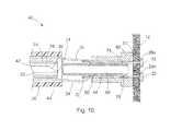
- FIGS. 5 and 9 - 10depict a locked configuration of the suture lock 48 (the unlocked configuration being shown in FIGS. 1-2 and 6 - 8 ).
- the interior passageway 58 of the retaining sleeve 52is sized to receive at least a portion of the plug 50 therein.
- the main body 64 and grip 70are received within the interior passageway 58 of the retaining sleeve 52 .
- the sutures 34are compressed between the grip 70 and the interior surface 56 of the tubular body 54 .
- the plug 50is advanced (i.e. distally) from left to right in FIGS.
- the tapered leading surface 76 of the grip 70allows the plug 50 to be translated distally relative to the sutures 34 and retaining sleeve 52 .
- the sutures 34are maintained in a fixed relationship relative to one another and to the tissue of the bodily wall 12 .
- the sutures 34are generally in tension, due in part to the natural elasticity of the bodily tissue 12 , which generally attempts to pull the sutures 34 distally. Accordingly, while the plug 50 may be advanced through the retaining sleeve 52 and slid alongside the sutures 34 into the locked configuration, the tension on the sutures 34 also exerts a distally directed force on the plug 50 via the grip 70 and its annular edge 74 .
- the suture lock 48is a form of self-motivating locking device that promotes secure fixation of the sutures 34 relative to the tissue 12 .
- the sutures 34may be pulled in the proximal direction to adjust suture tension, suture lock position, and/or perforation closure, even when the suture lock 48 is in the locked configuration.
- the main body 64is sized to at least partially compress the sutures 34 against the interior surface 56 of the tubular body 54 .
- the tapered portion 64 a and reduced diameter portion 64 bprovide an area of limited or no contact with the sutures 34 . These areas may be sized to adjust the level of friction between the sutures 34 and the suture lock 48 , for example based on the type and size of suture material.
- the stop 72abuts against a proximal end surface 55 of the tubular body 54 , thereby limiting the position of the plug 50 within the retaining sleeve 52 .
- the distally facing surface 79 of the stop 72is generally tapered to slightly compress the sutures 34 against the tubular body 54 , while still allowing the sutures 34 to exit the suture lock 48 and be translated in a proximal direction.
- the components of the suture lock 48may be constructed of various materials, such as stainless steel, titanium, nitinol or other metals/alloys, although various ceramics or plastics can also be employed, such as polycarbonates (PC), polyamides including NylonTM, polytetrafluorethylenes (i.e. PTFE and EPTFE), polyethylene ether ketones (PEEK), polyvinylchlorides (PVC), polyimides, polyurethanes, and polyethylenes (high, medium or low density), including multi-layer or single layer constructions with or without reinforcement wires, coils or filaments.
- PCpolycarbonates
- polyamides including NylonTMpolytetrafluorethylenes
- PEEKpolyethylene ether ketones
- PVCpolyvinylchlorides
- polyimidespolyurethanes
- polyurethanespolyurethanes
- polyethyleneshigh, medium or low density
- the plug 50has a length of about 0.259 in
- the main body 64has an outer diameter of about 0.065 in along a center region and an outer diameter of about 0.060 in along a proximal region (which is received within the inner sheath 24 ) and an inner diameter of about 0.045 in defining the interior passageway 68
- the stop 72has an outer diameter of about 0.080 in
- the annular edge 74 defining the grip 70has an outer diameter of about 0.072 in.
- the retaining sleeve 52has a length of about 0.150 in
- the tubular body 54has an outer diameter of about 0.100 in and an inner diameter of about 0.080 in defining the interior passageway 58 . While these dimensions of a currently preferred embodiment have been described, the dimensions may be increased or decreased, scaled up or down, to accommodate differently sized anchors, sutures, needles, sheaths, and bodily walls or tissue structures.
- the inner sheath 24defines an inner sheath lumen 42 which is sized to slidably receive the needle 22 therein.
- the inner sheath 24is sized and positioned to engage or abut the shoulder 82 of the plug 50 .
- the outer sheath 26defines an outer sheath lumen 44 which is sized to slidably receive the inner sheath 24 and the plug 50 therein.
- the outer sheath 26is sized and positioned to engage or abut the shoulder 62 of the retaining sleeve 52 .
- the inner sheath 24has an outer diameter of about 0.068 in and an inner diameter of about 0.045 in such that the proximal portion of the main body 64 of the plug 50 (having an outer diameter of about 0.060 in) is press fit within the distal end of the inner sheath 24 , wherein the inner sheath 24 stretches slightly to hold the plug 50 in place. The plug 50 can then be detached from the inner sheath 24 with a relatively low force.
- the outer sheath 26has an outer diameter of about 0.131 in and an inner diameter of about 0.095 in such that the tubular body 54 of the retaining sleeve 52 (having an outer diameter of about 0.100 in) is press fit within the distal end of the outer sheath 26 , wherein the outer sheath 26 stretches slightly to hold the retaining sleeve 52 in place. The retaining sleeve 52 can then be detached from the outer sheath 26 with a relatively low force.
- the inner diameter of the inner and outer sheaths 24 and 26may be sized larger relative to the plug 50 and retaining sleeve 52 , respectively. In this manner, the plug 50 and the retaining sleeve 52 are received by and maintained within the distal ends of the inner and outer sheaths 24 and 26 , respectively, by an adhesive or any other suitable means known in the art.
- the inner and outer sheaths 24 and 26are preferably formed of a plastic such as polytetrafluorethylene (PTFE), expanded polytetrafluorethylene (EPTFE), polyethylene ether ketone (PEEK), polyvinylchloride (PVC), polycarbonate (PC), polyamide including nylon, polyimide, polyurethane, polyethylene (high, medium or low density), or elastomers such as Santoprene®, including multi-layer or single layer constructions with or without reinforcement wires, coils or filaments.
- the needle 22 , inner and outer sheaths 24 and 26 , and the pusher 25are preferably elongated structures that are flexible, allowing navigation within a patient's body such as during endoscopic or laparoscopic procedures.
- a suitable handle or control mechanismwill be connected to the proximal ends of the needle 22 , inner and outer sheaths 24 and 26 , and the pusher 25 for relative translation of these components by the medical professional, as is known in the art.
- the medical devices 20 and systems 40are also applicable to other tissue anchor placement devices that may be used in open surgery, on external wounds, or that otherwise do not require an elongated medical device to access the targeted tissue.
- the medical device 20may be sized to be used through an accessory channel of an endoscope or alongside an endoscope, or in combination with other devices used in conjunction with endoscopes, for example, endoscopic suction devices or fluid injection devices.
- the medical device 20is operable between at least a delivery configuration, depicted in FIG. 6 , and a deployed configuration, depicted in FIGS. 7-8 .
- the needle 22is substantially contained within the outer sheath lumen 44 so as to protect bodily structures from the sharp distal tip 37 of the needle 22 during introduction of the medical device 20 .
- the needle 22is translated relative to the inner and outer sheaths 24 and 26 such that the needle 22 projects beyond the distal end 27 of the outer sheath 26 .
- the pusher 25is translated relative to the needle 22 such that the distal-most tissue anchor 28 a is urged distally out of the distal tip 37 of the needle 22 .
- the suture 34 connected to the tissue anchor 28 aalso slides distally within the needle slot 38 and exits the needle 22 .
- the needle 22is retracted within the outer sheath lumen 44 , the medical device 20 is repositioned, and the steps of translating the needle 22 relative to the outer sheath 26 and the pusher 25 relative to the needle 22 are repeated for additional tissue anchors.
- the methodincludes providing a medical system having a plurality of tissue anchors and at least one resorbable spacer member positioned in between adjacent tissue anchors, a needle and inner and outer sheaths, and a suture lock, such as the medical system 40 depicted in FIGS. 1 and 6 - 10 .
- the medical system 40is delivered to a position proximate the bodily tissue 12 that has been targeted for placement of the tissue anchors 28 .
- the medical system 40may include a visualization system for assisting in locating the tissue 12 , identifying a target site for deployment of the tissue anchors 28 , and monitoring operation of the medical device 20 and system 40 .
- visualization techniquesmay include catheter-based fiber optic systems, fluoroscopy, ultrasound or the like.
- the needle 22can have markings designed for viewing under fluoroscopy, and the distal end 36 of the needle 22 can have a surface of enhanced ultrasonic reflectivity, such by being roughened, having dimples or other incongruities, or having embedded particles.
- the tissue anchors 28are disposed within the needle lumen 30 at the distal end 36 of the needle 22 and a spacer member 46 is disposed between the tissue anchors 28 . Spaces between the spacer member 46 and the tissue anchors 28 a and 28 b have been shown for clarity, but the spacer member 46 and the tissue anchors 28 a and 28 b would generally be abutting end-to-end within the needle lumen 30 .
- the sutures 34follow a somewhat tortuous path from within the needle lumen 30 , through the needle slot 38 , extending proximally within the outer sheath lumen 44 between the interior surfaces of the retaining sleeve 52 and the outer sheath 26 and the exterior surfaces of the plug and the inner sheath 24 , the sutures 34 effectively being preloaded within the suture lock 48 and extending to a proximal end of the medical device 20 . Accordingly, this tortuous path can be sufficient to retain the tissue anchors 28 within the needle lumen 30 , through frictional engagement of the sutures 34 between the exterior surface of the inner sheath 24 and the interior surface of the outer sheath 26 .
- the medical device 20 and system 40are operated into their deployed configuration, as shown in FIG. 7 .
- the needle 22is deployed through the bodily tissue 12 by translating the needle 22 relative to the inner and outer sheaths 24 and 26 .
- the distal-most tissue anchor 28 ais then deployed from the needle 22 by translating the tissue anchor 28 a relative to the needle 22 so that the tissue anchor 28 a exits the needle lumen 30 .
- FIG. 7shows that the needle 22 is deployed through the bodily tissue 12 by translating the needle 22 relative to the inner and outer sheaths 24 and 26 .
- the distal-most tissue anchor 28 ais then deployed from the needle 22 by translating the tissue anchor 28 a relative to the needle 22 so that the tissue anchor 28 a exits the needle lumen 30 .
- the tissue anchors 28 and the spacer member 46 positioned therebetweenare shown aligned within the needle lumen 30 along the longitudinal axis 10 of the needle lumen 30 and medical device 20 such that the pusher 25 may be slidably received within the inner sheath lumen 24 and used to engage and press on the proximal-most tissue anchor 28 b .
- the pusher 25is advanced distally to press upon the anchoring member 32 of the proximal-most tissue anchor 28 b , which will in turn transmit force through the spacer member 46 and the distal-most tissue anchor 28 a , thus advancing the distal-most tissue anchor 28 a out of the needle lumen 30 .
- the spacer member 46is moved distally to a position slightly past the needle tip 37 , the pusher 25 may be retracted slightly and, due to the adequate clearance between the spacer member 46 and the inner diameter of the needle 22 , as the needle pierces the tissue 12 , the spacer member 46 is easily moved proximally within the needle lumen 30 to ensure that the sharpened needle tip 37 is able to pierce through the tissue 12 for deployment of the remaining tissue anchors 28 .
- the needle 22is retracted back through the bodily tissue 12 by translating the needle 22 proximally, repositioned at a different position about the perforation 14 , and redeployed back through the tissue 12 by translating the needle 22 relative to the inner and outer sheaths 24 and 26 .
- the pusher 25is then further advanced distally to deploy the spacer member 46 and the proximal tissue anchor 28 b , wherein the suture 34 of the tissue anchor 28 b is released from within the needle slot 38 .
- the spacer member 46may be deployed through the tissue 12 with the proximal tissue anchor 28 b , as shown in FIG. 8 .
- the spacer member 46may be deployed within the body prior to passing the needle 22 through the tissue 12 to deploy the proximal anchor 28 b .
- the spacer member 46may be deployed within the gastrointestinal tract, wherein the spacer member 46 passes naturally. Since the spacer member 46 is resorbable, it is inconsequential that it is left within the patient's body. Thus, if the spacer member 46 accidentally falls out of the tip 37 of the needle 22 before being deployed with the proximal tissue anchor 28 b , this is of no consequence.
- the proximal tissue anchor 28 bis still positioned sufficiently proximal within the needle lumen 30 to be deployed appropriately at the repositioned location.
- the spacer member 46may contain antibiotics or other drugs, hormones, or growth factors that facilitate healing of the tissue 12 around the implanted tissue anchors 28 .
- the medical system 40Rather than removing the medical device 20 from the body to reload the needle 22 with additional tissue anchors 28 , the medical system 40 , in accordance with the teachings of the present invention, provides the ability to sequentially deploy multiple tissue anchors, in which tissue anchors and spacer members disposed between adjacent tissue anchors are preloaded within the needle 22 . Accordingly, the longitudinal length of needle slot 38 can be sized to accommodate any number of sutures 34 .
- the methodmay therefore include withdrawing the needle 22 from the bodily tissue 12 by translating the needle 22 proximally, and then repeating the steps of translating the needle 22 through the tissue 12 and deploying another tissue anchor 28 therethrough.
- the needle 22is retracted back through to the proximal side of the bodily tissue 12 and retracted within the inner sheath lumen 42 .
- the needle 22may be removed from within the medical device 20 at this time or it may be removed with the entire medical device 20 after fixation of the sutures 34 relative to the tissue 12 .
- the suture lock 48is engaged to fix the sutures 34 relative to the bodily tissue 12 .
- the system 40again does not require removal from the body, as it includes the over-the-needle suture lock 48 .
- the retaining sleeve 52is fitted onto the distal end 27 of the outer sheath 26 .
- the outer sheath 26may take the form of any sheath or catheter known in the art, but preferably has sufficient strength and rigidity for both longitudinal and rotational force transmission, while still providing flexibility for navigation of a patient's body.
- Exemplary sheathsare sold by Cook Medical, Inc. It will also be recognized that other sheaths or pushing elements may be employed, such as solid wires or wire guides, clamps, graspers and the like. Magnets could likewise be employed to releasably connect the outer sheath 26 to the retaining sleeve 52 .
- the outer sheath lumen 44is sized to receive the tubular body 54 of the retaining sleeve 52 , while a distal end surface 29 of the outer sheath 26 abuts against the shoulder 62 of the retaining sleeve 52 .
- the outer sheath 26 and retaining sleeve 52are loosely press fit such that the retaining sleeve 52 may be readily controlled and positioned using the outer sheath 26 .
- the retaining sleeve 52maintains its connection to the outer sheath 26 during placement of the plug 50 within the retaining sleeve 52 , while at the same time the retaining sleeve 52 is also readily disconnected from the outer sheath 26 at the end of the procedure.
- outer sheath 26 and retaining sleeve 52need not be sized to frictionally engage, as the tensioned sutures 34 and the tissue 12 will generally maintain the position of the retaining sleeve 52 on the outer sheath 52 during placement of the plug 50 , such as is shown in FIGS. 9 and 10 .
- the sutures 34are preloaded or threaded through the interior passageway 58 of the retaining sleeve 52 and through the outer sheath lumen 44 .
- the outer sheath 26is used to distally translate the retaining sleeve 52 over the sutures 34 to a position proximate the tissue 12 and perforation 14 .
- the sutures 34are tensioned in order to draw the perforation 14 closed and press the tissue 12 against the peripheral rim 60 of the retaining sleeve 52 .
- the inner sheath 24is press fit with the plug 50 , although the two structures may simply abut each other for longitudinal translation.
- the inner sheath 24may have a construction similar to the outer sheath 26 or other catheter described above.
- the inner sheath 24includes a distal end 23 sized to abut against the shoulder 82 and receive the main body 64 of the plug 50 , respectively. Accordingly, the inner sheath 24 is connected to the plug 50 and together they are translated distally through the outer sheath lumen 44 . If the needle 22 has not yet been withdrawn from the medical device 20 during securing of the sutures 34 , the inner sheath 24 causes the plug 50 to slide over-the-needle 22 .
- the plug 50is pressed into engagement with the retaining sleeve 52 to fix the sutures 34 therebetween. With the sutures 34 in tension (e.g. by pulling them in a proximal direction), the plug 50 is advanced through the interior passageway 58 of the retaining sleeve 52 , whereby the sutures 34 are compressed between the grip 70 and the interior surface 56 of the retaining sleeve 52 . It can therefore be seen that relative translation of the outer sheath 26 and the inner sheath 24 controls the relative positions of the retaining sleeve 52 and the plug 50 to operate the suture lock 48 between a locked configuration and an unlocked configuration.
- the leading surface 76 of the grip 70is slid along the sutures 34 as the plug 50 is distally advanced through the interior passageway 56 .
- the main body 64also engages the sutures 34 and at least partially compresses them against the interior surface 56 of the retaining sleeve 52 .
- the annular shape of the grip 70allows the sutures 34 to be positioned anywhere around the outer periphery of the grip 70 and plug 50 . Distal movement of the plug 50 is eventually limited by the stop 72 , and namely the distally facing surface 79 of the stop 72 abutting against the proximal end surface 55 of the retaining sleeve 52 .
- the tension on the sutures 34grips into the annular edge 74 of the grip 70 , and serves to promote movement of the plug 50 in the distal direction, as well as resist proximal movement and unlocking of the suture lock 48 .
- the grip 70When in the locked configuration (and when partially locked such as when the plug 50 is partially placed within the retaining sleeve 52 but not fully seated), the grip 70 is structured to permit further translation of the sutures 34 proximally, i.e. away from the tissue 12 , and prevent translation of the sutures 34 distally, i.e. towards the tissue 12 . Further, the sutures 34 may be individually pulled or tensioned in order to orient the suture lock 48 relative to the bodily tissue 12 and perforation 14 , even when the sutures 34 are compressed by the plug 50 and retaining sleeve 52 , such as when the suture lock 48 is in the locked configuration. As such, tension on the sutures 34 may be modified to adjust how the perforation 14 is closed. This represents a marked improvement over existing suture locks, which typically are permanently fixed in position along the sutures such that adjustment during and after the locking procedure, i.e. in partially locked and finally locked configurations, is not possible.
- the tension on the sutures 34results in a force being transmitted through the sutures 34 to the grip 70 biasing it in the distal direction.
- the retaining sleeve 52 and plug 50are interconnected through their respective frictional engagement with the sutures 34 and compression thereof.
- the entire medical device 20may be removed from the patient at once, the inner and outer sheaths 24 and 26 being easily removed from the retaining sleeve 52 and the plug 50 , respectively.
- the inner sheath 24 and needle 22may be removed first and the outer sheath 26 removed separately.
- the sutures 34may be trimmed as necessary with endoscopic scissors and the like.
- the sutures 34may be cut, or the outer sheath 26 may be used to hold the retaining sleeve 52 while the plug 50 is grasped (such as with a snare, forceps, or similar device) and physically withdrawn against the friction and tension of the sutures 34 .
- the present inventionprovides a medical system and method capable of delivering multiple tissue anchors in a controlled manner, as well as locking the anchors together (e.g., to close a perforation) without needing to withdraw and introduce the system (or multiple medical devices) any number of times, thereby saving time and improving efficiency. Since the sutures connected to the tissue anchors are preloaded within the over-the-needle suture lock, one medical system is provided for both the delivery of multiple tissue anchors and the fixation of their sutures.
- the medical systemis simple and reliable in use, provides complete perforation closure, and is adaptable to a variety of suture fixation and perforation closure applications.
- any number of suture strandsmay be employed and the relative sizes of the plug and retaining sleeve may be adjusted based on suture size, perforation size and the like.
- the interconnection of the plug and retaining sleeveis such that the suture lock is self-motivated and biased towards a locked configuration, thereby assisting and promoting complete perforation closure as well as control over the position of the suture lock relative to the tissue being sutured through adjustment of the suture strands even when they are compressed. Further description of the interconnection between the plug and retaining sleeve may be found in co-pending U.S. application Ser. No. 12/125,525, the entire contents of which are incorporated by reference herein.
- Adjustment of individual suture tension and location of the suture lockare also possible during and after placement of the suture lock.
- the inner and outer sheathsprovide a simple system for deployment of multiple tissue anchors that can be traversed through the body of a patient to even the most remote locations.
Landscapes
- Health & Medical Sciences (AREA)
- Surgery (AREA)
- Life Sciences & Earth Sciences (AREA)
- Medical Informatics (AREA)
- Nuclear Medicine, Radiotherapy & Molecular Imaging (AREA)
- Engineering & Computer Science (AREA)
- Biomedical Technology (AREA)
- Heart & Thoracic Surgery (AREA)
- Molecular Biology (AREA)
- Animal Behavior & Ethology (AREA)
- General Health & Medical Sciences (AREA)
- Public Health (AREA)
- Veterinary Medicine (AREA)
- Rheumatology (AREA)
- Cardiology (AREA)
- Surgical Instruments (AREA)
Abstract
Description
L=LT(nT)+LS(nS)−½LT,
Claims (22)
Priority Applications (1)
| Application Number | Priority Date | Filing Date | Title |
|---|---|---|---|
| US12/753,111US8382776B2 (en) | 2009-04-03 | 2010-04-02 | Medical devices, systems and methods for rapid deployment and fixation of tissue anchors |
Applications Claiming Priority (2)
| Application Number | Priority Date | Filing Date | Title |
|---|---|---|---|
| US16636109P | 2009-04-03 | 2009-04-03 | |
| US12/753,111US8382776B2 (en) | 2009-04-03 | 2010-04-02 | Medical devices, systems and methods for rapid deployment and fixation of tissue anchors |
Publications (2)
| Publication Number | Publication Date |
|---|---|
| US20100256679A1 US20100256679A1 (en) | 2010-10-07 |
| US8382776B2true US8382776B2 (en) | 2013-02-26 |
Family
ID=42211655
Family Applications (1)
| Application Number | Title | Priority Date | Filing Date |
|---|---|---|---|
| US12/753,111Active2031-02-11US8382776B2 (en) | 2009-04-03 | 2010-04-02 | Medical devices, systems and methods for rapid deployment and fixation of tissue anchors |
Country Status (6)
| Country | Link |
|---|---|
| US (1) | US8382776B2 (en) |
| EP (1) | EP2413809B1 (en) |
| JP (1) | JP5619138B2 (en) |
| AU (1) | AU2010232485B2 (en) |
| CA (1) | CA2757494C (en) |
| WO (1) | WO2010115113A1 (en) |
Cited By (8)
| Publication number | Priority date | Publication date | Assignee | Title |
|---|---|---|---|---|
| US20110178534A1 (en)* | 2010-01-20 | 2011-07-21 | Whitman Michael P | Tissue repair implant and delivery device and method |
| US20110218191A1 (en)* | 2010-03-03 | 2011-09-08 | Boehringer Ingelheim Vetmedica Gmbh | Use of meloxicam for the long term-treatment of kidney disorders in cats |
| US9980708B2 (en) | 2010-01-20 | 2018-05-29 | Micro Interventional Devices, Inc. | Tissue closure device and method |
| US10058314B2 (en) | 2010-01-20 | 2018-08-28 | Micro Interventional Devices, Inc. | Tissue closure device and method |
| US10743854B2 (en) | 2010-01-20 | 2020-08-18 | Micro Interventional Devices, Inc. | Tissue closure device and method |
| US10959840B2 (en) | 2010-01-20 | 2021-03-30 | Micro Interventional Devices, Inc. | Systems and methods for affixing a prosthesis to tissue |
| US11311284B2 (en) | 2019-03-06 | 2022-04-26 | Speed Clip Solutions, LLC | Suture tensioning and securement device, system, and methods |
| US12383246B2 (en) | 2020-10-12 | 2025-08-12 | Abbott Cardiovascular Systems, Inc. | Vessel closure device with improved safety and tract hemostasis |
Families Citing this family (20)
| Publication number | Priority date | Publication date | Assignee | Title |
|---|---|---|---|---|
| EP1909655A2 (en) | 2005-06-20 | 2008-04-16 | Sutura, Inc. | Method and apparatus for applying a knot to a suture |
| US8246636B2 (en) | 2007-03-29 | 2012-08-21 | Nobles Medical Technologies, Inc. | Suturing devices and methods for closing a patent foramen ovale |
| WO2010042402A1 (en)* | 2008-10-06 | 2010-04-15 | Wilson-Cook Medical, Inc. | Endcap for safely deploying tissue anchors |
| US8377095B2 (en) | 2008-12-05 | 2013-02-19 | Cook Medical Technologies, LLC | Tissue anchors for purse-string closure of perforations |
| CA2757554A1 (en) | 2009-04-03 | 2010-10-07 | Cook Medical Technologies Llc | Tissue anchors and medical devices for rapid deployment of tissue anchors |
| US20120053619A1 (en)* | 2010-08-31 | 2012-03-01 | Boston Scientific Scimed, Inc. | Hemostatic compositions and methods of making and using same |
| CN103917200B (en)* | 2011-09-13 | 2016-03-30 | 恩克斯特拉公司 | Systems and methods for prostate treatment |
| WO2014129554A1 (en)* | 2013-02-22 | 2014-08-28 | 住友ベークライト株式会社 | Repeating-type organ-fastening tool |
| WO2015002815A1 (en) | 2013-07-02 | 2015-01-08 | Med-Venture Investments, Llc | Suturing devices and methods for suturing an anatomic structure |
| JP6469109B2 (en) | 2013-12-06 | 2019-02-13 | メッド − ベンチャー インベストメンツ、エルエルシー | Suture method and apparatus |
| US9757117B2 (en) | 2014-02-13 | 2017-09-12 | Medtronic Vascular, Inc. | Method and apparatus for forming a suture connector in situ |
| US9629620B2 (en)* | 2014-02-13 | 2017-04-25 | Medtronic Vascular, Inc. | Method and apparatus for forming a suture connector in situ |
| EP3229703B1 (en)* | 2014-12-10 | 2024-08-28 | Edwards Lifesciences AG | Multiple-firing securing device |
| US11026830B2 (en) | 2014-12-12 | 2021-06-08 | Koninklijke Philips N.V. | Tongue advancer system for use in a tongue manipulation system |
| CN206390950U (en) | 2015-07-07 | 2017-08-11 | 莫尔研究应用有限公司 | Equipment for two organizing segments to be stitched together |
| CN110248623B (en)* | 2016-11-29 | 2021-12-21 | 埃斯卡拉医疗公司 | Anchor delivery systems and methods |
| EP4115818A3 (en) | 2017-06-19 | 2023-04-05 | Heartstitch, Inc. | Suturing systems and methods for suturing body tissue |
| WO2019051379A1 (en) | 2017-09-11 | 2019-03-14 | Heartstitch, Inc. | Methods and devices for papillary suturing |
| WO2019152845A1 (en)* | 2018-02-01 | 2019-08-08 | Transluminal Technologies, Llc | Trans-radial closure device, deployment apparatus, and method of deploying a trans-radial closure device |
| EP4255316A4 (en)* | 2020-12-03 | 2024-12-04 | Xdot Medical Inc. | PERCUTANEOUS VASCULAR ACCESS SITE MANAGEMENT SYSTEM |
Citations (170)
| Publication number | Priority date | Publication date | Assignee | Title |
|---|---|---|---|---|
| US2199025A (en) | 1936-06-08 | 1940-04-30 | Carl E Conn | Means and method of closing surgical incisions |
| US3556079A (en) | 1967-05-16 | 1971-01-19 | Haruo Omizo | Method of puncturing a medical instrument under guidance of ultrasound |
| US3664345A (en) | 1970-07-06 | 1972-05-23 | Clyde Harwell Dabbs | Surgical buttons |
| US3766610A (en) | 1971-06-29 | 1973-10-23 | A Thorsbakken | Wedge locking device |
| US3952377A (en) | 1974-01-25 | 1976-04-27 | Juan Coll Morell | Conical wedges for gripping multi-ply rope or cable |
| US4059333A (en) | 1977-01-05 | 1977-11-22 | Amp Incorporated | Electrical connector |
| US4235238A (en) | 1978-05-11 | 1980-11-25 | Olympus Optical Co., Ltd. | Apparatus for suturing coeliac tissues |
| US4532926A (en) | 1983-06-20 | 1985-08-06 | Ethicon, Inc. | Two-piece tissue fastener with ratchet leg staple and sealable latching receiver |
| US4604094A (en) | 1984-09-06 | 1986-08-05 | The United States Of America As Represented By The Secretary Of The Department Of Health And Human Services | Toposcopic catheter and method of fabrication |
| US4669473A (en) | 1985-09-06 | 1987-06-02 | Acufex Microsurgical, Inc. | Surgical fastener |
| US4719671A (en) | 1984-11-08 | 1988-01-19 | Canon Kabushiki Kaisha | Strap connector |
| US5123914A (en) | 1986-05-19 | 1992-06-23 | Cook Incorporated | Visceral anchor for visceral wall mobilization |
| US5203787A (en) | 1990-11-19 | 1993-04-20 | Biomet, Inc. | Suture retaining arrangement |
| US5254126A (en) | 1992-06-24 | 1993-10-19 | Ethicon, Inc. | Endoscopic suture punch |
| US5258015A (en) | 1991-05-03 | 1993-11-02 | American Cyanamid Company | Locking filament caps |
| US5282832A (en) | 1992-10-09 | 1994-02-01 | United States Surgical Corporation | Suture clip |
| US5333624A (en) | 1992-02-24 | 1994-08-02 | United States Surgical Corporation | Surgical attaching apparatus |
| US5366480A (en) | 1990-12-24 | 1994-11-22 | American Cyanamid Company | Synthetic elastomeric buttressing pledget |
| US5383882A (en) | 1992-08-28 | 1995-01-24 | Ethicon, Inc. | Ligature and ligature applying endoscopic instrument |
| US5417691A (en) | 1982-05-20 | 1995-05-23 | Hayhurst; John O. | Apparatus and method for manipulating and anchoring tissue |
| US5423860A (en) | 1993-05-28 | 1995-06-13 | American Cyanamid Company | Protective carrier for suture anchor |
| US5464427A (en) | 1994-10-04 | 1995-11-07 | Synthes (U.S.A.) | Expanding suture anchor |
| US5486197A (en) | 1994-03-24 | 1996-01-23 | Ethicon, Inc. | Two-piece suture anchor with barbs |
| US5520700A (en) | 1992-11-13 | 1996-05-28 | Technion Research & Development Foundation, Ltd. | Stapler device particularly useful in medical suturing |
| US5527343A (en) | 1993-05-14 | 1996-06-18 | Bonutti; Peter M. | Suture anchor |
| US5531763A (en) | 1994-10-07 | 1996-07-02 | United States Surgical Corporation | Suture cinching apparatus |
| US5554183A (en) | 1994-01-19 | 1996-09-10 | Nazari; Stefano | Vascular prosthesis for the substitution or internal lining of blood vessels of medium or large diameter and device for its application |
| US5584835A (en) | 1993-10-18 | 1996-12-17 | Greenfield; Jon B. | Soft tissue to bone fixation device and method |
| US5630824A (en) | 1994-06-01 | 1997-05-20 | Innovasive Devices, Inc. | Suture attachment device |
| EP0774237A2 (en) | 1995-10-20 | 1997-05-21 | United States Surgical Corporation | Apparatus and method for vascular hole closure |
| US5662683A (en) | 1995-08-22 | 1997-09-02 | Ortho Helix Limited | Open helical organic tissue anchor and method of facilitating healing |
| US5690656A (en) | 1995-06-27 | 1997-11-25 | Cook Incorporated | Method and apparatus for creating abdominal visceral anastomoses |
| US5693060A (en) | 1992-11-17 | 1997-12-02 | Smith & Nephew, Inc. | Suture securing device and method |
| US5810848A (en) | 1996-08-21 | 1998-09-22 | Hayhurst; John O. | Suturing system |
| US5865791A (en) | 1995-06-07 | 1999-02-02 | E.P. Technologies Inc. | Atrial appendage stasis reduction procedure and devices |
| US5891159A (en) | 1997-05-02 | 1999-04-06 | Cardiothoratic Systems, Inc. | Automatic purse string suture device |
| US5931844A (en) | 1998-03-31 | 1999-08-03 | Smith & Nephew, Inc. | Surgical drive tool |
| US5948000A (en) | 1996-10-03 | 1999-09-07 | United States Surgical Corporation | System for suture anchor placement |
| US5968078A (en) | 1995-08-25 | 1999-10-19 | Ultraortho, Inc. | Stabilizer for human joints |
| US6086608A (en) | 1996-02-22 | 2000-07-11 | Smith & Nephew, Inc. | Suture collet |
| US6110183A (en) | 1998-12-22 | 2000-08-29 | Cook Incorporated | Suture anchor device |
| US6200329B1 (en) | 1998-08-31 | 2001-03-13 | Smith & Nephew, Inc. | Suture collet |
| US6290674B1 (en) | 1999-09-20 | 2001-09-18 | Appriva Medical, Inc. | Method and apparatus for closing intracardiac septal defects |
| US6293961B2 (en) | 1998-12-30 | 2001-09-25 | Ethicon, Inc. | Suture locking device |
| EP0643945B1 (en) | 1993-08-20 | 2002-03-20 | United States Surgical Corporation | Apparatus for applying and adjusting an anchoring device |
| US6423087B1 (en) | 1999-08-04 | 2002-07-23 | Olympus Optical Co., Ltd. | Internal organ walls joining instrument for an endoscope |
| US6482178B1 (en) | 1999-05-21 | 2002-11-19 | Cook Urological Incorporated | Localization device with anchoring barbs |
| US6491707B2 (en) | 1997-06-28 | 2002-12-10 | Transvascular, Inc. | Transluminal methods and devices for closing, forming attachments to, and/or forming anastomotic junctions in, luminal anatomical structures |
| US6551333B2 (en) | 2000-10-19 | 2003-04-22 | Ethicon Endo-Surgery, Inc. | Method for attaching hernia mesh |
| US6572629B2 (en) | 2000-08-17 | 2003-06-03 | Johns Hopkins University | Gastric reduction endoscopy |
| US6592559B1 (en) | 1998-12-09 | 2003-07-15 | Cook Incorporated | Hollow, curved, superlastic medical needle |
| US6699263B2 (en) | 2002-04-05 | 2004-03-02 | Cook Incorporated | Sliding suture anchor |
| US20040147958A1 (en) | 2002-12-11 | 2004-07-29 | Usgi Medical | Apparatus and methods for forming and securing gastrointestinal tissue folds |
| US20040153074A1 (en) | 2003-02-05 | 2004-08-05 | Bojarski Raymond A. | Tissue anchor and insertion tool |
| US20040167546A1 (en) | 2002-12-11 | 2004-08-26 | Vahid Saadat | Methods for reduction of a gastric lumen |
| US20040186514A1 (en) | 2001-05-18 | 2004-09-23 | Swain Christopher Paul | Flexible device for transfixing and joining tissue |
| US20040220596A1 (en) | 2003-02-04 | 2004-11-04 | Frazier Andrew G.C. | Patent foramen ovale closure system |
| US6840953B2 (en) | 2000-12-22 | 2005-01-11 | United States Surgical Corporation | Suture screw |
| US20050113851A1 (en) | 2002-05-17 | 2005-05-26 | Swain Christopher P. | Device for transfixing and joining tissue |
| US20050143762A1 (en) | 2003-09-15 | 2005-06-30 | Paraschac Joseph F. | Suture locking device and methods |
| US20050197594A1 (en) | 1998-09-01 | 2005-09-08 | Senorx, Inc. | Tissue accessing and anchoring device and method |
| US20050251165A1 (en) | 2004-05-07 | 2005-11-10 | Usgi Medical Inc. | Tissue manipulation and securement system |
| US20050251166A1 (en) | 2004-05-07 | 2005-11-10 | Usgi Medical Inc. | Tissue manipulation and securement system |
| US6966916B2 (en) | 2002-09-26 | 2005-11-22 | Kumar Sarbjeet S | Device and method for surgical repair of abdominal wall hernias |
| EP1598018A1 (en) | 2004-05-20 | 2005-11-23 | Olympus Corporation | Treatment system for living tissues |
| US20050277945A1 (en) | 2004-06-14 | 2005-12-15 | Usgi Medical Inc. | Apparatus and methods for performing transluminal gastrointestinal procedures |
| US20050277957A1 (en) | 2004-05-14 | 2005-12-15 | Kuhns Jesse J | Devices and methods of locking and cutting a suture in a medical procedure |
| US20050277981A1 (en) | 2004-06-09 | 2005-12-15 | Usgi Medical Inc. | Apparatus and methods for optimizing anchoring force |
| US20060004409A1 (en) | 2004-05-14 | 2006-01-05 | Nobis Rudolph H | Devices for locking and/or cutting a suture |
| US20060002852A1 (en) | 2004-07-01 | 2006-01-05 | Yale University | Targeted and high density drug loaded polymeric materials |
| US20060015006A1 (en) | 2004-06-01 | 2006-01-19 | Laurence Bernard H | System and method for accessing a body cavity |
| US20060015125A1 (en) | 2004-05-07 | 2006-01-19 | Paul Swain | Devices and methods for gastric surgery |
| US20060020274A1 (en) | 2004-07-23 | 2006-01-26 | Usgi Medical Inc. | Manipulatable grasping needle |
| US20060020277A1 (en) | 2004-07-20 | 2006-01-26 | Gostout Christopher J | Gastric reshaping devices and methods |
| US7033379B2 (en) | 2001-06-08 | 2006-04-25 | Incisive Surgical, Inc. | Suture lock having non-through bore capture zone |
| WO2006044837A2 (en) | 2004-10-18 | 2006-04-27 | Temple University Of The Commonwealth System Of Higher Education | Apparatus and method of endoscopic suturing |
| US7087073B2 (en) | 2000-05-03 | 2006-08-08 | Marctec, Llc | Method of securing body tissue |
| US20060190016A1 (en) | 2002-07-11 | 2006-08-24 | Olympus Corporation | Endoscopic suture apparatus |
| US20060206063A1 (en) | 2002-11-01 | 2006-09-14 | Jonathan Kagan | Attachment system for transmural attachment at the gastroesophageal junction |
| US20060217762A1 (en) | 2004-06-09 | 2006-09-28 | Usgi Medical, Inc. | Compressible tissue anchor assemblies |
| US20060235447A1 (en) | 1998-05-21 | 2006-10-19 | Walshe Christopher J | Tissue anchor system |
| US20060237022A1 (en) | 2005-04-26 | 2006-10-26 | Usgi Medical Inc. | Transgastric abdominal access |
| US20060237023A1 (en) | 2005-04-26 | 2006-10-26 | Usgi Medical Inc. | Transgastric tubal ligation |
| US20060241691A1 (en) | 2005-04-12 | 2006-10-26 | Wilk Patent, Llc | Medical treatment method and device utilizing magnetic elements |
| US20060253144A1 (en) | 2004-01-08 | 2006-11-09 | Olympus Corporation | Anastomosis instrument and method of excising wall portion of hollow organ within a living body |
| US20060271073A1 (en) | 2005-05-26 | 2006-11-30 | Usgi Medical Inc. | Methods and apparatus for securing and deploying tissue anchors |
| US20060271101A1 (en) | 2005-05-26 | 2006-11-30 | Usgi Medical Inc. | Methods and apparatus for securing and deploying tissue anchors |
| US7147652B2 (en) | 1997-08-01 | 2006-12-12 | Bonutti Ip, Llc | Method and apparatus for securing a suture |
| US20060286664A1 (en) | 1999-11-22 | 2006-12-21 | Cytograft Tissue Engineering, Inc. | Bioreactor for the Manufacture of Tissue Engineered Blood Vessels |
| US20070010835A1 (en) | 2003-08-22 | 2007-01-11 | Tom Breton | Eversion apparatus and methods |
| US20070027476A1 (en) | 2005-02-07 | 2007-02-01 | Regen Biologics, Inc. | System and method for all-inside suture fixation for implant attachment and soft tissue repair |
| US20070049970A1 (en) | 2005-09-01 | 2007-03-01 | Ovalis Inc. | Suture-based systems and methods for treating septal defects |
| US20070093858A1 (en) | 2000-03-03 | 2007-04-26 | C. R. Bard, Inc. | Suture clips, delivery devices and methods |
| US20070100375A1 (en) | 2003-06-06 | 2007-05-03 | Olympus Corporation | Suturing instrument |
| US7217279B2 (en) | 2003-11-14 | 2007-05-15 | Ethicon, Inc. | Suture loop anchor |
| US20070112362A1 (en) | 2005-11-14 | 2007-05-17 | Olympus Medical Systems Corp. | Perforation suturing method |
| US20070123840A1 (en) | 2005-10-18 | 2007-05-31 | Usgi Medical, Inc. | Instrument assisted abdominal access |
| US20070162052A1 (en) | 2006-01-06 | 2007-07-12 | Olympus Medical Systems Corp. | Loading device for indwelling implement |
| US20070208360A1 (en) | 2004-02-13 | 2007-09-06 | Demarais Denise M | Methods and devices for reducing hollow organ volume |
| US20070213702A1 (en) | 2006-03-08 | 2007-09-13 | Olympus Medical Systems Corp. | Medical procedure carried out via a natural opening |
| US20070219411A1 (en) | 2006-01-13 | 2007-09-20 | Olympus Medical Systems Corp. | Overtube and endoscopic treatment system |
| US20070255296A1 (en) | 2006-04-26 | 2007-11-01 | Lsi Solutions, Inc. | Medical instrument to place a pursestring suture, open a hole and pass a guidewire |
| US20070270889A1 (en) | 2006-05-19 | 2007-11-22 | Conlon Sean P | Combination knotting element and suture anchor applicator |
| US20070270752A1 (en) | 2006-05-18 | 2007-11-22 | Labombard Denis | Multifunctional instrument introducer |
| US7300451B2 (en) | 2003-12-22 | 2007-11-27 | Ethicon, Inc. | Suture anchoring device |
| US20070276416A1 (en) | 2000-12-07 | 2007-11-29 | Integrated Vascular Systems, Inc. | Closure device and methods for making and using them |
| US7316706B2 (en) | 2003-06-20 | 2008-01-08 | Medtronic Vascular, Inc. | Tensioning device, system, and method for treating mitral valve regurgitation |
| US20080009888A1 (en) | 2006-07-07 | 2008-01-10 | Usgi Medical, Inc. | Low profile tissue anchors, tissue anchor systems, and methods for their delivery and use |
| US7326231B2 (en) | 2000-02-09 | 2008-02-05 | Anson Medical Limited | Device for the repair of arteries |
| US7326221B2 (en) | 2004-04-07 | 2008-02-05 | Olympus Corporation | Ligature and suture device for medical application, and ligaturing and suturing method for medical application |
| US7335221B2 (en) | 2002-04-12 | 2008-02-26 | Ethicon, Inc. | Suture anchoring and tensioning device and method for using same |
| US20080058865A1 (en) | 2006-08-21 | 2008-03-06 | Wilk Peter J | Surgical closure device and associated method |
| US7371244B2 (en) | 2003-08-25 | 2008-05-13 | Ethicon, Inc. | Deployment apparatus for suture anchoring device |
| US7390329B2 (en) | 2004-05-07 | 2008-06-24 | Usgi Medical, Inc. | Methods for grasping and cinching tissue anchors |
| US20080154290A1 (en) | 2002-10-04 | 2008-06-26 | Steve Golden | Anastomosis apparatus and methods |
| EP1938760A1 (en) | 2005-09-28 | 2008-07-02 | Olympus Medical Systems Corp. | Suturing device |
| US20080172088A1 (en) | 2007-01-12 | 2008-07-17 | Ethicon Endo-Surgery, Inc. | Adjustable Compression Staple and Method for Stapling with Adjustable Compression |
| US20080185752A1 (en) | 2006-07-28 | 2008-08-07 | Ethicon, Inc. | Apparatus and method for making suture packages |
| US20080200930A1 (en) | 2004-05-07 | 2008-08-21 | Usgi Medical, Inc. | Apparatus for manipulating and securing tissue |
| US20080208161A1 (en) | 2007-02-26 | 2008-08-28 | Olympus Medical Systems Corp. | Application of procedure through natural orifice |
| US20080208219A1 (en) | 2007-02-27 | 2008-08-28 | Olympus Medical Systems Corporation | Endoscopic treatment instrument |
| US20080208218A1 (en) | 2007-02-27 | 2008-08-28 | Olympus Medical Systems Corp. | Suture tool |
| US20080208214A1 (en) | 2007-02-26 | 2008-08-28 | Olympus Medical Systems Corp. | Applicator and tissue fastening method through natural orifice |
| US20080208220A1 (en) | 2007-02-27 | 2008-08-28 | Olympus Medical Systems Corporation | Suture instrument |
| US20080208251A1 (en) | 2005-12-29 | 2008-08-28 | Ethicon, Inc. | Device for attaching, relocating and reinforcing tissue and methods of using same |
| US20080221619A1 (en) | 2007-03-08 | 2008-09-11 | Spivey James T | Surgical suture anchors and deployment device |
| US20080228202A1 (en) | 2007-03-16 | 2008-09-18 | Ethicon Endo-Surgery, Inc. | Endoscopic tissue approximation system |
| US20080228203A1 (en) | 2007-03-15 | 2008-09-18 | Minos Medical | System and method for translumenal closure in natural orifice surgery |
| US20080255423A1 (en) | 2006-01-13 | 2008-10-16 | Olympus Medical Systems Corp. | Medical device |
| US20080255422A1 (en) | 2006-01-13 | 2008-10-16 | Olympus Medical Systems Corp. | Medical device |
| US20080255427A1 (en) | 2007-01-26 | 2008-10-16 | Olympus Medical Systems Corp. | ligation apparatus and a ligation member |
| US20080262525A1 (en) | 2007-04-17 | 2008-10-23 | Usgi Medical, Inc. | Tissue penetration and grasping apparatus |
| US20080269566A1 (en) | 2007-04-30 | 2008-10-30 | Ethicon Endo-Surgery, Inc. | Endoscopic device |
| US20080275297A1 (en) | 2007-05-01 | 2008-11-06 | Ethicon Endo-Surgery, Inc. | Endoscopic guide device |
| US20080300629A1 (en) | 2007-05-31 | 2008-12-04 | Wilson-Cook Medical Inc. | Suture lock |
| US20080300627A1 (en) | 2007-05-30 | 2008-12-04 | Ethicon Endo-Surgery, Inc. | Surgical Instrument |
| US20080296344A1 (en) | 2007-05-30 | 2008-12-04 | Ethicon Endo-Surgery, Inc. | Surgical Instrument |
| US20080300547A1 (en) | 2007-06-01 | 2008-12-04 | Bakos Gregory J | Integrated securement and closure apparatus |
| US20080319257A1 (en) | 2006-07-05 | 2008-12-25 | Olympus Medical Systems Corp. | Living body wall fixing tool used in endoscope |
| US20090005800A1 (en) | 2007-06-29 | 2009-01-01 | Ethicon Endo-Surgery, Inc. | Insertion device and method of use |
| US20090069847A1 (en) | 2007-08-17 | 2009-03-12 | Wilson-Cook Medical Inc. | Suture lock |
| US20090076527A1 (en) | 2004-05-11 | 2009-03-19 | Olympus Corporation | Surgical instrument |
| EP2042105A2 (en) | 2007-09-28 | 2009-04-01 | Olympus Medical Systems Corporation | Suturing device |
| US20090088797A1 (en) | 2007-09-28 | 2009-04-02 | Ethicon, Inc. | Surgical anchor device |
| US20090125038A1 (en) | 2007-11-05 | 2009-05-14 | Usgi Medical, Inc. | Endoscopic ligation |
| US20090204147A1 (en) | 2007-12-05 | 2009-08-13 | Rahmani Emad Y | Methods and apparatuses for delivering achoring devices into body passage walls |
| US7601159B2 (en) | 2004-05-07 | 2009-10-13 | Usgi Medical, Inc. | Interlocking tissue anchor apparatus and methods |
| US7618426B2 (en) | 2002-12-11 | 2009-11-17 | Usgi Medical, Inc. | Apparatus and methods for forming gastrointestinal tissue approximations |
| US7622068B2 (en) | 2005-06-23 | 2009-11-24 | Ethicon, Inc. | Tissue repair device and fabrication thereof |
| US20090299406A1 (en) | 2008-05-30 | 2009-12-03 | Ethicon Endo-Surgery, Inc. | Multifunction surgical device |
| US20090326578A1 (en) | 2004-09-30 | 2009-12-31 | Usgi Medical, Inc. | Interlocking tissue anchor apparatus and methods |
| US20090326561A1 (en) | 2008-06-27 | 2009-12-31 | Ethicon Endo-Surgery, Inc. | Surgical suture arrangement |
| US7641836B2 (en) | 2005-06-23 | 2010-01-05 | Ethicon, Inc. | Tissue repair device and fabrication thereof |
| US20100042144A1 (en) | 2008-08-12 | 2010-02-18 | Steven Bennett | Medical Device for Wound Closure and Method of Use |
| US20100049213A1 (en) | 2007-10-19 | 2010-02-25 | Guided Delivery Systems Inc. | Devices and methods for termination |
| US7674275B2 (en) | 2006-10-05 | 2010-03-09 | Ethicon Endo-Surgery, Inc. | Suture anchor |
| US20100076488A1 (en) | 2008-09-25 | 2010-03-25 | Ethicon Endo-Surgery, Inc. | Methods and devices for delivering and applying multiple suture anchors |
| US20100076462A1 (en) | 2008-09-25 | 2010-03-25 | Ethicon Endo-Surgery, Inc. | Methods and devices for delivering and applying suture anchors |
| US7695493B2 (en) | 2004-06-09 | 2010-04-13 | Usgi Medical, Inc. | System for optimizing anchoring force |
| US20100094341A1 (en) | 2007-01-16 | 2010-04-15 | Board Of Regents, The University Of Texas System | Needle-electrode and tissue anchor system |
| US7704264B2 (en) | 1999-06-25 | 2010-04-27 | Usgi Medical, Inc. | Apparatus and methods for forming and securing gastrointestinal tissue folds |
| US20100106166A1 (en) | 2008-10-29 | 2010-04-29 | Ethicon Endo-Surgery, Inc. | Methods and devices for applying mulitple suture anchors |
| US20100113873A1 (en) | 2008-11-06 | 2010-05-06 | Takayuki Suzuki | Suturing device and suturing system |
| US20100121349A1 (en) | 2008-10-10 | 2010-05-13 | Meier Stephen C | Termination devices and related methods |
| US7736379B2 (en) | 2004-06-09 | 2010-06-15 | Usgi Medical, Inc. | Compressible tissue anchor assemblies |
| US7736378B2 (en) | 2004-05-07 | 2010-06-15 | Usgi Medical, Inc. | Apparatus and methods for positioning and securing anchors |
| US20100174296A1 (en) | 2009-01-07 | 2010-07-08 | Ethicon Endo-Surgery, Inc. | Suturing devices and methods |
| US20100198192A1 (en) | 2009-01-20 | 2010-08-05 | Eugene Serina | Anchor deployment devices and related methods |
| US7815659B2 (en) | 2005-11-15 | 2010-10-19 | Ethicon Endo-Surgery, Inc. | Suture anchor applicator |
| US20100268253A1 (en) | 2009-04-21 | 2010-10-21 | Sarah Ahlberg | System and method for closure of an internal opening in tissue, such as a trans-apical access opening |
Family Cites Families (1)
| Publication number | Priority date | Publication date | Assignee | Title |
|---|---|---|---|---|
| US9155532B2 (en)* | 2007-05-25 | 2015-10-13 | Cook Medical Technologies Llc | Medical devices, systems and methods for closing perforations |
- 2010
- 2010-04-02EPEP10712286.3Apatent/EP2413809B1/enactiveActive
- 2010-04-02USUS12/753,111patent/US8382776B2/enactiveActive
- 2010-04-02JPJP2012503743Apatent/JP5619138B2/enactiveActive
- 2010-04-02AUAU2010232485Apatent/AU2010232485B2/enactiveActive
- 2010-04-02WOPCT/US2010/029798patent/WO2010115113A1/enactiveApplication Filing
- 2010-04-02CACA2757494Apatent/CA2757494C/enactiveActive
Patent Citations (209)
| Publication number | Priority date | Publication date | Assignee | Title |
|---|---|---|---|---|
| US2199025A (en) | 1936-06-08 | 1940-04-30 | Carl E Conn | Means and method of closing surgical incisions |
| US3556079A (en) | 1967-05-16 | 1971-01-19 | Haruo Omizo | Method of puncturing a medical instrument under guidance of ultrasound |
| US3664345A (en) | 1970-07-06 | 1972-05-23 | Clyde Harwell Dabbs | Surgical buttons |
| US3766610A (en) | 1971-06-29 | 1973-10-23 | A Thorsbakken | Wedge locking device |
| US3952377A (en) | 1974-01-25 | 1976-04-27 | Juan Coll Morell | Conical wedges for gripping multi-ply rope or cable |
| US4059333A (en) | 1977-01-05 | 1977-11-22 | Amp Incorporated | Electrical connector |
| US4235238A (en) | 1978-05-11 | 1980-11-25 | Olympus Optical Co., Ltd. | Apparatus for suturing coeliac tissues |
| US5417691A (en) | 1982-05-20 | 1995-05-23 | Hayhurst; John O. | Apparatus and method for manipulating and anchoring tissue |
| US4532926A (en) | 1983-06-20 | 1985-08-06 | Ethicon, Inc. | Two-piece tissue fastener with ratchet leg staple and sealable latching receiver |
| US4604094A (en) | 1984-09-06 | 1986-08-05 | The United States Of America As Represented By The Secretary Of The Department Of Health And Human Services | Toposcopic catheter and method of fabrication |
| US4719671A (en) | 1984-11-08 | 1988-01-19 | Canon Kabushiki Kaisha | Strap connector |
| US4669473A (en) | 1985-09-06 | 1987-06-02 | Acufex Microsurgical, Inc. | Surgical fastener |
| US5123914A (en) | 1986-05-19 | 1992-06-23 | Cook Incorporated | Visceral anchor for visceral wall mobilization |
| US5203787A (en) | 1990-11-19 | 1993-04-20 | Biomet, Inc. | Suture retaining arrangement |
| US5366480A (en) | 1990-12-24 | 1994-11-22 | American Cyanamid Company | Synthetic elastomeric buttressing pledget |
| US5258015A (en) | 1991-05-03 | 1993-11-02 | American Cyanamid Company | Locking filament caps |
| US5333624A (en) | 1992-02-24 | 1994-08-02 | United States Surgical Corporation | Surgical attaching apparatus |
| US5254126A (en) | 1992-06-24 | 1993-10-19 | Ethicon, Inc. | Endoscopic suture punch |
| US5383882A (en) | 1992-08-28 | 1995-01-24 | Ethicon, Inc. | Ligature and ligature applying endoscopic instrument |
| US5282832A (en) | 1992-10-09 | 1994-02-01 | United States Surgical Corporation | Suture clip |
| US5520700A (en) | 1992-11-13 | 1996-05-28 | Technion Research & Development Foundation, Ltd. | Stapler device particularly useful in medical suturing |
| US5693060A (en) | 1992-11-17 | 1997-12-02 | Smith & Nephew, Inc. | Suture securing device and method |
| USRE36974E (en) | 1993-05-14 | 2000-11-28 | Bonutti; Peter M. | Suture anchor |
| US5527343A (en) | 1993-05-14 | 1996-06-18 | Bonutti; Peter M. | Suture anchor |
| US5423860A (en) | 1993-05-28 | 1995-06-13 | American Cyanamid Company | Protective carrier for suture anchor |
| EP0643945B1 (en) | 1993-08-20 | 2002-03-20 | United States Surgical Corporation | Apparatus for applying and adjusting an anchoring device |
| US5584835A (en) | 1993-10-18 | 1996-12-17 | Greenfield; Jon B. | Soft tissue to bone fixation device and method |
| US5554183A (en) | 1994-01-19 | 1996-09-10 | Nazari; Stefano | Vascular prosthesis for the substitution or internal lining of blood vessels of medium or large diameter and device for its application |
| US5486197A (en) | 1994-03-24 | 1996-01-23 | Ethicon, Inc. | Two-piece suture anchor with barbs |
| US5630824A (en) | 1994-06-01 | 1997-05-20 | Innovasive Devices, Inc. | Suture attachment device |
| US5464427A (en) | 1994-10-04 | 1995-11-07 | Synthes (U.S.A.) | Expanding suture anchor |
| US5531763A (en) | 1994-10-07 | 1996-07-02 | United States Surgical Corporation | Suture cinching apparatus |
| US5865791A (en) | 1995-06-07 | 1999-02-02 | E.P. Technologies Inc. | Atrial appendage stasis reduction procedure and devices |
| US5690656A (en) | 1995-06-27 | 1997-11-25 | Cook Incorporated | Method and apparatus for creating abdominal visceral anastomoses |
| US5662683A (en) | 1995-08-22 | 1997-09-02 | Ortho Helix Limited | Open helical organic tissue anchor and method of facilitating healing |
| US5968078A (en) | 1995-08-25 | 1999-10-19 | Ultraortho, Inc. | Stabilizer for human joints |
| EP0774237A2 (en) | 1995-10-20 | 1997-05-21 | United States Surgical Corporation | Apparatus and method for vascular hole closure |
| US6086608A (en) | 1996-02-22 | 2000-07-11 | Smith & Nephew, Inc. | Suture collet |
| US5810848A (en) | 1996-08-21 | 1998-09-22 | Hayhurst; John O. | Suturing system |
| US5948000A (en) | 1996-10-03 | 1999-09-07 | United States Surgical Corporation | System for suture anchor placement |
| US5891159A (en) | 1997-05-02 | 1999-04-06 | Cardiothoratic Systems, Inc. | Automatic purse string suture device |
| US6491707B2 (en) | 1997-06-28 | 2002-12-10 | Transvascular, Inc. | Transluminal methods and devices for closing, forming attachments to, and/or forming anastomotic junctions in, luminal anatomical structures |
| US7056325B1 (en) | 1997-06-28 | 2006-06-06 | Medtronic Vascular, Inc. | Transluminal methods and devices for closing, forming attachments to, and/or forming anastomotic junctions in, luminal anatomical structures |
| US7147652B2 (en) | 1997-08-01 | 2006-12-12 | Bonutti Ip, Llc | Method and apparatus for securing a suture |
| US5931844A (en) | 1998-03-31 | 1999-08-03 | Smith & Nephew, Inc. | Surgical drive tool |
| US20060235447A1 (en) | 1998-05-21 | 2006-10-19 | Walshe Christopher J | Tissue anchor system |
| US6200329B1 (en) | 1998-08-31 | 2001-03-13 | Smith & Nephew, Inc. | Suture collet |
| US20050197594A1 (en) | 1998-09-01 | 2005-09-08 | Senorx, Inc. | Tissue accessing and anchoring device and method |
| US6592559B1 (en) | 1998-12-09 | 2003-07-15 | Cook Incorporated | Hollow, curved, superlastic medical needle |
| US6110183A (en) | 1998-12-22 | 2000-08-29 | Cook Incorporated | Suture anchor device |
| US6319271B1 (en) | 1998-12-30 | 2001-11-20 | Depuy Orthopaedics, Inc. | Suture locking device |
| US6293961B2 (en) | 1998-12-30 | 2001-09-25 | Ethicon, Inc. | Suture locking device |
| US6432123B2 (en) | 1998-12-30 | 2002-08-13 | Ethicon, Inc. | Suture locking device |
| US7033380B2 (en) | 1998-12-30 | 2006-04-25 | Ethicon, Inc. | Suture locking device |
| US6482178B1 (en) | 1999-05-21 | 2002-11-19 | Cook Urological Incorporated | Localization device with anchoring barbs |
| US20100211086A1 (en) | 1999-06-25 | 2010-08-19 | Usgi Medical, Inc. | Apparatus and methods for forming and securing gastrointestinal tissue folds |
| US7704264B2 (en) | 1999-06-25 | 2010-04-27 | Usgi Medical, Inc. | Apparatus and methods for forming and securing gastrointestinal tissue folds |
| US7744613B2 (en) | 1999-06-25 | 2010-06-29 | Usgi Medical, Inc. | Apparatus and methods for forming and securing gastrointestinal tissue folds |
| US6423087B1 (en) | 1999-08-04 | 2002-07-23 | Olympus Optical Co., Ltd. | Internal organ walls joining instrument for an endoscope |
| US6290674B1 (en) | 1999-09-20 | 2001-09-18 | Appriva Medical, Inc. | Method and apparatus for closing intracardiac septal defects |
| US6328727B1 (en) | 1999-09-20 | 2001-12-11 | Appriva Medical, Inc. | Transluminal anastomosis method and apparatus |
| US7025756B2 (en) | 1999-09-20 | 2006-04-11 | Ev 3 Sunnyvale, Inc. | Method of securing tissue |
| US7115110B2 (en) | 1999-09-20 | 2006-10-03 | Ev3 Sunnyvale, Inc. | Method and apparatus for closing a body lumen |
| US6712804B2 (en) | 1999-09-20 | 2004-03-30 | Ev3 Sunnyvale, Inc. | Method of closing an opening in a wall of the heart |
| US6746472B2 (en) | 1999-09-20 | 2004-06-08 | Ev3 Sunnyvale, Inc. | Endoluminal anchor |
| US6641557B1 (en) | 1999-09-20 | 2003-11-04 | Ev3 Sunnyvale, Inc. | Method and apparatus for closing a body lumen |
| US6419669B1 (en) | 1999-09-20 | 2002-07-16 | Appriva Medical, Inc. | Method and apparatus for patching a tissue opening |
| US20060286664A1 (en) | 1999-11-22 | 2006-12-21 | Cytograft Tissue Engineering, Inc. | Bioreactor for the Manufacture of Tissue Engineered Blood Vessels |
| US20080114398A1 (en) | 2000-02-09 | 2008-05-15 | Peter Phillips | Device for the repair of arteries |
| US7326231B2 (en) | 2000-02-09 | 2008-02-05 | Anson Medical Limited | Device for the repair of arteries |
| US20070093858A1 (en) | 2000-03-03 | 2007-04-26 | C. R. Bard, Inc. | Suture clips, delivery devices and methods |
| US7087073B2 (en) | 2000-05-03 | 2006-08-08 | Marctec, Llc | Method of securing body tissue |
| US6572629B2 (en) | 2000-08-17 | 2003-06-03 | Johns Hopkins University | Gastric reduction endoscopy |
| US6551333B2 (en) | 2000-10-19 | 2003-04-22 | Ethicon Endo-Surgery, Inc. | Method for attaching hernia mesh |
| US20070276416A1 (en) | 2000-12-07 | 2007-11-29 | Integrated Vascular Systems, Inc. | Closure device and methods for making and using them |
| US6840953B2 (en) | 2000-12-22 | 2005-01-11 | United States Surgical Corporation | Suture screw |
| US20040186514A1 (en) | 2001-05-18 | 2004-09-23 | Swain Christopher Paul | Flexible device for transfixing and joining tissue |
| US7033379B2 (en) | 2001-06-08 | 2006-04-25 | Incisive Surgical, Inc. | Suture lock having non-through bore capture zone |
| US6699263B2 (en) | 2002-04-05 | 2004-03-02 | Cook Incorporated | Sliding suture anchor |
| US7335221B2 (en) | 2002-04-12 | 2008-02-26 | Ethicon, Inc. | Suture anchoring and tensioning device and method for using same |
| US20050113851A1 (en) | 2002-05-17 | 2005-05-26 | Swain Christopher P. | Device for transfixing and joining tissue |
| US7494496B2 (en) | 2002-05-17 | 2009-02-24 | Ucl Biomedica Plc | Device for transfixing and joining tissue |
| US20060190016A1 (en) | 2002-07-11 | 2006-08-24 | Olympus Corporation | Endoscopic suture apparatus |
| US6966916B2 (en) | 2002-09-26 | 2005-11-22 | Kumar Sarbjeet S | Device and method for surgical repair of abdominal wall hernias |
| US20080154290A1 (en) | 2002-10-04 | 2008-06-26 | Steve Golden | Anastomosis apparatus and methods |
| US20060206063A1 (en) | 2002-11-01 | 2006-09-14 | Jonathan Kagan | Attachment system for transmural attachment at the gastroesophageal junction |
| US7416554B2 (en) | 2002-12-11 | 2008-08-26 | Usgi Medical Inc | Apparatus and methods for forming and securing gastrointestinal tissue folds |
| US20100010457A1 (en) | 2002-12-11 | 2010-01-14 | Usgi Medical, Inc. | Apparatus and methods for forming gastrointestinal tissue approximations |
| US7618426B2 (en) | 2002-12-11 | 2009-11-17 | Usgi Medical, Inc. | Apparatus and methods for forming gastrointestinal tissue approximations |
| US20090018552A1 (en) | 2002-12-11 | 2009-01-15 | Usgi Medical, Inc. | Apparatus and methods for forming and securing gastrointestinal tissue folds |
| US20040167546A1 (en) | 2002-12-11 | 2004-08-26 | Vahid Saadat | Methods for reduction of a gastric lumen |
| US20040147958A1 (en) | 2002-12-11 | 2004-07-29 | Usgi Medical | Apparatus and methods for forming and securing gastrointestinal tissue folds |
| US20040220596A1 (en) | 2003-02-04 | 2004-11-04 | Frazier Andrew G.C. | Patent foramen ovale closure system |
| US20040153074A1 (en) | 2003-02-05 | 2004-08-05 | Bojarski Raymond A. | Tissue anchor and insertion tool |
| US20070276424A1 (en) | 2003-06-06 | 2007-11-29 | Olympus Corporation | Suturing instrument |
| US20070100375A1 (en) | 2003-06-06 | 2007-05-03 | Olympus Corporation | Suturing instrument |
| US20070100376A1 (en) | 2003-06-06 | 2007-05-03 | Olympus Corporation | Suturing instrument |
| US7316706B2 (en) | 2003-06-20 | 2008-01-08 | Medtronic Vascular, Inc. | Tensioning device, system, and method for treating mitral valve regurgitation |
| US20070010835A1 (en) | 2003-08-22 | 2007-01-11 | Tom Breton | Eversion apparatus and methods |
| US7371244B2 (en) | 2003-08-25 | 2008-05-13 | Ethicon, Inc. | Deployment apparatus for suture anchoring device |
| US20050143762A1 (en) | 2003-09-15 | 2005-06-30 | Paraschac Joseph F. | Suture locking device and methods |
| US20060265010A1 (en) | 2003-09-15 | 2006-11-23 | Abbott Laboratories | Suture locking device and methods |
| US7217279B2 (en) | 2003-11-14 | 2007-05-15 | Ethicon, Inc. | Suture loop anchor |
| US7300451B2 (en) | 2003-12-22 | 2007-11-27 | Ethicon, Inc. | Suture anchoring device |
| US20080065157A1 (en) | 2003-12-22 | 2008-03-13 | Ethicon, Inc. | Suture anchoring device |
| US20060253144A1 (en) | 2004-01-08 | 2006-11-09 | Olympus Corporation | Anastomosis instrument and method of excising wall portion of hollow organ within a living body |
| US20070208360A1 (en) | 2004-02-13 | 2007-09-06 | Demarais Denise M | Methods and devices for reducing hollow organ volume |
| US20080086153A1 (en) | 2004-04-07 | 2008-04-10 | Olympus Corporation | Ligature and suture device for medical application, and ligaturing and suturing method for medical application |
| US7326221B2 (en) | 2004-04-07 | 2008-02-05 | Olympus Corporation | Ligature and suture device for medical application, and ligaturing and suturing method for medical application |
| US7390329B2 (en) | 2004-05-07 | 2008-06-24 | Usgi Medical, Inc. | Methods for grasping and cinching tissue anchors |
| US7621925B2 (en) | 2004-05-07 | 2009-11-24 | Usgi Medical, Inc. | Needle assembly for tissue manipulation |
| US20080200930A1 (en) | 2004-05-07 | 2008-08-21 | Usgi Medical, Inc. | Apparatus for manipulating and securing tissue |
| US7601159B2 (en) | 2004-05-07 | 2009-10-13 | Usgi Medical, Inc. | Interlocking tissue anchor apparatus and methods |
| US20050251165A1 (en) | 2004-05-07 | 2005-11-10 | Usgi Medical Inc. | Tissue manipulation and securement system |
| US7736378B2 (en) | 2004-05-07 | 2010-06-15 | Usgi Medical, Inc. | Apparatus and methods for positioning and securing anchors |
| US20060015125A1 (en) | 2004-05-07 | 2006-01-19 | Paul Swain | Devices and methods for gastric surgery |
| US20050251166A1 (en) | 2004-05-07 | 2005-11-10 | Usgi Medical Inc. | Tissue manipulation and securement system |
| US20100042115A1 (en) | 2004-05-07 | 2010-02-18 | Usgi Medical, Inc. | Needle assembly for tissue manipulation |
| US20090076527A1 (en) | 2004-05-11 | 2009-03-19 | Olympus Corporation | Surgical instrument |
| US20060025819A1 (en) | 2004-05-14 | 2006-02-02 | Nobis Rudolph H | T-type suture anchoring devices and methods of using same |
| US20050277957A1 (en) | 2004-05-14 | 2005-12-15 | Kuhns Jesse J | Devices and methods of locking and cutting a suture in a medical procedure |
| US20060004409A1 (en) | 2004-05-14 | 2006-01-05 | Nobis Rudolph H | Devices for locking and/or cutting a suture |
| US7785348B2 (en) | 2004-05-14 | 2010-08-31 | Ethicon Endo-Surgery, Inc. | Devices and methods of locking and cutting a suture in a medical procedure |
| US20060004410A1 (en) | 2004-05-14 | 2006-01-05 | Nobis Rudolph H | Suture locking and cutting devices and methods |
| EP1598018A1 (en) | 2004-05-20 | 2005-11-23 | Olympus Corporation | Treatment system for living tissues |
| US20060015006A1 (en) | 2004-06-01 | 2006-01-19 | Laurence Bernard H | System and method for accessing a body cavity |
| US20100174312A1 (en) | 2004-06-09 | 2010-07-08 | Usgi Medical, Inc. | Compressible tissue anchor assemblies |
| US7736379B2 (en) | 2004-06-09 | 2010-06-15 | Usgi Medical, Inc. | Compressible tissue anchor assemblies |
| US7678135B2 (en) | 2004-06-09 | 2010-03-16 | Usgi Medical, Inc. | Compressible tissue anchor assemblies |
| US20060217762A1 (en) | 2004-06-09 | 2006-09-28 | Usgi Medical, Inc. | Compressible tissue anchor assemblies |
| US20050277981A1 (en) | 2004-06-09 | 2005-12-15 | Usgi Medical Inc. | Apparatus and methods for optimizing anchoring force |
| US7695493B2 (en) | 2004-06-09 | 2010-04-13 | Usgi Medical, Inc. | System for optimizing anchoring force |
| US20050277945A1 (en) | 2004-06-14 | 2005-12-15 | Usgi Medical Inc. | Apparatus and methods for performing transluminal gastrointestinal procedures |
| US20060002852A1 (en) | 2004-07-01 | 2006-01-05 | Yale University | Targeted and high density drug loaded polymeric materials |
| US20060020277A1 (en) | 2004-07-20 | 2006-01-26 | Gostout Christopher J | Gastric reshaping devices and methods |
| US20060020274A1 (en) | 2004-07-23 | 2006-01-26 | Usgi Medical Inc. | Manipulatable grasping needle |
| US20080177304A1 (en) | 2004-09-29 | 2008-07-24 | Usgi Medical, Inc. | Apparatus for grasping and cinching tissue anchors |
| US20090326578A1 (en) | 2004-09-30 | 2009-12-31 | Usgi Medical, Inc. | Interlocking tissue anchor apparatus and methods |
| WO2006044837A2 (en) | 2004-10-18 | 2006-04-27 | Temple University Of The Commonwealth System Of Higher Education | Apparatus and method of endoscopic suturing |
| US20070027476A1 (en) | 2005-02-07 | 2007-02-01 | Regen Biologics, Inc. | System and method for all-inside suture fixation for implant attachment and soft tissue repair |
| US20060241691A1 (en) | 2005-04-12 | 2006-10-26 | Wilk Patent, Llc | Medical treatment method and device utilizing magnetic elements |
| US20060237023A1 (en) | 2005-04-26 | 2006-10-26 | Usgi Medical Inc. | Transgastric tubal ligation |
| US20060237022A1 (en) | 2005-04-26 | 2006-10-26 | Usgi Medical Inc. | Transgastric abdominal access |
| US20060271101A1 (en) | 2005-05-26 | 2006-11-30 | Usgi Medical Inc. | Methods and apparatus for securing and deploying tissue anchors |
| US20060271073A1 (en) | 2005-05-26 | 2006-11-30 | Usgi Medical Inc. | Methods and apparatus for securing and deploying tissue anchors |
| US7641836B2 (en) | 2005-06-23 | 2010-01-05 | Ethicon, Inc. | Tissue repair device and fabrication thereof |
| US7622068B2 (en) | 2005-06-23 | 2009-11-24 | Ethicon, Inc. | Tissue repair device and fabrication thereof |
| US20070049970A1 (en) | 2005-09-01 | 2007-03-01 | Ovalis Inc. | Suture-based systems and methods for treating septal defects |
| US20080243148A1 (en) | 2005-09-28 | 2008-10-02 | Olympus Medical Systems Corp. | Suture instrument |
| EP1938760A1 (en) | 2005-09-28 | 2008-07-02 | Olympus Medical Systems Corp. | Suturing device |
| US20090125039A1 (en) | 2005-09-28 | 2009-05-14 | Olympus Medical Systems Corp. | Suture instrument |
| US20070123840A1 (en) | 2005-10-18 | 2007-05-31 | Usgi Medical, Inc. | Instrument assisted abdominal access |
| US20070112362A1 (en) | 2005-11-14 | 2007-05-17 | Olympus Medical Systems Corp. | Perforation suturing method |
| US7815659B2 (en) | 2005-11-15 | 2010-10-19 | Ethicon Endo-Surgery, Inc. | Suture anchor applicator |
| US20080208251A1 (en) | 2005-12-29 | 2008-08-28 | Ethicon, Inc. | Device for attaching, relocating and reinforcing tissue and methods of using same |
| US20070162052A1 (en) | 2006-01-06 | 2007-07-12 | Olympus Medical Systems Corp. | Loading device for indwelling implement |
| US20070219411A1 (en) | 2006-01-13 | 2007-09-20 | Olympus Medical Systems Corp. | Overtube and endoscopic treatment system |
| US20080255422A1 (en) | 2006-01-13 | 2008-10-16 | Olympus Medical Systems Corp. | Medical device |
| US20080255423A1 (en) | 2006-01-13 | 2008-10-16 | Olympus Medical Systems Corp. | Medical device |
| US20070213702A1 (en) | 2006-03-08 | 2007-09-13 | Olympus Medical Systems Corp. | Medical procedure carried out via a natural opening |
| US20070255296A1 (en) | 2006-04-26 | 2007-11-01 | Lsi Solutions, Inc. | Medical instrument to place a pursestring suture, open a hole and pass a guidewire |
| US20070270752A1 (en) | 2006-05-18 | 2007-11-22 | Labombard Denis | Multifunctional instrument introducer |
| US7758598B2 (en) | 2006-05-19 | 2010-07-20 | Ethicon Endo-Surgery, Inc. | Combination knotting element and suture anchor applicator |
| US20070270889A1 (en) | 2006-05-19 | 2007-11-22 | Conlon Sean P | Combination knotting element and suture anchor applicator |
| US7736376B2 (en) | 2006-07-05 | 2010-06-15 | Olympus Medical Systems Corp. | Living body wall fixing tool used in endoscope |
| US20080319257A1 (en) | 2006-07-05 | 2008-12-25 | Olympus Medical Systems Corp. | Living body wall fixing tool used in endoscope |
| US20080009888A1 (en) | 2006-07-07 | 2008-01-10 | Usgi Medical, Inc. | Low profile tissue anchors, tissue anchor systems, and methods for their delivery and use |
| US20080185752A1 (en) | 2006-07-28 | 2008-08-07 | Ethicon, Inc. | Apparatus and method for making suture packages |
| US20080058865A1 (en) | 2006-08-21 | 2008-03-06 | Wilk Peter J | Surgical closure device and associated method |
| US7674275B2 (en) | 2006-10-05 | 2010-03-09 | Ethicon Endo-Surgery, Inc. | Suture anchor |
| US20080172088A1 (en) | 2007-01-12 | 2008-07-17 | Ethicon Endo-Surgery, Inc. | Adjustable Compression Staple and Method for Stapling with Adjustable Compression |
| US20100094341A1 (en) | 2007-01-16 | 2010-04-15 | Board Of Regents, The University Of Texas System | Needle-electrode and tissue anchor system |
| US20080255427A1 (en) | 2007-01-26 | 2008-10-16 | Olympus Medical Systems Corp. | ligation apparatus and a ligation member |
| US20080208161A1 (en) | 2007-02-26 | 2008-08-28 | Olympus Medical Systems Corp. | Application of procedure through natural orifice |
| US20080208214A1 (en) | 2007-02-26 | 2008-08-28 | Olympus Medical Systems Corp. | Applicator and tissue fastening method through natural orifice |
| US20080208219A1 (en) | 2007-02-27 | 2008-08-28 | Olympus Medical Systems Corporation | Endoscopic treatment instrument |
| US20080208220A1 (en) | 2007-02-27 | 2008-08-28 | Olympus Medical Systems Corporation | Suture instrument |
| US20080208218A1 (en) | 2007-02-27 | 2008-08-28 | Olympus Medical Systems Corp. | Suture tool |
| US7780702B2 (en) | 2007-02-27 | 2010-08-24 | Olympus Medical Systems Corp. | Suture tool |
| US7815662B2 (en) | 2007-03-08 | 2010-10-19 | Ethicon Endo-Surgery, Inc. | Surgical suture anchors and deployment device |
| US20080221619A1 (en) | 2007-03-08 | 2008-09-11 | Spivey James T | Surgical suture anchors and deployment device |
| US20080228203A1 (en) | 2007-03-15 | 2008-09-18 | Minos Medical | System and method for translumenal closure in natural orifice surgery |
| US20080228202A1 (en) | 2007-03-16 | 2008-09-18 | Ethicon Endo-Surgery, Inc. | Endoscopic tissue approximation system |
| US20080262525A1 (en) | 2007-04-17 | 2008-10-23 | Usgi Medical, Inc. | Tissue penetration and grasping apparatus |
| US20080269566A1 (en) | 2007-04-30 | 2008-10-30 | Ethicon Endo-Surgery, Inc. | Endoscopic device |
| US20080275297A1 (en) | 2007-05-01 | 2008-11-06 | Ethicon Endo-Surgery, Inc. | Endoscopic guide device |
| US20080296344A1 (en) | 2007-05-30 | 2008-12-04 | Ethicon Endo-Surgery, Inc. | Surgical Instrument |
| US20080300627A1 (en) | 2007-05-30 | 2008-12-04 | Ethicon Endo-Surgery, Inc. | Surgical Instrument |
| US20080300629A1 (en) | 2007-05-31 | 2008-12-04 | Wilson-Cook Medical Inc. | Suture lock |
| US20080300547A1 (en) | 2007-06-01 | 2008-12-04 | Bakos Gregory J | Integrated securement and closure apparatus |
| US20090005800A1 (en) | 2007-06-29 | 2009-01-01 | Ethicon Endo-Surgery, Inc. | Insertion device and method of use |
| US20090069847A1 (en) | 2007-08-17 | 2009-03-12 | Wilson-Cook Medical Inc. | Suture lock |
| US20090088780A1 (en) | 2007-09-28 | 2009-04-02 | Olympus Medical Systems Corp. | Suturing device |
| US20090088797A1 (en) | 2007-09-28 | 2009-04-02 | Ethicon, Inc. | Surgical anchor device |
| EP2042105A2 (en) | 2007-09-28 | 2009-04-01 | Olympus Medical Systems Corporation | Suturing device |
| US20100049213A1 (en) | 2007-10-19 | 2010-02-25 | Guided Delivery Systems Inc. | Devices and methods for termination |
| US20090125038A1 (en) | 2007-11-05 | 2009-05-14 | Usgi Medical, Inc. | Endoscopic ligation |
| US20090204147A1 (en) | 2007-12-05 | 2009-08-13 | Rahmani Emad Y | Methods and apparatuses for delivering achoring devices into body passage walls |
| US20090299406A1 (en) | 2008-05-30 | 2009-12-03 | Ethicon Endo-Surgery, Inc. | Multifunction surgical device |
| US20090326561A1 (en) | 2008-06-27 | 2009-12-31 | Ethicon Endo-Surgery, Inc. | Surgical suture arrangement |
| US20100042144A1 (en) | 2008-08-12 | 2010-02-18 | Steven Bennett | Medical Device for Wound Closure and Method of Use |
| US20100076488A1 (en) | 2008-09-25 | 2010-03-25 | Ethicon Endo-Surgery, Inc. | Methods and devices for delivering and applying multiple suture anchors |
| US20100076462A1 (en) | 2008-09-25 | 2010-03-25 | Ethicon Endo-Surgery, Inc. | Methods and devices for delivering and applying suture anchors |
| US20100121349A1 (en) | 2008-10-10 | 2010-05-13 | Meier Stephen C | Termination devices and related methods |
| US20100106166A1 (en) | 2008-10-29 | 2010-04-29 | Ethicon Endo-Surgery, Inc. | Methods and devices for applying mulitple suture anchors |
| US20100113873A1 (en) | 2008-11-06 | 2010-05-06 | Takayuki Suzuki | Suturing device and suturing system |
| US20100174296A1 (en) | 2009-01-07 | 2010-07-08 | Ethicon Endo-Surgery, Inc. | Suturing devices and methods |
| US20100198192A1 (en) | 2009-01-20 | 2010-08-05 | Eugene Serina | Anchor deployment devices and related methods |
| US20100268253A1 (en) | 2009-04-21 | 2010-10-21 | Sarah Ahlberg | System and method for closure of an internal opening in tissue, such as a trans-apical access opening |
Non-Patent Citations (9)
| Title |
|---|
| David J. Desilets et al., Article Entitled "Loop-Anchor Purse-String Versus Endoscopic Clips for Gastric Closure: A Natural Orifice Transluminal Endoscopic Surgery Comparison Study Using Burst Pressures", Journal-Gastrointestinal Endoscopy, vol. 70, No. 6, (2009) pp. 1225-1230. |
| International Preliminary Report on Patentability for PCT/US2008/064513. |
| International Preliminary Report on Patentability for PCT/US2008/073080. |
| International Search Report/Written Opinion for PCT/US2008/064513. |
| International Search Report/Written Opinion for PCT/US2008/073080. |
| International Search Report/Written Opinion for PCT/US2010/029738. |
| International Search Report/Written Opinion for PCT/US2010/029798. |
| Office Action dated Mar. 23, 2011 in U.S. Appl. No. 12/125,525. |
| Office Action dated Oct. 29, 2010 in U.S. Appl. No. 12/125,525. |
Cited By (14)
| Publication number | Priority date | Publication date | Assignee | Title |
|---|---|---|---|---|
| US9980708B2 (en) | 2010-01-20 | 2018-05-29 | Micro Interventional Devices, Inc. | Tissue closure device and method |
| US12268595B2 (en) | 2010-01-20 | 2025-04-08 | Micro Interventional Devices, Inc. | Systems and methods for affixing a prosthesis to tissue |
| US10058314B2 (en) | 2010-01-20 | 2018-08-28 | Micro Interventional Devices, Inc. | Tissue closure device and method |
| US9050065B2 (en) | 2010-01-20 | 2015-06-09 | Micro Interventional Devices, Inc. | Tissue repair implant and delivery device and method |
| US9138211B2 (en) | 2010-01-20 | 2015-09-22 | Micro Interventional Devices, Inc. | Tissue repair implant and delivery device and method |
| US9427220B2 (en) | 2010-01-20 | 2016-08-30 | Micro Interventional Devices, Inc. | Tissue repair implant and delivery device and method |
| US10743854B2 (en) | 2010-01-20 | 2020-08-18 | Micro Interventional Devices, Inc. | Tissue closure device and method |
| US20110178534A1 (en)* | 2010-01-20 | 2011-07-21 | Whitman Michael P | Tissue repair implant and delivery device and method |
| US20110178537A1 (en)* | 2010-01-20 | 2011-07-21 | Whitman Michael P | Tissue repair implant and delivery device and method |
| US10959840B2 (en) | 2010-01-20 | 2021-03-30 | Micro Interventional Devices, Inc. | Systems and methods for affixing a prosthesis to tissue |
| US20110218191A1 (en)* | 2010-03-03 | 2011-09-08 | Boehringer Ingelheim Vetmedica Gmbh | Use of meloxicam for the long term-treatment of kidney disorders in cats |
| US11311284B2 (en) | 2019-03-06 | 2022-04-26 | Speed Clip Solutions, LLC | Suture tensioning and securement device, system, and methods |
| US12096927B2 (en) | 2019-03-06 | 2024-09-24 | Speed Clip Solutions, LLC | Suture tensioning and securement device, system, and methods |
| US12383246B2 (en) | 2020-10-12 | 2025-08-12 | Abbott Cardiovascular Systems, Inc. | Vessel closure device with improved safety and tract hemostasis |
Also Published As
| Publication number | Publication date |
|---|---|
| US20100256679A1 (en) | 2010-10-07 |
| AU2010232485A1 (en) | 2011-11-10 |
| EP2413809A1 (en) | 2012-02-08 |
| JP2012522600A (en) | 2012-09-27 |
| JP5619138B2 (en) | 2014-11-05 |
| AU2010232485B2 (en) | 2013-11-07 |
| CA2757494A1 (en) | 2010-10-07 |
| WO2010115113A1 (en) | 2010-10-07 |
| CA2757494C (en) | 2013-11-12 |
| EP2413809B1 (en) | 2014-10-08 |
Similar Documents
| Publication | Publication Date | Title |
|---|---|---|
| US8382776B2 (en) | Medical devices, systems and methods for rapid deployment and fixation of tissue anchors | |
| US12376847B2 (en) | Endoscopic tissue approximation system and methods | |
| US8647368B2 (en) | Tissue anchors and medical devices for rapid deployment of tissue anchors | |
| US8740937B2 (en) | Suture lock | |
| JP6817355B2 (en) | Tissue repair implants and delivery devices and delivery methods | |
| CN109310407B (en) | fistula treatment device | |
| JP2010536486A (en) | Suture lock | |
| US11446023B2 (en) | Helical tissue anchor device and delivery system | |
| US20180028180A1 (en) | Helical Tissue Anchor Device and Delivery System | |
| US9107654B2 (en) | Attachment device for tissue approximation and retraction | |
| US20230210558A1 (en) | Suturing breakaway anchor strip | |
| US20100145362A1 (en) | Apparatus and methods for controlled release of tacking devices | |
| EP3257455A1 (en) | A fistula treatment device | |
| US20240350133A1 (en) | Suturing breakaway anchor strip | |
| US20230293162A1 (en) | Fistula treatment device | |
| AU2010232538B2 (en) | Tissue anchors and medical devices for rapid deployment of tissue anchors |
Legal Events
| Date | Code | Title | Description |
|---|---|---|---|
| AS | Assignment | Owner name:WILSON-COOK MEDICAL INC., NORTH CAROLINA Free format text:ASSIGNMENT OF ASSIGNORS INTEREST;ASSIGNOR:DUCHARME, RICHARD W.;REEL/FRAME:024516/0536 Effective date:20100419 | |
| AS | Assignment | Owner name:COOK MEDICAL TECHNOLOGIES LLC, INDIANA Free format text:ASSIGNMENT OF ASSIGNORS INTEREST;ASSIGNOR:WILSON-COOK MEDICAL INC.;REEL/FRAME:029655/0318 Effective date:20130111 | |
| STCF | Information on status: patent grant | Free format text:PATENTED CASE | |
| FPAY | Fee payment | Year of fee payment:4 | |
| MAFP | Maintenance fee payment | Free format text:PAYMENT OF MAINTENANCE FEE, 8TH YEAR, LARGE ENTITY (ORIGINAL EVENT CODE: M1552); ENTITY STATUS OF PATENT OWNER: LARGE ENTITY Year of fee payment:8 | |
| AS | Assignment | Owner name:WILMINGTON TRUST, NATIONAL ASSOCIATION, AS COLLATERAL AGENT, DELAWARE Free format text:SECURITY INTEREST;ASSIGNOR:COOK MEDICAL TECHNOLOGIES LLC;REEL/FRAME:066700/0277 Effective date:20240227 | |
| MAFP | Maintenance fee payment | Free format text:PAYMENT OF MAINTENANCE FEE, 12TH YEAR, LARGE ENTITY (ORIGINAL EVENT CODE: M1553); ENTITY STATUS OF PATENT OWNER: LARGE ENTITY Year of fee payment:12 |