US8380533B2 - System and method of providing dynamic and customizable medical examination forms - Google Patents
System and method of providing dynamic and customizable medical examination formsDownload PDFInfo
- Publication number
- US8380533B2 US8380533B2US12/622,404US62240409AUS8380533B2US 8380533 B2US8380533 B2US 8380533B2US 62240409 AUS62240409 AUS 62240409AUS 8380533 B2US8380533 B2US 8380533B2
- Authority
- US
- United States
- Prior art keywords
- data
- exam
- examination
- report
- patient
- Prior art date
- Legal status (The legal status is an assumption and is not a legal conclusion. Google has not performed a legal analysis and makes no representation as to the accuracy of the status listed.)
- Active, expires
Links
Images


Classifications
- G—PHYSICS
- G16—INFORMATION AND COMMUNICATION TECHNOLOGY [ICT] SPECIALLY ADAPTED FOR SPECIFIC APPLICATION FIELDS
- G16H—HEALTHCARE INFORMATICS, i.e. INFORMATION AND COMMUNICATION TECHNOLOGY [ICT] SPECIALLY ADAPTED FOR THE HANDLING OR PROCESSING OF MEDICAL OR HEALTHCARE DATA
- G16H30/00—ICT specially adapted for the handling or processing of medical images
- G16H30/20—ICT specially adapted for the handling or processing of medical images for handling medical images, e.g. DICOM, HL7 or PACS
- G—PHYSICS
- G06—COMPUTING OR CALCULATING; COUNTING
- G06F—ELECTRIC DIGITAL DATA PROCESSING
- G06F21/00—Security arrangements for protecting computers, components thereof, programs or data against unauthorised activity
- G06F21/60—Protecting data
- G06F21/62—Protecting access to data via a platform, e.g. using keys or access control rules
- G06F21/6218—Protecting access to data via a platform, e.g. using keys or access control rules to a system of files or objects, e.g. local or distributed file system or database
- G06F21/6245—Protecting personal data, e.g. for financial or medical purposes
- G—PHYSICS
- G06—COMPUTING OR CALCULATING; COUNTING
- G06F—ELECTRIC DIGITAL DATA PROCESSING
- G06F40/00—Handling natural language data
- G06F40/10—Text processing
- G06F40/166—Editing, e.g. inserting or deleting
- G06F40/174—Form filling; Merging
- G—PHYSICS
- G06—COMPUTING OR CALCULATING; COUNTING
- G06Q—INFORMATION AND COMMUNICATION TECHNOLOGY [ICT] SPECIALLY ADAPTED FOR ADMINISTRATIVE, COMMERCIAL, FINANCIAL, MANAGERIAL OR SUPERVISORY PURPOSES; SYSTEMS OR METHODS SPECIALLY ADAPTED FOR ADMINISTRATIVE, COMMERCIAL, FINANCIAL, MANAGERIAL OR SUPERVISORY PURPOSES, NOT OTHERWISE PROVIDED FOR
- G06Q10/00—Administration; Management
- G06Q10/10—Office automation; Time management
- G—PHYSICS
- G16—INFORMATION AND COMMUNICATION TECHNOLOGY [ICT] SPECIALLY ADAPTED FOR SPECIFIC APPLICATION FIELDS
- G16H—HEALTHCARE INFORMATICS, i.e. INFORMATION AND COMMUNICATION TECHNOLOGY [ICT] SPECIALLY ADAPTED FOR THE HANDLING OR PROCESSING OF MEDICAL OR HEALTHCARE DATA
- G16H10/00—ICT specially adapted for the handling or processing of patient-related medical or healthcare data
- G16H10/20—ICT specially adapted for the handling or processing of patient-related medical or healthcare data for electronic clinical trials or questionnaires
- G—PHYSICS
- G16—INFORMATION AND COMMUNICATION TECHNOLOGY [ICT] SPECIALLY ADAPTED FOR SPECIFIC APPLICATION FIELDS
- G16H—HEALTHCARE INFORMATICS, i.e. INFORMATION AND COMMUNICATION TECHNOLOGY [ICT] SPECIALLY ADAPTED FOR THE HANDLING OR PROCESSING OF MEDICAL OR HEALTHCARE DATA
- G16H15/00—ICT specially adapted for medical reports, e.g. generation or transmission thereof
- G—PHYSICS
- G16—INFORMATION AND COMMUNICATION TECHNOLOGY [ICT] SPECIALLY ADAPTED FOR SPECIFIC APPLICATION FIELDS
- G16H—HEALTHCARE INFORMATICS, i.e. INFORMATION AND COMMUNICATION TECHNOLOGY [ICT] SPECIALLY ADAPTED FOR THE HANDLING OR PROCESSING OF MEDICAL OR HEALTHCARE DATA
- G16H50/00—ICT specially adapted for medical diagnosis, medical simulation or medical data mining; ICT specially adapted for detecting, monitoring or modelling epidemics or pandemics
- G16H50/50—ICT specially adapted for medical diagnosis, medical simulation or medical data mining; ICT specially adapted for detecting, monitoring or modelling epidemics or pandemics for simulation or modelling of medical disorders
- G—PHYSICS
- G16—INFORMATION AND COMMUNICATION TECHNOLOGY [ICT] SPECIALLY ADAPTED FOR SPECIFIC APPLICATION FIELDS
- G16H—HEALTHCARE INFORMATICS, i.e. INFORMATION AND COMMUNICATION TECHNOLOGY [ICT] SPECIALLY ADAPTED FOR THE HANDLING OR PROCESSING OF MEDICAL OR HEALTHCARE DATA
- G16H10/00—ICT specially adapted for the handling or processing of patient-related medical or healthcare data
- G16H10/60—ICT specially adapted for the handling or processing of patient-related medical or healthcare data for patient-specific data, e.g. for electronic patient records
Definitions
- This applicationrelates to the dynamic creation and customization of computer-based electronic forms which may be used for inputting, collecting, accessing, and presenting medical information.
- a method of providing an examination formincludes storing a plurality of examination form templates.
- the examination form templateseach comprise a plurality of data fields configured to receive data related to a medical examination. At least one data field is associated with a link to an external data source.
- the methodfurther includes receiving a selection of one of the plurality of examination form templates and storing examination form template presentation rules.
- An instance of an examination formis generated based on the selected examination form template for display to a user.
- the generated instanceis based at least in part on the template presentation rules.
- Datais automatically received into the examination form instance from one or more data sources based on the link, and user input is received which includes data which modifies information in the examination form.
- the modified examination form instanceis stored in a memory, and information is exported from the examination form instance to one or more destinations.
- a method of producing a medical report using an electronically generated medical examination formincludes receiving a request from a user for an examination form, the request comprising data indicative of selection criteria.
- the methodfurther includes identifying one or more medical examination forms from a collection of medical examination forms based at least in part on the data indicative of the selection criteria.
- One or more of the medical examination formsare selected from the identified examinations forms, and an instance of the selected form is generated for display to the user.
- the methodfurther includes receiving a plurality of data inputs into the medical examination form and automatically exporting the plurality of the data inputs.
- a method of providing an examination form for collaboration between multiple users within a medical organizationincludes receiving a selection of an examination form template comprising a plurality of data fields configured to receive data related to a medical examination and to export data received into the form.
- the methodfurther includes receiving indications of links between respective data fields of the examination form and respective data sources, the links comprising one or more import links and one or more export links.
- Datais automatically received into the examination form from one or more data sources based on the import links, and a first view of the examination form is generated for display to a first user having a first role. The first view is configured to receive data in a first subset of the data fields.
- the methodfurther comprises receiving data input into at least some of the first subset of data fields of the first view of the examination form, and generating a second different view of the examination form for display to a second user having a second role, wherein the second view is configured to receive data in a second subset of the data fields, wherein the first subset of data fields includes at least one data field not included in the second subset of data fields.
- Data input into at least some of the second subset of data fields of the second view of the examination formis received.
- the methodfurther includes determining one or more data fields associated with export links; and exporting data from the determined one or more fields to one or more locations.
- a method of providing dynamic medical examination forms to provide efficient comparison of medical examination resultsincludes storing a first medical examination form in a memory, the first medical examination form comprising one or more data fields having data associated with first imaging data generated for a first medical examination.
- the methodfurther includes generating a second medical examination form based on the first medical form, the second medical form comprising the data fields and data from the first medical form, and further comprising additional data fields configured to receive data associated with second imaging data generated for a second medical examination.
- the first imaging data, the second imaging data, and the second medical formare concurrently displayed, and data associated with the second imaging data into the additional data fields is then received.
- a system for creating dynamic medical examination formscomprises a first module configured to store a first medical examination form in a memory, the first medical examination form comprising one or more data fields having data associated with first imaging data generated for a first medical examination.
- the systemfurther includes a second module configured to generate a second medical examination form based on the first medical form, the second medical form comprising the data fields and data from the first medical form, and further comprising additional data fields configured to receive data associated with second imaging data generated for a second medical examination.
- a third moduleis configured to display the first imaging data, the second imaging data, and the second medical form at the same time, and a fourth module is configured to receive data associated with the second imaging data into the additional data fields.
- FIG. 1Ais a system diagram which shows one embodiment of a system for creating dynamic and customizable medical examination forms.
- FIG. 1Bis a block diagram illustrating a high-level view of the inputs and outputs from a medical examination form according to one or more embodiments.
- FIG. 2is a high level flowchart of one example process for using a dynamic medical examination form system such as the one shown in FIG. 1 .
- FIG. 3is a more detailed flowchart illustrating one embodiment of a dynamic medical examination form template creation process.
- FIG. 4is a more detailed flowchart illustrating one embodiment of a dynamic medical examination form template selection process.
- FIG. 5is a more detailed flowchart illustrating one embodiment of a dynamic medical examination form template instance creation process.
- FIG. 6is a more detailed flowchart illustrating one embodiment of a process for using a selected instance of a dynamic medical examination form.
- FIG. 7is more detailed flowchart illustrating one embodiment of a process for exporting information collected in a dynamic medical examination form as shown in FIG. 2 .
- FIGS. 8A-8Dare examples of graphical user interfaces which may be provided with dynamic medical examination form software according to one or more embodiments.
- FIG. 8Eis an example of a graphical user interface of a report generated through the use of dynamic medical examination form software
- FIG. 9is a sample graphical user interface illustrating how dynamic medical examination forms may be used in conjunction with external software in accordance with one or more embodiments.
- FIGS. 10A-10Gare examples of additional embodiments of graphical user interfaces used for creating and using dynamic medical examination forms and their associated reports.
- One or more embodiments disclosed hereinprovide a system and method for providing dynamic and customizable medical examination forms.
- these dynamic and customizable medical formsmay be automatically presented to users based on a predefined series of rules which allow multiple users having different roles in the clinical process to collaborate and contribute to a medical examination report, while at the same time maintaining an independent record of what was contributed and by whom it was contributed.
- medical examination formsmay be completed by a non-physician user such as a technologist. Some of that data may be automatically imported into a physician's reading report.
- the examination formsmay be associated with data which is indicative of who completed the form, when it was completed, who modified the form, and the like.
- the medical examination forms disclosed hereinmay be subject to file protections which automatically (or manually) determines who can view, create, modify, delete, name, and/or link the medical examination forms.
- the medical examination formsmay contain text, graphics, and/or pictures.
- the medical examination formsmay be used by various different parties having various different roles in an organization. For example, in the context of a medical practice, the medical examination forms may be used by a scheduler, a technician, a physician, or some other authorized user. Medical examination forms described herein may be created and/or used for various medical conditions, examination types, and the like. Moreover, certain medical examinations may involve the use of multiple examination forms.
- the choice of which medical examination form to use for a particular situationmay be manual or automatic and may be based on many different parameters. For a particular medical examination, there may be none, one, or more than one examination forms created.
- the choice of a medical examination formmay be based on one or more of medical imaging modality, examination type, clinical history of the patient, demographic information about the patient, prior examinations, facility at which an exam is conducted, the type of scanner used for an imaging scan, the type of insurance a patient holds, the location of the patient, whether the patient inpatient or outpatient, indications for the examination, referring physician, referring physician attributes (such as specialty) and/or the requested reading physician (or reading physician attributes).
- the medical examination formsmay be configured to provide information fields that are pre-populated with designated information from various external data sources, including but not limited to PACS, EMR, HIS, clinical laboratory systems, prior medical reports, and the like.
- the external data sourcesmay include systems provided by the same vendor as the examination forms, and the external data sources may also include systems provided by other vendors. Accordingly, an external data source may be any source of data or information that exists outside of the examination form itself.
- the dynamic medical examination formsmay include fields that may receive updated data (e.g., in a subsequent patient examinations) regarding detected anomalies found in prior patient examinations. In some embodiments, the examination forms themselves may be updated during follow up examinations to include additional detected anomalies which were undetected during prior examinations.
- the updates to the examinations formsmay include the addition of new fields or new links to external data made relevant by the additional detected anomalies.
- an examination formWhen an examination form has been completed, it may be exported to various different other systems. These systems may be systems provided by the same vendor as the examination forms. These other systems may also include systems provided by other vendors.
- the medical examination forms and or various contents of the formsmay be exported into a word processing document, an HL7 document, a PDF file, a database, an electronic medical record, a PACS system, or even a web-based registry or credentialing organization.
- the medical examination formsmay or may not adhere to cross-enterprise document sharing (XDS), one of IHE technical frameworks, which describes how to apply standards into information systems for the sharing of medical documents among hospitals.
- XDScross-enterprise document sharing
- IHE technical frameworkswhich describes how to apply standards into information systems for the sharing of medical documents among hospitals.
- CDAClinical Document Architecture
- FIG. 1is a system diagram which shows the various components of a system 100 for creating dynamic and customizable medical examination forms.
- the system 100may include a dynamic examination form computing device 150 .
- the dynamic examination form computing device 150may take various forms.
- the dynamic examination form computing device 150may be a computer workstation having dynamic examination form software modules 151 .
- the dynamic examination form software modules 151will be described in detail below.
- the computer workstationmay be a standard personal computer running off-the-shelf operating systems 159 such as a Windows, Linux, or MacOS.
- the computer workstation 150may also run a more specialized operating system which may be designed for the specific tasks performed by the computing device 150 .
- the computing device 150may be a laptop computer, a tablet computer, a notebook computer, a netbook computer, a handheld computing device, or some other type of computing device.
- the dynamic examination computing device 150may include one or more computing processors 156 .
- the computer processors 156may include central processing units (CPUs), and may further include dedicated processors such as graphics processor chips, and the like.
- the processorsgenerally are used to execute computer instructions based on the dynamic examination form modules 151 to cause the computing device to perform operations as specified by the modules 151 .
- the modules 151may include, by way of example, components, such as software components, object-oriented software components, class components and task components, processes, functions, attributes, procedures, subroutines, segments of program code, drivers, firmware, microcode, circuitry, data, databases, data structures, tables, arrays, and variables.
- modulesmay include software code written in a programming language, such as, for example, Java, Javascript, ActionScript, Visual Basic, Lua, C, C++, or C#. While “modules” are generally discussed herein with reference to software, any modules may alternatively be represented in hardware or firmware. Generally, the modules described herein refer to logical modules that may be combined with other modules or divided into sub-modules despite their physical organization or storage.
- the dynamic examination computing device 150may also include memory 157 .
- the memory 157may include volatile data storage such as RAM, SDRAM.
- the memory 157may also include more permanent forms of storage such as a hard disk drive, a flash disk, a solid state drive, or some other type of non-volatile storage.
- the input/output devices 158may include a video display, such as one or more high-resolution computer monitors.
- the input/output devices 158may also include a keyboard, mouse, touchscreen, microphone, voice command input system, and/or tablet, for example, that are configured to allow the user to provide input to the computing device 150 .
- the dynamic examination form computing device 150may communicate and/or interface with other systems and/or devices.
- the computer device 150may be connected to a computer network 205 .
- the computer network 205may take various forms. It may be a wired network or a wireless network, or it may be some combination of both.
- the computer network 205may be a single computer network, or it may be a combination or collection of different networks and network protocols.
- the computer network 205may include one or more local area network (LAN), wide area network (WAN), personal area network (PAN), and/or the Internet.
- Various devices and subsystemsmay be connected to the network 205 .
- one or more MRI scanners 120may be connected to the network.
- the MRI scanner 120may be used to acquire MRI images from patients, and may share the acquired images with other devices on the network 205 .
- the network 205may also include one or more CT scanners 122 .
- the CT scanners 122may also be used to acquire images and, like the MRI scanner device, may then store those images and/or share those images with other devices via the network 205 . Any other scanner or device capable of inputting images could be included, including ultrasound, angiography, nuclear medicine, radiography, endoscopy, pathology, dermatology, etc.
- PACSpicture archiving and communications system
- the PACS database server 130along with PACS image server 136 (used to serve images in response to client requests) and PACS workstation 138 (used to provide a client interface to the PACS server components), form part of a PACS system.
- the PACS database server 130may include a PACS database which stores image data.
- the PACS database server 130also includes a dynamic examination form template database 132 .
- the dynamic examination form template database 132may be present in a different server, for example in a server accessible on the local LAN or in a server that is located remotely and accessible via the Internet.
- the dynamic examination form template database 132stores examination form templates that have been created by the dynamic examination form software 151 . These stored templates may be used to create new dynamic examinations forms as is discussed below.
- the PACS systemis typically used for the storage, retrieval, distribution and presentation of images (such as those created and/or generated by the MRI scanner 120 and CT Scanner 122 ).
- the medical imagesmay be stored in an independent format, an open source format, or some other proprietary format.
- the most common format for image storage in the PACS systemis the Digital Imaging and Communications in Medicine (DICOM) format.
- DICOMDigital Imaging and Communications in Medicine
- the stored imagesmay be transmitted digitally via the PACS system, often reducing or eliminating the need for manually creating, filing, or transporting film jackets.
- the network 205may also be connected to a radiology information system 140 .
- the radiology information system 140is typically a computerized database system that is used by radiology departments to store, manipulate and distribute patient radiological data and imagery.
- an electronic medical record (EMR) system 142is also attached to the network 205 .
- the EMR system 142may be configured to store and make accessible to a plurality of medical practitioners computerized medical records.
- a clinical laboratory information system 144is typically a software system which stores information created or generated by clinical laboratory process.
- other computing devices that store, provide, acquire, and/or otherwise manipulate medical datamay also be coupled to the network 205 and may be in communication with one or more of the devices illustrated in FIG. 1 , such as with the examination form computing device 150 .
- the dynamic medical examination form computing device 150may be configured to interface with various networked computing devices in order to provide efficient and useful review of medical examination data that is stored among the various systems present in the network.
- dynamic medical examination formsmay be provided which allow multiple users in the healthcare environment to collaborate to contribute data and information to a medical report in such a way that efficiencies are realized by assigning specific and/or limited tasks to each party involved in the medical documentation process.
- efficienciesmay be gained by providing a single dynamic medical examination form which (1) automatically receives data already known by some other entity in the system to avoid repetitious data entry; (2) automatically exports data inputted into the examination form to the external systems which require the data; and/or (3) provides appropriate access to each party involved in the creation of the medical examination form.
- FIG. 1Bis a block diagram illustrating a high-level view of the inputs and outputs from a medical examination form according to one or more embodiments.
- a medical examination form 170may be connected to various data sources 180 .
- the data sources 180may be various different systems on the network 205 , such as those described above in connection with FIG. 1A .
- the dynamic medical examination formmay be defined to automatically receive information from these data sources 180 based on presentation rules defined for the creation of the forms. For example, when a medical examination form is generated for a specific patient, a general presentation rule may indicate that the patient's demographic information be automatically brought into the medical examination form from an external patient database, such as the EMR 142 . If the medical examination form is to be used for an MRI examination, for example, the MRI imaging data may be imported automatically into the form based on presentation rules for examination forms used in conjunction with MRI examinations.
- a reportmay need to be created by the physician specialist interpreting and reading a medical imaging examination so that the referring physician may be apprised of the examination outcome.
- a reportmay also need to be created which allows the reading physician to easily find the data crucial to such a review.
- Additional data captured during the examination process (and in the medical examination form)may also need to be exported to an external system used for quality assurance, tracking of specific critical results, credentialing, data mining, inventory control, or medical billing system.
- data repositoriesmay receive only those data portions that are relevant to the respective repository, which may increase the efficiency and accuracy of the data repositories in properly categorizing the received data, and may reduce bandwidth needs since not all of the data is transmitted to all data repositories.
- each repository and/or group of repositoriesmay be associated with delivery rules that indicate which portions of the data is transmitted to the repository.
- data received in the examination form 170is sent to various data destinations 190 . These data destinations may include a reading report 190 , a billing system 192 , a data repository 194 , or some other data destination 196 .
- the medical examination form 170may be configured to be presented in different ways to different users.
- how a medical examination form 170 appears to a usermay be predefined based on a series of rules that takes into account various attributes of the user, patient, exam, modality, location, etc.
- the rulesmay be defined so that aspects of an examination form presented to a user may be dependent on the user's role within an organization. A technologist may see certain specific portions of the examination form, while a doctor may be presented with other portions of the form because each is responsible for entering and reviewing different data in the form.
- an examination formmay have multiple views each based on a similar template, but each potentially importing different data, displaying different data, and/or exporting different data.
- various viewsare defined for the medical examination form 170 : a physician view 172 , a technologist view 174 , a nurse view 176 , and a billing view 178 . While specific views based on personnel roles are shown in FIG. 1B , it is to be appreciated that different views may be defined based on almost any attribute. For example, a doctor may be presented with one view of the form before the actual associated medical examination takes place, while he may be presented with an entirely different view after the examination has taken place and a technologist has entered examination data.
- the different viewsmay be used to protect or ensure the reliability of data entered into the medical examination form.
- fields in a medical examination formmay be designated as required data entry fields which prevent a user from saving an updated form unless they have entered appropriate data in the required data entry fields.
- Other fieldsmay be protected from access or modification so that unauthorized users do not inadvertently (or even intentionally) change data in an examination form.
- a single examination formmay be presented to a single or various different intended users.
- a single examination formmay be presented to various different users with different accessible or editable fields depending on the user, or other configurable conditions. Therefore, one can define a “view of an examination form” as an instance of an examination form created from an examination form template. One of more such views of an examination form can be presented during the life cycle of an examination, and in addition, there may be multiple different examination forms for any particular examination.
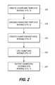
- FIG. 2is a high level flowchart of a process by which dynamic medical examination forms may be created according to one or more embodiments.
- the process shown in FIG. 2is typically performed by the dynamic medical examination forms software 151 , executing on the examination forms computing device 150 (or other computing device).
- the processmay be performed by more software distributed across a plurality of systems.
- a software application that is installed on the computing system 150may generate various user interfaces for interfacing with an operator (e.g., a physician or radiologist), and may include multiple software modules that interface with one another, such as a template creation module, template selection module, examination form creation module, and/or examination form export module, which are each described in further detail below.
- the term “medical examination forms software”refers to a software application that includes one or more of the modules discussed herein, and which may be executed on a computing device such as the computing device 150 , PACS workstation 138 , and/or any number of other computing devices.
- the method of FIG. 2may include fewer or additional blocks and/or may be performed in a different order than is illustrated. For example, in certain embodiments, one or more of the blocks shown in FIG. 2 may not be performed.
- Software code for performing the methods described herein, including the methods of FIGS. 2-7may be executed by examination form computing device 150 , the PACS workstation 138 , the EMR system 142 , the radiology information system 140 , and/or any other suitably configured computing device.
- the software codemay be embodied in a computer readable medium configured for reading by a computing device in order to store the software code in one or more memories of the computing device for execution.
- the processbegins at block 210 , where a template creation module is used to dynamically create an examination form template which may be stored in the examination form template database 132 . Additional details of the template creation process are provided below in connection with FIG. 3 .
- the processmay then move to block 215 , where a user of the medical examination forms software 151 may use the template selection module in order to select a stored template from the examination form template database 132 .
- the template selection moduleallows a user to find which existing examination form template best suits the examination for which they intend to use the form.
- the templatemay be chosen manually by the user.
- an automated processmay be used to select a dynamic examination form template based on specified criteria. These criteria may include the examination type that will be conducted, medical imaging modality, attributes of the specific type or model of scanner utilized, patient history, prior examinations, clinical information, the clinical indication for the exam, the facility at which the exam is conducted, insurance information, attributes related to people and entities that will receive the results of the examination such as tumor measurements required by clinical studies, attributes of referring/ordering physicians (including general attributes such as specialty as well as preferences related to individual physicians), and attributes related to the user for the form (including general attributes such as specialty or use role, and well as preferences of the specific users).
- criteriamay include the examination type that will be conducted, medical imaging modality, attributes of the specific type or model of scanner utilized, patient history, prior examinations, clinical information, the clinical indication for the exam, the facility at which the exam is conducted, insurance information, attributes related to people and entities that will receive the results of the examination such as tumor measurements required by clinical studies, attributes of referring/ordering physicians (including general attributes such as specialty as well as preferences related
- an instance of a dynamic medical examination formmay then be generated at block 220 based on the selected template.
- the instance created from the templateis a first view of the form.
- the medical examination forms softwareincludes an instance creation module that generates the examination form instance which may be used to capture data relating to a specific medical examination of a patient. An exemplary process for generating the examination form instance is discussed in additional detail below in connection with FIG. 5 .
- the processthen moves to block 225 , where the examination form may be accessed and filled out by an appropriate user. As will be discussed below in connection with FIG. 6 , this process may involve manually entering information on the examination form instance, and may additionally involve auto-populating the form with information related to the patient from other devices or systems on the network 205 .
- the processthen moves to block 230 , where the data collected in the view of the examination form may be exported to other systems on the network 205 for archiving or storage.
- a healthcare professional using the dynamic examination form software 151may use the software to dynamically create medical examination form templates. Views of electronic examination forms that are used in the course of patient medical examinations may be created using the templates that are created by the examination form template creation module.
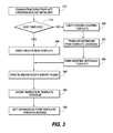
- FIG. 3is a more detailed flowchart of an exemplary process by which a dynamic medical examination form template may be created. Depending on the embodiment, the method of FIG. 3 may include fewer or additional blocks and/or may be performed in a different order than is illustrated.
- the processbegins at block 310 , where the template creation module is initialized, such as by the user making an appropriate menu selection in a user interface displayed on the computing device 150 .
- the processthen moves to decision block 315 , where the user is offered a choice as to whether to create an entirely new template, or to create the template out of an already existing examination form template.
- the processmoves to block 320 , where the user chooses an existing template.
- the existing templatesmay be presented to the user as a menu choice in which the template description is displayed to the user.
- the usermay be permitted to display a preview of the content of the templates presented in the menu. Allowing this type of preview helps to ensure a more accurate template choice by the user.
- the processthen moves to block 322 , where the selected template is retrieved from the dynamic examination form template database 132 , which may be on the PACS database server 130 .
- examination form templatesmay be stored local to the computing device (e.g., on a hard drive or optical drive, or available via a LAN) and selectable in block 320 .
- the processthen moves to block 324 , where the user modifies the selected template according to the current patient examination needs.
- the processthen moves to block 340 , where examination form export rules are modified.
- the existing template selected by the usermay already include export rules. In these instances, the user may adopt the existing export rules. Alternatively, the user may modify the already existing export rules to fit the needs of the modified template.
- the processmoves to block 330 , where the user constructs an entirely new template.
- One specific embodiment describing how the user may construct a new templateis discussed below in connection with FIG. 8A .
- the processthen moves to block 340 (discussed above), where export rules may be created for the template.
- the processthen moves to block 345 .
- the usermay indicate that the template is finished, and the template is stored in the examination form template database 132 (or other database, either local or remote).
- the template creation processends at block 350 , and the system exits the examination form template creation module.
- FIG. 4a flowchart provides one example of how a template stored in the template database 132 may be selected for use in a medical examination.
- the systemattempts to select the appropriate template for the user. If the appropriate template is not found, the user may manually select a template.
- the method of FIG. 4may include fewer or additional blocks and/or may be performed in a different order than is illustrated.
- the processbegins at block 410 where the template selection module is initialized to receive input from a user.
- a software modulemay be called in response to the user making a selection in the examination form software 151 (e.g., via a user interface generated by the software 151 and presented on a display device of the computing device 150 ) indicating a desire to create a new examination form based on a template.
- thismay occur automatically, for example when a medical examination is performed by technologist or presented for interpretation to radiologist or cardiologist on a PACS workstation.
- the processnext moves to block 415 , where the template selection module retrieves examination features related to one or more of the examination to be performed, the user, the location, etc., for which the template will be used
- the processnext moves to block 420 , where the retrieved examination features are compared with examination templates stored in the template database 132 . Based on that comparison, the template selection module makes an initial selection of the stored template that best fits the retrieved examination features at block 425 and presents that selection to the user.
- the processnext moves to decision block 430 , where the template selection module determines whether the user agrees with the automatic selection, such as by receiving an input from the user indicating such. If the user does not agree with the automatic selection, the process moves to block 435 , where the user manually selects a template from which to create the new examination form and the process then moves to block 450 and the template selection module exits. If, however, the user does agree with the automatic selection of a template from the database, the process moves directly to block 450 where the template selection process ends.
- the automatic template selection processselects more than one possibly matching template and presents the templates to the user sequentially (e.g., in response to the user indicating that an earlier presented template is not desired) and/or concurrently (e.g., thumbnails of multiple located templates may be displayed to the user).
- FIG. 5provides a more detailed view of one process of instantiating a dynamic examination form referenced at block 220 of FIG. 2 above.
- the method of FIG. 5may include fewer or additional blocks and/or may be performed in a different order than is illustrated.
- the processbegins at block 510 , where the examination form instance creation module is initiated.
- the processmoves to block 515 , where the information from the template selected by the template selection module (or manually by the user) is retrieved from the template database 132 .
- the processthen moves to block 520 , where an instance of the retrieved examination template is created.
- the instantiation of the templateprovides a new examination form that may be used in a patient examination.
- the processthen moves to block 525 , where one or more fields in the examination form may be pre-populated.
- the fieldsmay be populated by retrieving field values, such as data from the database or DICOM data stored in the PACS system, for example.
- fields in the examination formmay be mapped to specific data stored in these external systems, and the examination form may be configured to automatically retrieve these values. Additional details about the mapping process are discussed below in connection with FIGS. 8 and 9 .
- a view of the examination formis generated for display using the retrieved data.
- the viewis displayed to the user so that it may be reviewed, modified, and/or updated based on the results of a subsequent patient examination, for example.
- FIG. 6is a detailed example of how a dynamic examination form may be used in accordance with one or more embodiments. Depending on the embodiment, the method of FIG. 6 may include fewer or additional blocks and/or may be performed in a different order than is illustrated.
- the processbegins at block 610 , where the examination form utilization module is initiated.
- the selected and instantiated dynamic medical examination forme.g., the first view of the examination form
- data that has been pre-populated into the form from one or more data sourcesis displayed to the user.
- the userinteracts with the view of the examination form to enter and/or modify the appropriate data.
- Use and operation of an exemplary examination formwill be discussed in further detail below in connection with FIG. 9 .
- the data entered into the viewmay then be exported to other systems.
- the datamay be exported into a radiology report that may be used by the radiologist.
- the datamay further be exported into an external database, for example, a backup database.
- Collected datamay also be exported to the various other systems on the network 205 . These systems may include the PACS, RIS, EMR system or some other clinical information system.
- FIG. 7is a flowchart providing one example of a process by which information entered into the examination form (e.g., a particular view of an examination form) may be exported to other devices.
- the method of FIG. 7may include fewer or additional blocks and/or may be performed in a different order than is illustrated.
- the methodbegins at block 710 , where the examination form export module is initialized.
- the processthen moves to block 715 , where the export module retrieves the examination form export rules from the examination form template database 132 (or from another data source in other embodiments).
- data from the examination formis translated into the appropriate format for storage in the external system (if necessary) based on examination export rules retrieved from the template database 132 .
- the datais exported from the examination form to the selected destination system at block 730 .
- the processends at block 750 with the export module exiting.
- FIGS. 8A-8Eprovide an illustration of certain aspects of a graphical user interface environment in which a user may perform these processes according to one or more embodiments.
- a graphical user interface generated by a patient management software applicationsuch as the examination forms software 151 of FIG. 1 , includes a tab which allows a user to create a new medical examination form using a base template stored in the template database 132 , or to create a medical examination form using an already stored template, by modifying and updating the existing template to suit their needs.
- medical examination form templatesmay be created using the process described in FIG. 3 .
- the created examination form templatesmay be configured to interact with or link to external data by default.
- a templatemay be configured to map data items in the form template to text and/or image data in a medical data database such as the PACS database 131 , for example.
- the template creatormay also designate sections or fields in the template which may be automatically mapped into an external report, such as a reading physician's report that may be created based on the data in the examination form.
- the templatemay also be configured to import or pre-populate certain fields with data from external sources.
- the examination formsmay be uniquely named or associated with a unique identifier, and may be further cross-linked to a specific examination type in order to allow the user to more easily locate the appropriate examination form.
- the systemmay be configured so that the examination form automatically appears for appropriate users (including, but not limited to, clerical staff, technologists, and reading physicians) when an examination type is selected.
- the medical examination formsmay be cross-linked to other items such as a particular facility, an insurance company or type, patient attributes such as age or sex, clinical indications, patient history, and/or some other data item. Based on this cross-linking, appropriate examination forms (one or more) may be presented to a user based on the cross-linked items.
- an examination formWhen an examination form is created, it may be configured to contain multiple components and graphical user interface elements, including free text, drop down menus, radio buttons, checkboxes, textboxes, and/or data fields.
- the creator of the medical examination formmay designate fields that can be automatically imported from various data sources, such as medical records and/or imaging databases.
- the creatormay also designate examination form fields to be exported to a database or other repository.
- an examination formmay be defined which includes the following text and fields, where a field is indicated by brackets and surrounding text:
- FIG. 8Aprovides an illustration of a graphical user interface which may be used to create an examination form template with the types of cross-linking and mappings discussed above.
- a dialog box entitled “Report Demographics Layout” 380may be displayed as shown after the user has created the initial basic template.
- the user creating the templatemay select the scan button 381 , which causes the template creation module to identify and list each of the template fields 382 that has been added to the new template.
- Each of these template fieldsis shown inside brackets, indicating that it is not actual data, but rather is a data field.
- the usermay select one of the listed data fields 382 .
- the Map External Field button 385(shown as inactive in FIG. 8A ) becomes active, and the user may select the button.
- Selection of the Map Field button 385activates an additional dialog box (not shown) that lists each of the data items that is available for import into the examination form.
- These data elementsmay be accessed from internal or external data structures. For example, the data elements may be mapped from a PACS data structure or from an external data structure. The user may then select the desired choice for the mapping. Once the user has selected a desired field for the mapping, the mapped field is listed in the same row as a corresponding external data source field 383 for the selected data field 382 . In the example shown in FIG.
- the [RefDoc] field 382has been mapped to the “Ref Doc formatted name” data item 383 from the external data source.
- the useris also provided a Clear external data source field button which allows for the deletion of an association.
- a single examination form fieldmay be mapped to more than one external data field.
- a single examination form fieldmay also be mapped to external data fields which exist in multiple external data sources.
- mappingsmay also be created in which sections or fields in the examination form can be automatically mapped into one or more external or internal reports, such as a reading physicians report for example.
- Mapping data collected in an examination form to a physician's reading reportmay be accomplished using reporting software which allows for the creation of report templates which import data from external data sources.
- Data contained in any particular examination formcan be directed to multiple different destinations. For example, some data might go into a report describing the results of a radiology or cardiology exam. Some data from the same examination form might go into a database stored in the radiology information system, a cardiovascular information system, PACS, a national databank, credentialing organization, or any other local or remote storage device.
- a usercan use a report template, and then one can designate where one or more elements of the examination form will automatically appear when a document (report) is opened that is based on the report template that is linked to a particular examination form.
- a report templatemay be created using Microsoft Word®, any other text editor, or a native CDA standard text editor.
- the report templatemay be linked to a particular exam type, so that when exams of that type are presented, the report template is used to automatically generate a report document.
- the report templateitself may independently import data from one or more sources, including the examination form linked to that exam type.
- the report templatemight include the following mappings:
- the bracketsindicates positions in the report template where information can be automatically inserted from one or more of either internal data structures, external data structures, or where information can be inserted from a specified examination form linked to the same examination.
- the information inserted from the examination formmight include one or more lines of text, tables, diagrams, images, or other information. It may include the entire examination form, specified sections of the examination form, or specified fields of the examination form.
- An examination form that contains drop down menus, checklists, text areas, text fields, checkboxes, or radio buttonsmay be compiled during this insertion process such that only those items that are selected or completed are imported into the report.
- Whether or not information in a medical examination form is mapped to a destinationmay depend on the content entered into the form.
- the information in a formmay be processed using Natural Language Processing to determine whether the content is mapped and the destination or origination of that mapping. For example, if a specific critical clinical or imaging finding is reported in an examination form data field, the system may automatically generate a “critical results” report, or initiate an e-mail, automated phone call or other similar appropriate action to account for the situation.
- the view 360 a of the dynamic medical examination formincludes a text box 361 a which allows a user (such as a technologist or patient, for example) to enter comments about relevant medical examination.
- the view 360 aalso includes a family history field label 364 a and a family history input field 365 a for input of the patient's family history.
- This datamay already exist in a database or other structure within the PACS system. As a result, this data may be automatically retrieved and populated from the PACS database or other data source, as will be discussed more fully below.
- the view 360 aalso includes a menstrual history field label 366 a and a menstrual history input field 367 a . Additional field labels and fields are also provided such as the technique field label 368 a and its corresponding dropdown list 369 a ; the uterus size field label 370 a and its corresponding dropdown list 371 a ; the ovaries field label and its corresponding radio buttons 373 a and 375 a.
- view 360 amay have data fields that are cross-linked with a reading physicians report (that may be generated at any time in the future) so that the information in Section 2 , for example, may be directly mapped into the reading physician's report.
- the family history for the subject patientmay already by stored elsewhere in the system, such as in the PACS database, for example.
- datamay also be imported into the examination form from other sources.
- the family history data 365 amay be imported from an external database, such as an EMR system 142 .
- a view 360 b of the medical examination formis shown with the patient history data 365 b field having been pre-populated using data retrieved from one or more external systems (and/or entered by a technician, patient, or other user).
- a usersuch as clerical personnel or a technologist with appropriate user rights may see the view 360 b .
- the family historyis “Mother with history of ovarian cancer.”
- the retrieved datais displayed as underlined text to show that it is new data that has been added to the form. By providing a visual indication that the data was newly added, the user may be alerted that the veracity of the information may require confirmation.
- the pre-populated datawhen a user accesses this view (or any other view) for the first time, the pre-populated data will appear to the user (assuming, of course, that they have sufficient access permissions), and the user may then complete part or all of the remainder of the patient examination form using the user interface elements provided.
- the remaining fields in the view 360 bhave been completed by a user, as indicated by the underlined text.
- the data entered in the examination formmay be mapped to external systems as defined in the examination form template.
- views of medical examination formsmay be completed by a technologist or a clerical employee, and a report using various portions of the data input in the views may be later reviewed by a reading physician.
- the medical examination form(or portions of the medical examination form) may be completed by a patient, e.g., at home, in advance of an exam via the web or by some other remote user.
- the medical examination forms disclosed hereinreceive information mapped from other sources and map information to targeted data destinations (such as medical reports, quality assurance documents) as part of their initial set up.
- a reading physicianmay display a view of the examination form after various other views of the form have been used to acquire data regarding a specific patient examination.
- the reading physicianmay edit the examination form before or after its mapped contents have been transferred to a report.
- the physicianmay execute an action to cause updated contents of the form to be transmitted to the reading physicians report.
- the reading physicianmay further specify preferences which govern how the examination form is displayed when accessed by the reading physician.
- the reading physicianmay specify that the view displayed to the reading physician includes all of the data associated with the examination form.
- certain data fields in an examination formmay be cross-linked with data from external systems.
- an MRI image for a patientmay be cross-linked to an examination form, but the image itself is not automatically displayed in the form. Rather, a link to the image (such as a hyperlink to the PACS server, for example) may be provided in the form.
- the reading physicianmay specify a preference that the mapped fields of an examination form are automatically displayed in any report generated from the examination form.
- the reading physicianmay specify that any linked images (such as MRI images, for example) be automatically displayed in a report based on the report template for the examination form.
- FIG. 8Eis an example of a graphical user interface of a report generated by a report template based on the examination form 360 shown in FIGS. 8A-8D .
- the reportis typically intended to present information and not to allow information to be modified.
- only certain (or all) of the data associated with the examination formare shown in the report.
- that order of informationmay be different than in the medical examination form, and it might not include all information in the examination form.
- some reportsmay be configured to include information from multiple examination forms to provide additional relevant detail to the reader.
- medical examination formsmay be dynamically created based on a prior examination form (rather than based on an examination form template as described above) so that information from a prior examination may become organically part of the new composite examination form.
- a prior examination formbased on an examination form template as described above
- a medical examination formmay be dynamically modified while it is being used.
- new fieldsmay be added to existing examination forms on the fly in order to document new anomalies (such as new lesions, for example) that were not present in prior examinations, but would need to be following during future examinations.
- real-time bidirectional communication between an examination form and some other computer applicationmay be provided to allow for recording correlations of lesion measurements and positions within 2-dimensional images and 3-dimensional imaging volumes.
- FIG. 9is an exemplary screenshot of a user interface which illustrates the above-described dynamic embodiments.
- the figureincludes a screen window 805 which has two regions.
- the first region 810(the top 3 ⁇ 4 of the drawing) is related to a PACS workstation application
- the second region 840(the bottom 1 ⁇ 4 of the drawing) is related to a view of a patient examination form that has been pre-populated with information from a previous MRI scan of Jun. 12, 2008.
- the patient data shown in this exampleis the type of data that may be used by a radiologist to interpret a brain MRI scans in a patient with multiple metastatic brain lesions.
- an ultrasound technologistmay use the interface 805 to document thyroid nodules in a thyroid ultrasound.
- the first region 810is part of a PACS workstation graphical user interface.
- the PACS workstation user interfaceis typically used to interpret medical imaging examinations.
- the left column of the first region 810displays two images 822 and 824 from a patient's prior examination (dated Jun. 12, 2008 in this example) as shown by label 820 .
- the right column of the first region 810includes two images from follow-up examination (dated Sep. 14, 2008), as shown by label 830 .
- the second region 840includes a view of a dynamic examination form having a scroll bar 836 that allows the user to scroll to undisplayed portions.
- the second region 840includes a view of an examination form that was created on Jun. 12, 2008, as shown by label 812 . Certain fields used in the prior examination (which took place on Jun. 12, 2008) are replicated on the right side of the second region 840 in order to allow the fields from the old report to be used to allow efficient comparison between the Jun. 12, 2008 examination and the Sep. 14, 2008 examination. In this example, the user would fill in these fields on the right ( 854 , 860 , and 862 ) based on the findings of the corresponding exam on that date.
- the right side of the second region 840 of the user interface 805provides an interface by which a user can enter findings interpreting the images from the first region.
- Each of the right side and left side of the second region 840includes data fields for data relating to the corresponding imaging in the first region 810 .
- the “Ventricles” field label 842is associated with the “Ventricles” field value.
- the Ventricles field valueis set to “Normal,” while the new examination includes an active dropdown list 844 which allows the user to set the value for the new examination findings.
- the second region 840also includes a text label 850 which provides a heading for fields that are listed below.
- field label 850refers to a series of metastatic lesions identified in the previous medical examination dated Jun. 12, 2008.
- field label 851refers to a lesion observed on the “Right Medial Temporal” lobe of the patient.
- the associated field value 852lists the size of the lesion as observed in the June examination, “0.2” cm.
- a corresponding field 854has been created in the new medical examination form on the right side of the second region 840 .
- the userhas entered a new value “1.2” cm to reflect a the size of the metastatic lesion on the follow-up exam dated Sep. 14, 2008. This value could be typed in by the user or entered by other means.
- real-time bidirectional communication between the PACS workstation application and the examination formallows for measurements made in the PACS software to be linked to the examination form and entered automatically.
- measurements made by the user on the images in region 810may be immediately translated into values which appear in the measurement fields 854 , 860 , and 862 in the examination form provided in region 840 .
- the usermay click on a measurement field such as measurement field 854 , causing the field to become the active field (as indicated by the inverted black/white appearance). The user may then make a measurement of a lesion using the PACS workstation software.
- the measurementis performed on the image 832 as indicated by the positioning of two mouse cursors around a lesion near the center of the scan image.
- the measurement provided by the PACS workstation softwareis dynamically entered into the active field 854 .
- this dynamic behavioris provided by a mapping included in the base template for the examination form.
- the user interface 805 shown in FIG. 9also allows the examination forms to be dynamically enhanced to add fields for newly located lesions.
- an “Add New Lesion” button 838is provided which allows a user to dynamically create a new lesion field which would specify a location and a measurement.
- the newly added lesionmay be available for subsequent examinations if the new examination form is based on the prior examination form in the same manner as shown in FIG. 9 .
- additional user interface elementsmay be added to account and record additional examination findings that may be tracked in future examinations.
- the position where the measurement was takenwould be recorded in, for example, in a non-visible portion of the examination form, a database or file.
- the entry in the examination formmay be linked to 2-dimensional or 3-dimensional positions in images or image volumes.
- the bi-directional communication between the examination form software 151 and the external PACS system softwaremay further provide the ability to link examination form fields (such as fields 850 ) with regions and structures in the images.
- the linkingmay be implemented in both directions such that when a user clicks on an examination form field, the corresponding image and specific location in the image may be displayed in the first region 810 of the display 805 .
- linkingmay be implemented from the image to the examination form.
- a field associated with the portion of the image under the cursorbecomes activated.
- the userhas positioned the cursor over a portion of image 822 where a measurement has been made and a window labeled “Right Medial Temporal” is automatically displayed, the field name of the measurement made at that position.
- FIGS. 10A-10Gillustrate additional embodiments in which a graphical user interface environment is provided to allow for medical examination forms to be set up and create, copy, save, modify, and design examination forms.
- the dynamic medical examination forms describedare referred to as “ExamForms,” and it is to be appreciated that these are merely additional embodiments of the dynamic medical examination forms discussed above.
- a user interface 1000which includes a row of tabs 1002 that may be selected by the user.
- the tabs 1002 displayed in the user interface 1002may be dependent on the identity and/or role of the user. For example, a clerical employee may not have permission to see certain functionality and forms included in the system, in which case, certain tabs may be hidden from that user.
- a medical doctoron the other hand, may have extensive permissions within the system, and may therefore be shown additional tabs not presented to other users.
- the ExamForm Template tab 1004has been selected by the user, which causes the system to display a list of examination form templates 1006 from which a selection can be made.
- the usermay be taken through a series of template creation screens from which an examination form may be created and linked to specific medical examination types as will be discussed below.
- the examination form template editor 1008may include an information window 1010 and an editing window 1012 .
- the information window 1010may include information that provides instructions to the user in order to assist their design of the examination form.
- the information window 1010includes step-by-step instructions which provide detailed information to the user for setting up the examination form.
- the editing window 1012is used to provide an interface from which the actual design elements of an examination form may be presented and manipulated. In this particular example, multiple sections have been defined which can be associated with different examination form properties and functions.
- Sectionsmay also be defined based on the identity or role of the user that will complete that specific portion of the examination form.
- “Section 1 ” 1014may be designed so that its contents will be presented to technologists only.
- the first section 1014may include various information and fields that may be used by a technologist in an associated patient examination.
- the first section 1014may include Technologist Note text area 1018 .
- the Technologist Note area 1018provides a text area in which a technologist may enter notes regarding their role in a patient examination. The notes entered in this area may be visible to the technologist and to other users having sufficient permissions to access this portion of an examination form created from this template.
- the different sectionsmay also be defined based on whether the information entered into that section of the form will be mapped into an external data entity.
- the first section 1014may be configured so that none of the information in that section is mapped into the reading physician's report associated with that examination form.
- the information entered into the second section 1016 in an examination form created from the examination form templatemay be automatically mapped directly into a reading physicians report.
- the data entered into the exam data fields 1022may be automatically imported into a precisely mapped location in a physician's report defined for use with this examination form.
- external data fieldsmay be mapped into an examination form.
- a number of different patient information fields 1020are mapped into the examination form.
- the patient information fieldsare indicated by brackets. Mappings to external data may be created using a dialogue box such as that shown in the information window 1010 .
- the information window 1010provide the user with information on how database fields (indicated by bracketed areas near the top of the ExamForm-arrow) can be created, located, detected and mapped, so that information from an external database (in this example a DR Systems database) or some another datasource can be automatically imported when an ExamForm is created based on this ExamForm template.
- examination form templatesmay be linked to one or more specific examination types.
- an examination form templateWhen an examination form template is linked to a specific examination type, the appropriate examination form template may be presented to a user when the user wishes to create a new examination form.
- FIG. 10Cis an example of how an examination form template can be linked to one or more specific exam types. Linking an ExamForm template to an exam type is just one of many possible links that can govern which template should be used to create a particular examination form.
- a userhas selected an examination form template 1030 entitled “U S CARDIAC CART WITH NO”. In response to that selection, an linking window 1031 is displayed to the user.
- the linking window 1031indicates the template with which it is associated by listing a examination template identifier 1032 and an examination template name 1034 .
- a list of medical examinations 1036 from a pool of available examinationis presented to the user, from which one or more examinations may be selected for association with the current examination form template.
- the usermay create the link between the examination type and the examination template by selecting the “Add” button 1038 .
- a visual indication of the linkis provided by moving the selected examination types from the list 1036 to the linked examination list 1037 below. Links to specific examination types may be removed by selecting the specific examination type and pressing the “Remove” button 1040 .
- the windowmay be closed by selecting the “Exit” button 1042 .
- an examination formmay be designed such that specific sections of fields may be mapped to other templates, such as reporting templates, for example.
- FIG. 10Dan example of a physician's report template which may be configured to automatically receive data from a corresponding examination form and generate a reading physician's report using data received from, inter alia, an examination form.
- a reporting template file name 1050has been selected by a user.
- the reporting templatemay include a mapping 1048 from an examination form which is automatically imported when a physician's report is created based on this report template. The mapping may be created by utilizing the Map to Other Report Dialog box 1054 shown in the figure.
- some or all of the examination formmay be mapped to the physician's report.
- this user interfaceillustrates only one mapping location 1048 for the examination form into the physician's report, various different parts or fields from the examination form may be mapped into different sections of the physicians report.
- certain embodimentsmay provide multiple report templates (and subsequent reports) for the same examination or patient, each with different examination form mappings.
- FIG. 10Eis an example of a view of the examination form that was created from an examination form template.
- a plurality of data fields 1060have been automatically imported into the view of the examination form. These data fields 1060 include the patient name, the examination type, the data of birth for the patient, and the date of the examination.
- the information in the examination report shown in FIG. 10Emay be presented to a user to fill assist in completing a patient examination.
- the first section 1062includes information about the patient examination which may be presented to a user of the examination form.
- the usermay be a technologist who has been assigned to provide further information into the examination form.
- the technologistcan record comments in the Technologist Note text area 1064 that will be associated with the examination form, but not transmitted to other repositories or physicians' reports in this embodiment.
- the second section 1066 of the examination formmay provide additional dropdown menus or text fields as shown. Although not shown in this example, many other user interface elements, such as radiobuttons, checkboxes, required fields, etc. can be employed in the examination form design.
- certain data entered into the examination formmay be exported to a physicians report. In one embodiment, data is only exported if information is provided by the user. Thus, if no information is placed in the text area next to the Dose-Length Product 1068 , when the contents of Section 2 are imported into a physician's reading report, the entire text area will be omitted. In this way, the physician's reading report will not have the appearance of a form, even though it was created, in part, from a form.
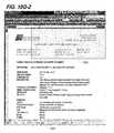
- an example of an examination formis provided in which an Auto-Import Note (Section 2 ) is provided for receiving data from a technologist.
- the datamay be entered into the examination form while a physician is performing a procedure.
- the assistant/technologistcan be completing information in the examination form in real-time during the examination procedure.
- the information entered into the examination formmay then be automatically exported into a physicians report, so that the physician need only edit and/or approve the report.
- some portions of the examination formmay be automatically completed when the form is generated.
- the information(LEFT HEART CATH, SERVICE CODE 93510) has been automatically imported from a data source.
- the examination formmay be updated to import supplies that were used during procedure.
- FIG. 10Gis an example of a user interface that a physician may use to evaluate a physician's report.
- a completed examination form 1080is displayed on the left, and the generated physician's report 1090 is displayed on the right.
- the reading physicianmay define preferences such that both the examination form 1080 and reading report 1090 are simultaneously displayed.
- the physicianmay be provided extensive control over the timing and location of displayed data. For example, a physician may prefer not to have an examination form displayed at all, but may set his preferences to simply see the physician's report on the right with the examination form mapped data already imported.
- the physician's report 1090contains information 1092 that did not come from the examination form, but instead came from other data mapping.
- the physicianmay have the right to edit the physician's report 1090 directly, or may edit the examination form 1080 and then resend the data to the physician's report.
- a physicianmight make various measurements or annotations of associated medical images, with such data transferred automatically to the examination form, the physician's report, both, neither, or other data stores.
- a general purpose processormay be a microprocessor, but in the alternative, the processor may be any conventional processor, controller, microcontroller, or state machine.
- a processormay also be implemented as a combination of computing devices, e.g., a combination of a DSP and a microprocessor, a plurality of microprocessors, one or more microprocessors in conjunction with a DSP core, or any other such configuration.
- a software modulemay reside in RAM memory, flash memory, ROM memory, EPROM memory, EEPROM memory, registers, hard disk, a removable disk, a CDROM, or any other form of storage medium known in the art.
- An exemplary storage mediumis coupled to the processor such the processor can read information from and write information to, the storage medium.
- the storage mediummay be integral to the processor.
- the processor and the storage mediummay reside in an ASIC.
- the ASICmay reside in a user terminal.
- the processor and the storage mediummay reside as discrete components in a user terminal.
Landscapes
- Engineering & Computer Science (AREA)
- Health & Medical Sciences (AREA)
- General Health & Medical Sciences (AREA)
- Medical Informatics (AREA)
- Theoretical Computer Science (AREA)
- Business, Economics & Management (AREA)
- Public Health (AREA)
- Primary Health Care (AREA)
- Epidemiology (AREA)
- Physics & Mathematics (AREA)
- General Physics & Mathematics (AREA)
- Strategic Management (AREA)
- Human Resources & Organizations (AREA)
- Entrepreneurship & Innovation (AREA)
- General Engineering & Computer Science (AREA)
- Data Mining & Analysis (AREA)
- Tourism & Hospitality (AREA)
- General Business, Economics & Management (AREA)
- Economics (AREA)
- Marketing (AREA)
- Bioethics (AREA)
- Databases & Information Systems (AREA)
- Operations Research (AREA)
- Quality & Reliability (AREA)
- Computational Linguistics (AREA)
- Audiology, Speech & Language Pathology (AREA)
- Artificial Intelligence (AREA)
- Nuclear Medicine, Radiotherapy & Molecular Imaging (AREA)
- Radiology & Medical Imaging (AREA)
- Computer Hardware Design (AREA)
- Computer Security & Cryptography (AREA)
- Software Systems (AREA)
- Biomedical Technology (AREA)
- Pathology (AREA)
- Medical Treatment And Welfare Office Work (AREA)
- Child & Adolescent Psychology (AREA)
- Measuring And Recording Apparatus For Diagnosis (AREA)
Abstract
Description
- Exam Title: [mapped, for example, from an internal or external data structure]
- Date: [mapped, for example, from an internal or external data structure]
- [Exam form contents, indicating one or more specific sections of examination form(s) that are mapped into the report].
Claims (19)
Priority Applications (3)
| Application Number | Priority Date | Filing Date | Title |
|---|---|---|---|
| US12/622,404US8380533B2 (en) | 2008-11-19 | 2009-11-19 | System and method of providing dynamic and customizable medical examination forms |
| US13/768,765US9501627B2 (en) | 2008-11-19 | 2013-02-15 | System and method of providing dynamic and customizable medical examination forms |
| US15/292,014US10592688B2 (en) | 2008-11-19 | 2016-10-12 | System and method of providing dynamic and customizable medical examination forms |
Applications Claiming Priority (2)
| Application Number | Priority Date | Filing Date | Title |
|---|---|---|---|
| US11619108P | 2008-11-19 | 2008-11-19 | |
| US12/622,404US8380533B2 (en) | 2008-11-19 | 2009-11-19 | System and method of providing dynamic and customizable medical examination forms |
Related Child Applications (1)
| Application Number | Title | Priority Date | Filing Date |
|---|---|---|---|
| US13/768,765ContinuationUS9501627B2 (en) | 2008-11-19 | 2013-02-15 | System and method of providing dynamic and customizable medical examination forms |
Publications (2)
| Publication Number | Publication Date |
|---|---|
| US20100138239A1 US20100138239A1 (en) | 2010-06-03 |
| US8380533B2true US8380533B2 (en) | 2013-02-19 |
Family
ID=42223633
Family Applications (3)
| Application Number | Title | Priority Date | Filing Date |
|---|---|---|---|
| US12/622,404Active2030-12-05US8380533B2 (en) | 2008-11-19 | 2009-11-19 | System and method of providing dynamic and customizable medical examination forms |
| US13/768,765Active2030-10-25US9501627B2 (en) | 2008-11-19 | 2013-02-15 | System and method of providing dynamic and customizable medical examination forms |
| US15/292,014Active2030-11-17US10592688B2 (en) | 2008-11-19 | 2016-10-12 | System and method of providing dynamic and customizable medical examination forms |
Family Applications After (2)
| Application Number | Title | Priority Date | Filing Date |
|---|---|---|---|
| US13/768,765Active2030-10-25US9501627B2 (en) | 2008-11-19 | 2013-02-15 | System and method of providing dynamic and customizable medical examination forms |
| US15/292,014Active2030-11-17US10592688B2 (en) | 2008-11-19 | 2016-10-12 | System and method of providing dynamic and customizable medical examination forms |
Country Status (1)
| Country | Link |
|---|---|
| US (3) | US8380533B2 (en) |
Cited By (20)
| Publication number | Priority date | Publication date | Assignee | Title |
|---|---|---|---|---|
| US20130117652A1 (en)* | 2011-11-04 | 2013-05-09 | Sas Institute Inc. | Techniques to generate custom electronic forms |
| US8554576B1 (en) | 2006-11-22 | 2013-10-08 | Dr Systems, Inc. | Automated document filing |
| US8610746B2 (en) | 2004-11-04 | 2013-12-17 | Dr Systems, Inc. | Systems and methods for viewing medical 3D imaging volumes |
| US8626527B1 (en) | 2004-11-04 | 2014-01-07 | Dr Systems, Inc. | Systems and methods for retrieval of medical data |
| US8650052B1 (en)* | 2012-03-28 | 2014-02-11 | Guidewire Software, Inc. | Configurable insurance policy forms inference |
| US8712120B1 (en) | 2009-09-28 | 2014-04-29 | Dr Systems, Inc. | Rules-based approach to transferring and/or viewing medical images |
| US8731259B2 (en) | 2004-11-04 | 2014-05-20 | Dr Systems, Inc. | Systems and methods for matching, naming, and displaying medical images |
| US20140172466A1 (en)* | 2012-12-17 | 2014-06-19 | Innodata Synodex, Llc | Shared Medical Data Platform for Insurance Underwriting |
| US8879807B2 (en) | 2004-11-04 | 2014-11-04 | Dr Systems, Inc. | Systems and methods for interleaving series of medical images |
| US8913808B2 (en) | 2004-11-04 | 2014-12-16 | Dr Systems, Inc. | Systems and methods for viewing medical images |
| US9092551B1 (en) | 2011-08-11 | 2015-07-28 | D.R. Systems, Inc. | Dynamic montage reconstruction |
| US9501627B2 (en) | 2008-11-19 | 2016-11-22 | D.R. Systems, Inc. | System and method of providing dynamic and customizable medical examination forms |
| US10381113B2 (en)* | 2015-04-27 | 2019-08-13 | Allscripts Software, Llc | Flexible encounter tracking systems and methods |
| US10642954B2 (en)* | 2013-12-10 | 2020-05-05 | Siemens Healthcare Gmbh | Medical scanner optimized workflow system |
| US10665342B2 (en) | 2013-01-09 | 2020-05-26 | Merge Healthcare Solutions Inc. | Intelligent management of computerized advanced processing |
| US20200279625A1 (en)* | 2019-02-28 | 2020-09-03 | Ebit Srl | Method and system to create and customize medical data sheets and reporting pages |
| US10789421B1 (en)* | 2013-08-15 | 2020-09-29 | Allscripts Software, Llc | Method for reusing and dynamically filtering content for structured document construction |
| US10909168B2 (en) | 2015-04-30 | 2021-02-02 | Merge Healthcare Solutions Inc. | Database systems and interactive user interfaces for dynamic interaction with, and review of, digital medical image data |
| WO2022099406A1 (en)* | 2020-11-13 | 2022-05-19 | Real-Time Engineering & Simulation Inc. | System and method for forming auditable electronic health record |
| US12014807B2 (en) | 2021-12-10 | 2024-06-18 | Merative Us L.P. | Automated report generation using artificial intelligence algorithms |
Families Citing this family (62)
| Publication number | Priority date | Publication date | Assignee | Title |
|---|---|---|---|---|
| US11080138B1 (en) | 2010-04-26 | 2021-08-03 | Pure Storage, Inc. | Storing integrity information in a vast storage system |
| US11340988B2 (en) | 2005-09-30 | 2022-05-24 | Pure Storage, Inc. | Generating integrity information in a vast storage system |
| US10866754B2 (en)* | 2010-04-26 | 2020-12-15 | Pure Storage, Inc. | Content archiving in a distributed storage network |
| DE102009018423B4 (en)* | 2009-04-22 | 2011-02-03 | Siemens Ag Österreich | Method for issuing medical documents |
| US8645861B2 (en) | 2009-07-01 | 2014-02-04 | Soft Computer Consultants, Inc. | Graphical display of actions for handling medical items |
| US20110161113A1 (en)* | 2009-12-27 | 2011-06-30 | Soft Computer Consultants, Inc. | Interpretive report generation |
| US10956292B1 (en) | 2010-04-26 | 2021-03-23 | Pure Storage, Inc. | Utilizing integrity information for data retrieval in a vast storage system |
| US8671118B2 (en)* | 2010-08-25 | 2014-03-11 | Fujifilm Corporation | Apparatus, method and program for assisting medical report creation and providing medical information |
| US20120117195A1 (en)* | 2010-11-05 | 2012-05-10 | Iberon, Llc | System for Generating one or more Citation Forms from a Single Workflow |
| US20120245954A1 (en)* | 2011-03-22 | 2012-09-27 | MRCS Holdings LLC | Medical Record Collection System |
| CN103460212B (en)* | 2011-03-25 | 2019-03-01 | 皇家飞利浦有限公司 | It is generated and is reported based on image data |
| US9177110B1 (en)* | 2011-06-24 | 2015-11-03 | D.R. Systems, Inc. | Automated report generation |
| WO2012174680A1 (en)* | 2011-06-24 | 2012-12-27 | Hewlett-Packard Development Company, L.P. | Identity based automated form filling |
| US10573408B2 (en)* | 2011-08-12 | 2020-02-25 | drchrono inc. | Dynamic forms |
| US9524283B2 (en)* | 2011-08-30 | 2016-12-20 | Sas Institutes Inc. | Techniques to remotely access form information and generate a form |
| EP2766832A1 (en)* | 2011-10-10 | 2014-08-20 | AbbVie Biotechnology Ltd | Managing healthcare services |
| US20130232026A1 (en)* | 2012-02-14 | 2013-09-05 | Steven Katzman | System and method for measurement based design selection |
| US8612261B1 (en) | 2012-05-21 | 2013-12-17 | Health Management Associates, Inc. | Automated learning for medical data processing system |
| US20130311201A1 (en)* | 2012-05-21 | 2013-11-21 | Health Management Associates | Medical record generation and processing |
| US10403403B2 (en)* | 2012-09-28 | 2019-09-03 | Cerner Innovation, Inc. | Adaptive medical documentation system |
| CN103890807B (en)* | 2012-10-16 | 2016-12-21 | 东芝医疗系统株式会社 | Cause of the death estimating unit and cause of the death estimation method thereof |
| WO2014099942A1 (en)* | 2012-12-21 | 2014-06-26 | Volcano Corporation | Display control for a multi-sensor medical device |
| KR101468918B1 (en)* | 2013-03-04 | 2014-12-04 | 서울대학교병원 (분사무소) | Apparatus and method for providing menu and bookmarking service based log-in in emr system |
| US10510449B1 (en)* | 2013-03-13 | 2019-12-17 | Merge Healthcare Solutions Inc. | Expert opinion crowdsourcing |
| WO2014153156A1 (en)* | 2013-03-14 | 2014-09-25 | Craven Jason | System and method for converting paper forms to an electronic format |
| US20140298243A1 (en)* | 2013-03-29 | 2014-10-02 | Alcatel-Lucent Usa Inc. | Adjustable gui for displaying information from a database |
| WO2014172671A1 (en)* | 2013-04-18 | 2014-10-23 | Digimarc Corporation | Physiologic data acquisition and analysis |
| US20140317552A1 (en)* | 2013-04-23 | 2014-10-23 | Lexmark International Technology Sa | Metadata Templates for Electronic Healthcare Documents |
| GB2515108A (en)* | 2013-06-14 | 2014-12-17 | Mastercard International Inc | A browser plug-in and a method of operating a browser plug-in |
| US9804768B1 (en)* | 2014-06-25 | 2017-10-31 | ABCDisability, Inc. | Method and system for generating an examination report |
| EP3018598A1 (en)* | 2014-11-05 | 2016-05-11 | Siemens Aktiengesellschaft | Evaluation method for medical data |
| WO2016125039A1 (en)* | 2015-02-05 | 2016-08-11 | Koninklijke Philips N.V. | Communication system for dynamic checklists to support radiology reporting |
| US10417326B2 (en)* | 2015-06-11 | 2019-09-17 | Fujifilm Medical Systems U.S.A., Inc. | Methods and apparatus for obtaining a snapshot of a medical imaging display |
| EP3329354A4 (en)* | 2015-07-31 | 2019-03-20 | WiseTech Global Limited | METHODS AND SYSTEMS FOR CREATING CONFIGURABLE FORMS, CONFIGURING FORMS AND FOR FORMULAR AND FORMULA FLOW CORRELATION |
| US20170132320A1 (en)* | 2015-11-10 | 2017-05-11 | Lexmark International Technology, SA | System and Methods for Transmitting Health level 7 Data from One or More Sending Applications to a Dictation System |
| US10769363B2 (en)* | 2015-11-10 | 2020-09-08 | Hyland Switzerland Sàrl | System and methods for transmitting clinical data having multi-segment fields from one or more modalities to a dictation machine |
| US10664568B2 (en)* | 2015-11-10 | 2020-05-26 | Hyland Switzerland Sàrl | System and methods for transmitting clinical data from one or more sending applications to a dictation system |
| TWI680411B (en)* | 2016-01-07 | 2019-12-21 | 葉振忠 | Electronic form building system and method |
| US12217036B2 (en) | 2016-02-10 | 2025-02-04 | Vignet Incorporated | Automating interactions for health data collection and patient engagement |
| US9928230B1 (en)* | 2016-09-29 | 2018-03-27 | Vignet Incorporated | Variable and dynamic adjustments to electronic forms |
| GB201603309D0 (en) | 2016-02-25 | 2016-04-13 | Goodmark Medical International Ltd | Patient test data processing system and method |
| WO2018065613A1 (en)* | 2016-10-06 | 2018-04-12 | Koninklijke Philips N.V. | Template-based medical summary interface generation system |
| CN108121708A (en)* | 2016-11-28 | 2018-06-05 | 英业达科技有限公司 | Examination question electronization access system and method |
| TW201835846A (en)* | 2017-03-16 | 2018-10-01 | 傅真 | Method for establishing mutually operable and usable medical data cloud-end integrated management system that establishes a cloud-end virtual medical data center architecture in chronic order of the entire life of an individual |
| US20180349556A1 (en)* | 2017-06-02 | 2018-12-06 | Nuance Communications, Inc. | Medical documentation systems and methods |
| CN108039193A (en)* | 2017-11-17 | 2018-05-15 | 哈尔滨工大服务机器人有限公司 | A kind of method and device for automatically generating physical examination report |
| US20190198137A1 (en)* | 2017-12-26 | 2019-06-27 | International Business Machines Corporation | Automatic Summarization of Patient Data Using Medically Relevant Summarization Templates |
| US20190198138A1 (en)* | 2017-12-26 | 2019-06-27 | International Business Machines Corporation | Automatic Expansion of Medically Relevant Summarization Templates Using Semantic Expansion |
| EP3779997A4 (en)* | 2018-04-04 | 2021-05-19 | FUJIFILM Corporation | Medical document display control device, medical document display control method, and medical document display control program |
| US11004546B2 (en)* | 2018-11-09 | 2021-05-11 | Curantis Solutions | Automated construction of patient care giver tool user interface using three-tiered architecture |
| US11610667B2 (en)* | 2018-11-19 | 2023-03-21 | RAD AI, Inc. | System and method for automated annotation of radiology findings |
| CN109817347A (en)* | 2019-01-15 | 2019-05-28 | 深圳市道通科技股份有限公司 | Inline diagnosis platform, its right management method and Rights Management System |
| US11373739B2 (en)* | 2019-04-17 | 2022-06-28 | Tempus Labs, Inc. | Systems and methods for interrogating clinical documents for characteristic data |
| CN110047577A (en)* | 2019-04-23 | 2019-07-23 | 杭州依图医疗技术有限公司 | Image distributor and image distribution method |
| CN110223750A (en)* | 2019-06-19 | 2019-09-10 | 飞依诺科技(苏州)有限公司 | The automatic matching method and system of ultrasonic diagnosis report |
| US20200410601A1 (en)* | 2019-06-25 | 2020-12-31 | Verata Health, Inc. | Automated prior authorization request generation and tracking |
| WO2021051091A1 (en) | 2019-09-13 | 2021-03-18 | Rad Al, Inc. | Method and system for automatically generating a section in a radiology report |
| US20220139509A1 (en)* | 2020-11-03 | 2022-05-05 | Nuance Communications, Inc. | Communication System and Method |
| US11915804B2 (en)* | 2020-11-17 | 2024-02-27 | Cerner Innovation, Inc. | Integrated report |
| CN112733494B (en)* | 2020-12-25 | 2023-08-01 | 浪潮通用软件有限公司 | Method and system for automatically generating standard detection report |
| WO2023170442A2 (en)* | 2022-03-01 | 2023-09-14 | Mofaip, Llc | Targeted isolation of anatomic sites for form generation and medical record generation and retrieval |
| WO2024220549A1 (en) | 2023-04-17 | 2024-10-24 | RAD AI, Inc. | System and method for radiology reporting |
Citations (135)
| Publication number | Priority date | Publication date | Assignee | Title |
|---|---|---|---|---|
| US4672683A (en) | 1984-03-20 | 1987-06-09 | Olympus Optical Co., Ltd. | Image retrieval and registration system |
| US5123056A (en) | 1990-02-02 | 1992-06-16 | Siemens Medical Systems, Inc. | Whole-leg x-ray image processing and display techniques |
| US5431161A (en) | 1993-04-15 | 1995-07-11 | Adac Laboratories | Method and apparatus for information acquistion, processing, and display within a medical camera system |
| US5452416A (en) | 1992-12-30 | 1995-09-19 | Dominator Radiology, Inc. | Automated system and a method for organizing, presenting, and manipulating medical images |
| US5542003A (en) | 1993-09-13 | 1996-07-30 | Eastman Kodak | Method for maximizing fidelity and dynamic range for a region of interest within digitized medical image display |
| US5734915A (en) | 1992-11-25 | 1998-03-31 | Eastman Kodak Company | Method and apparatus for composing digital medical imagery |
| US5740267A (en) | 1992-05-29 | 1998-04-14 | Echerer; Scott J. | Radiographic image enhancement comparison and storage requirement reduction system |
| US5779634A (en) | 1991-05-10 | 1998-07-14 | Kabushiki Kaisha Toshiba | Medical information processing system for supporting diagnosis |
| US5852646A (en) | 1996-05-21 | 1998-12-22 | U.S. Philips Corporation | X-ray imaging method |
| US5926568A (en) | 1997-06-30 | 1999-07-20 | The University Of North Carolina At Chapel Hill | Image object matching using core analysis and deformable shape loci |
| US5954650A (en) | 1996-11-13 | 1999-09-21 | Kabushiki Kaisha Toshiba | Medical image processing apparatus |
| US5976088A (en) | 1998-06-24 | 1999-11-02 | Ecton, Inc. | Ultrasound imaging systems and methods of increasing the effective acquisition frame rate |
| US5987345A (en) | 1996-11-29 | 1999-11-16 | Arch Development Corporation | Method and system for displaying medical images |
| US5995644A (en) | 1997-06-30 | 1999-11-30 | Siemens Corporate Research, Inc. | Robust and automatic adjustment of display window width and center for MR images |
| US6115486A (en) | 1996-11-06 | 2000-09-05 | Quinton Instrument Company | Teleradiology system for the storage and transmission of angiographic and related image sequences |
| US6151581A (en) | 1996-12-17 | 2000-11-21 | Pulsegroup Inc. | System for and method of collecting and populating a database with physician/patient data for processing to improve practice quality and healthcare delivery |
| US6175643B1 (en) | 1997-12-18 | 2001-01-16 | Siemens Corporate Research, Inc. | Neural network based auto-windowing system for MR images |
| US6185320B1 (en) | 1995-03-03 | 2001-02-06 | Arch Development Corporation | Method and system for detection of lesions in medical images |
| US20010016822A1 (en) | 1998-05-29 | 2001-08-23 | Luc Bessette | Method and apparatus for the management of data files |
| US6304667B1 (en) | 2000-06-21 | 2001-10-16 | Carmen T. Reitano | System and method for incorporating dyslexia detection in handwriting pattern recognition systems |
| US20010042124A1 (en) | 2000-03-27 | 2001-11-15 | Barron Robert H. | Web-based method, apparatus, and system for secure data storage |
| US20020016718A1 (en) | 2000-06-22 | 2002-02-07 | Rothschild Peter A. | Medical image management system and method |
| US6347329B1 (en) | 1996-09-27 | 2002-02-12 | Macneal Memorial Hospital Assoc. | Electronic medical records system |
| US20020021828A1 (en) | 2000-08-01 | 2002-02-21 | Arthur Papier | System and method to aid diagnoses using cross-referenced knowledge and image databases |
| US6351547B1 (en) | 1999-04-28 | 2002-02-26 | General Electric Company | Method and apparatus for formatting digital images to conform to communications standard |
| US20020044696A1 (en) | 1999-11-24 | 2002-04-18 | Sirohey Saad A. | Region of interest high resolution reconstruction for display purposes and a novel bookmarking capability |
| US20020073429A1 (en) | 2000-10-16 | 2002-06-13 | Beane John A. | Medical image capture system and method |
| US20020091659A1 (en) | 2000-09-12 | 2002-07-11 | Beaulieu Christopher F. | Portable viewing of medical images using handheld computers |
| US6424996B1 (en) | 1998-11-25 | 2002-07-23 | Nexsys Electronics, Inc. | Medical network system and method for transfer of information |
| US20020103827A1 (en) | 2001-01-26 | 2002-08-01 | Robert Sesek | System and method for filling out forms |
| US20020103673A1 (en) | 2001-02-01 | 2002-08-01 | Atwood Lindsay T. | Methods and apparatus for facilitating the provision of services |
| US20020110285A1 (en) | 2001-02-15 | 2002-08-15 | Wang Douglas W. | Method for internet browser-based digital image capture and management system |
| US6438533B1 (en) | 1998-10-30 | 2002-08-20 | College Of American Pathologists | System for retrieval of information from data structure of medical records |
| US6463169B1 (en) | 1996-06-26 | 2002-10-08 | Matsushita Electrical Industrial Co., Ltd. | X-ray imaging apparatus and recording medium therefor |
| US20020180883A1 (en) | 2001-04-27 | 2002-12-05 | Kazunari Tomizawa | Image processing circuit, image display device, and an image processing method |
| US20030005464A1 (en) | 2001-05-01 | 2003-01-02 | Amicas, Inc. | System and method for repository storage of private data on a network for direct client access |
| US20030028402A1 (en) | 2001-08-06 | 2003-02-06 | Ulrich Medical Concepts, Inc. | System and method for managing patient encounters |
| US20030037054A1 (en) | 2001-08-09 | 2003-02-20 | International Business Machines Corporation | Method for controlling access to medical information |
| US6532311B1 (en) | 1999-02-26 | 2003-03-11 | Lockheed Martin Corporation | Image browser |
| US6532299B1 (en) | 2000-04-28 | 2003-03-11 | Orametrix, Inc. | System and method for mapping a surface |
| US20030065613A1 (en) | 2001-09-28 | 2003-04-03 | Smith Diane K. | Software for financial institution monitoring and management and for assessing risk for a financial institution |
| US6556695B1 (en) | 1999-02-05 | 2003-04-29 | Mayo Foundation For Medical Education And Research | Method for producing high resolution real-time images, of structure and function during medical procedures |
| US6563950B1 (en) | 1996-06-25 | 2003-05-13 | Eyematic Interfaces, Inc. | Labeled bunch graphs for image analysis |
| US6574629B1 (en) | 1998-12-23 | 2003-06-03 | Agfa Corporation | Picture archiving and communication system |
| US6577753B2 (en) | 1998-03-31 | 2003-06-10 | Fuji Photo Film Co., Ltd. | Medical network system |
| US20030115083A1 (en) | 2001-11-07 | 2003-06-19 | Masarie Fred E. | HTML-based clinical content |
| US6603494B1 (en) | 1998-11-25 | 2003-08-05 | Ge Medical Systems Global Technology Company, Llc | Multiple modality interface for imaging systems including remote services over a network |
| US6630937B2 (en) | 1997-10-30 | 2003-10-07 | University Of South Florida | Workstation interface for use in digital mammography and associated methods |
| US20030190062A1 (en) | 2002-04-09 | 2003-10-09 | Omron Corporation | Apparatus for measuring biological data and exercise machines |
| US20030204420A1 (en) | 2002-04-30 | 2003-10-30 | Wilkes Gordon J. | Healthcare database management offline backup and synchronization system and method |
| US6678764B2 (en) | 2000-10-20 | 2004-01-13 | Sony Corporation | Medical image processing system |
| US20040024303A1 (en) | 1998-11-25 | 2004-02-05 | Banks Seth R. | Multiple modality interface for imaging systems |
| US6697506B1 (en) | 1999-03-17 | 2004-02-24 | Siemens Corporate Research, Inc. | Mark-free computer-assisted diagnosis method and system for assisting diagnosis of abnormalities in digital medical images using diagnosis based image enhancement |
| US20040088192A1 (en) | 2002-11-04 | 2004-05-06 | Schmidt Tim W. | Medical office electronic management system |
| US20040086163A1 (en) | 2002-10-31 | 2004-05-06 | Konica Minolta Holdings, Inc. | Medical image radiographing system, medical image management method and portable terminal |
| US20040114714A1 (en) | 2002-11-29 | 2004-06-17 | Minyard Thomas J. | Distributed architecture for mammographic image acquisition and processing |
| US20040143582A1 (en) | 2003-01-17 | 2004-07-22 | Jonathan Vu | System and method for structuring data in a computer system |
| US6775402B2 (en) | 1997-03-03 | 2004-08-10 | Bacus Laboratories, Inc. | Method and apparatus for creating a virtual microscope slide |
| US6778689B1 (en) | 2000-03-29 | 2004-08-17 | General Electric Company | System and method of real-time multiple field-of-view imaging |
| US20040161139A1 (en) | 2003-02-14 | 2004-08-19 | Yaseen Samara | Image data navigation method and apparatus |
| US20040161164A1 (en) | 2003-02-19 | 2004-08-19 | Agfa-Gevaert | Method of detecting the orientation of an object in an image |
| US20040165791A1 (en) | 2003-02-21 | 2004-08-26 | Ted Kaltanji | Dental image storage and retrieval apparatus |
| US6820100B2 (en) | 2000-09-29 | 2004-11-16 | Fuji Photo Film Co., Ltd. | Image management system and image management method |
| US20040243435A1 (en) | 2003-05-29 | 2004-12-02 | Med-Sched, Inc. | Medical information management system |
| US6829377B2 (en) | 2000-04-13 | 2004-12-07 | Giuceppe Milioto | Limb extremity positioning device and measurement method |
| US20040254816A1 (en) | 2001-10-30 | 2004-12-16 | Myers Gene E. | Network-connected personal medical information and billing system |
| US20050027570A1 (en) | 2000-08-11 | 2005-02-03 | Maier Frith Ann | Digital image collection and library system |
| US20050043970A1 (en) | 2003-08-21 | 2005-02-24 | Kent Hsieh | Electronic imaging dental record management system |
| US6864794B2 (en) | 2001-03-14 | 2005-03-08 | Siemens Aktiengesellschaft | Method and apparatus for evaluating medical examination images |
| US20050065424A1 (en) | 2003-06-06 | 2005-03-24 | Ge Medical Systems Information Technologies, Inc. | Method and system for volumemetric navigation supporting radiological reading in medical imaging systems |
| US6886133B2 (en) | 2001-06-07 | 2005-04-26 | Microsoft Corporation | Interactive formatting interface |
| US6891920B1 (en) | 2002-11-29 | 2005-05-10 | Fischer Imaging Corporation | Automated background processing mammographic image data |
| US20050108058A1 (en) | 2003-10-10 | 2005-05-19 | Eastman Kodak Company | Management of dental records and digital images |
| US20050114178A1 (en) | 2003-11-26 | 2005-05-26 | Anand Krishnamurthy | Method for processing a workflow for automated patient scheduling in a hospital information system |
| US20050114283A1 (en) | 2003-05-16 | 2005-05-26 | Philip Pearson | System and method for generating a report using a knowledge base |
| US20050114179A1 (en) | 2003-11-26 | 2005-05-26 | Brackett Charles C. | Method and apparatus for constructing and viewing a multi-media patient summary |
| US6917696B2 (en) | 2000-05-03 | 2005-07-12 | Aperio Technologies, Inc. | Fully automatic rapid microscope slide scanner |
| US20050184988A1 (en) | 2002-04-12 | 2005-08-25 | Yanof Jeffrey H. | Graphical apparatus and method for tracking image volume review |
| US20050197860A1 (en) | 2004-02-23 | 2005-09-08 | Rademr, Inc. | Data management system |
| US20050238218A1 (en) | 2004-01-07 | 2005-10-27 | Fuji Photo Film Co., Ltd. | Image display method, apparatus and program |
| US20050273009A1 (en) | 2004-06-02 | 2005-12-08 | Harald Deischinger | Method and apparatus for co-display of inverse mode ultrasound images and histogram information |
| US6996205B2 (en) | 2003-06-24 | 2006-02-07 | Ge Medical Ssytems Global Technology Company, Llc | Methods and apparatus to facilitate review of CT colonography exams |
| US7022073B2 (en) | 2003-04-02 | 2006-04-04 | Siemens Medical Solutions Usa, Inc. | Border detection for medical imaging |
| US7027633B2 (en) | 2000-11-30 | 2006-04-11 | Foran David J | Collaborative diagnostic systems |
| US7031846B2 (en) | 2001-08-16 | 2006-04-18 | Affymetrix, Inc. | Method, system, and computer software for the presentation and storage of analysis results |
| US20060093199A1 (en) | 2004-11-04 | 2006-05-04 | Fram Evan K | Systems and methods for viewing medical 3D imaging volumes |
| US20060095423A1 (en) | 2004-11-04 | 2006-05-04 | Reicher Murray A | Systems and methods for retrieval of medical data |
| US20060093198A1 (en) | 2004-11-04 | 2006-05-04 | Fram Evan K | Systems and methods for interleaving series of medical images |
| US20060095426A1 (en) | 2004-09-29 | 2006-05-04 | Katsuhiko Takachio | System and method for creating document abstract |
| US7043474B2 (en) | 2002-04-15 | 2006-05-09 | International Business Machines Corporation | System and method for measuring image similarity based on semantic meaning |
| US20060106642A1 (en) | 2004-11-04 | 2006-05-18 | Reicher Murray A | Systems and methods for matching, naming, and displaying medical images |
| US7050620B2 (en) | 2001-03-30 | 2006-05-23 | Heckman Carol A | Method of assaying shape and structural features in cells |
| US20060111941A1 (en) | 2004-11-24 | 2006-05-25 | Blom Michael G | Automated patient management system |
| US7092572B2 (en) | 2002-03-29 | 2006-08-15 | Sun Microsystems, Inc. | Method and apparatus for global image quantification verification |
| US20060181548A1 (en) | 2002-10-29 | 2006-08-17 | Christopher Hafey | Methods and apparatus for controlling the display of medical images |
| US7103205B2 (en) | 2000-11-24 | 2006-09-05 | U-Systems, Inc. | Breast cancer screening with ultrasound image overlays |
| US7106479B2 (en) | 2000-10-10 | 2006-09-12 | Stryker Corporation | Systems and methods for enhancing the viewing of medical images |
| US7110616B2 (en) | 2001-08-22 | 2006-09-19 | Siemens Aktiengesellschaft | Device for processing images, in particular medical images |
| US7113186B2 (en) | 2000-11-25 | 2006-09-26 | Infinitt Co., Ltd. | 3 Dimensional slab rendering system method and computer-readable medium |
| US20060230072A1 (en) | 2005-04-08 | 2006-10-12 | Dlcom Grid Inc. | Secure digital couriering system and method |
| US20060277075A1 (en) | 2005-06-07 | 2006-12-07 | Salwan Angadbir S | Physician to patient network system for real-time electronic communications & transfer of patient health information |
| US7149334B2 (en) | 2004-09-10 | 2006-12-12 | Medicsight Plc | User interface for computed tomography (CT) scan analysis |
| US20060282408A1 (en) | 2003-09-30 | 2006-12-14 | Wisely David R | Search system and method via proxy server |
| US7155043B2 (en) | 2001-11-21 | 2006-12-26 | Confirma, Incorporated | User interface having analysis status indicators |
| US7170532B2 (en) | 2002-11-21 | 2007-01-30 | Canon Kabushiki Kaisha | Image display method, apparatus, program and storage medium |
| US7174054B2 (en) | 2003-09-23 | 2007-02-06 | Amazon Technologies, Inc. | Method and system for access to electronic images of text based on user ownership of corresponding physical text |
| US20070050701A1 (en)* | 2005-08-31 | 2007-03-01 | Khaled El Emam | Method, system and computer program product for medical form creation |
| US20070055550A1 (en) | 2005-09-06 | 2007-03-08 | Patient Practitioners Llc | Personal transportable healthcare data base |
| US20070067124A1 (en) | 2005-08-01 | 2007-03-22 | Tom Kimpe | Method and device for improved display standard conformance |
| US20070073556A1 (en) | 2005-09-23 | 2007-03-29 | General Electric Co. | System and method for coordinating examination scheduling |
| US7209149B2 (en) | 2000-06-05 | 2007-04-24 | Fujifilm Corporation | Image cropping and synthesizing method, and imaging apparatus |
| US7218763B2 (en) | 2003-02-27 | 2007-05-15 | Eastman Kodak Company | Method for automated window-level settings for magnetic resonance images |
| US7224852B2 (en) | 2001-03-23 | 2007-05-29 | Objectvideo, Inc. | Video segmentation using statistical pixel modeling |
| US20070174079A1 (en) | 2005-12-01 | 2007-07-26 | Kraus Steven J | Apparatus and method for digital imaging, education, and internal marketing software and system |
| US20070192140A1 (en) | 2005-08-17 | 2007-08-16 | Medcommons, Inc. | Systems and methods for extending an information standard through compatible online access |
| US7260249B2 (en) | 2002-09-27 | 2007-08-21 | Confirma Incorporated | Rules-based approach for processing medical images |
| US7263710B1 (en) | 1999-12-31 | 2007-08-28 | General Electric Company | Medical diagnostic system with on-line real-time video training |
| US7272610B2 (en) | 2001-11-02 | 2007-09-18 | Medrecon, Ltd. | Knowledge management system |
| US20070239481A1 (en) | 2006-04-07 | 2007-10-11 | Disilvestro Mark R | System and method for managing patient-related data |
| US20080059245A1 (en) | 2006-08-30 | 2008-03-06 | Fujifilm Corporation | Medical image management method, medical image management apparatus, and medical network system |
| US20080103828A1 (en) | 2006-11-01 | 2008-05-01 | Squilla John R | Automated custom report generation system for medical information |
| US7412111B2 (en) | 2004-11-19 | 2008-08-12 | General Electric Company | Enhanced image processing method for the presentation of digitally-combined medical images |
| US20080275913A1 (en)* | 2007-05-01 | 2008-11-06 | Van Arragon Paul | Dynamic assignment of statements for selected exams using clinical concepts |
| US7450747B2 (en) | 2002-07-12 | 2008-11-11 | Ge Medical Systems Global Technology Company, Llc | System and method for efficiently customizing an imaging system |
| US7526114B2 (en) | 2002-11-15 | 2009-04-28 | Bioarray Solutions Ltd. | Analysis, secure access to, and transmission of array images |
| US7545965B2 (en) | 2003-11-10 | 2009-06-09 | The University Of Chicago | Image modification and detection using massive training artificial neural networks (MTANN) |
| US20090198514A1 (en)* | 2008-01-31 | 2009-08-06 | Decisionbase | Knowledge based clinical dental records management systems |
| US7583861B2 (en) | 2002-11-27 | 2009-09-01 | Teramedica, Inc. | Intelligent medical image management system |
| US7613335B2 (en) | 2003-02-12 | 2009-11-03 | The University Of Iowa Research Foundation | Methods and devices useful for analyzing color medical images |
| US7634121B2 (en) | 2005-03-01 | 2009-12-15 | General Electric Company | Method and system for rule-based comparison study matching to customize a hanging protocol |
| US7636413B2 (en) | 2002-04-16 | 2009-12-22 | General Electric Company | Method and apparatus of multi-energy imaging |
| US7660488B2 (en) | 2004-11-04 | 2010-02-09 | Dr Systems, Inc. | Systems and methods for viewing medical images |
| US20100138239A1 (en) | 2008-11-19 | 2010-06-03 | Dr Systems, Inc. | System and method of providing dynamic and customizable medical examination forms |
| US20100198608A1 (en) | 2005-10-24 | 2010-08-05 | CellTrak Technologies, Inc. | Home health point-of-care and administration system |
| US7953614B1 (en) | 2006-11-22 | 2011-05-31 | Dr Systems, Inc. | Smart placement rules |
Family Cites Families (238)
| Publication number | Priority date | Publication date | Assignee | Title |
|---|---|---|---|---|
| US1015768A (en) | 1911-03-13 | 1912-01-30 | Randall Telephone Mfg Company | Automatic telephone circuit and repeater. |
| US5179651A (en) | 1988-11-08 | 1993-01-12 | Massachusetts General Hospital | Apparatus for retrieval and processing of selected archived images for display at workstation terminals |
| US5172419A (en) | 1991-03-05 | 1992-12-15 | Lumisys, Inc. | Medical image processing system |
| EP0973116A1 (en) | 1993-03-01 | 2000-01-19 | Kabushiki Kaisha Toshiba | Medical information processing system for supporting diagnosis |
| US5515375A (en) | 1993-07-30 | 1996-05-07 | Motorola, Inc. | Method and apparatus for multiplexing fixed length message data and variably coded speech |
| JP3277677B2 (en) | 1994-04-01 | 2002-04-22 | ソニー株式会社 | Signal encoding method and apparatus, signal recording medium, signal transmission method, and signal decoding method and apparatus |
| JP3544557B2 (en) | 1994-04-08 | 2004-07-21 | オリンパス株式会社 | Image file device |
| US5857030A (en) | 1995-08-18 | 1999-01-05 | Eastman Kodak Company | Automated method and system for digital image processing of radiologic images utilizing artificial neural networks |
| US6128002A (en) | 1996-07-08 | 2000-10-03 | Leiper; Thomas | System for manipulation and display of medical images |
| US6820093B2 (en) | 1996-07-30 | 2004-11-16 | Hyperphrase Technologies, Llc | Method for verifying record code prior to an action based on the code |
| US5986662A (en) | 1996-10-16 | 1999-11-16 | Vital Images, Inc. | Advanced diagnostic viewer employing automated protocol selection for volume-rendered imaging |
| US6243095B1 (en) | 1996-12-05 | 2001-06-05 | Peter E. Shile | Navigation and display system for digital radiographs |
| JPH10276461A (en) | 1997-03-28 | 1998-10-13 | Sharp Corp | Receiving machine |
| US6008813A (en) | 1997-08-01 | 1999-12-28 | Mitsubishi Electric Information Technology Center America, Inc. (Ita) | Real-time PC based volume rendering system |
| US5999639A (en) | 1997-09-04 | 1999-12-07 | Qualia Computing, Inc. | Method and system for automated detection of clustered microcalcifications from digital mammograms |
| US6606171B1 (en) | 1997-10-09 | 2003-08-12 | Howtek, Inc. | Digitizing scanner |
| US6130671A (en) | 1997-11-26 | 2000-10-10 | Vital Images, Inc. | Volume rendering lighting using dot product methodology |
| US6313841B1 (en) | 1998-04-13 | 2001-11-06 | Terarecon, Inc. | Parallel volume rendering system with a resampling module for parallel and perspective projections |
| US6427022B1 (en) | 1998-11-10 | 2002-07-30 | Western Research Company, Inc. | Image comparator system and method for detecting changes in skin lesions |
| US6356265B1 (en) | 1998-11-12 | 2002-03-12 | Terarecon, Inc. | Method and apparatus for modulating lighting with gradient magnitudes of volume data in a rendering pipeline |
| US6404429B1 (en) | 1998-11-12 | 2002-06-11 | Terarecon, Inc. | Method for modulating volume samples with gradient magnitude vectors and step functions |
| US6211884B1 (en) | 1998-11-12 | 2001-04-03 | Mitsubishi Electric Research Laboratories, Inc | Incrementally calculated cut-plane region for viewing a portion of a volume data set in real-time |
| US6369816B1 (en) | 1998-11-12 | 2002-04-09 | Terarecon, Inc. | Method for modulating volume samples using gradient magnitudes and complex functions over a range of values |
| US6266733B1 (en) | 1998-11-12 | 2001-07-24 | Terarecon, Inc | Two-level mini-block storage system for volume data sets |
| US6297799B1 (en) | 1998-11-12 | 2001-10-02 | James Knittel | Three-dimensional cursor for a real-time volume rendering system |
| US6411296B1 (en) | 1998-11-12 | 2002-06-25 | Trrarecon, Inc. | Method and apparatus for applying modulated lighting to volume data in a rendering pipeline |
| US6512517B1 (en) | 1998-11-12 | 2003-01-28 | Terarecon, Inc. | Volume rendering integrated circuit |
| US6342885B1 (en) | 1998-11-12 | 2002-01-29 | Tera Recon Inc. | Method and apparatus for illuminating volume data in a rendering pipeline |
| US6426749B1 (en) | 1998-11-12 | 2002-07-30 | Terarecon, Inc. | Method and apparatus for mapping reflectance while illuminating volume data in a rendering pipeline |
| US6081267A (en) | 1998-11-19 | 2000-06-27 | Columbia Scientific Incorporated | Computerized apparatus and method for displaying X-rays and the like for radiological analysis and manipulation and transmission of data |
| US6310620B1 (en) | 1998-12-22 | 2001-10-30 | Terarecon, Inc. | Method and apparatus for volume rendering with multiple depth buffers |
| US7107253B1 (en) | 1999-04-05 | 2006-09-12 | American Board Of Family Practice, Inc. | Computer architecture and process of patient generation, evolution and simulation for computer based testing system using bayesian networks as a scripting language |
| US6388687B1 (en) | 1999-04-28 | 2002-05-14 | General Electric Company | Operator-interactive display menu showing status of image transfer to remotely located devices |
| US6407737B1 (en) | 1999-05-20 | 2002-06-18 | Terarecon, Inc. | Rendering a shear-warped partitioned volume data set |
| US6424346B1 (en) | 1999-07-15 | 2002-07-23 | Tera Recon, Inc. | Method and apparatus for mapping samples in a rendering pipeline |
| US6476810B1 (en) | 1999-07-15 | 2002-11-05 | Terarecon, Inc. | Method and apparatus for generating a histogram of a volume data set |
| US6421057B1 (en) | 1999-07-15 | 2002-07-16 | Terarecon, Inc. | Configurable volume rendering pipeline |
| US6785410B2 (en) | 1999-08-09 | 2004-08-31 | Wake Forest University Health Sciences | Image reporting method and system |
| US6697067B1 (en) | 1999-09-28 | 2004-02-24 | Cedera Software Corp. | Method and system for storing information regarding a selected view of a three dimensional image generated from a multi-frame object |
| US20020190984A1 (en) | 1999-10-01 | 2002-12-19 | Larry D. Seiler | Voxel and sample pruning in a parallel pipelined volume rendering system |
| US6654012B1 (en) | 1999-10-01 | 2003-11-25 | Terarecon, Inc. | Early ray termination in a parallel pipelined volume rendering system |
| US6909436B1 (en) | 1999-10-27 | 2005-06-21 | The Board Of Supervisors Of Louisiana State University And Agrigultural & Mechanical College | Radiologist workstation |
| US6621918B1 (en) | 1999-11-05 | 2003-09-16 | H Innovation, Inc. | Teleradiology systems for rendering and visualizing remotely-located volume data sets |
| US6734880B2 (en) | 1999-11-24 | 2004-05-11 | Stentor, Inc. | User interface for a medical informatics systems |
| US6556724B1 (en) | 1999-11-24 | 2003-04-29 | Stentor Inc. | Methods and apparatus for resolution independent image collaboration |
| US6383135B1 (en) | 2000-02-16 | 2002-05-07 | Oleg K. Chikovani | System and method for providing self-screening of patient symptoms |
| JP2002165787A (en) | 2000-02-22 | 2002-06-11 | Nemoto Kyorindo:Kk | Medical tomogram display device |
| US6988075B1 (en) | 2000-03-15 | 2006-01-17 | Hacker L Leonard | Patient-controlled medical information system and method |
| FR2807543B1 (en) | 2000-04-06 | 2004-11-05 | Imstar S A | IMAGING APPARATUS ASSOCIATED WITH AN IMAGE DATABASE |
| US6618060B1 (en) | 2000-04-24 | 2003-09-09 | Ge Medical Systems Global Technology Company, Llc | Method and apparatus for formatting digital images in accordance with user-selected communications standard |
| DE10041165B4 (en) | 2000-08-21 | 2005-07-07 | Leica Microsystems Heidelberg Gmbh | Method and arrangement for controlling the analysis and adjustment processes of a microscope |
| AU2001291175A1 (en) | 2000-09-21 | 2002-04-02 | Md Online Inc. | Medical image processing systems |
| US6760755B1 (en) | 2000-09-22 | 2004-07-06 | Ge Medical Systems Global Technology Company, Llc | Imaging system with user-selectable prestored files for configuring communication with remote devices |
| US7031504B1 (en) | 2000-09-26 | 2006-04-18 | Vital Images, Inc. | Image data based retrospective temporal selection of medical images |
| JP4176299B2 (en) | 2000-09-29 | 2008-11-05 | 富士フイルム株式会社 | Medical image display system |
| US6680735B1 (en) | 2000-10-04 | 2004-01-20 | Terarecon, Inc. | Method for correcting gradients of irregular spaced graphic data |
| US6614447B1 (en) | 2000-10-04 | 2003-09-02 | Terarecon, Inc. | Method and apparatus for correcting opacity values in a rendering pipeline |
| US6925200B2 (en) | 2000-11-22 | 2005-08-02 | R2 Technology, Inc. | Graphical user interface for display of anatomical information |
| FR2818781B1 (en) | 2000-12-22 | 2003-03-21 | Ge Med Sys Global Tech Co Llc | METHOD OF SIMULTANEOUSLY DISPLAYING ORGAN IMAGES |
| US20020090119A1 (en) | 2001-01-08 | 2002-07-11 | Motoaki Saito | Displaying multiple slice images |
| US6537219B2 (en) | 2001-04-04 | 2003-03-25 | Koninklijke Philips Electronics N.V. | Static focus ultrasound apparatus and method |
| JP3766608B2 (en) | 2001-05-02 | 2006-04-12 | テラリコン・インコーポレイテッド | 3D image display device in network environment |
| US6826297B2 (en) | 2001-05-18 | 2004-11-30 | Terarecon, Inc. | Displaying three-dimensional medical images |
| US20020172409A1 (en) | 2001-05-18 | 2002-11-21 | Motoaki Saito | Displaying three-dimensional medical images |
| ATE417333T1 (en) | 2001-05-23 | 2008-12-15 | Vital Images Inc | COVER MASKING FOR VOLUME DISPLAYING AN OBJECT ORDER |
| US7350231B2 (en) | 2001-06-06 | 2008-03-25 | Yahoo ! Inc. | System and method for controlling access to digital content, including streaming media |
| US7130457B2 (en) | 2001-07-17 | 2006-10-31 | Accuimage Diagnostics Corp. | Systems and graphical user interface for analyzing body images |
| US7039723B2 (en) | 2001-08-31 | 2006-05-02 | Hinnovation, Inc. | On-line image processing and communication system |
| JP3704492B2 (en) | 2001-09-11 | 2005-10-12 | テラリコン・インコーポレイテッド | Reporting system in network environment |
| DE10149634B4 (en) | 2001-10-09 | 2006-09-07 | Mevis Breastcare Gmbh & Co. Kg | Method for displaying images |
| US20030120516A1 (en) | 2001-11-19 | 2003-06-26 | Perednia Douglas A. | Interactive record-keeping system and method |
| US7992105B2 (en) | 2001-11-21 | 2011-08-02 | Jose Rodriguez | System process and logic element for providing and managing record keeping applications |
| US7054473B1 (en) | 2001-11-21 | 2006-05-30 | R2 Technology, Inc. | Method and apparatus for an improved computer aided diagnosis system |
| US20030101291A1 (en) | 2001-11-23 | 2003-05-29 | Mussack Christopher Joseph | Application programming interface for provision of DICOM services |
| US7647320B2 (en) | 2002-01-18 | 2010-01-12 | Peoplechart Corporation | Patient directed system and method for managing medical information |
| US7016952B2 (en) | 2002-01-24 | 2006-03-21 | Ge Medical Technology Services, Inc. | System and method for universal remote access and display of diagnostic images for service delivery |
| WO2003071779A1 (en) | 2002-02-19 | 2003-08-28 | Siemens Corporate Research, Inc. | System and method for generating movie loop display from medical image data |
| US6772943B2 (en) | 2002-02-22 | 2004-08-10 | Softmed Systems, Inc. | System and method for document storage management |
| US7139416B2 (en) | 2002-02-22 | 2006-11-21 | Agfa-Gevaert N.V. | Method for enhancing the contrast of an image |
| US7296239B2 (en) | 2002-03-04 | 2007-11-13 | Siemens Corporate Research, Inc. | System GUI for identification and synchronized display of object-correspondence in CT volume image sets |
| JP3889650B2 (en) | 2002-03-28 | 2007-03-07 | 三洋電機株式会社 | Image processing method, image processing apparatus, computer program, and recording medium |
| US20030187689A1 (en) | 2002-03-28 | 2003-10-02 | Barnes Robert D. | Method and apparatus for a single database engine driven, configurable RIS-PACS functionality |
| JP4070493B2 (en) | 2002-04-03 | 2008-04-02 | 株式会社東芝 | X-ray diagnostic apparatus and medical image analysis apparatus |
| US8050938B1 (en) | 2002-04-19 | 2011-11-01 | Greenway Medical Technologies, Inc. | Integrated medical software system with enhanced portability |
| US7295691B2 (en) | 2002-05-15 | 2007-11-13 | Ge Medical Systems Global Technology Company, Llc | Computer aided diagnosis of an image set |
| WO2004057439A2 (en) | 2002-05-31 | 2004-07-08 | University Of Utah Research Foundation | System and method for visual annotation and knowledge representation |
| JP2004072398A (en) | 2002-08-06 | 2004-03-04 | Sony Corp | Information processing system, information processing apparatus and method therefor, program storing medium, and program |
| US20040061889A1 (en) | 2002-09-27 | 2004-04-01 | Confirma, Inc. | System and method for distributing centrally located pre-processed medical image data to remote terminals |
| US20040077952A1 (en) | 2002-10-21 | 2004-04-22 | Rafter Patrick G. | System and method for improved diagnostic image displays |
| US7760924B2 (en) | 2002-11-27 | 2010-07-20 | Hologic, Inc. | System and method for generating a 2D image from a tomosynthesis data set |
| US7831296B2 (en) | 2002-11-27 | 2010-11-09 | Hologic, Inc. | X-ray mammography with tomosynthesis |
| US7123684B2 (en) | 2002-11-27 | 2006-10-17 | Hologic, Inc. | Full field mammography with tissue exposure control, tomosynthesis, and dynamic field of view processing |
| US7616801B2 (en) | 2002-11-27 | 2009-11-10 | Hologic, Inc. | Image handling and display in x-ray mammography and tomosynthesis |
| SE524847C2 (en) | 2002-11-29 | 2004-10-12 | Sectra Imtec Ab | Method for interpreting images |
| US7346199B2 (en) | 2002-11-30 | 2008-03-18 | Intuitive Software, Inc. | Anatomic triangulation |
| US20040172306A1 (en) | 2002-12-02 | 2004-09-02 | Recare, Inc. | Medical data entry interface |
| US20040122705A1 (en) | 2002-12-18 | 2004-06-24 | Sabol John M. | Multilevel integrated medical knowledge base system and method |
| US20040122787A1 (en) | 2002-12-18 | 2004-06-24 | Avinash Gopal B. | Enhanced computer-assisted medical data processing system and method |
| DE10300545B4 (en) | 2003-01-09 | 2010-10-07 | Siemens Ag | Device, method, storage medium and data structure for the identification and storage of data |
| US7778490B2 (en) | 2003-01-13 | 2010-08-17 | Koninklijke Philips Electronics N.V. | Method of image registration and medical image data processing apparatus |
| KR101038489B1 (en) | 2003-03-05 | 2011-06-01 | 삼성테크윈 주식회사 | Image transmitting apparatus and method using CDMA communication network |
| US8292811B2 (en) | 2003-03-20 | 2012-10-23 | Siemens Medical Solutions Usa, Inc. | Advanced application framework system and method for use with a diagnostic medical ultrasound streaming application |
| US7944478B2 (en) | 2003-03-28 | 2011-05-17 | Konica Minolta Holdings, Inc. | Medical image photographing system and medical image managing method |
| US6909795B2 (en) | 2003-06-16 | 2005-06-21 | R2 Technology, Inc. | Communicating computer-aided detection results in a standards-based medical imaging environment |
| EP1643370A4 (en) | 2003-07-02 | 2009-04-01 | Satoshi Yamatake | Image database system |
| US20050010531A1 (en) | 2003-07-09 | 2005-01-13 | Kushalnagar Nandakishore R. | System and method for distributing digital rights management digital content in a controlled network ensuring digital rights |
| US20050027569A1 (en) | 2003-07-31 | 2005-02-03 | Sohrab Gollogly | Systems and methods for documentation of encounters and communications regarding same |
| AU2004266022B2 (en) | 2003-08-13 | 2009-03-05 | Siemens Healthcare Gmbh | Computer-aided decision support systems and methods |
| US7366992B2 (en) | 2003-09-19 | 2008-04-29 | Siemens Medical Solutions Usa, Inc. | Method and system for displaying and/or manipulating medical image data |
| US20050063575A1 (en) | 2003-09-22 | 2005-03-24 | Ge Medical Systems Global Technology, Llc | System and method for enabling a software developer to introduce informational attributes for selective inclusion within image headers for medical imaging apparatus applications |
| US20050074150A1 (en) | 2003-10-03 | 2005-04-07 | Andrew Bruss | Systems and methods for emulating an angiogram using three-dimensional image data |
| US7840067B2 (en) | 2003-10-24 | 2010-11-23 | Arcsoft, Inc. | Color matching and color correction for images forming a panoramic image |
| AU2004288358A1 (en)* | 2003-11-06 | 2005-05-19 | Atsushi Matsunaga | Medical information computerized system, program and medium |
| JP2005149107A (en)* | 2003-11-14 | 2005-06-09 | Konica Minolta Medical & Graphic Inc | Medical image management system |
| US20050110791A1 (en) | 2003-11-26 | 2005-05-26 | Prabhu Krishnamoorthy | Systems and methods for segmenting and displaying tubular vessels in volumetric imaging data |
| US7574030B2 (en) | 2003-11-26 | 2009-08-11 | Ge Medical Systems Information Technologies, Inc. | Automated digitized film slicing and registration tool |
| WO2005055148A1 (en) | 2003-11-29 | 2005-06-16 | Vital Images, Inc. | Segmented volume rendering using a programmable graphics pipeline |
| US20050171818A1 (en) | 2004-01-23 | 2005-08-04 | Mclaughlin Barbara K. | Patient communication device and method |
| US7672491B2 (en) | 2004-03-23 | 2010-03-02 | Siemens Medical Solutions Usa, Inc. | Systems and methods providing automated decision support and medical imaging |
| US7492970B2 (en) | 2004-05-12 | 2009-02-17 | Terarecon, Inc. | Reporting system in a networked environment |
| US7639879B2 (en) | 2004-05-31 | 2009-12-29 | Kabushiki Kaisha Toshiba | Group information generating system and group information generating method |
| EP1617344A1 (en) | 2004-07-07 | 2006-01-18 | Canon Kabushiki Kaisha | Image management device and method for managing a plurality of images |
| US20060031097A1 (en) | 2004-08-09 | 2006-02-09 | Catalis, Inc. | Practice management system |
| US7310651B2 (en) | 2004-08-18 | 2007-12-18 | Ashok Dave | Medical media file management system and method |
| WO2006026468A2 (en) | 2004-08-25 | 2006-03-09 | Washington University | Method and apparatus for acquiring overlapped medical image slices |
| US7375745B2 (en) | 2004-09-03 | 2008-05-20 | Seiko Epson Corporation | Method for digital image stitching and apparatus for performing the same |
| US7734119B2 (en) | 2004-09-21 | 2010-06-08 | General Electric Company | Method and system for progressive multi-resolution three-dimensional image reconstruction using region of interest information |
| US7656543B2 (en) | 2004-11-12 | 2010-02-02 | Hewlett-Packard Development Company, L.P. | Albuming images |
| US20060171574A1 (en) | 2004-11-12 | 2006-08-03 | Delmonego Brian | Graphical healthcare order processing system and method |
| US20060122482A1 (en) | 2004-11-22 | 2006-06-08 | Foresight Imaging Inc. | Medical image acquisition system for receiving and transmitting medical images instantaneously and method of using the same |
| US7834891B2 (en) | 2004-11-23 | 2010-11-16 | General Electric Company | System and method for perspective-based procedure analysis |
| US7516417B2 (en) | 2004-11-29 | 2009-04-07 | Canon U.S.A. | Display parameter adjustment |
| US7525554B2 (en) | 2005-01-03 | 2009-04-28 | General Electric Company | Content based hanging protocols facilitated by rules based system |
| US7698152B2 (en) | 2005-01-07 | 2010-04-13 | Siemens Medical Solutions Health Services Corporation | Medical image viewing management and status system |
| US8145503B2 (en) | 2005-02-25 | 2012-03-27 | Virtual Radiologic Corporation | Medical image metadata processing |
| KR100777503B1 (en) | 2005-04-26 | 2007-11-20 | 가부시끼가이샤 도시바 | Medical picture filing apparatus and medical picture filing method |
| JP2006338630A (en) | 2005-05-31 | 2006-12-14 | Terarikon Inc | Three-dimensional image display device for creating three-dimensional image while sequentially and partially decompressing compressed image data |
| US8249687B2 (en) | 2005-06-02 | 2012-08-21 | Vital Images, Inc. | Systems and methods for virtual identification of polyps |
| EP1736907A3 (en) | 2005-06-10 | 2016-07-06 | Siemens Healthcare GmbH | Improvement of data acquisition and image reconstruction for MR images |
| WO2007002685A2 (en) | 2005-06-24 | 2007-01-04 | Volcano Corporation | Co-registration of graphical image data representing three-dimensional vascular features |
| US7236558B2 (en) | 2005-07-07 | 2007-06-26 | Terarecon, Inc. | Three-dimensional image display device creating three-dimensional image directly from projection data |
| US20070021977A1 (en) | 2005-07-19 | 2007-01-25 | Witt Biomedical Corporation | Automated system for capturing and archiving information to verify medical necessity of performing medical procedure |
| US20070064984A1 (en) | 2005-09-19 | 2007-03-22 | General Electric Company | System and method for dynamic configuration of PACS workstation displays |
| US8117549B2 (en) | 2005-10-26 | 2012-02-14 | Bruce Reiner | System and method for capturing user actions within electronic workflow templates |
| US8384729B2 (en) | 2005-11-01 | 2013-02-26 | Kabushiki Kaisha Toshiba | Medical image display system, medical image display method, and medical image display program |
| US20070109299A1 (en) | 2005-11-15 | 2007-05-17 | Vital Images, Inc. | Surface-based characteristic path generation |
| US7660481B2 (en) | 2005-11-17 | 2010-02-09 | Vital Images, Inc. | Image enhancement using anisotropic noise filtering |
| US7574029B2 (en) | 2005-11-23 | 2009-08-11 | Vital Images, Inc. | Characteristic path-based colon segmentation |
| US7991210B2 (en) | 2005-11-23 | 2011-08-02 | Vital Images, Inc. | Automatic aortic detection and segmentation in three-dimensional image data |
| US7590272B2 (en) | 2005-11-23 | 2009-09-15 | Vital Images, Inc. | Colon characteristic path registration |
| US20070165917A1 (en) | 2005-11-26 | 2007-07-19 | Zhujiang Cao | Fully automatic vessel tree segmentation |
| US7574452B2 (en) | 2005-11-28 | 2009-08-11 | General Electric Company | Transactional storage and workflow routing for medical image objects |
| US7725658B2 (en) | 2005-11-29 | 2010-05-25 | Siemens Aktiengesellschaft | Self-optimizing caching system and method for data records |
| MY140578A (en)* | 2005-12-07 | 2009-12-31 | Malaysian Agricultural Res And Dev Inst Mardi | Modified coconut oils with broad antimicrobial spectrum |
| US20070140536A1 (en) | 2005-12-19 | 2007-06-21 | Eastman Kodak Company | Medical image processing method and apparatus |
| US20070245308A1 (en)* | 2005-12-31 | 2007-10-18 | Hill John E | Flexible XML tagging |
| US7657566B2 (en) | 2006-01-10 | 2010-02-02 | Siemens Aktiengesellschaft | Computer implemented method and system for hanging protocol configuration simulator displaying desired order of medical images data |
| US20070162433A1 (en) | 2006-01-11 | 2007-07-12 | Peters James D | System and method for a secure process to perform distributed transactions |
| US7899514B1 (en) | 2006-01-26 | 2011-03-01 | The United States Of America As Represented By The Secretary Of The Army | Medical image processing methodology for detection and discrimination of objects in tissue |
| US20070192138A1 (en) | 2006-02-16 | 2007-08-16 | Motoaki Saito | Medical record system in a wide-area network environment |
| JP2007275312A (en) | 2006-04-06 | 2007-10-25 | Terarikon Inc | Three-dimensional image display device with preprocessor based on analysis protocol |
| WO2007131157A2 (en) | 2006-05-04 | 2007-11-15 | Xoran Technologies, Inc. | Medical imaging exchange network |
| US10387612B2 (en) | 2006-06-14 | 2019-08-20 | Koninklijke Philips N.V. | Multi-modality medical image layout editor |
| US8280483B2 (en) | 2006-06-14 | 2012-10-02 | Koninklijke Philips Electronics N.V. | Multi-modality medical image viewing |
| US20080021877A1 (en) | 2006-07-20 | 2008-01-24 | Terarecon, Inc. | Medical image processing system in a wide-area network environment |
| US7634733B2 (en) | 2006-09-18 | 2009-12-15 | Agfa Inc. | Imaging history display system and method |
| EP1913868A1 (en) | 2006-10-19 | 2008-04-23 | Esaote S.p.A. | System for determining diagnostic indications |
| US8223143B2 (en) | 2006-10-27 | 2012-07-17 | Carl Zeiss Meditec, Inc. | User interface for efficiently displaying relevant OCT imaging data |
| DE102006053261B4 (en) | 2006-11-11 | 2015-04-16 | Visus Technology Transfer Gmbh | System for the reproduction of medical images |
| US7970188B2 (en) | 2006-11-22 | 2011-06-28 | General Electric Company | Systems and methods for automatic routing and prioritization of exams based on image classification |
| US7787679B2 (en) | 2006-11-22 | 2010-08-31 | Agfa Healthcare Inc. | Study navigation system and method |
| US20080125846A1 (en) | 2006-11-29 | 2008-05-29 | General Electric Company | Method and system for stent placement |
| US9665597B2 (en) | 2006-12-05 | 2017-05-30 | Qualcomm Incorporated | Method and system for processing images using time and location filters |
| US7885828B2 (en) | 2007-01-17 | 2011-02-08 | Siemens Aktiengesellschaft | Knowledge-based ordering systeming for radiological procedures |
| DE102007009183A1 (en) | 2007-02-26 | 2008-08-28 | Siemens Ag | Image representing method for object i.e. human, involves simultaneously planning measurements of imaging methods based on overview picture, and simultaneously executing planned measurements of imaging methods |
| US20080300484A1 (en) | 2007-04-13 | 2008-12-04 | Jing Wang | Delay insensitive SVD algorithm for perfusion analysis |
| GB0708600D0 (en) | 2007-05-03 | 2007-06-13 | Ucl Business Plc | Registration method |
| US9427201B2 (en) | 2007-06-30 | 2016-08-30 | Accuray Incorporated | Non-invasive method for using 2D angiographic images for radiosurgical target definition |
| US8335359B2 (en) | 2007-07-20 | 2012-12-18 | General Electric Company | Systems, apparatus and processes for automated medical image segmentation |
| JP2009022626A (en) | 2007-07-23 | 2009-02-05 | Ge Medical Systems Global Technology Co Llc | Ultrasonic imager and image diagnostic system |
| US8379051B2 (en) | 2007-08-22 | 2013-02-19 | The Boeing Company | Data set conversion systems and methods |
| US7936910B2 (en) | 2007-09-20 | 2011-05-03 | James Hamilton Watt | Method, system and software for displaying medical images |
| US20090091566A1 (en) | 2007-10-05 | 2009-04-09 | Turney Stephen G | System and methods for thick specimen imaging using a microscope based tissue sectioning device |
| US8065166B2 (en) | 2007-10-30 | 2011-11-22 | Onemednet Corporation | Methods, systems, and devices for managing medical images and records |
| US8520978B2 (en) | 2007-10-31 | 2013-08-27 | Mckesson Technologies Inc. | Methods, computer program products, apparatuses, and systems for facilitating viewing and manipulation of an image on a client device |
| US8111810B2 (en) | 2007-11-13 | 2012-02-07 | Wisconsin Alumni Research Foundation | Method for producing highly constrained ultrasound images |
| US10438694B2 (en)* | 2007-11-19 | 2019-10-08 | Medicalis Corporation | Management of medical workflow |
| US8150175B2 (en) | 2007-11-20 | 2012-04-03 | General Electric Company | Systems and methods for image handling and presentation |
| US20090150481A1 (en) | 2007-12-08 | 2009-06-11 | David Garcia | Organizing And Publishing Assets In UPnP Networks |
| US20090164247A1 (en) | 2007-12-21 | 2009-06-25 | Siemens Aktiengesellschaft | Data and Display Protocols |
| US20090182577A1 (en) | 2008-01-15 | 2009-07-16 | Carestream Health, Inc. | Automated information management process |
| DE102008005923B4 (en) | 2008-01-24 | 2022-07-07 | Siemens Healthcare Gmbh | Method and device for automatic contrast agent phase classification of image data |
| US20090248442A1 (en) | 2008-03-28 | 2009-10-01 | Medicalis Corp | Processing of clinical data for validation of selected clinical procedures |
| US8406491B2 (en) | 2008-05-08 | 2013-03-26 | Ut-Battelle, Llc | Image registration method for medical image sequences |
| DE102008030244A1 (en) | 2008-06-25 | 2009-12-31 | Siemens Aktiengesellschaft | Method for supporting percutaneous interventions |
| US8370293B2 (en) | 2008-08-21 | 2013-02-05 | Terarecon Inc. | Workflow template management for medical image data processing |
| US8072504B2 (en) | 2008-08-27 | 2011-12-06 | Micron Technology, Inc. | Method and system for aiding user alignment for capturing partially overlapping digital images |
| US7941462B2 (en) | 2008-09-24 | 2011-05-10 | Toshiba Medical Visualization Systems Europe, Limited | Method and apparatus for classification of coronary artery image data |
| JP5491405B2 (en) | 2008-10-03 | 2014-05-14 | 株式会社日立メディコ | Ultrasonic diagnostic apparatus and image processing apparatus for ultrasonic diagnosis |
| US8270695B2 (en) | 2008-10-07 | 2012-09-18 | Carestream Health, Inc. | Diagnostic image processing with automatic self image quality validation |
| US9357974B2 (en) | 2008-10-27 | 2016-06-07 | Carestream Health, Inc. | Integrated portable digital X-ray imaging system |
| US8577696B2 (en) | 2008-11-19 | 2013-11-05 | Dr Systems, Inc. | System and method for communication of medical information |
| US8214756B2 (en) | 2008-11-25 | 2012-07-03 | Vital Images, Inc. | User interface for iterative image modification |
| US8150708B2 (en) | 2009-02-17 | 2012-04-03 | Virtual Radiologic Corporation | Organizing medical images for display |
| US8634677B2 (en) | 2009-03-30 | 2014-01-21 | The Regents Of The University Of California | PACS optimization techniques |
| CN102460444B (en) | 2009-06-09 | 2016-01-27 | 皇家飞利浦电子股份有限公司 | For the apparatus and method sorted to the image stored |
| JP5258694B2 (en) | 2009-07-27 | 2013-08-07 | 富士フイルム株式会社 | Medical image processing apparatus and method, and program |
| US8712120B1 (en) | 2009-09-28 | 2014-04-29 | Dr Systems, Inc. | Rules-based approach to transferring and/or viewing medical images |
| JP5670079B2 (en) | 2009-09-30 | 2015-02-18 | 富士フイルム株式会社 | MEDICAL IMAGE DISPLAY DEVICE AND METHOD, AND PROGRAM |
| US9292654B2 (en) | 2009-10-15 | 2016-03-22 | Esaote Europe N.V. | Apparatus and method for performing diagnostic imaging examinations with tutorial means for the user, both in the preparatory step and in the operative step |
| US8452063B2 (en) | 2009-11-03 | 2013-05-28 | Mela Sciences, Inc. | Showing skin lesion information |
| US8520920B2 (en) | 2009-11-11 | 2013-08-27 | Siemens Corporation | System for dynamically improving medical image acquisition quality |
| US9037988B2 (en) | 2009-11-25 | 2015-05-19 | Vital Images, Inc. | User interface for providing clinical applications and associated data sets based on image data |
| JP5846755B2 (en) | 2010-05-14 | 2016-01-20 | 株式会社東芝 | Image diagnostic apparatus and medical image display apparatus |
| US8526694B2 (en) | 2010-05-25 | 2013-09-03 | Siemens Medical Solutions Usa, Inc. | Medical image processing and registration system |
| US8724908B2 (en) | 2010-09-03 | 2014-05-13 | Adobe Systems Incorporated | System and method for labeling a collection of images |
| JP5725797B2 (en) | 2010-10-29 | 2015-05-27 | 株式会社東芝 | Medical image processing device |
| US20120130730A1 (en) | 2010-11-24 | 2012-05-24 | General Electric Company | Multi-department healthcare real-time dashboard |
| US8797350B2 (en) | 2010-12-20 | 2014-08-05 | Dr Systems, Inc. | Dynamic customizable human-computer interaction behavior |
| US20120324400A1 (en) | 2011-06-15 | 2012-12-20 | Caliendo Jr Neal Robert | Rotation Of Multi-Workspace Environment Containing Tiles |
| US9075899B1 (en) | 2011-08-11 | 2015-07-07 | D.R. Systems, Inc. | Automated display settings for categories of items |
| US9182935B2 (en) | 2011-09-27 | 2015-11-10 | Z124 | Secondary single screen mode activation through menu option |
| US9152760B2 (en) | 2011-11-23 | 2015-10-06 | General Electric Company | Smart 3D PACS workflow by learning |
| DE102012201169A1 (en) | 2012-01-27 | 2013-08-01 | Siemens Aktiengesellschaft | Automatic registration of image pairs of medical image data sets |
| US9552147B2 (en) | 2012-02-01 | 2017-01-24 | Facebook, Inc. | Hierarchical user interface |
| US9324188B1 (en) | 2012-04-30 | 2016-04-26 | Dr Systems, Inc. | Manipulation of 3D medical objects |
| US20130297331A1 (en) | 2012-05-01 | 2013-11-07 | Siemens Aktiengesellschaft | Medical Imaging Guideline Compliance System |
| JP6188288B2 (en) | 2012-07-20 | 2017-08-30 | キヤノン株式会社 | Information processing apparatus and control method thereof |
| US9098183B2 (en) | 2012-09-28 | 2015-08-04 | Qualcomm Incorporated | Drag and drop application launches of user interface objects |
| JP6143433B2 (en) | 2012-10-31 | 2017-06-07 | キヤノン株式会社 | Medical image photographing apparatus and medical image display method |
| US9495604B1 (en) | 2013-01-09 | 2016-11-15 | D.R. Systems, Inc. | Intelligent management of computerized advanced processing |
| US8976190B1 (en) | 2013-03-15 | 2015-03-10 | Pme Ip Australia Pty Ltd | Method and system for rule based display of sets of images |
| US20140378810A1 (en) | 2013-04-18 | 2014-12-25 | Digimarc Corporation | Physiologic data acquisition and analysis |
| US20150046349A1 (en) | 2013-08-12 | 2015-02-12 | Thomas J. Michael, JR. | Mobile Platform for Legal Process |
| US9536106B2 (en) | 2013-10-08 | 2017-01-03 | D.R. Systems, Inc. | System and method for the display of restricted information on private displays |
| US8954884B1 (en) | 2013-11-18 | 2015-02-10 | Maestro Devices, LLC | Navigation system for viewing an image data-stack in less time with less effort and less repetitive motions |
| US9898156B2 (en) | 2014-07-30 | 2018-02-20 | Change Healthcare Llc | Method and computing device for window width and window level adjustment utilizing a multitouch user interface |
| US20170046483A1 (en) | 2015-04-30 | 2017-02-16 | D.R. Systems, Inc. | Database systems and interactive user interfaces for dynamic interaction with, and comparison of, digital medical image data |
- 2009
- 2009-11-19USUS12/622,404patent/US8380533B2/enactiveActive
- 2013
- 2013-02-15USUS13/768,765patent/US9501627B2/enactiveActive
- 2016
- 2016-10-12USUS15/292,014patent/US10592688B2/enactiveActive
Patent Citations (147)
| Publication number | Priority date | Publication date | Assignee | Title |
|---|---|---|---|---|
| US4672683A (en) | 1984-03-20 | 1987-06-09 | Olympus Optical Co., Ltd. | Image retrieval and registration system |
| US5123056A (en) | 1990-02-02 | 1992-06-16 | Siemens Medical Systems, Inc. | Whole-leg x-ray image processing and display techniques |
| US5779634A (en) | 1991-05-10 | 1998-07-14 | Kabushiki Kaisha Toshiba | Medical information processing system for supporting diagnosis |
| US5740267A (en) | 1992-05-29 | 1998-04-14 | Echerer; Scott J. | Radiographic image enhancement comparison and storage requirement reduction system |
| US5734915A (en) | 1992-11-25 | 1998-03-31 | Eastman Kodak Company | Method and apparatus for composing digital medical imagery |
| US5452416A (en) | 1992-12-30 | 1995-09-19 | Dominator Radiology, Inc. | Automated system and a method for organizing, presenting, and manipulating medical images |
| US5431161A (en) | 1993-04-15 | 1995-07-11 | Adac Laboratories | Method and apparatus for information acquistion, processing, and display within a medical camera system |
| US5542003A (en) | 1993-09-13 | 1996-07-30 | Eastman Kodak | Method for maximizing fidelity and dynamic range for a region of interest within digitized medical image display |
| US6185320B1 (en) | 1995-03-03 | 2001-02-06 | Arch Development Corporation | Method and system for detection of lesions in medical images |
| US5852646A (en) | 1996-05-21 | 1998-12-22 | U.S. Philips Corporation | X-ray imaging method |
| US6563950B1 (en) | 1996-06-25 | 2003-05-13 | Eyematic Interfaces, Inc. | Labeled bunch graphs for image analysis |
| US6463169B1 (en) | 1996-06-26 | 2002-10-08 | Matsushita Electrical Industrial Co., Ltd. | X-ray imaging apparatus and recording medium therefor |
| US6347329B1 (en) | 1996-09-27 | 2002-02-12 | Macneal Memorial Hospital Assoc. | Electronic medical records system |
| US6115486A (en) | 1996-11-06 | 2000-09-05 | Quinton Instrument Company | Teleradiology system for the storage and transmission of angiographic and related image sequences |
| US5954650A (en) | 1996-11-13 | 1999-09-21 | Kabushiki Kaisha Toshiba | Medical image processing apparatus |
| US5987345A (en) | 1996-11-29 | 1999-11-16 | Arch Development Corporation | Method and system for displaying medical images |
| US6151581A (en) | 1996-12-17 | 2000-11-21 | Pulsegroup Inc. | System for and method of collecting and populating a database with physician/patient data for processing to improve practice quality and healthcare delivery |
| US6775402B2 (en) | 1997-03-03 | 2004-08-10 | Bacus Laboratories, Inc. | Method and apparatus for creating a virtual microscope slide |
| US5995644A (en) | 1997-06-30 | 1999-11-30 | Siemens Corporate Research, Inc. | Robust and automatic adjustment of display window width and center for MR images |
| US5926568A (en) | 1997-06-30 | 1999-07-20 | The University Of North Carolina At Chapel Hill | Image object matching using core analysis and deformable shape loci |
| US6630937B2 (en) | 1997-10-30 | 2003-10-07 | University Of South Florida | Workstation interface for use in digital mammography and associated methods |
| US6175643B1 (en) | 1997-12-18 | 2001-01-16 | Siemens Corporate Research, Inc. | Neural network based auto-windowing system for MR images |
| US6577753B2 (en) | 1998-03-31 | 2003-06-10 | Fuji Photo Film Co., Ltd. | Medical network system |
| US20010016822A1 (en) | 1998-05-29 | 2001-08-23 | Luc Bessette | Method and apparatus for the management of data files |
| US5976088A (en) | 1998-06-24 | 1999-11-02 | Ecton, Inc. | Ultrasound imaging systems and methods of increasing the effective acquisition frame rate |
| US6438533B1 (en) | 1998-10-30 | 2002-08-20 | College Of American Pathologists | System for retrieval of information from data structure of medical records |
| US6603494B1 (en) | 1998-11-25 | 2003-08-05 | Ge Medical Systems Global Technology Company, Llc | Multiple modality interface for imaging systems including remote services over a network |
| US20040024303A1 (en) | 1998-11-25 | 2004-02-05 | Banks Seth R. | Multiple modality interface for imaging systems |
| US6424996B1 (en) | 1998-11-25 | 2002-07-23 | Nexsys Electronics, Inc. | Medical network system and method for transfer of information |
| US6574629B1 (en) | 1998-12-23 | 2003-06-03 | Agfa Corporation | Picture archiving and communication system |
| US6556695B1 (en) | 1999-02-05 | 2003-04-29 | Mayo Foundation For Medical Education And Research | Method for producing high resolution real-time images, of structure and function during medical procedures |
| US6532311B1 (en) | 1999-02-26 | 2003-03-11 | Lockheed Martin Corporation | Image browser |
| US6697506B1 (en) | 1999-03-17 | 2004-02-24 | Siemens Corporate Research, Inc. | Mark-free computer-assisted diagnosis method and system for assisting diagnosis of abnormalities in digital medical images using diagnosis based image enhancement |
| US6351547B1 (en) | 1999-04-28 | 2002-02-26 | General Electric Company | Method and apparatus for formatting digital images to conform to communications standard |
| US20020044696A1 (en) | 1999-11-24 | 2002-04-18 | Sirohey Saad A. | Region of interest high resolution reconstruction for display purposes and a novel bookmarking capability |
| US7263710B1 (en) | 1999-12-31 | 2007-08-28 | General Electric Company | Medical diagnostic system with on-line real-time video training |
| US20010042124A1 (en) | 2000-03-27 | 2001-11-15 | Barron Robert H. | Web-based method, apparatus, and system for secure data storage |
| US6778689B1 (en) | 2000-03-29 | 2004-08-17 | General Electric Company | System and method of real-time multiple field-of-view imaging |
| US6829377B2 (en) | 2000-04-13 | 2004-12-07 | Giuceppe Milioto | Limb extremity positioning device and measurement method |
| US6532299B1 (en) | 2000-04-28 | 2003-03-11 | Orametrix, Inc. | System and method for mapping a surface |
| US6917696B2 (en) | 2000-05-03 | 2005-07-12 | Aperio Technologies, Inc. | Fully automatic rapid microscope slide scanner |
| US7209149B2 (en) | 2000-06-05 | 2007-04-24 | Fujifilm Corporation | Image cropping and synthesizing method, and imaging apparatus |
| US6304667B1 (en) | 2000-06-21 | 2001-10-16 | Carmen T. Reitano | System and method for incorporating dyslexia detection in handwriting pattern recognition systems |
| US20020016718A1 (en) | 2000-06-22 | 2002-02-07 | Rothschild Peter A. | Medical image management system and method |
| US20020021828A1 (en) | 2000-08-01 | 2002-02-21 | Arthur Papier | System and method to aid diagnoses using cross-referenced knowledge and image databases |
| US20050027570A1 (en) | 2000-08-11 | 2005-02-03 | Maier Frith Ann | Digital image collection and library system |
| US20020091659A1 (en) | 2000-09-12 | 2002-07-11 | Beaulieu Christopher F. | Portable viewing of medical images using handheld computers |
| US6820100B2 (en) | 2000-09-29 | 2004-11-16 | Fuji Photo Film Co., Ltd. | Image management system and image management method |
| US7106479B2 (en) | 2000-10-10 | 2006-09-12 | Stryker Corporation | Systems and methods for enhancing the viewing of medical images |
| US20020073429A1 (en) | 2000-10-16 | 2002-06-13 | Beane John A. | Medical image capture system and method |
| US6678764B2 (en) | 2000-10-20 | 2004-01-13 | Sony Corporation | Medical image processing system |
| US7103205B2 (en) | 2000-11-24 | 2006-09-05 | U-Systems, Inc. | Breast cancer screening with ultrasound image overlays |
| US7113186B2 (en) | 2000-11-25 | 2006-09-26 | Infinitt Co., Ltd. | 3 Dimensional slab rendering system method and computer-readable medium |
| US7027633B2 (en) | 2000-11-30 | 2006-04-11 | Foran David J | Collaborative diagnostic systems |
| US20020103827A1 (en) | 2001-01-26 | 2002-08-01 | Robert Sesek | System and method for filling out forms |
| US20020103673A1 (en) | 2001-02-01 | 2002-08-01 | Atwood Lindsay T. | Methods and apparatus for facilitating the provision of services |
| US20020110285A1 (en) | 2001-02-15 | 2002-08-15 | Wang Douglas W. | Method for internet browser-based digital image capture and management system |
| US6864794B2 (en) | 2001-03-14 | 2005-03-08 | Siemens Aktiengesellschaft | Method and apparatus for evaluating medical examination images |
| US7224852B2 (en) | 2001-03-23 | 2007-05-29 | Objectvideo, Inc. | Video segmentation using statistical pixel modeling |
| US7050620B2 (en) | 2001-03-30 | 2006-05-23 | Heckman Carol A | Method of assaying shape and structural features in cells |
| US20020180883A1 (en) | 2001-04-27 | 2002-12-05 | Kazunari Tomizawa | Image processing circuit, image display device, and an image processing method |
| US20030005464A1 (en) | 2001-05-01 | 2003-01-02 | Amicas, Inc. | System and method for repository storage of private data on a network for direct client access |
| US6886133B2 (en) | 2001-06-07 | 2005-04-26 | Microsoft Corporation | Interactive formatting interface |
| US20030028402A1 (en) | 2001-08-06 | 2003-02-06 | Ulrich Medical Concepts, Inc. | System and method for managing patient encounters |
| US20030037054A1 (en) | 2001-08-09 | 2003-02-20 | International Business Machines Corporation | Method for controlling access to medical information |
| US7031846B2 (en) | 2001-08-16 | 2006-04-18 | Affymetrix, Inc. | Method, system, and computer software for the presentation and storage of analysis results |
| US7110616B2 (en) | 2001-08-22 | 2006-09-19 | Siemens Aktiengesellschaft | Device for processing images, in particular medical images |
| US20030065613A1 (en) | 2001-09-28 | 2003-04-03 | Smith Diane K. | Software for financial institution monitoring and management and for assessing risk for a financial institution |
| US20040254816A1 (en) | 2001-10-30 | 2004-12-16 | Myers Gene E. | Network-connected personal medical information and billing system |
| US7272610B2 (en) | 2001-11-02 | 2007-09-18 | Medrecon, Ltd. | Knowledge management system |
| US20030115083A1 (en) | 2001-11-07 | 2003-06-19 | Masarie Fred E. | HTML-based clinical content |
| US7155043B2 (en) | 2001-11-21 | 2006-12-26 | Confirma, Incorporated | User interface having analysis status indicators |
| US7092572B2 (en) | 2002-03-29 | 2006-08-15 | Sun Microsystems, Inc. | Method and apparatus for global image quantification verification |
| US20030190062A1 (en) | 2002-04-09 | 2003-10-09 | Omron Corporation | Apparatus for measuring biological data and exercise machines |
| US20050184988A1 (en) | 2002-04-12 | 2005-08-25 | Yanof Jeffrey H. | Graphical apparatus and method for tracking image volume review |
| US7043474B2 (en) | 2002-04-15 | 2006-05-09 | International Business Machines Corporation | System and method for measuring image similarity based on semantic meaning |
| US7636413B2 (en) | 2002-04-16 | 2009-12-22 | General Electric Company | Method and apparatus of multi-energy imaging |
| US20030204420A1 (en) | 2002-04-30 | 2003-10-30 | Wilkes Gordon J. | Healthcare database management offline backup and synchronization system and method |
| US7450747B2 (en) | 2002-07-12 | 2008-11-11 | Ge Medical Systems Global Technology Company, Llc | System and method for efficiently customizing an imaging system |
| US7260249B2 (en) | 2002-09-27 | 2007-08-21 | Confirma Incorporated | Rules-based approach for processing medical images |
| US20060181548A1 (en) | 2002-10-29 | 2006-08-17 | Christopher Hafey | Methods and apparatus for controlling the display of medical images |
| US20040086163A1 (en) | 2002-10-31 | 2004-05-06 | Konica Minolta Holdings, Inc. | Medical image radiographing system, medical image management method and portable terminal |
| US20040088192A1 (en) | 2002-11-04 | 2004-05-06 | Schmidt Tim W. | Medical office electronic management system |
| US7526114B2 (en) | 2002-11-15 | 2009-04-28 | Bioarray Solutions Ltd. | Analysis, secure access to, and transmission of array images |
| US7170532B2 (en) | 2002-11-21 | 2007-01-30 | Canon Kabushiki Kaisha | Image display method, apparatus, program and storage medium |
| US7583861B2 (en) | 2002-11-27 | 2009-09-01 | Teramedica, Inc. | Intelligent medical image management system |
| US20040114714A1 (en) | 2002-11-29 | 2004-06-17 | Minyard Thomas J. | Distributed architecture for mammographic image acquisition and processing |
| US6891920B1 (en) | 2002-11-29 | 2005-05-10 | Fischer Imaging Corporation | Automated background processing mammographic image data |
| US20040143582A1 (en) | 2003-01-17 | 2004-07-22 | Jonathan Vu | System and method for structuring data in a computer system |
| US7613335B2 (en) | 2003-02-12 | 2009-11-03 | The University Of Iowa Research Foundation | Methods and devices useful for analyzing color medical images |
| US7212661B2 (en) | 2003-02-14 | 2007-05-01 | Ge Medical Systems Information Technologies. Inc. | Image data navigation method and apparatus |
| US20040161139A1 (en) | 2003-02-14 | 2004-08-19 | Yaseen Samara | Image data navigation method and apparatus |
| US20040161164A1 (en) | 2003-02-19 | 2004-08-19 | Agfa-Gevaert | Method of detecting the orientation of an object in an image |
| US20040165791A1 (en) | 2003-02-21 | 2004-08-26 | Ted Kaltanji | Dental image storage and retrieval apparatus |
| US7218763B2 (en) | 2003-02-27 | 2007-05-15 | Eastman Kodak Company | Method for automated window-level settings for magnetic resonance images |
| US7022073B2 (en) | 2003-04-02 | 2006-04-04 | Siemens Medical Solutions Usa, Inc. | Border detection for medical imaging |
| US20050114283A1 (en) | 2003-05-16 | 2005-05-26 | Philip Pearson | System and method for generating a report using a knowledge base |
| US20040243435A1 (en) | 2003-05-29 | 2004-12-02 | Med-Sched, Inc. | Medical information management system |
| US20050065424A1 (en) | 2003-06-06 | 2005-03-24 | Ge Medical Systems Information Technologies, Inc. | Method and system for volumemetric navigation supporting radiological reading in medical imaging systems |
| US6996205B2 (en) | 2003-06-24 | 2006-02-07 | Ge Medical Ssytems Global Technology Company, Llc | Methods and apparatus to facilitate review of CT colonography exams |
| US20050043970A1 (en) | 2003-08-21 | 2005-02-24 | Kent Hsieh | Electronic imaging dental record management system |
| US7174054B2 (en) | 2003-09-23 | 2007-02-06 | Amazon Technologies, Inc. | Method and system for access to electronic images of text based on user ownership of corresponding physical text |
| US20060282408A1 (en) | 2003-09-30 | 2006-12-14 | Wisely David R | Search system and method via proxy server |
| US20050108058A1 (en) | 2003-10-10 | 2005-05-19 | Eastman Kodak Company | Management of dental records and digital images |
| US7545965B2 (en) | 2003-11-10 | 2009-06-09 | The University Of Chicago | Image modification and detection using massive training artificial neural networks (MTANN) |
| US20050114178A1 (en) | 2003-11-26 | 2005-05-26 | Anand Krishnamurthy | Method for processing a workflow for automated patient scheduling in a hospital information system |
| US20050114179A1 (en) | 2003-11-26 | 2005-05-26 | Brackett Charles C. | Method and apparatus for constructing and viewing a multi-media patient summary |
| US20050238218A1 (en) | 2004-01-07 | 2005-10-27 | Fuji Photo Film Co., Ltd. | Image display method, apparatus and program |
| US20050197860A1 (en) | 2004-02-23 | 2005-09-08 | Rademr, Inc. | Data management system |
| US20050273009A1 (en) | 2004-06-02 | 2005-12-08 | Harald Deischinger | Method and apparatus for co-display of inverse mode ultrasound images and histogram information |
| US7149334B2 (en) | 2004-09-10 | 2006-12-12 | Medicsight Plc | User interface for computed tomography (CT) scan analysis |
| US20060095426A1 (en) | 2004-09-29 | 2006-05-04 | Katsuhiko Takachio | System and method for creating document abstract |
| US20060106642A1 (en) | 2004-11-04 | 2006-05-18 | Reicher Murray A | Systems and methods for matching, naming, and displaying medical images |
| US20110267339A1 (en) | 2004-11-04 | 2011-11-03 | Dr Systems, Inc. | Systems and methods for viewing medical 3d imaging volumes |
| US20120194540A1 (en) | 2004-11-04 | 2012-08-02 | Dr Systems, Inc. | Systems and methods for matching, naming, and displaying medical images |
| US20110016430A1 (en) | 2004-11-04 | 2011-01-20 | Dr Systems, Inc. | Systems and methods for interleaving series of medical images |
| US20060093198A1 (en) | 2004-11-04 | 2006-05-04 | Fram Evan K | Systems and methods for interleaving series of medical images |
| US20060095423A1 (en) | 2004-11-04 | 2006-05-04 | Reicher Murray A | Systems and methods for retrieval of medical data |
| US20060093199A1 (en) | 2004-11-04 | 2006-05-04 | Fram Evan K | Systems and methods for viewing medical 3D imaging volumes |
| US8094901B1 (en) | 2004-11-04 | 2012-01-10 | Dr Systems, Inc. | Systems and methods for matching, naming, and displaying medical images |
| US20110316873A1 (en) | 2004-11-04 | 2011-12-29 | Dr Systems, Inc. | Systems and methods for viewing medical images |
| US7660488B2 (en) | 2004-11-04 | 2010-02-09 | Dr Systems, Inc. | Systems and methods for viewing medical images |
| US7920152B2 (en) | 2004-11-04 | 2011-04-05 | Dr Systems, Inc. | Systems and methods for viewing medical 3D imaging volumes |
| US8019138B2 (en) | 2004-11-04 | 2011-09-13 | Dr Systems, Inc. | Systems and methods for viewing medical images |
| US20100201714A1 (en) | 2004-11-04 | 2010-08-12 | Dr Systems, Inc. | Systems and methods for viewing medical images |
| US7885440B2 (en) | 2004-11-04 | 2011-02-08 | Dr Systems, Inc. | Systems and methods for interleaving series of medical images |
| US7787672B2 (en) | 2004-11-04 | 2010-08-31 | Dr Systems, Inc. | Systems and methods for matching, naming, and displaying medical images |
| US7970625B2 (en) | 2004-11-04 | 2011-06-28 | Dr Systems, Inc. | Systems and methods for retrieval of medical data |
| US7412111B2 (en) | 2004-11-19 | 2008-08-12 | General Electric Company | Enhanced image processing method for the presentation of digitally-combined medical images |
| US20060111941A1 (en) | 2004-11-24 | 2006-05-25 | Blom Michael G | Automated patient management system |
| US7634121B2 (en) | 2005-03-01 | 2009-12-15 | General Electric Company | Method and system for rule-based comparison study matching to customize a hanging protocol |
| US20060230072A1 (en) | 2005-04-08 | 2006-10-12 | Dlcom Grid Inc. | Secure digital couriering system and method |
| US20060277075A1 (en) | 2005-06-07 | 2006-12-07 | Salwan Angadbir S | Physician to patient network system for real-time electronic communications & transfer of patient health information |
| US20070067124A1 (en) | 2005-08-01 | 2007-03-22 | Tom Kimpe | Method and device for improved display standard conformance |
| US20070192140A1 (en) | 2005-08-17 | 2007-08-16 | Medcommons, Inc. | Systems and methods for extending an information standard through compatible online access |
| US20070050701A1 (en)* | 2005-08-31 | 2007-03-01 | Khaled El Emam | Method, system and computer program product for medical form creation |
| US20070055550A1 (en) | 2005-09-06 | 2007-03-08 | Patient Practitioners Llc | Personal transportable healthcare data base |
| US20070073556A1 (en) | 2005-09-23 | 2007-03-29 | General Electric Co. | System and method for coordinating examination scheduling |
| US20100198608A1 (en) | 2005-10-24 | 2010-08-05 | CellTrak Technologies, Inc. | Home health point-of-care and administration system |
| US20070174079A1 (en) | 2005-12-01 | 2007-07-26 | Kraus Steven J | Apparatus and method for digital imaging, education, and internal marketing software and system |
| US20070239481A1 (en) | 2006-04-07 | 2007-10-11 | Disilvestro Mark R | System and method for managing patient-related data |
| US20080059245A1 (en) | 2006-08-30 | 2008-03-06 | Fujifilm Corporation | Medical image management method, medical image management apparatus, and medical network system |
| US20080103828A1 (en) | 2006-11-01 | 2008-05-01 | Squilla John R | Automated custom report generation system for medical information |
| US7953614B1 (en) | 2006-11-22 | 2011-05-31 | Dr Systems, Inc. | Smart placement rules |
| US20080275913A1 (en)* | 2007-05-01 | 2008-11-06 | Van Arragon Paul | Dynamic assignment of statements for selected exams using clinical concepts |
| US20090198514A1 (en)* | 2008-01-31 | 2009-08-06 | Decisionbase | Knowledge based clinical dental records management systems |
| US20100138239A1 (en) | 2008-11-19 | 2010-06-03 | Dr Systems, Inc. | System and method of providing dynamic and customizable medical examination forms |
Non-Patent Citations (75)
| Title |
|---|
| Crowley, Rebecca et al., Development of Visual Diagnostic Expertise in Pathology: an Information-processing Study, Jan. 2003, Journal of the American medical Informatics Association, vol. 10, No. 1, pp. 39-51. |
| Final Office Action dated Apr. 20, 2011 in U.S. Appl. No. 11/944,000. |
| Final Office Action dated Dec. 15, 2011, in U.S. Appl. No. 12/857,915. |
| Final Office Action dated Dec. 22, 2009 in U.S. Appl. No. 11/265,979. |
| Final Office Action dated Dec. 23, 2010 in U.S. Appl. No. 11/265,979. |
| Final Office Action dated Dec. 23, 2010 in U.S. Appl. No. 11/944,027. |
| Final Office Action dated Feb. 17, 2011 in U.S. Appl. No. 12/702,976. |
| Final Office Action dated Jul. 24, 2009, in U.S. Appl. No. 11/179,384. |
| Final Office Action dated Jun. 26, 2008 in U.S. Appl. No. 11/179,384. |
| Final Office Action dated Nov. 26, 2010 in U.S. Appl. No. 11/942,673. |
| Final Office Action dated Oct. 28, 2010 in U.S. Appl. No. 11/268,262. |
| Final Office Action, dated Apr. 13, 2012, in U.S. Appl. No. 13/118,085. |
| Final Office Action, dated Apr. 5, 2011 in U.S. Appl. No. 11/942,687. |
| Interview Summary dated Dec. 1, 2010 in U.S. Appl. No. 11/268,262. |
| Interview Summary dated Dec. 1, 2010, 2010 in U.S. Appl. No. 12/702,976. |
| Interview Summary dated Dec. 3, 2010 in U.S. Appl. No. 11/942,687. |
| Interview Summary dated Jul. 26, 2010 in U.S. Appl. No. 11/942,673. |
| Interview Summary dated Jul. 31, 2012 in U.S. Appl. No. 13/171,081. |
| Interview Summary dated Jun. 28, 2012 in U.S. Appl. No. 11/944,027. |
| Interview Summary dated Jun. 7, 2011 in U.S. Appl.No. 11/944,000. |
| Interview Summary dated Mar. 17, 2011 in U.S. Appl. No. 11/265,979. |
| Interview Summary dated Mar. 2, 2011 in U.S. Appl. No. 11/942,674. |
| Interview Summary dated Mar. 31, 2011 in U.S. Appl. No. 11/944,027. |
| Interview Summary dated Mar. 4, 2010 in U.S. Appl. No. 11/265,979. |
| Interview Summary dated May 12, 2010 in U.S. Appl. No. 11/268,262. |
| Interview Summary dated May 13, 2010 in U.S. Appl. No. 11/944,027. |
| Interview Summary dated May 14, 2010, in U.S. Appl. No. 11/268,261. |
| Interview Summary dated May 31, 2011 in U.S. Appl. No. 12/702,976. |
| Interview Summary dated Nov. 16, 2010 in U.S. Appl. No. 11/265,979. |
| Interview Summary dated Nov. 24, 2009 in U.S. Appl. No. 11/268,262. |
| Interview Summary dated Sep. 6, 2011, in U.S. Appl. No. 12/857,915. |
| Interview Summary, dated Feb. 17, 2012, in U.S. Appl. No. 13/118,085. |
| Mendelson, et al., "Informatics in Radiology-Image Exchange: IHE and the Evolution of Image Sharing," RadioGraphics, Nov.-Dec. 2008, vol. 28, No. 7. |
| Mendelson, et al., "Informatics in Radiology—Image Exchange: IHE and the Evolution of Image Sharing," RadioGraphics, Nov.-Dec. 2008, vol. 28, No. 7. |
| Non Final Office Action Dated Nov. 10, 2011 in U.S. Appl. No. 13/118,085. |
| Non Final Office Action dated Sep. 16, 2010 in U.S. Appl. No. 11/942,687. |
| Non-Final Office Action dated Apr. 14, 2010 in U.S. Appl. No. 11/944,027. |
| Non-Final Office Action dated Apr. 16, 2010 in U.S. Appl. No. 11/268,262. |
| Non-Final Office Action dated Aug. 18, 2010 in U.S. Appl. No. 12/702,976. |
| Non-Final Office Action dated Aug. 24, 2009 in U.S. Appl. No. 11/268,262. |
| Non-Final Office Action dated Aug. 28, 2007 in U.S. Appl. No. 11/179,384. |
| Non-Final Office Action dated Dec. 29, 2008 in U.S. Appl. No. 11/179,384. |
| Non-Final Office Action dated Jan. 11, 2012 in U.S. Appl. No. 13/079,597. |
| Non-Final Office Action dated Jul. 27, 2009 in U.S. Appl. No. 11/265,978. |
| Non-Final Office Action dated Jul. 8, 2010 in U.S. Appl. No. 11/265,979. |
| Non-Final Office Action dated May 13, 2009, in U.S. Appl. No. 11/265,979. |
| Non-Final Office Action dated May 26, 2010 in U.S. Appl. No. 11/942,673. |
| Non-Final Office Action dated May 5, 2011 in U.S. Appl. No. 12/870,645. |
| Non-Final Office Action dated Oct. 1, 2009, in U.S. Appl. No. 11/268,261. |
| Non-Final Office Action dated Sep. 29, 2010 in U.S. Appl. No. 11/944,000. |
| Notice of Allowance dated Apr. 19, 2010 in U.S. Appl. No. 11/265,978. |
| Notice of Allowance dated Apr. 25, 2012, in U.S. Appl. No. 13/079,597. |
| Notice of Allowance dated Dec. 1, 2010 in U.S. Appl. No. 11/268,262. |
| Notice of Allowance dated Dec. 3, 2010, in U.S. Appl. No. 11/268,261. |
| Notice of Allowance dated Feb. 2, 2010, in U.S. Appl. No. 11/268,261. |
| Notice of Allowance dated Feb. 25, 2011 in U.S. Appl. No. 11/268,262. |
| Notice of Allowance dated Feb. 6, 2012, in U.S. Appl. No. 13/228,349. |
| Notice of Allowance dated Jan. 6, 2011, in U.S. Appl. No. 11/268,261. |
| Notice of Allowance dated Jul. 20, 2011, in U.S. Appl. No. 12/702,976. |
| Notice of Allowance dated Jul. 20, 2012, in U.S. Appl. No. 13/228,349. |
| Notice of Allowance dated May 17, 2010, in U.S. Appl. No. 11/268,261. |
| Notice of Allowance dated May 26, 2011 in U.S. Appl. No. 11/265,979. |
| Notice of Allowance dated Nov. 19, 2009 in U.S. Appl. No. 11/265,978. |
| Notice of Allowance dated Nov. 3, 2009, in U.S. Appl. No. 11/179,384. |
| Notice of Allowance dated Oct. 8, 2010, in U.S. Appl. No. 11/268,261. |
| Notice of Allowance, dated Apr. 1, 2011 in U.S. Appl. No. 11/942,674. |
| Office Action dated Apr. 19, 2012 in U.S. Appl. No. 11/944,027. |
| Office Action dated Dec. 1, 2011, in U.S. Appl. No. 13/228,349. |
| Office Action dated Jun. 12, 2012, in U.S. Appl. No. 12/857,915. |
| Office Action dated Jun. 8, 2012 in U.S. Appl. No. 13/171,081. |
| Office Action dated May 16, 2011, in U.S. Appl. No. 12/857,915. |
| Supplemental Notice of Allowance dated Aug. 3, 2010 in U.S. Appl. No. 11/265,978. |
| Supplemental Notice of Allowance dated Aug. 6, 2010, in U.S. Appl. No. 11/268,261. |
| Supplemental Notice of Allowance dated May 3, 2010 in U.S. Appl. No. 11/265,978. |
| US 7,801,341, 09/2010, Fram et al. (withdrawn) |
Cited By (52)
| Publication number | Priority date | Publication date | Assignee | Title |
|---|---|---|---|---|
| US10540763B2 (en) | 2004-11-04 | 2020-01-21 | Merge Healthcare Solutions Inc. | Systems and methods for matching, naming, and displaying medical images |
| US8879807B2 (en) | 2004-11-04 | 2014-11-04 | Dr Systems, Inc. | Systems and methods for interleaving series of medical images |
| US8610746B2 (en) | 2004-11-04 | 2013-12-17 | Dr Systems, Inc. | Systems and methods for viewing medical 3D imaging volumes |
| US8626527B1 (en) | 2004-11-04 | 2014-01-07 | Dr Systems, Inc. | Systems and methods for retrieval of medical data |
| US11177035B2 (en) | 2004-11-04 | 2021-11-16 | International Business Machines Corporation | Systems and methods for matching, naming, and displaying medical images |
| US10096111B2 (en) | 2004-11-04 | 2018-10-09 | D.R. Systems, Inc. | Systems and methods for interleaving series of medical images |
| US10437444B2 (en) | 2004-11-04 | 2019-10-08 | Merge Healthcare Soltuions Inc. | Systems and methods for viewing medical images |
| US8731259B2 (en) | 2004-11-04 | 2014-05-20 | Dr Systems, Inc. | Systems and methods for matching, naming, and displaying medical images |
| US9501863B1 (en) | 2004-11-04 | 2016-11-22 | D.R. Systems, Inc. | Systems and methods for viewing medical 3D imaging volumes |
| US9734576B2 (en) | 2004-11-04 | 2017-08-15 | D.R. Systems, Inc. | Systems and methods for interleaving series of medical images |
| US8913808B2 (en) | 2004-11-04 | 2014-12-16 | Dr Systems, Inc. | Systems and methods for viewing medical images |
| US9727938B1 (en) | 2004-11-04 | 2017-08-08 | D.R. Systems, Inc. | Systems and methods for retrieval of medical data |
| US10790057B2 (en) | 2004-11-04 | 2020-09-29 | Merge Healthcare Solutions Inc. | Systems and methods for retrieval of medical data |
| US10614615B2 (en) | 2004-11-04 | 2020-04-07 | Merge Healthcare Solutions Inc. | Systems and methods for viewing medical 3D imaging volumes |
| US9542082B1 (en) | 2004-11-04 | 2017-01-10 | D.R. Systems, Inc. | Systems and methods for matching, naming, and displaying medical images |
| US9471210B1 (en) | 2004-11-04 | 2016-10-18 | D.R. Systems, Inc. | Systems and methods for interleaving series of medical images |
| US10896745B2 (en) | 2006-11-22 | 2021-01-19 | Merge Healthcare Solutions Inc. | Smart placement rules |
| US8751268B1 (en) | 2006-11-22 | 2014-06-10 | Dr Systems, Inc. | Smart placement rules |
| US8554576B1 (en) | 2006-11-22 | 2013-10-08 | Dr Systems, Inc. | Automated document filing |
| US9672477B1 (en) | 2006-11-22 | 2017-06-06 | D.R. Systems, Inc. | Exam scheduling with customer configured notifications |
| US10157686B1 (en) | 2006-11-22 | 2018-12-18 | D.R. Systems, Inc. | Automated document filing |
| US9754074B1 (en) | 2006-11-22 | 2017-09-05 | D.R. Systems, Inc. | Smart placement rules |
| US9501627B2 (en) | 2008-11-19 | 2016-11-22 | D.R. Systems, Inc. | System and method of providing dynamic and customizable medical examination forms |
| US10592688B2 (en) | 2008-11-19 | 2020-03-17 | Merge Healthcare Solutions Inc. | System and method of providing dynamic and customizable medical examination forms |
| US9042617B1 (en) | 2009-09-28 | 2015-05-26 | Dr Systems, Inc. | Rules-based approach to rendering medical imaging data |
| US9501617B1 (en) | 2009-09-28 | 2016-11-22 | D.R. Systems, Inc. | Selective display of medical images |
| US9892341B2 (en) | 2009-09-28 | 2018-02-13 | D.R. Systems, Inc. | Rendering of medical images using user-defined rules |
| US9934568B2 (en) | 2009-09-28 | 2018-04-03 | D.R. Systems, Inc. | Computer-aided analysis and rendering of medical images using user-defined rules |
| US8712120B1 (en) | 2009-09-28 | 2014-04-29 | Dr Systems, Inc. | Rules-based approach to transferring and/or viewing medical images |
| US9684762B2 (en) | 2009-09-28 | 2017-06-20 | D.R. Systems, Inc. | Rules-based approach to rendering medical imaging data |
| US10607341B2 (en) | 2009-09-28 | 2020-03-31 | Merge Healthcare Solutions Inc. | Rules-based processing and presentation of medical images based on image plane |
| US9386084B1 (en) | 2009-09-28 | 2016-07-05 | D.R. Systems, Inc. | Selective processing of medical images |
| US9092551B1 (en) | 2011-08-11 | 2015-07-28 | D.R. Systems, Inc. | Dynamic montage reconstruction |
| US9092727B1 (en) | 2011-08-11 | 2015-07-28 | D.R. Systems, Inc. | Exam type mapping |
| US10579903B1 (en) | 2011-08-11 | 2020-03-03 | Merge Healthcare Solutions Inc. | Dynamic montage reconstruction |
| US9645989B2 (en)* | 2011-11-04 | 2017-05-09 | Sas Institute Inc. | Techniques to generate custom electronic forms using custom content |
| US20130117652A1 (en)* | 2011-11-04 | 2013-05-09 | Sas Institute Inc. | Techniques to generate custom electronic forms |
| US8650052B1 (en)* | 2012-03-28 | 2014-02-11 | Guidewire Software, Inc. | Configurable insurance policy forms inference |
| US20140172466A1 (en)* | 2012-12-17 | 2014-06-19 | Innodata Synodex, Llc | Shared Medical Data Platform for Insurance Underwriting |
| US10665342B2 (en) | 2013-01-09 | 2020-05-26 | Merge Healthcare Solutions Inc. | Intelligent management of computerized advanced processing |
| US10672512B2 (en) | 2013-01-09 | 2020-06-02 | Merge Healthcare Solutions Inc. | Intelligent management of computerized advanced processing |
| US11094416B2 (en) | 2013-01-09 | 2021-08-17 | International Business Machines Corporation | Intelligent management of computerized advanced processing |
| US10789421B1 (en)* | 2013-08-15 | 2020-09-29 | Allscripts Software, Llc | Method for reusing and dynamically filtering content for structured document construction |
| US10642954B2 (en)* | 2013-12-10 | 2020-05-05 | Siemens Healthcare Gmbh | Medical scanner optimized workflow system |
| US11120898B1 (en)* | 2015-04-27 | 2021-09-14 | Allscripts Software, Llc | Flexible encounter tracking systems and methods |
| US10381113B2 (en)* | 2015-04-27 | 2019-08-13 | Allscripts Software, Llc | Flexible encounter tracking systems and methods |
| US10909168B2 (en) | 2015-04-30 | 2021-02-02 | Merge Healthcare Solutions Inc. | Database systems and interactive user interfaces for dynamic interaction with, and review of, digital medical image data |
| US10929508B2 (en) | 2015-04-30 | 2021-02-23 | Merge Healthcare Solutions Inc. | Database systems and interactive user interfaces for dynamic interaction with, and indications of, digital medical image data |
| US20200279625A1 (en)* | 2019-02-28 | 2020-09-03 | Ebit Srl | Method and system to create and customize medical data sheets and reporting pages |
| US11488696B2 (en)* | 2019-02-28 | 2022-11-01 | Ebit Srl | Method and system to create and customize medical data sheets and reporting pages |
| WO2022099406A1 (en)* | 2020-11-13 | 2022-05-19 | Real-Time Engineering & Simulation Inc. | System and method for forming auditable electronic health record |
| US12014807B2 (en) | 2021-12-10 | 2024-06-18 | Merative Us L.P. | Automated report generation using artificial intelligence algorithms |
Also Published As
| Publication number | Publication date |
|---|---|
| US20170039350A1 (en) | 2017-02-09 |
| US9501627B2 (en) | 2016-11-22 |
| US10592688B2 (en) | 2020-03-17 |
| US20130159019A1 (en) | 2013-06-20 |
| US20100138239A1 (en) | 2010-06-03 |
Similar Documents
| Publication | Publication Date | Title |
|---|---|---|
| US10592688B2 (en) | System and method of providing dynamic and customizable medical examination forms | |
| JP7335938B2 (en) | An informatics platform for integrated clinical care | |
| US8335694B2 (en) | Gesture-based communication and reporting system | |
| US10579903B1 (en) | Dynamic montage reconstruction | |
| US10372802B2 (en) | Generating a report based on image data | |
| US9330454B2 (en) | Method and apparatus for image-centric standardized tool for quality assurance analysis in medical imaging | |
| US20120221347A1 (en) | Medical reconciliation, communication, and educational reporting tools | |
| US11500920B2 (en) | Visual annotations on medical images | |
| US20140006926A1 (en) | Systems and methods for natural language processing to provide smart links in radiology reports | |
| US20140358585A1 (en) | Method and apparatus for data recording, tracking, and analysis in critical results medical communication | |
| US20100174558A1 (en) | System and method for data collection and management | |
| JP2008506188A (en) | Gesture-based reporting method and system | |
| JP2008522283A (en) | Manual therapy workflow management | |
| US20140172457A1 (en) | Medical information processing apparatus and recording medium | |
| JP7547041B2 (en) | Configuration and display of user interface including medical test data | |
| US20190355455A1 (en) | Document tracking panel apparatus | |
| US12308100B2 (en) | Medical care support device | |
| Reiner | Customization of medical report data | |
| Kotter | Basic workflow of medical imaging | |
| van Ooijen et al. | Medical imaging informatics in nuclear medicine | |
| US20080059528A1 (en) | Patient Care Order and Scanned Document Processing System | |
| Smedema et al. | Developments in clinical information technology | |
| Schmidt et al. | IHE Radiology White Paper |
Legal Events
| Date | Code | Title | Description |
|---|---|---|---|
| AS | Assignment | Owner name:DR SYSTEMS, INC., CALIFORNIA Free format text:ASSIGNMENT OF ASSIGNORS INTEREST;ASSIGNORS:REICHER, MURRAY A., M.D.;FRAM, EVAN K.;SIGNING DATES FROM 20110413 TO 20110414;REEL/FRAME:026158/0588 | |
| STCF | Information on status: patent grant | Free format text:PATENTED CASE | |
| AS | Assignment | Owner name:GUGGENHEIM CORPORATE FUNDING, LLC, AS COLLATERAL A Free format text:SECURITY INTEREST;ASSIGNOR:D.R. SYSTEMS, INC.;REEL/FRAME:035626/0423 Effective date:20150511 | |
| AS | Assignment | Owner name:D.R. SYSTEMS, INC., CALIFORNIA Free format text:RELEASE BY SECURED PARTY;ASSIGNOR:GUGGENHEIM CORPORATE FUNDING, LLC;REEL/FRAME:036874/0785 Effective date:20151013 | |
| FEPP | Fee payment procedure | Free format text:PAYOR NUMBER ASSIGNED (ORIGINAL EVENT CODE: ASPN); ENTITY STATUS OF PATENT OWNER: LARGE ENTITY | |
| FEPP | Fee payment procedure | Free format text:PAT HOLDER NO LONGER CLAIMS SMALL ENTITY STATUS, ENTITY STATUS SET TO UNDISCOUNTED (ORIGINAL EVENT CODE: STOL); ENTITY STATUS OF PATENT OWNER: LARGE ENTITY | |
| FPAY | Fee payment | Year of fee payment:4 | |
| AS | Assignment | Owner name:D.R. SYSTEMS, INC., CALIFORNIA Free format text:CORRECTIVE ASSIGNMENT TO CORRECT THE RECEIVIN PREVIOUSLY RECORDED AT REEL: FRAME: . ASSIGNOR(S) HEREBY CONFIRMS THE;ASSIGNOR:DR SYSTEMS, INC.;REEL/FRAME:049384/0575 Effective date:20180426 | |
| AS | Assignment | Owner name:MERGE HEALTHCARE SOLUTIONS INC., WISCONSIN Free format text:AFFIDAVIT;ASSIGNOR:D.R. SYSTEMS, INC.;REEL/FRAME:049854/0340 Effective date:20190218 | |
| MAFP | Maintenance fee payment | Free format text:PAYMENT OF MAINTENANCE FEE, 8TH YEAR, LARGE ENTITY (ORIGINAL EVENT CODE: M1552); ENTITY STATUS OF PATENT OWNER: LARGE ENTITY Year of fee payment:8 | |
| AS | Assignment | Owner name:INTERNATIONAL BUSINESS MACHINES CORPORATION, NEW YORK Free format text:ASSIGNMENT OF ASSIGNORS INTEREST;ASSIGNOR:MERGE HEALTHCARE SOLUTIONS INC.;REEL/FRAME:055617/0985 Effective date:20210315 | |
| AS | Assignment | Owner name:MERATIVE US L.P., MICHIGAN Free format text:ASSIGNMENT OF ASSIGNORS INTEREST;ASSIGNOR:INTERNATIONAL BUSINESS MACHINES CORPORATION;REEL/FRAME:061496/0752 Effective date:20220630 | |
| MAFP | Maintenance fee payment | Free format text:PAYMENT OF MAINTENANCE FEE, 12TH YEAR, LARGE ENTITY (ORIGINAL EVENT CODE: M1553); ENTITY STATUS OF PATENT OWNER: LARGE ENTITY Year of fee payment:12 |