US8369920B2 - Mucosal sensor adaptor - Google Patents
Mucosal sensor adaptorDownload PDFInfo
- Publication number
- US8369920B2 US8369920B2US10/860,829US86082904AUS8369920B2US 8369920 B2US8369920 B2US 8369920B2US 86082904 AUS86082904 AUS 86082904AUS 8369920 B2US8369920 B2US 8369920B2
- Authority
- US
- United States
- Prior art keywords
- seal
- sensor
- patient
- holder
- mucosal
- Prior art date
- Legal status (The legal status is an assumption and is not a legal conclusion. Google has not performed a legal analysis and makes no representation as to the accuracy of the status listed.)
- Active - Reinstated, expires
Links
- 210000004400mucous membraneAnatomy0.000claimsdescription17
- 239000000463materialSubstances0.000claimsdescription5
- 238000007789sealingMethods0.000claimsdescription3
- 239000012858resilient materialSubstances0.000abstractdescription6
- 239000012528membraneSubstances0.000abstract1
- 238000005259measurementMethods0.000description5
- 210000001519tissueAnatomy0.000description5
- 238000010276constructionMethods0.000description3
- 239000013536elastomeric materialSubstances0.000description3
- 230000004087circulationEffects0.000description2
- 238000012986modificationMethods0.000description2
- 230000004048modificationEffects0.000description2
- 208000032843HemorrhageDiseases0.000description1
- QVGXLLKOCUKJST-UHFFFAOYSA-Natomic oxygenChemical compound[O]QVGXLLKOCUKJST-UHFFFAOYSA-N0.000description1
- 208000034158bleedingDiseases0.000description1
- 230000000740bleeding effectEffects0.000description1
- 230000017531blood circulationEffects0.000description1
- 238000009529body temperature measurementMethods0.000description1
- 230000006835compressionEffects0.000description1
- 238000007906compressionMethods0.000description1
- 235000012489doughnutsNutrition0.000description1
- 239000006260foamSubstances0.000description1
- 238000009434installationMethods0.000description1
- 208000010125myocardial infarctionDiseases0.000description1
- 229910052760oxygenInorganic materials0.000description1
- 239000001301oxygenSubstances0.000description1
Images


Classifications
- A—HUMAN NECESSITIES
- A61—MEDICAL OR VETERINARY SCIENCE; HYGIENE
- A61B—DIAGNOSIS; SURGERY; IDENTIFICATION
- A61B5/00—Measuring for diagnostic purposes; Identification of persons
- A61B5/68—Arrangements of detecting, measuring or recording means, e.g. sensors, in relation to patient
- A61B5/6801—Arrangements of detecting, measuring or recording means, e.g. sensors, in relation to patient specially adapted to be attached to or worn on the body surface
- A61B5/6813—Specially adapted to be attached to a specific body part
- A61B5/6814—Head
- A61B5/682—Mouth, e.g., oral cavity; tongue; Lips; Teeth
- A—HUMAN NECESSITIES
- A61—MEDICAL OR VETERINARY SCIENCE; HYGIENE
- A61B—DIAGNOSIS; SURGERY; IDENTIFICATION
- A61B5/00—Measuring for diagnostic purposes; Identification of persons
- A61B5/06—Devices, other than using radiation, for detecting or locating foreign bodies ; Determining position of diagnostic devices within or on the body of the patient
- A61B5/061—Determining position of a probe within the body employing means separate from the probe, e.g. sensing internal probe position employing impedance electrodes on the surface of the body
- A—HUMAN NECESSITIES
- A61—MEDICAL OR VETERINARY SCIENCE; HYGIENE
- A61B—DIAGNOSIS; SURGERY; IDENTIFICATION
- A61B5/00—Measuring for diagnostic purposes; Identification of persons
- A61B5/145—Measuring characteristics of blood in vivo, e.g. gas concentration or pH-value ; Measuring characteristics of body fluids or tissues, e.g. interstitial fluid or cerebral tissue
- A—HUMAN NECESSITIES
- A61—MEDICAL OR VETERINARY SCIENCE; HYGIENE
- A61B—DIAGNOSIS; SURGERY; IDENTIFICATION
- A61B5/00—Measuring for diagnostic purposes; Identification of persons
- A61B5/08—Measuring devices for evaluating the respiratory organs
Definitions
- a sensor apparatusthat can be easily applied to a patient to hold a sensor, such as a CO 2 sensor, against a location on the surface of mucosal tissue in the mouth area of the patient; the apparatus is constructed to not only hold the sensor in place, but to continually seal a region immediately around the sensor, without requiring manual holding of any part.
- the apparatuscomprises a sensor arrangement that includes a sensor end with an axis that extends normal (perpendicular) to the sensed location of the mucosal surface.
- the sensor arrangementalso includes a seal that extends 360° around the sensor end.
- the sealis preferably of elastomeric material.
- a holder that holds the sensor arrangement against the mucosal surfacecontinually applies a spring force to the sensor arrangement that presses the sensor arrangement against the mucosal surface with a controlled force.
- the holderis preferably a clasp with first and second end portions that are resiliently biased towards each other.
- the sensor arrangementis mounted on the first end portion of the clasp. The first end portion with the sensor arrangement mounted thereon is inserted into the mouth of the patient and placed against a location on mucosal tissue in the mouth, while the second end portion lies outside the patient's mouth and presses thereagainst.
- One form of holderincludes a curved rod of resilient material that extends in a loop.
- Another form of holderincludes a pair of rods forming the first and second end portions, and a spring that biases the rod end portions towards each other, the sensor arrangement being mounted on one of the rod end portions.
- FIG. 1is an isometric view of a mucosal tissue sensing apparatus of a first embodiment of the invention, wherein the holder is a clasp that extends in about a 180° loop.
- FIG. 2is a partial sectional view of a cheek of a patient and of a portion of the apparatus of FIG. 1 prior to installation on the mouth region of the patient.
- FIG. 3is an enlarged view of a portion of the patient's cheek and the apparatus of FIG. 2 fully installed on the patient.
- FIG. 4is a partial sectional isometric view of mucosal tissue sensing apparatus of another embodiment of the invention wherein the holder transforms in cross section to more easily fit between the lips of the patient.
- FIG. 5is an isometric view of the apparatus of FIG. 4 and other apparatus, shown installed on a patient.
- FIG. 6is a partial isometric view of apparatus similar to that of FIG. 1 except that the seal is in the form of a suction cup.
- FIG. 7is an isometric view of apparatus of another embodiment of the invention wherein the holder is in the general form of a clothespin.
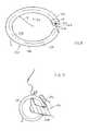
- FIG. 8is an isometric view of apparatus of another embodiment of the invention wherein the holder is formed of a curved bar extending in about a 360° loop.
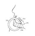
- FIG. 9is a sectional view of a portion of a patient, with the apparatus of FIG. 8 installed thereon.
- FIG. 1illustrates an apparatus 10 for taking measurements at the surface of mucosal tissue in the mouth region of a patient.
- the apparatuscomprises a sensor arrangement 12 that includes sensors 14 with sensor end portions 16 that can engage a mucosal surface (surface of mucosal tissue) in the mouth region of the patient, and a seal 20 extending around the sensor end portions.
- a holder 22holds the sensor arrangement 12 so the sensor end portions engage the mucosal surface, while the seal 20 seals the microenvironment 23 lying immediately around the sensor arrangement and within a hollow in the seal.
- the holder 22 shown in FIG. 1is a clasp comprising a bar of resilient material (elastomeric or not) extending in about a 180° loop.
- the clasp 22has first and second opposite end portions 24 , 26 and a middle portion 30 that connects them.
- the end portionsare substantially straight while the middle portion forms the 180° loop around a loop axis 32 .
- the barhas a bar axis 33 .
- the seal 20is in the form of a donut of elastomeric material (material with a Young's modulus of elasticity of no more that 50,000 psi).
- the sensors 14comprise a CO 2 sensor 40 and a temperature sensor 42 .
- the end portions of the sensorshave tips 44 , 46 that should contact the surface 48 of mucosal tissue 50 of the patient, in order to obtain accurate readings of the CO 2 partial pressure in the patient's tissue and to measure the temperature of the patient's body. It is possible for the seal 20 to be integral with the clasp 22 , but the seal can be herein referred to separately.
- FIG. 3shows the sensor tips 44 , 46 engaged with a location 52 on the mucosal tissue, with the seal 20 pressed against the mucosal tissue around the location 52 .
- the sealextends completely around the location, or in other words 360° around the location 52 , to prevent air circulation though the microenvironment 23 that lies within the seal. Any such air circulation would carry away some of the CO 2 and cool the tissue location, resulting in inaccurate CO 2 and temperature measurements.
- the sensor arrangementhas an axis 56 that extends normal (perpendicular) to the tissue surface 48 , and the seal extends in a 360° around the axis.
- Applicantprefers to position the sensor tips 44 , 46 so they are slightly recessed from the outer surface 54 of the undeformed elastomeric seal 20 , as shown in FIG. 2 .
- the sensor arrangementpresses with a force such as 100 grams against the mucosal tissue.
- the second end portion 26 of the clasp 22presses against the outside of the body in the mouth region, opposite the sensor arrangement 12 , the particular location shown being on an upper portion of the cheek of the patient. It is possible to use a seal of rigid material, in which case the sensor tips are close to being flush with the seal surface.
- FIG. 4shows a modified mucosal tissue sensing apparatus 60 wherein the clasp 62 has a cross-section that changes along its length.
- the seal axis 76is horizontal so the clasp 62 extends horizontally and through the patient's lips.
- the cross-sectionhas a horizontal width 70 that is at least as great as its vertical thickness 72 , to fit between the lips of the patient while enabling easy closing of the lips, as when oxygen is being administered.
- the clasphas a greater vertical V thickness (if the patient were standing) and the same or smaller horizontal width to hold a sensor arrangement of appreciable seal diameter. This construction facilitates mounting the apparatus 60 as shown in FIG. 5 wherein the clasp extends largely horizontally (with respect to the patient if he/she were standing).
- FIG. 6illustrates a portion of an apparatus 80 wherein the clasp type holder 22 is of the same construction as in FIG. 1 , but the sensor arrangement 82 is different.
- the sensor arrangement 82includes a suction cup 84 for the seal.
- the tips 44 , 46 of the sensor end portionsare recessed deeper with respect to the rim 86 of the suction cup, to enable compression of the suction cup and slight expansion to form a vacuum to hold it in place.
- This constructionis generally not preferred because it requires pressing the end portions of the clasp together with a higher force (e.g. 200 grams) and releasing the force to form a vacuum in the suction cup, and because the depression of the sensor tips into the mucosal tissue is more difficult to predict.
- FIG. 7illustrates a modified apparatus 100 wherein the clasp is in the general form of a clothespin. That is, the clasp includes first and second end portions 102 , 104 in the form of largely straight bars, and a spring 106 that urges the far ends 112 , 114 of the end portions towards each other. A sensor arrangement 116 lies on the first far end 112 .
- the first bar at 102can be constructed with a portion of reduced width to better fit between the lips of the patient.
- FIG. 8illustrates a modified apparatus 130 in which the clasp 132 is in the form of a bar of resilient material that is bent in a loop 135 of about 360° about a loop axis 133 .
- the barhas an axis 134 that extends along the center of the length of the bent bar, this axis also extending in about a 360° loop.
- the radius of curvature R of the bendis at least about 2 centimeters and preferably no more than 7 centimeters, so the clasp can fit around the chin of the patient, as shown in FIG. 9 without being cumbersome, with the first end portion 140 that bears a sensor arrangement 142 lying against sublingual mucosal tissue (tissue under the patient's tongue).
- the second end portion 144lies against the outside of the body, against the under-chin of the patient.
- the apparatus 130also can be placed against the cheek, as shown at 130 A in FIG. 5 .
- the seal 145 of the sensor arrangementcan be formed by the end of the bar, or by a separate part as shown in FIG. 8 .
- the axis 146 of the sensor arrangementis about parallel to the curved axis 134 of the curved bar, at the first end of the bar.
- the seal axis 56 at the part of the seal that engages the mucosal surfaceis perpendicular to the length of the first clasp end portion 24 .
- the inventionprovides apparatus for measuring a characteristic of a patient, such as the partial pressure of CO 2 and/or the temperature of the patient, by one or more sensors that press against mucosal tissue in the mouth region of the patient.
- the apparatusincludes a holder that holds a sensor, and a seal that extends 360° around the sensor, and that continually presses the sensor and seal against a mucosal surface location.
- the holdercomprises a clasp with first and second end portions that are pressed towards each other. The second end portion presses against the outside of the patient while the first end portion, which carries the seal and sensor, presses them against the mucosal surface at the inside of the patient's mouth.
- One claspis formed by a bar of resilient material that has a portion that extends in about a 180° loop and is especially useful for a sensor that presses against the cheek area.
- Another claspis formed by a bar of resilient material that extends in about a 360° loop and is especially useful to press against a mucosal surface location that lies under the tongue although it can be applied to the cheek.
- Another claspis in the general form of a clothespin.
- the sealis preferably formed by a ring (or square) of elastomeric material, but can be a suction cup.
Landscapes
- Health & Medical Sciences (AREA)
- Life Sciences & Earth Sciences (AREA)
- Engineering & Computer Science (AREA)
- Physics & Mathematics (AREA)
- Molecular Biology (AREA)
- General Health & Medical Sciences (AREA)
- Pathology (AREA)
- Biomedical Technology (AREA)
- Heart & Thoracic Surgery (AREA)
- Medical Informatics (AREA)
- Veterinary Medicine (AREA)
- Surgery (AREA)
- Animal Behavior & Ethology (AREA)
- Biophysics (AREA)
- Public Health (AREA)
- Human Computer Interaction (AREA)
- Optics & Photonics (AREA)
- Dentistry (AREA)
- Oral & Maxillofacial Surgery (AREA)
- Measuring And Recording Apparatus For Diagnosis (AREA)
- Measurement Of The Respiration, Hearing Ability, Form, And Blood Characteristics Of Living Organisms (AREA)
Abstract
Description
Claims (1)
Priority Applications (1)
| Application Number | Priority Date | Filing Date | Title |
|---|---|---|---|
| US10/860,829US8369920B2 (en) | 2004-03-09 | 2004-06-04 | Mucosal sensor adaptor |
Applications Claiming Priority (2)
| Application Number | Priority Date | Filing Date | Title |
|---|---|---|---|
| US55154604P | 2004-03-09 | 2004-03-09 | |
| US10/860,829US8369920B2 (en) | 2004-03-09 | 2004-06-04 | Mucosal sensor adaptor |
Publications (2)
| Publication Number | Publication Date |
|---|---|
| US20050203362A1 US20050203362A1 (en) | 2005-09-15 |
| US8369920B2true US8369920B2 (en) | 2013-02-05 |
Family
ID=34922738
Family Applications (1)
| Application Number | Title | Priority Date | Filing Date |
|---|---|---|---|
| US10/860,829Active - Reinstated2029-08-23US8369920B2 (en) | 2004-03-09 | 2004-06-04 | Mucosal sensor adaptor |
Country Status (1)
| Country | Link |
|---|---|
| US (1) | US8369920B2 (en) |
Families Citing this family (3)
| Publication number | Priority date | Publication date | Assignee | Title |
|---|---|---|---|---|
| US12115001B2 (en) | 2019-10-15 | 2024-10-15 | Exostat Medical, Inc. | Tissue perfusion sensor and placement device |
| EP4045901A4 (en)* | 2019-10-15 | 2023-11-08 | Exostat Medical, Inc. | CARBON DIOXIDE SENSOR |
| CN112401846B (en)* | 2020-11-20 | 2021-09-21 | 南通市第二人民医院 | Nursing system and method for mucosa in oral cavity |
Citations (25)
| Publication number | Priority date | Publication date | Assignee | Title |
|---|---|---|---|---|
| US5109849A (en)* | 1983-08-30 | 1992-05-05 | Nellcor, Inc. | Perinatal pulse oximetry sensor |
| US5511546A (en)* | 1993-09-20 | 1996-04-30 | Hon; Edward H. | Finger apparatus for measuring continuous cutaneous blood pressure and electrocardiogram electrode |
| US5579763A (en) | 1995-07-06 | 1996-12-03 | Institute Of Critical Care Medicine | Measurement of systemic perfusion |
| US5665477A (en)* | 1994-04-04 | 1997-09-09 | Graphic Controls Corporation | Hydrogel adhesive for attaching medical device to patient |
| US5800349A (en)* | 1996-10-15 | 1998-09-01 | Nonin Medical, Inc. | Offset pulse oximeter sensor |
| US6055447A (en)* | 1995-07-06 | 2000-04-25 | Institute Of Critical Care Medicine | Patient CO2 Measurement |
| US6071237A (en)* | 1999-02-19 | 2000-06-06 | Institute Of Critical Care Medicine | Device and method for assessing perfusion failure in a patient during endotracheal intubation |
| US6216024B1 (en)* | 1995-07-06 | 2001-04-10 | Institute Of Critical Care Medicine | Method and device for assessing perfusion failure in a patient |
| US6258046B1 (en)* | 1995-07-06 | 2001-07-10 | Institute Of Critical Care Medicine | Method and device for assessing perfusion failure in a patient by measurement of blood flow |
| US20010009265A1 (en)* | 1999-08-26 | 2001-07-26 | Schulz Christian E. | Shielded optical probe and method |
| US6285899B1 (en)* | 1999-02-18 | 2001-09-04 | Motorola, Inc. | Remotely interrogated biomedical sensor |
| US20010029324A1 (en)* | 2000-02-11 | 2001-10-11 | Walker Steven C. | Pacifier pulse oximeter sensor |
| US20010045532A1 (en)* | 1991-03-21 | 2001-11-29 | Schulz Christian E. | Shielded optical probe having an electrical connector |
| US20020028990A1 (en)* | 1998-09-09 | 2002-03-07 | Shepherd John M. | Device and method for monitoring arterial oxygen saturation |
| US6411834B1 (en)* | 1999-09-03 | 2002-06-25 | Nihon Kohden Corporation | Biological sensor |
| US20030225324A1 (en)* | 2002-06-03 | 2003-12-04 | Anderson Edward J. | Noninvasive detection of a physiologic Parameter within a body tissue of a patient |
| US20030236452A1 (en)* | 2002-06-20 | 2003-12-25 | Richard Melker | Novel non-invasive perfusion monitor and system, specially configured oximeter probes, methods of using same, and covers for probes |
| US20040006263A1 (en)* | 2002-06-03 | 2004-01-08 | Anderson Edward J. | Noninvasive detection of a physiologic parameter within a body tissue of a patient |
| US20040054291A1 (en)* | 2002-09-14 | 2004-03-18 | Christian Schulz | Pulse oximetry ear sensor |
| US20040230108A1 (en)* | 2002-06-20 | 2004-11-18 | Melker Richard J. | Novel specially configured nasal pulse oximeter/photoplethysmography probes, and combined nasal probe/cannula, selectively with sampler for capnography, and covering sleeves for same |
| US20050085704A1 (en)* | 2003-10-14 | 2005-04-21 | Christian Schulz | Variable pressure reusable sensor |
| US20070078307A1 (en)* | 2005-09-30 | 2007-04-05 | Debreczeny Martin P | Sensor for tissue gas detection and technique for using the same |
| US20070078318A1 (en)* | 2005-09-30 | 2007-04-05 | Carl Kling | Mucosal sensor for the assessment of tissue and blood constituents and technique for using the same |
| US20070078317A1 (en)* | 2005-09-30 | 2007-04-05 | Matlock George L | Folding medical sensor and technique for using the same |
| US20070106168A1 (en)* | 2005-11-10 | 2007-05-10 | O'neil Michael P | Medical sensor and technique for using the same |
- 2004
- 2004-06-04USUS10/860,829patent/US8369920B2/enactiveActive - Reinstated
Patent Citations (29)
| Publication number | Priority date | Publication date | Assignee | Title |
|---|---|---|---|---|
| US5109849A (en)* | 1983-08-30 | 1992-05-05 | Nellcor, Inc. | Perinatal pulse oximetry sensor |
| US20010045532A1 (en)* | 1991-03-21 | 2001-11-29 | Schulz Christian E. | Shielded optical probe having an electrical connector |
| US20030162414A1 (en)* | 1991-03-21 | 2003-08-28 | Schulz Christian E. | Shielded optical probe having an electrical connector |
| US5511546A (en)* | 1993-09-20 | 1996-04-30 | Hon; Edward H. | Finger apparatus for measuring continuous cutaneous blood pressure and electrocardiogram electrode |
| US5665477A (en)* | 1994-04-04 | 1997-09-09 | Graphic Controls Corporation | Hydrogel adhesive for attaching medical device to patient |
| US5579763A (en) | 1995-07-06 | 1996-12-03 | Institute Of Critical Care Medicine | Measurement of systemic perfusion |
| US6055447A (en)* | 1995-07-06 | 2000-04-25 | Institute Of Critical Care Medicine | Patient CO2 Measurement |
| US6216024B1 (en)* | 1995-07-06 | 2001-04-10 | Institute Of Critical Care Medicine | Method and device for assessing perfusion failure in a patient |
| US6258046B1 (en)* | 1995-07-06 | 2001-07-10 | Institute Of Critical Care Medicine | Method and device for assessing perfusion failure in a patient by measurement of blood flow |
| US5800349A (en)* | 1996-10-15 | 1998-09-01 | Nonin Medical, Inc. | Offset pulse oximeter sensor |
| US20020028990A1 (en)* | 1998-09-09 | 2002-03-07 | Shepherd John M. | Device and method for monitoring arterial oxygen saturation |
| US6285899B1 (en)* | 1999-02-18 | 2001-09-04 | Motorola, Inc. | Remotely interrogated biomedical sensor |
| US6071237A (en)* | 1999-02-19 | 2000-06-06 | Institute Of Critical Care Medicine | Device and method for assessing perfusion failure in a patient during endotracheal intubation |
| US20010009265A1 (en)* | 1999-08-26 | 2001-07-26 | Schulz Christian E. | Shielded optical probe and method |
| US6411834B1 (en)* | 1999-09-03 | 2002-06-25 | Nihon Kohden Corporation | Biological sensor |
| US20010029324A1 (en)* | 2000-02-11 | 2001-10-11 | Walker Steven C. | Pacifier pulse oximeter sensor |
| US20040006263A1 (en)* | 2002-06-03 | 2004-01-08 | Anderson Edward J. | Noninvasive detection of a physiologic parameter within a body tissue of a patient |
| US20030225324A1 (en)* | 2002-06-03 | 2003-12-04 | Anderson Edward J. | Noninvasive detection of a physiologic Parameter within a body tissue of a patient |
| US20040260161A1 (en)* | 2002-06-20 | 2004-12-23 | Melker Richard J. | Novel specially configured lip/cheek pulse oximeter/photoplethysmography probes, selectively with sampler for capnography, and covering sleeves for same |
| US20040230108A1 (en)* | 2002-06-20 | 2004-11-18 | Melker Richard J. | Novel specially configured nasal pulse oximeter/photoplethysmography probes, and combined nasal probe/cannula, selectively with sampler for capnography, and covering sleeves for same |
| US20030236452A1 (en)* | 2002-06-20 | 2003-12-25 | Richard Melker | Novel non-invasive perfusion monitor and system, specially configured oximeter probes, methods of using same, and covers for probes |
| US7127278B2 (en)* | 2002-06-20 | 2006-10-24 | University Of Florida Research Foundation, Inc. | Specially configured lip/cheek pulse oximeter/photoplethysmography probes, selectively with sampler for capnography, and covering sleeves for same |
| US20070027375A1 (en)* | 2002-06-20 | 2007-02-01 | Melker Richard J | Optimized gas supply using photoplethysmography |
| US20040054291A1 (en)* | 2002-09-14 | 2004-03-18 | Christian Schulz | Pulse oximetry ear sensor |
| US20050085704A1 (en)* | 2003-10-14 | 2005-04-21 | Christian Schulz | Variable pressure reusable sensor |
| US20070078307A1 (en)* | 2005-09-30 | 2007-04-05 | Debreczeny Martin P | Sensor for tissue gas detection and technique for using the same |
| US20070078318A1 (en)* | 2005-09-30 | 2007-04-05 | Carl Kling | Mucosal sensor for the assessment of tissue and blood constituents and technique for using the same |
| US20070078317A1 (en)* | 2005-09-30 | 2007-04-05 | Matlock George L | Folding medical sensor and technique for using the same |
| US20070106168A1 (en)* | 2005-11-10 | 2007-05-10 | O'neil Michael P | Medical sensor and technique for using the same |
Also Published As
| Publication number | Publication date |
|---|---|
| US20050203362A1 (en) | 2005-09-15 |
Similar Documents
| Publication | Publication Date | Title |
|---|---|---|
| US7054680B1 (en) | Device for detecting electrical potentials in the forehead-area of a patient | |
| US5660168A (en) | Attachment arrangement | |
| JP4721130B2 (en) | Pulse oximeter probe | |
| US7263396B2 (en) | Ear sensor assembly | |
| US5218970A (en) | Tracheal tube cuff pressure monitor | |
| CA1135525A (en) | Capsule for detecting changes in the shape of a body wall | |
| US4989615A (en) | Apparatus for non-invasive monitoring of uterine contractions | |
| US20100222706A1 (en) | Probe for measuring oral pressure, device for measuring oral pressure using the same, and training tool for restoring oral function | |
| TW200631553A (en) | Sphygmomanometer cuff and sphygmomanometer | |
| EP1374761A3 (en) | Blood-pressure determining apparatus | |
| JP2001275994A (en) | Instrument and probe for measuring oral cavity-related pressure | |
| RU2006108086A (en) | CUFF OF THE APPARATUS FOR MEASUREMENT OF ARTERIAL PRESSURE AND THE APPARATUS FOR MEASUREMENT OF ARTERIAL PRESSURE | |
| US8369920B2 (en) | Mucosal sensor adaptor | |
| US6190335B1 (en) | Orofacial myographic measurement probe | |
| CN210871562U (en) | A sphygmomanometer for preventing cross infection | |
| JP4743533B2 (en) | Oral pressure measuring probe and oral pressure measuring apparatus using the same | |
| CN113167761A (en) | Carbon dioxide sensor | |
| WO2012165427A1 (en) | Respiration detection device | |
| JP3731183B2 (en) | Contact pressure blood flow sensor | |
| JP2007206000A (en) | Lip pressure measuring device | |
| JPWO2014102961A1 (en) | Respiration detection device | |
| US8104468B2 (en) | Laryngeal mask airway and clip device | |
| KR102688645B1 (en) | Breathalyzer with non-contact mouthpiece separation function | |
| JP7752882B2 (en) | Tissue perfusion sensor and placement device | |
| WO2004011873A3 (en) | Tape measure with securement |
Legal Events
| Date | Code | Title | Description |
|---|---|---|---|
| AS | Assignment | Owner name:INSTITUTE OF CRITICAL CARE MEDICINE, CALIFORNIA Free format text:ASSIGNMENT OF ASSIGNORS INTEREST;ASSIGNORS:CASTILLO, CARLOS;WEIL, MAX HARRY;BISERA, JOE;AND OTHERS;REEL/FRAME:015441/0927 Effective date:20040602 | |
| AS | Assignment | Owner name:WEIL INSTITUTE OF CRITICAL CARE MEDICINE, CALIFORN Free format text:CHANGE OF NAME;ASSIGNOR:INSTITUTE OF CRITICAL CARE MEDICINE;REEL/FRAME:033180/0021 Effective date:20110513 | |
| REMI | Maintenance fee reminder mailed | ||
| LAPS | Lapse for failure to pay maintenance fees | ||
| FP | Lapsed due to failure to pay maintenance fee | Effective date:20170205 | |
| AS | Assignment | Owner name:SUNLIFE SCIENCE (SUZHOU) INC., CHINA Free format text:ASSIGNMENT OF ASSIGNORS INTEREST;ASSIGNOR:WEIL INSTITUTE OF CRITICAL CARE MEDICINE;REEL/FRAME:044754/0426 Effective date:20171211 | |
| FEPP | Fee payment procedure | Free format text:SURCHARGE, PETITION TO ACCEPT PYMT AFTER EXP, UNINTENTIONAL. (ORIGINAL EVENT CODE: M2558); ENTITY STATUS OF PATENT OWNER: SMALL ENTITY Free format text:PETITION RELATED TO MAINTENANCE FEES GRANTED (ORIGINAL EVENT CODE: PMFG) Free format text:PETITION RELATED TO MAINTENANCE FEES FILED (ORIGINAL EVENT CODE: PMFP) | |
| MAFP | Maintenance fee payment | Free format text:PAYMENT OF MAINTENANCE FEE, 4TH YR, SMALL ENTITY (ORIGINAL EVENT CODE: M2551) Year of fee payment:4 | |
| PRDP | Patent reinstated due to the acceptance of a late maintenance fee | Effective date:20171211 | |
| STCF | Information on status: patent grant | Free format text:PATENTED CASE | |
| MAFP | Maintenance fee payment | Free format text:PAYMENT OF MAINTENANCE FEE, 8TH YR, SMALL ENTITY (ORIGINAL EVENT CODE: M2552); ENTITY STATUS OF PATENT OWNER: SMALL ENTITY Year of fee payment:8 | |
| MAFP | Maintenance fee payment | Free format text:PAYMENT OF MAINTENANCE FEE, 12TH YR, SMALL ENTITY (ORIGINAL EVENT CODE: M2553); ENTITY STATUS OF PATENT OWNER: SMALL ENTITY Year of fee payment:12 |