US8363918B2 - Method and system for anatomic landmark detection using constrained marginal space learning and geometric inference - Google Patents
Method and system for anatomic landmark detection using constrained marginal space learning and geometric inferenceDownload PDFInfo
- Publication number
- US8363918B2 US8363918B2US12/604,495US60449509AUS8363918B2US 8363918 B2US8363918 B2US 8363918B2US 60449509 AUS60449509 AUS 60449509AUS 8363918 B2US8363918 B2US 8363918B2
- Authority
- US
- United States
- Prior art keywords
- detecting
- image
- landmarks
- orientation
- landmark
- Prior art date
- Legal status (The legal status is an assumption and is not a legal conclusion. Google has not performed a legal analysis and makes no representation as to the accuracy of the status listed.)
- Expired - Fee Related, expires
Links
Images


Classifications
- G—PHYSICS
- G06—COMPUTING OR CALCULATING; COUNTING
- G06T—IMAGE DATA PROCESSING OR GENERATION, IN GENERAL
- G06T7/00—Image analysis
- G06T7/70—Determining position or orientation of objects or cameras
- G06T7/73—Determining position or orientation of objects or cameras using feature-based methods
- G06T7/75—Determining position or orientation of objects or cameras using feature-based methods involving models
- G—PHYSICS
- G06—COMPUTING OR CALCULATING; COUNTING
- G06T—IMAGE DATA PROCESSING OR GENERATION, IN GENERAL
- G06T7/00—Image analysis
- G06T7/70—Determining position or orientation of objects or cameras
- G06T7/77—Determining position or orientation of objects or cameras using statistical methods
- G—PHYSICS
- G06—COMPUTING OR CALCULATING; COUNTING
- G06V—IMAGE OR VIDEO RECOGNITION OR UNDERSTANDING
- G06V10/00—Arrangements for image or video recognition or understanding
- G06V10/20—Image preprocessing
- G06V10/25—Determination of region of interest [ROI] or a volume of interest [VOI]
- G—PHYSICS
- G06—COMPUTING OR CALCULATING; COUNTING
- G06V—IMAGE OR VIDEO RECOGNITION OR UNDERSTANDING
- G06V30/00—Character recognition; Recognising digital ink; Document-oriented image-based pattern recognition
- G06V30/10—Character recognition
- G06V30/24—Character recognition characterised by the processing or recognition method
- G06V30/248—Character recognition characterised by the processing or recognition method involving plural approaches, e.g. verification by template match; Resolving confusion among similar patterns, e.g. "O" versus "Q"
- G06V30/2504—Coarse or fine approaches, e.g. resolution of ambiguities or multiscale approaches
- A—HUMAN NECESSITIES
- A61—MEDICAL OR VETERINARY SCIENCE; HYGIENE
- A61B—DIAGNOSIS; SURGERY; IDENTIFICATION
- A61B34/00—Computer-aided surgery; Manipulators or robots specially adapted for use in surgery
- A61B34/10—Computer-aided planning, simulation or modelling of surgical operations
- A61B2034/101—Computer-aided simulation of surgical operations
- A61B2034/105—Modelling of the patient, e.g. for ligaments or bones
- G—PHYSICS
- G06—COMPUTING OR CALCULATING; COUNTING
- G06T—IMAGE DATA PROCESSING OR GENERATION, IN GENERAL
- G06T2207/00—Indexing scheme for image analysis or image enhancement
- G06T2207/10—Image acquisition modality
- G06T2207/10072—Tomographic images
- G06T2207/10081—Computed x-ray tomography [CT]
- G—PHYSICS
- G06—COMPUTING OR CALCULATING; COUNTING
- G06T—IMAGE DATA PROCESSING OR GENERATION, IN GENERAL
- G06T2207/00—Indexing scheme for image analysis or image enhancement
- G06T2207/10—Image acquisition modality
- G06T2207/10072—Tomographic images
- G06T2207/10088—Magnetic resonance imaging [MRI]
- G—PHYSICS
- G06—COMPUTING OR CALCULATING; COUNTING
- G06T—IMAGE DATA PROCESSING OR GENERATION, IN GENERAL
- G06T2207/00—Indexing scheme for image analysis or image enhancement
- G06T2207/10—Image acquisition modality
- G06T2207/10132—Ultrasound image
- G—PHYSICS
- G06—COMPUTING OR CALCULATING; COUNTING
- G06T—IMAGE DATA PROCESSING OR GENERATION, IN GENERAL
- G06T2207/00—Indexing scheme for image analysis or image enhancement
- G06T2207/30—Subject of image; Context of image processing
- G06T2207/30004—Biomedical image processing
- G06T2207/30016—Brain
- G—PHYSICS
- G06—COMPUTING OR CALCULATING; COUNTING
- G06V—IMAGE OR VIDEO RECOGNITION OR UNDERSTANDING
- G06V2201/00—Indexing scheme relating to image or video recognition or understanding
- G06V2201/03—Recognition of patterns in medical or anatomical images
- G06V2201/031—Recognition of patterns in medical or anatomical images of internal organs
Definitions
- the present inventionrelates to 3D object detection in images, and more particularly, to automated detection of 3D anatomical structures in medical images using marginal space learning.
- Efficiently localizing anatomical structurese.g., heart, liver, kidney, etc.
- medical imagesare often a prerequisite for further diagnostic image processing procedures, such as segmentation, measuring, and classification.
- Detecting and segmenting human anatomic structures in 3D medical image volumesis a challenging problem, which is typically more difficult than detecting anatomic structures in 2D images.
- MSLmarginal space learning
- the full parameter space for 3D object localizationhas nine dimensions: three for position (P x , P y , and P z ), three for orientation (represented with Euler angles, ⁇ , ⁇ , and ⁇ ), and three for anisotropic scaling (S x , S y , and S z ).
- parameter estimationis performed in a series of marginal spaces with increasing dimensionality.
- the object detectionis split into three steps: object position estimation, position-orientation estimation, and similarity transformation estimation. Each step results in a relatively small number of candidates, which are used in the following step.
- MSLhas been successfully applied to many 3D anatomical detection problems in medical imaging, such as ileocecal valves, polyps, and livers in abdominal CT, brain tissues and heart chambers in ultrasound images, and heart chambers in MRI.
- MSLcan reduce the number of testing hypotheses by approximately six orders of magnitude as compared with uniformly searching the nine-dimensional parameter space. However, in many cases MSL tests more testing hypotheses than necessary for accurate object detection. Accordingly, it is desirable to further increase the efficiency of anatomical object detection using MSL.
- the present inventionprovides efficient detection of 3D anatomical landmarks in medical images using constrained marginal space learning (MSL) and geometric inference.
- MSLconstrained marginal space learning
- Embodiments of the present inventioncan be used to constrain the search space for landmark detection by exploiting geometric constraints among object pose parameters (i.e., position, orientation, and scale) of multiple 3D anatomical landmarks.
- a first landmark of a plurality of anatomical landmarksis detected in an image using marginal space learning (MSL). Locations of remaining landmarks of the plurality of anatomical landmarks are estimated in the image based on the detected first landmark using a learned geometric model relating the plurality of anatomical landmarks. Each of the remaining landmarks is then detected using MSL in a portion of the image constrained based on the estimated location of each of the remaining landmarks.
- MSLmarginal space learning
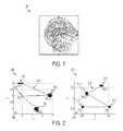
- FIG. 1illustrates a slice of an MR brain volume showing the four anatomic landmarks
- FIG. 2illustrates exemplary geometric models learned from training data
- FIG. 3illustrates a method of detecting multiple anatomic landmarks in a 3D volume according to an embodiment of the present invention
- FIG. 4illustrates stages of marginal space learning (MSL).
- FIG. 5illustrates results of estimating anatomic landmarks in a brain MRI volume based on a first detected anatomic landmark using a learned geometric model
- FIG. 6illustrates exemplary detection results of anatomical structures in a brain MRI volume
- FIG. 7is a high level block diagram of a computer capable of implementing the present invention.
- the present inventionis directed to a method for detecting anatomical landmarks in medical images, such as computed tomography (CT), magnetic resonance imaging (MRI), ultrasound, etc.
- CTcomputed tomography
- MRImagnetic resonance imaging
- Embodiments of the present inventionare described herein to give a visual understanding of the anatomical landmark detection method.
- a digital imageis often composed of digital representations of one or more objects (or shapes).
- the digital representation of an objectis often described herein in terms of identifying and manipulating the objects.
- Such manipulationsare virtual manipulations accomplished in the memory or other circuitry/hardware of a computer system. Accordingly, is to be understood that embodiments of the present invention may be performed within a computer system using data stored within the computer system.
- an anatomical structureis detected in a 3D medical image using constrained marginal space learning (MSL).
- MSLis a technique for efficiently detecting or localizing a 3D object in an image using learned discriminative classifiers.
- the full parameter space for 3D object localizationhas nine dimensions: three for position (P x , P y , and P z ), three for orientation (represented with Euler angles, ⁇ , ⁇ , and ⁇ ), and three for anisotropic scaling (S x , S y , and S z ).
- P x , P y , and P zthree for orientation (represented with Euler angles, ⁇ , ⁇ , and ⁇ )
- S x , S y , and S zthree for anisotropic scaling
- the object detectionis split into three steps: object position estimation, position-orientation estimation, and similarity transformation estimation.
- Each of these stepsuses a separately trained classifier.
- a classifier for each detection stepcan be trained based on training data using the concept of the probabilistic boosting tree (PBT).
- PBTprobabilistic boosting tree
- the present inventionis not limited to a PBT and other classifiers can be similarly applied.
- MSLhas been successfully applied to many 3D anatomical structures detection problems in medical imaging (e.g., ileocecal valve, polyps, and livers in abdominal CT images, brain tissues and heart chambers in ultrasound images, and heart chambers in MRI images). Depending on the application scenario, it is possible to reduce the search range within each marginal space.
- medical imaginge.g., ileocecal valve, polyps, and livers in abdominal CT images, brain tissues and heart chambers in ultrasound images, and heart chambers in MRI images.
- MSLcan be used to detect multiple anatomic landmarks in a volume.
- a localizer image of the brainis acquired.
- Various landmarkssuch as the crista galli, tip of the occipital bone, anterior of the corpus collosum, and posterior of the corpus collosum, are detected in the localizer image and the scan prescription is set based on the detected landmarks.
- Each of these landmarksmay be detected using separate MSL searches.
- Embodiments of the present inventionexploit geometric relationships between anatomic landmarks in a volume to constrain the search space of multiple MSL searches.
- Embodiments of the present inventionare directed to detecting a number N t of anatomic landmarks in a medical image.
- Embodiments of the present inventionare directed to detecting anatomic landmarks in 3D medical imaging volumes, and are described herein using the example of detecting multiple landmarks in a brain MRI volume.
- the present inventionis not limited to any particular anatomic location, number of landmarks, or medical imaging modality. Further, the present invention is not limited to 3D volumes, and can be similarly applied to 2D images as well.
- FIG. 1illustrates a cross-section 100 of an MR brain volume showing the four anatomic landmarks.
- the four anatomic landmarksare the crista galli (CG), the tip of the occipital bone (OB), the anterior of the corpus callosum (ACC), and the posterior of the corpus callosum (PCC).
- Geometric models relating the multiple parameterscan be trained based on annotated training data. For all available training volumes t, a 3 ⁇ N t dimensional ground truth vector
- the local geometric modelsare obtained from the mean shape, i.e. by applying:
- FIG. 2illustrates exemplary geometric models 200 and 210 learned from training data.
- model 200annotations 202 for the CG, OB, and PCC from multiple training volumes are converted to a local coordinate system 206 , and the mean shape 204 of the geometric model is calculated.
- annotations 212 for the CG, OB, ACC, and PCC from multiple training volumesare converted to a local coordinate system 216 , and the mean shape 214 of the geometric model is calculated.
- MSLcan be applied to detect multiple parameters by training a detector for each geometric model.
- N d detectorsare obtained.
- the detection of landmarkscan be approached successively using a first detected location and geometric inference.
- FIG. 3illustrates a method of detecting multiple anatomic landmarks in a 3D volume according to an embodiment of the present invention.
- FIG. 3is described by referring to the example of detecting the CG, OB, ACC, and PCC in a brain MRI volume, but the method is not limited thereto, and can be used for detection of any number of landmarks in any anatomical location and any medical imaging modality.
- a 3D volumeis received.
- the 3D volumecan be a brain MRI volume.
- the present inventionis not limited thereto, and the 3D volume can be a medical image resulting from any type of imaging modality.
- the 3D volumecan be received directly from an image acquisition device, such as an MR scanner. It is also possible that the 3D volume can be received by loading a 3D volume stored, for example, on a memory or storage of a computer system or some other computer readable storage medium.
- a first anatomical landmarkis detected in the 3D volume using MSL.
- first landmark detectorthat has been trained based on training data is used to scan the entire 3D volume to detect the first landmark.
- the first landmark detectorutilizes three classifiers corresponding to the three stages of MSL.
- FIG. 4illustrates the stages of MSL.
- the first landmarkis detected in the 3D volume using a position estimation stage 402 , a position-orientation stage 404 , and a full similarity transformation stage 406 .
- a classifieris trained for each of the MSL stages 402 , 404 , and 406 based on training data using a PBT.
- the position classifierdetects candidates for the position of the first landmark in the 3D volume.
- Position-orientation hypothesesare generated from detected position candidates.
- the position-orientation classifierdetects position-orientation candidates from the position-orientation hypotheses.
- Similarity transformation hypothesesare generated from the position-orientation candidates.
- the similarity transformation classifierthan detects similarity transformation candidates from the similarity transformation hypotheses.
- the similarity transformation (position, orientation, and scale) for the first anatomic landmarkcan be determined by selecting the best similarity transformation candidate or by aggregating a certain number of best similarity transformation candidates.
- any one of the CG, OB, ACC, and PCCcan be detected as the first landmark using MSL.
- the locations of the remaining anatomic landmarksare estimated in the 3D volume based on the detected first anatomic landmarks using a learned geometric model.
- geometric models relating the multiple anatomic landmarkssuch as those shown in FIG. 2 , are trained based on annotated training data.
- the detection of the first anatomic landmarkresults in a nine parameter similarity transformation. This similarity transformation is used to transform the mean shape of the learned geometric model obtained by Equations (1) and (2) in order to infer the locations of all of the remaining anatomical landmarks based on the first anatomic landmark.
- FIG. 5illustrates results of estimating anatomic landmarks in a brain MRI volume based on a first detected anatomic landmark using a learned geometric model.
- image 500is a cross-section of a brain MRI volume showing annotated ground truth locations for the ACC 502 , PCC 504 , CG 506 , and OB 508 .
- Image 500also shows estimated locations of the ACC 512 , PCC 524 , CG 516 , and OB 508 resulting from detecting one or the landmarks using MSL and inferring the remaining landmarks based on the detected landmark using a geometric model.
- each of the remaining anatomical landmarksare detected using MSL constrained to a portion of the 3D volume surrounding the estimated location of the each anatomical landmark.
- the trained MSL-based detector for each landmarkis used to detect the landmark in a constrained parameter space surrounding the estimated location of the landmark.
- the position classifier of each landmark detectormay only scan voxels within a certain distance of the estimated location of the landmark as potential candidates for the position of the landmark. MSL detection can then continues as describe above or stop after position detection.
- the constrained MSL detection of each landmarkrefines the position of each landmark estimated using the geometric model.
- the first anatomical landmarkis not required to be further refined using constrained MSL, according to a possible implementation, the first anatomical landmark can also be refined using MSL detection constrained based on the previously detected location of the first anatomical landmark.
- the landmarkscan be constrained to a plane (i.e., the mid-sagittal plane).
- the landmarkscan be constrained by solving the least square minimization problem:
- Equation (3)y j denotes the detected anatomical landmarks, p denotes the position of the plane, and n denotes the plane normal.
- the anatomical landmark detection resultsare output.
- the detection resultscan be output by displaying the detection results on a display of a computer system.
- the anatomical landmark detection resultscan also be output by storing the detection results, for example, on a memory or storage of a computer system or on a computer readable storage medium. It is possible to use the anatomical landmark detection results in subsequent medical imaging techniques. For example, detected anatomical landmarks in a localizer brain MRI volume can be used in MRI scan planning to determine an alignment of an MR scanner for a high resolution scan.
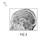
- FIG. 6illustrates exemplary detection results of anatomical structures in a brain MRI volume.
- image 600is a cross-section of a brain MRI volume showing annotated ground truth locations for the ACC 602 , PCC 604 , CG 606 , and OB 608 as well as the final detected locations for the ACC 612 , PCC 614 , CG 616 , and OB 618 .
- the final detected anatomical landmark locations 612 , 614 , 616 , and 618are detected using MSL constrained based on the estimated anatomical landmark locations 512 , 514 , 516 , and 518 shown in FIG. 5 .
- Computer 702contains a processor 704 which controls the overall operation of the computer 702 by executing computer program instructions which define such operation.
- the computer program instructionsmay be stored in a storage device 712 (e.g., magnetic disk) and loaded into memory 710 when execution of the computer program instructions is desired.
- a storage device 712e.g., magnetic disk
- the steps of the methods of FIG. 3may be defined by the computer program instructions stored in the memory 710 and/or storage 712 and controlled by the processor 704 executing the computer program instructions.
- An image acquisition device 720such as an MRI scanning device, can be connected to the computer 702 to input the 3D images (volumes) to the computer 702 . It is possible to implement the image acquisition device 720 and the computer 702 as one device. It is also possible that the image acquisition device 720 and the computer 702 communicate wirelessly through a network.
- the computer 702also includes one or more network interfaces 706 for communicating with other devices via a network.
- the computer 702also includes other input/output devices 708 that enable user interaction with the computer 702 (e.g., display, keyboard, mouse, speakers, buttons, etc.)
- FIG. 7is a high level representation of some of the components of such a computer for illustrative purposes.
Landscapes
- Engineering & Computer Science (AREA)
- Physics & Mathematics (AREA)
- General Physics & Mathematics (AREA)
- Theoretical Computer Science (AREA)
- Computer Vision & Pattern Recognition (AREA)
- Multimedia (AREA)
- Life Sciences & Earth Sciences (AREA)
- Bioinformatics & Cheminformatics (AREA)
- Bioinformatics & Computational Biology (AREA)
- Evolutionary Biology (AREA)
- Probability & Statistics with Applications (AREA)
- Magnetic Resonance Imaging Apparatus (AREA)
Abstract
Description
of annotations is needed. Multiple geometric models iε{1, . . . Nd} can be constructed. For each training volume t, the geometric model includes a position ti,tε

yi,j,t=Si,t−1Ri,tT(yj,t−ti,t). (1)
The local geometric models are obtained from the mean shape, i.e. by applying:
The learned mean geometric model resulting from Equation (2) is used in the landmark detection method described below.
and projecting the detected results to the plane using, for example, a well-known Gram-Schimdt optimization. In Equation (3), yjdenotes the detected anatomical landmarks, p denotes the position of the plane, and n denotes the plane normal.
Claims (23)
Priority Applications (1)
| Application Number | Priority Date | Filing Date | Title |
|---|---|---|---|
| US12/604,495US8363918B2 (en) | 2008-11-12 | 2009-10-23 | Method and system for anatomic landmark detection using constrained marginal space learning and geometric inference |
Applications Claiming Priority (2)
| Application Number | Priority Date | Filing Date | Title |
|---|---|---|---|
| US11370508P | 2008-11-12 | 2008-11-12 | |
| US12/604,495US8363918B2 (en) | 2008-11-12 | 2009-10-23 | Method and system for anatomic landmark detection using constrained marginal space learning and geometric inference |
Publications (2)
| Publication Number | Publication Date |
|---|---|
| US20100119137A1 US20100119137A1 (en) | 2010-05-13 |
| US8363918B2true US8363918B2 (en) | 2013-01-29 |
Family
ID=42165254
Family Applications (1)
| Application Number | Title | Priority Date | Filing Date |
|---|---|---|---|
| US12/604,495Expired - Fee RelatedUS8363918B2 (en) | 2008-11-12 | 2009-10-23 | Method and system for anatomic landmark detection using constrained marginal space learning and geometric inference |
Country Status (1)
| Country | Link |
|---|---|
| US (1) | US8363918B2 (en) |
Cited By (5)
| Publication number | Priority date | Publication date | Assignee | Title |
|---|---|---|---|---|
| US10318839B2 (en) | 2015-01-13 | 2019-06-11 | Council Of Scientific And Industrial Research | Method for automatic detection of anatomical landmarks in volumetric data |
| KR102044237B1 (en) | 2018-10-23 | 2019-11-13 | 연세대학교 산학협력단 | Shadowed 2D image-based machine learning, and automatic 3D landmark detection method and apparatus using thereof |
| KR20210066074A (en) | 2019-11-27 | 2021-06-07 | 기초과학연구원 | 3D anatomical landmark detection method and apparatus |
| US11486950B2 (en)* | 2018-08-01 | 2022-11-01 | General Electric Company | Systems and methods for automated graphical prescription with deep neural networks |
| US12263017B2 (en) | 2018-08-01 | 2025-04-01 | General Electric Company | Plane selection using localizer images |
Families Citing this family (10)
| Publication number | Priority date | Publication date | Assignee | Title |
|---|---|---|---|---|
| US8605969B2 (en) | 2010-04-06 | 2013-12-10 | Siemens Corporation | Method and system for multiple object detection by sequential Monte Carlo and hierarchical detection network |
| US8811697B2 (en) | 2010-04-06 | 2014-08-19 | Siemens Aktiengesellschaft | Data transmission in remote computer assisted detection |
| US8787635B2 (en) | 2010-05-03 | 2014-07-22 | Siemens Aktiengesellschaft | Optimization of multiple candidates in medical device or feature tracking |
| EP2603136B1 (en)* | 2010-08-13 | 2023-07-12 | Smith & Nephew, Inc. | Detection of anatomical landmarks |
| KR101805624B1 (en)* | 2011-08-29 | 2017-12-08 | 삼성전자주식회사 | Method and apparatus for generating organ medel image |
| RU2530220C1 (en) | 2013-03-18 | 2014-10-10 | Корпорация "САМСУНГ ЭЛЕКТРОНИКС Ко., Лтд." | System and method for automatic detection of anatomical points in three-dimensional medical images |
| KR102130963B1 (en)* | 2018-07-09 | 2020-07-08 | 라온피플 주식회사 | Apparatus and method for analyzing cephalic image |
| CN110021021B (en)* | 2018-07-09 | 2024-06-07 | 乐人美德株式会社 | Head image analysis device and image analysis method |
| KR102348036B1 (en)* | 2019-12-05 | 2022-01-10 | 울산대학교 산학협력단 | Prediction apparatus for predicting anatomical landmarks and a prediction method thereof |
| KR102706418B1 (en)* | 2021-12-14 | 2024-09-11 | 사회복지법인 삼성생명공익재단 | Cephalometric landmark detection method based on deep reinforcement learning and analysis apparatus |
Citations (5)
| Publication number | Priority date | Publication date | Assignee | Title |
|---|---|---|---|---|
| US20080085050A1 (en) | 2006-09-28 | 2008-04-10 | Siemens Corporate Research, Inc. | System and Method For Detecting An Object In A High Dimensional Space |
| US20080101676A1 (en) | 2006-09-28 | 2008-05-01 | Siemens Corporate Research, Inc. | System and Method For Segmenting Chambers Of A Heart In A Three Dimensional Image |
| US20080211812A1 (en) | 2007-02-02 | 2008-09-04 | Adrian Barbu | Method and system for detection and registration of 3D objects using incremental parameter learning |
| US20090080745A1 (en) | 2007-09-21 | 2009-03-26 | Yefeng Zheng | Method and system for measuring left ventricle volume |
| US20090087066A1 (en) | 2007-10-02 | 2009-04-02 | Odry Benjamin L | Method and system for vessel enhancement and artifact reduction in TOF MR angiography of brain |
- 2009
- 2009-10-23USUS12/604,495patent/US8363918B2/ennot_activeExpired - Fee Related
Patent Citations (5)
| Publication number | Priority date | Publication date | Assignee | Title |
|---|---|---|---|---|
| US20080085050A1 (en) | 2006-09-28 | 2008-04-10 | Siemens Corporate Research, Inc. | System and Method For Detecting An Object In A High Dimensional Space |
| US20080101676A1 (en) | 2006-09-28 | 2008-05-01 | Siemens Corporate Research, Inc. | System and Method For Segmenting Chambers Of A Heart In A Three Dimensional Image |
| US20080211812A1 (en) | 2007-02-02 | 2008-09-04 | Adrian Barbu | Method and system for detection and registration of 3D objects using incremental parameter learning |
| US20090080745A1 (en) | 2007-09-21 | 2009-03-26 | Yefeng Zheng | Method and system for measuring left ventricle volume |
| US20090087066A1 (en) | 2007-10-02 | 2009-04-02 | Odry Benjamin L | Method and system for vessel enhancement and artifact reduction in TOF MR angiography of brain |
Non-Patent Citations (3)
| Title |
|---|
| Carneiro et al., Detection and Measurement of Fetal Anatomies from Ultrasound Images using a Constrained Probabilistic Boosting Tree, Published Jul. 29, 2008, IEEE, vol. 27, No. 9, pp. 1342-1355.* |
| Zheng et al., Constrained Marginal Space Learning for Efficient Anatomical Structure detection in Medical Images, Jun. 2009, IEEE Conference on Computer Vision and Pattern Recognition, CVPR 2009, pp. 194-201.* |
| Zheng et al., Four-Chambered Heart Modeling and Automatic Segmentation for 3D Cardiac CT Volumes, Mar. 11, 2008, SPIE vol. 6914, pp. 16-1 to 16-12.* |
Cited By (5)
| Publication number | Priority date | Publication date | Assignee | Title |
|---|---|---|---|---|
| US10318839B2 (en) | 2015-01-13 | 2019-06-11 | Council Of Scientific And Industrial Research | Method for automatic detection of anatomical landmarks in volumetric data |
| US11486950B2 (en)* | 2018-08-01 | 2022-11-01 | General Electric Company | Systems and methods for automated graphical prescription with deep neural networks |
| US12263017B2 (en) | 2018-08-01 | 2025-04-01 | General Electric Company | Plane selection using localizer images |
| KR102044237B1 (en) | 2018-10-23 | 2019-11-13 | 연세대학교 산학협력단 | Shadowed 2D image-based machine learning, and automatic 3D landmark detection method and apparatus using thereof |
| KR20210066074A (en) | 2019-11-27 | 2021-06-07 | 기초과학연구원 | 3D anatomical landmark detection method and apparatus |
Also Published As
| Publication number | Publication date |
|---|---|
| US20100119137A1 (en) | 2010-05-13 |
Similar Documents
| Publication | Publication Date | Title |
|---|---|---|
| US8363918B2 (en) | Method and system for anatomic landmark detection using constrained marginal space learning and geometric inference | |
| US8116548B2 (en) | Method and system for detecting 3D anatomical structures using constrained marginal space learning | |
| US8837771B2 (en) | Method and system for joint multi-organ segmentation in medical image data using local and global context | |
| US8406496B2 (en) | Method and system for left ventricle detection in 2D magnetic resonance images | |
| US8867802B2 (en) | Automatic organ localization | |
| US8958614B2 (en) | Image-based detection using hierarchical learning | |
| US9710730B2 (en) | Image registration | |
| US9218542B2 (en) | Localization of anatomical structures using learning-based regression and efficient searching or deformation strategy | |
| US9025841B2 (en) | Method and system for segmentation of the prostate in 3D magnetic resonance images | |
| US10417777B2 (en) | Image processing apparatus, image processing method, and non-transitory computer-readable storage medium | |
| US20080101676A1 (en) | System and Method For Segmenting Chambers Of A Heart In A Three Dimensional Image | |
| US8218849B2 (en) | Method and system for automatic landmark detection using discriminative joint context | |
| US8948484B2 (en) | Method and system for automatic view planning for cardiac magnetic resonance imaging acquisition | |
| US8135189B2 (en) | System and method for organ segmentation using surface patch classification in 2D and 3D images | |
| US8009900B2 (en) | System and method for detecting an object in a high dimensional space | |
| US8605969B2 (en) | Method and system for multiple object detection by sequential Monte Carlo and hierarchical detection network | |
| US20120170823A1 (en) | System and method for image based multiple-modality cardiac image alignment | |
| US9542741B2 (en) | Method and system for automatic pelvis unfolding from 3D computed tomography images | |
| US9135696B2 (en) | Implant pose determination in medical imaging | |
| US9355449B2 (en) | System and method for automatic planning of two-dimensional views in 3D medical images | |
| US8879810B2 (en) | Method and system for automatic lung segmentation in magnetic resonance imaging videos | |
| US20130077841A1 (en) | Method and System for Automatic Rib Centerline Extraction Using Learning Base Deformable Template Matching | |
| US8340385B2 (en) | Method and system for left ventricle detection in 2D magnetic resonance images using ranking based multi-detector aggregation | |
| Lu et al. | Simultaneous detection and registration for ileo-cecal valve detection in 3D CT colonography | |
| Potesil et al. | Learning new parts for landmark localization in whole-body CT scans |
Legal Events
| Date | Code | Title | Description |
|---|---|---|---|
| AS | Assignment | Owner name:SIEMENS AKTIENGESELLSCHAFT,GERMANY Free format text:ASSIGNMENT OF ASSIGNORS INTEREST;ASSIGNOR:HARDER, MARTIN;REEL/FRAME:023413/0774 Effective date:20091002 Owner name:SIEMENS CORPORATION,NEW JERSEY Free format text:ASSIGNMENT OF ASSIGNORS INTEREST;ASSIGNORS:SCHWING, ALEXANDER;ZHENG, YEFENG;COMANICIU, DORIN;SIGNING DATES FROM 20091005 TO 20091021;REEL/FRAME:023413/0801 Owner name:SIEMENS CORPORATION, NEW JERSEY Free format text:ASSIGNMENT OF ASSIGNORS INTEREST;ASSIGNORS:SCHWING, ALEXANDER;ZHENG, YEFENG;COMANICIU, DORIN;SIGNING DATES FROM 20091005 TO 20091021;REEL/FRAME:023413/0801 Owner name:SIEMENS AKTIENGESELLSCHAFT, GERMANY Free format text:ASSIGNMENT OF ASSIGNORS INTEREST;ASSIGNOR:HARDER, MARTIN;REEL/FRAME:023413/0774 Effective date:20091002 | |
| AS | Assignment | Owner name:SIEMENS AKTIENGESELLSCHAFT, GERMANY Free format text:ASSIGNMENT OF ASSIGNORS INTEREST;ASSIGNOR:SIEMENS CORPORATION;REEL/FRAME:025774/0578 Effective date:20110125 | |
| STCF | Information on status: patent grant | Free format text:PATENTED CASE | |
| FPAY | Fee payment | Year of fee payment:4 | |
| AS | Assignment | Owner name:SIEMENS HEALTHCARE GMBH, GERMANY Free format text:ASSIGNMENT OF ASSIGNORS INTEREST;ASSIGNOR:SIEMENS AKTIENGESELLSCHAFT;REEL/FRAME:039271/0561 Effective date:20160610 | |
| MAFP | Maintenance fee payment | Free format text:PAYMENT OF MAINTENANCE FEE, 8TH YEAR, LARGE ENTITY (ORIGINAL EVENT CODE: M1552); ENTITY STATUS OF PATENT OWNER: LARGE ENTITY Year of fee payment:8 | |
| AS | Assignment | Owner name:SIEMENS HEALTHINEERS AG, GERMANY Free format text:ASSIGNMENT OF ASSIGNORS INTEREST;ASSIGNOR:SIEMENS HEALTHCARE GMBH;REEL/FRAME:066088/0256 Effective date:20231219 | |
| AS | Assignment | Owner name:SIEMENS HEALTHINEERS AG, GERMANY Free format text:CORRECTIVE ASSIGNMENT TO CORRECT THE ASSIGNEE PREVIOUSLY RECORDED AT REEL: 066088 FRAME: 0256. ASSIGNOR(S) HEREBY CONFIRMS THE ASSIGNMENT;ASSIGNOR:SIEMENS HEALTHCARE GMBH;REEL/FRAME:071178/0246 Effective date:20231219 | |
| FEPP | Fee payment procedure | Free format text:MAINTENANCE FEE REMINDER MAILED (ORIGINAL EVENT CODE: REM.); ENTITY STATUS OF PATENT OWNER: LARGE ENTITY | |
| LAPS | Lapse for failure to pay maintenance fees | Free format text:PATENT EXPIRED FOR FAILURE TO PAY MAINTENANCE FEES (ORIGINAL EVENT CODE: EXP.); ENTITY STATUS OF PATENT OWNER: LARGE ENTITY | |
| STCH | Information on status: patent discontinuation | Free format text:PATENT EXPIRED DUE TO NONPAYMENT OF MAINTENANCE FEES UNDER 37 CFR 1.362 | |
| FP | Lapsed due to failure to pay maintenance fee | Effective date:20250129 |