US8353856B2 - Glaucoma drainage shunts and methods of use - Google Patents
Glaucoma drainage shunts and methods of useDownload PDFInfo
- Publication number
- US8353856B2 US8353856B2US12/265,145US26514508AUS8353856B2US 8353856 B2US8353856 B2US 8353856B2US 26514508 AUS26514508 AUS 26514508AUS 8353856 B2US8353856 B2US 8353856B2
- Authority
- US
- United States
- Prior art keywords
- plate
- tube
- drainage tube
- incision
- glaucoma
- Prior art date
- Legal status (The legal status is an assumption and is not a legal conclusion. Google has not performed a legal analysis and makes no representation as to the accuracy of the status listed.)
- Active, expires
Links
Images


Classifications
- A—HUMAN NECESSITIES
- A61—MEDICAL OR VETERINARY SCIENCE; HYGIENE
- A61F—FILTERS IMPLANTABLE INTO BLOOD VESSELS; PROSTHESES; DEVICES PROVIDING PATENCY TO, OR PREVENTING COLLAPSING OF, TUBULAR STRUCTURES OF THE BODY, e.g. STENTS; ORTHOPAEDIC, NURSING OR CONTRACEPTIVE DEVICES; FOMENTATION; TREATMENT OR PROTECTION OF EYES OR EARS; BANDAGES, DRESSINGS OR ABSORBENT PADS; FIRST-AID KITS
- A61F9/00—Methods or devices for treatment of the eyes; Devices for putting in contact-lenses; Devices to correct squinting; Apparatus to guide the blind; Protective devices for the eyes, carried on the body or in the hand
- A61F9/007—Methods or devices for eye surgery
- A61F9/00781—Apparatus for modifying intraocular pressure, e.g. for glaucoma treatment
- A—HUMAN NECESSITIES
- A61—MEDICAL OR VETERINARY SCIENCE; HYGIENE
- A61F—FILTERS IMPLANTABLE INTO BLOOD VESSELS; PROSTHESES; DEVICES PROVIDING PATENCY TO, OR PREVENTING COLLAPSING OF, TUBULAR STRUCTURES OF THE BODY, e.g. STENTS; ORTHOPAEDIC, NURSING OR CONTRACEPTIVE DEVICES; FOMENTATION; TREATMENT OR PROTECTION OF EYES OR EARS; BANDAGES, DRESSINGS OR ABSORBENT PADS; FIRST-AID KITS
- A61F9/00—Methods or devices for treatment of the eyes; Devices for putting in contact-lenses; Devices to correct squinting; Apparatus to guide the blind; Protective devices for the eyes, carried on the body or in the hand
- A61F9/007—Methods or devices for eye surgery
- A—HUMAN NECESSITIES
- A61—MEDICAL OR VETERINARY SCIENCE; HYGIENE
- A61M—DEVICES FOR INTRODUCING MEDIA INTO, OR ONTO, THE BODY; DEVICES FOR TRANSDUCING BODY MEDIA OR FOR TAKING MEDIA FROM THE BODY; DEVICES FOR PRODUCING OR ENDING SLEEP OR STUPOR
- A61M27/00—Drainage appliance for wounds or the like, i.e. wound drains, implanted drains
- A61M27/002—Implant devices for drainage of body fluids from one part of the body to another
- A—HUMAN NECESSITIES
- A61—MEDICAL OR VETERINARY SCIENCE; HYGIENE
- A61F—FILTERS IMPLANTABLE INTO BLOOD VESSELS; PROSTHESES; DEVICES PROVIDING PATENCY TO, OR PREVENTING COLLAPSING OF, TUBULAR STRUCTURES OF THE BODY, e.g. STENTS; ORTHOPAEDIC, NURSING OR CONTRACEPTIVE DEVICES; FOMENTATION; TREATMENT OR PROTECTION OF EYES OR EARS; BANDAGES, DRESSINGS OR ABSORBENT PADS; FIRST-AID KITS
- A61F9/00—Methods or devices for treatment of the eyes; Devices for putting in contact-lenses; Devices to correct squinting; Apparatus to guide the blind; Protective devices for the eyes, carried on the body or in the hand
- A61F9/007—Methods or devices for eye surgery
- A61F9/008—Methods or devices for eye surgery using laser
- A61F2009/00885—Methods or devices for eye surgery using laser for treating a particular disease
- A61F2009/00891—Glaucoma
- A—HUMAN NECESSITIES
- A61—MEDICAL OR VETERINARY SCIENCE; HYGIENE
- A61M—DEVICES FOR INTRODUCING MEDIA INTO, OR ONTO, THE BODY; DEVICES FOR TRANSDUCING BODY MEDIA OR FOR TAKING MEDIA FROM THE BODY; DEVICES FOR PRODUCING OR ENDING SLEEP OR STUPOR
- A61M2210/00—Anatomical parts of the body
- A61M2210/06—Head
- A61M2210/0612—Eyes
- Y—GENERAL TAGGING OF NEW TECHNOLOGICAL DEVELOPMENTS; GENERAL TAGGING OF CROSS-SECTIONAL TECHNOLOGIES SPANNING OVER SEVERAL SECTIONS OF THE IPC; TECHNICAL SUBJECTS COVERED BY FORMER USPC CROSS-REFERENCE ART COLLECTIONS [XRACs] AND DIGESTS
- Y10—TECHNICAL SUBJECTS COVERED BY FORMER USPC
- Y10S—TECHNICAL SUBJECTS COVERED BY FORMER USPC CROSS-REFERENCE ART COLLECTIONS [XRACs] AND DIGESTS
- Y10S623/00—Prosthesis, i.e. artificial body members, parts thereof, or aids and accessories therefor
- Y10S623/902—Method of implanting
- Y10S623/905—Eye
Definitions
- FIG. 2Ais a detailed sectional view through the corneal/scleral tissue showing a flattened profile of the drainage tube just after insertion through the incision;
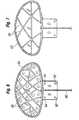
- FIG. 3Ais a first perspective view of an exemplary glaucoma shunt of the present invention from the plate side;
- the inner surface 44 of the plate 40curves to conform to the curvature of the eye 12 , specifically the curvature of the sclera 14 , and the ridge 54 follows that curvature.
- a plurality of cross beams 56( FIGS. 3B and 5 ) for structural strength may also be provided traversing the plate 40 and connecting to the ridge 54 around the periphery, and to each other.
- the cross beams 56may have the same thickness as the ridge 54 or be somewhat thinner.
- FIG. 2Ashows the tube 46 after having been inserted through the incision 72 .
- the pressure of the incision wallscollapses the tube 46 , as mentioned above,
- the tube 46possesses sufficient resiliency to eventually push outward against the incision walls as seen in FIG. 2B and restore patency to the tube 46 .
- This transitionis assisted at all times by internal aqueous fluid pressure from the eye 12 in the portion of the tube lumen leading to the anterior chamber 30 .
- the presence of aqueous fluid“wets” the tube and the output fluid pressure assists the process of opening the tube. Therefore, the combined forces of the internal fluid pressure and inherent resiliency of the wall of the tube 46 work against the inward pressure of the incision walls. As the latter inward force gradually diminishes, from a tissue indentation effect described below, the tube 46 gradually opens.
Landscapes
- Health & Medical Sciences (AREA)
- Ophthalmology & Optometry (AREA)
- Life Sciences & Earth Sciences (AREA)
- Animal Behavior & Ethology (AREA)
- Engineering & Computer Science (AREA)
- Biomedical Technology (AREA)
- Heart & Thoracic Surgery (AREA)
- Veterinary Medicine (AREA)
- Public Health (AREA)
- General Health & Medical Sciences (AREA)
- Surgery (AREA)
- Nuclear Medicine, Radiotherapy & Molecular Imaging (AREA)
- Vascular Medicine (AREA)
- Otolaryngology (AREA)
- Anesthesiology (AREA)
- Hematology (AREA)
- Prostheses (AREA)
Abstract
Description
Claims (20)
Priority Applications (8)
| Application Number | Priority Date | Filing Date | Title |
|---|---|---|---|
| US12/265,145US8353856B2 (en) | 2008-11-05 | 2008-11-05 | Glaucoma drainage shunts and methods of use |
| CA2742746ACA2742746C (en) | 2008-11-05 | 2009-11-05 | Glaucoma drainage shunts |
| PCT/US2009/063332WO2010054035A1 (en) | 2008-11-05 | 2009-11-05 | Glaucoma drainage shunts and methods of use |
| AU2009313582AAU2009313582B2 (en) | 2008-11-05 | 2009-11-05 | Glaucoma drainage shunts and methods of use |
| EP09748673.2AEP2349147B1 (en) | 2008-11-05 | 2009-11-05 | Glaucoma drainage shunts |
| US13/714,050US8920357B2 (en) | 2008-11-05 | 2012-12-13 | Glaucoma drainage shunts and methods of use |
| US14/572,324US9468558B2 (en) | 2008-11-05 | 2014-12-16 | Glaucoma drainage shunts and methods of use |
| US15/288,986US10492948B2 (en) | 2008-11-05 | 2016-10-07 | Glaucoma drainage shunts and methods of use |
Applications Claiming Priority (1)
| Application Number | Priority Date | Filing Date | Title |
|---|---|---|---|
| US12/265,145US8353856B2 (en) | 2008-11-05 | 2008-11-05 | Glaucoma drainage shunts and methods of use |
Related Child Applications (1)
| Application Number | Title | Priority Date | Filing Date |
|---|---|---|---|
| US13/714,050DivisionUS8920357B2 (en) | 2008-11-05 | 2012-12-13 | Glaucoma drainage shunts and methods of use |
Publications (2)
| Publication Number | Publication Date |
|---|---|
| US20100114006A1 US20100114006A1 (en) | 2010-05-06 |
| US8353856B2true US8353856B2 (en) | 2013-01-15 |
Family
ID=41571313
Family Applications (4)
| Application Number | Title | Priority Date | Filing Date |
|---|---|---|---|
| US12/265,145Active2030-08-10US8353856B2 (en) | 2008-11-05 | 2008-11-05 | Glaucoma drainage shunts and methods of use |
| US13/714,050Active2029-02-03US8920357B2 (en) | 2008-11-05 | 2012-12-13 | Glaucoma drainage shunts and methods of use |
| US14/572,324ActiveUS9468558B2 (en) | 2008-11-05 | 2014-12-16 | Glaucoma drainage shunts and methods of use |
| US15/288,986Active2029-06-18US10492948B2 (en) | 2008-11-05 | 2016-10-07 | Glaucoma drainage shunts and methods of use |
Family Applications After (3)
| Application Number | Title | Priority Date | Filing Date |
|---|---|---|---|
| US13/714,050Active2029-02-03US8920357B2 (en) | 2008-11-05 | 2012-12-13 | Glaucoma drainage shunts and methods of use |
| US14/572,324ActiveUS9468558B2 (en) | 2008-11-05 | 2014-12-16 | Glaucoma drainage shunts and methods of use |
| US15/288,986Active2029-06-18US10492948B2 (en) | 2008-11-05 | 2016-10-07 | Glaucoma drainage shunts and methods of use |
Country Status (5)
| Country | Link |
|---|---|
| US (4) | US8353856B2 (en) |
| EP (1) | EP2349147B1 (en) |
| AU (1) | AU2009313582B2 (en) |
| CA (1) | CA2742746C (en) |
| WO (1) | WO2010054035A1 (en) |
Cited By (8)
| Publication number | Priority date | Publication date | Assignee | Title |
|---|---|---|---|---|
| US20130317411A1 (en)* | 2012-05-23 | 2013-11-28 | Ghansham Das AGARWAL | Device for Treatment of Glaucoma |
| US9414962B2 (en) | 2012-04-23 | 2016-08-16 | Das Agarwal Ghansham | Device for treatment of glaucoma and prevention of sub-scleral fibrosis and blockage |
| US9468558B2 (en)* | 2008-11-05 | 2016-10-18 | Abbott Medical Optics Inc. | Glaucoma drainage shunts and methods of use |
| WO2020083824A1 (en) | 2018-10-25 | 2020-04-30 | Amo Groningen B.V. | Bleb control glaucoma shunts |
| WO2021236892A1 (en)* | 2020-05-20 | 2021-11-25 | Iantrek, Inc. | System for shaping and implanting biologic intraocular stent for increased aqueous outflow and lowering of intraocular pressure |
| EP4176855A1 (en)* | 2021-11-09 | 2023-05-10 | Valsigna GmbH | Glaucoma implant device |
| US11925580B2 (en) | 2019-06-14 | 2024-03-12 | Iantrek, Inc. | Implantable biologic stent and system for biologic material shaping and preparation in the treatment of glaucoma |
| US12440378B1 (en) | 2025-02-13 | 2025-10-14 | Iantrek, Inc. | Devices and systems for cutting, loading, and delivering biologic intraocular implants for increased aqueous outflow and lowering of intraocular pressure |
Families Citing this family (76)
| Publication number | Priority date | Publication date | Assignee | Title |
|---|---|---|---|---|
| KR20020035476A (en) | 1999-04-26 | 2002-05-11 | 지엠피 비젼 솔루션즈 인코포레이티드 | Shunt device and method for treating glaucoma |
| US7867186B2 (en) | 2002-04-08 | 2011-01-11 | Glaukos Corporation | Devices and methods for treatment of ocular disorders |
| US6638239B1 (en) | 2000-04-14 | 2003-10-28 | Glaukos Corporation | Apparatus and method for treating glaucoma |
| US7431710B2 (en) | 2002-04-08 | 2008-10-07 | Glaukos Corporation | Ocular implants with anchors and methods thereof |
| AU2002258754B2 (en) | 2001-04-07 | 2006-08-17 | Glaukos Corporation | Glaucoma stent and methods thereof for glaucoma treatment |
| US7331984B2 (en) | 2001-08-28 | 2008-02-19 | Glaukos Corporation | Glaucoma stent for treating glaucoma and methods of use |
| US20120123315A1 (en)* | 2010-11-15 | 2012-05-17 | Aquesys, Inc. | Intraocular shunts |
| US8308701B2 (en) | 2010-11-15 | 2012-11-13 | Aquesys, Inc. | Methods for deploying intraocular shunts |
| US8663303B2 (en) | 2010-11-15 | 2014-03-04 | Aquesys, Inc. | Methods for deploying an intraocular shunt from a deployment device and into an eye |
| US8852137B2 (en) | 2010-11-15 | 2014-10-07 | Aquesys, Inc. | Methods for implanting a soft gel shunt in the suprachoroidal space |
| US20120123316A1 (en) | 2010-11-15 | 2012-05-17 | Aquesys, Inc. | Intraocular shunts for placement in the intra-tenon's space |
| US8852256B2 (en) | 2010-11-15 | 2014-10-07 | Aquesys, Inc. | Methods for intraocular shunt placement |
| US8758290B2 (en) | 2010-11-15 | 2014-06-24 | Aquesys, Inc. | Devices and methods for implanting a shunt in the suprachoroidal space |
| US8828070B2 (en) | 2010-11-15 | 2014-09-09 | Aquesys, Inc. | Devices for deploying intraocular shunts |
| US8721702B2 (en) | 2010-11-15 | 2014-05-13 | Aquesys, Inc. | Intraocular shunt deployment devices |
| US8801766B2 (en) | 2010-11-15 | 2014-08-12 | Aquesys, Inc. | Devices for deploying intraocular shunts |
| US8974511B2 (en) | 2010-11-15 | 2015-03-10 | Aquesys, Inc. | Methods for treating closed angle glaucoma |
| US8257295B2 (en) | 2009-09-21 | 2012-09-04 | Alcon Research, Ltd. | Intraocular pressure sensor with external pressure compensation |
| US8585629B2 (en) | 2010-11-15 | 2013-11-19 | Aquesys, Inc. | Systems for deploying intraocular shunts |
| US10603214B2 (en) | 2011-01-14 | 2020-03-31 | Ecole Polytechnique Federale De Lausanne (Epfl) | Apparatus and methods for treating excess intraocular fluid |
| US8771220B2 (en) | 2011-12-07 | 2014-07-08 | Alcon Research, Ltd. | Glaucoma active pressure regulation shunt |
| US8765210B2 (en) | 2011-12-08 | 2014-07-01 | Aquesys, Inc. | Systems and methods for making gelatin shunts |
| US9610195B2 (en) | 2013-02-27 | 2017-04-04 | Aquesys, Inc. | Intraocular shunt implantation methods and devices |
| US8852136B2 (en)* | 2011-12-08 | 2014-10-07 | Aquesys, Inc. | Methods for placing a shunt into the intra-scleral space |
| US9339187B2 (en) | 2011-12-15 | 2016-05-17 | Alcon Research, Ltd. | External pressure measurement system and method for an intraocular implant |
| DE102012200411A1 (en)* | 2012-01-12 | 2013-07-18 | Geuder Ag | Device for use in glaucoma surgery |
| CA2868341C (en) | 2012-03-26 | 2021-01-12 | Glaukos Corporation | System and method for delivering multiple ocular implants |
| US8888734B2 (en) | 2012-06-05 | 2014-11-18 | Alcon Research, Ltd. | Functionally graded material tube and method for use of the same in implantation |
| US9572712B2 (en) | 2012-12-17 | 2017-02-21 | Novartis Ag | Osmotically actuated fluidic valve |
| US9295389B2 (en) | 2012-12-17 | 2016-03-29 | Novartis Ag | Systems and methods for priming an intraocular pressure sensor in an intraocular implant |
| US9528633B2 (en) | 2012-12-17 | 2016-12-27 | Novartis Ag | MEMS check valve |
| US10780243B2 (en)* | 2013-01-25 | 2020-09-22 | Javier G. Reyes | Method and apparatus for treatment of human urinary incontinence |
| ES2699439T3 (en)* | 2013-02-19 | 2019-02-11 | Aquesys Inc | Adjustable flow pressure relief |
| US10159600B2 (en) | 2013-02-19 | 2018-12-25 | Aquesys, Inc. | Adjustable intraocular flow regulation |
| US9125723B2 (en) | 2013-02-19 | 2015-09-08 | Aquesys, Inc. | Adjustable glaucoma implant |
| US10517759B2 (en) | 2013-03-15 | 2019-12-31 | Glaukos Corporation | Glaucoma stent and methods thereof for glaucoma treatment |
| KR20150034010A (en)* | 2013-09-25 | 2015-04-02 | 사회복지법인 삼성생명공익재단 | An Apparatus for Treating Ocular Diseases Induced by Increased Intraocular Pressure |
| US9795503B2 (en) | 2013-10-18 | 2017-10-24 | Rodolfo Alfredo PEREZ GROSSMANN | Method and apparatus for trabeculectomy and suprachoroidal shunt surgery |
| EP3068354B1 (en) | 2013-11-14 | 2023-06-28 | Aquesys, Inc. | Intraocular shunt inserter |
| US9044301B1 (en) | 2013-11-25 | 2015-06-02 | Innfocus, Inc. | Methods, systems and devices for treating glaucoma |
| EP3060180B1 (en)* | 2014-02-24 | 2024-10-09 | National University of Singapore | Ocular drainage device and method of manufacturing thereof |
| EP3677229A1 (en) | 2014-05-29 | 2020-07-08 | Glaukos Corporation | Implants with controlled drug delivery features |
| US10342702B2 (en) | 2014-08-29 | 2019-07-09 | Camras Vision Inc. | Apparatus and method for reducing intraocular pressure |
| US10201451B2 (en) | 2014-08-29 | 2019-02-12 | Camras Vision Inc. | Device and method for reducing intraocular pressure |
| US10507101B2 (en) | 2014-10-13 | 2019-12-17 | W. L. Gore & Associates, Inc. | Valved conduit |
| US11925578B2 (en) | 2015-09-02 | 2024-03-12 | Glaukos Corporation | Drug delivery implants with bi-directional delivery capacity |
| US10524958B2 (en) | 2015-09-30 | 2020-01-07 | Alievio, Inc. | Method and apparatus for reducing intraocular pressure |
| RU2613414C1 (en)* | 2016-01-21 | 2017-03-16 | Федеральное государственное автономное учреждение "Межотраслевой научно-технический комплекс "Микрохирургия глаза" имени академика С.Н. Федорова" Министерства здравоохранения Российской Федерации | Drainage for glaucoma surgery |
| US20190038463A1 (en)* | 2016-01-28 | 2019-02-07 | Aq Biomed, Llc | Ophthalamic implant for reduction of intraocular pressure in glaucomatous eyes and method of use |
| US20200078215A1 (en)* | 2016-07-06 | 2020-03-12 | MicroOptx Inc. | Glaucoma treatment devices and methods |
| RU2019115938A (en)* | 2016-11-02 | 2020-12-03 | Ликид Медикал Проприетари Лимитед | SHUNT SYSTEM, SHUNT AND METHOD FOR TREATMENT OF EYE DISORDER |
| US11406533B2 (en) | 2017-03-17 | 2022-08-09 | W. L. Gore & Associates, Inc. | Integrated aqueous shunt for glaucoma treatment |
| US11166849B2 (en) | 2017-07-20 | 2021-11-09 | Shifamed Holdings, Llc | Adjustable flow glaucoma shunts and methods for making and using same |
| EP4218692A3 (en) | 2017-07-20 | 2023-09-06 | Shifamed Holdings, LLC | Adjustable flow glaucoma shunts and methods for making and using same |
| EP3441069B1 (en) | 2017-08-11 | 2023-04-05 | Unity Biotechnology, Inc. | Treatment of diabetic retinopathy using pharmaceutical agents that eliminate senescent cells |
| US20200229978A1 (en)* | 2017-08-11 | 2020-07-23 | University Of Florida Research Foundation, Incorporated | Enhanced drainage of failed glaucoma drainage device (gdd) |
| US11116625B2 (en) | 2017-09-28 | 2021-09-14 | Glaukos Corporation | Apparatus and method for controlling placement of intraocular implants |
| WO2020047221A1 (en) | 2018-08-29 | 2020-03-05 | W. L. Gore & Associates, Inc. | Drug therapy delivery systems and methods |
| CN112689488B (en)* | 2018-09-06 | 2024-04-12 | 洛桑联邦理工学院 | Device for treating excess intraocular fluid with elastic membrane |
| RU192822U1 (en)* | 2018-09-20 | 2019-10-02 | федеральное государственное автономное образовательное учреждение высшего образования "Российский университет дружбы народов" (РУДН) | Supraciliary Microshunt |
| US11678983B2 (en) | 2018-12-12 | 2023-06-20 | W. L. Gore & Associates, Inc. | Implantable component with socket |
| EP3911285A4 (en) | 2019-01-18 | 2022-10-19 | Shifamed Holdings, LLC | Adjustable flow glaucoma shunts and methods for making and using same |
| JP7614193B2 (en) | 2019-10-10 | 2025-01-15 | シファメド・ホールディングス・エルエルシー | Adjustable flow glaucoma shunts and related systems and methods |
| AU2021209698A1 (en) | 2020-01-23 | 2022-08-04 | Shifamed Holdings, Llc | Adjustable flow glaucoma shunts and associated systems and methods |
| US11291585B2 (en) | 2020-02-14 | 2022-04-05 | Shifamed Holdings, Llc | Shunting systems with rotation-based flow control assemblies, and associated systems and methods |
| US11737920B2 (en) | 2020-02-18 | 2023-08-29 | Shifamed Holdings, Llc | Adjustable flow glaucoma shunts having non-linearly arranged flow control elements, and associated systems and methods |
| WO2021176332A1 (en)* | 2020-03-06 | 2021-09-10 | Ecole Polytechnique De Lausanne (Epfl) | Apparatus for treating excess intraocular fluid having an elastic membrane |
| WO2021188952A1 (en) | 2020-03-19 | 2021-09-23 | Shifamed Holdings, Llc | Intraocular shunts with low-profile actuation elements and associated systems and methods |
| CN115867237A (en) | 2020-04-16 | 2023-03-28 | 施菲姆德控股有限责任公司 | Adjustable glaucoma treatment devices and related systems and methods |
| US11419761B2 (en)* | 2020-07-22 | 2022-08-23 | The Eye Hospital Of Wenzhou Medical University | Glaucoma aqueous humor drainage device and glaucoma aqueous humor drainage method |
| EP4281144A4 (en) | 2021-01-22 | 2024-11-27 | Shifamed Holdings, LLC | ADJUSTABLE SHUNTING SYSTEMS WITH PLATE ARRANGEMENTS AND RELATED SYSTEMS AND METHODS |
| CN112790911B (en)* | 2021-01-29 | 2024-08-16 | 赵秀琴 | Glaucoma drainage device |
| EP4376784A4 (en)* | 2021-07-30 | 2025-01-29 | Shifamed Holdings, LLC | SHUNTING SYSTEMS WITH FORMABLE ELEMENTS AND ASSOCIATED DEVICES AND METHODS |
| AU2023207083A1 (en)* | 2022-01-12 | 2024-07-04 | W. L. Gore & Associates, Inc. | Biological fluid drainage devices, systems, and methods |
| US12042432B1 (en) | 2024-01-11 | 2024-07-23 | Michael Reynard | Method and device for the treatment of glaucoma |
| US12274640B1 (en) | 2024-11-08 | 2025-04-15 | Michael Reynard | Implant for electrolysis of aqueous humor |
Citations (77)
| Publication number | Priority date | Publication date | Assignee | Title |
|---|---|---|---|---|
| US1246971A (en) | 1915-11-27 | 1917-11-20 | Friedrich Maier | Eye-washing device. |
| US2969066A (en) | 1956-10-02 | 1961-01-24 | Holter Company | Device for draining ventricular fluid in cases of hydrocephalus |
| US3109429A (en) | 1962-01-30 | 1963-11-05 | Schwartz Samuel | Ventriculo-venous shunt device for treatment of hydrocephalus |
| US3159161A (en) | 1962-11-14 | 1964-12-01 | Ness Richard Alton | Fistulizing canaliculus |
| US3527226A (en) | 1966-02-03 | 1970-09-08 | Cordis Corp | Ventricular catheter with valve and pump flushing means |
| US3726284A (en) | 1971-04-05 | 1973-04-10 | R Parker | Replacement tube for the lacrimal drainage ducts |
| US3788327A (en) | 1971-03-30 | 1974-01-29 | H Donowitz | Surgical implant device |
| FR2233028A1 (en) | 1973-06-12 | 1975-01-10 | Bonnaud Philippe | Drainer for secretions from wounds - is of rubber strip with channels for plastics tubes |
| US3860008A (en) | 1973-10-03 | 1975-01-14 | Dow Corning | Flat drain |
| US3915172A (en) | 1970-05-27 | 1975-10-28 | Ceskoslovenska Akademie Ved | Capillary drain for glaucoma |
| US4030480A (en) | 1976-05-13 | 1977-06-21 | Ernst Jochen Meyer | Ocular decompression process |
| US4240434A (en) | 1978-10-10 | 1980-12-23 | Newkirk John B | Peritoneo-venous shunt |
| US4298994A (en) | 1979-10-26 | 1981-11-10 | Clayman Henry M | Posterior chamber intra-ocular transplant device |
| SU906561A1 (en) | 1979-11-11 | 1982-02-23 | Военно-Медицинская Ордена Ленина Краснознаменная Академия Им.С.М.Кирова | Apparatus for draining eye anterior chamber |
| GB2101891A (en) | 1981-07-18 | 1983-01-26 | Anthony Christopher Be Molteno | Device for draining aqueous humour |
| US4373218A (en) | 1980-11-17 | 1983-02-15 | Schachar Ronald A | Variable power intraocular lens and method of implanting into the posterior chamber |
| US4402681A (en) | 1980-08-23 | 1983-09-06 | Haas Joseph S | Artificial implant valve for the regulation of intraocular pressure |
| US4428746A (en) | 1981-07-29 | 1984-01-31 | Antonio Mendez | Glaucoma treatment device |
| EP0102747A1 (en) | 1982-07-28 | 1984-03-14 | Thomas C. White | Ocular pressure relief device |
| US4457757A (en) | 1981-07-20 | 1984-07-03 | Molteno Anthony C B | Device for draining aqueous humour |
| US4521210A (en)* | 1982-12-27 | 1985-06-04 | Wong Vernon G | Eye implant for relieving glaucoma, and device and method for use therewith |
| GB2160778A (en) | 1984-06-28 | 1986-01-02 | Neil Howard Joseph | Aqueous humour drainage device |
| US4604087A (en) | 1985-02-26 | 1986-08-05 | Joseph Neil H | Aqueous humor drainage device |
| US4634418A (en) | 1984-04-06 | 1987-01-06 | Binder Perry S | Hydrogel seton |
| GB2187963A (en) | 1986-03-07 | 1987-09-23 | Anthony Christopher Be Molteno | Implant for drainage of aqueous humour in glaucoma |
| US4722724A (en) | 1986-06-23 | 1988-02-02 | Stanley Schocket | Anterior chamber tube shunt to an encircling band, and related surgical procedure |
| US4729761A (en) | 1985-11-27 | 1988-03-08 | White Thomas C | Tissue-implantable, fluid-dissipating device |
| US4767400A (en) | 1987-10-27 | 1988-08-30 | Cordis Corporation | Porous ventricular catheter |
| US4836457A (en) | 1986-07-23 | 1989-06-06 | Lindemann Maschinenfabrik G.M.B.H. | Screen for comminuting machines |
| US4863457A (en) | 1986-11-24 | 1989-09-05 | Lee David A | Drug delivery device |
| US4865601A (en) | 1987-07-07 | 1989-09-12 | Caldwell Delmar R | Intraocular prostheses |
| US4886488A (en) | 1987-08-06 | 1989-12-12 | White Thomas C | Glaucoma drainage the lacrimal system and method |
| US4902292A (en) | 1987-03-30 | 1990-02-20 | Joseph Neil H | Vitreous body prosthesis device |
| US4915684A (en) | 1988-06-21 | 1990-04-10 | Mackeen Donald L | Method and apparatus for modulating the flow of lacrimal fluid through a punctum and associated canaliculus |
| US4932968A (en) | 1987-07-07 | 1990-06-12 | Caldwell Delmar R | Intraocular prostheses |
| US4936825A (en) | 1988-04-11 | 1990-06-26 | Ungerleider Bruce A | Method for reducing intraocular pressure caused by glaucoma |
| US4946436A (en) | 1989-11-17 | 1990-08-07 | Smith Stewart G | Pressure-relieving device and process for implanting |
| US4955909A (en) | 1989-01-31 | 1990-09-11 | Bioplasty, Inc. | Textured silicone implant prosthesis |
| US4968296A (en) | 1989-12-20 | 1990-11-06 | Robert Ritch | Transscleral drainage implant device for the treatment of glaucoma |
| WO1991012037A1 (en) | 1990-02-12 | 1991-08-22 | Ahmed Abdul Mateen | Medical valve |
| WO1991012046A1 (en) | 1990-02-12 | 1991-08-22 | Atos Medical Ab | Glaucoma valve |
| WO1991018568A1 (en) | 1990-05-31 | 1991-12-12 | Wright Medical, Inc. | Glaucoma implant |
| US5092837A (en) | 1989-12-20 | 1992-03-03 | Robert Ritch | Method for the treatment of glaucoma |
| US5171213A (en) | 1991-08-14 | 1992-12-15 | Price Jr Francis W | Technique for fistulization of the eye and an eye filtration prosthesis useful therefor |
| US5192315A (en) | 1992-03-24 | 1993-03-09 | Jacob Labarre Jean T | Total ocular replacement apparatus with muscle attachment sites |
| WO1993020783A1 (en) | 1992-04-13 | 1993-10-28 | Iovision, Inc. | Glaucoma implant |
| US5282851A (en) | 1987-07-07 | 1994-02-01 | Jacob Labarre Jean | Intraocular prostheses |
| WO1994002081A1 (en) | 1992-07-16 | 1994-02-03 | Wong Vernon G | Eye implant suitable for relief of glaucoma |
| US5300020A (en) | 1991-05-31 | 1994-04-05 | Medflex Corporation | Surgically implantable device for glaucoma relief |
| US5338291A (en) | 1993-02-03 | 1994-08-16 | Pudenz-Schulte Medical Research Corporation | Glaucoma shunt and method for draining aqueous humor |
| US5397300A (en)* | 1990-05-31 | 1995-03-14 | Iovision, Inc. | Glaucoma implant |
| US5476445A (en) | 1990-05-31 | 1995-12-19 | Iovision, Inc. | Glaucoma implant with a temporary flow restricting seal |
| US5549670A (en) | 1995-05-09 | 1996-08-27 | Allergan, Inc. | IOL for reducing secondary opacification |
| US5704907A (en) | 1994-07-22 | 1998-01-06 | Wound Healing Of Oklahoma | Method and apparatus for lowering the intraocular pressure of an eye |
| US5725493A (en) | 1994-12-12 | 1998-03-10 | Avery; Robert Logan | Intravitreal medicine delivery |
| US5752928A (en)* | 1997-07-14 | 1998-05-19 | Rdo Medical, Inc. | Glaucoma pressure regulator |
| US5882327A (en)* | 1997-04-17 | 1999-03-16 | Jacob; Jean T. | Long-term glaucoma drainage implant |
| US6050970A (en) | 1997-05-08 | 2000-04-18 | Pharmacia & Upjohn Company | Method and apparatus for inserting a glaucoma implant in an anterior and posterior segment of the eye |
| US6203513B1 (en) | 1997-11-20 | 2001-03-20 | Optonol Ltd. | Flow regulating implant, method of manufacture, and delivery device |
| US6261256B1 (en)* | 1996-12-20 | 2001-07-17 | Abdul Mateen Ahmed | Pocket medical valve & method |
| US20050125003A1 (en) | 2003-12-05 | 2005-06-09 | Leonard Pinchuk | Glaucoma implant device |
| US20050267398A1 (en)* | 2004-05-27 | 2005-12-01 | Dimitri Protopsaltis | Glaucoma shunt |
| US6984392B2 (en) | 2000-08-31 | 2006-01-10 | Bio-Gate Bioinnovative Materials Gmbh | Antimicrobial material for implanting in bones |
| WO2006012009A2 (en) | 2004-06-25 | 2006-02-02 | Optonol, Ltd | Flow regulating implants |
| US7160264B2 (en) | 2002-12-19 | 2007-01-09 | Medtronic-Xomed, Inc. | Article and method for ocular aqueous drainage |
| US7207965B2 (en) | 2003-06-16 | 2007-04-24 | Solx, Inc. | Shunt for the treatment of glaucoma |
| US7220238B2 (en) | 1999-04-26 | 2007-05-22 | Gmp Vision Solutions, Inc. | Shunt device and method for treating glaucoma |
| WO2007087061A2 (en) | 2006-01-17 | 2007-08-02 | Transcend Medical, Inc. | Glaucoma treatment device |
| US7297130B2 (en) | 2000-04-14 | 2007-11-20 | Glaukos Corporation | Implant with anchor |
| US20070293872A1 (en) | 2006-06-20 | 2007-12-20 | Minu, L.L.C. | Ocular Drainage Device |
| US20080077238A1 (en) | 2006-09-21 | 2008-03-27 | Advanced Medical Optics, Inc. | Intraocular lenses for managing glare, adhesion, and cell migration |
| US7357778B2 (en) | 2004-05-20 | 2008-04-15 | Ajay Bhalla | Aqueous drainage and flow regulating implant |
| US20080200860A1 (en) | 2001-04-07 | 2008-08-21 | Glaukos Corporation | System for treating ocular disorders and methods thereof |
| US7476698B2 (en) | 2001-09-18 | 2009-01-13 | Bio-Gate Ag | Antimicrobial adhesive and coating substance and method for the production thereof |
| US7547302B2 (en) | 1999-07-19 | 2009-06-16 | I-Flow Corporation | Anti-microbial catheter |
| US7635358B2 (en) | 2003-10-15 | 2009-12-22 | Boston Scientific Scimed, Inc. | Medical device having anti-microbial properties and a false lumen and method of making the same |
| US7641627B2 (en) | 2005-02-23 | 2010-01-05 | Camras Carl B | Method and apparatus for reducing intraocular pressure |
Family Cites Families (8)
| Publication number | Priority date | Publication date | Assignee | Title |
|---|---|---|---|---|
| US4037604A (en)* | 1976-01-05 | 1977-07-26 | Newkirk John B | Artifical biological drainage device |
| US5454796A (en)* | 1991-04-09 | 1995-10-03 | Hood Laboratories | Device and method for controlling intraocular fluid pressure |
| US5370607A (en)* | 1992-10-28 | 1994-12-06 | Annuit Coeptis, Inc. | Glaucoma implant device and method for implanting same |
| US6007510A (en)* | 1996-10-25 | 1999-12-28 | Anamed, Inc. | Implantable devices and methods for controlling the flow of fluids within the body |
| US5713844A (en)* | 1997-01-10 | 1998-02-03 | Peyman; Gholam A. | Device and method for regulating intraocular pressure |
| US6589203B1 (en)* | 2000-01-26 | 2003-07-08 | Peter Mitrev | Glaucoma drainage device implant |
| US7699882B2 (en)* | 2002-09-17 | 2010-04-20 | Iscience Interventional Corporation | Apparatus and method for surgical bypass of aqueous humor |
| US8353856B2 (en)* | 2008-11-05 | 2013-01-15 | Abbott Medical Optics Inc. | Glaucoma drainage shunts and methods of use |
- 2008
- 2008-11-05USUS12/265,145patent/US8353856B2/enactiveActive
- 2009
- 2009-11-05CACA2742746Apatent/CA2742746C/ennot_activeExpired - Fee Related
- 2009-11-05WOPCT/US2009/063332patent/WO2010054035A1/enactiveApplication Filing
- 2009-11-05EPEP09748673.2Apatent/EP2349147B1/enactiveActive
- 2009-11-05AUAU2009313582Apatent/AU2009313582B2/ennot_activeCeased
- 2012
- 2012-12-13USUS13/714,050patent/US8920357B2/enactiveActive
- 2014
- 2014-12-16USUS14/572,324patent/US9468558B2/enactiveActive
- 2016
- 2016-10-07USUS15/288,986patent/US10492948B2/enactiveActive
Patent Citations (84)
| Publication number | Priority date | Publication date | Assignee | Title |
|---|---|---|---|---|
| US1246971A (en) | 1915-11-27 | 1917-11-20 | Friedrich Maier | Eye-washing device. |
| US2969066A (en) | 1956-10-02 | 1961-01-24 | Holter Company | Device for draining ventricular fluid in cases of hydrocephalus |
| US3109429A (en) | 1962-01-30 | 1963-11-05 | Schwartz Samuel | Ventriculo-venous shunt device for treatment of hydrocephalus |
| US3159161A (en) | 1962-11-14 | 1964-12-01 | Ness Richard Alton | Fistulizing canaliculus |
| US3527226A (en) | 1966-02-03 | 1970-09-08 | Cordis Corp | Ventricular catheter with valve and pump flushing means |
| US3915172A (en) | 1970-05-27 | 1975-10-28 | Ceskoslovenska Akademie Ved | Capillary drain for glaucoma |
| US3788327A (en) | 1971-03-30 | 1974-01-29 | H Donowitz | Surgical implant device |
| US3726284A (en) | 1971-04-05 | 1973-04-10 | R Parker | Replacement tube for the lacrimal drainage ducts |
| FR2233028A1 (en) | 1973-06-12 | 1975-01-10 | Bonnaud Philippe | Drainer for secretions from wounds - is of rubber strip with channels for plastics tubes |
| US3860008A (en) | 1973-10-03 | 1975-01-14 | Dow Corning | Flat drain |
| US4030480A (en) | 1976-05-13 | 1977-06-21 | Ernst Jochen Meyer | Ocular decompression process |
| US4240434A (en) | 1978-10-10 | 1980-12-23 | Newkirk John B | Peritoneo-venous shunt |
| US4298994A (en) | 1979-10-26 | 1981-11-10 | Clayman Henry M | Posterior chamber intra-ocular transplant device |
| US4298994B1 (en) | 1979-10-26 | 1991-08-06 | M Clayman Henry | |
| SU906561A1 (en) | 1979-11-11 | 1982-02-23 | Военно-Медицинская Ордена Ленина Краснознаменная Академия Им.С.М.Кирова | Apparatus for draining eye anterior chamber |
| US4402681A (en) | 1980-08-23 | 1983-09-06 | Haas Joseph S | Artificial implant valve for the regulation of intraocular pressure |
| US4373218A (en) | 1980-11-17 | 1983-02-15 | Schachar Ronald A | Variable power intraocular lens and method of implanting into the posterior chamber |
| GB2101891A (en) | 1981-07-18 | 1983-01-26 | Anthony Christopher Be Molteno | Device for draining aqueous humour |
| US4457757A (en) | 1981-07-20 | 1984-07-03 | Molteno Anthony C B | Device for draining aqueous humour |
| US4428746A (en) | 1981-07-29 | 1984-01-31 | Antonio Mendez | Glaucoma treatment device |
| EP0102747A1 (en) | 1982-07-28 | 1984-03-14 | Thomas C. White | Ocular pressure relief device |
| US4521210A (en)* | 1982-12-27 | 1985-06-04 | Wong Vernon G | Eye implant for relieving glaucoma, and device and method for use therewith |
| US4634418A (en) | 1984-04-06 | 1987-01-06 | Binder Perry S | Hydrogel seton |
| EP0168201A1 (en) | 1984-06-28 | 1986-01-15 | Neil Howard Joseph | Aqueous humour drainage device |
| GB2160778A (en) | 1984-06-28 | 1986-01-02 | Neil Howard Joseph | Aqueous humour drainage device |
| US4604087A (en) | 1985-02-26 | 1986-08-05 | Joseph Neil H | Aqueous humor drainage device |
| US4729761A (en) | 1985-11-27 | 1988-03-08 | White Thomas C | Tissue-implantable, fluid-dissipating device |
| GB2187963A (en) | 1986-03-07 | 1987-09-23 | Anthony Christopher Be Molteno | Implant for drainage of aqueous humour in glaucoma |
| US4750901A (en) | 1986-03-07 | 1988-06-14 | Molteno Anthony C B | Implant for drainage of aqueous humour |
| US4722724A (en) | 1986-06-23 | 1988-02-02 | Stanley Schocket | Anterior chamber tube shunt to an encircling band, and related surgical procedure |
| US4836457A (en) | 1986-07-23 | 1989-06-06 | Lindemann Maschinenfabrik G.M.B.H. | Screen for comminuting machines |
| US4863457A (en) | 1986-11-24 | 1989-09-05 | Lee David A | Drug delivery device |
| US4902292A (en) | 1987-03-30 | 1990-02-20 | Joseph Neil H | Vitreous body prosthesis device |
| US4865601A (en) | 1987-07-07 | 1989-09-12 | Caldwell Delmar R | Intraocular prostheses |
| US5282851A (en) | 1987-07-07 | 1994-02-01 | Jacob Labarre Jean | Intraocular prostheses |
| US4932968A (en) | 1987-07-07 | 1990-06-12 | Caldwell Delmar R | Intraocular prostheses |
| US4886488A (en) | 1987-08-06 | 1989-12-12 | White Thomas C | Glaucoma drainage the lacrimal system and method |
| US4767400A (en) | 1987-10-27 | 1988-08-30 | Cordis Corporation | Porous ventricular catheter |
| US4936825A (en) | 1988-04-11 | 1990-06-26 | Ungerleider Bruce A | Method for reducing intraocular pressure caused by glaucoma |
| US5372577A (en) | 1988-04-11 | 1994-12-13 | Ungerleider; Bruce A. | Apparatus for reducing intraocular pressure |
| US4915684A (en) | 1988-06-21 | 1990-04-10 | Mackeen Donald L | Method and apparatus for modulating the flow of lacrimal fluid through a punctum and associated canaliculus |
| US4955909A (en) | 1989-01-31 | 1990-09-11 | Bioplasty, Inc. | Textured silicone implant prosthesis |
| US4946436A (en) | 1989-11-17 | 1990-08-07 | Smith Stewart G | Pressure-relieving device and process for implanting |
| US4968296A (en) | 1989-12-20 | 1990-11-06 | Robert Ritch | Transscleral drainage implant device for the treatment of glaucoma |
| US5092837A (en) | 1989-12-20 | 1992-03-03 | Robert Ritch | Method for the treatment of glaucoma |
| WO1991012037A1 (en) | 1990-02-12 | 1991-08-22 | Ahmed Abdul Mateen | Medical valve |
| WO1991012046A1 (en) | 1990-02-12 | 1991-08-22 | Atos Medical Ab | Glaucoma valve |
| WO1991018568A1 (en) | 1990-05-31 | 1991-12-12 | Wright Medical, Inc. | Glaucoma implant |
| US5178604A (en) | 1990-05-31 | 1993-01-12 | Iovision, Inc. | Glaucoma implant |
| US5397300A (en)* | 1990-05-31 | 1995-03-14 | Iovision, Inc. | Glaucoma implant |
| US5476445A (en) | 1990-05-31 | 1995-12-19 | Iovision, Inc. | Glaucoma implant with a temporary flow restricting seal |
| US5558629A (en) | 1990-05-31 | 1996-09-24 | Iovision, Inc. | Glaucoma implant |
| US5300020A (en) | 1991-05-31 | 1994-04-05 | Medflex Corporation | Surgically implantable device for glaucoma relief |
| US5171213A (en) | 1991-08-14 | 1992-12-15 | Price Jr Francis W | Technique for fistulization of the eye and an eye filtration prosthesis useful therefor |
| US5192315A (en) | 1992-03-24 | 1993-03-09 | Jacob Labarre Jean T | Total ocular replacement apparatus with muscle attachment sites |
| WO1993020783A1 (en) | 1992-04-13 | 1993-10-28 | Iovision, Inc. | Glaucoma implant |
| WO1994002081A1 (en) | 1992-07-16 | 1994-02-03 | Wong Vernon G | Eye implant suitable for relief of glaucoma |
| US5338291A (en) | 1993-02-03 | 1994-08-16 | Pudenz-Schulte Medical Research Corporation | Glaucoma shunt and method for draining aqueous humor |
| US5704907A (en) | 1994-07-22 | 1998-01-06 | Wound Healing Of Oklahoma | Method and apparatus for lowering the intraocular pressure of an eye |
| US5725493A (en) | 1994-12-12 | 1998-03-10 | Avery; Robert Logan | Intravitreal medicine delivery |
| US5549670A (en) | 1995-05-09 | 1996-08-27 | Allergan, Inc. | IOL for reducing secondary opacification |
| US6261256B1 (en)* | 1996-12-20 | 2001-07-17 | Abdul Mateen Ahmed | Pocket medical valve & method |
| US5882327A (en)* | 1997-04-17 | 1999-03-16 | Jacob; Jean T. | Long-term glaucoma drainage implant |
| US6050970A (en) | 1997-05-08 | 2000-04-18 | Pharmacia & Upjohn Company | Method and apparatus for inserting a glaucoma implant in an anterior and posterior segment of the eye |
| US5752928A (en)* | 1997-07-14 | 1998-05-19 | Rdo Medical, Inc. | Glaucoma pressure regulator |
| US6203513B1 (en) | 1997-11-20 | 2001-03-20 | Optonol Ltd. | Flow regulating implant, method of manufacture, and delivery device |
| US7220238B2 (en) | 1999-04-26 | 2007-05-22 | Gmp Vision Solutions, Inc. | Shunt device and method for treating glaucoma |
| US7547302B2 (en) | 1999-07-19 | 2009-06-16 | I-Flow Corporation | Anti-microbial catheter |
| US7297130B2 (en) | 2000-04-14 | 2007-11-20 | Glaukos Corporation | Implant with anchor |
| US6984392B2 (en) | 2000-08-31 | 2006-01-10 | Bio-Gate Bioinnovative Materials Gmbh | Antimicrobial material for implanting in bones |
| US20080200860A1 (en) | 2001-04-07 | 2008-08-21 | Glaukos Corporation | System for treating ocular disorders and methods thereof |
| US7476698B2 (en) | 2001-09-18 | 2009-01-13 | Bio-Gate Ag | Antimicrobial adhesive and coating substance and method for the production thereof |
| US7160264B2 (en) | 2002-12-19 | 2007-01-09 | Medtronic-Xomed, Inc. | Article and method for ocular aqueous drainage |
| US7207965B2 (en) | 2003-06-16 | 2007-04-24 | Solx, Inc. | Shunt for the treatment of glaucoma |
| US7635358B2 (en) | 2003-10-15 | 2009-12-22 | Boston Scientific Scimed, Inc. | Medical device having anti-microbial properties and a false lumen and method of making the same |
| US20050125003A1 (en) | 2003-12-05 | 2005-06-09 | Leonard Pinchuk | Glaucoma implant device |
| US7357778B2 (en) | 2004-05-20 | 2008-04-15 | Ajay Bhalla | Aqueous drainage and flow regulating implant |
| US20050267398A1 (en)* | 2004-05-27 | 2005-12-01 | Dimitri Protopsaltis | Glaucoma shunt |
| US20080077071A1 (en) | 2004-06-25 | 2008-03-27 | Optonol Ltd. | Flow Regulating Implants |
| WO2006012009A2 (en) | 2004-06-25 | 2006-02-02 | Optonol, Ltd | Flow regulating implants |
| US7641627B2 (en) | 2005-02-23 | 2010-01-05 | Camras Carl B | Method and apparatus for reducing intraocular pressure |
| WO2007087061A2 (en) | 2006-01-17 | 2007-08-02 | Transcend Medical, Inc. | Glaucoma treatment device |
| US20070293872A1 (en) | 2006-06-20 | 2007-12-20 | Minu, L.L.C. | Ocular Drainage Device |
| US20080077238A1 (en) | 2006-09-21 | 2008-03-27 | Advanced Medical Optics, Inc. | Intraocular lenses for managing glare, adhesion, and cell migration |
Non-Patent Citations (6)
| Title |
|---|
| Bickford, "Molteno Implant System, for Patient with Previously Unsuccessful Glaucoms Surger," Journal of Ophthalmic Nursing & Technology, vol. 6. No. 6, 1987, pp. 224-229. |
| Kakaday, et al. In "Advances in Telemetric Continuous Intraocular Pressure Assessment<" British Journal of Ophthalmology, 2009; 93:992-996. |
| Krupin, et al., "Filtering Valve Implant Surgery for Eyes with Neovascular Glaucoma." American Journal of Ophthalmology, vol. 89, No. 3 1980, pp. 338-343. |
| Lee, et al., "Aqueous-Venous Shunt for Glaucoma," Arch Ophthalmol, vol. 99, Nov. 1981, pp. 2007-2012. |
| Mokwa in "Ophthalmic Implants," 2003 IEEE Publication, 980-986, Institute of Materials in Electrical Engineering, RWTH Aachen Univ., Aachen, Germany. |
| Molteno, "Use of Molteno Implants to Treat Secondary Glaucoma," Glaucoma, Grune & Stratton, Ltd., 1986, pp. 211-238. |
Cited By (14)
| Publication number | Priority date | Publication date | Assignee | Title |
|---|---|---|---|---|
| US9468558B2 (en)* | 2008-11-05 | 2016-10-18 | Abbott Medical Optics Inc. | Glaucoma drainage shunts and methods of use |
| US10492948B2 (en) | 2008-11-05 | 2019-12-03 | Johnson & Johnson Surgical Vision, Inc. | Glaucoma drainage shunts and methods of use |
| US9414962B2 (en) | 2012-04-23 | 2016-08-16 | Das Agarwal Ghansham | Device for treatment of glaucoma and prevention of sub-scleral fibrosis and blockage |
| US20130317411A1 (en)* | 2012-05-23 | 2013-11-28 | Ghansham Das AGARWAL | Device for Treatment of Glaucoma |
| US20230310212A1 (en)* | 2018-10-25 | 2023-10-05 | Amo Groningen B.V. | Bleb control glaucoma shunts |
| WO2020083824A1 (en) | 2018-10-25 | 2020-04-30 | Amo Groningen B.V. | Bleb control glaucoma shunts |
| EP4438015A2 (en) | 2018-10-25 | 2024-10-02 | Amo Groningen B.V. | Bleb control glaucoma shunts |
| US11672701B2 (en) | 2018-10-25 | 2023-06-13 | Amo Groningen B.V. | Bleb control glaucoma shunts |
| US11925580B2 (en) | 2019-06-14 | 2024-03-12 | Iantrek, Inc. | Implantable biologic stent and system for biologic material shaping and preparation in the treatment of glaucoma |
| US12257188B2 (en) | 2019-06-14 | 2025-03-25 | Iantrek, Inc. | Implantable biologic stent and system for biologic material shaping, preparation, and intraocular stenting for increased aqueous outflow and lowering of intraocular pressure |
| WO2021236892A1 (en)* | 2020-05-20 | 2021-11-25 | Iantrek, Inc. | System for shaping and implanting biologic intraocular stent for increased aqueous outflow and lowering of intraocular pressure |
| US12318328B2 (en) | 2020-05-20 | 2025-06-03 | Iantrek, Inc. | System for shaping and implanting biologic intraocular stent for increased aqueous outflow and lowering of intraocular pressure |
| EP4176855A1 (en)* | 2021-11-09 | 2023-05-10 | Valsigna GmbH | Glaucoma implant device |
| US12440378B1 (en) | 2025-02-13 | 2025-10-14 | Iantrek, Inc. | Devices and systems for cutting, loading, and delivering biologic intraocular implants for increased aqueous outflow and lowering of intraocular pressure |
Also Published As
| Publication number | Publication date |
|---|---|
| US9468558B2 (en) | 2016-10-18 |
| US10492948B2 (en) | 2019-12-03 |
| US8920357B2 (en) | 2014-12-30 |
| US20170020731A1 (en) | 2017-01-26 |
| AU2009313582B2 (en) | 2014-11-20 |
| CA2742746A1 (en) | 2010-05-14 |
| EP2349147B1 (en) | 2015-03-18 |
| CA2742746C (en) | 2017-01-24 |
| US20150100010A1 (en) | 2015-04-09 |
| US20100114006A1 (en) | 2010-05-06 |
| AU2009313582A1 (en) | 2010-05-14 |
| WO2010054035A1 (en) | 2010-05-14 |
| US20130102949A1 (en) | 2013-04-25 |
| EP2349147A1 (en) | 2011-08-03 |
Similar Documents
| Publication | Publication Date | Title |
|---|---|---|
| US10492948B2 (en) | Glaucoma drainage shunts and methods of use | |
| EP0773759B1 (en) | Glaucoma implant with a temporary flow-restricting seal | |
| US5178604A (en) | Glaucoma implant | |
| US6050970A (en) | Method and apparatus for inserting a glaucoma implant in an anterior and posterior segment of the eye | |
| US12274643B2 (en) | Apparatus for treating excess intraocular fluid having an elastic membrane | |
| US7357778B2 (en) | Aqueous drainage and flow regulating implant | |
| US5397300A (en) | Glaucoma implant | |
| US11779489B2 (en) | Apparatus for treating excess intraocular fluid having an elastic membrane | |
| AU2010229789B2 (en) | Glaucoma shunts with flow management and improved surgical performance | |
| US20030236483A1 (en) | Dual drainage ocular shunt for glaucoma | |
| US20190046356A1 (en) | Methods Materials Assemblies Apparatuses and Implants for Surgical Reduction of Intraocular Pressure to Suprachoidal Space Ab Externo and Subconjunctival Space | |
| WO1993020783A1 (en) | Glaucoma implant | |
| WO2013155252A1 (en) | Ophthalmic implant | |
| AU2016228241A1 (en) | Glaucoma shunts with flow management and improved surgical performance |
Legal Events
| Date | Code | Title | Description |
|---|---|---|---|
| AS | Assignment | Owner name:ADVANCED MEDICAL OPTICS, INC.,CALIFORNIA Free format text:ASSIGNMENT OF ASSIGNORS INTEREST;ASSIGNOR:BAERVELDT, GEORGE;REEL/FRAME:021790/0629 Effective date:20081105 Owner name:ADVANCED MEDICAL OPTICS, INC., CALIFORNIA Free format text:ASSIGNMENT OF ASSIGNORS INTEREST;ASSIGNOR:BAERVELDT, GEORGE;REEL/FRAME:021790/0629 Effective date:20081105 | |
| AS | Assignment | Owner name:ABBOTT MEDICAL OPTICS INC.,CALIFORNIA Free format text:MERGER;ASSIGNOR:ADVANCED MEDICAL OPTICS, INC.;REEL/FRAME:023234/0277 Effective date:20090226 Owner name:ABBOTT MEDICAL OPTICS INC., CALIFORNIA Free format text:MERGER;ASSIGNOR:ADVANCED MEDICAL OPTICS, INC.;REEL/FRAME:023234/0277 Effective date:20090226 | |
| STCF | Information on status: patent grant | Free format text:PATENTED CASE | |
| FPAY | Fee payment | Year of fee payment:4 | |
| AS | Assignment | Owner name:JOHNSON & JOHNSON SURGICAL VISION, INC., CALIFORNI Free format text:CHANGE OF NAME;ASSIGNOR:ABBOTT MEDICAL OPTICS INC.;REEL/FRAME:047151/0070 Effective date:20180209 | |
| MAFP | Maintenance fee payment | Free format text:PAYMENT OF MAINTENANCE FEE, 8TH YEAR, LARGE ENTITY (ORIGINAL EVENT CODE: M1552); ENTITY STATUS OF PATENT OWNER: LARGE ENTITY Year of fee payment:8 |