US8343051B2 - Apparatus and methods for the destruction of adipose tissue - Google Patents
Apparatus and methods for the destruction of adipose tissueDownload PDFInfo
- Publication number
- US8343051B2 US8343051B2US12/559,239US55923909AUS8343051B2US 8343051 B2US8343051 B2US 8343051B2US 55923909 AUS55923909 AUS 55923909AUS 8343051 B2US8343051 B2US 8343051B2
- Authority
- US
- United States
- Prior art keywords
- adipose tissue
- volume
- tissue
- energy
- targeted volume
- Prior art date
- Legal status (The legal status is an assumption and is not a legal conclusion. Google has not performed a legal analysis and makes no representation as to the accuracy of the status listed.)
- Expired - Lifetime, expires
Links
Images


Classifications
- A—HUMAN NECESSITIES
- A61—MEDICAL OR VETERINARY SCIENCE; HYGIENE
- A61N—ELECTROTHERAPY; MAGNETOTHERAPY; RADIATION THERAPY; ULTRASOUND THERAPY
- A61N7/00—Ultrasound therapy
- A—HUMAN NECESSITIES
- A61—MEDICAL OR VETERINARY SCIENCE; HYGIENE
- A61B—DIAGNOSIS; SURGERY; IDENTIFICATION
- A61B8/00—Diagnosis using ultrasonic, sonic or infrasonic waves
- A61B8/08—Clinical applications
- A61B8/0858—Clinical applications involving measuring tissue layers, e.g. skin, interfaces
- A—HUMAN NECESSITIES
- A61—MEDICAL OR VETERINARY SCIENCE; HYGIENE
- A61N—ELECTROTHERAPY; MAGNETOTHERAPY; RADIATION THERAPY; ULTRASOUND THERAPY
- A61N7/00—Ultrasound therapy
- A61N7/02—Localised ultrasound hyperthermia
- A—HUMAN NECESSITIES
- A61—MEDICAL OR VETERINARY SCIENCE; HYGIENE
- A61N—ELECTROTHERAPY; MAGNETOTHERAPY; RADIATION THERAPY; ULTRASOUND THERAPY
- A61N7/00—Ultrasound therapy
- A61N2007/0004—Applications of ultrasound therapy
- A61N2007/0008—Destruction of fat cells
Definitions
- the present inventionrelates to using ultrasound apparatus and methods for the noninvasive modification of adipose tissue.
- Body sculptinghas developed into a highly sought after procedure for restoring people to a leaner, trimmer physique.
- the field of cosmetic surgeryhas ballooned considerably with developments in both tools and techniques.
- One of the more popular for quick body sculptingis liposuction.
- Liposuctionis a method of body contouring that can dramatically improve the shape and contour of different body areas by sculpting and removing unwanted fat.
- liposuction proceduresare performed annually.
- Recent innovations and advances in the field of liposuctioninclude the tumescent technique and an ultrasonic assisted technique.
- Traditional liposuctionwas done by making small incisions in desired locations, then inserting a hollow tube or cannula under the skin in the fat layer. The cannula is connected to a vacuum and the fat is vacuumed out. This procedure indiscriminately removed fat, connective tissue, blood vessels and nerve tissue. The procedure caused bleeding, bruising, trauma, and blood loss; the combination of which restricts the amount of fat that can be removed safely in any given procedure.
- Tumescent liposuctioninvolves injecting saline and adrenalin solution before suctioning. A cannula is again used with a suction device to remove fat. This procedure reduces the bleeding of traditional liposuction. However the procedure still removes a significant amount of non-fat tissue.
- a more refined liposuction techniqueis Ultrasound Assisted Lipoplasty (UAL).
- UALis similar to the Tumescent technique, but adds a cannula (or probe) vibrating at ultrasonic frequencies. This vibration disrupts the near volume fat cells and essentially liquefies them for easy removal.
- UALuses a low power suction and draws the fat material only in the near vicinity of the cannula tip. This technique is more refined and gentle to the tissue, there is less blood loss, less bruising, less pain, and a significantly faster recovery. All liposuction techniques are invasive and present risks of infection and surgery risks to the patients who under go these procedures.
- the skinmay become baggy or loose.
- a patientmay elect to have the extra skin removed (by under going a skin excision operation) or elect a skin tightening procedure.
- a method of modifying tissue using high intensity focused ultrasoundcomprises determining a volume of adipose tissue to be treated, identifying a corresponding surface area of skin over the volume of adipose tissue, moving a HIFU therapy transducer over the surface of the skin and applying therapeutic ultrasound energy into the volume of adipose tissue so that a plurality of cells of tissue necrosis and denatured collagen fibrils are produced.
- the methodmay include marking the patient's skin surface to create contour lines and/or guidelines for the HIFU therapy transducer.
- the motion of the transducermay be continuous or discontinuous.
- the application of therapy ultrasound energymay be done while the transducer is moving or between segments of movement depending on the desired energy distribution in the volume of adipose tissue.
- HIFU energyis desirably focused in the volume of adipose tissue to raise the temperature to a level where the adipose tissue is destroyed (or no longer viable) and the collagen fibrils are permanently denatured.
- an apparatus for the delivery of HIFU energy into a patienthaving at least one ultrasound transducer adapted for being moved while applying therapy and being capable of depositing an energy flux (EF) greater than 35 J/cm 2 , typically in the range from 35 J/cm 2 to 460 J/cm 2 , wherein EF is determined by the formula: [(p ⁇ (l/v) ⁇ (dc) ⁇ (nl)]/(sa)
- nlnumber of lines
- the apparatusis adapted to deposit sufficient ultrasonic energy to produce an EF value greater than 109 J/cm 2 . Additional embodiments and equivalents will be clear upon a detailed study of the following description.
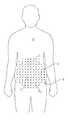
- FIG. 1shows contour and gridlines on a patient.
- FIG. 2illustrates the motion of a HIFU treatment device over the patient.
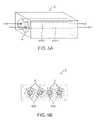
- FIGS. 3A-5Billustrate various treatment approaches.
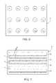
- FIGS. 6-8illustrate various ultrasound treatment patterns.
- FIG. 9illustrates a stencil
- FIG. 11shows a mosaic of treatment sites used to cover a treatment area.
- FIGS. 12-13show histology slides of actual treated tissue.
- FIG. 14is a flow chart showing steps for providing multiple treatments to a single location in accordance with an embodiment.
- FIG. 15is a representation of an ultrasound treatment pattern in accordance with an embodiment.
- a method of modifying tissue using high intensity focused ultrasoundcomprises the steps of determining a volume of adipose tissue to be treated, identifying a corresponding surface area of skin over the volume of adipose tissue; and moving a HIFU therapy transducer on the surface area of skin, and applying therapeutic ultrasound energy into the volume of adipose tissue so that a plurality of cells or pockets of tissue necroses and denatured collagen fibrils are produced.
- the depth of the adipose tissueshould be sufficient to allow the focal zone of the HIFU transducer to be safely in the adipose tissue with some margin of safety both above and below the focal point of the transducer, it should be understood that varying the focal depth of the transducer, as well as the shape and focus of the transducer can allow for more precise control over the delivery of HIFU energy, while simultaneously reducing the clearance zones needed for safe operation. That is to say a highly focused transducer provides sufficient control and focus to allow for a reduced safety clearance.
- the physicianshould determine the corresponding surface area over the volume that can be treated. Once again, borrowing from existing techniques in liposuction, the physician may proceed directly to treating the patient using a HIFU transducer, or she can create one or more contour lines as part of the treatment planning phase of an ordinary liposuction procedure. During this step the physician may draw or otherwise indicate on a patient skin surface, a region that can safely be treated using a HIFU transducer. Pens or markers may be used to create these contour lines.
- HIFU energyinto the volume of adipose tissue.
- a HIFU transduceris moved over the surface area identified above.
- the transduceremits energy to the focal zone in sufficient strength (power) and intensity (pressure) to cause cellular necrosis and collagen fibril denaturing.
- powerpower
- intensitypressure
- a plurality of discrete treatment cellswill be produced. Desirably each treatment cell will absorb sufficient energy from the transducer to cause cellular necrosis of all cells in the focal zone, as well as collagen denaturing in the same region.
- the volume of tissue affected at the focal zone of the transduceris the lesion field 630 ( FIG. 3A-5B ).
- the application of HIFU energy into the volume of adipose energymay involve multiple treatments at the same location.
- the cumulative strength (power) and intensity (pressure)is sufficient to cause cellular necrosis and collagen fibril denaturing. This cumulative effect permits each individual treatment to be of insufficient power and intensity to cause cellular necrosis and collagen fibril denaturing.
- FIG. 14is a flow chart showing steps for providing multiple treatments to a single location in accordance with an embodiment.
- a first application of HIFU energyi.e., a first treatment
- a pauseis taken, in which treatment may be applied to another location.
- an additional treatmentis applied to the same location.
- a determinationis made whether the power of the cumulative treatments is sufficient to cause cellular necrosis and collagen fibril denaturing. If not, the process branches back to step 1400 , and a further treatment is applied. If so, the applications at that location may be completed.
- adipose tissue in the lesion fieldis not restricted to adipocytes (fat cells) alone.
- the methods described hereinare intended to destroy biological tissue within the focal zone by whatever mechanism the HIFU transducer can produce.
- the thermal energy which radiates from the lesion fielddestroys the surrounding tissue forming the halo field.
- This thermal radiationis not intended to be of a particular temperature for selective preservation of any biological material.
- the temperature in the halo fieldshould be sufficient to destroy the adipose tissue and denature the collagen fibrils. Thus, it is likely that other cells or tissue types within the lesion and halo field will be destroyed.
- the various treatment sites 2 which form the treatment zone 3 on a patientmay be uniform or different in both size of each treatment site 2 within the treatment zone 3 , as well as having any mixture of lesion fields 630 , contiguous lesion fields 630 c , cooperative lesion fields, halo fields 6 , contiguous halo fields and cooperative halo fields.
- multiple treatmentsmay be provided, with the multiple treatments having any of these forms.
- the transducermay be used to deposit energy and produce lesion fields of varying shapes and sizes. If the transducer is left to reside in a single position (such as using an incremental movement), the transducer may initially create a small lesion field. By allowing the transducer to loiter, thermal energy will build up and radiate out from the lesion field. The transducer may be moved slowly or have higher energy output while moved in a regular movement pattern to produce larger contiguous lesion fields (produce thicker scan lines). By analogy, one may envision the way a fountain pen leaves ink on a page.
- Increasing the power broadcast into the tissuemay be achieved by moving the transducer slowly, varying the parameters of the transducer, so that more energy radiates from the lesion field into the surrounding tissue, thus producing an enlarged halo field.
- the lesion fielditself may also increase in size.
- halo fieldsmay be overlapped so that each location has four halo effects at each lesion field.
- the systemmay be arranged so that the cumulative power applied at each lesion field is sufficient to cause cellular necrosis and collagen fibril denaturing.
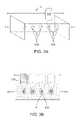
- the motion of the transducer over the patient skincan follow any number of patterns.
- a basic motionis shown in FIG. 4A .
- a transducer 500is moved in a linear path over the patient skin.
- the transducerhas a focal zone 630 which creates a lesion field. If the transducer is moved in a controlled manner the lesion field formed by the HIFU therapy transducer may form a single, contiguous line of destroyed tissue 630 c .
- the axis of the focal zone in tissueis referred to herein as the scan line 4 .
- Surrounding the scan line 4is a region of thermal effect desirably raising the local tissue to temperatures sufficient to kill adipose tissue and denature collagen fibrils.
- This halo field 6 about the scan line 4represents the volume of tissue which receives sufficient thermal radiation from the lesion field 630 , 630 c to also be destroyed and denatured.
- the halo 6may be large or small depending on how quickly the transducer is moved, and how much power the transducer produces.
- a single scan line 4is shown within a single treatment site 2 for clarity.
- a cross section view of a scan line 4is shown in FIG. 4B .
- a scan linemay be repeated.
- scan linesmay cross or overlap to provide a desired accumulation.
- the transducer 500may be made to produce high intensity pulses or pulse bursts (rapid sequence of discrete pulses) to produce discrete lesions 630 along a scan line 4 ( FIG. 3A ).
- the transduceris desirably moved over the patient skin surface and the transducer is programmed to deliver discrete bursts of HIFU ultrasound energy to produce individual or discrete “cells” of destroyed tissue.
- the burst of ultrasound energycan produce any variety and number of discrete lesions in the tissue.
- a halo 6may also be found surrounding each lesion depending on the operating parameters of the transducer. Again, the pattern of lesion fields and halos are also presented in cross section shown in FIG. 3B .
- FIGS. 5A-BAnother embodiment for applying ultrasound energy is illustrated in FIGS. 5A-B .
- two scan lines 4 , 4 ′are shown in close proximity so that the contiguous lesion fields 630 c , 630 c ′ are parallel.
- the halo zone 6 of each scan linerun together to form a region of cooperative effect and enlarge the halo zone.
- Multiple scan linesmay be placed side by side to form a large layer of mechanical and thermal effect ( FIG. 5B ).
- individual scan linesmay be repeated at the same location, or slightly moved over so as to overlap a previous line.
- scan linesmay cross or overlap to provide a desired accumulation.
- a large number of scan linesmay be utilized for a treatment area with several overlaps in the scan lines and so that cumulative power at most or all locations is sufficient for cellular necrosis and collagen fibril denaturing.
- Collagen denaturingcan occur at temperatures above 37° C. However denatured collagen at temperatures close to normal body temperature may recover, relax and resume their normal length. Desirably then, collagen in the treatment zone is exposed to temperatures above 37° C. More desirably collagen fibrils in the treatment zone are exposed to temperatures above 46° C. and even more preferably to temperatures above 56° C.
- Collagen Fibrilrefers to the collagen material found in adipose tissue or sub dermal regions where collagen concentration tends to be sparse and used by the body as a lattice connective tissue rather than a major structural component (contrast with regions like the nose, ears, skin or tendons and the like). Contraction of collagen fibrils refers to using thermal energy to denature the collagen and force the collagen fibrils to shorten lengthwise.
- adipose tissueis heated using HIFU energy so the temperature in the lesion field is raised as high as practical and as fast as possible.
- Parameters of the HIFU transducermay be adjusted to produce the desired fast heating needed to destroy adipose tissue and denature collagen fibrils.
- the fast heatingis balanced with the volume and dimensions of the adipose tissue to be treated. The longer the transducer remains active on one location, the larger the halo field.
- the moving of the HIFU transducer and the applying of therapeutic ultrasound energydo not produce lesion or halo fields which extend beyond the dimensions of the adipose tissue volume.
- Additional parameters that affect the size of the lesion and halo fieldsare those parameters electronically controlled through the transducer, and parameters of the transducer itself. These parameters include (but are not limited to) power, frequency, duty cycle, focus, size (of transducer), and pulse repetition frequency.
- the size of the lesion and halo fieldsare desirably minimized. This is particularly true where the adipose tissue depth necessitates a tightly controlled lesion and halo field due to proximity of muscle, organs or skin. This can be accomplished by distributing the individual lesion fields within a treatment site apart from each other in both distance and time. If the treatment site is represented by a defined field area 2 , then the individual spot lesions may be laid down one at a time in a sequence from L 1 to L 15 ( FIG. 6 ). For multiple treatments at each location, the sequence may be repeated or may be performed in a different order. Here the lesions are temporally separated as well as being spatially separated. This pattern allows for the individual lesions to have a minimum cooperative thermal effect between lesions. The size of each lesion (L 1-n ) may also be controlled by adjusting the parameters of the ultrasound transducer used in the treatment.
- the lesion and halo fieldsmay be maximized by permitting the HIFU transducer to produce contiguous lesion fields and cooperative halo fields.
- An example of such a maximizing movement schemeis illustrated now in FIG. 7 .
- the energy required to produce cellular necrosis and collagen contractionis lessened due to the co-operative effect of having the transducer operate in narrowly spaced treatment lines and in rapid succession of laying down treatment lines near each other in both time and space. Movement of the transducer is desirably machine controlled for uniformity and simultaneous control of the transducer.
- the transducercan treat patient tissue volume by moving over the surface of the tissue volume in any variety of patterns including, but not limited to, spiral, raster scan, or patterned.
- Thermal cooperationcan be maximized by delivering the ultrasound energy as a contiguous lesion field 630 within the treatment site 2 .
- a raster scan type pattern( FIG. 7 ) may be used with a relatively close line spacing to provide for a maximum of thermal cooperation to produce a large halo region.
- the horizontal scan lines 4may be connected with vertical transit lines 5 where the transducer is active, or the vertical transit lines may be “empty” if the transducer is not active while moving vertically.
- the spacing between the horizontal lines 4may be close together or physically overlapping to provide for the maximum overlap of ultrasound energy.
- the raster patternmay be repeated or different crossing or overlapping patterns may be used to provided desired accumulation at each location. Careful planning and consideration in the applying of ultrasound energy in the methods described herein can produce the desired volume of tissue modification in both the amount of adipose tissue destroyed, and collagen denatured.
- a balancing of speed (velocity of the focal zone in the tissue being treated) and the power and intensity of the transducerare needed to produce the desired effect.
- a method of determining the various parameters to use in a tissue modificationis now described.
- the methodcomprises the steps of determining a volume of adipose tissue to be treated; marking out a corresponding surface area of skin and applying high intensity focused ultrasound energy to said area in a manner sufficient to induce the gradual destruction of said adipose tissue and denaturing of collagen fibrils, the energy flux being of at least than 35 J/cm 2 .
- the speed of destructionmay be quickened by providing higher EF values.
- Accumulationcan provide a desired EF value without the application of high energy flux pulses. For example, two separate treatments, each having 33 J/cm 2 , may result is an accumulated EF of 66 J/cm 2 , without having to resort to a treatment exceeding 35 J/cm 2 . As such, efficacy may be enhanced with greater patient tolerance.
- the transducercan be programmed to consistently and accurately deposit the same amount of energy into each of the lesion fields (also referred to as the focal zone).
- tissue ablation of adipose tissue and collagen contractioncan occur at energy fluxes above 35 joules per square centimeter. Variations in desired outcomes and tissue variations from patient to patient make calling out an exact energy flux figure impossible.
- empirical data from multiple study sourcessuggest the energy flux value, from cumulative or a single treatment, should be greater than 35 joules per square centimeter and are probably most efficacious for the dual purpose of destroying adipose tissue and denaturing collagen fibrils at or above 109 joules per square centimeter.
- an apparatus for the delivery of therapeutic ultrasound energy into a patienthaving at least one ultrasound transducer adapted for being moved while applying therapy and being capable of depositing an energy flux (EF) greater than 35 J/cm 2 , wherein EF is determined by the formula: [(p) ⁇ (l/v) ⁇ (dc) ⁇ (nl)]/(sa)
- the formulation providedprovides for a calculation when the transducer is moving continuously while applying ultrasound energy.
- nsnumber of lesions
- Variations in the formulacan be derived by those skilled in the art to determine the proper calculations for a therapy program having a mixed set of moving and non-moving treatment sites.
- the therapy controllerdesirably allows for a wide variation in parameters which a user may manually feed into the therapy controller prior to each application of ultrasound.
- the therapy controllerdetermines which variables are to be used and weights them accordingly.
- An example of a medical instrument system for use with the methods described hereinis further described in co-pending U.S. patent application Ser. No. 11/027,912 entitled “Ultrasound Therapy Head with Movement Control” the contents of which are herein incorporated by reference.
- the apparatus for the delivery of therapeutic ultrasound energy into a patienthas a scan head, suspension device for supporting the scan head, and a therapy controller.
- the therapy controlleris adapted to monitor the position and energy deliver of the scan head. This apparatus may be used to deliver multiple treatments to the same location by having the scan head return multiple times.
- FIG. 16Another example is shown in FIG. 16 , where a robot arm 200 moves a scan head 202 over multiple markers, for example on a patient's body.
- the scanner head 202may be directed by a physician to the markers, and then instructed to apply a treatment.
- the robot armmay remember the position, for example using kinematic information, and after the physician has placed the scanner head at each treatment location, return automatically to each of the locations so that multiple treatments may be applied to the each location.
- the robotmay remember a location, for example via kinematics, and count the number of applications applied by a physician.
- the scanner headmay include optical recognition hardware, and may automatically find a marker and apply a treatment.
- the various parameters of the Energy Flux equationcan be programmed into the therapy controller.
- the apparatusmay have some parameter data programmed in fixed memory and not adjustable by the user. Some elements may include maximum and minimum settings of the transducer to prevent the apparatus from being operated in an unsafe manner.
- a usercan provide variables into the system to help the system determine the proper EF to be used during a procedure. For example if the user wishes to increase cooperative heating between scan lines, the scan lines (nl) may be set to a higher value. Alternatively the velocity may be reduced to promote larger halo fields, or the velocity may be increased to decrease halo fields as might be required for regions of adipose tissue which have smaller margins.
- a stencil or template 24can be used to assist a physician in planning the treatment ( FIG. 9 ).
- the template 24has a series of apertures 26 in the form of “crosshairs” which can be used to guide the ultrasound transducer during the treatment procedure.
- the template 24desirably is created so the apertures match the foot print of the transducer to be used (or therapy device depending on the ultrasound system selected).
- the templatemay be used across the skin prior to the creation of contour lines or prior even to the evaluation of the adipose tissue in the target region. Desirably a physician will mark the contour lines and crosshair marks after making the determination of suitable adipose tissue depth in the patients target treatment region.
- the stencil 24can be laid across the patient ( FIG. 10 ) and then the crosshairs drawn in using a medical marker.
- the combination of crosshairs and contour lines shown in FIG. 1combine to provide visual markers for the safe placement of a HIFU transducer in an ordered fashion (using the guide marks) within a known depth of adipose tissue (using the contour lines).
- the physicianneed only line up the ultrasound treatment device with the crosshairs and contour lines ( FIG. 2 ) to produce a mosaic of treatment sites 2 ( FIG. 11 ).
- the volume of tissue to be treatedcan be done using techniques already adopted by physicians in the ordinary practice of procedures like UAL.
- the physiciancan use a manual pinch test, calipers or diagnostic ultrasound to determine the depth of the fat tissue to be treated and draw circles around the region to be treated, similar to relief lines on a topographical map.
- the individual marks from the stencilmay be made before the volume is determined, or after.
- the contour lines representing varying levels of tissue volume, and therapy head land marksoverlap to provide the user with a defined safe area to treat, as well as a guide for treatment using the ultrasound therapy head.
- the lesion field 22shows both the collapse and destruction of adipose tissue and the denaturing of collagen fibrils which contract the tissue volume as the destroyed tissue mass is gradually removed from the body (through the body's natural wound healing response).
- the reduction of adipose tissue volume in this mannerprovides a similar long term result to liposuction. Since the tissue loss is gradual, there is no sudden looseness of the skin layer, nor skin deformation observed immediately after a patient undergoes a treatment using the methods described herein.
Landscapes
- Health & Medical Sciences (AREA)
- Life Sciences & Earth Sciences (AREA)
- Engineering & Computer Science (AREA)
- Biomedical Technology (AREA)
- Nuclear Medicine, Radiotherapy & Molecular Imaging (AREA)
- Radiology & Medical Imaging (AREA)
- Animal Behavior & Ethology (AREA)
- General Health & Medical Sciences (AREA)
- Public Health (AREA)
- Veterinary Medicine (AREA)
- Physics & Mathematics (AREA)
- Biophysics (AREA)
- Pathology (AREA)
- Heart & Thoracic Surgery (AREA)
- Medical Informatics (AREA)
- Molecular Biology (AREA)
- Surgery (AREA)
- Surgical Instruments (AREA)
- Prostheses (AREA)
- Thermotherapy And Cooling Therapy Devices (AREA)
Abstract
Description
[(p×(l/v)×(dc)×(nl)]/(sa)
[(p)×(l/v)×(dc)×(nl)]/(sa)
EF=[(p)×(t)×(dc)×(ns)]/(sa)
Claims (15)
Priority Applications (4)
| Application Number | Priority Date | Filing Date | Title |
|---|---|---|---|
| US12/559,239US8343051B2 (en) | 2003-12-30 | 2009-09-14 | Apparatus and methods for the destruction of adipose tissue |
| PCT/US2010/048498WO2011032017A2 (en) | 2009-09-14 | 2010-09-10 | Apparatus and methods for the destruction of adipose tissue |
| ARP100103354AAR078422A1 (en) | 2009-09-14 | 2010-09-14 | A COSMETIC METHOD FOR THE DESTRUCTION OF ADIPOSE TISSUE |
| US13/674,546US9180314B2 (en) | 2003-12-30 | 2012-11-12 | Apparatus and methods for the destruction of adipose tissue |
Applications Claiming Priority (6)
| Application Number | Priority Date | Filing Date | Title |
|---|---|---|---|
| US53395803P | 2003-12-30 | 2003-12-30 | |
| US11/026,519US7993289B2 (en) | 2003-12-30 | 2004-12-29 | Systems and methods for the destruction of adipose tissue |
| US67619705P | 2005-04-29 | 2005-04-29 | |
| US11/414,080US7857773B2 (en) | 2003-12-30 | 2006-04-27 | Apparatus and methods for the destruction of adipose tissue |
| US9720508P | 2008-09-15 | 2008-09-15 | |
| US12/559,239US8343051B2 (en) | 2003-12-30 | 2009-09-14 | Apparatus and methods for the destruction of adipose tissue |
Related Parent Applications (1)
| Application Number | Title | Priority Date | Filing Date |
|---|---|---|---|
| US11/414,080Continuation-In-PartUS7857773B2 (en) | 2003-12-30 | 2006-04-27 | Apparatus and methods for the destruction of adipose tissue |
Related Child Applications (1)
| Application Number | Title | Priority Date | Filing Date |
|---|---|---|---|
| US13/674,546ContinuationUS9180314B2 (en) | 2003-12-30 | 2012-11-12 | Apparatus and methods for the destruction of adipose tissue |
Publications (2)
| Publication Number | Publication Date |
|---|---|
| US20100042019A1 US20100042019A1 (en) | 2010-02-18 |
| US8343051B2true US8343051B2 (en) | 2013-01-01 |
Family
ID=43063885
Family Applications (2)
| Application Number | Title | Priority Date | Filing Date |
|---|---|---|---|
| US12/559,239Expired - LifetimeUS8343051B2 (en) | 2003-12-30 | 2009-09-14 | Apparatus and methods for the destruction of adipose tissue |
| US13/674,546Active2026-01-08US9180314B2 (en) | 2003-12-30 | 2012-11-12 | Apparatus and methods for the destruction of adipose tissue |
Family Applications After (1)
| Application Number | Title | Priority Date | Filing Date |
|---|---|---|---|
| US13/674,546Active2026-01-08US9180314B2 (en) | 2003-12-30 | 2012-11-12 | Apparatus and methods for the destruction of adipose tissue |
Country Status (3)
| Country | Link |
|---|---|
| US (2) | US8343051B2 (en) |
| AR (1) | AR078422A1 (en) |
| WO (1) | WO2011032017A2 (en) |
Cited By (27)
| Publication number | Priority date | Publication date | Assignee | Title |
|---|---|---|---|---|
| US9283409B2 (en) | 2004-10-06 | 2016-03-15 | Guided Therapy Systems, Llc | Energy based fat reduction |
| US9283410B2 (en) | 2004-10-06 | 2016-03-15 | Guided Therapy Systems, L.L.C. | System and method for fat and cellulite reduction |
| US9289188B2 (en) | 2012-12-03 | 2016-03-22 | Liposonix, Inc. | Ultrasonic transducer |
| US9320537B2 (en) | 2004-10-06 | 2016-04-26 | Guided Therapy Systems, Llc | Methods for noninvasive skin tightening |
| US9421029B2 (en) | 2004-10-06 | 2016-08-23 | Guided Therapy Systems, Llc | Energy based hyperhidrosis treatment |
| US9427601B2 (en) | 2004-10-06 | 2016-08-30 | Guided Therapy Systems, Llc | Methods for face and neck lifts |
| US9427600B2 (en) | 2004-10-06 | 2016-08-30 | Guided Therapy Systems, L.L.C. | Systems for treating skin laxity |
| US9440096B2 (en) | 2004-10-06 | 2016-09-13 | Guided Therapy Systems, Llc | Method and system for treating stretch marks |
| US9510802B2 (en) | 2012-09-21 | 2016-12-06 | Guided Therapy Systems, Llc | Reflective ultrasound technology for dermatological treatments |
| US9694212B2 (en) | 2004-10-06 | 2017-07-04 | Guided Therapy Systems, Llc | Method and system for ultrasound treatment of skin |
| US9827449B2 (en) | 2004-10-06 | 2017-11-28 | Guided Therapy Systems, L.L.C. | Systems for treating skin laxity |
| US9861410B2 (en) | 2016-05-06 | 2018-01-09 | Medos International Sarl | Methods, devices, and systems for blood flow |
| US10420960B2 (en) | 2013-03-08 | 2019-09-24 | Ulthera, Inc. | Devices and methods for multi-focus ultrasound therapy |
| US10537304B2 (en) | 2008-06-06 | 2020-01-21 | Ulthera, Inc. | Hand wand for ultrasonic cosmetic treatment and imaging |
| US10603521B2 (en) | 2014-04-18 | 2020-03-31 | Ulthera, Inc. | Band transducer ultrasound therapy |
| US10864385B2 (en) | 2004-09-24 | 2020-12-15 | Guided Therapy Systems, Llc | Rejuvenating skin by heating tissue for cosmetic treatment of the face and body |
| US11207548B2 (en) | 2004-10-07 | 2021-12-28 | Guided Therapy Systems, L.L.C. | Ultrasound probe for treating skin laxity |
| US11224895B2 (en) | 2016-01-18 | 2022-01-18 | Ulthera, Inc. | Compact ultrasound device having annular ultrasound array peripherally electrically connected to flexible printed circuit board and method of assembly thereof |
| US11235179B2 (en) | 2004-10-06 | 2022-02-01 | Guided Therapy Systems, Llc | Energy based skin gland treatment |
| US11241218B2 (en) | 2016-08-16 | 2022-02-08 | Ulthera, Inc. | Systems and methods for cosmetic ultrasound treatment of skin |
| US11338156B2 (en) | 2004-10-06 | 2022-05-24 | Guided Therapy Systems, Llc | Noninvasive tissue tightening system |
| US11724133B2 (en) | 2004-10-07 | 2023-08-15 | Guided Therapy Systems, Llc | Ultrasound probe for treatment of skin |
| US11883688B2 (en) | 2004-10-06 | 2024-01-30 | Guided Therapy Systems, Llc | Energy based fat reduction |
| US11944849B2 (en) | 2018-02-20 | 2024-04-02 | Ulthera, Inc. | Systems and methods for combined cosmetic treatment of cellulite with ultrasound |
| US12076591B2 (en) | 2018-01-26 | 2024-09-03 | Ulthera, Inc. | Systems and methods for simultaneous multi-focus ultrasound therapy in multiple dimensions |
| US12102473B2 (en) | 2008-06-06 | 2024-10-01 | Ulthera, Inc. | Systems for ultrasound treatment |
| US12377293B2 (en) | 2019-07-15 | 2025-08-05 | Ulthera, Inc. | Systems and methods for measuring elasticity with imaging of ultrasound multi-focus shearwaves in multiple dimensions |
Families Citing this family (4)
| Publication number | Priority date | Publication date | Assignee | Title |
|---|---|---|---|---|
| US8932238B2 (en)* | 2009-09-29 | 2015-01-13 | Liposonix, Inc. | Medical ultrasound device with liquid dispensing device coupled to a therapy head |
| WO2012018562A1 (en) | 2010-07-24 | 2012-02-09 | Medicis Technologies Corporation | Apparatus and methods for non-invasive body contouring |
| KR101574951B1 (en)* | 2015-08-13 | 2015-12-07 | 김유인 | High Intensity Focused Ultrasonic Portable Medical Instrument |
| WO2024191921A1 (en)* | 2023-03-10 | 2024-09-19 | R2 Technologies, Inc. | Cryo methods and systems for medical treatments |
Citations (3)
| Publication number | Priority date | Publication date | Assignee | Title |
|---|---|---|---|---|
| US20030171701A1 (en)* | 2002-03-06 | 2003-09-11 | Eilaz Babaev | Ultrasonic method and device for lypolytic therapy |
| US20040039312A1 (en)* | 2002-02-20 | 2004-02-26 | Liposonix, Inc. | Ultrasonic treatment and imaging of adipose tissue |
| US7857773B2 (en)* | 2003-12-30 | 2010-12-28 | Medicis Technologies Corporation | Apparatus and methods for the destruction of adipose tissue |
Family Cites Families (2)
| Publication number | Priority date | Publication date | Assignee | Title |
|---|---|---|---|---|
| US2791204A (en) | 1951-08-16 | 1957-05-07 | Smith Corp A O | Water heater utilizing heat of crystallization |
| US2651904A (en) | 1951-11-03 | 1953-09-15 | Franklin W Jatunn | Adjustable cutter bar for lawn mowers |
- 2009
- 2009-09-14USUS12/559,239patent/US8343051B2/ennot_activeExpired - Lifetime
- 2010
- 2010-09-10WOPCT/US2010/048498patent/WO2011032017A2/enactiveApplication Filing
- 2010-09-14ARARP100103354Apatent/AR078422A1/enunknown
- 2012
- 2012-11-12USUS13/674,546patent/US9180314B2/enactiveActive
Patent Citations (4)
| Publication number | Priority date | Publication date | Assignee | Title |
|---|---|---|---|---|
| US20040039312A1 (en)* | 2002-02-20 | 2004-02-26 | Liposonix, Inc. | Ultrasonic treatment and imaging of adipose tissue |
| US20030171701A1 (en)* | 2002-03-06 | 2003-09-11 | Eilaz Babaev | Ultrasonic method and device for lypolytic therapy |
| US20050015024A1 (en)* | 2002-03-06 | 2005-01-20 | Eilaz Babaev | Ultrasonic method and device for lypolytic therapy |
| US7857773B2 (en)* | 2003-12-30 | 2010-12-28 | Medicis Technologies Corporation | Apparatus and methods for the destruction of adipose tissue |
Cited By (74)
| Publication number | Priority date | Publication date | Assignee | Title |
|---|---|---|---|---|
| US9895560B2 (en) | 2004-09-24 | 2018-02-20 | Guided Therapy Systems, Llc | Methods for rejuvenating skin by heating tissue for cosmetic treatment of the face and body |
| US11590370B2 (en) | 2004-09-24 | 2023-02-28 | Guided Therapy Systems, Llc | Rejuvenating skin by heating tissue for cosmetic treatment of the face and body |
| US10864385B2 (en) | 2004-09-24 | 2020-12-15 | Guided Therapy Systems, Llc | Rejuvenating skin by heating tissue for cosmetic treatment of the face and body |
| US10328289B2 (en) | 2004-09-24 | 2019-06-25 | Guided Therapy Systems, Llc | Rejuvenating skin by heating tissue for cosmetic treatment of the face and body |
| US10532230B2 (en) | 2004-10-06 | 2020-01-14 | Guided Therapy Systems, Llc | Methods for face and neck lifts |
| US9707412B2 (en) | 2004-10-06 | 2017-07-18 | Guided Therapy Systems, Llc | System and method for fat and cellulite reduction |
| US9427600B2 (en) | 2004-10-06 | 2016-08-30 | Guided Therapy Systems, L.L.C. | Systems for treating skin laxity |
| US9440096B2 (en) | 2004-10-06 | 2016-09-13 | Guided Therapy Systems, Llc | Method and system for treating stretch marks |
| US11883688B2 (en) | 2004-10-06 | 2024-01-30 | Guided Therapy Systems, Llc | Energy based fat reduction |
| US9522290B2 (en) | 2004-10-06 | 2016-12-20 | Guided Therapy Systems, Llc | System and method for fat and cellulite reduction |
| US10603523B2 (en) | 2004-10-06 | 2020-03-31 | Guided Therapy Systems, Llc | Ultrasound probe for tissue treatment |
| US9694212B2 (en) | 2004-10-06 | 2017-07-04 | Guided Therapy Systems, Llc | Method and system for ultrasound treatment of skin |
| US9694211B2 (en) | 2004-10-06 | 2017-07-04 | Guided Therapy Systems, L.L.C. | Systems for treating skin laxity |
| US10603519B2 (en) | 2004-10-06 | 2020-03-31 | Guided Therapy Systems, Llc | Energy based fat reduction |
| US9713731B2 (en) | 2004-10-06 | 2017-07-25 | Guided Therapy Systems, Llc | Energy based fat reduction |
| US11717707B2 (en) | 2004-10-06 | 2023-08-08 | Guided Therapy Systems, Llc | System and method for noninvasive skin tightening |
| US9827450B2 (en) | 2004-10-06 | 2017-11-28 | Guided Therapy Systems, L.L.C. | System and method for fat and cellulite reduction |
| US9827449B2 (en) | 2004-10-06 | 2017-11-28 | Guided Therapy Systems, L.L.C. | Systems for treating skin laxity |
| US9833640B2 (en) | 2004-10-06 | 2017-12-05 | Guided Therapy Systems, L.L.C. | Method and system for ultrasound treatment of skin |
| US9833639B2 (en) | 2004-10-06 | 2017-12-05 | Guided Therapy Systems, L.L.C. | Energy based fat reduction |
| US11697033B2 (en) | 2004-10-06 | 2023-07-11 | Guided Therapy Systems, Llc | Methods for lifting skin tissue |
| US9421029B2 (en) | 2004-10-06 | 2016-08-23 | Guided Therapy Systems, Llc | Energy based hyperhidrosis treatment |
| US9974982B2 (en) | 2004-10-06 | 2018-05-22 | Guided Therapy Systems, Llc | System and method for noninvasive skin tightening |
| US10010724B2 (en) | 2004-10-06 | 2018-07-03 | Guided Therapy Systems, L.L.C. | Ultrasound probe for treating skin laxity |
| US10010725B2 (en) | 2004-10-06 | 2018-07-03 | Guided Therapy Systems, Llc | Ultrasound probe for fat and cellulite reduction |
| US10010726B2 (en) | 2004-10-06 | 2018-07-03 | Guided Therapy Systems, Llc | Ultrasound probe for treatment of skin |
| US10010721B2 (en) | 2004-10-06 | 2018-07-03 | Guided Therapy Systems, L.L.C. | Energy based fat reduction |
| US10046182B2 (en) | 2004-10-06 | 2018-08-14 | Guided Therapy Systems, Llc | Methods for face and neck lifts |
| US10046181B2 (en) | 2004-10-06 | 2018-08-14 | Guided Therapy Systems, Llc | Energy based hyperhidrosis treatment |
| US10238894B2 (en) | 2004-10-06 | 2019-03-26 | Guided Therapy Systems, L.L.C. | Energy based fat reduction |
| US10245450B2 (en) | 2004-10-06 | 2019-04-02 | Guided Therapy Systems, Llc | Ultrasound probe for fat and cellulite reduction |
| US10252086B2 (en) | 2004-10-06 | 2019-04-09 | Guided Therapy Systems, Llc | Ultrasound probe for treatment of skin |
| US10265550B2 (en) | 2004-10-06 | 2019-04-23 | Guided Therapy Systems, L.L.C. | Ultrasound probe for treating skin laxity |
| US9320537B2 (en) | 2004-10-06 | 2016-04-26 | Guided Therapy Systems, Llc | Methods for noninvasive skin tightening |
| US9283410B2 (en) | 2004-10-06 | 2016-03-15 | Guided Therapy Systems, L.L.C. | System and method for fat and cellulite reduction |
| US10525288B2 (en) | 2004-10-06 | 2020-01-07 | Guided Therapy Systems, Llc | System and method for noninvasive skin tightening |
| US9283409B2 (en) | 2004-10-06 | 2016-03-15 | Guided Therapy Systems, Llc | Energy based fat reduction |
| US11400319B2 (en) | 2004-10-06 | 2022-08-02 | Guided Therapy Systems, Llc | Methods for lifting skin tissue |
| US9533175B2 (en) | 2004-10-06 | 2017-01-03 | Guided Therapy Systems, Llc | Energy based fat reduction |
| US11338156B2 (en) | 2004-10-06 | 2022-05-24 | Guided Therapy Systems, Llc | Noninvasive tissue tightening system |
| US9427601B2 (en) | 2004-10-06 | 2016-08-30 | Guided Therapy Systems, Llc | Methods for face and neck lifts |
| US10610705B2 (en) | 2004-10-06 | 2020-04-07 | Guided Therapy Systems, L.L.C. | Ultrasound probe for treating skin laxity |
| US10610706B2 (en) | 2004-10-06 | 2020-04-07 | Guided Therapy Systems, Llc | Ultrasound probe for treatment of skin |
| US11235180B2 (en) | 2004-10-06 | 2022-02-01 | Guided Therapy Systems, Llc | System and method for noninvasive skin tightening |
| US11235179B2 (en) | 2004-10-06 | 2022-02-01 | Guided Therapy Systems, Llc | Energy based skin gland treatment |
| US10888716B2 (en) | 2004-10-06 | 2021-01-12 | Guided Therapy Systems, Llc | Energy based fat reduction |
| US10888717B2 (en) | 2004-10-06 | 2021-01-12 | Guided Therapy Systems, Llc | Probe for ultrasound tissue treatment |
| US10888718B2 (en) | 2004-10-06 | 2021-01-12 | Guided Therapy Systems, L.L.C. | Ultrasound probe for treating skin laxity |
| US10960236B2 (en) | 2004-10-06 | 2021-03-30 | Guided Therapy Systems, Llc | System and method for noninvasive skin tightening |
| US11207547B2 (en) | 2004-10-06 | 2021-12-28 | Guided Therapy Systems, Llc | Probe for ultrasound tissue treatment |
| US11179580B2 (en) | 2004-10-06 | 2021-11-23 | Guided Therapy Systems, Llc | Energy based fat reduction |
| US11167155B2 (en) | 2004-10-06 | 2021-11-09 | Guided Therapy Systems, Llc | Ultrasound probe for treatment of skin |
| US11207548B2 (en) | 2004-10-07 | 2021-12-28 | Guided Therapy Systems, L.L.C. | Ultrasound probe for treating skin laxity |
| US11724133B2 (en) | 2004-10-07 | 2023-08-15 | Guided Therapy Systems, Llc | Ultrasound probe for treatment of skin |
| US11123039B2 (en) | 2008-06-06 | 2021-09-21 | Ulthera, Inc. | System and method for ultrasound treatment |
| US12102473B2 (en) | 2008-06-06 | 2024-10-01 | Ulthera, Inc. | Systems for ultrasound treatment |
| US11723622B2 (en) | 2008-06-06 | 2023-08-15 | Ulthera, Inc. | Systems for ultrasound treatment |
| US10537304B2 (en) | 2008-06-06 | 2020-01-21 | Ulthera, Inc. | Hand wand for ultrasonic cosmetic treatment and imaging |
| US9510802B2 (en) | 2012-09-21 | 2016-12-06 | Guided Therapy Systems, Llc | Reflective ultrasound technology for dermatological treatments |
| US9802063B2 (en) | 2012-09-21 | 2017-10-31 | Guided Therapy Systems, Llc | Reflective ultrasound technology for dermatological treatments |
| US9289188B2 (en) | 2012-12-03 | 2016-03-22 | Liposonix, Inc. | Ultrasonic transducer |
| US10420960B2 (en) | 2013-03-08 | 2019-09-24 | Ulthera, Inc. | Devices and methods for multi-focus ultrasound therapy |
| US11517772B2 (en) | 2013-03-08 | 2022-12-06 | Ulthera, Inc. | Devices and methods for multi-focus ultrasound therapy |
| US11969609B2 (en) | 2013-03-08 | 2024-04-30 | Ulthera, Inc. | Devices and methods for multi-focus ultrasound therapy |
| US10603521B2 (en) | 2014-04-18 | 2020-03-31 | Ulthera, Inc. | Band transducer ultrasound therapy |
| US11351401B2 (en) | 2014-04-18 | 2022-06-07 | Ulthera, Inc. | Band transducer ultrasound therapy |
| US11224895B2 (en) | 2016-01-18 | 2022-01-18 | Ulthera, Inc. | Compact ultrasound device having annular ultrasound array peripherally electrically connected to flexible printed circuit board and method of assembly thereof |
| US9861410B2 (en) | 2016-05-06 | 2018-01-09 | Medos International Sarl | Methods, devices, and systems for blood flow |
| US10639085B2 (en) | 2016-05-06 | 2020-05-05 | Medos International Sarl | Methods, devices, and systems for blood flow |
| US11026729B2 (en) | 2016-05-06 | 2021-06-08 | Medos International Sarl | Methods, devices, and systems for blood flow |
| US11241218B2 (en) | 2016-08-16 | 2022-02-08 | Ulthera, Inc. | Systems and methods for cosmetic ultrasound treatment of skin |
| US12076591B2 (en) | 2018-01-26 | 2024-09-03 | Ulthera, Inc. | Systems and methods for simultaneous multi-focus ultrasound therapy in multiple dimensions |
| US11944849B2 (en) | 2018-02-20 | 2024-04-02 | Ulthera, Inc. | Systems and methods for combined cosmetic treatment of cellulite with ultrasound |
| US12377293B2 (en) | 2019-07-15 | 2025-08-05 | Ulthera, Inc. | Systems and methods for measuring elasticity with imaging of ultrasound multi-focus shearwaves in multiple dimensions |
Also Published As
| Publication number | Publication date |
|---|---|
| US9180314B2 (en) | 2015-11-10 |
| AR078422A1 (en) | 2011-11-09 |
| US20100042019A1 (en) | 2010-02-18 |
| US20130072825A1 (en) | 2013-03-21 |
| WO2011032017A2 (en) | 2011-03-17 |
Similar Documents
| Publication | Publication Date | Title |
|---|---|---|
| US8343051B2 (en) | Apparatus and methods for the destruction of adipose tissue | |
| US7857773B2 (en) | Apparatus and methods for the destruction of adipose tissue | |
| US9623267B2 (en) | Ultrasonic treatment of adipose tissue at multiple depths | |
| US8128618B2 (en) | Targeted muscle ablation for reducing signs of aging | |
| US20060122509A1 (en) | System and methods for destroying adipose tissue | |
| US8206299B2 (en) | Image guided high intensity focused ultrasound treatment of nerves | |
| US20030171701A1 (en) | Ultrasonic method and device for lypolytic therapy | |
| WO2012018385A2 (en) | System and method for treating cartilage | |
| KR102190881B1 (en) | Laser devices for treating high intensity pain using complex wavelengths and programmed scanning handpieces | |
| KR20170048911A (en) | Handpiece of therapy system for treatment of fatness using hifu |
Legal Events
| Date | Code | Title | Description |
|---|---|---|---|
| AS | Assignment | Owner name:MEDICIS TECHNOLOGIES CORPORATION,WASHINGTON Free format text:ASSIGNMENT OF ASSIGNORS INTEREST;ASSIGNORS:DESILETS, CHARLES S.;POLLOCK, CAMERON;SIGNING DATES FROM 20090927 TO 20091005;REEL/FRAME:023405/0884 Owner name:MEDICIS TECHNOLOGIES CORPORATION, WASHINGTON Free format text:ASSIGNMENT OF ASSIGNORS INTEREST;ASSIGNORS:DESILETS, CHARLES S.;POLLOCK, CAMERON;SIGNING DATES FROM 20090927 TO 20091005;REEL/FRAME:023405/0884 | |
| AS | Assignment | Owner name:LIPOSONIX, INC., WASHINGTON Free format text:CHANGE OF NAME;ASSIGNOR:MEDICIS TECHNOLOGIES CORPORATION;REEL/FRAME:027595/0307 Effective date:20111101 | |
| STCF | Information on status: patent grant | Free format text:PATENTED CASE | |
| CC | Certificate of correction | ||
| AS | Assignment | Owner name:SILICON VALLEY BANK, CALIFORNIA Free format text:SECURITY AGREEMENT;ASSIGNOR:LIPOSONIX, INC.;REEL/FRAME:030147/0642 Effective date:20121031 | |
| AS | Assignment | Owner name:SILICON VALLEY BANK, CALIFORNIA Free format text:SECURITY INTEREST - MEZZANINE LOAN;ASSIGNOR:LIPOSONIX, INC.;REEL/FRAME:030249/0268 Effective date:20120829 | |
| AS | Assignment | Owner name:PARALLEL INVESTMENT OPPORTUNITIES PARTNERS II L.P. Free format text:SHORT-FORM PATENT SECURITY AGREEMENT;ASSIGNOR:LIPOSONIX, INC.;REEL/FRAME:031674/0454 Effective date:20131114 Owner name:CAPITAL ROYALTY PARTNERS II L.P., TEXAS Free format text:SHORT-FORM PATENT SECURITY AGREEMENT;ASSIGNOR:LIPOSONIX, INC.;REEL/FRAME:031674/0454 Effective date:20131114 Owner name:CAPITAL ROYALTY PARTNERS II ? PARALLEL FUND ?A? L. Free format text:SHORT-FORM PATENT SECURITY AGREEMENT;ASSIGNOR:LIPOSONIX, INC.;REEL/FRAME:031674/0454 Effective date:20131114 Owner name:CAPITAL ROYALTY PARTNERS II - PARALLEL FUND "A" L. Free format text:SHORT-FORM PATENT SECURITY AGREEMENT;ASSIGNOR:LIPOSONIX, INC.;REEL/FRAME:031674/0454 Effective date:20131114 | |
| AS | Assignment | Owner name:LIPOSONIX, INC., CALIFORNIA Free format text:RELEASE OF SECURITY INTEREST IN PATENTS;ASSIGNORS:CAPITAL ROYALTY PARTNERS II L.P.;CAPITAL ROYALTY PARTNERS II - PARALLEL FUND "A" L.P.;PARALLEL INVESTMENT OPPORTUNITIES PARTNERS II L.P.;REEL/FRAME:032126/0370 Effective date:20140123 Owner name:LIPOSONIX, INC., CALIFORNIA Free format text:RELEASE OF SECURITY INTEREST IN PATENTS;ASSIGNOR:SILICON VALLEY BANK;REEL/FRAME:032126/0531 Effective date:20140123 | |
| FEPP | Fee payment procedure | Free format text:PAT HOLDER NO LONGER CLAIMS SMALL ENTITY STATUS, ENTITY STATUS SET TO UNDISCOUNTED (ORIGINAL EVENT CODE: STOL); ENTITY STATUS OF PATENT OWNER: LARGE ENTITY | |
| FPAY | Fee payment | Year of fee payment:4 | |
| AS | Assignment | Owner name:THE BANK OF NEW YORK MELLON, AS NOTES COLLATERAL A Free format text:SECURITY INTEREST;ASSIGNORS:BAUSCH HEALTH AMERICAS, INC.;BAUSCH & LOMB INCORPORATED;BAUSCH HEALTH US, LLC;AND OTHERS;REEL/FRAME:048556/0758 Effective date:20190308 Owner name:THE BANK OF NEW YORK MELLON, AS NOTES COLLATERAL AGENT, NEW YORK Free format text:SECURITY INTEREST;ASSIGNORS:BAUSCH HEALTH AMERICAS, INC.;BAUSCH & LOMB INCORPORATED;BAUSCH HEALTH US, LLC;AND OTHERS;REEL/FRAME:048556/0758 Effective date:20190308 | |
| MAFP | Maintenance fee payment | Free format text:PAYMENT OF MAINTENANCE FEE, 8TH YEAR, LARGE ENTITY (ORIGINAL EVENT CODE: M1552); ENTITY STATUS OF PATENT OWNER: LARGE ENTITY Year of fee payment:8 | |
| AS | Assignment | Owner name:SOLTA MEDICAL, INC., WASHINGTON Free format text:MERGER;ASSIGNORS:AESTHERA;LIPOSONIX, INC.;REEL/FRAME:056227/0174 Effective date:20190124 | |
| AS | Assignment | Owner name:THE BANK OF NEW YORK MELLON, AS NOTES COLLATERAL AGENT, NEW YORK Free format text:SECURITY INTEREST;ASSIGNORS:BAUSCH & LOMB INCORPORATED;BAUSCH HEALTH US, LLC;SOLTA MEDICAL, INC.;AND OTHERS;REEL/FRAME:056811/0814 Effective date:20210608 | |
| AS | Assignment | Owner name:THE BANK OF NEW YORK MELLON, NEW YORK Free format text:SECURITY AGREEMENT;ASSIGNORS:BAUSCH & LOMB INCORPORATED;BAUSCH HEALTH US, LLC;SOLTA MEDICAL, INC.;AND OTHERS;REEL/FRAME:059121/0001 Effective date:20220210 | |
| AS | Assignment | Owner name:THE BANK OF NEW YORK MELLON, NEW YORK Free format text:SECURITY INTEREST;ASSIGNORS:BAUSCH HEALTH COMPANIES INC.;BAUSCH HEALTH, CANADA INC.;BAUSCH HEALTH IRELAND LIMITED;AND OTHERS;REEL/FRAME:060346/0705 Effective date:20220610 | |
| AS | Assignment | Owner name:THE BANK OF NEW YORK MELLON, AS NOTES COLLATERAL AGENT, NEW YORK Free format text:SECURITY AGREEMENT (FIRST LIEN);ASSIGNORS:BAUSCH HEALTH US, LLC;BAUSCH HEALTH AMERICAS, INC.;MEDICIS PHARMACEUTICAL CORPORATION;AND OTHERS;REEL/FRAME:061595/0066 Effective date:20220930 | |
| AS | Assignment | Owner name:THE BANK OF NEW YORK MELLON, AS NOTES COLLATERAL AGENT, NEW YORK Free format text:SECURITY AGREEMENT (SECOND LIEN);ASSIGNORS:BAUSCH HEALTH US, LLC;BAUSCH HEALTH AMERICAS, INC.;MEDICIS PHARMACEUTICAL CORPORATION;AND OTHERS;REEL/FRAME:061606/0468 Effective date:20220930 | |
| MAFP | Maintenance fee payment | Free format text:PAYMENT OF MAINTENANCE FEE, 12TH YEAR, LARGE ENTITY (ORIGINAL EVENT CODE: M1553); ENTITY STATUS OF PATENT OWNER: LARGE ENTITY Year of fee payment:12 | |
| AS | Assignment | Owner name:JPMORGAN CHASE BANK, N.A., ILLINOIS Free format text:U.S. PATENT SECURITY AGREEMENT;ASSIGNORS:MEDICIS PHARMACEUTICAL CORPORATION;ORAPHARMA, INC.;BAUSCH HEALTH US, LLC (F/K/A VALEANT PHARMACEUTICALS NORTH AMERICA LLC);AND OTHERS;REEL/FRAME:071566/0200 Effective date:20250408 Owner name:THE BANK OF NEW YORK MELLON, NEW YORK Free format text:U.S. PATENT SECURITY AGREEMENT;ASSIGNORS:MEDICIS PHARMACEUTICAL CORPORATION;ORAPHARMA, INC.;BAUSCH HEALTH US, LLC (F/K/A VALEANT PHARMACEUTICALS NORTH AMERICA LLC);AND OTHERS;REEL/FRAME:071564/0463 Effective date:20250408 |