US8342182B2 - Functional assessment and treatment catheters and methods for their use in the lung - Google Patents
Functional assessment and treatment catheters and methods for their use in the lungDownload PDFInfo
- Publication number
- US8342182B2 US8342182B2US11/845,296US84529607AUS8342182B2US 8342182 B2US8342182 B2US 8342182B2US 84529607 AUS84529607 AUS 84529607AUS 8342182 B2US8342182 B2US 8342182B2
- Authority
- US
- United States
- Prior art keywords
- flow
- component
- catheter
- lung
- flow restrictive
- Prior art date
- Legal status (The legal status is an assumption and is not a legal conclusion. Google has not performed a legal analysis and makes no representation as to the accuracy of the status listed.)
- Active, expires
Links
Images


Classifications
- A—HUMAN NECESSITIES
- A61—MEDICAL OR VETERINARY SCIENCE; HYGIENE
- A61B—DIAGNOSIS; SURGERY; IDENTIFICATION
- A61B5/00—Measuring for diagnostic purposes; Identification of persons
- A61B5/48—Other medical applications
- A61B5/4836—Diagnosis combined with treatment in closed-loop systems or methods
- A—HUMAN NECESSITIES
- A61—MEDICAL OR VETERINARY SCIENCE; HYGIENE
- A61B—DIAGNOSIS; SURGERY; IDENTIFICATION
- A61B17/00—Surgical instruments, devices or methods
- A61B17/12—Surgical instruments, devices or methods for ligaturing or otherwise compressing tubular parts of the body, e.g. blood vessels or umbilical cord
- A61B17/12022—Occluding by internal devices, e.g. balloons or releasable wires
- A61B17/12099—Occluding by internal devices, e.g. balloons or releasable wires characterised by the location of the occluder
- A61B17/12104—Occluding by internal devices, e.g. balloons or releasable wires characterised by the location of the occluder in an air passage
- A—HUMAN NECESSITIES
- A61—MEDICAL OR VETERINARY SCIENCE; HYGIENE
- A61B—DIAGNOSIS; SURGERY; IDENTIFICATION
- A61B5/00—Measuring for diagnostic purposes; Identification of persons
- A61B5/08—Measuring devices for evaluating the respiratory organs
- A—HUMAN NECESSITIES
- A61—MEDICAL OR VETERINARY SCIENCE; HYGIENE
- A61B—DIAGNOSIS; SURGERY; IDENTIFICATION
- A61B5/00—Measuring for diagnostic purposes; Identification of persons
- A61B5/08—Measuring devices for evaluating the respiratory organs
- A61B5/085—Measuring impedance of respiratory organs or lung elasticity
- A—HUMAN NECESSITIES
- A61—MEDICAL OR VETERINARY SCIENCE; HYGIENE
- A61B—DIAGNOSIS; SURGERY; IDENTIFICATION
- A61B5/00—Measuring for diagnostic purposes; Identification of persons
- A61B5/08—Measuring devices for evaluating the respiratory organs
- A61B5/097—Devices for facilitating collection of breath or for directing breath into or through measuring devices
- A—HUMAN NECESSITIES
- A61—MEDICAL OR VETERINARY SCIENCE; HYGIENE
- A61B—DIAGNOSIS; SURGERY; IDENTIFICATION
- A61B5/00—Measuring for diagnostic purposes; Identification of persons
- A61B5/68—Arrangements of detecting, measuring or recording means, e.g. sensors, in relation to patient
- A61B5/6846—Arrangements of detecting, measuring or recording means, e.g. sensors, in relation to patient specially adapted to be brought in contact with an internal body part, i.e. invasive
- A61B5/6847—Arrangements of detecting, measuring or recording means, e.g. sensors, in relation to patient specially adapted to be brought in contact with an internal body part, i.e. invasive mounted on an invasive device
- A61B5/6852—Catheters
- A61B5/6853—Catheters with a balloon
- A—HUMAN NECESSITIES
- A61—MEDICAL OR VETERINARY SCIENCE; HYGIENE
- A61M—DEVICES FOR INTRODUCING MEDIA INTO, OR ONTO, THE BODY; DEVICES FOR TRANSDUCING BODY MEDIA OR FOR TAKING MEDIA FROM THE BODY; DEVICES FOR PRODUCING OR ENDING SLEEP OR STUPOR
- A61M16/00—Devices for influencing the respiratory system of patients by gas treatment, e.g. ventilators; Tracheal tubes
- A61M16/04—Tracheal tubes
- A61M16/0402—Special features for tracheal tubes not otherwise provided for
- A61M16/0404—Special features for tracheal tubes not otherwise provided for with means for selective or partial lung respiration
- A61M16/0406—Special features for tracheal tubes not otherwise provided for with means for selective or partial lung respiration implanted flow modifiers
- A—HUMAN NECESSITIES
- A61—MEDICAL OR VETERINARY SCIENCE; HYGIENE
- A61M—DEVICES FOR INTRODUCING MEDIA INTO, OR ONTO, THE BODY; DEVICES FOR TRANSDUCING BODY MEDIA OR FOR TAKING MEDIA FROM THE BODY; DEVICES FOR PRODUCING OR ENDING SLEEP OR STUPOR
- A61M16/00—Devices for influencing the respiratory system of patients by gas treatment, e.g. ventilators; Tracheal tubes
- A61M16/08—Bellows; Connecting tubes ; Water traps; Patient circuits
- A61M16/0866—Passive resistors therefor
- A—HUMAN NECESSITIES
- A61—MEDICAL OR VETERINARY SCIENCE; HYGIENE
- A61M—DEVICES FOR INTRODUCING MEDIA INTO, OR ONTO, THE BODY; DEVICES FOR TRANSDUCING BODY MEDIA OR FOR TAKING MEDIA FROM THE BODY; DEVICES FOR PRODUCING OR ENDING SLEEP OR STUPOR
- A61M25/00—Catheters; Hollow probes
- A61M25/0067—Catheters; Hollow probes characterised by the distal end, e.g. tips
- A61M25/0074—Dynamic characteristics of the catheter tip, e.g. openable, closable, expandable or deformable
- A61M25/0075—Valve means
- A—HUMAN NECESSITIES
- A61—MEDICAL OR VETERINARY SCIENCE; HYGIENE
- A61M—DEVICES FOR INTRODUCING MEDIA INTO, OR ONTO, THE BODY; DEVICES FOR TRANSDUCING BODY MEDIA OR FOR TAKING MEDIA FROM THE BODY; DEVICES FOR PRODUCING OR ENDING SLEEP OR STUPOR
- A61M25/00—Catheters; Hollow probes
- A61M25/01—Introducing, guiding, advancing, emplacing or holding catheters
- A61M25/06—Body-piercing guide needles or the like
- A61M25/0662—Guide tubes
- A—HUMAN NECESSITIES
- A61—MEDICAL OR VETERINARY SCIENCE; HYGIENE
- A61F—FILTERS IMPLANTABLE INTO BLOOD VESSELS; PROSTHESES; DEVICES PROVIDING PATENCY TO, OR PREVENTING COLLAPSING OF, TUBULAR STRUCTURES OF THE BODY, e.g. STENTS; ORTHOPAEDIC, NURSING OR CONTRACEPTIVE DEVICES; FOMENTATION; TREATMENT OR PROTECTION OF EYES OR EARS; BANDAGES, DRESSINGS OR ABSORBENT PADS; FIRST-AID KITS
- A61F2/00—Filters implantable into blood vessels; Prostheses, i.e. artificial substitutes or replacements for parts of the body; Appliances for connecting them with the body; Devices providing patency to, or preventing collapsing of, tubular structures of the body, e.g. stents
- A61F2/02—Prostheses implantable into the body
- A61F2/04—Hollow or tubular parts of organs, e.g. bladders, tracheae, bronchi or bile ducts
- A61F2002/043—Bronchi
- A—HUMAN NECESSITIES
- A61—MEDICAL OR VETERINARY SCIENCE; HYGIENE
- A61M—DEVICES FOR INTRODUCING MEDIA INTO, OR ONTO, THE BODY; DEVICES FOR TRANSDUCING BODY MEDIA OR FOR TAKING MEDIA FROM THE BODY; DEVICES FOR PRODUCING OR ENDING SLEEP OR STUPOR
- A61M25/00—Catheters; Hollow probes
- A61M2025/0004—Catheters; Hollow probes having two or more concentrically arranged tubes for forming a concentric catheter system
- A—HUMAN NECESSITIES
- A61—MEDICAL OR VETERINARY SCIENCE; HYGIENE
- A61M—DEVICES FOR INTRODUCING MEDIA INTO, OR ONTO, THE BODY; DEVICES FOR TRANSDUCING BODY MEDIA OR FOR TAKING MEDIA FROM THE BODY; DEVICES FOR PRODUCING OR ENDING SLEEP OR STUPOR
- A61M25/00—Catheters; Hollow probes
- A61M25/01—Introducing, guiding, advancing, emplacing or holding catheters
- A61M25/06—Body-piercing guide needles or the like
- A61M25/0662—Guide tubes
- A61M2025/0681—Systems with catheter and outer tubing, e.g. sheath, sleeve or guide tube
Definitions
- the present inventionrelates generally to medical methods and apparatus. More particularly, the present invention relates to methods and apparatus for the assessment and treatment of lung diseases, such as chronic obstructive pulmonary disease, by detecting the status of the disease and determining an appropriate treatment protocol.
- lung diseasessuch as chronic obstructive pulmonary disease
- COPDchronic obstructive pulmonary disease
- COPDCOPD Management of COPD is largely medical and infrequently surgical. Initially, exercise and smoking cessation are encouraged. Medications including bronchodilators and anti-inflammatories are routinely prescribed. Pulmonary rehabilitation has been shown to improve quality of life and sense of well being. Long term oxygen is generally reserved for the more severely affected patients.
- Emphysemais a condition of the lung characterized by the abnormal permanent enlargement of the airspaces distal to the terminal bronchiole, accompanied by the destruction of their walls. It is known that emphysema and other pulmonary diseases reduce the ability of part of the lungs to fully expel air during the exhalation phase of the breathing cycle. During breathing, the diseased portion of the lung does not fully recoil due to the diseased lung tissue being less elastic than healthy tissue. Consequently, as the airways normally held open by the elastic pull of the lungs become floppy and the diseased lung tissue exerts a diminished driving force during exhalation, the airways close prematurely resulting in air trapping and hyperinflation.
- hyper-expanded lung tissueoccupies more of the pleural space than healthy lung tissue. In most cases, only a part of the lung is diseased while the remaining portion is relatively healthy and therefore still able to efficiently carry out oxygen exchange. By taking up more of the pleural space, the hyper-expanded lung tissue reduces the space available to accommodate the healthy, functioning lung tissue. As a result, the hyper-expanded lung tissue causes inefficient breathing by compressing the adjacent functional airways, alveolar units, and capillaries in relatively healthier lung tissue.
- Lung function in patients suffering from some forms of COPDcan be improved by reducing the effective lung volume, typically by resecting diseased portions of the lung. Resection of diseased portions of the lungs both promotes expansion of the non-diseased regions of the lung and decreases the portion of inhaled air which goes into the lungs but is unable to transfer oxygen to the blood. Accordingly, recruitment of previously compressed functional airways, alveolar units, and capillaries in relatively healthier lung is possible resulting in more gas exchange in addition to better matching of lung and chest wall dimensions. Lung reduction is conventionally performed in open chest or thoracoscopic procedures where the lung is resected, typically using stapling devices having integral cutting blades.
- LVRSlung volume reduction surgery
- endobronchial volume reductionuses endobronchially introduced devices which plug or otherwise isolate a diseased compartment from healthier regions of the lung in order to achieve volume reduction of the diseased compartment.
- Isolation devicesmay be implanted in the main airways feeding the diseased region of the lung, and volume reduction takes place via absorption atelectasis after implantation or via collapse by actively suctioning of the target compartment prior to implantation.
- implanted isolation devicescan be, for example, self-expanding occlusive stents that prevent air flow in both directions or one-way valves that allow flow in the exhalation direction only.
- EVRcan have a limited therapeutic benefit when the treated region in the lung is exposed to collateral ventilation from adjacent regions.
- the lungscomprise a plurality of compartments, referred to as lung compartments or lobes, which are separated from one another by a double layer of enfolded reflections of visceral pleura, referred to as fissures. While the fissures which separate the compartments are typically impermeable, in patients suffering from COPD, the fissures are frequently incomplete, leaving a pathway for collateral airflow or inter-lobular collateral ventilation. Such collateral airflow can result in the intrusion of air into the isolated lung compartments treated by LVR, thus reducing or eliminating the desired volume reduction.
- EMRendobronchial volume reduction
- other lung therapiesin an efficient and effective manner.
- methods and apparatuswhich permit both the detection of collateral ventilation and subsequent treatment of diseased lung compartments in a single protocol where the treatment is completed only for those patients having no or an acceptable level of collateral ventilation. At least some of these objectives will be met by the inventions described hereinbelow.
- Exemplary methods for treating diseased lung compartments by isolating the diseased regionsare described, for example, in U.S. Pat. Nos. 6,287,290; 6,679,264; 6,722,360; 7,011,094; and printed publication U.S. 2007/0005083.
- Methods for detecting collateral ventilation prior to treatment of diseased lung regionsare described in patent publications U.S. 2006/0264772A1 and U.S. 2007/0142742A1, the full disclosures of which have been previously incorporated herein by reference.
- the present inventionprovides improved methods and apparatus for treating targeted lung compartments, typically diseased lung compartments, in patients suffering from emphysema or other forms of COPD.
- the methodsallow for both detecting collateral ventilation in the target lung compartment and for treating the target lung compartment in a single protocol, thus reducing the time and expense necessary for treating patients and providing a more convenient and acceptable therapy for the patient.
- the methodsutilize a catheter having a flow restrictive component connected thereto, typically at a distal end.
- the flow restrictive componentis deployed in a main bronchus which feeds the target lung compartment while said component is connected to the catheter. While the target lung compartment remains isolated by the flow restrictive component, a determination is made of whether collateral ventilation exists in the target lung compartment. Any of the protocols described in published applications U.S. 2006/0264772 or U.S. 2007/0142742, both of which have been previously incorporated herein by reference, may be employed.
- the flow restrictive componentmay be detached from the catheter and left in place to effect a permanent isolation of the compartment to complete what is likely to be a successful LVR therapy. If, on the contrary, collateral ventilation is found to exist to a degree which would make successful LVR treatment unlikely, the flow restrictive component will be left attached to the catheter, and the catheter may be used to withdraw the flow restrictive element from the lung. The patient may then be treated by other therapies.
- the flow restrictive elementboth for the detection of collateral ventilation and for the optional treatment using EVR protocols, treatment time is reduced and the patient's comfort is increased.
- the flow restrictive componentmay be the same as or similar to many components already described in the patent and medical literature. That is, a flow restrictive element may be intended to effect a complete blockage of flow into and out of the isolated lung compartment.
- Such blocking elementsmay be referred to as “occlusive stents,” and are described in commonly-assigned U.S. Pat. Nos. 6,527,761 and 6,997,918, the full disclosures of which are incorporated herein by reference.
- the flow restrictive elementsmay comprise a restrictor which includes a small orifice, small diameter tube, perforated membrane, densely braided structure, perimeter channel, or other fixed-resistance element which impedes flow, but allows a low flow in both directions.
- Such flow-permitting restrictorsare referred to as “restrictor stents,” and are described in copending application Ser. No. 11/682,986, the full disclosure of which is incorporated herein by reference.
- the flow restrictorprovide for a flow path from the catheter to the isolated lung compartment to permit performance of the diagnostic test for collateral ventilation.
- the tests described in the previously incorporated patent applicationsgenerally rely on detecting flow from the isolated compartment or introducing gas into the isolated compartment in order to determine compliance.
- the flow restrictorWhile used in the diagnostic or determining mode, it will usually be necessary that the flow restrictor have the ability to allow gas flow into and/or out of the compartment.
- restrictive elements or components which allow for flow therethroughcan be provided in a number of ways.
- the restrictive stents described in application Ser. No. 11/682,986each have an orifice, lumen, or other flow channel present therein which can be relied on for gas exchange in the methods of the present invention.
- the present inventionfurther provides functional assessment catheters comprising a catheter shaft and a flow restrictive component thereon.
- the catheter shafthas a distal end, a proximal end, and a central passage therebetween.
- the flow restrictive componentis disposed on or at the distal end of the catheter shaft and has an expanded configuration and a contracted configuration.
- a separate obturatoris disposed in the central passage of the catheter shaft and is shiftable between a distally advanced position and a proximally retracted position.
- the distal end of the obturatorengages and elongates the flow restrictive component which causes the component to assume the contracted configuration.
- the flow restrictive componentis allowed to resume or “spring back” to its expanded configuration.
- the flow restrictive componentcan be delivered to the bronchus feeding the lung compartment by first advancing the component in its contracted configuration with the obturator advanced and then deploying the component by retracting the obturator to allow the flow restrictive component to expand in situ at a desired location immediately upstream of the target lung compartment.
- the functional assessment cathetermay have a permanently attached flow restrictive element, in which case it is useful only for performing the diagnostic function and not for releasing the flow restrictive element to treat the patient.
- additional means for separating flow restrictive element from the catheter shaftwould be provided.
- a functional assessment and treatment catheteris specifically designed to permit release of the flow restrictive component from the catheter shaft.
- the flow restrictive componentis secured to a distal end of the catheter shaft by a selective release mechanism.
- suitable selective release mechanismsinclude mechanical mechanisms, such as screws, lock and release mechanisms, spiral screw mechanisms, shape memory release mechanisms, collets, latches, jaws, and the like.
- Electrical and electromechanical release mechanismswould also be available, including piezoelectric detachment mechanisms, electrical heating and expansion release mechanisms, and the like. Additionally, magnetic release mechanisms would be available.
- a variety of release mechanisms of the type used in embolic coil releasewould be useful in the release structures of the present invention. Such release mechanisms are described, for example, in U.S. Pat. Nos. RE37/117; 6,099,546; 5,800,455; and 5,624,449; the full disclosures of which are incorporated herein by reference.
- the flow restrictive componentwill preferably comprise a resilient scaffold having an elastomeric covering over at least a portion thereof.
- the resilient scaffoldcomprises counterwound helical supports formed from stainless steel, spring steel with coating, memory polymers, nickel-titanium alloys, and the like.
- the elastomeric coveringcan be formed from a variety of polymers, including silicones, polyurethanes, polyethylenes, polyvinylchlorides, and the like.
- FIG. 1illustrates a functional assessment catheter of the present invention, employing an obturator for expanding and contracting a flow resistive component.
- FIGS. 2A and 2Billustrate the flow restrictive component of FIG. 1 in both the expanded ( FIG. 2A ) and contracted ( FIG. 2B ) configurations.
- FIG. 3illustrates a functional assessment and therapy catheter of the present invention, where the flow restrictive component is detachable and is constrained during delivery by an outer tubular delivery member.
- FIGS. 3A through 3Cillustrate an exemplary release mechanism for selectively detaching a flow restrictive component from a catheter shaft.
- FIGS. 4A through 4Dillustrate delivery and release of the flow restrictive component from the catheter of FIG. 3 .
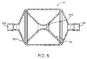
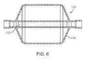
- FIGS. 5-8illustrate specific flow restrictive components useful with the catheter of FIG. 3 .
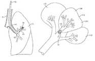
- FIG. 9is an anatomical diagram illustrating the lobar structure of the lungs of a patient.
- FIG. 10illustrates the trans-esophageal endobronchial placement of the functional assessment and therapy catheter of the present invention in an airway leading to a diseased lung compartment.
- FIG. 11illustrates the initial placement of a flow restrictive component in accordance with the principles of the methods of the present invention.
- FIG. 12illustrates the use of the functional assessment catheter for determining collateral ventilation.
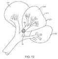
- FIG. 13illustrates release of the flow restrictive component into the lung passageway after it has been determined that collateral ventilation does not exist in the diseased lung compartment.
- the shaft 12will have dimensions and mechanical properties suitable for trans-bronchial introduction into the passageways of the lung, typically where the flow restrictive component 14 may be placed into the branching bronchii of the lung and advanced to locations in the main bronchus feeding a target lung compartment.
- the shaftwill comprise a braid-reinforced polymer, such as a polyvinylchloride, a polytetrafluoroethylene (PTFE), a polypropylene, a polyethylene terephthalate (PET), a polyurethane, a polyurethane/polycarbonate mixture, or any one of a variety of other suitable polymers.
- a catheter shaftwill comprise a relatively soft distal region 18 and a relatively harder proximal region 20 .
- the distal regioncan be formed from a 55D durometer polyethylene block polyamide (PEBAX) and the proximal region 20 can be formed from a 72D durometer PEBAX.
- the flow restrictive component 14will normally be in an expanded configuration, as shown in FIGS. 1 and 2A , but may be axially elongated in order to assume a contracted or narrow diameter configuration, as shown in FIG. 2B .
- the component 14will typically comprise a scaffold structure 40 , typically formed from counterwound helical elements, covered at least partially by an elastomeric covering 42 .
- the individual helical elementswill be formed from an elastic material, typically an elastic metal but optionally a shape memory polymer. Suitable elastic metals include stainless steel, spring stainless steel, nickel-titanium alloy, and the like. In an exemplary embodiments, the elastic elements are made from 0.125 mm nitinol wires counterwound into a braid.
- the individual nitinol wiresare joined at a distal end by end cap 44 and are connected at their proximal ends to the distal end of the distal region 18 of the shaft 12 .
- the scaffold structurecould be made by chemically etching a thin layer of a suitable elastic metal and forming into the shape of the restrictive component.
- the membranecan be made from any of the materials described earlier, and in the exemplary embodiment will be formed from a silicone formed over the proximal portion of the scaffold 40 and extending over the mid-section of the scaffold, leaving a distal region 46 of the scaffold open. While the distal region is open, the mid-section of the flow restrictive component 14 will be able to engage the interior of the bronchus in which it is expanded in order to form a tight seal, at least while the flow restrictive component 14 remains attached to the shaft 12 .
- the flow restrictive component 14is intended to remain fixed to the shaft 12 , so the catheter 10 is intended only for assessment, not for therapy. It will be appreciated that this structure could be modified, or a separate releasing device could be provided in order to detach the flow restrictive component 14 from the shaft in order to leave the component in place should the patient be a good candidate for therapy.
- Other embodiments of the catheter, described hereinafter,are shown with specific detachment means for use in both diagnostic and therapeutic applications.
- An obturator assembly 30is provided in order to elongate and constrict the diameter of the flow restrictive component 14 .
- the obturator assembly 30comprises a flexible rod 32 , typically a coiled wire formed from a metal or semi-rigid plastic material. Suitable metals include stainless steel, titanium, nickel-titanium alloy, or any other metal of the type conventionally used in construction of medical guidewires.
- Metal shaftsmay be coated with PTFE or other material in order to enhance the lubricity as it is introduced through a lumen of the shaft 12 into the interior of the flow restrictive component 14 .
- a distal tip 33engages the end cap 44 of the flow restrictive component 14 , as best seen in FIG. 2A .
- an advancement actuator 34may be connected to a proximal end of the rod 32 .
- the actuator 34may comprise a connector 36 which is mountable on a luer or other fitting 17 on proximal hub 16 .
- a plunger 38may be depressed in order to advance the rod 32 in the direction of arrow 48 in FIG. 2B .
- a detent or other locking mechanismmay be provided in the actuator 34 in order to hold the flow restrictive component 14 in its narrow diameter configuration during introduction into the bronchii.
- the spring force in the flow restrictive component 14will push the rod 32 proximally and allow the component to reassume its expanded or large diameter configuration within the main bronchus leading to the target lung compartment.
- the catheter 50differs from the functional assessment catheter 10 described previously in that it is adapted to selectively release a flow restrictive component 54 from a distal end of a catheter shaft 52 .
- a release mechanism 58is formed or otherwise provided at a proximal end of the flow restrictive component 54 .
- the release mechanism 58may take any of a wide variety of forms, including mechanical, electrical, chemical (e.g., dissolvable), or combinations thereof.
- the release mechanism 58will retain the flow restrictive component 54 firmly on the distal end of the shaft 52 until such a time as it may be desired to release the component within a bronchii.
- the ability to permit gas exchange through the catheter shaft 52 and flow restrictive component 54is desirable to allow performance of collateral ventilation or other diagnostic procedures while the flow resistive component 54 is expanded within the bronchii and still attached to the catheter shaft 52 .
- FIGS. 3A through 3CAn exemplary release mechanism 158 for selectively detaching a flow restrictive component 154 from a catheter shaft 152 is illustrated in FIGS. 3A through 3C .
- the release mechanism 158comprises a collar 153 attached at a proximal end of the self-expanding flow restrictive component 154 .
- An attachment ball 151projects from a proximal end of the sleeve 153 .
- Both the attachment ball 151 and the sleeve 153have an internal passage 157 which permits air flow through the release mechanism 158 so that air or other gases may be exchanged from the catheter shaft 152 through an open aperture 159 at a distal end of the flow restrictive component 154 .
- the attachment ball 151is received in an opening 155 ( FIG.
- a separate delivery sheath 60will be provided for facilitating delivery of the flow restrictive component 54 of the catheter 50 .
- the sheath 60will have a diameter 60 suitable for introduction into the target bronchii, typically having an outer diameter in the range from 1 mm to 3 mm.
- the flow restrictive component 54will be radially constrained and introduced through an interior lumen of the delivery sheath, simply by pushing the shaft 52 distally so that the constrained flow restrictive component 54 is advanced through the sheath 50 as shown in FIG. 4A . Initially, the flow restrictive component 54 will be fully contained within the sheath 60 . As the shaft 52 continues forward advancement, the flow restrictive component 54 will emerge from a distal end of the sheath 60 , as shown in FIG. 4B .
- the flow restrictive component 54Upon further advancement, the flow restrictive component 54 will be fully released from the sheath, as shown in FIG. 4C .
- the flow restrictive componentmay be selectively released by actuating mechanism 58 , as shown in FIG. 4D .
- flow restrictive component 54may be fully closed in order to provide for total occlusion of the bronchii in which it has been deployed.
- a smaller controlled flow pathmay remain through the flow restrictive component 54 in order to provide for controlled atelectasis or hypoxic pulmonary vasoconstriction (HPV), as described in copending application Ser. No. 11/682,986, the full disclosure of which has been previously incorporated herein by reference.
- HPVcontrolled atelectasis or hypoxic pulmonary vasoconstriction
- FIGS. 5-8Specific examples of flow restrictive elements suitable for permitting continued air exchange with the isolated lung compartment and controlled atelectasis and/or HPV are illustrated in FIGS. 5-8 .
- FIG. 5illustrates a flow restrictive component 160 in which a housing 162 houses a funnel-shaped (or hourglass-shaped) diaphragm 164 which provides a gas flow orifice 166 in the center of the diaphragm.
- Distal and proximal apertures 168 and 170respectively, allow air flow into and out of the housing 162 , and the tapered orifice 166 defined by the diaphragm 164 restricts the flow.
- the diameter of the orifice 166can be selected to provide a desired flow resistance.
- the housing 162can have a uni-body construction or be a wire braided structure encapsulated with silicone or other elastomere.
- the diaphragmcan be a flexible silicone material or other elastomere in order to facilitate compressibility of the restrictor 160 for insertion into the lung via a delivery sheath lumen.
- FIG. 6illustrates flow restrictive component 170 in which a gas flow tube 172 is axially aligned in a housing 174 .
- Construction of the housing 174can be similar to any of the concepts previously described.
- the gas flow tube 172can be constructed of any tubular material, preferably being a flexible polymer. Flexibility is advantageous since a flexible tube will facilitate insertion into the lung.
- the housing 174can have any of the constructions described previously.
- FIG. 7is a cross-sectional view of a flow restrictive component 130 in which a housing 132 includes a gas flow orifice tube 134 on its distal end 136 .
- the housingcan have a “uni-body” construction, typically being molded or cast from silicone or another biocompatible elastomer.
- the housing 132can have composite construction of wire frame with silicone membrane coating, or be formed from a variety of materials and construction methods. It can be collapsible and self expanding for a catheter based delivery.
- the flow restrictive componentcan be malleable to allow plastic deformation and expansion by a balloon or other expandable deployment on the delivery catheter.
- FIG. 8illustrates a flow restrictive component 140 in which a housing 142 comprises a plurality of windows 144 in a wall of a distal section 46 in order to permit gas flow in and out of the housing.
- An orifice 148 at the opposite proximal endcompletes the gas flow path such that the device restricts but does not obstruct gas flow.
- the housing 142can have a uni-body construction or comprise a wire frame with silicone or other membrane covering. It can be either collapsible and self expanding or balloon expandable.
- the respiratory system of a patientstarts at the mouth and extends through the vocal cords and into the trachea where it then joins the main stem bronchi B which leads into the right lung RL and the left lung LL.
- the bronchi going into the right lungdivide into the three lobar bronchi which lead into the upper lobe RUL, the middle lobe RML and the lower lobe RLL.
- the lobes of the right lunginclude a total of ten segments (three in the RUL, two in the RML, and five in the RLL) which are discrete units of the lung separated from each other by a fibrous septum generally referred to as a lung wall.
- the left lung LLincludes only an upper lobe LUL and a lower lobe LLL, where the individual lobes include four to five segments each.
- Each lung segmentalso referred to as a bronchopulmonary segment, is an anatomically distinct unit or compartment of the lung which is fed air by a tertiary bronchus and which oxygenates blood through a tertiary artery.
- the lung segment and its surrounding fibrous septumare intact units which can be surgically removed or separated from the remainder of the lung without interrupting the function of the surrounding lung segments.
- the fibrous septum separating the lobes or segmentsmay be perforate or broken, thus allowing air flow between the segments, referred to as “collateral ventilation.”
- the sheath 60is advanced through the mouth, down through the trachea T and through the main bronchus into the left lung LL.
- a distal end 62 of the sheath 60is advanced into the left lung LL, and further advanced to an airway or bronchus which feeds a diseased lung region DR.
- the sheath 60may be introduced through the main bronchus B and into the left lung LL without the use of a bronchoscope or other primary introducing catheter, as illustrated in FIG. 10 .
- the sheath 60may be introduced through a conventional bronchoscope (now shown) which is positioned in the main bronchus B above the branch between the right and left lungs. Still further alternatively, the sheath 60 may be introduced into the lung through a scope, such as a visualizing endotracheal tube (not shown) which is capable of being advanced into the branching bronchii of the lung and which may be advantageous since it facilitates positioning of the sheath 60 at the desired airway leading to the target diseased lung segment. Construction and use of a visualizing endotracheal tube is taught, for example, in U.S. Pat. No. 5,285,778, the full disclosure of which is incorporated herein by reference.
- the sheathmay be optionally immobilized by inflating a balloon or cuff 64 at or near the proximal end of the sheath 60 .
- the catheter shaft 52 of catheter 50may be distally advanced in order to deploy the flow restrictive component 54 into the feeding bronchus FB leading to the diseased lung region DR, as shown in FIG. 12 .
- a diagnostic procedure for determining the extent and/or treatability of the diseasemay be performed, generally as described in previous application Ser. Nos.
- the flow restrictive component 54may be released and implanted in the feeding bronchus FB, as shown in FIG. 13 . If, however, the patient is determined to be unsuitable for such treatment, the flow restrictive component 54 may be removed from the feeding bronchus FB, typically by retraction into the delivery sheath 60 and subsequent removal of the sheath from the lung.
Landscapes
- Health & Medical Sciences (AREA)
- Life Sciences & Earth Sciences (AREA)
- Pulmonology (AREA)
- Veterinary Medicine (AREA)
- Biomedical Technology (AREA)
- Heart & Thoracic Surgery (AREA)
- Engineering & Computer Science (AREA)
- Animal Behavior & Ethology (AREA)
- General Health & Medical Sciences (AREA)
- Public Health (AREA)
- Biophysics (AREA)
- Hematology (AREA)
- Anesthesiology (AREA)
- Surgery (AREA)
- Medical Informatics (AREA)
- Molecular Biology (AREA)
- Physics & Mathematics (AREA)
- Pathology (AREA)
- Emergency Medicine (AREA)
- Physiology (AREA)
- Reproductive Health (AREA)
- Vascular Medicine (AREA)
- Nuclear Medicine, Radiotherapy & Molecular Imaging (AREA)
- Surgical Instruments (AREA)
- Apparatus For Radiation Diagnosis (AREA)
Abstract
Description
Claims (5)
Priority Applications (6)
| Application Number | Priority Date | Filing Date | Title |
|---|---|---|---|
| US11/845,296US8342182B2 (en) | 2006-08-28 | 2007-08-27 | Functional assessment and treatment catheters and methods for their use in the lung |
| EP07841495AEP2056715B1 (en) | 2006-08-28 | 2007-08-28 | Functional assessment and treatment catheters |
| PCT/US2007/077023WO2008027913A2 (en) | 2006-08-28 | 2007-08-28 | Functional assessment and treatment catheters and methods for their use in the lung |
| US13/689,344US9439583B2 (en) | 2006-08-28 | 2012-11-29 | Functional assessment and treatment catheters and methods for their use in the lung |
| US15/250,917US10517529B2 (en) | 2006-08-28 | 2016-08-30 | Functional assessment and treatment catheters and methods for their use in the lung |
| US16/706,474US11931170B2 (en) | 2006-08-28 | 2019-12-06 | Functional assessment and treatment catheters and methods for their use in the lung |
Applications Claiming Priority (3)
| Application Number | Priority Date | Filing Date | Title |
|---|---|---|---|
| US82373406P | 2006-08-28 | 2006-08-28 | |
| US82849606P | 2006-10-06 | 2006-10-06 | |
| US11/845,296US8342182B2 (en) | 2006-08-28 | 2007-08-27 | Functional assessment and treatment catheters and methods for their use in the lung |
Related Child Applications (1)
| Application Number | Title | Priority Date | Filing Date |
|---|---|---|---|
| US13/689,344DivisionUS9439583B2 (en) | 2006-08-28 | 2012-11-29 | Functional assessment and treatment catheters and methods for their use in the lung |
Publications (2)
| Publication Number | Publication Date |
|---|---|
| US20080051719A1 US20080051719A1 (en) | 2008-02-28 |
| US8342182B2true US8342182B2 (en) | 2013-01-01 |
Family
ID=39136804
Family Applications (4)
| Application Number | Title | Priority Date | Filing Date |
|---|---|---|---|
| US11/845,296Active2030-12-06US8342182B2 (en) | 2006-08-28 | 2007-08-27 | Functional assessment and treatment catheters and methods for their use in the lung |
| US13/689,344Active2028-09-16US9439583B2 (en) | 2006-08-28 | 2012-11-29 | Functional assessment and treatment catheters and methods for their use in the lung |
| US15/250,917Active2029-03-30US10517529B2 (en) | 2006-08-28 | 2016-08-30 | Functional assessment and treatment catheters and methods for their use in the lung |
| US16/706,474Active2028-11-25US11931170B2 (en) | 2006-08-28 | 2019-12-06 | Functional assessment and treatment catheters and methods for their use in the lung |
Family Applications After (3)
| Application Number | Title | Priority Date | Filing Date |
|---|---|---|---|
| US13/689,344Active2028-09-16US9439583B2 (en) | 2006-08-28 | 2012-11-29 | Functional assessment and treatment catheters and methods for their use in the lung |
| US15/250,917Active2029-03-30US10517529B2 (en) | 2006-08-28 | 2016-08-30 | Functional assessment and treatment catheters and methods for their use in the lung |
| US16/706,474Active2028-11-25US11931170B2 (en) | 2006-08-28 | 2019-12-06 | Functional assessment and treatment catheters and methods for their use in the lung |
Country Status (3)
| Country | Link |
|---|---|
| US (4) | US8342182B2 (en) |
| EP (1) | EP2056715B1 (en) |
| WO (1) | WO2008027913A2 (en) |
Cited By (4)
| Publication number | Priority date | Publication date | Assignee | Title |
|---|---|---|---|---|
| US8770199B2 (en) | 2012-12-04 | 2014-07-08 | Ino Therapeutics Llc | Cannula for minimizing dilution of dosing during nitric oxide delivery |
| US9439583B2 (en) | 2006-08-28 | 2016-09-13 | Pulmonx Corporation | Functional assessment and treatment catheters and methods for their use in the lung |
| US9795756B2 (en) | 2012-12-04 | 2017-10-24 | Mallinckrodt Hospital Products IP Limited | Cannula for minimizing dilution of dosing during nitric oxide delivery |
| US10905836B2 (en) | 2015-04-02 | 2021-02-02 | Hill-Rom Services Pte. Ltd. | Manifold for respiratory device |
Families Citing this family (18)
| Publication number | Priority date | Publication date | Assignee | Title |
|---|---|---|---|---|
| US20100158795A1 (en) | 2008-06-12 | 2010-06-24 | Pulmonx | Methods and systems for assessing lung function and delivering therapeutic agents |
| US8496006B2 (en)* | 2005-01-20 | 2013-07-30 | Pulmonx Corporation | Methods and devices for passive residual lung volume reduction and functional lung volume expansion |
| US11883029B2 (en) | 2005-01-20 | 2024-01-30 | Pulmonx Corporation | Methods and devices for passive residual lung volume reduction and functional lung volume expansion |
| US20080228137A1 (en) | 2007-03-12 | 2008-09-18 | Pulmonx | Methods and devices for passive residual lung volume reduction and functional lung volume expansion |
| FI3060276T3 (en) | 2013-10-24 | 2023-06-07 | Infusion system for preventing mischanneling of multiple medicaments | |
| US20150216688A1 (en)* | 2014-02-04 | 2015-08-06 | The Texas A&M University System | Intravascular Medical Device Release System |
| CN104367325A (en)* | 2014-12-01 | 2015-02-25 | 田庆 | Lung collateral ventilation detection device |
| WO2016126701A1 (en)* | 2015-02-02 | 2016-08-11 | Northwestern University | Systems, methods, and apparatus for esophageal panometry |
| WO2017053935A1 (en)* | 2015-09-25 | 2017-03-30 | Soffio Medical Inc. | Expandable structure to treat hyperinflated lung |
| US11419490B2 (en)* | 2016-08-02 | 2022-08-23 | Covidien Lp | System and method of using an endoscopic catheter as a port in laparoscopic surgery |
| BR112019013985A2 (en) | 2017-01-06 | 2020-03-03 | Trustees Of Boston University | INFUSION SYSTEM AND COMPONENTS OF THE SAME |
| CN106823122B (en)* | 2017-03-16 | 2023-08-11 | 中国医学科学院北京协和医院 | A radiotherapy applicator with an expanded stent |
| US20220211481A1 (en)* | 2019-04-22 | 2022-07-07 | Eolo Medical Inc. | Devices for the treatment of pulmonary disorders with implantable valves |
| CN110313969B (en)* | 2019-06-10 | 2020-08-18 | 中南大学湘雅医院 | hemostatic components |
| CA3146964A1 (en) | 2019-07-16 | 2021-01-21 | Beta Bionics, Inc. | Ambulatory device and components thereof |
| US11278661B2 (en) | 2020-03-10 | 2022-03-22 | Beta Bionics, Inc. | Infusion system and components thereof |
| USD1031975S1 (en) | 2020-03-10 | 2024-06-18 | Beta Bionics, Inc. | Medicament infusion pump device |
| CN112333110A (en)* | 2020-10-30 | 2021-02-05 | 深圳壹账通智能科技有限公司 | Request verification processing method based on funnel flow-limiting model and related equipment |
Citations (27)
| Publication number | Priority date | Publication date | Assignee | Title |
|---|---|---|---|---|
| US5624449A (en) | 1993-11-03 | 1997-04-29 | Target Therapeutics | Electrolytically severable joint for endovascular embolic devices |
| US5649906A (en)* | 1991-07-17 | 1997-07-22 | Gory; Pierre | Method for implanting a removable medical apparatus in a human body |
| EP0791340A1 (en) | 1996-02-22 | 1997-08-27 | Cordis Corporation | Temporary filter catheter |
| EP0815803A1 (en) | 1996-07-03 | 1998-01-07 | Cordis Europa N.V. | Catheter with temporary vena cava filter |
| US5795322A (en) | 1995-04-10 | 1998-08-18 | Cordis Corporation | Catheter with filter and thrombus-discharge device |
| US5800455A (en) | 1993-04-19 | 1998-09-01 | Target Therapeutics, Inc. | Detachable embolic coil assembly |
| USRE37117E1 (en) | 1992-09-22 | 2001-03-27 | Target Therapeutics, Inc. | Detachable embolic coil assembly using interlocking clasps and method of use |
| US6287290B1 (en) | 1999-07-02 | 2001-09-11 | Pulmonx | Methods, systems, and kits for lung volume reduction |
| US20010037808A1 (en)* | 2000-03-04 | 2001-11-08 | Deem Mark E. | Methods and devices for use in performing pulmonary procedures |
| US6371971B1 (en)* | 1999-11-15 | 2002-04-16 | Scimed Life Systems, Inc. | Guidewire filter and methods of use |
| WO2002038038A2 (en)* | 2000-10-27 | 2002-05-16 | Pulmonx | Methods and devices for obstructing and aspirating lung tissue segments |
| US20020143387A1 (en) | 2001-03-27 | 2002-10-03 | Soetikno Roy M. | Stent repositioning and removal |
| WO2003022124A2 (en) | 2001-09-11 | 2003-03-20 | Spiration, Inc. | Removable lung reduction devices, systems, and methods |
| WO2003022221A2 (en) | 2001-09-10 | 2003-03-20 | Pulmonx | Method and apparatus for endobronchial diagnosis |
| US20030154988A1 (en)* | 2002-02-21 | 2003-08-21 | Spiration, Inc. | Intra-bronchial device that provides a medicant intra-bronchially to the patient |
| US6722360B2 (en) | 2000-06-16 | 2004-04-20 | Rajiv Doshi | Methods and devices for improving breathing in patients with pulmonary disease |
| US6941950B2 (en) | 2001-10-11 | 2005-09-13 | Emphasys Medical, Inc. | Bronchial flow control devices and methods of use |
| US7011094B2 (en) | 2001-03-02 | 2006-03-14 | Emphasys Medical, Inc. | Bronchial flow control devices and methods of use |
| US20060102186A1 (en) | 2004-11-18 | 2006-05-18 | Mark Adler | Intra-bronchial apparatus for aspiration and insufflation of lung regions distal to placement or cross communication and deployment and placement system therefor |
| WO2006055692A2 (en) | 2004-11-16 | 2006-05-26 | Pulmonx | Pulmonary occlusal stent delivery catheter, loading system and methods of use |
| US20060122647A1 (en) | 2004-09-24 | 2006-06-08 | Callaghan David J | Occluder device double securement system for delivery/recovery of such occluder device |
| WO2006078451A2 (en) | 2005-01-20 | 2006-07-27 | Pulmonx | Minimally invasive determination of collateral ventilation in lungs |
| US20060264772A1 (en) | 2001-09-10 | 2006-11-23 | Pulmonx | Minimally invasive determination of collateral ventilation in lungs |
| US20070005083A1 (en) | 1997-04-30 | 2007-01-04 | Sabaratham Sabanathan | Occlusion device |
| US20070096048A1 (en) | 2005-10-14 | 2007-05-03 | Claude Clerc | Bronchoscopic lung volume reduction valve |
| US20070142742A1 (en) | 2005-07-13 | 2007-06-21 | Pulmonx | Methods and systems for segmental lung diagnostics |
| US20070225747A1 (en) | 2006-03-08 | 2007-09-27 | Pulmonx | Methods and devices to induce controlled atelectasis and hypoxic pulmonary vasoconstriction |
Family Cites Families (18)
| Publication number | Priority date | Publication date | Assignee | Title |
|---|---|---|---|---|
| US2230226A (en)* | 1938-03-14 | 1941-02-04 | Davol Rubber Co | Catheter and its manufacture |
| US4921484A (en)* | 1988-07-25 | 1990-05-01 | Cordis Corporation | Mesh balloon catheter device |
| US5285778A (en) | 1991-04-19 | 1994-02-15 | Mackin Robert A | Endotracheal tube wih fibers optic illumination and viewing and auxiliary tube |
| US5261916A (en)* | 1991-12-12 | 1993-11-16 | Target Therapeutics | Detachable pusher-vasoocclusive coil assembly with interlocking ball and keyway coupling |
| DE69230385T2 (en)* | 1991-12-12 | 2000-04-06 | Target Therapeutics, Inc. | Detachable, slidable, vessel-closing spiral with interlocking coupling elements |
| US5749883A (en)* | 1995-08-30 | 1998-05-12 | Halpern; David Marcos | Medical instrument |
| US5895391A (en)* | 1996-09-27 | 1999-04-20 | Target Therapeutics, Inc. | Ball lock joint and introducer for vaso-occlusive member |
| JP2003522550A (en)* | 1998-02-10 | 2003-07-29 | アーテミス・メディカル・インコーポレイテッド | Occlusion, fixation, tensioning, and diverting devices and methods of use |
| US6179860B1 (en)* | 1998-08-19 | 2001-01-30 | Artemis Medical, Inc. | Target tissue localization device and method |
| EP1400204A1 (en)* | 1999-08-05 | 2004-03-24 | Broncus Technologies, Inc. | Methods and devices for creating collateral channels in the lungs |
| US6911038B2 (en)* | 2001-05-08 | 2005-06-28 | Scimed Life Systems, Inc. | Matched balloon to stent shortening |
| US6958074B2 (en)* | 2002-01-07 | 2005-10-25 | Cordis Corporation | Releasable and retrievable vascular filter system |
| AU2003220124A1 (en)* | 2002-03-08 | 2003-09-22 | Emphasys Medical, Inc. | Methods and devices for inducing collapse in lung regions fed by collateral pathways |
| EP3542736A1 (en)* | 2004-06-16 | 2019-09-25 | PneumRx, Inc. | Intra-bronchial lung volume reduction system |
| US7608089B2 (en)* | 2004-12-22 | 2009-10-27 | Boston Scientific Scimed, Inc. | Vaso-occlusive device having pivotable coupling |
| US8672990B2 (en)* | 2005-05-27 | 2014-03-18 | Boston Scientific Scimed, Inc. | Fiber mesh controlled expansion balloon catheter |
| US20070186933A1 (en)* | 2006-01-17 | 2007-08-16 | Pulmonx | Systems and methods for delivering flow restrictive element to airway in lungs |
| US8342182B2 (en) | 2006-08-28 | 2013-01-01 | Pulmonx Corporation | Functional assessment and treatment catheters and methods for their use in the lung |
- 2007
- 2007-08-27USUS11/845,296patent/US8342182B2/enactiveActive
- 2007-08-28WOPCT/US2007/077023patent/WO2008027913A2/enactiveApplication Filing
- 2007-08-28EPEP07841495Apatent/EP2056715B1/enactiveActive
- 2012
- 2012-11-29USUS13/689,344patent/US9439583B2/enactiveActive
- 2016
- 2016-08-30USUS15/250,917patent/US10517529B2/enactiveActive
- 2019
- 2019-12-06USUS16/706,474patent/US11931170B2/enactiveActive
Patent Citations (34)
| Publication number | Priority date | Publication date | Assignee | Title |
|---|---|---|---|---|
| US5649906A (en)* | 1991-07-17 | 1997-07-22 | Gory; Pierre | Method for implanting a removable medical apparatus in a human body |
| USRE37117E1 (en) | 1992-09-22 | 2001-03-27 | Target Therapeutics, Inc. | Detachable embolic coil assembly using interlocking clasps and method of use |
| US5800455A (en) | 1993-04-19 | 1998-09-01 | Target Therapeutics, Inc. | Detachable embolic coil assembly |
| US6099546A (en) | 1993-04-19 | 2000-08-08 | Target Therapeutics, Inc. | Detachable embolic coil assembly using interlocking hooks and slots |
| US5624449A (en) | 1993-11-03 | 1997-04-29 | Target Therapeutics | Electrolytically severable joint for endovascular embolic devices |
| US5795322A (en) | 1995-04-10 | 1998-08-18 | Cordis Corporation | Catheter with filter and thrombus-discharge device |
| EP0791340A1 (en) | 1996-02-22 | 1997-08-27 | Cordis Corporation | Temporary filter catheter |
| EP0815803A1 (en) | 1996-07-03 | 1998-01-07 | Cordis Europa N.V. | Catheter with temporary vena cava filter |
| US20070005083A1 (en) | 1997-04-30 | 2007-01-04 | Sabaratham Sabanathan | Occlusion device |
| US6287290B1 (en) | 1999-07-02 | 2001-09-11 | Pulmonx | Methods, systems, and kits for lung volume reduction |
| US6371971B1 (en)* | 1999-11-15 | 2002-04-16 | Scimed Life Systems, Inc. | Guidewire filter and methods of use |
| US20010037808A1 (en)* | 2000-03-04 | 2001-11-08 | Deem Mark E. | Methods and devices for use in performing pulmonary procedures |
| US6679264B1 (en) | 2000-03-04 | 2004-01-20 | Emphasys Medical, Inc. | Methods and devices for use in performing pulmonary procedures |
| US6722360B2 (en) | 2000-06-16 | 2004-04-20 | Rajiv Doshi | Methods and devices for improving breathing in patients with pulmonary disease |
| US20060095002A1 (en) | 2000-10-27 | 2006-05-04 | Pulmonx | Methods and devices for obstructing and aspirating lung tissue segments |
| US20040073191A1 (en)* | 2000-10-27 | 2004-04-15 | Pulmonx | Methods and devices for obstructing and aspirating lung tissue segments |
| WO2002038038A2 (en)* | 2000-10-27 | 2002-05-16 | Pulmonx | Methods and devices for obstructing and aspirating lung tissue segments |
| US6997918B2 (en) | 2000-10-27 | 2006-02-14 | Pulmonx | Methods and devices for obstructing and aspirating lung tissue segments |
| US6527761B1 (en) | 2000-10-27 | 2003-03-04 | Pulmonx, Inc. | Methods and devices for obstructing and aspirating lung tissue segments |
| US7011094B2 (en) | 2001-03-02 | 2006-03-14 | Emphasys Medical, Inc. | Bronchial flow control devices and methods of use |
| US20020143387A1 (en) | 2001-03-27 | 2002-10-03 | Soetikno Roy M. | Stent repositioning and removal |
| US20060264772A1 (en) | 2001-09-10 | 2006-11-23 | Pulmonx | Minimally invasive determination of collateral ventilation in lungs |
| WO2003022221A2 (en) | 2001-09-10 | 2003-03-20 | Pulmonx | Method and apparatus for endobronchial diagnosis |
| WO2003022221A3 (en) | 2001-09-10 | 2003-07-03 | Pulmonx | Method and apparatus for endobronchial diagnosis |
| WO2003022124A2 (en) | 2001-09-11 | 2003-03-20 | Spiration, Inc. | Removable lung reduction devices, systems, and methods |
| US6941950B2 (en) | 2001-10-11 | 2005-09-13 | Emphasys Medical, Inc. | Bronchial flow control devices and methods of use |
| US20030154988A1 (en)* | 2002-02-21 | 2003-08-21 | Spiration, Inc. | Intra-bronchial device that provides a medicant intra-bronchially to the patient |
| US20060122647A1 (en) | 2004-09-24 | 2006-06-08 | Callaghan David J | Occluder device double securement system for delivery/recovery of such occluder device |
| WO2006055692A2 (en) | 2004-11-16 | 2006-05-26 | Pulmonx | Pulmonary occlusal stent delivery catheter, loading system and methods of use |
| US20060102186A1 (en) | 2004-11-18 | 2006-05-18 | Mark Adler | Intra-bronchial apparatus for aspiration and insufflation of lung regions distal to placement or cross communication and deployment and placement system therefor |
| WO2006078451A2 (en) | 2005-01-20 | 2006-07-27 | Pulmonx | Minimally invasive determination of collateral ventilation in lungs |
| US20070142742A1 (en) | 2005-07-13 | 2007-06-21 | Pulmonx | Methods and systems for segmental lung diagnostics |
| US20070096048A1 (en) | 2005-10-14 | 2007-05-03 | Claude Clerc | Bronchoscopic lung volume reduction valve |
| US20070225747A1 (en) | 2006-03-08 | 2007-09-27 | Pulmonx | Methods and devices to induce controlled atelectasis and hypoxic pulmonary vasoconstriction |
Non-Patent Citations (4)
| Title |
|---|
| Choostent(TM), Covered Esophageal Stent, pp. 1-2, Jul. 26, 2005.* |
| Choostent™, Covered Esophageal Stent, pp. 1-2, Jul. 26, 2005.* |
| International Search Report and Written Opinion of PCT Application No. PCT/US2007/077023, dated Apr. 8, 2008, 10 pages total. |
| Supplementary European Search Report of EP Patent Application No. 07841495.0, mailed Jul. 28, 2009, 7 pages total. |
Cited By (13)
| Publication number | Priority date | Publication date | Assignee | Title |
|---|---|---|---|---|
| US10517529B2 (en) | 2006-08-28 | 2019-12-31 | Pulmonx Corporation | Functional assessment and treatment catheters and methods for their use in the lung |
| US11931170B2 (en) | 2006-08-28 | 2024-03-19 | Pulmonx Corporation | Functional assessment and treatment catheters and methods for their use in the lung |
| US9439583B2 (en) | 2006-08-28 | 2016-09-13 | Pulmonx Corporation | Functional assessment and treatment catheters and methods for their use in the lung |
| US9550039B2 (en) | 2012-12-04 | 2017-01-24 | Mallinckrodt Hospital Products IP Limited | Cannula for minimizing dilution of dosing during nitric oxide delivery |
| US9795756B2 (en) | 2012-12-04 | 2017-10-24 | Mallinckrodt Hospital Products IP Limited | Cannula for minimizing dilution of dosing during nitric oxide delivery |
| US10130783B2 (en) | 2012-12-04 | 2018-11-20 | Mallinckrodt Hospital Products IP Limited | Cannula for minimizing dilution of dosing during nitric oxide delivery |
| US8770199B2 (en) | 2012-12-04 | 2014-07-08 | Ino Therapeutics Llc | Cannula for minimizing dilution of dosing during nitric oxide delivery |
| US10556082B2 (en) | 2012-12-04 | 2020-02-11 | Mallinckrodt Hospital Products IP Limited | Cannula for minimizing dilution of dosing during nitric oxide delivery |
| US10918819B2 (en) | 2012-12-04 | 2021-02-16 | Mallinckrodt Hospital Products IP Limited | Cannula for minimizing dilution of dosing during nitric oxide delivery |
| US9032959B2 (en) | 2012-12-04 | 2015-05-19 | Ino Therapeutics Llc | Cannula for minimizing dilution of dosing during nitric oxide delivery |
| US10905836B2 (en) | 2015-04-02 | 2021-02-02 | Hill-Rom Services Pte. Ltd. | Manifold for respiratory device |
| US10905837B2 (en) | 2015-04-02 | 2021-02-02 | Hill-Rom Services Pte. Ltd. | Respiratory therapy cycle control and feedback |
| US11992611B2 (en) | 2015-04-02 | 2024-05-28 | Hill-Rom Services Pte. Ltd. | Respiratory therapy apparatus control |
Also Published As
| Publication number | Publication date |
|---|---|
| EP2056715B1 (en) | 2013-02-13 |
| US10517529B2 (en) | 2019-12-31 |
| WO2008027913A3 (en) | 2008-07-03 |
| EP2056715A2 (en) | 2009-05-13 |
| US20170128004A1 (en) | 2017-05-11 |
| US20130338524A1 (en) | 2013-12-19 |
| US11931170B2 (en) | 2024-03-19 |
| EP2056715A4 (en) | 2009-08-26 |
| WO2008027913A2 (en) | 2008-03-06 |
| US20080051719A1 (en) | 2008-02-28 |
| US20200107776A1 (en) | 2020-04-09 |
| US9439583B2 (en) | 2016-09-13 |
Similar Documents
| Publication | Publication Date | Title |
|---|---|---|
| US11931170B2 (en) | Functional assessment and treatment catheters and methods for their use in the lung | |
| EP1993648B1 (en) | Devices to induce controlled atelectasis and hypoxic pulmonary vasoconstriction. | |
| US10729528B2 (en) | Bronchoscopic lung volume reduction valve | |
| US20030164168A1 (en) | Bronchiopulmonary occulsion devices and lung volume reduction methods | |
| US20070005083A1 (en) | Occlusion device | |
| AU2001260840A1 (en) | Bronchiopulmonary occlusion devices and lung volume reduction methods | |
| WO2003022124A2 (en) | Removable lung reduction devices, systems, and methods | |
| WO2003034927A1 (en) | Bronchial obstruction device deployment system and method | |
| US12310594B2 (en) | Collateral flow channel sealant delivery methods and systems | |
| EP3247270B1 (en) | Bronchial sealant delivery systems |
Legal Events
| Date | Code | Title | Description |
|---|---|---|---|
| AS | Assignment | Owner name:PULMONX, CALIFORNIA Free format text:ASSIGNMENT OF ASSIGNORS INTEREST;ASSIGNORS:NAIR, AJIT;GIA, SON;FARHNOLTZ, ROGER;REEL/FRAME:019927/0086;SIGNING DATES FROM 20070927 TO 20070928 Owner name:PULMONX, CALIFORNIA Free format text:ASSIGNMENT OF ASSIGNORS INTEREST;ASSIGNORS:NAIR, AJIT;GIA, SON;FARHNOLTZ, ROGER;SIGNING DATES FROM 20070927 TO 20070928;REEL/FRAME:019927/0086 | |
| AS | Assignment | Owner name:PULMONX, CALIFORNIA Free format text:ASSIGNMENT OF ASSIGNORS INTEREST;ASSIGNOR:ALJURI, NIKOLAI;REEL/FRAME:023843/0309 Effective date:20071001 | |
| STCF | Information on status: patent grant | Free format text:PATENTED CASE | |
| FPAY | Fee payment | Year of fee payment:4 | |
| AS | Assignment | Owner name:OXFORD FINANCE LLC, AS COLLATERAL AGENT, VIRGINIA Free format text:SECURITY INTEREST;ASSIGNOR:PULMONX CORPORATION;REEL/FRAME:042466/0349 Effective date:20170515 | |
| AS | Assignment | Owner name:BOSTON SCIENTIFIC CORPORATION, MASSACHUSETTS Free format text:SECURITY INTEREST;ASSIGNOR:PULMONX CORPORATION;REEL/FRAME:043349/0725 Effective date:20170515 | |
| AS | Assignment | Owner name:PULMONX CORPORATION, DELAWARE Free format text:MERGER;ASSIGNOR:PULMONX;REEL/FRAME:048964/0563 Effective date:20131230 | |
| AS | Assignment | Owner name:CANADIAN IMPERIAL BANK OF COMMERCE, CANADA Free format text:SECURITY INTEREST;ASSIGNOR:PULMONX CORPORATION;REEL/FRAME:052916/0213 Effective date:20200220 | |
| MAFP | Maintenance fee payment | Free format text:PAYMENT OF MAINTENANCE FEE, 8TH YR, SMALL ENTITY (ORIGINAL EVENT CODE: M2552); ENTITY STATUS OF PATENT OWNER: SMALL ENTITY Year of fee payment:8 | |
| AS | Assignment | Owner name:PULMONX CORPORATION, CALIFORNIA Free format text:RELEASE BY SECURED PARTY;ASSIGNOR:OXFORD FINANCE LLC, AS COLLATERAL AGENT;REEL/FRAME:053952/0044 Effective date:20200930 Owner name:PULMONX CORPORATION, CALIFORNIA Free format text:RELEASE BY SECURED PARTY;ASSIGNOR:BOSTON SCIENTIFIC CORPORATION;REEL/FRAME:053953/0548 Effective date:20200930 |