US8340379B2 - Systems and methods for displaying guidance data based on updated deformable imaging data - Google Patents
Systems and methods for displaying guidance data based on updated deformable imaging dataDownload PDFInfo
- Publication number
- US8340379B2 US8340379B2US12/399,899US39989909AUS8340379B2US 8340379 B2US8340379 B2US 8340379B2US 39989909 AUS39989909 AUS 39989909AUS 8340379 B2US8340379 B2US 8340379B2
- Authority
- US
- United States
- Prior art keywords
- imaging data
- time
- imaging
- tracking units
- tracking
- Prior art date
- Legal status (The legal status is an assumption and is not a legal conclusion. Google has not performed a legal analysis and makes no representation as to the accuracy of the status listed.)
- Active, expires
Links
Images


Classifications
- G—PHYSICS
- G06—COMPUTING OR CALCULATING; COUNTING
- G06V—IMAGE OR VIDEO RECOGNITION OR UNDERSTANDING
- G06V10/00—Arrangements for image or video recognition or understanding
- G06V10/20—Image preprocessing
- G06V10/24—Aligning, centring, orientation detection or correction of the image
- G06V10/245—Aligning, centring, orientation detection or correction of the image by locating a pattern; Special marks for positioning
- A—HUMAN NECESSITIES
- A61—MEDICAL OR VETERINARY SCIENCE; HYGIENE
- A61B—DIAGNOSIS; SURGERY; IDENTIFICATION
- A61B34/00—Computer-aided surgery; Manipulators or robots specially adapted for use in surgery
- A61B34/20—Surgical navigation systems; Devices for tracking or guiding surgical instruments, e.g. for frameless stereotaxis
- G—PHYSICS
- G06—COMPUTING OR CALCULATING; COUNTING
- G06T—IMAGE DATA PROCESSING OR GENERATION, IN GENERAL
- G06T7/00—Image analysis
- G06T7/0002—Inspection of images, e.g. flaw detection
- G06T7/0012—Biomedical image inspection
- A—HUMAN NECESSITIES
- A61—MEDICAL OR VETERINARY SCIENCE; HYGIENE
- A61B—DIAGNOSIS; SURGERY; IDENTIFICATION
- A61B34/00—Computer-aided surgery; Manipulators or robots specially adapted for use in surgery
- A61B34/10—Computer-aided planning, simulation or modelling of surgical operations
- A61B2034/101—Computer-aided simulation of surgical operations
- A61B2034/105—Modelling of the patient, e.g. for ligaments or bones
- A—HUMAN NECESSITIES
- A61—MEDICAL OR VETERINARY SCIENCE; HYGIENE
- A61B—DIAGNOSIS; SURGERY; IDENTIFICATION
- A61B34/00—Computer-aided surgery; Manipulators or robots specially adapted for use in surgery
- A61B34/20—Surgical navigation systems; Devices for tracking or guiding surgical instruments, e.g. for frameless stereotaxis
- A61B2034/2046—Tracking techniques
- A61B2034/2051—Electromagnetic tracking systems
- A—HUMAN NECESSITIES
- A61—MEDICAL OR VETERINARY SCIENCE; HYGIENE
- A61B—DIAGNOSIS; SURGERY; IDENTIFICATION
- A61B34/00—Computer-aided surgery; Manipulators or robots specially adapted for use in surgery
- A61B34/20—Surgical navigation systems; Devices for tracking or guiding surgical instruments, e.g. for frameless stereotaxis
- A61B2034/2046—Tracking techniques
- A61B2034/2055—Optical tracking systems
- A—HUMAN NECESSITIES
- A61—MEDICAL OR VETERINARY SCIENCE; HYGIENE
- A61B—DIAGNOSIS; SURGERY; IDENTIFICATION
- A61B90/00—Instruments, implements or accessories specially adapted for surgery or diagnosis and not covered by any of the groups A61B1/00 - A61B50/00, e.g. for luxation treatment or for protecting wound edges
- A61B90/36—Image-producing devices or illumination devices not otherwise provided for
- A61B2090/364—Correlation of different images or relation of image positions in respect to the body
- A61B2090/367—Correlation of different images or relation of image positions in respect to the body creating a 3D dataset from 2D images using position information
- A—HUMAN NECESSITIES
- A61—MEDICAL OR VETERINARY SCIENCE; HYGIENE
- A61B—DIAGNOSIS; SURGERY; IDENTIFICATION
- A61B90/00—Instruments, implements or accessories specially adapted for surgery or diagnosis and not covered by any of the groups A61B1/00 - A61B50/00, e.g. for luxation treatment or for protecting wound edges
- A61B90/36—Image-producing devices or illumination devices not otherwise provided for
- A61B90/37—Surgical systems with images on a monitor during operation
- A61B2090/378—Surgical systems with images on a monitor during operation using ultrasound
- G—PHYSICS
- G06—COMPUTING OR CALCULATING; COUNTING
- G06V—IMAGE OR VIDEO RECOGNITION OR UNDERSTANDING
- G06V2201/00—Indexing scheme relating to image or video recognition or understanding
- G06V2201/03—Recognition of patterns in medical or anatomical images
Definitions
- CTcomputed tomography
- MRImagnetic resonance imaging
- intra operative datasuch as two dimensional (“2D”) ultrasound or three dimensional (“3D”) ultrasound while they are in the operating room.
- 2Dtwo dimensional
- 3Dthree dimensional
- imaging data related to an anatomical sitethat include obtaining, at a first time, a first set of imaging data related to the anatomical site. Thereafter, tracking information for a movable imaging device controlled by a user is obtained at a second time, after the first time. Then desired emplacement information is determined for an image of the first set of imaging data based on the tracking information. Finally, image guidance data is determined for display based on the first set of imaging data and the desired emplacement information.
- FIG. 1depicts one embodiment of a system capable of updating deformable imaging data.
- FIG. 2illustrates an example of an anatomical site with tracking units.
- FIG. 3illustrates the deformation of an anatomical site with tracking units.
- FIG. 4depicts an embodiment of a tracking unit.
- FIG. 5illustrates another example of an anatomical site within deformable tissue with tracking units.
- FIG. 6depicts an example process for providing guidance data based on updated deformable tracking information.
- FIGS. 7A-7Ddepict marking and viewing features in imaging data.
- FIG. 8depicts an embodiment of image guidance data in which first set of imaging data is presented with second set of imaging data.
- FIG. 1depicts merely one exemplary embodiment of a system 100 capable of updating deformable imaging data.
- system 100may be joined together to form a single module and may even be implemented in a single computer or machine.
- the position sensing units 110 and 140may be combined and track all relevant tracking units 145 and movable imaging units 155 , as discussed in more detail below.
- imaging unit 150may be excluded and only imaging data from the image guidance unit 130 may be shown on display unit 120 .
- the system 100comprises a first position sensing unit 110 , a display unit 120 , and the second position sensing unit 140 all coupled to an image guidance unit 130 .
- the first position sensing unit 110 , the displaying unit 120 , the second position sensing unit 140 , and the image guidance unit 130are all physically connected to stand 170 .
- the image guidance unit 130may be used to produce images 125 that are presented on display unit 120 .
- the images 125 shown on the display unit 120 by the image guidance unit 130may be determined based on imaging data, such as a CT scan, MRI, open-magnet MRI, optical coherence tomography, positron emission tomography (“PET”) scans, fluoroscopy, ultrasound, or other preoperative or intraoperative anatomical imaging data and any 3D anatomical imaging data.
- the images 125 producedmay also be based on intraoperative or real-time data obtained using a movable imaging unit 155 , which is coupled to imaging unit 150 . Real-time may imply instantaneous or near-instantaneous obtaining of data. Real-time may also imply that it is taken with the intention to be used immediately.
- Imaging unit 150may be coupled to image guidance unit 130 .
- imaging unit 150may be coupled to a second display unit 151 .
- the second display unit 151may present imaging data from imaging unit 150 .
- the imaging data displayed on display unit 120 and displayed on second display unit 151are not necessarily the same.
- the imaging unit 150is an ultrasound machine 150
- the movable imaging device 155is an ultrasound transducer 155 or ultrasound probe 155
- the second display unit 151is a display associated with the ultrasound machine 150 that shows the imaging data from the ultrasound machine.
- the second position sensing unit 140is coupled to one or more tracking units 145 .
- the second position sensing unit 140 and tracking units 145may together comprise a magnetic tracking system, an optical tracking system, or any other appropriate tracking system.
- the second position sensing unit 140 and tracking units 145may be used to track the deformation of tissue at a target anatomical site on user 160 .
- User 160may be in an operating room, lying on an operating table, such as operating table 180 , or in any other appropriate place or position.
- second position sensing unit 140may be an Ascension Flock of Birds, Nest of Birds, driveBAY, medSAFE, trakSTAR, miniBIRD, MotionSTAR, or pciBIRD, and tracking units 145 may be magnetic tracking coils.
- the second position sensing unit 140may be an Aurora® Electromagnetic Measurement System using sensor coils for tracking units 145 .
- the first position sensing unit 110may also be an optical 3D tracking system using fiducials as tracking units 145 .
- optical 3D tracking systemsmay include the NDI Polaris Spectra, Vicra, Certus, PhaseSpace IMPULSE, Vicon MX, InterSense IS-900, NaturalPoint OptiTrack, Polhemus FastTrak, IsoTrak, or Claron MicronTracker2.
- Tracking unit 145 as used hereinis a broad term and includes without limitation all types of magnetic coils or other magnetic field sensing devices for use with magnetic trackers, fiducials or other optically detectable markers for use with optical trackers, such as those discussed above and below.
- Tracking units 145could also include optical position sensing devices such as the HiBall tracking system and the first and second position sensing units 110 and 140 may be HiBall tracking systems.
- Tracking units 145may also include a GPS device or signal-emitting device that would allow for tracking of the position and, optionally, orientation of the tracking unit.
- a signal-emitting devicemight include a radio-frequency identifier (RFID).
- RFIDradio-frequency identifier
- the first and/or second position sensing unit 110 and 140may take in the GPS coordinates of the tracking units 145 or may, for example, triangulate the radio frequency signal being emitted by the RFID associated with tracking units 145 .
- the first position sensing unit 110may be used to track the position of movable imaging unit 155 . Tracking the position of movable imaging unit 155 allows for the determination of the relative emplacement, where emplacement may refer to position and orientation or merely position, of imaging data received using the movable imaging unit 155 and imaging unit 150 with that data being sent to image guidance unit 130 .
- image guidance unit 130may contain CT data which is being updated and deformed based on the relative emplacements of tracking units 145 as received by the second position sensing unit 140 .
- the image guidance unit 130may take in the emplacements, such as positions and orientations, of the tracking units 145 and from that determine an updated model for CT data stored in imaging guidance unit 130 .
- imaging guidance unit 130may produce images based on the current ultrasound imaging data coming from imaging unit 150 and also based on an updated model determined based on the emplacements of tracking units 145 .
- the images produced 125made be presented on display unit 120 .
- An example image 125is shown in FIG. 8 .
- a movable imaging unit 155may not be connected directly to an imagining unit 150 , but may instead be connected to imaging guidance unit 130 .
- the movable imaging unit 155may be useful for allowing a user to indicate what portions of a first set of imaging data should be displayed.
- the movable imaging unit 155may be an ultrasound transducer or a tracked operative needle, for example, and may be used by a user to indicate what portions of a pre-operative CT scan to show on a display unit 120 as image 125 .
- each of the first and third sets of imaging datacould be deformed based on updated positions of the tracking units 145 and the updated, deformed versions of the two sets of imaging data could be shown together or otherwise provide image guidance images 125 for presentation on display 120 .
- First position sensing unit 110may be an optical tracker, a magnetic tracker, or any other appropriate type of position sensing device.
- first position sensing unit 110may be an Ascension Flock of Birds, Nest of Birds, driveBAY, medSAFE, trakSTAR, miniBIRD, MotionSTAR, or pciBIRD.
- the first position sensing unitmay be an Aurora® Electromagnetic Measurement System using sensor coils.
- the first position sensing unit 110may also be an optical 3D tracking system such as the NDI Polaris Spectra, Vicra, Certus, PhaseSpace IMPULSE, Vicon MX, InterSense IS-900, NaturalPoint OptiTrack, Polhemus FastTrak, IsoTrak, or Claron MicronTracker2.
- the first position sensing unit 110senses the position of movable imaging unit 155 . If first position sensing unit 110 is an optical tracker, then movable imaging unit 155 may have fiducials placed thereon to make visual position and/or orientation detection possible. If first position sensing unit 110 is a magnetic tracker, then movable imaging unit 155 they have placed thereon magnetic tracking units.
- the display unit 120displays 3D images to a user. This can be accomplished by a stereoscopic display, a lenticular auto-stereoscopic display, or any other appropriate type of display.
- a usermay wear a head mounted display in order to receive 3D images from the image guidance unit 130 .
- a separate displaysuch as the pictured display unit 120 , may be omitted.
- first position sensing unit 110there is no first position sensing unit 110 and the emplacements of both the movable imaging unit 155 and tracking units 145 are determined using the second position sensing unit 140 .
- the first position sensing unit 110may track the emplacements of both the movable imaging unit 155 and tracking units 145 and the second position sensing unit 140 may not be present.
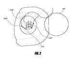
- FIG. 2illustrates an example of an anatomical site 210 with tracking units 145 A, 145 B, and 145 C.
- An anatomical site 210can be anywhere within or on the body 160 , human or otherwise.
- tracking units 145 A, 145 B, and 145 Cmay be implantable needles containing a magnetic tracking coil. If the tracking units 145 A, 145 B, and 145 C are implantable, then the tracking units may be placed in or near an anatomical site 210 . In some embodiments, tracking units 145 A, 145 B, and 145 C may be placed on the surface of an anatomical site 210 .
- tracking units 145 A, 145 B, and 145 C of known dimensionsmay be placed partially inside and partially external to an anatomical site 210 .
- a portion of the tracking unit 145 A, 145 B, and 145 Cmay be in or near the anatomical site 210 while another portion may be external to the body or the anatomical site and allow tracking external to the body.
- This embodimentis useful, for example, if it is desired that the second position sensing unit 140 be an optical tracker or if there are other reasons, such as the size of a magnetic tracking coil, for not implanting that portion of the tracking units 145 A, 145 B, and 145 C.
- FIG. 3illustrates the deformation of an anatomical site 210 to 310 with tracking units 145 A, 145 B, and 145 C.
- three tracking units 145 A, 145 B, and 145 Chave been placed near an anatomical site 210 .
- the letters on the vertices of anatomical site 210illustrate that anatomical site 210 may be deformed into deformed anatomical site 310 .
- the lettersillustrate which vertices in anatomical site 210 correspond to which vertices in deformed anatomical site 310 .
- the deformation of anatomical site 210 into anatomical site 310can be due to patient movement, breathing, pressure, force, or any other effect that may deform deformable tissue comprising and/or surrounding anatomical sites 210 and 310 .
- FIG. 3illustrates that the tracking units 145 A, 145 B, and 145 C move from locations on anatomical sites 210 to corresponding locations in deformed anatomical site 310 .
- the first set of imaging datasuch as a CT scan, MRI, open-magnet MRI, fluoroscopy, PET scan, 3D ultrasound, or any other type of imaging data may be received in a format that is usable to perform the deformation techniques described herein.
- a 3D model of that datamay be produced.
- 3D modelssuch as finite element models, volumetric models, or polygonal models. These include manual, human-driven techniques, such as tracing the boundaries of organs and tumors (also known as contouring), and automatic techniques such as iso-surface extraction (marching cubes, watershed), or hybrid techniques such as m-rep based segmentation.
- a model of the anatomical sites 210can be updated to estimate the deformed anatomical site 310 .
- This updatingmay be accomplished using known techniques for the various underlying models. For example, in some computer graphics hardware systems, one can use 3D textures.
- Each tracking unitcan be associated with a texture location within the 3D texture, where the 3D texture comprises the first set of imaging data. Once updated positions of the tracking units 145 A, 145 B, and 145 C are known, then the 3D texture can be updated using linear interpolation.
- Image guidance unit 130may contain general hardware, such as a CPU, or specialized hardware, such as a graphics card, that is capable of performing linear interpolation on 3D textures.
- general hardwaresuch as a CPU
- specialized hardwaresuch as a graphics card
- the particular slice for displaymay be determined based on the corresponding slice of the updated 3D texture. See, e.g., Yinghui, C., Jing, W., and Xiaohui, L. 2006, Real - time deformation using modal analysis on graphics hardware , in Proceedings of the 4 th international Conference on Computer Graphics and interactive Techniques in Australasia and Southeast Asia (Kuala Lumpur, Malaysia, Nov. 29-Dec. 2, 2006). GRAPHITE '06.
- the tracking units 145 A, 145 B, and 145 Cprovide position data and not orientation data. Deformation of the 3D model can be accomplished based on the position of the tracking units 145 A, 145 B, and 145 C. In some embodiments, the tracking units 145 A, 145 B, and 145 C will provide both position and orientation data. The additional information on orientation can be used to provide a different kind of deformation of the model.
- the updated modelcan be used, for example, by image guidance unit 130 of FIG. 1 in order to produce image guidance data that is based on a combination of imaging data from imaging unit 150 and movable imaging unit 155 .
- This image guidance datamay be displayed as imaging data 125 on display unit 120 .
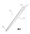
- FIG. 4depicts an embodiment of a tracking unit 145 .
- the tracking unit 145comprises a shaft 410 , a magnetic coil 430 , and a cable 420 .
- the shaft 410may be a hollow needle or any implantable unit.
- the shaft 410may be hollow in order to accommodate insertion of the cable 420 and magnetic coil 430 , or may be solid, in which case magnetic coil 430 and cable 420 must be built into the shaft 410 or the shaft 410 must be constructed around the magnetic coil 430 and cable 420 .
- a tracking unit 145may include an optical device, such as a fiducial (not pictured), in order to allow proper tracking.
- a tracking unit 145may include an implantable portion in addition to and separate from the tracking portion (not pictured).
- FIG. 5illustrates another example of an anatomical site 210 within deformable tissue 510 with implanted tracking units 145 A, 145 B, 145 C, and 145 D.
- FIG. 5illustrates that the deformable tissue 510 deforms into deformable tissue 520 .
- anatomical site 210is deformed into deformed anatomical site 310 .
- Tracking units 145 A, 145 B, 145 C, and 145 Dare shown implanted near anatomical site 210 .
- the tracking units 145 A, 145 B, 145 C, and 145 Dremain near the anatomical site after deformable tissue 510 has been deformed into deformed deformable tissue 520 , and anatomical site 210 has deformed into deformed anatomical site 310 .
- a 3D model of anatomical site 210can be deformed and updated based on the relative emplacements of the tracking units 145 A, 145 B, 145 C, and 145 D at the time before and after deformation.
- FIG. 6depicts one of many possible example processes 600 for providing guidance data based on updated deformable tracking information.
- all or portions of process 600may be performed by image guidance system 130 or by any other appropriate unit or module.
- a first set of imaging data from anatomical site 210 and tracking units 145is obtained.
- This first set of imaging datamay be a CT scan, MRI, open-magnet MRI, fluoroscopy, PET scan, 3D ultrasound, or any other imaging data.
- Obtaining the relative emplacements of the tracking units 145 when taking the data for the anatomical site 210provides the ability to determine how the relative emplacements of the tracking units 145 have changed, from the time that the first set of imaging data is taken, and until any time later at which the emplacements of the tracking units are known. Generally, the tracking units 145 will be visible in the first set of imaging data, but this is not necessary. The tracking units 145 must simply be close enough to an anatomical site of interest to provide information on deformation of the anatomical site.
- a 3D model of the first set of imaging datais produced.
- a 3D model of the first set of imaging datais produced at a later time or is not produced at all, and deformation of the first set of imaging data is accomplished without using a 3D model. The production of a 3D model from the first set of imaging data is discussed above.
- a second set of imaging data of the anatomical siteis obtained in step 620 .
- the second set of imaging datamay, like the first set of imaging data, be any of a variety of types of imaging data.
- a second set of imaging datamay be 2D ultrasound, 3D ultrasound, fluoroscopy, or any other type of imaging data.
- the tracking units 145may, but need not, be visible in the second set of imaging data.
- the ultrasound image obtainedmay include a plane or slice of the anatomical site 210 , but tracking units 145 need not be visible in that particular slice or plane.
- a deformed version of the first set of imaging datais determined.
- updated emplacements of the tracking units 145at the time the second set of imaging data is obtained, are used to determine an updated or deformed model of the first set of imaging data. This is discussed above.
- the relative emplacements of those two sets of imaging dataare determined. This may be accomplished based on both the emplacements of the tracking units 145 , in order to determine the emplacement of the deformed version of the first set of imaging data, and the emplacement of the second set of imaging data.
- the emplacements of the second set of imaging datamay be determined based on, for example, the location of a movable imaging unit 155 , as depicted in FIG. 1 .
- a movable imaging unit 155is a 2D ultrasound wand
- tracking the location of a movable imaging unit 155allows determination of the position and orientation of the second set of imaging data.
- the movable imaging unit 155may be tracked using the first position sensing unit 110 .
- the determination of relative emplacements of the two imaging data setsmay take the form of a 3D transformation or other mathematical relationship.
- the image guidance datacan be determined and displayed in step 650 .
- the image guidance datashows features within the deformed version of the first set of imaging data in combination with a second set of imaging data, such as that depicted in FIG. 8 .
- the image guidance datamay be an overlay or a combination of the second set of imaging data and of the deformed version of the first set of imaging data.
- Other examples of image guidance data that may be displayed in step 650are those depicted in FIGS. 7A-7D , discussed below.
- process 600may repeat starting at step 620 .
- Process 600may also be restarted from step 610 , especially in scenarios, such as resection at the anatomical site, where warping of the first set of imaging data is no longer possible. In such a case, the first set of imaging data may be re-obtained in step 610 .
- the deformationis accomplished by linear deformation.
- Linear deformationmay be accomplished in number of ways, including using graphics hardware.
- linear deformationconsider an original volume image I (as scanned, for example, by the CT scanner at time t) as an anatomical site of interest.
- the tracking units' positionsare pt 1 . . . pt n .
- 3d texture coordinate, tcthat indicates the position of the tracking sensor, in image I's coordinate system.
- the eight corners of the image I in the 3d texture's coordinate systemmay be (0,0,0), (0,0,1), (0,1,0), (0,1,1), (1,1,0), (1,1,1).
- the pointsmay then be stored in a table-like data structure as follows:
- Points tablepoint #1, pt 1 .x, pt 1 .y, pt 1 .z, tc 1 .u, tc 1 .v, tc 1 .w, point #2, pt 2 .x, pt 2 .y, pt 2 .z, tc 2 .u, tc 2 .v, tc 2 .w, point #3, pt 3 .x, pt 3 .y, pt 3 .z, tc 3 .u, tc 3 .v, tc 3 .w, . . .
- the volumemay then be tessellated into tetrahedra.
- the corner point of each tetrahedron kmay be one of n tracking units, at position pt k .
- the edges of the tessellationmay be stored in a data structure as follows:
- Edges tableedge #1, point a 1 , point b 1 , edge #2, point a 2 , point b 2 , . . . edge #m, point a n , point b n , where each edge connects two points, each of which is a reference to a point #.
- point a 1may refer to point #1 and point b 1 may refer to point #4.
- Tetrahedra tabletetrahedron #1, edge a 1 , edge b 1 , edge c 1 , edge d 1 , edge e 1 , edge f 1 , tetrahedron #2, edge a 2 , edge b 2 , edge c 2 , edge d 2 , edge e 2 , edge f 2 , . . . , where edge a 1 and a 2 may refer, for example, to edges #5 and edge #2, respectively.
- image Imay lie outside of the convex hull of the points pt 1 . . . pt n . These portions of image I may be ignored or other algorithms may be used to determine their distortion.
- the tissuemay have changed shape, and image I (which represents the anatomical site at time t) may no longer represent the.
- the positions of the tracking sensors at time jare pj 1 . . . pj n .
- the deformed version of the first set of imaging datamay be projected onto the plane of the planar fluoroscopy and the two images may be combined in order to produce the image guidance data.
- the image guidance data determined in step 650 from the deformed version of the first set of imaging datacould be used to approximate another imaging modality.
- the first set of imaging datamay be a CT scan and a user may wish to have an approximation of a biplane fluoroscopy performed without exposing a patient to the harmful radiation associated with such a fluoroscopy.
- the deformed version of the first set of imaging datamay be projected onto what would be the two planes of the biplane fluoroscopy. This would approximate the biplane fluoroscopy using the updated tracking unit information without exposing the patient to the radiation associated with the biplane fluoroscopy. Further, this approximation could be updated at a rate that exceeds that of conventional biplane fluoroscopy as the tracking units move with the surrounding tissues to which they are affixed, without harming the patient or disturbing the ongoing operation.
- FIGS. 7A-7Ddepict marking and viewing features in imaging data.
- FIGS. 7A-7Dshow the manual creation of features within a first set of imaging data and updating the placement of the feature based on the deformed model of the first set of imaging data.
- FIG. 7Ashows a feature selection unit 710 being used to highlight, in a particular plane or visual slice of the first set of imaging data 720 , a feature 730 within the first of imaging data.
- the userpoints to the feature 730 and selects the feature in order to signify selected feature 740 .
- the selected feature 740will still appear in its original position, as depicted in FIG. 7C .
- FIG. 7Dillustrates that if a plane of the first set of imaging data 720 is displayed, and the selected feature 740 may still be displayed even if it is not within the visual slice or plane or display data of the first set of imaging data 720 .
- the displacement of the feature 740 from the visual slice of the first set of imaging data 720may be shown with a displacement marker 750 .
- the displacement of the selected feature 740 from the visual slice of the first set of imaging data 720may be shown with other visual techniques, such as visual depth on a 3D display, shadowing, foreshortening, or any other known technique.
- the selected feature 740may be shown in a new location based on the deformation of the first set of imaging data 720 . Marking these features may be useful so that a user or surgeon could identify and later find points of interest, such as locations of tumors or lesions.
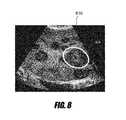
- FIG. 8depicts an embodiment of image guidance data in which a first set of imaging data is presented with a second set of imaging data.
- the second set of imaging datais displayed in approximately the form that is received as described above.
- the first of imaging datamay be deformed based on the updated emplacements of tracking units 145 .
- the updated or deformed model corresponding to the first set of imaging datamay be used to provide image guidance, such as the location of an important feature of the anatomical site.
- the ultrasound image 810(the second set of imaging data) is augmented with a feature 820 from the first set of imaging data, such as a CT scan.
- This featuremay be the location of a tumor, necrosed tissue, or any other relevant feature. It may have been detected or selected by a user, as illustrated in FIGS. 7A-7D .
- the model for the first set of imaging datamay be deformed and therefore the feature 820 shown from the second set of imaging data would also be updated and deformed.
- the movement and updated emplacements of the tracking units 145 in an anatomical sitewould cause the deformation and movement of the feature of the first set of imaging data 820 .
- the updated emplacement of the feature 820would continue to be shown with the newly received second set of imaging data 810 .
- the deformation of the first set of imaging dataapproximates the deformation of the underlying tissue. Therefore, the approximate placement of the feature 820 would approximate the location of the underlying anatomical feature within the second set of imaging data 810 .
- Computer systemsmay include a bus or other communication mechanism for communicating information, and a processor coupled with the bus for processing information.
- a computer systemmay have a main memory, such as a random access memory or other dynamic storage device, coupled to the bus. The main memory may be used to store instructions and temporary variables.
- the computer systemmay also include a read-only memory or other static storage device coupled to the bus for storing static information and instructions.
- the computer systemmay also be coupled to a display, such as a CRT or LCD monitor.
- Input devicesmay also be coupled to the computer system. These input devices may include a mouse, a trackball, or cursor direction keys.
- Computer systems described hereinmay include the image guidance unit 130 , first and second position sensing units 110 and 140 , and imaging unit 150 .
- Each computer systemmay be implemented using one or more physical computers or computer systems or portions thereof.
- the instructions executed by the computer systemmay also be read in from a computer-readable medium.
- the computer-readable mediummay be a CD, DVD, optical or magnetic disk, laser disc, carrier wave, or any other medium that is readable by the computer system.
- hardwired circuitrymay be used in place of or in combination with software instructions executed by the processor.
- All of the methods and processes described abovemay be embodied in, and fully automated via, software code modules executed by one or more general purpose computers or processors, such as those computer systems described above.
- the code modulesmay be stored in any type of computer-readable medium or other computer storage device. Some or all of the methods may alternatively be embodied in specialized computer hardware.
Landscapes
- Engineering & Computer Science (AREA)
- Health & Medical Sciences (AREA)
- Life Sciences & Earth Sciences (AREA)
- Surgery (AREA)
- Theoretical Computer Science (AREA)
- General Health & Medical Sciences (AREA)
- Medical Informatics (AREA)
- Nuclear Medicine, Radiotherapy & Molecular Imaging (AREA)
- Physics & Mathematics (AREA)
- General Physics & Mathematics (AREA)
- Biomedical Technology (AREA)
- Multimedia (AREA)
- Robotics (AREA)
- Heart & Thoracic Surgery (AREA)
- Molecular Biology (AREA)
- Animal Behavior & Ethology (AREA)
- Public Health (AREA)
- Veterinary Medicine (AREA)
- Computer Vision & Pattern Recognition (AREA)
- Quality & Reliability (AREA)
- Radiology & Medical Imaging (AREA)
- Apparatus For Radiation Diagnosis (AREA)
- Ultra Sonic Daignosis Equipment (AREA)
Abstract
Description
| Points table: |
| point #1, pt1.x, pt1.y, pt1.z, tc1.u, tc1.v, tc1.w, | |
| point #2, pt2.x, pt2.y, pt2.z, tc2.u, tc2.v, tc2.w, | |
| point #3, pt3.x, pt3.y, pt3.z, tc3.u, tc3.v, tc3.w, | |
| . . . | |
| point #n, ptn.x, ptn.y, ptn.z, tcn.u, tcn.v, tcn.w, | |
where ptk.y and tck.u refer, for example, to point ptk's y coordinate and tck's u coordinate, respectively.
| Edges table: |
| edge #1, point a1, point b1, | |
| edge #2, point a2, point b2, | |
| . . . | |
| edge #m, point an, point bn, | |
where each edge connects two points, each of which is a reference to a point #. For example, point a1may refer to point #1 and point b1may refer to point #4.
| Tetrahedra table: |
| tetrahedron #1, edge a1, edge b1, edge c1, edge d1, edge e1, edge f1, | |
| tetrahedron #2, edge a2, edge b2, edge c2, edge d2, edge e2, edge f2, | |
| . . . , | |
where edge a1and a2may refer, for example, to edges #5 and edge #2, respectively.
- Update the points table, and replace each ptkwith pjkwhere k={1 . . . n}, thereby updating the position of each tracking unit, at time j, as supplied, for example, by
position sensing system 140. - Iterate through the tetrahedra table. For each tetrahedron, iterate through each of its six edges. For each such edge, compute its intersection with the cross-sectional plane. For those edges that do intersect with the plane, we compute the intersection point P. We then compute the texture coordinate for P, by linearly interpolating between the texture coordinates at its endpoints (those texture coordinates are stored in the points table).
- For each tetrahedron, there will be 0, 1, 2, 3, or 4 edges that intersect with the plane (resulting in 0, 1, 2, 3, or 4 intersections points P, and their corresponding texture coordinates).
- When there are 0, 1, or 2 intersections, render nothing associated with the tetrahedron.
- When there are 3 or 4 intersections of the cross-sectional plane, render a textured polygon using traditional graphics hardware commands (e.g. OpenGL or DirectX). The texture coordinates index into the image I, but because the point P will have moved from the original location, the result will be a portion of image J.
- Repeat this for each tetrahedron. After all tetrahedra have been processed, the graphics hardware will have rendered the intersection of the warped image J, with the chosen cross-sectional plane.
- Update the points table, and replace each ptkwith pjkwhere k={1 . . . n}, thereby updating the position of each tracking unit, at time j, as supplied, for example, by
Claims (17)
Priority Applications (2)
| Application Number | Priority Date | Filing Date | Title |
|---|---|---|---|
| US12/399,899US8340379B2 (en) | 2008-03-07 | 2009-03-06 | Systems and methods for displaying guidance data based on updated deformable imaging data |
| US13/723,705US8831310B2 (en) | 2008-03-07 | 2012-12-21 | Systems and methods for displaying guidance data based on updated deformable imaging data |
Applications Claiming Priority (2)
| Application Number | Priority Date | Filing Date | Title |
|---|---|---|---|
| US6846908P | 2008-03-07 | 2008-03-07 | |
| US12/399,899US8340379B2 (en) | 2008-03-07 | 2009-03-06 | Systems and methods for displaying guidance data based on updated deformable imaging data |
Related Child Applications (1)
| Application Number | Title | Priority Date | Filing Date |
|---|---|---|---|
| US13/723,705DivisionUS8831310B2 (en) | 2008-03-07 | 2012-12-21 | Systems and methods for displaying guidance data based on updated deformable imaging data |
Publications (2)
| Publication Number | Publication Date |
|---|---|
| US20090226069A1 US20090226069A1 (en) | 2009-09-10 |
| US8340379B2true US8340379B2 (en) | 2012-12-25 |
Family
ID=41053646
Family Applications (2)
| Application Number | Title | Priority Date | Filing Date |
|---|---|---|---|
| US12/399,899Active2031-03-30US8340379B2 (en) | 2008-03-07 | 2009-03-06 | Systems and methods for displaying guidance data based on updated deformable imaging data |
| US13/723,705ActiveUS8831310B2 (en) | 2008-03-07 | 2012-12-21 | Systems and methods for displaying guidance data based on updated deformable imaging data |
Family Applications After (1)
| Application Number | Title | Priority Date | Filing Date |
|---|---|---|---|
| US13/723,705ActiveUS8831310B2 (en) | 2008-03-07 | 2012-12-21 | Systems and methods for displaying guidance data based on updated deformable imaging data |
Country Status (1)
| Country | Link |
|---|---|
| US (2) | US8340379B2 (en) |
Cited By (46)
| Publication number | Priority date | Publication date | Assignee | Title |
|---|---|---|---|---|
| US8482606B2 (en) | 2006-08-02 | 2013-07-09 | Inneroptic Technology, Inc. | System and method of providing real-time dynamic imagery of a medical procedure site using multiple modalities |
| US8554307B2 (en) | 2010-04-12 | 2013-10-08 | Inneroptic Technology, Inc. | Image annotation in image-guided medical procedures |
| US8585598B2 (en) | 2009-02-17 | 2013-11-19 | Inneroptic Technology, Inc. | Systems, methods, apparatuses, and computer-readable media for image guided surgery |
| US20130311174A1 (en)* | 2010-12-20 | 2013-11-21 | Nikon Corporation | Audio control device and imaging device |
| US8641621B2 (en) | 2009-02-17 | 2014-02-04 | Inneroptic Technology, Inc. | Systems, methods, apparatuses, and computer-readable media for image management in image-guided medical procedures |
| US8670816B2 (en) | 2012-01-30 | 2014-03-11 | Inneroptic Technology, Inc. | Multiple medical device guidance |
| US8831310B2 (en) | 2008-03-07 | 2014-09-09 | Inneroptic Technology, Inc. | Systems and methods for displaying guidance data based on updated deformable imaging data |
| US9265572B2 (en) | 2008-01-24 | 2016-02-23 | The University Of North Carolina At Chapel Hill | Methods, systems, and computer readable media for image guided ablation |
| US9675319B1 (en) | 2016-02-17 | 2017-06-13 | Inneroptic Technology, Inc. | Loupe display |
| WO2017156482A1 (en) | 2013-09-06 | 2017-09-14 | Brigham And Women's Hospital, Inc. | System and method for a tissue resection margin measurement device |
| US9901406B2 (en) | 2014-10-02 | 2018-02-27 | Inneroptic Technology, Inc. | Affected region display associated with a medical device |
| US9949700B2 (en) | 2015-07-22 | 2018-04-24 | Inneroptic Technology, Inc. | Medical device approaches |
| US9974525B2 (en) | 2014-10-31 | 2018-05-22 | Covidien Lp | Computed tomography enhanced fluoroscopic system, device, and method of utilizing the same |
| US10188467B2 (en) | 2014-12-12 | 2019-01-29 | Inneroptic Technology, Inc. | Surgical guidance intersection display |
| US10278778B2 (en) | 2016-10-27 | 2019-05-07 | Inneroptic Technology, Inc. | Medical device navigation using a virtual 3D space |
| US10314559B2 (en) | 2013-03-14 | 2019-06-11 | Inneroptic Technology, Inc. | Medical device guidance |
| US10674982B2 (en) | 2015-08-06 | 2020-06-09 | Covidien Lp | System and method for local three dimensional volume reconstruction using a standard fluoroscope |
| US10699448B2 (en) | 2017-06-29 | 2020-06-30 | Covidien Lp | System and method for identifying, marking and navigating to a target using real time two dimensional fluoroscopic data |
| US10702226B2 (en) | 2015-08-06 | 2020-07-07 | Covidien Lp | System and method for local three dimensional volume reconstruction using a standard fluoroscope |
| US10716525B2 (en) | 2015-08-06 | 2020-07-21 | Covidien Lp | System and method for navigating to target and performing procedure on target utilizing fluoroscopic-based local three dimensional volume reconstruction |
| US10893843B2 (en) | 2017-10-10 | 2021-01-19 | Covidien Lp | System and method for identifying and marking a target in a fluoroscopic three-dimensional reconstruction |
| US10905498B2 (en) | 2018-02-08 | 2021-02-02 | Covidien Lp | System and method for catheter detection in fluoroscopic images and updating displayed position of catheter |
| US11020144B2 (en) | 2015-07-21 | 2021-06-01 | 3Dintegrated Aps | Minimally invasive surgery system |
| US11033182B2 (en) | 2014-02-21 | 2021-06-15 | 3Dintegrated Aps | Set comprising a surgical instrument |
| US11039734B2 (en) | 2015-10-09 | 2021-06-22 | 3Dintegrated Aps | Real time correlated depiction system of surgical tool |
| US11051886B2 (en) | 2016-09-27 | 2021-07-06 | Covidien Lp | Systems and methods for performing a surgical navigation procedure |
| US11172895B2 (en) | 2015-12-07 | 2021-11-16 | Covidien Lp | Visualization, navigation, and planning with electromagnetic navigation bronchoscopy and cone beam computed tomography integrated |
| US11259879B2 (en) | 2017-08-01 | 2022-03-01 | Inneroptic Technology, Inc. | Selective transparency to assist medical device navigation |
| US11331120B2 (en) | 2015-07-21 | 2022-05-17 | 3Dintegrated Aps | Cannula assembly kit |
| US11375889B2 (en) | 2016-12-27 | 2022-07-05 | The Charlotte-Mecklenburg Hospital Authority | Devices, systems, and methods for image guided laparoscopic surgery |
| US11464578B2 (en) | 2009-02-17 | 2022-10-11 | Inneroptic Technology, Inc. | Systems, methods, apparatuses, and computer-readable media for image management in image-guided medical procedures |
| US11642173B2 (en) | 2018-04-06 | 2023-05-09 | Medtronic, Inc. | Image-based navigation system and method of using same |
| US11750794B2 (en) | 2015-03-24 | 2023-09-05 | Augmedics Ltd. | Combining video-based and optic-based augmented reality in a near eye display |
| US11801115B2 (en) | 2019-12-22 | 2023-10-31 | Augmedics Ltd. | Mirroring in image guided surgery |
| US11896445B2 (en) | 2021-07-07 | 2024-02-13 | Augmedics Ltd. | Iliac pin and adapter |
| US11974887B2 (en) | 2018-05-02 | 2024-05-07 | Augmedics Ltd. | Registration marker for an augmented reality system |
| US11980429B2 (en) | 2018-11-26 | 2024-05-14 | Augmedics Ltd. | Tracking methods for image-guided surgery |
| US11980506B2 (en) | 2019-07-29 | 2024-05-14 | Augmedics Ltd. | Fiducial marker |
| US12044858B2 (en) | 2022-09-13 | 2024-07-23 | Augmedics Ltd. | Adjustable augmented reality eyewear for image-guided medical intervention |
| US12150821B2 (en) | 2021-07-29 | 2024-11-26 | Augmedics Ltd. | Rotating marker and adapter for image-guided surgery |
| US12178666B2 (en) | 2019-07-29 | 2024-12-31 | Augmedics Ltd. | Fiducial marker |
| US12186028B2 (en) | 2020-06-15 | 2025-01-07 | Augmedics Ltd. | Rotating marker for image guided surgery |
| US12239385B2 (en) | 2020-09-09 | 2025-03-04 | Augmedics Ltd. | Universal tool adapter |
| US12354227B2 (en) | 2022-04-21 | 2025-07-08 | Augmedics Ltd. | Systems for medical image visualization |
| US12383349B2 (en) | 2022-07-26 | 2025-08-12 | Elucent Medical, Inc. | Systems and methods for wireless localization |
| US12417595B2 (en) | 2021-08-18 | 2025-09-16 | Augmedics Ltd. | Augmented-reality surgical system using depth sensing |
Families Citing this family (18)
| Publication number | Priority date | Publication date | Assignee | Title |
|---|---|---|---|---|
| JP5332369B2 (en)* | 2008-07-18 | 2013-11-06 | ソニー株式会社 | Image processing apparatus, image processing method, and computer program |
| US9282947B2 (en) | 2009-12-01 | 2016-03-15 | Inneroptic Technology, Inc. | Imager focusing based on intraoperative data |
| US20110172526A1 (en) | 2010-01-12 | 2011-07-14 | Martin Lachaine | Feature Tracking Using Ultrasound |
| US9248316B2 (en) | 2010-01-12 | 2016-02-02 | Elekta Ltd. | Feature tracking using ultrasound |
| JP5737858B2 (en)* | 2010-04-21 | 2015-06-17 | キヤノン株式会社 | Image processing apparatus, image processing method, and program |
| US20110306873A1 (en)* | 2010-05-07 | 2011-12-15 | Krishna Shenai | System for performing highly accurate surgery |
| US8532738B2 (en)* | 2010-11-04 | 2013-09-10 | Biosense Webster (Israel), Ltd. | Visualization of catheter-tissue contact by map distortion |
| US8768019B2 (en)* | 2011-02-03 | 2014-07-01 | Medtronic, Inc. | Display of an acquired cine loop for procedure navigation |
| US11406278B2 (en) | 2011-02-24 | 2022-08-09 | Koninklijke Philips N.V. | Non-rigid-body morphing of vessel image using intravascular device shape |
| RU2013143160A (en)* | 2011-02-24 | 2015-03-27 | Конинклейке Филипс Электроникс Н.В. | NON-RIGID TRANSFORMATION OF THE IMAGE OF THE VESSEL USING THE FORM OF AN INJURAL DEVICE |
| US9520072B2 (en)* | 2011-09-21 | 2016-12-13 | University Of South Florida | Systems and methods for projecting images onto an object |
| EP2869754A2 (en)* | 2012-07-09 | 2015-05-13 | Koninklijke Philips N.V. | Method and system for adaptive image guided intervention |
| DE102014226240A1 (en)* | 2014-12-17 | 2016-06-23 | Kuka Roboter Gmbh | System for robot-assisted medical treatment |
| CN109964249A (en)* | 2016-09-21 | 2019-07-02 | 皇家飞利浦有限公司 | The device delineated for the adaptive profile to body part |
| KR102366242B1 (en)* | 2017-12-07 | 2022-02-23 | 삼성전자주식회사 | Method for controlling depth of objects in mirror display system |
| EP4022558A1 (en)* | 2019-08-28 | 2022-07-06 | Intuitive Surgical Operations, Inc. | Systems and methods for registering imaging data from different imaging modalities based on subsurface image scanning |
| EP4072426B1 (en)* | 2019-12-13 | 2025-04-16 | Smith&Nephew, Inc. | Anatomical feature extraction and presentation using augmented reality |
| CN116916848A (en)* | 2021-02-03 | 2023-10-20 | 加利福尼亚大学董事会 | A surgical perception framework for robotic tissue manipulation |
Citations (163)
| Publication number | Priority date | Publication date | Assignee | Title |
|---|---|---|---|---|
| USRE30397E (en) | 1976-04-27 | 1980-09-09 | Three-dimensional ultrasonic imaging of animal soft tissue | |
| US4294544A (en) | 1979-08-03 | 1981-10-13 | Altschuler Bruce R | Topographic comparator |
| US4862873A (en) | 1987-05-27 | 1989-09-05 | Olympus Optical Co., Ltd. | Stereo endoscope |
| US4884219A (en) | 1987-01-21 | 1989-11-28 | W. Industries Limited | Method and apparatus for the perception of computer-generated imagery |
| US5109276A (en) | 1988-05-27 | 1992-04-28 | The University Of Connecticut | Multi-dimensional multi-spectral imaging system |
| US5193120A (en) | 1991-02-27 | 1993-03-09 | Mechanical Technology Incorporated | Machine vision three dimensional profiling system |
| US5249581A (en) | 1991-07-15 | 1993-10-05 | Horbal Mark T | Precision bone alignment |
| US5261404A (en) | 1991-07-08 | 1993-11-16 | Mick Peter R | Three-dimensional mammal anatomy imaging system and method |
| US5307153A (en) | 1990-06-19 | 1994-04-26 | Fujitsu Limited | Three-dimensional measuring apparatus |
| US5323002A (en) | 1992-03-25 | 1994-06-21 | Texas Instruments Incorporated | Spatial light modulator based optical calibration system |
| US5371543A (en) | 1993-03-03 | 1994-12-06 | Texas Instruments Incorporated | Monolithic color wheel |
| US5383454A (en) | 1990-10-19 | 1995-01-24 | St. Louis University | System for indicating the position of a surgical probe within a head on an image of the head |
| US5446798A (en) | 1989-06-20 | 1995-08-29 | Fujitsu Limited | Method and apparatus for measuring position and orientation of an object based on a sequence of projected points |
| US5452024A (en) | 1993-11-01 | 1995-09-19 | Texas Instruments Incorporated | DMD display system |
| US5457493A (en) | 1993-09-15 | 1995-10-10 | Texas Instruments Incorporated | Digital micro-mirror based image simulation system |
| US5488431A (en) | 1993-11-04 | 1996-01-30 | Texas Instruments Incorporated | Video data formatter for a multi-channel digital television system without overlap |
| US5489952A (en) | 1993-07-14 | 1996-02-06 | Texas Instruments Incorporated | Method and device for multi-format television |
| US5491510A (en) | 1993-12-03 | 1996-02-13 | Texas Instruments Incorporated | System and method for simultaneously viewing a scene and an obscured object |
| US5517990A (en) | 1992-11-30 | 1996-05-21 | The Cleveland Clinic Foundation | Stereotaxy wand and tool guide |
| US5526051A (en) | 1993-10-27 | 1996-06-11 | Texas Instruments Incorporated | Digital television system |
| US5532997A (en) | 1990-06-06 | 1996-07-02 | Texas Instruments Incorporated | Optical tracking system |
| US5541723A (en) | 1993-06-21 | 1996-07-30 | Minolta Camera Kabushiki Kaisha | Distance measuring device |
| US5579026A (en) | 1993-05-14 | 1996-11-26 | Olympus Optical Co., Ltd. | Image display apparatus of head mounted type |
| US5588948A (en) | 1993-02-17 | 1996-12-31 | Olympus Optical Co. Ltd. | Stereoscopic endoscope |
| US5611353A (en) | 1993-06-21 | 1997-03-18 | Osteonics Corp. | Method and apparatus for locating functional structures of the lower leg during knee surgery |
| US5612753A (en) | 1995-01-27 | 1997-03-18 | Texas Instruments Incorporated | Full-color projection display system using two light modulators |
| US5625408A (en) | 1993-06-24 | 1997-04-29 | Canon Kabushiki Kaisha | Three-dimensional image recording/reconstructing method and apparatus therefor |
| US5630027A (en) | 1994-12-28 | 1997-05-13 | Texas Instruments Incorporated | Method and apparatus for compensating horizontal and vertical alignment errors in display systems |
| US5629794A (en) | 1995-05-31 | 1997-05-13 | Texas Instruments Incorporated | Spatial light modulator having an analog beam for steering light |
| US5699444A (en) | 1995-03-31 | 1997-12-16 | Synthonics Incorporated | Methods and apparatus for using image data to determine camera location and orientation |
| US5726670A (en) | 1992-07-20 | 1998-03-10 | Olympus Optical Co., Ltd. | Display apparatus to be mounted on the head or face of an individual |
| US5766135A (en) | 1995-03-08 | 1998-06-16 | Terwilliger; Richard A. | Echogenic needle tip |
| US5784098A (en) | 1995-08-28 | 1998-07-21 | Olympus Optical Co., Ltd. | Apparatus for measuring three-dimensional configurations |
| US5807395A (en) | 1993-08-27 | 1998-09-15 | Medtronic, Inc. | Method and apparatus for RF ablation and hyperthermia |
| US5820554A (en) | 1993-08-31 | 1998-10-13 | Medtronic, Inc. | Ultrasound biopsy needle |
| US5870136A (en) | 1997-12-05 | 1999-02-09 | The University Of North Carolina At Chapel Hill | Dynamic generation of imperceptible structured light for tracking and acquisition of three dimensional scene geometry and surface characteristics in interactive three dimensional computer graphics applications |
| US5891034A (en) | 1990-10-19 | 1999-04-06 | St. Louis University | System for indicating the position of a surgical probe within a head on an image of the head |
| US6019724A (en) | 1995-02-22 | 2000-02-01 | Gronningsaeter; Aage | Method for ultrasound guidance during clinical procedures |
| US6064749A (en) | 1996-08-02 | 2000-05-16 | Hirota; Gentaro | Hybrid tracking for augmented reality using both camera motion detection and landmark tracking |
| US6095982A (en) | 1995-03-14 | 2000-08-01 | Board Of Regents, The University Of Texas System | Spectroscopic method and apparatus for optically detecting abnormal mammalian epithelial tissue |
| US6108130A (en) | 1999-09-10 | 2000-08-22 | Intel Corporation | Stereoscopic image sensor |
| US6122541A (en) | 1995-05-04 | 2000-09-19 | Radionics, Inc. | Head band for frameless stereotactic registration |
| US6167296A (en) | 1996-06-28 | 2000-12-26 | The Board Of Trustees Of The Leland Stanford Junior University | Method for volumetric image navigation |
| US6216029B1 (en) | 1995-07-16 | 2001-04-10 | Ultraguide Ltd. | Free-hand aiming of a needle guide |
| US6246898B1 (en) | 1995-03-28 | 2001-06-12 | Sonometrics Corporation | Method for carrying out a medical procedure using a three-dimensional tracking and imaging system |
| US6261234B1 (en) | 1998-05-07 | 2001-07-17 | Diasonics Ultrasound, Inc. | Method and apparatus for ultrasound imaging with biplane instrument guidance |
| US20010016804A1 (en) | 1996-09-04 | 2001-08-23 | Cunningham Richard L. | Surgical simulation interface device and method |
| US20010045979A1 (en) | 1995-03-29 | 2001-11-29 | Sanyo Electric Co., Ltd. | Methods for creating an image for a three-dimensional display, for calculating depth information, and for image processing using the depth information |
| US6341016B1 (en) | 1999-08-06 | 2002-01-22 | Michael Malione | Method and apparatus for measuring three-dimensional shape of object |
| US20020010384A1 (en) | 2000-03-30 | 2002-01-24 | Ramin Shahidi | Apparatus and method for calibrating an endoscope |
| US6348058B1 (en) | 1997-12-12 | 2002-02-19 | Surgical Navigation Technologies, Inc. | Image guided spinal surgery guide, system, and method for use thereof |
| US20020049375A1 (en) | 1999-05-18 | 2002-04-25 | Mediguide Ltd. | Method and apparatus for real time quantitative three-dimensional image reconstruction of a moving organ and intra-body navigation |
| US6385475B1 (en) | 1997-03-11 | 2002-05-07 | Philippe Cinquin | Process and device for the preoperative determination of the positioning data of endoprosthetic parts |
| US20020077543A1 (en) | 2000-06-27 | 2002-06-20 | Robert Grzeszczuk | Method and apparatus for tracking a medical instrument based on image registration |
| US20020077540A1 (en) | 2000-11-17 | 2002-06-20 | Kienzle Thomas C. | Enhanced graphic features for computer assisted surgery system |
| US6442417B1 (en) | 1999-11-29 | 2002-08-27 | The Board Of Trustees Of The Leland Stanford Junior University | Method and apparatus for transforming view orientations in image-guided surgery |
| US6456868B2 (en) | 1999-03-30 | 2002-09-24 | Olympus Optical Co., Ltd. | Navigation apparatus and surgical operation image acquisition/display apparatus using the same |
| US20020135673A1 (en) | 2000-11-03 | 2002-09-26 | Favalora Gregg E. | Three-dimensional display systems |
| US20020138008A1 (en) | 2000-01-13 | 2002-09-26 | Kazuhiro Tsujita | Method and apparatus for displaying fluorescence images and method and apparatus for acquiring endoscope images |
| US20020140814A1 (en) | 2001-03-28 | 2002-10-03 | Koninkiijke Philips Electronics N.V. | Method for assisting an automated video tracking system in reaquiring a target |
| US6470207B1 (en) | 1999-03-23 | 2002-10-22 | Surgical Navigation Technologies, Inc. | Navigational guidance via computer-assisted fluoroscopic imaging |
| US20020156375A1 (en)* | 1999-10-28 | 2002-10-24 | Paul Kessman | Navigation information overlay onto ultrasound imagery |
| US6477400B1 (en) | 1998-08-20 | 2002-11-05 | Sofamor Danek Holdings, Inc. | Fluoroscopic image guided orthopaedic surgery system with intraoperative registration |
| US6478793B1 (en) | 1999-06-11 | 2002-11-12 | Sherwood Services Ag | Ablation treatment of bone metastases |
| US20020198451A1 (en) | 2001-02-27 | 2002-12-26 | Carson Christopher P. | Surgical navigation systems and processes for high tibial osteotomy |
| US6503195B1 (en) | 1999-05-24 | 2003-01-07 | University Of North Carolina At Chapel Hill | Methods and systems for real-time structured light depth extraction and endoscope using real-time structured light depth extraction |
| US6518939B1 (en) | 1996-11-08 | 2003-02-11 | Olympus Optical Co., Ltd. | Image observation apparatus |
| US6527443B1 (en) | 1999-04-20 | 2003-03-04 | Brainlab Ag | Process and apparatus for image guided treatment with an integration of X-ray detection and navigation system |
| US6545706B1 (en) | 1999-07-30 | 2003-04-08 | Electric Planet, Inc. | System, method and article of manufacture for tracking a head of a camera-generated image of a person |
| US6546279B1 (en) | 2001-10-12 | 2003-04-08 | University Of Florida | Computer controlled guidance of a biopsy needle |
| US6551325B2 (en) | 2000-09-26 | 2003-04-22 | Brainlab Ag | Device, system and method for determining the position of an incision block |
| US6570566B1 (en) | 1999-06-10 | 2003-05-27 | Sony Corporation | Image processing apparatus, image processing method, and program providing medium |
| US6587711B1 (en) | 1999-07-22 | 2003-07-01 | The Research Foundation Of Cuny | Spectral polarizing tomographic dermatoscope |
| US6594517B1 (en) | 1998-05-15 | 2003-07-15 | Robin Medical, Inc. | Method and apparatus for generating controlled torques on objects particularly objects inside a living body |
| US6597818B2 (en) | 1997-05-09 | 2003-07-22 | Sarnoff Corporation | Method and apparatus for performing geo-spatial registration of imagery |
| US20030164172A1 (en) | 2000-06-09 | 2003-09-04 | Chumas Nicole Jane | Method and apparatus for guiding a surgical instrument |
| US6626832B1 (en) | 1999-04-15 | 2003-09-30 | Ultraguide Ltd. | Apparatus and method for detecting the bending of medical invasive tools in medical interventions |
| US20030231789A1 (en) | 2002-06-18 | 2003-12-18 | Scimed Life Systems, Inc. | Computer generated representation of the imaging pattern of an imaging device |
| US6689067B2 (en) | 2001-11-28 | 2004-02-10 | Siemens Corporate Research, Inc. | Method and apparatus for ultrasound guidance of needle biopsies |
| US20040034313A1 (en) | 2000-12-15 | 2004-02-19 | Aesculap Ag & Co. Kg | Method and device for determining the mechanical axis of a femur |
| US6711429B1 (en) | 1998-09-24 | 2004-03-23 | Super Dimension Ltd. | System and method for determining the location of a catheter during an intra-body medical procedure |
| US6725082B2 (en) | 1999-03-17 | 2004-04-20 | Synthes U.S.A. | System and method for ligament graft placement |
| US20040078036A1 (en) | 2002-10-21 | 2004-04-22 | Yaron Keidar | Real-time monitoring and mapping of ablation lesion formation in the heart |
| US6733458B1 (en) | 2001-09-25 | 2004-05-11 | Acuson Corporation | Diagnostic medical ultrasound systems and methods using image based freehand needle guidance |
| US20040095507A1 (en) | 2002-11-18 | 2004-05-20 | Medicapture, Inc. | Apparatus and method for capturing, processing and storing still images captured inline from an analog video stream and storing in a digital format on removable non-volatile memory |
| US6764449B2 (en) | 2001-12-31 | 2004-07-20 | Medison Co., Ltd. | Method and apparatus for enabling a biopsy needle to be observed |
| US6766184B2 (en) | 2000-03-28 | 2004-07-20 | Board Of Regents, The University Of Texas System | Methods and apparatus for diagnostic multispectral digital imaging |
| US6768496B2 (en) | 2000-03-30 | 2004-07-27 | Siemens Aktiengesellschaft | System and method for generating an image from an image dataset and a video image |
| US20040147920A1 (en) | 2002-10-21 | 2004-07-29 | Yaron Keidar | Prediction and assessment of ablation of cardiac tissue |
| US6775404B1 (en) | 1999-03-18 | 2004-08-10 | University Of Washington | Apparatus and method for interactive 3D registration of ultrasound and magnetic resonance images based on a magnetic position sensor |
| US6783524B2 (en) | 2001-04-19 | 2004-08-31 | Intuitive Surgical, Inc. | Robotic surgical tool with ultrasound cauterizing and cutting instrument |
| US20040238732A1 (en) | 2001-10-19 | 2004-12-02 | Andrei State | Methods and systems for dynamic virtual convergence and head mountable display |
| US20040243148A1 (en) | 2003-04-08 | 2004-12-02 | Wasielewski Ray C. | Use of micro- and miniature position sensing devices for use in TKA and THA |
| US20040254454A1 (en) | 2001-06-13 | 2004-12-16 | Kockro Ralf Alfons | Guide system and a probe therefor |
| JP2005058584A (en) | 2003-08-19 | 2005-03-10 | Toshiba Corp | Ultrasonic diagnostic equipment |
| US6873867B2 (en) | 2000-04-05 | 2005-03-29 | Brainlab Ag | Referencing or registering a patient or a patient body part in a medical navigation system by means of irradiation of light points |
| US20050085717A1 (en) | 2003-10-21 | 2005-04-21 | Ramin Shahidi | Systems and methods for intraoperative targetting |
| US20050085718A1 (en) | 2003-10-21 | 2005-04-21 | Ramin Shahidi | Systems and methods for intraoperative targetting |
| US20050090742A1 (en) | 2003-08-19 | 2005-04-28 | Yoshitaka Mine | Ultrasonic diagnostic apparatus |
| US6895268B1 (en) | 1999-06-28 | 2005-05-17 | Siemens Aktiengesellschaft | Medical workstation, imaging system, and method for mixing two images |
| US20050111733A1 (en) | 2003-11-26 | 2005-05-26 | Fors Steven L. | Automated digitized film slicing and registration tool |
| US20050159641A1 (en) | 2004-01-15 | 2005-07-21 | Pentax Corporation | Optical system for stereoscopic rigid endoscope |
| US20050182316A1 (en) | 2002-08-29 | 2005-08-18 | Burdette Everette C. | Method and system for localizing a medical tool |
| US6947783B2 (en) | 2000-09-26 | 2005-09-20 | Brainlab Ag | System for the navigation-assisted positioning of elements |
| US20050222574A1 (en) | 2002-09-27 | 2005-10-06 | Aesculap Ag & Co. Kg | Method and apparatus for determining the position of the tibial exit point of the anterior cruciate ligament |
| US20050219552A1 (en) | 2002-06-07 | 2005-10-06 | Ackerman Jermy D | Methods and systems for laser based real-time structured light depth extraction |
| US20050251148A1 (en) | 2002-11-05 | 2005-11-10 | Aesculap Ag & Co. Kg | Method and device for determining the position of a knee-joint endoprosthesis |
| JP2005323669A (en) | 2004-05-12 | 2005-11-24 | Toshiba Corp | Ultrasonic diagnostic equipment for puncture therapy |
| US6978167B2 (en) | 2002-07-01 | 2005-12-20 | Claron Technology Inc. | Video pose tracking system and method |
| US20060004275A1 (en) | 2004-06-30 | 2006-01-05 | Vija A H | Systems and methods for localized image registration and fusion |
| US20060036162A1 (en) | 2004-02-02 | 2006-02-16 | Ramin Shahidi | Method and apparatus for guiding a medical instrument to a subsurface target site in a patient |
| US7008373B2 (en) | 2001-11-08 | 2006-03-07 | The Johns Hopkins University | System and method for robot targeting under fluoroscopy based on image servoing |
| US20060052792A1 (en) | 2003-02-26 | 2006-03-09 | Aesculap Ag & Co. Kg | Patella reference device |
| US20060100505A1 (en) | 2004-10-26 | 2006-05-11 | Viswanathan Raju R | Surgical navigation using a three-dimensional user interface |
| US20060122495A1 (en) | 2002-11-14 | 2006-06-08 | Kienzle Thomas C Iii | Interchangeable localizing devices for use with tracking systems |
| US7072707B2 (en) | 2001-06-27 | 2006-07-04 | Vanderbilt University | Method and apparatus for collecting and processing physical space data for use while performing image-guided surgery |
| US20060184040A1 (en) | 2004-12-09 | 2006-08-17 | Keller Kurtis P | Apparatus, system and method for optically analyzing a substrate |
| US20060193504A1 (en) | 2003-03-27 | 2006-08-31 | Koninklijke Philips Electronics N.V. | Guidance of invasive medical devices by three dimensional ultrasonic imaging |
| US7110013B2 (en) | 2000-03-15 | 2006-09-19 | Information Decision Technology | Augmented reality display integrated with self-contained breathing apparatus |
| US20060229594A1 (en) | 2000-01-19 | 2006-10-12 | Medtronic, Inc. | Method for guiding a medical device |
| US20060235538A1 (en) | 2005-04-13 | 2006-10-19 | Tornier | Surgical apparatus for implantation of a partial of total knee prosthesis |
| US20060235290A1 (en) | 2005-04-04 | 2006-10-19 | Aesculap Ag & Co. Kg | Method and apparatus for positioning a cutting tool for orthopedic surgery using a localization system |
| US20060253030A1 (en) | 2005-04-26 | 2006-11-09 | Altmann Andres C | Registration of electro-anatomical map with pre-acquired image using ultrasound |
| US20060271056A1 (en) | 2005-05-10 | 2006-11-30 | Smith & Nephew, Inc. | System and method for modular navigated osteotome |
| US20060282023A1 (en) | 2002-12-03 | 2006-12-14 | Aesculap Ag & Co. Kg | Method of determining the position of the articular point of a joint |
| US20060293643A1 (en) | 2004-03-05 | 2006-12-28 | Wallace Daniel T | Robotic catheter system |
| US20070032906A1 (en) | 2002-08-13 | 2007-02-08 | Sutherland Garnette R | Microsurgical robot system |
| US20070167701A1 (en) | 2005-12-26 | 2007-07-19 | Depuy Products, Inc. | Computer assisted orthopaedic surgery system with light source and associated method |
| US20070167699A1 (en) | 2005-12-20 | 2007-07-19 | Fabienne Lathuiliere | Methods and systems for segmentation and surface matching |
| US20070167801A1 (en) | 2005-12-02 | 2007-07-19 | Webler William E | Methods and apparatuses for image guided medical procedures |
| US7248232B1 (en) | 1998-02-25 | 2007-07-24 | Semiconductor Energy Laboratory Co., Ltd. | Information processing device |
| US20070225553A1 (en) | 2003-10-21 | 2007-09-27 | The Board Of Trustees Of The Leland Stanford Junio | Systems and Methods for Intraoperative Targeting |
| US20070239281A1 (en) | 2006-01-10 | 2007-10-11 | Brainlab Ab | Femur head center localization |
| US20070244488A1 (en) | 2006-03-03 | 2007-10-18 | Robert Metzger | Tensor for use in surgical navigation |
| US20070270718A1 (en) | 2005-04-13 | 2007-11-22 | Tornier | Surgical apparatus for implantation of a partial or total knee prosthesis |
| US20080004516A1 (en) | 2006-06-30 | 2008-01-03 | Disilvestro Mark R | Registration pointer and method for registering a bone of a patient to a computer assisted orthopaedic surgery system |
| US20080030578A1 (en) | 2006-08-02 | 2008-02-07 | Inneroptic Technology Inc. | System and method of providing real-time dynamic imagery of a medical procedure site using multiple modalities |
| US20080051910A1 (en) | 2006-08-08 | 2008-02-28 | Aesculap Ag & Co. Kg | Method and apparatus for positioning a bone prosthesis using a localization system |
| US20080091106A1 (en) | 2006-10-17 | 2008-04-17 | Medison Co., Ltd. | Ultrasound system for fusing an ultrasound image and an external medical image |
| US7392076B2 (en) | 2003-11-04 | 2008-06-24 | Stryker Leibinger Gmbh & Co. Kg | System and method of registering image data to intra-operatively digitized landmarks |
| US20080161824A1 (en) | 2006-12-27 | 2008-07-03 | Howmedica Osteonics Corp. | System and method for performing femoral sizing through navigation |
| US7398116B2 (en) | 2003-08-11 | 2008-07-08 | Veran Medical Technologies, Inc. | Methods, apparatuses, and systems useful in conducting image guided interventions |
| US20080200794A1 (en) | 2007-02-19 | 2008-08-21 | Robert Teichman | Multi-configuration tracknig array and related method |
| US20080208081A1 (en) | 2005-05-02 | 2008-08-28 | Smith & Nephew, Inc. | System and Method For Determining Tibial Rotation |
| US20080214932A1 (en) | 2005-06-15 | 2008-09-04 | Aesculap Ag & Co. Kg | Method and surgical navigation system for creating a recess to receive an acetabulum |
| US20080232679A1 (en) | 2005-08-17 | 2008-09-25 | Hahn Daniel V | Apparatus and Method for 3-Dimensional Scanning of an Object |
| US20080287805A1 (en) | 2007-05-16 | 2008-11-20 | General Electric Company | System and method to guide an instrument through an imaged subject |
| US7505809B2 (en) | 2003-01-13 | 2009-03-17 | Mediguide Ltd. | Method and system for registering a first image with a second image relative to the body of a patient |
| US20090226069A1 (en) | 2008-03-07 | 2009-09-10 | Inneroptic Technology, Inc. | Systems and methods for displaying guidance data based on updated deformable imaging data |
| US20090312629A1 (en) | 2008-06-13 | 2009-12-17 | Inneroptic Technology Inc. | Correction of relative tracking errors based on a fiducial |
| US20100268067A1 (en) | 2009-02-17 | 2010-10-21 | Inneroptic Technology Inc. | Systems, methods, apparatuses, and computer-readable media for image guided surgery |
| US7833221B2 (en) | 2004-10-22 | 2010-11-16 | Ethicon Endo-Surgery, Inc. | System and method for treatment of tissue using the tissue as a fiducial |
| US7876942B2 (en) | 2006-03-30 | 2011-01-25 | Activiews Ltd. | System and method for optical position measurement and guidance of a rigid or semi-flexible tool to a target |
| US20110043612A1 (en) | 2009-07-31 | 2011-02-24 | Inneroptic Technology Inc. | Dual-tube stereoscope |
| US20110057930A1 (en) | 2006-07-26 | 2011-03-10 | Inneroptic Technology Inc. | System and method of using high-speed, high-resolution depth extraction to provide three-dimensional imagery for endoscopy |
| US7920909B2 (en) | 2005-09-13 | 2011-04-05 | Veran Medical Technologies, Inc. | Apparatus and method for automatic image guided accuracy verification |
| US20110082351A1 (en) | 2009-10-07 | 2011-04-07 | Inneroptic Technology, Inc. | Representing measurement information during a medical procedure |
| US20110130641A1 (en) | 2009-12-01 | 2011-06-02 | Inneroptic Technology, Inc. | Imager focusing based on intraoperative data |
| US20110137156A1 (en) | 2009-02-17 | 2011-06-09 | Inneroptic Technology, Inc. | Systems, methods, apparatuses, and computer-readable media for image management in image-guided medical procedures |
| US20110251483A1 (en) | 2010-04-12 | 2011-10-13 | Inneroptic Technology, Inc. | Image annotation in image-guided medical procedures |
| US8041413B2 (en) | 2006-10-02 | 2011-10-18 | Hansen Medical, Inc. | Systems and methods for three-dimensional ultrasound mapping |
| US8052636B2 (en) | 2004-03-05 | 2011-11-08 | Hansen Medical, Inc. | Robotic catheter system and methods |
| US8073528B2 (en) | 2007-09-30 | 2011-12-06 | Intuitive Surgical Operations, Inc. | Tool tracking systems, methods and computer products for image guided surgery |
Family Cites Families (102)
| Publication number | Priority date | Publication date | Assignee | Title |
|---|---|---|---|---|
| US4249539A (en)* | 1979-02-09 | 1981-02-10 | Technicare Corporation | Ultrasound needle tip localization system |
| US4407294A (en) | 1982-01-07 | 1983-10-04 | Technicare Corporation | Ultrasound tissue probe localization system |
| US4567896A (en)* | 1984-01-20 | 1986-02-04 | Elscint, Inc. | Method and apparatus for calibrating a biopsy attachment for ultrasonic imaging apparatus |
| US4671292A (en)* | 1985-04-30 | 1987-06-09 | Dymax Corporation | Concentric biopsy probe |
| FR2595891B1 (en)* | 1986-03-11 | 1988-06-10 | Labo Electronique Physique | METHOD FOR STRENGTHENING THE CONTOURS OF DIGITAL SIGNALS AND PROCESSING DEVICE FOR CARRYING OUT SAID METHOD |
| US5078140A (en)* | 1986-05-08 | 1992-01-07 | Kwoh Yik S | Imaging device - aided robotic stereotaxis system |
| US4945305A (en)* | 1986-10-09 | 1990-07-31 | Ascension Technology Corporation | Device for quantitatively measuring the relative position and orientation of two bodies in the presence of metals utilizing direct current magnetic fields |
| US4911173A (en)* | 1987-11-13 | 1990-03-27 | Diasonics, Inc. | Biopsy attachment for ultrasound probe |
| US4899756A (en)* | 1988-07-18 | 1990-02-13 | Sonek Jiri D | Articulated needle guide for ultrasound imaging and method of using same |
| DE69026196T2 (en) | 1989-11-08 | 1996-09-05 | George S Allen | Mechanical arm for an interactive, image-controlled, surgical system |
| US5095910A (en)* | 1990-04-18 | 1992-03-17 | Advanced Technology Laboratories, Inc. | Ultrasonic imaging of biopsy needle |
| GB9025431D0 (en)* | 1990-11-22 | 1991-01-09 | Advanced Tech Lab | Three dimensional ultrasonic imaging |
| US5662111A (en) | 1991-01-28 | 1997-09-02 | Cosman; Eric R. | Process of stereotactic optical navigation |
| US5608849A (en)* | 1991-08-27 | 1997-03-04 | King, Jr.; Donald | Method of visual guidance for positioning images or data in three-dimensional space |
| FR2694881B1 (en) | 1992-07-31 | 1996-09-06 | Univ Joseph Fourier | METHOD FOR DETERMINING THE POSITION OF AN ORGAN. |
| US5309913A (en)* | 1992-11-30 | 1994-05-10 | The Cleveland Clinic Foundation | Frameless stereotaxy system |
| US5483961A (en)* | 1993-03-19 | 1996-01-16 | Kelly; Patrick J. | Magnetic field digitizer for stereotactic surgery |
| EP0700269B1 (en)* | 1993-04-22 | 2002-12-11 | Image Guided Technologies, Inc. | System for locating relative positions of objects |
| US5526812A (en)* | 1993-06-21 | 1996-06-18 | General Electric Company | Display system for enhancing visualization of body structures during medical procedures |
| US5494039A (en)* | 1993-07-16 | 1996-02-27 | Cryomedical Sciences, Inc. | Biopsy needle insertion guide and method of use in prostate cryosurgery |
| US5558091A (en) | 1993-10-06 | 1996-09-24 | Biosense, Inc. | Magnetic determination of position and orientation |
| US5411026A (en)* | 1993-10-08 | 1995-05-02 | Nomos Corporation | Method and apparatus for lesion position verification |
| US5394875A (en)* | 1993-10-21 | 1995-03-07 | Lewis; Judith T. | Automatic ultrasonic localization of targets implanted in a portion of the anatomy |
| IL107523A (en)* | 1993-11-07 | 2000-01-31 | Ultraguide Ltd | Articulated needle guide for ultrasound imaging and method of using same |
| US5531227A (en)* | 1994-01-28 | 1996-07-02 | Schneider Medical Technologies, Inc. | Imaging device and method |
| GB9405299D0 (en)* | 1994-03-17 | 1994-04-27 | Roke Manor Research | Improvements in or relating to video-based systems for computer assisted surgery and localisation |
| US5793701A (en)* | 1995-04-07 | 1998-08-11 | Acuson Corporation | Method and apparatus for coherent image formation |
| US5701898A (en) | 1994-09-02 | 1997-12-30 | The United States Of America As Represented By The Department Of Health And Human Services | Method and system for Doppler ultrasound measurement of blood flow |
| US5829444A (en) | 1994-09-15 | 1998-11-03 | Visualization Technology, Inc. | Position tracking and imaging system for use in medical applications |
| US5503152A (en)* | 1994-09-28 | 1996-04-02 | Tetrad Corporation | Ultrasonic transducer assembly and method for three-dimensional imaging |
| US5728044A (en)* | 1995-03-10 | 1998-03-17 | Shan; Yansong | Sensor device for spacial imaging of endoscopes |
| US5797849A (en)* | 1995-03-28 | 1998-08-25 | Sonometrics Corporation | Method for carrying out a medical procedure using a three-dimensional tracking and imaging system |
| US5817022A (en) | 1995-03-28 | 1998-10-06 | Sonometrics Corporation | System for displaying a 2-D ultrasound image within a 3-D viewing environment |
| US5660185A (en)* | 1995-04-13 | 1997-08-26 | Neovision Corporation | Image-guided biopsy apparatus with enhanced imaging and methods |
| US5611345A (en)* | 1995-04-24 | 1997-03-18 | Hibbeln; John F. | Medical instrument with improved ultrasonic visibility |
| US5829439A (en) | 1995-06-28 | 1998-11-03 | Hitachi Medical Corporation | Needle-like ultrasonic probe for ultrasonic diagnosis apparatus, method of producing same, and ultrasonic diagnosis apparatus using same |
| US5758650A (en)* | 1996-09-30 | 1998-06-02 | Siemens Medical Systems, Inc. | Universal needle guide for ultrasonic transducers |
| US5810008A (en) | 1996-12-03 | 1998-09-22 | Isg Technologies Inc. | Apparatus and method for visualizing ultrasonic images |
| US6122538A (en) | 1997-01-16 | 2000-09-19 | Acuson Corporation | Motion--Monitoring method and system for medical devices |
| WO1998031273A1 (en)* | 1997-01-22 | 1998-07-23 | Barzell Whitmore Maroon Bells, Inc. | Omni-directional precision instrument platform |
| US5961527A (en) | 1997-01-22 | 1999-10-05 | Barzell Whitmore Maroon Bells, Inc. | Omni-directional precision instrument platform |
| US20030163142A1 (en)* | 1997-11-27 | 2003-08-28 | Yoav Paltieli | System and method for guiding the movements of a device to a target particularly for medical applications |
| IL122839A0 (en) | 1997-12-31 | 1998-08-16 | Ultra Guide Ltd | Calibration method and apparatus for calibrating position sensors on scanning transducers |
| US6048312A (en)* | 1998-04-23 | 2000-04-11 | Ishrak; Syed Omar | Method and apparatus for three-dimensional ultrasound imaging of biopsy needle |
| US6245017B1 (en)* | 1998-10-30 | 2001-06-12 | Kabushiki Kaisha Toshiba | 3D ultrasonic diagnostic apparatus |
| EP1208799A1 (en) | 2000-11-16 | 2002-05-29 | Kretztechnik Aktiengesellschaft | Method for determining the insertion direction of a biopsy needle and for controlling its trajectory |
| US6695786B2 (en)* | 2001-03-16 | 2004-02-24 | U-Systems, Inc. | Guide and position monitor for invasive medical instrument |
| US8221322B2 (en)* | 2002-06-07 | 2012-07-17 | Verathon Inc. | Systems and methods to improve clarity in ultrasound images |
| US6875179B2 (en)* | 2002-06-17 | 2005-04-05 | Board Of Trustees Of The University Of Arkansas | Ultrasonic guided catheter deployment system |
| US20040243146A1 (en) | 2002-11-18 | 2004-12-02 | Chesbrough Richard M | Method and apparatus for supporting a medical device |
| US7662128B2 (en)* | 2002-12-23 | 2010-02-16 | Salcudean Septimiu E | Steerable needle |
| US7351205B2 (en)* | 2003-01-03 | 2008-04-01 | Civco Medical Instruments Co., Inc. | Shallow angle needle guide apparatus and method |
| US6936048B2 (en) | 2003-01-16 | 2005-08-30 | Charlotte-Mecklenburg Hospital Authority | Echogenic needle for transvaginal ultrasound directed reduction of uterine fibroids and an associated method |
| US20120035473A1 (en)* | 2003-03-10 | 2012-02-09 | Focus Surgery, Inc. | Laparoscopic hifu probe |
| US7570791B2 (en) | 2003-04-25 | 2009-08-04 | Medtronic Navigation, Inc. | Method and apparatus for performing 2D to 3D registration |
| US7901357B2 (en)* | 2003-07-21 | 2011-03-08 | The John Hopkins University | Robotic 5-dimensional ultrasound |
| US20050159676A1 (en) | 2003-08-13 | 2005-07-21 | Taylor James D. | Targeted biopsy delivery system |
| US7379769B2 (en)* | 2003-09-30 | 2008-05-27 | Sunnybrook Health Sciences Center | Hybrid imaging method to monitor medical device delivery and patient support for use in the method |
| US7244234B2 (en)* | 2003-11-11 | 2007-07-17 | Soma Development Llc | Ultrasound guided probe device and method of using same |
| US7588541B2 (en) | 2003-12-10 | 2009-09-15 | Sonosite, Inc. | Method and system for positioning a medical device at one or more angles relative to an imaging probe |
| US7466303B2 (en) | 2004-02-10 | 2008-12-16 | Sunnybrook Health Sciences Center | Device and process for manipulating real and virtual objects in three-dimensional space |
| US20060020204A1 (en) | 2004-07-01 | 2006-01-26 | Bracco Imaging, S.P.A. | System and method for three-dimensional space management and visualization of ultrasound data ("SonoDEX") |
| US7846103B2 (en) | 2004-09-17 | 2010-12-07 | Medical Equipment Diversified Services, Inc. | Probe guide for use with medical imaging systems |
| WO2006116163A2 (en) | 2005-04-21 | 2006-11-02 | Biotelligent Inc. | Ultrasound guided tissue measurement system |
| JP4381344B2 (en)* | 2005-05-17 | 2009-12-09 | ジーイー・メディカル・システムズ・グローバル・テクノロジー・カンパニー・エルエルシー | Ultrasonic diagnostic equipment |
| US8038631B1 (en) | 2005-06-01 | 2011-10-18 | Sanghvi Narendra T | Laparoscopic HIFU probe |
| US20070167705A1 (en)* | 2005-08-04 | 2007-07-19 | Chiang Alice M | Integrated ultrasound imaging system |
| CN100556367C (en)* | 2005-08-11 | 2009-11-04 | 株式会社东芝 | Ultrasonic diagnostic device, ultrasonic probe, and puncture adapter |
| US8147408B2 (en)* | 2005-08-31 | 2012-04-03 | Sonosite, Inc. | Medical device guide locator |
| US8852111B2 (en)* | 2005-09-02 | 2014-10-07 | Ultrasound Ventures, Llc | Ultrasound guidance system |
| US20090118724A1 (en)* | 2005-10-05 | 2009-05-07 | Roni Zvuloni | Method and Apparatus for Positioning a Medical Instrument |
| CN100525725C (en)* | 2005-12-20 | 2009-08-12 | 深圳迈瑞生物医疗电子股份有限公司 | Puncture needle holder structure attached to ultrasonic probe |
| US7874986B2 (en) | 2006-04-20 | 2011-01-25 | Gynesonics, Inc. | Methods and devices for visualization and ablation of tissue |
| US9612142B2 (en) | 2006-04-27 | 2017-04-04 | General Electric Company | Method and system for measuring flow through a heart valve |
| US8425418B2 (en)* | 2006-05-18 | 2013-04-23 | Eigen, Llc | Method of ultrasonic imaging and biopsy of the prostate |
| WO2007147232A1 (en) | 2006-06-19 | 2007-12-27 | Robarts Research Institute | Apparatus for guiding a medical tool |
| US20080004481A1 (en)* | 2006-06-28 | 2008-01-03 | Jeffrey Bax | Apparatus and method for guiding insertion of a medical tool |
| US8532742B2 (en)* | 2006-11-15 | 2013-09-10 | Wisconsin Alumni Research Foundation | System and method for simultaneous 3DPR device tracking and imaging under MR-guidance for therapeutic endovascular interventions |
| EP2104919A2 (en) | 2006-11-27 | 2009-09-30 | Koninklijke Philips Electronics N.V. | System and method for fusing real-time ultrasound images with pre-acquired medical images |
| US20080208039A1 (en) | 2007-02-28 | 2008-08-28 | Wisconsin Alumni Research Foundation | System and method of performing therapeutic endovascular interventions |
| US8066644B2 (en) | 2007-05-17 | 2011-11-29 | Vanderbilt University | System, method and device for positioning a target located within soft tissue in a path of an instrument |
| US7976469B2 (en)* | 2007-06-04 | 2011-07-12 | Medtronic, Inc. | Percutaneous needle guide |
| WO2008154183A2 (en)* | 2007-06-05 | 2008-12-18 | Ascension Technology Corporation | Systems and methods for compensating for large moving objects in magnetic-tracking environments |
| US10531858B2 (en)* | 2007-07-20 | 2020-01-14 | Elekta, LTD | Methods and systems for guiding the acquisition of ultrasound images |
| CN101861186B (en) | 2007-11-15 | 2014-03-05 | 皇家飞利浦电子股份有限公司 | Method and apparatus for positional tracking of therapeutic ultrasound transducer |
| WO2009063423A1 (en) | 2007-11-16 | 2009-05-22 | Koninklijke Philips Electronics, N.V. | Interventional navigation using 3d contrast-enhanced ultrasound |
| JP5416900B2 (en)* | 2007-11-22 | 2014-02-12 | 株式会社東芝 | Ultrasonic diagnostic apparatus and puncture support control program |
| US9521961B2 (en)* | 2007-11-26 | 2016-12-20 | C. R. Bard, Inc. | Systems and methods for guiding a medical instrument |
| JP5208495B2 (en) | 2007-12-27 | 2013-06-12 | オリンパスメディカルシステムズ株式会社 | Medical system |
| WO2009094646A2 (en)* | 2008-01-24 | 2009-07-30 | The University Of North Carolina At Chapel Hill | Methods, systems, and computer readable media for image guided ablation |
| US20100063392A1 (en)* | 2008-09-08 | 2010-03-11 | Olympus Medical Systems Corp. | Ultrasound-guided ablation method and ultrasound-guided ablation system |
| US8086298B2 (en) | 2008-09-29 | 2011-12-27 | Civco Medical Instruments Co., Inc. | EM tracking systems for use with ultrasound and other imaging modalities |
| US20110301451A1 (en) | 2008-11-24 | 2011-12-08 | The University Of British Columbia | Apparatus And Method For Imaging A Medical Instrument |
| WO2010125505A1 (en)* | 2009-04-28 | 2010-11-04 | Koninklijke Philips Electronics N.V. | Biopsy guide system with an ultrasound transducer and method of using same |
| US20100305448A1 (en) | 2009-05-26 | 2010-12-02 | Anne Cecile Dagonneau | Apparatus and method for indicating ultrasound probe orientation and activation status |
| US20100312121A1 (en) | 2009-06-09 | 2010-12-09 | Zhonghui Guan | Apparatus for a needle director for an ultrasound transducer probe |
| EP2912999B1 (en) | 2010-05-28 | 2022-06-29 | C. R. Bard, Inc. | Apparatus for use with needle insertion guidance system |
| EP4122385A1 (en) | 2010-05-28 | 2023-01-25 | C. R. Bard, Inc. | Insertion guidance system for needles and medical components |
| US8425425B2 (en)* | 2010-09-20 | 2013-04-23 | M. Dexter Hagy | Virtual image formation method for an ultrasound device |
| US20120143055A1 (en)* | 2010-12-01 | 2012-06-07 | General Electric Company | Method and system for ultrasound imaging |
| US9974516B2 (en)* | 2010-12-22 | 2018-05-22 | C. R. Bard, Inc. | Selectable angle needle guide |
| US20120259210A1 (en) | 2011-04-11 | 2012-10-11 | Harhen Edward P | Ultrasound guided positioning of cardiac replacement valves with 3d visualization |
- 2009
- 2009-03-06USUS12/399,899patent/US8340379B2/enactiveActive
- 2012
- 2012-12-21USUS13/723,705patent/US8831310B2/enactiveActive
Patent Citations (199)
| Publication number | Priority date | Publication date | Assignee | Title |
|---|---|---|---|---|
| USRE30397E (en) | 1976-04-27 | 1980-09-09 | Three-dimensional ultrasonic imaging of animal soft tissue | |
| US4294544A (en) | 1979-08-03 | 1981-10-13 | Altschuler Bruce R | Topographic comparator |
| US4884219A (en) | 1987-01-21 | 1989-11-28 | W. Industries Limited | Method and apparatus for the perception of computer-generated imagery |
| US4862873A (en) | 1987-05-27 | 1989-09-05 | Olympus Optical Co., Ltd. | Stereo endoscope |
| US5109276A (en) | 1988-05-27 | 1992-04-28 | The University Of Connecticut | Multi-dimensional multi-spectral imaging system |
| US5446798A (en) | 1989-06-20 | 1995-08-29 | Fujitsu Limited | Method and apparatus for measuring position and orientation of an object based on a sequence of projected points |
| US5532997A (en) | 1990-06-06 | 1996-07-02 | Texas Instruments Incorporated | Optical tracking system |
| US5307153A (en) | 1990-06-19 | 1994-04-26 | Fujitsu Limited | Three-dimensional measuring apparatus |
| US5383454B1 (en) | 1990-10-19 | 1996-12-31 | Univ St Louis | System for indicating the position of a surgical probe within a head on an image of the head |
| US5891034A (en) | 1990-10-19 | 1999-04-06 | St. Louis University | System for indicating the position of a surgical probe within a head on an image of the head |
| US5383454A (en) | 1990-10-19 | 1995-01-24 | St. Louis University | System for indicating the position of a surgical probe within a head on an image of the head |
| US5193120A (en) | 1991-02-27 | 1993-03-09 | Mechanical Technology Incorporated | Machine vision three dimensional profiling system |
| US5261404A (en) | 1991-07-08 | 1993-11-16 | Mick Peter R | Three-dimensional mammal anatomy imaging system and method |
| US5249581A (en) | 1991-07-15 | 1993-10-05 | Horbal Mark T | Precision bone alignment |
| US5323002A (en) | 1992-03-25 | 1994-06-21 | Texas Instruments Incorporated | Spatial light modulator based optical calibration system |
| US5726670A (en) | 1992-07-20 | 1998-03-10 | Olympus Optical Co., Ltd. | Display apparatus to be mounted on the head or face of an individual |
| US5517990A (en) | 1992-11-30 | 1996-05-21 | The Cleveland Clinic Foundation | Stereotaxy wand and tool guide |
| US5588948A (en) | 1993-02-17 | 1996-12-31 | Olympus Optical Co. Ltd. | Stereoscopic endoscope |
| US5371543A (en) | 1993-03-03 | 1994-12-06 | Texas Instruments Incorporated | Monolithic color wheel |
| US5579026A (en) | 1993-05-14 | 1996-11-26 | Olympus Optical Co., Ltd. | Image display apparatus of head mounted type |
| US5611353A (en) | 1993-06-21 | 1997-03-18 | Osteonics Corp. | Method and apparatus for locating functional structures of the lower leg during knee surgery |
| US5541723A (en) | 1993-06-21 | 1996-07-30 | Minolta Camera Kabushiki Kaisha | Distance measuring device |
| US5625408A (en) | 1993-06-24 | 1997-04-29 | Canon Kabushiki Kaisha | Three-dimensional image recording/reconstructing method and apparatus therefor |
| US5489952A (en) | 1993-07-14 | 1996-02-06 | Texas Instruments Incorporated | Method and device for multi-format television |
| US5570135A (en) | 1993-07-14 | 1996-10-29 | Texas Instruments Incorporated | Method and device for multi-format television |
| US5608468A (en) | 1993-07-14 | 1997-03-04 | Texas Instruments Incorporated | Method and device for multi-format television |
| US5807395A (en) | 1993-08-27 | 1998-09-15 | Medtronic, Inc. | Method and apparatus for RF ablation and hyperthermia |
| US5820554A (en) | 1993-08-31 | 1998-10-13 | Medtronic, Inc. | Ultrasound biopsy needle |
| US5457493A (en) | 1993-09-15 | 1995-10-10 | Texas Instruments Incorporated | Digital micro-mirror based image simulation system |
| US5526051A (en) | 1993-10-27 | 1996-06-11 | Texas Instruments Incorporated | Digital television system |
| US5452024A (en) | 1993-11-01 | 1995-09-19 | Texas Instruments Incorporated | DMD display system |
| US5488431A (en) | 1993-11-04 | 1996-01-30 | Texas Instruments Incorporated | Video data formatter for a multi-channel digital television system without overlap |
| US5491510A (en) | 1993-12-03 | 1996-02-13 | Texas Instruments Incorporated | System and method for simultaneously viewing a scene and an obscured object |
| US5630027A (en) | 1994-12-28 | 1997-05-13 | Texas Instruments Incorporated | Method and apparatus for compensating horizontal and vertical alignment errors in display systems |
| US5612753A (en) | 1995-01-27 | 1997-03-18 | Texas Instruments Incorporated | Full-color projection display system using two light modulators |
| US6019724A (en) | 1995-02-22 | 2000-02-01 | Gronningsaeter; Aage | Method for ultrasound guidance during clinical procedures |
| US5766135A (en) | 1995-03-08 | 1998-06-16 | Terwilliger; Richard A. | Echogenic needle tip |
| US6095982A (en) | 1995-03-14 | 2000-08-01 | Board Of Regents, The University Of Texas System | Spectroscopic method and apparatus for optically detecting abnormal mammalian epithelial tissue |
| US6246898B1 (en) | 1995-03-28 | 2001-06-12 | Sonometrics Corporation | Method for carrying out a medical procedure using a three-dimensional tracking and imaging system |
| US20010045979A1 (en) | 1995-03-29 | 2001-11-29 | Sanyo Electric Co., Ltd. | Methods for creating an image for a three-dimensional display, for calculating depth information, and for image processing using the depth information |
| US5699444A (en) | 1995-03-31 | 1997-12-16 | Synthonics Incorporated | Methods and apparatus for using image data to determine camera location and orientation |
| US6122541A (en) | 1995-05-04 | 2000-09-19 | Radionics, Inc. | Head band for frameless stereotactic registration |
| US5629794A (en) | 1995-05-31 | 1997-05-13 | Texas Instruments Incorporated | Spatial light modulator having an analog beam for steering light |
| US6216029B1 (en) | 1995-07-16 | 2001-04-10 | Ultraguide Ltd. | Free-hand aiming of a needle guide |
| US5784098A (en) | 1995-08-28 | 1998-07-21 | Olympus Optical Co., Ltd. | Apparatus for measuring three-dimensional configurations |
| US20010007919A1 (en) | 1996-06-28 | 2001-07-12 | Ramin Shahidi | Method and apparatus for volumetric image navigation |
| US6591130B2 (en) | 1996-06-28 | 2003-07-08 | The Board Of Trustees Of The Leland Stanford Junior University | Method of image-enhanced endoscopy at a patient site |
| US6529758B2 (en) | 1996-06-28 | 2003-03-04 | The Board Of Trustees Of The Leland Stanford Junior University | Method and apparatus for volumetric image navigation |
| US6167296A (en) | 1996-06-28 | 2000-12-26 | The Board Of Trustees Of The Leland Stanford Junior University | Method for volumetric image navigation |
| US6064749A (en) | 1996-08-02 | 2000-05-16 | Hirota; Gentaro | Hybrid tracking for augmented reality using both camera motion detection and landmark tracking |
| US20010016804A1 (en) | 1996-09-04 | 2001-08-23 | Cunningham Richard L. | Surgical simulation interface device and method |
| US6518939B1 (en) | 1996-11-08 | 2003-02-11 | Olympus Optical Co., Ltd. | Image observation apparatus |
| US20040181144A1 (en) | 1997-03-11 | 2004-09-16 | Aesculap Ag & Co. Kg | Process and device for the preoperative determination of the positioning data of endoprosthetic parts |
| US7033360B2 (en) | 1997-03-11 | 2006-04-25 | Aesculap Ag & Co. Kg | Process and device for the preoperative determination of the positioning data endoprosthetic parts |
| US6915150B2 (en) | 1997-03-11 | 2005-07-05 | Aesculap Ag & Co. Kg | Process and device for the preoperative determination of the positioning data of endoprosthetic parts |
| US6385475B1 (en) | 1997-03-11 | 2002-05-07 | Philippe Cinquin | Process and device for the preoperative determination of the positioning data of endoprosthetic parts |
| US6597818B2 (en) | 1997-05-09 | 2003-07-22 | Sarnoff Corporation | Method and apparatus for performing geo-spatial registration of imagery |
| US5870136A (en) | 1997-12-05 | 1999-02-09 | The University Of North Carolina At Chapel Hill | Dynamic generation of imperceptible structured light for tracking and acquisition of three dimensional scene geometry and surface characteristics in interactive three dimensional computer graphics applications |
| US6348058B1 (en) | 1997-12-12 | 2002-02-19 | Surgical Navigation Technologies, Inc. | Image guided spinal surgery guide, system, and method for use thereof |
| US7248232B1 (en) | 1998-02-25 | 2007-07-24 | Semiconductor Energy Laboratory Co., Ltd. | Information processing device |
| US6261234B1 (en) | 1998-05-07 | 2001-07-17 | Diasonics Ultrasound, Inc. | Method and apparatus for ultrasound imaging with biplane instrument guidance |
| US6594517B1 (en) | 1998-05-15 | 2003-07-15 | Robin Medical, Inc. | Method and apparatus for generating controlled torques on objects particularly objects inside a living body |
| US6477400B1 (en) | 1998-08-20 | 2002-11-05 | Sofamor Danek Holdings, Inc. | Fluoroscopic image guided orthopaedic surgery system with intraoperative registration |
| US6711429B1 (en) | 1998-09-24 | 2004-03-23 | Super Dimension Ltd. | System and method for determining the location of a catheter during an intra-body medical procedure |
| US6725082B2 (en) | 1999-03-17 | 2004-04-20 | Synthes U.S.A. | System and method for ligament graft placement |
| US6775404B1 (en) | 1999-03-18 | 2004-08-10 | University Of Washington | Apparatus and method for interactive 3D registration of ultrasound and magnetic resonance images based on a magnetic position sensor |
| US6470207B1 (en) | 1999-03-23 | 2002-10-22 | Surgical Navigation Technologies, Inc. | Navigational guidance via computer-assisted fluoroscopic imaging |
| US20030073901A1 (en) | 1999-03-23 | 2003-04-17 | Simon David A. | Navigational guidance via computer-assisted fluoroscopic imaging |
| US6456868B2 (en) | 1999-03-30 | 2002-09-24 | Olympus Optical Co., Ltd. | Navigation apparatus and surgical operation image acquisition/display apparatus using the same |
| US6626832B1 (en) | 1999-04-15 | 2003-09-30 | Ultraguide Ltd. | Apparatus and method for detecting the bending of medical invasive tools in medical interventions |
| US6527443B1 (en) | 1999-04-20 | 2003-03-04 | Brainlab Ag | Process and apparatus for image guided treatment with an integration of X-ray detection and navigation system |
| US20020049375A1 (en) | 1999-05-18 | 2002-04-25 | Mediguide Ltd. | Method and apparatus for real time quantitative three-dimensional image reconstruction of a moving organ and intra-body navigation |
| US6503195B1 (en) | 1999-05-24 | 2003-01-07 | University Of North Carolina At Chapel Hill | Methods and systems for real-time structured light depth extraction and endoscope using real-time structured light depth extraction |
| US6570566B1 (en) | 1999-06-10 | 2003-05-27 | Sony Corporation | Image processing apparatus, image processing method, and program providing medium |
| US6881214B2 (en) | 1999-06-11 | 2005-04-19 | Sherwood Services Ag | Ablation treatment of bone metastases |
| US20030040743A1 (en) | 1999-06-11 | 2003-02-27 | Cosman Eric R. | Ablation treatment of bone metastases |
| US6478793B1 (en) | 1999-06-11 | 2002-11-12 | Sherwood Services Ag | Ablation treatment of bone metastases |
| US20050192564A1 (en) | 1999-06-11 | 2005-09-01 | Cosman Eric R. | Ablation treatment of bone metastases |
| US7480533B2 (en) | 1999-06-11 | 2009-01-20 | Covidien Ag | Ablation treatment of bone metastases |
| US6895268B1 (en) | 1999-06-28 | 2005-05-17 | Siemens Aktiengesellschaft | Medical workstation, imaging system, and method for mixing two images |
| US6587711B1 (en) | 1999-07-22 | 2003-07-01 | The Research Foundation Of Cuny | Spectral polarizing tomographic dermatoscope |
| US6545706B1 (en) | 1999-07-30 | 2003-04-08 | Electric Planet, Inc. | System, method and article of manufacture for tracking a head of a camera-generated image of a person |
| US6341016B1 (en) | 1999-08-06 | 2002-01-22 | Michael Malione | Method and apparatus for measuring three-dimensional shape of object |
| US6108130A (en) | 1999-09-10 | 2000-08-22 | Intel Corporation | Stereoscopic image sensor |
| US20020156375A1 (en)* | 1999-10-28 | 2002-10-24 | Paul Kessman | Navigation information overlay onto ultrasound imagery |
| US6968224B2 (en) | 1999-10-28 | 2005-11-22 | Surgical Navigation Technologies, Inc. | Method of detecting organ matter shift in a patient |
| US6442417B1 (en) | 1999-11-29 | 2002-08-27 | The Board Of Trustees Of The Leland Stanford Junior University | Method and apparatus for transforming view orientations in image-guided surgery |
| US20020138008A1 (en) | 2000-01-13 | 2002-09-26 | Kazuhiro Tsujita | Method and apparatus for displaying fluorescence images and method and apparatus for acquiring endoscope images |
| US20060229594A1 (en) | 2000-01-19 | 2006-10-12 | Medtronic, Inc. | Method for guiding a medical device |
| US7110013B2 (en) | 2000-03-15 | 2006-09-19 | Information Decision Technology | Augmented reality display integrated with self-contained breathing apparatus |
| US6766184B2 (en) | 2000-03-28 | 2004-07-20 | Board Of Regents, The University Of Texas System | Methods and apparatus for diagnostic multispectral digital imaging |
| US20020010384A1 (en) | 2000-03-30 | 2002-01-24 | Ramin Shahidi | Apparatus and method for calibrating an endoscope |
| US6768496B2 (en) | 2000-03-30 | 2004-07-27 | Siemens Aktiengesellschaft | System and method for generating an image from an image dataset and a video image |
| US6511418B2 (en) | 2000-03-30 | 2003-01-28 | The Board Of Trustees Of The Leland Stanford Junior University | Apparatus and method for calibrating and endoscope |
| US6873867B2 (en) | 2000-04-05 | 2005-03-29 | Brainlab Ag | Referencing or registering a patient or a patient body part in a medical navigation system by means of irradiation of light points |
| US20030164172A1 (en) | 2000-06-09 | 2003-09-04 | Chumas Nicole Jane | Method and apparatus for guiding a surgical instrument |
| US6782287B2 (en) | 2000-06-27 | 2004-08-24 | The Board Of Trustees Of The Leland Stanford Junior University | Method and apparatus for tracking a medical instrument based on image registration |
| US20020077543A1 (en) | 2000-06-27 | 2002-06-20 | Robert Grzeszczuk | Method and apparatus for tracking a medical instrument based on image registration |
| US6551325B2 (en) | 2000-09-26 | 2003-04-22 | Brainlab Ag | Device, system and method for determining the position of an incision block |
| US6947783B2 (en) | 2000-09-26 | 2005-09-20 | Brainlab Ag | System for the navigation-assisted positioning of elements |
| US20020135673A1 (en) | 2000-11-03 | 2002-09-26 | Favalora Gregg E. | Three-dimensional display systems |
| US6917827B2 (en) | 2000-11-17 | 2005-07-12 | Ge Medical Systems Global Technology Company, Llc | Enhanced graphic features for computer assisted surgery system |
| US20020077540A1 (en) | 2000-11-17 | 2002-06-20 | Kienzle Thomas C. | Enhanced graphic features for computer assisted surgery system |
| US7331932B2 (en) | 2000-12-15 | 2008-02-19 | Aesculap Ag & Co. Kg | Method and device for determining the mechanical axis of a femur |
| US20040034313A1 (en) | 2000-12-15 | 2004-02-19 | Aesculap Ag & Co. Kg | Method and device for determining the mechanical axis of a femur |
| US6923817B2 (en) | 2001-02-27 | 2005-08-02 | Smith & Nephew, Inc. | Total knee arthroplasty systems and processes |
| US6827723B2 (en) | 2001-02-27 | 2004-12-07 | Smith & Nephew, Inc. | Surgical navigation systems and processes for unicompartmental knee arthroplasty |
| US20020198451A1 (en) | 2001-02-27 | 2002-12-26 | Carson Christopher P. | Surgical navigation systems and processes for high tibial osteotomy |
| US20020140814A1 (en) | 2001-03-28 | 2002-10-03 | Koninkiijke Philips Electronics N.V. | Method for assisting an automated video tracking system in reaquiring a target |
| US6783524B2 (en) | 2001-04-19 | 2004-08-31 | Intuitive Surgical, Inc. | Robotic surgical tool with ultrasound cauterizing and cutting instrument |
| US20040254454A1 (en) | 2001-06-13 | 2004-12-16 | Kockro Ralf Alfons | Guide system and a probe therefor |
| US7072707B2 (en) | 2001-06-27 | 2006-07-04 | Vanderbilt University | Method and apparatus for collecting and processing physical space data for use while performing image-guided surgery |
| US6733458B1 (en) | 2001-09-25 | 2004-05-11 | Acuson Corporation | Diagnostic medical ultrasound systems and methods using image based freehand needle guidance |
| US6546279B1 (en) | 2001-10-12 | 2003-04-08 | University Of Florida | Computer controlled guidance of a biopsy needle |
| US20100045783A1 (en) | 2001-10-19 | 2010-02-25 | Andrei State | Methods and systems for dynamic virtual convergence and head mountable display using same |
| US20040238732A1 (en) | 2001-10-19 | 2004-12-02 | Andrei State | Methods and systems for dynamic virtual convergence and head mountable display |
| US7008373B2 (en) | 2001-11-08 | 2006-03-07 | The Johns Hopkins University | System and method for robot targeting under fluoroscopy based on image servoing |
| US6689067B2 (en) | 2001-11-28 | 2004-02-10 | Siemens Corporate Research, Inc. | Method and apparatus for ultrasound guidance of needle biopsies |
| US6764449B2 (en) | 2001-12-31 | 2004-07-20 | Medison Co., Ltd. | Method and apparatus for enabling a biopsy needle to be observed |
| US20050219552A1 (en) | 2002-06-07 | 2005-10-06 | Ackerman Jermy D | Methods and systems for laser based real-time structured light depth extraction |
| US7385708B2 (en) | 2002-06-07 | 2008-06-10 | The University Of North Carolina At Chapel Hill | Methods and systems for laser based real-time structured light depth extraction |
| US20030231789A1 (en) | 2002-06-18 | 2003-12-18 | Scimed Life Systems, Inc. | Computer generated representation of the imaging pattern of an imaging device |
| US6978167B2 (en) | 2002-07-01 | 2005-12-20 | Claron Technology Inc. | Video pose tracking system and method |
| US20070032906A1 (en) | 2002-08-13 | 2007-02-08 | Sutherland Garnette R | Microsurgical robot system |
| US20050182316A1 (en) | 2002-08-29 | 2005-08-18 | Burdette Everette C. | Method and system for localizing a medical tool |
| US20050222574A1 (en) | 2002-09-27 | 2005-10-06 | Aesculap Ag & Co. Kg | Method and apparatus for determining the position of the tibial exit point of the anterior cruciate ligament |
| US20040078036A1 (en) | 2002-10-21 | 2004-04-22 | Yaron Keidar | Real-time monitoring and mapping of ablation lesion formation in the heart |
| US20040147920A1 (en) | 2002-10-21 | 2004-07-29 | Yaron Keidar | Prediction and assessment of ablation of cardiac tissue |
| US20050251148A1 (en) | 2002-11-05 | 2005-11-10 | Aesculap Ag & Co. Kg | Method and device for determining the position of a knee-joint endoprosthesis |
| US20060122495A1 (en) | 2002-11-14 | 2006-06-08 | Kienzle Thomas C Iii | Interchangeable localizing devices for use with tracking systems |
| US20040095507A1 (en) | 2002-11-18 | 2004-05-20 | Medicapture, Inc. | Apparatus and method for capturing, processing and storing still images captured inline from an analog video stream and storing in a digital format on removable non-volatile memory |
| US7209776B2 (en) | 2002-12-03 | 2007-04-24 | Aesculap Ag & Co. Kg | Method of determining the position of the articular point of a joint |
| US20060282023A1 (en) | 2002-12-03 | 2006-12-14 | Aesculap Ag & Co. Kg | Method of determining the position of the articular point of a joint |
| US7505809B2 (en) | 2003-01-13 | 2009-03-17 | Mediguide Ltd. | Method and system for registering a first image with a second image relative to the body of a patient |
| US20060052792A1 (en) | 2003-02-26 | 2006-03-09 | Aesculap Ag & Co. Kg | Patella reference device |
| US20060193504A1 (en) | 2003-03-27 | 2006-08-31 | Koninklijke Philips Electronics N.V. | Guidance of invasive medical devices by three dimensional ultrasonic imaging |
| US20040243148A1 (en) | 2003-04-08 | 2004-12-02 | Wasielewski Ray C. | Use of micro- and miniature position sensing devices for use in TKA and THA |
| US7398116B2 (en) | 2003-08-11 | 2008-07-08 | Veran Medical Technologies, Inc. | Methods, apparatuses, and systems useful in conducting image guided interventions |
| US20050090742A1 (en) | 2003-08-19 | 2005-04-28 | Yoshitaka Mine | Ultrasonic diagnostic apparatus |
| JP2005058584A (en) | 2003-08-19 | 2005-03-10 | Toshiba Corp | Ultrasonic diagnostic equipment |
| CN100381108C (en) | 2003-08-19 | 2008-04-16 | 株式会社东芝 | Ultrasonic diagnostic device |
| CN1636520A (en) | 2003-08-19 | 2005-07-13 | 株式会社东芝 | Ultrasonic diagnostic apparatus |
| US20050085718A1 (en) | 2003-10-21 | 2005-04-21 | Ramin Shahidi | Systems and methods for intraoperative targetting |
| US20070225553A1 (en) | 2003-10-21 | 2007-09-27 | The Board Of Trustees Of The Leland Stanford Junio | Systems and Methods for Intraoperative Targeting |
| US20070276234A1 (en) | 2003-10-21 | 2007-11-29 | The Board Of Trustees Of The Leland Stanford Junior University | Systems and Methods for Intraoperative Targeting |
| US20050085717A1 (en) | 2003-10-21 | 2005-04-21 | Ramin Shahidi | Systems and methods for intraoperative targetting |
| US7392076B2 (en) | 2003-11-04 | 2008-06-24 | Stryker Leibinger Gmbh & Co. Kg | System and method of registering image data to intra-operatively digitized landmarks |
| US20050111733A1 (en) | 2003-11-26 | 2005-05-26 | Fors Steven L. | Automated digitized film slicing and registration tool |
| US20050159641A1 (en) | 2004-01-15 | 2005-07-21 | Pentax Corporation | Optical system for stereoscopic rigid endoscope |
| US20060036162A1 (en) | 2004-02-02 | 2006-02-16 | Ramin Shahidi | Method and apparatus for guiding a medical instrument to a subsurface target site in a patient |
| US8052636B2 (en) | 2004-03-05 | 2011-11-08 | Hansen Medical, Inc. | Robotic catheter system and methods |
| US20060293643A1 (en) | 2004-03-05 | 2006-12-28 | Wallace Daniel T | Robotic catheter system |
| JP2005323669A (en) | 2004-05-12 | 2005-11-24 | Toshiba Corp | Ultrasonic diagnostic equipment for puncture therapy |
| US20060004275A1 (en) | 2004-06-30 | 2006-01-05 | Vija A H | Systems and methods for localized image registration and fusion |
| US7833221B2 (en) | 2004-10-22 | 2010-11-16 | Ethicon Endo-Surgery, Inc. | System and method for treatment of tissue using the tissue as a fiducial |
| US20060100505A1 (en) | 2004-10-26 | 2006-05-11 | Viswanathan Raju R | Surgical navigation using a three-dimensional user interface |
| US20060184040A1 (en) | 2004-12-09 | 2006-08-17 | Keller Kurtis P | Apparatus, system and method for optically analyzing a substrate |
| US20060235290A1 (en) | 2005-04-04 | 2006-10-19 | Aesculap Ag & Co. Kg | Method and apparatus for positioning a cutting tool for orthopedic surgery using a localization system |
| US20070270718A1 (en) | 2005-04-13 | 2007-11-22 | Tornier | Surgical apparatus for implantation of a partial or total knee prosthesis |
| US20060235538A1 (en) | 2005-04-13 | 2006-10-19 | Tornier | Surgical apparatus for implantation of a partial of total knee prosthesis |
| US20060253030A1 (en) | 2005-04-26 | 2006-11-09 | Altmann Andres C | Registration of electro-anatomical map with pre-acquired image using ultrasound |
| US20060253032A1 (en) | 2005-04-26 | 2006-11-09 | Altmann Andres C | Display of catheter tip with beam direction for ultrasound system |
| US20080208081A1 (en) | 2005-05-02 | 2008-08-28 | Smith & Nephew, Inc. | System and Method For Determining Tibial Rotation |
| US20060271056A1 (en) | 2005-05-10 | 2006-11-30 | Smith & Nephew, Inc. | System and method for modular navigated osteotome |
| US20080214932A1 (en) | 2005-06-15 | 2008-09-04 | Aesculap Ag & Co. Kg | Method and surgical navigation system for creating a recess to receive an acetabulum |
| US20080232679A1 (en) | 2005-08-17 | 2008-09-25 | Hahn Daniel V | Apparatus and Method for 3-Dimensional Scanning of an Object |
| US7920909B2 (en) | 2005-09-13 | 2011-04-05 | Veran Medical Technologies, Inc. | Apparatus and method for automatic image guided accuracy verification |
| US7962193B2 (en) | 2005-09-13 | 2011-06-14 | Veran Medical Technologies, Inc. | Apparatus and method for image guided accuracy verification |
| EP1955284A2 (en) | 2005-12-02 | 2008-08-13 | Advanced Cardiovascular Systems, Inc. | Image-guidance in medical image processing |
| JP2009517177A (en) | 2005-12-02 | 2009-04-30 | アボット・カーディオヴァスキュラー・システムズ・インコーポレーテッド | Method and apparatus for image guided medical procedures |
| US20070167801A1 (en) | 2005-12-02 | 2007-07-19 | Webler William E | Methods and apparatuses for image guided medical procedures |
| US20070167699A1 (en) | 2005-12-20 | 2007-07-19 | Fabienne Lathuiliere | Methods and systems for segmentation and surface matching |
| US20070167701A1 (en) | 2005-12-26 | 2007-07-19 | Depuy Products, Inc. | Computer assisted orthopaedic surgery system with light source and associated method |
| US20070239281A1 (en) | 2006-01-10 | 2007-10-11 | Brainlab Ab | Femur head center localization |
| US20070244488A1 (en) | 2006-03-03 | 2007-10-18 | Robert Metzger | Tensor for use in surgical navigation |
| US7876942B2 (en) | 2006-03-30 | 2011-01-25 | Activiews Ltd. | System and method for optical position measurement and guidance of a rigid or semi-flexible tool to a target |
| US20080004516A1 (en) | 2006-06-30 | 2008-01-03 | Disilvestro Mark R | Registration pointer and method for registering a bone of a patient to a computer assisted orthopaedic surgery system |
| US20110057930A1 (en) | 2006-07-26 | 2011-03-10 | Inneroptic Technology Inc. | System and method of using high-speed, high-resolution depth extraction to provide three-dimensional imagery for endoscopy |
| WO2008017051A2 (en)* | 2006-08-02 | 2008-02-07 | Inneroptic Technology Inc. | System and method of providing real-time dynamic imagery of a medical procedure site using multiple modalities |
| US7728868B2 (en) | 2006-08-02 | 2010-06-01 | Inneroptic Technology, Inc. | System and method of providing real-time dynamic imagery of a medical procedure site using multiple modalities |
| US20100198045A1 (en) | 2006-08-02 | 2010-08-05 | Inneroptic Technology Inc. | System and method of providing real-time dynamic imagery of a medical procedure site using multiple modalities |
| US20120108955A1 (en) | 2006-08-02 | 2012-05-03 | Inneroptic Technology Inc. | System and method of providing real-time dynamic imagery of a medical procedure site using multiple modalities |
| US20080030578A1 (en) | 2006-08-02 | 2008-02-07 | Inneroptic Technology Inc. | System and method of providing real-time dynamic imagery of a medical procedure site using multiple modalities |
| US20080051910A1 (en) | 2006-08-08 | 2008-02-28 | Aesculap Ag & Co. Kg | Method and apparatus for positioning a bone prosthesis using a localization system |
| US8041413B2 (en) | 2006-10-02 | 2011-10-18 | Hansen Medical, Inc. | Systems and methods for three-dimensional ultrasound mapping |
| US20080091106A1 (en) | 2006-10-17 | 2008-04-17 | Medison Co., Ltd. | Ultrasound system for fusing an ultrasound image and an external medical image |
| US20080161824A1 (en) | 2006-12-27 | 2008-07-03 | Howmedica Osteonics Corp. | System and method for performing femoral sizing through navigation |
| US20080200794A1 (en) | 2007-02-19 | 2008-08-21 | Robert Teichman | Multi-configuration tracknig array and related method |
| US20080287805A1 (en) | 2007-05-16 | 2008-11-20 | General Electric Company | System and method to guide an instrument through an imaged subject |
| US8073528B2 (en) | 2007-09-30 | 2011-12-06 | Intuitive Surgical Operations, Inc. | Tool tracking systems, methods and computer products for image guided surgery |
| US20090226069A1 (en) | 2008-03-07 | 2009-09-10 | Inneroptic Technology, Inc. | Systems and methods for displaying guidance data based on updated deformable imaging data |
| US20090312629A1 (en) | 2008-06-13 | 2009-12-17 | Inneroptic Technology Inc. | Correction of relative tracking errors based on a fiducial |
| US20110137156A1 (en) | 2009-02-17 | 2011-06-09 | Inneroptic Technology, Inc. | Systems, methods, apparatuses, and computer-readable media for image management in image-guided medical procedures |
| US20120101370A1 (en) | 2009-02-17 | 2012-04-26 | Inneroptic Technology Inc. | Systems, methods, apparatuses, and computer-readable media for image guided surgery |
| US20100268067A1 (en) | 2009-02-17 | 2010-10-21 | Inneroptic Technology Inc. | Systems, methods, apparatuses, and computer-readable media for image guided surgery |
| US20110043612A1 (en) | 2009-07-31 | 2011-02-24 | Inneroptic Technology Inc. | Dual-tube stereoscope |
| US20110082351A1 (en) | 2009-10-07 | 2011-04-07 | Inneroptic Technology, Inc. | Representing measurement information during a medical procedure |
| US20110130641A1 (en) | 2009-12-01 | 2011-06-02 | Inneroptic Technology, Inc. | Imager focusing based on intraoperative data |
| US20110251483A1 (en) | 2010-04-12 | 2011-10-13 | Inneroptic Technology, Inc. | Image annotation in image-guided medical procedures |
Non-Patent Citations (118)
| Title |
|---|
| "3D Laparoscope Technology," http://www.inneroptic.com/tech-3DL.htm, copyright 2007 InnerOptic Technology, Inc. printed Sep. 19, 2007, 2 pages. |
| "Cancer Facts & Figures 2004," www.cancer.org/downloads/STT/CAFF-finalPWSecured.pdf, copyright 2004 American Cancer Society, Inc., printed Sep. 19, 2007, 60 pages. |
| "David Laserscanner <-Latest News <- Institute for Robotics and Process Control <- Te . . . ," http://www/rob.cs.tu-bs.de/en/news/david, printed Sep. 19, 2007, 1 page. |
| "Laser scanned 3d model Final" video, still image of video attached, http://www.youtube.com/watch?v+DaLglgmoUf8, copyright 2007 YouTube, LLC, printed Sep. 19, 2007, 2 pages. |
| "Mike robbins-Computer Vision Research-Stereo Depth Perception," http://www.compumike.com/vision/stereodepth. php, copyright 2007 Michael F. Robbins, printed Sep. 19, 2007, 3 pages. |
| "Olympus Endoscopic Ultrasound System," www.olympusamerica.com/msg-section/download-brochures/135-b-gfum130.pdf, printed Sep. 20, 2007, 20 pages. |
| "Point Grey Research Inc.-Imaging Products-Triclops SDK Samples," http://www.ptgrey.com/products/triclopsSDK/samples.asp, copyright 2007 Point Grey Research Inc., printed Sep. 19, 2007, 1 page. |
| "RUE: Registered Ultrasound-Endoscope," copyright 2007 InnerOptic Technology, Inc., 2 pages. |
| Advertisement, "Inspeck 3DC 3D Capturor," Inspeck 3DC 3D Capturor (www.inspeck.com), 1998. |
| Advertisement, "Virtual 3D High Speed Non-Contact Surface Perception," Virtual 3-D Technologies Corporation (www.virtual3dtech.com)., Dec. 21, 1998. |
| Advertisements, "Virtuoso," Visual Interface, Inc. (www.visint.com), Dec. 21, 1998. |
| Akka, "Automatic Software Control of Display Parameters for Stereoscopic Graphics Images," SPIE vol. 1669: Stereoscopic Displays and Applications III, pp. 31-38 (1992). |
| Alan J. Herline, M.D., Jeannette L. Herring, Ph.D., James D. Stefansic, M.S., William C. Chapman, M.D., Robert L. Galloway, Jr., Ph.D., Benoit M. Dawant, Ph.D. Surface Registration for Use in Interactive, Image-Guided Liver Surgery, Computer Aided Surgery 5:11-17 (2000). |
| Ali et al., "Near Infrared Spectroscopy and Imaging to Probe Differences in Water Content in Normal and Cancer Human Prostate Tissues," Technology in Cancer Research & Treatment; Oct. 2004; 3(5):491-497; Adenine Press. |
| Amir Lass, "Assessment of Ovarian Reserve," Human Reproduction, 2004, vol. 19(3), pp. 467-469, available from http://humrep.oxfordjournals.orgcgi/reprint/119/3/467, printed Sep. 20, 2007, 3 pages. |
| Andrei State et al., "Case Study: Observing a Volume Rendered Fetus within a Pregnant Patient," Proceedings of IEEE Visualization 1994, pp. 364-368, available from www.cs.unc.edu/~fuchs/publications/cs-ObservVolRendFetus94.pdf, printed Sep. 20, 2007, 5 pages. |
| Andrei State et al., "Case Study: Observing a Volume Rendered Fetus within a Pregnant Patient," Proceedings of IEEE Visualization 1994, pp. 364-368, available from www.cs.unc.edu/˜fuchs/publications/cs-ObservVolRendFetus94.pdf, printed Sep. 20, 2007, 5 pages. |
| Andrei State et al., "Interactive Volume Visualization on a Heterogenous Message-Passing Multicomputer," Proceedings of 1995 Symposium on Interactive 3D Graphics, 1995, pp. 69-74, 208, available from www.cs.unc.edu/~andrei/pubs/1995-13D-vol2-Mac.pdf, printed Sep. 20, 2007. |
| Andrei State et al., "Interactive Volume Visualization on a Heterogenous Message-Passing Multicomputer," Proceedings of 1995 Symposium on Interactive 3D Graphics, 1995, pp. 69-74, 208, available from www.cs.unc.edu/˜andrei/pubs/1995—13D—vol2—Mac.pdf, printed Sep. 20, 2007. |
| Andrei State et al., "Simulation-Based Design and Rapid Prototyping of a Parallax-Free, Orthoscopic Video See-Through Head-Mounted Display," Proceedings of International Symposium on Mixed and Augmented Reality (ISMAR) 2005, available from www.cs.unc.edu/~andrei/pubs/2005-ISMAR-VSTHMD-design.pdf, printed Sep. 20, 2007, 4 pages. |
| Andrei State et al., "Simulation-Based Design and Rapid Prototyping of a Parallax-Free, Orthoscopic Video See-Through Head-Mounted Display," Proceedings of International Symposium on Mixed and Augmented Reality (ISMAR) 2005, available from www.cs.unc.edu/˜andrei/pubs/2005—ISMAR—VSTHMD—design.pdf, printed Sep. 20, 2007, 4 pages. |
| Andrei State et al., "Stereo Imagery from the UNC Augmented Reality System for Breast Biopsy Guidance" Proc. Medicine Meets Virtual Reality (MMVR) 2003 (Newport Beach, CA, Jan. 22-25, 2003). |
| Andrei State et al., "Superior Augmented Reality Registration by Integrating Landmark Tracking and Magnetic Tracking," ACM SIGGRAPH Computer Graphics, Proceedings of SIGGRAPH 1996, pp. 429-438, available from www.cs.princeton.edu/courses/archive/fall01/cs597d/papers/state96.pdf, printed Sep. 20, 20007, 10 pages. |
| Andrei State et al., "Technologies for Augmented Reality Systems: Realizing Ultrasound-Guided Needle Biopsies," Computer Graphics, Proceedings of SIGGRAPH 1996, pp. 429-438, available from www.cs.princeton.edu/courses/archive/fall01/cs597d/papers/state96.pdf, printed Sep. 20, 2007. |
| Andrei State. "Exact Eye Contact with Virtual Humans." Proc. IEEE International Workshop on Human Computer Interaction 2007 (Rio de Janeiro, Brazil, Oct. 20, 2007), pp. 138-145. |
| Armond L. Levy, Timothy J. Schaewe, Michael I. Miller, Kurt R. Smith, Abed M. Hammoud, Jaimie M. Henderson, Sarang Joshi, Kevin E. Mark, Christophewr D. Sturm, Leslie L. McDurmont, Jr. and Richard D. Bucholz, An Internet-Connected, Patient Specific, Deformable Brain Atlas Integrated into a Surgical Navigation System, Journal of Digital Imaging, vol. 10, No. 3. Suppl. 1 (Aug.) 1997: pp. 231-237. |
| Aylward et al., Analysis of the Parameter Space of a Metric for Registering 3D Vascular Images, in W. Niessen and M. Viergever (Eds.): MICCAI 2001, LNCS 2208, pp. 932-939, 2001. |
| Aylward et al., Intra-Operative 3D Ultrasound Augmentation, Proceedings of the IEEE International Symposium on Biomedical Imaging, Washington, Jul. 2002. |
| Aylward et al., Registration and Analysis of Vascular Images, International Journal of Computer Vision 55(2/3), 123-138, 2003. |
| Azuma et al., "Improving Static and Dynamic Registration in an Optical See-Through HMD," Paper Presented at SIGGRAPH '94 Annual Conference in Orlando, FL (1994). |
| Azuma, "A Survey of Augmented Reality," Presence: Teleoperators and Virtual Environments 6, 4:1-48 (Aug. 1997). |
| Azuma, R., Bishop, G.; Improving Static and Dynamic Registration in an Optical See-Through HMO; Proceedings of SIGGRAPH '94, Computer Graphics, Annual Conference Series, 1994, 197-204 (1994). |
| Benavides et al., "Multispectral digital colposcopy for in vivo detection of cervical cancer," Optics Express; May 19, 2003; 11(1 0) Optical Society of America; USA. |
| Beraldin, J.A. et al., "Optimized Position Sensors for Flying-Spot Active Triangulation Systems," Proceedings of the Fourth International Conference on a 3-D Digital Imaging and Modeling (3DIM), Banff, Alberta, Canada, Oct. 6-10, 2003, pp. 334-341, NRC 47083, copyright 2003 National Research Council of Canada, http:/iit-iti.nrc-cnrc.gc.ca/iit-publications-iti/docs/NRC-47083.Pdf, printed Sep. 19, 2007, 9 pages. |
| Billinghurst, M., Henrysson, A.; Research Directions in Handheld AR; Int. J. of Virtual Reality 5(2),51-58 (2006). |
| Blais, F., "Review of 20 Years of Range Sensor Development," Journal of Electronic Imaging, 13(1): 231-240, Jan. 2004, NRC 46531, copyright 2004 National Research Council of Canada, http://iit-iti.nrc-cnrc.gc.ca/iit-publications-iti/docs/NRC-46531.pdf, printed Sep. 19, 2007, 14 pages. |
| Brian W. Progue et al., "Analysis of acetic acid-induced whitening of high-grade squamous intraepitheliallesions," Journal of Biomedical Optics; Oct. 2001; 6(4):397-403. |
| Buxton et al.; "Colposcopically directed punch biopsy: a potentially misleading investigation," British Journal of Obstetrics and Gynecology; Dec. 1991; 98:1273-1276. |
| Cancer Prevention & Early Detection Facts & Figures 2004; National Center for Tobacco-Free Kids; 2004; American Cancer Society; USA. |
| Cantor et al., "Cost-Effectiveness Analysis of Diagnosis and Management of Cervical Squamous Intraepithelial Lesions," Diagnostic Strategies for SILs; Feb. 1998; 91(2):270-277. |
| Catalano et al. "Multiphase helical CT findings after percutaneous ablation procedures for hepatocellular carcinoma." Abdom. Imaging, 25(6),2000, pp. 607-614. |
| Che Yinghui, Wang Jing, Liang Ziaohui Real-Time Deformation Using Modal Analysis on Graphics Hardware, Graphite 2006, Kuala Lumpur, Malaysia, Nov. 29-Dec. 2, 2006. |
| Chiriboga et al., "Infrared Spectroscopy of Human Tissue. IV. Detection of Dysplastic and Neoplastic Changes of Human Cervical Tissue Via Infrared Microscopy," Cellular and Molecular Biology; 1998; 44(1): 219-229. |
| Depiero et al., "3-D Computer Vision Using Structured Light: Design, Calibration and Implementation Issues," The University of Tennessee, pp. 1-46, (1996). |
| Drascic et al., "Perceptual Issues in Augmented Reality," SPIE vol. 2653: Stereoscopic Displays and Virtual Reality Systems III, pp. 123-134 (Feb. 1996). |
| E. David Crawford et al., "Computer Modeling of Prostate Biopsy: Tumor Size and Location-Not Clinical Significance-Determine Cancer Detection," Journal of Urology, Apr. 1998, vol. 159(4), pp. 1260-1264, 5 pages. |
| Fahey et al., "Meta-analysis of Pap Test Accuracy; American Journal of Epidemiology," 1995 141(7):680-689; The John Hopkins University School of Hygiene and Public Health; USA. |
| Foxlin et al., An Inertial Head-Orientation Tracker with Automatic Drift Compensation for Use with HMD's, in Virtual Reality Software & Technology, Proceedings of the VRST Conference, pp. 159-173, Singapore, Aug. 23-26, 1994. |
| Fronheiser et al., Real-Time 3D Color Doppler for Guidance of Vibrating Interventional Devices, IEEE Ultrasonics Symposium, pp. 149-152 (2004). |
| Fuhrmann, A., Splechtna, R., Pikryl, J.; Comprehensive calibration and registration procedures for augmented reality; Proc. Eurographics Workshop on Virtual Environments 2001,219-228. |
| G.D. Dodd et al. "Minimally invasive treatment of malignant hepatic tumors: at the threshold of a major breakthrough." Radiographies 20(1),2000, pp. 9-27. |
| Garrett, William F. et al., "Real-Time Incremental Visualization of Dynamic Ultrasound Volumes Using Parallel BSP Trees,"Proceedings of IEEE Visualization 1996, pp. 235-240, available from www.cs.unc.edu/~andrei/pubs/1996-VIS-dualBSP-Mac.pdf, printed Sep. 20, 2007, 7 pages. |
| Garrett, William F. et al., "Real-Time Incremental Visualization of Dynamic Ultrasound Volumes Using Parallel BSP Trees,"Proceedings of IEEE Visualization 1996, pp. 235-240, available from www.cs.unc.edu/˜andrei/pubs/1996—VIS—dualBSP—Mac.pdf, printed Sep. 20, 2007, 7 pages. |
| Georgakoudi et al., "Trimodal spectroscopy for the detection and characterization of cervical precancers in vivo," American Journal of Obstetrics and Gynecology; Mar. 2002; 186(3):374-382; USA. |
| Henry Fuchs et al., "Augmented Reality Visualization for Laparoscopic Surgery," Proceedings of Medical Image Computing and Computer-Assisted Intervention (MICCAI) 1998, pp. 934-943, available from www.cs.unc.edu/~fuchs/publications /AugRealVis13 LaparoSurg98.pdf, printed Sep. 20, 2007, 10 pages. |
| Henry Fuchs et al., "Augmented Reality Visualization for Laparoscopic Surgery," Proceedings of Medical Image Computing and Computer-Assisted Intervention (MICCAI) 1998, pp. 934-943, available from www.cs.unc.edu/˜fuchs/publications /AugRealVis13 LaparoSurg98.pdf, printed Sep. 20, 2007, 10 pages. |
| Herline et al., Surface Registration for Use in Interactive, Image-Guided Liver Surgery, Computer Aided Surgery 5:11-17 (2000). |
| Holloway, R.; Registration Error Analysis for Augmented Reality; Presence: Teleoperators and Virtual Environments 6(4), 413-432 (1997). |
| Hornung et al., "Quantitative near-infrared spectroscopy of cervical dysplasia in vivo," Human Reproduction; 1999; 14(11):2908-2916; European Society of Human Reproduction and Embryology. |
| InnerAim Brochure; 3D Visualization Software for Simpler, Safer, more Precise Aiming, Published no earlier than Apr. 1, 2010. |
| International Search Report and Written Opinion received in corresponding PCT Application No. PCT/US2010/024378, mailed Oct. 13, 2010, 9 pages. |
| InVision System Brochure; A "GPS" for Real-Time 3D Needle Visualization & Guidance, Published no. earlier than Mar. 1, 2008. |
| InVision User Manual; Professional Instructions for Use, Published no earlier than Dec. 1, 2008. |
| Jacobs, Marco C. et al., "Managing Latency in Complex Augmented Reality Systems," ACM SIGGRAPH Proceedings of the Symposium of Interactive 3D Graphics 1997, pp. 49-54, available from www.cs.unc.edu/˜us/Latency//ManagingRelativeLatency.html, printed Sep. 20, 2007, 12 pages. |
| Jean-Yves Bouguet, "Camera Calibration Toolbox for Matlab," www.vision.caltech.edu/bouguetj/calib-doc, printed Sep. 20, 2007, 5 pages. |
| Kanbara et al., "A Stereoscopic Video See-through Augmented Reality System Based on Real-time Vision-Based Registration," Nara Institute of Science and Technology, pp. 1-8 (2000). |
| Lee et al., "Modeling Real Objects Using Video See-Through Augmented Reality," Presence, 11(2):144-157 (Apr. 2002). |
| Leven et al., DaVinci Canvas: A Telerobotic Surgical System with Integrated, Robot-Assisted, Laparoscopic Ultrasound Capability, in J. Duncan and G. Gerig (Eds.): MICCAI 2005, LNCS 3749, pp. 811-818, 2005. |
| Levy et al., An Internet-Connected, Patient Specific, Deformable Brain Atlas Integrated into a Surgical Navigation System, Journal of Digital Imaging, vol. 10, No. 3. Suppl. 1 Aug. 1997: pp. 231-237. |
| Livingston, Mark A. et al., "Magnetic Tracker Calibration for Improved Augmented Reality Registration," Presence: Teleoperators and Virtual Environments, 1997, vol. 6(5), pp. 532-546, available from www.cs.unc.edu/˜andrei/pubs/1997—Presence—calibr.pdf, printed Sep. 20, 2007, 14 pages. |
| Marco C. Jacobs et al., "Managing Latency in Complex Augmented Reality Systems," ACM SIGGRAPH Proceedings of the Symposium of Interactive 3D Graphics 1997, pp. 49-54, available from www.cs.unc.edu/~us/Latency//ManagingRelativeLatency.html, printed Sep. 20, 2007, 12 pages. |
| Marco C. Jacobs et al., "Managing Latency in Complex Augmented Reality Systems," ACM SIGGRAPH Proceedings of the Symposium of Interactive 3D Graphics 1997, pp. 49-54, available from www.cs.unc.edu/˜us/Latency//ManagingRelativeLatency.html, printed Sep. 20, 2007, 12 pages. |
| Mark A. Livingston et al., "Magnetic Tracker Calibration for Improved Augmented Reality Registration," Presence: Teleoperators and Virtual Environments, 1997, vol. 6(5), pp. 532-546, available from www.cs.unc.edu/~andrei/pubs/1997-Presence-calibr.pdf, printed Sep. 20, 2007, 14 pages. |
| Mark A. Livingston et al., "Magnetic Tracker Calibration for Improved Augmented Reality Registration," Presence: Teleoperators and Virtual Environments, 1997, vol. 6(5), pp. 532-546, available from www.cs.unc.edu/˜andrei/pubs/1997—Presence—calibr.pdf, printed Sep. 20, 2007, 14 pages. |
| Marseea H. Howard, M.D., Rendon C. Nelson, M.D., Erik K. Paulson, M.D., Mark A. Kliewer, M.D., Douglas H. Sheafor, M.D., An Electronic Device for Needle Placement during Sonographically Guided Percutaneous intervention, Radiology 2001; 218:905-911. |
| Matsunaga et al., "The Effect of the Ratio Difference of Overlapped Areas of Stereoscopic Images on each Eye in a Teleoperalion," Stereoscopic Displays and Virtual Reality Systems VII, Proceedings of SPIE, 3957:236-243 (2000). |
| Michael Bajura et al., "Merging Virtual Objects with the Real World: Seeing Ultrasound Imagery within the Patient," Computer Graphics, Proceedings of SIGGRAPH 1992, vol. 26(2), pp. 203-210, available from www.cs.unc.edu/~fuchs/publications/MergVirtObjs92.pdf, printed Sep. 20, 2007, 8 pages. |
| Michael Bajura et al., "Merging Virtual Objects with the Real World: Seeing Ultrasound Imagery within the Patient," Computer Graphics, Proceedings of SIGGRAPH 1992, vol. 26(2), pp. 203-210, available from www.cs.unc.edu/˜fuchs/publications/MergVirtObjs92.pdf, printed Sep. 20, 2007, 8 pages. |
| Michael Deering. "High Resolution Virtual Reality." Proceedings of SIGGRAPH '92, Computer Graphics, 26(2), 1992, pp. 195-202. |
| Michael Meehan et al., "Effect of Latency on Presence in Stressful Virtual Environment," Proceedings of IEEE Virtual Reality 2003, pp. 141-148, available from http://www.cs.unc.edu/~eve/pubs.html, printed Sep. 20, 2007, 9 pages. |
| Michael Meehan et al., "Effect of Latency on Presence in Stressful Virtual Environment," Proceedings of IEEE Virtual Reality 2003, pp. 141-148, available from http://www.cs.unc.edu/˜eve/pubs.html, printed Sep. 20, 2007, 9 pages. |
| Michael Rosenthal et al., "Augmented Reality Guidance for Needle Biopsies: An Initial Randomized, Controlled Trial in Phantoms," Proceedings of Medical Image Analysis, Sep. 2002, vol. 6(3), pp. 313-320, available from www.cs.unc.edu/~fuchs/publications/AugRealGuida-NeedleBiop02.pdf, printed Sep. 20, 2007, 8 pages. |
| Michael Rosenthal et al., "Augmented Reality Guidance for Needle Biopsies: An Initial Randomized, Controlled Trial in Phantoms," Proceedings of Medical Image Analysis, Sep. 2002, vol. 6(3), pp. 313-320, available from www.cs.unc.edu/˜fuchs/publications/AugRealGuida—NeedleBiop02.pdf, printed Sep. 20, 2007, 8 pages. |
| Michael Rosenthal, et al. "Augmented Reality Guidance for Needle Biopsies: A Randomized, Controlled Trial in Phantoms," Proceedings of MICCAI 2001, eds. W. Niessen and M. Viergever, Lecture Notes in Computer Science, 2001, vol. 2208, pp. 240-248, available from www.cs.unc.edu/~us/AugmentedRealityAssistance.pdf, printed Sep. 20, 2007, 9 pages. |
| Michael Rosenthal, et al. "Augmented Reality Guidance for Needle Biopsies: A Randomized, Controlled Trial in Phantoms," Proceedings of MICCAI 2001, eds. W. Niessen and M. Viergever, Lecture Notes in Computer Science, 2001, vol. 2208, pp. 240-248, available from www.cs.unc.edu/˜us/AugmentedRealityAssistance.pdf, printed Sep. 20, 2007, 9 pages. |
| Milgram et al., "Adaptation Effects in Stereo due to Online Changes in Camera Configuration," SPIE vol. 1669-13, Stereoscopic Displays and Applications III, pp. 1-12 (1992). |
| Mitchell et al., "Colposcopy for the Diagnosis of Squamous Intraepithelial lesions: A metaanalysis," Obstetrics and Gynecology; Apr. 1998; 91(4):626-631. |
| Nakamoto et al., 3D Ultrasound System Using a Magneto-optic Hybrid Tracker for Augmented Reality Visualization in Laparoscopic Liver Surgery, in T. Dohi and R. Kikinis (Eds.): MICCAI 2002, LNCS 2489, pp. 148-155, 2002. |
| Nordstrom et al., "Identification of Cervical Intraepithelial Neoplasia (CIN) Using UV-Excited Fluorescence and Diffuse-Reflectance Tissue Spectroscopy," Lasers in Surgery and Medicine; 2001; 29; pp. 118-127; Wiley-Liss, Inc. |
| Ohbuchi et al. "An Incremental Volume Rendering Algorithm for Interactive 3D Ultrasound Imaging", UNC-CH Computer Science Technical Report TR91-003, (1991). |
| Ohbuchi et al., "Incremental Volume Reconstruction and Rendering for 3D Ultrasound Imaging," Visualization in Biomedical Computing, SPIE Proceedings, pp. 312-323, (Oct. 13, 1992). |
| Ohbuchi, "Incremental Acquisition and Visualization of 3D Ultrasound Images," Ph.D. Dissertation, UNC-CH Computer Science Technical Report TR95-023, (1993). |
| Paul A. Viola, Alignment by Maximization of Mutual Information, Ph.D. Dissertation, MIT-Artificial Intelligence Laboratory Technical Report No. 1548 (Jun. 1995). |
| PCT, The International Search Report of the International Searching Authority, mailed Sep. 9, 2009, for case PCT/US2009/032028. |
| PCT, The Written Opinion of the International Searching Authority, mailed Mar. 3, 2011, for case PCT/US2010/043760. |
| PCT. The Written Opinion of the International Searching Authority, mailed Sep. 9, 2009, for case PCT/US2009/032028. |
| Raij, A.B., et al., Comparing Interpersonal Interactions with a Virtual Human to Those with a Real Human; IEEE Transactions on Visualization and Computer Graphics 13(3), 443-457 (2007). |
| Raij, A.B., Johnsen, K., Dickerson, RF., Lok, B.C., Cohen, M.S., Duerson, M., Pauly, RR, Stevens, A.D., Wagner, P., Lind, D.S.; Comparing Interpersonal Interactions with a Virtual Human to Those with a Real Human; IEEE Transactions on Visualization and Computer Graphics 13(3), 443-457 (2007). |
| Raz et al, Real-Time Magnetic Resonance Imaging—Guided Focal Laser Therapy in Patients with Low-Risk Prostate Cancer, European Urology 58, pp. 173-177. Mar. 12, 2010. |
| Robinett et al., "A Computational Model for the Stereoscopic Optics of a Head-Mounted Display," SPIE vol. 1457, Stereoscopic Displays and Applications II, pp. 140-160 (1991). |
| Rolland et al., "Towards Quantifying Depth and Size Perception in 3D Virtual Environments," Presence: Teleoperators and Virtual Environments 4, 1:1-21 (1995). |
| Rolland et al., Towards Quantifying Depth and Size Perception in Virtual Environments, Presence: Teleoperators and Virtual Environments, Winter 1995, vol. 4, Issue 1, pp. 24-49. |
| Rosenthal, Michael et al., "Augmented Reality Guidance for Needle Biopsies: An Initial Randomized, Controlled Trial in Phantoms," Proceedings of Medical Image Analysis, Sep. 2002, vol. 6(3), pp. 313-320, available from www.cs.unc.edu/˜fuchs/publications/. |
| Stephen R. Aylward, Julien Jomier, Sue Weeks and Elizabeth Bullitt, Registration and Analysis of Vascular Images, International Journal of Computer Vision 55(2/3), 123-138, 2003. |
| Stephen R. Aylward, Julien Momier, Jean-Philippe Guyon, Susan Weeks, Intra-Operative 3D Ultrasound Augmentation, Proceedings of the IEEE International Symposium on Biomedical Imaging, Washington, Jul. 2002. |
| Stephen R. Aylward, Sue Weeks, and Elizabeth Bullitt Analysis of the Parameter Space of a Metric for Registering 3D Vascular Images, in W. Niessen and M. Viergever (Eds.): MICCAI 2001, LNCS 2208, pp. 932-939, 2001. |
| Takagi et al., "Development of a Stereo Video See-through HMD for AR Systems," IEEE, pp. 68-77 (2000). |
| U.S. Appl. No. 11/828,826, filed Jul. 26, 2007, Kurtis P. Keller et al. |
| Ultraguide 1000 System, Ultraguide, www.ultraguideinc.com, 1998. |
| van Staveren et al., "Light Scattering in Intralipid-10% in the wavelength range of 400-1100 nm," Applied Optics; Nov. 1991; 30(31):4507-4514. |
| Viola et al., "Alignment by Maximization of Mutual Information," International Journal of Computer Vision, vol. 24, No. 2, pp. 1-29 (1997). |
| Ware et al., "Dynamic Adjustment of Stereo Display Parameters," IEEE Transactions on Systems, Many and Cybernetics, 28(1):1-19 (Publication Date Unknown). |
| Watson et al., "Using Texture Maps to Correct for Optical Distortion in Head-Mounted Displays," Proceedings of the Virtual Reality Annual Symposium '95, IEEE, pp. 1-7 (1995). |
| Welch, Hybrid Self-Tracker: An Inertial/Optical Hybrid Three-Dimensional Tracking System, University of North Carolina Chapel Hill Department of Computer Science, TR 95-048. |
| William F. Garrett et al., "Real-Time Incremental Visualization of Dynamic Ultrasound Volumes Using Parallel BSP Trees,"Proceedings of IEEE Visualization 1996, pp. 235-240, available from www.cs.unc.edu/~andrei/pubs/1996-VIS-dualBSP-Mac.pdf, printed Sep. 20, 2007, 7 pages. |
| William F. Garrett et al., "Real-Time Incremental Visualization of Dynamic Ultrasound Volumes Using Parallel BSP Trees,"Proceedings of IEEE Visualization 1996, pp. 235-240, available from www.cs.unc.edu/˜andrei/pubs/1996—VIS—dualBSP—Mac.pdf, printed Sep. 20, 2007, 7 pages. |
| Yinghui et al., Real-Time Deformation Using Modal Analysis on Graphics Hardware, Graphite 2006, Kuala Lumpur, Malaysia, Nov. 29-Dec. 2, 2006. |
| Zitnick et al., "Multi-Base Stereo Using Surface Extraction," Visual Interface Inc., (Nov. 24, 1996). |
Cited By (100)
| Publication number | Priority date | Publication date | Assignee | Title |
|---|---|---|---|---|
| US10127629B2 (en) | 2006-08-02 | 2018-11-13 | Inneroptic Technology, Inc. | System and method of providing real-time dynamic imagery of a medical procedure site using multiple modalities |
| US11481868B2 (en) | 2006-08-02 | 2022-10-25 | Inneroptic Technology, Inc. | System and method of providing real-time dynamic imagery of a medical procedure she using multiple modalities |
| US8482606B2 (en) | 2006-08-02 | 2013-07-09 | Inneroptic Technology, Inc. | System and method of providing real-time dynamic imagery of a medical procedure site using multiple modalities |
| US10733700B2 (en) | 2006-08-02 | 2020-08-04 | Inneroptic Technology, Inc. | System and method of providing real-time dynamic imagery of a medical procedure site using multiple modalities |
| US9659345B2 (en) | 2006-08-02 | 2017-05-23 | Inneroptic Technology, Inc. | System and method of providing real-time dynamic imagery of a medical procedure site using multiple modalities |
| US9265572B2 (en) | 2008-01-24 | 2016-02-23 | The University Of North Carolina At Chapel Hill | Methods, systems, and computer readable media for image guided ablation |
| US8831310B2 (en) | 2008-03-07 | 2014-09-09 | Inneroptic Technology, Inc. | Systems and methods for displaying guidance data based on updated deformable imaging data |
| US9364294B2 (en) | 2009-02-17 | 2016-06-14 | Inneroptic Technology, Inc. | Systems, methods, apparatuses, and computer-readable media for image management in image-guided medical procedures |
| US8641621B2 (en) | 2009-02-17 | 2014-02-04 | Inneroptic Technology, Inc. | Systems, methods, apparatuses, and computer-readable media for image management in image-guided medical procedures |
| US8690776B2 (en) | 2009-02-17 | 2014-04-08 | Inneroptic Technology, Inc. | Systems, methods, apparatuses, and computer-readable media for image guided surgery |
| US10398513B2 (en) | 2009-02-17 | 2019-09-03 | Inneroptic Technology, Inc. | Systems, methods, apparatuses, and computer-readable media for image management in image-guided medical procedures |
| US9398936B2 (en) | 2009-02-17 | 2016-07-26 | Inneroptic Technology, Inc. | Systems, methods, apparatuses, and computer-readable media for image guided surgery |
| US10136951B2 (en) | 2009-02-17 | 2018-11-27 | Inneroptic Technology, Inc. | Systems, methods, apparatuses, and computer-readable media for image guided surgery |
| US12419695B2 (en) | 2009-02-17 | 2025-09-23 | Inneroptic Technology, Inc. | Systems, methods, apparatuses, and computer-readable media for image management in image-guided medical procedures |
| US8585598B2 (en) | 2009-02-17 | 2013-11-19 | Inneroptic Technology, Inc. | Systems, methods, apparatuses, and computer-readable media for image guided surgery |
| US11464578B2 (en) | 2009-02-17 | 2022-10-11 | Inneroptic Technology, Inc. | Systems, methods, apparatuses, and computer-readable media for image management in image-guided medical procedures |
| US11464575B2 (en) | 2009-02-17 | 2022-10-11 | Inneroptic Technology, Inc. | Systems, methods, apparatuses, and computer-readable media for image guided surgery |
| US9107698B2 (en) | 2010-04-12 | 2015-08-18 | Inneroptic Technology, Inc. | Image annotation in image-guided medical procedures |
| US8554307B2 (en) | 2010-04-12 | 2013-10-08 | Inneroptic Technology, Inc. | Image annotation in image-guided medical procedures |
| US20130311174A1 (en)* | 2010-12-20 | 2013-11-21 | Nikon Corporation | Audio control device and imaging device |
| US8670816B2 (en) | 2012-01-30 | 2014-03-11 | Inneroptic Technology, Inc. | Multiple medical device guidance |
| US10314559B2 (en) | 2013-03-14 | 2019-06-11 | Inneroptic Technology, Inc. | Medical device guidance |
| WO2017156482A1 (en) | 2013-09-06 | 2017-09-14 | Brigham And Women's Hospital, Inc. | System and method for a tissue resection margin measurement device |
| CN109152550A (en)* | 2013-09-06 | 2019-01-04 | 布里格姆女子医院有限公司 | System and method for tissue cutting edge measuring device |
| US12023108B2 (en) | 2013-09-06 | 2024-07-02 | The Brigham And Women's Hospital, Inc. | System and method for a tissue resection margin measurement device |
| US11033182B2 (en) | 2014-02-21 | 2021-06-15 | 3Dintegrated Aps | Set comprising a surgical instrument |
| US12075981B2 (en) | 2014-02-21 | 2024-09-03 | Cilag Gmbh International | Set comprising a surgical instrument |
| US12262960B2 (en) | 2014-10-02 | 2025-04-01 | Inneroptic Technology, Inc. | Affected region display associated with a medical device |
| US11684429B2 (en) | 2014-10-02 | 2023-06-27 | Inneroptic Technology, Inc. | Affected region display associated with a medical device |
| US10820944B2 (en) | 2014-10-02 | 2020-11-03 | Inneroptic Technology, Inc. | Affected region display based on a variance parameter associated with a medical device |
| US9901406B2 (en) | 2014-10-02 | 2018-02-27 | Inneroptic Technology, Inc. | Affected region display associated with a medical device |
| US10314564B2 (en) | 2014-10-31 | 2019-06-11 | Covidien Lp | Computed tomography enhanced fluoroscopic system, device, and method of utilizing the same |
| US9986983B2 (en) | 2014-10-31 | 2018-06-05 | Covidien Lp | Computed tomography enhanced fluoroscopic system, device, and method of utilizing the same |
| US10321898B2 (en) | 2014-10-31 | 2019-06-18 | Covidien Lp | Computed tomography enhanced fluoroscopic system, device, and method of utilizing the same |
| US11871913B2 (en) | 2014-10-31 | 2024-01-16 | Covidien Lp | Computed tomography enhanced fluoroscopic system, device, and method of utilizing the same |
| US9974525B2 (en) | 2014-10-31 | 2018-05-22 | Covidien Lp | Computed tomography enhanced fluoroscopic system, device, and method of utilizing the same |
| US10820946B2 (en) | 2014-12-12 | 2020-11-03 | Inneroptic Technology, Inc. | Surgical guidance intersection display |
| US11931117B2 (en) | 2014-12-12 | 2024-03-19 | Inneroptic Technology, Inc. | Surgical guidance intersection display |
| US10188467B2 (en) | 2014-12-12 | 2019-01-29 | Inneroptic Technology, Inc. | Surgical guidance intersection display |
| US11534245B2 (en) | 2014-12-12 | 2022-12-27 | Inneroptic Technology, Inc. | Surgical guidance intersection display |
| US12069233B2 (en) | 2015-03-24 | 2024-08-20 | Augmedics Ltd. | Head-mounted augmented reality near eye display device |
| US12206837B2 (en) | 2015-03-24 | 2025-01-21 | Augmedics Ltd. | Combining video-based and optic-based augmented reality in a near eye display |
| US11750794B2 (en) | 2015-03-24 | 2023-09-05 | Augmedics Ltd. | Combining video-based and optic-based augmented reality in a near eye display |
| US12063345B2 (en) | 2015-03-24 | 2024-08-13 | Augmedics Ltd. | Systems for facilitating augmented reality-assisted medical procedures |
| US11020144B2 (en) | 2015-07-21 | 2021-06-01 | 3Dintegrated Aps | Minimally invasive surgery system |
| US11331120B2 (en) | 2015-07-21 | 2022-05-17 | 3Dintegrated Aps | Cannula assembly kit |
| US11103200B2 (en) | 2015-07-22 | 2021-08-31 | Inneroptic Technology, Inc. | Medical device approaches |
| US9949700B2 (en) | 2015-07-22 | 2018-04-24 | Inneroptic Technology, Inc. | Medical device approaches |
| US11992349B2 (en) | 2015-08-06 | 2024-05-28 | Covidien Lp | System and method for local three dimensional volume reconstruction using a standard fluoroscope |
| US11559266B2 (en) | 2015-08-06 | 2023-01-24 | Covidien Lp | System and method for local three dimensional volume reconstruction using a standard fluoroscope |
| US10702226B2 (en) | 2015-08-06 | 2020-07-07 | Covidien Lp | System and method for local three dimensional volume reconstruction using a standard fluoroscope |
| US10716525B2 (en) | 2015-08-06 | 2020-07-21 | Covidien Lp | System and method for navigating to target and performing procedure on target utilizing fluoroscopic-based local three dimensional volume reconstruction |
| US10674982B2 (en) | 2015-08-06 | 2020-06-09 | Covidien Lp | System and method for local three dimensional volume reconstruction using a standard fluoroscope |
| US11707241B2 (en) | 2015-08-06 | 2023-07-25 | Covidien Lp | System and method for local three dimensional volume reconstruction using a standard fluoroscope |
| US11547377B2 (en) | 2015-08-06 | 2023-01-10 | Covidien Lp | System and method for navigating to target and performing procedure on target utilizing fluoroscopic-based local three dimensional volume reconstruction |
| US11039734B2 (en) | 2015-10-09 | 2021-06-22 | 3Dintegrated Aps | Real time correlated depiction system of surgical tool |
| US11925493B2 (en) | 2015-12-07 | 2024-03-12 | Covidien Lp | Visualization, navigation, and planning with electromagnetic navigation bronchoscopy and cone beam computed tomography integrated |
| US11172895B2 (en) | 2015-12-07 | 2021-11-16 | Covidien Lp | Visualization, navigation, and planning with electromagnetic navigation bronchoscopy and cone beam computed tomography integrated |
| US9675319B1 (en) | 2016-02-17 | 2017-06-13 | Inneroptic Technology, Inc. | Loupe display |
| US11179136B2 (en) | 2016-02-17 | 2021-11-23 | Inneroptic Technology, Inc. | Loupe display |
| US10433814B2 (en) | 2016-02-17 | 2019-10-08 | Inneroptic Technology, Inc. | Loupe display |
| US11051886B2 (en) | 2016-09-27 | 2021-07-06 | Covidien Lp | Systems and methods for performing a surgical navigation procedure |
| US10278778B2 (en) | 2016-10-27 | 2019-05-07 | Inneroptic Technology, Inc. | Medical device navigation using a virtual 3D space |
| US10772686B2 (en) | 2016-10-27 | 2020-09-15 | Inneroptic Technology, Inc. | Medical device navigation using a virtual 3D space |
| US11369439B2 (en) | 2016-10-27 | 2022-06-28 | Inneroptic Technology, Inc. | Medical device navigation using a virtual 3D space |
| US11375889B2 (en) | 2016-12-27 | 2022-07-05 | The Charlotte-Mecklenburg Hospital Authority | Devices, systems, and methods for image guided laparoscopic surgery |
| US10699448B2 (en) | 2017-06-29 | 2020-06-30 | Covidien Lp | System and method for identifying, marking and navigating to a target using real time two dimensional fluoroscopic data |
| US11341692B2 (en) | 2017-06-29 | 2022-05-24 | Covidien Lp | System and method for identifying, marking and navigating to a target using real time two dimensional fluoroscopic data |
| US10846893B2 (en) | 2017-06-29 | 2020-11-24 | Covidien Lp | System and method for identifying, marking and navigating to a target using real time three dimensional fluoroscopic data |
| US11259879B2 (en) | 2017-08-01 | 2022-03-01 | Inneroptic Technology, Inc. | Selective transparency to assist medical device navigation |
| US12064280B2 (en) | 2017-10-10 | 2024-08-20 | Covidien Lp | System and method for identifying and marking a target in a fluoroscopic three-dimensional reconstruction |
| US10893843B2 (en) | 2017-10-10 | 2021-01-19 | Covidien Lp | System and method for identifying and marking a target in a fluoroscopic three-dimensional reconstruction |
| US11564649B2 (en) | 2017-10-10 | 2023-01-31 | Covidien Lp | System and method for identifying and marking a target in a fluoroscopic three-dimensional reconstruction |
| US11253325B2 (en) | 2018-02-08 | 2022-02-22 | Covidien Lp | System and method for catheter detection in fluoroscopic images and updating displayed position of catheter |
| US10905498B2 (en) | 2018-02-08 | 2021-02-02 | Covidien Lp | System and method for catheter detection in fluoroscopic images and updating displayed position of catheter |
| US11701184B2 (en) | 2018-02-08 | 2023-07-18 | Covidien Lp | System and method for catheter detection in fluoroscopic images and updating displayed position of catheter |
| US12144566B2 (en) | 2018-02-08 | 2024-11-19 | Covidien Lp | System and method for catheter detection in fluoroscopic images and updating displayed position of catheter |
| US12383340B2 (en) | 2018-04-06 | 2025-08-12 | Medtronic, Inc. | Image-based navigation system and method of using same |
| US11642173B2 (en) | 2018-04-06 | 2023-05-09 | Medtronic, Inc. | Image-based navigation system and method of using same |
| US12290416B2 (en) | 2018-05-02 | 2025-05-06 | Augmedics Ltd. | Registration of a fiducial marker for an augmented reality system |
| US11980508B2 (en) | 2018-05-02 | 2024-05-14 | Augmedics Ltd. | Registration of a fiducial marker for an augmented reality system |
| US11980507B2 (en) | 2018-05-02 | 2024-05-14 | Augmedics Ltd. | Registration of a fiducial marker for an augmented reality system |
| US11974887B2 (en) | 2018-05-02 | 2024-05-07 | Augmedics Ltd. | Registration marker for an augmented reality system |
| US11980429B2 (en) | 2018-11-26 | 2024-05-14 | Augmedics Ltd. | Tracking methods for image-guided surgery |
| US12201384B2 (en) | 2018-11-26 | 2025-01-21 | Augmedics Ltd. | Tracking systems and methods for image-guided surgery |
| US11980506B2 (en) | 2019-07-29 | 2024-05-14 | Augmedics Ltd. | Fiducial marker |
| US12178666B2 (en) | 2019-07-29 | 2024-12-31 | Augmedics Ltd. | Fiducial marker |
| US12076196B2 (en) | 2019-12-22 | 2024-09-03 | Augmedics Ltd. | Mirroring in image guided surgery |
| US12383369B2 (en) | 2019-12-22 | 2025-08-12 | Augmedics Ltd. | Mirroring in image guided surgery |
| US11801115B2 (en) | 2019-12-22 | 2023-10-31 | Augmedics Ltd. | Mirroring in image guided surgery |
| US12186028B2 (en) | 2020-06-15 | 2025-01-07 | Augmedics Ltd. | Rotating marker for image guided surgery |
| US12239385B2 (en) | 2020-09-09 | 2025-03-04 | Augmedics Ltd. | Universal tool adapter |
| US11896445B2 (en) | 2021-07-07 | 2024-02-13 | Augmedics Ltd. | Iliac pin and adapter |
| US12150821B2 (en) | 2021-07-29 | 2024-11-26 | Augmedics Ltd. | Rotating marker and adapter for image-guided surgery |
| US12417595B2 (en) | 2021-08-18 | 2025-09-16 | Augmedics Ltd. | Augmented-reality surgical system using depth sensing |
| US12354227B2 (en) | 2022-04-21 | 2025-07-08 | Augmedics Ltd. | Systems for medical image visualization |
| US12412346B2 (en) | 2022-04-21 | 2025-09-09 | Augmedics Ltd. | Methods for medical image visualization |
| US12383349B2 (en) | 2022-07-26 | 2025-08-12 | Elucent Medical, Inc. | Systems and methods for wireless localization |
| US12044856B2 (en) | 2022-09-13 | 2024-07-23 | Augmedics Ltd. | Configurable augmented reality eyewear for image-guided medical intervention |
| US12044858B2 (en) | 2022-09-13 | 2024-07-23 | Augmedics Ltd. | Adjustable augmented reality eyewear for image-guided medical intervention |
Also Published As
| Publication number | Publication date |
|---|---|
| US8831310B2 (en) | 2014-09-09 |
| US20090226069A1 (en) | 2009-09-10 |
| US20130129175A1 (en) | 2013-05-23 |
Similar Documents
| Publication | Publication Date | Title |
|---|---|---|
| US8340379B2 (en) | Systems and methods for displaying guidance data based on updated deformable imaging data | |
| US12226072B2 (en) | Systems and methods for identifying differentiated blood vessels and registering imaging data from different imaging modalities based on subsurface image scanning | |
| JP6395995B2 (en) | Medical video processing method and apparatus | |
| EP2637593B1 (en) | Visualization of anatomical data by augmented reality | |
| RU2711140C2 (en) | Editing medical images | |
| US20180189966A1 (en) | System and method for guidance of laparoscopic surgical procedures through anatomical model augmentation | |
| US8145012B2 (en) | Device and process for multimodal registration of images | |
| US12293472B2 (en) | Systems and methods for masking a recognized object during an application of a synthetic element to an original image | |
| Wen et al. | Projection-based visual guidance for robot-aided RF needle insertion | |
| US20150015582A1 (en) | Method and system for 2d-3d image registration | |
| US7477776B2 (en) | Method and apparatus for determining a plane of symmetry of a three-dimensional object | |
| CN116528752A (en) | Automatic segmentation and registration system and method | |
| Reichard et al. | Intraoperative on-the-fly organ-mosaicking for laparoscopic surgery | |
| US11941765B2 (en) | Representation apparatus for displaying a graphical representation of an augmented reality | |
| Lee et al. | Fast‐MICP for frameless image‐guided surgery | |
| US20230145531A1 (en) | Systems and methods for registering visual representations of a surgical space | |
| Huang et al. | A CT-ultrasound-coregistered augmented reality enhanced image-guided surgery system and its preliminary study on brain-shift estimation | |
| US20220409283A1 (en) | Presentation device for displaying a graphical presentation of an augmented reality | |
| Sioutis | Registration of prostate surfaces for image-guided robotic surgery via the da Vinci System | |
| Huang et al. | Augmented Reality Enhanced Image-Guided Surgery System Using CT and Ultrasound Registration for Brain-Shift Estimation | |
| Lang | Improvement of Speckle-Tracked Freehand 3-D Ultrasound Through the Use of Sensor Fusion | |
| Soper | A navigation system for an ultrathin scanning fiber bronchoscope in the peripheral airways | |
| Bax | Systems for Real-time Image-guided Therapy |
Legal Events
| Date | Code | Title | Description |
|---|---|---|---|
| AS | Assignment | Owner name:INNEROPTIC TECHNOLOGY, INC.,NORTH CAROLINA Free format text:ASSIGNMENT OF ASSIGNORS INTEREST;ASSIGNORS:RAZZAQUE, SHARIF;STATE, ANDREI;SIGNING DATES FROM 20100218 TO 20100223;REEL/FRAME:023978/0299 Owner name:INNEROPTIC TECHNOLOGY, INC., NORTH CAROLINA Free format text:ASSIGNMENT OF ASSIGNORS INTEREST;ASSIGNORS:RAZZAQUE, SHARIF;STATE, ANDREI;SIGNING DATES FROM 20100218 TO 20100223;REEL/FRAME:023978/0299 | |
| STCF | Information on status: patent grant | Free format text:PATENTED CASE | |
| FPAY | Fee payment | Year of fee payment:4 | |
| MAFP | Maintenance fee payment | Free format text:PAYMENT OF MAINTENANCE FEE, 8TH YR, SMALL ENTITY (ORIGINAL EVENT CODE: M2552); ENTITY STATUS OF PATENT OWNER: SMALL ENTITY Year of fee payment:8 | |
| MAFP | Maintenance fee payment | Free format text:PAYMENT OF MAINTENANCE FEE, 12TH YR, SMALL ENTITY (ORIGINAL EVENT CODE: M2553); ENTITY STATUS OF PATENT OWNER: SMALL ENTITY Year of fee payment:12 |