US8338166B2 - Sorting, amplification, detection, and identification of nucleic acid subsequences in a complex mixture - Google Patents
Sorting, amplification, detection, and identification of nucleic acid subsequences in a complex mixtureDownload PDFInfo
- Publication number
- US8338166B2 US8338166B2US11/650,363US65036307AUS8338166B2US 8338166 B2US8338166 B2US 8338166B2US 65036307 AUS65036307 AUS 65036307AUS 8338166 B2US8338166 B2US 8338166B2
- Authority
- US
- United States
- Prior art keywords
- sample
- pathogenic
- identifying
- organisms
- unknown
- Prior art date
- Legal status (The legal status is an assumption and is not a legal conclusion. Google has not performed a legal analysis and makes no representation as to the accuracy of the status listed.)
- Expired - Fee Related, expires
Links
Images


Classifications
- C—CHEMISTRY; METALLURGY
- C12—BIOCHEMISTRY; BEER; SPIRITS; WINE; VINEGAR; MICROBIOLOGY; ENZYMOLOGY; MUTATION OR GENETIC ENGINEERING
- C12Q—MEASURING OR TESTING PROCESSES INVOLVING ENZYMES, NUCLEIC ACIDS OR MICROORGANISMS; COMPOSITIONS OR TEST PAPERS THEREFOR; PROCESSES OF PREPARING SUCH COMPOSITIONS; CONDITION-RESPONSIVE CONTROL IN MICROBIOLOGICAL OR ENZYMOLOGICAL PROCESSES
- C12Q1/00—Measuring or testing processes involving enzymes, nucleic acids or microorganisms; Compositions therefor; Processes of preparing such compositions
- C12Q1/02—Measuring or testing processes involving enzymes, nucleic acids or microorganisms; Compositions therefor; Processes of preparing such compositions involving viable microorganisms
- C12Q1/04—Determining presence or kind of microorganism; Use of selective media for testing antibiotics or bacteriocides; Compositions containing a chemical indicator therefor
- B—PERFORMING OPERATIONS; TRANSPORTING
- B01—PHYSICAL OR CHEMICAL PROCESSES OR APPARATUS IN GENERAL
- B01L—CHEMICAL OR PHYSICAL LABORATORY APPARATUS FOR GENERAL USE
- B01L3/00—Containers or dishes for laboratory use, e.g. laboratory glassware; Droppers
- B01L3/50—Containers for the purpose of retaining a material to be analysed, e.g. test tubes
- B01L3/502—Containers for the purpose of retaining a material to be analysed, e.g. test tubes with fluid transport, e.g. in multi-compartment structures
- B01L3/5027—Containers for the purpose of retaining a material to be analysed, e.g. test tubes with fluid transport, e.g. in multi-compartment structures by integrated microfluidic structures, i.e. dimensions of channels and chambers are such that surface tension forces are important, e.g. lab-on-a-chip
- B01L3/502753—Containers for the purpose of retaining a material to be analysed, e.g. test tubes with fluid transport, e.g. in multi-compartment structures by integrated microfluidic structures, i.e. dimensions of channels and chambers are such that surface tension forces are important, e.g. lab-on-a-chip characterised by bulk separation arrangements on lab-on-a-chip devices, e.g. for filtration or centrifugation
- B—PERFORMING OPERATIONS; TRANSPORTING
- B01—PHYSICAL OR CHEMICAL PROCESSES OR APPARATUS IN GENERAL
- B01L—CHEMICAL OR PHYSICAL LABORATORY APPARATUS FOR GENERAL USE
- B01L3/00—Containers or dishes for laboratory use, e.g. laboratory glassware; Droppers
- B01L3/50—Containers for the purpose of retaining a material to be analysed, e.g. test tubes
- B01L3/502—Containers for the purpose of retaining a material to be analysed, e.g. test tubes with fluid transport, e.g. in multi-compartment structures
- B01L3/5027—Containers for the purpose of retaining a material to be analysed, e.g. test tubes with fluid transport, e.g. in multi-compartment structures by integrated microfluidic structures, i.e. dimensions of channels and chambers are such that surface tension forces are important, e.g. lab-on-a-chip
- B01L3/502761—Containers for the purpose of retaining a material to be analysed, e.g. test tubes with fluid transport, e.g. in multi-compartment structures by integrated microfluidic structures, i.e. dimensions of channels and chambers are such that surface tension forces are important, e.g. lab-on-a-chip specially adapted for handling suspended solids or molecules independently from the bulk fluid flow, e.g. for trapping or sorting beads, for physically stretching molecules
- B—PERFORMING OPERATIONS; TRANSPORTING
- B01—PHYSICAL OR CHEMICAL PROCESSES OR APPARATUS IN GENERAL
- B01L—CHEMICAL OR PHYSICAL LABORATORY APPARATUS FOR GENERAL USE
- B01L3/00—Containers or dishes for laboratory use, e.g. laboratory glassware; Droppers
- B01L3/50—Containers for the purpose of retaining a material to be analysed, e.g. test tubes
- B01L3/502—Containers for the purpose of retaining a material to be analysed, e.g. test tubes with fluid transport, e.g. in multi-compartment structures
- B01L3/5027—Containers for the purpose of retaining a material to be analysed, e.g. test tubes with fluid transport, e.g. in multi-compartment structures by integrated microfluidic structures, i.e. dimensions of channels and chambers are such that surface tension forces are important, e.g. lab-on-a-chip
- B01L3/502769—Containers for the purpose of retaining a material to be analysed, e.g. test tubes with fluid transport, e.g. in multi-compartment structures by integrated microfluidic structures, i.e. dimensions of channels and chambers are such that surface tension forces are important, e.g. lab-on-a-chip characterised by multiphase flow arrangements
- B01L3/502784—Containers for the purpose of retaining a material to be analysed, e.g. test tubes with fluid transport, e.g. in multi-compartment structures by integrated microfluidic structures, i.e. dimensions of channels and chambers are such that surface tension forces are important, e.g. lab-on-a-chip characterised by multiphase flow arrangements specially adapted for droplet or plug flow, e.g. digital microfluidics
- C—CHEMISTRY; METALLURGY
- C12—BIOCHEMISTRY; BEER; SPIRITS; WINE; VINEGAR; MICROBIOLOGY; ENZYMOLOGY; MUTATION OR GENETIC ENGINEERING
- C12Q—MEASURING OR TESTING PROCESSES INVOLVING ENZYMES, NUCLEIC ACIDS OR MICROORGANISMS; COMPOSITIONS OR TEST PAPERS THEREFOR; PROCESSES OF PREPARING SUCH COMPOSITIONS; CONDITION-RESPONSIVE CONTROL IN MICROBIOLOGICAL OR ENZYMOLOGICAL PROCESSES
- C12Q1/00—Measuring or testing processes involving enzymes, nucleic acids or microorganisms; Compositions therefor; Processes of preparing such compositions
- C12Q1/68—Measuring or testing processes involving enzymes, nucleic acids or microorganisms; Compositions therefor; Processes of preparing such compositions involving nucleic acids
- C12Q1/6876—Nucleic acid products used in the analysis of nucleic acids, e.g. primers or probes
- C12Q1/6888—Nucleic acid products used in the analysis of nucleic acids, e.g. primers or probes for detection or identification of organisms
- G—PHYSICS
- G01—MEASURING; TESTING
- G01N—INVESTIGATING OR ANALYSING MATERIALS BY DETERMINING THEIR CHEMICAL OR PHYSICAL PROPERTIES
- G01N33/00—Investigating or analysing materials by specific methods not covered by groups G01N1/00 - G01N31/00
- G01N33/48—Biological material, e.g. blood, urine; Haemocytometers
- G01N33/50—Chemical analysis of biological material, e.g. blood, urine; Testing involving biospecific ligand binding methods; Immunological testing
- G01N33/53—Immunoassay; Biospecific binding assay; Materials therefor
- G01N33/543—Immunoassay; Biospecific binding assay; Materials therefor with an insoluble carrier for immobilising immunochemicals
- G01N33/54313—Immunoassay; Biospecific binding assay; Materials therefor with an insoluble carrier for immobilising immunochemicals the carrier being characterised by its particulate form
- G01N33/5432—Liposomes or microcapsules
- G—PHYSICS
- G01—MEASURING; TESTING
- G01N—INVESTIGATING OR ANALYSING MATERIALS BY DETERMINING THEIR CHEMICAL OR PHYSICAL PROPERTIES
- G01N33/00—Investigating or analysing materials by specific methods not covered by groups G01N1/00 - G01N31/00
- G01N33/48—Biological material, e.g. blood, urine; Haemocytometers
- G01N33/50—Chemical analysis of biological material, e.g. blood, urine; Testing involving biospecific ligand binding methods; Immunological testing
- G01N33/53—Immunoassay; Biospecific binding assay; Materials therefor
- G01N33/569—Immunoassay; Biospecific binding assay; Materials therefor for microorganisms, e.g. protozoa, bacteria, viruses
- G—PHYSICS
- G01—MEASURING; TESTING
- G01N—INVESTIGATING OR ANALYSING MATERIALS BY DETERMINING THEIR CHEMICAL OR PHYSICAL PROPERTIES
- G01N33/00—Investigating or analysing materials by specific methods not covered by groups G01N1/00 - G01N31/00
- G01N33/48—Biological material, e.g. blood, urine; Haemocytometers
- G01N33/50—Chemical analysis of biological material, e.g. blood, urine; Testing involving biospecific ligand binding methods; Immunological testing
- G01N33/58—Chemical analysis of biological material, e.g. blood, urine; Testing involving biospecific ligand binding methods; Immunological testing involving labelled substances
- G01N33/585—Chemical analysis of biological material, e.g. blood, urine; Testing involving biospecific ligand binding methods; Immunological testing involving labelled substances with a particulate label, e.g. coloured latex
- G01N33/587—Nanoparticles
- B—PERFORMING OPERATIONS; TRANSPORTING
- B01—PHYSICAL OR CHEMICAL PROCESSES OR APPARATUS IN GENERAL
- B01L—CHEMICAL OR PHYSICAL LABORATORY APPARATUS FOR GENERAL USE
- B01L2200/00—Solutions for specific problems relating to chemical or physical laboratory apparatus
- B01L2200/06—Fluid handling related problems
- B01L2200/0673—Handling of plugs of fluid surrounded by immiscible fluid
- B—PERFORMING OPERATIONS; TRANSPORTING
- B01—PHYSICAL OR CHEMICAL PROCESSES OR APPARATUS IN GENERAL
- B01L—CHEMICAL OR PHYSICAL LABORATORY APPARATUS FOR GENERAL USE
- B01L2200/00—Solutions for specific problems relating to chemical or physical laboratory apparatus
- B01L2200/10—Integrating sample preparation and analysis in single entity, e.g. lab-on-a-chip concept
- B—PERFORMING OPERATIONS; TRANSPORTING
- B01—PHYSICAL OR CHEMICAL PROCESSES OR APPARATUS IN GENERAL
- B01L—CHEMICAL OR PHYSICAL LABORATORY APPARATUS FOR GENERAL USE
- B01L2200/00—Solutions for specific problems relating to chemical or physical laboratory apparatus
- B01L2200/12—Specific details about manufacturing devices
- B—PERFORMING OPERATIONS; TRANSPORTING
- B01—PHYSICAL OR CHEMICAL PROCESSES OR APPARATUS IN GENERAL
- B01L—CHEMICAL OR PHYSICAL LABORATORY APPARATUS FOR GENERAL USE
- B01L2300/00—Additional constructional details
- B01L2300/08—Geometry, shape and general structure
- B01L2300/0809—Geometry, shape and general structure rectangular shaped
- B01L2300/0816—Cards, e.g. flat sample carriers usually with flow in two horizontal directions
- B—PERFORMING OPERATIONS; TRANSPORTING
- B01—PHYSICAL OR CHEMICAL PROCESSES OR APPARATUS IN GENERAL
- B01L—CHEMICAL OR PHYSICAL LABORATORY APPARATUS FOR GENERAL USE
- B01L2300/00—Additional constructional details
- B01L2300/08—Geometry, shape and general structure
- B01L2300/0861—Configuration of multiple channels and/or chambers in a single devices
- B01L2300/0864—Configuration of multiple channels and/or chambers in a single devices comprising only one inlet and multiple receiving wells, e.g. for separation, splitting
- B—PERFORMING OPERATIONS; TRANSPORTING
- B01—PHYSICAL OR CHEMICAL PROCESSES OR APPARATUS IN GENERAL
- B01L—CHEMICAL OR PHYSICAL LABORATORY APPARATUS FOR GENERAL USE
- B01L2300/00—Additional constructional details
- B01L2300/08—Geometry, shape and general structure
- B01L2300/0861—Configuration of multiple channels and/or chambers in a single devices
- B01L2300/0867—Multiple inlets and one sample wells, e.g. mixing, dilution
- B—PERFORMING OPERATIONS; TRANSPORTING
- B01—PHYSICAL OR CHEMICAL PROCESSES OR APPARATUS IN GENERAL
- B01L—CHEMICAL OR PHYSICAL LABORATORY APPARATUS FOR GENERAL USE
- B01L2300/00—Additional constructional details
- B01L2300/08—Geometry, shape and general structure
- B01L2300/0861—Configuration of multiple channels and/or chambers in a single devices
- B01L2300/087—Multiple sequential chambers
- B—PERFORMING OPERATIONS; TRANSPORTING
- B01—PHYSICAL OR CHEMICAL PROCESSES OR APPARATUS IN GENERAL
- B01L—CHEMICAL OR PHYSICAL LABORATORY APPARATUS FOR GENERAL USE
- B01L2300/00—Additional constructional details
- B01L2300/08—Geometry, shape and general structure
- B01L2300/0887—Laminated structure
- B—PERFORMING OPERATIONS; TRANSPORTING
- B01—PHYSICAL OR CHEMICAL PROCESSES OR APPARATUS IN GENERAL
- B01L—CHEMICAL OR PHYSICAL LABORATORY APPARATUS FOR GENERAL USE
- B01L2300/00—Additional constructional details
- B01L2300/18—Means for temperature control
- B01L2300/1805—Conductive heating, heat from thermostatted solids is conducted to receptacles, e.g. heating plates, blocks
- B01L2300/1822—Conductive heating, heat from thermostatted solids is conducted to receptacles, e.g. heating plates, blocks using Peltier elements
- B—PERFORMING OPERATIONS; TRANSPORTING
- B01—PHYSICAL OR CHEMICAL PROCESSES OR APPARATUS IN GENERAL
- B01L—CHEMICAL OR PHYSICAL LABORATORY APPARATUS FOR GENERAL USE
- B01L2300/00—Additional constructional details
- B01L2300/18—Means for temperature control
- B01L2300/1805—Conductive heating, heat from thermostatted solids is conducted to receptacles, e.g. heating plates, blocks
- B01L2300/1827—Conductive heating, heat from thermostatted solids is conducted to receptacles, e.g. heating plates, blocks using resistive heater
- B—PERFORMING OPERATIONS; TRANSPORTING
- B01—PHYSICAL OR CHEMICAL PROCESSES OR APPARATUS IN GENERAL
- B01L—CHEMICAL OR PHYSICAL LABORATORY APPARATUS FOR GENERAL USE
- B01L2300/00—Additional constructional details
- B01L2300/18—Means for temperature control
- B01L2300/1861—Means for temperature control using radiation
- B—PERFORMING OPERATIONS; TRANSPORTING
- B01—PHYSICAL OR CHEMICAL PROCESSES OR APPARATUS IN GENERAL
- B01L—CHEMICAL OR PHYSICAL LABORATORY APPARATUS FOR GENERAL USE
- B01L2400/00—Moving or stopping fluids
- B01L2400/04—Moving fluids with specific forces or mechanical means
- B01L2400/0403—Moving fluids with specific forces or mechanical means specific forces
- B01L2400/0415—Moving fluids with specific forces or mechanical means specific forces electrical forces, e.g. electrokinetic
- B01L2400/0421—Moving fluids with specific forces or mechanical means specific forces electrical forces, e.g. electrokinetic electrophoretic flow
- B—PERFORMING OPERATIONS; TRANSPORTING
- B01—PHYSICAL OR CHEMICAL PROCESSES OR APPARATUS IN GENERAL
- B01L—CHEMICAL OR PHYSICAL LABORATORY APPARATUS FOR GENERAL USE
- B01L2400/00—Moving or stopping fluids
- B01L2400/04—Moving fluids with specific forces or mechanical means
- B01L2400/0475—Moving fluids with specific forces or mechanical means specific mechanical means and fluid pressure
- B01L2400/0487—Moving fluids with specific forces or mechanical means specific mechanical means and fluid pressure fluid pressure, pneumatics
- B—PERFORMING OPERATIONS; TRANSPORTING
- B01—PHYSICAL OR CHEMICAL PROCESSES OR APPARATUS IN GENERAL
- B01L—CHEMICAL OR PHYSICAL LABORATORY APPARATUS FOR GENERAL USE
- B01L7/00—Heating or cooling apparatus; Heat insulating devices
- B01L7/52—Heating or cooling apparatus; Heat insulating devices with provision for submitting samples to a predetermined sequence of different temperatures, e.g. for treating nucleic acid samples
- B01L7/525—Heating or cooling apparatus; Heat insulating devices with provision for submitting samples to a predetermined sequence of different temperatures, e.g. for treating nucleic acid samples with physical movement of samples between temperature zones
Definitions
- the present inventionrelates to sorting, amplification, detection, and identification and more particularly to sorting, amplification, detection, and identification of nucleic acid subsequences in a complex mixture.
- the (Patent Application No. 2005/0032729) inventionis based on the development of a sensitive, reliable nucleic acid-based diagnostic test for the detection of WNV in biological samples, particularly blood samples, from potentially infected subjects.
- the techniques described hereinutilize extracted sample nucleic acid as a template for amplification of conserved genomic regions of the WNV sequence using transcription-mediated amplification (TMA), as well as in a 5′ nuclease assay, such as the TaqMan® technique.
- TMAtranscription-mediated amplification
- the methodsallow for the detection of as few as 10 copies of the target WNV sequence in viremic samples. Moreover, the methods described herein provide for a one-pot analysis wherein captured sample nucleic acids can be subjected to amplification and detection in the same container. Using the methods of the invention, infected samples can be identified and excluded from the blood supply for transfusion, as well as for the preparation of blood derivatives.”
- RT-PCR based detection systems for avian leukosis/sarcoma virus in unfertilized chicken eggshave been developed.
- the viruscan be directly isolated from the egg albumen and the viral RNA efficiently screened by RT-PCR.
- the amplified RT-PCR productis then directly sequenced, in order to determine avian leukosis/sarcoma virus viral subgroup specificity.
- Systems specifically designed for effective detection of avian leukosis/sarcoma virus in chicken eggshave been refined, modifications of such systems for use in adult birds are also available.
- RT-PCRreverse transcriptase reverse transcriptase
- direct sequencing of the RT-PCR productprovides a new approach for identifying ALSV-infected poultry.
- present inventionmakes available molecular-based diagnostic methods for the rapid detection of ALSV retroviruses for use by the poultry industry and public health agencies.”
- the (Patent Application No. 2005/0233314) inventionprovides a method for detecting RNA or DNA pathogens in a sample.
- the (Patent Application No. 2005/0233314) inventionalso provides a method for quantifying RNA or DNA pathogens in a sample. Both methods comprise subjecting a sample suspected of containing an RNA or DNA pathogen, to real-time nested PCR.
- ‘Real-time’ detectionallows one to measure the accumulation of amplified product during the course of the reaction, rather than simply analyzing the final product amount following the course of sequential cycles of amplification.
- ‘Nested’ PCRgenerally comprises a two-staged polymerase chain reaction process. In a first-stage polymerase chain reaction, a pair of ‘outer’ oligonucleotide primers are used to amplify a first nucleotide sequence. In a second-stage polymerase chain reaction, a second set of ‘inner’ or ‘nested’ oligonucleotide primers are used to amplify a smaller second nucleotide sequence that is contained within the first nucleotide sequence.
- both stages of nested PCRare based on real-time amplification.
- the method of the inventionis capable of detecting or quantifying less than 10 copies of RNA or DNA in a sample.
- the method of the inventionmay be used to detect or quantify SARS-CoV in a sample.”
- SARS-CoVThe genome of SARS-CoV was recently sequenced and initial diagnostic tests have been developed, including tests to detect antibodies to the virus and polymerase chain reaction (PCR) assays to detect viral sequences.
- the antibody testsare inadequate because 10-14 days or more are required for antibodies to the virus to develop to detectable levels.
- the PCR tests initially developedappeared to be highly specific but were sensitive in only about 50% of suspected cases. These PCR tests all amplified a sequence located in the region from about nucleotide 15000 to nucleotide 19000 in the genome.
- the low sensitivity of these initial PCR testsmay have several causes.
- the PCR primersmay be cross-reacting with other sequences in the samples, thereby resulting in the production of unwanted amplification products.
- the amount of nucleic acid from SARS-CoVmay be below a threshold level of detection or inhibitors in the reaction mixture may be digesting the target nucleic acid or interfering with amplification and/or detection.
- SARS-CoVcontains genomic RNA
- these initial PCR testsmay be performing an inefficient reverse transcription step prior to amplification by PCR.
- the (Patent Application No. 2005/0042597) inventionrelates to oligonucleotides useful for determining the presence of SARS coronavirus in a test sample.
- the oligonucleotides . . .may be incorporated into detection probes, capture probes and amplification oligonucleotides, or used in various combinations thereof.”
- virusesThere are an estimated 10 31 viruses on Earth, making them by far the most abundant biological entities. Identifying and measuring viruses in clinical or environmental sample is extremely challenging. Many viruses are impossible to culture, making traditional phenotypic characterization infeasible. Viruses, compared to micro-organisms and higher life forms, evolve rapidly (particularly RNA viruses) making large fractions of the genome susceptible to genetic drift and shift. It is not unusual for two descendent viruses that produce similar disease to have multiple mutations across the genomes. With no gene fidelity, profiling (including detection) cannot be accomplished using conserved sequences.
- the present inventionprovides a sample analysis system capable of performing, singly or in combination, reagent and analyte mixing, cell lysing, nucleic acid amplification, optical detection and discrimination, and nucleic acid detection and characterization.
- a key component of the systemis a chip-based device for sorting, amplification, detection, and identification of nucleic acid subsequences in a complex mixture.
- One embodiment of the present inventionprovides an apparatus for identifying all of the known and unknown pathogenic or non-pathogenic organisms in a sample.
- the Merriam-Webster dictionarydefines “organism” as: a complex structure of interdependent and subordinate elements whose relations and properties are largely determined by their function in the whole.
- organismincludes viruses, bacteria, protozoa, microbes, and other pathogenic or non-pathogenic entities.
- the organisms of the sampleinclude nucleic acids.
- the apparatusincludes a droplet generator for creating droplets from the sample.
- the dropletsconstitute sub-nanoliter volume reactors containing the organism sized particles.
- a lysis deviceperforms lysis of the organisms to release the nucleic acids.
- An amplifieramplifies the nucleic acids.
- a fractionaterreleases the nucleic acids from the droplets.
- a parallel analyzeridentifies all of the known and unknown pathogenic or non-pathogenic organisms in the sample. In one embodiment the parallel analyzer is a genomic analyzer. In another embodiment the parallel analyzer is a proteomic analyzer.
- an apparatusutilizes micro-channels in a chip.
- the micro-channelsprovide a flow circuit.
- the micro-channelsinclude initial processing channels and capillary electrophoresis (CE) lanes that provide analysis.
- CEcapillary electrophoresis
- the present inventionhas many uses.
- the present inventioncan be used in biowarfare detection applications for identifying, detecting, and monitoring bio-threat agents that contain nucleic acid signatures, such as spores, bacteria, etc.
- the present inventionhas biomedical applications where it can be used for tracking, identifying, and monitoring outbreaks of infectious disease.
- the present inventioncan also be used for automated processing, amplification, and detection of host or microbial DNA in biological fluids for medical purposes including infectious disease diagnosis and treatment, cancer detection and monitoring, and pathology.
- the present inventionhas forensic applications and can be used for automated processing, amplification, and detection DNA in biological fluids for forensic purposes.
- the present inventionhas use for food and beverage safety and can be used for automated food testing for bacterial or viral contamination.
- FIG. 1illustrates one embodiment of an apparatus for identifying all of the known and unknown pathogenic or non-pathogenic organisms in a sample.
- FIG. 2illustrates another embodiment of an apparatus for identifying all of the known and unknown pathogenic or non-pathogenic organisms in a sample.
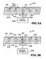
- FIGS. 3A and 3 billustrate two embodiments of lysising devices.
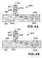
- FIGS. 4A and 4Billustrate systems and methods of forming droplets.
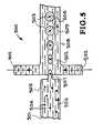
- FIG. 5shows another system and method of forming droplets.
- FIGS. 6A and 6Bshow systems and methods of shunting rejected droplets to waste.
- FIG. 7illustrates a system and method for directing droplets into the analyzer channels.
- FIGS. 8A and 8Billustrate another system and method for directing droplets into the analyzer channels.
- FIGS. 9A and 9Billustrate two systems and methods of amplifying the nucleic acid into the droplets using PCR.
- FIG. 1one embodiment of an apparatus for identifying all of the known and unknown pathogenic or non-pathogenic organisms in a sample wherein the organisms include nucleic acids is illustrated.
- the apparatusis designated generally by the reference numeral 100 .
- the apparatus 100identifies substantially all of the known and unknown pathogenic or non-pathogenic organisms in the sample.
- the apparatus 100provides capillary electrophoresis (CE) lanes 111 on a chip 113 .
- a sample 101is directed into the apparatus 100 .
- the sample 101contains known and unknown pathogenic or non-pathogenic organisms 105 .
- the sample 101is prepared to contain primers, probes, and dNTPs.
- An emulsifier 102is added to the sample 101 .
- the portion of the chip 113 wherein the sample 101 and the emulsifier 102 come togetherforms a droplet maker 103 .
- the sample 101 and the emulsifier 102are injected into the flow channels generating the droplet which constitute isolated mobile PCR reactors.
- the droplet maker 103creates droplets 104 from the sample 101 .
- the droplets 104constitute sub-nanoliter volume reactors containing organism sized particles 105 .
- the droplets 104are created within emulsified shells by forcing them through an appropriately sized mechanical orifice in the droplet maker 103 . This may be accomplished using microfluidic T-junctions, microjet, inkjet, pin systems, or other ways of creating droplets.
- a device 106provides lysis of the organisms 105 to release the nucleic acids 107 .
- the lysis device 106is an optical window for delivering light. Additionally, chemical agents in the droplets may also be used to induce lysis.
- Individual droplets 104are irradiated to lyse cells using the optical window in the lysis device 106 . Lysis of the organisms 105 releases the nucleic acids 107 . Lysis can also be performed by droplet heating, in which case the optical window may be replaced by a resistive, conductive, or radiative heating element.
- An amplifier 108amplifies the organisms 105 .
- the nucleic acids 107have been released from the organisms 105 and the nucleic acids 107 are amplified using the amplifier 108 .
- the amplifier 108can be a thermocycler.
- the nucleic acids 107can be amplified in-line before arraying them. As amplification occurs, detection of fluorescence-labeled TaqMan type probes occurs if desired. Following amplification, the system does not need decontamination due to the isolation of the chemical reactants.
- a droplet selector 109identifies droplets with amplified nucleic acid and directs them to further analysis while allowing empty droplets to pass to waste 112 . Amplified droplets then release their nucleic acids 110 into the analysis channels of the parallel analyzer 111 .
- the organisms 105are arranged for parallel analysis in the parallel analyzer 111 .
- Selected droplets 104may be assigned to one of the many available CE channels 111 for electrophoretic separation characterization. Voltage actuation of channel electrodes and acoustic, magnetic, or optical actuation may be employed to force the droplets 104 into the analysis channel 111 .
- overlaid high pressure padsmay combine with electrostatic potential to force the droplets 104 into the CE channel 111 for characterization if the device substrate is PDMS or another suitable polymer.
- Wasteis directed to the waste reservoir 112 . All organisms 105 in the sample 101 are analyzed by the apparatus 100 . This can be accomplished by a genomic analyzer and/or a parallel physical/proteomic analyzer.
- the apparatus 100is manufactured by different processes.
- the apparatus 100is manufactured by a photolithography process utilizing a wet etch in glass or borosilicate of the bottom and top layers which are then aligned and bonded together. Individual devices are then cut from the bonded wafers on the diamond saw.
- the apparatus 100is manufactured by a photolithography process utilizing a front and backside Deep Reactive Ion Etch (DRIE) process where the front side of a Si wafer contains the microfluidic channels and the back side etch creates the fluid vias to connect to the channel.
- DRIEDeep Reactive Ion Etch
- the apparatus 100is manufactured by a lithography process where SU-8 photoresist is patterned into a positive-relief of the channel architecture using the standard photolithography process. This patterned structure then becomes a mold for the addition of liquid Polydimethylsiloxane (PDMS) which is flowed over the SU-8 and cured. The elastomeric, cured PDMS is then pulled from the mold. A glass coverslip is spin coated with a small layer of PDMS and cured. The 2 layers (glass plus PDMS with channels) are then brought together and cured such that the PDMS forms a complete seal around the channel geometry. Fluidic ports are then cored out of the polymeric PDMS.
- PDMSPolydimethylsiloxane
- the method of operation of the apparatus 100includes a series of steps.
- the sample 101is processed to isolate organism sized particles.
- the sample 101is also processed by adding primers, probes, and dNTPs.
- An emulsifier 102is added to the sample 101 .
- the portion of the apparatus 100 wherein the sample 101 and the emulsifier 102 are injectedforms a droplet maker 103 .
- the droplet maker 103creates droplets 104 from the sample 101 wherein the droplets 104 constitute sub-nanoliter volume reactors containing organism sized particles 105 .
- Lysis 106 of the organisms 105releases the nucleic acids 107 .
- the organisms 105are amplified by amplifier 106 .
- the nucleic acids 107have been released from the organisms and the nucleic acids 107 are amplified using amplification techniques.
- the droplets 104are fractionated or formatted by the droplet splitter 103 to release the amplified nucleic acids 107 . This can be accomplished by releasing amplified nucleic acids 107 from each droplet 104 or by dissolving/disrupting the emulsification shells of the droplets 104 .
- the organisms 105are arranged for parallel analysis. This is accomplished by the parallel analyzer 111 . All organisms 105 in the sample 101 are analyzed. This accomplished by a genomic analyzer and/or a physical/proteomic analyzer.
- the apparatus 100can be used in clinical applications for identification of known and unknown respiratory illnesses, unknown causes of death, drug efficacy testing, and other identification.
- the apparatus 100can be used in medical surveillance for identification of new and emerging infectious disease such as SARS.
- the apparatus 100can be used for identification of genetically modified biological threats.
- the apparatus 100can also be used for identification of environmental biological background characterization for planning, response, forensics, and attribution.
- FIG. 2another embodiment of an apparatus for identifying all of the known and unknown pathogenic or non-pathogenic organisms in a sample wherein nucleic acid from the organisms is illustrated.
- the apparatusis designated generally by the reference numeral 200 .
- the apparatus 200identifies substantially all of the known and unknown pathogenic or non-pathogenic organisms in the sample.
- the apparatus 200utilizes micro-channels 211 , 213 , and 214 in a chip 212 .
- the micro-channels 213 and 211provide a flow circuit.
- the micro-channels 213provide initial processing and the capillary electrophoresis (CE) lanes 211 and 214 provide analysis.
- CEcapillary electrophoresis
- a complex environmental or clinical sample 201is prepared using known physical (ultracentrifugation, filtering, diffusion separation, electrophoresis, cytometry etc.), chemical (pH), and biological (selective enzymatic degradation) techniques to extract and separate target nucleic acids or intact individual particles 205 (e.g., virus particles) from background (i.e., intra- and extra-cellular RNA/DNA from host cells, pollen, dust, etc.).
- This samplecontaining relatively purified nucleic acid or particles containing nucleic acids (e.g., viruses), can be split into multiple parallel channels and mixed with appropriate reagents required for reverse transcription and subsequent PCR (primers/probes/dNTPs/enzymes/buffer).
- each of these mixesare then introduced into the system in such a way that statistically no more than a single RNA/DNA is present in any given microreactor. For example, a sample containing 10 6 target RNA/DNA would require millions of microreators to ensure single RNA/DNA distribution.
- the sample 201is directed into the apparatus 200 .
- the sample 201contains known and unknown pathogenic or non-pathogenic organisms 205 .
- An emulsifier 202is added to the sample 201 .
- the apparatus 200utilizes a Microreactor Generator System (MGS).
- MGSMicroreactor Generator System
- the MGS systemperforms analyte mixing and injection, sample isolation, and system decontamination functions.
- a hydrophobic carrier fluidtypically a syringe pump
- a fluid propulsion and metering devicetypically a syringe pump
- a fluidic channel with a T or cross junctionforcing the dispersion of the analyte and reagent aqueous solution into the hydrophobic carrier fluid
- a multi-port selection valvefor channel priming
- a variable width main channelfor controlling droplet spacing and velocity.
- the pumpis used to draw and pump fluids through the flow circuit.
- the hydrophobic carrier fluidprovides the medium for translating the pump movements into fluid motion and for creating the spherical droplets that serve as the micron-scale reactors. This occurs due to the immiscibility of the hydrophilic droplets within the hydrophobic flow, as the sheared aqueous fluid relaxes into a spherical form to minimize surface tension (by minimizing surface area). Continuous flow of both the hydrophobic carrier fluid and the aqueous reagent fluid ensures both the production and separation of the microscale reactors, eliminating the chance of cross-contamination.
- Dhis the hydraulic radius of the channel at the junction
- Q 0is the volumetric flow rate in m3/s
- ⁇is the surface tension in kg/s2
- ⁇is the viscosity in kg/(m*s).
- the aqueous inlet channelserves to mix various assay components (i.e., analyte, oligonucleotides, primer, probe, enzymes etc.) in preparation for amplification and detection. This prevents contamination of the syringe pump, and is easily decontaminated by rinsing with buffer.
- the channel geometryallows for dividing the sample into multiple aliquots for subsequent analysis serially or in parallel with multiple streams. The scalability of the architecture allows for multiple different reactions to be tested against aliquots from the same sample. Decontamination by flushing the channels dilute solution of sodium hypochlorite, followed by deionized water could also be used.
- the portion of the chip 212 wherein the sample 201 and the emulsifier 202 come togetherforms a droplet generator or droplet maker 203 .
- the sample 201 and the emulsifier 202are injected into the flow channels generating the droplets which constitute isolated mobile PCR reactors.
- the droplet maker 203creates droplets 204 from the sample 201 .
- the droplets 204constitute sub-nanoliter volume reactors containing organism sized particles 206 .
- the droplets 204are created within emulsified shells by forcing them through an appropriately sized mechanical orifice in the droplet maker 203 . This may be accomplished using microfluidic T-junctions, microjet, inkjet, pin systems, or other ways of creating droplets.
- An amplifier 207provides Nucleic Acid Amplification. This may be accomplished by the Polymerase Chain Reaction (PCR) process, an exponential process whereby the amount of target DNA is doubled through each reaction cycle utilizing a polymerase enzyme, excess nucleic acid bases, primers, catalysts (MgCl2), etc.
- the reactionis powered by cycling the temperature from an annealing temperature whereby the primers bind to single-stranded DNA (ssDNA) through an extension temperature whereby the polymerase extends from the primer, adding nucleic acid bases until the complement strand is complete, to the melt temperature whereby the newly-created double-stranded DNA (dsDNA) is denatured into 2 separate strands.
- PCRPolymerase Chain Reaction
- the amplifier 207amplifies the organisms 206 .
- The-nucleic acids 208have been released from the organisms 206 and the nucleic acids 208 are amplified using the amplifier 207 .
- the amplifier 207can be a thermocycler.
- the nucleic acids 208can be amplified in-line before arraying them. As amplification occurs, detection of fluorescence-labeled TaqMan type probes occurs if desired. Following amplification, the system does not need decontamination due to the isolation of the chemical reactants.
- the proposed sorting systemwill advantageously only select the droplets that have a sufficient quantity of post-amplified nucleic acid material to characterize. This is performed by the interrogation of each droplet by an orthogonal laser beam or LED, to excite fluorescent reporters supplied to each droplet in the reagent mix. This could be an intercalating dye that only fluoresces when bound to double stranded nucleic acids such as segments of PCR product.
- the fluorescent reportercould also be a Taqman type FRET probe. A detector senses the fluorescence if applicable and reports to the controller the presence of a “hot” droplet.
- This droplet, moving along the centerline of the flow channelis then selected for characterization by capillary electrophoresis and/or archival.
- Other types of luminescence techniquescould be used for optical droplet discrimination, including chemiluminescence or bioluminescence which do not require an external excitation source simplifying instrumentation design and have inherently low back-ground emission for highly sensitive detection.
- Addition of this droplet selector component 209greatly simplifies the design of the instrument, since it greatly reduces the number of parallel capillary electrophoresis or electrophoresis channels that are necessary to characterize the selected amplicons.
- droplets selected for electrophoresiswill be sorted to the electrophoresis channel by optical trapping while the “empty” droplets move on to waste.
- droplets selected for electrophoresiswill be sorted to the electrophoresis channel by pneumatic valve actuation to transfer the droplet to another channel while the rest of the droplets continue to waste.
- droplets selected for electrophoresiswill be sorted to the electrophoresis channel by magnetic attraction to transfer the droplet to another channel while the rest of the droplets continue to waste 210 .
- droplets selected for electrophoresiswill be sorted to the electrophoresis channel by acoustic pressure from a piezoelectric transducer to transfer the droplet to another channel while the rest of the droplets continue to waste.
- the apparatus 200employs an optical window (described subsequently) to the flow channels 213 , 211 , and 214 to provide for detection and analysis of the droplet contents in the parallel analyzer such as capillary electrophoresis.
- a fractionater or droplet splitterreleases the amplified nucleic acids by opening the droplets and releasing the amplified nucleic acids.
- the organismsare arranged for parallel analysis in the parallel analyzer. Selected droplets may be assigned to one of the many available CE channels for electrophoretic characterization. Capillary electrophoresis is provided under the relation:
- Voltage actuation of channel electrodes and acoustic, magnetic, or optical actuationmay be employed to force the droplets into the CE channel.
- overlaid high pressure padsmay combine with electrostatic potential to force the droplets into the CE channel for characterization.
- Wasteis directed to the waste reservoir. All organisms in the sample are analyzed by the apparatus. This can be accomplished by a genomic analyzer and/or a parallel physical/proteomic analyzer.
- real-time detection of amplified nucleic acid sequencesis accomplished using optical-based assays that either increase or decrease the emission from fluorescence-labeled probes during each amplification step.
- One technique for real-time PCRis TaqMan, a homogeneous PCR test that uses a fluorescence resonance energy transfer probe. This probe typically contains a “reporter” dye at the 5′ end and a “quencher” dye at the 3′ end. Intact, there is very little fluorescent emission from the probe, since the proximity of the quencher to the reporter dye serves to suppress the reporter emission.
- the probeanneals to a targeted complementary amplicon strand and begins extending one of the primers.
- An enzyme(Taq polymerase) cleaves the probe and displaces both dye molecules, allowing them to separate and diffuse into the surrounding fluid. The resulting increase in reporter emission can be monitored and correlated PCR product concentration.
- This apparatus 200provides for nucleic acid characterization for novel or unknown viruses and bacteria by microcapillary, capillary, or gel electrophoresis due to the ability to interface with an electrophoresis system.
- the apparatus 200maintains the presence of an array of selectable, independently programmable capillary electrophoresis (CE) lanes on the chip or orthogonal to it running perpendicular to the main channel flow.
- CEcapillary electrophoresis
- flowcan be slowed and an electric potential fired on the CE electrodes causing migration of the droplet of interest through the port in the main flow channel and into the CE channel.
- the dropletcan be captured by electrostatic attraction alone.
- a combination of electrostatic attraction and mechanical actuationcan be combined to capture individual droplets.
- the dropletmay be bifurcated prior to CE channel entrance to allow for a fraction of the droplet to be carried downstream to an archival aspiration port.
- a combination of electrostatic and magnetic forcemay be employed to move the droplets into the CE channels.
- a combination of acoustic pressure from piezoelectric transducers and electrostatic attractionmay be used to move the droplets into the channel.
- a combination of optical pressure from an integrated optical trapmay be used to with electrostatic force to move droplets into the channel.
- An applied potential field in the electrophoresis channelsattracts the nucleic acid fragments and separates them according to their charge to size ratio due to the presence of an appropriate molecular sieve.
- the sieveacts to retard the nucleic acid flow. Because of this action the differing lengths of nucleic acids become separated into bands as they migrate with solvent ions along the electrophoresis channel.
- This artdescribes a system that will then image the CE channels to detect the fluorescence of tagged nucleic acid bands as they migrate down the channels.
- the systemcontains multiple CE channels in parallel with a charge coupled device (CCD) imaging system detecting the banding patterns.
- CCDcharge coupled device
- nucleic acid “Ladders”sequences of different lengths that vary by a constant number of bases.
- Laddersnucleic acid sequences of different lengths that vary by a constant number of bases.
- Characterizing the products generated by the polymerase chain reactioncan give information about the target genome that was amplified.
- the PCR reactioncan be designed to generate specific products, or amplicons, with distinct sizes (i.e., lengths, number of bases).
- Electrophoresiscan be used to separate PCR products according to size. It is important to have size reference standards that can be used for calibrating the electrophoresis process.
- DNA ladders or size reference standardscan be incorporated into individual droplets and transported to the electrophoresis system. They could also be directly injected into the electrophoresis system.
- a synthetic virus construct such as armored RNAcan be used as an end-to-end system control and would very closely mimic the behavior of real virus or biological particle that could be present in the sample. It can be spiked to the sample or added in line.
- the controlwould provide information of sample addition, mixing, droplet formation, reagent addition, extraction, sample purity, sample preparation, particle lysis, reverse transcription, PCR amplification and detection.
- the controlcould have its own set of PCR primers and could either co-exist in a droplet with the target or in its own droplet.
- the PCR primers for the controlcan be designed to generate products that have distinct sizes that cover the range and resolution required to identify and characterize electropherograms from targets, essentially generating size ladders or reference standards in situ.
- the sequence target used for calibrationcan be made synthetically so that the products can be used as sequencing controls or other down stream characterization processes.
- the controlcan also yield information regarding any loss of specificity or sensitivity of the device.
- dropletscan be barcoded and tracked as they are transported throughout each module of the system. Barcoding can be done with particles, such as beads, crystals, and identified using fluorescence, spectral signature or other unique signature identifiers. Barcodes can be made from unique combinations of particles, or an array of uniquely identifiable particles. Their size could be tailored (micrometers to nanometers) and the materials can be inert so as not to affect performance of the system or the assays. If droplets need to be manipulated, such as split one droplet into 2, the identity of the original droplet can be tracked and correlated with results from different (parallel) detection platforms.
- the lysis deviceprovides lysis of the organisms to release the nucleic acids.
- the embodiments of FIGS. 3A and 3Bwill be described in greater detail.
- the lysis device 300includes a micro-channel 301 with fluid 302 that provides a flow circuit flowing in the direction indicated by the arrow 303 .
- a complex environmental or clinical samplehas been formed into individual droplets 304 which constitute isolated mobile PCR reactors.
- the sample droplets 304contain known and unknown pathogenic or non-pathogenic organisms 305 and a fluid 306 .
- the device 300provides lysis of the organisms 305 to release the nucleic acids.
- the lysis device 300includes an optical window 307 .
- a light source 308such as a laser produces a light or laser beam 309 that is directed through the optical window 307 .
- the light or laser beam 309directs electromagnetic radiation to generate a plasma that creates a shock wave inside the droplets 304 sufficient to lyse the bacterial cell wall or protein capsids, releasing target nucleic acids (RNA and DNA) 310 within the droplets 304 .
- the individual droplets 304are irradiated to lyse cells using the optical window 307 in the lysis device 300 .
- Lysis of the organisms 305releases the nucleic acids 310 .
- Lysis of the organisms 305can also be achieved by radiative heating from the laser 308 and laser beam 309 .
- Lysis of the organisms 305can also be achieved using ultrasound-generating piezoelectric actuators in place of the laser 308 to focus acoustic pressure on the cell walls. Lysing is necessary to make the nucleic acids accessible to the reagents used for amplification and or detection.
- the lysis device 311includes a micro-channel 312 with fluid 313 that provides a flow circuit flowing in the direction indicated by the arrow 314 .
- a complex environmental or clinical samplehas been formed into individual droplets 315 which constitute isolated mobile PCR reactors.
- the sample droplets 304contain known and unknown pathogenic or non-pathogenic organisms 316 and a fluid 317 .
- the device 311provides lysis of the organisms 316 to release the nucleic acids.
- the lysis device 311includes a window 318 . Lysis of the droplets 315 is achieved using an ultrasound-generating piezoelectric actuator or power source 319 to focus acoustic pressure 320 on the cell walls 321 . Lysing is necessary to make the nucleic acids 322 accessible to the reagents used for amplification and or detection.
- Resistive or conductive heatingmay also be used to lyse the organisms 316 .
- a resistive heater 318 or Peltier device 318is used to heat the droplets 315 , instead of using a piezoelectric actuator. Lysing is necessary to make the nucleic acids 322 accessible to the reagents used for amplification and or detection.
- the method of operation of the apparatusincludes a series of steps. In the first step the sample is processed to isolate organism sized particles. The sample is also processed by adding primers, probes, and dNTPs. An emulsifier is added to the sample.
- the portion of the apparatus wherein the sample and the emulsifier are injectedforms a droplet generator.
- the droplet makercreates droplets from the sample wherein the droplets constitute sub-nanoliter volume reactors containing organism sized particles. Lysis of the organisms releases the nucleic acids.
- the organismsare amplified by amplifier. The nucleic acids have been released from the organisms and the nucleic acids are amplified using amplification techniques.
- the dropletsare fractionated or formatted to release the amplified nucleic acids. This can be accomplished by releasing amplified nucleic acids from each droplet or by dissolving/disrupting the emulsification shells of the droplets.
- the organismsare arranged for parallel analysis. This is accomplished by the parallel analyzer. All organisms in the sample are analyzed. This accomplished by a genomic analyzer and/or a physical/proteomic analyzer.
- the apparatuscan be used in clinical applications for identification of unknown respiratory illnesses, unknown causes of death, drug efficacy testing, and other identification.
- the apparatuscan be used in medical surveillance for identification of new and emerging infectious disease such as SARS.
- the apparatuscan be used for identification of genetically modified biological threats.
- the apparatuscan also be used for identification of environmental biological background characterization for planning, response, forensics, and attribution.
- FIGS. 4A and 4Ba system and method of forming droplets is illustrated.
- FIGS. 4A and 4Billustrates the portion of the chip wherein the sample 401 and the emulsifier 402 come together to form a droplet generator or droplet maker.
- the droplet generator or droplet makeris designated generally by the reference numeral 400 .
- the sample 401 and the emulsifier 402generate droplets 403 which constitute isolated mobile PCR reactors.
- the droplet maker 400creates the droplets 403 from the sample 401 .
- the droplets 403constitute sub-nanoliter volume reactors containing organism sized particles 404 .
- the emulsifier 402flows in a flow channel 405 in the direction indicated by the arrow 406 .
- the sample 401is directed into the flow channel 405 by the droplet maker 400 by a sample channel 407 .
- the organism sized particles 404are carried in the sample channel 407 by a fluid 404 .
- the droplet maker 400creates the droplets 403 from the sample 401 and the droplets contain the organism sized particles 404 .
- the droplets 403constitute isolated mobile PCR reactors.
- aqueous solution 408 containing a pathogen or organism 404is drawn into the flow of emulsifier 402 and eventually breaks free of the sample channel 407 .
- the aqueous solution 408 containing a pathogen or organism 404becomes a spherical droplet 403 as it is carried in the flow channel 405 .
- the droplet 403As shown in FIG. 4B , after the droplet 403 has been formed it is entrained in the flow of emulsifier 402 .
- the aqueous solution 408 in the sample channel 407is ready to start forming another droplet.
- FIG. 5another embodiment of a system and method of forming droplets is illustrated.
- This embodimentis designated generally by the reference numeral 500 .
- FIG. 5illustrates the portion of the chip wherein the sample 501 and the emulsifier 502 come together to form the droplet generator or droplet maker 500 .
- the sample 501 and the emulsifier 502generate droplets 503 which constitute isolated mobile PCR reactors.
- the droplet maker 500creates the droplets 503 from the sample 501 .
- the droplets 503constitute sub-nanoliter volume reactors containing organism sized particles 504 .
- the aqueous solution 505 containing pathogens 504flows in a sample channel 505 as indicated by the arrow 506 .
- the aqueous solution 505 containing the organisms or pathogens 504is forced through an orifice 507 .
- a portion of the aqueous solution 505 containing the organisms or pathogens 504breaks off and eventually becomes a spherical droplet 503 as it is carried in the flow channel 508 .
- the droplet 503After the droplet 503 has been formed it is entrained in the flow of emulsifier 502 as indicated by the arrow 509 .
- the droplet maker 500creates the droplets 503 from the sample 501 and the droplets contain the organism sized particles 504 .
- the droplets 503constitute isolated mobile PCR reactors.
- FIGS. 6A and 6Ba system and method of shunting rejected droplets to waste is illustrated.
- a micro-channel 690 with droplets 604is illustrated. Some of the droplets contain nucleic acid 604 and some do not contain the nucleic acid.
- the droplets 604are entrained in the emulsifier 602 and flow in the direction of arrow 614 .
- a detector/controller 630determines which droplets are to be shunted to waste 612 .
- FIG. 6Bshow that a droplet that does not contain any nucleic acid has been detected.
- the detector/controller 620opens the valves 626 and 627 and a puff from the pressure source 624 sends the rejected droplet into a conduit that leads to the waste receptacle 612 .
- a micro-channel 790 with emulsifier 702 and entrained droplets of nucleic acidmoves in the direction of arrow 714 .
- a power supply 740 and circuit 742 with contacts 746create a field that causes the droplets to move in the direction of arrow 715 and into the analyzer channels 711 .
- FIG. 8Ashows micro-channel 890 with droplets 804 entrained in emulsifier 802 moving slowly in direction of arrow 814 .
- a pressure supply 840is shown with conduits connected to flexible areas 850 of the micro-channel covering.
- valve 826When valve 826 is opened, pressure distorts the microchannel covering and the droplets 804 are forced into the analyzer channels 811 where the droplets can be dissolved to release the nucleic acid.
- FIGS. 9A and 9Btwo systems and methods of amplifying the nucleic acid into the droplets using PCR are illustrated. Part of the PCR reaction is to alternately heat and cool the material to be replicated so in FIGS. 9A and 9B methods and systems for heating and cooling the droplets are illustrated.
- FIG. 9Ashows droplets 904 in emulsifier 902 traveling in the direction of arrow 914 while in micro-channel 990 the droplets will pass through alternate heating 964 and cooling 966 zones.
- a power supply 960provides power to the resistance heater and cooling zones. This can be accomplished with a cooling liquid or gas.
- FIG. 9Bshows the droplets passing by thermo-electric units 972 which are powered and controlled by unit 968 and circuit 970 . Thermo-electric units are Peltier devices that can both heat and cool.
Landscapes
- Health & Medical Sciences (AREA)
- Chemical & Material Sciences (AREA)
- Life Sciences & Earth Sciences (AREA)
- Engineering & Computer Science (AREA)
- Immunology (AREA)
- Analytical Chemistry (AREA)
- Molecular Biology (AREA)
- Hematology (AREA)
- General Health & Medical Sciences (AREA)
- Biomedical Technology (AREA)
- Physics & Mathematics (AREA)
- Organic Chemistry (AREA)
- Urology & Nephrology (AREA)
- Biochemistry (AREA)
- Proteomics, Peptides & Aminoacids (AREA)
- Microbiology (AREA)
- Biotechnology (AREA)
- Zoology (AREA)
- Wood Science & Technology (AREA)
- Dispersion Chemistry (AREA)
- Medicinal Chemistry (AREA)
- Pathology (AREA)
- General Physics & Mathematics (AREA)
- Food Science & Technology (AREA)
- Cell Biology (AREA)
- Chemical Kinetics & Catalysis (AREA)
- Clinical Laboratory Science (AREA)
- Biophysics (AREA)
- Bioinformatics & Cheminformatics (AREA)
- General Engineering & Computer Science (AREA)
- Genetics & Genomics (AREA)
- Toxicology (AREA)
- Tropical Medicine & Parasitology (AREA)
- Virology (AREA)
- Nanotechnology (AREA)
- Fluid Mechanics (AREA)
- Measuring Or Testing Involving Enzymes Or Micro-Organisms (AREA)
- Apparatus Associated With Microorganisms And Enzymes (AREA)
Abstract
Description
Q=hA(Twall−T∞)
The
The
Claims (28)
Priority Applications (3)
| Application Number | Priority Date | Filing Date | Title |
|---|---|---|---|
| US11/650,363US8338166B2 (en) | 2007-01-04 | 2007-01-04 | Sorting, amplification, detection, and identification of nucleic acid subsequences in a complex mixture |
| PCT/US2007/010748WO2008082432A1 (en) | 2007-01-04 | 2007-05-01 | Sorting, amplification, detection, and identification of nucleic acid subsequences in a complex mixture |
| US11/818,623US9422586B2 (en) | 2007-01-04 | 2007-06-15 | Method for genetic identification of unknown organisms |
Applications Claiming Priority (1)
| Application Number | Priority Date | Filing Date | Title |
|---|---|---|---|
| US11/650,363US8338166B2 (en) | 2007-01-04 | 2007-01-04 | Sorting, amplification, detection, and identification of nucleic acid subsequences in a complex mixture |
Publications (2)
| Publication Number | Publication Date |
|---|---|
| US20080166793A1 US20080166793A1 (en) | 2008-07-10 |
| US8338166B2true US8338166B2 (en) | 2012-12-25 |
Family
ID=38556331
Family Applications (2)
| Application Number | Title | Priority Date | Filing Date |
|---|---|---|---|
| US11/650,363Expired - Fee RelatedUS8338166B2 (en) | 2007-01-04 | 2007-01-04 | Sorting, amplification, detection, and identification of nucleic acid subsequences in a complex mixture |
| US11/818,623Active2034-01-10US9422586B2 (en) | 2007-01-04 | 2007-06-15 | Method for genetic identification of unknown organisms |
Family Applications After (1)
| Application Number | Title | Priority Date | Filing Date |
|---|---|---|---|
| US11/818,623Active2034-01-10US9422586B2 (en) | 2007-01-04 | 2007-06-15 | Method for genetic identification of unknown organisms |
Country Status (2)
| Country | Link |
|---|---|
| US (2) | US8338166B2 (en) |
| WO (1) | WO2008082432A1 (en) |
Cited By (13)
| Publication number | Priority date | Publication date | Assignee | Title |
|---|---|---|---|---|
| US20130037149A1 (en)* | 2010-04-09 | 2013-02-14 | The Hong Kong University Of Science And Technology | Liquid-electronic hybrid divider |
| US9222623B2 (en) | 2013-03-15 | 2015-12-29 | Genmark Diagnostics, Inc. | Devices and methods for manipulating deformable fluid vessels |
| US20160129445A1 (en)* | 2014-11-11 | 2016-05-12 | Genmark Diagnostics, Inc. | Instrument for processing cartridge for performing assays in a closed sample preparation and reaction system |
| US20160175835A1 (en)* | 2013-07-29 | 2016-06-23 | Atlas Genetics Limited | Fluidic cartridge and method for processing a liquid sample |
| US9598722B2 (en) | 2014-11-11 | 2017-03-21 | Genmark Diagnostics, Inc. | Cartridge for performing assays in a closed sample preparation and reaction system |
| US9957553B2 (en) | 2012-10-24 | 2018-05-01 | Genmark Diagnostics, Inc. | Integrated multiplex target analysis |
| US10005080B2 (en) | 2014-11-11 | 2018-06-26 | Genmark Diagnostics, Inc. | Instrument and cartridge for performing assays in a closed sample preparation and reaction system employing electrowetting fluid manipulation |
| US10227650B2 (en) | 2014-11-14 | 2019-03-12 | Athena Diagnostics, Inc. | Methods to detect a silent carrier of a null allele genotype |
| US10260111B1 (en) | 2014-01-20 | 2019-04-16 | Brett Eric Etchebarne | Method of detecting sepsis-related microorganisms and detecting antibiotic-resistant sepsis-related microorganisms in a fluid sample |
| US10495656B2 (en) | 2012-10-24 | 2019-12-03 | Genmark Diagnostics, Inc. | Integrated multiplex target analysis |
| USD881409S1 (en) | 2013-10-24 | 2020-04-14 | Genmark Diagnostics, Inc. | Biochip cartridge |
| US11358137B2 (en) | 2018-12-26 | 2022-06-14 | Industrial Technology Research Institute | Tubular structure for producing droplets and method for producing droplets |
| US12339477B2 (en)* | 2020-05-26 | 2025-06-24 | Lawrence Livermore National Security, Llc | Amorphous or nanocrystalline molybdenum nitride and silicon nitride multilayers |
Families Citing this family (182)
| Publication number | Priority date | Publication date | Assignee | Title |
|---|---|---|---|---|
| US6753161B2 (en)* | 1997-03-27 | 2004-06-22 | Oncosis Llc | Optoinjection methods |
| EP2281879B1 (en) | 1999-11-30 | 2016-01-27 | Intrexon Corporation | Apparatus for determining a morphological or physiological characteristic of individual cells in a biological specimen |
| US6804385B2 (en) | 2000-10-24 | 2004-10-12 | Oncosis | Method and device for selectively targeting cells within a three-dimensional specimen |
| US7041481B2 (en) | 2003-03-14 | 2006-05-09 | The Regents Of The University Of California | Chemical amplification based on fluid partitioning |
| US20100022414A1 (en) | 2008-07-18 | 2010-01-28 | Raindance Technologies, Inc. | Droplet Libraries |
| EP2407243B1 (en) | 2003-07-31 | 2020-04-22 | Handylab, Inc. | Multilayered microfluidic device |
| US9597644B2 (en)* | 2003-09-05 | 2017-03-21 | Stokes Bio Limited | Methods for culturing and analyzing cells |
| US20050095578A1 (en)* | 2003-10-31 | 2005-05-05 | Koller Manfred R. | Method and apparatus for cell permeabilization |
| US7425426B2 (en) | 2004-03-15 | 2008-09-16 | Cyntellect, Inc. | Methods for purification of cells based on product secretion |
| US7968287B2 (en) | 2004-10-08 | 2011-06-28 | Medical Research Council Harvard University | In vitro evolution in microfluidic systems |
| KR101198038B1 (en) | 2005-01-28 | 2012-11-06 | 듀크 유니버서티 | Apparatuses and methods for manipulating droplets on a printed circuit board |
| AU2006247752B2 (en) | 2005-05-11 | 2012-04-12 | Advanced Liquid Logic, Inc. | Method and device for conducting biochemical or chemical reactions at multiple temperatures |
| US20100137163A1 (en) | 2006-01-11 | 2010-06-03 | Link Darren R | Microfluidic Devices and Methods of Use in The Formation and Control of Nanoreactors |
| EP2298438A1 (en) | 2006-02-07 | 2011-03-23 | Stokes Bio Limited | A microfluidic droplet queuing network |
| WO2007112114A2 (en) | 2006-03-24 | 2007-10-04 | Handylab, Inc. | Integrated system for processing microfluidic samples, and method of using same |
| US9476856B2 (en) | 2006-04-13 | 2016-10-25 | Advanced Liquid Logic, Inc. | Droplet-based affinity assays |
| US20140193807A1 (en) | 2006-04-18 | 2014-07-10 | Advanced Liquid Logic, Inc. | Bead manipulation techniques |
| US8809068B2 (en) | 2006-04-18 | 2014-08-19 | Advanced Liquid Logic, Inc. | Manipulation of beads in droplets and methods for manipulating droplets |
| US7727723B2 (en) | 2006-04-18 | 2010-06-01 | Advanced Liquid Logic, Inc. | Droplet-based pyrosequencing |
| US7439014B2 (en) | 2006-04-18 | 2008-10-21 | Advanced Liquid Logic, Inc. | Droplet-based surface modification and washing |
| US10078078B2 (en) | 2006-04-18 | 2018-09-18 | Advanced Liquid Logic, Inc. | Bead incubation and washing on a droplet actuator |
| US8637324B2 (en) | 2006-04-18 | 2014-01-28 | Advanced Liquid Logic, Inc. | Bead incubation and washing on a droplet actuator |
| WO2009111769A2 (en) | 2008-03-07 | 2009-09-11 | Advanced Liquid Logic, Inc. | Reagent and sample preparation and loading on a fluidic device |
| ATE540750T1 (en) | 2006-05-11 | 2012-01-15 | Raindance Technologies Inc | MICROFLUIDIC DEVICE AND METHOD |
| US9562837B2 (en) | 2006-05-11 | 2017-02-07 | Raindance Technologies, Inc. | Systems for handling microfludic droplets |
| WO2008097559A2 (en) | 2007-02-06 | 2008-08-14 | Brandeis University | Manipulation of fluids and reactions in microfluidic systems |
| AU2008212808B2 (en) | 2007-02-09 | 2013-09-12 | Advanced Liquid Logic, Inc. | Droplet actuator devices and methods employing magnetic beads |
| WO2008109176A2 (en) | 2007-03-07 | 2008-09-12 | President And Fellows Of Harvard College | Assays and other reactions involving droplets |
| WO2008130623A1 (en) | 2007-04-19 | 2008-10-30 | Brandeis University | Manipulation of fluids, fluid components and reactions in microfluidic systems |
| US8951732B2 (en)* | 2007-06-22 | 2015-02-10 | Advanced Liquid Logic, Inc. | Droplet-based nucleic acid amplification in a temperature gradient |
| US9186677B2 (en) | 2007-07-13 | 2015-11-17 | Handylab, Inc. | Integrated apparatus for performing nucleic acid extraction and diagnostic testing on multiple biological samples |
| US8702938B2 (en) | 2007-09-04 | 2014-04-22 | Advanced Liquid Logic, Inc. | Droplet actuator with improved top substrate |
| EP2235210B1 (en) | 2007-12-21 | 2015-03-25 | President and Fellows of Harvard College | Methods for nucleic acid sequencing |
| CN101945767B (en) | 2007-12-23 | 2013-10-30 | 先进液体逻辑公司 | Droplet actuator configuration and method for directing droplet manipulation |
| US20090226971A1 (en)* | 2008-01-22 | 2009-09-10 | Neil Reginald Beer | Portable Rapid Microfluidic Thermal Cycler for Extremely Fast Nucleic Acid Amplification |
| US9170060B2 (en)* | 2008-01-22 | 2015-10-27 | Lawrence Livermore National Security, Llc | Rapid microfluidic thermal cycler for nucleic acid amplification |
| US9409177B2 (en)* | 2008-03-21 | 2016-08-09 | Lawrence Livermore National Security, Llc | Chip-based device for parallel sorting, amplification, detection, and identification of nucleic acid subsequences |
| US8969767B2 (en)* | 2008-03-21 | 2015-03-03 | Lawrence Livermore National Security, Llc | Microwave heating of aqueous samples on a micro-optical-electro-mechanical system |
| US8367976B2 (en)* | 2008-03-21 | 2013-02-05 | Lawrence Livermore National Security, Llc | Laser heating of aqueous samples on a micro-optical-electro-mechanical system |
| US9011777B2 (en) | 2008-03-21 | 2015-04-21 | Lawrence Livermore National Security, Llc | Monodisperse microdroplet generation and stopping without coalescence |
| WO2009137415A2 (en) | 2008-05-03 | 2009-11-12 | Advanced Liquid Logic, Inc. | Reagent and sample preparation, loading, and storage |
| EP2672259A1 (en)* | 2008-05-13 | 2013-12-11 | Advanced Liquid Logic, Inc. | Droplet actuator devices, systems and methods |
| US20110097763A1 (en)* | 2008-05-13 | 2011-04-28 | Advanced Liquid Logic, Inc. | Thermal Cycling Method |
| US12038438B2 (en) | 2008-07-18 | 2024-07-16 | Bio-Rad Laboratories, Inc. | Enzyme quantification |
| US10123380B2 (en) | 2008-08-08 | 2018-11-06 | Lawrence Livermore National Security, Llc | Instantaneous in-line heating of samples on a monolithic microwave integrated circuit microfluidic device |
| US8709762B2 (en) | 2010-03-02 | 2014-04-29 | Bio-Rad Laboratories, Inc. | System for hot-start amplification via a multiple emulsion |
| US9132394B2 (en) | 2008-09-23 | 2015-09-15 | Bio-Rad Laboratories, Inc. | System for detection of spaced droplets |
| US9492797B2 (en) | 2008-09-23 | 2016-11-15 | Bio-Rad Laboratories, Inc. | System for detection of spaced droplets |
| US9764322B2 (en) | 2008-09-23 | 2017-09-19 | Bio-Rad Laboratories, Inc. | System for generating droplets with pressure monitoring |
| US9417190B2 (en) | 2008-09-23 | 2016-08-16 | Bio-Rad Laboratories, Inc. | Calibrations and controls for droplet-based assays |
| US12162008B2 (en) | 2008-09-23 | 2024-12-10 | Bio-Rad Laboratories, Inc. | Partition-based method of analysis |
| US8663920B2 (en) | 2011-07-29 | 2014-03-04 | Bio-Rad Laboratories, Inc. | Library characterization by digital assay |
| US9156010B2 (en) | 2008-09-23 | 2015-10-13 | Bio-Rad Laboratories, Inc. | Droplet-based assay system |
| US10512910B2 (en) | 2008-09-23 | 2019-12-24 | Bio-Rad Laboratories, Inc. | Droplet-based analysis method |
| US8633015B2 (en) | 2008-09-23 | 2014-01-21 | Bio-Rad Laboratories, Inc. | Flow-based thermocycling system with thermoelectric cooler |
| US12090480B2 (en) | 2008-09-23 | 2024-09-17 | Bio-Rad Laboratories, Inc. | Partition-based method of analysis |
| US8951939B2 (en) | 2011-07-12 | 2015-02-10 | Bio-Rad Laboratories, Inc. | Digital assays with multiplexed detection of two or more targets in the same optical channel |
| US11130128B2 (en) | 2008-09-23 | 2021-09-28 | Bio-Rad Laboratories, Inc. | Detection method for a target nucleic acid |
| GB2477053B (en) | 2008-09-23 | 2013-11-13 | Quantalife Inc | Droplet-based assay system |
| US7993583B2 (en)* | 2008-10-29 | 2011-08-09 | Lawrence Livermore National Security, Llc | Passive microfluidic array card and reader |
| US8748094B2 (en) | 2008-12-19 | 2014-06-10 | President And Fellows Of Harvard College | Particle-assisted nucleic acid sequencing |
| CN102341505A (en)* | 2009-01-09 | 2012-02-01 | 英特拉克森公司 | Genetic analysis of cells |
| KR20110108390A (en) | 2009-01-12 | 2011-10-05 | 신텔렉트 인코포레이티드 | Laser Mediated Thin Film Incision and Migration of Cell Colonies |
| US10066977B2 (en)* | 2009-01-26 | 2018-09-04 | Canon U.S. Life Sciences, Inc. | Microfluidic flow monitoring |
| PT3299800T (en)* | 2009-03-16 | 2020-03-04 | Alere Tech Gmbh | Microfluidic device |
| EP3415235A1 (en) | 2009-03-23 | 2018-12-19 | Raindance Technologies Inc. | Manipulation of microfluidic droplets |
| US10196700B2 (en) | 2009-03-24 | 2019-02-05 | University Of Chicago | Multivolume devices, kits and related methods for quantification and detection of nucleic acids and other analytes |
| CN104722342B (en) | 2009-03-24 | 2017-01-11 | 芝加哥大学 | Slip chip device and method |
| US9464319B2 (en) | 2009-03-24 | 2016-10-11 | California Institute Of Technology | Multivolume devices, kits and related methods for quantification of nucleic acids and other analytes |
| US9447461B2 (en) | 2009-03-24 | 2016-09-20 | California Institute Of Technology | Analysis devices, kits, and related methods for digital quantification of nucleic acids and other analytes |
| US8926065B2 (en) | 2009-08-14 | 2015-01-06 | Advanced Liquid Logic, Inc. | Droplet actuator devices and methods |
| JP6155418B2 (en) | 2009-09-02 | 2017-07-05 | バイオ−ラッド・ラボラトリーズ・インコーポレーテッド | System for mixing fluids by combining multiple emulsions |
| US9625454B2 (en)* | 2009-09-04 | 2017-04-18 | The Research Foundation For The State University Of New York | Rapid and continuous analyte processing in droplet microfluidic devices |
| EP3842150A1 (en)* | 2009-10-27 | 2021-06-30 | President and Fellows of Harvard College | Droplet creation techniques |
| US9091649B2 (en)* | 2009-11-06 | 2015-07-28 | Advanced Liquid Logic, Inc. | Integrated droplet actuator for gel; electrophoresis and molecular analysis |
| EP2516669B1 (en) | 2009-12-21 | 2016-10-12 | Advanced Liquid Logic, Inc. | Enzyme assays on a droplet actuator |
| CA2789425C (en)* | 2010-02-12 | 2020-04-28 | Raindance Technologies, Inc. | Digital analyte analysis with polymerase error correction |
| US9366632B2 (en) | 2010-02-12 | 2016-06-14 | Raindance Technologies, Inc. | Digital analyte analysis |
| US10351905B2 (en) | 2010-02-12 | 2019-07-16 | Bio-Rad Laboratories, Inc. | Digital analyte analysis |
| US9399797B2 (en) | 2010-02-12 | 2016-07-26 | Raindance Technologies, Inc. | Digital analyte analysis |
| US8399198B2 (en) | 2010-03-02 | 2013-03-19 | Bio-Rad Laboratories, Inc. | Assays with droplets transformed into capsules |
| EP2556170A4 (en) | 2010-03-25 | 2014-01-01 | Quantalife Inc | Droplet transport system for detection |
| CA2767182C (en) | 2010-03-25 | 2020-03-24 | Bio-Rad Laboratories, Inc. | Droplet generation for droplet-based assays |
| EP2550351A4 (en) | 2010-03-25 | 2014-07-09 | Quantalife Inc | Detection system for droplet-based assays |
| WO2012009320A2 (en)* | 2010-07-15 | 2012-01-19 | Advanced Liquid Logic, Inc. | Systems for and methods of promoting cell lysis in droplet actuators |
| CA2807552A1 (en) | 2010-08-06 | 2012-02-09 | Moderna Therapeutics, Inc. | Engineered nucleic acids and methods of use thereof |
| JP6059145B2 (en)* | 2010-08-31 | 2017-01-11 | キヤノン ユー.エス. ライフ サイエンシズ, インコーポレイテッドCanon U.S. Life Sciences, Inc. | Slag control during thermal circulation |
| EP2622103B2 (en) | 2010-09-30 | 2022-11-16 | Bio-Rad Laboratories, Inc. | Sandwich assays in droplets |
| PL4108671T3 (en) | 2010-10-01 | 2025-02-24 | Modernatx, Inc. | MODIFIED NUCLEOSIDES, NUCLEOTIDES AND NUCLEIC ACIDS AND THEIR USES |
| US8720209B1 (en) | 2010-10-06 | 2014-05-13 | Lawrence Livermore National Security, Llc | Solid state rapid thermocycling |
| CA3215088A1 (en) | 2010-11-01 | 2012-05-10 | Bio-Rad Laboratories, Inc. | System for forming emulsions |
| US20120164633A1 (en)* | 2010-12-27 | 2012-06-28 | Ibis Biosciences, Inc. | Digital droplet sequencing |
| US8765455B2 (en) | 2011-01-27 | 2014-07-01 | Lawrence Livermore National Security, Llc | Chip-based droplet sorting |
| WO2012109600A2 (en) | 2011-02-11 | 2012-08-16 | Raindance Technologies, Inc. | Methods for forming mixed droplets |
| US12097495B2 (en) | 2011-02-18 | 2024-09-24 | Bio-Rad Laboratories, Inc. | Methods and compositions for detecting genetic material |
| EP2675819B1 (en) | 2011-02-18 | 2020-04-08 | Bio-Rad Laboratories, Inc. | Compositions and methods for molecular labeling |
| EP2686449B1 (en) | 2011-03-18 | 2020-11-18 | Bio-Rad Laboratories, Inc. | Multiplexed digital assays with combinatorial use of signals |
| DE12722942T1 (en) | 2011-03-31 | 2021-09-30 | Modernatx, Inc. | RELEASE AND FORMULATION OF MANIPULATED NUCLEIC ACIDS |
| DE102011016059B4 (en) | 2011-04-01 | 2019-02-21 | Friedrich-Schiller-Universität Jena | Method for correcting Raman spectroscopic data in a multiphase system |
| EP3395957B1 (en) | 2011-04-25 | 2020-08-12 | Bio-Rad Laboratories, Inc. | Methods and compositions for nucleic acid analysis |
| WO2012154745A2 (en) | 2011-05-09 | 2012-11-15 | Advanced Liquid Logic, Inc. | Microfluidic feedback using impedance detection |
| US8841071B2 (en)* | 2011-06-02 | 2014-09-23 | Raindance Technologies, Inc. | Sample multiplexing |
| EP2714970B1 (en) | 2011-06-02 | 2017-04-19 | Raindance Technologies, Inc. | Enzyme quantification |
| CA2840949A1 (en) | 2011-07-06 | 2013-01-10 | Advanced Liquid Logic Inc | Reagent storage on a droplet actuator |
| US9513253B2 (en) | 2011-07-11 | 2016-12-06 | Advanced Liquid Logic, Inc. | Droplet actuators and techniques for droplet-based enzymatic assays |
| US8658430B2 (en) | 2011-07-20 | 2014-02-25 | Raindance Technologies, Inc. | Manipulating droplet size |
| WO2013016413A2 (en) | 2011-07-25 | 2013-01-31 | Advanced Liquid Logic Inc | Droplet actuator apparatus and system |
| US9464124B2 (en) | 2011-09-12 | 2016-10-11 | Moderna Therapeutics, Inc. | Engineered nucleic acids and methods of use thereof |
| KR102014061B1 (en) | 2011-10-03 | 2019-08-28 | 모더나 세라퓨틱스, 인코포레이티드 | Modified nucleosides, nucleotides, and nucleic acids, and uses thereof |
| DE102011085473A1 (en)* | 2011-10-28 | 2013-05-02 | Albert-Ludwigs-Universität Freiburg | Method for the identification of aptamers |
| WO2013067202A1 (en)* | 2011-11-04 | 2013-05-10 | Handylab, Inc. | Polynucleotide sample preparation device |
| US10731199B2 (en) | 2011-11-21 | 2020-08-04 | Advanced Liquid Logic, Inc. | Glucose-6-phosphate dehydrogenase assays |
| EP2791364A4 (en)* | 2011-12-14 | 2015-11-11 | Moderna Therapeutics Inc | METHODS OF RESPONSE TO A BIOLOGICAL THREAT |
| CA3018046A1 (en) | 2011-12-16 | 2013-06-20 | Moderna Therapeutics, Inc. | Modified nucleoside, nucleotide, and nucleic acid compositions |
| HK1206636A1 (en) | 2012-04-02 | 2016-01-15 | Modernatx, Inc. | Modified polynucleotides for the production of oncology-related proteins and peptides |
| US9283287B2 (en) | 2012-04-02 | 2016-03-15 | Moderna Therapeutics, Inc. | Modified polynucleotides for the production of nuclear proteins |
| US9572897B2 (en) | 2012-04-02 | 2017-02-21 | Modernatx, Inc. | Modified polynucleotides for the production of cytoplasmic and cytoskeletal proteins |
| US9303079B2 (en) | 2012-04-02 | 2016-04-05 | Moderna Therapeutics, Inc. | Modified polynucleotides for the production of cytoplasmic and cytoskeletal proteins |
| WO2013155531A2 (en) | 2012-04-13 | 2013-10-17 | Bio-Rad Laboratories, Inc. | Sample holder with a well having a wicking promoter |
| CN104428651B (en) | 2012-04-20 | 2019-01-11 | 达丽斯生物医学公司 | Fluidic devices and systems for sample preparation or autonomous analysis |
| WO2013159116A1 (en) | 2012-04-20 | 2013-10-24 | University Of Chicago | Fluidic devices for biospecimen preservation |
| US9803237B2 (en) | 2012-04-24 | 2017-10-31 | California Institute Of Technology | Slip-induced compartmentalization |
| JP6222671B2 (en) | 2012-06-27 | 2017-11-01 | アドバンスト リキッド ロジック インコーポレイテッドAdvanced Liquid Logic, Inc. | Technology and droplet actuator design to reduce bubble formation |
| CA2881783A1 (en) | 2012-08-13 | 2014-02-20 | The Regents Of The University Of California | Methods and systems for detecting biological components |
| US10323279B2 (en) | 2012-08-14 | 2019-06-18 | 10X Genomics, Inc. | Methods and systems for processing polynucleotides |
| US11591637B2 (en) | 2012-08-14 | 2023-02-28 | 10X Genomics, Inc. | Compositions and methods for sample processing |
| US9701998B2 (en) | 2012-12-14 | 2017-07-11 | 10X Genomics, Inc. | Methods and systems for processing polynucleotides |
| CN113528634A (en) | 2012-08-14 | 2021-10-22 | 10X基因组学有限公司 | Microcapsule compositions and methods |
| US10400280B2 (en) | 2012-08-14 | 2019-09-03 | 10X Genomics, Inc. | Methods and systems for processing polynucleotides |
| SMT202200337T1 (en) | 2012-11-26 | 2022-09-14 | Modernatx Inc | Terminally modified rna |
| EP2925447B1 (en) | 2012-11-30 | 2020-04-08 | The Broad Institute, Inc. | High-throughput dynamic reagent delivery system |
| US10533221B2 (en) | 2012-12-14 | 2020-01-14 | 10X Genomics, Inc. | Methods and systems for processing polynucleotides |
| AU2014214682B2 (en)* | 2013-02-08 | 2018-07-26 | 10X Genomics, Inc. | Polynucleotide barcode generation |
| US8980864B2 (en) | 2013-03-15 | 2015-03-17 | Moderna Therapeutics, Inc. | Compositions and methods of altering cholesterol levels |
| US10410890B2 (en)* | 2013-06-21 | 2019-09-10 | Applied Materials, Inc. | Light pipe window structure for thermal chamber applications and processes |
| US9352502B2 (en) | 2013-06-25 | 2016-05-31 | Lawrence Livermore National Security, Llc | Porous media heat transfer for injection molding |
| US9341639B2 (en) | 2013-07-26 | 2016-05-17 | Industrial Technology Research Institute | Apparatus for microfluid detection |
| EP3052106A4 (en) | 2013-09-30 | 2017-07-19 | ModernaTX, Inc. | Polynucleotides encoding immune modulating polypeptides |
| BR112016007255A2 (en) | 2013-10-03 | 2017-09-12 | Moderna Therapeutics Inc | polynucleotides encoding low density lipoprotein receptor |
| US11901041B2 (en) | 2013-10-04 | 2024-02-13 | Bio-Rad Laboratories, Inc. | Digital analysis of nucleic acid modification |
| US9944977B2 (en) | 2013-12-12 | 2018-04-17 | Raindance Technologies, Inc. | Distinguishing rare variations in a nucleic acid sequence from a sample |
| EP3090063B1 (en)* | 2013-12-31 | 2019-11-06 | Bio-Rad Laboratories, Inc. | Method for detection of latent retrovirus |
| KR101605947B1 (en) | 2014-03-05 | 2016-03-23 | 한국과학기술원 | System and method for culturing single cell in fine droplet |
| KR102755843B1 (en) | 2014-06-26 | 2025-01-15 | 10엑스 제노믹스, 인크. | Methods of analyzing nucleic acids from individual cells or cell populations |
| US12312640B2 (en) | 2014-06-26 | 2025-05-27 | 10X Genomics, Inc. | Analysis of nucleic acid sequences |
| EP3160654A4 (en)* | 2014-06-27 | 2017-11-15 | The Regents of The University of California | Pcr-activated sorting (pas) |
| GB201414177D0 (en)* | 2014-08-11 | 2014-09-24 | Sphere Fluidics Ltd | Droplet Sorting |
| WO2016065056A1 (en) | 2014-10-22 | 2016-04-28 | The Regents Of The University Of California | High definition microdroplet printer |
| US9623415B2 (en) | 2014-12-31 | 2017-04-18 | Click Diagnostics, Inc. | Devices and methods for molecular diagnostic testing |
| CN107530654A (en) | 2015-02-04 | 2018-01-02 | 加利福尼亚大学董事会 | Sequence nucleic acids by barcoding in discrete entities |
| US10647981B1 (en) | 2015-09-08 | 2020-05-12 | Bio-Rad Laboratories, Inc. | Nucleic acid library generation methods and compositions |
| US10898896B2 (en) | 2015-09-25 | 2021-01-26 | Arizona Board Of Regents On Behalf Of The University Of Arizona | Thermally-actuated valve for metering of biological samples |
| WO2017152357A1 (en)* | 2016-03-08 | 2017-09-14 | Coyote Bioscience Co., Ltd. | Methods and systems for analyzing nucleic acids |
| FR3050269B1 (en)* | 2016-04-15 | 2018-05-11 | Ecole Superieure De Physique Et De Chimie Industrielles De La Ville De Paris (Espci) | METHOD FOR SELECTING AND RECOVERING PRODUCTS AND ASSOCIATED SYSTEM |
| US10987674B2 (en) | 2016-04-22 | 2021-04-27 | Visby Medical, Inc. | Printed circuit board heater for an amplification module |
| WO2017197040A1 (en) | 2016-05-11 | 2017-11-16 | Click Diagnostics, Inc. | Devices and methods for nucleic acid extraction |
| US11293057B2 (en)* | 2016-05-28 | 2022-04-05 | University Of Notre Dame Du Lac | AC electrosprayed droplets for digital and emulsion PCR |
| US20220008918A1 (en) | 2016-07-08 | 2022-01-13 | California Institute Of Technology | Methods and devices for performing flow-through capture of low-concentration analytes |
| US11142791B2 (en) | 2016-08-10 | 2021-10-12 | The Regents Of The University Of California | Combined multiple-displacement amplification and PCR in an emulsion microdroplet |
| US20200018749A1 (en)* | 2016-12-20 | 2020-01-16 | Indevr, Inc. | Plug-in expertise for pathogen identification using modular neural networks |
| CA3047328A1 (en) | 2016-12-21 | 2018-06-28 | The Regents Of The University Of California | Single cell genomic sequencing using hydrogel based droplets |
| NL2019320B1 (en)* | 2017-03-02 | 2018-09-21 | Biosparq B V | Diagnostic method and system for diagnosis |
| WO2018158399A1 (en)* | 2017-03-02 | 2018-09-07 | Biosparq B.V. | Diagnostic method and system for diagnosis |
| CN108804430A (en)* | 2017-04-26 | 2018-11-13 | 广东原昇信息科技有限公司 | A kind of SEM launches data sorting system and its sorting technique |
| CN107583692B (en)* | 2017-05-23 | 2022-11-11 | 深圳市博瑞生物科技有限公司 | Liquid drop micro-fluidic chip and preparation method thereof |
| US10501739B2 (en) | 2017-10-18 | 2019-12-10 | Mission Bio, Inc. | Method, systems and apparatus for single cell analysis |
| WO2019094784A1 (en) | 2017-11-09 | 2019-05-16 | Click Diagnostics, Inc. | Portable molecular diagnostic device and methods for the detection of target viruses |
| CN109772480B (en) | 2017-11-15 | 2020-11-10 | 中国科学院青岛生物能源与过程研究所 | Method for forming and respectively guiding single particle-wrapped liquid drops in microfluidic chip |
| FR3082440B1 (en)* | 2018-06-14 | 2020-12-11 | Paris Sciences Lettres Quartier Latin | MATERIAL TRANSFER METHOD IN A MICROFLUIDIC OR MILLIFLUIDIC DEVICE |
| US11440015B2 (en) | 2018-08-08 | 2022-09-13 | Lawrence Livermore National Security, Llc | Integrated solid-state rapid thermo-cycling system |
| WO2020051156A1 (en) | 2018-09-03 | 2020-03-12 | Visby Medical, Inc. | Devices and methods for antibiotic susceptibility testing |
| CN111036315B (en)* | 2018-10-15 | 2021-09-21 | 京东方科技集团股份有限公司 | Generation method and generation chip of micro sample |
| CN109731621B (en)* | 2019-01-02 | 2020-07-24 | 京东方科技集团股份有限公司 | Microfluidic substrate, preparation method thereof and microfluidic panel |
| SG11202110286PA (en) | 2019-04-28 | 2021-10-28 | Visby Medical Inc | Molecular diagnostic devices with digital detection capability and wireless connectivity |
| US11365441B2 (en) | 2019-05-22 | 2022-06-21 | Mission Bio, Inc. | Method and apparatus for simultaneous targeted sequencing of DNA, RNA and protein |
| CA3141069A1 (en)* | 2019-05-31 | 2020-12-03 | Amberstone Biosciences, Inc. | Microfluidic determination of low abundance events |
| WO2021003255A1 (en) | 2019-07-01 | 2021-01-07 | Mission Bio | Method and apparatus to normalize quantitative readouts in single-cell experiments |
| DK182109B1 (en)* | 2019-08-02 | 2025-08-18 | Samplix Aps | System and method for amplifying nucleic acids from single cells |
| JP7388131B2 (en)* | 2019-10-31 | 2023-11-29 | ソニーグループ株式会社 | Microparticle recovery method, microchip for microparticle separation, microparticle recovery device, emulsion manufacturing method, and emulsion |
| WO2021138544A1 (en) | 2020-01-03 | 2021-07-08 | Visby Medical, Inc. | Devices and methods for antibiotic susceptibility testing |
| LT6852B (en)* | 2020-01-27 | 2021-09-27 | Innovation Fort Ltd | HIGH PARALLELITY SPEED CELL SCANNER AND COLLECTOR |
| WO2024137351A1 (en) | 2022-12-19 | 2024-06-27 | Anchorline Biolabs, Inc. | Capillary partitioning microfluidics |
Citations (27)
| Publication number | Priority date | Publication date | Assignee | Title |
|---|---|---|---|---|
| US5498392A (en) | 1992-05-01 | 1996-03-12 | Trustees Of The University Of Pennsylvania | Mesoscale polynucleotide amplification device and method |
| US20020032242A1 (en) | 1998-11-16 | 2002-03-14 | Markus Antonietti | Polyadditions in aqueous and non-aqueous miniemulsions |
| US20020055167A1 (en)* | 1999-06-25 | 2002-05-09 | Cepheid | Device incorporating a microfluidic chip for separating analyte from a sample |
| US20020058332A1 (en) | 2000-09-15 | 2002-05-16 | California Institute Of Technology | Microfabricated crossflow devices and methods |
| US20030175944A1 (en)* | 2002-03-18 | 2003-09-18 | Mengsu Yang | Apparatus and methods for on-chip monitoring of cellular reactions |
| US20040180346A1 (en) | 2003-03-14 | 2004-09-16 | The Regents Of The University Of California. | Chemical amplification based on fluid partitioning |
| WO2004083443A1 (en) | 2002-12-20 | 2004-09-30 | Caliper Life Sciences, Inc. | Single molecule amplification and detection of dna |
| US20040200909A1 (en) | 1999-05-28 | 2004-10-14 | Cepheid | Apparatus and method for cell disruption |
| US20040248167A1 (en) | 2000-06-05 | 2004-12-09 | Quake Stephen R. | Integrated active flux microfluidic devices and methods |
| WO2005002730A1 (en) | 2003-07-02 | 2005-01-13 | The University Of Manchester | Microfluidic method and device |
| US20050032729A1 (en) | 2002-12-12 | 2005-02-10 | Chiron Corporation | Identification of oligonucleotides for the capture, detection and quantitation of West Nile virus |
| US20050032240A1 (en) | 2003-02-11 | 2005-02-10 | The Regents Of The University Of California | Microfluidic devices for controlled viscous shearing and formation of amphiphilic vesicles |
| US20050042597A1 (en) | 1997-10-07 | 2005-02-24 | Pham Thuy Diem | Viral detection system |
| US20050048581A1 (en)* | 2003-08-25 | 2005-03-03 | Chiu Daniel T. | Method and device for biochemical detection and analysis of subcellular compartments from a single cell |
| US20050084421A1 (en) | 2003-04-03 | 2005-04-21 | Fluidigm Corporation | Microfluidic devices and methods of using same |
| US20050194316A1 (en) | 1997-12-24 | 2005-09-08 | Cepheid | Method for separating analyte from a sample |
| US20050221339A1 (en) | 2004-03-31 | 2005-10-06 | Medical Research Council Harvard University | Compartmentalised screening by microfluidic control |
| US20050233314A1 (en) | 2003-06-30 | 2005-10-20 | National Health Research Institutes | Sensitive and quantitative detection of pathogens by real-time nested PCR |
| US20050239192A1 (en) | 2002-08-26 | 2005-10-27 | The Regents Of The University Of California | Hybrid automated continuous nucleic acid and protein analyzer using real-time PCR and liquid bead arrays |
| US6964994B1 (en) | 1999-07-22 | 2005-11-15 | Max-Planck-Gesellschaft Zur Forderung Der Wissenschaften E.V. | Polyreactions in non-aqueous miniemulsions |
| US20050252773A1 (en) | 2003-04-03 | 2005-11-17 | Fluidigm Corporation | Thermal reaction device and method for using the same |
| US20050266398A1 (en)* | 2004-06-01 | 2005-12-01 | Peter Lea | Method and device for rapid detection and quantitation of macro and micro matrices |
| US20060084165A1 (en) | 2004-10-19 | 2006-04-20 | Jeong-Gun Lee | Method and apparatus for the rapid disruption of cells or viruses using micro magnetic beads and laser |
| WO2006125458A1 (en) | 2005-05-27 | 2006-11-30 | ETH Zürich | Parallel sequencing of transformed nucleic acids in encapsulated cells |
| US20070111302A1 (en)* | 2005-11-17 | 2007-05-17 | The Regents Of The University Of Michigan | Compositions and methods for liquid metering in microchannels |
| US20080050804A1 (en)* | 2001-03-28 | 2008-02-28 | Kalyan Handique | Moving microdroplets in a microfluidic device |
| US20080057572A1 (en)* | 1999-05-28 | 2008-03-06 | Cepheid | Device for extracting nucleic acid from a sample |
Family Cites Families (3)
| Publication number | Priority date | Publication date | Assignee | Title |
|---|---|---|---|---|
| US6924094B1 (en) | 1996-02-08 | 2005-08-02 | Affymetrix, Inc. | Chip-based species identification and phenotypic characterization of microorganisms |
| JP2003519495A (en)* | 2000-01-11 | 2003-06-24 | マキシジェン, インコーポレイテッド | Integrated systems and methods for diversity generation and screening |
| US7010391B2 (en) | 2001-03-28 | 2006-03-07 | Handylab, Inc. | Methods and systems for control of microfluidic devices |
- 2007
- 2007-01-04USUS11/650,363patent/US8338166B2/ennot_activeExpired - Fee Related
- 2007-05-01WOPCT/US2007/010748patent/WO2008082432A1/enactiveApplication Filing
- 2007-06-15USUS11/818,623patent/US9422586B2/enactiveActive
Patent Citations (28)
| Publication number | Priority date | Publication date | Assignee | Title |
|---|---|---|---|---|
| US5498392A (en) | 1992-05-01 | 1996-03-12 | Trustees Of The University Of Pennsylvania | Mesoscale polynucleotide amplification device and method |
| US20050042597A1 (en) | 1997-10-07 | 2005-02-24 | Pham Thuy Diem | Viral detection system |
| US20050194316A1 (en) | 1997-12-24 | 2005-09-08 | Cepheid | Method for separating analyte from a sample |
| US20020032242A1 (en) | 1998-11-16 | 2002-03-14 | Markus Antonietti | Polyadditions in aqueous and non-aqueous miniemulsions |
| US20080057572A1 (en)* | 1999-05-28 | 2008-03-06 | Cepheid | Device for extracting nucleic acid from a sample |
| US20040200909A1 (en) | 1999-05-28 | 2004-10-14 | Cepheid | Apparatus and method for cell disruption |
| US20020055167A1 (en)* | 1999-06-25 | 2002-05-09 | Cepheid | Device incorporating a microfluidic chip for separating analyte from a sample |
| US6964994B1 (en) | 1999-07-22 | 2005-11-15 | Max-Planck-Gesellschaft Zur Forderung Der Wissenschaften E.V. | Polyreactions in non-aqueous miniemulsions |
| US20040248167A1 (en) | 2000-06-05 | 2004-12-09 | Quake Stephen R. | Integrated active flux microfluidic devices and methods |
| US20020058332A1 (en) | 2000-09-15 | 2002-05-16 | California Institute Of Technology | Microfabricated crossflow devices and methods |
| US20080050804A1 (en)* | 2001-03-28 | 2008-02-28 | Kalyan Handique | Moving microdroplets in a microfluidic device |
| US20030175944A1 (en)* | 2002-03-18 | 2003-09-18 | Mengsu Yang | Apparatus and methods for on-chip monitoring of cellular reactions |
| US20050239192A1 (en) | 2002-08-26 | 2005-10-27 | The Regents Of The University Of California | Hybrid automated continuous nucleic acid and protein analyzer using real-time PCR and liquid bead arrays |
| US20060057599A1 (en) | 2002-08-26 | 2006-03-16 | The Regents Of The University Of California | System for autonomous monitoring of bioagents |
| US20050032729A1 (en) | 2002-12-12 | 2005-02-10 | Chiron Corporation | Identification of oligonucleotides for the capture, detection and quantitation of West Nile virus |
| WO2004083443A1 (en) | 2002-12-20 | 2004-09-30 | Caliper Life Sciences, Inc. | Single molecule amplification and detection of dna |
| US20050032240A1 (en) | 2003-02-11 | 2005-02-10 | The Regents Of The University Of California | Microfluidic devices for controlled viscous shearing and formation of amphiphilic vesicles |
| US20040180346A1 (en) | 2003-03-14 | 2004-09-16 | The Regents Of The University Of California. | Chemical amplification based on fluid partitioning |
| US20050084421A1 (en) | 2003-04-03 | 2005-04-21 | Fluidigm Corporation | Microfluidic devices and methods of using same |
| US20050252773A1 (en) | 2003-04-03 | 2005-11-17 | Fluidigm Corporation | Thermal reaction device and method for using the same |
| US20050233314A1 (en) | 2003-06-30 | 2005-10-20 | National Health Research Institutes | Sensitive and quantitative detection of pathogens by real-time nested PCR |
| WO2005002730A1 (en) | 2003-07-02 | 2005-01-13 | The University Of Manchester | Microfluidic method and device |
| US20050048581A1 (en)* | 2003-08-25 | 2005-03-03 | Chiu Daniel T. | Method and device for biochemical detection and analysis of subcellular compartments from a single cell |
| US20050221339A1 (en) | 2004-03-31 | 2005-10-06 | Medical Research Council Harvard University | Compartmentalised screening by microfluidic control |
| US20050266398A1 (en)* | 2004-06-01 | 2005-12-01 | Peter Lea | Method and device for rapid detection and quantitation of macro and micro matrices |
| US20060084165A1 (en) | 2004-10-19 | 2006-04-20 | Jeong-Gun Lee | Method and apparatus for the rapid disruption of cells or viruses using micro magnetic beads and laser |
| WO2006125458A1 (en) | 2005-05-27 | 2006-11-30 | ETH Zürich | Parallel sequencing of transformed nucleic acids in encapsulated cells |
| US20070111302A1 (en)* | 2005-11-17 | 2007-05-17 | The Regents Of The University Of Michigan | Compositions and methods for liquid metering in microchannels |
Non-Patent Citations (8)
| Title |
|---|
| Diehl, F., et al, "BEAMing: single-molecule PCR on microparticles in water-in-oil emulsions," Nature Methods, vol. 3, No. 7, Jul. 2006, pp. 551-559. |
| He, M., et al., Selective Encapsulation of Single Cells and Subcellular Organelles into Picoliter- and Femtoliter-Volume Droplets, Anal. Chem., V. 77, 2005, pp. 1539-1544. |
| Joanicot, M. et al., "Droplet Control for Microfluidics," Science, vol. 309, Aug. 5, 2005, pp. 887-888. |
| Ksiazek, T.G., et al., "A Novel Coronavirus Associated with Severe Acute Respiratory Syndrome," The New England Journal of Medicine, V. 348, May 15, 2003, 16 pgs. |
| Leamon, J. H., et al., "Overview: methods and applications for droplet compartmentalization of biology," Nature Methods, vol. 3, No. 7, Jul. 2006, pp. 541-543. |
| Musyanovych, A., et al., "Miniemulsion Droplets as single Molecule Nanoreactors for Polymerase Chain Reaction," Biomacromolecules, vol. 6, 2005, pp. 1824-1828. |
| Nakano, M., et al., "Single-molecule PCR using water-in-oil emulsion," Journal of Biotechnology, 102, 2003, pp. 117-124. |
| Williams, R., et al., "Amplification of complex gene libraries by emulsion PCR," Nature Methods, vol. 3, No. 7, Jul. 2006, pp. 545-550. |
Cited By (25)
| Publication number | Priority date | Publication date | Assignee | Title |
|---|---|---|---|---|
| US20130037149A1 (en)* | 2010-04-09 | 2013-02-14 | The Hong Kong University Of Science And Technology | Liquid-electronic hybrid divider |
| US9132425B2 (en)* | 2010-04-09 | 2015-09-15 | The Hong Kong University Of Science And Technology | Liquid-electronic hybrid divider |
| US10495656B2 (en) | 2012-10-24 | 2019-12-03 | Genmark Diagnostics, Inc. | Integrated multiplex target analysis |
| USD900330S1 (en) | 2012-10-24 | 2020-10-27 | Genmark Diagnostics, Inc. | Instrument |
| US11952618B2 (en) | 2012-10-24 | 2024-04-09 | Roche Molecular Systems, Inc. | Integrated multiplex target analysis |
| US9957553B2 (en) | 2012-10-24 | 2018-05-01 | Genmark Diagnostics, Inc. | Integrated multiplex target analysis |
| US9222623B2 (en) | 2013-03-15 | 2015-12-29 | Genmark Diagnostics, Inc. | Devices and methods for manipulating deformable fluid vessels |
| US9453613B2 (en) | 2013-03-15 | 2016-09-27 | Genmark Diagnostics, Inc. | Apparatus, devices, and methods for manipulating deformable fluid vessels |
| US10807090B2 (en) | 2013-03-15 | 2020-10-20 | Genmark Diagnostics, Inc. | Apparatus, devices, and methods for manipulating deformable fluid vessels |
| US9410663B2 (en) | 2013-03-15 | 2016-08-09 | Genmark Diagnostics, Inc. | Apparatus and methods for manipulating deformable fluid vessels |
| US10391489B2 (en) | 2013-03-15 | 2019-08-27 | Genmark Diagnostics, Inc. | Apparatus and methods for manipulating deformable fluid vessels |
| US9662650B2 (en)* | 2013-07-29 | 2017-05-30 | Atlas Genetics Limited | Fluidic cartridge and method for processing a liquid sample |
| US20160175835A1 (en)* | 2013-07-29 | 2016-06-23 | Atlas Genetics Limited | Fluidic cartridge and method for processing a liquid sample |
| USD881409S1 (en) | 2013-10-24 | 2020-04-14 | Genmark Diagnostics, Inc. | Biochip cartridge |
| US10570465B1 (en) | 2014-01-20 | 2020-02-25 | Brett Eric Etchebarne | Method of improved identification of and antibiotic resistance of sepsis-related microorganisms |
| US10260111B1 (en) | 2014-01-20 | 2019-04-16 | Brett Eric Etchebarne | Method of detecting sepsis-related microorganisms and detecting antibiotic-resistant sepsis-related microorganisms in a fluid sample |
| US10005080B2 (en) | 2014-11-11 | 2018-06-26 | Genmark Diagnostics, Inc. | Instrument and cartridge for performing assays in a closed sample preparation and reaction system employing electrowetting fluid manipulation |
| US10864522B2 (en) | 2014-11-11 | 2020-12-15 | Genmark Diagnostics, Inc. | Processing cartridge and method for detecting a pathogen in a sample |
| US20160129445A1 (en)* | 2014-11-11 | 2016-05-12 | Genmark Diagnostics, Inc. | Instrument for processing cartridge for performing assays in a closed sample preparation and reaction system |
| US9598722B2 (en) | 2014-11-11 | 2017-03-21 | Genmark Diagnostics, Inc. | Cartridge for performing assays in a closed sample preparation and reaction system |
| US9498778B2 (en)* | 2014-11-11 | 2016-11-22 | Genmark Diagnostics, Inc. | Instrument for processing cartridge for performing assays in a closed sample preparation and reaction system |
| US10227650B2 (en) | 2014-11-14 | 2019-03-12 | Athena Diagnostics, Inc. | Methods to detect a silent carrier of a null allele genotype |
| US10697016B2 (en) | 2014-11-14 | 2020-06-30 | Athena Diagnostics, Inc. | Methods to detect a silent carrier genotype |
| US11358137B2 (en) | 2018-12-26 | 2022-06-14 | Industrial Technology Research Institute | Tubular structure for producing droplets and method for producing droplets |
| US12339477B2 (en)* | 2020-05-26 | 2025-06-24 | Lawrence Livermore National Security, Llc | Amorphous or nanocrystalline molybdenum nitride and silicon nitride multilayers |
Also Published As
| Publication number | Publication date |
|---|---|
| US20080166793A1 (en) | 2008-07-10 |
| US20100055677A1 (en) | 2010-03-04 |
| US9422586B2 (en) | 2016-08-23 |
| WO2008082432A1 (en) | 2008-07-10 |
Similar Documents
| Publication | Publication Date | Title |
|---|---|---|
| US8338166B2 (en) | Sorting, amplification, detection, and identification of nucleic acid subsequences in a complex mixture | |
| US20220154248A1 (en) | Combined multiple-displacement amplification and pcr in an emulsion microdroplet | |
| US9409177B2 (en) | Chip-based device for parallel sorting, amplification, detection, and identification of nucleic acid subsequences | |
| US9364829B2 (en) | Analysis using microfluidic partitioning devices | |
| AU2022203056A1 (en) | Multiple-emulsion nucleic acid amplification | |
| CN101990516B (en) | Multiplex sample preparation system and the use in integrated analysis system thereof | |
| KR102502083B1 (en) | Portable nucleic acid analysis system and high-performance microfluidic electroactive polymer actuators | |
| Rane et al. | Droplet microfluidics for amplification-free genetic detection of single cells | |
| US9243288B2 (en) | Cartridge with lysis chamber and droplet generator | |
| US8815576B2 (en) | Chip-based sequencing nucleic acids | |
| CN110760571B (en) | Digital quantitative multi-target co-detection and biological detection | |
| CN1767898B (en) | Microfluidic device with thin-film electronic devices | |
| CN102405402A (en) | Droplet-based assay system | |
| Lien et al. | A microfluidic-based system using reverse transcription polymerase chain reactions for rapid detection of aquaculture diseases | |
| CN108291251A (en) | System and method for foranalysis of nucleic acids | |
| CA2701069A1 (en) | Apparatus and method for detection of an analyte in a sample | |
| HK1195593A (en) | Analysis using microfluidic partitioning devices to generate single cell samples | |
| HK1248284B (en) | Multiple-emulsion nucleic acid amplification |
Legal Events
| Date | Code | Title | Description |
|---|---|---|---|
| AS | Assignment | Owner name:THE REGENTS OF THE UNIVERSITY OF CALIFORNIA, CALIF Free format text:ASSIGNMENT OF ASSIGNORS INTEREST;ASSIGNORS:BEER, NEIL R.;HINDSON, BENJAMIN J.;COLSTON, JR., BILLY W.;AND OTHERS;REEL/FRAME:018778/0481 Effective date:20061220 | |
| AS | Assignment | Owner name:LAWRENCE LIVERMORE NATIONAL SECURITY, LLC, CALIFOR Free format text:ASSIGNMENT OF ASSIGNORS INTEREST;ASSIGNOR:REGENTS OF THE UNIVERSITY OF CALIFORNIA, THE;REEL/FRAME:020012/0032 Effective date:20070924 Owner name:LAWRENCE LIVERMORE NATIONAL SECURITY, LLC,CALIFORN Free format text:ASSIGNMENT OF ASSIGNORS INTEREST;ASSIGNOR:REGENTS OF THE UNIVERSITY OF CALIFORNIA, THE;REEL/FRAME:020012/0032 Effective date:20070924 | |
| ZAAA | Notice of allowance and fees due | Free format text:ORIGINAL CODE: NOA | |
| ZAAB | Notice of allowance mailed | Free format text:ORIGINAL CODE: MN/=. | |
| STCF | Information on status: patent grant | Free format text:PATENTED CASE | |
| AS | Assignment | Owner name:LAWRENCE LIVERMORE NATIONAL SECURITY, LLC, CALIFOR Free format text:ASSIGNMENT OF ASSIGNORS INTEREST;ASSIGNOR:THE REGENTS OF THE UNIVERSITY OF CALIFORNIA;REEL/FRAME:032477/0245 Effective date:20140314 | |
| AS | Assignment | Owner name:U.S. DEPARTMENT OF ENERGY, DISTRICT OF COLUMBIA Free format text:CONFIRMATORY LICENSE;ASSIGNOR:LAWRENCE LIVERMORE NATIONAL SECURITY, LLC;REEL/FRAME:033918/0323 Effective date:20140110 | |
| FPAY | Fee payment | Year of fee payment:4 | |
| MAFP | Maintenance fee payment | Free format text:PAYMENT OF MAINTENANCE FEE, 8TH YEAR, LARGE ENTITY (ORIGINAL EVENT CODE: M1552); ENTITY STATUS OF PATENT OWNER: LARGE ENTITY Year of fee payment:8 | |
| FEPP | Fee payment procedure | Free format text:MAINTENANCE FEE REMINDER MAILED (ORIGINAL EVENT CODE: REM.); ENTITY STATUS OF PATENT OWNER: LARGE ENTITY | |
| LAPS | Lapse for failure to pay maintenance fees | Free format text:PATENT EXPIRED FOR FAILURE TO PAY MAINTENANCE FEES (ORIGINAL EVENT CODE: EXP.); ENTITY STATUS OF PATENT OWNER: LARGE ENTITY | |
| STCH | Information on status: patent discontinuation | Free format text:PATENT EXPIRED DUE TO NONPAYMENT OF MAINTENANCE FEES UNDER 37 CFR 1.362 | |
| FP | Lapsed due to failure to pay maintenance fee | Effective date:20241225 |