US8337514B2 - Slitted tissue fixation devices and assemblies for deploying the same - Google Patents
Slitted tissue fixation devices and assemblies for deploying the sameDownload PDFInfo
- Publication number
- US8337514B2 US8337514B2US12/803,077US80307710AUS8337514B2US 8337514 B2US8337514 B2US 8337514B2US 80307710 AUS80307710 AUS 80307710AUS 8337514 B2US8337514 B2US 8337514B2
- Authority
- US
- United States
- Prior art keywords
- tissue
- members
- fastener
- wire
- deployment wire
- Prior art date
- Legal status (The legal status is an assumption and is not a legal conclusion. Google has not performed a legal analysis and makes no representation as to the accuracy of the status listed.)
- Expired - Lifetime, expires
Links
- 230000000712assemblyEffects0.000title1
- 238000000429assemblyMethods0.000title1
- 208000014674injuryDiseases0.000abstractdescription3
- 230000008733traumaEffects0.000abstract1
- 210000001519tissueAnatomy0.000description104
- 210000002784stomachAnatomy0.000description29
- 210000003238esophagusAnatomy0.000description24
- 208000021302gastroesophageal reflux diseaseDiseases0.000description21
- 238000000034methodMethods0.000description16
- 239000002253acidSubstances0.000description11
- 210000003236esophagogastric junctionAnatomy0.000description10
- 238000010992refluxMethods0.000description9
- 210000000111lower esophageal sphincterAnatomy0.000description7
- 239000000463materialSubstances0.000description7
- 238000013459approachMethods0.000description6
- 210000003484anatomyAnatomy0.000description5
- 230000008439repair processEffects0.000description5
- 206010067171RegurgitationDiseases0.000description4
- 230000000747cardiac effectEffects0.000description4
- 239000004033plasticSubstances0.000description4
- 229920003023plasticPolymers0.000description4
- 206010015137EructationDiseases0.000description3
- 230000008901benefitEffects0.000description3
- 210000000038chestAnatomy0.000description3
- 230000006835compressionEffects0.000description3
- 238000007906compressionMethods0.000description3
- 210000003736gastrointestinal contentAnatomy0.000description3
- 210000004072lungAnatomy0.000description3
- 238000001356surgical procedureMethods0.000description3
- 208000024891symptomDiseases0.000description3
- 208000036764Adenocarcinoma of the esophagusDiseases0.000description2
- 208000023514Barrett esophagusDiseases0.000description2
- 208000023665Barrett oesophagusDiseases0.000description2
- 208000019505Deglutition diseaseDiseases0.000description2
- 206010030137Oesophageal adenocarcinomaDiseases0.000description2
- 208000035965Postoperative ComplicationsDiseases0.000description2
- 208000027418Wounds and injuryDiseases0.000description2
- 230000000151anti-reflux effectEffects0.000description2
- 230000004888barrier functionEffects0.000description2
- 230000006378damageEffects0.000description2
- 230000006866deteriorationEffects0.000description2
- 230000010339dilationEffects0.000description2
- 210000001198duodenumAnatomy0.000description2
- 201000006549dyspepsiaDiseases0.000description2
- 208000028653esophageal adenocarcinomaDiseases0.000description2
- 235000013305foodNutrition0.000description2
- 208000024798heartburnDiseases0.000description2
- 239000007788liquidSubstances0.000description2
- 238000013160medical therapyMethods0.000description2
- 239000002184metalSubstances0.000description2
- -1polypropylenePolymers0.000description2
- 230000002035prolonged effectEffects0.000description2
- 238000000926separation methodMethods0.000description2
- 230000000087stabilizing effectEffects0.000description2
- 210000001942upper esophageal sphincterAnatomy0.000description2
- 238000012800visualizationMethods0.000description2
- 206010008590Choking sensationDiseases0.000description1
- 208000017667Chronic DiseaseDiseases0.000description1
- 206010011224CoughDiseases0.000description1
- 206010013952DysphoniaDiseases0.000description1
- 206010014561EmphysemaDiseases0.000description1
- 206010016717FistulaDiseases0.000description1
- 208000010473HoarsenessDiseases0.000description1
- 206010061218InflammationDiseases0.000description1
- 208000032376Lung infectionDiseases0.000description1
- 206010030216OesophagitisDiseases0.000description1
- 208000031481Pathologic ConstrictionDiseases0.000description1
- 239000004698PolyethyleneSubstances0.000description1
- 229920000954PolyglycolidePolymers0.000description1
- 239000004743PolypropyleneSubstances0.000description1
- 208000037656Respiratory SoundsDiseases0.000description1
- 208000025865UlcerDiseases0.000description1
- 206010047924WheezingDiseases0.000description1
- 230000003187abdominal effectEffects0.000description1
- 230000001154acute effectEffects0.000description1
- 229940069428antacidDrugs0.000description1
- 239000003159antacid agentSubstances0.000description1
- 208000006673asthmaDiseases0.000description1
- 238000011001backwashingMethods0.000description1
- 210000000941bileAnatomy0.000description1
- 230000015572biosynthetic processEffects0.000description1
- 230000000740bleeding effectEffects0.000description1
- 230000008859changeEffects0.000description1
- 208000013116chronic coughDiseases0.000description1
- 230000009693chronic damageEffects0.000description1
- 230000001684chronic effectEffects0.000description1
- 230000006020chronic inflammationEffects0.000description1
- 208000037976chronic inflammationDiseases0.000description1
- 230000008602contractionEffects0.000description1
- 230000007423decreaseEffects0.000description1
- 230000001419dependent effectEffects0.000description1
- 238000011161developmentMethods0.000description1
- 229940079593drugDrugs0.000description1
- 239000003814drugSubstances0.000description1
- 239000013013elastic materialSubstances0.000description1
- 229920001971elastomerPolymers0.000description1
- 238000001839endoscopyMethods0.000description1
- 210000000981epitheliumAnatomy0.000description1
- 230000003628erosive effectEffects0.000description1
- 208000028299esophageal diseaseDiseases0.000description1
- 208000006881esophagitisDiseases0.000description1
- 210000003195fasciaAnatomy0.000description1
- 230000003890fistulaEffects0.000description1
- 230000006870functionEffects0.000description1
- 239000007789gasSubstances0.000description1
- 230000002496gastric effectEffects0.000description1
- 210000004051gastric juiceAnatomy0.000description1
- 239000007943implantSubstances0.000description1
- 238000000338in vitroMethods0.000description1
- 238000001727in vivoMethods0.000description1
- 230000004054inflammatory processEffects0.000description1
- 238000002350laparotomyMethods0.000description1
- 235000013372meatNutrition0.000description1
- 238000012986modificationMethods0.000description1
- 230000004048modificationEffects0.000description1
- 210000003205muscleAnatomy0.000description1
- 230000017074necrotic cell deathEffects0.000description1
- HLXZNVUGXRDIFK-UHFFFAOYSA-Nnickel titaniumChemical compound[Ti].[Ti].[Ti].[Ti].[Ti].[Ti].[Ti].[Ti].[Ti].[Ti].[Ti].[Ni].[Ni].[Ni].[Ni].[Ni].[Ni].[Ni].[Ni].[Ni].[Ni].[Ni].[Ni].[Ni].[Ni]HLXZNVUGXRDIFK-UHFFFAOYSA-N0.000description1
- 229910001000nickel titaniumInorganic materials0.000description1
- 210000000056organAnatomy0.000description1
- 239000002245particleSubstances0.000description1
- 239000000049pigmentSubstances0.000description1
- 229920000573polyethylenePolymers0.000description1
- 239000004633polyglycolic acidSubstances0.000description1
- 229920001155polypropylenePolymers0.000description1
- 229920002635polyurethanePolymers0.000description1
- 239000004814polyurethaneSubstances0.000description1
- 230000009979protective mechanismEffects0.000description1
- 229940126409proton pump inhibitorDrugs0.000description1
- 239000000612proton pump inhibitorSubstances0.000description1
- 208000025644recurrent pneumoniaDiseases0.000description1
- 230000037390scarringEffects0.000description1
- 239000007787solidSubstances0.000description1
- 210000005070sphincterAnatomy0.000description1
- 229910001220stainless steelInorganic materials0.000description1
- 239000010935stainless steelSubstances0.000description1
- 210000001562sternumAnatomy0.000description1
- 239000003351stiffenerSubstances0.000description1
- 230000009747swallowingEffects0.000description1
- 229920002725thermoplastic elastomerPolymers0.000description1
- 230000036269ulcerationEffects0.000description1
- 210000001260vocal cordAnatomy0.000description1
- 238000005406washingMethods0.000description1
Images


Classifications
- A—HUMAN NECESSITIES
- A61—MEDICAL OR VETERINARY SCIENCE; HYGIENE
- A61B—DIAGNOSIS; SURGERY; IDENTIFICATION
- A61B17/00—Surgical instruments, devices or methods
- A61B17/064—Surgical staples, i.e. penetrating the tissue
- A—HUMAN NECESSITIES
- A61—MEDICAL OR VETERINARY SCIENCE; HYGIENE
- A61B—DIAGNOSIS; SURGERY; IDENTIFICATION
- A61B17/00—Surgical instruments, devices or methods
- A61B17/068—Surgical staplers, e.g. containing multiple staples or clamps
- A—HUMAN NECESSITIES
- A61—MEDICAL OR VETERINARY SCIENCE; HYGIENE
- A61B—DIAGNOSIS; SURGERY; IDENTIFICATION
- A61B17/00—Surgical instruments, devices or methods
- A61B17/08—Wound clamps or clips, i.e. not or only partly penetrating the tissue ; Devices for bringing together the edges of a wound
- A—HUMAN NECESSITIES
- A61—MEDICAL OR VETERINARY SCIENCE; HYGIENE
- A61B—DIAGNOSIS; SURGERY; IDENTIFICATION
- A61B17/00—Surgical instruments, devices or methods
- A61B17/10—Surgical instruments, devices or methods for applying or removing wound clamps, e.g. containing only one clamp or staple; Wound clamp magazines
- A—HUMAN NECESSITIES
- A61—MEDICAL OR VETERINARY SCIENCE; HYGIENE
- A61B—DIAGNOSIS; SURGERY; IDENTIFICATION
- A61B17/00—Surgical instruments, devices or methods
- A61B2017/00526—Methods of manufacturing
- A—HUMAN NECESSITIES
- A61—MEDICAL OR VETERINARY SCIENCE; HYGIENE
- A61B—DIAGNOSIS; SURGERY; IDENTIFICATION
- A61B17/00—Surgical instruments, devices or methods
- A61B2017/00743—Type of operation; Specification of treatment sites
- A61B2017/00818—Treatment of the gastro-intestinal system
- A61B2017/00827—Treatment of gastro-esophageal reflux
- A—HUMAN NECESSITIES
- A61—MEDICAL OR VETERINARY SCIENCE; HYGIENE
- A61B—DIAGNOSIS; SURGERY; IDENTIFICATION
- A61B17/00—Surgical instruments, devices or methods
- A61B2017/00831—Material properties
- A61B2017/00862—Material properties elastic or resilient
- A—HUMAN NECESSITIES
- A61—MEDICAL OR VETERINARY SCIENCE; HYGIENE
- A61B—DIAGNOSIS; SURGERY; IDENTIFICATION
- A61B17/00—Surgical instruments, devices or methods
- A61B2017/00831—Material properties
- A61B2017/00955—Material properties thermoplastic
- A—HUMAN NECESSITIES
- A61—MEDICAL OR VETERINARY SCIENCE; HYGIENE
- A61B—DIAGNOSIS; SURGERY; IDENTIFICATION
- A61B17/00—Surgical instruments, devices or methods
- A61B17/064—Surgical staples, i.e. penetrating the tissue
- A61B2017/0645—Surgical staples, i.e. penetrating the tissue being elastically deformed for insertion
- A—HUMAN NECESSITIES
- A61—MEDICAL OR VETERINARY SCIENCE; HYGIENE
- A61B—DIAGNOSIS; SURGERY; IDENTIFICATION
- A61B17/00—Surgical instruments, devices or methods
- A61B17/064—Surgical staples, i.e. penetrating the tissue
- A61B2017/0647—Surgical staples, i.e. penetrating the tissue having one single leg, e.g. tacks
- A—HUMAN NECESSITIES
- A61—MEDICAL OR VETERINARY SCIENCE; HYGIENE
- A61B—DIAGNOSIS; SURGERY; IDENTIFICATION
- A61B17/00—Surgical instruments, devices or methods
- A61B17/22—Implements for squeezing-off ulcers or the like on inner organs of the body; Implements for scraping-out cavities of body organs, e.g. bones; for invasive removal or destruction of calculus using mechanical vibrations; for removing obstructions in blood vessels, not otherwise provided for
- A61B2017/22038—Implements for squeezing-off ulcers or the like on inner organs of the body; Implements for scraping-out cavities of body organs, e.g. bones; for invasive removal or destruction of calculus using mechanical vibrations; for removing obstructions in blood vessels, not otherwise provided for with a guide wire
- A61B2017/22042—Details of the tip of the guide wire
- A61B2017/22044—Details of the tip of the guide wire with a pointed tip
- A—HUMAN NECESSITIES
- A61—MEDICAL OR VETERINARY SCIENCE; HYGIENE
- A61B—DIAGNOSIS; SURGERY; IDENTIFICATION
- A61B90/00—Instruments, implements or accessories specially adapted for surgery or diagnosis and not covered by any of the groups A61B1/00 - A61B50/00, e.g. for luxation treatment or for protecting wound edges
- A61B90/03—Automatic limiting or abutting means, e.g. for safety
- A61B2090/037—Automatic limiting or abutting means, e.g. for safety with a frangible part, e.g. by reduced diameter
- A—HUMAN NECESSITIES
- A61—MEDICAL OR VETERINARY SCIENCE; HYGIENE
- A61B—DIAGNOSIS; SURGERY; IDENTIFICATION
- A61B90/00—Instruments, implements or accessories specially adapted for surgery or diagnosis and not covered by any of the groups A61B1/00 - A61B50/00, e.g. for luxation treatment or for protecting wound edges
- A61B90/39—Markers, e.g. radio-opaque or breast lesions markers
- A61B2090/3937—Visible markers
Definitions
- the present inventiongenerally relates to tissue fixation devices, and more particularly to devices for treating gastroesophageal reflux disease using the same.
- the present inventionmore particularly relates to such tissue fixation devices which may be used in surgical environments and which are self-deploying.
- Gastroesophageal reflux diseaseis a chronic condition caused by the failure of the anti-reflux barrier located at the gastroesophageal junction to keep the contents of the stomach from splashing into the esophagus.
- the splashingis known as gastroesophageal reflux.
- the stomach acidis designed to digest meat, and will digest esophageal tissue when persistently splashed into the esophagus.
- a principal reason for regurgitation associated with GERDis the mechanical failure of a deteriorated gastroesophageal flap to close and seal against high pressure in the stomach. Due to reasons including lifestyle, a Grade I normal gastroesophageal flap may deteriorate into a malfunctioning Grade III or absent valve Grade IV gastroesophageal flap. With a deteriorated gastroesophageal flap, the stomach contents are more likely to be regurgitated into the esophagus, the mouth, and even the lungs. The regurgitation is referred to as “heartburn” because the most common symptom is a burning discomfort in the chest under the breastbone.
- GSDgastroesophageal reflux disease
- Esophagitisinflammation of the esophagus
- erosions and ulcerationsbreaks in the lining of the esophagus
- GERDhas been shown to be one of the most important risk factors for the development of esophageal adenocarcinoma.
- GERDGERD recurrent pneumonia
- asthmawheezing
- a chronic coughfrom acid backing up into the esophagus and all the way up through the upper esophageal sphincter into the lungs. In many instances, this occurs at night, while the person is in a supine position and sleeping.
- a person with severe GERDwill be awakened from sleep with a choking sensation. Hoarseness can also occur due to acid reaching the vocal cords, causing a chronic inflammation or injury.
- GERDnever improves without intervention. Life style changes combined with both medical and surgical treatments exist for GERD.
- Medical therapiesinclude antacids and proton pump inhibitors. However, the medical therapies only mask the reflux. Patients still get reflux and perhaps emphysema because of particles refluxed into the lungs. Barrett's esophagus results in about 10% of the GERD cases. The esophageal epithelium changes into tissue that tends to become cancerous from repeated acid washing despite the medication.
- the Nissen approachtypically involves a 360-degree wrap of the fundus around the gastroesophageal junction. The procedure has a high incidence of postoperative complications.
- the Nissen approachcreates a 360-degree moveable flap without a fixed portion. Hence, Nissen does not restore the normal movable flap. The patient cannot burp because the fundus was used to make the repair, and may frequently experience dysphagia.
- Another surgical approach to treating GERDis the Belsey Mark IV (Belsey) fundoplication.
- the Belsey procedureinvolves creating a valve by suturing a portion of the stomach to an anterior surface of the esophagus.
- New, less surgically invasive approaches to treating GERDinvolve transoral endoscopic procedures.
- One procedurecontemplates a machine device with robotic arms that is inserted transorally into the stomach. While observing through an endoscope, an endoscopist guides the machine within the stomach to engage a portion of the fundus with a corkscrew-like device on one arm. The arm then pulls on the engaged portion to create a fold of tissue or radial plication at the gastroesophageal junction. Another arm of the machine pinches the excess tissue together and fastens the excess tissue with one pre-tied implant. This procedure does not restore normal anatomy. The fold created does not have anything in common with a valve. In fact, the direction of the radial fold prevents the fold or plication from acting as a flap of a valve.
- Another transoral procedurecontemplates making a fold of fundus tissue near the deteriorated gastroesophageal flap to recreate the lower esophageal sphincter (LES).
- the procedurerequires placing multiple U-shaped tissue clips around the folded fundus to hold it in shape and in place.
- Esophageal tissueis fragile and weak, in part due to the fact, that the esophagus is not covered by serosa, a layer of very sturdy, yet very thin tissue, covering and stabilizing all intraabdominal organs, similar like a fascia covering and stabilizing muscle.
- Involvement of esophageal tissue in the repair of a gastroesophageal flap valveposes unnecessary risks to the patient, such as an increased risk of fistulas between the esophagus and the stomach.
- a new and improved apparatus and method for restoration of a gastroesophageal flap valveis fully disclosed in copending U.S. application Ser. No. 10/150,740, filed May 17, 2002, for TRANSORAL ENDOSCOPIC GASTROESOPHAGEAL FLAP VALVE RESTORATION DEVICE, ASSEMBLY, SYSTEM AND METHOD, is assigned to the assignee of this invention, and is incorporated herein by reference.
- That apparatus and methodprovides a transoral endoscopic gastroesophageal flap valve restoration.
- a longitudinal member arranged for transoral placement into a stomachcarries a tissue shaper that non-invasively grips and shapes stomach tissue.
- a tissue fixation deviceis then deployed to maintain the shaped stomach tissue in a shape approximating a gastroesophageal flap.
- the fasteners employedmust be truly able to securely maintain the tissue. Still further, the fastener must be readily deployable. Also, quite obviously, the fasteners are preferably deployable in the tissue in a manner, which does not unduly traumatize the tissue.
- the inventionprovides a fastener for use in a mammalian body.
- the fastenercomprises a first member and a second member, wherein the first and second members have first and second ends.
- the fastenerfurther comprises a connecting member fixed to each of the first and second members intermediate the first and second ends and extending between the first and second members.
- the first and second membersare separated by the connecting member and one of the first and second members has a through channel along the axis arranged to be slidingly received on a tissue piercing deployment wire, and a slit extending between the first and second ends and communicating with the through channel.
- the slitis substantially parallel to the through channel.
- the slitmay include an elongated slot portion dimensioned to receive the tissue piercing deployment wire.
- the slitfurther has a width less than the diameter of the through channel.
- the inventionfurther provides a fastener assembly for use in a mammalian body, comprising a fastener including a first member and a second member.
- the first and second membershave first and second ends.
- the fastenerfurther comprises a connecting member fixed to each of the first and second members intermediate the first and second ends and extending between the first and second members.
- the first and second membersare separated by the connecting member, and one of the first and second members has a longitudinal axis, a through channel along the axis, and a slit between the first and second ends and communicating with the through channel.
- the assemblyfurther comprises a deployment wire arranged to be slidingly received by the through channel of the one of the first and second members and to pierce into the tissue.
- the deployment wireis arranged to be received by the slit to enable early deployment of the one of the first and second members and reduced tissue compression.
- the assemblyfurther comprises a pusher that pushes the one of first and second members into the tissue while on the deployment wire.
- the inventionfurther comprises a fastener assembly for use in a mammalian body compromising a fastener including a first member, and a second member, wherein the first and second members have first and second ends.
- the fastenerfurther comprises a connecting member fixed to each of the first and second members intermediate the first and second ends and extends between and separates the first and second members.
- the one of the first and second membershas a longitudinal axis, a through channel along the axis, and a slit extending between the first and second ends and communicating with the through channel.
- the assemblyfurther comprises a deployment wire arranged to be slidingly received by the through channel of the one of the first and second members and has a pointed tip to pierce into tissue, the pointed tip having a cross-sectional dimension equal to or greater than the cross-sectional dimension of the through channel.
- the assemblyfurther comprises a pusher that pushes the one of first and second members into the tissue while on the deployment wire.
- the inventionstill further provides a fastener assembly for use in a mammalian body, comprising a deployment wire having an end arranged to pierce into tissue to be fastened, a fastener including a member having a through channel dimensioned to be slidingly received on the deployment wire, and a pusher that pushes the fastener into the tissue while on the deployment wire.
- the pusheris tubular having a distal end, a sidewall, a lumen, and an opening in the sidewall communicating with the lumen.
- the pusheris carried on the deployment wire with the deployment wire extending through the sidewall opening, into the lumen, and beyond the distal end of the pusher.
- the fastener memberis carried on the deployment wire between the deployment wire end and the distal end of the pusher arranged to pierce into the tissue upon being pushed by the pusher.
- the inventionfurther provides a fastener for use in a mammalian body comprising a first member, a second member, the first and second members having first and second ends, and a connecting member fixed to each of the first and second members intermediate the first and second ends and extending between the first and second members.
- the first and second membersare separated by the connecting member and one of the first and second members has a through channel arranged to be slidingly received on a tissue piercing deployment wire and a configuration alterable by the tissue piercing deployment wire that permits release of the fastener from the tissue piercing deployment wire.
- the configuration of the one of the first and second membersis alterable by being tearable by the tissue piercing deployment wire.
- the membermay have a sidewall that is tearable by the tissue piercing deployment wire.
- the sidewallmay have a varying thickness, such as by a scoring line, to assist in the sidewall tearing.
- the configuration of the one of the first and second membersmay be alterable by being deformable by the tissue piercing deployment wire.
- the membermay have a sidewall that is deformable by the tissue piercing deployment wire.
- the sidewallmay include a lengthwise slit.
- the lengthwise slitmay be continuous from the first end to the second end.
- the slitmay include a slot portion.
- the inventionstill further provides a fastener assembly for use in a mammalian body, comprising a fastener including a first member, a second member, the first and second members having first and second ends, and a connecting member fixed to each of the first and second members intermediate the first and second ends and extending between the first and second members.
- the first and second membersare separated by the connecting member and one of the first and second members has a longitudinal axis and a through channel along the axis.
- the assemblyfurther includes a deployment wire arranged to be slidingly received by the through channel of the one of the first and second members and to pierce into the tissue and a pusher that pushes the one of first and second members into the tissue while on the deployment wire.
- the one of the first and second membershas a configuration that is alterable by the tissue piercing deployment wire that permits release of the fastener from the tissue piercing deployment wire upon relative movement of the one of the first and second members and the tissue piercing deployment wire.
- the pusheris tubular having a distal end, a sidewall, a lumen, and an opening in the sidewall communicating with the lumen.
- the pusheris carried on the tissue piercing deployment wire, which extends through the sidewall opening, into the lumen, and beyond the distal end of the pusher.
- the one of the first and second membersis carried on the deployment wire between the deployment wire end and the distal end of the pusher to be arranged to pierce into the tissue upon being pushed by the pusher.
- FIG. 1is a front cross-sectional view of the esophageal-gastro-intestinal tract from a lower portion of the esophagus to the duodenum;
- FIG. 2is a front cross-sectional view of the esophageal-gastro-intestinal tract illustrating a Grade I normal appearance movable flap of the gastroesophageal flap valve (in dashed lines) and a Grade III reflux appearance gastroesophageal flap of the gastroesophageal flap valve (in solid lines);
- FIG. 3is a perspective view of a fastener embodying the invention.
- FIG. 4is a side view of the fastener of FIG. 3 ;
- FIG. 5is a perspective view with portions cut away of a fastener assembly according to a first embodiment of the invention in an early stage of deploying the fastener of FIGS. 3 and 4 ;
- FIG. 6is a perspective view of the assembly of FIG. 5 shown with the fastener being driven in the tissue layers to be fastened;
- FIG. 7is a perspective view of the assembly of FIG. 5 shown with the fastener in an intermediate stage of deployment;
- FIG. 8is a perspective view of the assembly of FIG. 5 shown with the fastener almost completely deployed;
- FIG. 9is a perspective view showing the fastener of the assembly of FIG. 5 fully deployed and securely fastening a pair of tissue layers together;
- FIG. 10is an exploded side view of a deployment wire and pusher arrangement according to an embodiment of the invention.
- FIG. 11is a perspective view with portions cut away of a fastener assembly according to a second embodiment of the invention in an early stage of deploying a further fastener embodiment of the invention
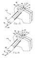
- FIG. 12is a perspective view of the assembly of FIG. 11 shown with the fastener being driven in the tissue layers to be fastened;
- FIG. 13is a perspective view of the assembly of FIG. 11 shown with the fastener in an intermediate stage of deployment;
- FIG. 14is a perspective view of the assembly of FIG. 11 shown with the fastener almost completely deployed;
- FIG. 15is a perspective view showing the fastener of the assembly of FIG. 11 fully deployed and securely fastening a pair of tissue layers together;
- FIG. 16is a perspective view with portions cut away of a fastener assembly according to a still further embodiment of the invention in an early stage of deploying a still further fastener embodiment of the invention;
- FIG. 17is a perspective view of the assembly of FIG. 16 shown with the fastener being driven in the tissue layers to be fastened;
- FIG. 18is a perspective view of the assembly of FIG. 16 shown with the fastener in an intermediate stage of deployment;
- FIG. 19is a perspective view of the assembly of FIG. 16 shown with the fastener almost completely deployed.
- FIG. 20is a perspective view showing the fastener of the assembly of FIG. 16 fully deployed and securely fastening a pair of tissue layers together.
- FIG. 1is a front cross-sectional view of the esophageal-gastro-intestinal tract 40 from a lower portion of the esophagus 41 to the duodenum 42 .
- the stomach 43is characterized by the greater curvature 44 on the anatomical left side and the lesser curvature 45 on the anatomical right side.
- the tissue of the outer surfaces of those curvaturesis referred to in the art as serosa tissue. As will be seen subsequently, the nature of the serosa tissue is used to advantage for its ability to bond to like serosa tissue.
- the fundus 46 of the greater curvature 44forms the superior portion of the stomach 43 , and traps gas and air bubbles for burping.
- the esophageal tract 41enters the stomach 43 at an esophageal orifice below the superior portion of the fundus 46 , forming a cardiac notch 47 and an acute angle with respect to the fundus 46 known as the Angle of His 57 .
- the lower esophageal sphincter (LES) 48is a discriminating sphincter able to distinguish between burping gas, liquids, and solids, and works in conjunction with the fundus 46 to burp.
- the gastroesophageal flap valve (GEFV) 49includes a moveable portion and an opposing more stationary portion.
- the moveable portion of the GEFV 49is an approximately 180 degree, semicircular, gastroesophageal flap 50 (alternatively referred to as a “normal moveable flap” or “moveable flap”) formed of tissue at the intersection between the esophagus 41 and the stomach 43 .
- the opposing more stationary portion of the GEFV 49comprises a portion of the lesser curvature 45 of the stomach 43 adjacent to its junction with the esophagus 41 .
- the gastroesophageal flap 50 of the GEFV 49principally comprises tissue adjacent to the fundus 46 portion of the stomach 43 , is about 4 to 5 cm long ( 51 ) at it longest portion, and the length may taper at its anterior and posterior ends.
- the gastroesophageal flap 50is partially held against the lesser curvature 45 portion of the stomach 43 by the pressure differential between the stomach 43 and the thorax, and partially by the resiliency and the anatomical structure of the GEFV 49 , thus providing the valving function.
- the GEFV 49is similar to a flutter valve, with the gastroesophageal flap 50 being flexible and closeable against the other more stationary side.
- the esophageal tractis controlled by an upper esophageal sphincter (UES)in the neck near the mouth for swallowing, and by the LES 48 and the GEFV 49 at the stomach.
- the normal anti-reflux barrieris primarily formed by the LES 48 and the GEFV 49 acting in concert to allow food and liquid to enter the stomach, and to considerably resist reflux of stomach contents into the esophagus 41 past the gastroesophageal tissue junction 52 .
- Tissue aboral of the gastroesophageal tissue junction 52is generally considered part of the stomach because the tissue protected from stomach acid by its own protective mechanisms.
- Tissue oral of the gastroesophageal junction 52is generally considered part of the esophagus and it is not protected from injury by prolonged exposure to stomach acid.
- the juncture of the stomach and esophageal tissuesform a zigzag line, which is sometimes referred to as the “Z-line.”
- “stomach”means the tissue aboral of the gastroesophageal junction 52 .
- FIG. 2is a front cross-sectional view of an esophageal-gastro-intestinal tract illustrating a Grade I normal appearance movable flap 50 of the GEFV 49 (shown in dashed lines) and a deteriorated Grade III gastroesophageal flap 55 of the GEFV 49 (shown in solid lines).
- a principal reason for regurgitation associated with GERDis the mechanical failure of the deteriorated (or reflux appearance) gastroesophageal flap 55 of the GEFV 49 to close and seal against the higher pressure in the stomach. Due to reasons including lifestyle, a Grade I normal gastroesophageal flap 50 of the GEFV 49 may deteriorate into a Grade III deteriorated gastroesophageal flap 55 .
- the anatomical results of the deteriorationinclude moving a portion of the esophagus 41 that includes the gastroesophageal junction 52 and LES 48 toward the mouth, straightening of the cardiac notch 47 , and increasing the Angle of His 57 .
- the deteriorated gastroesophageal flap 55illustrates a gastroesophageal flap valve 49 and cardiac notch 47 that have both significantly degraded.
- Dr. Hill and colleaguesdeveloped a grading system to describe the appearance of the GEFV and the likelihood that a patient will experience chronic acid reflux. L. D.
- the normal movable flap 50 of the GEFV 49illustrates a Grade I flap valve that is the least likely to experience reflux.
- the deteriorated gastroesophageal flap 55 of the GEFV 49illustrates a Grade III (almost Grade IV) flap valve.
- a Grade IV flap valveis the most likely to experience reflux.
- Grades II and IIIreflect intermediate grades of deterioration and, as in the case of III, a high likelihood of experiencing reflux.
- the stomach contentsare presented a funnel-like opening directing the contents into the esophagus 41 and the greatest likelihood of experiencing reflux.
- a fastener and assemblywhich may be employed to advantage in restoring the normal gastroesophageal flap valve anatomy.
- FIG. 3is a perspective view and FIG. 4 is a side view of a fastener 100 embodying the present invention.
- the fastener 100generally includes a first member 102 , a second member 104 , and a connecting member 106 .
- the first member 102 and second member 104are substantially parallel to each other and substantially perpendicular to the connecting member 106 which connects the first member 102 to the second member 104 .
- the first member 102is generally cylindrical or can have any other shape. It has a longitudinal axis 108 and a through channel 112 along the longitudinal axis 108 .
- the through channel 112is formed by a through bore which is dimensioned to be slidingly received on a tissue piercing deployment wire to be described.
- the first member 102also includes a first end 116 and a second end 118 .
- the second member 104includes a first end 120 and a second end 122 .
- the first end 116 of member 102forms a pointed dilation tip 124 .
- the dilation tip 124may be conical and more particularly takes the shape of a truncated cone.
- the tipcan also be shaped to have a cutting edge in order to reduce tissue resistance.
- the first and second members 102 and 104 and the connecting member 106may be formed of different materials and have different textures. These materials may include, for example, plastic materials such as polypropylene, polyethylene, polyglycolic acid, polyurethane, or a thermoplastic elastomer. The plastic materials may include a pigment contrasting with body tissue color to enable better visualization of the fastener during its deployment. Alternatively, the fastener may be formed of a metal, such as stainless steel or a shape memory metal, such as Nitinol.
- the connecting member 106has a vertical dimension 128 and a horizontal dimension 130 which is transverse to the vertical dimension.
- the horizontal dimensionis substantially less than the vertical dimension to render the connecting member 106 readily bendable in a horizontal plane.
- the connecting memberis further rendered bendable by the nature of the material from which the fastener 100 is formed.
- the connecting membermay be formed from either an elastic plastic or a permanently deformable plastic. An elastic material would prevent compression necrosis in some applications.
- the first member 102has a continuous lengthwise slit 125 extending between the first and second ends 116 and 118 .
- the slit 125includes an optional slot portion 126 that communications with the through channel 112 .
- the slot 126has a transverse dimension for more readily enabling receipt of a tissue piercing deployment wire during deployment of the fastener 100 .
- the fastener number 102is formed of flexible material, the slit 125 may be made larger through separation to allow the deployment wire to be snapped into and released from the through channel 112 as will be seen subsequently. This permits early release of the first member during deployment 102 and decreases compression on the tissue layers.
- the slit 125extends substantially parallel to the through channel 112 and the center axis 108 of the first member 102 . It may also be noted that the slit 125 has a width dimension that is smaller or less than the diameter D of the through channel 112 . This assures that the fastener 100 will remain on a tissue piercing deployment wire as it is pushed towards and into the tissue as will be seen subsequently.
- FIG. 5it is a perspective view with portions cut away of a fastener assembly 200 embodying the present invention for deploying the fastener 100 .
- the tissue layer portions above the fastener 100have been shown cut away in FIGS. 5-9 to enable the deployment procedure to be seen more clearly.
- the assembly 200generally includes the fastener 100 , a deployment wire 164 , a pusher 166 , and a guide tube 168 .
- the first member 102 of the fastener 100is slidingly received on the deployment wire 164 .
- the deployment wire 164has a pointed tip 178 for piercing the tissue layers 180 and 182 to be fastened together.
- the tip 178is enlarged with respect to the diameter of the deployment wire 164 and preferably has a cross-sectional dimension greater than that of the through channel and preferably the first member 102 . This permits the tip 178 to cut sufficient tissue to enable the fastener member 102 to readily pass through the tissue layers 180 and 182 . It may also serve as a guide to guide the wire 164 off of the member 102 at the end of the deployment.
- the tissue piercing wire 164 , fastener 100 , and the pusher 166are all within the guide tube 168 .
- the guide tube 168may take the form of a catheter, for example, as previously mentioned, or a guide channel within a block of material.
- the second member 104is disposed along side the first member 102 . This is rendered possible by the flexibility of the connecting member 106 .
- the tip 178 of the tissue piercing wire 164pierces the tissue layers 180 and 182 .
- the subassembly of the tissue piercing wire 164 , fastener 100 , and pusher 166may be guided to its intended location relative to the tissue layers 180 and 182 by the guide tube 168 .
- the tissue piercing wire 164has pierced the tissue layers 180 and 182 and the pusher 166 has pushed the first member 102 of the fastener 100 through the tissue layers 180 and 182 on the tissue piercing wire 164 . This may be accomplished by moving the wire 164 and the pusher 166 together.
- wire 164has been pushed further forward and independently from the first member 102 .
- the first member 102has also been pushed forward by the pusher 166 to cause the second member 104 to engage the tissue layer 180 .
- Continued pushing of the first member 102causes the first member to pivot in a counter clockwise direction because the second member 104 is held by the tissue layer 180 .
- the counter clockwise movement of the first member 102causes the wire 164 to spread the slit 125 open, to pass down the slit to enter slot portion 126 and to eventually pass through the slit 125 at end 118 .
- the fastener 100is then released from the wire 164 .
- FIG. 8it will now be seen that the second end 118 of the first member 102 has cleared the wire 164 and tissue layer 182 .
- the tissue piercing wire 164may now be retracted into the pusher 166 and the tissue piercing wire 164 and pusher 166 may be withdrawn.
- FIG. 9illustrates the fastener 100 in its fully deployed position. It will be noted that the fastener has returned to its original shape.
- the tissue layers 180 and 182are fastened together between the first member 102 of the fastener 100 and the second member 104 of the fastener 100 .
- the connecting member 106extends through the tissue layers 180 and 182 .
- the release of the fastener 100 from the wire 164 with minimal damage to the tissue layers 180 and 182is made possible because the first member 102 has a configuration alterable by the wire 164 .
- the slit 125 in member 102assists in the configuration change and release.
- FIG. 10it shows a deployment wire/pusher arrangement 160 which may be employed in the assembly 200 of FIGS. 5-9 .
- the arrangement 160includes the deployment piercing wire 164 and the pusher 166 .
- the wire 164includes the tissue piercing tip 178 .
- the pusher 166is tubular and has a distal end 165 , a lumen 162 , and an opening 167 through the pusher sidewall 161 and communicating with the lumen 162 .
- the lumen 162is configured to permit the pusher 166 to slidingly be carried on the wire 164 . To that end, the wire extends within the lumen 162 from the distal end 165 and through the opening 167 .
- a fastener 100has been snapped onto the deployment wire 164 between the deployment wire tip 178 and the distal end 165 of the pusher 166 .
- An optional stiffener 169may also be provided for the pusher 166 .
- the pusher 166may be retracted from the patient while the pusher is still on the deployment wire 164 , a further fastener may then be snapped onto the wire through the slit 125 . The pusher may then be advanced down the deployment wire to deploy the fastener as previously described.
- FIGS. 11-15show a deployment sequence of another fastener assembly 210 embodying the invention.
- the assembly 210includes the deployment wire 164 , the pusher 166 , the guide tube 168 , and a fastener 300 .
- the fastener 300includes a first member 302 , a second member 304 , and a connecting member 306 .
- the fastener 300is similar to the fastener 100 previously described except that to render the configuration of the first member 302 alterable by the deployment wire 164 as the first member 102 is so alterable, the first member 302 is stretchable to obviate the need for a slit.
- the first member 302may be formed of rubber, for example, to permit the wire 164 to be separated from the member 302 by simply pulling the wire tip 178 back through through the member. Deformation of the member 302 occurs to permit this separation.
- FIG. 11shows the fastener 300 on the wire with the wire tip 178 just piercing the tissue layer.
- the first member 302is carried on the wire 164 which extends through the through channel of the first member 302 .
- the pusher 166 and wire 164are moved forward together to cause the wire 164 and member 302 to extend through the tissue layers 180 and 186 .
- the pusherpushes the member 302 forward.
- the second member 304first engages tissue layer 180 and once so engaged, the first member 302 is caused to rotate (counter clockwise in FIG. 13 ).
- the elasticity of the member 302causes the wire tip to stretch the sidewall of the first member 302 .
- the end 318 of the member 302pops free of the wire tip. This may be seen in FIG. 14 .
- the end 318 of the memberis now free of the wire 164 .
- the pusher 166 and deployment wire 164may now be extracted. As shown in FIG. 15 , the tissue layers 180 and 182 are now fixed together between the first and second members 302 and 304 with the connecting member 306 extending therebetween.
- FIGS. 16-20show a deployment sequence of another fastener assembly 220 embodying the invention.
- the assembly 220includes the deployment wire 164 , the pusher 166 , the guide tube 168 , and a fastener 400 .
- the fastener 400includes a first member 402 , a second member 404 , and a connecting member 406 .
- the fastener 400is similar to the fasteners 100 and 300 previously described except that, to render the configuration of the first member 402 alterable by the deployment wire 164 , the first member 402 is tearable by tip 178 of the wire 164 .
- the first member 402may be formed, for example, to permit the wire 164 to tear the member 402 lengthwise as the wire tip 178 is pulled back through through the member 402 .
- the member sidewallmay have a varying thickness, such as a tearing or score line 403 .
- FIG. 16shows the fastener 400 on the wire with the wire tip 178 just piercing the tissue layer.
- the first member 402is carried on the wire 164 which extends through the through channel of the first member 402 .
- the pusher 166 and wire 164are moved forward together to cause the wire 164 and member 402 to extend through the tissue layers 180 and 186 . From this point, the pusher 166 pushes the member 402 to cause the tip 178 to begin tearing the member 402 along the score line 402 . As may be seen in FIG. 18 , the pusher pushes the member 402 forward. The second member 404 first engages tissue layer 180 and once so engaged, the first member 402 is caused to rotate (counter clockwise in FIG. 18 ). The turning of the member 402 causes the wire tip 178 to tear through the sidewall of the first member 402 . As the member 402 is continued to be pushed, it continues to be turned lengthwise. Eventually, the end 418 of the member 402 releases from the wire tip. This may be seen in FIG. 19 . The end 418 of the member is now free of the wire 164 .
- the pusher 166 and deployment wire 164may now be extracted. As shown in FIG. 20 , the tissue layers 180 and 182 are now fixed together between the first and second members 402 and 404 with the connecting member 406 extending therebetween.
Landscapes
- Health & Medical Sciences (AREA)
- Life Sciences & Earth Sciences (AREA)
- Surgery (AREA)
- Heart & Thoracic Surgery (AREA)
- Engineering & Computer Science (AREA)
- Biomedical Technology (AREA)
- Nuclear Medicine, Radiotherapy & Molecular Imaging (AREA)
- Medical Informatics (AREA)
- Molecular Biology (AREA)
- Animal Behavior & Ethology (AREA)
- General Health & Medical Sciences (AREA)
- Public Health (AREA)
- Veterinary Medicine (AREA)
- Surgical Instruments (AREA)
- Prostheses (AREA)
Abstract
Description
Claims (10)
Priority Applications (5)
| Application Number | Priority Date | Filing Date | Title |
|---|---|---|---|
| US12/803,077US8337514B2 (en) | 2005-01-25 | 2010-06-17 | Slitted tissue fixation devices and assemblies for deploying the same |
| US13/680,944US9358007B2 (en) | 2005-01-25 | 2012-11-19 | Slitted tissue fixation devices and assemblies for deploying the same |
| US15/152,337US9572578B2 (en) | 2005-01-25 | 2016-05-11 | Slitted tissue fixation devices and assemblies for deploying the same |
| US15/400,693US10478180B2 (en) | 2005-01-25 | 2017-01-06 | Slitted tissue fixation devices and assemblies for deploying the same |
| US16/676,644US11484305B2 (en) | 2005-01-25 | 2019-11-07 | Slitted tissue fixation devices and assemblies for deploying the same |
Applications Claiming Priority (2)
| Application Number | Priority Date | Filing Date | Title |
|---|---|---|---|
| US11/043,903US20060167481A1 (en) | 2005-01-25 | 2005-01-25 | Slitted tissue fixation devices and assemblies for deploying the same |
| US12/803,077US8337514B2 (en) | 2005-01-25 | 2010-06-17 | Slitted tissue fixation devices and assemblies for deploying the same |
Related Parent Applications (1)
| Application Number | Title | Priority Date | Filing Date |
|---|---|---|---|
| US11/043,903ContinuationUS20060167481A1 (en) | 2005-01-25 | 2005-01-25 | Slitted tissue fixation devices and assemblies for deploying the same |
Related Child Applications (1)
| Application Number | Title | Priority Date | Filing Date |
|---|---|---|---|
| US13/680,944ContinuationUS9358007B2 (en) | 2005-01-25 | 2012-11-19 | Slitted tissue fixation devices and assemblies for deploying the same |
Publications (2)
| Publication Number | Publication Date |
|---|---|
| US20100262169A1 US20100262169A1 (en) | 2010-10-14 |
| US8337514B2true US8337514B2 (en) | 2012-12-25 |
Family
ID=36697906
Family Applications (6)
| Application Number | Title | Priority Date | Filing Date |
|---|---|---|---|
| US11/043,903AbandonedUS20060167481A1 (en) | 2005-01-25 | 2005-01-25 | Slitted tissue fixation devices and assemblies for deploying the same |
| US12/803,077Expired - LifetimeUS8337514B2 (en) | 2005-01-25 | 2010-06-17 | Slitted tissue fixation devices and assemblies for deploying the same |
| US13/680,944Active2026-09-21US9358007B2 (en) | 2005-01-25 | 2012-11-19 | Slitted tissue fixation devices and assemblies for deploying the same |
| US15/152,337Expired - LifetimeUS9572578B2 (en) | 2005-01-25 | 2016-05-11 | Slitted tissue fixation devices and assemblies for deploying the same |
| US15/400,693Active2025-11-17US10478180B2 (en) | 2005-01-25 | 2017-01-06 | Slitted tissue fixation devices and assemblies for deploying the same |
| US16/676,644Active2026-08-05US11484305B2 (en) | 2005-01-25 | 2019-11-07 | Slitted tissue fixation devices and assemblies for deploying the same |
Family Applications Before (1)
| Application Number | Title | Priority Date | Filing Date |
|---|---|---|---|
| US11/043,903AbandonedUS20060167481A1 (en) | 2005-01-25 | 2005-01-25 | Slitted tissue fixation devices and assemblies for deploying the same |
Family Applications After (4)
| Application Number | Title | Priority Date | Filing Date |
|---|---|---|---|
| US13/680,944Active2026-09-21US9358007B2 (en) | 2005-01-25 | 2012-11-19 | Slitted tissue fixation devices and assemblies for deploying the same |
| US15/152,337Expired - LifetimeUS9572578B2 (en) | 2005-01-25 | 2016-05-11 | Slitted tissue fixation devices and assemblies for deploying the same |
| US15/400,693Active2025-11-17US10478180B2 (en) | 2005-01-25 | 2017-01-06 | Slitted tissue fixation devices and assemblies for deploying the same |
| US16/676,644Active2026-08-05US11484305B2 (en) | 2005-01-25 | 2019-11-07 | Slitted tissue fixation devices and assemblies for deploying the same |
Country Status (5)
| Country | Link |
|---|---|
| US (6) | US20060167481A1 (en) |
| EP (1) | EP1848347B1 (en) |
| JP (1) | JP2008528132A (en) |
| ES (1) | ES2573849T3 (en) |
| WO (1) | WO2006081368A2 (en) |
Families Citing this family (84)
| Publication number | Priority date | Publication date | Assignee | Title |
|---|---|---|---|---|
| US7608092B1 (en) | 2004-02-20 | 2009-10-27 | Biomet Sports Medicince, LLC | Method and apparatus for performing meniscus repair |
| US20050187565A1 (en) | 2004-02-20 | 2005-08-25 | Baker Steve G. | Tissue fixation devices and a transoral endoscopic gastroesophageal flap valve restoration device and assembly using same |
| US7632287B2 (en) | 2004-02-20 | 2009-12-15 | Endogastric Solutions, Inc. | Tissue fixation devices and assemblies for deploying the same |
| US9801708B2 (en) | 2004-11-05 | 2017-10-31 | Biomet Sports Medicine, Llc | Method and apparatus for coupling soft tissue to a bone |
| US7905904B2 (en) | 2006-02-03 | 2011-03-15 | Biomet Sports Medicine, Llc | Soft tissue repair device and associated methods |
| US7857830B2 (en) | 2006-02-03 | 2010-12-28 | Biomet Sports Medicine, Llc | Soft tissue repair and conduit device |
| US9017381B2 (en) | 2007-04-10 | 2015-04-28 | Biomet Sports Medicine, Llc | Adjustable knotless loops |
| US8361113B2 (en) | 2006-02-03 | 2013-01-29 | Biomet Sports Medicine, Llc | Method and apparatus for coupling soft tissue to a bone |
| US7658751B2 (en) | 2006-09-29 | 2010-02-09 | Biomet Sports Medicine, Llc | Method for implanting soft tissue |
| US8137382B2 (en) | 2004-11-05 | 2012-03-20 | Biomet Sports Medicine, Llc | Method and apparatus for coupling anatomical features |
| US20060189993A1 (en) | 2004-11-09 | 2006-08-24 | Arthrotek, Inc. | Soft tissue conduit device |
| US8118836B2 (en) | 2004-11-05 | 2012-02-21 | Biomet Sports Medicine, Llc | Method and apparatus for coupling soft tissue to a bone |
| US8303604B2 (en) | 2004-11-05 | 2012-11-06 | Biomet Sports Medicine, Llc | Soft tissue repair device and method |
| US7749250B2 (en) | 2006-02-03 | 2010-07-06 | Biomet Sports Medicine, Llc | Soft tissue repair assembly and associated method |
| US8128658B2 (en) | 2004-11-05 | 2012-03-06 | Biomet Sports Medicine, Llc | Method and apparatus for coupling soft tissue to bone |
| US8840645B2 (en) | 2004-11-05 | 2014-09-23 | Biomet Sports Medicine, Llc | Method and apparatus for coupling soft tissue to a bone |
| US7909851B2 (en) | 2006-02-03 | 2011-03-22 | Biomet Sports Medicine, Llc | Soft tissue repair device and associated methods |
| US8298262B2 (en) | 2006-02-03 | 2012-10-30 | Biomet Sports Medicine, Llc | Method for tissue fixation |
| US8088130B2 (en) | 2006-02-03 | 2012-01-03 | Biomet Sports Medicine, Llc | Method and apparatus for coupling soft tissue to a bone |
| US8998949B2 (en) | 2004-11-09 | 2015-04-07 | Biomet Sports Medicine, Llc | Soft tissue conduit device |
| US20060116697A1 (en) | 2004-11-30 | 2006-06-01 | Esophyx, Inc. | Flexible transoral endoscopic gastroesophageal flap valve restoration device and method |
| US20060167481A1 (en) | 2005-01-25 | 2006-07-27 | Esophyx, Inc. | Slitted tissue fixation devices and assemblies for deploying the same |
| US20070005082A1 (en) | 2005-06-29 | 2007-01-04 | Esophyx, Inc. | Apparatus and method for manipulating stomach tissue and treating gastroesophageal reflux disease |
| US20070038232A1 (en) | 2005-08-12 | 2007-02-15 | Kraemer Stefan J M | Apparatus and method for securing the stomach to the diaphragm for use, for example, in treating hiatal hernias and gastroesophageal reflux disease |
| US20070088373A1 (en) | 2005-10-18 | 2007-04-19 | Endogastric Solutions, Inc. | Invaginator for gastroesophageal flap valve restoration device |
| US20070129738A1 (en) | 2005-12-01 | 2007-06-07 | Endogastric Solutions, Inc. | Apparatus and method for concurrently forming a gastroesophageal valve and tightening the lower esophageal sphincter |
| US9161754B2 (en) | 2012-12-14 | 2015-10-20 | Endogastric Solutions, Inc. | Apparatus and method for concurrently forming a gastroesophageal valve and tightening the lower esophageal sphincter |
| US8652172B2 (en) | 2006-02-03 | 2014-02-18 | Biomet Sports Medicine, Llc | Flexible anchors for tissue fixation |
| US8562647B2 (en) | 2006-09-29 | 2013-10-22 | Biomet Sports Medicine, Llc | Method and apparatus for securing soft tissue to bone |
| US9538998B2 (en) | 2006-02-03 | 2017-01-10 | Biomet Sports Medicine, Llc | Method and apparatus for fracture fixation |
| US8771352B2 (en) | 2011-05-17 | 2014-07-08 | Biomet Sports Medicine, Llc | Method and apparatus for tibial fixation of an ACL graft |
| US8597327B2 (en) | 2006-02-03 | 2013-12-03 | Biomet Manufacturing, Llc | Method and apparatus for sternal closure |
| US9271713B2 (en) | 2006-02-03 | 2016-03-01 | Biomet Sports Medicine, Llc | Method and apparatus for tensioning a suture |
| US7959650B2 (en) | 2006-09-29 | 2011-06-14 | Biomet Sports Medicine, Llc | Adjustable knotless loops |
| US10517587B2 (en) | 2006-02-03 | 2019-12-31 | Biomet Sports Medicine, Llc | Method and apparatus for forming a self-locking adjustable loop |
| US8562645B2 (en) | 2006-09-29 | 2013-10-22 | Biomet Sports Medicine, Llc | Method and apparatus for forming a self-locking adjustable loop |
| US11311287B2 (en) | 2006-02-03 | 2022-04-26 | Biomet Sports Medicine, Llc | Method for tissue fixation |
| US8574235B2 (en) | 2006-02-03 | 2013-11-05 | Biomet Sports Medicine, Llc | Method for trochanteric reattachment |
| US9078644B2 (en) | 2006-09-29 | 2015-07-14 | Biomet Sports Medicine, Llc | Fracture fixation device |
| US8652171B2 (en) | 2006-02-03 | 2014-02-18 | Biomet Sports Medicine, Llc | Method and apparatus for soft tissue fixation |
| US8251998B2 (en) | 2006-08-16 | 2012-08-28 | Biomet Sports Medicine, Llc | Chondral defect repair |
| US8801783B2 (en) | 2006-09-29 | 2014-08-12 | Biomet Sports Medicine, Llc | Prosthetic ligament system for knee joint |
| US9149267B2 (en) | 2006-02-03 | 2015-10-06 | Biomet Sports Medicine, Llc | Method and apparatus for coupling soft tissue to a bone |
| US9468433B2 (en) | 2006-02-03 | 2016-10-18 | Biomet Sports Medicine, Llc | Method and apparatus for forming a self-locking adjustable loop |
| US11259792B2 (en) | 2006-02-03 | 2022-03-01 | Biomet Sports Medicine, Llc | Method and apparatus for coupling anatomical features |
| US8968364B2 (en) | 2006-02-03 | 2015-03-03 | Biomet Sports Medicine, Llc | Method and apparatus for fixation of an ACL graft |
| US8506597B2 (en) | 2011-10-25 | 2013-08-13 | Biomet Sports Medicine, Llc | Method and apparatus for interosseous membrane reconstruction |
| US8672969B2 (en) | 2006-09-29 | 2014-03-18 | Biomet Sports Medicine, Llc | Fracture fixation device |
| US8500818B2 (en) | 2006-09-29 | 2013-08-06 | Biomet Manufacturing, Llc | Knee prosthesis assembly with ligament link |
| US11259794B2 (en) | 2006-09-29 | 2022-03-01 | Biomet Sports Medicine, Llc | Method for implanting soft tissue |
| US9918826B2 (en) | 2006-09-29 | 2018-03-20 | Biomet Sports Medicine, Llc | Scaffold for spring ligament repair |
| WO2008085994A2 (en) | 2007-01-08 | 2008-07-17 | Endogastric Solutions | Connected fasteners, delivery device and method |
| US12419632B2 (en) | 2008-08-22 | 2025-09-23 | Biomet Sports Medicine, Llc | Method and apparatus for coupling anatomical features |
| US12245759B2 (en) | 2008-08-22 | 2025-03-11 | Biomet Sports Medicine, Llc | Method and apparatus for coupling soft tissue to bone |
| US8449573B2 (en) | 2008-12-05 | 2013-05-28 | Boston Scientific Scimed, Inc. | Insertion device and method for delivery of a mesh carrier |
| US9028509B2 (en) | 2008-12-05 | 2015-05-12 | Boston Scientific Scimed, Inc. | Insertion device for delivery of a mesh carrier |
| US8906037B2 (en) | 2009-03-18 | 2014-12-09 | Endogastric Solutions, Inc. | Methods and devices for forming a tissue fold |
| US8343227B2 (en) | 2009-05-28 | 2013-01-01 | Biomet Manufacturing Corp. | Knee prosthesis assembly with ligament link |
| US12096928B2 (en) | 2009-05-29 | 2024-09-24 | Biomet Sports Medicine, Llc | Method and apparatus for coupling soft tissue to a bone |
| US9737289B2 (en)* | 2010-10-29 | 2017-08-22 | Vectec S.A. | Single use, disposable, tissue suspender device |
| US12329373B2 (en) | 2011-05-02 | 2025-06-17 | Biomet Sports Medicine, Llc | Method and apparatus for soft tissue fixation |
| US20130066338A1 (en) | 2011-09-09 | 2013-03-14 | Richard Romley | Methods and devices for manipulating and fastening tissue |
| US9572571B2 (en) | 2011-09-09 | 2017-02-21 | Endogastric Solutions, Inc. | Methods and devices for manipulating and fastening tissue |
| US9955957B2 (en) | 2011-09-09 | 2018-05-01 | Endogastric Solutions, Inc. | Methods and devices for manipulating and fastening tissue |
| US9357991B2 (en) | 2011-11-03 | 2016-06-07 | Biomet Sports Medicine, Llc | Method and apparatus for stitching tendons |
| US9381013B2 (en) | 2011-11-10 | 2016-07-05 | Biomet Sports Medicine, Llc | Method for coupling soft tissue to a bone |
| US9370350B2 (en) | 2011-11-10 | 2016-06-21 | Biomet Sports Medicine, Llc | Apparatus for coupling soft tissue to a bone |
| US9314241B2 (en) | 2011-11-10 | 2016-04-19 | Biomet Sports Medicine, Llc | Apparatus for coupling soft tissue to a bone |
| US9259217B2 (en) | 2012-01-03 | 2016-02-16 | Biomet Manufacturing, Llc | Suture Button |
| US9757119B2 (en) | 2013-03-08 | 2017-09-12 | Biomet Sports Medicine, Llc | Visual aid for identifying suture limbs arthroscopically |
| US9918827B2 (en) | 2013-03-14 | 2018-03-20 | Biomet Sports Medicine, Llc | Scaffold for spring ligament repair |
| US20150032130A1 (en) | 2013-07-24 | 2015-01-29 | Covidien Lp | Expanding absorbable tack |
| US10136886B2 (en) | 2013-12-20 | 2018-11-27 | Biomet Sports Medicine, Llc | Knotless soft tissue devices and techniques |
| US9615822B2 (en) | 2014-05-30 | 2017-04-11 | Biomet Sports Medicine, Llc | Insertion tools and method for soft anchor |
| US9700291B2 (en) | 2014-06-03 | 2017-07-11 | Biomet Sports Medicine, Llc | Capsule retractor |
| US10039543B2 (en) | 2014-08-22 | 2018-08-07 | Biomet Sports Medicine, Llc | Non-sliding soft anchor |
| US9955980B2 (en) | 2015-02-24 | 2018-05-01 | Biomet Sports Medicine, Llc | Anatomic soft tissue repair |
| US9974534B2 (en) | 2015-03-31 | 2018-05-22 | Biomet Sports Medicine, Llc | Suture anchor with soft anchor of electrospun fibers |
| JP6087040B1 (en)* | 2015-09-17 | 2017-03-01 | オリンパス株式会社 | Endoscopic treatment tool |
| EP3351189B1 (en) | 2015-09-17 | 2020-08-12 | Olympus Corporation | Endoscopic treatment tool |
| CN114173685A (en)* | 2019-04-01 | 2022-03-11 | 都柏林圣三一学院教务长、研究员、基金会学者及董事会其他成员 | wound closure |
| US11382613B1 (en)* | 2021-04-08 | 2022-07-12 | Integrity Orthopaedics, Inc. | Methods for transtendinous implantation of knotless micro suture anchors and anchor arrays |
| US11382611B1 (en)* | 2021-04-08 | 2022-07-12 | Integrity Orthopaedics, Inc. | Knotless micro-suture anchors and anchor arrays for anatomical attachment of soft tissue to bone |
| KR102818778B1 (en)* | 2022-08-23 | 2025-06-10 | 충남대학교 산학협력단 | Pin for Suturing Soft tissue and Suturing Apparatus using the same |
Citations (40)
| Publication number | Priority date | Publication date | Assignee | Title |
|---|---|---|---|---|
| US4595007A (en) | 1983-03-14 | 1986-06-17 | Ethicon, Inc. | Split ring type tissue fastener |
| US4895148A (en)* | 1986-05-20 | 1990-01-23 | Concept, Inc. | Method of joining torn parts of bodily tissue in vivo with a biodegradable tack member |
| US5041129A (en) | 1990-07-02 | 1991-08-20 | Acufex Microsurgical, Inc. | Slotted suture anchor and method of anchoring a suture |
| US5080543A (en)* | 1990-01-08 | 1992-01-14 | Engineered Construction Components (America) Inc. | Fastening sleeves and fastening systems employing same |
| US5254126A (en) | 1992-06-24 | 1993-10-19 | Ethicon, Inc. | Endoscopic suture punch |
| US5403326A (en) | 1993-02-01 | 1995-04-04 | The Regents Of The University Of California | Method for performing a gastric wrap of the esophagus for use in the treatment of esophageal reflux |
| US5411508A (en) | 1991-10-29 | 1995-05-02 | The Trustees Of Columbia University In The City Of New York | Gastrointestinal approximating and tissue attaching device |
| US5571116A (en) | 1994-10-02 | 1996-11-05 | United States Surgical Corporation | Non-invasive treatment of gastroesophageal reflux disease |
| US5626614A (en)* | 1995-12-22 | 1997-05-06 | Applied Medical Resources Corporation | T-anchor suturing device and method for using same |
| US5713903A (en)* | 1991-03-22 | 1998-02-03 | United States Surgical Corporation | Orthopedic fastener |
| US5887594A (en) | 1997-09-22 | 1999-03-30 | Beth Israel Deaconess Medical Center Inc. | Methods and devices for gastroesophageal reflux reduction |
| US6086600A (en) | 1997-11-03 | 2000-07-11 | Symbiosis Corporation | Flexible endoscopic surgical instrument for invagination and fundoplication |
| US6113611A (en) | 1998-05-28 | 2000-09-05 | Advanced Vascular Technologies, Llc | Surgical fastener and delivery system |
| WO2000078227A1 (en) | 1999-06-22 | 2000-12-28 | Ndo Surgical, Inc. | Gerd treatment apparatus and method |
| WO2001035834A1 (en) | 1999-11-12 | 2001-05-25 | Boston Scientific Limited | Gerd treatment apparatus and method |
| US6302311B1 (en) | 1996-06-14 | 2001-10-16 | Boston Scientific Corporation | Endoscopic stapler |
| WO2001085034A1 (en) | 2000-05-10 | 2001-11-15 | Boston Scientific Limited | Devices and related methods for securing a tissue fold |
| WO2002024080A2 (en) | 2000-09-22 | 2002-03-28 | Boston Scientific Limited | Methods and devices for folding and securing tissue |
| US20020040226A1 (en) | 1999-06-22 | 2002-04-04 | Laufer Michael D. | Tissue reconfiguration |
| WO2002028289A1 (en) | 2000-09-29 | 2002-04-11 | Boston Scientific Limited | Method for performing endoluminal fundoplication and apparatus for use in the method |
| US20020078967A1 (en) | 2000-12-06 | 2002-06-27 | Robert Sixto | Methods for the endoluminal treatment of gastroesophageal reflux disease (GERD) |
| US6447524B1 (en) | 2000-10-19 | 2002-09-10 | Ethicon Endo-Surgery, Inc. | Fastener for hernia mesh fixation |
| US6491707B2 (en) | 1997-06-28 | 2002-12-10 | Transvascular, Inc. | Transluminal methods and devices for closing, forming attachments to, and/or forming anastomotic junctions in, luminal anatomical structures |
| US20020198541A1 (en) | 2001-06-25 | 2002-12-26 | Smith Kevin W. | Flexible surgical clip applier |
| US20030055442A1 (en) | 1999-06-22 | 2003-03-20 | Laufer Michael D. | Method and devices for tissue reconfiguration |
| US6551333B2 (en) | 2000-10-19 | 2003-04-22 | Ethicon Endo-Surgery, Inc. | Method for attaching hernia mesh |
| US20030187465A1 (en) | 2002-03-08 | 2003-10-02 | Sofradim Production | Appliance for storing, distributing and placing couched I-shaped surgical fasteners |
| US20030216613A1 (en) | 2002-03-19 | 2003-11-20 | Anthony Kalloo | Anastomosis system |
| US20030220657A1 (en) | 2001-05-23 | 2003-11-27 | Ronald Adams | Endoluminal fundoplication device and related method |
| US6663639B1 (en) | 1999-06-22 | 2003-12-16 | Ndo Surgical, Inc. | Methods and devices for tissue reconfiguration |
| US20040044364A1 (en) | 2002-08-29 | 2004-03-04 | Devries Robert | Tissue fasteners and related deployment systems and methods |
| WO2004019788A2 (en) | 2002-08-29 | 2004-03-11 | Boston Scientific Limited | Devices and methods for fastening tissue layers |
| US20040116949A1 (en) | 2002-12-11 | 2004-06-17 | Ewers Richard C. | Apparatus and methods for forming gastrointestinal tissue approximations |
| US20040147958A1 (en) | 2002-12-11 | 2004-07-29 | Usgi Medical | Apparatus and methods for forming and securing gastrointestinal tissue folds |
| US20040153102A1 (en) | 2001-06-21 | 2004-08-05 | Michel Therin | Kit comprising a medical fixing element and a device for placing said fixing element |
| US20040153103A1 (en) | 1998-12-30 | 2004-08-05 | Schwartz Herbert E. | Soft tissue locking device |
| US20050004575A1 (en) | 2000-11-24 | 2005-01-06 | Jean-Claude Sgro | Fastener for fixing a prosthesis, and device for delivering this fastener |
| US20050187565A1 (en)* | 2004-02-20 | 2005-08-25 | Baker Steve G. | Tissue fixation devices and a transoral endoscopic gastroesophageal flap valve restoration device and assembly using same |
| US20050228413A1 (en)* | 2004-04-12 | 2005-10-13 | Binmoeller Kenneth F | Automated transluminal tissue targeting and anchoring devices and methods |
| US7632287B2 (en) | 2004-02-20 | 2009-12-15 | Endogastric Solutions, Inc. | Tissue fixation devices and assemblies for deploying the same |
Family Cites Families (116)
| Publication number | Priority date | Publication date | Assignee | Title |
|---|---|---|---|---|
| US2753870A (en) | 1955-03-15 | 1956-07-10 | James A Muffly | Instrument for probing the reticulum |
| US3541591A (en)* | 1968-04-26 | 1970-11-17 | Henry J Hoegerman | Method and apparatus for closing wounds |
| US3789851A (en)* | 1971-07-01 | 1974-02-05 | H Leveen | Wound splints |
| US3875928A (en) | 1973-08-16 | 1975-04-08 | Angelchik Jean P | Method for maintaining the reduction of a sliding esophageal hiatal hernia |
| US4006747A (en) | 1975-04-23 | 1977-02-08 | Ethicon, Inc. | Surgical method |
| US4271828A (en) | 1979-09-13 | 1981-06-09 | Angelchik Jean P | Method for maintaining the reduction of a sliding esophageal hiatal hernia |
| US4823794A (en)* | 1982-07-12 | 1989-04-25 | Pierce William S | Surgical pledget |
| US4576772A (en) | 1984-07-20 | 1986-03-18 | Warner-Lambert Technologies, Inc. | Catheter with optimum resistance to bending and method of manufacture |
| DE3442736C2 (en)* | 1984-11-23 | 1987-03-05 | Tassilo Dr.med. 7800 Freiburg Bonzel | Dilatation catheter |
| US4696300A (en)* | 1985-04-11 | 1987-09-29 | Dennison Manufacturing Company | Fastener for joining materials |
| US4669473A (en) | 1985-09-06 | 1987-06-02 | Acufex Microsurgical, Inc. | Surgical fastener |
| DE3619197A1 (en) | 1986-06-07 | 1987-12-10 | Ethicon Gmbh | UPHOLSTERY IMPLANT |
| US4921479A (en) | 1987-10-02 | 1990-05-01 | Joseph Grayzel | Catheter sheath with longitudinal seam |
| JPH01148524A (en)* | 1987-12-04 | 1989-06-09 | Sumitomo Heavy Ind Ltd | Insert type core rotating mold assembly |
| US4846836A (en) | 1988-10-03 | 1989-07-11 | Reich Jonathan D | Artificial lower gastrointestinal valve |
| US5314473A (en) | 1989-07-20 | 1994-05-24 | Godin Norman J | Prosthesis for preventing gastric reflux into the esophagus |
| US5006106A (en) | 1990-10-09 | 1991-04-09 | Angelchik Jean P | Apparatus and method for laparoscopic implantation of anti-reflux prosthesis |
| US5088979A (en) | 1990-10-11 | 1992-02-18 | Wilson-Cook Medical Inc. | Method for esophageal invagination and devices useful therein |
| US5372146A (en)* | 1990-11-06 | 1994-12-13 | Branch; Thomas P. | Method and apparatus for re-approximating tissue |
| US5289963A (en) | 1991-10-18 | 1994-03-01 | United States Surgical Corporation | Apparatus and method for applying surgical staples to attach an object to body tissue |
| US5411520A (en) | 1991-11-08 | 1995-05-02 | Kensey Nash Corporation | Hemostatic vessel puncture closure system utilizing a plug located within the puncture tract spaced from the vessel, and method of use |
| WO1994003142A1 (en) | 1992-07-30 | 1994-02-17 | Temple University - Of The Commonwealth System Of Higher Education | Direct manual cardiac compression device and method of use thereof |
| WO1994015535A1 (en)* | 1993-01-07 | 1994-07-21 | Hayhurst, John, O. | Clip for suture |
| US5549621A (en) | 1993-05-14 | 1996-08-27 | Byron C. Sutherland | Apparatus and method for performing vertical banded gastroplasty |
| US5464426A (en)* | 1993-05-14 | 1995-11-07 | Bonutti; Peter M. | Method of closing discontinuity in tissue |
| US5540718A (en) | 1993-09-20 | 1996-07-30 | Bartlett; Edwin C. | Apparatus and method for anchoring sutures |
| CA2172129A1 (en) | 1993-09-20 | 1995-04-06 | Bruce H. Diamond | Multiple biopsy sampling device |
| US5582616A (en) | 1994-08-05 | 1996-12-10 | Origin Medsystems, Inc. | Surgical helical fastener with applicator |
| US5938668A (en) | 1994-10-07 | 1999-08-17 | United States Surgical | Surgical suturing apparatus |
| CH688174A5 (en) | 1995-03-28 | 1997-06-13 | Norman Godin | Prosthesis to oppose the gastric reflux into the esophagus. |
| US5759151A (en) | 1995-06-07 | 1998-06-02 | Carnegie Mellon University | Flexible steerable device for conducting exploratory procedures |
| US5791022A (en)* | 1996-01-29 | 1998-08-11 | Bohman; Lars | Cord locking mechanism |
| US6007567A (en)* | 1996-08-19 | 1999-12-28 | Bonutti; Peter M. | Suture anchor |
| US5718717A (en)* | 1996-08-19 | 1998-02-17 | Bonutti; Peter M. | Suture anchor |
| US5814054A (en) | 1996-09-23 | 1998-09-29 | Symbiosis Corporation | Automatic needle-passer suturing instrument |
| US6254642B1 (en) | 1997-12-09 | 2001-07-03 | Thomas V. Taylor | Perorally insertable gastroesophageal anti-reflux valve prosthesis and tool for implantation thereof |
| US6295990B1 (en) | 1998-02-03 | 2001-10-02 | Salient Interventional Systems, Inc. | Methods and systems for treating ischemia |
| US6113609A (en) | 1998-05-26 | 2000-09-05 | Scimed Life Systems, Inc. | Implantable tissue fastener and system for treating gastroesophageal reflux disease |
| US6264700B1 (en) | 1998-08-27 | 2001-07-24 | Endonetics, Inc. | Prosthetic gastroesophageal valve |
| CA2338518C (en) | 1998-08-31 | 2007-09-25 | Wilson-Cook Medical Inc. | Anti-reflux esophageal prosthesis |
| US6113612A (en) | 1998-11-06 | 2000-09-05 | St. Jude Medical Cardiovascular Group, Inc. | Medical anastomosis apparatus |
| US6066160A (en)* | 1998-11-23 | 2000-05-23 | Quickie Llc | Passive knotless suture terminator for use in minimally invasive surgery and to facilitate standard tissue securing |
| US6315789B1 (en)* | 1999-02-08 | 2001-11-13 | Andrew H. Cragg | Medical device anchoring system and method |
| US6159146A (en) | 1999-03-12 | 2000-12-12 | El Gazayerli; Mohamed Mounir | Method and apparatus for minimally-invasive fundoplication |
| US6098629A (en) | 1999-04-07 | 2000-08-08 | Endonetics, Inc. | Submucosal esophageal bulking device |
| US6375668B1 (en) | 1999-06-02 | 2002-04-23 | Hanson S. Gifford | Devices and methods for treating vascular malformations |
| US7160312B2 (en) | 1999-06-25 | 2007-01-09 | Usgi Medical, Inc. | Implantable artificial partition and methods of use |
| US7637905B2 (en) | 2003-01-15 | 2009-12-29 | Usgi Medical, Inc. | Endoluminal tool deployment system |
| US7744613B2 (en) | 1999-06-25 | 2010-06-29 | Usgi Medical, Inc. | Apparatus and methods for forming and securing gastrointestinal tissue folds |
| US6231561B1 (en) | 1999-09-20 | 2001-05-15 | Appriva Medical, Inc. | Method and apparatus for closing a body lumen |
| EP1095622A1 (en) | 1999-10-29 | 2001-05-02 | Biomedix S.A. | Endoscopic suturing instrument |
| US6428548B1 (en) | 1999-11-18 | 2002-08-06 | Russell F. Durgin | Apparatus and method for compressing body tissue |
| AU2001229674B2 (en) | 2000-01-21 | 2005-02-24 | Miravant Medical Technologies | Local drug delivery using photosensitizer-mediated and electromagnetic radiation-enhanced vascular permeability |
| JP5073905B2 (en) | 2000-02-29 | 2012-11-14 | ゼネラル・エレクトリック・カンパニイ | Nickel-base superalloy and turbine parts manufactured from the superalloy |
| IL138632A (en) | 2000-09-21 | 2008-06-05 | Minelu Zonnenschein | Multiple view endoscopes |
| JP4477280B2 (en) | 2000-03-16 | 2010-06-09 | メディガス リミテッド | Gastric fistula wall forming device |
| ES2435094T3 (en) | 2000-05-19 | 2013-12-18 | C.R. Bard, Inc. | Device and method of tissue capture and suturing |
| US6443944B1 (en) | 2000-05-19 | 2002-09-03 | Rajiv Doshi | Surgical devices comprising articulated members and methods for using the same |
| US6743239B1 (en) | 2000-05-25 | 2004-06-01 | St. Jude Medical, Inc. | Devices with a bendable tip for medical procedures |
| US6676698B2 (en) | 2000-06-26 | 2004-01-13 | Rex Medicol, L.P. | Vascular device with valve for approximating vessel wall |
| US6921361B2 (en) | 2000-07-24 | 2005-07-26 | Olympus Corporation | Endoscopic instrument for forming an artificial valve |
| TW510788B (en) | 2000-08-24 | 2002-11-21 | Surgical Connections Inc | Surgical stabilizer devices and methods |
| US20040093024A1 (en)* | 2000-09-01 | 2004-05-13 | James Lousararian | Advanced wound site management systems and methods |
| US20020139606A1 (en) | 2001-04-03 | 2002-10-03 | Williams Donald J. | Electric power steering system including a segmented stator switched reluctance motor |
| US7083629B2 (en) | 2001-05-30 | 2006-08-01 | Satiety, Inc. | Overtube apparatus for insertion into a body |
| US7115136B2 (en) | 2001-06-20 | 2006-10-03 | Park Medical Llc | Anastomotic device |
| JP4768154B2 (en) | 2001-06-29 | 2011-09-07 | テルモ株式会社 | Medical energy irradiation device |
| AUPR626401A0 (en)* | 2001-07-10 | 2001-08-02 | Australian Surgical Design And Manufacture Pty Limited | Surgical fixation device |
| WO2003061480A1 (en) | 2001-10-20 | 2003-07-31 | Applied Medical Resources Corporation | Wound retraction apparatus and method |
| US7318833B2 (en) | 2001-12-19 | 2008-01-15 | Nmt Medical, Inc. | PFO closure device with flexible thrombogenic joint and improved dislodgement resistance |
| AU2002348207B2 (en) | 2001-12-20 | 2008-01-24 | Rex Medical, L.P. | Apparatus and method for treating gastroesophageal reflux disease |
| US6699263B2 (en) | 2002-04-05 | 2004-03-02 | Cook Incorporated | Sliding suture anchor |
| US7077850B2 (en) | 2002-05-01 | 2006-07-18 | Scimed Life Systems, Inc. | Tissue fastening devices and related insertion tools and methods |
| US6790214B2 (en) | 2002-05-17 | 2004-09-14 | Esophyx, Inc. | Transoral endoscopic gastroesophageal flap valve restoration device, assembly, system and method |
| US20050085829A1 (en) | 2002-05-17 | 2005-04-21 | Esophyx, Inc. | Transoral endoscopic gastroesophageal flap valve restoration device, assembly, system and method |
| US6773440B2 (en) | 2002-07-02 | 2004-08-10 | Satiety, Inc. | Method and device for use in tissue approximation and fixation |
| US20040138704A1 (en) | 2002-09-06 | 2004-07-15 | Gambale Richard A. | Tissue capturing devices |
| ES2289334T3 (en) | 2002-10-04 | 2008-02-01 | Tyco Healthcare Group Lp | TOOL ASSEMBLY FOR SURGICAL STAPLING DEVICE. |
| US20090149871A9 (en) | 2002-11-01 | 2009-06-11 | Jonathan Kagan | Devices and methods for treating morbid obesity |
| US7037344B2 (en) | 2002-11-01 | 2006-05-02 | Valentx, Inc. | Apparatus and methods for treatment of morbid obesity |
| WO2004049982A2 (en) | 2002-12-02 | 2004-06-17 | Gi Dynamics, Inc. | Bariatric sleeve |
| EP1596723A2 (en)* | 2003-02-04 | 2005-11-23 | ev3 Sunnyvale, Inc. | Patent foramen ovale closure system |
| EP2481356B1 (en) | 2003-07-14 | 2013-09-11 | W.L. Gore & Associates, Inc. | Tubular patent foramen ovale (PFO) closure device with catch system |
| JP3833199B2 (en) | 2003-07-24 | 2006-10-11 | 沖電気工業株式会社 | Complementary signal generation circuit |
| US20050247320A1 (en) | 2003-10-10 | 2005-11-10 | Stack Richard S | Devices and methods for retaining a gastro-esophageal implant |
| US7347863B2 (en) | 2004-05-07 | 2008-03-25 | Usgi Medical, Inc. | Apparatus and methods for manipulating and securing tissue |
| US20050177176A1 (en) | 2004-02-05 | 2005-08-11 | Craig Gerbi | Single-fold system for tissue approximation and fixation |
| US20060009792A1 (en) | 2004-02-20 | 2006-01-12 | Esophyx, Inc. | Tissue fixation assembly having prepositioned fasteners and method |
| US8252009B2 (en) | 2004-03-09 | 2012-08-28 | Ethicon Endo-Surgery, Inc. | Devices and methods for placement of partitions within a hollow body organ |
| US8628547B2 (en) | 2004-03-09 | 2014-01-14 | Ethicon Endo-Surgery, Inc. | Devices and methods for placement of partitions within a hollow body organ |
| US7255675B2 (en) | 2004-03-23 | 2007-08-14 | Michael Gertner | Devices and methods to treat a patient |
| US7390329B2 (en)* | 2004-05-07 | 2008-06-24 | Usgi Medical, Inc. | Methods for grasping and cinching tissue anchors |
| US8057511B2 (en) | 2004-05-07 | 2011-11-15 | Usgi Medical, Inc. | Apparatus and methods for positioning and securing anchors |
| US20050251176A1 (en) | 2004-05-07 | 2005-11-10 | Usgi Medical Inc. | System for treating gastroesophageal reflux disease |
| US7850704B2 (en) | 2004-09-27 | 2010-12-14 | Theranova, Llc | Method and apparatus for anchoring implants |
| US20060167481A1 (en) | 2005-01-25 | 2006-07-27 | Esophyx, Inc. | Slitted tissue fixation devices and assemblies for deploying the same |
| US20060190018A1 (en) | 2005-02-18 | 2006-08-24 | Esophyx, Inc. | Transoral endoscopic gastroesophageal flap valve restoration device having a guided tissue gripper |
| US7833244B2 (en)* | 2005-04-20 | 2010-11-16 | Arthroscopic Innovations Llc | Suture fixation device and method for surgical repair |
| US20060253130A1 (en) | 2005-05-03 | 2006-11-09 | Esophyx, Inc. | Tissue fixation assemblies having a plurality of fasteners ready for serial deployment |
| US7674271B2 (en) | 2005-05-04 | 2010-03-09 | InTailor Surgical, Inc. | Endoluminal gastric ring and method |
| US20070021760A1 (en) | 2005-07-19 | 2007-01-25 | Brian Kelleher | Methods and apparatus for securing an anchor to soft tissue |
| US20070112363A1 (en) | 2005-11-15 | 2007-05-17 | Endogastric Solutions, Inc. | Apparatus including multiple invaginators for restoring a gastroesophageal flap valve and method |
| US20070129738A1 (en) | 2005-12-01 | 2007-06-07 | Endogastric Solutions, Inc. | Apparatus and method for concurrently forming a gastroesophageal valve and tightening the lower esophageal sphincter |
| US20070191871A1 (en) | 2006-02-10 | 2007-08-16 | Endogastric Solutions, Inc. | Transesophageal gastric reduction method and device for reducing the size of a previously formed gastric reduction pouch |
| US20070191870A1 (en) | 2006-02-10 | 2007-08-16 | Endogastric Solutions, Inc. | Transesophageal gastric reduction method and device for practicing same |
| US20070276409A1 (en) | 2006-05-25 | 2007-11-29 | Ethicon Endo-Surgery, Inc. | Endoscopic gastric restriction methods and devices |
| IL176889A0 (en) | 2006-07-16 | 2006-10-31 | Medigus Ltd | Devices and methods for treating morbid obesity |
| CA2691269C (en) | 2007-05-12 | 2016-04-12 | Barosense, Inc. | Devices and methods for stomach partitioning |
| US7954687B2 (en) | 2007-11-06 | 2011-06-07 | Tyco Healthcare Group Lp | Coated surgical staples and an illuminated staple cartridge for a surgical stapling instrument |
| MX2010008003A (en) | 2008-01-29 | 2010-09-24 | Milux Holding Sa | Apparatus for treating obesity. |
| US8020741B2 (en) | 2008-03-18 | 2011-09-20 | Barosense, Inc. | Endoscopic stapling devices and methods |
| ES2735225T3 (en)* | 2008-04-02 | 2019-12-17 | Linvatec Corp | Meniscus repair device |
| WO2010014825A1 (en)* | 2008-07-30 | 2010-02-04 | Neotract, Inc. | Slotted anchor device |
| US20120089157A1 (en) | 2009-01-29 | 2012-04-12 | Milux Holding S.A. | Stomach instrument and method |
| US8906037B2 (en) | 2009-03-18 | 2014-12-09 | Endogastric Solutions, Inc. | Methods and devices for forming a tissue fold |
| KR20140043889A (en)* | 2011-03-03 | 2014-04-11 | 엔도 파마슈티컬즈, 인크. | Systems and methods for treating urinary incontinence |
- 2005
- 2005-01-25USUS11/043,903patent/US20060167481A1/ennot_activeAbandoned
- 2006
- 2006-01-25ESES06719606.3Tpatent/ES2573849T3/enactiveActive
- 2006-01-25WOPCT/US2006/002815patent/WO2006081368A2/enactiveApplication Filing
- 2006-01-25EPEP06719606.3Apatent/EP1848347B1/enactiveActive
- 2006-01-25JPJP2007552411Apatent/JP2008528132A/enactivePending
- 2010
- 2010-06-17USUS12/803,077patent/US8337514B2/ennot_activeExpired - Lifetime
- 2012
- 2012-11-19USUS13/680,944patent/US9358007B2/enactiveActive
- 2016
- 2016-05-11USUS15/152,337patent/US9572578B2/ennot_activeExpired - Lifetime
- 2017
- 2017-01-06USUS15/400,693patent/US10478180B2/enactiveActive
- 2019
- 2019-11-07USUS16/676,644patent/US11484305B2/enactiveActive
Patent Citations (44)
| Publication number | Priority date | Publication date | Assignee | Title |
|---|---|---|---|---|
| US4595007A (en) | 1983-03-14 | 1986-06-17 | Ethicon, Inc. | Split ring type tissue fastener |
| US4895148A (en)* | 1986-05-20 | 1990-01-23 | Concept, Inc. | Method of joining torn parts of bodily tissue in vivo with a biodegradable tack member |
| US5080543A (en)* | 1990-01-08 | 1992-01-14 | Engineered Construction Components (America) Inc. | Fastening sleeves and fastening systems employing same |
| US5041129A (en) | 1990-07-02 | 1991-08-20 | Acufex Microsurgical, Inc. | Slotted suture anchor and method of anchoring a suture |
| US5713903A (en)* | 1991-03-22 | 1998-02-03 | United States Surgical Corporation | Orthopedic fastener |
| US5411508A (en) | 1991-10-29 | 1995-05-02 | The Trustees Of Columbia University In The City Of New York | Gastrointestinal approximating and tissue attaching device |
| US5254126A (en) | 1992-06-24 | 1993-10-19 | Ethicon, Inc. | Endoscopic suture punch |
| US5403326A (en) | 1993-02-01 | 1995-04-04 | The Regents Of The University Of California | Method for performing a gastric wrap of the esophagus for use in the treatment of esophageal reflux |
| US5571116A (en) | 1994-10-02 | 1996-11-05 | United States Surgical Corporation | Non-invasive treatment of gastroesophageal reflux disease |
| US5897562A (en) | 1994-10-02 | 1999-04-27 | United States Surgical Corporation | Non-invasive apparatus for treatment of gastroesophageal reflux disease |
| US5626614A (en)* | 1995-12-22 | 1997-05-06 | Applied Medical Resources Corporation | T-anchor suturing device and method for using same |
| US6302311B1 (en) | 1996-06-14 | 2001-10-16 | Boston Scientific Corporation | Endoscopic stapler |
| US6491707B2 (en) | 1997-06-28 | 2002-12-10 | Transvascular, Inc. | Transluminal methods and devices for closing, forming attachments to, and/or forming anastomotic junctions in, luminal anatomical structures |
| US5887594A (en) | 1997-09-22 | 1999-03-30 | Beth Israel Deaconess Medical Center Inc. | Methods and devices for gastroesophageal reflux reduction |
| US6086600A (en) | 1997-11-03 | 2000-07-11 | Symbiosis Corporation | Flexible endoscopic surgical instrument for invagination and fundoplication |
| US6113611A (en) | 1998-05-28 | 2000-09-05 | Advanced Vascular Technologies, Llc | Surgical fastener and delivery system |
| US20040153103A1 (en) | 1998-12-30 | 2004-08-05 | Schwartz Herbert E. | Soft tissue locking device |
| WO2000078227A1 (en) | 1999-06-22 | 2000-12-28 | Ndo Surgical, Inc. | Gerd treatment apparatus and method |
| US20020040226A1 (en) | 1999-06-22 | 2002-04-04 | Laufer Michael D. | Tissue reconfiguration |
| US6663639B1 (en) | 1999-06-22 | 2003-12-16 | Ndo Surgical, Inc. | Methods and devices for tissue reconfiguration |
| US20030055442A1 (en) | 1999-06-22 | 2003-03-20 | Laufer Michael D. | Method and devices for tissue reconfiguration |
| WO2001035834A1 (en) | 1999-11-12 | 2001-05-25 | Boston Scientific Limited | Gerd treatment apparatus and method |
| WO2001085034A1 (en) | 2000-05-10 | 2001-11-15 | Boston Scientific Limited | Devices and related methods for securing a tissue fold |
| US20020082621A1 (en) | 2000-09-22 | 2002-06-27 | Schurr Marc O. | Methods and devices for folding and securing tissue |
| WO2002024080A2 (en) | 2000-09-22 | 2002-03-28 | Boston Scientific Limited | Methods and devices for folding and securing tissue |
| WO2002028289A1 (en) | 2000-09-29 | 2002-04-11 | Boston Scientific Limited | Method for performing endoluminal fundoplication and apparatus for use in the method |
| US6551333B2 (en) | 2000-10-19 | 2003-04-22 | Ethicon Endo-Surgery, Inc. | Method for attaching hernia mesh |
| US6447524B1 (en) | 2000-10-19 | 2002-09-10 | Ethicon Endo-Surgery, Inc. | Fastener for hernia mesh fixation |
| US20050004575A1 (en) | 2000-11-24 | 2005-01-06 | Jean-Claude Sgro | Fastener for fixing a prosthesis, and device for delivering this fastener |
| US20020078967A1 (en) | 2000-12-06 | 2002-06-27 | Robert Sixto | Methods for the endoluminal treatment of gastroesophageal reflux disease (GERD) |
| US20030220657A1 (en) | 2001-05-23 | 2003-11-27 | Ronald Adams | Endoluminal fundoplication device and related method |
| US20040153102A1 (en) | 2001-06-21 | 2004-08-05 | Michel Therin | Kit comprising a medical fixing element and a device for placing said fixing element |
| US20020198541A1 (en) | 2001-06-25 | 2002-12-26 | Smith Kevin W. | Flexible surgical clip applier |
| US20030187465A1 (en) | 2002-03-08 | 2003-10-02 | Sofradim Production | Appliance for storing, distributing and placing couched I-shaped surgical fasteners |
| US20030216613A1 (en) | 2002-03-19 | 2003-11-20 | Anthony Kalloo | Anastomosis system |
| US20040087976A1 (en) | 2002-08-29 | 2004-05-06 | Devries Robert B. | Devices and methods for fastening tissue layers |
| WO2004019788A2 (en) | 2002-08-29 | 2004-03-11 | Boston Scientific Limited | Devices and methods for fastening tissue layers |
| WO2004019787A2 (en) | 2002-08-29 | 2004-03-11 | Boston Scientific Limited | Tissue fasteners and related deployment systems and methods |
| US20040044364A1 (en) | 2002-08-29 | 2004-03-04 | Devries Robert | Tissue fasteners and related deployment systems and methods |
| US20040116949A1 (en) | 2002-12-11 | 2004-06-17 | Ewers Richard C. | Apparatus and methods for forming gastrointestinal tissue approximations |
| US20040147958A1 (en) | 2002-12-11 | 2004-07-29 | Usgi Medical | Apparatus and methods for forming and securing gastrointestinal tissue folds |
| US20050187565A1 (en)* | 2004-02-20 | 2005-08-25 | Baker Steve G. | Tissue fixation devices and a transoral endoscopic gastroesophageal flap valve restoration device and assembly using same |
| US7632287B2 (en) | 2004-02-20 | 2009-12-15 | Endogastric Solutions, Inc. | Tissue fixation devices and assemblies for deploying the same |
| US20050228413A1 (en)* | 2004-04-12 | 2005-10-13 | Binmoeller Kenneth F | Automated transluminal tissue targeting and anchoring devices and methods |
Also Published As
| Publication number | Publication date |
|---|---|
| WO2006081368A2 (en) | 2006-08-03 |
| US20060167481A1 (en) | 2006-07-27 |
| US9572578B2 (en) | 2017-02-21 |
| ES2573849T3 (en) | 2016-06-10 |
| US20100262169A1 (en) | 2010-10-14 |
| JP2008528132A (en) | 2008-07-31 |
| US9358007B2 (en) | 2016-06-07 |
| US10478180B2 (en) | 2019-11-19 |
| US20160270787A1 (en) | 2016-09-22 |
| US20130274764A1 (en) | 2013-10-17 |
| EP1848347A2 (en) | 2007-10-31 |
| EP1848347B1 (en) | 2016-04-06 |
| EP1848347A4 (en) | 2013-02-20 |
| WO2006081368A3 (en) | 2007-10-18 |
| US11484305B2 (en) | 2022-11-01 |
| US20170112494A1 (en) | 2017-04-27 |
| US20200085426A1 (en) | 2020-03-19 |
Similar Documents
| Publication | Publication Date | Title |
|---|---|---|
| US11484305B2 (en) | Slitted tissue fixation devices and assemblies for deploying the same | |
| US20210338230A1 (en) | Tissue fixation devices and a transoral endoscopic gastroesophageal flap valve restoration device and assembly using same | |
| US10357245B2 (en) | Tissue fixation devices and assemblies for deploying the same | |
| US20060253130A1 (en) | Tissue fixation assemblies having a plurality of fasteners ready for serial deployment | |
| US20060253131A1 (en) | Tissue fixation assemblies providing single stroke deployment | |
| US8337523B2 (en) | Tissue fixation assembly having prepositioned fasteners and method | |
| US20070005080A1 (en) | Bolt action fastener delivery assembly | |
| US8961540B2 (en) | Tissue fixation assembly having prepositioned fasteners and method | |
| US11272926B2 (en) | Tissue fixation devices and assemblies for deploying the same |
Legal Events
| Date | Code | Title | Description |
|---|---|---|---|
| STCF | Information on status: patent grant | Free format text:PATENTED CASE | |
| AS | Assignment | Owner name:CRG SERVICING LLC, TEXAS Free format text:SECURITY INTEREST;ASSIGNOR:ENDOGASTRIC SOLUTIONS, INC.;REEL/FRAME:037172/0673 Effective date:20151125 | |
| FPAY | Fee payment | Year of fee payment:4 | |
| MAFP | Maintenance fee payment | Free format text:PAYMENT OF MAINTENANCE FEE, 8TH YR, SMALL ENTITY (ORIGINAL EVENT CODE: M2552); ENTITY STATUS OF PATENT OWNER: SMALL ENTITY Year of fee payment:8 | |
| MAFP | Maintenance fee payment | Free format text:PAYMENT OF MAINTENANCE FEE, 12TH YR, SMALL ENTITY (ORIGINAL EVENT CODE: M2553); ENTITY STATUS OF PATENT OWNER: SMALL ENTITY Year of fee payment:12 | |
| AS | Assignment | Owner name:ENDO GASTRIC SOLUTIONS, INC., WASHINGTON Free format text:CHANGE OF NAME;ASSIGNOR:ESOPHYX, INC.;REEL/FRAME:067698/0935 Effective date:20050711 | |
| AS | Assignment | Owner name:ESOPHYX, INC., WASHINGTON Free format text:ASSIGNMENT OF ASSIGNORS INTEREST;ASSIGNORS:BAKER, STEVE G.;KRAEMER, STEFAN J.M.;WOLNIEWICZ, RAYMOND MICHAEL, III;REEL/FRAME:067831/0571 Effective date:20050228 Owner name:ENDOGASTRIC SOLUTIONS, INC., WASHINGTON Free format text:CHANGE OF NAME;ASSIGNOR:ENDO GASTRIC SOLUTIONS, INC.;REEL/FRAME:067838/0774 Effective date:20050830 | |
| AS | Assignment | Owner name:ENDOGASTRIC SOLUTIONS, INC., WASHINGTON Free format text:RELEASE BY SECURED PARTY;ASSIGNOR:CRG SERVICING LLC;REEL/FRAME:068118/0166 Effective date:20240701 | |
| AS | Assignment | Owner name:ENDOGASTRIC SOLUTIONS, INC., WASHINGTON Free format text:RELEASE BY SECURED PARTY;ASSIGNOR:CRG SERVICING LLC;REEL/FRAME:068047/0253 Effective date:20240701 | |
| AS | Assignment | Owner name:MERIT MEDICAL SYSTEMS, INC., UTAH Free format text:ASSIGNMENT OF ASSIGNORS INTEREST;ASSIGNOR:ENDOGASTRIC SOLUTIONS, INC.;REEL/FRAME:068325/0865 Effective date:20240701 | |
| FEPP | Fee payment procedure | Free format text:ENTITY STATUS SET TO UNDISCOUNTED (ORIGINAL EVENT CODE: BIG.); ENTITY STATUS OF PATENT OWNER: LARGE ENTITY | |
| AS | Assignment | Owner name:WELLS FARGO BANK, NATIONAL ASSOCIATION, AS ADMINISTRATIVE AGENT, NORTH CAROLINA Free format text:SECURITY INTEREST;ASSIGNOR:MERIT MEDICAL SYSTEMS, INC.;REEL/FRAME:070060/0361 Effective date:20250128 |