US8313508B2 - Biopsy incision closure device - Google Patents
Biopsy incision closure deviceDownload PDFInfo
- Publication number
- US8313508B2 US8313508B2US13/286,378US201113286378AUS8313508B2US 8313508 B2US8313508 B2US 8313508B2US 201113286378 AUS201113286378 AUS 201113286378AUS 8313508 B2US8313508 B2US 8313508B2
- Authority
- US
- United States
- Prior art keywords
- frame
- closure device
- base
- biopsy
- incision closure
- Prior art date
- Legal status (The legal status is an assumption and is not a legal conclusion. Google has not performed a legal analysis and makes no representation as to the accuracy of the status listed.)
- Expired - Fee Related
Links
- 238000001574biopsyMethods0.000titleclaimsabstractdescription53
- 239000013536elastomeric materialSubstances0.000claimsabstractdescription4
- 230000007246mechanismEffects0.000claimsdescription24
- 239000000463materialSubstances0.000claimsdescription17
- 230000002787reinforcementEffects0.000claimsdescription10
- 239000000853adhesiveSubstances0.000claimsdescription7
- 230000001070adhesive effectEffects0.000claimsdescription7
- 239000002184metalSubstances0.000claimsdescription6
- 229910052751metalInorganic materials0.000claimsdescription6
- 229920002457flexible plasticPolymers0.000claimsdescription3
- 229920002379silicone rubberPolymers0.000claimsdescription3
- 239000004945silicone rubberSubstances0.000claimsdescription3
- 230000006835compressionEffects0.000claims1
- 238000007906compressionMethods0.000claims1
- 210000001519tissueAnatomy0.000description53
- 210000003491skinAnatomy0.000description14
- 206010052428WoundDiseases0.000description5
- 208000027418Wounds and injuryDiseases0.000description5
- 230000035876healingEffects0.000description5
- 238000000034methodMethods0.000description5
- 238000013459approachMethods0.000description2
- 239000000835fiberSubstances0.000description2
- 208000015181infectious diseaseDiseases0.000description2
- 239000010410layerSubstances0.000description2
- 230000002093peripheral effectEffects0.000description2
- 239000004033plasticSubstances0.000description2
- 229920003023plasticPolymers0.000description2
- 229920002635polyurethanePolymers0.000description2
- 239000004814polyurethaneSubstances0.000description2
- 230000001737promoting effectEffects0.000description2
- 230000037390scarringEffects0.000description2
- 239000002759woven fabricSubstances0.000description2
- NIXOWILDQLNWCW-UHFFFAOYSA-MAcrylateChemical compound[O-]C(=O)C=CNIXOWILDQLNWCW-UHFFFAOYSA-M0.000description1
- 241000239290AraneaeSpecies0.000description1
- 239000012790adhesive layerSubstances0.000description1
- 210000004207dermisAnatomy0.000description1
- 238000013461designMethods0.000description1
- 238000009826distributionMethods0.000description1
- 230000000694effectsEffects0.000description1
- 239000013013elastic materialSubstances0.000description1
- 210000004177elastic tissueAnatomy0.000description1
- 238000007387excisional biopsyMethods0.000description1
- 239000004744fabricSubstances0.000description1
- 238000010348incorporationMethods0.000description1
- 238000003780insertionMethods0.000description1
- 230000037431insertionEffects0.000description1
- 239000011159matrix materialSubstances0.000description1
- 150000002739metalsChemical class0.000description1
- 238000012986modificationMethods0.000description1
- 230000004048modificationEffects0.000description1
- 238000000465mouldingMethods0.000description1
- 244000052769pathogenSpecies0.000description1
- 230000037361pathwayEffects0.000description1
- 238000003825pressingMethods0.000description1
- 230000003014reinforcing effectEffects0.000description1
- 239000012858resilient materialSubstances0.000description1
- 231100000241scarToxicity0.000description1
- 238000007920subcutaneous administrationMethods0.000description1
- 238000009827uniform distributionMethods0.000description1
Images


Classifications
- A—HUMAN NECESSITIES
- A61—MEDICAL OR VETERINARY SCIENCE; HYGIENE
- A61B—DIAGNOSIS; SURGERY; IDENTIFICATION
- A61B17/00—Surgical instruments, devices or methods
- A61B17/08—Wound clamps or clips, i.e. not or only partly penetrating the tissue ; Devices for bringing together the edges of a wound
- A61B17/085—Wound clamps or clips, i.e. not or only partly penetrating the tissue ; Devices for bringing together the edges of a wound with adhesive layer
- A—HUMAN NECESSITIES
- A61—MEDICAL OR VETERINARY SCIENCE; HYGIENE
- A61B—DIAGNOSIS; SURGERY; IDENTIFICATION
- A61B17/00—Surgical instruments, devices or methods
- A61B17/02—Surgical instruments, devices or methods for holding wounds open, e.g. retractors; Tractors
- A61B17/0293—Surgical instruments, devices or methods for holding wounds open, e.g. retractors; Tractors with ring member to support retractor elements
- A—HUMAN NECESSITIES
- A61—MEDICAL OR VETERINARY SCIENCE; HYGIENE
- A61B—DIAGNOSIS; SURGERY; IDENTIFICATION
- A61B17/00—Surgical instruments, devices or methods
- A61B2017/00367—Details of actuation of instruments, e.g. relations between pushing buttons, or the like, and activation of the tool, working tip, or the like
- A61B2017/00407—Ratchet means
- A—HUMAN NECESSITIES
- A61—MEDICAL OR VETERINARY SCIENCE; HYGIENE
- A61B—DIAGNOSIS; SURGERY; IDENTIFICATION
- A61B17/00—Surgical instruments, devices or methods
- A61B2017/00831—Material properties
- A61B2017/00862—Material properties elastic or resilient
- A—HUMAN NECESSITIES
- A61—MEDICAL OR VETERINARY SCIENCE; HYGIENE
- A61B—DIAGNOSIS; SURGERY; IDENTIFICATION
- A61B50/00—Containers, covers, furniture or holders specially adapted for surgical or diagnostic appliances or instruments, e.g. sterile covers
- A61B2050/005—Containers, covers, furniture or holders specially adapted for surgical or diagnostic appliances or instruments, e.g. sterile covers with a lid or cover
- A61B2050/0067—Types of closures or fasteners
- A61B2050/008—Pegs inserted, e.g. forced, into openings
Definitions
- the present inventionrelates generally to medical devices and methods. More particularly, the present invention relates to a device and method for closing a wound resulting from tissue biopsy.
- Excisional biopsytypically removes an elliptical section of tissue, usually containing the full dermis and in some cases the subcutaneous fatty layer as well.
- Such biopsiestypically leave an elliptical opening in the skin that requires closing.
- Such elliptical biopsy openingshave been conventionally closed by suturing which places the skin on each side of the closure in tension. The edges of the skin must stretch in order for the previously separated incision edges to meet in the center.
- multiple, interrupted sutures or a running suturemay be employed, both of which take significant physician time and often result in an unsightly scar. Additionally, such suturing techniques leave pathways through the skin through which pathogens can enter and cause infection.
- Biopsy incision closure devicescomprising an elliptical or oval base material which can be closed using an external clip or device are described in copending, commonly owned PCT Application PCT/US2010/00430, the full disclosure of which is incorporated herein by reference.
- Other relevant referencesinclude U.S. Pat. Nos. 3,933,158; 4,038,989; and 4,114,624; and US Published Application Numbers 2006/0200198; 2007/0088339; 2007/026078; 2008/0081951; 2008/0114396; and 2008/0287864.
- the present inventionprovides a biopsy incision closure device including a base having an opening and a tissue-adhering surface, and a frame incorporated in the base, where the frame has a first leg disposed along one side of the opening and a second leg disposed along an opposed side of the opening.
- the framehas a first configuration wherein the legs hold the opening in an elliptical shape through which the biopsy can be performed and a second configuration wherein the legs close the opening along generally straight lines.
- ellipticalit is meant that the opening is wider in the middle and generally tapered at each end.
- the frameis made of a material which is more rigid than the material of the base so that the frame distributes the closure forces more evenly along the opposed edges of a biopsy or other tissue cavity as the base is closed by the frame.
- Such even distribution of forcecan reduce or eliminate the point-load forces that are created by the use of discreet or running sutures, thus promoting faster and more uniform healing with minimal scarring.
- the basewill typically comprise a soft elastic or elastomeric material, such as silicone rubber, a polyurethane, and the like.
- the basecould comprise a woven fabric, optionally at least partially woven from elastic fibers or threads, or could be a laminated structure comprising two or more layers.
- the baseit is necessary that the base be able to elongate in at least the axial direction since the base will be axially elongated as the frame closes, as described in greater detail below.
- the basewill be isotropically elastic in all directions, but in other embodiments, the base may be anisotropically elastic so that the material of the base preferentially stretches in the axial direction and resists stretching in the lateral direction. As will be described below, the ability to resist stretching in the lateral direction is advantageous since it improves the traction applied to the tissue as the tissue is closed by the assembly of the frame and base.
- At least a portion of a surface of the base which contacts the skinwill be adapted to attach to the tissue surface to be closed, typically being covered with an adhesive to allow the base to be removably attached to the skin or other tissue surface.
- Suitable adhesivesinclude acrylate-based adhesives, silicone rubber-based adhesives, and the like. In some instances, however, it may be desirable to alternatively or additionally attach the base to the skin or tissue surface using sutures, staples, fasteners, and the like, although such alternative or additional attachment will usually not be needed.
- the framewill comprise a resilient material that is resistant to axial elongation (stretching) so that the legs of the frame can define the elliptical opening, maintain the peripheral dimension of the frame is opening, and move the edges of the elliptical opening in the base as the individual legs of the frame are moved toward one another.
- the framemay comprise a variety of hard, flexible plastics or metals, with an exemplary frame being formed from polyurethane.
- the closure devices of the present inventionmay be formed by molding an elastomeric base material over a flexible plastic or metal frame. For example, metal wire or stamped metal frames could find use in addition to molded hard plastics.
- the framemay be “self-opening” or “self-closing.” Self-closing devices are closed when no biasing forces are applied to the frame. Since the legs are in the closed configuration, the physician typicallyopens the by axially compressing the ends of the frame to cause the legs to bow apart from each other. The frame and the base may then be attached to the skin or other tissue surface while the frame is held open by an amount judged by the doctor to be sufficient to perform the subsequent biopsy. While the legs will be biased to closing, usually the closure force is not sufficient to close the tissue after biopsy, and a further latching or other closure device will be needed to close the tissue opening, as described below.
- the frameis “self-opening” and in its elliptically open configuration when no biasing forces are applied to the frame.
- Such self-opening devicesmay be secured to the target skin or tissue surface without the need to axially compress the frame as needed with the previous embodiment.
- a latching or other closure mechanismwill be used to close the frame as well as the elliptical opening in the base after the biopsy, as described in more detail below.
- the biopsy incision closure devices of the present inventionmay further include a latching mechanism which can hold the legs in a closed or partially closed configuration, where the latching mechanism may be built into the frame or less commonly into the base itself. Often the latching mechanism will be adjustable so that the legs may be closed together at various spacings as desired by the physician.
- the latching mechanismmay comprise a ratchet member which extends between the first and second legs. In most instances, the latching member will be hinged to or otherwise connected with the frame of the closure device. In other embodiments, however, the ratchet mechanism or member could be separate from the frame and base of the closure device and inserted only after the biopsy has been completed.
- hingesmay take a variety of forms, and in the exemplary devices which are illustrated below, the hinges are either a keyhole or a living hinge.
- Other conventional hingesmay be employed such as a ball and socket, a barrel and pin, a coil spring, or simple separate ball ends on the legs of the frame member, where the ball ends are embedded in the base or in another elastomeric block.
- the hingemay comprise a leaf spring structure which applies an outward force to the tissue as the frame is closed.
- the outward forcein turn, can flatten the tissue at each end of the incision when the frame is closed, thus resisting tissue puckering and allowing the closure device to have a shorter length-to-width aspect ratio.
- biopsieshave typically been performed with a relatively large length-to-width ratio in order to minimize deformation and scarring of the tissue at each end of the incision after the incision is closed.
- Providing a closure mechanism which can flatten the tissue at each end of the incisioncan reduce the need for excessively long incisions.
- the baseneeds to be able to stretch in the axial direction since the legs of the frame will elongate as the frame is closed. There is no corresponding need, however, for the base td stretch in a lateral dimension, and in fact it's preferable that stretching of the base material be limited in the lateral direction to improve the traction on the underlying tissue as the base and frame are closed.
- One way of achieving such selective stretchabilityis to employ an anisotropic material as the base or a portion of the base, where the material has a higher elasticity in an axial direction than in a lateral direction.
- anisotropic materialsmay comprise woven fabrics where the threads or fibers in one direction are elastic while in the other direction are inelastic.
- fabrics made entirely of an elastic materialcan be reinforced (by inelastic fibers, wire, threads, or other elements) in only a single (lateral) direction in order to achieve the desired anisotropticity.
- the anisotropic stretching of the baseis achieved by providing reinforcement members projecting laterally outwardly from the legs of the frame. Such reinforcement members are embedded in the base material and inhibit stretching in the lateral direction while allowing the stretching in the axial direction.
- the reinforcement membersmay provide structural benefits as well.
- the membersmay minimize unwanted tissue inversion effects which could result from torque applied to the legs of the frame by the closing mechanism. Since the latch mounts above the skin, a moment arm is created wherever the latch(es) attaches to the device which can twist the mounting point and the frame resulting in inverted incision edges.
- the “spider leg” geometry of the reinforcement memberscan act as a struts or ribs to counteract this torque because they convert torque forces into normal forces (perpendicular to the skin plane) under each reinforcement member. Since the reinforcement member extends relatively far from the incision, twisting of the frame and subsequent wound inversion is inhibited.
- the present inventioncan provide biopsy closure devices which can evert the edges of the tissue as they are brought together in order to improve healing.
- an eversion lipwill be provided along the edges of the first and second legs of the frame so that the lips engage the tissue and extend inwardly from the elliptical periphery of the frame when present on the tissue.
- the eversion lipis attached to the frame with a living hinge or otherwise so that it will evert upwardly as the frame is closed, thus lifting the tissue to provide the desired tissue edge eversion.
- Such tissue eversion promotingmay also be achieved by deflecting the frame legs so that they are “normally” in a lifted state, so that when the base is adhered to the skin (it must be pressed down a bit to fully contact the skin), the upwardly deflected inner portions of the base will lift the skin slightly to promote eversion upon closure.
- This approachmay be in addition to or an alternative to the hinged approach described above.
- the present inventionfurther provides methods for biopsying tissue.
- a base having an openingis adhered to a tissue surface, where the base includes a resilient frame which surrounds the opening.
- the tissueis excised through the opening, leaving a cavity having opposed, laterally spaced-apart edges in the tissue.
- the frameis then closed to apply a generally uniform distribution of lateral closing forces along opposite of the opening to evenly close the edges of tissue along the cavity.
- the opening in the basewill be elliptical, as defined above, and the frame will be closed using a latching mechanism of the type described above.
- the framemay be open by axially compressing the ends of the frame.
- the basemay preferentially stretch in an axial direction to accommodate elongation of the cavity as the opposite sides are closed in a lateral direction, and typically the base is inhibited from stretching in the lateral direction by reinforcing elements on the frame or otherwise disposed in the base itself.
- the frameis closed by advancing a ratchet from one side of the frame to a laterally opposite side of the frame.
- the hinge or other mechanism at either both axial endsmay apply an outward force to flatten the tissue in order to reduce tissue deformation during healing.
- the framemay also comprise a lip configured to raise an inner periphery of the base opening to evert the tissue adhered to the base as the frame is closed.
- FIGS. 1A-1Dillustrate a first embodiment of a biopsy incision closure device constructed in importance with the principles of the present invention.
- FIGS. 2A-2Dillustrate a second embodiment of a biopsy incision closure device constructed in accordance with the principles of the present invention, and including an integrated latching mechanism.
- FIG. 3illustrates a frame member incorporating an alternative embodiment of a latching mechanism.
- FIG. 4illustrates an alternative frame design without a latching mechanism.
- FIGS. 5A-5Iillustrate different hinge structures which may be used to attach the axial ends of the leg members of the frames of the present invention.
- FIG. 6illustrates yet another embodiment of the biopsy incision enclosure device of the present invention incorporating a separate, unattached ratchet closure device.
- FIGS. 7A and 7Billustrate use of a template for marking an elliptical or fusiform opening before biopsy.
- FIGS. 8A and 8Billustrate everting lips on the frame in order to raise tissue as the frame is closed.
- FIGS. 9A through 9Dillustrate use of the biopsy incision closure device of FIGS. 2A-2E for taking a biopsy and subsequently closing the biopsy cavity in accordance with the principles of the present invention.
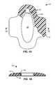
- a biopsy incision closure device 10comprises a base 12 having an integrated or embedded frame 14 , typically formed by overmolding a soft polymeric base material over a preformed metal or hard plastic frame.
- the frame 14is resilient and, when free from biasing forces, assumes the elliptical or oval configuration seen in FIG. 1A .
- the frame 14has living hinges 18 at each end (only one of which is visible in the broken-away section of the base) which allow the base to be closed by applying laterally inward forces to the frame, as shown in FIGS. 1C and 1D . Laterally inward forces may be provided by any one of a variety of external closure devices which could be simple tapes, patches, sutures, or the like.
- Closure devicescould be more complex, including zippers, clips, and other structures as taught in copending PCT Application PCT/US2010/00430, the full disclosure of which has been previously incorporated herein by reference.
- the opposed legs of the frame 14can be brought from their curved or arcuate (arched) configuration on the frame and base are free from vising forces, as shown in FIGS. 1A and 1B , to a generally straight configuration as shown in FIGS. 1C and 1D .
- Such straight closureis advantageous for closing elliptical biopsy cavities as described in more detail below.
- the framedefines an opening 20 in the base which is available for performing the biopsy after the device 10 has been adhered to a target tissue surface, typically using an adhesive layer 16 on a bottom surface of the base and frame, as best seen in FIGS. 1B and 1D .
- FIGS. 2A-2DA second biopsy incision closure device 24 is illustrated in FIGS. 2A-2D .
- the closure device 24includes a base 26 and frame 28 , similar to the device 10 , but differs from the device 10 in that device 24 includes a latch mechanism 34 for effecting closure as will be described below.
- Other differencesinclude the use of keyhole hinges 30 at the axial ends of each leg of the frame 28 and the presence of cut outs 32 around the perimeter of the base 26 .
- the cut outsfurther increase the axial elasticity of the base and allow it to both stretch and conform to the tissue as the frame closes and the base elongates, as shown in FIGS. 2C and 2B (although the cut outs 32 are not shown in those figures).
- the base 26 and frame 28together define an elliptical opening 42 which is fully open when ratchet number 36 of the latching mechanism 34 is open, as shown in FIGS. 2A and 2B .
- the physicianmay close the opening 42 by pressing laterally inwardly or “squeezing” the frame 28 to cause the ratchets of ratchet member 36 to move through the coupler 38 .
- the frame 28 and base 26can be partially closed, as shown in FIG. 2C , or fully closed as shown in FIG. 2D , depending on the desires of the physician.
- the base 26will typically have an adhesive on the surface which engages tissue, although an adhesive could be separately applied to the tissue or other attachment devices, such as sutures or staples could be used.
- a further difference in the device 24is that it includes a plurality of reinforcement members 44 ( FIG. 2B ) which project laterally outwardly from the legs of the frame 28 . These reinforcement numbers are embedded in the base material in order to provide for lateral reinforcement to inhibit lateral stretching of the base material as the legs are closed as well as to inhibit inward twisting of the legs of the frame.
- FIG. 3An alternative latching mechanism 50 is shown on a frame 52 in FIG. 3 .
- the latching mechanism 50does not include a ratchet but instead includes an arm 54 attached by a hinge 56 and having a plurality of holes 58 along its length.
- the holes 58may be snapped over pin 60 , with each individual hole representing a different closure spacing for the legs of the frame 52 .
- FIG. 4An alternative frame structure 70 is shown in FIG. 4 where the frame is not a simply elliptically shaped element but instead comprises a plurality of cells 72 which together form an elliptical scaffold for incorporation in the elastomeric or other base. This structure can improve the adherence when the frame is overmolded with the base material.
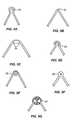
- FIGS. 5A through 5Gin addition to a simple keyhole hinge 80 ( FIG. 5A ) and simple living hinge 82 ( FIG. 5B ), individual legs of the frame maybe joined by a variety of other hinge structures.
- the double living hinge 84is illustrated at FIG. 5C and a ball and socket hinge 86 is illustrated in FIG. 5D .
- a coil spring hinge 86is illustrated in FIG. 5E and a pivoted hinge 88 is illustrated in FIG. 5F .
- the ends of the legs of the frameneed not be directly in contact and can instead be connected by a third element, such as an elastomeric matrix 90 as shown in FIG. 5G .
- FIGS. 5H and 5Iillustrate a particularly useful hinge configuration for the individual legs of the frame of the present invention.
- individual legs 102 and 104 of a frame and a base 106can be loosely attached in a “scissored” or “leaf spring” configuration 108 at each end.
- the legsthen extend to outwardly flared tips 110 and individual pods 112 of the base which can be attached to tissue in the configuration as shown in FIG. 5H .
- the closure device at FIG. 5Ais closed, as shown in FIG. 5I , the central portion of the closure device will apply laterally inward forces as shown by the arrows, while the pod elements 112 at each end will apply laterally outward forces as shown by the arrows at those end.
- Such as a “leaf spring” frame structureboth closes the incision to a vertical line, as shown in FIG. 5I , and also provide for outward movement of the tissue at the ends which will flatten the tissue and improve healing.
- the closure device 120includes a base 122 and frame 124 having different configurations but serving the same purposes as described in the previous embodiments.
- the most significant difference with device 120is that a latch element 126 is formed as a separate piece, i.e., it is not attached to the frame or to the base.
- Latch element 126will typically have a ratcheting structure (not shown) and can be inserted through an insertion channel 128 , across the opening 130 and into an aperture 132 which is adjacent a ratcheting closure mechanism 134 .
- a template 140may be inserted into opening 142 of any one of the biopsy closure devices, shown generically as closure device 144 .
- closure device 144By then drawing a line around the inner periphery of the template 140 , removing the template, and cutting along the drawn line, a precisely defined tissue cavity will be formed.
- the closure devicewill then close the incision with small marginally or peripheral edges of the tissue being brought together to optimally compress and close the wound.
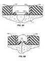
- FIGS. 8A and 8Ban alternative mechanism for improving the tissue apposition is illustrated.
- Everting rails 150maybe formed at the base of individual legs 152 and a closure device having a base 154 . After an incision 1 is formed in the tissue T, the legs 152 will be closed as describe above, causing the everting rails 150 to rise and raise the edges of the tissue, as shown in FIG. 8B . Such raised tissue edges can improve the healing and reduce any cavities remaining below the surface of the tissue.
- FIGS. 9A through 9Buse of the biopsy incision closure device 24 illustrated in FIGS. 2A through 2D for forming and closing a biopsy incision will be described.
- a target site TSis identified in a tissue surface T.
- the device 24is then placed over the target site TS with the opening in the device generally symmetrically placed over the site.
- the template 140 FIG. 7Ais used to draw an incision line, and then an incision is cut or then the opening of the device 24 , shown in FIG. 9B .
- the ratchet member 36is closed and inserted in the coupler 38 , as illustrated in FIG. 9C .
- Physiciancan then squeeze the opposed legs of the frame 28 together so that the ratchet member 36 advances from the coupler 38 and eventually closes the tissue to the extent desired by the physician, as shown in FIG. 9D .
- the closure devicecan then be left in place for time sufficient for the wound to heal.
Landscapes
- Health & Medical Sciences (AREA)
- Life Sciences & Earth Sciences (AREA)
- Surgery (AREA)
- Heart & Thoracic Surgery (AREA)
- Engineering & Computer Science (AREA)
- Biomedical Technology (AREA)
- Nuclear Medicine, Radiotherapy & Molecular Imaging (AREA)
- Medical Informatics (AREA)
- Molecular Biology (AREA)
- Animal Behavior & Ethology (AREA)
- General Health & Medical Sciences (AREA)
- Public Health (AREA)
- Veterinary Medicine (AREA)
- Surgical Instruments (AREA)
Abstract
Description
Claims (18)
Priority Applications (1)
| Application Number | Priority Date | Filing Date | Title |
|---|---|---|---|
| US13/286,378US8313508B2 (en) | 2010-05-03 | 2011-11-01 | Biopsy incision closure device |
Applications Claiming Priority (5)
| Application Number | Priority Date | Filing Date | Title |
|---|---|---|---|
| US34371610P | 2010-05-03 | 2010-05-03 | |
| US39760410P | 2010-06-14 | 2010-06-14 | |
| US201161462329P | 2011-02-01 | 2011-02-01 | |
| US13/096,602US8439945B2 (en) | 2010-05-03 | 2011-04-28 | Methods for biopsying tissue |
| US13/286,378US8313508B2 (en) | 2010-05-03 | 2011-11-01 | Biopsy incision closure device |
Related Parent Applications (1)
| Application Number | Title | Priority Date | Filing Date |
|---|---|---|---|
| US13/096,602ContinuationUS8439945B2 (en) | 2010-05-03 | 2011-04-28 | Methods for biopsying tissue |
Publications (2)
| Publication Number | Publication Date |
|---|---|
| US20120046691A1 US20120046691A1 (en) | 2012-02-23 |
| US8313508B2true US8313508B2 (en) | 2012-11-20 |
Family
ID=44903997
Family Applications (3)
| Application Number | Title | Priority Date | Filing Date |
|---|---|---|---|
| US13/096,602ActiveUS8439945B2 (en) | 2010-05-03 | 2011-04-28 | Methods for biopsying tissue |
| US13/286,378Expired - Fee RelatedUS8313508B2 (en) | 2010-05-03 | 2011-11-01 | Biopsy incision closure device |
| US13/874,046AbandonedUS20140074156A1 (en) | 2010-05-03 | 2013-04-30 | Biopsy incision closure device |
Family Applications Before (1)
| Application Number | Title | Priority Date | Filing Date |
|---|---|---|---|
| US13/096,602ActiveUS8439945B2 (en) | 2010-05-03 | 2011-04-28 | Methods for biopsying tissue |
Family Applications After (1)
| Application Number | Title | Priority Date | Filing Date |
|---|---|---|---|
| US13/874,046AbandonedUS20140074156A1 (en) | 2010-05-03 | 2013-04-30 | Biopsy incision closure device |
Country Status (6)
| Country | Link |
|---|---|
| US (3) | US8439945B2 (en) |
| EP (1) | EP2566398B1 (en) |
| JP (1) | JP5700875B2 (en) |
| CN (1) | CN102946812B (en) |
| AU (1) | AU2011248462B2 (en) |
| WO (1) | WO2011139912A1 (en) |
Cited By (22)
| Publication number | Priority date | Publication date | Assignee | Title |
|---|---|---|---|---|
| US20120016410A1 (en)* | 2010-05-03 | 2012-01-19 | Zipline Medical, Inc. | Biopsy incision closure device |
| US20120109161A1 (en)* | 2010-10-27 | 2012-05-03 | Salvatore Privitera | Appendage clamp deployment assist device |
| WO2014070922A1 (en) | 2012-10-31 | 2014-05-08 | Zipline Medical, Inc. | Surgical incision and closure apparatus |
| US8834514B2 (en) | 2006-08-30 | 2014-09-16 | Xennovate Medical Llc | Resilient band medical device |
| US9050086B2 (en) | 2011-11-01 | 2015-06-09 | Zipline Medical, Inc. | Surgical incision and closure apparatus |
| US9089328B2 (en) | 2011-11-01 | 2015-07-28 | Zipline Medical, Inc. | Surgical incision and closure apparatus |
| US9561034B2 (en) | 2011-11-01 | 2017-02-07 | Zipline Medical, Inc. | Surgical incision and closure apparatus |
| US9603596B2 (en) | 2004-08-31 | 2017-03-28 | Dermaclip Us, Llc | Systems and methods for closing a tissue opening |
| US9955960B2 (en) | 2011-02-26 | 2018-05-01 | Abbott Cardiovascular Systems, Inc. | Hinged tissue support device |
| US10010710B2 (en) | 2009-09-17 | 2018-07-03 | Zipline Medical, Inc. | Rapid closing surgical closure device |
| US10123801B2 (en) | 2011-11-01 | 2018-11-13 | Zipline Medical, Inc. | Means to prevent wound dressings from adhering to closure device |
| US10143460B2 (en) | 2013-01-17 | 2018-12-04 | Abbott Cardiovascular Systems, Inc. | Access device for accessing tissue |
| US10182824B2 (en) | 2010-11-11 | 2019-01-22 | Atricure, Inc. | Clip applicator |
| US10441261B2 (en) | 2014-04-29 | 2019-10-15 | Roffe Medical Holdings Pty Ltd. | Tissue closing method and apparatus |
| US10631862B2 (en) | 2015-08-05 | 2020-04-28 | Dq Holdings, Llc | Non-invasive wound closure device |
| US10888269B2 (en) | 2014-01-05 | 2021-01-12 | Zipline Medical, Inc. | Instrumented wound closure device |
| US11051988B2 (en) | 2010-06-14 | 2021-07-06 | Zipline Medical, Inc. | Methods and apparatus for inhibiting scar formation |
| US11864766B2 (en) | 2018-08-31 | 2024-01-09 | Zipline Medical, Inc. | Closure apparatuses and methods for ulcers and irregular skin defects |
| US11998212B2 (en) | 2013-11-21 | 2024-06-04 | Atricure, Inc. | Occlusion clip |
| US12004752B2 (en) | 2012-11-21 | 2024-06-11 | Atricure, Inc. | Occlusion clip |
| US12096937B2 (en) | 2019-04-25 | 2024-09-24 | Dq Holdings, Llc | Skin closure devices |
| US12171432B2 (en) | 2011-11-01 | 2024-12-24 | Zipline Medical, Inc. | Closure apparatuses and methods for ulcers and irregular skin defects |
Families Citing this family (22)
| Publication number | Priority date | Publication date | Assignee | Title |
|---|---|---|---|---|
| WO2011043786A1 (en) | 2009-09-17 | 2011-04-14 | Zipline Medical, Inc. | Rapid closing surgical closure device |
| US8323313B1 (en) | 2011-11-01 | 2012-12-04 | Zipline Medical, Inc. | Surgical incision and closure apparatus with integrated force distribution |
| US10888375B2 (en) | 2012-07-06 | 2021-01-12 | The General Hospital Corporation | Method and apparatus for dermatological treatment |
| JP6185580B2 (en)* | 2012-07-06 | 2017-08-23 | ザ ジェネラル ホスピタル コーポレイション | Method and apparatus for skin treatment |
| US9610069B2 (en)* | 2013-04-26 | 2017-04-04 | Medtronic-Xomed, Inc. | Tissue stabilization and repair device |
| GB201310190D0 (en)* | 2013-06-07 | 2013-07-24 | Brownson Peter | Sutureless wound closure |
| KR101826450B1 (en)* | 2013-07-24 | 2018-02-06 | 집라인 메디칼, 인크. | Surgical incision and closure apparatus |
| WO2015103423A1 (en) | 2013-12-31 | 2015-07-09 | Confluence Llc | Medical devices, dressings, and methods for closing openings in tissue |
| CN103690208B (en)* | 2013-12-31 | 2016-04-06 | 常州洛克曼医疗器械有限公司 | Needle-free skin surface anastomat |
| CN103948407B (en)* | 2014-05-20 | 2016-03-02 | 李绪国 | A kind of wound closer |
| WO2018081795A1 (en) | 2016-10-31 | 2018-05-03 | Zipline Medical, Inc. | Systems and methods for monitoring physical therapy of the knee and other joints |
| WO2019221201A1 (en)* | 2018-05-16 | 2019-11-21 | テルモ株式会社 | Compression device and compression method |
| GB2574074B (en) | 2018-07-27 | 2020-05-20 | Mclaren Applied Tech Ltd | Time synchronisation |
| CN109589152B (en)* | 2019-01-14 | 2020-05-22 | 郑州大学第一附属医院 | Portable wound suturing tool for tumor surgery |
| WO2020196857A1 (en)* | 2019-03-28 | 2020-10-01 | テルモ株式会社 | Fusion promotion device |
| CN113646016B (en)* | 2019-03-28 | 2023-07-11 | 泰尔茂株式会社 | medical instruments |
| WO2020196856A1 (en)* | 2019-03-28 | 2020-10-01 | テルモ株式会社 | Medical device |
| JP7386232B2 (en)* | 2019-03-28 | 2023-11-24 | テルモ株式会社 | medical device |
| CN111839635B (en)* | 2019-04-24 | 2025-01-21 | 上海锦辰医药科技有限公司 | Wound closure device and connection unit |
| GB2588236B (en) | 2019-10-18 | 2024-03-20 | Mclaren Applied Ltd | Gyroscope bias estimation |
| CN111249066B (en)* | 2020-03-20 | 2022-09-23 | 段冬雨 | Breathable and medicine-applying band-aid |
| KR102789467B1 (en)* | 2022-06-29 | 2025-04-03 | 고려대학교 산학협력단 | Non-invasive surgical skin closure with retractor |
Citations (41)
| Publication number | Priority date | Publication date | Assignee | Title |
|---|---|---|---|---|
| US2012755A (en) | 1934-07-12 | 1935-08-27 | Muth Otto De | Surgical dressing |
| US2747248A (en) | 1952-10-04 | 1956-05-29 | Bert M Mercer | Pin type fastening device |
| US3487836A (en)* | 1968-07-16 | 1970-01-06 | Benjamin W Niebel | Surgical strip stitch |
| US3516409A (en) | 1968-02-28 | 1970-06-23 | Robert B Howell | Slide fastener employing skin closure appliances and techniques |
| US3863640A (en) | 1973-04-27 | 1975-02-04 | Charles B Haverstock | Bandage construction |
| US3926193A (en) | 1971-12-17 | 1975-12-16 | Harrith M Hasson | Surgical closure having ease of assembly |
| US3933158A (en) | 1974-02-15 | 1976-01-20 | Haverstock Charles B | Skin closure means |
| US3971384A (en)* | 1971-03-12 | 1976-07-27 | Hasson Harrith M | Surgical closure |
| US4038989A (en) | 1975-07-04 | 1977-08-02 | Canadian Patents And Development Limited | Surgical skin closure |
| US4526173A (en) | 1982-04-12 | 1985-07-02 | Kells Medical, Inc. | Skin closure device |
| US4535772A (en) | 1983-03-10 | 1985-08-20 | Kells Medical, Incorporated | Skin closure device |
| US4605005A (en) | 1982-04-12 | 1986-08-12 | Kells Medical, Inc. | Wound closure device and method for using same |
| US4676245A (en) | 1983-02-09 | 1987-06-30 | Mamoru Fukuda | Interlocking surgical staple assembly |
| US4881546A (en) | 1986-12-16 | 1989-11-21 | Opti-Patents-, Forschungs-Und Fabrikations-Ag | Wound-closure device and method |
| US4905694A (en) | 1989-04-04 | 1990-03-06 | Ethicon, Inc. | Intracorporeal temporary wound closure |
| US4976726A (en) | 1989-04-27 | 1990-12-11 | Haverstock Charles B | Skin closure devices |
| US5377695A (en) | 1994-01-13 | 1995-01-03 | An Haack; Karl W. | Wound-closing strip |
| US5514155A (en) | 1993-12-14 | 1996-05-07 | Daneshvar; Yousef | Device for applying pressure to a person's groin |
| US5665108A (en) | 1996-09-16 | 1997-09-09 | Galindo; Eugene R. | Surgical dressing strap |
| US6126615A (en) | 1998-07-10 | 2000-10-03 | Allen; Michael E | Sutureless guided skin biopsy system |
| US6176868B1 (en) | 1999-11-22 | 2001-01-23 | Didier Detour | Device for the non-invasive sutureless closure of the open edges of wound in the skin of a mammal |
| US20050020956A1 (en) | 2003-07-24 | 2005-01-27 | Clozex Medical, Llc | Device for laceration or incision closure |
| US20050234485A1 (en) | 2001-02-16 | 2005-10-20 | Charles Seegert | Skin grafting devices and methods |
| US20060200198A1 (en) | 2004-08-31 | 2006-09-07 | Riskin Daniel J | Systems and methods for closing a tissue opening |
| US20070026078A1 (en) | 2002-02-15 | 2007-02-01 | Transform Pharmaceuticals, Inc. | Pharmaceutical co-crystal compositions |
| US20070088339A1 (en) | 2005-10-07 | 2007-04-19 | Luchetti Pablo C | Incision and closure surgical device |
| US20070141130A1 (en) | 2005-12-15 | 2007-06-21 | Kimberly-Clark Worldwide, Inc. | Wound or surgical dressing |
| US20070260278A1 (en) | 2006-05-03 | 2007-11-08 | Raptor Ridge, Llc | Systems and methods of tissue closure |
| US20080033334A1 (en) | 2006-08-03 | 2008-02-07 | Gurtner Geoffrey C | Devices and bandages for the treatment or prevention of scars and/or keloids and methods and kits therefor |
| US20080081951A1 (en) | 2006-09-29 | 2008-04-03 | Depuy Spine, Inc. | Inflatable retractor |
| US20080114396A1 (en) | 2006-11-15 | 2008-05-15 | Precision Closure Llc | Adjustable non-invasive wound closure system |
| US20080287864A1 (en) | 1999-09-22 | 2008-11-20 | Rosenberg Zeil B | Method and Apparatus for the Transdermal Administration of a Substance |
| US7455681B2 (en) | 2004-09-13 | 2008-11-25 | Wound Care Technologies, Llc | Wound closure product |
| US20090036922A1 (en) | 2004-08-31 | 2009-02-05 | Riskin Daniel J | Systems and methods for closing a tissue opening |
| US20090099496A1 (en) | 2005-10-05 | 2009-04-16 | Medtreo, Llc | Pressure bandage with medication delivery system |
| US20090299257A1 (en) | 2008-05-30 | 2009-12-03 | Justin Alexander Long | Reduced-pressure surgical wound treatment systems and methods |
| US20090299303A1 (en) | 2008-05-30 | 2009-12-03 | Charles Alan Seegert | Reduced-pressure, linear wound closing bolsters and systems |
| US7645285B2 (en)* | 2004-05-26 | 2010-01-12 | Idx Medical, Ltd | Apparatus and methods for occluding a hollow anatomical structure |
| US20100121286A1 (en) | 2008-11-07 | 2010-05-13 | Christopher Brian Locke | Reduced-pressure, wound-treatment dressings and systems |
| US20100280545A1 (en)* | 2007-05-10 | 2010-11-04 | Seraffix Ltd. | System and method for bonding living tissue |
| WO2011043786A1 (en) | 2009-09-17 | 2011-04-14 | Zipline Medical, Inc. | Rapid closing surgical closure device |
Family Cites Families (12)
| Publication number | Priority date | Publication date | Assignee | Title |
|---|---|---|---|---|
| US2371978A (en) | 1941-12-13 | 1945-03-20 | Roy G Perham | Clamp for retaining the edges of a wound in apposition |
| CA1006064A (en)* | 1971-12-17 | 1977-03-01 | Harrith M. Hasson | Surgical closure |
| US4576163A (en)* | 1983-08-08 | 1986-03-18 | Bliss Robert J | Skin marker for use in biopsy excisions |
| US4539990A (en)* | 1983-09-16 | 1985-09-10 | Stivala Oscar G | Sutureless closure system |
| US4871367A (en)* | 1987-09-03 | 1989-10-03 | Sutter Biomedical Corporation | Surgically implanted prosthesis |
| US7361185B2 (en)* | 2001-05-09 | 2008-04-22 | Canica Design, Inc. | Clinical and surgical system and method for moving and stretching plastic tissue |
| MXPA02010751A (en)* | 2000-05-10 | 2003-03-10 | Canica Design Inc | System and method for moving and stretching plastic tissue. |
| US7306568B2 (en)* | 2003-01-06 | 2007-12-11 | Richard Diana | Method and device for treatment of edema |
| US7799042B2 (en)* | 2004-05-13 | 2010-09-21 | The Cleveland Clinic Foundation | Skin lesion exciser and skin-closure device therefor |
| EP2444009A1 (en)* | 2005-05-12 | 2012-04-25 | Canica Design Inc. | Dynamic tensioning system and method |
| WO2010117908A1 (en)* | 2009-04-06 | 2010-10-14 | University Of Virginia Patent Foundation | Anisotropic reinforcement and related method thereof |
| US8439945B2 (en) | 2010-05-03 | 2013-05-14 | Zipline Medical, Inc. | Methods for biopsying tissue |
- 2011
- 2011-04-28USUS13/096,602patent/US8439945B2/enactiveActive
- 2011-04-29CNCN201180021453.7Apatent/CN102946812B/enactiveActive
- 2011-04-29EPEP11778067.6Apatent/EP2566398B1/ennot_activeNot-in-force
- 2011-04-29WOPCT/US2011/034649patent/WO2011139912A1/enactiveApplication Filing
- 2011-04-29JPJP2013509130Apatent/JP5700875B2/ennot_activeExpired - Fee Related
- 2011-04-29AUAU2011248462Apatent/AU2011248462B2/ennot_activeCeased
- 2011-11-01USUS13/286,378patent/US8313508B2/ennot_activeExpired - Fee Related
- 2013
- 2013-04-30USUS13/874,046patent/US20140074156A1/ennot_activeAbandoned
Patent Citations (43)
| Publication number | Priority date | Publication date | Assignee | Title |
|---|---|---|---|---|
| US2012755A (en) | 1934-07-12 | 1935-08-27 | Muth Otto De | Surgical dressing |
| US2747248A (en) | 1952-10-04 | 1956-05-29 | Bert M Mercer | Pin type fastening device |
| US3516409A (en) | 1968-02-28 | 1970-06-23 | Robert B Howell | Slide fastener employing skin closure appliances and techniques |
| US3487836A (en)* | 1968-07-16 | 1970-01-06 | Benjamin W Niebel | Surgical strip stitch |
| US3971384A (en)* | 1971-03-12 | 1976-07-27 | Hasson Harrith M | Surgical closure |
| US3926193A (en) | 1971-12-17 | 1975-12-16 | Harrith M Hasson | Surgical closure having ease of assembly |
| US3863640A (en) | 1973-04-27 | 1975-02-04 | Charles B Haverstock | Bandage construction |
| US3933158A (en) | 1974-02-15 | 1976-01-20 | Haverstock Charles B | Skin closure means |
| US4114624A (en) | 1974-02-15 | 1978-09-19 | Haverstock Charles B | Skin closure means |
| US4038989A (en) | 1975-07-04 | 1977-08-02 | Canadian Patents And Development Limited | Surgical skin closure |
| US4526173A (en) | 1982-04-12 | 1985-07-02 | Kells Medical, Inc. | Skin closure device |
| US4605005A (en) | 1982-04-12 | 1986-08-12 | Kells Medical, Inc. | Wound closure device and method for using same |
| US4676245A (en) | 1983-02-09 | 1987-06-30 | Mamoru Fukuda | Interlocking surgical staple assembly |
| US4535772A (en) | 1983-03-10 | 1985-08-20 | Kells Medical, Incorporated | Skin closure device |
| US4881546A (en) | 1986-12-16 | 1989-11-21 | Opti-Patents-, Forschungs-Und Fabrikations-Ag | Wound-closure device and method |
| US4905694A (en) | 1989-04-04 | 1990-03-06 | Ethicon, Inc. | Intracorporeal temporary wound closure |
| US4976726A (en) | 1989-04-27 | 1990-12-11 | Haverstock Charles B | Skin closure devices |
| US5514155A (en) | 1993-12-14 | 1996-05-07 | Daneshvar; Yousef | Device for applying pressure to a person's groin |
| US5377695A (en) | 1994-01-13 | 1995-01-03 | An Haack; Karl W. | Wound-closing strip |
| US5665108A (en) | 1996-09-16 | 1997-09-09 | Galindo; Eugene R. | Surgical dressing strap |
| US6126615A (en) | 1998-07-10 | 2000-10-03 | Allen; Michael E | Sutureless guided skin biopsy system |
| US20080287864A1 (en) | 1999-09-22 | 2008-11-20 | Rosenberg Zeil B | Method and Apparatus for the Transdermal Administration of a Substance |
| US6176868B1 (en) | 1999-11-22 | 2001-01-23 | Didier Detour | Device for the non-invasive sutureless closure of the open edges of wound in the skin of a mammal |
| US20050234485A1 (en) | 2001-02-16 | 2005-10-20 | Charles Seegert | Skin grafting devices and methods |
| US20070026078A1 (en) | 2002-02-15 | 2007-02-01 | Transform Pharmaceuticals, Inc. | Pharmaceutical co-crystal compositions |
| US20050020956A1 (en) | 2003-07-24 | 2005-01-27 | Clozex Medical, Llc | Device for laceration or incision closure |
| US7645285B2 (en)* | 2004-05-26 | 2010-01-12 | Idx Medical, Ltd | Apparatus and methods for occluding a hollow anatomical structure |
| US20060200198A1 (en) | 2004-08-31 | 2006-09-07 | Riskin Daniel J | Systems and methods for closing a tissue opening |
| US20090036922A1 (en) | 2004-08-31 | 2009-02-05 | Riskin Daniel J | Systems and methods for closing a tissue opening |
| US7455681B2 (en) | 2004-09-13 | 2008-11-25 | Wound Care Technologies, Llc | Wound closure product |
| US20090099496A1 (en) | 2005-10-05 | 2009-04-16 | Medtreo, Llc | Pressure bandage with medication delivery system |
| US20070088339A1 (en) | 2005-10-07 | 2007-04-19 | Luchetti Pablo C | Incision and closure surgical device |
| US20070141130A1 (en) | 2005-12-15 | 2007-06-21 | Kimberly-Clark Worldwide, Inc. | Wound or surgical dressing |
| US20070260278A1 (en) | 2006-05-03 | 2007-11-08 | Raptor Ridge, Llc | Systems and methods of tissue closure |
| US20080033334A1 (en) | 2006-08-03 | 2008-02-07 | Gurtner Geoffrey C | Devices and bandages for the treatment or prevention of scars and/or keloids and methods and kits therefor |
| US20080081951A1 (en) | 2006-09-29 | 2008-04-03 | Depuy Spine, Inc. | Inflatable retractor |
| US20080114396A1 (en) | 2006-11-15 | 2008-05-15 | Precision Closure Llc | Adjustable non-invasive wound closure system |
| US20100280545A1 (en)* | 2007-05-10 | 2010-11-04 | Seraffix Ltd. | System and method for bonding living tissue |
| US20090299255A1 (en) | 2008-05-30 | 2009-12-03 | Kazala Jr Richard Marvin | Reduced-pressure dressing assemblies for use in applying a closing force |
| US20090299303A1 (en) | 2008-05-30 | 2009-12-03 | Charles Alan Seegert | Reduced-pressure, linear wound closing bolsters and systems |
| US20090299257A1 (en) | 2008-05-30 | 2009-12-03 | Justin Alexander Long | Reduced-pressure surgical wound treatment systems and methods |
| US20100121286A1 (en) | 2008-11-07 | 2010-05-13 | Christopher Brian Locke | Reduced-pressure, wound-treatment dressings and systems |
| WO2011043786A1 (en) | 2009-09-17 | 2011-04-14 | Zipline Medical, Inc. | Rapid closing surgical closure device |
Non-Patent Citations (6)
| Title |
|---|
| International search report and written opinion dated Jul. 29, 2011 for PCT/US2011/034649. |
| International search report and written opinion dated Oct. 21, 2011 for PCT Application No. US2011/40213. |
| International search report dated Jul. 30, 2010 for PCT/US2010/000430. |
| U.S. Appl. No. 13/096,602, filed Apr. 28, 2011, Belson et al. |
| U.S. Appl. No. 13/286,757, filed Nov. 1, 2011, Belson et al. |
| U.S. Appl. No. 13/414,176, filed Mar. 7, 2012, Belson et al. |
Cited By (42)
| Publication number | Priority date | Publication date | Assignee | Title |
|---|---|---|---|---|
| US9603596B2 (en) | 2004-08-31 | 2017-03-28 | Dermaclip Us, Llc | Systems and methods for closing a tissue opening |
| US8834514B2 (en) | 2006-08-30 | 2014-09-16 | Xennovate Medical Llc | Resilient band medical device |
| US10159825B2 (en) | 2009-09-17 | 2018-12-25 | Zipline Medical, Inc. | Rapid closing surgical closure device |
| US10010710B2 (en) | 2009-09-17 | 2018-07-03 | Zipline Medical, Inc. | Rapid closing surgical closure device |
| US8439945B2 (en)* | 2010-05-03 | 2013-05-14 | Zipline Medical, Inc. | Methods for biopsying tissue |
| US20120016410A1 (en)* | 2010-05-03 | 2012-01-19 | Zipline Medical, Inc. | Biopsy incision closure device |
| US11051988B2 (en) | 2010-06-14 | 2021-07-06 | Zipline Medical, Inc. | Methods and apparatus for inhibiting scar formation |
| US10433854B2 (en) | 2010-10-27 | 2019-10-08 | Atricure, Inc. | Appendage clamp deployment assist device |
| US9017349B2 (en)* | 2010-10-27 | 2015-04-28 | Atricure, Inc. | Appendage clamp deployment assist device |
| US11883035B2 (en) | 2010-10-27 | 2024-01-30 | Atricure, Inc. | Appendage clamp deployment assist device |
| US20120109161A1 (en)* | 2010-10-27 | 2012-05-03 | Salvatore Privitera | Appendage clamp deployment assist device |
| US10182824B2 (en) | 2010-11-11 | 2019-01-22 | Atricure, Inc. | Clip applicator |
| US9955960B2 (en) | 2011-02-26 | 2018-05-01 | Abbott Cardiovascular Systems, Inc. | Hinged tissue support device |
| US9554800B2 (en) | 2011-11-01 | 2017-01-31 | Zipline Medical, Inc. | Surgical incision and closure apparatus |
| US9089328B2 (en) | 2011-11-01 | 2015-07-28 | Zipline Medical, Inc. | Surgical incision and closure apparatus |
| US9642622B2 (en) | 2011-11-01 | 2017-05-09 | Zipline Medical, Inc. | Surgical incision and closure apparatus |
| US9561034B2 (en) | 2011-11-01 | 2017-02-07 | Zipline Medical, Inc. | Surgical incision and closure apparatus |
| US10123800B2 (en) | 2011-11-01 | 2018-11-13 | Zipline Medical, Inc. | Surgical incision and closure apparatus with integrated force distribution |
| US10123801B2 (en) | 2011-11-01 | 2018-11-13 | Zipline Medical, Inc. | Means to prevent wound dressings from adhering to closure device |
| US11439395B2 (en) | 2011-11-01 | 2022-09-13 | Zipline Medical, Inc. | Surgical incision and closure apparatus |
| US9554799B2 (en) | 2011-11-01 | 2017-01-31 | Zipline Medical, Inc. | Surgical incision and closure apparatus |
| US9474529B2 (en) | 2011-11-01 | 2016-10-25 | Zipline Medical, Inc. | Surgical incision and closure apparatus |
| US9050086B2 (en) | 2011-11-01 | 2015-06-09 | Zipline Medical, Inc. | Surgical incision and closure apparatus |
| US9642621B2 (en) | 2011-11-01 | 2017-05-09 | ZipLine Medical, Inc | Surgical incision and closure apparatus |
| US10456136B2 (en) | 2011-11-01 | 2019-10-29 | Zipline Medical, Inc. | Surgical incision and closure apparatus |
| US12171432B2 (en) | 2011-11-01 | 2024-12-24 | Zipline Medical, Inc. | Closure apparatuses and methods for ulcers and irregular skin defects |
| EP3574848A1 (en) | 2012-10-31 | 2019-12-04 | Zipline Medical, Inc. | Surgical incision and closure apparatus |
| WO2014070922A1 (en) | 2012-10-31 | 2014-05-08 | Zipline Medical, Inc. | Surgical incision and closure apparatus |
| US12004752B2 (en) | 2012-11-21 | 2024-06-11 | Atricure, Inc. | Occlusion clip |
| US12193680B2 (en) | 2012-11-21 | 2025-01-14 | Atricure, Inc. | Occlusion clip |
| US10143460B2 (en) | 2013-01-17 | 2018-12-04 | Abbott Cardiovascular Systems, Inc. | Access device for accessing tissue |
| US11998211B2 (en) | 2013-11-21 | 2024-06-04 | Atricure, Inc. | Occlusion clip |
| US11998212B2 (en) | 2013-11-21 | 2024-06-04 | Atricure, Inc. | Occlusion clip |
| US12076019B2 (en) | 2013-11-21 | 2024-09-03 | Atricure, Inc. | Occlusion clip |
| US11844625B2 (en) | 2014-01-05 | 2023-12-19 | Zipline Medical, Inc. | Instrumented wound closure device |
| US10888269B2 (en) | 2014-01-05 | 2021-01-12 | Zipline Medical, Inc. | Instrumented wound closure device |
| US10441261B2 (en) | 2014-04-29 | 2019-10-15 | Roffe Medical Holdings Pty Ltd. | Tissue closing method and apparatus |
| US10631862B2 (en) | 2015-08-05 | 2020-04-28 | Dq Holdings, Llc | Non-invasive wound closure device |
| US11033270B2 (en) | 2015-08-07 | 2021-06-15 | Zipline Medical, Inc. | Means to prevent wound dressings from adhering to closure device |
| US11864766B2 (en) | 2018-08-31 | 2024-01-09 | Zipline Medical, Inc. | Closure apparatuses and methods for ulcers and irregular skin defects |
| US12390221B2 (en) | 2018-08-31 | 2025-08-19 | Zipline Medical Inc. | Closure apparatuses and methods for ulcers and irregular skin defects |
| US12096937B2 (en) | 2019-04-25 | 2024-09-24 | Dq Holdings, Llc | Skin closure devices |
Also Published As
| Publication number | Publication date |
|---|---|
| EP2566398A1 (en) | 2013-03-13 |
| US20120046691A1 (en) | 2012-02-23 |
| EP2566398B1 (en) | 2017-06-07 |
| EP2566398A4 (en) | 2014-01-22 |
| US20140074156A1 (en) | 2014-03-13 |
| AU2011248462B2 (en) | 2014-10-09 |
| CN102946812B (en) | 2015-02-04 |
| US8439945B2 (en) | 2013-05-14 |
| CN102946812A (en) | 2013-02-27 |
| JP2013525070A (en) | 2013-06-20 |
| AU2011248462A1 (en) | 2012-11-01 |
| US20120016410A1 (en) | 2012-01-19 |
| JP5700875B2 (en) | 2015-04-15 |
| WO2011139912A1 (en) | 2011-11-10 |
Similar Documents
| Publication | Publication Date | Title |
|---|---|---|
| US8313508B2 (en) | Biopsy incision closure device | |
| US5584859A (en) | Suture assembly | |
| US8262567B2 (en) | Tissue retractor, tissue retractor kit and method of use thereof | |
| CA1230027A (en) | Retractors | |
| US20090281568A1 (en) | Devices and Methods for Adjustable, Knotless Tissue Approximation | |
| EP1596726B1 (en) | Sheathed elastic surgigal thread | |
| CN104248457B (en) | An artificial chord device, threading element and kit | |
| WO1996041581A1 (en) | Sternal closure device | |
| KR20210154846A (en) | One-way lifting tying thread, lifting device and forming method using the same | |
| US11571203B2 (en) | Deformable suture bridge having an insert and methods of manufacturing and using same | |
| CN113974738B (en) | Wound closer | |
| RU2465844C2 (en) | Face and neck lifting device | |
| US9060766B2 (en) | Suture fixation kit of parts, system, and device | |
| JP2023515758A (en) | Medical systems, devices and methods suitable for tissue fixation and tissue defect reduction | |
| RU2500357C1 (en) | Method of face and neck lifting | |
| CN216060622U (en) | Hook needle pull strip type skin stretching closer | |
| CN216317770U (en) | Bearded needle pull rod type skin stretching closer | |
| KR102207317B1 (en) | Apparatus of thread for surgery | |
| CN113812995A (en) | Crochet Pull Rod Skin Stretch Closer | |
| CN113796909A (en) | Hook needle pull strip type skin stretching closer | |
| CN219250279U (en) | Skin tightening, pulling and sewing combined hook | |
| US12075997B2 (en) | Non-invasive tissue retractor | |
| CN216257268U (en) | Bidirectional crochet type skin stretching closer | |
| RU2824278C2 (en) | Lifting suture for skin surgery | |
| CN209074745U (en) | A kind of Distraction Apparatus |
Legal Events
| Date | Code | Title | Description |
|---|---|---|---|
| AS | Assignment | Owner name:ZIPLINE MEDICAL, INC., CALIFORNIA Free format text:ASSIGNMENT OF ASSIGNORS INTEREST;ASSIGNORS:BELSON, AMIR;STORNE, ERIC;BECKEY, BRIAN;SIGNING DATES FROM 20110705 TO 20111001;REEL/FRAME:027210/0342 | |
| AS | Assignment | Owner name:VENTURE LENDING & LEASING VI, INC., CALIFORNIA Free format text:SECURITY AGREEMENT;ASSIGNOR:ZIPLINE MEDICAL, INC.;REEL/FRAME:028118/0109 Effective date:20120329 | |
| ZAAA | Notice of allowance and fees due | Free format text:ORIGINAL CODE: NOA | |
| ZAAB | Notice of allowance mailed | Free format text:ORIGINAL CODE: MN/=. | |
| STCF | Information on status: patent grant | Free format text:PATENTED CASE | |
| CC | Certificate of correction | ||
| FPAY | Fee payment | Year of fee payment:4 | |
| AS | Assignment | Owner name:ZIPLINE MEDICAL, INC., CALIFORNIA Free format text:RELEASE BY SECURED PARTY;ASSIGNOR:VENTURE LENDING & LEASING VI, INC.;REEL/FRAME:045897/0124 Effective date:20180524 | |
| FEPP | Fee payment procedure | Free format text:ENTITY STATUS SET TO UNDISCOUNTED (ORIGINAL EVENT CODE: BIG.); ENTITY STATUS OF PATENT OWNER: LARGE ENTITY | |
| MAFP | Maintenance fee payment | Free format text:PAYMENT OF MAINTENANCE FEE, 8TH YEAR, LARGE ENTITY (ORIGINAL EVENT CODE: M1552); ENTITY STATUS OF PATENT OWNER: LARGE ENTITY Year of fee payment:8 | |
| LAPS | Lapse for failure to pay maintenance fees | Free format text:PATENT EXPIRED FOR FAILURE TO PAY MAINTENANCE FEES (ORIGINAL EVENT CODE: EXP.); ENTITY STATUS OF PATENT OWNER: LARGE ENTITY | |
| STCH | Information on status: patent discontinuation | Free format text:PATENT EXPIRED DUE TO NONPAYMENT OF MAINTENANCE FEES UNDER 37 CFR 1.362 | |
| FP | Lapsed due to failure to pay maintenance fee | Effective date:20241120 |