US8311303B2 - Method and system for semantics driven image registration - Google Patents
Method and system for semantics driven image registrationDownload PDFInfo
- Publication number
- US8311303B2 US8311303B2US12/986,212US98621211AUS8311303B2US 8311303 B2US8311303 B2US 8311303B2US 98621211 AUS98621211 AUS 98621211AUS 8311303 B2US8311303 B2US 8311303B2
- Authority
- US
- United States
- Prior art keywords
- image
- images
- semantic information
- registration
- organs
- Prior art date
- Legal status (The legal status is an assumption and is not a legal conclusion. Google has not performed a legal analysis and makes no representation as to the accuracy of the status listed.)
- Active, expires
Links
Images


Classifications
- G—PHYSICS
- G06—COMPUTING OR CALCULATING; COUNTING
- G06T—IMAGE DATA PROCESSING OR GENERATION, IN GENERAL
- G06T7/00—Image analysis
- G06T7/30—Determination of transform parameters for the alignment of images, i.e. image registration
- G06T7/33—Determination of transform parameters for the alignment of images, i.e. image registration using feature-based methods
- G—PHYSICS
- G06—COMPUTING OR CALCULATING; COUNTING
- G06V—IMAGE OR VIDEO RECOGNITION OR UNDERSTANDING
- G06V10/00—Arrangements for image or video recognition or understanding
- G06V10/70—Arrangements for image or video recognition or understanding using pattern recognition or machine learning
- G06V10/74—Image or video pattern matching; Proximity measures in feature spaces
- G06V10/75—Organisation of the matching processes, e.g. simultaneous or sequential comparisons of image or video features; Coarse-fine approaches, e.g. multi-scale approaches; using context analysis; Selection of dictionaries
- G06V10/755—Deformable models or variational models, e.g. snakes or active contours
- G—PHYSICS
- G16—INFORMATION AND COMMUNICATION TECHNOLOGY [ICT] SPECIALLY ADAPTED FOR SPECIFIC APPLICATION FIELDS
- G16H—HEALTHCARE INFORMATICS, i.e. INFORMATION AND COMMUNICATION TECHNOLOGY [ICT] SPECIALLY ADAPTED FOR THE HANDLING OR PROCESSING OF MEDICAL OR HEALTHCARE DATA
- G16H30/00—ICT specially adapted for the handling or processing of medical images
- G16H30/40—ICT specially adapted for the handling or processing of medical images for processing medical images, e.g. editing
- G—PHYSICS
- G16—INFORMATION AND COMMUNICATION TECHNOLOGY [ICT] SPECIALLY ADAPTED FOR SPECIFIC APPLICATION FIELDS
- G16H—HEALTHCARE INFORMATICS, i.e. INFORMATION AND COMMUNICATION TECHNOLOGY [ICT] SPECIALLY ADAPTED FOR THE HANDLING OR PROCESSING OF MEDICAL OR HEALTHCARE DATA
- G16H70/00—ICT specially adapted for the handling or processing of medical references
- G16H70/60—ICT specially adapted for the handling or processing of medical references relating to pathologies
- G—PHYSICS
- G06—COMPUTING OR CALCULATING; COUNTING
- G06T—IMAGE DATA PROCESSING OR GENERATION, IN GENERAL
- G06T2207/00—Indexing scheme for image analysis or image enhancement
- G06T2207/10—Image acquisition modality
- G06T2207/10072—Tomographic images
- G06T2207/10081—Computed x-ray tomography [CT]
- G—PHYSICS
- G06—COMPUTING OR CALCULATING; COUNTING
- G06T—IMAGE DATA PROCESSING OR GENERATION, IN GENERAL
- G06T2207/00—Indexing scheme for image analysis or image enhancement
- G06T2207/10—Image acquisition modality
- G06T2207/10072—Tomographic images
- G06T2207/10088—Magnetic resonance imaging [MRI]
- G—PHYSICS
- G06—COMPUTING OR CALCULATING; COUNTING
- G06T—IMAGE DATA PROCESSING OR GENERATION, IN GENERAL
- G06T2207/00—Indexing scheme for image analysis or image enhancement
- G06T2207/10—Image acquisition modality
- G06T2207/10116—X-ray image
- G—PHYSICS
- G06—COMPUTING OR CALCULATING; COUNTING
- G06T—IMAGE DATA PROCESSING OR GENERATION, IN GENERAL
- G06T2207/00—Indexing scheme for image analysis or image enhancement
- G06T2207/10—Image acquisition modality
- G06T2207/10132—Ultrasound image
- G—PHYSICS
- G06—COMPUTING OR CALCULATING; COUNTING
- G06T—IMAGE DATA PROCESSING OR GENERATION, IN GENERAL
- G06T2207/00—Indexing scheme for image analysis or image enhancement
- G06T2207/30—Subject of image; Context of image processing
- G06T2207/30004—Biomedical image processing
- G06T2207/30096—Tumor; Lesion
Definitions
- the present inventionrelates to registration of medical images, and more particularly, to automatic semantics driven registration of medical images of a patient.
- Image registrationis a crucial technique to provide comparisons of medical images of a patient.
- image registrationcan be used to compare medical images of a tumor before and after some treatment is administered or for pre and post interventional (e.g., stent placements) medical image comparisons.
- Some state of the art workstationsprovide tools and algorithms for rigid image alignment (i.e., translation and rotation).
- rigid image alignmenti.e., translation and rotation
- the limited degrees of freedommay not be sufficient to ensure that corresponding anatomical structures in different medical images are well-aligned to each other.
- a variety of elastic image registration techniqueshave recently been proposed. In such techniques, a number of image similarity measures are typically used together with various optimization algorithms to attempt to ensure that corresponding structures in the medical images are matched to each other.
- these approachesutilize a regularization term that imposes some smoothness on the deformation field to make the problem less ill-posed. The resulting deformation field is therefore a compromise between attention to detail and numerical stability and divergence.
- the present inventionprovides a method and system for automatic semantic driven registration of medical images.
- Embodiments of the present inventionprovide a semantics drive image registration framework that can be applied to 2D and 3D image data from various imaging modalities, including magnetic resonance (MR), computed tomography (CT), ultrasound, X-ray, etc.
- MRmagnetic resonance
- CTcomputed tomography
- X-rayultrasound
- knowledge about anatomy, pathology, and imaging protocol parameters, such as contrast bolus phasecan be extracted from images by automatic parsing and semantic labeling.
- Clinical Context informationcan be obtained by semantic parsing of text-based data, such as Radiology Information System (RIS) information, clinical reports, or other DICOM header information. Additional clinical context information may be derived from user interactions during image reading. All extracted knowledge can be used to tune the registration focus to situation-specific diagnostic needs to ensure that anatomical structures of a particular diagnostic interest are aligned as precisely as possible.
- RISRadiology Information System
- anatomic landmarks and organsare detected in a first image and a second image.
- Semantic informationis automatically extracted from at least one text-based document associated with at least one of the first and second images.
- the second imageis registered to the first image based at least in part on the detected anatomic landmarks and organs and the extracted semantic information.
- Pathologiescan also be detected in the first and second images, and the second image can be registered to the first image based at least in part on the detected anatomic landmarks, organs, and pathologies, and the extracted semantic information.
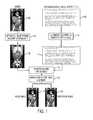
- FIG. 1illustrates the semantics driven image registration framework according to an embodiment of the present invention
- FIG. 2illustrates a method for automatic semantics driven medical image registration according to an embodiment of the present invention
- FIG. 3illustrates exemplary registration results refined based on user interaction
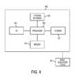
- FIG. 4is a high level block diagram of a computer capable of implementing the present invention.
- the present inventionis directed to a method and system for automatic semantics driven registration of medical images, such as computed tomography (CT), magnetic resonance (MR) images, ultrasound images, X-ray images, etc.
- CTcomputed tomography
- MRmagnetic resonance
- Embodiments of the present inventionare described herein to give a visual understanding of the image registration method.
- a digital imageis often composed of digital representations of one or more objects (or shapes).
- the digital representation of an objectis often described herein in terms of identifying and manipulating the objects.
- Such manipulationsare virtual manipulations accomplished in the memory or other circuitry/hardware of a computer system. Accordingly, it is to be understood that embodiments of the present invention may be performed within a computer system using data stored within the computer system.
- Embodiments of the present inventionare directed a semantics driven image registration framework that can be applied to 2D and 3D medical image data acquired using different imaging modalities, such as MR, CT, ultrasound, X-ray, etc.
- FIG. 1illustrates the semantics driven image registration framework according to an embodiment of the present invention.
- knowledge about anatomy, pathologies, and imaging protocol parameters, such as contrast bolus phaseis extracted from images 102 by automatic image parsing and semantic labeling at block 104 .
- the automatic image parsing for the anatomy and pathologyis used to obtain parsed image data 106 .
- Clinical contextual information 112is obtained by semantic parsing of text-based data 108 , such as radiology information system (RIS) information, clinical reports (narrative or DICOM Structured Reports (DICOM SR)), or other image DICOM header information using automatic text parsing of the text-based data 108 for both anatomy and pathology at block 110 .
- Additional clinical context informationmay also be derived from user interactions during image reading, such as switches to structure-specific windowing settings (e.g., lung windowing), performing measurements in a given image area, or eye focusing on a given image area during reading (e.g., using information obtained by an eye tracking system).
- the clinical context information 112can be correlated with the parsed image data 106 using ontology based semantic linking at block 114 .
- a model image 118is registered to a reference image 120 using knowledge-driven image alignment.
- all extracted knowledgeis used to tune the registration focus to situation specific diagnostic needs to ensure that structures of a given diagnostic interest are aligned as precisely as possible. Additional details regarding the framework of FIG. 1 are described below with reference to FIG. 2 .
- An advantageous aspect of the above-described semantics driven image registration frameworkis that is provides situation-specific alignment that is optimized for a given diagnostic need.
- This approachprovides fully-automatic grouping of image datasets into sets of corresponding anatomical regions and contrast phases such that corresponding data can be aligned during an automatic pre-processing step before a user begins reading the case. Since the fully-elastic image registration has a high computational complexity and sometimes has too many degrees of freedom that make it prone to undesired misalignments and divergence, embodiments of the present invention ensure that image structures of given diagnostic interest are aligned more precisely using transformation models with optimal degrees of freedom and model parameters.
- Embodiments of the present inventionmay render elastic registration approaches more robust and accurate by enforcing the precise alignment of identified anchor landmark pairs and segmented and labeled organs and structures. Rather than keeping a fixed, pre-computed image alignment, embodiments of the present invention may “interpret” user interactions (e.g., change of windowing settings or eye focus) and optimize the image alignment to situation-specific needs.
- FIG. 2illustrates a method for automatic semantics driven medical image registration according to an embodiment of the present invention.
- the method of FIG. 2transforms medical image data representing the anatomy of a patient to register two or more medical images of a patient to each other.
- at step 202at least one first image and at least one second image are received.
- the first and second imagescan be 2D or 3D medical images generated using any type of medical imaging modality, such as MR, CT, X-ray, ultrasound, etc.
- the medical imagescan be received directly from an image acquisition device (e.g., MR scanner, CT scanner, etc.) or can be received by loading a medical image that was previously stored, for example on a memory or storage of a computer system or a computer readable medium.
- the first imageis a stored previously acquired reference image and the second image is a newly acquired model image received from an image acquisition device.
- the at least one first imagemay include a plurality of reference scans acquired at different contrast phases in order to register a second image with the reference scan having the most similar contrast phase.
- anatomic landmarks and organsare detected independently in to the first image and the second image.
- automatic image parsingis used to detect and label anatomic structures, including anatomic landmarks and organs in the first and second images.
- anatomic landmarks and organscan be detected in the first and second images using the method described in United States Published Patent Application No. 2010/0080434, which is incorporated herein by reference.
- one or more predetermined slices of a 3D medical imagecan be detected.
- a plurality of anatomic landmarks and organ centerscan then be detected in the image using a discriminative anatomical network, where each landmark and organ center is detected in a portion of the image constrained by at least one of the detected slices.
- a plurality of organssuch as heart, liver, kidneys, spleen, bladder, and prostate, can then be detected as a bounding box and segmented in the image, where the detection of each organ bounding box is constrained by the detected organ centers and anatomic landmarks.
- the organ segmentationcan be performed via a database-guided segmentation method.
- the hierarchical parsing methodcan be used to automatically detect and label various anatomic structures including, but not limited to body landmarks, including: bones, such as the sternum, hip bones, knees, etc.; airways, such as the tracheal bifurcation (carina); and organs, such as the lung tips, liver dome and liver lobe tips, and kidney center; and vascular landmarks, such as vessel bifurcations (e.g., iliac and renal arteries); portal vein; and aortic arch.
- body landmarksincluding: bones, such as the sternum, hip bones, knees, etc.
- airwayssuch as the tracheal bifurcation (carina)
- organssuch as the lung tips, liver dome and liver lobe tips, and kidney center
- vascular landmarkssuch as vessel bifurcations (e.g., iliac and renal arteries); portal vein; and aortic arch.
- Each anatomical landmark xis associated with a detection confidence scored that expresses the confidence of the detection to be a true positive. Beyond landmarks (positions in space), bounding boxes and organ segmentations, such as for the liver, lungs, etc., are detected automatically. All anatomical structures can be labeled based on existing ontologies, such as the Foundational Model of Anatomy (FMA).
- FMAFoundational Model of Anatomy
- the contrast phasecan be automatically determined for each of the first and second images. For example, in order to detect the contrast phase, a local volume of interest can be estimated at each of the detected plurality of anatomic landmarks and features can be extracted from each local volume of interest. The contrast phase of the 3D volume can then be determined based on the extracted features using a trained classifier. This method of automatically determining a contrast phase is described in greater detail in Unite States Published Patent Application No. 2011/0002520, which is incorporated herein by reference.
- pathologiesare detected in the first and second images.
- pathology landmarksare detected.
- the detected pathologiesmay include, but are not limited t: lesions, such as lung lesions, liver lesions, lymph nodes, and bone lesions; and vascular abnormalities, such as calcifications, aneurysms, and thrombi.
- the pathologiesmay be detected in the first and second images using the integrated approach for lesion detection described in U.S. patent application Ser. No. 12/831,392, filed Jul. 7, 2010 and entitled “Method and System for Database-Guided Lesion Detection”, which is incorporated herein by reference.
- search regionsare defined in the 3D medical image based on the detected anatomical landmarks, organs, and bone structures. Lesions are then detected in each search region using a trained region-specific lesion detector. This method returns the position and bounding box of lesions, which may then also be used to trigger automatic lesion segmentation.
- Each pathology landmark x iis associated with a detection confidence score ⁇ i that expresses the confidence of the detection to be a true positive. All extracted pathologies are labeled based on existing ontologies, such as the International Classification of Diseases (ICD-10).
- semantic informationis extracted from text-based documents associated with the first and second images.
- Semantic information corresponding to the detected anatomy and pathologies in the first and second imagescan be extracted from text-based documents, such as Radiology Information System (RIS) information, such as the requested procedure, clinical reports (e.g., narrative or DICOM Structure Reports (SR)), or other DICOM header information, associated with the images.
- RISRadiology Information System
- SRDICOM Structure Reports
- Information obtained from such text-based documents, such as the requested procedure in the RISconveys important information about the diagnostic interest of structures in the image data. Examples of requested procedures include “Bone lesion follow-up” or “Liver lesion follow-up”.
- the semantic parsingis also applied to requests of automatic image pre-processing (e.g., detection of lung nodules by a CAD algorithm) that is sent along with the image data.
- Information about the contrast bolus and phasee.g., native, arterial, venous
- DICOM header tagsSeries Description, Image Comments
- the knowledge extractionis realized by a fully automatic search and mapping of formal concepts using existing ontologies, such as the FMA and ICD-10 ontologies. Identified semantic concepts in clinical reports can be highlighted and displayed as a hyperlink.
- the first and second imagesare registered based on detected anatomy and pathologies and the extracted semantic information.
- f Mdenote the model image (second image) and f r denote the reference image (first image) to which f M is transformed.
- the transformed model imageis denoted by f M (g(x)) where g(x) is the model transformation to be estimated.
- Various similarity measurements E Dcan be used to evaluate differences between the transformed model image f M and the reference image f r .
- One such similarity measureis the sum of squared differences, which can be expressed as:
- E D⁇ x k ⁇ f r ⁇ ( f M ⁇ ( g ⁇ ( x k ) ) - f r ⁇ ( x k ) ) 2 where the summation is taken over all pixels x k that belong to the overlap of the reference image and the transformed model image.
- Sdenotes the number of springs (corresponding landmark pairs)
- ⁇ idenotes the weighting factor corresponding to each spring's stiffness
- x i and z iare the landmark positions in the model and reference image, respectively.
- the weighting factors ⁇ iare typically chosen manually to equal a fixed constant.
- the above described image registration formulationcan be automatically tuned to situation-specific needs by incorporating the extracted semantic knowledge.
- the extracted semantic information from the image datai.e., the detected anatomical landmarks, organs, and pathologies
- the extracted semantic information from the image datacan be used to automatically identify subsets of image data that cover corresponding body regions, e.g., head scans from multiple time points are assigned to one body region group and thorax scans in another group, etc.
- datasets belonging to corresponding contrast phasescan be grouped together. This prevents non-corresponding data in the images from being aligned to each other.
- Anatomical landmarkscan also be used for quick initialization of the image alignment.
- image regions of specific diagnostic interestare aligned more precisely than others given the number of degrees of freedom of a transformation model
- the influence of regions of interestcan be increases by introducing weights w k into the data term E D as follows:
- E D⁇ x k ⁇ f r ⁇ w k ⁇ ( f M ⁇ ( g ⁇ ( x k ) ) - f r ⁇ ( x k ) ) 2 .
- high weightscan be assigned to bone structure regions for a requested procedure, such as “Bone lesion follow up”. In the case of a “Liver lesion follow-up”, the automatically segmented liver regions will get higher weights than other regions such as bones.
- Weights w kcan be normalized across the whole image dataset or a sub-region to which a given transformation model is applied. It can be noted that the example of SSD similarity measure is used above, but the present invention is not limited to any particular type of similarity measure and any pixel-based similarity measure can be used.
- weighting factors ⁇ iin the term E S , can be selected according to the specific diagnostic relevance of the related structures. Structures identified in the clinical reports and RIS information parsing will automatically be assigned higher weights ⁇ i than others.
- the schematic informationcan also be used to apply appropriate transformation models and optimal degrees of freedom for different anatomical regions identified in the image. If, for example, the diagnostic focus is on bone structures, a piecewise rigid transformation model may be applied. For soft tissue regions, such as the liver, an elastic transformation model may be applied.
- the actual organ segmentationsmay also be used to render the image alignment more accurate. This may be achieved by incorporating shape matching alignment terms into the cost function E. Such shape matching alignment terms ensure that the shapes/surfaces of corresponding segmented structures are well aligned.
- the registration resultsare output.
- the registration resultscan be displayed on a display of a computer system. It is also possible that the registration results can be stored on a memory or storage of a computer system or on a computer readable medium. According to an advantageous implementation, the registration results can be displayed by displaying the registered images and also displaying a relevant portion of the text-based documents, such as clinical reports with the relevant portion highlighted as a hyperlink.
- the registrationis automatically refined based on user interactions with the output registration results.
- the registrationis adapted based on the user interaction.
- the registrationis re-calculated (as described in step 210 ) with an increased focus on the corresponding anatomic structure. For example, if the user switches to lung windowing settings during reading of CT data, the image alignment focus is shifted to the automatically identified lung region in the images and the potentially identified lesions in the lung.
- the registrationis similarly refocused when the user switches to windowing settings for other structures, such as the liver, brain, soft-tissue, or bone.
- labelinge.g. setting of markers
- measurementse.g., distance or angle measurements
- identified semantic conceptscan be highlighted as hyperlinks in the parsed clinical reports displayed with the registered images. If the user selects an identified semantic concept in the parsed clinical report, the image registration focus is shifted to the corresponding region. For example, if the user clicks on a “bladder” hyperlink in the parsed clinical reports, the images are re-registered with increased weight on the bladder region.
- additional user inputmay be obtained without the knowledge of the user.
- eyes of the usermay be tracked with an eye tracking system, and the areas of the image can be weighted based on the amount of time the eyes of the user have spent focusing on each image area.
- the image registrationcan then be weighted using the eye-tracking based weights.
- the methodreturns to step 212 and outputs the refined registration results.
- the methodmay then continually refine the registration results based on ongoing user interactions.
- semantics driven image registration described hereinis not restricted to the specific similarity measure E D and spring term E S described exemplarily above, but is also applicable to other similarity terms as well.
- embodiments of the present inventionapply to all transformation models g(x) (rigid, affine, and elastic).
- the semantics driven image registration methodis also not restricted to the registration of only two image datasets, but can also be applied to align multiple image datasets, e.g., multiple follow-up scans across time.
- FIG. 3illustrates exemplary registration results refined based on user interaction.
- image registration (alignment) resultsare shown for a model image 302 and a reference image.
- a parsed clinical report 306is also displayed.
- the focus of the alignmentis automatically set to the bladder in images 302 and 304 , which is the area of user-interest.
- Computer 402contains a processor 404 which controls the overall operation of the computer 402 by executing computer program instructions which define such operations.
- the computer program instructionsmay be stored in a storage device 412 , or other computer readable medium (e.g., magnetic disk, CD ROM, etc.) and loaded into memory 410 when execution of the computer program instructions is desired.
- the steps of the method of FIGS. 1 and 2may be defined by the computer program instructions stored in the memory 410 and/or storage 412 and controlled by the processor 404 executing the computer program instructions.
- An image acquisition device 420such as an MR scanning device or a CT scanning device, can be connected to the computer 402 to input medical images to the computer 402 . It is possible to implement the image acquisition device 420 and the computer 402 as one device. It is also possible that the image acquisition device 420 and the computer 402 communicate wirelessly through a network.
- the computer 402also includes one or more network interfaces 406 for communicating with other devices via a network.
- the computer 402also includes other input/output devices 408 that enable user interaction with the computer 402 (e.g., display, keyboard, mouse, speakers, buttons, etc.).
- FIG. 4is a high level representation of some of the components of such a computer for illustrative purposes.
Landscapes
- Engineering & Computer Science (AREA)
- Health & Medical Sciences (AREA)
- Computer Vision & Pattern Recognition (AREA)
- General Health & Medical Sciences (AREA)
- Medical Informatics (AREA)
- Theoretical Computer Science (AREA)
- General Physics & Mathematics (AREA)
- Software Systems (AREA)
- Public Health (AREA)
- Primary Health Care (AREA)
- Epidemiology (AREA)
- Physics & Mathematics (AREA)
- Computing Systems (AREA)
- Multimedia (AREA)
- Nuclear Medicine, Radiotherapy & Molecular Imaging (AREA)
- Radiology & Medical Imaging (AREA)
- Evolutionary Computation (AREA)
- Databases & Information Systems (AREA)
- Artificial Intelligence (AREA)
- Apparatus For Radiation Diagnosis (AREA)
- Measuring And Recording Apparatus For Diagnosis (AREA)
Abstract
Description
where the summation is taken over all pixels xkthat belong to the overlap of the reference image and the transformed model image. The landmark information detected in the images can be incorporated by tying each pair of corresponding anatomical landmark points to each other, for example using the concept of virtual springs. This may be performed by augmenting the data term EDwith a term ES, corresponding to the potential energy of the springs, and minimizing the sum:
E=ED+ES
where the spring term is:
S denotes the number of springs (corresponding landmark pairs), αidenotes the weighting factor corresponding to each spring's stiffness, and xiand ziare the landmark positions in the model and reference image, respectively. In conventional image registration, the weighting factors αiare typically chosen manually to equal a fixed constant.
For example, high weights can be assigned to bone structure regions for a requested procedure, such as “Bone lesion follow up”. In the case of a “Liver lesion follow-up”, the automatically segmented liver regions will get higher weights than other regions such as bones. Weights wkcan be normalized across the whole image dataset or a sub-region to which a given transformation model is applied. It can be noted that the example of SSD similarity measure is used above, but the present invention is not limited to any particular type of similarity measure and any pixel-based similarity measure can be used.
Claims (32)
Priority Applications (1)
| Application Number | Priority Date | Filing Date | Title |
|---|---|---|---|
| US12/986,212US8311303B2 (en) | 2010-01-12 | 2011-01-07 | Method and system for semantics driven image registration |
Applications Claiming Priority (2)
| Application Number | Priority Date | Filing Date | Title |
|---|---|---|---|
| US29425610P | 2010-01-12 | 2010-01-12 | |
| US12/986,212US8311303B2 (en) | 2010-01-12 | 2011-01-07 | Method and system for semantics driven image registration |
Publications (2)
| Publication Number | Publication Date |
|---|---|
| US20120014559A1 US20120014559A1 (en) | 2012-01-19 |
| US8311303B2true US8311303B2 (en) | 2012-11-13 |
Family
ID=45467024
Family Applications (1)
| Application Number | Title | Priority Date | Filing Date |
|---|---|---|---|
| US12/986,212Active2031-07-27US8311303B2 (en) | 2010-01-12 | 2011-01-07 | Method and system for semantics driven image registration |
Country Status (1)
| Country | Link |
|---|---|
| US (1) | US8311303B2 (en) |
Cited By (7)
| Publication number | Priority date | Publication date | Assignee | Title |
|---|---|---|---|---|
| US20130136329A1 (en)* | 2011-11-30 | 2013-05-30 | General Electric Company | Method and system for automatically setting a landmark for brain scans |
| WO2015184742A1 (en)* | 2014-06-06 | 2015-12-10 | 中山大学深圳研究院 | Cardiac failure detection method based on combination of semantic technology and medical image segmentation |
| US20160374760A1 (en)* | 2015-06-24 | 2016-12-29 | Edda Technology, Inc. | Method and System for Interactive 3D Scope Placement and Measurements for Kidney Stone Removal Procedure |
| US20170236272A1 (en)* | 2012-02-22 | 2017-08-17 | Veran Medical Technologies, Inc. | Systems, methods and devices for forming respiratory-gated point cloud for four dimensional soft tissue navigation |
| US11010630B2 (en) | 2017-04-27 | 2021-05-18 | Washington University | Systems and methods for detecting landmark pairs in images |
| US20210183499A1 (en)* | 2019-12-16 | 2021-06-17 | International Business Machines Corporation | Method for automatic visual annotation of radiological images from patient clinical data |
| DE102021204238A1 (en) | 2021-04-28 | 2022-11-03 | Siemens Healthcare Gmbh | Method and system for generating and structuring medical examination information |
Families Citing this family (31)
| Publication number | Priority date | Publication date | Assignee | Title |
|---|---|---|---|---|
| US8370293B2 (en)* | 2008-08-21 | 2013-02-05 | Terarecon Inc. | Workflow template management for medical image data processing |
| US9314148B2 (en)* | 2010-12-06 | 2016-04-19 | Lensvector, Inc. | Motionless adaptive stereoscopic scene capture with tuneable liquid crystal lenses and stereoscopic auto-focusing methods |
| US8843852B2 (en)* | 2010-12-17 | 2014-09-23 | Orca Health, Inc. | Medical interface, annotation and communication systems |
| US20120166462A1 (en)* | 2010-12-28 | 2012-06-28 | Microsoft Corporation | Automated image data processing and visualization |
| EP2487602A3 (en)* | 2011-02-11 | 2013-01-16 | Siemens Aktiengesellschaft | Assignment of measurement data to information data |
| US8942917B2 (en) | 2011-02-14 | 2015-01-27 | Microsoft Corporation | Change invariant scene recognition by an agent |
| JP5684382B2 (en)* | 2011-06-10 | 2015-03-11 | 株式会社日立メディコ | Image diagnosis support apparatus and method |
| US20130011027A1 (en)* | 2011-07-05 | 2013-01-10 | Sonja Zillner | System and method for composing a medical image analysis |
| DE102011080260B4 (en) | 2011-08-02 | 2021-07-15 | Siemens Healthcare Gmbh | Method and arrangement for the computer-aided display and evaluation of medical examination data |
| US8971644B1 (en)* | 2012-01-18 | 2015-03-03 | Google Inc. | System and method for determining an annotation for an image |
| KR102070427B1 (en)* | 2012-08-08 | 2020-01-28 | 삼성전자주식회사 | Method and Apparatus for tracking the position of tumor |
| US9857470B2 (en) | 2012-12-28 | 2018-01-02 | Microsoft Technology Licensing, Llc | Using photometric stereo for 3D environment modeling |
| CA2841440A1 (en)* | 2013-02-01 | 2014-08-01 | Sunnybrook Health Sciences Centre | Method for classifying tissue response to cancer treatment using photoacoustics signal analysis |
| US9940553B2 (en) | 2013-02-22 | 2018-04-10 | Microsoft Technology Licensing, Llc | Camera/object pose from predicted coordinates |
| CN105473059B (en)* | 2013-07-30 | 2019-03-08 | 皇家飞利浦有限公司 | Matches to findings between imaging datasets |
| US10552931B2 (en) | 2013-09-05 | 2020-02-04 | Optum360, Llc | Automated clinical indicator recognition with natural language processing |
| US10541053B2 (en) | 2013-09-05 | 2020-01-21 | Optum360, LLCq | Automated clinical indicator recognition with natural language processing |
| US10133727B2 (en) | 2013-10-01 | 2018-11-20 | A-Life Medical, Llc | Ontologically driven procedure coding |
| JP2015102944A (en)* | 2013-11-22 | 2015-06-04 | コニカミノルタ株式会社 | Medical information processing device |
| US20150287188A1 (en)* | 2014-04-02 | 2015-10-08 | Algotec Systems Ltd. | Organ-specific image display |
| US9779505B2 (en)* | 2014-09-30 | 2017-10-03 | Toshiba Medical Systems Corporation | Medical data processing apparatus and method |
| US9563979B2 (en)* | 2014-11-28 | 2017-02-07 | Toshiba Medical Systems Corporation | Apparatus and method for registering virtual anatomy data |
| WO2016192759A2 (en)* | 2015-05-29 | 2016-12-08 | Brainlab Ag | Method for registering articulated anatomical structures |
| EP3324852B1 (en)* | 2015-07-17 | 2025-06-25 | Elekta, Inc. | Guidance for lung cancer radiation |
| US10152571B1 (en)* | 2017-05-25 | 2018-12-11 | Enlitic, Inc. | Chest x-ray differential diagnosis system |
| CN109583440B (en)* | 2017-09-28 | 2021-12-17 | 北京西格码列顿信息技术有限公司 | Medical image auxiliary diagnosis method and system combining image recognition and report editing |
| US10719702B2 (en) | 2017-11-08 | 2020-07-21 | International Business Machines Corporation | Evaluating image-text consistency without reference |
| US11436741B2 (en) | 2018-03-06 | 2022-09-06 | Google Llc | Adaptive image alignment using locally optimal transformations |
| US11759110B2 (en)* | 2019-11-18 | 2023-09-19 | Koninklijke Philips N.V. | Camera view and screen scraping for information extraction from imaging scanner consoles |
| US11348259B2 (en)* | 2020-05-23 | 2022-05-31 | Ping An Technology (Shenzhen) Co., Ltd. | Device and method for alignment of multi-modal clinical images using joint synthesis, segmentation, and registration |
| US12136484B2 (en) | 2021-11-05 | 2024-11-05 | Altis Labs, Inc. | Method and apparatus utilizing image-based modeling in healthcare |
Citations (5)
| Publication number | Priority date | Publication date | Assignee | Title |
|---|---|---|---|---|
| US6101238A (en)* | 1998-11-25 | 2000-08-08 | Siemens Corporate Research, Inc. | System for generating a compound x-ray image for diagnosis |
| US20100080434A1 (en)* | 2008-09-26 | 2010-04-01 | Siemens Corporate Research, Inc. | Method and System for Hierarchical Parsing and Semantic Navigation of Full Body Computed Tomography Data |
| US7855723B2 (en)* | 2006-03-21 | 2010-12-21 | Biosense Webster, Inc. | Image registration using locally-weighted fitting |
| US20110002520A1 (en)* | 2009-07-01 | 2011-01-06 | Siemens Corporation | Method and System for Automatic Contrast Phase Classification |
| US8218849B2 (en)* | 2008-09-04 | 2012-07-10 | Siemens Corporation | Method and system for automatic landmark detection using discriminative joint context |
- 2011
- 2011-01-07USUS12/986,212patent/US8311303B2/enactiveActive
Patent Citations (5)
| Publication number | Priority date | Publication date | Assignee | Title |
|---|---|---|---|---|
| US6101238A (en)* | 1998-11-25 | 2000-08-08 | Siemens Corporate Research, Inc. | System for generating a compound x-ray image for diagnosis |
| US7855723B2 (en)* | 2006-03-21 | 2010-12-21 | Biosense Webster, Inc. | Image registration using locally-weighted fitting |
| US8218849B2 (en)* | 2008-09-04 | 2012-07-10 | Siemens Corporation | Method and system for automatic landmark detection using discriminative joint context |
| US20100080434A1 (en)* | 2008-09-26 | 2010-04-01 | Siemens Corporate Research, Inc. | Method and System for Hierarchical Parsing and Semantic Navigation of Full Body Computed Tomography Data |
| US20110002520A1 (en)* | 2009-07-01 | 2011-01-06 | Siemens Corporation | Method and System for Automatic Contrast Phase Classification |
Cited By (15)
| Publication number | Priority date | Publication date | Assignee | Title |
|---|---|---|---|---|
| US20130136329A1 (en)* | 2011-11-30 | 2013-05-30 | General Electric Company | Method and system for automatically setting a landmark for brain scans |
| US11403753B2 (en) | 2012-02-22 | 2022-08-02 | Veran Medical Technologies, Inc. | Surgical catheter having side exiting medical instrument and related systems and methods for four dimensional soft tissue navigation |
| US11830198B2 (en) | 2012-02-22 | 2023-11-28 | Veran Medical Technologies, Inc. | Systems, methods and devices for forming respiratory-gated point cloud for four dimensional soft tissue navigation |
| US20170236272A1 (en)* | 2012-02-22 | 2017-08-17 | Veran Medical Technologies, Inc. | Systems, methods and devices for forming respiratory-gated point cloud for four dimensional soft tissue navigation |
| US10140704B2 (en)* | 2012-02-22 | 2018-11-27 | Veran Medical Technologies, Inc. | Systems, methods and devices for forming respiratory-gated point cloud for four dimensional soft tissue navigation |
| US10460437B2 (en) | 2012-02-22 | 2019-10-29 | Veran Medical Technologies, Inc. | Method for placing a localization element in an organ of a patient for four dimensional soft tissue navigation |
| US10977789B2 (en) | 2012-02-22 | 2021-04-13 | Veran Medical Technologies, Inc. | Systems, methods and devices for forming respiratory-gated point cloud for four dimensional soft tissue navigation |
| US11551359B2 (en) | 2012-02-22 | 2023-01-10 | Veran Medical Technologies, Inc | Systems, methods and devices for forming respiratory-gated point cloud for four dimensional soft tissue navigation |
| WO2015184742A1 (en)* | 2014-06-06 | 2015-12-10 | 中山大学深圳研究院 | Cardiac failure detection method based on combination of semantic technology and medical image segmentation |
| US10716626B2 (en)* | 2015-06-24 | 2020-07-21 | Edda Technology, Inc. | Method and system for interactive 3D scope placement and measurements for kidney stone removal procedure |
| US20160374760A1 (en)* | 2015-06-24 | 2016-12-29 | Edda Technology, Inc. | Method and System for Interactive 3D Scope Placement and Measurements for Kidney Stone Removal Procedure |
| US11010630B2 (en) | 2017-04-27 | 2021-05-18 | Washington University | Systems and methods for detecting landmark pairs in images |
| US20210183499A1 (en)* | 2019-12-16 | 2021-06-17 | International Business Machines Corporation | Method for automatic visual annotation of radiological images from patient clinical data |
| US11676702B2 (en)* | 2019-12-16 | 2023-06-13 | International Business Machines Corporation | Method for automatic visual annotation of radiological images from patient clinical data |
| DE102021204238A1 (en) | 2021-04-28 | 2022-11-03 | Siemens Healthcare Gmbh | Method and system for generating and structuring medical examination information |
Also Published As
| Publication number | Publication date |
|---|---|
| US20120014559A1 (en) | 2012-01-19 |
Similar Documents
| Publication | Publication Date | Title |
|---|---|---|
| US8311303B2 (en) | Method and system for semantics driven image registration | |
| Ansari et al. | Dense-PSP-UNet: A neural network for fast inference liver ultrasound segmentation | |
| US9547902B2 (en) | Method and system for physiological image registration and fusion | |
| US8867822B2 (en) | Model-based coronary artery calcium scoring | |
| EP2901419B1 (en) | Multi-bone segmentation for 3d computed tomography | |
| JP5954769B2 (en) | Medical image processing apparatus, medical image processing method, and abnormality detection program | |
| US10878564B2 (en) | Systems and methods for processing 3D anatomical volumes based on localization of 2D slices thereof | |
| CN110475505A (en) | Utilize the automatic segmentation of full convolutional network | |
| US20130182925A1 (en) | Method of registering image data | |
| US9336457B2 (en) | Adaptive anatomical region prediction | |
| US20100067764A1 (en) | Method and System for Automatic Landmark Detection Using Discriminative Joint Context | |
| CN119487545A (en) | Analyzing medical images | |
| WO2023276834A1 (en) | Image processing device, correct-answer data generation device, similar image retrieval device, image processing method, and program | |
| Liu et al. | Jssr: A joint synthesis, segmentation, and registration system for 3d multi-modal image alignment of large-scale pathological ct scans | |
| WO2021238732A1 (en) | Device and method for alignment of multi-modal clinical images using joint synthesis, segmentation, and registration | |
| Huynh et al. | Fully automated MR liver volumetry using watershed segmentation coupled with active contouring | |
| Kern et al. | 3D bounding box detection in volumetric medical image data: A systematic literature review | |
| EP3807845A1 (en) | Atlas-based location determination of an anatomical region of interest | |
| US9361701B2 (en) | Method and system for binary and quasi-binary atlas-based auto-contouring of volume sets in medical images | |
| Zeng et al. | Learning-based US-MR liver image registration with spatial priors | |
| US20250022579A1 (en) | Method and system for creation and interactive display of a precision human body biomap | |
| Shanmuganathan et al. | Two-step rigid and non-rigid image registration for the alignment of three-dimensional echocardiography sequences from multiple views | |
| Zhang et al. | VDVM: An automatic vertebrae detection and vertebral segment matching framework for C-arm X-ray image identification | |
| Gao et al. | Automatic aortic root segmentation in CTA whole-body dataset | |
| Xu et al. | A two-stage fully automatic segmentation scheme using both 2D and 3D U-Net for multi-sequence cardiac MR |
Legal Events
| Date | Code | Title | Description |
|---|---|---|---|
| AS | Assignment | Owner name:SIEMENS AKTIENGESELLSCHAFT, GERMANY Free format text:ASSIGNMENT OF ASSIGNORS INTEREST;ASSIGNORS:HUBER, MARTIN;SOZA, GRZEGORZ;SIGNING DATES FROM 20110125 TO 20110330;REEL/FRAME:026047/0708 Owner name:SIEMENS CORPORATION, NEW JERSEY Free format text:ASSIGNMENT OF ASSIGNORS INTEREST;ASSIGNOR:SUEHLING, MICHAEL;REEL/FRAME:026047/0677 Effective date:20110208 | |
| STCF | Information on status: patent grant | Free format text:PATENTED CASE | |
| AS | Assignment | Owner name:SIEMENS AKTIENGESELLSCHAFT, GERMANY Free format text:ASSIGNMENT OF ASSIGNORS INTEREST;ASSIGNOR:SIEMENS CORPORATION;REEL/FRAME:032151/0103 Effective date:20130626 | |
| FPAY | Fee payment | Year of fee payment:4 | |
| AS | Assignment | Owner name:SIEMENS HEALTHCARE GMBH, GERMANY Free format text:ASSIGNMENT OF ASSIGNORS INTEREST;ASSIGNOR:SIEMENS AKTIENGESELLSCHAFT;REEL/FRAME:039271/0561 Effective date:20160610 | |
| MAFP | Maintenance fee payment | Free format text:PAYMENT OF MAINTENANCE FEE, 8TH YEAR, LARGE ENTITY (ORIGINAL EVENT CODE: M1552); ENTITY STATUS OF PATENT OWNER: LARGE ENTITY Year of fee payment:8 | |
| AS | Assignment | Owner name:SIEMENS HEALTHINEERS AG, GERMANY Free format text:ASSIGNMENT OF ASSIGNORS INTEREST;ASSIGNOR:SIEMENS HEALTHCARE GMBH;REEL/FRAME:066088/0256 Effective date:20231219 | |
| AS | Assignment | Owner name:SIEMENS HEALTHINEERS AG, GERMANY Free format text:CORRECTIVE ASSIGNMENT TO CORRECT THE ASSIGNEE PREVIOUSLY RECORDED AT REEL: 066088 FRAME: 0256. ASSIGNOR(S) HEREBY CONFIRMS THE ASSIGNMENT;ASSIGNOR:SIEMENS HEALTHCARE GMBH;REEL/FRAME:071178/0246 Effective date:20231219 | |
| MAFP | Maintenance fee payment | Free format text:PAYMENT OF MAINTENANCE FEE, 12TH YEAR, LARGE ENTITY (ORIGINAL EVENT CODE: M1553); ENTITY STATUS OF PATENT OWNER: LARGE ENTITY Year of fee payment:12 |