US8292907B2 - Balloon assisted occlusion device - Google Patents
Balloon assisted occlusion deviceDownload PDFInfo
- Publication number
- US8292907B2 US8292907B2US11/848,777US84877707AUS8292907B2US 8292907 B2US8292907 B2US 8292907B2US 84877707 AUS84877707 AUS 84877707AUS 8292907 B2US8292907 B2US 8292907B2
- Authority
- US
- United States
- Prior art keywords
- lumen
- balloon
- proximal
- distal
- occlusion
- Prior art date
- Legal status (The legal status is an assumption and is not a legal conclusion. Google has not performed a legal analysis and makes no representation as to the accuracy of the status listed.)
- Expired - Fee Related, expires
Links
Images


Classifications
- A—HUMAN NECESSITIES
- A61—MEDICAL OR VETERINARY SCIENCE; HYGIENE
- A61M—DEVICES FOR INTRODUCING MEDIA INTO, OR ONTO, THE BODY; DEVICES FOR TRANSDUCING BODY MEDIA OR FOR TAKING MEDIA FROM THE BODY; DEVICES FOR PRODUCING OR ENDING SLEEP OR STUPOR
- A61M25/00—Catheters; Hollow probes
- A61M25/10—Balloon catheters
- A61M25/1011—Multiple balloon catheters
- A—HUMAN NECESSITIES
- A61—MEDICAL OR VETERINARY SCIENCE; HYGIENE
- A61B—DIAGNOSIS; SURGERY; IDENTIFICATION
- A61B17/00—Surgical instruments, devices or methods
- A61B17/00491—Surgical glue applicators
- A—HUMAN NECESSITIES
- A61—MEDICAL OR VETERINARY SCIENCE; HYGIENE
- A61B—DIAGNOSIS; SURGERY; IDENTIFICATION
- A61B17/00—Surgical instruments, devices or methods
- A61B17/0057—Implements for plugging an opening in the wall of a hollow or tubular organ, e.g. for sealing a vessel puncture or closing a cardiac septal defect
- A—HUMAN NECESSITIES
- A61—MEDICAL OR VETERINARY SCIENCE; HYGIENE
- A61B—DIAGNOSIS; SURGERY; IDENTIFICATION
- A61B17/00—Surgical instruments, devices or methods
- A61B17/12—Surgical instruments, devices or methods for ligaturing or otherwise compressing tubular parts of the body, e.g. blood vessels or umbilical cord
- A61B17/12022—Occluding by internal devices, e.g. balloons or releasable wires
- A61B17/12027—Type of occlusion
- A61B17/1204—Type of occlusion temporary occlusion
- A61B17/12045—Type of occlusion temporary occlusion double occlusion, e.g. during anastomosis
- A—HUMAN NECESSITIES
- A61—MEDICAL OR VETERINARY SCIENCE; HYGIENE
- A61B—DIAGNOSIS; SURGERY; IDENTIFICATION
- A61B17/00—Surgical instruments, devices or methods
- A61B17/12—Surgical instruments, devices or methods for ligaturing or otherwise compressing tubular parts of the body, e.g. blood vessels or umbilical cord
- A61B17/12022—Occluding by internal devices, e.g. balloons or releasable wires
- A61B17/12131—Occluding by internal devices, e.g. balloons or releasable wires characterised by the type of occluding device
- A61B17/12136—Balloons
- A—HUMAN NECESSITIES
- A61—MEDICAL OR VETERINARY SCIENCE; HYGIENE
- A61B—DIAGNOSIS; SURGERY; IDENTIFICATION
- A61B17/00—Surgical instruments, devices or methods
- A61B17/12—Surgical instruments, devices or methods for ligaturing or otherwise compressing tubular parts of the body, e.g. blood vessels or umbilical cord
- A61B17/12022—Occluding by internal devices, e.g. balloons or releasable wires
- A61B17/12131—Occluding by internal devices, e.g. balloons or releasable wires characterised by the type of occluding device
- A61B17/12181—Occluding by internal devices, e.g. balloons or releasable wires characterised by the type of occluding device formed by fluidized, gelatinous or cellular remodelable materials, e.g. embolic liquids, foams or extracellular matrices
- A61B17/12186—Occluding by internal devices, e.g. balloons or releasable wires characterised by the type of occluding device formed by fluidized, gelatinous or cellular remodelable materials, e.g. embolic liquids, foams or extracellular matrices liquid materials adapted to be injected
- A—HUMAN NECESSITIES
- A61—MEDICAL OR VETERINARY SCIENCE; HYGIENE
- A61B—DIAGNOSIS; SURGERY; IDENTIFICATION
- A61B17/00—Surgical instruments, devices or methods
- A61B17/0057—Implements for plugging an opening in the wall of a hollow or tubular organ, e.g. for sealing a vessel puncture or closing a cardiac septal defect
- A61B2017/00575—Implements for plugging an opening in the wall of a hollow or tubular organ, e.g. for sealing a vessel puncture or closing a cardiac septal defect for closure at remote site, e.g. closing atrial septum defects
- A61B2017/00623—Introducing or retrieving devices therefor
- A—HUMAN NECESSITIES
- A61—MEDICAL OR VETERINARY SCIENCE; HYGIENE
- A61B—DIAGNOSIS; SURGERY; IDENTIFICATION
- A61B17/00—Surgical instruments, devices or methods
- A61B17/0057—Implements for plugging an opening in the wall of a hollow or tubular organ, e.g. for sealing a vessel puncture or closing a cardiac septal defect
- A61B2017/00646—Type of implements
- A61B2017/0065—Type of implements the implement being an adhesive
- A—HUMAN NECESSITIES
- A61—MEDICAL OR VETERINARY SCIENCE; HYGIENE
- A61B—DIAGNOSIS; SURGERY; IDENTIFICATION
- A61B17/00—Surgical instruments, devices or methods
- A61B17/22—Implements for squeezing-off ulcers or the like on inner organs of the body; Implements for scraping-out cavities of body organs, e.g. bones; for invasive removal or destruction of calculus using mechanical vibrations; for removing obstructions in blood vessels, not otherwise provided for
- A61B2017/22051—Implements for squeezing-off ulcers or the like on inner organs of the body; Implements for scraping-out cavities of body organs, e.g. bones; for invasive removal or destruction of calculus using mechanical vibrations; for removing obstructions in blood vessels, not otherwise provided for with an inflatable part, e.g. balloon, for positioning, blocking, or immobilisation
- A61B2017/22054—Implements for squeezing-off ulcers or the like on inner organs of the body; Implements for scraping-out cavities of body organs, e.g. bones; for invasive removal or destruction of calculus using mechanical vibrations; for removing obstructions in blood vessels, not otherwise provided for with an inflatable part, e.g. balloon, for positioning, blocking, or immobilisation with two balloons
- A—HUMAN NECESSITIES
- A61—MEDICAL OR VETERINARY SCIENCE; HYGIENE
- A61B—DIAGNOSIS; SURGERY; IDENTIFICATION
- A61B17/00—Surgical instruments, devices or methods
- A61B17/22—Implements for squeezing-off ulcers or the like on inner organs of the body; Implements for scraping-out cavities of body organs, e.g. bones; for invasive removal or destruction of calculus using mechanical vibrations; for removing obstructions in blood vessels, not otherwise provided for
- A61B2017/22051—Implements for squeezing-off ulcers or the like on inner organs of the body; Implements for scraping-out cavities of body organs, e.g. bones; for invasive removal or destruction of calculus using mechanical vibrations; for removing obstructions in blood vessels, not otherwise provided for with an inflatable part, e.g. balloon, for positioning, blocking, or immobilisation
- A61B2017/22065—Functions of balloons
- A—HUMAN NECESSITIES
- A61—MEDICAL OR VETERINARY SCIENCE; HYGIENE
- A61M—DEVICES FOR INTRODUCING MEDIA INTO, OR ONTO, THE BODY; DEVICES FOR TRANSDUCING BODY MEDIA OR FOR TAKING MEDIA FROM THE BODY; DEVICES FOR PRODUCING OR ENDING SLEEP OR STUPOR
- A61M25/00—Catheters; Hollow probes
- A61M25/10—Balloon catheters
- A61M2025/1043—Balloon catheters with special features or adapted for special applications
- A61M2025/1052—Balloon catheters with special features or adapted for special applications for temporarily occluding a vessel for isolating a sector
Definitions
- the present inventiongenerally relates to vascular occlusion devices. More specifically, the invention relates to a vascular occlusion device for closing an atrial septal defect.
- a number of different devicesmay be used to occlude a body cavity including, for example, a blood vessel.
- an inflatable balloonWhen it is desirable to quickly occlude a blood vessel, an inflatable balloon may be used.
- balloonshave the disadvantage of being temporary.
- Another example of an occlusion deviceincludes embolization coils. Embolization coils are permanent and promote blood clots or tissue growth over a period of time, thereby occluding the body cavity.
- bloodmay continue to flow past the coil and through the body cavity. It may take a significant period of time for sufficient tissue to grow to fully occlude the body cavity. This leaves a patient open to a risk of injury from the condition which requires the body cavity be occluded.
- An example of such a conditionincludes, but is not limited to, an atrial septal defect such as a patent foramen ovale.
- the present inventionprovides a vascular occlusion device for occluding a body cavity.
- the deviceincludes an elongate member extending from a proximal end to a distal end.
- a first portionextends distally from the proximal end and a second portion extends distally from the first portion.
- a first inflation lumen and an occlusion lumenare formed in the first portion.
- a second inflation lumenis formed in the second portion.
- An inflatable proximal balloonis disposed about the first portion and has a balloon wall defining a proximal balloon interior.
- An inflatable distal balloonis disposed about the second portion and has a balloon wall defining a distal balloon interior.
- the first and second portionshave respective first and second inflation orifices.
- the first inflation lumenis configured to introduce an inflation fluid into at least one of the balloon interiors by way of the respective inflation orifices.
- the elongate memberhas an occlusion port defined between the proximal and distal balloons and the occlusion lumen is configured to introduce an occlusive material into the body cavity by way of the occlusion port.
- the second lumenis in fluid communication with the first inflation lumen.
- the first inflation lumenis configured to introduce the inflation fluid into both of the balloon interiors.
- the elongate memberin a second embodiment includes an inner wall defining an inner lumen.
- the elongate memberfurther includes an elongate inner element extending from the proximal end to the distal end of the elongate member.
- the inner elementis coaxially disposed within the inner lumen for relative axial movement and includes the second portion, the distal balloon, and the second inflation lumen such that the distal balloon may translate axially relative to the proximal balloon.
- the second inflation lumenis configured to introduce the inflation fluid into the distal balloon interior by way of the second inflation orifice.
- the second inflation lumenis configured to introduce the inflation fluid into the distal balloon interior independently of the first inflation lumen introducing the inflation fluid into the proximal balloon interior.
- the occlusion lumen and the occlusion portare defined in the first portion of the elongate member.
- the inner elementincludes an element wall defining an element lumen. Other examples may have an atraumatic taper formed at a distal segment of the first portion.
- the occlusive materialincludes at least one of an adhesive material and small intestine submucosa.
- the adhesive materialmay include, for example, at least one of a polyvinyl alcohol and cyanoacrylate.
- an occlusion devicemay be disposed about the occlusion port to provide a support structure for the occlusive material.
- the occlusion deviceincludes an embolization coil.
- a removable filmis attached to a distal part of the proximal balloon and a proximal part of the distal balloon. This allows the balloons to be deflated and removed once the occlusive material cures without the balloons attaching to the occlusive material.
- the balloonsmay be removably attached to the elongate member, allowing the balloons to be permanently left in the body cavity.
- the present inventionalso includes a vascular occlusion assembly for occluding a body cavity.
- the assemblyincludes an outer sheath having a proximal section extending to a distal section and defining a sheath lumen therein. Any of the elongate vascular occlusion devices described herein may be disposed within the sheath lumen.
- the outer sheathis configured to translate axially relative to the elongate occlusion device.
- a wire guideis disposed within an inner lumen of the elongate member. In other examples, the wire guide is disposed within an element lumen of the inner element. In any example, the wire guide is configured such that the elongate occlusion device may translate axially along the wire guide.
- the present inventionalso includes a method of occluding a body.
- the methodincludes inserting a wire guide within the body cavity to a desired treatment area and directing an occlusion device along the wire guide to the treatment area.
- the occlusion devicemay include any of the devices described herein.
- the methodfurther includes inflating the proximal and distal balloons with an inflation fluid conveyed through the elongate member to an interior of each of the balloons; contacting the body walls with the inflated balloons; injecting an occlusive material into the body cavity between the balloons; and occluding the body cavity with the occlusive material.
- the body cavityincludes a patent foramen ovale of, for example, a human heart.
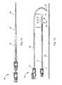
- FIG. 1Ais a plan view of an occlusion assembly incorporating a vascular occlusion device according to the present invention
- FIG. 1Bis an exploded plan view of the occlusion assembly of FIG. 1A ;
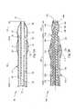
- FIG. 2Ais a section view through a distal end of one embodiment of the vascular occlusion device of the present invention.
- FIG. 2Bis a section view through a distal end of another embodiment of the vascular occlusion device of the present invention.
- FIG. 3is a section view of a human heart showing the assembly of FIG. 1A introducing the device of FIG. 2 into a patent foramen ovale;
- FIGS. 4A-4Cshow various embodiments of the present invention occluding the patent foramen ovale of FIG. 3 ;
- FIG. 5is a flow chart illustrating a method of occluding a body cavity.
- the device 10includes an elongate member 12 extending from a proximal end 14 to a distal end 16 .
- An inflatable proximal balloon 18is disposed about a first portion 20 of the elongate member 12 and an inflatable distal balloon 22 is disposed about a second portion 24 of the elongate member 12 .
- An occlusion port 26is defined between the proximal and distal balloons 18 and 22 .
- FIG. 2Aa sectional view of one embodiment of the elongate member 12 along line 2 - 2 of FIG. 1B .
- the elongate member 12includes the first portion 20 extending distally from the proximal end 14 and the second portion 24 extending distally from the first portion 20 to the distal end 16 .
- a first inflation lumen 28 and an occlusion lumen 30are longitudinally formed in the first portion 20 .
- a second inflation lumen 32is longitudinally formed within the second portion 24 .
- the occlusion port 26is in fluid communication with the occlusion lumen 30 and is configured to introduce an occlusive material provided from, for example, the proximal end 14 of the elongate member 12 , into a body cavity (not shown).
- an inner wall 34defines an inner lumen 36 longitudinally extending through the elongate member 12 .
- the proximal and distal balloons 18 and 22respectively have a proximal balloon wall 38 and a distal balloon wall 40 .
- the balloon walls 38 and 40are respectively disposed about the circumference of the first and second portions 20 and 24 and respectively define a proximal balloon interior 39 and a distal balloon interior 41 .
- the first and second portions 20 and 24respectively have a first inflation orifice 21 and a second inflation orifice 25 .
- the first inflation orifice 21is configured to introduce an inflation fluid provided from, for example, the proximal end 14 of the elongate member 12 through the first inflation lumen 28 , into the proximal balloon interior 39 to inflate and expand the proximal balloon 18 .
- an inflation fluidprovided from, for example, the proximal end 14 of the elongate member 12 through the first inflation lumen 28 , into the proximal balloon interior 39 to inflate and expand the proximal balloon 18 .
- the second inflation lumen 32is in fluid communication with the first inflation lumen 28 .
- the first inflation lumen 28will additionally introduce the inflation fluid into the distal balloon interior 41 by way of the second inflation lumen 32 and the second inflation orifice 25 .
- proximal and distal balloons 18 and 22are shown as being separate parts attached to the elongate member 12 , it is also possible for them to be integrally formed with the elongate member 12 .
- the elongate member 12may be made of any appropriate material. Some examples include, but are not limited to, rubber, latex, polytetrafluoroethylene, polyamides, and polyimides.
- the inflation fluidmay include any appropriate biocompatible fluid for inflating the balloons 18 and 22 and later deflating of the balloons 18 and 22 .
- the occlusive materialmay include any appropriate biocompatible material having an appropriate viscosity allowing it to flow through the occlusion lumen 30 and occlusion port 26 into the body cavity.
- the occlusive materialmay be an appropriate adhesive for permanently bonding to body tissue to occlude the body cavity.
- the occlusive materialmay be configured to promote body tissue growth to occlude the body cavity.
- Some examples of an adhesiveinclude, but are not limited to, polyvinyl alcohol (PVA) and cyanoacrylate adhesives.
- An example of a material to promote body tissue growthincludes, but is not limited to, extracellular matrix (ECM). In other examples, it may be possible to use a combination of an adhesive and the extracellular matrix to occlude the body cavity.
- ECMis a complex structural entity surrounding and supporting cells found within tissues. More specifically, ECM includes structural proteins (for example, collagen and elastin), specialized protein (for example, fibrillin, fibronectin, and laminin), and proteoglycans, a protein core to which are attached long chains of repeating disaccharide units termed glycosaminoglycans.
- structural proteinsfor example, collagen and elastin
- specialized proteinfor example, fibrillin, fibronectin, and laminin
- proteoglycansa protein core to which are attached long chains of repeating disaccharide units termed glycosaminoglycans.
- the extracellular matrixis comprised of small intestinal submucosa (SIS).
- SISsmall intestinal submucosa
- SISis a resorbable, acellular, naturally occurring tissue matrix composed of ECM proteins and various growth factors.
- SISis derived from the porcine jejunum and functions as a remodeling bioscaffold for tissue repair.
- SIShas characteristics of an ideal tissue engineered biomaterial and can act as a bioscaffold for remodeling of many body tissues including skin, body wall, musculoskeletal structure, urinary bladder, and also supports new blood vessel growth.
- SISmay be used to induce site-specific remodeling of both organs and tissues depending on the site of implantation. In practice, host cells are stimulated to proliferate and differentiate into site-specific connective tissue structures, which have been shown to completely replace the SIS material in time.
- SISmay be provided in a fluid form including, for example, a gel.
- the gel SISmay be used to adhere to walls of the body cavity in which the device 10 is deployed and to promote body tissue growth within the body cavity.
- SIShas a natural adherence or wetability to body fluids and connective cells comprising the connective tissue of the walls of a body cavity. Since the occlusive material provided by the device 10 is intended to permanently occlude the body cavity, the distal end 16 is positioned such that the SIS may be introduced into contact with host cells of the wall such that the walls will adhere to the SIS and subsequently differentiate, growing into the SIS and eventually occluding the body cavity with the tissue of the walls to which the substance was originally introduced.
- FIG. 2Ba sectional view of another embodiment of the present invention is shown.
- like features to the embodiment of FIG. 2Ahave like numbers indexed by 100 .
- an elongate member 112is equivalent to the elongate member 12 and a proximal end 114 of the elongate member 112 is equivalent to the proximal end 14 .
- Other examplesinclude a proximal balloon 118 being equivalent to the proximal balloon 18 , an inner lumen 136 being equivalent to the inner lumen 36 , and a first portion 120 being equivalent to the first portion 20 .
- the elongate member 112includes an elongate inner element 142 extending from the proximal end 114 to a distal end 116 .
- the inner element 142includes a second portion 144 extending distally from the first portion 120 and a distal balloon 146 adjacent the distal end 116 .
- the elongate inner element 142is disposed within the inner lumen 136 for relative axial movement therein such that the distal balloon 146 may translate axially relative to the proximal balloon 118 .
- the distal balloon 146includes a distal balloon wall 148 disposed about a circumference of a part of the second portion 144 , defining a distal balloon interior 150 .
- a second inflation lumen 152is formed longitudinally within the inner element 142 and is configured to introduce the inflation fluid into the distal balloon interior 150 by way of a second inflation orifice 154 .
- the second inflation lumen 152is configured to introduce the inflation fluid into the distal balloon interior 150 independently of a first inflation lumen 128 introducing the inflation fluid into a proximal balloon interior 139 .
- the inner element 142includes an element wall defining an element lumen 156 extending longitudinally therethrough.
- an occlusion lumen 130 and occlusion port 126are defined wholly within the first portion 120 of the elongate member 12 .
- a distal segment 160is formed with an atraumatic taper to prevent damage to the body vessel.
- the distal end 116it is also possible for the distal end 116 to include an atraumatic taper (not shown).
- FIGS. 4B and 4Cother variations of the present invention are shown. While these drawings show the embodiment of FIG. 2B , these variations also apply equally, but are not limited, to the embodiment of FIG. 2A .
- a removable film 162is shown attached to each of the proximal and distal balloons 118 and 146 .
- a first piece of removable film 162 ′(or a first removable film) is attached to a distal part of the proximal balloon 118 and a second piece of removable film 162 ′′ (or a second removable film) is attached to a proximal part of the distal balloon 146 .
- Each piece of removable film 162is configured to inflate with the balloons 118 and 146 , attach to the occlusive material following the introduction of the occlusive material into the body cavity, and detach from the balloons 118 and 146 upon deflation. This allows the elongate member 112 and the balloons 118 and 146 to be withdrawn from the body cavity while leaving the occlusive material in place to occlude the cavity.
- proximal and distal balloons 118 and 146may be detachable from the elongate member 112 (not shown). In this case, the removable film 162 is omitted. As a result, the balloons may be permanently attached to the occlusive material.
- an occlusion device 164is disposed about the occlusion port 126 of the elongate member 112 .
- the occlusion device 164is included to provide a support structure upon which the occlusive material may adhere to facilitate occlusion of the body cavity.
- the occlusion deviceincludes, but is not limited to, an embolization coil.
- an embolization coilincludes, but is not limited to, U.S. Pat. No. 5,797,953, which is herein incorporated by reference.
- a vascular occlusion assembly 60for introducing and retrieving the vascular occlusion device 10 is shown in accordance with another embodiment of the present invention.
- the delivery assembly 60includes a polytetrafluoroethylene (PTFE) introducer sheath 62 for percutaneously introducing an outer sheath 66 into a body vessel.
- PTFEpolytetrafluoroethylene
- any other suitable material for the introducer sheath 62may be used without falling beyond the scope or spirit of the present invention.
- the introducer sheath 62may have any suitable size, for example, between about three-french to eight-french.
- the introducer sheath 62serves to allow the outer sheath 66 and the elongate member 12 to be percutaneously inserted to a desired location in a body cavity through the body vessel. For purposes of illustration, only the elongate member 12 is shown, but the elongate member 112 may also be employed.
- the introducer sheath 62receives the outer sheath 66 and provides stability to the outer sheath 66 at a desired entry location of the body vessel. For example, the introducer sheath 62 is held stationary within a common visceral artery, and adds stability to the outer sheath 66 as it is advanced through the introducer sheath 62 to an occlusion area in the body cavity.
- the assembly 60may also include a wire guide 64 configured to be percutaneously inserted within the body vessel to guide the outer sheath 66 to the occlusion area.
- the wire guide 64provides the outer sheath 66 with a path to follow as it is advanced within the body vessel.
- the size of the wire guide 64is based on an inside diameter of, for example, the inner lumen 32 of the elongate member 12 and the diameter of the body vessels that must be traversed to reach the desired body cavity.
- elongate member 12is advanced along the wire guide 64 and through the outer sheath 66 to position the distal end 12 and the occlusion port 26 adjacent an area to be occluded within the body cavity, for example, a patent foramen ovale in a human heart.
- the outer sheath 66also has a proximal portion 70 including a hub 72 to receive the elongate member 12 to be advanced therethrough.
- the size of the outer sheath 66is based on the size of the body vessel in which it percutaneously inserts, and the size of the elongate member 12 .
- FIG. 3shows a sectional view of the human heart 80 having a right atrium 82 and a left atrium 84 .
- An atrial septum 86divides the right atrium 82 from the left atrium 84 and defines a patent foramen oval 88 .
- the patent foramen oval 88is an opening in the atrial septum 86 that allows blood in the right and left atria 82 and 84 to fluidly communicate therebetween.
- a foramen ovaleis a natural hole in the atrial septum 88 that allows blood to bypass the fetus' lungs when in a mother's womb since the fetus relies on the mother to provide oxygen through the umbilical cord.
- the foramen ovalenormally closes when increased blood pressure in the left atrium forces the opening to close. Over time tissue growth closes the opening permanently. However, in some people the opening does not close permanently, in which case the opening is called a patent foramen ovale.
- the patent foramen ovale 88acts like a flap valve, having a right flap 92 and a left flap 94 , between the two atria 82 and 84 .
- the flapsmay open and blood may travel from the right atrium 82 to the left atrium 84 .
- a clotis present in the right atrium 82 it can, for example, enter the left atrium 84 and travel from there to the brain (causing a stroke) or into a coronary artery (causing a heart attack).
- the delivery assembly 60may be percutaneously introduced into a body vessel 90 and directed into, for example, the right atrium 82 and maneuvered adjacent the patent foramen ovale 88 .
- the outer sheath 66is retracted proximally from the occlusion device 100 .
- the wire guide 64may be directed into the patent foramen ovale 88 to provide a path for the elongate member 112 to follow between the right and left flaps 92 and 94 .
- the occlusion port 126is positioned between each of the flaps 92 and 94 .
- the proximal and distal balloons 118 and 146are inflated into contact with the flaps 92 and 94 , thereby forming an occlusion area 96 between the balloons and the flaps (see FIG. 4A ).
- the radial size of the occlusion area 96may be adjusted, for example, by varying the amount the balloons are inflated.
- the axial size of the occlusion area 96may be adjusted by axially translating the inner element 142 .
- the occlusive substanceis then introduced to the occlusion area 96 and into contact with the flaps 92 and 94 , thereby permanently closing the patent foramen ovale 88 .
- FIG. 5is a flow chart illustrating a method 200 of occluding a body cavity.
- the method 200includes at box 202 positioning any of the occlusion devices described herein within a body cavity.
- Box 204includes inflating balloons of the occlusion device proximally and distally of an occlusion area within the body cavity.
- Box 206includes introducing an occlusive material between the balloons and into the occlusion area, and box 208 attaches the occlusive material to the body walls of the body cavity.
Landscapes
- Health & Medical Sciences (AREA)
- Life Sciences & Earth Sciences (AREA)
- Surgery (AREA)
- Heart & Thoracic Surgery (AREA)
- Veterinary Medicine (AREA)
- Public Health (AREA)
- General Health & Medical Sciences (AREA)
- Engineering & Computer Science (AREA)
- Biomedical Technology (AREA)
- Animal Behavior & Ethology (AREA)
- Molecular Biology (AREA)
- Medical Informatics (AREA)
- Nuclear Medicine, Radiotherapy & Molecular Imaging (AREA)
- Vascular Medicine (AREA)
- Reproductive Health (AREA)
- Cardiology (AREA)
- Child & Adolescent Psychology (AREA)
- Biophysics (AREA)
- Pulmonology (AREA)
- Anesthesiology (AREA)
- Hematology (AREA)
- Surgical Instruments (AREA)
Abstract
Description
Claims (15)
Priority Applications (1)
| Application Number | Priority Date | Filing Date | Title |
|---|---|---|---|
| US11/848,777US8292907B2 (en) | 2007-08-31 | 2007-08-31 | Balloon assisted occlusion device |
Applications Claiming Priority (1)
| Application Number | Priority Date | Filing Date | Title |
|---|---|---|---|
| US11/848,777US8292907B2 (en) | 2007-08-31 | 2007-08-31 | Balloon assisted occlusion device |
Publications (2)
| Publication Number | Publication Date |
|---|---|
| US20090062836A1 US20090062836A1 (en) | 2009-03-05 |
| US8292907B2true US8292907B2 (en) | 2012-10-23 |
Family
ID=40408673
Family Applications (1)
| Application Number | Title | Priority Date | Filing Date |
|---|---|---|---|
| US11/848,777Expired - Fee RelatedUS8292907B2 (en) | 2007-08-31 | 2007-08-31 | Balloon assisted occlusion device |
Country Status (1)
| Country | Link |
|---|---|
| US (1) | US8292907B2 (en) |
Families Citing this family (20)
| Publication number | Priority date | Publication date | Assignee | Title |
|---|---|---|---|---|
| WO2008094706A2 (en)* | 2007-02-01 | 2008-08-07 | Cook Incorporated | Closure device and method of closing a bodily opening |
| US8617205B2 (en) | 2007-02-01 | 2013-12-31 | Cook Medical Technologies Llc | Closure device |
| EP2627265B8 (en) | 2010-10-15 | 2019-02-20 | Cook Medical Technologies LLC | Occlusion device for blocking fluid flow through bodily passages |
| EP2522307B1 (en)* | 2011-05-08 | 2020-09-30 | ITSO Medical AB | Device for delivery of medical devices to a cardiac valve |
| EP3281608B1 (en) | 2012-02-10 | 2020-09-16 | CVDevices, LLC | Medical product comprising a frame and visceral pleura |
| EP2732794A1 (en) | 2012-11-14 | 2014-05-21 | Contego AB | Improved embolic protection device and method |
| US20140142620A1 (en)* | 2012-11-19 | 2014-05-22 | Cook Medical Technologies Llc | Degradable balloon device and method for closure of openings in a tissue wall |
| CA2900862C (en) | 2013-02-11 | 2017-10-03 | Cook Medical Technologies Llc | Expandable support frame and medical device |
| US10080657B2 (en) | 2013-03-07 | 2018-09-25 | Cedars-Sinai Medical Center | Catheter based apical approach heart prostheses delivery system |
| WO2014138482A1 (en) | 2013-03-07 | 2014-09-12 | Cedars-Sinai Medical Center | Method and apparatus for percutaneous delivery and deployment of a cardiovascular prosthesis |
| US10966697B2 (en) | 2013-05-06 | 2021-04-06 | Caveomed Gmbh | Vascular closure device and method of positioning vascular closure device |
| EP2801325B1 (en) | 2013-05-06 | 2016-03-16 | CaveoMed GmbH | Vascular closure device |
| EP3169260B1 (en)* | 2014-07-16 | 2019-09-25 | Fractyl Laboratories, Inc. | System for treating diabetes and related diseases and disorders |
| WO2016040526A1 (en) | 2014-09-10 | 2016-03-17 | Cedars-Sinai Medical Center | Method and apparatus for percutaneous delivery and deployment of a cardiac valve prosthesis |
| US10758265B2 (en)* | 2014-11-14 | 2020-09-01 | Cedars-Sinai Medical Center | Cardiovascular access and device delivery system |
| EP3324855B1 (en) | 2015-07-23 | 2024-03-20 | Cedars-Sinai Medical Center | Device for securing heart valve leaflets |
| WO2018140535A1 (en) | 2017-01-25 | 2018-08-02 | Cedars-Sinai Medical Center | Device for securing heart valve leaflets |
| US11291544B2 (en) | 2018-02-02 | 2022-04-05 | Cedars-Sinai Medical Center | Delivery platforms, devices, and methods for tricuspid valve repair |
| WO2020010144A1 (en)* | 2018-07-03 | 2020-01-09 | Subramaniam Krishnan | Systems and methods for treating patent foramen ovale |
| JP2022185719A (en)* | 2021-06-03 | 2022-12-15 | 株式会社カネカ | METHOD FOR MANUFACTURING MEDICAL FILM DELIVERY DEVICE AND MEDICAL DEVICE |
Citations (55)
| Publication number | Priority date | Publication date | Assignee | Title |
|---|---|---|---|---|
| US4573966A (en)* | 1981-11-24 | 1986-03-04 | Schneider Medintag Ag | Method and apparatus for removing and/or enlarging constricted areas in vessels conducting body fluids |
| US4836204A (en)* | 1987-07-06 | 1989-06-06 | Landymore Roderick W | Method for effecting closure of a perforation in the septum of the heart |
| US4911163A (en) | 1986-06-12 | 1990-03-27 | Ernesto Fina | Two ballooned catheter device for diagnostic and operative use |
| JPH02307480A (en) | 1989-05-23 | 1990-12-20 | Nobuyuki Tanaka | Intravenous atrial septal defect hole closing apparatus |
| US5167614A (en) | 1991-10-29 | 1992-12-01 | Medical Engineering Corporation | Prostatic stent |
| US5167628A (en) | 1991-05-02 | 1992-12-01 | Boyles Paul W | Aortic balloon catheter assembly for indirect infusion of the coronary arteries |
| US5176692A (en) | 1991-12-09 | 1993-01-05 | Wilk Peter J | Method and surgical instrument for repairing hernia |
| US5188595A (en)* | 1991-06-28 | 1993-02-23 | Laserscope | Method for enhanced retention of balloon catheter in body cavity |
| US5192301A (en) | 1989-01-17 | 1993-03-09 | Nippon Zeon Co., Ltd. | Closing plug of a defect for medical use and a closing plug device utilizing it |
| US5275826A (en)* | 1992-11-13 | 1994-01-04 | Purdue Research Foundation | Fluidized intestinal submucosa and its use as an injectable tissue graft |
| US5486195A (en) | 1993-07-26 | 1996-01-23 | Myers; Gene | Method and apparatus for arteriotomy closure |
| US5591197A (en) | 1995-03-14 | 1997-01-07 | Advanced Cardiovascular Systems, Inc. | Expandable stent forming projecting barbs and method for deploying |
| US5634936A (en) | 1995-02-06 | 1997-06-03 | Scimed Life Systems, Inc. | Device for closing a septal defect |
| US5702421A (en) | 1995-01-11 | 1997-12-30 | Schneidt; Bernhard | Closure device for closing a vascular opening, such as patent ductus arteriosus |
| US5749922A (en)* | 1988-08-24 | 1998-05-12 | Endoluminal Therapeutics, Inc. | Biodegradable polymeric endoluminal sealing process, apparatus and polymeric products for use therein |
| US5766219A (en) | 1995-04-20 | 1998-06-16 | Musc Foundation For Research Development | Anatomically shaped vasoocclusive device and method for deploying same |
| US5772632A (en)* | 1994-04-13 | 1998-06-30 | Schneider (Usa) Inc. | Dilation-drug delivery catheter |
| US5861003A (en) | 1996-10-23 | 1999-01-19 | The Cleveland Clinic Foundation | Apparatus and method for occluding a defect or aperture within body surface |
| US5899917A (en)* | 1997-03-12 | 1999-05-04 | Cardiosynopsis, Inc. | Method for forming a stent in situ |
| US5976174A (en) | 1997-12-15 | 1999-11-02 | Ruiz; Carlos E. | Medical hole closure device and methods of use |
| US6063112A (en)* | 1995-12-28 | 2000-05-16 | Sofradim Production | Kit for surgical treatment of intracorporeal lumens |
| US6238416B1 (en) | 1998-11-13 | 2001-05-29 | Eleftherios B. Sideris | Transcatheter surgical patch |
| US6270524B1 (en) | 1996-11-12 | 2001-08-07 | Medtronic, Inc. | Flexible, radially expansible luminal prostheses |
| US6296657B1 (en) | 1998-10-07 | 2001-10-02 | Gregory G. Brucker | Vascular sealing device and method |
| US20020029051A1 (en) | 1996-12-18 | 2002-03-07 | Edward J. Lynch | Occluding device and method of use |
| US20020072763A1 (en)* | 2000-12-07 | 2002-06-13 | Scimed Life Systems, Inc. | Intravascular balloon catheter for embolic coil delivery |
| US20020082685A1 (en) | 2000-12-22 | 2002-06-27 | Motasim Sirhan | Apparatus and methods for controlled substance delivery from implanted prostheses |
| US20020151968A1 (en) | 1999-07-20 | 2002-10-17 | Medtronic, Inc. | Transmural concentric multilayer ingrowth matrix within well-defined porosity |
| US6517573B1 (en) | 2000-04-11 | 2003-02-11 | Endovascular Technologies, Inc. | Hook for attaching to a corporeal lumen and method of manufacturing |
| US20030045860A1 (en)* | 2001-09-04 | 2003-03-06 | Jomed Gmbh | Methods for minimally invasive, localized delivery of sclerotherapeutic agents |
| US20030051735A1 (en) | 2001-07-26 | 2003-03-20 | Cook Biotech Incorporated | Vessel closure member, delivery apparatus, and method of inserting the member |
| US20030130713A1 (en)* | 2001-05-21 | 2003-07-10 | Stewart Mark T. | Trans-septal catheter with retention mechanism |
| US20030229366A1 (en) | 1996-02-02 | 2003-12-11 | Transvascular, Inc. | Implantable lumen occluding devices and methods |
| US6692458B2 (en) | 2000-12-19 | 2004-02-17 | Edwards Lifesciences Corporation | Intra-pericardial drug delivery device with multiple balloons and method for angiogenesis |
| US20040186561A1 (en) | 2000-06-26 | 2004-09-23 | Mcguckin James F. | Vascular device for valve leaflet apposition |
| US20040220596A1 (en) | 2003-02-04 | 2004-11-04 | Frazier Andrew G.C. | Patent foramen ovale closure system |
| US20040260340A1 (en) | 2000-05-19 | 2004-12-23 | Jacobs Daniel Irwin | Remotely anchored tissue fixation device and method |
| US20040267191A1 (en) | 2003-03-27 | 2004-12-30 | Cierra, Inc. | Methods and apparatus for treatment of patent foramen ovale |
| US20050010248A1 (en) | 2003-07-10 | 2005-01-13 | Scimed Life Systems, Inc. | System for closing an opening in a body cavity |
| US20050034735A1 (en) | 2003-03-27 | 2005-02-17 | Cierra, Inc. | Methods and apparatus for treatment of patent foramen ovale |
| US20050085770A1 (en)* | 2001-09-07 | 2005-04-21 | Don Michael T. A. | Devices for observing and treating body passages |
| US20050149175A1 (en) | 2003-11-10 | 2005-07-07 | Angiotech International Ag | Intravascular devices and fibrosis-inducing agents |
| US20050192626A1 (en) | 2004-01-30 | 2005-09-01 | Nmt Medical, Inc. | Devices, systems, and methods for closure of cardiac openings |
| US20050209633A1 (en) | 2004-02-02 | 2005-09-22 | Ovion, Inc. | Enhancing tissue ingrowth for contraception |
| US6949116B2 (en) | 1996-05-08 | 2005-09-27 | Carag Ag | Device for plugging an opening such as in a wall of a hollow or tubular organ including biodegradable elements |
| US20050251201A1 (en) | 2004-02-20 | 2005-11-10 | Roue Chad C | Devices and methods for closing a patent foramen ovale using a countertraction element |
| US20050256532A1 (en) | 2004-05-12 | 2005-11-17 | Asha Nayak | Cardiovascular defect patch device and method |
| US6994092B2 (en) | 1999-11-08 | 2006-02-07 | Ev3 Sunnyvale, Inc. | Device for containing embolic material in the LAA having a plurality of tissue retention structures |
| US20060105015A1 (en)* | 2004-11-12 | 2006-05-18 | Venu Perla | System and method for attaching soft tissue to an implant |
| US20060106418A1 (en) | 2002-07-31 | 2006-05-18 | Abbott Laboratories Vascular Enterprises, Limited | Apparatus for sealing surgical punctures |
| US20060212055A1 (en) | 2005-01-25 | 2006-09-21 | Karabey Halil I | Expandable occlusive structure |
| US20060235467A1 (en) | 2002-04-16 | 2006-10-19 | Devore Lauri J | Removable anchored lung volume reduction device and methods |
| US7128073B1 (en) | 1998-11-06 | 2006-10-31 | Ev3 Endovascular, Inc. | Method and device for left atrial appendage occlusion |
| US20070014870A1 (en)* | 2005-07-15 | 2007-01-18 | Cormatrix Cardiovascular, Inc. | Compositions for regenerating defective or absent myocardium |
| US7591268B2 (en) | 1999-08-23 | 2009-09-22 | Conceptus, Inc. | Deployment actuation system for intrafallopian contraception |
- 2007
- 2007-08-31USUS11/848,777patent/US8292907B2/ennot_activeExpired - Fee Related
Patent Citations (61)
| Publication number | Priority date | Publication date | Assignee | Title |
|---|---|---|---|---|
| US4573966A (en)* | 1981-11-24 | 1986-03-04 | Schneider Medintag Ag | Method and apparatus for removing and/or enlarging constricted areas in vessels conducting body fluids |
| US4911163A (en) | 1986-06-12 | 1990-03-27 | Ernesto Fina | Two ballooned catheter device for diagnostic and operative use |
| US4836204A (en)* | 1987-07-06 | 1989-06-06 | Landymore Roderick W | Method for effecting closure of a perforation in the septum of the heart |
| US5749922A (en)* | 1988-08-24 | 1998-05-12 | Endoluminal Therapeutics, Inc. | Biodegradable polymeric endoluminal sealing process, apparatus and polymeric products for use therein |
| US20040024419A1 (en)* | 1988-08-24 | 2004-02-05 | Endoluminal Therapeutics, Inc. | Biodegradable polymeric endoluminal sealing process, apparatus and polymeric products for use therein |
| US5192301A (en) | 1989-01-17 | 1993-03-09 | Nippon Zeon Co., Ltd. | Closing plug of a defect for medical use and a closing plug device utilizing it |
| JPH02307480A (en) | 1989-05-23 | 1990-12-20 | Nobuyuki Tanaka | Intravenous atrial septal defect hole closing apparatus |
| US5167628A (en) | 1991-05-02 | 1992-12-01 | Boyles Paul W | Aortic balloon catheter assembly for indirect infusion of the coronary arteries |
| US5188595A (en)* | 1991-06-28 | 1993-02-23 | Laserscope | Method for enhanced retention of balloon catheter in body cavity |
| US5167614A (en) | 1991-10-29 | 1992-12-01 | Medical Engineering Corporation | Prostatic stent |
| US5176692A (en) | 1991-12-09 | 1993-01-05 | Wilk Peter J | Method and surgical instrument for repairing hernia |
| US5275826A (en)* | 1992-11-13 | 1994-01-04 | Purdue Research Foundation | Fluidized intestinal submucosa and its use as an injectable tissue graft |
| US5486195A (en) | 1993-07-26 | 1996-01-23 | Myers; Gene | Method and apparatus for arteriotomy closure |
| US5772632A (en)* | 1994-04-13 | 1998-06-30 | Schneider (Usa) Inc. | Dilation-drug delivery catheter |
| US5702421A (en) | 1995-01-11 | 1997-12-30 | Schneidt; Bernhard | Closure device for closing a vascular opening, such as patent ductus arteriosus |
| US5634936A (en) | 1995-02-06 | 1997-06-03 | Scimed Life Systems, Inc. | Device for closing a septal defect |
| US5591197A (en) | 1995-03-14 | 1997-01-07 | Advanced Cardiovascular Systems, Inc. | Expandable stent forming projecting barbs and method for deploying |
| US5766219A (en) | 1995-04-20 | 1998-06-16 | Musc Foundation For Research Development | Anatomically shaped vasoocclusive device and method for deploying same |
| US6063112A (en)* | 1995-12-28 | 2000-05-16 | Sofradim Production | Kit for surgical treatment of intracorporeal lumens |
| US20030229366A1 (en) | 1996-02-02 | 2003-12-11 | Transvascular, Inc. | Implantable lumen occluding devices and methods |
| US6949116B2 (en) | 1996-05-08 | 2005-09-27 | Carag Ag | Device for plugging an opening such as in a wall of a hollow or tubular organ including biodegradable elements |
| US5861003A (en) | 1996-10-23 | 1999-01-19 | The Cleveland Clinic Foundation | Apparatus and method for occluding a defect or aperture within body surface |
| US6270524B1 (en) | 1996-11-12 | 2001-08-07 | Medtronic, Inc. | Flexible, radially expansible luminal prostheses |
| US20020029051A1 (en) | 1996-12-18 | 2002-03-07 | Edward J. Lynch | Occluding device and method of use |
| US20030029457A1 (en) | 1996-12-18 | 2003-02-13 | Callister Jeffrey P. | Contraceptive system and method of use |
| US5899917A (en)* | 1997-03-12 | 1999-05-04 | Cardiosynopsis, Inc. | Method for forming a stent in situ |
| US5976174A (en) | 1997-12-15 | 1999-11-02 | Ruiz; Carlos E. | Medical hole closure device and methods of use |
| US6296657B1 (en) | 1998-10-07 | 2001-10-02 | Gregory G. Brucker | Vascular sealing device and method |
| US7128073B1 (en) | 1998-11-06 | 2006-10-31 | Ev3 Endovascular, Inc. | Method and device for left atrial appendage occlusion |
| US6238416B1 (en) | 1998-11-13 | 2001-05-29 | Eleftherios B. Sideris | Transcatheter surgical patch |
| US20020151968A1 (en) | 1999-07-20 | 2002-10-17 | Medtronic, Inc. | Transmural concentric multilayer ingrowth matrix within well-defined porosity |
| US7591268B2 (en) | 1999-08-23 | 2009-09-22 | Conceptus, Inc. | Deployment actuation system for intrafallopian contraception |
| US6994092B2 (en) | 1999-11-08 | 2006-02-07 | Ev3 Sunnyvale, Inc. | Device for containing embolic material in the LAA having a plurality of tissue retention structures |
| US6517573B1 (en) | 2000-04-11 | 2003-02-11 | Endovascular Technologies, Inc. | Hook for attaching to a corporeal lumen and method of manufacturing |
| US20040260340A1 (en) | 2000-05-19 | 2004-12-23 | Jacobs Daniel Irwin | Remotely anchored tissue fixation device and method |
| US20040186561A1 (en) | 2000-06-26 | 2004-09-23 | Mcguckin James F. | Vascular device for valve leaflet apposition |
| US20020072763A1 (en)* | 2000-12-07 | 2002-06-13 | Scimed Life Systems, Inc. | Intravascular balloon catheter for embolic coil delivery |
| US6692458B2 (en) | 2000-12-19 | 2004-02-17 | Edwards Lifesciences Corporation | Intra-pericardial drug delivery device with multiple balloons and method for angiogenesis |
| US20020082685A1 (en) | 2000-12-22 | 2002-06-27 | Motasim Sirhan | Apparatus and methods for controlled substance delivery from implanted prostheses |
| US20030130713A1 (en)* | 2001-05-21 | 2003-07-10 | Stewart Mark T. | Trans-septal catheter with retention mechanism |
| US20030051735A1 (en) | 2001-07-26 | 2003-03-20 | Cook Biotech Incorporated | Vessel closure member, delivery apparatus, and method of inserting the member |
| US20030045860A1 (en)* | 2001-09-04 | 2003-03-06 | Jomed Gmbh | Methods for minimally invasive, localized delivery of sclerotherapeutic agents |
| US20050085770A1 (en)* | 2001-09-07 | 2005-04-21 | Don Michael T. A. | Devices for observing and treating body passages |
| US20060235467A1 (en) | 2002-04-16 | 2006-10-19 | Devore Lauri J | Removable anchored lung volume reduction device and methods |
| US20060106418A1 (en) | 2002-07-31 | 2006-05-18 | Abbott Laboratories Vascular Enterprises, Limited | Apparatus for sealing surgical punctures |
| US20040220596A1 (en) | 2003-02-04 | 2004-11-04 | Frazier Andrew G.C. | Patent foramen ovale closure system |
| US20050034735A1 (en) | 2003-03-27 | 2005-02-17 | Cierra, Inc. | Methods and apparatus for treatment of patent foramen ovale |
| US7165552B2 (en) | 2003-03-27 | 2007-01-23 | Cierra, Inc. | Methods and apparatus for treatment of patent foramen ovale |
| US20040267191A1 (en) | 2003-03-27 | 2004-12-30 | Cierra, Inc. | Methods and apparatus for treatment of patent foramen ovale |
| US20050010248A1 (en) | 2003-07-10 | 2005-01-13 | Scimed Life Systems, Inc. | System for closing an opening in a body cavity |
| US20050149175A1 (en) | 2003-11-10 | 2005-07-07 | Angiotech International Ag | Intravascular devices and fibrosis-inducing agents |
| US20050149173A1 (en) | 2003-11-10 | 2005-07-07 | Angiotech International Ag | Intravascular devices and fibrosis-inducing agents |
| US20050177103A1 (en)* | 2003-11-10 | 2005-08-11 | Angiotech International Ag | Intravascular devices and fibrosis-inducing agents |
| US20050192626A1 (en) | 2004-01-30 | 2005-09-01 | Nmt Medical, Inc. | Devices, systems, and methods for closure of cardiac openings |
| US20060009798A1 (en) | 2004-02-02 | 2006-01-12 | Ams Research Corporation | Methods and devices for occluding body lumens and/or enhancing tissue ingrowth |
| US20050209633A1 (en) | 2004-02-02 | 2005-09-22 | Ovion, Inc. | Enhancing tissue ingrowth for contraception |
| US20050251201A1 (en) | 2004-02-20 | 2005-11-10 | Roue Chad C | Devices and methods for closing a patent foramen ovale using a countertraction element |
| US20050256532A1 (en) | 2004-05-12 | 2005-11-17 | Asha Nayak | Cardiovascular defect patch device and method |
| US20060105015A1 (en)* | 2004-11-12 | 2006-05-18 | Venu Perla | System and method for attaching soft tissue to an implant |
| US20060212055A1 (en) | 2005-01-25 | 2006-09-21 | Karabey Halil I | Expandable occlusive structure |
| US20070014870A1 (en)* | 2005-07-15 | 2007-01-18 | Cormatrix Cardiovascular, Inc. | Compositions for regenerating defective or absent myocardium |
Also Published As
| Publication number | Publication date |
|---|---|
| US20090062836A1 (en) | 2009-03-05 |
Similar Documents
| Publication | Publication Date | Title |
|---|---|---|
| US8292907B2 (en) | Balloon assisted occlusion device | |
| US5919200A (en) | Balloon catheter for abrading a patent foramen ovale and method of using the balloon catheter | |
| US9017375B2 (en) | Occluding device | |
| KR101681346B1 (en) | Heart occlusion devices | |
| AU2006320507B2 (en) | Devices, systems, and methods for occluding a defect | |
| US6790220B2 (en) | Method and apparatus for sealing access | |
| US4836204A (en) | Method for effecting closure of a perforation in the septum of the heart | |
| JP3693673B2 (en) | Device for closing the body passage | |
| US8858584B2 (en) | Emergency transection device | |
| US10143457B2 (en) | Fistula plugs having increased column strength and fistula plug delivery apparatuses and methods | |
| WO1999018864A1 (en) | A balloon catheter for abrading a patent foramen ovale and method of using the balloon catheter | |
| EP2567663A1 (en) | A collapsible medical closing device, a method and a medical system for delivering an object | |
| WO1999018870A1 (en) | A balloon catheter for causing thermal trauma to a patent foramen ovale and method of using the balloon catheter | |
| US9427235B2 (en) | Apparatus and method for treating bleeding arising from left atrial appendage | |
| WO2006002407A2 (en) | Vascular occlusion device | |
| US20190142432A1 (en) | Vascular access devices, systems, and methods | |
| JP2002102260A (en) | Shunt tube inside artery and its using method | |
| US20050049637A1 (en) | Method and apparatus for sealing access | |
| JPH04673B2 (en) | ||
| WO2022170187A1 (en) | Detachable balloon embolization device and methods | |
| US9005234B2 (en) | Occlusion device | |
| US20250107806A1 (en) | Medical device and method for safely closing, isolating, or adjusting the volume of a structure in the human body | |
| ES2897755T3 (en) | aneurysm occluder | |
| US9089312B2 (en) | Tamponade for biopsy surgery and method of operation | |
| CN115252033A (en) | Air bag suction type left auricle stabilizing and ligating system |
Legal Events
| Date | Code | Title | Description |
|---|---|---|---|
| AS | Assignment | Owner name:COOK INCORPORATED, INDIANA Free format text:ASSIGNMENT OF ASSIGNORS INTEREST;ASSIGNOR:KURRUS, MICHAEL R.;REEL/FRAME:019964/0714 Effective date:20070813 | |
| ZAAA | Notice of allowance and fees due | Free format text:ORIGINAL CODE: NOA | |
| ZAAB | Notice of allowance mailed | Free format text:ORIGINAL CODE: MN/=. | |
| AS | Assignment | Owner name:COOK MEDICAL TECHNOLOGIES LLC, INDIANA Free format text:ASSIGNMENT OF ASSIGNORS INTEREST;ASSIGNOR:COOK INCORPORATED;REEL/FRAME:028978/0689 Effective date:20120827 | |
| STCF | Information on status: patent grant | Free format text:PATENTED CASE | |
| CC | Certificate of correction | ||
| FPAY | Fee payment | Year of fee payment:4 | |
| MAFP | Maintenance fee payment | Free format text:PAYMENT OF MAINTENANCE FEE, 8TH YEAR, LARGE ENTITY (ORIGINAL EVENT CODE: M1552); ENTITY STATUS OF PATENT OWNER: LARGE ENTITY Year of fee payment:8 | |
| AS | Assignment | Owner name:WILMINGTON TRUST, NATIONAL ASSOCIATION, AS COLLATERAL AGENT, DELAWARE Free format text:SECURITY INTEREST;ASSIGNOR:COOK MEDICAL TECHNOLOGIES LLC;REEL/FRAME:066700/0277 Effective date:20240227 | |
| FEPP | Fee payment procedure | Free format text:MAINTENANCE FEE REMINDER MAILED (ORIGINAL EVENT CODE: REM.); ENTITY STATUS OF PATENT OWNER: LARGE ENTITY | |
| LAPS | Lapse for failure to pay maintenance fees | Free format text:PATENT EXPIRED FOR FAILURE TO PAY MAINTENANCE FEES (ORIGINAL EVENT CODE: EXP.); ENTITY STATUS OF PATENT OWNER: LARGE ENTITY | |
| STCH | Information on status: patent discontinuation | Free format text:PATENT EXPIRED DUE TO NONPAYMENT OF MAINTENANCE FEES UNDER 37 CFR 1.362 | |
| FP | Lapsed due to failure to pay maintenance fee | Effective date:20241023 |