US8282623B2 - Method for treating obesity by extracting food - Google Patents
Method for treating obesity by extracting foodDownload PDFInfo
- Publication number
- US8282623B2 US8282623B2US12/604,140US60414009AUS8282623B2US 8282623 B2US8282623 B2US 8282623B2US 60414009 AUS60414009 AUS 60414009AUS 8282623 B2US8282623 B2US 8282623B2
- Authority
- US
- United States
- Prior art keywords
- patient
- tube
- food
- stomach
- outlet
- Prior art date
- Legal status (The legal status is an assumption and is not a legal conclusion. Google has not performed a legal analysis and makes no representation as to the accuracy of the status listed.)
- Expired - Fee Related, expires
Links
Images


Classifications
- A—HUMAN NECESSITIES
- A61—MEDICAL OR VETERINARY SCIENCE; HYGIENE
- A61F—FILTERS IMPLANTABLE INTO BLOOD VESSELS; PROSTHESES; DEVICES PROVIDING PATENCY TO, OR PREVENTING COLLAPSING OF, TUBULAR STRUCTURES OF THE BODY, e.g. STENTS; ORTHOPAEDIC, NURSING OR CONTRACEPTIVE DEVICES; FOMENTATION; TREATMENT OR PROTECTION OF EYES OR EARS; BANDAGES, DRESSINGS OR ABSORBENT PADS; FIRST-AID KITS
- A61F5/00—Orthopaedic methods or devices for non-surgical treatment of bones or joints; Nursing devices ; Anti-rape devices
- A61F5/0003—Apparatus for the treatment of obesity; Anti-eating devices
- A61F5/0013—Implantable devices or invasive measures
- A61F5/003—Implantable devices or invasive measures inflatable
- A—HUMAN NECESSITIES
- A61—MEDICAL OR VETERINARY SCIENCE; HYGIENE
- A61F—FILTERS IMPLANTABLE INTO BLOOD VESSELS; PROSTHESES; DEVICES PROVIDING PATENCY TO, OR PREVENTING COLLAPSING OF, TUBULAR STRUCTURES OF THE BODY, e.g. STENTS; ORTHOPAEDIC, NURSING OR CONTRACEPTIVE DEVICES; FOMENTATION; TREATMENT OR PROTECTION OF EYES OR EARS; BANDAGES, DRESSINGS OR ABSORBENT PADS; FIRST-AID KITS
- A61F5/00—Orthopaedic methods or devices for non-surgical treatment of bones or joints; Nursing devices ; Anti-rape devices
- A61F5/0003—Apparatus for the treatment of obesity; Anti-eating devices
- A61F5/0013—Implantable devices or invasive measures
- A61F5/0036—Intragastrical devices
- A61F5/004—Intragastrical devices remotely adjustable
- A—HUMAN NECESSITIES
- A61—MEDICAL OR VETERINARY SCIENCE; HYGIENE
- A61F—FILTERS IMPLANTABLE INTO BLOOD VESSELS; PROSTHESES; DEVICES PROVIDING PATENCY TO, OR PREVENTING COLLAPSING OF, TUBULAR STRUCTURES OF THE BODY, e.g. STENTS; ORTHOPAEDIC, NURSING OR CONTRACEPTIVE DEVICES; FOMENTATION; TREATMENT OR PROTECTION OF EYES OR EARS; BANDAGES, DRESSINGS OR ABSORBENT PADS; FIRST-AID KITS
- A61F5/00—Orthopaedic methods or devices for non-surgical treatment of bones or joints; Nursing devices ; Anti-rape devices
- A61F5/0003—Apparatus for the treatment of obesity; Anti-eating devices
- A61F5/0013—Implantable devices or invasive measures
- A61F5/0076—Implantable devices or invasive measures preventing normal digestion, e.g. Bariatric or gastric sleeves
- A—HUMAN NECESSITIES
- A61—MEDICAL OR VETERINARY SCIENCE; HYGIENE
- A61M—DEVICES FOR INTRODUCING MEDIA INTO, OR ONTO, THE BODY; DEVICES FOR TRANSDUCING BODY MEDIA OR FOR TAKING MEDIA FROM THE BODY; DEVICES FOR PRODUCING OR ENDING SLEEP OR STUPOR
- A61M1/00—Suction or pumping devices for medical purposes; Devices for carrying-off, for treatment of, or for carrying-over, body-liquids; Drainage systems
- A61M1/80—Suction pumps
- A61M1/81—Piston pumps, e.g. syringes
- A—HUMAN NECESSITIES
- A61—MEDICAL OR VETERINARY SCIENCE; HYGIENE
- A61M—DEVICES FOR INTRODUCING MEDIA INTO, OR ONTO, THE BODY; DEVICES FOR TRANSDUCING BODY MEDIA OR FOR TAKING MEDIA FROM THE BODY; DEVICES FOR PRODUCING OR ENDING SLEEP OR STUPOR
- A61M1/00—Suction or pumping devices for medical purposes; Devices for carrying-off, for treatment of, or for carrying-over, body-liquids; Drainage systems
- A61M1/80—Suction pumps
- A61M1/82—Membrane pumps, e.g. bulbs
- A—HUMAN NECESSITIES
- A61—MEDICAL OR VETERINARY SCIENCE; HYGIENE
- A61M—DEVICES FOR INTRODUCING MEDIA INTO, OR ONTO, THE BODY; DEVICES FOR TRANSDUCING BODY MEDIA OR FOR TAKING MEDIA FROM THE BODY; DEVICES FOR PRODUCING OR ENDING SLEEP OR STUPOR
- A61M1/00—Suction or pumping devices for medical purposes; Devices for carrying-off, for treatment of, or for carrying-over, body-liquids; Drainage systems
- A61M1/84—Drainage tubes; Aspiration tips
- A—HUMAN NECESSITIES
- A61—MEDICAL OR VETERINARY SCIENCE; HYGIENE
- A61M—DEVICES FOR INTRODUCING MEDIA INTO, OR ONTO, THE BODY; DEVICES FOR TRANSDUCING BODY MEDIA OR FOR TAKING MEDIA FROM THE BODY; DEVICES FOR PRODUCING OR ENDING SLEEP OR STUPOR
- A61M27/00—Drainage appliance for wounds or the like, i.e. wound drains, implanted drains
- A61M27/002—Implant devices for drainage of body fluids from one part of the body to another
- A—HUMAN NECESSITIES
- A61—MEDICAL OR VETERINARY SCIENCE; HYGIENE
- A61F—FILTERS IMPLANTABLE INTO BLOOD VESSELS; PROSTHESES; DEVICES PROVIDING PATENCY TO, OR PREVENTING COLLAPSING OF, TUBULAR STRUCTURES OF THE BODY, e.g. STENTS; ORTHOPAEDIC, NURSING OR CONTRACEPTIVE DEVICES; FOMENTATION; TREATMENT OR PROTECTION OF EYES OR EARS; BANDAGES, DRESSINGS OR ABSORBENT PADS; FIRST-AID KITS
- A61F2/00—Filters implantable into blood vessels; Prostheses, i.e. artificial substitutes or replacements for parts of the body; Appliances for connecting them with the body; Devices providing patency to, or preventing collapsing of, tubular structures of the body, e.g. stents
- A61F2/02—Prostheses implantable into the body
- A61F2/04—Hollow or tubular parts of organs, e.g. bladders, tracheae, bronchi or bile ducts
- A61F2002/044—Oesophagi or esophagi or gullets
- A—HUMAN NECESSITIES
- A61—MEDICAL OR VETERINARY SCIENCE; HYGIENE
- A61J—CONTAINERS SPECIALLY ADAPTED FOR MEDICAL OR PHARMACEUTICAL PURPOSES; DEVICES OR METHODS SPECIALLY ADAPTED FOR BRINGING PHARMACEUTICAL PRODUCTS INTO PARTICULAR PHYSICAL OR ADMINISTERING FORMS; DEVICES FOR ADMINISTERING FOOD OR MEDICINES ORALLY; BABY COMFORTERS; DEVICES FOR RECEIVING SPITTLE
- A61J15/00—Feeding-tubes for therapeutic purposes
- A61J15/0015—Gastrostomy feeding-tubes
- A—HUMAN NECESSITIES
- A61—MEDICAL OR VETERINARY SCIENCE; HYGIENE
- A61M—DEVICES FOR INTRODUCING MEDIA INTO, OR ONTO, THE BODY; DEVICES FOR TRANSDUCING BODY MEDIA OR FOR TAKING MEDIA FROM THE BODY; DEVICES FOR PRODUCING OR ENDING SLEEP OR STUPOR
- A61M1/00—Suction or pumping devices for medical purposes; Devices for carrying-off, for treatment of, or for carrying-over, body-liquids; Drainage systems
- A61M1/71—Suction drainage systems
- A61M1/73—Suction drainage systems comprising sensors or indicators for physical values
- A—HUMAN NECESSITIES
- A61—MEDICAL OR VETERINARY SCIENCE; HYGIENE
- A61M—DEVICES FOR INTRODUCING MEDIA INTO, OR ONTO, THE BODY; DEVICES FOR TRANSDUCING BODY MEDIA OR FOR TAKING MEDIA FROM THE BODY; DEVICES FOR PRODUCING OR ENDING SLEEP OR STUPOR
- A61M1/00—Suction or pumping devices for medical purposes; Devices for carrying-off, for treatment of, or for carrying-over, body-liquids; Drainage systems
- A61M1/71—Suction drainage systems
- A61M1/77—Suction-irrigation systems
- A61M1/772—Suction-irrigation systems operating alternately
- A—HUMAN NECESSITIES
- A61—MEDICAL OR VETERINARY SCIENCE; HYGIENE
- A61M—DEVICES FOR INTRODUCING MEDIA INTO, OR ONTO, THE BODY; DEVICES FOR TRANSDUCING BODY MEDIA OR FOR TAKING MEDIA FROM THE BODY; DEVICES FOR PRODUCING OR ENDING SLEEP OR STUPOR
- A61M2210/00—Anatomical parts of the body
- A61M2210/10—Trunk
- A61M2210/1042—Alimentary tract
- A61M2210/1053—Stomach
- A—HUMAN NECESSITIES
- A61—MEDICAL OR VETERINARY SCIENCE; HYGIENE
- A61M—DEVICES FOR INTRODUCING MEDIA INTO, OR ONTO, THE BODY; DEVICES FOR TRANSDUCING BODY MEDIA OR FOR TAKING MEDIA FROM THE BODY; DEVICES FOR PRODUCING OR ENDING SLEEP OR STUPOR
- A61M25/00—Catheters; Hollow probes
- A61M25/01—Introducing, guiding, advancing, emplacing or holding catheters
- A61M25/02—Holding devices, e.g. on the body
- A61M25/04—Holding devices, e.g. on the body in the body, e.g. expansible
Definitions
- Obesityis a major health problem in the United States and other countries.
- the National Health and Nutrition Examination Survey (1988-1994)reported that approximately 20-25% of Americans are obese, while another study estimated the percentage of overweight Americans to be between 60% and 65% (Flegal K M, Carroll M D, Ogden C L, Johnson C L “Prevalence and trends in obesity among US adults, 1999-2000” JAMA 2002; 288:1723-1727).
- Obesitycan cause numerous health problems, including diabetes, degenerative joint disease, hypertension, and heart disease.
- Weight reductioncan be achieved by increased caloric expenditure through exercise and/or by reduced caloric consumption through diet. However, in most cases, weight gain often recurs and improvements in related co-morbidities are often not sustained.
- Surgical procedurespresent an increasingly common solution for obese patients.
- Surgical proceduresinclude, for example, stapled gastroplasty, banded gastroplasty, gastric banding, gastric bypass surgery, and bilopancreatic bypass.
- these surgical proceduresare invasive, risky and expensive to perform, and many patients regain a substantial portion of the lost weight.
- the present inventionis directed to apparatuses and methods for treating obesity or facilitating weight loss.
- a passagewayis introduced into a patient's upper digestive system such that it passes through the patient's abdominal wall.
- the patientis allowed to carry out his/her everyday affairs including ingesting food. After the patient has ingested food, the food is extracted by pumping it out of the upper digestive system through the passageway.
- This approachis less invasive than the procedures discussed above, easy to perform, easy to reverse and has successfully resulted in significant weight loss in obese patients.
- FIG. 1is a schematic view of a first embodiment of the present invention installed in a patient
- FIG. 1Ais a schematic view of a tube
- FIG. 1Bis an alternate view of a tube
- FIG. 1Cis a cross sectional schematic view of a tube
- FIG. 2is a schematic view of a variation of an embodiment of the present invention that uses a manual bulb pump;
- FIG. 3is a schematic view of a variation of an embodiment of the present invention that uses a syringe as a pump;
- FIG. 4is a schematic view of a variation of an embodiment of the present invention that uses a bag connected to a pump;
- FIG. 5is a schematic view of how an embodiment of the present invention can be cleaned
- FIG. 6is a schematic view of a second embodiment of the present invention that uses an inflated balloon anchor
- FIG. 7is an axial cross sectional schematic view showing valves provided in the lumens of a tube in an embodiment of the present invention.
- FIG. 8is a schematic view of a third embodiment of the present invention having a tube with two balloons attached to that portion of the tube that is disposed within the patient's digestive system;
- FIG. 9is a schematic view of a fourth embodiment of the present invention having a tube with a curved configuration and a plurality of holes in a sidewall;
- FIG. 10is a schematic view of a fifth embodiment of the present invention having a tube with a curved configuration, multiple holes in a sidewall, and a morcellation device housed within a cage at its distal end portion;
- FIG. 11is a schematic view of the proximal end portion of a tube lying substantially flush with a patient's abdominal wall;
- FIG. 12is a schematic view of a luer lock at the proximal end portion of a tube
- FIG. 13is a schematic view of a variation of an embodiment of the present invention having a tube with a funnel shaped tip;
- FIG. 14is a schematic view of a sixth embodiment of the present invention having two intake tubes
- FIGS. 15A and FIG. 15Bare schematic views of an embodiment of the present invention installed in a patient illustrating how the apparatus accommodates changes in thickness of the abdominal wall of a patient;
- FIG. 16illustrates how an embodiment of the present invention installed in a patient is used.
- the term “food”includes both solid and liquid substances that have been ingested by the patient, the term “ingest” or “ingested” includes eating and drinking, and the term “upper digestive system” includes the stomach 3 , duodenum 4 and proximal jejunum of the patient.
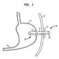
- a transabdominal tube 1is placed through a patient's abdominal wall such that a distal end portion 17 of the tube 1 is disposed inside the stomach 3 of the patient and a proximal end portion 16 of the tube 1 extends out from the skin 5 of the patient.
- the tube 1may be stiffened, made durable and less collapsible by, for example, braiding the tube using nylon.
- the tubemay be wrapped with wire material. Suitable materials for the tube 1 include polyurethane, silicone and other similar materials.
- the tube 1may be opaque.
- a retention memberis attached to the tube 1 to prevent the tube 1 from falling out of the patient.
- the retention memberis inflatable such as the inflation portion 2 (balloon anchor) shown in FIG. 1 .
- the inflation portion 2is provided at the distal end portion 17 of the tube 1 to prevent the tube 1 from coming out of the stomach 3 .
- FIG. 1also illustrates a non-inflatable retention member flange 2 ′ at the proximal end portion 16 of the tube 1 to prevent the tube 1 from falling into the patient's upper digestive system.
- a cap 13is detachably provided at the end of the proximal end portion 16 and seals the tube 1 when it is attached. The cap 13 is removed when a pump 8 , 9 (shown in FIGS. 2 and 3 , respectively) is attached to the tube 1 to remove food from the upper digestive system of the patient.
- the tube 1may be inserted, for example, through a procedure similar to insertion of feeding tubes by Percutaneous Endoscopic Gastrostomy (PEG).
- PEGPercutaneous Endoscopic Gastrostomy
- a variety of methods of performing PEGare well known in the art, and any one of the methods may be used to insert the tube 1 .
- PEG procedureshave been successfully completed in over 90 percent of attempts.
- PEGmay be performed under conscious sedation induced by, for example, meperidine and midazolam.
- an endoscopeis inserted into the stomach through the mouth of the patient.
- the stomachis insufflated by blowing air into the stomach through the endoscope.
- the insufflationbrings the stomach in apposition to the abdominal wall and allows for direct access from the skin to the stomach of the patient.
- An insertion siteis located by surveying the interior of the stomach with the endoscope.
- the endoscopeis then used to illuminate the selected insertion site in such a way that the light of the endoscope is visible from outside of the patient's body through the skin of the patient.
- An incisionis made at the place on the patient's skin indicated by the light from the endoscope and at the corresponding location on the exterior wall of the stomach.
- a cannulais then inserted through the incision and a guide wire is inserted into the stomach through the cannula.
- Graspers on the end of the endoscopegrab hold of the distal portion of the guide wire in the stomach and the endoscope is withdrawn from the patient while the graspers hold the guide wire.
- the guide wireis of sufficient length to allow a proximal portion of it to extend out of the patient from the cannula after the distal portion is withdrawn from the stomach and through the patient's mouth by the endoscope.
- the end of the guide wire extending out from the patient's mouthis attached to the proximal end of the tube 1 , which is drawn though the mouth and esophagus and into the stomach of the patient by pulling on the proximal end of the guide wire.
- the tube 1is then pulled through the incision in the stomach and skin of the patient until only the distal end portion 17 and the inflation portion 2 of the tube 1 remain inside of the stomach.
- the tube 1may have a coned tip to help move the tube 1 through the incision in the stomach.
- a wire at the tip of the conemay be used for pulling the tube 1 through the incision.
- the coned tipmay be cut off
- the cannulais removed as the proximal end 16 of the tube 1 is drawn through the incision in the stomach, and is removed entirely when the proximal end 16 of the tube 1 is disposed at the patient's skin.
- the inflation portion 2 of the tube 1is then inflated by introducing fluid into the inflation portion 2 through the inflation lumen 26 .
- the inflated inflation portionholds the tube 1 in place and the guide wire is removed from the tube 1 .
- a non-inflatable retention membersuch as a flange 2 ′ may be placed on the proximal end portion 16 of the tube 1 to keep the tube 1 disposed at the patient's skin.
- PEGPEG
- An alternate method of PEG known as push PEGmay also be used to insert the tube 1 .
- the tube 1is pushed through the incision in the stomach and the skin of the patient until it is disposed as described hereinabove with respect to the pull method.
- a third method which may be used for inserting the tube 1 via PEGis known as the Russell method.
- the insertion siteis located via endoscopy.
- An incisionis made in the skin and stomach and a guide wire is inserted through the incision into the stomach via a cannula or needle.
- a dilator (or introducer) with a peel away sheathis guided along the guide wire and inserted into the stomach.
- the dilatoris removed and the tube 1 is inserted along the guide wire and through the peel away sheath.
- the sheathis then peeled away and the tube 1 is fixed in place.
- the tube 1may also be inserted without using an endoscope, for example, through a procedure similar to insertion of feeding tubes by Percutaneous Radiological Gastrostomy (PRG).
- PRGPercutaneous Radiological Gastrostomy
- the stomachis insufflated via a nasogastric tube.
- Organs which may be interposed between the stomach and the abdominal wall, such as the colon,are excluded by CT scan or ultrasonography. Exclusion of interposed organs may also be accomplished after insufflation by fluoroscopy.
- the selection of the insertion siteis also determined by fluoroscopy or a similar method.
- the tube 1may be inserted transabdominally as in the Russell method of PEG.
- a guide wiremay be inserted as in the endoscopic pull method. The wire is then maneuvered through the stomach and esophagus and out of the patient's mouth and is used to guide the tube 1 back through the mouth, esophagus and stomach and out of the insertion site (see, e.g., Mustafa N. Zmen et al. “Percutaneous Radiologic Gastrostomy” European Journal of Radiology 43:186-95).
- the tube 1may be inserted surgically.
- One suitable surgical technique that may be used to insert the tube 1is the laparoscopic method. In this method, after pneumoperitoneum has been created, a 5 mm trocar is used to grasp a site on the anterior stomach wall that is appropriate for tube placement without excessive tension on the stomach. A skin incision down to the rectus sheath is made. A trocar is placed through the rectus sheath and the stomach wall is grasped and pulled upwards. An incision is made in the stomach and the tube 1 is inserted. Using the retention member at the distal end portion 17 of the tube 1 , the stomach is brought snugly against the abdominal wall. The tissue is sutured around the tube 1 . (see, e.g., Andrew Luck et al. “Laparoscopic Gastrostomy: Towards the Ideal Technique” Aust. N. Z. J. Surg. (1998) 68:281-283).
- the tube 1may be inserted in other portions of the upper digestive system besides the stomach.
- direct jejunostomywherein a tube is inserted transabdominally into the jejunum, may be accomplished through methods similar to those described hereinabove with reference to gastrostomy tube placement.
- the retention member of the deviceshould generally be smaller for jejunostomy procedures to avoid irritation of the jejunum or obstruction of the jejunal lumen.
- FIG. 1illustrates an inflatable retention member, i.e. the inflation portion 2 , that is attached to the tube 1 to prevent the tube 1 from falling out of the patient.
- FIGS. 1 , 1 A and 1 Billustrate two alternative non-inflatable retention members that may be used in place of and/or in addition to the inflatable portion 2 .
- FIGS. 1 and 1Aillustrate a flange 2 ′ and FIG. 1B illustrates a dome 2 ′′.
- a flange 2 ′ or dome 2 ′′that is located at the distal end portion 17 of the tube 1 helps to prevent the tube 1 from coming out of the stomach 3 or other section of the upper digestive system.
- a flange 2 ′ or dome 2 ′′that is located at the proximal end portion 16 of the tube 1 helps to prevent the tube from falling into the patient's upper digestive system.
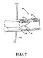
- the tube 1When an inflatable retention member is used, the tube 1 preferably has an inflation lumen 26 so that the inflatable retention member can be inflated.
- FIG. 1Cshows a cross section of the tube 1 taken perpendicular to the axis of tube 1 .
- Inflation lumen 26extends from the inflation portion 2 to the proximal end portion 16 of the tube 1 and is a pathway for introducing fluid, such as water or air, to the inflation portion 2 from outside of the patient.
- Removal lumen 25extends from the proximal end portion 16 to the distal end portion 17 of the tube 1 and is a pathway for the removal of food from the stomach 3 or other part of the upper digestive system of the patient.
- the inflation lumen 26is preferably minimal in size to allow the removal lumen 25 to be as wide as possible within the tube 1 .
- valves 15 , 27are provided in lumens 25 , 26 , respectively, as shown in FIG. 7 .
- the second lumen 26 in tube 1can be eliminated.
- Inflatable retention membersare suitable for use with procedures similar to the push method, while either inflatable or rigid retention members are suitable for use with procedures similar to the pull method.
- One example of a tube that has an inflatable retention memberis taught in Tiefenthal et al. (U.S. Pat. No. 6,506,179), the entire contents of which are incorporated herein by reference.
- An alternative deformable retention memberis taught in Snow et al. (U.S. Pat. No. 6,077,250), the entire contents of which are incorporated herein by reference.
- Retention membersthat may be deformed in situ allow the tube 1 to be removed without additional endoscopy.
- the retention memberis deflated or deformed and the tube 1 is pulled out using traction.
- the tube 1may be cut close to the skin and removed endoscopically.
- the stomachit is preferable for the stomach to be positioned up against the inner abdominal wall. This may be accomplished by insufflation during the tube placement procedure and after the tube 1 has been placed due to the retention member.
- retention members at the proximal end portion 16 and distal end portion 17 of the tube 1anchor the stomach up against the abdominal wall.
- the stomachmay also be anchored to the abdominal wall by gastropexy, which may prevent complications arising from tube placement and may facilitate the placement procedure.
- jejunopexyis important in jejunostomy procedures in order to secure the jejunum during the tube placement procedure (see Zmen et al., supra).
- T-shaped metal or nylon fixing membersmay be inserted trans-gastrically or trans-jejunally close to the tube insertion site.
- the fixing membersassume a T shape after insertion and are tied near to the skin.
- Four fixing membersare typically disposed in a square pattern around the tube insertion site to secure the stomach or jejunum.
- FIGS. 2 and 3display pumps 8 and 9 which are attachable to the proximal end portion 16 of the tube 1 for removal of food from the stomach 3 or upper digestive system of the patient. It would be suitable to use a pump that extracts more than 750 ml of food from the upper digestive system of a patient within 30 minutes or less.
- the pumpmay be operated intermittently to prevent tube collapse, tube clogging or mucosal irritation.
- the pumpmay be manual or battery operated.
- a rechargeable power supplymay be incorporated into the pump, and the pump may be configured to be carried on a patient's belt.
- FIG. 2depicts a manual bulb pump 8 that is attached to the proximal end portion 16 of the tube 1 and is operated to remove food from the patient's upper digestive system through the tube 1 .
- the manual bulb pump 8preferably comprises silicone rubber or a similar flexible material so as to permit the contents of the bulb pump 8 to be evacuated by squeezing the bulbous end of the bulb pump 8 .
- the circumference of a tapered endessentially corresponds to an interior circumference of the lumen 25 of the tube 1 .
- the manual bulb pump 8To operate the manual bulb pump 8 , air is first evacuated from the bulb pump 8 by squeezing the bulb, and then the tapered end of the bulb pump 8 is inserted into the lumen 25 of the proximal end portion 16 of the tube 1 so as to create a seal between the tapered end and the tube 1 . The bulb is then released to allow it to re-inflate. The negative pressure in the bulb pump 8 (when it is released) causes food to flow out from the upper digestive system toward the proximal end portion 16 of the tube 1 and into the bulb of the manual bulb pump 8 . The bulb pump 8 is then disengaged from the tube 1 and the removed food is evacuated from the bulb. The cycle may be repeated until a desired amount of food is removed from the upper digestive system of the patient.
- FIG. 3depicts another pumping arrangement in which a pump in the form of a syringe 9 is attached to the proximal end portion 16 of the tube 1 and is operated to remove food from the patient's upper digestive system through the tube 1 .
- the syringe 9preferably comprises a tapered end portion with an aperture at the distal end thereof.
- the circumference of the tapered end portion 9 acorresponds to the interior circumference of the lumen 25 of the tube 1 .
- the contents (air or food) of the syringe 9are evacuated by depressing the plunger.
- the tapered end portion 9 a of the syringe 9is inserted into the proximal end portion 16 of the tube 1 so as to create a seal between the tapered end portion 9 a and the tube 1 .
- the plunger of the syringe 9is then withdrawn so as to create negative pressure to draw food out from the upper digestive system through the tube 1 and into the syringe 9 .
- the syringe 9is then disengaged from the tube 1 and evacuated by, for example, depressing the plunger thereof.
- 60 ccis an example of a suitable size for the syringe 9 .
- the cyclemay be repeated until a desired amount of food is removed from the upper digestive system of the patient.
- the manual bulb pump 8 and syringe 9may be activated by the patient or by a health care provider at a predetermined time after eating.
- the predetermined timeis preferably set by a physician and, for example, may be 20-30 minutes.
- a physicianmay also determine a maximum volume of food to be removed from the upper digestive system of the patient after each meal. The maximum volume may be set in terms of a maximum number of pumping cycles which is told to the patient or health care provider if the pump 8 , 9 is manually operated.
- the pump that is used to extract food from the patient's upper digestive systemperiodically reverses direction and pumps air and/or water into the upper digestive system of the patient during the periods of reverse operation.
- the air and/or waterhelps to solubilize or breakdown the food in the upper digestive system so that it can be pumped out easily.
- the air and/or waterhelps prevent the tube 1 from being suctioned up against the stomach wall while food is extracted from the upper digestive out through the tube 1 . For example, every seven seconds of pumping may be followed by two seconds of reverse operation.
- FIG. 4illustrates a variation of an embodiment of the present invention in which the extracted food is evacuated from a pump 6 into a bag 12 that is attached to the pump 6 .
- the foodmay be stored in a bag 12 that is attachable to the proximal end portion of the pump 6 .
- the bag 12may be opaque, scented, biodegradeable and worn by the patient on a belt or other strap.
- the foodmay be pumped from the patient's upper digestive system into the pump 6 and then into a tube 28 attached to the pump 6 .
- the contents of the tube 28 attached to the pump 6may be emptied into a toilet.
- the tube 28may be opaque, scented, biodegradeable and flushable down the toilet.
- FIG. 5illustrates a cleaning device being used to clean the tube 1 after food has been extracted from the patient's upper digestive system through the tube 1 .
- the tube 1may be cleaned using a brush 14 that is adapted to clean the inside of the tube 1 .
- the pump 6 , manual bulb pump 8 and syringe 9may be cleaned by flushing them with saline and/or a disinfectant solution after use.
- FIG. 6illustrates a second embodiment of the present invention in which a feeling of satiety is created in the patient by inflating the balloon anchor. Creating a feeling of satiety curbs the patient's hunger and desire to eat food thereby allowing the patient to eat less and lose weight.
- the inflation portion 2which is the retention member that holds the tube 1 in the patient's stomach, also serves the function of decreasing stomach capacity to create a feeling of satiety when it is inflated.
- the inflation portion 2may be variably inflated by adding or removing fluid through the inflation lumen 26 of the tube 1 (shown in FIG. 1C ).
- FIG. 7shows an axial cross sectional view of the tube 1 extending out from the skin 5 of the patient in which the removal lumen 25 and the inflation lumen 26 are visible.
- a valve 15is provided at the proximal end portion 16 of the tube 1 in the removal lumen 25 .
- the valve 15ordinarily prevents food from leaving the tube 1 .
- the valve 15is opened when a pump is attached to the proximal end portion 16 of the tube 1 .
- the tapered end portion of the manual bulb pump 8shown in FIG. 2
- the tapered end portion of the syringe 9shown in FIG.
- a cap 13(shown in FIG. 1 ) is preferably placed on the proximal end portion 16 of the tube 1 when a pump is not attached. The cap 13 may be pressed onto the end of the tube 1 , threaded on the end of the tube 1 , or may have projections which are frictionally inserted into the ends of lumens 25 , 26 to seal them in a closed condition.
- FIG. 7also shows a valve 27 provided at the proximal end portion 16 of the tube 1 in the inflation lumen 26 .
- the valve 27prevents the fluid used to inflate the inflation portion 2 from escaping the inflation portion 2 through the inflation lumen 26 . That is, the valve 27 prevents the inflation portion 2 from deflating. If it becomes necessary to deflate the inflation portion 2 to remove the tube 1 from the upper digestive system of the patient, or to further inflate the portion 2 , a needle on a syringe may be inserted into the inflation portion 26 so as to open the valve 27 by pushing the needle through the valve members. The fluid used to inflate the inflation portion 2 may then be removed or added with the syringe.
- FIG. 8illustrates a third embodiment of the present invention showing a tube having two balloons attached to that portion of the tube that is disposed within the patient's upper digestive system.
- the balloon anchor 2is expandable to about 10 ml and is positioned up against the stomach wall to prevent the tube 1 from falling out.
- the inflatable balloon 29is expandable from about 100 ml to about 850 ml and may be expanded intermittently to limit the capacity of the stomach.
- the balloon 29may be inflated via an inflation lumen prior to a meal to create the sensation of being full. After the meal, the balloon 29 may be deflated to prevent chronic accommodation.
- An electrically or a manually operated pumpmay be used to cause the inflation.
- the tube 1 in this embodimenthas a long inner tube length of about 10 cm or longer and a diameter of 28 French (9.3 mm) in size or greater.
- the tube 1may have multiple holes 32 in the sidewall of its distal end portion 17 as shown in FIG. 8 and also in FIGS. 10 and 13 - 15 B.
- the holes 32may be 5 ⁇ 7 mm in size.
- the holes 32provide non-vascular drainage from the patient.
- the holes 32are arranged in a spiral pattern 1 cm to 1.5 cm apart without losing structural integrity.
- cushions or bumpersare located on the tube 1 and in between the holes 32 to prevent the tube from being sucked up against the stomach wall while food is extracted from the upper digestive system out through the tube 1 .
- cushions or bumpersthat are raised 3-4 mm above the surface of the tube 1 may be used for this purpose.
- a second retention member 33may be attached at the proximal end portion 16 of the tube 1 to keep the tube 1 fixed to the abdominal surface.
- This second retention membermay be similar to the retention members described hereinabove and shown in FIGS. 1 , 1 A, 1 B and 6 .
- the distance between the second retention member 33 at the proximal end portion 16 of the tube 1 and the balloon anchor 2 at the distal end portion 17 of the tube 1can be adjusted to account for the varying amount of intervening tissue 40 , 40 ′ as shown in FIGS. 15A and 15B .
- the second retention member 33may be attached to the tube 1 via an interference or friction fit.
- the second retention member 33may be placed around the outer surface of the proximal end portion 16 of the tube 1 and held in place on the tube 1 if it has an inner diameter that is slightly smaller than the outer diameter of the tube 1 . As the patient loses weight, the proximal end portion 16 of the tube 1 extends farther and farther away from the patient's abdominal surface. A physician or the patient can slide the second retention member 33 down towards the abdominal surface and the excess amount of the tube 1 can be cut off.
- FIG. 9illustrates a fourth embodiment of the present invention with a tube 1 having a curved configuration at its distal end portion 17 and a plurality of holes 32 in a sidewall.
- the distal end portion 17 of the tube 1is adapted to assume a curved configuration when disposed in the upper digestive system of a patient.
- the distal end portion 17 of the tube 1is flexible to facilitate insertion and removal from the patient. When the distal end portion 17 of the tube 1 is disposed in the upper digestive system of the patient, it returns to its natural curved configuration.
- the tube's tendency to return to its natural curved configurationmay be achieved, for example, by bending the tube into a desired curved shape during the manufacturing process before the tube has fully cured or cooled, or by incorporating shape memory materials into the tube.
- the term “curved”includes flexed, bent, rounded, arched, curled, coiled, spiral, and pigtail. This curved configuration is preferable because it increases the intake area within the upper digestive system.
- the coiled distal end portion 17 of the tube 1 as shown in FIG. 10helps to maintain the position of the tube 1 within the patient's upper digestive system.
- the distal end portion 17 of the tube 1may, for example, be about 10 cm long or longer to improve the intake of the food from the upper digestive system.
- Retention members(not shown) similar to the ones described in the above embodiments may also be used in this embodiment.
- an actuating mechanismis configured to bend the distal end portion 17 of the tube 1 into a curved configuration.
- the actuating mechanismmay, for example, be a string attached to the distal end portion 17 of the tube 1 that, when retracted causes the tube to assume a curved configuration (e.g. a loop with an arc that measures between about 270°-360°).
- a Cope Loopis a well known example of this arrangement.
- FIG. 10illustrates a fifth embodiment of the present invention showing a tube 1 having a curved configuration, multiple holes 32 in a sidewall, and a morcellation device 36 housed within a housing 37 at its distal end portion 17 .
- morcellation devicesare disclosed in U.S. Pat. Nos. 5,618,296, 5,741,287 and 5,520,634, herein incorporated by reference in their entirety.
- a morcellation device 36is provided at the distal end portion 17 of the tube 1 to divide and grind food into smaller pieces as it enters the tube 1 .
- the morcellation device 36thus allows large food to be removed from the patient without clogging the tube 1 .
- the morcellation device 36can be, for example, a mechanical propeller provided within a housing 37 at the distal end portion 17 of the tube 1 .
- the housing 37is constructed to protect body tissue from the morcellation device 36 .
- the housing 37has an opening to permit the entry of food from the patient into the tube 1 and may, for example, be a cage that surrounds the morcellation device 36 at the distal end portion 17 of the tube 1 . It is preferable that the housing 37 is collapsible in both directions so that it can be easily inserted into and taken out of the patient.
- the housing 37is necessary to prevent damage to the stomach.
- FIG. 11illustrates a feature that may be used with any embodiment of the present invention in which the proximal end portion 16 of the tube 1 lies substantially flush with the outer surface of the patient's abdomen. This may be achieved by using ribbons attached to the tube 1 , for example at the internal retention member. The ribbons are used to pull the tube 1 taut when the distal end portion 17 of the tube 1 is disposed in the upper digestive system of a patient.
- the proximal end portion 16 of the tube 1is cut so that the proximal end portion 16 lies flush with the abdominal surface and a thin, hollow cylinder with flanges is wedged onto the outside or inside surface of the tube 1 via friction or by screwing it onto the tube 1 to retain the tube 1 in its position and to keep it flush with the abdominal surface.
- the proximal end portion 16 of the tube 1may extend out past the abdominal surface by any desired length (e.g., 1-10 inches).
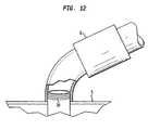
- FIG. 12illustrates another feature that may be used with any embodiment of the present invention in which a luer lock 34 is utilized at the proximal end portion 16 of the tube 1 .
- the pump 6is attached to the tube 1 by screwing the pump 6 onto the tube 1 around the external portion of the proximal end portion 16 of the tube 1 rather than being inserted into the tube 1 .
- the proximal end portion 16 of the tube 1comprises concentric grooves or threads on the outside to accommodate the pump 6 , which prevents the pump 6 from reducing the size of the removal lumen 25 .
- the pump 6may have corresponding concentric grooves or threads that allow it to interact and connect with the luer lock 34 .
- the pump 6is coupled to or threaded onto the outside of the proximal end portion 16 of the tube 1 .
- FIG. 13illustrates yet another feature that may be used with any embodiment of the present invention in which the tube 1 has a funnel shaped tip 35 .
- the funnel tipis advantageous because it facilitates the extraction of larger pieces of food into the tube 1 from the patient's digestive system.
- FIG. 14illustrates a sixth embodiment of the present invention that has two intake tubes.
- both of the intake tubes 38have a curved configuration and a sidewall with a plurality of holes 32 located therein.
- Each intake tube 38comprises a proximal end portion 39 and distal end portion 40 .
- the apparatusalso comprises an output tube 41 having a proximal end portion and a distal end portion 42 .
- One or more retention membersare preferably attached to the output tube 41 to prevent the apparatus from coming out of the upper digestive system.
- the plurality of intake tubes 38are configured to be disposed in the upper digestive system of the patient and the output tube 41 is configured to pass through the patient's abdominal wall when the plurality of intake tubes 38 are so disposed.
- the distal end portion 42 of the output tube 41is operatively connected to the proximal end portion 39 of each of the plurality of intake tubes 38 so that food can be extracted from the upper digestive system of the patient through the distal end portion 40 of each of the plurality of intake tubes 38 and out through the proximal end portion of the output tube 41 .
- pressure and/or flow sensorsmay be placed on and/or in the tube 1 .
- Pressure sensors placed on the tube 1 inside and outside the stomach 3may be used to estimate the satiety of the patient.
- flow sensors that are placed inside the tube 1may be used to calculate the volume of food extracted through the tube 1 .
- a tubeis positioned so that it passes through a patient's abdominal wall into his/her upper digestive system.
- the patientis allowed to go about his/her daily activities including ingesting food.
- the foodis extracted from the upper digestive system of the patient through the tube.
- the patientmay eat and extract the eaten food from his/her upper digestive system through the tube repeatedly until a desired weight loss is attained.
- the food that has been extractedis not reintroduced into the patient.
- the tubemay be kept in the patient's upper digestive system for extended periods of time (e.g., one month or more) while the eating/extracting is repeated numerous times (e.g., 20 times or more) while the tube is in place.
- a tubeis positioned so that it passes through the obese patient's abdominal wall into his/her upper digestive system.
- the obese patientis allowed to go about his/her daily activities including ingesting food.
- the foodis extracted from the upper digestive system of the obese patient through the tube.
- the obese patientmay eat and extract the eaten food from his/her upper digestive system through the tube repeatedly until the obese patient has lost at least 40 pounds.
- the food that has been extractedis not reintroduced back into the obese patient.
- a tubeis positioned so that it passes through a patient's abdominal wall into the upper digestive system of the patient whose gastrointestinal tract is unobstructed.
- the patientis allowed to go about his/her daily activities including ingesting food. After the patient has ingested the food, the food is extracted from the upper digestive system of the patient through the tube. The patient may eat and extract the eaten food from his/her upper digestive system through the tube repeatedly until a desired weight loss is attained.
- the tubemay be kept in the patient's upper digestive system for extended periods of time (e.g., one month or more) while the eating/extracting is repeated numerous times (e.g., 20 times or more) while the tube is in place.
- the food extraction apparatuses and methods described aboveare preferably combined with a behavior modification program that ideally educates patients in modifying caloric intake, lifestyle and attitudes toward food. Learned activities and support for weight loss may include activities such as self-monitoring by recording food intake and physical activity, avoiding triggers that prompt eating, assistance from family and friends, problem solving skills and relapse prevention.
- the programmay be taught by an instructor or offered over the internet.
- the programpreferably includes a series of regular check-ups by a health care provider. The check-ups ideally include regularly testing blood for electrolytes, supplementing patients' diets with vitamins, and administering medications to prevent gallstone formation as needed.
- the behavior modification programwill educate patients to change their lifestyle so as to eliminate the need for food extraction.
- the above described embodimentsallow obese patients to lose weight without undergoing drastic and invasive surgeries. As a result, obese patients avoid many of the complications associated with such surgeries.
- the present inventionis easy to perform, easy to reverse and allows obese patients to live a normal and active lifestyle with fewer adverse side effects.
Landscapes
- Health & Medical Sciences (AREA)
- Heart & Thoracic Surgery (AREA)
- Animal Behavior & Ethology (AREA)
- General Health & Medical Sciences (AREA)
- Veterinary Medicine (AREA)
- Engineering & Computer Science (AREA)
- Biomedical Technology (AREA)
- Public Health (AREA)
- Life Sciences & Earth Sciences (AREA)
- Vascular Medicine (AREA)
- Anesthesiology (AREA)
- Hematology (AREA)
- Child & Adolescent Psychology (AREA)
- Obesity (AREA)
- Orthopedic Medicine & Surgery (AREA)
- Nursing (AREA)
- Surgery (AREA)
- Ophthalmology & Optometry (AREA)
- Otolaryngology (AREA)
- Surgical Instruments (AREA)
Abstract
Description
| weight | |||
| Week | (kg) | ||
| 0 | 100.9 | ||
| 2 | 96.8 | ||
| 3 | 96.8 | ||
| 4 | 94.7 | ||
| 4 | 94.7 | ||
| 5 | 94.0 | ||
| 7 | 93.6 | ||
| 8 | 90.9 | ||
| 9 | 92.9 | ||
| 10 | 92.7 | ||
| 11 | 90.4 | ||
| 12 | 89 | ||
| 13 | 89.3 | ||
| 14 | 88.6 | ||
| 15 | 87.7 | ||
| 16 | 86.5 | ||
| 17 | 86.5 | ||
| 18 | 86.3 | ||
| 19 | 85.9 | ||
| 20 | 83.9 | ||
| 21 | 82.9 | ||
| 22 | 81.6 | ||
| 23 | 80.45 | ||
| 24 | 79.7 | ||
| 25 | 78.6 | ||
| 26 | 78.6 | ||
| 27 | 77.2 | ||
| 28 | 78 | ||
| 29 | 76.2 | ||
| 30 | 76 | ||
| 31 | 75.2 | ||
| 31 | 77.1 | ||
| 32 | 76.4 | ||
| 33 | 76.4 | ||
| 34 | 76.4 | ||
| 35 | 74 | ||
| 36 | 74 | ||
| 37 | 74 | ||
| 38 | 73.6 | ||
| 39 | 73.5 | ||
| 40 | 73.2 | ||
| 41 | 72.6 | ||
| 42 | 71.22 | ||
| 43 | 69.5 | ||
| 44 | 69.8 | ||
| 45 | 69.45 | ||
| 46 | 68.45 | ||
| 47 | 66.6 | ||
| 48 | 65.5 | ||
| 49 | 65.5 | ||
| 50 | 65.5 | ||
| 51 | 65.2 | ||
| 52 | 65 | ||
| 53 | 65 | ||
| 54 | 64.5 | ||
| 55 | 64.8 | ||
| 56 | 64.8 | ||
| 57 | 63.8 | ||
| 58 | 63 | ||
| 59 | 62.45 | ||
Claims (14)
Priority Applications (1)
| Application Number | Priority Date | Filing Date | Title |
|---|---|---|---|
| US12/604,140US8282623B2 (en) | 2002-11-04 | 2009-10-22 | Method for treating obesity by extracting food |
Applications Claiming Priority (6)
| Application Number | Priority Date | Filing Date | Title |
|---|---|---|---|
| US42364502P | 2002-11-04 | 2002-11-04 | |
| US10/702,194US20040220516A1 (en) | 2002-11-04 | 2003-11-04 | Food extraction apparatus and method |
| US60049604P | 2004-08-10 | 2004-08-10 | |
| US61834604P | 2004-10-12 | 2004-10-12 | |
| US11/191,466US7740624B2 (en) | 2002-11-04 | 2005-07-27 | Method for treating obesity by extracting food |
| US12/604,140US8282623B2 (en) | 2002-11-04 | 2009-10-22 | Method for treating obesity by extracting food |
Related Parent Applications (1)
| Application Number | Title | Priority Date | Filing Date |
|---|---|---|---|
| US11/191,466DivisionUS7740624B2 (en) | 2002-11-04 | 2005-07-27 | Method for treating obesity by extracting food |
Publications (2)
| Publication Number | Publication Date |
|---|---|
| US20100106131A1 US20100106131A1 (en) | 2010-04-29 |
| US8282623B2true US8282623B2 (en) | 2012-10-09 |
Family
ID=46304894
Family Applications (2)
| Application Number | Title | Priority Date | Filing Date |
|---|---|---|---|
| US11/191,466Expired - LifetimeUS7740624B2 (en) | 2002-11-04 | 2005-07-27 | Method for treating obesity by extracting food |
| US12/604,140Expired - Fee RelatedUS8282623B2 (en) | 2002-11-04 | 2009-10-22 | Method for treating obesity by extracting food |
Family Applications Before (1)
| Application Number | Title | Priority Date | Filing Date |
|---|---|---|---|
| US11/191,466Expired - LifetimeUS7740624B2 (en) | 2002-11-04 | 2005-07-27 | Method for treating obesity by extracting food |
Country Status (1)
| Country | Link |
|---|---|
| US (2) | US7740624B2 (en) |
Cited By (6)
| Publication number | Priority date | Publication date | Assignee | Title |
|---|---|---|---|---|
| US9011365B2 (en) | 2013-03-12 | 2015-04-21 | Medibotics Llc | Adjustable gastrointestinal bifurcation (AGB) for reduced absorption of unhealthy food |
| US9067070B2 (en) | 2013-03-12 | 2015-06-30 | Medibotics Llc | Dysgeusia-inducing neurostimulation for modifying consumption of a selected nutrient type |
| US9456916B2 (en) | 2013-03-12 | 2016-10-04 | Medibotics Llc | Device for selectively reducing absorption of unhealthy food |
| US9861734B2 (en)* | 2016-03-21 | 2018-01-09 | King Saud University | Bifurcated peritoneal catheter |
| WO2018138600A1 (en) | 2017-01-25 | 2018-08-02 | Ethicon, Inc. | Modified apparatus for food extraction and obesity treatment |
| US10085866B2 (en) | 2013-02-23 | 2018-10-02 | Aspire Bariatrics, Inc. | Apparatus and method for draining material from a stomach |
Families Citing this family (13)
| Publication number | Priority date | Publication date | Assignee | Title |
|---|---|---|---|---|
| US7815629B2 (en) | 2002-11-04 | 2010-10-19 | Deka Products Limited Partnership | Apparatus for treating obesity by extracting food |
| US7740624B2 (en) | 2002-11-04 | 2010-06-22 | Aspiration Medical Technology, Llc | Method for treating obesity by extracting food |
| US9055995B2 (en) | 2002-11-04 | 2015-06-16 | Aspire Bariatrics, Inc. | Method for treating obesity by extracting food |
| JP2009542349A (en)* | 2006-07-05 | 2009-12-03 | アスピレーション・メディカル・テクノロジー・エルエルシー | Short-circuit device to treat obesity by extracting food |
| US20110082442A1 (en)* | 2006-07-05 | 2011-04-07 | Solovay Kenneth S | Externally reinforced percutaneous gastrostomy tube with customizable smooth tube length |
| US8062285B2 (en) | 2006-08-03 | 2011-11-22 | Aspire Bariatrics, Llc | Systems and methods for removing ingested material from a stomach |
| AU2015202650B2 (en)* | 2006-08-03 | 2016-09-01 | Aspire Bariatrics, Inc. | Systems and methods for removing ingested material from a stomach |
| US8632513B2 (en) | 2006-08-03 | 2014-01-21 | Aspire Bariatrics, Inc. | Systems and methods for removing ingested material from a stomach |
| US8746911B2 (en)* | 2012-02-16 | 2014-06-10 | Shenzhen China Star Optoelectronics Technology Co., Ltd. | Backlight module and LCD device comprising backlight module |
| WO2015200627A1 (en) | 2014-06-25 | 2015-12-30 | Ismail Muhammad Sami | Bariatric device and method |
| US10987131B2 (en) | 2017-05-25 | 2021-04-27 | Coopersurgical, Inc. | Tissue containment systems and related methods |
| WO2019195093A1 (en)* | 2018-04-02 | 2019-10-10 | Georgetown University | Diverting jejunostomy tube |
| KR200499620Y1 (en)* | 2023-10-12 | 2025-09-26 | 한림대학교 산학협력단 | Gastrostomy tube |
Citations (139)
| Publication number | Priority date | Publication date | Assignee | Title |
|---|---|---|---|---|
| US2933140A (en) | 1956-12-31 | 1960-04-19 | Gamewell Co | Selector valve |
| US3066672A (en) | 1960-09-27 | 1962-12-04 | Jr William H Crosby | Method and apparatus for serial sampling of intestinal juice |
| US3144868A (en) | 1960-10-21 | 1964-08-18 | Mario E Jascalevich | Drainage and feeding cannulae |
| US3214069A (en) | 1960-04-12 | 1965-10-26 | Continental Can Co | Plastic captive seal closure |
| US3232578A (en) | 1963-03-14 | 1966-02-01 | Coastal Dynamics Corp | Pivoted disc valve having a particular mounting arrangement |
| US3384342A (en) | 1965-10-23 | 1968-05-21 | Passer Fastener Corp | Replacement faucet washer |
| US3506237A (en) | 1968-08-12 | 1970-04-14 | Chemtrox Corp | Plastic rotary valve |
| US3598150A (en) | 1969-06-16 | 1971-08-10 | Hollister Inc | Medical-surgical valve arrangement |
| US3752158A (en) | 1970-11-23 | 1973-08-14 | Snyder Manuf Co Inc | Apparatus and method for suprapubic drainage of the urinary bladder |
| US3860000A (en) | 1973-07-12 | 1975-01-14 | Lear Siegler Inc | Medical apparatus and method for feeding and aspirating |
| US3884808A (en) | 1973-06-20 | 1975-05-20 | Res Dev Systems Inc | Wearable, self-regenerating dialysis appliance |
| US3924625A (en) | 1974-11-11 | 1975-12-09 | Hans D D Peterson | Powered bovine stomach pump and tube |
| US4082095A (en) | 1975-10-09 | 1978-04-04 | Barry Mendelson | Stomach pump |
| US4116589A (en) | 1977-04-15 | 1978-09-26 | Avco Corporation | Extracorporeal pulsatile blood pump comprised of side by side bladders |
| US4189795A (en) | 1978-09-20 | 1980-02-26 | Conti Angelo J | Toilet flush valve |
| US4315509A (en) | 1977-01-10 | 1982-02-16 | Smit Julie A | Insertion and removal catheters and intestinal tubes for restricting absorption |
| US4315513A (en) | 1980-03-10 | 1982-02-16 | Nawash Michael S | Gastrostomy and other percutaneous transport tubes |
| US4344435A (en) | 1978-12-15 | 1982-08-17 | Aubin Norbert T | Method and surgically implantable apparatus for providing fluid communication with the interior of the body |
| US4356824A (en) | 1980-07-30 | 1982-11-02 | Vazquez Richard M | Multiple lumen gastrostomy tube |
| US4381765A (en) | 1981-04-02 | 1983-05-03 | Waters Instruments, Inc. | Ileostomy valve |
| US4393873A (en) | 1980-03-10 | 1983-07-19 | Nawash Michael S | Gastrostomy and other percutaneous transport tubes |
| US4449972A (en) | 1981-08-29 | 1984-05-22 | Krueger Christian | Stomach flushing devices |
| US4464175A (en) | 1982-08-25 | 1984-08-07 | Altman Alan R | Multipurpose tamponade and thrombosclerotherapy tube |
| US4525156A (en) | 1983-02-16 | 1985-06-25 | Benusa John E | Method for stomach lavage |
| US4538653A (en) | 1984-12-04 | 1985-09-03 | Genoa Group Inc. | Closure and valve for liquid container |
| US4551130A (en) | 1984-05-08 | 1985-11-05 | Herbert William B | Surgical drainage and irrigation apparatus for post operative patient care |
| US4553960A (en) | 1978-09-25 | 1985-11-19 | Harrison Lazarus | Peritoneal fluid treatment apparatus, package and method |
| US4642092A (en) | 1984-12-10 | 1987-02-10 | Gerald Moss | Gastrointestinal aspirating device with suction breakers |
| US4668225A (en) | 1985-12-23 | 1987-05-26 | Superior Healthcare Group, Inc. | Gastrostomy tube and gastrostomy-jejunal feeding tube combination |
| US4685901A (en) | 1984-11-05 | 1987-08-11 | Medical Innovations Corporation | Gastro-jejunal feeding device |
| US4723547A (en) | 1985-05-07 | 1988-02-09 | C. R. Bard, Inc. | Anti-obesity balloon placement system |
| US4790812A (en) | 1985-11-15 | 1988-12-13 | Hawkins Jr Irvin F | Apparatus and method for removing a target object from a body passsageway |
| US4804375A (en) | 1987-05-06 | 1989-02-14 | Santa Barbara Medical Foundation Clinic | Ileostomy valve having a rotatable shut off |
| US4822338A (en) | 1987-09-03 | 1989-04-18 | Longmore Wayne D | Method of removing material from the stomach using a collapsible funnel |
| US4834724A (en) | 1987-04-06 | 1989-05-30 | Geiss Alan C | Device for aspirating fluids from a body cavity or hollow organ |
| FR2630011A1 (en) | 1988-04-13 | 1989-10-20 | Agencinox | Apparatus for introducing a clean washing liquid into a container and removing the used liquid by siphoning, in particular for washing the stomach of a patient |
| US4899747A (en) | 1981-12-10 | 1990-02-13 | Garren Lloyd R | Method and appartus for treating obesity |
| EP0194980B1 (en) | 1985-02-27 | 1990-04-04 | The Institute for Applied Biotechnology | Abdominal wall pathway |
| US4935009A (en) | 1988-06-10 | 1990-06-19 | Caldwell James B | Emergency drug injection system |
| US5071405A (en) | 1989-06-02 | 1991-12-10 | Abbott Laboratories | Gastrostomy tube |
| US5074850A (en) | 1990-05-08 | 1991-12-24 | Anthony Chion | Extracorporeal gastrointestinal device |
| US5098378A (en) | 1989-06-02 | 1992-03-24 | Abbott Laboratories | Method of jejunal feeding |
| US5234454A (en) | 1991-08-05 | 1993-08-10 | Akron City Hospital | Percutaneous intragastric balloon catheter and method for controlling body weight therewith |
| US5259399A (en) | 1992-03-02 | 1993-11-09 | Alan Brown | Device and method of causing weight loss using removable variable volume intragastric bladder |
| US5263367A (en) | 1991-01-08 | 1993-11-23 | Medical Support Gmbh | Method and apparatus for determining delivery amounts and rates of pumps in the medicotechnical field |
| US5306300A (en) | 1992-09-22 | 1994-04-26 | Berry H Lee | Tubular digestive screen |
| US5345949A (en) | 1992-09-02 | 1994-09-13 | Shlain Leonard M | Methods for use in surgical gastroplastic procedure |
| US5358488A (en) | 1993-11-16 | 1994-10-25 | Chinda Suriyapa | Device to control gastrostomy leakage |
| US5379926A (en) | 1993-03-26 | 1995-01-10 | Aptargroup, Inc. | Dispensing closure with a twist sleeve and two internal passages |
| US5411022A (en) | 1993-07-01 | 1995-05-02 | Mccue; Michael | Continuous pH monitoring system and method of using same |
| US5417664A (en) | 1993-10-25 | 1995-05-23 | C. R. Bard, Inc. | Reflux containment device for nasogastric tubes |
| US5468240A (en) | 1992-12-03 | 1995-11-21 | Conmed Corporation | Manual control device for laparoscopic instrument |
| US5507419A (en) | 1992-01-08 | 1996-04-16 | Tri-Made Products, Inc. | Multi-functional, enviornmentally-oriented, tamper-evident container closure |
| US5520634A (en) | 1993-04-23 | 1996-05-28 | Ethicon, Inc. | Mechanical morcellator |
| US5520307A (en) | 1993-06-23 | 1996-05-28 | Ronee Miller | Dispensing top for pill case |
| US5520662A (en) | 1994-09-09 | 1996-05-28 | Moss; Gerald | Gastrointestinal aspirating and feeding device with removable sleeve |
| US5527280A (en) | 1995-03-29 | 1996-06-18 | The Children's Seashore House | Multi-lumen enteral feeding and medicating device |
| US5549657A (en) | 1994-05-12 | 1996-08-27 | C.R. Bard, Inc. | Low profile adaptor for gastrostomy feeding tube |
| US5601604A (en) | 1993-05-27 | 1997-02-11 | Inamed Development Co. | Universal gastric band |
| US5601213A (en) | 1996-05-02 | 1997-02-11 | Daniello; Jennifer J. | Container lid with quantity measures |
| US5618296A (en) | 1995-07-24 | 1997-04-08 | Endomedix Corporation/Box 330 | Tissue morcellator system and method |
| US5730322A (en) | 1995-12-26 | 1998-03-24 | Allergan | Multiple flow volume dispensing cap |
| US5741287A (en) | 1996-11-01 | 1998-04-21 | Femrx, Inc. | Surgical tubular cutter having a tapering cutting chamber |
| US5743468A (en) | 1995-04-06 | 1998-04-28 | Incro Limited | Spraying apparatus and nozzle devices |
| US5868141A (en) | 1997-05-14 | 1999-02-09 | Ellias; Yakub A. | Endoscopic stomach insert for treating obesity and method for use |
| US5871475A (en) | 1995-06-05 | 1999-02-16 | Frassica; James J. | Catheter system |
| US5895377A (en) | 1994-08-08 | 1999-04-20 | United States Surgical Corporation | Valve system for cannula assembly |
| US5897530A (en) | 1997-12-24 | 1999-04-27 | Baxter International Inc. | Enclosed ambulatory pump |
| US5925075A (en) | 1993-08-18 | 1999-07-20 | W. L. Gore & Associates, Inc. | Intraluminal stent graft |
| US5972399A (en) | 1995-10-02 | 1999-10-26 | Hercules Incorporated | Coated food |
| US5989231A (en) | 1998-01-15 | 1999-11-23 | Scimed Life Systems, Inc. | Optical gastrostomy and jejunostomy |
| US6019746A (en) | 1996-05-17 | 2000-02-01 | Applied Medical Technology, Inc. | Low profile balloon feeding device |
| JP3018378B2 (en) | 1990-03-29 | 2000-03-13 | ジェイエスアール株式会社 | Thermoplastic resin composition |
| US6039251A (en) | 1998-04-16 | 2000-03-21 | Holowko; Paul L. | Method and system for secure control of a medical device |
| US6048329A (en) | 1996-12-19 | 2000-04-11 | Ep Technologies, Inc. | Catheter distal assembly with pull wires |
| US6077243A (en) | 1996-01-11 | 2000-06-20 | C.R. Bard, Inc. | Retention balloon for a corporeal access tube assembly |
| US6077250A (en) | 1997-10-01 | 2000-06-20 | Boston Scientific Corporation | Apparatus and method for percutaneously placing gastrostomy tubes |
| US6152911A (en) | 1998-08-27 | 2000-11-28 | Chase Medical, Inc. | Venous return catheter having multiple helical support members |
| US6210347B1 (en) | 1998-08-13 | 2001-04-03 | Peter Forsell | Remote control food intake restriction device |
| US6245039B1 (en) | 1998-10-05 | 2001-06-12 | Vasca, Inc. | Methods and apparatus for performing flow-through peritoneal dialysis |
| US6315170B1 (en) | 1999-10-01 | 2001-11-13 | Susan A Thomson | Device for dispensing granular material |
| US20010049490A1 (en) | 2000-04-10 | 2001-12-06 | Jozef Slanda | Locking catheter |
| US6328720B1 (en) | 2000-02-18 | 2001-12-11 | Zevex, Inc. | Low-profile enterostomy device |
| US6341737B1 (en) | 2000-06-21 | 2002-01-29 | Jung-Hsien Chang | Pistol nozzle |
| US6381495B1 (en) | 1997-05-28 | 2002-04-30 | Transneuronix, Inc. | Medical device for use in laparoscopic surgery |
| EP0691868B1 (en) | 1993-03-19 | 2002-06-12 | Venetec International, Inc. | Catheter anchoring system |
| US6447472B1 (en) | 2000-10-19 | 2002-09-10 | Gerald Moss | Method and pump apparatus for combined gastro-intestinal feeding and aspiration |
| US6453907B1 (en) | 1999-08-12 | 2002-09-24 | Obtech Medical Ag | Food intake restriction with energy transfer device |
| US6454785B2 (en) | 2000-02-24 | 2002-09-24 | DE HOYOS GARZA ANDRéS | Percutaneous intragastric balloon catheter for the treatment of obesity |
| US6506179B1 (en) | 2001-10-12 | 2003-01-14 | Abbott Laboratories | Tube having a retention member |
| US6511490B2 (en) | 2001-06-22 | 2003-01-28 | Antoine Jean Henri Robert | Gastric banding device and method |
| US20030040808A1 (en) | 2001-08-27 | 2003-02-27 | Stack Richard S. | Satiation devices and methods |
| US6533734B1 (en) | 1999-06-11 | 2003-03-18 | The Board Of Trustees Of The University Of Illinois | Time-integrated sampler of bodily fluid |
| US20030069553A1 (en) | 2001-10-05 | 2003-04-10 | Talamonti Anthony R. | Oral gastric lavage kit with matched aspiration stream apertures |
| US6572629B2 (en) | 2000-08-17 | 2003-06-03 | Johns Hopkins University | Gastric reduction endoscopy |
| US20030109935A1 (en) | 2001-11-09 | 2003-06-12 | Boston Scientific Corporation | Intragastric prosthesis for the treatment of morbid obesity |
| US6579301B1 (en) | 2000-11-17 | 2003-06-17 | Syntheon, Llc | Intragastric balloon device adapted to be repeatedly varied in volume without external assistance |
| US6615084B1 (en) | 2000-11-15 | 2003-09-02 | Transneuronix, Inc. | Process for electrostimulation treatment of morbid obesity |
| US6627206B2 (en) | 2001-07-25 | 2003-09-30 | Greg A. Lloyd | Method and apparatus for treating obesity and for delivering time-released medicaments |
| US6626884B1 (en) | 1998-10-26 | 2003-09-30 | Noble House Group Pty. Ltd. | Sampling in blood collection |
| US20030208113A1 (en) | 2001-07-18 | 2003-11-06 | Mault James R | Closed loop glycemic index system |
| US6645183B2 (en) | 1999-09-22 | 2003-11-11 | Advanced Infusion, Inc. | Catheter with adjustable flow rate |
| US20030225369A1 (en) | 2002-05-31 | 2003-12-04 | Kimberly-Clark Worldwide, Inc. | Low profile transpyloric jejunostomy system |
| US6659974B1 (en) | 2000-10-19 | 2003-12-09 | Gerald Moss | Single lumen gastro-intestinal feeding-decompression tubes |
| EP1374930A1 (en) | 1998-05-08 | 2004-01-02 | Cardeon Corporation | Circulatory support system for isolated segmental perfusion |
| US6691981B1 (en) | 1998-06-19 | 2004-02-17 | Robert Hart | Gate valve |
| DE10239443A1 (en) | 2002-08-28 | 2004-03-11 | Boehringer Ingelheim International Gmbh | Mechanism comprising a bent leaf spring, is useful for blocking further operation of a device after a specified number of relative rotations of parts of this device |
| US20040055948A1 (en) | 2002-08-02 | 2004-03-25 | Blum Bradley J. | System for adding consumable enhancing additives to drinking water |
| US20040082909A1 (en) | 2002-10-28 | 2004-04-29 | Benedict Shia | Automatic valve |
| US6736336B2 (en) | 2000-10-13 | 2004-05-18 | International Concepts, Inc. | Shower head |
| US6743193B2 (en) | 2001-07-17 | 2004-06-01 | Nx Stage Medical, Inc. | Hermetic flow selector valve |
| US6752790B2 (en) | 2002-02-27 | 2004-06-22 | Karla Coombs | Dosage vessel for use with an indwelling feeding tube |
| US6758836B2 (en) | 2002-02-07 | 2004-07-06 | C. R. Bard, Inc. | Split tip dialysis catheter |
| US6757957B2 (en) | 1998-12-22 | 2004-07-06 | Owens-Brockway Plastic Products Inc. | Dispenser package for fluent products and method of manufacture |
| US20040220516A1 (en) | 2002-11-04 | 2004-11-04 | Stephen Solomon | Food extraction apparatus and method |
| JP2005522269A (en) | 2002-04-08 | 2005-07-28 | シネコー・エルエルシー | Saturation device and method |
| US6923786B2 (en) | 1999-12-09 | 2005-08-02 | Kimberly-Clark Worldwide, Inc. | Silicone elastomer material for use with enteric feeding device |
| US20050277900A1 (en) | 2002-11-04 | 2005-12-15 | Samuel Klein | Apparatus for treating obesity by extracting food |
| US6976980B2 (en) | 2002-05-09 | 2005-12-20 | Scimed Life Systems, Inc. | Low profile adaptor for use with a medical catheter |
| US20050283130A1 (en) | 2002-11-04 | 2005-12-22 | Samuel Klein | Method for treating obesity by extracting food |
| US7025791B2 (en) | 2002-12-02 | 2006-04-11 | Gi Dynamics, Inc. | Bariatric sleeve |
| US20060079853A1 (en) | 2004-10-12 | 2006-04-13 | C. R. Bard, Inc. | Corporeal drainage system |
| US20060264983A1 (en) | 2005-05-20 | 2006-11-23 | Henry Holsten | Gastric restrictor assembly and method of use |
| US20060270970A1 (en) | 2003-04-18 | 2006-11-30 | Gerald Moss | Method and apparatus for suctioning and refeeding gastric juices |
| US20060289011A1 (en) | 2005-06-27 | 2006-12-28 | Helsel Paula A | Resilient nasal intubation tube supporter |
| US7175612B2 (en) | 2003-02-26 | 2007-02-13 | C.R. Bard, Inc. | Suction limiting device with variable control |
| US7174916B2 (en) | 2004-08-26 | 2007-02-13 | Globe Union Industrial Corp. | Flow and temperature separation control valve |
| US20070187406A1 (en) | 2006-02-15 | 2007-08-16 | Nobile David P | Lid assembly |
| US20080033364A1 (en) | 2006-08-03 | 2008-02-07 | Dean Kamen | Systems and methods for removing ingested material from a stomach |
| US20080091146A1 (en) | 2006-07-05 | 2008-04-17 | Aspiration Medical Technology, Llc | Shunt apparatus for treating obesity by extracting food |
| US7383852B2 (en) | 2001-12-07 | 2008-06-10 | Intersurgical Ag | Self sealing water trap |
| US7434594B1 (en) | 2005-08-12 | 2008-10-14 | Robbins James A | Inflation/deflation valve for cargo dunnage |
| US7524445B2 (en) | 2004-12-31 | 2009-04-28 | Boston Scientific Scimed, Inc. | Method for making ePTFE and structure containing such ePTFE, such as a vascular graft |
| US7641648B2 (en) | 2002-02-19 | 2010-01-05 | Boston Scientific Scimed, Inc. | Low profile adaptor for use with a medical catheter |
| US20100106130A1 (en) | 2002-11-04 | 2010-04-29 | Solovay Kenneth S | Method for treating obesity by extracting food |
| US7708724B2 (en) | 2004-04-05 | 2010-05-04 | Blue Sky Medical Group Incorporated | Reduced pressure wound cupping treatment system |
| US20110082442A1 (en) | 2006-07-05 | 2011-04-07 | Solovay Kenneth S | Externally reinforced percutaneous gastrostomy tube with customizable smooth tube length |
| US20110190719A1 (en) | 2006-08-03 | 2011-08-04 | Aspire Bariatrics, Llc | Systems And Methods For Removing Ingested Material From A Stomach |
| US8002727B2 (en) | 2003-11-07 | 2011-08-23 | Nxstage Medical, Inc. | Methods and apparatus for leak detection in blood processing systems |
Family Cites Families (9)
| Publication number | Priority date | Publication date | Assignee | Title |
|---|---|---|---|---|
| US3864808A (en)* | 1973-09-06 | 1975-02-11 | Gen Electric | Method of deforming sintered magnets without significantly reducing magnetic properties |
| US4116689A (en)* | 1975-12-24 | 1978-09-26 | General Dynamics Corporation | Material and method for securing boron filaments to each other and to a substrate and cutting tools therefrom |
| US4393875A (en)* | 1980-12-22 | 1983-07-19 | International Playtex, Inc. | Brassiere |
| JPS62224358A (en) | 1986-03-25 | 1987-10-02 | パ−フエクトメカニゼイシヨン株式会社 | Method and apparatus for removing secreta in stomach under suction |
| EP0683684B1 (en) | 1993-01-07 | 2001-08-08 | Medical Innovations Corporation | Gastrostomy catheter system |
| US5417644A (en)* | 1993-12-02 | 1995-05-23 | Lee; Ming L. | Reciprocating massage apparatus |
| US5993473A (en) | 1997-11-19 | 1999-11-30 | Chan; Yung C. | Expandable body device for the gastric cavity and method |
| JP2001029434A (en) | 1999-07-16 | 2001-02-06 | Create Medic Co Ltd | Enteral gastroenterostomy catheter |
| FR2805986B1 (en) | 2000-03-13 | 2002-10-11 | Districlass Madical | INTRA-GASTRIC DEVICE WITH VARIABLE VOLUME |
- 2005
- 2005-07-27USUS11/191,466patent/US7740624B2/ennot_activeExpired - Lifetime
- 2009
- 2009-10-22USUS12/604,140patent/US8282623B2/ennot_activeExpired - Fee Related
Patent Citations (159)
| Publication number | Priority date | Publication date | Assignee | Title |
|---|---|---|---|---|
| US2933140A (en) | 1956-12-31 | 1960-04-19 | Gamewell Co | Selector valve |
| US3214069A (en) | 1960-04-12 | 1965-10-26 | Continental Can Co | Plastic captive seal closure |
| US3066672A (en) | 1960-09-27 | 1962-12-04 | Jr William H Crosby | Method and apparatus for serial sampling of intestinal juice |
| US3144868A (en) | 1960-10-21 | 1964-08-18 | Mario E Jascalevich | Drainage and feeding cannulae |
| US3232578A (en) | 1963-03-14 | 1966-02-01 | Coastal Dynamics Corp | Pivoted disc valve having a particular mounting arrangement |
| US3384342A (en) | 1965-10-23 | 1968-05-21 | Passer Fastener Corp | Replacement faucet washer |
| US3506237A (en) | 1968-08-12 | 1970-04-14 | Chemtrox Corp | Plastic rotary valve |
| US3598150A (en) | 1969-06-16 | 1971-08-10 | Hollister Inc | Medical-surgical valve arrangement |
| US3752158A (en) | 1970-11-23 | 1973-08-14 | Snyder Manuf Co Inc | Apparatus and method for suprapubic drainage of the urinary bladder |
| US3884808A (en) | 1973-06-20 | 1975-05-20 | Res Dev Systems Inc | Wearable, self-regenerating dialysis appliance |
| US3860000A (en) | 1973-07-12 | 1975-01-14 | Lear Siegler Inc | Medical apparatus and method for feeding and aspirating |
| US3924625A (en) | 1974-11-11 | 1975-12-09 | Hans D D Peterson | Powered bovine stomach pump and tube |
| US4082095A (en) | 1975-10-09 | 1978-04-04 | Barry Mendelson | Stomach pump |
| US4315509A (en) | 1977-01-10 | 1982-02-16 | Smit Julie A | Insertion and removal catheters and intestinal tubes for restricting absorption |
| US4116589A (en) | 1977-04-15 | 1978-09-26 | Avco Corporation | Extracorporeal pulsatile blood pump comprised of side by side bladders |
| US4189795A (en) | 1978-09-20 | 1980-02-26 | Conti Angelo J | Toilet flush valve |
| US4553960A (en) | 1978-09-25 | 1985-11-19 | Harrison Lazarus | Peritoneal fluid treatment apparatus, package and method |
| US4344435A (en) | 1978-12-15 | 1982-08-17 | Aubin Norbert T | Method and surgically implantable apparatus for providing fluid communication with the interior of the body |
| US4315513A (en) | 1980-03-10 | 1982-02-16 | Nawash Michael S | Gastrostomy and other percutaneous transport tubes |
| US4393873A (en) | 1980-03-10 | 1983-07-19 | Nawash Michael S | Gastrostomy and other percutaneous transport tubes |
| US4356824A (en) | 1980-07-30 | 1982-11-02 | Vazquez Richard M | Multiple lumen gastrostomy tube |
| US4381765A (en) | 1981-04-02 | 1983-05-03 | Waters Instruments, Inc. | Ileostomy valve |
| US4449972A (en) | 1981-08-29 | 1984-05-22 | Krueger Christian | Stomach flushing devices |
| US4899747A (en) | 1981-12-10 | 1990-02-13 | Garren Lloyd R | Method and appartus for treating obesity |
| US4464175A (en) | 1982-08-25 | 1984-08-07 | Altman Alan R | Multipurpose tamponade and thrombosclerotherapy tube |
| US4525156A (en) | 1983-02-16 | 1985-06-25 | Benusa John E | Method for stomach lavage |
| US4551130A (en) | 1984-05-08 | 1985-11-05 | Herbert William B | Surgical drainage and irrigation apparatus for post operative patient care |
| US4685901A (en) | 1984-11-05 | 1987-08-11 | Medical Innovations Corporation | Gastro-jejunal feeding device |
| US4538653A (en) | 1984-12-04 | 1985-09-03 | Genoa Group Inc. | Closure and valve for liquid container |
| US4642092A (en) | 1984-12-10 | 1987-02-10 | Gerald Moss | Gastrointestinal aspirating device with suction breakers |
| EP0194980B1 (en) | 1985-02-27 | 1990-04-04 | The Institute for Applied Biotechnology | Abdominal wall pathway |
| US4723547A (en) | 1985-05-07 | 1988-02-09 | C. R. Bard, Inc. | Anti-obesity balloon placement system |
| US4790812A (en) | 1985-11-15 | 1988-12-13 | Hawkins Jr Irvin F | Apparatus and method for removing a target object from a body passsageway |
| US4668225A (en) | 1985-12-23 | 1987-05-26 | Superior Healthcare Group, Inc. | Gastrostomy tube and gastrostomy-jejunal feeding tube combination |
| US4834724A (en) | 1987-04-06 | 1989-05-30 | Geiss Alan C | Device for aspirating fluids from a body cavity or hollow organ |
| US4804375A (en) | 1987-05-06 | 1989-02-14 | Santa Barbara Medical Foundation Clinic | Ileostomy valve having a rotatable shut off |
| US4822338A (en) | 1987-09-03 | 1989-04-18 | Longmore Wayne D | Method of removing material from the stomach using a collapsible funnel |
| FR2630011A1 (en) | 1988-04-13 | 1989-10-20 | Agencinox | Apparatus for introducing a clean washing liquid into a container and removing the used liquid by siphoning, in particular for washing the stomach of a patient |
| US4935009A (en) | 1988-06-10 | 1990-06-19 | Caldwell James B | Emergency drug injection system |
| US5071405A (en) | 1989-06-02 | 1991-12-10 | Abbott Laboratories | Gastrostomy tube |
| US5098378A (en) | 1989-06-02 | 1992-03-24 | Abbott Laboratories | Method of jejunal feeding |
| JP3018378B2 (en) | 1990-03-29 | 2000-03-13 | ジェイエスアール株式会社 | Thermoplastic resin composition |
| US5074850A (en) | 1990-05-08 | 1991-12-24 | Anthony Chion | Extracorporeal gastrointestinal device |
| US5263367A (en) | 1991-01-08 | 1993-11-23 | Medical Support Gmbh | Method and apparatus for determining delivery amounts and rates of pumps in the medicotechnical field |
| US5234454A (en) | 1991-08-05 | 1993-08-10 | Akron City Hospital | Percutaneous intragastric balloon catheter and method for controlling body weight therewith |
| US5507419A (en) | 1992-01-08 | 1996-04-16 | Tri-Made Products, Inc. | Multi-functional, enviornmentally-oriented, tamper-evident container closure |
| US5259399A (en) | 1992-03-02 | 1993-11-09 | Alan Brown | Device and method of causing weight loss using removable variable volume intragastric bladder |
| US5345949A (en) | 1992-09-02 | 1994-09-13 | Shlain Leonard M | Methods for use in surgical gastroplastic procedure |
| US5306300A (en) | 1992-09-22 | 1994-04-26 | Berry H Lee | Tubular digestive screen |
| US5468240A (en) | 1992-12-03 | 1995-11-21 | Conmed Corporation | Manual control device for laparoscopic instrument |
| EP0691868B1 (en) | 1993-03-19 | 2002-06-12 | Venetec International, Inc. | Catheter anchoring system |
| US5379926A (en) | 1993-03-26 | 1995-01-10 | Aptargroup, Inc. | Dispensing closure with a twist sleeve and two internal passages |
| US5520634A (en) | 1993-04-23 | 1996-05-28 | Ethicon, Inc. | Mechanical morcellator |
| US5601604A (en) | 1993-05-27 | 1997-02-11 | Inamed Development Co. | Universal gastric band |
| US5520307A (en) | 1993-06-23 | 1996-05-28 | Ronee Miller | Dispensing top for pill case |
| US5411022A (en) | 1993-07-01 | 1995-05-02 | Mccue; Michael | Continuous pH monitoring system and method of using same |
| US5925075A (en) | 1993-08-18 | 1999-07-20 | W. L. Gore & Associates, Inc. | Intraluminal stent graft |
| US5417664A (en) | 1993-10-25 | 1995-05-23 | C. R. Bard, Inc. | Reflux containment device for nasogastric tubes |
| US5358488A (en) | 1993-11-16 | 1994-10-25 | Chinda Suriyapa | Device to control gastrostomy leakage |
| US5549657A (en) | 1994-05-12 | 1996-08-27 | C.R. Bard, Inc. | Low profile adaptor for gastrostomy feeding tube |
| JP2006102539A (en) | 1994-08-08 | 2006-04-20 | United States Surgical Corp | Valve assembling body which receives slender object in sealing state |
| US5895377A (en) | 1994-08-08 | 1999-04-20 | United States Surgical Corporation | Valve system for cannula assembly |
| US5520662A (en) | 1994-09-09 | 1996-05-28 | Moss; Gerald | Gastrointestinal aspirating and feeding device with removable sleeve |
| US5527280A (en) | 1995-03-29 | 1996-06-18 | The Children's Seashore House | Multi-lumen enteral feeding and medicating device |
| US5743468A (en) | 1995-04-06 | 1998-04-28 | Incro Limited | Spraying apparatus and nozzle devices |
| US5927604A (en) | 1995-04-06 | 1999-07-27 | Incro Limited | Spraying apparatus nozzle |
| US5871475A (en) | 1995-06-05 | 1999-02-16 | Frassica; James J. | Catheter system |
| US5618296A (en) | 1995-07-24 | 1997-04-08 | Endomedix Corporation/Box 330 | Tissue morcellator system and method |
| US5972399A (en) | 1995-10-02 | 1999-10-26 | Hercules Incorporated | Coated food |
| US5730322A (en) | 1995-12-26 | 1998-03-24 | Allergan | Multiple flow volume dispensing cap |
| US6077243A (en) | 1996-01-11 | 2000-06-20 | C.R. Bard, Inc. | Retention balloon for a corporeal access tube assembly |
| US5601213A (en) | 1996-05-02 | 1997-02-11 | Daniello; Jennifer J. | Container lid with quantity measures |
| US6019746A (en) | 1996-05-17 | 2000-02-01 | Applied Medical Technology, Inc. | Low profile balloon feeding device |
| US5741287A (en) | 1996-11-01 | 1998-04-21 | Femrx, Inc. | Surgical tubular cutter having a tapering cutting chamber |
| US6048329A (en) | 1996-12-19 | 2000-04-11 | Ep Technologies, Inc. | Catheter distal assembly with pull wires |
| US5868141A (en) | 1997-05-14 | 1999-02-09 | Ellias; Yakub A. | Endoscopic stomach insert for treating obesity and method for use |
| US6381495B1 (en) | 1997-05-28 | 2002-04-30 | Transneuronix, Inc. | Medical device for use in laparoscopic surgery |
| US6077250A (en) | 1997-10-01 | 2000-06-20 | Boston Scientific Corporation | Apparatus and method for percutaneously placing gastrostomy tubes |
| US5897530A (en) | 1997-12-24 | 1999-04-27 | Baxter International Inc. | Enclosed ambulatory pump |
| US6322495B1 (en) | 1998-01-15 | 2001-11-27 | Scimed Life Systems, Inc. | Method for placing a feeding tube inside a gastro-intestinal tract |
| US5989231A (en) | 1998-01-15 | 1999-11-23 | Scimed Life Systems, Inc. | Optical gastrostomy and jejunostomy |
| US6039251A (en) | 1998-04-16 | 2000-03-21 | Holowko; Paul L. | Method and system for secure control of a medical device |
| EP1374930A1 (en) | 1998-05-08 | 2004-01-02 | Cardeon Corporation | Circulatory support system for isolated segmental perfusion |
| US6691981B1 (en) | 1998-06-19 | 2004-02-17 | Robert Hart | Gate valve |
| US6210347B1 (en) | 1998-08-13 | 2001-04-03 | Peter Forsell | Remote control food intake restriction device |
| US6152911A (en) | 1998-08-27 | 2000-11-28 | Chase Medical, Inc. | Venous return catheter having multiple helical support members |
| US6245039B1 (en) | 1998-10-05 | 2001-06-12 | Vasca, Inc. | Methods and apparatus for performing flow-through peritoneal dialysis |
| US6585681B2 (en) | 1998-10-05 | 2003-07-01 | Vasca, Inc. | Methods and apparatus for performing flow-through peritoneal dialysis |
| US6626884B1 (en) | 1998-10-26 | 2003-09-30 | Noble House Group Pty. Ltd. | Sampling in blood collection |
| US6757957B2 (en) | 1998-12-22 | 2004-07-06 | Owens-Brockway Plastic Products Inc. | Dispenser package for fluent products and method of manufacture |
| US6533734B1 (en) | 1999-06-11 | 2003-03-18 | The Board Of Trustees Of The University Of Illinois | Time-integrated sampler of bodily fluid |
| US6453907B1 (en) | 1999-08-12 | 2002-09-24 | Obtech Medical Ag | Food intake restriction with energy transfer device |
| US6645183B2 (en) | 1999-09-22 | 2003-11-11 | Advanced Infusion, Inc. | Catheter with adjustable flow rate |
| US6315170B1 (en) | 1999-10-01 | 2001-11-13 | Susan A Thomson | Device for dispensing granular material |
| US6923786B2 (en) | 1999-12-09 | 2005-08-02 | Kimberly-Clark Worldwide, Inc. | Silicone elastomer material for use with enteric feeding device |
| US6902541B2 (en) | 2000-02-18 | 2005-06-07 | Zevex, Inc. | Low-profile enterostomy device |
| US7682346B2 (en) | 2000-02-18 | 2010-03-23 | Zevex, Inc. | Low-profile enterostomy device |
| US6328720B1 (en) | 2000-02-18 | 2001-12-11 | Zevex, Inc. | Low-profile enterostomy device |
| US6454785B2 (en) | 2000-02-24 | 2002-09-24 | DE HOYOS GARZA ANDRéS | Percutaneous intragastric balloon catheter for the treatment of obesity |
| US20010049490A1 (en) | 2000-04-10 | 2001-12-06 | Jozef Slanda | Locking catheter |
| US6341737B1 (en) | 2000-06-21 | 2002-01-29 | Jung-Hsien Chang | Pistol nozzle |
| US6572629B2 (en) | 2000-08-17 | 2003-06-03 | Johns Hopkins University | Gastric reduction endoscopy |
| US6736336B2 (en) | 2000-10-13 | 2004-05-18 | International Concepts, Inc. | Shower head |
| US6447472B1 (en) | 2000-10-19 | 2002-09-10 | Gerald Moss | Method and pump apparatus for combined gastro-intestinal feeding and aspiration |
| US6659974B1 (en) | 2000-10-19 | 2003-12-09 | Gerald Moss | Single lumen gastro-intestinal feeding-decompression tubes |
| US6615084B1 (en) | 2000-11-15 | 2003-09-02 | Transneuronix, Inc. | Process for electrostimulation treatment of morbid obesity |
| US6579301B1 (en) | 2000-11-17 | 2003-06-17 | Syntheon, Llc | Intragastric balloon device adapted to be repeatedly varied in volume without external assistance |
| US6511490B2 (en) | 2001-06-22 | 2003-01-28 | Antoine Jean Henri Robert | Gastric banding device and method |
| US6743193B2 (en) | 2001-07-17 | 2004-06-01 | Nx Stage Medical, Inc. | Hermetic flow selector valve |
| US20030208113A1 (en) | 2001-07-18 | 2003-11-06 | Mault James R | Closed loop glycemic index system |
| US6627206B2 (en) | 2001-07-25 | 2003-09-30 | Greg A. Lloyd | Method and apparatus for treating obesity and for delivering time-released medicaments |
| US20030040808A1 (en) | 2001-08-27 | 2003-02-27 | Stack Richard S. | Satiation devices and methods |
| US20030069553A1 (en) | 2001-10-05 | 2003-04-10 | Talamonti Anthony R. | Oral gastric lavage kit with matched aspiration stream apertures |
| US6506179B1 (en) | 2001-10-12 | 2003-01-14 | Abbott Laboratories | Tube having a retention member |
| US6755869B2 (en) | 2001-11-09 | 2004-06-29 | Boston Scientific Corporation | Intragastric prosthesis for the treatment of morbid obesity |
| US20030109935A1 (en) | 2001-11-09 | 2003-06-12 | Boston Scientific Corporation | Intragastric prosthesis for the treatment of morbid obesity |
| US7383852B2 (en) | 2001-12-07 | 2008-06-10 | Intersurgical Ag | Self sealing water trap |
| US6758836B2 (en) | 2002-02-07 | 2004-07-06 | C. R. Bard, Inc. | Split tip dialysis catheter |
| US7641648B2 (en) | 2002-02-19 | 2010-01-05 | Boston Scientific Scimed, Inc. | Low profile adaptor for use with a medical catheter |
| US6752790B2 (en) | 2002-02-27 | 2004-06-22 | Karla Coombs | Dosage vessel for use with an indwelling feeding tube |
| JP2005522269A (en) | 2002-04-08 | 2005-07-28 | シネコー・エルエルシー | Saturation device and method |
| US6976980B2 (en) | 2002-05-09 | 2005-12-20 | Scimed Life Systems, Inc. | Low profile adaptor for use with a medical catheter |
| US20030225369A1 (en) | 2002-05-31 | 2003-12-04 | Kimberly-Clark Worldwide, Inc. | Low profile transpyloric jejunostomy system |
| US20040055948A1 (en) | 2002-08-02 | 2004-03-25 | Blum Bradley J. | System for adding consumable enhancing additives to drinking water |
| DE10239443A1 (en) | 2002-08-28 | 2004-03-11 | Boehringer Ingelheim International Gmbh | Mechanism comprising a bent leaf spring, is useful for blocking further operation of a device after a specified number of relative rotations of parts of this device |
| US7713246B2 (en) | 2002-10-28 | 2010-05-11 | Covidien Ag | Automatic valve |
| US20040082909A1 (en) | 2002-10-28 | 2004-04-29 | Benedict Shia | Automatic valve |
| JP2006508711A (en) | 2002-10-28 | 2006-03-16 | シャーウッド サーヴィシス アクチェンゲゼルシャフト | Automatic valve |
| US7815629B2 (en) | 2002-11-04 | 2010-10-19 | Deka Products Limited Partnership | Apparatus for treating obesity by extracting food |
| US20100241090A1 (en) | 2002-11-04 | 2010-09-23 | Samuel Klein | Apparatus for treating obesity by extracting food |
| US20040220516A1 (en) | 2002-11-04 | 2004-11-04 | Stephen Solomon | Food extraction apparatus and method |
| US7740624B2 (en) | 2002-11-04 | 2010-06-22 | Aspiration Medical Technology, Llc | Method for treating obesity by extracting food |
| US20100106130A1 (en) | 2002-11-04 | 2010-04-29 | Solovay Kenneth S | Method for treating obesity by extracting food |
| US20050283130A1 (en) | 2002-11-04 | 2005-12-22 | Samuel Klein | Method for treating obesity by extracting food |
| US20050277900A1 (en) | 2002-11-04 | 2005-12-15 | Samuel Klein | Apparatus for treating obesity by extracting food |
| US7025791B2 (en) | 2002-12-02 | 2006-04-11 | Gi Dynamics, Inc. | Bariatric sleeve |
| US7175612B2 (en) | 2003-02-26 | 2007-02-13 | C.R. Bard, Inc. | Suction limiting device with variable control |
| US20060270970A1 (en) | 2003-04-18 | 2006-11-30 | Gerald Moss | Method and apparatus for suctioning and refeeding gastric juices |
| US8002727B2 (en) | 2003-11-07 | 2011-08-23 | Nxstage Medical, Inc. | Methods and apparatus for leak detection in blood processing systems |
| US7708724B2 (en) | 2004-04-05 | 2010-05-04 | Blue Sky Medical Group Incorporated | Reduced pressure wound cupping treatment system |
| US7174916B2 (en) | 2004-08-26 | 2007-02-13 | Globe Union Industrial Corp. | Flow and temperature separation control valve |
| US20060079853A1 (en) | 2004-10-12 | 2006-04-13 | C. R. Bard, Inc. | Corporeal drainage system |
| US7524445B2 (en) | 2004-12-31 | 2009-04-28 | Boston Scientific Scimed, Inc. | Method for making ePTFE and structure containing such ePTFE, such as a vascular graft |
| US20060264983A1 (en) | 2005-05-20 | 2006-11-23 | Henry Holsten | Gastric restrictor assembly and method of use |
| US20060289011A1 (en) | 2005-06-27 | 2006-12-28 | Helsel Paula A | Resilient nasal intubation tube supporter |
| US7434594B1 (en) | 2005-08-12 | 2008-10-14 | Robbins James A | Inflation/deflation valve for cargo dunnage |
| US20070187406A1 (en) | 2006-02-15 | 2007-08-16 | Nobile David P | Lid assembly |
| JP2009542349A (en) | 2006-07-05 | 2009-12-03 | アスピレーション・メディカル・テクノロジー・エルエルシー | Short-circuit device to treat obesity by extracting food |
| US20080091146A1 (en) | 2006-07-05 | 2008-04-17 | Aspiration Medical Technology, Llc | Shunt apparatus for treating obesity by extracting food |
| US20110178480A1 (en) | 2006-07-05 | 2011-07-21 | Aspire Bariatrics, Llc | Shunt Apparatus For Treating Obesity By Extracting Food |
| US20110082442A1 (en) | 2006-07-05 | 2011-04-07 | Solovay Kenneth S | Externally reinforced percutaneous gastrostomy tube with customizable smooth tube length |
| US7648479B2 (en) | 2006-08-03 | 2010-01-19 | Deka Products Limited Partnership | Systems and methods for removing ingested material from a stomach |
| US20080033364A1 (en) | 2006-08-03 | 2008-02-07 | Dean Kamen | Systems and methods for removing ingested material from a stomach |
| US20110190719A1 (en) | 2006-08-03 | 2011-08-04 | Aspire Bariatrics, Llc | Systems And Methods For Removing Ingested Material From A Stomach |
| JP2009545383A (en) | 2006-08-03 | 2009-12-24 | アスピレーション・メディカル・テクノロジー・エルエルシー | System and method for removing intake from stomach |
| US8002758B2 (en) | 2006-08-03 | 2011-08-23 | Aspire Bariatrics, Llc | Systems and methods for removing ingested material from a stomach |
| US8062285B2 (en) | 2006-08-03 | 2011-11-22 | Aspire Bariatrics, Llc | Systems and methods for removing ingested material from a stomach |
| EP2389962A1 (en) | 2006-08-03 | 2011-11-30 | DEKA Products Limited Partnership | Systems and methods for removing ingested material from a stomach |
| EP2412393A1 (en) | 2006-08-03 | 2012-02-01 | Aspire Bariatrics, LLC | Valve assembly for alternately providing and preventing access to a fluid pathway |
Non-Patent Citations (33)
| Title |
|---|
| Annex to form PCT/ISA/206 Communication Relating to the Results of the Partial International Search for International Application No. PCT/US2007/015479 dated Dec. 7, 2007, 2 pages. |
| Brolin, "Bariatric surgery and long-term control of morbid obesity", JAMA, 2002, 288(22), 2793-2796. |
| Buchwald et al., "Bariatric Surgery, a Systematic Review and Meta-Analysis", JAMA, Oct. 13, 2004, 292(14), 1724-1737. |
| Cantor et al., "Animal Models of Human Psychology", Animals' Agenda, 1997 (Book Review), 18(3), 1 page. |
| Doenz et al., "Versatility of the Proximal Cope Loop Catheter", American Journal of Roentgenolog, Jan. 1989, 152, 1 page. |
| Duszak, "Percutaneous Gastrostomy and Jejunostomy", eMedicine Instant access to the Minds of Medicine, http://www.emedicine.com/radio/topic798.htm Jul. 8, 2005, 18 pages. |
| European Patent Application No. EP 11179953: Extended European Search Report dated Jan. 20, 2012, 7 pages. |
| Felsher et al., "Decompressive Percutaneous Endoscopic Gastrotomy in Nonmalignant Disease", The American Journal of Surgery, 2004, 187, 254-256. |
| Flegal et al., "Prevalence and Trends in Obesity Among US Adults, 1999-2000", JAMA, Oct. 9, 2002, 288(1), 1723-1727. |
| Gehman et al., "Percutaneous Gastrojejunostomy with a Modified Cope Loop Catheter", American Journal of Roentgenology, Jul. 1990, 155, 79-80. |
| Goldstein, "Beneficial Health Effects of Modest Weight Loss", International Journal of Obesity and Related Metabolic Disorders: Journal of the International Association for the Study of Obesity, Jun. 1992, 16(6), 397-415. |
| Gray et al., "Modified Catheter for Percutaneous Gastrojejunostomy", Radiology, Oct. 1989, 173(1), 276-278. |
| Harper et al., "The Long Term Outcome in Crohn's Disease", Am. Soc. Gastrointestinal Endoscopy, Mar. 1987, 30(3), 174-179. |
| Herman et al., "Percutaneous Endoscopic Gastrostomy for Decompression of the Stomach and Small Bowel", Gastrointestinal Endoscopy, May-Jun. 1992, 38(3), 314-318. |
| Japanese Application No. 2009-518367: Notice of Reasons for Rejection dated May 15, 2012, 5 pages (English Translation Attached). |
| Japanese Application No. 2009-522889: Notice of Reasons for Rejection dated May 22, 2012, 7 pages (English Translation Attached). |
| Lorentzen et al., "Percutaneous Gastrostomy guided by Ultrasound and Fluorscopy", ACTA Radiologica, 1995, 3, 159-162. |
| Luck, et al., "Laparoscopic Gastrostomy: Towards the Ideal Technique", Aust. N. Z. J. Surg., 1998, 68, 281-283. |
| Meissner, "Adjuvant Surgical Decompression Gastrostomy: Audit of a Procedure Coming of Age", Hepatogastroenterology, Mar.-Apr. 2004, 51(56), 462-464. |
| Michaud et al., "Gastrostomy as a Decompression Technique in Children with Chronic Gastrointestinal Obstruction", J. Pediatr. Gastroenterol Nutr., Jan. 2001, 32(1), 82-85. |
| Michaud, Laurent et al, "Gastrostomy as a Decompression Technique in Children with Chronic Gastrointestinal Obstruction", J. Ped. Gastroenterology & Nutritiion, 32:82-85, 2001.* |
| Nassif, "Efficient Decompression and Immediate Enteral Hyperaliment via Gastrostomy as an Adjunct to Gastroplasty", Obes Surg., Mar. 1991, 1(1), 99-102. |
| Ozmen et al., "Percutaneous Radiological Gastrrostomy", European Journal of Radiology, Sep. 2002, 43(3), 186-195. |
| Pearce et al., "The 'cut and push' Method of Percutaneous Endoscopic Gastrostomy Tube Removal", Clinical Nutrition, 2000, 19(2),133-135. |
| Shapiro, "Animal Models of Human Psychology: Critique of Science, Ethics, and Policy", Seattle: Hogrefe and Huber, 1998, Chapter 4, 111-211. |
| Shike "Percutaneous Endoscopic Stomas for Enteral Feeding and Drainage", Oncology (Huntingt), Jan. 1995, 9(1), 39-44. |
| Shike et al., "An Active Esophageal Prosthesis", Gastrointestinal Endoscopy, Jan. 1995, 41(1), 64-67. |
| Shike et al., "Combined Gastric Drainage and Jejunal feeding through a Percutaneous Endoscopic Stoma", Gastrointestinal Endoscopy, May-Jun. 1990, 36(3), 290-292. |
| Shike et al., "External Biliary Duodenal Drainage through a Percutaneous Endoscopic Duodensotomy", Gastrointestinal Endoscopy, 1989, 35(2), 104-105. |
| Shike et al., "Skin-level Gastrostomies and Jejunostomies for Long-Term Enter Feeding", JPEN J Parenter Enteral Nurt., Nov.-Dec. 1989, 13(6), 648-650. |
| Shike, "Percutaneous Endoscopic Gastrostomy and Jejunostomy for long-term feeding in Patients with Cancer of the head and neck", Otolaryngology Head and Neck Surgery, Nov. 1989, 101(5), 549-554. |
| Suazo-Barahona et al., "Obesity: A Risk Factor for Severe Acute Biliary and Alcoholic Pancreatitis". Am. J. Gastroenterology, Aug. 1998 98(3), 1324-1328. |
| Thornton, et al., "Percutaneous Radiologic Gastrostomy with and without T-Fastener Gastropexy: a Randomized Comparison Study", Cardiovasc. Intervent Radiol., 2002, 25(6), 467-471. |
Cited By (7)
| Publication number | Priority date | Publication date | Assignee | Title |
|---|---|---|---|---|
| US10085866B2 (en) | 2013-02-23 | 2018-10-02 | Aspire Bariatrics, Inc. | Apparatus and method for draining material from a stomach |
| US9011365B2 (en) | 2013-03-12 | 2015-04-21 | Medibotics Llc | Adjustable gastrointestinal bifurcation (AGB) for reduced absorption of unhealthy food |
| US9067070B2 (en) | 2013-03-12 | 2015-06-30 | Medibotics Llc | Dysgeusia-inducing neurostimulation for modifying consumption of a selected nutrient type |
| US9456916B2 (en) | 2013-03-12 | 2016-10-04 | Medibotics Llc | Device for selectively reducing absorption of unhealthy food |
| US9861734B2 (en)* | 2016-03-21 | 2018-01-09 | King Saud University | Bifurcated peritoneal catheter |
| WO2018138600A1 (en) | 2017-01-25 | 2018-08-02 | Ethicon, Inc. | Modified apparatus for food extraction and obesity treatment |
| US10765546B2 (en) | 2017-01-25 | 2020-09-08 | Ethicon, Inc. | Modified apparatus for food extraction and obesity treatment |
Also Published As
| Publication number | Publication date |
|---|---|
| US7740624B2 (en) | 2010-06-22 |
| US20050283130A1 (en) | 2005-12-22 |
| US20100106131A1 (en) | 2010-04-29 |
Similar Documents
| Publication | Publication Date | Title |
|---|---|---|
| US9039677B2 (en) | Apparatus for treating obesity by extracting food | |
| US8282623B2 (en) | Method for treating obesity by extracting food | |
| US9055995B2 (en) | Method for treating obesity by extracting food | |
| US20110178480A1 (en) | Shunt Apparatus For Treating Obesity By Extracting Food | |
| HK1219687A1 (en) | Apparatus and method for treating obesity by extracting food | |
| US20110082442A1 (en) | Externally reinforced percutaneous gastrostomy tube with customizable smooth tube length | |
| US6454785B2 (en) | Percutaneous intragastric balloon catheter for the treatment of obesity | |
| US5234454A (en) | Percutaneous intragastric balloon catheter and method for controlling body weight therewith | |
| US20040220516A1 (en) | Food extraction apparatus and method | |
| US20060025799A1 (en) | Endoscopically placed gastric balloon (EPGB) device and method for treating obesity involving the same | |
| US20160206460A1 (en) | Systems and methods for treatment of obesity and type 2 diabetes | |
| WO2011031679A1 (en) | Externally reinforced percutaneous gastrostomy tube with customizable smooth tube length | |
| AU2011242131B2 (en) | Apparatus and method for treating obesity by extracting food | |
| AU2011242124B2 (en) | Apparatus and method for treating obesity by extracting food | |
| WO2006022709A1 (en) | Food extraction apparatus and method |
Legal Events
| Date | Code | Title | Description |
|---|---|---|---|
| AS | Assignment | Owner name:ASPIRATION MEDICAL TECHNOLOGY, LLC,FLORIDA Free format text:ASSIGNMENT OF ASSIGNORS INTEREST;ASSIGNORS:KLEIN, SAMUEL;SOLOMON, STEPHEN B.;SHIKE, MOSHE;SIGNING DATES FROM 20091221 TO 20091224;REEL/FRAME:023776/0775 Owner name:ASPIRATION MEDICAL TECHNOLOGY, LLC, FLORIDA Free format text:ASSIGNMENT OF ASSIGNORS INTEREST;ASSIGNORS:KLEIN, SAMUEL;SOLOMON, STEPHEN B.;SHIKE, MOSHE;SIGNING DATES FROM 20091221 TO 20091224;REEL/FRAME:023776/0775 | |
| AS | Assignment | Owner name:ASPIRE BARIATRICS, LLC, DELAWARE Free format text:CHANGE OF NAME;ASSIGNOR:ASPIRATION MEDICAL TECHNOLOGY, LLC;REEL/FRAME:025814/0614 Effective date:20101124 | |
| FEPP | Fee payment procedure | Free format text:PAYOR NUMBER ASSIGNED (ORIGINAL EVENT CODE: ASPN); ENTITY STATUS OF PATENT OWNER: SMALL ENTITY | |
| STCF | Information on status: patent grant | Free format text:PATENTED CASE | |
| AS | Assignment | Owner name:ASPIRE BARIATRICS, INC., PENNSYLVANIA Free format text:ASSIGNMENT OF ASSIGNORS INTEREST;ASSIGNOR:ASPIRE BARIATRICS, LLC;REEL/FRAME:030257/0879 Effective date:20130402 | |
| AS | Assignment | Owner name:HERCULES TECHNOLOGY GROWTH CAPITAL, INC., CALIFORN Free format text:SECURITY AGREEMENT;ASSIGNOR:ASPIRE BARIATRICS, INC.;REEL/FRAME:034841/0623 Effective date:20150128 | |
| FPAY | Fee payment | Year of fee payment:4 | |
| MAFP | Maintenance fee payment | Free format text:PAYMENT OF MAINTENANCE FEE, 8TH YR, SMALL ENTITY (ORIGINAL EVENT CODE: M2552); ENTITY STATUS OF PATENT OWNER: SMALL ENTITY Year of fee payment:8 | |
| FEPP | Fee payment procedure | Free format text:MAINTENANCE FEE REMINDER MAILED (ORIGINAL EVENT CODE: REM.); ENTITY STATUS OF PATENT OWNER: SMALL ENTITY | |
| LAPS | Lapse for failure to pay maintenance fees | Free format text:PATENT EXPIRED FOR FAILURE TO PAY MAINTENANCE FEES (ORIGINAL EVENT CODE: EXP.); ENTITY STATUS OF PATENT OWNER: SMALL ENTITY | |
| STCH | Information on status: patent discontinuation | Free format text:PATENT EXPIRED DUE TO NONPAYMENT OF MAINTENANCE FEES UNDER 37 CFR 1.362 | |
| FP | Lapsed due to failure to pay maintenance fee | Effective date:20241009 |