US8282573B2 - Biopsy device with selectable tissue receiving aperture orientation and site illumination - Google Patents
Biopsy device with selectable tissue receiving aperture orientation and site illuminationDownload PDFInfo
- Publication number
- US8282573B2 US8282573B2US10/911,106US91110604AUS8282573B2US 8282573 B2US8282573 B2US 8282573B2US 91110604 AUS91110604 AUS 91110604AUS 8282573 B2US8282573 B2US 8282573B2
- Authority
- US
- United States
- Prior art keywords
- tissue
- elongated
- tubular
- cutting member
- marker delivery
- Prior art date
- Legal status (The legal status is an assumption and is not a legal conclusion. Google has not performed a legal analysis and makes no representation as to the accuracy of the status listed.)
- Active, expires
Links
- 238000001574biopsyMethods0.000titleclaimsdescription35
- 238000005286illuminationMethods0.000title1
- 238000005520cutting processMethods0.000claimsabstractdescription79
- 239000000523sampleSubstances0.000claimsabstractdescription78
- 230000000149penetrating effectEffects0.000claimsabstractdescription8
- 239000003550markerSubstances0.000claimsdescription73
- 238000000034methodMethods0.000claimsdescription14
- 238000004891communicationMethods0.000claimsdescription4
- 230000010355oscillationEffects0.000abstractdescription4
- 239000000853adhesiveSubstances0.000description6
- 230000001070adhesive effectEffects0.000description6
- 239000012530fluidSubstances0.000description6
- 230000009471actionEffects0.000description4
- 239000000463materialSubstances0.000description3
- 238000005070samplingMethods0.000description3
- 238000000926separation methodMethods0.000description3
- 238000012800visualizationMethods0.000description3
- IJGRMHOSHXDMSA-UHFFFAOYSA-NAtomic nitrogenChemical compoundN#NIJGRMHOSHXDMSA-UHFFFAOYSA-N0.000description2
- CURLTUGMZLYLDI-UHFFFAOYSA-NCarbon dioxideChemical compoundO=C=OCURLTUGMZLYLDI-UHFFFAOYSA-N0.000description2
- PXHVJJICTQNCMI-UHFFFAOYSA-NNickelChemical compound[Ni]PXHVJJICTQNCMI-UHFFFAOYSA-N0.000description2
- 229910045601alloyInorganic materials0.000description2
- 239000000956alloySubstances0.000description2
- 210000000481breastAnatomy0.000description2
- 239000011248coating agentSubstances0.000description2
- 238000000576coating methodMethods0.000description2
- 238000003780insertionMethods0.000description2
- 230000037431insertionEffects0.000description2
- 238000002595magnetic resonance imagingMethods0.000description2
- 229910052751metalInorganic materials0.000description2
- 239000002184metalSubstances0.000description2
- 150000002739metalsChemical class0.000description2
- 238000012986modificationMethods0.000description2
- 230000004048modificationEffects0.000description2
- 230000001575pathological effectEffects0.000description2
- BASFCYQUMIYNBI-UHFFFAOYSA-NplatinumChemical compound[Pt]BASFCYQUMIYNBI-UHFFFAOYSA-N0.000description2
- 229910001220stainless steelInorganic materials0.000description2
- 239000010935stainless steelSubstances0.000description2
- 238000012546transferMethods0.000description2
- 238000013022ventingMethods0.000description2
- 206010028980NeoplasmDiseases0.000description1
- BQCADISMDOOEFD-UHFFFAOYSA-NSilverChemical compound[Ag]BQCADISMDOOEFD-UHFFFAOYSA-N0.000description1
- 239000003570airSubstances0.000description1
- 239000012080ambient airSubstances0.000description1
- 230000003444anaesthetic effectEffects0.000description1
- 229920001222biopolymerPolymers0.000description1
- 229910052797bismuthInorganic materials0.000description1
- JCXGWMGPZLAOME-UHFFFAOYSA-Nbismuth atomChemical compound[Bi]JCXGWMGPZLAOME-UHFFFAOYSA-N0.000description1
- 229910002092carbon dioxideInorganic materials0.000description1
- 239000001569carbon dioxideSubstances0.000description1
- 239000000919ceramicSubstances0.000description1
- 239000000788chromium alloySubstances0.000description1
- 230000000295complement effectEffects0.000description1
- 239000000032diagnostic agentSubstances0.000description1
- 229940039227diagnostic agentDrugs0.000description1
- 239000003814drugSubstances0.000description1
- 229920002313fluoropolymerPolymers0.000description1
- 239000004811fluoropolymerSubstances0.000description1
- 239000007789gasSubstances0.000description1
- 239000011521glassSubstances0.000description1
- PCHJSUWPFVWCPO-UHFFFAOYSA-NgoldChemical compound[Au]PCHJSUWPFVWCPO-UHFFFAOYSA-N0.000description1
- 229910052737goldInorganic materials0.000description1
- 239000010931goldSubstances0.000description1
- 238000003384imaging methodMethods0.000description1
- 230000008676importEffects0.000description1
- 229910052741iridiumInorganic materials0.000description1
- GKOZUEZYRPOHIO-UHFFFAOYSA-Niridium atomChemical compound[Ir]GKOZUEZYRPOHIO-UHFFFAOYSA-N0.000description1
- 239000007788liquidSubstances0.000description1
- 230000013011matingEffects0.000description1
- 229910052759nickelInorganic materials0.000description1
- 229910001000nickel titaniumInorganic materials0.000description1
- 229910052757nitrogenInorganic materials0.000description1
- 238000002559palpationMethods0.000description1
- 239000002504physiological saline solutionSubstances0.000description1
- 229910052697platinumInorganic materials0.000description1
- 229920000642polymerPolymers0.000description1
- 229910052703rhodiumInorganic materials0.000description1
- 239000010948rhodiumSubstances0.000description1
- MHOVAHRLVXNVSD-UHFFFAOYSA-Nrhodium atomChemical compound[Rh]MHOVAHRLVXNVSD-UHFFFAOYSA-N0.000description1
- 238000007789sealingMethods0.000description1
- 229910052709silverInorganic materials0.000description1
- 239000004332silverSubstances0.000description1
- 238000007920subcutaneous administrationMethods0.000description1
- 229910052715tantalumInorganic materials0.000description1
- GUVRBAGPIYLISA-UHFFFAOYSA-Ntantalum atomChemical compound[Ta]GUVRBAGPIYLISA-UHFFFAOYSA-N0.000description1
- 229940124597therapeutic agentDrugs0.000description1
- 230000001225therapeutic effectEffects0.000description1
- WFKWXMTUELFFGS-UHFFFAOYSA-NtungstenChemical compound[W]WFKWXMTUELFFGS-UHFFFAOYSA-N0.000description1
- 229910052721tungstenInorganic materials0.000description1
- 239000010937tungstenSubstances0.000description1
- 238000002604ultrasonographyMethods0.000description1
- 238000012285ultrasound imagingMethods0.000description1
Images


Classifications
- A—HUMAN NECESSITIES
- A61—MEDICAL OR VETERINARY SCIENCE; HYGIENE
- A61B—DIAGNOSIS; SURGERY; IDENTIFICATION
- A61B10/00—Instruments for taking body samples for diagnostic purposes; Other methods or instruments for diagnosis, e.g. for vaccination diagnosis, sex determination or ovulation-period determination; Throat striking implements
- A61B10/02—Instruments for taking cell samples or for biopsy
- A61B10/0233—Pointed or sharp biopsy instruments
- A61B10/0266—Pointed or sharp biopsy instruments means for severing sample
- A61B10/0275—Pointed or sharp biopsy instruments means for severing sample with sample notch, e.g. on the side of inner stylet
- A—HUMAN NECESSITIES
- A61—MEDICAL OR VETERINARY SCIENCE; HYGIENE
- A61B—DIAGNOSIS; SURGERY; IDENTIFICATION
- A61B10/00—Instruments for taking body samples for diagnostic purposes; Other methods or instruments for diagnosis, e.g. for vaccination diagnosis, sex determination or ovulation-period determination; Throat striking implements
- A61B10/02—Instruments for taking cell samples or for biopsy
- A61B10/0233—Pointed or sharp biopsy instruments
- A61B10/0266—Pointed or sharp biopsy instruments means for severing sample
- A—HUMAN NECESSITIES
- A61—MEDICAL OR VETERINARY SCIENCE; HYGIENE
- A61B—DIAGNOSIS; SURGERY; IDENTIFICATION
- A61B90/00—Instruments, implements or accessories specially adapted for surgery or diagnosis and not covered by any of the groups A61B1/00 - A61B50/00, e.g. for luxation treatment or for protecting wound edges
- A61B90/30—Devices for illuminating a surgical field, the devices having an interrelation with other surgical devices or with a surgical procedure
- A—HUMAN NECESSITIES
- A61—MEDICAL OR VETERINARY SCIENCE; HYGIENE
- A61M—DEVICES FOR INTRODUCING MEDIA INTO, OR ONTO, THE BODY; DEVICES FOR TRANSDUCING BODY MEDIA OR FOR TAKING MEDIA FROM THE BODY; DEVICES FOR PRODUCING OR ENDING SLEEP OR STUPOR
- A61M1/00—Suction or pumping devices for medical purposes; Devices for carrying-off, for treatment of, or for carrying-over, body-liquids; Drainage systems
- A61M1/71—Suction drainage systems
- A61M1/77—Suction-irrigation systems
- A—HUMAN NECESSITIES
- A61—MEDICAL OR VETERINARY SCIENCE; HYGIENE
- A61M—DEVICES FOR INTRODUCING MEDIA INTO, OR ONTO, THE BODY; DEVICES FOR TRANSDUCING BODY MEDIA OR FOR TAKING MEDIA FROM THE BODY; DEVICES FOR PRODUCING OR ENDING SLEEP OR STUPOR
- A61M1/00—Suction or pumping devices for medical purposes; Devices for carrying-off, for treatment of, or for carrying-over, body-liquids; Drainage systems
- A61M1/90—Negative pressure wound therapy devices, i.e. devices for applying suction to a wound to promote healing, e.g. including a vacuum dressing
- A61M1/92—Negative pressure wound therapy devices, i.e. devices for applying suction to a wound to promote healing, e.g. including a vacuum dressing with liquid supply means
- A—HUMAN NECESSITIES
- A61—MEDICAL OR VETERINARY SCIENCE; HYGIENE
- A61B—DIAGNOSIS; SURGERY; IDENTIFICATION
- A61B10/00—Instruments for taking body samples for diagnostic purposes; Other methods or instruments for diagnosis, e.g. for vaccination diagnosis, sex determination or ovulation-period determination; Throat striking implements
- A61B10/02—Instruments for taking cell samples or for biopsy
- A61B10/0233—Pointed or sharp biopsy instruments
- A61B10/0283—Pointed or sharp biopsy instruments with vacuum aspiration, e.g. caused by retractable plunger or by connected syringe
- A—HUMAN NECESSITIES
- A61—MEDICAL OR VETERINARY SCIENCE; HYGIENE
- A61B—DIAGNOSIS; SURGERY; IDENTIFICATION
- A61B17/00—Surgical instruments, devices or methods
- A61B17/32—Surgical cutting instruments
- A61B17/320016—Endoscopic cutting instruments, e.g. arthroscopes, resectoscopes
- A61B17/32002—Endoscopic cutting instruments, e.g. arthroscopes, resectoscopes with continuously rotating, oscillating or reciprocating cutting instruments
- A—HUMAN NECESSITIES
- A61—MEDICAL OR VETERINARY SCIENCE; HYGIENE
- A61B—DIAGNOSIS; SURGERY; IDENTIFICATION
- A61B17/00—Surgical instruments, devices or methods
- A61B17/34—Trocars; Puncturing needles
- A61B17/3417—Details of tips or shafts, e.g. grooves, expandable, bendable; Multiple coaxial sliding cannulas, e.g. for dilating
- A—HUMAN NECESSITIES
- A61—MEDICAL OR VETERINARY SCIENCE; HYGIENE
- A61B—DIAGNOSIS; SURGERY; IDENTIFICATION
- A61B10/00—Instruments for taking body samples for diagnostic purposes; Other methods or instruments for diagnosis, e.g. for vaccination diagnosis, sex determination or ovulation-period determination; Throat striking implements
- A61B10/02—Instruments for taking cell samples or for biopsy
- A61B2010/0208—Biopsy devices with actuators, e.g. with triggered spring mechanisms
- A—HUMAN NECESSITIES
- A61—MEDICAL OR VETERINARY SCIENCE; HYGIENE
- A61B—DIAGNOSIS; SURGERY; IDENTIFICATION
- A61B10/00—Instruments for taking body samples for diagnostic purposes; Other methods or instruments for diagnosis, e.g. for vaccination diagnosis, sex determination or ovulation-period determination; Throat striking implements
- A61B10/02—Instruments for taking cell samples or for biopsy
- A61B2010/0225—Instruments for taking cell samples or for biopsy for taking multiple samples
- A—HUMAN NECESSITIES
- A61—MEDICAL OR VETERINARY SCIENCE; HYGIENE
- A61B—DIAGNOSIS; SURGERY; IDENTIFICATION
- A61B17/00—Surgical instruments, devices or methods
- A61B2017/0046—Surgical instruments, devices or methods with a releasable handle; with handle and operating part separable
- A—HUMAN NECESSITIES
- A61—MEDICAL OR VETERINARY SCIENCE; HYGIENE
- A61B—DIAGNOSIS; SURGERY; IDENTIFICATION
- A61B17/00—Surgical instruments, devices or methods
- A61B2017/0046—Surgical instruments, devices or methods with a releasable handle; with handle and operating part separable
- A61B2017/00473—Distal part, e.g. tip or head
- A—HUMAN NECESSITIES
- A61—MEDICAL OR VETERINARY SCIENCE; HYGIENE
- A61B—DIAGNOSIS; SURGERY; IDENTIFICATION
- A61B17/00—Surgical instruments, devices or methods
- A61B2017/00477—Coupling
- A61B2017/00486—Adaptors for coupling parts with incompatible geometries
- A—HUMAN NECESSITIES
- A61—MEDICAL OR VETERINARY SCIENCE; HYGIENE
- A61B—DIAGNOSIS; SURGERY; IDENTIFICATION
- A61B17/00—Surgical instruments, devices or methods
- A61B17/34—Trocars; Puncturing needles
- A61B17/3403—Needle locating or guiding means
- A61B2017/3405—Needle locating or guiding means using mechanical guide means
- A—HUMAN NECESSITIES
- A61—MEDICAL OR VETERINARY SCIENCE; HYGIENE
- A61B—DIAGNOSIS; SURGERY; IDENTIFICATION
- A61B17/00—Surgical instruments, devices or methods
- A61B17/34—Trocars; Puncturing needles
- A61B17/3417—Details of tips or shafts, e.g. grooves, expandable, bendable; Multiple coaxial sliding cannulas, e.g. for dilating
- A61B2017/3454—Details of tips
- A—HUMAN NECESSITIES
- A61—MEDICAL OR VETERINARY SCIENCE; HYGIENE
- A61B—DIAGNOSIS; SURGERY; IDENTIFICATION
- A61B90/00—Instruments, implements or accessories specially adapted for surgery or diagnosis and not covered by any of the groups A61B1/00 - A61B50/00, e.g. for luxation treatment or for protecting wound edges
- A61B90/08—Accessories or related features not otherwise provided for
- A61B2090/0807—Indication means
- A61B2090/0811—Indication means for the position of a particular part of an instrument with respect to the rest of the instrument, e.g. position of the anvil of a stapling instrument
- A—HUMAN NECESSITIES
- A61—MEDICAL OR VETERINARY SCIENCE; HYGIENE
- A61B—DIAGNOSIS; SURGERY; IDENTIFICATION
- A61B90/00—Instruments, implements or accessories specially adapted for surgery or diagnosis and not covered by any of the groups A61B1/00 - A61B50/00, e.g. for luxation treatment or for protecting wound edges
- A61B90/30—Devices for illuminating a surgical field, the devices having an interrelation with other surgical devices or with a surgical procedure
- A61B2090/309—Devices for illuminating a surgical field, the devices having an interrelation with other surgical devices or with a surgical procedure using white LEDs
- A—HUMAN NECESSITIES
- A61—MEDICAL OR VETERINARY SCIENCE; HYGIENE
- A61B—DIAGNOSIS; SURGERY; IDENTIFICATION
- A61B90/00—Instruments, implements or accessories specially adapted for surgery or diagnosis and not covered by any of the groups A61B1/00 - A61B50/00, e.g. for luxation treatment or for protecting wound edges
- A61B90/39—Markers, e.g. radio-opaque or breast lesions markers
- A61B2090/3904—Markers, e.g. radio-opaque or breast lesions markers specially adapted for marking specified tissue
- A61B2090/3908—Soft tissue, e.g. breast tissue
- A—HUMAN NECESSITIES
- A61—MEDICAL OR VETERINARY SCIENCE; HYGIENE
- A61B—DIAGNOSIS; SURGERY; IDENTIFICATION
- A61B90/00—Instruments, implements or accessories specially adapted for surgery or diagnosis and not covered by any of the groups A61B1/00 - A61B50/00, e.g. for luxation treatment or for protecting wound edges
- A61B90/39—Markers, e.g. radio-opaque or breast lesions markers
- A61B2090/3987—Applicators for implanting markers
- A—HUMAN NECESSITIES
- A61—MEDICAL OR VETERINARY SCIENCE; HYGIENE
- A61B—DIAGNOSIS; SURGERY; IDENTIFICATION
- A61B90/00—Instruments, implements or accessories specially adapted for surgery or diagnosis and not covered by any of the groups A61B1/00 - A61B50/00, e.g. for luxation treatment or for protecting wound edges
- A61B90/39—Markers, e.g. radio-opaque or breast lesions markers
- B—PERFORMING OPERATIONS; TRANSPORTING
- B23—MACHINE TOOLS; METAL-WORKING NOT OTHERWISE PROVIDED FOR
- B23Q—DETAILS, COMPONENTS, OR ACCESSORIES FOR MACHINE TOOLS, e.g. ARRANGEMENTS FOR COPYING OR CONTROLLING; MACHINE TOOLS IN GENERAL CHARACTERISED BY THE CONSTRUCTION OF PARTICULAR DETAILS OR COMPONENTS; COMBINATIONS OR ASSOCIATIONS OF METAL-WORKING MACHINES, NOT DIRECTED TO A PARTICULAR RESULT
- B23Q17/00—Arrangements for observing, indicating or measuring on machine tools
- B23Q17/24—Arrangements for observing, indicating or measuring on machine tools using optics or electromagnetic waves
- B23Q17/2404—Arrangements for improving direct observation of the working space, e.g. using mirrors or lamps
- F—MECHANICAL ENGINEERING; LIGHTING; HEATING; WEAPONS; BLASTING
- F21—LIGHTING
- F21V—FUNCTIONAL FEATURES OR DETAILS OF LIGHTING DEVICES OR SYSTEMS THEREOF; STRUCTURAL COMBINATIONS OF LIGHTING DEVICES WITH OTHER ARTICLES, NOT OTHERWISE PROVIDED FOR
- F21V33/00—Structural combinations of lighting devices with other articles, not otherwise provided for
- F21V33/0064—Health, life-saving or fire-fighting equipment
- F21V33/0068—Medical equipment
Definitions
- the present inventionrelates generally to tissue removing devices such as biopsy devices and the methods of using such devices. More specifically, it is directed to a device and method for accessing and removing pathologically suspect tissue from within a patient's body.
- a biopsyin which a specimen of the suspicious tissue is removed for pathological examination and analysis.
- the suspicious tissueis located in a subcutaneous site, such as inside a human breast.
- a small instrumentinto the patient's body to access the targeted site and to extract the biopsy specimen therefrom.
- Electrosurgical techniqueshave been used in a variety of biopsy procedures.
- high frequency electrical energyis typically applied to patient tissue through an active electrode, the electrical circuit being completed by a return electrode in contact with the patent's tissue.
- Electrical energy flowing through the tissue from the active electrodeis effective to ablate tissue near the active electrode, forming an opening in the tissue and so allowing insertion of the instrument into a patient's body.
- a return electrodemay be placed on the exterior of the patient's body or may be incorporated into the device itself.
- the return electrodeis typically attached to the patient at a point remote from where the primary or active electrode contacts the tissue. However, in the case of a bipolar electrode for example, the return electrode may be disposed near to the active electrode.
- An electrosurgical biopsy instrumentis disclosed and claimed in U.S. patent application Ser. No. 09/159,467 for “Electrosurgical Biopsy Device and Method,” now U.S. Pat. No. 6,261,241, assigned to the assignee of the present application, and which is hereby incorporated by reference in its
- This inventionis directed to devices for accessing and severing tissue from a target site within a patient and methods for utilizing such devices.
- the devices embodying features of the inventionprovide access to a targeted tissue site within a patient and provide for the selection, separation and capture of a tissue specimen from supporting tissue at the targeted site.
- a tissue collection device and system having features of the inventiongenerally include an elongated, preferably disposable probe component having a plurality of operative elements and a driver component configured to receive the elongated probe component and drive the various operative elements of the probe component.
- the elongated probe componenthas a distal shaft portion with a tissue penetrating distal tip, a tubular section proximal to the distal tip, an inner lumen extending within the tubular section and an open, tissue receiving aperture in the tubular section which provides access to tissue at the targeted site.
- the probe componentincludes an elongated tissue-cutting member, which is preferably at least in part cylindrically shaped.
- the tissue cutting memberis provided with at least one tissue cutting edge which is configured to sever tissue extending into the interior of the tubular section through the aperture thereof.
- the cutting edge on the tissue cutting membermay be configured for longitudinal cutting movement and may include oscillating rotational motion and/or reciprocating longitudinal motion to sever specimen tissue extending through the aperture from supporting tissue at the targeted site.
- the cutting surfaces or edgesare radially spaced from a longitudinal axis of the probe component and are generally transversely oriented with respect to the longitudinal axis.
- the tissue cutteris preferably slidably disposed within the inner lumen of the tubular section, although it may be disposed about the tubular section.
- the probe componentmay also have a handle which releasably engages the driver component.
- the cutting memberhas an inner lumen preferably extending to the proximal end thereof for tissue specimen removal. While mechanical withdrawal of the tissue specimen may be employed, it is preferred to provide a vacuum within the cutting member from the proximal end of the cutting member.
- the proximal end of the cutting membermay be configured to be in fluid communication with a vacuum source to aspirate the severed tissue specimen through the inner lumen of the cutting member to a tissue collection station.
- a higher fluid pressuremay be maintained in the inner lumen of the cutting member distal to the tissue specimen to aid in transporting the specimen proximally through the inner lumen. In this manner, the mechanical withdrawal and/or the vacuum on the proximal end of the specimen and a higher pressure on the distal end of the specimen can move the specimen through the inner lumen of the cutting member to a tissue collection station.
- the handle of the probe componentis secured, preferably releasably secured, to the driver housing provided to operably connect the various operative elements of the probe with operative elements of the driver component.
- the tissue cutting memberis operatively connected to at least one driver to provide the desired cutting motion.
- the proximal end of the tubular sectionis rotatably secured within the handle housing so that the orientation thereof with respect to the longitudinal axis and therefore the orientation of the tissue receiving aperture within the tubular section, can be selected.
- the orientation of the aperturemay be selected manually such as described in copending application Ser. No. 10/642,406, filed February Aug. 15, 2003 or it may be preset or selected electronically by a control module which also controls the operation of the cutting member and electrical power.
- the aperture orientation settingmay be selected before or after the distal portion of the probe component is inserted into the patient.
- the driver componenthas at least two and preferably three driver units for operating the probe component secured to the driver component.
- the driver componenthas a first driver unit for rotating the tubular section of the probe component, a second driver unit for moving the cutting member along a longitudinal axis of the cutting member and optionally a third driving unit for rotating or oscillating the cutting member about the longitudinal axis.
- the first driver unitrotates the tubular section of the probe component, preferably in discrete steps, so that the location of the tissue receiving aperture in the distal extremity of the tubular section can be selected prior to or during the procedure.
- the discrete rotational steps of the tubular sectionare preferably in 30° or multiples thereof so that the rotational movement will follow 12 hour clock markings.
- the second and third driver unitsare operable together so that the cutting member may rotate or oscillate about a longitudinal axis as the cutter member is moved longitudinally. This allows a rotation or an oscillation of the cutter during the cutting process which can aid in cutting tissue.
- the driver componentmay have one or more light sources in a distal portion thereof to illuminate the accessing site during the procedure.
- a method of cutting and collecting a tissue specimen with a tissue collection device embodying features of the inventionincludes advancing such a device at least partially into tissue at a desired site within the patient's body with the tissue penetrating distal tip of the outer cannula disposed distal to the tissue specimen to be separated from the target site.
- a vacuumis established within the inner lumen of the tubular section to draw tissue through the aperture therein into the inner lumen of the tubular section.
- the cutting memberwhich is slidable disposed within the inner lumen of the tubular section, may then be moved longitudinally to cut a tissue specimen from supporting tissue at the target site by the longitudinal motion, which preferably includes oscillating rotational movement and/or reciprocating longitudinal movement.
- the vacuum established within the inner lumen of the tubular sectionmay be applied through the inner lumen of the tissue cutting member when the tissue cutting member is disposed within the tubular section.
- the applied vacuum within the inner lumen of the tissue cutting membermay also be utilized to pull or aspirate the separated tissue sample proximally.
- a higher fluid pressuremay be maintained in a distal part of the inner lumen of the tubular section, distal to the specimen, to push the tissue specimen proximally, Alternatively, the tissue specimen may be mechanically withdrawn.
- Fluid pressuremay include pressure from a liquid delivered into the interior of the device, such as a physiological saline solution, and may include a gas, such as pressurized carbon dioxide, nitrogen or air, delivered into the interior of the device.
- Access to ambient aircan also maintain a sufficiently high pressure differential to move the specimen through the inner lumen of the cutting member.
- Anestheticmay be injected to the target site through the outer cannula or the inner lumen of the cutting member.
- the tissue specimenmay then be subjected to pathological examination.
- the tissue separation systemmay be repositioned for further tissue separation and collection or it may be withdrawn from the patient.
- the tubular section of the probeprovides the support for the probe to enable precise location of the accessing port to the desired location at the target site with its radial orientations being preset before the device is introduced into the patient or selected after the tubular section is disposed within the patient.
- the cutting memberquickly and cleanly severs the tissue specimen drawn into the interior of the tubular section though the aperture by the action of the vacuum or otherwise.
- the tissue receiving aperturemay be radially repositioned about the longitudinal axis of the tubular section of the probe component so that a plurality of specimens may be taken from the target site.
- the orientation of the tissue receiving aperture during the proceduremay follow a preselected pattern or may be selected by the physician for other selected tissue specimens.
- a tissue acquisition system assembly embodying features of the inventionmay include a device for delivery of one or more marker bodies through a tubular member of a biopsy device such as the tubular cutting member.
- a marker delivery deviceincludes an elongated shaft having an inner lumen and a discharge opening in a distal portion of the elongated shaft, at least one marker body which is disposed within the inner lumen of the elongated shaft, a pusher element which is slidably disposed within the delivery device and which is configured to urge at least one marker body out the discharge opening in the distal portion of the elongated shaft.
- the marker delivery devicehas a distally flared guide member which is slidably disposed on the elongated shaft to guide the distal portion of the elongated shaft into a proximal end of the tubular member of a biopsy device.
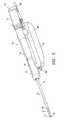
- FIG. 1is an exploded view of the elongated tissue biopsy system embodying features of the invention.
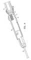
- FIG. 2is a perspective view of the embodiment shown in FIG. 1 in an assembled condition without a housing cover for the probe component.
- FIG. 3is a side elevational view of the tissue biopsy device shown in the FIG. 2 .
- FIG. 4Ais a longitudinal cross-section of the probe shown in FIG. 3 taken along the lines 4 - 4 with the tissue cutting element in a withdrawn position.
- FIG. 4Bis a longitudinal cross-section of the probe shown in FIG. 3 taken along the lines 4 - 4 with the tissue cutting element in a forward or closed position.
- FIG. 5is a transverse cross-sectional view of the probe shown in FIG. 4B taken along the lines 5 - 5 .
- FIG. 6is a perspective view of the underside of the probe shown in FIG. 1 .
- FIG. 7is an enlarged perspective view of the distal end of the driver unit shown in FIG. 1 .
- FIG. 8is an enlarged perspective view of the distal end of the probe housing illustrating a marker element which depicts the orientation of the aperture in the tubular section of the biopsy device.
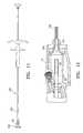
- FIG. 9is a perspective view of the tissue biopsy system shown in FIG. 1 assembled and mounted on a stereotactic frame.
- FIG. 10is a perspective view of the underside of the driver shown in FIG. 1 .
- FIG. 11is an elevational view of a marker delivery device with a flared guide on the distal end of the shaft which facilitates guiding the distal tip of a marker delivery device into the interior of the proximal end of the tissue cutter.
- FIG. 12is a longitudinal cross-sectional view of the distal end of the marker delivery device and flared guide disposed within the tissue collection component shown in FIG. 1 .
- FIG. 13is a longitudinal cross sectional view of the proximal end of the marker delivery device with the flared guide at the proximal end of the shaft and with the shaft deployed within the inner lumen of the tissue cutter.
- FIGS. 1-3illustrate a biopsy system 10 embodying features of the invention which includes a disposable probe component 11 , a driver component 12 and specimen collector 13 .
- the probe component 11generally includes an elongated distal shaft 14 having a tubular section or cannula 15 with a tissue penetrating tip 16 on the distal end thereof and an open, tissue receiving aperture 17 .
- the probe component 11also includes a probe housing 18 with a housing cover 19 which is configured to interfit with the driver component 12 .
- a tissue cutter 20is slidably disposed within the probe and has a distal cutting surface 21 which severs tissue which extends through the tissue receiving aperture 17 .
- the probe housing 18has a mechanical system for rotating the housing and the tubular section 15 secured thereto to control the angular position of the tissue receiving aperture 17 and for moving the tissue cutter 20 slidably disposed within the probe component 11 .
- the mechanical system of the driver component 12has first driving gear 22 that is configured to engage the probe gear 23 and rotate the probe housing 18 so as to adjust the orientation of aperture 17 in the distal extremity of the tubular section 15 .
- the probe gear 23is secured to the rotating connector body 24 by adhesive 25 .
- the proximal extremity of the tubular section 15is secured to the rotating connector body 24 by adhesive 26 .
- An end cap 27retains the connector body 24 within the probe housing 18 .
- Rotation of the probe gear 23rotates the connector body 24 and the attached tubular section 15 .
- the rotationis preferably controlled so that the tubular section 15 rotates in discrete steps about the longitudinal axis 28 to adjust the angular orientation of the aperture 17 about the longitudinal axis.
- these discrete orientationsmay be provided in increments of 30° which can be readily indicated by arrow 29 at the distal end of the probe housing 18 as shown in FIG. 8 .
- the second driving gear 30is configured to drive the tissue cutter 20 longitudinally.
- the driving gear 30engages probe gear 31 which drives cutter traverse nut 32 and cutter screw 33 threadably connected to the cutter traverse nut.
- the distal end of the cutter screw 33is provided with a recess 34 which receives the rib 35 of the cutter shuttle 36 .
- the cutter shuttle 36is secured to the tissue cutter 20 by adhesive 37 .
- the probe gear 31is secured to the cutter traverse nut 32 by adhesive 38 .
- Rotation of the probe gear 31adjusts the relative axial position of the cutter screw 33 with respect to the cutter traverse nut 32 which is secured to the cutter shuttle 36 .
- Longitudinal movement of the tissue cutter 20follows the longitudinal movement of the cutter shuttle 36 resulting from the movement of cutter screw 33 .
- the length of the tissue receiving aperture 17and as a result the length of the specimen, can be controlled by adjusting the initial longitudinal position of the distal end of the tissue cutter 20 within the aperture, before cutting.
- the third driving gear 40is configured to rotate or oscillate the tissue cutter 20 as the cutter moves along the longitudinal axis 28 to facilitate the cutting action of the cutting surface 21 on the distal end of the cutter.
- the third driving gear 40engages probe gear 41 which is secured to cutter oscillation shaft 42 by adhesive 43 .
- the probe gear 41may be oscillated back and forth about the longitudinal axis 28 or rotated continuously in a single direction about the longitudinal axis, or both depending upon the desired rotational movement of the tissue cutter.
- a biased valve assembly 44is provided in the distal end of the probe housing 18 to ensure sealing when a vacuum is developed within the interior 45 of the tissue cutter 20 while providing an atmospheric vent 46 between the interior surface 47 of the tubular section 15 and the exterior surface 48 of the tissue cutter 20 .
- the valve assembly 44includes a spring 49 , valve body 50 and a valve collar 51 which is secured to the proximal end of the tubular section 15 by adhesive 52 .
- the proximal end of the valve spring 49rests against the shoulder 53 provided in the exterior of the valve body 50 .
- a biased cutter shaft seal 54slidably engages the exterior 48 of the tissue cutter 20 .
- the tissue specimen collector 13is secured to the proximal end of the housing of probe component 11 and has an interior 55 in fluid communication with the inner lumen 56 extending within the tissue cutter 20 and has a removable proximal wall 57 of specimen receiving cartridge 58 which gives access to the interior 55 and any tissue specimens which may have been drawn therein.
- a vacuumis generated within the interior 55 to draw tissue specimens through the inner lumen 45 into the interior 55 .
- Tubular member 59has a distal end which is in fluid communication with the interior 55 of the tissue specimen collector 13 and has a proximal end (not shown) which is configured to be connected to a vacuum source. Application of a vacuum within the tubular member 59 aids in pulling tissue into the interior 17 of the tubular section 15 and transfer of the severed tissue specimen through the inner lumen 45 of the tissue cutter 20 to the specimen cartridge 58 .
- the driver 12has a housing 60 with an upper concave surface 61 which is configured to receive the lower surface 62 of the probe housing 18 .
- Three partially exposed driving gears 22 , 30 and 40are provided on the proximal end of the driver 12 which are configured to engage the probe gears 23 , 31 and 41 respectively.
- the drive 12is provided with three separately operating drive motors (not shown) which drive the drive gears 22 , 30 and 40 .
- the separate drive motors (not shown)are connected to and the operation thereof controlled by a control module, such as described in copending application Ser. No. 10/847,699, filed on May 17, 2004.
- the control modulecontrols the motors which move the individual drive gears 22 , 30 and 40 .
- the gear 22engages gear 23 in the probe 11 to control the rotation of the probe housing 18 and the location and orientation of the tissue receiving aperture 17 .
- the drive gear 30engages probe gear 31 to control the longitudinal position and motion of the tissue cutter 20 along the longitudinal axis 28 .
- Drive gear 40engages probe gear 41 to control the oscillation or rotation of the tissue cutter 20 about the longitudinal axis 28 .
- the front face of the driver component 12is provided with light sources 66 and 67 and a manually activatable switch 68 to activate the light sources and enable the physician and other operating personnel to better view the operating site on the patient.
- Other manual switchese.g. a foot activated switch, may be employed.
- the light sourcesmay be automatically activated when the probe component 11 is installed on the driver 12 or other events such as when electrical power is turned on.
- the driver component 12may have a battery pack for the light sources 66 and 67 .
- the penetrating distal tip 16may have a variety of tip shapes. A particularly suitable distal tip shape is described in co-pending provisional application Ser. No. 60/532,277, filed on Dec. 23, 2003. Alternatively, the distal tip may be provided with an arcuate RF electrode such as disclosed in U.S. Pat. No. 6,261,241, and U.S. Pat. No. 6,471,700, both assigned to the present assignee.
- the separate driver component 12allows the entire probe unit to be disposable.
- the drive gears of the drive component 12control the motion of the tissue cutting member 20 for cutting and the motion of the tubular section 15 to orient the aperture 17 .
- Other meansmay provide mechanical and electrical power, vacuum, and control to the probe device. Examples of replaceable snap-in type probe units are disclosed in Burbank et al., U.S. patent application Ser. No. 10/179,933, “Apparatus and Methods for Accessing a Body Site” hereby incorporated by reference in its entirety.
- Drive unitssuch as that described in WO 02/069808 (which corresponds to co-pending U.S. application Ser. No. 09/707,022, filed Nov. 6, 2000 and U.S. application Ser. No. 09/864,021, filed May 23, 2001), which are assigned to the present assignee, may be readily modified by those skilled in the art to accommodate the movement of the cutting member 20 .
- the distal end of the probe component 11is advanced within the patient with the tissue cutter 20 in a forward or closed position ( FIG. 4B ), until the aperture 17 of the tubular section 15 is located in a desired location for taking a tissue specimen.
- the tissue cutter 20is then withdrawn proximally to an open position to open the aperture 17 .
- the withdrawal of the tissue cuttercan be used to control the length of the aperture which is opened in order to control the length of the specimen which is severed.
- a vacuumis applied to the interior 45 of the tissue cutter 20 to draw tissue at the site into the inner lumen of the tubular section 15 through the aperture 17 .
- the tissue cutter 20is then driven distally by rotation of probe gear 30 and rotated or oscillated by drive gear 40 engaging probe gear 41 to sever the aspirated tissue from the supporting tissue at the target site with the tissue cutting surface 21 .
- the vacuum within the interior of the tissue cutter 20causes the tissue specimen to be drawn through the inner lumen 45 of the tissue cutter 20 and into the cartridge 58 of specimen collector 13 shown in FIG. 2 .
- Positive pressure or even ambient conditions distal to the tissue specimencan facilitate tissue passing through the interior 45 of tissue cutter 20 .
- the tubular section 15may be rotated by the drive gear 22 engaging the probe gear 23 in one or more steps to repeat obtaining another tissue specimen in the same manner without otherwise moving the probe component 11 .
- a first tissue specimenis obtained with the aperture 17 of the probe 11 in the 12 o-clock position, the second at the 3 o-clock position, the third at the 9 o-clock position and the fourth at the 6 o-clock position.
- the location of the second and third specimensmay be reversed.
- the position of the aperture 17may be indicated by a marker arrow 29 at the end cap 27 so that the physician or other operating personnel can readily determine what the orientation of the aperture 17 within the patient.
- the biopsy system 10may be hand held for some biopsy procedures or the system may be mounted on a stereotactic mounting stage 80 as shown in FIG. 9 .
- a shoe 81is slidably mounted to a rail 82 of a Fisher stage.
- the mounting member 83is secured to the shoe 81 by a threaded post (not shown) secured to thumbwheel 84 .
- the bottom surface 85 of the driver component 12is configured to conform at least in part to the upper surface of the mounting member 83 .
- the sampling and vacuum switches 86 and 87 respectively on the driver component 12are actuated by the optional sampling and vacuum actuating elements 88 and 89 on the mounting member 83 .
- sampling and vacuummay be actuated with a foot pedal. As shown in FIG.
- the driver componenthas an operator dial 90 which when turned opens a threaded hole 91 for receiving a threaded post (not shown) secured to the thumbwheel 84 and the locating pin holes 92 and 93 which receive the complementary posts (not shown) in the mounting member 83 .
- venting valvecan provide ambient pressure behind the tissue specimen in the cutter interior 45 from the interior of the tubular section 15 .
- the valve body 50is opened for atmospheric venting when the tissue cutter 20 is in the forward position upon the completion of severing the specimen from the tissue site. However, when the tissue cutter 20 is pulled back proximally the valve spring 49 urges the valve body 50 back to a closed position.
- tissue cutter 20is shown with a tissue cutting surface 21 which is perpendicular to the longitudinal axis 28 , the tissue cutting surface may be at an angle or even parallel to the longitudinal axis as described in co-pending application Ser. No. 10/642,406, filed Aug. 15, 2003.
- the distal cutting edge 21 of the tissue cutter 20may initially be located proximal to the aperture 17 to provide a full aperture for receiving tissue or it can be initially located within the aperture 17 in order to control the length of the specimen.
- the cutting action of tissue cutter 20preferably continues until the beveled cutting surface 21 has completely traversed the aperture 17 to ensure that the tissue drawn through the aperture is completely severed from supporting tissue at the biopsy site.
- a vacuummay be applied to aspirate the severed tissue specimen through the inner lumen of the tissue cutter 20 to the cartridge in the specimen collector at the proximal end of the biopsy device. Positive pressure or access to ambient conditions may be provided in the distal end of the tubular section to aid in the specimen transfer.
- marker delivery devicesare shown in co-pending application Ser. No. 10/753,694, filed on Jan. 7, 2004 and co-pending application Ser. No. 10/444,770, filed May 23, 2003.
- the distal ends of these marker delivery devicesare very small and they can be difficult to insert into the proximal end of the tissue cutter 20 which is just slightly larger to accommodate the marker delivery shaft.
- FIG. 11illustrates a marker delivery device 100 which is particularly suitable to facilitate the introduction of the distal end of the shaft 101 into the inner lumen 45 of the tissue cutter 20 and the advancement therein.
- the distal tipis preferably provided with an outwardly flared guide 102 which is slidably mounted on the shaft 103 of the marker delivery device 100 .
- the proximal end of the tubular cutter 20 , the flared guide 102 and/or the distal tip 101may be provided with mating guide elements which orient the marker delivery device so that one or more markers are discharged through the aperture 17 when the pusher element slidably disposed within the delivery device is urged distally to press at least one marker body out the discharge opening in the distal portion of the elongated shaft of the marker delivery device.
- flared proximal guide 102While the slidably mounted, flared proximal guide 102 is described with respect to being disposed on the shaft 103 of marker delivery device 101 , the flared guide 102 has wide application within a variety of biopsy and other devices where one small diameter tubular member is to be inserted into a slightly larger, but still small diameter second tubular member.
- the elongated probe component 11 of the biopsy system 10has a length of about 3 to about 20 cm, preferably, about 5 to about 13 cm, and more specifically, about 8 to about 9 cm for breast biopsy use.
- the distal extremity of the tubular sectionmay be provided with a marker at a desirable location that provide enhanced visualization by eye, by ultrasound, by X-ray, MRI or other imaging or visualization means. Manual palpation may also be employed.
- An echogenic polymer coatingthat increases contrast resolution in ultrasound imaging devices (such as ECHOCOATTM by STS Biopolymers, of Henrietta, N.Y.) is suitable for ultrasonic visualization.
- Radiopaque markersmay be made with, for example, stainless steel, platinum, gold, iridium, tantalum, tungsten, silver, rhodium, nickel, bismuth, other radiopaque metals, alloys and oxides of these metals.
- the surfaces of the device in contact with tissue or other components of the devicemay be provided with a suitable lubricious coating such as a hydrophilic material or a fluoropolymer.
- the tubular section and the tissue cutterare preferably formed of a surgical grade stainless steel.
- other high strength materialssuch as MP35N, other cobalt-chromium alloys, NiTi alloys, ceramics, glasses, and high strength polymeric materials or combinations thereof may be suitable.
- a patient's skinusually must be breached in order to gain access to a body site where a tissue specimen is to be obtained.
- a scalpel or other surgical instrumentmay be used to make an initial incision in the skin.
- the biopsy devicemay be removed from the patient. The entire device may be removed; however, in some embodiments, the cartridge 58 may be removed from the system 10 and a delivery cannula may be inserted through the inner lumen of the cutter 20 to deliver markers to the biopsy site through the aperture 17 .
- a delivery cannulamay be inserted through the inner lumen of the cutter 20 to deliver markers to the biopsy site through the aperture 17 .
- other types of instrumentsmay be inserted into the tissue site through the tissue cutter in addition to or in place of the instruments described above.
- therapeutic or diagnostic agentsmay be delivered through the tissue cutter 20 or the tubular section 15 .
Landscapes
- Health & Medical Sciences (AREA)
- Life Sciences & Earth Sciences (AREA)
- Heart & Thoracic Surgery (AREA)
- General Health & Medical Sciences (AREA)
- Animal Behavior & Ethology (AREA)
- Engineering & Computer Science (AREA)
- Veterinary Medicine (AREA)
- Public Health (AREA)
- Biomedical Technology (AREA)
- Surgery (AREA)
- Pathology (AREA)
- Molecular Biology (AREA)
- Medical Informatics (AREA)
- Vascular Medicine (AREA)
- Anesthesiology (AREA)
- Hematology (AREA)
- Nuclear Medicine, Radiotherapy & Molecular Imaging (AREA)
- Oral & Maxillofacial Surgery (AREA)
- Pulmonology (AREA)
- Surgical Instruments (AREA)
- Sampling And Sample Adjustment (AREA)
Abstract
Description
This application is a continuation-in-part of application Ser. No. 10/642,406, filed Aug. 15, 2003, now U.S. Pat. No. 7,819,819, which is a continuation-in-part of application Ser. No. 10/374,915, filed on Feb. 24, 2003 now U.S. Pat. No. 7,189,206. Both of these applications are incorporated herein in their entirety.
The present invention relates generally to tissue removing devices such as biopsy devices and the methods of using such devices. More specifically, it is directed to a device and method for accessing and removing pathologically suspect tissue from within a patient's body.
In diagnosing and treating certain medical conditions, such as potentially cancerous tumors, it is usually desirable to perform a biopsy, in which a specimen of the suspicious tissue is removed for pathological examination and analysis. In many instances, the suspicious tissue is located in a subcutaneous site, such as inside a human breast. To minimize surgical intrusion into the patient's body, it is desirable to be able to insert a small instrument into the patient's body to access the targeted site and to extract the biopsy specimen therefrom.
Electrosurgical techniques have been used in a variety of biopsy procedures. In electrosurgery, high frequency electrical energy is typically applied to patient tissue through an active electrode, the electrical circuit being completed by a return electrode in contact with the patent's tissue. Electrical energy flowing through the tissue from the active electrode is effective to ablate tissue near the active electrode, forming an opening in the tissue and so allowing insertion of the instrument into a patient's body. A return electrode may be placed on the exterior of the patient's body or may be incorporated into the device itself. The return electrode is typically attached to the patient at a point remote from where the primary or active electrode contacts the tissue. However, in the case of a bipolar electrode for example, the return electrode may be disposed near to the active electrode. An electrosurgical biopsy instrument is disclosed and claimed in U.S. patent application Ser. No. 09/159,467 for “Electrosurgical Biopsy Device and Method,” now U.S. Pat. No. 6,261,241, assigned to the assignee of the present application, and which is hereby incorporated by reference in its entirety.
While these electrosurgical biopsy devices have been found to be effective in many instances, they may not always be suitable for use in conjunction with magnetic resonance imaging.
This invention is directed to devices for accessing and severing tissue from a target site within a patient and methods for utilizing such devices. The devices embodying features of the invention provide access to a targeted tissue site within a patient and provide for the selection, separation and capture of a tissue specimen from supporting tissue at the targeted site.
A tissue collection device and system having features of the invention generally include an elongated, preferably disposable probe component having a plurality of operative elements and a driver component configured to receive the elongated probe component and drive the various operative elements of the probe component.
The elongated probe component has a distal shaft portion with a tissue penetrating distal tip, a tubular section proximal to the distal tip, an inner lumen extending within the tubular section and an open, tissue receiving aperture in the tubular section which provides access to tissue at the targeted site. The probe component includes an elongated tissue-cutting member, which is preferably at least in part cylindrically shaped. The tissue cutting member is provided with at least one tissue cutting edge which is configured to sever tissue extending into the interior of the tubular section through the aperture thereof. The cutting edge on the tissue cutting member may be configured for longitudinal cutting movement and may include oscillating rotational motion and/or reciprocating longitudinal motion to sever specimen tissue extending through the aperture from supporting tissue at the targeted site. The cutting surfaces or edges are radially spaced from a longitudinal axis of the probe component and are generally transversely oriented with respect to the longitudinal axis. The tissue cutter is preferably slidably disposed within the inner lumen of the tubular section, although it may be disposed about the tubular section. The probe component may also have a handle which releasably engages the driver component.
In one embodiment of the invention, the cutting member has an inner lumen preferably extending to the proximal end thereof for tissue specimen removal. While mechanical withdrawal of the tissue specimen may be employed, it is preferred to provide a vacuum within the cutting member from the proximal end of the cutting member. The proximal end of the cutting member may be configured to be in fluid communication with a vacuum source to aspirate the severed tissue specimen through the inner lumen of the cutting member to a tissue collection station. A higher fluid pressure may be maintained in the inner lumen of the cutting member distal to the tissue specimen to aid in transporting the specimen proximally through the inner lumen. In this manner, the mechanical withdrawal and/or the vacuum on the proximal end of the specimen and a higher pressure on the distal end of the specimen can move the specimen through the inner lumen of the cutting member to a tissue collection station.
In at least one embodiment, the handle of the probe component is secured, preferably releasably secured, to the driver housing provided to operably connect the various operative elements of the probe with operative elements of the driver component. The tissue cutting member is operatively connected to at least one driver to provide the desired cutting motion. The proximal end of the tubular section is rotatably secured within the handle housing so that the orientation thereof with respect to the longitudinal axis and therefore the orientation of the tissue receiving aperture within the tubular section, can be selected. The orientation of the aperture may be selected manually such as described in copending application Ser. No. 10/642,406, filed February Aug. 15, 2003 or it may be preset or selected electronically by a control module which also controls the operation of the cutting member and electrical power. The aperture orientation setting may be selected before or after the distal portion of the probe component is inserted into the patient.
The driver component has at least two and preferably three driver units for operating the probe component secured to the driver component. Specifically, the driver component has a first driver unit for rotating the tubular section of the probe component, a second driver unit for moving the cutting member along a longitudinal axis of the cutting member and optionally a third driving unit for rotating or oscillating the cutting member about the longitudinal axis. The first driver unit rotates the tubular section of the probe component, preferably in discrete steps, so that the location of the tissue receiving aperture in the distal extremity of the tubular section can be selected prior to or during the procedure. The discrete rotational steps of the tubular section are preferably in 30° or multiples thereof so that the rotational movement will follow 12 hour clock markings. Preferably, the second and third driver units are operable together so that the cutting member may rotate or oscillate about a longitudinal axis as the cutter member is moved longitudinally. This allows a rotation or an oscillation of the cutter during the cutting process which can aid in cutting tissue.
The driver component may have one or more light sources in a distal portion thereof to illuminate the accessing site during the procedure.
A method of cutting and collecting a tissue specimen with a tissue collection device embodying features of the invention includes advancing such a device at least partially into tissue at a desired site within the patient's body with the tissue penetrating distal tip of the outer cannula disposed distal to the tissue specimen to be separated from the target site. A vacuum is established within the inner lumen of the tubular section to draw tissue through the aperture therein into the inner lumen of the tubular section. The cutting member, which is slidable disposed within the inner lumen of the tubular section, may then be moved longitudinally to cut a tissue specimen from supporting tissue at the target site by the longitudinal motion, which preferably includes oscillating rotational movement and/or reciprocating longitudinal movement. The vacuum established within the inner lumen of the tubular section may be applied through the inner lumen of the tissue cutting member when the tissue cutting member is disposed within the tubular section. The applied vacuum within the inner lumen of the tissue cutting member, may also be utilized to pull or aspirate the separated tissue sample proximally. In addition, or alternatively, a higher fluid pressure may be maintained in a distal part of the inner lumen of the tubular section, distal to the specimen, to push the tissue specimen proximally, Alternatively, the tissue specimen may be mechanically withdrawn. Fluid pressure may include pressure from a liquid delivered into the interior of the device, such as a physiological saline solution, and may include a gas, such as pressurized carbon dioxide, nitrogen or air, delivered into the interior of the device. Access to ambient air can also maintain a sufficiently high pressure differential to move the specimen through the inner lumen of the cutting member. Anesthetic may be injected to the target site through the outer cannula or the inner lumen of the cutting member. Upon removal from the patient, the tissue specimen may then be subjected to pathological examination. After acquisition of a tissue specimen or specimens, the tissue separation system may be repositioned for further tissue separation and collection or it may be withdrawn from the patient.
The tubular section of the probe provides the support for the probe to enable precise location of the accessing port to the desired location at the target site with its radial orientations being preset before the device is introduced into the patient or selected after the tubular section is disposed within the patient. The cutting member quickly and cleanly severs the tissue specimen drawn into the interior of the tubular section though the aperture by the action of the vacuum or otherwise. Upon removal of the tissue specimen, the tissue receiving aperture may be radially repositioned about the longitudinal axis of the tubular section of the probe component so that a plurality of specimens may be taken from the target site. The orientation of the tissue receiving aperture during the procedure may follow a preselected pattern or may be selected by the physician for other selected tissue specimens.
A tissue acquisition system assembly embodying features of the invention may include a device for delivery of one or more marker bodies through a tubular member of a biopsy device such as the tubular cutting member. Such a marker delivery device includes an elongated shaft having an inner lumen and a discharge opening in a distal portion of the elongated shaft, at least one marker body which is disposed within the inner lumen of the elongated shaft, a pusher element which is slidably disposed within the delivery device and which is configured to urge at least one marker body out the discharge opening in the distal portion of the elongated shaft. The marker delivery device has a distally flared guide member which is slidably disposed on the elongated shaft to guide the distal portion of the elongated shaft into a proximal end of the tubular member of a biopsy device.
These and other advantages of the invention will become more apparent from the following detailed description of the invention and the accompanying exemplary drawings.
Theprobe component 11 generally includes an elongateddistal shaft 14 having a tubular section orcannula 15 with atissue penetrating tip 16 on the distal end thereof and an open,tissue receiving aperture 17. Theprobe component 11 also includes aprobe housing 18 with ahousing cover 19 which is configured to interfit with thedriver component 12. Atissue cutter 20 is slidably disposed within the probe and has adistal cutting surface 21 which severs tissue which extends through thetissue receiving aperture 17.
Details of theprobe component 11 are further shown inFIGS. 4A and 4B . Theprobe housing 18 has a mechanical system for rotating the housing and thetubular section 15 secured thereto to control the angular position of thetissue receiving aperture 17 and for moving thetissue cutter 20 slidably disposed within theprobe component 11.
The mechanical system of thedriver component 12 has first drivinggear 22 that is configured to engage theprobe gear 23 and rotate theprobe housing 18 so as to adjust the orientation ofaperture 17 in the distal extremity of thetubular section 15. Theprobe gear 23 is secured to therotating connector body 24 byadhesive 25. The proximal extremity of thetubular section 15 is secured to therotating connector body 24 byadhesive 26. Anend cap 27 retains theconnector body 24 within theprobe housing 18. Rotation of theprobe gear 23 rotates theconnector body 24 and the attachedtubular section 15. The rotation is preferably controlled so that thetubular section 15 rotates in discrete steps about thelongitudinal axis 28 to adjust the angular orientation of theaperture 17 about the longitudinal axis. Preferably these discrete orientations may be provided in increments of 30° which can be readily indicated byarrow 29 at the distal end of theprobe housing 18 as shown inFIG. 8 .
Thesecond driving gear 30 is configured to drive thetissue cutter 20 longitudinally. Thedriving gear 30 engagesprobe gear 31 which drivescutter traverse nut 32 andcutter screw 33 threadably connected to the cutter traverse nut. The distal end of thecutter screw 33 is provided with arecess 34 which receives therib 35 of thecutter shuttle 36. Thecutter shuttle 36 is secured to thetissue cutter 20 byadhesive 37. Theprobe gear 31 is secured to thecutter traverse nut 32 byadhesive 38. Rotation of theprobe gear 31 adjusts the relative axial position of thecutter screw 33 with respect to thecutter traverse nut 32 which is secured to thecutter shuttle 36. Longitudinal movement of thetissue cutter 20 follows the longitudinal movement of thecutter shuttle 36 resulting from the movement ofcutter screw 33. The length of thetissue receiving aperture 17, and as a result the length of the specimen, can be controlled by adjusting the initial longitudinal position of the distal end of thetissue cutter 20 within the aperture, before cutting.
Thethird driving gear 40 is configured to rotate or oscillate thetissue cutter 20 as the cutter moves along thelongitudinal axis 28 to facilitate the cutting action of the cuttingsurface 21 on the distal end of the cutter. Thethird driving gear 40 engagesprobe gear 41 which is secured tocutter oscillation shaft 42 byadhesive 43. Theprobe gear 41 may be oscillated back and forth about thelongitudinal axis 28 or rotated continuously in a single direction about the longitudinal axis, or both depending upon the desired rotational movement of the tissue cutter.
Abiased valve assembly 44 is provided in the distal end of theprobe housing 18 to ensure sealing when a vacuum is developed within theinterior 45 of thetissue cutter 20 while providing anatmospheric vent 46 between theinterior surface 47 of thetubular section 15 and theexterior surface 48 of thetissue cutter 20. Thevalve assembly 44 includes aspring 49, valve body50 and avalve collar 51 which is secured to the proximal end of thetubular section 15 byadhesive 52. The proximal end of thevalve spring 49 rests against theshoulder 53 provided in the exterior of the valve body50. A biased cutter shaft seal54 slidably engages theexterior 48 of thetissue cutter 20.
Thetissue specimen collector 13 is secured to the proximal end of the housing ofprobe component 11 and has an interior55 in fluid communication with the inner lumen56 extending within thetissue cutter 20 and has a removableproximal wall 57 ofspecimen receiving cartridge 58 which gives access to the interior55 and any tissue specimens which may have been drawn therein. A vacuum is generated within the interior55 to draw tissue specimens through theinner lumen 45 into the interior55.Tubular member 59 has a distal end which is in fluid communication with the interior55 of thetissue specimen collector 13 and has a proximal end (not shown) which is configured to be connected to a vacuum source. Application of a vacuum within thetubular member 59 aids in pulling tissue into the interior17 of thetubular section 15 and transfer of the severed tissue specimen through theinner lumen 45 of thetissue cutter 20 to thespecimen cartridge 58.
Thedriver 12 has ahousing 60 with an upperconcave surface 61 which is configured to receive thelower surface 62 of theprobe housing 18. Three partially exposed driving gears22,30 and40 are provided on the proximal end of thedriver 12 which are configured to engage the probe gears23,31 and41 respectively. Thedrive 12 is provided with three separately operating drive motors (not shown) which drive the drive gears22,30 and40. The separate drive motors (not shown) are connected to and the operation thereof controlled by a control module, such as described in copending application Ser. No. 10/847,699, filed on May 17, 2004. The control module controls the motors which move the individual drive gears22,30 and40. Thegear 22 engagesgear 23 in theprobe 11 to control the rotation of theprobe housing 18 and the location and orientation of thetissue receiving aperture 17. Thedrive gear 30 engagesprobe gear 31 to control the longitudinal position and motion of thetissue cutter 20 along thelongitudinal axis 28.Drive gear 40 engagesprobe gear 41 to control the oscillation or rotation of thetissue cutter 20 about thelongitudinal axis 28.
As shown inFIG. 7 , the front face of thedriver component 12 is provided withlight sources activatable switch 68 to activate the light sources and enable the physician and other operating personnel to better view the operating site on the patient. Other manual switches, e.g. a foot activated switch, may be employed. Alternatively, the light sources may be automatically activated when theprobe component 11 is installed on thedriver 12 or other events such as when electrical power is turned on. Thedriver component 12 may have a battery pack for thelight sources
The penetratingdistal tip 16 may have a variety of tip shapes. A particularly suitable distal tip shape is described in co-pending provisional application Ser. No. 60/532,277, filed on Dec. 23, 2003. Alternatively, the distal tip may be provided with an arcuate RF electrode such as disclosed in U.S. Pat. No. 6,261,241, and U.S. Pat. No. 6,471,700, both assigned to the present assignee.
Theseparate driver component 12 allows the entire probe unit to be disposable. The drive gears of thedrive component 12 control the motion of thetissue cutting member 20 for cutting and the motion of thetubular section 15 to orient theaperture 17. Other means (not shown) may provide mechanical and electrical power, vacuum, and control to the probe device. Examples of replaceable snap-in type probe units are disclosed in Burbank et al., U.S. patent application Ser. No. 10/179,933, “Apparatus and Methods for Accessing a Body Site” hereby incorporated by reference in its entirety. Drive units such as that described in WO 02/069808 (which corresponds to co-pending U.S. application Ser. No. 09/707,022, filed Nov. 6, 2000 and U.S. application Ser. No. 09/864,021, filed May 23, 2001), which are assigned to the present assignee, may be readily modified by those skilled in the art to accommodate the movement of the cuttingmember 20.
The distal end of theprobe component 11 is advanced within the patient with thetissue cutter 20 in a forward or closed position (FIG. 4B ), until theaperture 17 of thetubular section 15 is located in a desired location for taking a tissue specimen. Thetissue cutter 20 is then withdrawn proximally to an open position to open theaperture 17. The withdrawal of the tissue cutter can be used to control the length of the aperture which is opened in order to control the length of the specimen which is severed. A vacuum is applied to the interior45 of thetissue cutter 20 to draw tissue at the site into the inner lumen of thetubular section 15 through theaperture 17. Thetissue cutter 20 is then driven distally by rotation ofprobe gear 30 and rotated or oscillated bydrive gear 40 engagingprobe gear 41 to sever the aspirated tissue from the supporting tissue at the target site with thetissue cutting surface 21. The vacuum within the interior of thetissue cutter 20 causes the tissue specimen to be drawn through theinner lumen 45 of thetissue cutter 20 and into thecartridge 58 ofspecimen collector 13 shown inFIG. 2 . Positive pressure or even ambient conditions distal to the tissue specimen can facilitate tissue passing through the interior45 oftissue cutter 20. If another tissue specimen is desired, thetubular section 15 may be rotated by thedrive gear 22 engaging theprobe gear 23 in one or more steps to repeat obtaining another tissue specimen in the same manner without otherwise moving theprobe component 11. Typically, a first tissue specimen is obtained with theaperture 17 of theprobe 11 in the 12 o-clock position, the second at the 3 o-clock position, the third at the 9 o-clock position and the fourth at the 6 o-clock position. The location of the second and third specimens may be reversed. The position of theaperture 17 may be indicated by amarker arrow 29 at theend cap 27 so that the physician or other operating personnel can readily determine what the orientation of theaperture 17 within the patient.
Thebiopsy system 10 may be hand held for some biopsy procedures or the system may be mounted on a stereotactic mountingstage 80 as shown inFIG. 9 . Ashoe 81 is slidably mounted to arail 82 of a Fisher stage. The mountingmember 83 is secured to theshoe 81 by a threaded post (not shown) secured tothumbwheel 84. As shown inFIG. 10 , thebottom surface 85 of thedriver component 12 is configured to conform at least in part to the upper surface of the mountingmember 83. The sampling and vacuum switches86 and87 respectively on thedriver component 12 are actuated by the optional sampling andvacuum actuating elements member 83. Alternatively, sampling and vacuum may be actuated with a foot pedal. As shown inFIG. 10 , the driver component has anoperator dial 90 which when turned opens a threadedhole 91 for receiving a threaded post (not shown) secured to thethumbwheel 84 and the locating pin holes92 and93 which receive the complementary posts (not shown) in the mountingmember 83.
As mentioned above, positive pressure or even ambient conditions will aid in passing the severed tissue specimen through theinner lumen 45 oftissue cutter 20 into thecartridge 58 ofspecimen collector 13. As shown inFIGS. 4A and 4B venting valve can provide ambient pressure behind the tissue specimen in the cutter interior45 from the interior of thetubular section 15. The valve body50 is opened for atmospheric venting when thetissue cutter 20 is in the forward position upon the completion of severing the specimen from the tissue site. However, when thetissue cutter 20 is pulled back proximally thevalve spring 49 urges the valve body50 back to a closed position. While thetissue cutter 20 is shown with atissue cutting surface 21 which is perpendicular to thelongitudinal axis 28, the tissue cutting surface may be at an angle or even parallel to the longitudinal axis as described in co-pending application Ser. No. 10/642,406, filed Aug. 15, 2003.
Thedistal cutting edge 21 of thetissue cutter 20 may initially be located proximal to theaperture 17 to provide a full aperture for receiving tissue or it can be initially located within theaperture 17 in order to control the length of the specimen. The cutting action oftissue cutter 20 preferably continues until the beveled cuttingsurface 21 has completely traversed theaperture 17 to ensure that the tissue drawn through the aperture is completely severed from supporting tissue at the biopsy site. A vacuum may be applied to aspirate the severed tissue specimen through the inner lumen of thetissue cutter 20 to the cartridge in the specimen collector at the proximal end of the biopsy device. Positive pressure or access to ambient conditions may be provided in the distal end of the tubular section to aid in the specimen transfer.
After theremovable wall 57 of thespecimen receiving cartridge 58 is removed and the specimens therein removed, it is frequently desirable to deliver one or more markers to the target site from which the specimens have been removed. Such marker delivery devices are shown in co-pending application Ser. No. 10/753,694, filed on Jan. 7, 2004 and co-pending application Ser. No. 10/444,770, filed May 23, 2003. However, the distal ends of these marker delivery devices are very small and they can be difficult to insert into the proximal end of thetissue cutter 20 which is just slightly larger to accommodate the marker delivery shaft.
Theelongated probe component 11 of thebiopsy system 10 has a length of about 3 to about 20 cm, preferably, about 5 to about 13 cm, and more specifically, about 8 to about 9 cm for breast biopsy use. To assist in properly locating theprobe 11 during advancement thereof into a patient's body, the distal extremity of the tubular section may be provided with a marker at a desirable location that provide enhanced visualization by eye, by ultrasound, by X-ray, MRI or other imaging or visualization means. Manual palpation may also be employed. An echogenic polymer coating that increases contrast resolution in ultrasound imaging devices (such as ECHOCOAT™ by STS Biopolymers, of Henrietta, N.Y.) is suitable for ultrasonic visualization. Radiopaque markers may be made with, for example, stainless steel, platinum, gold, iridium, tantalum, tungsten, silver, rhodium, nickel, bismuth, other radiopaque metals, alloys and oxides of these metals. In addition, the surfaces of the device in contact with tissue or other components of the device may be provided with a suitable lubricious coating such as a hydrophilic material or a fluoropolymer.
The tubular section and the tissue cutter are preferably formed of a surgical grade stainless steel. However, other high strength materials such as MP35N, other cobalt-chromium alloys, NiTi alloys, ceramics, glasses, and high strength polymeric materials or combinations thereof may be suitable.
A patient's skin usually must be breached in order to gain access to a body site where a tissue specimen is to be obtained. A scalpel or other surgical instrument may be used to make an initial incision in the skin. After the specimens have been taken, the biopsy device may be removed from the patient. The entire device may be removed; however, in some embodiments, thecartridge 58 may be removed from thesystem 10 and a delivery cannula may be inserted through the inner lumen of thecutter 20 to deliver markers to the biopsy site through theaperture 17. In addition, it will be readily appreciated that other types of instruments may be inserted into the tissue site through the tissue cutter in addition to or in place of the instruments described above. Moreover, therapeutic or diagnostic agents may be delivered through thetissue cutter 20 or thetubular section 15.
While particular forms of the invention have been illustrated and described herein, it will be apparent that various modifications and improvements can be made to the invention. For example, while the various embodiments of the invention have been described herein in terms of a biopsy device, it should be apparent that the devices and methods of utilizing the device may be employed to remove tissue for purposes other than for biopsy, i.e. for treatment or other diagnoses. Other modifications include, for example, a tissue cutter slidably mounted around the tubular section of the probe component rather than within the tubular section. Moreover, individual features of embodiments of the invention may be shown in some drawings and not in others, but those skilled in the art will recognize that individual features of one embodiment of the invention can be combined with any or all the features of another embodiment. Accordingly, it is not intended that the invention be limited to the specific embodiments illustrated. It is therefore intended that this invention to be defined by the scope of the appended claims as broadly as the prior art will permit.
Terms such a “element”, “member”, “device”, “section”, “portion”, “means”, “step” and words of similar import, when used herein, shall not be construed as invoking the provisions of 35 U.S.C. §112(6) unless the following claims expressly use the term “means” followed by a particular function without specific structure or the term “step” followed by a particular function without specific action. All patents and patent applications referred to above are hereby incorporated by reference in their entirety.
Claims (4)
1. A device for delivery of one or more marker bodies through a tubular cutter member of a biopsy device, comprising:
a. an elongated marker delivery shaft having an exterior, an inner lumen, a discharge opening in a distal portion of the elongated shaft and a handle on a proximal portion of the elongated shaft;
b. at least one marker body which is disposed within the inner lumen of the elongated marker delivery shaft;
c. a guide member which is slidably disposed on the exterior of the elongated marker delivery shaft and which has an inner distally oriented, outwardly flared surface configured to guide the distal portion of the elongated marker delivery shaft into a proximal end of the tubular cutter member of the biopsy device and to allow the elongated marker delivery shaft to advance within an inner lumen of the tubular cutter member;
d. the handle having a stopping shoulder proximal to the guide member that is configured to stop the proximal end of the tubular cutting member of the biopsy device with respect to the elongated marker delivery shaft; and
e. a plunger slidably disposed within the inner lumen of the elongated marker delivery shaft which is long enough to discharge the at least one marker body through the discharge opening in the elongated marker delivery shaft and a tissue receiving aperture in the tubular cutting member when the proximal end of the tubular cutting member engages the stopping shoulder.
2. The device ofclaim 1 including a pusher element which is slidably disposed within the delivery device and which is configured to urge at least one marker body out the discharge opening in the distal portion of the elongated marker delivery shaft.
3. A tissue biopsy system for collecting at least one tissue specimen from a target site within a patient and delivering at least one marker to the target site after the at least one tissue specimen has been removed from the site, comprising:
a. a disposable elongated probe component comprising
i. a distal shaft portion with a penetrating distal tip, a tubular section proximal to the distal tip, an inner lumen extending within the tubular section and an open, tissue receiving aperture in the tubular section which provides access to tissue at the targeted site, and a proximal extremity configured to be secured to a driver unit, and
ii. an elongated tubular tissue cutting member which has at least one tissue cutting edge at a distal end, which has an opening at a proximal end, which has a tissue specimen receiving opening in the distal end and which has an inner lumen extending therein between the tissue specimen receiving opening and the opening in the proximal end;
b. a driver component which is configured to receive a proximal portion of the probe component in an operational relationship, which is configured to rotate the tubular section of the probe component to orient the tissue receiving aperture in the distal end of the tubular section and which is configured to drive the tissue cutting member longitudinally to cut tissue extending into the tubular section through the tissue receiving aperture thereof; and
c. a marker delivery component for delivery of at least one marker bodies through the tubular tissue cutting member, including:
i. an elongated marker delivery shaft which has an inner lumen, a discharge opening in a distal portion of the elongated marker delivery shaft, an outer diameter smaller than the inner diameter of the tubular tissue cutting member of the probe component and a handle on a proximal portion of the shaft having a stopping shoulder to stop the proximal end of the tissue cutting member with respect to the elongated marker delivery shaft;
ii. at least one marker body which is disposed within the inner lumen of the elongated marker delivery shaft;
iii. a pusher element which is slidably disposed within the marker delivery component and which is configured to urge at least one marker body out the discharge opening in the distal portion of the elongated marker delivery shaft and the tissue specimen receiving opening in the tissue cutting member; and
iv. a guide member which is slidably disposed on the elongated marker delivery shaft of the marker delivery component, which has an inner distally oriented, outwardly flared surface having a distal diameter much greater than the outer diameter of the proximal end of the tubular tissue cutting member and which is configured to guide the distal portion of the elongated marker delivery shaft of the marker delivery component into communication with the proximal end of the tubular cutting member and to allow the elongated marker delivery shaft to advance within an inner lumen of the tubular cutting member.
4. A method for collecting one or more tissue specimens from a target site within a patient and delivering at least one marker body to the target site, comprising:
a. providing a biopsy device having:
i. a disposable elongated probe component comprising:
an elongated tubular section which has a longitudinal axis, which has an inner lumen extending therein, which has a penetrating distal tip and which has an distal portion proximal to the penetrating distal tip having a tissue receiving aperture configured to receive tissue from the target site, and
an elongated tissue cutting member which is slidably disposed within the elongated tubular section, which has a distal end and a proximal end, which has at least one tissue cutting edge on the distal end, which has a tissue specimen receiving aperture, which has an inner lumen extending between the proximal and distal end and which is disposed within the tubular tissue cutting member with the cutting edge distal to the tissue receiving aperture opening so as to close the opening; and
ii. a driver component which is configured to receive a proximal portion of the probe component in an operational relationship, which has a first driver motor configured to rotate the tubular section of the probe component to orient the tissue receiving aperture in the distal portion of the tubular section and which has a second driver motor configured to drive the tissue cutting member longitudinally within the tubular section to cut a tissue specimen from tissue extending into the tissue receiving aperture of the elongated tubular member;
b. advancing the probe component at least partially into or adjacent to tissue at the target site with the tissue receiving aperture closed by the tissue cutter;
c. rotating the tubular section of the probe component about its longitudinal axis to provide a desired orientation to the tissue receiving aperture;
d. withdrawing the tissue cutter proximally to open a desired length of the tissue receiving aperture;
e. applying a vacuum within the tubular section to draw tissue at the tissue site through the tissue receiving aperture of the tubular section into the interior thereof;
f. advancing the tissue cutting member distally within the tubular section of the probe member to cut a tissue specimen from the tissue extending into the tubular section;
g. transporting a cut tissue specimen through the inner lumen of the tissue cutting member and out a discharge opening in the proximal end thereof;
h. providing a marker delivery device for delivery of at least one marker bodies through the tubular tissue cutting member, including:
i. an elongated marker delivery shaft which has an inner lumen, a discharge opening in a distal portion of the elongated marker delivery shaft and an outer diameter smaller than the inner diameter of the tubular tissue cutting member of the probe component and a proximal portion with a handle having a stopping shoulder for stopping movement of the proximal end of the tissue cutting member with respect to the elongated marker delivery shaft;
ii. at least one marker body which is disposed within the inner lumen of the elongated marker delivery shaft;
iii. a pusher element which is slidably disposed within the elongated marker delivery shaft and which is configured to urge at least one marker body out the discharge opening in the distal portion of the elongated marker delivery shaft; and
iv. a distally oriented, outwardly flared guide member which is slidably disposed on the elongated marker delivery shaft, which has a distal diameter much greater than the outer diameter of the proximal end of the tubular tissue cutting member and which is configured to guide the distal portion of the elongated marker delivery shaft into the proximal end of the tubular cutting member;
i. advancing the marker delivery device toward the proximal end of the tubular tissue cutting member of the probe component until the distal end of the flared guide member surrounds the proximal end of the tubular tissue cutting member;
j. further advancing the marker delivery device so the elongated marker delivery shaft thereof extends into the inner lumen of the tubular tissue cutting member until the proximal end of the tissue cutting member engages the stopping shoulder of the handle while the distally flared guide member slides along the elongated marker delivery shaft and the discharge opening in the elongated marker delivery shaft of the marker delivery device is aligned with the tissue receiving aperture of the elongated tubular section; and
k. pressing the pusher element of the marker delivery device to eject at least one marker body through the discharge opening in the marker delivery device, through the tissue receiving aperture and into the target site.
Priority Applications (22)
| Application Number | Priority Date | Filing Date | Title |
|---|---|---|---|
| US10/911,106US8282573B2 (en) | 2003-02-24 | 2004-08-03 | Biopsy device with selectable tissue receiving aperture orientation and site illumination |
| US11/014,413US9408592B2 (en) | 2003-12-23 | 2004-12-16 | Biopsy device with aperture orientation and improved tip |
| EP11184500.4AEP2407119B1 (en) | 2004-08-03 | 2005-07-29 | A device for delivery of oner or more marker bodies through a tubular member of a biopsy device |
| JP2007524872AJP2008508942A (en) | 2004-08-03 | 2005-07-29 | Biopsy device with selectable direction of tissue receiving opening and site illumination |
| ES05776770TES2377131T3 (en) | 2004-08-03 | 2005-07-29 | Biopsy device with selectable orientation of the tissue reception opening and zone illumination |
| ES11184498.1TES2557586T3 (en) | 2004-08-03 | 2005-07-29 | Biopsy device with selectable orientation of the tissue reception opening and site lighting |
| AT05776770TATE536807T1 (en) | 2004-08-03 | 2005-07-29 | BIOPSY DEVICE WITH SELECTABLE TISSUE-RECEIVING OPENING ORIENTATION AND ILLUMINATION |
| PCT/US2005/027071WO2006017400A2 (en) | 2004-08-03 | 2005-07-29 | Biopsy device with selectable tissue receiving aperture orientation and site illumination |
| CA2575805ACA2575805C (en) | 2004-08-03 | 2005-07-29 | Biopsy device with selectable tissue receiving aperture orientation and site illumination |
| EP05776770AEP1781178B1 (en) | 2004-08-03 | 2005-07-29 | Biopsy device with selectable tissue receiving aperture orientation and site illumination |
| EP11184498.1AEP2407111B1 (en) | 2004-08-03 | 2005-07-29 | Biopsy device with selectable tissue receiving aperture orientation and site illumination |
| ES11184500.4TES2539457T3 (en) | 2004-08-03 | 2005-07-29 | Marker application device through a tubular element of a biopsy device |
| US12/229,553US10172595B2 (en) | 2003-02-24 | 2008-08-25 | Biopsy device with selectable tissue receiving aperture orientation and site illumination |
| US12/229,545US9044215B2 (en) | 2003-02-24 | 2008-08-25 | Biopsy device with selectable tissue receiving aperature orientation and site illumination |
| US12/794,480US9204866B2 (en) | 2003-02-24 | 2010-06-04 | Biopsy device with selectable tissue receiving aperture orientation and site illumination |
| US13/301,024US9149341B2 (en) | 1999-02-02 | 2011-11-21 | Deployment of polysaccharide markers for treating a site within a patient |
| US13/301,297US20120078092A1 (en) | 1999-02-02 | 2011-11-21 | Remotely imageable marker system and polysaccharide marker for use in same |
| US14/700,574US10335127B2 (en) | 2003-02-24 | 2015-04-30 | Biopsy device with selectable tissue receiving aperature orientation and site illumination |
| US14/868,663US9820824B2 (en) | 1999-02-02 | 2015-09-29 | Deployment of polysaccharide markers for treating a site within a patent |
| US15/818,270US20180071048A1 (en) | 1999-02-02 | 2017-11-20 | Deployment of polysaccharide markers for treating a site within a patient |
| US16/239,629US11534147B2 (en) | 2003-02-24 | 2019-01-04 | Biopsy device with a removable sample recieving cartridge |
| US16/419,482US11589849B2 (en) | 2003-02-24 | 2019-05-22 | Biopsy device with selectable tissue receiving aperature orientation and site illumination |
Applications Claiming Priority (3)
| Application Number | Priority Date | Filing Date | Title |
|---|---|---|---|
| US10/374,915US7189206B2 (en) | 2003-02-24 | 2003-02-24 | Biopsy device with inner cutter |
| US10/642,406US7819819B2 (en) | 2003-02-24 | 2003-08-15 | Biopsy device with inner cutting member |
| US10/911,106US8282573B2 (en) | 2003-02-24 | 2004-08-03 | Biopsy device with selectable tissue receiving aperture orientation and site illumination |
Related Parent Applications (1)
| Application Number | Title | Priority Date | Filing Date |
|---|---|---|---|
| US10/642,406Continuation-In-PartUS7819819B2 (en) | 1999-02-02 | 2003-08-15 | Biopsy device with inner cutting member |
Related Child Applications (5)
| Application Number | Title | Priority Date | Filing Date |
|---|---|---|---|
| US10/174,401Continuation-In-PartUS7651505B2 (en) | 1999-02-02 | 2002-06-17 | Plugged tip delivery for marker placement |
| US11/014,413Continuation-In-PartUS9408592B2 (en) | 2003-12-23 | 2004-12-16 | Biopsy device with aperture orientation and improved tip |
| US12/229,545DivisionUS9044215B2 (en) | 2003-02-24 | 2008-08-25 | Biopsy device with selectable tissue receiving aperature orientation and site illumination |
| US12/229,553DivisionUS10172595B2 (en) | 2003-02-24 | 2008-08-25 | Biopsy device with selectable tissue receiving aperture orientation and site illumination |
| US13/301,024Continuation-In-PartUS9149341B2 (en) | 1999-02-02 | 2011-11-21 | Deployment of polysaccharide markers for treating a site within a patient |
Publications (2)
| Publication Number | Publication Date |
|---|---|
| US20050065453A1 US20050065453A1 (en) | 2005-03-24 |
| US8282573B2true US8282573B2 (en) | 2012-10-09 |
Family
ID=35285593
Family Applications (2)
| Application Number | Title | Priority Date | Filing Date |
|---|---|---|---|
| US10/911,106Active2027-11-17US8282573B2 (en) | 1999-02-02 | 2004-08-03 | Biopsy device with selectable tissue receiving aperture orientation and site illumination |
| US16/419,482Expired - LifetimeUS11589849B2 (en) | 2003-02-24 | 2019-05-22 | Biopsy device with selectable tissue receiving aperature orientation and site illumination |
Family Applications After (1)
| Application Number | Title | Priority Date | Filing Date |
|---|---|---|---|
| US16/419,482Expired - LifetimeUS11589849B2 (en) | 2003-02-24 | 2019-05-22 | Biopsy device with selectable tissue receiving aperature orientation and site illumination |
Country Status (7)
| Country | Link |
|---|---|
| US (2) | US8282573B2 (en) |
| EP (3) | EP2407111B1 (en) |
| JP (1) | JP2008508942A (en) |
| AT (1) | ATE536807T1 (en) |
| CA (1) | CA2575805C (en) |
| ES (3) | ES2539457T3 (en) |
| WO (1) | WO2006017400A2 (en) |
Cited By (21)
| Publication number | Priority date | Publication date | Assignee | Title |
|---|---|---|---|---|
| US20080319468A1 (en)* | 2003-02-24 | 2008-12-25 | Senorx, Inc. | Biopsy device with selectable tissue receiving aperature orientation and site illumination |
| US8702623B2 (en) | 2008-12-18 | 2014-04-22 | Devicor Medical Products, Inc. | Biopsy device with discrete tissue chambers |
| US8740810B2 (en) | 2010-11-12 | 2014-06-03 | FUJIFULM Corporation | Biopsy needle, sample extracting unit, biopsy apparatus, and method of controlling biopsy needle |
| US8932233B2 (en) | 2004-05-21 | 2015-01-13 | Devicor Medical Products, Inc. | MRI biopsy device |
| US9265485B2 (en) | 2004-09-29 | 2016-02-23 | Devicor Medical Products, Inc. | Biopsy device with integral vacuum assist and tissue sample and fluid capturing canister |
| EP3009076A1 (en) | 2014-10-13 | 2016-04-20 | NeoDynamics AB | Trocar arrangement for tissue sampling device |
| EP3009075A1 (en) | 2014-10-13 | 2016-04-20 | NeoDynamics AB | Distal tip tissue sampling arrangement |
| US9468424B2 (en) | 2009-06-12 | 2016-10-18 | Devicor Medical Products, Inc. | Cutter drive assembly for biopsy device |
| US9498193B2 (en) | 2008-12-16 | 2016-11-22 | Devicor Medical Products, Inc. | Biopsy device |
| US9622822B2 (en) | 2007-05-01 | 2017-04-18 | Suros Surgical Systems, Inc. | Securement for a surgical site marker and deployment device for same |
| US9636082B2 (en) | 2001-08-24 | 2017-05-02 | The Cooper Companies Global Holdings Lp | Medical-surgical devices |
| US9668717B2 (en) | 2005-03-04 | 2017-06-06 | Devicor Medical Products, Inc. | Biopsy device with variable side aperture |
| US9795365B2 (en) | 2004-05-21 | 2017-10-24 | Devicor Medical Products, Inc. | MRI biopsy apparatus incorporating a sleeve and multi-function obturator |
| US10045756B2 (en) | 2003-03-29 | 2018-08-14 | The Cooper Companies Global Holdings Lp | Medical devices |
| US10456120B2 (en) | 2013-11-05 | 2019-10-29 | C. R. Bard, Inc. | Biopsy device having integrated vacuum |
| US10463350B2 (en) | 2015-05-01 | 2019-11-05 | C. R. Bard, Inc. | Biopsy device |
| EP3616625A1 (en) | 2018-09-03 | 2020-03-04 | NeoDynamics AB | A biopsy arrangement |
| EP3791794A1 (en) | 2019-09-12 | 2021-03-17 | NeoDynamics AB (publ) | A biopsy arrangement |
| US11179141B2 (en) | 2006-12-13 | 2021-11-23 | Devicor Medical Products, Inc. | Biopsy system |
| USD952487S1 (en)* | 2020-04-14 | 2022-05-24 | Ching-Lung Lin | Thickness measurement tool |
| CN114711838A (en)* | 2022-04-12 | 2022-07-08 | 浙江伽奈维医疗科技有限公司 | Rotary-cut mammary gland biopsy equipment and method |
Families Citing this family (126)
| Publication number | Priority date | Publication date | Assignee | Title |
|---|---|---|---|---|
| US7637948B2 (en) | 1997-10-10 | 2009-12-29 | Senorx, Inc. | Tissue marking implant |
| US8668737B2 (en) | 1997-10-10 | 2014-03-11 | Senorx, Inc. | Tissue marking implant |
| US6161034A (en)* | 1999-02-02 | 2000-12-12 | Senorx, Inc. | Methods and chemical preparations for time-limited marking of biopsy sites |
| US6347241B2 (en)* | 1999-02-02 | 2002-02-12 | Senorx, Inc. | Ultrasonic and x-ray detectable biopsy site marker and apparatus for applying it |
| US8361082B2 (en) | 1999-02-02 | 2013-01-29 | Senorx, Inc. | Marker delivery device with releasable plug |
| US8498693B2 (en) | 1999-02-02 | 2013-07-30 | Senorx, Inc. | Intracorporeal marker and marker delivery device |
| US20090030309A1 (en)* | 2007-07-26 | 2009-01-29 | Senorx, Inc. | Deployment of polysaccharide markers |
| US6862470B2 (en) | 1999-02-02 | 2005-03-01 | Senorx, Inc. | Cavity-filling biopsy site markers |
| US20080039819A1 (en)* | 2006-08-04 | 2008-02-14 | Senorx, Inc. | Marker formed of starch or other suitable polysaccharide |
| US7651505B2 (en) | 2002-06-17 | 2010-01-26 | Senorx, Inc. | Plugged tip delivery for marker placement |
| US6725083B1 (en)* | 1999-02-02 | 2004-04-20 | Senorx, Inc. | Tissue site markers for in VIVO imaging |
| US7983734B2 (en)* | 2003-05-23 | 2011-07-19 | Senorx, Inc. | Fibrous marker and intracorporeal delivery thereof |
| US6575991B1 (en) | 1999-06-17 | 2003-06-10 | Inrad, Inc. | Apparatus for the percutaneous marking of a lesion |
| GB2361544B (en)* | 2000-04-20 | 2004-07-07 | Goring Kerr Ltd | Metal detector |
| CA2659518A1 (en) | 2000-11-20 | 2002-05-30 | Senorx, Inc. | Tissue site markers for in vivo imaging |
| EP1524940B1 (en) | 2002-03-19 | 2011-08-24 | Bard Dublin ITC Limited | Biopsy device and biopsy needle module that can be inserted into the biopsy device |
| US8109885B2 (en) | 2002-03-19 | 2012-02-07 | C. R. Bard, Inc. | Biopsy device for removing tissue specimens using a vacuum |
| US20060036158A1 (en) | 2003-11-17 | 2006-02-16 | Inrad, Inc. | Self-contained, self-piercing, side-expelling marking apparatus |
| DE10314240B4 (en) | 2003-03-29 | 2025-05-28 | Bard Dublin Itc Ltd. | Pressure generation unit |
| US7877133B2 (en) | 2003-05-23 | 2011-01-25 | Senorx, Inc. | Marker or filler forming fluid |
| US20050119562A1 (en)* | 2003-05-23 | 2005-06-02 | Senorx, Inc. | Fibrous marker formed of synthetic polymer strands |
| US20050273002A1 (en) | 2004-06-04 | 2005-12-08 | Goosen Ryan L | Multi-mode imaging marker |
| US20090312817A1 (en)* | 2003-11-26 | 2009-12-17 | Wicab, Inc. | Systems and methods for altering brain and body functions and for treating conditions and diseases of the same |
| CA2557726C (en) | 2004-03-26 | 2012-10-16 | Brivant Research & Development Limited | A guide wire for use in re-canalising a vascular occlusion in a human or animal subject |
| JP4814229B2 (en) | 2004-07-09 | 2011-11-16 | バード ペリフェラル ヴァスキュラー インコーポレイテッド | Transport device for biopsy device |
| US20060074344A1 (en)* | 2004-09-29 | 2006-04-06 | Hibner John A | Fluid control for biopsy device |
| US7276032B2 (en)* | 2004-09-29 | 2007-10-02 | Ethicon Endo-Surgery, Inc. | Biopsy apparatus and method |
| US7517321B2 (en) | 2005-01-31 | 2009-04-14 | C. R. Bard, Inc. | Quick cycle biopsy system |
| US20060200041A1 (en) | 2005-03-04 | 2006-09-07 | Ethicon Endo-Surgery, Inc. | Biopsy device incorporating an adjustable probe sleeve |
| US20060217635A1 (en)* | 2005-03-24 | 2006-09-28 | Mccombs Elizabeth S | Biopsy device marker deployment |
| US9974930B2 (en) | 2005-03-24 | 2018-05-22 | Brivant Research & Development Limited | Guide wire for use in re-canalising a vascular occlusion in a human or animal subject |
| US10357328B2 (en) | 2005-04-20 | 2019-07-23 | Bard Peripheral Vascular, Inc. and Bard Shannon Limited | Marking device with retractable cannula |
| US7854707B2 (en) | 2005-08-05 | 2010-12-21 | Devicor Medical Products, Inc. | Tissue sample revolver drum biopsy device |
| US7867173B2 (en)* | 2005-08-05 | 2011-01-11 | Devicor Medical Products, Inc. | Biopsy device with replaceable probe and incorporating vibration insertion assist and static vacuum source sample stacking retrieval |
| US7662109B2 (en)* | 2006-02-01 | 2010-02-16 | Ethicon Endo-Surgery, Inc. | Biopsy device with replaceable probe incorporating static vacuum source dual valve sample stacking retrieval and saline flush |
| USRE46135E1 (en) | 2005-08-05 | 2016-09-06 | Devicor Medical Products, Inc. | Vacuum syringe assisted biopsy device |
| US7572236B2 (en)* | 2005-08-05 | 2009-08-11 | Senorx, Inc. | Biopsy device with fluid delivery to tissue specimens |
| US8317725B2 (en) | 2005-08-05 | 2012-11-27 | Senorx, Inc. | Biopsy device with fluid delivery to tissue specimens |
| ES2539578T3 (en) | 2005-08-10 | 2015-07-02 | C.R. Bard, Inc. | Multi-sample biopsy device and single insert with various transport systems |
| EP1921998B8 (en) | 2005-08-10 | 2021-07-07 | C.R.Bard, Inc. | Single-insertion, multiple sampling biopsy device with linear drive |
| JP4991723B2 (en) | 2005-08-10 | 2012-08-01 | シー・アール・バード・インコーポレーテッド | Single insertion multiple sampling biopsy device with integrated marker |
| EP1759635A1 (en)* | 2005-09-03 | 2007-03-07 | Boehringer Mannheim Gmbh | Pricking method and hand held device therefore |
| US8187294B2 (en)* | 2005-09-26 | 2012-05-29 | Suros Surgical Systems, Inc. | Rotating surgical cutter |
| US8052658B2 (en) | 2005-10-07 | 2011-11-08 | Bard Peripheral Vascular, Inc. | Drug-eluting tissue marker |
| US8349443B2 (en)* | 2006-02-23 | 2013-01-08 | Meadwestvaco Corporation | Method for treating a substrate |
| US7766843B2 (en)* | 2006-03-03 | 2010-08-03 | Ethicon Endo-Surgery, Inc. | Biopsy method |
| US20070232954A1 (en)* | 2006-04-04 | 2007-10-04 | Harris Jeffrey P | Automated skin biopsy device |
| EP3417792B1 (en) | 2006-08-21 | 2022-03-02 | C. R. Bard, Inc. | Self-contained handheld biopsy needle |
| SI2086418T1 (en) | 2006-10-06 | 2011-05-31 | Bard Peripheral Vascular Inc | Tissue handling system with reduced operator exposure |
| WO2008051749A2 (en) | 2006-10-23 | 2008-05-02 | C. R. Bard, Inc. | Breast marker |
| US8262586B2 (en) | 2006-10-24 | 2012-09-11 | C. R. Bard, Inc. | Large sample low aspect ratio biopsy needle |
| US9579077B2 (en) | 2006-12-12 | 2017-02-28 | C.R. Bard, Inc. | Multiple imaging mode tissue marker |
| US8480595B2 (en) | 2006-12-13 | 2013-07-09 | Devicor Medical Products, Inc. | Biopsy device with motorized needle cocking |
| US7981049B2 (en)* | 2006-12-13 | 2011-07-19 | Devicor Medical Products, Inc. | Engagement interface for biopsy system vacuum module |
| US9345457B2 (en)* | 2006-12-13 | 2016-05-24 | Devicor Medical Products, Inc. | Presentation of biopsy sample by biopsy device |
| WO2008076973A2 (en) | 2006-12-18 | 2008-06-26 | C.R.Bard Inc. | Biopsy marker with in situ-generated imaging properties |
| ATE549977T1 (en)* | 2007-01-15 | 2012-04-15 | Jacques Phillibert Janssens | PNEUMATIC DEVICE FOR COLLECTING A TISSUE SAMPLE |
| US8202229B2 (en)* | 2007-10-01 | 2012-06-19 | Suros Surgical Systems, Inc. | Surgical device |
| US8808200B2 (en) | 2007-10-01 | 2014-08-19 | Suros Surgical Systems, Inc. | Surgical device and method of using same |
| US8241225B2 (en) | 2007-12-20 | 2012-08-14 | C. R. Bard, Inc. | Biopsy device |
| US7854706B2 (en) | 2007-12-27 | 2010-12-21 | Devicor Medical Products, Inc. | Clutch and valving system for tetherless biopsy device |
| WO2009099767A2 (en) | 2008-01-31 | 2009-08-13 | C.R. Bard, Inc. | Biopsy tissue marker |
| US20090209853A1 (en)* | 2008-02-19 | 2009-08-20 | Parihar Shailendra K | Biopsy site marker applier |
| US9327061B2 (en) | 2008-09-23 | 2016-05-03 | Senorx, Inc. | Porous bioabsorbable implant |
| US20100106015A1 (en)* | 2008-10-23 | 2010-04-29 | Norris Perry R | Medical device alignment |
| US8162852B2 (en)* | 2008-10-23 | 2012-04-24 | Devicor Medical Products, Inc. | Methods for medical device alignment |
| KR101167529B1 (en)* | 2008-11-18 | 2012-07-20 | 박희붕 | Biopsy device |
| US20100152610A1 (en)* | 2008-12-16 | 2010-06-17 | Parihar Shailendra K | Hand Actuated Tetherless Biopsy Device with Pistol Grip |
| EP4215147A3 (en) | 2008-12-30 | 2023-10-18 | C. R. Bard, Inc. | Marker delivery device for tissue marker placement |
| WO2010107424A1 (en)* | 2009-03-16 | 2010-09-23 | C.R. Bard, Inc. | Biopsy device having rotational cutting |
| AU2009344276B2 (en) | 2009-04-15 | 2014-06-05 | C.R. Bard, Inc. | Biopsy apparatus having integrated fluid management |
| US9386942B2 (en)* | 2009-06-26 | 2016-07-12 | Cianna Medical, Inc. | Apparatus, systems, and methods for localizing markers or tissue structures within a body |
| EP3106089B1 (en) | 2009-06-26 | 2020-12-02 | Cianna Medical, Inc. | System for localizing markers or tissue structures within a body |
| US9173641B2 (en) | 2009-08-12 | 2015-11-03 | C. R. Bard, Inc. | Biopsy apparatus having integrated thumbwheel mechanism for manual rotation of biopsy cannula |
| US8430824B2 (en) | 2009-10-29 | 2013-04-30 | Bard Peripheral Vascular, Inc. | Biopsy driver assembly having a control circuit for conserving battery power |
| US8283890B2 (en) | 2009-09-25 | 2012-10-09 | Bard Peripheral Vascular, Inc. | Charging station for battery powered biopsy apparatus |
| RU2579736C2 (en)* | 2009-10-01 | 2016-04-10 | Смит Энд Нефью, Инк. | Surgical tip for endoscopic resection |
| JP2013521994A (en)* | 2010-03-24 | 2013-06-13 | ユナイテッド ステイツ エンドスコピー グループ,インコーポレイテッド | Multiple biopsy device |
| US9220485B2 (en) | 2010-08-28 | 2015-12-29 | Endochoice, Inc. | Tissue collection and separation device |
| US9492130B2 (en) | 2010-11-24 | 2016-11-15 | Hologic, Inc. | System for improved tissue-handling and in line analysis of the tissue |
| US8888802B2 (en) | 2010-12-21 | 2014-11-18 | Alcon Research, Ltd. | Vitrectomy probe with adjustable cutter port size |
| US9101441B2 (en) | 2010-12-21 | 2015-08-11 | Alcon Research, Ltd. | Vitrectomy probe with adjustable cutter port size |
| USD660430S1 (en)* | 2011-04-25 | 2012-05-22 | Devicor Medical Products, Inc. | Biopsy device |
| US8938285B2 (en) | 2011-08-08 | 2015-01-20 | Devicor Medical Products, Inc. | Access chamber and markers for biopsy device |
| US9186166B2 (en) | 2011-09-01 | 2015-11-17 | Depuy Mitek, Llc | Tissue shavers |
| WO2013068017A1 (en) | 2011-11-09 | 2013-05-16 | Tomas Gundberg | Handheld tissue sample extraction device |
| USD671644S1 (en)* | 2011-12-07 | 2012-11-27 | Devicor Medical Products, Inc. | Ultrasound biopsy probe |
| US9517161B2 (en) | 2011-12-20 | 2016-12-13 | Alcon Research, Ltd. | Vitrectomy probe with adjustable cutter port size |
| EP2838435B1 (en) | 2012-04-16 | 2020-03-25 | Hathaway, Jeff M. | Biopsy device |
| KR101459398B1 (en)* | 2012-11-08 | 2014-11-07 | 연세대학교 원주산학협력단 | Indicator for aspiration biopsy |
| CA2902221A1 (en) | 2013-03-20 | 2014-09-25 | Bard Peripheral Vascular, Inc. | Biopsy device |
| USD715442S1 (en) | 2013-09-24 | 2014-10-14 | C. R. Bard, Inc. | Tissue marker for intracorporeal site identification |
| USD716451S1 (en) | 2013-09-24 | 2014-10-28 | C. R. Bard, Inc. | Tissue marker for intracorporeal site identification |
| USD715942S1 (en) | 2013-09-24 | 2014-10-21 | C. R. Bard, Inc. | Tissue marker for intracorporeal site identification |
| USD716450S1 (en) | 2013-09-24 | 2014-10-28 | C. R. Bard, Inc. | Tissue marker for intracorporeal site identification |
| US9782193B2 (en) | 2013-12-11 | 2017-10-10 | Medos International Sàrl | Tissue shaving device having a fluid removal path |
| WO2015134277A1 (en) | 2014-03-05 | 2015-09-11 | Faxitron Bioptics, Llc | System and method for multi-axis imaging of specimens |
| US9572666B2 (en) | 2014-03-17 | 2017-02-21 | Evalve, Inc. | Mitral valve fixation device removal devices and methods |
| ES2675938T3 (en) | 2014-09-25 | 2018-07-13 | Hologic, Inc. | Biopsy device with suction valve |
| US9456808B2 (en) | 2014-09-25 | 2016-10-04 | Hologic, Inc. | Biopsy device with automatic biopsy parameter adjustment |
| US10646208B2 (en)* | 2015-05-06 | 2020-05-12 | Devicor Medical Products, Inc. | Marker delivery device for use with MRI breast biopsy system |
| WO2017040977A1 (en) | 2015-09-04 | 2017-03-09 | Faxitron Bioptics, Llc | Multi-axis specimen imaging device with embedded orientation markers |
| US10786224B2 (en) | 2016-04-21 | 2020-09-29 | Covidien Lp | Biopsy devices and methods of use thereof |
| US10314470B2 (en)* | 2016-06-14 | 2019-06-11 | Ribcure Co. Ltd. | Body cavity illumination apparatus |
| US10736632B2 (en) | 2016-07-06 | 2020-08-11 | Evalve, Inc. | Methods and devices for valve clip excision |
| US11071564B2 (en) | 2016-10-05 | 2021-07-27 | Evalve, Inc. | Cardiac valve cutting device |
| US11083426B2 (en) | 2016-11-04 | 2021-08-10 | Hologic, Inc. | Specimen radiography system comprising cabinet and a specimen drawer positionable by a controller in the cabinet |
| EP4215124B1 (en) | 2016-11-21 | 2024-11-27 | C. R. Bard, Inc. | Biopsy device having a hydraulic drive assembly |
| CA3062311A1 (en) | 2017-05-03 | 2018-11-08 | Hologic, Inc. | Device for reducing fluid in the imaging field of a tissue handling apparatus for improving biopsy system imaging quality |
| WO2019051496A1 (en) | 2017-09-11 | 2019-03-14 | Faxitron Bioptics, Llc | Imaging system with adaptive object magnification |
| US11331161B2 (en) | 2018-03-23 | 2022-05-17 | Covidien Lp | Surgical assemblies facilitating tissue marking and methods of use thereof |
| US10576248B2 (en)* | 2018-07-23 | 2020-03-03 | Crossbay Medical, Inc. | Apparatus and method for everting catheter for uterine access for biopsy and cytology |
| US12102531B2 (en) | 2018-10-22 | 2024-10-01 | Evalve, Inc. | Tissue cutting systems, devices and methods |
| CN109330632B (en)* | 2018-11-09 | 2023-09-29 | 上海导向医疗系统有限公司 | Biopsy rotary cutting device |
| EP4183347B1 (en) | 2018-12-26 | 2024-05-15 | Hologic, Inc. | Tissue imaging in presence of fluid during biopsy procedure |
| US11517294B2 (en) | 2019-05-07 | 2022-12-06 | Covidien Lp | Biopsy devices and methods of use thereof |
| CN110680411B (en)* | 2019-10-09 | 2020-10-02 | 山东大学 | A kind of cervical needle biopsy forceps and using method thereof |
| JP7629025B2 (en) | 2020-03-31 | 2025-02-12 | ホロジック, インコーポレイテッド | System and method for x-ray imaging of tissue samples - Patents.com |
| US12048448B2 (en) | 2020-05-06 | 2024-07-30 | Evalve, Inc. | Leaflet grasping and cutting device |
| US12178444B2 (en) | 2020-05-06 | 2024-12-31 | Evalve, Inc. | Clip removal systems and methods |
| US12414811B2 (en) | 2020-05-06 | 2025-09-16 | Evalve, Inc. | Devices and methods for leaflet cutting |
| US12171485B2 (en) | 2020-05-06 | 2024-12-24 | Evalve, Inc. | Systems and methods for leaflet cutting using a hook catheter |
| US12171486B2 (en) | 2020-05-06 | 2024-12-24 | Evalve, Inc. | Devices and methods for clip separation |
| EP4214672B1 (en) | 2020-09-16 | 2025-01-01 | Hologic, Inc. | Systems and methods for confirming tissue specimens removed using contrast-enhanced x-ray imaging |
| CN114451965A (en)* | 2021-12-01 | 2022-05-10 | 利福昇(苏州)医疗科技有限公司 | A fixed force tissue cutting protection system |
| CN114027940A (en)* | 2021-12-01 | 2022-02-11 | 利福昇(苏州)医疗科技有限公司 | Medical rotary-cut system and rotary-cut knife tackle thereof |
Citations (162)
| Publication number | Priority date | Publication date | Assignee | Title |
|---|---|---|---|---|
| US2032860A (en) | 1933-03-24 | 1936-03-03 | Wappler Frederick Charles | Method for electrosurgical treatment of tissue |
| DE1225813B (en) | 1961-10-23 | 1966-09-29 | Optique Et Prec De Levallois S | Suction biopsy probe |
| US3326785A (en) | 1962-09-17 | 1967-06-20 | Anocut Eng Co | Electrolytic polishing apparatus and method |
| US3805791A (en) | 1971-07-01 | 1974-04-23 | K Seuberth | Apparatus for the diathermic removal of growths |
| US3844272A (en) | 1969-02-14 | 1974-10-29 | A Banko | Surgical instruments |
| US3847153A (en) | 1973-09-14 | 1974-11-12 | B Weissman | Disposable probe tip for electro-surgical device |
| US3902498A (en) | 1974-03-04 | 1975-09-02 | Minnesota Mining & Mfg | Surgical cutting instrument |
| US3910279A (en) | 1973-06-20 | 1975-10-07 | Olympus Optical Co | Electrosurgical instrument |
| US3945375A (en) | 1972-04-04 | 1976-03-23 | Surgical Design Corporation | Rotatable surgical instrument |
| US3955578A (en) | 1974-12-23 | 1976-05-11 | Cook Inc. | Rotatable surgical snare |
| US4007732A (en) | 1975-09-02 | 1977-02-15 | Robert Carl Kvavle | Method for location and removal of soft tissue in human biopsy operations |
| CH602084A5 (en) | 1976-06-08 | 1978-07-31 | Kaspar Menzi | Medical spatula for throat examination |
| US4202338A (en) | 1977-11-18 | 1980-05-13 | Richard Wolf Gmbh | Device for removing excrescences and polyps |
| GB1571926A (en) | 1976-03-19 | 1980-07-23 | Hamer Geerd | Cutting device |
| US4243048A (en) | 1976-09-21 | 1981-01-06 | Jim Zegeer | Biopsy device |
| US4294254A (en) | 1977-12-08 | 1981-10-13 | Chamness Dale L | Surgical apparatus |
| US4311143A (en) | 1978-10-12 | 1982-01-19 | Olympus Optical Co., Ltd. | Apparatus for resecting tissue inside the body cavity utilizing high-frequency currents |
| US4362160A (en) | 1979-07-24 | 1982-12-07 | Richard Wolf Gmbh | Endoscopes |
| US4503855A (en) | 1981-12-31 | 1985-03-12 | Harald Maslanka | High frequency surgical snare electrode |
| EP0146699A1 (en) | 1983-12-22 | 1985-07-03 | GebràDer Sulzer Aktiengesellschaft | Implanted marker |
| US4565200A (en) | 1980-09-24 | 1986-01-21 | Cosman Eric R | Universal lesion and recording electrode system |
| US4576162A (en) | 1983-03-30 | 1986-03-18 | Mccorkle Charles E | Apparatus and method for separation of scar tissue in venous pathway |
| US4603694A (en) | 1983-03-08 | 1986-08-05 | Richards Medical Company | Arthroscopic shaver |
| US4638802A (en) | 1984-09-21 | 1987-01-27 | Olympus Optical Co., Ltd. | High frequency instrument for incision and excision |
| US4643187A (en) | 1983-05-30 | 1987-02-17 | Olympus Optical Co., Ltd. | High-frequency incising and excising instrument |
| US4682606A (en) | 1986-02-03 | 1987-07-28 | Decaprio Vincent H | Localizing biopsy apparatus |
| US4718419A (en) | 1985-08-05 | 1988-01-12 | Olympus Optical Co., Ltd. | Snare assembly for endoscope |
| EP0255123A2 (en) | 1986-07-30 | 1988-02-03 | Sumitomo Pharmaceuticals Company, Limited | Solid preparation administering instrument |
| US4724836A (en) | 1985-01-08 | 1988-02-16 | Olympus Optical Co., Ltd. | High-frequency incision tool |
| US4800896A (en) | 1985-11-08 | 1989-01-31 | Jalowayski Alfredo A | Cell sample collector probe |
| US4926858A (en) | 1984-05-30 | 1990-05-22 | Devices For Vascular Intervention, Inc. | Atherectomy device for severe occlusions |
| US5007908A (en) | 1989-09-29 | 1991-04-16 | Everest Medical Corporation | Electrosurgical instrument having needle cutting electrode and spot-coag electrode |
| US5041124A (en) | 1989-07-14 | 1991-08-20 | Kensey Nash Corporation | Apparatus and method for sclerosing of body tissue |
| US5047027A (en) | 1990-04-20 | 1991-09-10 | Everest Medical Corporation | Tumor resector |
| US5059204A (en) | 1989-10-26 | 1991-10-22 | Site Microsurgical Systems, Inc. | Ocular cutter with enhanced cutting action |
| US5064424A (en) | 1989-05-18 | 1991-11-12 | Richard Wolf Gmbh | Electro-surgical instrument |
| US5080660A (en) | 1990-05-11 | 1992-01-14 | Applied Urology, Inc. | Electrosurgical electrode |
| US5085659A (en) | 1990-11-21 | 1992-02-04 | Everest Medical Corporation | Biopsy device with bipolar coagulation capability |
| EP0472368A2 (en) | 1990-08-21 | 1992-02-26 | Med Institute, Inc. | Ablation catheter |
| USRE33925E (en) | 1984-05-22 | 1992-05-12 | Cordis Corporation | Electrosurgical catheter aned method for vascular applications |
| WO1992007500A2 (en) | 1990-10-25 | 1992-05-14 | Devices For Vascular Intervention, Inc. | Atherectomy catheter having axially-disposed cutting edge |
| US5133359A (en) | 1990-11-14 | 1992-07-28 | Du-Kedem Technologies Ltd. | Hard tissue biopsy instrument with rotary drive |
| US5133360A (en) | 1991-03-07 | 1992-07-28 | Spears Colin P | Spears retriever |
| USRE34056E (en) | 1989-07-31 | 1992-09-08 | C.R. Bard, Inc. | Tissue sampling device |
| EP0509670A2 (en) | 1991-04-15 | 1992-10-21 | Everest Medical Corporation | Bipolar electrosurgical scalpel with paired loop electrodes |
| US5158561A (en) | 1992-03-23 | 1992-10-27 | Everest Medical Corporation | Monopolar polypectomy snare with coagulation electrode |
| US5217458A (en) | 1992-04-09 | 1993-06-08 | Everest Medical Corporation | Bipolar biopsy device utilizing a rotatable, single-hinged moving element |
| US5219350A (en) | 1990-10-05 | 1993-06-15 | Emerson Clarence A | Medical instrument |
| US5320613A (en) | 1993-01-06 | 1994-06-14 | Scimed Life Systems, Inc. | Medical lumen flushing and guide wire loading device and method |
| US5324288A (en) | 1991-04-30 | 1994-06-28 | Utah Medical Products, Inc. | Electrosurgical loop with a depth gauge |
| US5335671A (en) | 1989-11-06 | 1994-08-09 | Mectra Labs, Inc. | Tissue removal assembly with provision for an electro-cautery device |
| US5344420A (en) | 1991-02-13 | 1994-09-06 | Applied Medical Resources Corporation | Surgical trocar |
| EP0601709A3 (en) | 1992-11-09 | 1994-10-12 | Vance Products Inc | Cutting surgical instrument for coring tissue. |
| US5366468A (en) | 1993-11-09 | 1994-11-22 | Linvatec Corporation | Double bladed surgical router having aspiration ports within flutes |
| US5374188A (en) | 1993-07-19 | 1994-12-20 | Bei Medical Systems, Inc. | Electro-surgical instrument and method for use with dental implantations |
| US5380321A (en) | 1992-11-04 | 1995-01-10 | Yoon; Inbae | Shielded energy transmitting surgical instrument and methods therefor |
| WO1995002370A2 (en) | 1993-07-15 | 1995-01-26 | Aws Shakir Mustafa Salim | Tunnelling catheter |
| WO1995002371A2 (en) | 1993-07-15 | 1995-01-26 | Aws Shakir Mustafa Salim | Rectal and rectosigmoid cancer tunnelling umbrella |
| WO1995003843A1 (en) | 1993-07-30 | 1995-02-09 | The Regents Of The University Of California | Endocardial infusion catheter |
| US5395312A (en) | 1991-10-18 | 1995-03-07 | Desai; Ashvin | Surgical tool |
| US5415656A (en) | 1993-09-28 | 1995-05-16 | American Medical Systems, Inc. | Electrosurgical apparatus |
| US5417687A (en) | 1993-04-30 | 1995-05-23 | Medical Scientific, Inc. | Bipolar electrosurgical trocar |
| US5423809A (en) | 1992-01-21 | 1995-06-13 | Valleylab Inc. | Electrosurgical control for a trocar |
| US5437665A (en) | 1993-10-12 | 1995-08-01 | Munro; Malcolm G. | Electrosurgical loop electrode instrument for laparoscopic surgery |
| US5441503A (en) | 1988-09-24 | 1995-08-15 | Considine; John | Apparatus for removing tumors from hollow organs of the body |
| US5441498A (en) | 1994-02-16 | 1995-08-15 | Envision Surgical Systems, Inc. | Method of using a multimodality probe with extendable bipolar electrodes |
| US5470308A (en) | 1992-08-12 | 1995-11-28 | Vidamed, Inc. | Medical probe with biopsy stylet |
| US5476102A (en)* | 1994-06-29 | 1995-12-19 | Baxter International Inc. | Biopsy needle assembly and guide |
| US5477862A (en) | 1994-03-14 | 1995-12-26 | Haaga; John R. | Cutting tip for biopsy needle |
| US5501654A (en) | 1993-07-15 | 1996-03-26 | Ethicon, Inc. | Endoscopic instrument having articulating element |
| US5507743A (en) | 1993-11-08 | 1996-04-16 | Zomed International | Coiled RF electrode treatment apparatus |
| US5526822A (en) | 1994-03-24 | 1996-06-18 | Biopsys Medical, Inc. | Method and apparatus for automated biopsy and collection of soft tissue |
| US5527331A (en) | 1993-10-13 | 1996-06-18 | Femrx | Method for prostatic tissue resection |
| US5538010A (en) | 1994-10-05 | 1996-07-23 | Proact Ltd. | Biopsy needle device |
| US5575794A (en) | 1993-02-12 | 1996-11-19 | Walus; Richard L. | Tool for implanting a fiducial marker |
| US5578030A (en) | 1994-11-04 | 1996-11-26 | Levin; John M. | Biopsy needle with cauterization feature |
| US5595185A (en) | 1994-08-11 | 1997-01-21 | N.M.B. Medical Applications Ltd. | Single puncture multi-biopsy gun |
| US5599347A (en) | 1991-02-13 | 1997-02-04 | Applied Medical Resources Corporation | Surgical trocar with cutoff circuit |
| DE19528440A1 (en) | 1995-08-02 | 1997-02-06 | Harald Dr Med Kuebler | Surgical cutting instrument for tissue removal - has cutting device enclosed by tubular end section of instrument during insertion and curved transverse to its axial direction in working position |
| US5649547A (en) | 1994-03-24 | 1997-07-22 | Biopsys Medical, Inc. | Methods and devices for automated biopsy and collection of soft tissue |
| US5653718A (en) | 1994-05-16 | 1997-08-05 | Yoon; Inbae | Cannula anchoring system |
| WO1997029702A1 (en) | 1996-02-14 | 1997-08-21 | Rita Medical Systems, Inc. | Multiple antenna ablation apparatus and method |
| GB2311468A (en) | 1996-03-27 | 1997-10-01 | Valleylab Inc | Electrosurgical interstitial resector |
| US5674184A (en) | 1994-03-15 | 1997-10-07 | Ethicon Endo-Surgery, Inc. | Surgical trocars with cutting electrode and viewing rod |
| US5676663A (en) | 1995-11-21 | 1997-10-14 | Kim; David S. | Cone biopsy instrument |
| US5683384A (en) | 1993-11-08 | 1997-11-04 | Zomed | Multiple antenna ablation apparatus |
| US5687739A (en) | 1995-12-06 | 1997-11-18 | Interventional Concepts, Inc. | Biopsy specimen cutter |
| US5769086A (en) | 1995-12-06 | 1998-06-23 | Biopsys Medical, Inc. | Control system and method for automated biopsy device |
| US5772660A (en) | 1995-03-20 | 1998-06-30 | United States Surgical Corporation | Trocar assembly with electrocautery penetrating tip |
| US5782775A (en) | 1995-10-20 | 1998-07-21 | United States Surgical Corporation | Apparatus and method for localizing and removing tissue |
| US5794626A (en) | 1994-08-18 | 1998-08-18 | Kieturakis; Maciej J. | Excisional stereotactic apparatus |
| EP0858774A2 (en) | 1997-01-21 | 1998-08-19 | Ethicon Endo-Surgery, Inc. | Devices for collection of soft tissue |
| US5797907A (en) | 1989-11-06 | 1998-08-25 | Mectra Labs, Inc. | Electrocautery cutter |
| US5810806A (en) | 1996-08-29 | 1998-09-22 | Ethicon Endo-Surgery | Methods and devices for collection of soft tissue |
| US5814044A (en) | 1995-02-10 | 1998-09-29 | Enable Medical Corporation | Apparatus and method for morselating and removing tissue from a patient |
| WO1998043531A1 (en) | 1997-04-01 | 1998-10-08 | Vilos George A | Debris aspirating resectoscope |
| WO1998046147A1 (en) | 1997-04-11 | 1998-10-22 | Coherent Inc. | Method and apparatus for transurethral resection of the prostate |
| US5846513A (en) | 1997-07-08 | 1998-12-08 | Carewise Medical Products Corporation | Tumor localization and removal system using penetratable detection probe and removal instrument |
| US5848978A (en) | 1995-11-14 | 1998-12-15 | Genx International, Inc. | Surgical biopsy device |
| US5857981A (en) | 1995-09-12 | 1999-01-12 | Bucalo; Brian D. | Biopsy instrument with tissue specimen retaining and retrieval device |
| US5857982A (en) | 1995-09-08 | 1999-01-12 | United States Surgical Corporation | Apparatus and method for removing tissue |
| US5871454A (en) | 1997-04-22 | 1999-02-16 | Majlessi; Heshmat | Percutaneous excisional biopsy device |
| US5873886A (en) | 1995-04-04 | 1999-02-23 | United States Surgical Corporation | Surgical cutting apparatus |
| US5876340A (en) | 1997-04-17 | 1999-03-02 | Irvine Biomedical, Inc. | Ablation apparatus with ultrasonic imaging capabilities |
| US5882316A (en) | 1996-08-29 | 1999-03-16 | City Of Hope | Minimally invasive biopsy device |
| US5902272A (en) | 1992-01-07 | 1999-05-11 | Arthrocare Corporation | Planar ablation probe and method for electrosurgical cutting and ablation |
| US5925044A (en) | 1996-07-01 | 1999-07-20 | Gebrueder Berchtold Gmbh & Co. | Trocar for laparoscopic operations |
| US5947964A (en) | 1995-03-03 | 1999-09-07 | Neothermia Corporation | Methods and apparatus for therapeutic cauterization of predetermined volumes of biological tissue |
| US5954670A (en) | 1994-10-05 | 1999-09-21 | Baker; Gary H. | Mandrel-guided tandem multiple channel biopsy guide device and method of use |
| US5964716A (en) | 1998-05-14 | 1999-10-12 | Ethicon Endo-Surgery, Inc. | Method of use for a multi-port biopsy instrument |
| US5984919A (en) | 1991-02-13 | 1999-11-16 | Applied Medical Resources Corporation | Surgical trocar |
| US5997560A (en) | 1994-07-21 | 1999-12-07 | Sdgi Holdings, Inc. | Surgical cutting instrument |
| US6050955A (en) | 1997-09-19 | 2000-04-18 | United States Surgical Corporation | Biopsy apparatus and method |
| US6056700A (en) | 1998-10-13 | 2000-05-02 | Emx, Inc. | Biopsy marker assembly and method of use |
| US6077230A (en) | 1998-05-14 | 2000-06-20 | Ethicon Endo-Surgery, Inc. | Biopsy instrument with removable extractor |
| US6086543A (en) | 1998-06-24 | 2000-07-11 | Rubicor Medical, Inc. | Fine needle and core biopsy devices and methods |
| US6120462A (en) | 1999-03-31 | 2000-09-19 | Ethicon Endo-Surgery, Inc. | Control method for an automated surgical biopsy device |
| EP0919190A3 (en) | 1997-11-24 | 2000-11-02 | Ethicon Endo-Surgery, Inc. | Biopsy instrument with continuous tissue receiving chamber |
| US6142955A (en) | 1997-09-19 | 2000-11-07 | United States Surgical Corporation | Biopsy apparatus and method |
| US6176865B1 (en) | 1998-03-30 | 2001-01-23 | Agilent Technologies, Inc. | Apparatus and method for incising |
| EP1072288A2 (en) | 1999-07-26 | 2001-01-31 | Indigo Medical, Incorporated | Brachytherapy cartridge including absorbable and autoclaveable spacers |
| US6220248B1 (en) | 1998-10-21 | 2001-04-24 | Ethicon Endo-Surgery, Inc. | Method for implanting a biopsy marker |
| WO2001028631A1 (en) | 1999-10-15 | 2001-04-26 | Deschutes Medical Products, Inc. | Brachytherapy instrument and methods |
| US20010002250A1 (en) | 1998-03-03 | 2001-05-31 | Burbank Fred H. | Sentinel node location and biopsy |
| US6258111B1 (en) | 1997-10-03 | 2001-07-10 | Scieran Technologies, Inc. | Apparatus and method for performing ophthalmic procedures |
| US6258000B1 (en) | 1998-05-21 | 2001-07-10 | Liechty, Ii Victor Jay | Penetration enhancing aerodynamically favorable arrowhead |
| WO2001049184A2 (en) | 2000-01-04 | 2001-07-12 | Senorx, Inc. | Radiofrequency apparatus and method for accessing a biopsy site |
| US6261241B1 (en) | 1998-03-03 | 2001-07-17 | Senorx, Inc. | Electrosurgical biopsy device and method |
| US6261302B1 (en) | 1998-06-26 | 2001-07-17 | Ethicon Endo-Surgery, Inc. | Applier for implantable surgical marker |
| WO2001058505A2 (en) | 2000-02-09 | 2001-08-16 | Vygon | Part for easily slipping on a catheter tube on a guide and assembly comprising same |
| US6277083B1 (en) | 1999-12-27 | 2001-08-21 | Neothermia Corporation | Minimally invasive intact recovery of tissue |
| US6312429B1 (en) | 1998-09-01 | 2001-11-06 | Senorx, Inc. | Electrosurgical lesion location device |
| US6331166B1 (en) | 1998-03-03 | 2001-12-18 | Senorx, Inc. | Breast biopsy system and method |
| DE10066004A1 (en) | 1999-12-29 | 2001-12-20 | Kaltenbach & Voigt | Medical or dental medical treatment instrument with electrical load, e.g. light source, has inductive current transfer coils connected to supply lead mounted on front and rear instrument parts |
| US6344026B1 (en) | 1998-04-08 | 2002-02-05 | Senorx, Inc. | Tissue specimen encapsulation device and method thereof |
| US20020022768A1 (en) | 2000-07-27 | 2002-02-21 | Asahi Kogaku Kogyo Kabushiki Kaisha | Optical system for the light source device of a video endoscope system |
| US6350244B1 (en) | 2000-02-21 | 2002-02-26 | Biopsy Sciences, Llc | Bioabsorable markers for use in biopsy procedures |
| US6454727B1 (en) | 1998-03-03 | 2002-09-24 | Senorx, Inc. | Tissue acquisition system and method of use |
| WO2002074214A1 (en) | 2001-03-16 | 2002-09-26 | Sub-Q, Inc. | Method of hydrating a sponge material for delivery to a body |
| US20020143270A1 (en) | 2001-04-02 | 2002-10-03 | Temple University Of The Commonwealth System Of Higher Education | Multiple biopsy device |
| US6494881B1 (en) | 1997-09-30 | 2002-12-17 | Scimed Life Systems, Inc. | Apparatus and method for electrode-surgical tissue removal having a selectively insulated electrode |
| US20030004407A1 (en) | 1997-11-18 | 2003-01-02 | Carewise Medical Products Corporation | Minimally invasive surgical |
| US6514215B1 (en) | 1999-10-13 | 2003-02-04 | Pentax Corporation | Endoscopic tissue collecting instrument |
| US6514248B1 (en) | 1999-10-15 | 2003-02-04 | Neothermia Corporation | Accurate cutting about and into tissue volumes with electrosurgically deployed electrodes |
| US6540695B1 (en) | 1998-04-08 | 2003-04-01 | Senorx, Inc. | Biopsy anchor device with cutter |
| US6562032B1 (en) | 2001-03-26 | 2003-05-13 | Ellman Alan G | Electrosurgical instrument with vibration |
| US6575991B1 (en)* | 1999-06-17 | 2003-06-10 | Inrad, Inc. | Apparatus for the percutaneous marking of a lesion |
| US20030125639A1 (en) | 2002-01-02 | 2003-07-03 | Fisher John S. | Biopsy needle having rotating core for shearing tissue |
| US6589157B2 (en) | 2000-05-18 | 2003-07-08 | Integrated Implant Systems, L.L.C. | Drive mechanism for medical instrument |
| EP0769281B1 (en) | 1995-10-20 | 2003-07-30 | United States Surgical Corporation | Surgical apparatus for marking tissue location |
| US20030199760A1 (en)* | 2000-09-29 | 2003-10-23 | Mammoco Inc. | Device for permanently marking a selected tissue location within a patient ' s body |
| US20030236471A1 (en) | 2001-08-09 | 2003-12-25 | Fisher John S. | Dual action aspiration biopsy needle |
| US20040024304A1 (en) | 1994-09-16 | 2004-02-05 | Foerster Seth A. | Methods and devices for defining and marking tissue |
| US6689145B2 (en) | 1998-09-03 | 2004-02-10 | Rubicor Medical, Inc. | Excisional biopsy devices and methods |
| US6712773B1 (en) | 2000-09-11 | 2004-03-30 | Tyco Healthcare Group Lp | Biopsy system |
| US6758824B1 (en) | 2000-11-06 | 2004-07-06 | Suros Surgical Systems, Inc. | Biopsy apparatus |
| US6835173B2 (en) | 2001-10-05 | 2004-12-28 | Scimed Life Systems, Inc. | Robotic endoscope |
| WO2005020925A2 (en) | 2003-08-29 | 2005-03-10 | Emisphere Technologies, Inc. | Method and apparatus for dosing solids |
| EP0970658B1 (en) | 1998-06-30 | 2005-08-17 | Ethicon Endo-Surgery, Inc. | Surgical biopsy device |
| US6994667B2 (en) | 2000-01-10 | 2006-02-07 | Singh Errol O | Method and apparatus for facilitating urological procedures |
| US7189206B2 (en) | 2003-02-24 | 2007-03-13 | Senorx, Inc. | Biopsy device with inner cutter |
| US7452357B2 (en)* | 2004-10-22 | 2008-11-18 | Ethicon Endo-Surgery, Inc. | System and method for planning treatment of tissue |
Family Cites Families (39)
| Publication number | Priority date | Publication date | Assignee | Title |
|---|---|---|---|---|
| US4461305A (en) | 1981-09-04 | 1984-07-24 | Cibley Leonard J | Automated biopsy device |
| DE3202193A1 (en) | 1982-01-25 | 1983-08-04 | Merck Patent Gmbh, 6100 Darmstadt | SURGICAL BONE GRINDING INSTRUMENT |
| US5507764A (en) | 1982-09-24 | 1996-04-16 | Joseph J. Berke | Powered rotary scalpel method |
| US4660267A (en) | 1983-03-08 | 1987-04-28 | Richards Medical Company | Method for fabricating an arthroscopic shaver |
| US4983179A (en) | 1986-12-30 | 1991-01-08 | Smith & Nephew Dyonics Inc. | Arthroscopic surgical instrument |
| US5275609A (en) | 1990-06-22 | 1994-01-04 | Vance Products Incorporated | Surgical cutting instrument |
| JPH06114070A (en) | 1990-06-22 | 1994-04-26 | Vance Prod Inc | Tissue abscission device for surgery |
| US5285795A (en) | 1991-09-12 | 1994-02-15 | Surgical Dynamics, Inc. | Percutaneous discectomy system having a bendable discectomy probe and a steerable cannula |
| US5602449A (en) | 1992-04-13 | 1997-02-11 | Smith & Nephew Endoscopy, Inc. | Motor controlled surgical system and method having positional control |
| US5311859A (en) | 1992-09-11 | 1994-05-17 | Welch Allyn, Inc. | Add-on video camera arrangement for optical laparoscope |
| US5423330A (en) | 1993-03-10 | 1995-06-13 | The University Of Miami | Capsule suction punch instrument and method of use |
| US5897567A (en) | 1993-04-29 | 1999-04-27 | Scimed Life Systems, Inc. | Expandable intravascular occlusion material removal devices and methods of use |
| US5782764A (en) | 1995-11-07 | 1998-07-21 | Iti Medical Technologies, Inc. | Fiber composite invasive medical instruments and methods for use in interventional imaging procedures |
| US5823970A (en) | 1996-03-22 | 1998-10-20 | Medical Device Technologies, Inc. | Biopsy needle set |
| US5908433A (en)* | 1996-05-10 | 1999-06-01 | Stryker Corporation | Carpal tunnel knife |
| US5910150A (en) | 1996-12-02 | 1999-06-08 | Angiotrax, Inc. | Apparatus for performing surgery |
| US6156049A (en) | 1997-04-11 | 2000-12-05 | Coherent Inc. | Method and apparatus for transurethral resection of the prostate |
| US6540693B2 (en) | 1998-03-03 | 2003-04-01 | Senorx, Inc. | Methods and apparatus for securing medical instruments to desired locations in a patients body |
| JP3449277B2 (en)* | 1999-02-05 | 2003-09-22 | 株式会社日立製作所 | Hybrid vehicle and control device thereof |
| US6689146B1 (en)* | 1999-04-29 | 2004-02-10 | Stryker Corporation | Powered surgical handpiece with integrated irrigator and suction application |
| AU4545600A (en) | 1999-06-17 | 2001-01-09 | Kai Desinger | Surgical, grooved director for collecting tissue in a minimally invasive manner |
| US6471659B2 (en) | 1999-12-27 | 2002-10-29 | Neothermia Corporation | Minimally invasive intact recovery of tissue |
| US6428487B1 (en) | 1999-12-17 | 2002-08-06 | Ethicon Endo-Surgery, Inc. | Surgical biopsy system with remote control for selecting an operational mode |
| US6231522B1 (en) | 2000-02-18 | 2001-05-15 | Ethicon Endo-Surgery, Inc. | Biopsy instrument with breakable sample segments |
| US6773443B2 (en) | 2000-07-31 | 2004-08-10 | Regents Of The University Of Minnesota | Method and apparatus for taking a biopsy |
| US6485436B1 (en) | 2000-08-10 | 2002-11-26 | Csaba Truckai | Pressure-assisted biopsy needle apparatus and technique |
| WO2002069808A2 (en) | 2000-11-06 | 2002-09-12 | Suros Surgical Systems, Inc. | Biopsy apparatus |
| WO2002062227A1 (en) | 2001-02-05 | 2002-08-15 | Tyco Healthcare Group Lp | Biopsy apparatus and method |
| US20020105797A1 (en) | 2001-02-06 | 2002-08-08 | Technology Creations, Inc. | Tool light and method of use |
| US6673023B2 (en) | 2001-03-23 | 2004-01-06 | Stryker Puerto Rico Limited | Micro-invasive breast biopsy device |
| US7510563B2 (en) | 2001-10-26 | 2009-03-31 | Smith & Nephew, Inc. | Reciprocating rotary arthroscopic surgical instrument |
| EP1524940B1 (en) | 2002-03-19 | 2011-08-24 | Bard Dublin ITC Limited | Biopsy device and biopsy needle module that can be inserted into the biopsy device |
| US7740597B2 (en)* | 2002-12-11 | 2010-06-22 | Ethicon Endo-Surgery, Inc. | Biopsy device with sample tube |
| US20050119562A1 (en)* | 2003-05-23 | 2005-06-02 | Senorx, Inc. | Fibrous marker formed of synthetic polymer strands |
| US9408592B2 (en)* | 2003-12-23 | 2016-08-09 | Senorx, Inc. | Biopsy device with aperture orientation and improved tip |
| US7517322B2 (en) | 2005-03-04 | 2009-04-14 | Ethicon Endo-Surgery, Inc. | Biopsy device with variable side aperture |
| US20060217635A1 (en) | 2005-03-24 | 2006-09-28 | Mccombs Elizabeth S | Biopsy device marker deployment |
| US20100106015A1 (en) | 2008-10-23 | 2010-04-29 | Norris Perry R | Medical device alignment |
| US8162852B2 (en) | 2008-10-23 | 2012-04-24 | Devicor Medical Products, Inc. | Methods for medical device alignment |
- 2004
- 2004-08-03USUS10/911,106patent/US8282573B2/enactiveActive
- 2005
- 2005-07-29ATAT05776770Tpatent/ATE536807T1/enactive
- 2005-07-29ESES11184500.4Tpatent/ES2539457T3/ennot_activeExpired - Lifetime
- 2005-07-29ESES05776770Tpatent/ES2377131T3/ennot_activeExpired - Lifetime
- 2005-07-29CACA2575805Apatent/CA2575805C/ennot_activeExpired - Lifetime
- 2005-07-29JPJP2007524872Apatent/JP2008508942A/enactivePending
- 2005-07-29EPEP11184498.1Apatent/EP2407111B1/ennot_activeExpired - Lifetime
- 2005-07-29ESES11184498.1Tpatent/ES2557586T3/ennot_activeExpired - Lifetime
- 2005-07-29EPEP05776770Apatent/EP1781178B1/ennot_activeExpired - Lifetime
- 2005-07-29WOPCT/US2005/027071patent/WO2006017400A2/enactiveSearch and Examination
- 2005-07-29EPEP11184500.4Apatent/EP2407119B1/ennot_activeExpired - Lifetime
- 2019
- 2019-05-22USUS16/419,482patent/US11589849B2/ennot_activeExpired - Lifetime
Patent Citations (175)
| Publication number | Priority date | Publication date | Assignee | Title |
|---|---|---|---|---|
| US2032860A (en) | 1933-03-24 | 1936-03-03 | Wappler Frederick Charles | Method for electrosurgical treatment of tissue |
| DE1225813B (en) | 1961-10-23 | 1966-09-29 | Optique Et Prec De Levallois S | Suction biopsy probe |
| US3326785A (en) | 1962-09-17 | 1967-06-20 | Anocut Eng Co | Electrolytic polishing apparatus and method |
| US3844272A (en) | 1969-02-14 | 1974-10-29 | A Banko | Surgical instruments |
| US3805791A (en) | 1971-07-01 | 1974-04-23 | K Seuberth | Apparatus for the diathermic removal of growths |
| US3945375A (en) | 1972-04-04 | 1976-03-23 | Surgical Design Corporation | Rotatable surgical instrument |
| US3910279A (en) | 1973-06-20 | 1975-10-07 | Olympus Optical Co | Electrosurgical instrument |
| US3847153A (en) | 1973-09-14 | 1974-11-12 | B Weissman | Disposable probe tip for electro-surgical device |
| US3902498A (en) | 1974-03-04 | 1975-09-02 | Minnesota Mining & Mfg | Surgical cutting instrument |
| US3955578A (en) | 1974-12-23 | 1976-05-11 | Cook Inc. | Rotatable surgical snare |
| US4007732A (en) | 1975-09-02 | 1977-02-15 | Robert Carl Kvavle | Method for location and removal of soft tissue in human biopsy operations |
| GB1571926A (en) | 1976-03-19 | 1980-07-23 | Hamer Geerd | Cutting device |
| CH602084A5 (en) | 1976-06-08 | 1978-07-31 | Kaspar Menzi | Medical spatula for throat examination |
| US4243048A (en) | 1976-09-21 | 1981-01-06 | Jim Zegeer | Biopsy device |
| US4202338A (en) | 1977-11-18 | 1980-05-13 | Richard Wolf Gmbh | Device for removing excrescences and polyps |
| US4294254A (en) | 1977-12-08 | 1981-10-13 | Chamness Dale L | Surgical apparatus |
| US4311143A (en) | 1978-10-12 | 1982-01-19 | Olympus Optical Co., Ltd. | Apparatus for resecting tissue inside the body cavity utilizing high-frequency currents |
| US4362160A (en) | 1979-07-24 | 1982-12-07 | Richard Wolf Gmbh | Endoscopes |
| US4565200A (en) | 1980-09-24 | 1986-01-21 | Cosman Eric R | Universal lesion and recording electrode system |
| US4503855A (en) | 1981-12-31 | 1985-03-12 | Harald Maslanka | High frequency surgical snare electrode |
| US4603694A (en) | 1983-03-08 | 1986-08-05 | Richards Medical Company | Arthroscopic shaver |
| US4576162A (en) | 1983-03-30 | 1986-03-18 | Mccorkle Charles E | Apparatus and method for separation of scar tissue in venous pathway |
| US4643187A (en) | 1983-05-30 | 1987-02-17 | Olympus Optical Co., Ltd. | High-frequency incising and excising instrument |
| EP0146699A1 (en) | 1983-12-22 | 1985-07-03 | GebràDer Sulzer Aktiengesellschaft | Implanted marker |
| USRE33925E (en) | 1984-05-22 | 1992-05-12 | Cordis Corporation | Electrosurgical catheter aned method for vascular applications |
| US4926858A (en) | 1984-05-30 | 1990-05-22 | Devices For Vascular Intervention, Inc. | Atherectomy device for severe occlusions |
| US4638802A (en) | 1984-09-21 | 1987-01-27 | Olympus Optical Co., Ltd. | High frequency instrument for incision and excision |
| US4724836A (en) | 1985-01-08 | 1988-02-16 | Olympus Optical Co., Ltd. | High-frequency incision tool |
| US4718419A (en) | 1985-08-05 | 1988-01-12 | Olympus Optical Co., Ltd. | Snare assembly for endoscope |
| US4800896A (en) | 1985-11-08 | 1989-01-31 | Jalowayski Alfredo A | Cell sample collector probe |
| US4682606A (en) | 1986-02-03 | 1987-07-28 | Decaprio Vincent H | Localizing biopsy apparatus |
| EP0255123A2 (en) | 1986-07-30 | 1988-02-03 | Sumitomo Pharmaceuticals Company, Limited | Solid preparation administering instrument |
| US5441503A (en) | 1988-09-24 | 1995-08-15 | Considine; John | Apparatus for removing tumors from hollow organs of the body |
| US5064424A (en) | 1989-05-18 | 1991-11-12 | Richard Wolf Gmbh | Electro-surgical instrument |
| US5041124A (en) | 1989-07-14 | 1991-08-20 | Kensey Nash Corporation | Apparatus and method for sclerosing of body tissue |
| USRE34056E (en) | 1989-07-31 | 1992-09-08 | C.R. Bard, Inc. | Tissue sampling device |
| US5007908A (en) | 1989-09-29 | 1991-04-16 | Everest Medical Corporation | Electrosurgical instrument having needle cutting electrode and spot-coag electrode |
| US5059204A (en) | 1989-10-26 | 1991-10-22 | Site Microsurgical Systems, Inc. | Ocular cutter with enhanced cutting action |
| US5797907A (en) | 1989-11-06 | 1998-08-25 | Mectra Labs, Inc. | Electrocautery cutter |
| US5335671A (en) | 1989-11-06 | 1994-08-09 | Mectra Labs, Inc. | Tissue removal assembly with provision for an electro-cautery device |
| US5047027A (en) | 1990-04-20 | 1991-09-10 | Everest Medical Corporation | Tumor resector |
| US5080660A (en) | 1990-05-11 | 1992-01-14 | Applied Urology, Inc. | Electrosurgical electrode |
| EP0472368A2 (en) | 1990-08-21 | 1992-02-26 | Med Institute, Inc. | Ablation catheter |
| US5219350A (en) | 1990-10-05 | 1993-06-15 | Emerson Clarence A | Medical instrument |
| WO1992007500A2 (en) | 1990-10-25 | 1992-05-14 | Devices For Vascular Intervention, Inc. | Atherectomy catheter having axially-disposed cutting edge |
| US5133359A (en) | 1990-11-14 | 1992-07-28 | Du-Kedem Technologies Ltd. | Hard tissue biopsy instrument with rotary drive |
| US5085659A (en) | 1990-11-21 | 1992-02-04 | Everest Medical Corporation | Biopsy device with bipolar coagulation capability |
| US5984919A (en) | 1991-02-13 | 1999-11-16 | Applied Medical Resources Corporation | Surgical trocar |
| US5344420A (en) | 1991-02-13 | 1994-09-06 | Applied Medical Resources Corporation | Surgical trocar |
| US5599347A (en) | 1991-02-13 | 1997-02-04 | Applied Medical Resources Corporation | Surgical trocar with cutoff circuit |
| US5133360A (en) | 1991-03-07 | 1992-07-28 | Spears Colin P | Spears retriever |
| EP0509670A2 (en) | 1991-04-15 | 1992-10-21 | Everest Medical Corporation | Bipolar electrosurgical scalpel with paired loop electrodes |
| US5324288A (en) | 1991-04-30 | 1994-06-28 | Utah Medical Products, Inc. | Electrosurgical loop with a depth gauge |
| US5395312A (en) | 1991-10-18 | 1995-03-07 | Desai; Ashvin | Surgical tool |
| US5902272A (en) | 1992-01-07 | 1999-05-11 | Arthrocare Corporation | Planar ablation probe and method for electrosurgical cutting and ablation |
| US5423809A (en) | 1992-01-21 | 1995-06-13 | Valleylab Inc. | Electrosurgical control for a trocar |
| US5158561A (en) | 1992-03-23 | 1992-10-27 | Everest Medical Corporation | Monopolar polypectomy snare with coagulation electrode |
| US5217458A (en) | 1992-04-09 | 1993-06-08 | Everest Medical Corporation | Bipolar biopsy device utilizing a rotatable, single-hinged moving element |
| US5607389A (en) | 1992-08-12 | 1997-03-04 | Vidamed, Inc. | Medical probe with biopsy stylet |
| US5470308A (en) | 1992-08-12 | 1995-11-28 | Vidamed, Inc. | Medical probe with biopsy stylet |
| US5380321A (en) | 1992-11-04 | 1995-01-10 | Yoon; Inbae | Shielded energy transmitting surgical instrument and methods therefor |
| EP0601709A3 (en) | 1992-11-09 | 1994-10-12 | Vance Products Inc | Cutting surgical instrument for coring tissue. |
| US5488958A (en) | 1992-11-09 | 1996-02-06 | Vance Products Incorporated | Surgical cutting instrument for coring tissue affixed thereto |
| US5320613A (en) | 1993-01-06 | 1994-06-14 | Scimed Life Systems, Inc. | Medical lumen flushing and guide wire loading device and method |
| US5575794A (en) | 1993-02-12 | 1996-11-19 | Walus; Richard L. | Tool for implanting a fiducial marker |
| US5417687A (en) | 1993-04-30 | 1995-05-23 | Medical Scientific, Inc. | Bipolar electrosurgical trocar |
| US5501654A (en) | 1993-07-15 | 1996-03-26 | Ethicon, Inc. | Endoscopic instrument having articulating element |
| WO1995002370A2 (en) | 1993-07-15 | 1995-01-26 | Aws Shakir Mustafa Salim | Tunnelling catheter |
| WO1995002371A2 (en) | 1993-07-15 | 1995-01-26 | Aws Shakir Mustafa Salim | Rectal and rectosigmoid cancer tunnelling umbrella |
| US5374188A (en) | 1993-07-19 | 1994-12-20 | Bei Medical Systems, Inc. | Electro-surgical instrument and method for use with dental implantations |
| WO1995003843A1 (en) | 1993-07-30 | 1995-02-09 | The Regents Of The University Of California | Endocardial infusion catheter |
| US5415656A (en) | 1993-09-28 | 1995-05-16 | American Medical Systems, Inc. | Electrosurgical apparatus |
| US5437665A (en) | 1993-10-12 | 1995-08-01 | Munro; Malcolm G. | Electrosurgical loop electrode instrument for laparoscopic surgery |
| US5527331A (en) | 1993-10-13 | 1996-06-18 | Femrx | Method for prostatic tissue resection |
| US5507743A (en) | 1993-11-08 | 1996-04-16 | Zomed International | Coiled RF electrode treatment apparatus |
| US5683384A (en) | 1993-11-08 | 1997-11-04 | Zomed | Multiple antenna ablation apparatus |
| US5366468A (en) | 1993-11-09 | 1994-11-22 | Linvatec Corporation | Double bladed surgical router having aspiration ports within flutes |
| US5441498A (en) | 1994-02-16 | 1995-08-15 | Envision Surgical Systems, Inc. | Method of using a multimodality probe with extendable bipolar electrodes |
| US5477862A (en) | 1994-03-14 | 1995-12-26 | Haaga; John R. | Cutting tip for biopsy needle |
| US5674184A (en) | 1994-03-15 | 1997-10-07 | Ethicon Endo-Surgery, Inc. | Surgical trocars with cutting electrode and viewing rod |
| US5775333A (en) | 1994-03-24 | 1998-07-07 | Ethicon Endo-Surgery, Inc. | Apparatus for automated biopsy and collection of soft tissue |
| US5649547A (en) | 1994-03-24 | 1997-07-22 | Biopsys Medical, Inc. | Methods and devices for automated biopsy and collection of soft tissue |
| US5928164A (en) | 1994-03-24 | 1999-07-27 | Ethicon Endo-Surgery, Inc. | Apparatus for automated biopsy and collection of soft tissue |
| US5526822A (en) | 1994-03-24 | 1996-06-18 | Biopsys Medical, Inc. | Method and apparatus for automated biopsy and collection of soft tissue |
| US7226424B2 (en) | 1994-03-24 | 2007-06-05 | Ethicon Endo-Surgery, Inc. | Methods and devices for automated biopsy and collection of soft tissue |
| US5980469A (en) | 1994-03-24 | 1999-11-09 | Ethicon Endo-Surgery, Inc. | Method and apparatus for automated biopsy and collection of soft tissue |
| US20040019299A1 (en) | 1994-03-24 | 2004-01-29 | Ritchart Mark A. | Methods and devices for automated biopsy and collection of soft tissue |
| US5653718A (en) | 1994-05-16 | 1997-08-05 | Yoon; Inbae | Cannula anchoring system |
| US5476102A (en)* | 1994-06-29 | 1995-12-19 | Baxter International Inc. | Biopsy needle assembly and guide |
| US5997560A (en) | 1994-07-21 | 1999-12-07 | Sdgi Holdings, Inc. | Surgical cutting instrument |
| US5595185A (en) | 1994-08-11 | 1997-01-21 | N.M.B. Medical Applications Ltd. | Single puncture multi-biopsy gun |
| US5794626A (en) | 1994-08-18 | 1998-08-18 | Kieturakis; Maciej J. | Excisional stereotactic apparatus |
| US20040024304A1 (en) | 1994-09-16 | 2004-02-05 | Foerster Seth A. | Methods and devices for defining and marking tissue |
| US5954670A (en) | 1994-10-05 | 1999-09-21 | Baker; Gary H. | Mandrel-guided tandem multiple channel biopsy guide device and method of use |
| US5538010A (en) | 1994-10-05 | 1996-07-23 | Proact Ltd. | Biopsy needle device |
| US5578030A (en) | 1994-11-04 | 1996-11-26 | Levin; John M. | Biopsy needle with cauterization feature |
| US6036681A (en) | 1995-02-10 | 2000-03-14 | Enable Medical Corporation | Apparatus and method for morselating and removing tissue from a patient |
| US5814044A (en) | 1995-02-10 | 1998-09-29 | Enable Medical Corporation | Apparatus and method for morselating and removing tissue from a patient |
| US5947964A (en) | 1995-03-03 | 1999-09-07 | Neothermia Corporation | Methods and apparatus for therapeutic cauterization of predetermined volumes of biological tissue |
| US5772660A (en) | 1995-03-20 | 1998-06-30 | United States Surgical Corporation | Trocar assembly with electrocautery penetrating tip |
| US5873886A (en) | 1995-04-04 | 1999-02-23 | United States Surgical Corporation | Surgical cutting apparatus |
| DE19528440A1 (en) | 1995-08-02 | 1997-02-06 | Harald Dr Med Kuebler | Surgical cutting instrument for tissue removal - has cutting device enclosed by tubular end section of instrument during insertion and curved transverse to its axial direction in working position |
| US5857982A (en) | 1995-09-08 | 1999-01-12 | United States Surgical Corporation | Apparatus and method for removing tissue |
| US5857981A (en) | 1995-09-12 | 1999-01-12 | Bucalo; Brian D. | Biopsy instrument with tissue specimen retaining and retrieval device |
| EP0769281B1 (en) | 1995-10-20 | 2003-07-30 | United States Surgical Corporation | Surgical apparatus for marking tissue location |
| US5782775A (en) | 1995-10-20 | 1998-07-21 | United States Surgical Corporation | Apparatus and method for localizing and removing tissue |
| US5848978A (en) | 1995-11-14 | 1998-12-15 | Genx International, Inc. | Surgical biopsy device |
| US5676663A (en) | 1995-11-21 | 1997-10-14 | Kim; David S. | Cone biopsy instrument |
| US5769086A (en) | 1995-12-06 | 1998-06-23 | Biopsys Medical, Inc. | Control system and method for automated biopsy device |
| US5687739A (en) | 1995-12-06 | 1997-11-18 | Interventional Concepts, Inc. | Biopsy specimen cutter |
| WO1997029702A1 (en) | 1996-02-14 | 1997-08-21 | Rita Medical Systems, Inc. | Multiple antenna ablation apparatus and method |
| GB2311468A (en) | 1996-03-27 | 1997-10-01 | Valleylab Inc | Electrosurgical interstitial resector |
| US5925044A (en) | 1996-07-01 | 1999-07-20 | Gebrueder Berchtold Gmbh & Co. | Trocar for laparoscopic operations |
| US5913857A (en) | 1996-08-29 | 1999-06-22 | Ethicon End0-Surgery, Inc. | Methods and devices for collection of soft tissue |
| US5882316A (en) | 1996-08-29 | 1999-03-16 | City Of Hope | Minimally invasive biopsy device |
| US5810806A (en) | 1996-08-29 | 1998-09-22 | Ethicon Endo-Surgery | Methods and devices for collection of soft tissue |
| EP0858774A2 (en) | 1997-01-21 | 1998-08-19 | Ethicon Endo-Surgery, Inc. | Devices for collection of soft tissue |
| WO1998043531A1 (en) | 1997-04-01 | 1998-10-08 | Vilos George A | Debris aspirating resectoscope |
| WO1998046147A1 (en) | 1997-04-11 | 1998-10-22 | Coherent Inc. | Method and apparatus for transurethral resection of the prostate |
| US5876340A (en) | 1997-04-17 | 1999-03-02 | Irvine Biomedical, Inc. | Ablation apparatus with ultrasonic imaging capabilities |
| US5871454A (en) | 1997-04-22 | 1999-02-16 | Majlessi; Heshmat | Percutaneous excisional biopsy device |
| US5846513A (en) | 1997-07-08 | 1998-12-08 | Carewise Medical Products Corporation | Tumor localization and removal system using penetratable detection probe and removal instrument |
| US5846513B1 (en) | 1997-07-08 | 2000-11-28 | Carewise Medical Products Corp | Tumor localization and removal system using penetratable detection probe and removal instrument |
| US6142955A (en) | 1997-09-19 | 2000-11-07 | United States Surgical Corporation | Biopsy apparatus and method |
| US6050955A (en) | 1997-09-19 | 2000-04-18 | United States Surgical Corporation | Biopsy apparatus and method |
| US6494881B1 (en) | 1997-09-30 | 2002-12-17 | Scimed Life Systems, Inc. | Apparatus and method for electrode-surgical tissue removal having a selectively insulated electrode |
| US6258111B1 (en) | 1997-10-03 | 2001-07-10 | Scieran Technologies, Inc. | Apparatus and method for performing ophthalmic procedures |
| US20030004407A1 (en) | 1997-11-18 | 2003-01-02 | Carewise Medical Products Corporation | Minimally invasive surgical |
| EP0919190A3 (en) | 1997-11-24 | 2000-11-02 | Ethicon Endo-Surgery, Inc. | Biopsy instrument with continuous tissue receiving chamber |
| US6712775B2 (en) | 1998-03-03 | 2004-03-30 | Senorx, Inc. | Tissue acquisition system and method of use |
| US20010002250A1 (en) | 1998-03-03 | 2001-05-31 | Burbank Fred H. | Sentinel node location and biopsy |
| US6261241B1 (en) | 1998-03-03 | 2001-07-17 | Senorx, Inc. | Electrosurgical biopsy device and method |
| US6454727B1 (en) | 1998-03-03 | 2002-09-24 | Senorx, Inc. | Tissue acquisition system and method of use |
| US6331166B1 (en) | 1998-03-03 | 2001-12-18 | Senorx, Inc. | Breast biopsy system and method |
| US6176865B1 (en) | 1998-03-30 | 2001-01-23 | Agilent Technologies, Inc. | Apparatus and method for incising |
| US6540695B1 (en) | 1998-04-08 | 2003-04-01 | Senorx, Inc. | Biopsy anchor device with cutter |
| US6344026B1 (en) | 1998-04-08 | 2002-02-05 | Senorx, Inc. | Tissue specimen encapsulation device and method thereof |
| US6077230A (en) | 1998-05-14 | 2000-06-20 | Ethicon Endo-Surgery, Inc. | Biopsy instrument with removable extractor |
| US5964716A (en) | 1998-05-14 | 1999-10-12 | Ethicon Endo-Surgery, Inc. | Method of use for a multi-port biopsy instrument |
| US6258000B1 (en) | 1998-05-21 | 2001-07-10 | Liechty, Ii Victor Jay | Penetration enhancing aerodynamically favorable arrowhead |
| US6086543A (en) | 1998-06-24 | 2000-07-11 | Rubicor Medical, Inc. | Fine needle and core biopsy devices and methods |
| US6261302B1 (en) | 1998-06-26 | 2001-07-17 | Ethicon Endo-Surgery, Inc. | Applier for implantable surgical marker |
| EP0966925B1 (en) | 1998-06-26 | 2003-04-02 | Ethicon Endo-Surgery, Inc. | Applier for implantable surgical marker |
| EP0970658B1 (en) | 1998-06-30 | 2005-08-17 | Ethicon Endo-Surgery, Inc. | Surgical biopsy device |
| US6312429B1 (en) | 1998-09-01 | 2001-11-06 | Senorx, Inc. | Electrosurgical lesion location device |
| US6689145B2 (en) | 1998-09-03 | 2004-02-10 | Rubicor Medical, Inc. | Excisional biopsy devices and methods |
| US6056700A (en) | 1998-10-13 | 2000-05-02 | Emx, Inc. | Biopsy marker assembly and method of use |
| US6220248B1 (en) | 1998-10-21 | 2001-04-24 | Ethicon Endo-Surgery, Inc. | Method for implanting a biopsy marker |
| US6120462A (en) | 1999-03-31 | 2000-09-19 | Ethicon Endo-Surgery, Inc. | Control method for an automated surgical biopsy device |
| US6575991B1 (en)* | 1999-06-17 | 2003-06-10 | Inrad, Inc. | Apparatus for the percutaneous marking of a lesion |
| EP1072288A2 (en) | 1999-07-26 | 2001-01-31 | Indigo Medical, Incorporated | Brachytherapy cartridge including absorbable and autoclaveable spacers |
| US6514215B1 (en) | 1999-10-13 | 2003-02-04 | Pentax Corporation | Endoscopic tissue collecting instrument |
| WO2001028631A1 (en) | 1999-10-15 | 2001-04-26 | Deschutes Medical Products, Inc. | Brachytherapy instrument and methods |
| US6514248B1 (en) | 1999-10-15 | 2003-02-04 | Neothermia Corporation | Accurate cutting about and into tissue volumes with electrosurgically deployed electrodes |
| US6277083B1 (en) | 1999-12-27 | 2001-08-21 | Neothermia Corporation | Minimally invasive intact recovery of tissue |
| DE10066004A1 (en) | 1999-12-29 | 2001-12-20 | Kaltenbach & Voigt | Medical or dental medical treatment instrument with electrical load, e.g. light source, has inductive current transfer coils connected to supply lead mounted on front and rear instrument parts |
| WO2001049184A2 (en) | 2000-01-04 | 2001-07-12 | Senorx, Inc. | Radiofrequency apparatus and method for accessing a biopsy site |
| US6994667B2 (en) | 2000-01-10 | 2006-02-07 | Singh Errol O | Method and apparatus for facilitating urological procedures |
| WO2001058505A2 (en) | 2000-02-09 | 2001-08-16 | Vygon | Part for easily slipping on a catheter tube on a guide and assembly comprising same |
| US6350244B1 (en) | 2000-02-21 | 2002-02-26 | Biopsy Sciences, Llc | Bioabsorable markers for use in biopsy procedures |
| US6589157B2 (en) | 2000-05-18 | 2003-07-08 | Integrated Implant Systems, L.L.C. | Drive mechanism for medical instrument |
| US20020022768A1 (en) | 2000-07-27 | 2002-02-21 | Asahi Kogaku Kogyo Kabushiki Kaisha | Optical system for the light source device of a video endoscope system |
| US6712773B1 (en) | 2000-09-11 | 2004-03-30 | Tyco Healthcare Group Lp | Biopsy system |
| US20030199760A1 (en)* | 2000-09-29 | 2003-10-23 | Mammoco Inc. | Device for permanently marking a selected tissue location within a patient ' s body |
| US6758824B1 (en) | 2000-11-06 | 2004-07-06 | Suros Surgical Systems, Inc. | Biopsy apparatus |
| WO2002074214A1 (en) | 2001-03-16 | 2002-09-26 | Sub-Q, Inc. | Method of hydrating a sponge material for delivery to a body |
| US6562032B1 (en) | 2001-03-26 | 2003-05-13 | Ellman Alan G | Electrosurgical instrument with vibration |
| US20020143270A1 (en) | 2001-04-02 | 2002-10-03 | Temple University Of The Commonwealth System Of Higher Education | Multiple biopsy device |
| US20030236471A1 (en) | 2001-08-09 | 2003-12-25 | Fisher John S. | Dual action aspiration biopsy needle |
| US6835173B2 (en) | 2001-10-05 | 2004-12-28 | Scimed Life Systems, Inc. | Robotic endoscope |
| US20030125639A1 (en) | 2002-01-02 | 2003-07-03 | Fisher John S. | Biopsy needle having rotating core for shearing tissue |
| US7189206B2 (en) | 2003-02-24 | 2007-03-13 | Senorx, Inc. | Biopsy device with inner cutter |
| US7819819B2 (en) | 2003-02-24 | 2010-10-26 | Senorx, Inc. | Biopsy device with inner cutting member |
| WO2005020925A2 (en) | 2003-08-29 | 2005-03-10 | Emisphere Technologies, Inc. | Method and apparatus for dosing solids |
| US7452357B2 (en)* | 2004-10-22 | 2008-11-18 | Ethicon Endo-Surgery, Inc. | System and method for planning treatment of tissue |
Non-Patent Citations (11)
| Title |
|---|
| International Preliminary Examination Report on Patentability for international application No. PCT/US/27071, mailed Nov. 14, 2008. |
| International Search Report for PCT/US2004/043021 mailed Jul. 26, 2005. |
| International Search Report for PCT/US2004/043021, mailed Apr. 27, 2005. |
| International Search Report for PCT/US2005/027071 mailed Dec. 2, 2005. |
| International Search Report for PCT/US2005/027071 mailed Jul. 5, 2006. |
| International Search Report for PCT/US2005/027071 mailed Mar. 21, 2006. |
| Timothy L. Micklos, Purcutaneous Biopsy Techniques, Manual of Oncologic Therapeutics (1989/1990), pp. 39-42. |
| Whitman et al., Coaxial Core Needle Biopsy Under Mammographic Guidance: Indications and Applications, AJR:171, Jul. 1998, pp. 67-70. |
| Written Opinion for PCT/US2005/027071 mailed Mar. 21, 2006. |
| Written Opinion of the International Searching Authority for PCT/US2004/043021 mailed Jul. 26, 2005. |
| Written Opinion of the International Searching authority for PCT/US2005/027071 mailed Jul. 5, 2006. |
Cited By (39)
| Publication number | Priority date | Publication date | Assignee | Title |
|---|---|---|---|---|
| US10478150B2 (en) | 2001-08-24 | 2019-11-19 | The Cooper Companies Global Holdings Lp | Medico—surgical devices |
| US9743904B2 (en) | 2001-08-24 | 2017-08-29 | The Cooper Companies Global Holdings Lp | Medico-surgical devices |
| US9642591B2 (en) | 2001-08-24 | 2017-05-09 | The Cooper Companies Global Holdings Lp | Medical-surgical devices |
| US9636082B2 (en) | 2001-08-24 | 2017-05-02 | The Cooper Companies Global Holdings Lp | Medical-surgical devices |
| US20080319468A1 (en)* | 2003-02-24 | 2008-12-25 | Senorx, Inc. | Biopsy device with selectable tissue receiving aperature orientation and site illumination |
| US9044215B2 (en)* | 2003-02-24 | 2015-06-02 | Senorx, Inc. | Biopsy device with selectable tissue receiving aperature orientation and site illumination |
| US10639002B2 (en) | 2003-03-29 | 2020-05-05 | The Cooper Companies Global Holdings Lp | Medical devices |
| US10045756B2 (en) | 2003-03-29 | 2018-08-14 | The Cooper Companies Global Holdings Lp | Medical devices |
| US9392999B2 (en) | 2004-05-21 | 2016-07-19 | Devicor Medical Products, Inc. | MRI biopsy device |
| US8932233B2 (en) | 2004-05-21 | 2015-01-13 | Devicor Medical Products, Inc. | MRI biopsy device |
| US9795365B2 (en) | 2004-05-21 | 2017-10-24 | Devicor Medical Products, Inc. | MRI biopsy apparatus incorporating a sleeve and multi-function obturator |
| US9504453B2 (en) | 2004-05-21 | 2016-11-29 | Devicor Medical Products, Inc. | MRI biopsy device |
| US9468425B2 (en) | 2004-09-29 | 2016-10-18 | Devicor Medical Products, Inc. | Biopsy device with integral vacuum assist and tissue sample and fluid capturing canister |
| US9265485B2 (en) | 2004-09-29 | 2016-02-23 | Devicor Medical Products, Inc. | Biopsy device with integral vacuum assist and tissue sample and fluid capturing canister |
| US9757100B2 (en) | 2004-09-29 | 2017-09-12 | Devicor Medical Products, Inc. | Biopsy device with integral vacuum assist and tissue sample and fluid capturing canister |
| US9668717B2 (en) | 2005-03-04 | 2017-06-06 | Devicor Medical Products, Inc. | Biopsy device with variable side aperture |
| US11179141B2 (en) | 2006-12-13 | 2021-11-23 | Devicor Medical Products, Inc. | Biopsy system |
| US10159540B2 (en) | 2007-05-01 | 2018-12-25 | Suros Surgical Systems, Inc. | Securement for a surgical site marker and deployment device for same |
| US9622822B2 (en) | 2007-05-01 | 2017-04-18 | Suros Surgical Systems, Inc. | Securement for a surgical site marker and deployment device for same |
| US9498193B2 (en) | 2008-12-16 | 2016-11-22 | Devicor Medical Products, Inc. | Biopsy device |
| US8702623B2 (en) | 2008-12-18 | 2014-04-22 | Devicor Medical Products, Inc. | Biopsy device with discrete tissue chambers |
| US9468424B2 (en) | 2009-06-12 | 2016-10-18 | Devicor Medical Products, Inc. | Cutter drive assembly for biopsy device |
| US8740810B2 (en) | 2010-11-12 | 2014-06-03 | FUJIFULM Corporation | Biopsy needle, sample extracting unit, biopsy apparatus, and method of controlling biopsy needle |
| US11534148B2 (en) | 2013-11-05 | 2022-12-27 | C. R. Bard, Inc. | Biopsy device having integrated vacuum |
| US10456120B2 (en) | 2013-11-05 | 2019-10-29 | C. R. Bard, Inc. | Biopsy device having integrated vacuum |
| US10342519B2 (en) | 2014-10-13 | 2019-07-09 | Neodynamics Ab | Trocar arrangement for tissue sampling device |
| US11083441B2 (en) | 2014-10-13 | 2021-08-10 | Neodynamics Ab | Distal tip tissue sampling arrangement |
| EP3009075A1 (en) | 2014-10-13 | 2016-04-20 | NeoDynamics AB | Distal tip tissue sampling arrangement |
| EP3009076A1 (en) | 2014-10-13 | 2016-04-20 | NeoDynamics AB | Trocar arrangement for tissue sampling device |
| WO2016058844A1 (en) | 2014-10-13 | 2016-04-21 | Neodynamics Ab | Distal tip tissue sampling arrangement |
| US10463350B2 (en) | 2015-05-01 | 2019-11-05 | C. R. Bard, Inc. | Biopsy device |
| US11179142B2 (en) | 2015-05-01 | 2021-11-23 | C.R. Bard, Inc. | Biopsy device |
| WO2020048888A1 (en) | 2018-09-03 | 2020-03-12 | Neodynamics Ab (Publ) | A biopsy arrangement |
| EP3616625A1 (en) | 2018-09-03 | 2020-03-04 | NeoDynamics AB | A biopsy arrangement |
| US12369894B2 (en) | 2018-09-03 | 2025-07-29 | Neonavia Ab | Biopsy arrangement |
| EP3791794A1 (en) | 2019-09-12 | 2021-03-17 | NeoDynamics AB (publ) | A biopsy arrangement |
| WO2021047984A1 (en) | 2019-09-12 | 2021-03-18 | Neodynamics Ab (Publ) | A biopsy arrangement |
| USD952487S1 (en)* | 2020-04-14 | 2022-05-24 | Ching-Lung Lin | Thickness measurement tool |
| CN114711838A (en)* | 2022-04-12 | 2022-07-08 | 浙江伽奈维医疗科技有限公司 | Rotary-cut mammary gland biopsy equipment and method |
Also Published As
| Publication number | Publication date |
|---|---|
| EP1781178A2 (en) | 2007-05-09 |
| CA2575805A1 (en) | 2006-02-16 |
| WO2006017400A3 (en) | 2006-05-18 |
| ES2377131T3 (en) | 2012-03-22 |
| WO2006017400A2 (en) | 2006-02-16 |
| ATE536807T1 (en) | 2011-12-15 |
| US20050065453A1 (en) | 2005-03-24 |
| WO2006017400A8 (en) | 2006-10-05 |
| EP2407119B1 (en) | 2015-03-18 |
| US20190298319A1 (en) | 2019-10-03 |
| EP2407111A1 (en) | 2012-01-18 |
| ES2557586T3 (en) | 2016-01-27 |
| EP2407119A1 (en) | 2012-01-18 |
| ES2539457T3 (en) | 2015-07-01 |
| CA2575805C (en) | 2014-08-26 |
| US11589849B2 (en) | 2023-02-28 |
| JP2008508942A (en) | 2008-03-27 |
| EP2407111B1 (en) | 2015-10-07 |
| EP1781178B1 (en) | 2011-12-14 |
Similar Documents
| Publication | Publication Date | Title |
|---|---|---|
| US11589849B2 (en) | Biopsy device with selectable tissue receiving aperature orientation and site illumination | |
| US11534147B2 (en) | Biopsy device with a removable sample recieving cartridge | |
| US20220125417A1 (en) | Biopsy device with aperture orientation and improved tip | |
| US9408592B2 (en) | Biopsy device with aperture orientation and improved tip | |
| US10687733B2 (en) | Graphical user interface for tissue biopsy system |
Legal Events
| Date | Code | Title | Description |
|---|---|---|---|
| AS | Assignment | Owner name:SENORX, INC., CALIFORNIA Free format text:ASSIGNMENT OF ASSIGNORS INTEREST;ASSIGNORS:SHABAZ, MARTIN V.;QUICK, RICHARD L.;LOUW, FRANK R.;AND OTHERS;REEL/FRAME:016046/0188 Effective date:20040818 | |
| STCF | Information on status: patent grant | Free format text:PATENTED CASE | |
| FPAY | Fee payment | Year of fee payment:4 | |
| MAFP | Maintenance fee payment | Free format text:PAYMENT OF MAINTENANCE FEE, 8TH YEAR, LARGE ENTITY (ORIGINAL EVENT CODE: M1552); ENTITY STATUS OF PATENT OWNER: LARGE ENTITY Year of fee payment:8 | |
| MAFP | Maintenance fee payment | Free format text:PAYMENT OF MAINTENANCE FEE, 12TH YEAR, LARGE ENTITY (ORIGINAL EVENT CODE: M1553); ENTITY STATUS OF PATENT OWNER: LARGE ENTITY Year of fee payment:12 |