US8273304B2 - Analytical biochemistry system with robotically carried bioarray - Google Patents
Analytical biochemistry system with robotically carried bioarrayDownload PDFInfo
- Publication number
- US8273304B2 US8273304B2US11/670,165US67016507AUS8273304B2US 8273304 B2US8273304 B2US 8273304B2US 67016507 AUS67016507 AUS 67016507AUS 8273304 B2US8273304 B2US 8273304B2
- Authority
- US
- United States
- Prior art keywords
- substrate
- holder
- sample
- array
- reactants
- Prior art date
- Legal status (The legal status is an assumption and is not a legal conclusion. Google has not performed a legal analysis and makes no representation as to the accuracy of the status listed.)
- Expired - Fee Related, expires
Links
Images


Classifications
- G—PHYSICS
- G01—MEASURING; TESTING
- G01N—INVESTIGATING OR ANALYSING MATERIALS BY DETERMINING THEIR CHEMICAL OR PHYSICAL PROPERTIES
- G01N35/00—Automatic analysis not limited to methods or materials provided for in any single one of groups G01N1/00 - G01N33/00; Handling materials therefor
- G01N35/0099—Automatic analysis not limited to methods or materials provided for in any single one of groups G01N1/00 - G01N33/00; Handling materials therefor comprising robots or similar manipulators
- B—PERFORMING OPERATIONS; TRANSPORTING
- B01—PHYSICAL OR CHEMICAL PROCESSES OR APPARATUS IN GENERAL
- B01J—CHEMICAL OR PHYSICAL PROCESSES, e.g. CATALYSIS OR COLLOID CHEMISTRY; THEIR RELEVANT APPARATUS
- B01J19/00—Chemical, physical or physico-chemical processes in general; Their relevant apparatus
- B01J19/0046—Sequential or parallel reactions, e.g. for the synthesis of polypeptides or polynucleotides; Apparatus and devices for combinatorial chemistry or for making molecular arrays
- B—PERFORMING OPERATIONS; TRANSPORTING
- B01—PHYSICAL OR CHEMICAL PROCESSES OR APPARATUS IN GENERAL
- B01L—CHEMICAL OR PHYSICAL LABORATORY APPARATUS FOR GENERAL USE
- B01L3/00—Containers or dishes for laboratory use, e.g. laboratory glassware; Droppers
- B01L3/02—Burettes; Pipettes
- B01L3/0275—Interchangeable or disposable dispensing tips
- B—PERFORMING OPERATIONS; TRANSPORTING
- B01—PHYSICAL OR CHEMICAL PROCESSES OR APPARATUS IN GENERAL
- B01L—CHEMICAL OR PHYSICAL LABORATORY APPARATUS FOR GENERAL USE
- B01L3/00—Containers or dishes for laboratory use, e.g. laboratory glassware; Droppers
- B01L3/50—Containers for the purpose of retaining a material to be analysed, e.g. test tubes
- B01L3/508—Containers for the purpose of retaining a material to be analysed, e.g. test tubes rigid containers not provided for above
- B—PERFORMING OPERATIONS; TRANSPORTING
- B82—NANOTECHNOLOGY
- B82Y—SPECIFIC USES OR APPLICATIONS OF NANOSTRUCTURES; MEASUREMENT OR ANALYSIS OF NANOSTRUCTURES; MANUFACTURE OR TREATMENT OF NANOSTRUCTURES
- B82Y30/00—Nanotechnology for materials or surface science, e.g. nanocomposites
- G—PHYSICS
- G01—MEASURING; TESTING
- G01N—INVESTIGATING OR ANALYSING MATERIALS BY DETERMINING THEIR CHEMICAL OR PHYSICAL PROPERTIES
- G01N21/00—Investigating or analysing materials by the use of optical means, i.e. using sub-millimetre waves, infrared, visible or ultraviolet light
- G01N21/62—Systems in which the material investigated is excited whereby it emits light or causes a change in wavelength of the incident light
- G01N21/63—Systems in which the material investigated is excited whereby it emits light or causes a change in wavelength of the incident light optically excited
- G01N21/64—Fluorescence; Phosphorescence
- G01N21/645—Specially adapted constructive features of fluorimeters
- G01N21/6452—Individual samples arranged in a regular 2D-array, e.g. multiwell plates
- G—PHYSICS
- G01—MEASURING; TESTING
- G01N—INVESTIGATING OR ANALYSING MATERIALS BY DETERMINING THEIR CHEMICAL OR PHYSICAL PROPERTIES
- G01N35/00—Automatic analysis not limited to methods or materials provided for in any single one of groups G01N1/00 - G01N33/00; Handling materials therefor
- G01N35/00029—Automatic analysis not limited to methods or materials provided for in any single one of groups G01N1/00 - G01N33/00; Handling materials therefor provided with flat sample substrates, e.g. slides
- B—PERFORMING OPERATIONS; TRANSPORTING
- B01—PHYSICAL OR CHEMICAL PROCESSES OR APPARATUS IN GENERAL
- B01J—CHEMICAL OR PHYSICAL PROCESSES, e.g. CATALYSIS OR COLLOID CHEMISTRY; THEIR RELEVANT APPARATUS
- B01J2219/00—Chemical, physical or physico-chemical processes in general; Their relevant apparatus
- B01J2219/00274—Sequential or parallel reactions; Apparatus and devices for combinatorial chemistry or for making arrays; Chemical library technology
- B01J2219/00277—Apparatus
- B01J2219/00279—Features relating to reactor vessels
- B01J2219/00281—Individual reactor vessels
- B01J2219/00286—Reactor vessels with top and bottom openings
- B01J2219/00292—Reactor vessels with top and bottom openings in the shape of pipette tips
- B—PERFORMING OPERATIONS; TRANSPORTING
- B01—PHYSICAL OR CHEMICAL PROCESSES OR APPARATUS IN GENERAL
- B01J—CHEMICAL OR PHYSICAL PROCESSES, e.g. CATALYSIS OR COLLOID CHEMISTRY; THEIR RELEVANT APPARATUS
- B01J2219/00—Chemical, physical or physico-chemical processes in general; Their relevant apparatus
- B01J2219/00274—Sequential or parallel reactions; Apparatus and devices for combinatorial chemistry or for making arrays; Chemical library technology
- B01J2219/00277—Apparatus
- B01J2219/00279—Features relating to reactor vessels
- B01J2219/00306—Reactor vessels in a multiple arrangement
- B01J2219/00313—Reactor vessels in a multiple arrangement the reactor vessels being formed by arrays of wells in blocks
- B01J2219/00315—Microtiter plates
- B—PERFORMING OPERATIONS; TRANSPORTING
- B01—PHYSICAL OR CHEMICAL PROCESSES OR APPARATUS IN GENERAL
- B01J—CHEMICAL OR PHYSICAL PROCESSES, e.g. CATALYSIS OR COLLOID CHEMISTRY; THEIR RELEVANT APPARATUS
- B01J2219/00—Chemical, physical or physico-chemical processes in general; Their relevant apparatus
- B01J2219/00274—Sequential or parallel reactions; Apparatus and devices for combinatorial chemistry or for making arrays; Chemical library technology
- B01J2219/00277—Apparatus
- B01J2219/00351—Means for dispensing and evacuation of reagents
- B01J2219/00378—Piezoelectric or ink jet dispensers
- B—PERFORMING OPERATIONS; TRANSPORTING
- B01—PHYSICAL OR CHEMICAL PROCESSES OR APPARATUS IN GENERAL
- B01J—CHEMICAL OR PHYSICAL PROCESSES, e.g. CATALYSIS OR COLLOID CHEMISTRY; THEIR RELEVANT APPARATUS
- B01J2219/00—Chemical, physical or physico-chemical processes in general; Their relevant apparatus
- B01J2219/00274—Sequential or parallel reactions; Apparatus and devices for combinatorial chemistry or for making arrays; Chemical library technology
- B01J2219/00277—Apparatus
- B01J2219/00351—Means for dispensing and evacuation of reagents
- B01J2219/00427—Means for dispensing and evacuation of reagents using masks
- B01J2219/0043—Means for dispensing and evacuation of reagents using masks for direct application of reagents, e.g. through openings in a shutter
- B—PERFORMING OPERATIONS; TRANSPORTING
- B01—PHYSICAL OR CHEMICAL PROCESSES OR APPARATUS IN GENERAL
- B01J—CHEMICAL OR PHYSICAL PROCESSES, e.g. CATALYSIS OR COLLOID CHEMISTRY; THEIR RELEVANT APPARATUS
- B01J2219/00—Chemical, physical or physico-chemical processes in general; Their relevant apparatus
- B01J2219/00274—Sequential or parallel reactions; Apparatus and devices for combinatorial chemistry or for making arrays; Chemical library technology
- B01J2219/00277—Apparatus
- B01J2219/00351—Means for dispensing and evacuation of reagents
- B01J2219/00427—Means for dispensing and evacuation of reagents using masks
- B01J2219/00432—Photolithographic masks
- B—PERFORMING OPERATIONS; TRANSPORTING
- B01—PHYSICAL OR CHEMICAL PROCESSES OR APPARATUS IN GENERAL
- B01J—CHEMICAL OR PHYSICAL PROCESSES, e.g. CATALYSIS OR COLLOID CHEMISTRY; THEIR RELEVANT APPARATUS
- B01J2219/00—Chemical, physical or physico-chemical processes in general; Their relevant apparatus
- B01J2219/00274—Sequential or parallel reactions; Apparatus and devices for combinatorial chemistry or for making arrays; Chemical library technology
- B01J2219/00277—Apparatus
- B01J2219/00497—Features relating to the solid phase supports
- B01J2219/00513—Essentially linear supports
- B—PERFORMING OPERATIONS; TRANSPORTING
- B01—PHYSICAL OR CHEMICAL PROCESSES OR APPARATUS IN GENERAL
- B01J—CHEMICAL OR PHYSICAL PROCESSES, e.g. CATALYSIS OR COLLOID CHEMISTRY; THEIR RELEVANT APPARATUS
- B01J2219/00—Chemical, physical or physico-chemical processes in general; Their relevant apparatus
- B01J2219/00274—Sequential or parallel reactions; Apparatus and devices for combinatorial chemistry or for making arrays; Chemical library technology
- B01J2219/00277—Apparatus
- B01J2219/00497—Features relating to the solid phase supports
- B01J2219/00527—Sheets
- B—PERFORMING OPERATIONS; TRANSPORTING
- B01—PHYSICAL OR CHEMICAL PROCESSES OR APPARATUS IN GENERAL
- B01J—CHEMICAL OR PHYSICAL PROCESSES, e.g. CATALYSIS OR COLLOID CHEMISTRY; THEIR RELEVANT APPARATUS
- B01J2219/00—Chemical, physical or physico-chemical processes in general; Their relevant apparatus
- B01J2219/00274—Sequential or parallel reactions; Apparatus and devices for combinatorial chemistry or for making arrays; Chemical library technology
- B01J2219/00583—Features relative to the processes being carried out
- B01J2219/00585—Parallel processes
- B—PERFORMING OPERATIONS; TRANSPORTING
- B01—PHYSICAL OR CHEMICAL PROCESSES OR APPARATUS IN GENERAL
- B01J—CHEMICAL OR PHYSICAL PROCESSES, e.g. CATALYSIS OR COLLOID CHEMISTRY; THEIR RELEVANT APPARATUS
- B01J2219/00—Chemical, physical or physico-chemical processes in general; Their relevant apparatus
- B01J2219/00274—Sequential or parallel reactions; Apparatus and devices for combinatorial chemistry or for making arrays; Chemical library technology
- B01J2219/00583—Features relative to the processes being carried out
- B01J2219/0059—Sequential processes
- B—PERFORMING OPERATIONS; TRANSPORTING
- B01—PHYSICAL OR CHEMICAL PROCESSES OR APPARATUS IN GENERAL
- B01J—CHEMICAL OR PHYSICAL PROCESSES, e.g. CATALYSIS OR COLLOID CHEMISTRY; THEIR RELEVANT APPARATUS
- B01J2219/00—Chemical, physical or physico-chemical processes in general; Their relevant apparatus
- B01J2219/00274—Sequential or parallel reactions; Apparatus and devices for combinatorial chemistry or for making arrays; Chemical library technology
- B01J2219/00583—Features relative to the processes being carried out
- B01J2219/00596—Solid-phase processes
- B—PERFORMING OPERATIONS; TRANSPORTING
- B01—PHYSICAL OR CHEMICAL PROCESSES OR APPARATUS IN GENERAL
- B01J—CHEMICAL OR PHYSICAL PROCESSES, e.g. CATALYSIS OR COLLOID CHEMISTRY; THEIR RELEVANT APPARATUS
- B01J2219/00—Chemical, physical or physico-chemical processes in general; Their relevant apparatus
- B01J2219/00274—Sequential or parallel reactions; Apparatus and devices for combinatorial chemistry or for making arrays; Chemical library technology
- B01J2219/00583—Features relative to the processes being carried out
- B01J2219/00603—Making arrays on substantially continuous surfaces
- B01J2219/00605—Making arrays on substantially continuous surfaces the compounds being directly bound or immobilised to solid supports
- B01J2219/0061—The surface being organic
- B—PERFORMING OPERATIONS; TRANSPORTING
- B01—PHYSICAL OR CHEMICAL PROCESSES OR APPARATUS IN GENERAL
- B01J—CHEMICAL OR PHYSICAL PROCESSES, e.g. CATALYSIS OR COLLOID CHEMISTRY; THEIR RELEVANT APPARATUS
- B01J2219/00—Chemical, physical or physico-chemical processes in general; Their relevant apparatus
- B01J2219/00274—Sequential or parallel reactions; Apparatus and devices for combinatorial chemistry or for making arrays; Chemical library technology
- B01J2219/00583—Features relative to the processes being carried out
- B01J2219/00603—Making arrays on substantially continuous surfaces
- B01J2219/00605—Making arrays on substantially continuous surfaces the compounds being directly bound or immobilised to solid supports
- B01J2219/00612—Making arrays on substantially continuous surfaces the compounds being directly bound or immobilised to solid supports the surface being inorganic
- B—PERFORMING OPERATIONS; TRANSPORTING
- B01—PHYSICAL OR CHEMICAL PROCESSES OR APPARATUS IN GENERAL
- B01J—CHEMICAL OR PHYSICAL PROCESSES, e.g. CATALYSIS OR COLLOID CHEMISTRY; THEIR RELEVANT APPARATUS
- B01J2219/00—Chemical, physical or physico-chemical processes in general; Their relevant apparatus
- B01J2219/00274—Sequential or parallel reactions; Apparatus and devices for combinatorial chemistry or for making arrays; Chemical library technology
- B01J2219/00583—Features relative to the processes being carried out
- B01J2219/00603—Making arrays on substantially continuous surfaces
- B01J2219/00605—Making arrays on substantially continuous surfaces the compounds being directly bound or immobilised to solid supports
- B01J2219/00623—Immobilisation or binding
- B01J2219/00626—Covalent
- B—PERFORMING OPERATIONS; TRANSPORTING
- B01—PHYSICAL OR CHEMICAL PROCESSES OR APPARATUS IN GENERAL
- B01J—CHEMICAL OR PHYSICAL PROCESSES, e.g. CATALYSIS OR COLLOID CHEMISTRY; THEIR RELEVANT APPARATUS
- B01J2219/00—Chemical, physical or physico-chemical processes in general; Their relevant apparatus
- B01J2219/00274—Sequential or parallel reactions; Apparatus and devices for combinatorial chemistry or for making arrays; Chemical library technology
- B01J2219/00583—Features relative to the processes being carried out
- B01J2219/00603—Making arrays on substantially continuous surfaces
- B01J2219/00605—Making arrays on substantially continuous surfaces the compounds being directly bound or immobilised to solid supports
- B01J2219/00623—Immobilisation or binding
- B01J2219/0063—Other, e.g. van der Waals forces, hydrogen bonding
- B—PERFORMING OPERATIONS; TRANSPORTING
- B01—PHYSICAL OR CHEMICAL PROCESSES OR APPARATUS IN GENERAL
- B01J—CHEMICAL OR PHYSICAL PROCESSES, e.g. CATALYSIS OR COLLOID CHEMISTRY; THEIR RELEVANT APPARATUS
- B01J2219/00—Chemical, physical or physico-chemical processes in general; Their relevant apparatus
- B01J2219/00274—Sequential or parallel reactions; Apparatus and devices for combinatorial chemistry or for making arrays; Chemical library technology
- B01J2219/00583—Features relative to the processes being carried out
- B01J2219/00603—Making arrays on substantially continuous surfaces
- B01J2219/00657—One-dimensional arrays
- B—PERFORMING OPERATIONS; TRANSPORTING
- B01—PHYSICAL OR CHEMICAL PROCESSES OR APPARATUS IN GENERAL
- B01J—CHEMICAL OR PHYSICAL PROCESSES, e.g. CATALYSIS OR COLLOID CHEMISTRY; THEIR RELEVANT APPARATUS
- B01J2219/00—Chemical, physical or physico-chemical processes in general; Their relevant apparatus
- B01J2219/00274—Sequential or parallel reactions; Apparatus and devices for combinatorial chemistry or for making arrays; Chemical library technology
- B01J2219/00583—Features relative to the processes being carried out
- B01J2219/00603—Making arrays on substantially continuous surfaces
- B01J2219/00659—Two-dimensional arrays
- B—PERFORMING OPERATIONS; TRANSPORTING
- B01—PHYSICAL OR CHEMICAL PROCESSES OR APPARATUS IN GENERAL
- B01J—CHEMICAL OR PHYSICAL PROCESSES, e.g. CATALYSIS OR COLLOID CHEMISTRY; THEIR RELEVANT APPARATUS
- B01J2219/00—Chemical, physical or physico-chemical processes in general; Their relevant apparatus
- B01J2219/00274—Sequential or parallel reactions; Apparatus and devices for combinatorial chemistry or for making arrays; Chemical library technology
- B01J2219/0068—Means for controlling the apparatus of the process
- B01J2219/00686—Automatic
- B01J2219/00691—Automatic using robots
- B—PERFORMING OPERATIONS; TRANSPORTING
- B01—PHYSICAL OR CHEMICAL PROCESSES OR APPARATUS IN GENERAL
- B01J—CHEMICAL OR PHYSICAL PROCESSES, e.g. CATALYSIS OR COLLOID CHEMISTRY; THEIR RELEVANT APPARATUS
- B01J2219/00—Chemical, physical or physico-chemical processes in general; Their relevant apparatus
- B01J2219/00274—Sequential or parallel reactions; Apparatus and devices for combinatorial chemistry or for making arrays; Chemical library technology
- B01J2219/00709—Type of synthesis
- B01J2219/00711—Light-directed synthesis
- B—PERFORMING OPERATIONS; TRANSPORTING
- B01—PHYSICAL OR CHEMICAL PROCESSES OR APPARATUS IN GENERAL
- B01J—CHEMICAL OR PHYSICAL PROCESSES, e.g. CATALYSIS OR COLLOID CHEMISTRY; THEIR RELEVANT APPARATUS
- B01J2219/00—Chemical, physical or physico-chemical processes in general; Their relevant apparatus
- B01J2219/00274—Sequential or parallel reactions; Apparatus and devices for combinatorial chemistry or for making arrays; Chemical library technology
- B01J2219/00718—Type of compounds synthesised
- B01J2219/0072—Organic compounds
- B01J2219/00722—Nucleotides
- B—PERFORMING OPERATIONS; TRANSPORTING
- B01—PHYSICAL OR CHEMICAL PROCESSES OR APPARATUS IN GENERAL
- B01L—CHEMICAL OR PHYSICAL LABORATORY APPARATUS FOR GENERAL USE
- B01L2300/00—Additional constructional details
- B01L2300/06—Auxiliary integrated devices, integrated components
- B01L2300/0627—Sensor or part of a sensor is integrated
- B01L2300/0636—Integrated biosensor, microarrays
- B—PERFORMING OPERATIONS; TRANSPORTING
- B01—PHYSICAL OR CHEMICAL PROCESSES OR APPARATUS IN GENERAL
- B01L—CHEMICAL OR PHYSICAL LABORATORY APPARATUS FOR GENERAL USE
- B01L3/00—Containers or dishes for laboratory use, e.g. laboratory glassware; Droppers
- B01L3/50—Containers for the purpose of retaining a material to be analysed, e.g. test tubes
- B01L3/508—Containers for the purpose of retaining a material to be analysed, e.g. test tubes rigid containers not provided for above
- B01L3/5085—Containers for the purpose of retaining a material to be analysed, e.g. test tubes rigid containers not provided for above for multiple samples, e.g. microtitration plates
- C—CHEMISTRY; METALLURGY
- C40—COMBINATORIAL TECHNOLOGY
- C40B—COMBINATORIAL CHEMISTRY; LIBRARIES, e.g. CHEMICAL LIBRARIES
- C40B40/00—Libraries per se, e.g. arrays, mixtures
- C40B40/04—Libraries containing only organic compounds
- C40B40/06—Libraries containing nucleotides or polynucleotides, or derivatives thereof
- C—CHEMISTRY; METALLURGY
- C40—COMBINATORIAL TECHNOLOGY
- C40B—COMBINATORIAL CHEMISTRY; LIBRARIES, e.g. CHEMICAL LIBRARIES
- C40B60/00—Apparatus specially adapted for use in combinatorial chemistry or with libraries
- C40B60/14—Apparatus specially adapted for use in combinatorial chemistry or with libraries for creating libraries
- G—PHYSICS
- G01—MEASURING; TESTING
- G01N—INVESTIGATING OR ANALYSING MATERIALS BY DETERMINING THEIR CHEMICAL OR PHYSICAL PROPERTIES
- G01N35/00—Automatic analysis not limited to methods or materials provided for in any single one of groups G01N1/00 - G01N33/00; Handling materials therefor
- G01N35/00029—Automatic analysis not limited to methods or materials provided for in any single one of groups G01N1/00 - G01N33/00; Handling materials therefor provided with flat sample substrates, e.g. slides
- G01N2035/00099—Characterised by type of test elements
- G01N2035/00108—Test strips, e.g. paper
- G01N2035/00118—Test strips, e.g. paper for multiple tests
- G—PHYSICS
- G01—MEASURING; TESTING
- G01N—INVESTIGATING OR ANALYSING MATERIALS BY DETERMINING THEIR CHEMICAL OR PHYSICAL PROPERTIES
- G01N35/00—Automatic analysis not limited to methods or materials provided for in any single one of groups G01N1/00 - G01N33/00; Handling materials therefor
- G01N35/00029—Automatic analysis not limited to methods or materials provided for in any single one of groups G01N1/00 - G01N33/00; Handling materials therefor provided with flat sample substrates, e.g. slides
- G01N2035/00099—Characterised by type of test elements
- G01N2035/00158—Elements containing microarrays, i.e. "biochip"
- G—PHYSICS
- G01—MEASURING; TESTING
- G01N—INVESTIGATING OR ANALYSING MATERIALS BY DETERMINING THEIR CHEMICAL OR PHYSICAL PROPERTIES
- G01N35/00—Automatic analysis not limited to methods or materials provided for in any single one of groups G01N1/00 - G01N33/00; Handling materials therefor
- G01N35/10—Devices for transferring samples or any liquids to, in, or from, the analysis apparatus, e.g. suction devices, injection devices
- G01N2035/1027—General features of the devices
- G01N2035/1048—General features of the devices using the transfer device for another function
- G01N2035/1055—General features of the devices using the transfer device for another function for immobilising reagents, e.g. dried reagents
- G—PHYSICS
- G01—MEASURING; TESTING
- G01N—INVESTIGATING OR ANALYSING MATERIALS BY DETERMINING THEIR CHEMICAL OR PHYSICAL PROPERTIES
- G01N35/00—Automatic analysis not limited to methods or materials provided for in any single one of groups G01N1/00 - G01N33/00; Handling materials therefor
- G01N35/10—Devices for transferring samples or any liquids to, in, or from, the analysis apparatus, e.g. suction devices, injection devices
- G01N2035/1027—General features of the devices
- G01N2035/1048—General features of the devices using the transfer device for another function
- G01N2035/1062—General features of the devices using the transfer device for another function for testing the liquid while it is in the transfer device
- Y—GENERAL TAGGING OF NEW TECHNOLOGICAL DEVELOPMENTS; GENERAL TAGGING OF CROSS-SECTIONAL TECHNOLOGIES SPANNING OVER SEVERAL SECTIONS OF THE IPC; TECHNICAL SUBJECTS COVERED BY FORMER USPC CROSS-REFERENCE ART COLLECTIONS [XRACs] AND DIGESTS
- Y10—TECHNICAL SUBJECTS COVERED BY FORMER USPC
- Y10T—TECHNICAL SUBJECTS COVERED BY FORMER US CLASSIFICATION
- Y10T436/00—Chemistry: analytical and immunological testing
- Y10T436/11—Automated chemical analysis
- Y10T436/112499—Automated chemical analysis with sample on test slide
- Y—GENERAL TAGGING OF NEW TECHNOLOGICAL DEVELOPMENTS; GENERAL TAGGING OF CROSS-SECTIONAL TECHNOLOGIES SPANNING OVER SEVERAL SECTIONS OF THE IPC; TECHNICAL SUBJECTS COVERED BY FORMER USPC CROSS-REFERENCE ART COLLECTIONS [XRACs] AND DIGESTS
- Y10—TECHNICAL SUBJECTS COVERED BY FORMER USPC
- Y10T—TECHNICAL SUBJECTS COVERED BY FORMER US CLASSIFICATION
- Y10T436/00—Chemistry: analytical and immunological testing
- Y10T436/25—Chemistry: analytical and immunological testing including sample preparation
Definitions
- This inventionrelates to a system and methods for detecting the presence of target biomolecules within samples with robotic assistance for a sample holder carrying an array of reactants.
- Assays for the detection of target biomolecules within a sample, especially of multiple target biomolecules within a sample,are often performed by applying a volume of the sample to a test slide, membrane, or other substrate having immobilized reactants which may interact with the target or targets to form detectable complexes. These immobilized reactants are usually disposed at fixed locations, with samples brought to these locations.
- U.S. Pat. No. 5,139,743discloses a biochemical analysis apparatus wherein an applicator takes up a liquid sample and applies the sample to a fixed position test film for chemical analysis of the sample.
- complexes of target biomolecules and reactantsare visually detectable directly after an appropriate incubation period for the sample and reactants, or after numerous development steps wherein development chemicals, such as fluorescent dye-conjugated molecules, are allowed to interact with the complexes.
- development chemicalssuch as fluorescent dye-conjugated molecules
- the detection mechanism in U.S. Pat. No. 5,296,194involves optically detecting a color change in a blood drop applied to a test slide.
- U.S. Pat. No. 4,877,745discloses methods for preparing immobilized reagents and applying samples to immobilized reagents.
- this patentdiscloses dispensing precisely controlled volumes of droplets onto a medium at precisely controlled locations, to form arrays of immobilized reagents by a jet head.
- An x-y plottermay be modified to carry a jet head so that reagent may be dispensed over an area.
- Robotic laboratory workstationssuch as the Biomek 1000 and 2000 of Beckman Instruments, Inc. have been developed for automatically carrying out assays involving multiple reactants and multiple samples.
- workstationsare designed to deliver robotically precise volumes of reactants to a number of different samples located at known areas within the workstation.
- workstationscan robotically move samples to reagents.
- U.S. Pat. No. 5,171,537 to Wainwright et al.teaches activated immunodiagnostic pipette tips.
- the pipette tiphouses a spherical element which is coated with a single ligand having affinity for a target molecule of a sample. With this device, the test element may be brought to contact the sample, as by aspirating the sample into the pipette tip.
- These pipette tipsare limited in their sample throughput because they house only a single ligand reagent and thus preclude the detection of multiple analytes within a sample.
- optical biosensorsA class of devices known as optical biosensors, characterized by immobilized assay species within a supporter and a light collection device coupled to an optical waveguide, is also known.
- Optical biosensorsmay be used for detecting and quantifying the presence of specific species in test fluid samples, such as in clinical diagnostic reactions.
- U.S. Pat. No. 4,857,273discloses an optical biosensor for immunoassays and certain other reactions.
- Other examples, involving use of an optical fiberare U.S. Pat. No. 5,143,066 and U.S. Pat. No. 5,401,469.
- the present inventionachieves the above objects by providing an analytical biochemistry system for automated analysis of samples for the presence of target biomolecules.
- the systemincludes a solid substrate which is supported by a holder and carried by a manipulator, such as a robotic arm. Immobilized on the solid substrate surface at discrete, site-specific locations are reactants in an array which are capable of binding with target biomolecules in specific binding reactions to form immobilized biomolecule complexes.
- a bioarraySuch an array is termed a “bioarray”.
- the presence of target biomolecules in the sampleis determined by detecting immobilized biomolecule complexes on the bioarray with some kind of probe, e.g. a fluorescence detector.
- the manipulatormoves the bioarray to contact the substrate surface with a volume of sample. Then the manipulator moves the contacted bioarray to a detection station to detect the absence or presence of immobilized biomolecule complexes.
- the bioarrayis stationary and a sample manipulator moves samples to the holder.
- the bioarrayis mobile, being carried by a manipulator.
- a detection stationis located near the sample to probe the substrate after interaction between the reactants and sample or samples has occurred.
- Distinct reactants specific to different target biomoleculesare immobilized on a preferably flat, non-porous substrate. These reactants form a plurality of active sites on the substrate at known locations.
- the substratemay be a planar strip with linearly-arranged reactants forming separable spots or bands, or may be a planar sheet having an area-wide arrangement of reactants, forming spots or dots in a two-dimensional array, or may be a fiber or rod with substrate disposed in a manner similar to a strip.
- the holdersupports the bioarray and is carried by the manipulator which transports the substrate to the location of the fixed sample, and then to the location of the detection assembly.
- the substratecould be fixed and the sample transported.
- a holderis a pipette or a pipette tip, within which a bioarray is affixed.
- the sampleis drawn up into the pipette tip, as with aspiration from a bulb or vacuum pump, or withdrawal of a plunger.
- the sampleis thus placed in contact with the substrate, allowing any target molecules which may be present within the sample to interact with the appropriate reactive sites on the substrate.
- the samplemay be removed from the pipette tip, as by air pressure or positive displacement with a plunger.
- a useful holderis a pipette adapter resembling a truncated pipette tip and having a bracket or a flat surface for supporting the substrate.
- the pipette adaptermay be placed directly into a sample, such as in a well of a microtiter plate or in a vial, in order to provide contact of the holder and the sample.
- the pipette adapter and accompanying substrateare then removed from the sample to a detector station.
- the various holders of the present inventionmay be adaptations of standard pipetting tools.
- the holdersalso are designed to require minimal sample volumes and to allow optical inspection of the substrate with minimal interference by the holder.
- the method for detecting target biomolecules within a sampleincludes the steps of treating a substrate with a plurality of distinct reactants to form reagents immobilized on the substrate at fixed, known positions defining an array, i.e. a bioarray.
- the reactantsare selected to bind one or more target molecules to form a complex having a detectable and identifiable characteristic, such as a fluorescent signature.
- the bioarrayis supported in the holder.
- the holderhas a shape which can be picked up by a manipulator which moves the substrate for contact with the fixed sample, and then removes and possibly rinses the substrate at another location to remove unbound biomolecules.
- the manipulatormoves the substrate to a probing station, such as an optical inspection location for probing the active sites of the substrate with a beam for determining complementation of the target biomolecules by detecting the optically detectable characteristic.
- Inspectionmay include detection of fluorescence, light scattering, absorbance, reflectance, chemiluminescence, radioactive emission, conductivity or electronic property. Depending on the nature of the substrate, detection of transmitted light is also possible. Prior to probing, intermediary steps to enhance visualization or realization of complementation, such as treatment with development chemicals, fluorescent dyes, etc. may be desired.
- Optical inspection of the substrate within the pipette tipis possible by use of an optical surface on the pipette tip. Optical inspection on the pipette adapter is unencumbered.
- a manipulator in the form of a robotic arm gripping the pipette tip or pipette adapter type of substrate holdermay place the bioarray in contact with the sample, and subsequently transfer the substrate to a detection assembly. Multiple sample transfers are thus eliminated.
- a computer controlling the robotic arm movement, the incubation times, and providing further analysis or display of detected signals from the substrateis preferred.
- An automated instrumentincludes a detection assembly, which in one embodiment includes a laser source providing an excitation beam to impinge upon the active sites of the substrate, a light collector for gathering signals emitted from the substrate, and a detector, such as a photomultiplier tube or CCD array. Alternatively, it may have multiple detection assemblies, depending on the requirements of the sample and the substrate chemistries. Relative movement of an excitation beam and the bioarray may be provided by the robotic arm holding the substrate or by scanning optics, such as a galvo mirror, within the excitation path of the detection assembly.
- a substrate intended for use in the present inventionmay be an oligonucleotide array, a peptide array, or an immunochemical array, among others, and may be created on a separate member, such as a small slide, and affixed to the holder, or it may be created directly on the holder.
- Creation of the bioarraymay be via biopolymer synthesis on a solid phase member or deposition of reactants, e.g. by movable nozzles, such as the type used for ink jet printing, or by some other method.
- the reactantsmay be affixed to the member via specific or non-specific covalent linkages, physical adsorption, or some other form of adhesion.
- the interaction or complexing of the target biomolecules and the immobilized reactantsmay be by affinity linkages, ionic linkages, adsorption, or some other reasonably secure manner.
- the present inventionprovides a simple, highly adaptable method and apparatus for quickly and easily assessing samples for the presence of biomolecules.
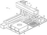
- FIG. 1is a perspective view of an automated instrument for performing the target biomolecule detection in accord with the system of the present invention.
- FIG. 2is a plan view of a linearly-arranged substrate for use in the system of FIG. 1 .
- FIG. 3is a plan view of a two-dimensional substrate for use in the system of FIG. 1 .
- FIG. 4is a plan view of a pipette tip having a substrate for use in the system of FIG. 1 .
- FIG. 5is a plan view of a plunger-type pipette tip having a substrate for use in the system of FIG. 1 .
- FIG. 6is a front view of a pipette adapter with bracket for use in the system of FIG. 1 .
- FIG. 7is a side view of the pipette adapter of FIG. 6 .
- FIG. 8is a perspective view of a pipette adapter, supporting a substrate for use in the system of FIG. 1 .
- FIG. 9is an end view of the pipette adapter of FIG. 8 , showing details of the supported substrate.
- FIG. 10is a plan view of a pipette adapter positioned within the sample well of a microtiter plate.
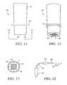
- FIG. 11is a front view of a flat bottom pipette adapter for use in the system of FIG. 1 .
- FIG. 12is a perspective view of the flat bottom pipette adapter of FIG. 11 , showing a substrate at the base of the adapter.
- FIG. 13is an end view of a flat bottom pipette adapter of FIG. 11 , showing details of the substrate.
- FIG. 14is a perspective view of the elements of an optical detection station for use in the system of FIG. 1 .
- FIG. 15is a plan view of an alternate embodiment of the detection station for use in the system of FIG. 1 .
- FIG. 16presents yet another alternate embodiment of the detection station for use in the system of FIG. 1 .
- FIG. 17is a perspective view of a device which may be utilized for biopolymer synthesis on a substrate to create a substrate in accord with the present invention.
- FIG. 18presents a cross sectional view of the device of FIG. 17 .
- FIG. 19is a plan view of a substrate and backing plate for biopolymer synthesis, showing a one-dimensional biopolymer array for a substrate in accord with the present invention.
- FIG. 20is a plan view of a substrate and backing plate, showing two-dimensional biopolymer synthesis for a substrate in accord with the present invention.
- FIG. 21is a perspective view of a jet head-type reagent deposition apparatus for creating a substrate in accord with the present invention.
- FIG. 22is a plan view of the internal elements of a jet head of FIG. 21 .
- FIG. 23illustrates a method of affixing a substrate to a manipulator of the present invention.
- FIG. 24illustrates a first alternative method of affixing a substrate to a manipulator of the present invention.
- FIG. 25illustrates a second alternative method of affixing a substrate to a manipulator of the present invention.
- FIG. 26is a plan perspective view of a version of the instrument of FIG. 1 , modified to include a jet-head substrate treating apparatus.
- a system 10 utilizing a movable bioarrayis shown.
- a robotic arm 12carries a holder 20 which fits and transports bioarray 11 , first to the sample, which may be in well 17 of microtiter plate 15 or in vial 16 of rack 14 .
- a robotic armis one form of manipulator which may be used, other simpler manipulators may be employed, such as mechanical movements.
- a preferred type of manipulator deviceis the Biomek, a trademark for an instrument of Beckman Instruments, Inc.
- the substrate portion of bioarray 11is mounted in a holder having a support region which may be quite small. After the bioarray and the sample have had a sufficient incubation or reaction time for interaction of reactants on the substrate and any target biomolecules which may be present within the sample, the robotic arm 12 moves the substrate 11 to the detection assembly 18 of instrument 10 .
- optical detection station 18is presented in a cutaway view showing laser 19 , light collector 21 , and detector 22 .
- Both the detection station 18 and the robotic arm 12may be attached to a computer, not shown, which generates commands for movement of the robotic arm and receives signals from the detection assembly which may, in turn, be analyzed to determine whether a specific target biomolecule is present and which may be displayed.
- the substrate itselfis held within a holder 20 which may be coupled to the cantilevered robotic arm 12 via pick-up shaft 23 or by some other coupling method.
- a wide range of motionis available over a base the size of a desktop.
- Many sample wellsmay be reached as well as many substrate holders having treated substrates or untreated substrates which may be treated by motion to a nearby location where reactants may be sprayed or otherwise applied to the substrate.
- FIG. 2presents a linearly-arranged, flat substrate as a support portion of a bioarray, with active sites 30 forming bands along substrate 28 which is shaped as a strip. Spaces 26 are provided to place the plurality of active sites 30 in a spaced-apart relation along substrate 28 .
- an area-wide treated substrateis created by positioning the active sites in a two-dimensional relation on a planar sheet substrate. Active sites 30 are positioned in spaced-apart relation on substrate 28 , as before.
- the active sites 30may be bands, as in FIG. 2 , or spots, as in FIG. 3 , or some other shape. The spots are in known locations and are specific for a target biomolecule.
- a plurality of linear substrates of FIG. 2may also be arranged in parallel to create a two-dimensional bioarray. The size of the substrate is typically a few centimeters on a side, but could be smaller or larger.
- the reactants forming the active sitesmay comprise complementary DNA strands for detection by DNA hybridization or they may comprise immunological biomolecules for detection by immunological complexing, such as formation of antigen-antibody complexes.
- the device of FIG. 4represents one example of a substrate holder which may be used to present the bioarray to the sample.
- a pipette tip 27is shown with a substrate 11 supported longitudinally along an inside wall.
- the substrateis preferably positioned along an inside wall of the pipette tip and the pipette tip is comprised of an optical glass or plastic allowing optical inspection of the substrate while the substrate is positioned inside the pipette tip.
- the sampleis drawn into the pipette tip by aspiration and allowed to interact with substrate 11 .
- Pipette tip 27preferably has at least one flat surface, i.e. the surface opposite the substrate, for accurate optical inspections. This feature and a narrow bore also help minimize the amount of sample necessary and place the sample and substrate in close proximity.
- the pipette tip 27may be used in conjunction with a rubber bulb, vacuum pump, robotic pipettor as in FIG. 1 , or other device.
- the term “pipette tip”is meant to include pipettes, such as long cylindrical glass or plastic pipettes which are designed to operate in connection with a simple suction device.
- FIG. 5shows a second embodiment of a substrate holder wherein the substrate 11 is housed within a plunger-type pipette tip, in the same manner as the FIG. 4 embodiment.
- Pipette tip 29also has a narrow bore and flattened surface. The sample is drawn into the plunger-type pipette tip 29 through withdrawal of plunger 31 . Positive displacement of the sample is used to eject the sample from the pipette, as by depression of plunger 31 or by some other fluid manipulation.
- the embodiment of FIG. 5has an advantage over the embodiment of FIG. 4 in that the sample does not drain from the pipette tip when the pipette tip of FIG. 5 is detached from a pipetting tool.
- a substrate holdertakes the form of a pipette adapter 32 characterized by a bracket 33 at one end.
- the brackethas opposed prongs 36 a - b , easily visible in FIG. 8 , which support the ends of the substrate which is part of the bioarray.
- the opposite end of the bracketed pipette adapterpreferably has a coupler 37 for joining a robotic or standard pipetting tool.
- the coupler 37is depicted as a hollow cone which may fit the conical shaft of a robotic or standard pipetting tool with an appropriate securing mechanism, such as a friction fit, with provision for ejection of the adapter for use.
- an appropriate securing mechanismsuch as a friction fit
- Many different types of couplersmay be used, however.
- the pipette adapter 32need not have any coupler, but it is preferred that the adapter have a gripper or other means for manipulating the adapter so, for example, the adapter may be moved into and out of sample wells easily.
- the bioarrayis preferably oriented so that the active sites face downward.
- Pipette adapter 32is preferably equipped with knobs 35 on the prongs of the bracket, visible in FIGS. 6-9 . These knobs position the substrate slightly above the bottom of the sample well, and protect the treated substrate from physical abrasion and contamination. Without such knobs, placement of the adapter into the well may press the substrate so close to the bottom of the well as to exclude sample from the face of the substrate, preventing proper contact and interaction of the sample and the substrate.
- Bracketed pipette adapter 32also preferably contains a ring or disk-shaped evaporation barrier 34 which is disposed about the midsection of the adapter, or at the base of the bracket portion. Because some samples may easily evaporate, evaporation barrier 34 is preferably provided to protect the sample during the substrate and sample incubation period. In FIG. 10 , evaporation ring 34 is seen providing a barrier when pipette adapter 32 is inserted into well 17 , thus limiting the exposure of sample 38 .
- the diameter of pipette adapter 32 , and particularly bracket 33is sufficiently narrow in order to easily fit within the microtiter plate's well, as also seen in FIG. 10 .
- FIG. 9presents an end view of the bracketed pipette adapter 32 .
- This viewis indicated by axis 9 - 9 of FIG. 6 , viewed in the direction of the arrows. From the end view, the substrate 11 is more clearly visible in its preferred downward facing orientation.
- the substrate of FIG. 9is in a linearly arranged, but segmented, form. Thus strips 11 a , 11 b , and 11 c are positioned in a generally parallel arrangement and secured by prongs 36 a - b of the bracket 33 .
- the nature and shape of the substratemay be easily adapted to sample, applicator, and space considerations.
- the two-dimensional substrate of FIG. 3would adapt easily to bracketed pipette adapter 32 .
- the substrateis held by prongs 36 a - b by adhesion, welding, clamping, or any other means for gripping which will not interfere with the testing of the sample.
- evaporation disk 34is visible beyond the prongs and the substrate.
- a flat-bottom pipette adapter 39is utilized to support and transport the bioarray.
- Flat-bottom pipette adapter 39has a flat bottom surface 40 , visible in FIG. 12 , and may have a coupler 37 at an end opposite to the flat bottom surface 40 for fitting the adapter to pipetting tools or a robotic arm, as in the bracketed pipette adapter embodiment.
- flat bottom pipette adapter 39may have a simple means for manually gripping the pipette adapter and applying the substrate 11 to the sample.
- the flat bottom adapter 39is preferably outfitted with knobs 35 and evaporation ring 34 . In FIG.
- the substrate 11is seen to be a two-dimensional array of spots or dots, as in FIG. 3 .
- Evaporation ring 34is visible in this view, taken along axis 13 - 13 of FIG. 11 , and situated beyond flat bottom surface 40 .
- FIG. 14details of the internal elements of a biomolecule probe station are shown.
- the optical detection station 18is an example.
- the stationmay be part of an analysis machine having a robotic arm, such as that shown in FIG. 1 .
- the robotic arm, pipetting tool, or other substrate holderpositions the substrate, after it has interacted with the sample, in the path of a laser beam.
- Laser 41creates a beam which impinges upon the active sites of the substrate 11 , which is held within bracketed pipette adapter 32 .
- the wavelength of the beamis selected to cause the return of a radiation signature from target molecules bound to the substrate.
- Such a signaturecomes from an optically detectable characteristic radiation pattern of the bound target molecules when excited by radiation of the beam, such as a characteristic band of fluorescent wavelengths.
- Time gated fluorescence, or other optical signal enhancement techniquesmay optionally be used.
- the incident beam from the laseris scanned across the active sites of the treated substrate by relative motion of the substrate and the beam.
- Light emitted from the active sitesis collected by light collector 42 and directed to detector 43 , which may be a photomultiplier tube, CCD array, or other detection device, and which is preferably associated with a computer for any further analysis or display of the signals received from the substrate.
- Additional optical elements, such as wavelength selective filters,may be disposed in either the incident beam or the return light, as required by the characteristic radiation signature.
- Scanningmay be accomplished by moving the substrate relative to the laser beam, by utilizing a scanning reflector such as a galvo mirror or polygonal mirror, or by some other well known means.
- a scanning reflectorsuch as a galvo mirror or polygonal mirror, or by some other well known means.
- the area of the laser beammay be expanded such that the entire area of the array is illuminated simultaneously, and scanning is not required.
- the bioarrayis optically probed by the beam for determining the extent of complexing of the reactants in the active sites of the substrate with target biomolecules in the sample.
- the optical inspectionmay be for fluorescent signals, reflectance, absorbance, light scattering, or chemiluminescence, among others. Details of the optical system may vary according to the nature of the signal to be detected.
- FIG. 14illustrates a substrate 11 within bracketed pipette adapter 32 and facing in a downward orientation for impingement by the laser beam. This arrangement of the elements of the detection assembly is presented as an example of the arrangement of the detection assembly 18 of FIG. 1 . In either case, the robotic arm may easily move the substrate and the associated bracketed pipette adapter to the detection assembly after the appropriate sample incubation period.
- the robotic armmay, however, be capable of moving the substrate so that it is oriented vertically, or in some other manner, relative to the laser beam.
- the laser source for the excitation pathmay be positioned in a manner other than shown in order to impinge upon the substrate.
- Optical fibersmay be employed to direct the beam or the return signal.
- FIG. 15presents a detection arrangement for probing of a substrate within a pipette tip.
- Pipette tip 27has an optical surface so as not to interfere with the optical inspection.
- laser 41impinges upon the substrate 11 and emitted signals from the substrate are gathered by light collector 42 and directed to detector 43 where the signals may be sent onto a computer for further analysis.
- the excitation beam from the laserimpinges on the substrate through the wall of the pipette tip.
- Arrow A of FIG. 15indicates one example of how the substrate may be scanned, i.e. by providing a vertical motion to the pipette tip, via the robotic arm or some other mechanism.
- the substrate 11 within pipette tip 27is scanned by the incident laser beam through the action of a scanning reflector 44 , which may be rotated in direction B to cause scanning of the substrate 11 .
- An automated apparatussuch as instrument 10 of FIG. 1 may have a plurality of detection assemblies to which the robotic arm may move the substrate for reading, depending upon the type of manipulator used for the substrate, the type of signals to be read from the substrate, and the nature of the substrate and target biomolecules thereon.
- the substratemay be formed by the device shown in FIGS. 17-20 , which allows biopolymer synthesis on a solid support.
- the substratemay be used directly, or the biopolymers created by the device may be cleaved from the substrate and affixed to another substrate in any desired format.
- the method and apparatus depicted in FIGS. 17-20are the subject of commonly-assigned U.S. Pat. No. 5,429,807, which is incorporated herein by reference.
- the device of FIG. 17presents a synthesis device 45 which is a thick block having a plate surface 46 within which are a plurality of grooves or channels 50 . Channels 50 are connected to tubing 47 at the underside of block 45 for flowing reagents through channels 50 .
- tubing 47 ais connected to an inlet tubing connector 49 which communicates with channel 50 .
- Tubing 47 bcommunicates with outlet tubing connector 52 which in turn communicates with channel 50 .
- reagentsmay be caused to flow through any of the channels 50 .
- a solid support materialsuch as a sheet of activated polypropylene
- a backing platemay be used to sandwich the polypropylene substrate, allowing the flexible polypropylene to seal against the channels 50 of the block 45 .
- the backing plate 52 of FIGS. 19 and 20may have holes 53 which may be aligned with holes 48 of block 45 .
- the backing plate and the blockmay then be secured to one another.
- Synthesis or biopolymerizationmay be performed by activating the surface of the substrate, if necessary, and by flowing reagents through the channels, to cause formation of strands of biopolymers anchored to the substrate. This results in a one-dimensional array of biopolymers 54 , as seen in FIG.
- the blockmay be repositioned with respect to the substrate 55 and the process repeated.
- FIG. 20one-dimensional arrays 54 a - b are presented in 90° offset orientations. The areas of overlap 56 provide new biopolymers having elements of each one-dimensional array 54 a - b .
- Indexing pins 58visible in FIGS. 19 and 20 , may be utilized to position the substrate 55 in relation to the applicator. Indexing pins 58 mate with holes 57 in block 45 . The resulting arrays may be utilized as is, or may be cleaved from the polypropylene substrate 55 and affixed to some other support.
- the substrate 55 having the arraysmay be segmented and attached to substrate 28 , in the manner of FIGS. 8 and 9 .
- channels 50are illustrated, cavities for reagent flow having some other shape may be used.
- polypropyleneis presented in the above discussion, other substrates such as glass, Pyrex, silicon, polystyrene, etc. may be utilized as supports for synthesis, as suggested in PCT application No. WO 93/09668.
- a support 28is positioned on support frame 65 within a spray station.
- a plurality of ink jet-type heads 60 with nozzles 61 at the spray stationare used to selectively deposit reactants on support 28 to create the plurality of active sites.
- Such headsare well-known in the field of ink jet printing. In the present invention, such heads are adapted for dispensing the reactants onto the desired locations of the substrate. If necessary, the substrate may be activated to receive and immobilize the reactants.
- a plurality of different jet heads 60may be moved on respective rails 62 in the direction indicated by arrow C.
- Each of the jet heads 60dispenses a different reactant.
- Motion control of the jet heads 60 along rails 62may be provided by a computer.
- the arraymay be moved by an actuator 63 , which causes an arm 64 to move support frame 65 carrying the support 28 .
- the actuator 63may be a linear motor similar to that used to move magnetic heads in disc drives. In the situation where the substrate for the treated substrate is a strip, fixed position jet heads may be desired.
- FIG. 22illustrates a typical ink jet-type dispensing head as applied in this invention.
- Reactant contained in reservoir 67passes through supply tube 69 to piezoelectric pumping chamber 66 , and through chamber 66 to nozzle 69 .
- Electrical pulses applied to pumping chamber 66cause it to expand and contract in volume. Each time a pulse is applied and removed, this expansion and contraction event ejects a droplet 70 of reactant from the nozzle. Additional details of the design and operation of such a reactant dispensing device are disclosed in previously referenced U.S. Pat. No. 4,877,745.
- a dispensing headmight be employed to dispense ink or dye onto the substrate to form barcode patterns for machine reading of the identification of a bioarray.
- Another method of preparing the bioarrayis by a technique analogous to a printing method.
- an analyteis deposited on a substrate by stamping or embossing a very thin layer with an array of analyte spots at desired locations.
- an antigen attached to a molecule anchored to the substrate by pressure contactwill combine with an appropriate antibody associated with a specific target biomolecule.
- the antibodymay be fluorescent for optical detection.
- a method of imaging, i.e. probing, a substrate having microscopic featuresis by means of condensation figures (CFs) described in a journal article entitled “Imaging of Features on Surfaces by Condensation Figures” by G. Lopez et al. in Science, Apr. 30, 1993, p. 647.
- the authorsdescribe the formation of an array of tiny droplets on a cold surface having an array of spots which are not wet by the droplets.
- the spotscould be the complex compounds described in the preceding paragraph.
- the dropletsare observed with microscope optics.
- Still another bioarray forming techniqueis described in an article by B. Healey et al. in Science, Aug. 25, 1995 p. 1078.
- the authorsdeposited microscopic polymer arrays on a flat substrate by depositing a layer of polymerization solution on a flat plate, such as a glass chip which had been activated for adhesion with the solution.
- a bundle of fiberswas brought into contact with the solution and then backed off and the substrate rinsed. Light was directed into the non-contacted end of the fiber bundle to cause polymer deposition on the substrate below the fibers of the fiber bundle. Polymer spots of a 2.0 micrometer diameter and a spacing of 4.0 micrometers were produced.
- Yet another bioarray forming techniqueis the Southern blotting method in which hybridization is used simultaneously on a large number of DNA segments.
- DNAis fragmented, electrophoresed, denatured and transferred from a gel to filter paper. Positions of numerous fragments are established.
- the DNA fragmentsare robotically moved in accord with the present invention and combined with radioactive phosphorous labelled RNA which can be identified.
- the degree of DNA-RNA complementation, i.e. probing of the sample,can be determined by autoradiography.
- a polyunsaturated polymerized lipid layeris applied to a support.
- the lipidshave a member of a specific binding pair bound to one end.
- the lipidshave an optical characteristic which is modified upon complexing the other member of the binding pair. Such an optical characteristic can be polarization of light and such light is used to probe the bioarray.
- FIG. 23affixing of bioarray 11 to the flat bottom surface 40 of flat bottom pipette adapter 39 , seen in FIGS. 10-12 , is given as an example.
- a two-dimensional bioarray 11is preformed and then attached to the flat bottom surface 40 of pipette adapter 39 .
- FIG. 24presents an alternative wherein the substrate 28 is affixed to flat bottom surface 40 of the pipette adapter, before reactants are caused to become immobilized on the substrate to form the bioarray 11 .
- This alternativeworks particularly well with the printing type of creation for substrates as discussed with reference to FIGS. 21 and 22 .
- FIG. 21 and 22In FIG.
- the bare substrate 28may be an integral part of the adapter as fabricated, for instance by injection molding.
- the substrate 28is then activated, indicated by shading in FIG. 25 , and then the reactants are deposited or otherwise caused to attach to substrate 28 .
- the method of the present inventionis designed for detection of target biomolecules in a sample
- quantification of the target biomoleculesis possible by, for example, recording the sample volume exposed to the substrate, quantifying the degree of complementation at the active sites of the substrate, and calculating the amount of target biomolecule present from these two values. Quantification of the degree of complementation may be performed, e.g., by measuring the percentage of active sites which are fluorescently-labeled or give some other optical signal indicating complementation. Additionally, affixing an excess amount of reactants to the substrate compared to the amount of suspected target biomolecules of the sample is a preferred practice and makes quantification more accurate.
- FIG. 26an alternate embodiment of instrument 10 of FIG. 1 , incorporating the jethead-type reagent dispensing means, is shown.
- the instrumentcontains a detection assembly 18 and has a location for placement of the samples. In this case, the samples are within wells 17 of microtiter plate 15 .
- FIG. 26shows a location for a holder 20 for a substrate.
- pipette adapter 32has a bioarray held within opposed prongs of a bracket, as illustrated in FIGS. 6-9 .
- pipette adapter rack 81is shown having a plurality of pipette adapters 32 a .
- the instrumentalso contains a jethead dispensing device 80 .
- the dispensing deviceis of the type discussed with regard to FIGS. 21 and 22 .
- this variationhas a robotic arm 12 attached to a tower 13 which, in turn, is attached to a base 59 .
- tower 13 and base 59have tracks for providing both vertical and horizontal motion to the robotic arm.
- the detection assembly 18 and dispensing device 80are depicted as blocks having holes. These blocks illustrate that the instrument is provided with various stations, each having dedicated operations. The holes enable access to the holder which is attached to the robotic arm.
- the instrument of FIG. 26provides motion to the bioarray via the robotic arm which picks up a pipette adapter having a substrate support from rack 81 , and then moves the pipette adapter into dispensing device 80 .
- the supportmay be activated, if necessary, and dispensing or printing of reactants upon the support to create a substrate occurs.
- robotic arm 12moves the pipette adapter from dispensing device 80 into contact with a sample, as by placing pipette adapter 32 within a sample well 17 of *microtiter plate 15 .
- the pipette adapters 32 b shown in the microtiter plate 15have been deposited into the wells by robotic arm 12 for interaction of the substrates and the samples. After the appropriate incubation time, robotic arm 12 picks up each pipette adapter and moves it to detection assembly 18 for detection, as before.
- the robotic armmoved the pipette adapter, with holder and bioarray, to a sample location, such as a microtiter plate.
- the robotic armcould pick up sample in a pipettor and bring it to a stationary holder where the pipettor could dispense the sample onto the holder. Then, the same robotic arm, or another one, with an appropriate gripper could move the holder to a detection station.
- the detection stationcould be any of the optical types described above, but could also be a radioactive tag detector if the immobilized reactants for the target biomolecule had been radioactive. Also, if the tag was a moiety suitable for detection by laser desorption mass spectrometry (LD-MS), then an LD-MS detection system could be used. Other tags and detection systems will be evident to those skilled in the art.
- LD-MSlaser desorption mass spectrometry
Landscapes
- Chemical & Material Sciences (AREA)
- Health & Medical Sciences (AREA)
- General Physics & Mathematics (AREA)
- Physics & Mathematics (AREA)
- Engineering & Computer Science (AREA)
- General Health & Medical Sciences (AREA)
- Analytical Chemistry (AREA)
- Chemical Kinetics & Catalysis (AREA)
- Pathology (AREA)
- Biochemistry (AREA)
- Immunology (AREA)
- Life Sciences & Earth Sciences (AREA)
- Organic Chemistry (AREA)
- Clinical Laboratory Science (AREA)
- Nanotechnology (AREA)
- Composite Materials (AREA)
- Materials Engineering (AREA)
- Condensed Matter Physics & Semiconductors (AREA)
- Robotics (AREA)
- Hematology (AREA)
- Nuclear Medicine, Radiotherapy & Molecular Imaging (AREA)
- Crystallography & Structural Chemistry (AREA)
- Automatic Analysis And Handling Materials Therefor (AREA)
- Apparatus Associated With Microorganisms And Enzymes (AREA)
- Investigating Or Analysing Biological Materials (AREA)
- Investigating, Analyzing Materials By Fluorescence Or Luminescence (AREA)
- Measuring Or Testing Involving Enzymes Or Micro-Organisms (AREA)
- Investigating Or Analysing Materials By The Use Of Chemical Reactions (AREA)
- Investigating Or Analysing Materials By Optical Means (AREA)
Abstract
Description
Claims (20)
Priority Applications (1)
| Application Number | Priority Date | Filing Date | Title |
|---|---|---|---|
| US11/670,165US8273304B2 (en) | 1996-01-16 | 2007-02-01 | Analytical biochemistry system with robotically carried bioarray |
Applications Claiming Priority (3)
| Application Number | Priority Date | Filing Date | Title |
|---|---|---|---|
| US08/586,116US6660233B1 (en) | 1996-01-16 | 1996-01-16 | Analytical biochemistry system with robotically carried bioarray |
| US10/200,720US20020182117A1 (en) | 1996-01-16 | 2002-07-22 | Analytical biochemistry system with robotically carried bioarray |
| US11/670,165US8273304B2 (en) | 1996-01-16 | 2007-02-01 | Analytical biochemistry system with robotically carried bioarray |
Related Parent Applications (1)
| Application Number | Title | Priority Date | Filing Date |
|---|---|---|---|
| US10/200,720ContinuationUS20020182117A1 (en) | 1996-01-16 | 2002-07-22 | Analytical biochemistry system with robotically carried bioarray |
Publications (2)
| Publication Number | Publication Date |
|---|---|
| US20070128084A1 US20070128084A1 (en) | 2007-06-07 |
| US8273304B2true US8273304B2 (en) | 2012-09-25 |
Family
ID=24344385
Family Applications (6)
| Application Number | Title | Priority Date | Filing Date |
|---|---|---|---|
| US08/586,116Expired - LifetimeUS6660233B1 (en) | 1996-01-16 | 1996-01-16 | Analytical biochemistry system with robotically carried bioarray |
| US10/200,720AbandonedUS20020182117A1 (en) | 1996-01-16 | 2002-07-22 | Analytical biochemistry system with robotically carried bioarray |
| US11/073,288AbandonedUS20060057029A1 (en) | 1996-01-16 | 2005-03-04 | Analytical biochemistry system with robotically carried bioarray |
| US11/123,344AbandonedUS20050271552A1 (en) | 1996-01-16 | 2005-05-05 | Analytical biochemistry system with robotically carried bioarray |
| US11/258,707AbandonedUS20060045812A1 (en) | 1996-01-16 | 2005-10-26 | Analytical biochemistry system with robotically carried bioarray |
| US11/670,165Expired - Fee RelatedUS8273304B2 (en) | 1996-01-16 | 2007-02-01 | Analytical biochemistry system with robotically carried bioarray |
Family Applications Before (5)
| Application Number | Title | Priority Date | Filing Date |
|---|---|---|---|
| US08/586,116Expired - LifetimeUS6660233B1 (en) | 1996-01-16 | 1996-01-16 | Analytical biochemistry system with robotically carried bioarray |
| US10/200,720AbandonedUS20020182117A1 (en) | 1996-01-16 | 2002-07-22 | Analytical biochemistry system with robotically carried bioarray |
| US11/073,288AbandonedUS20060057029A1 (en) | 1996-01-16 | 2005-03-04 | Analytical biochemistry system with robotically carried bioarray |
| US11/123,344AbandonedUS20050271552A1 (en) | 1996-01-16 | 2005-05-05 | Analytical biochemistry system with robotically carried bioarray |
| US11/258,707AbandonedUS20060045812A1 (en) | 1996-01-16 | 2005-10-26 | Analytical biochemistry system with robotically carried bioarray |
Country Status (5)
| Country | Link |
|---|---|
| US (6) | US6660233B1 (en) |
| EP (1) | EP0819256A1 (en) |
| JP (4) | JPH11502937A (en) |
| AU (1) | AU734126C (en) |
| WO (1) | WO1997026539A1 (en) |
Cited By (7)
| Publication number | Priority date | Publication date | Assignee | Title |
|---|---|---|---|---|
| US20110275058A1 (en)* | 2010-02-23 | 2011-11-10 | Rheonix, Inc. | Self-contained biological assay apparatus, methods, and applications |
| USD697223S1 (en)* | 2012-01-25 | 2014-01-07 | Ge Healthcare Uk Limited | Sample card holder |
| US8986614B2 (en)* | 2010-02-23 | 2015-03-24 | Rheonix, Inc. | Self-contained biological assay apparatus, methods, and applications |
| WO2015054305A1 (en)* | 2013-10-09 | 2015-04-16 | University Of Utah Research Foundation | Sample tube adapters and methods of use thereof |
| US9102979B2 (en) | 2010-02-23 | 2015-08-11 | Rheonix, Inc. | Self-contained biological assay apparatus, methods, and applications |
| US9835640B2 (en) | 2015-02-13 | 2017-12-05 | Abbott Laboratories | Automated storage modules for diagnostic analyzer liquids and related systems and methods |
| WO2023196430A3 (en)* | 2022-04-05 | 2023-11-30 | Aavantibio, Inc. | Gradient ultracentrifugation automated collector |
Families Citing this family (118)
| Publication number | Priority date | Publication date | Assignee | Title |
|---|---|---|---|---|
| EP0695941B1 (en)* | 1994-06-08 | 2002-07-31 | Affymetrix, Inc. | Method and apparatus for packaging a chip |
| US6287850B1 (en)* | 1995-06-07 | 2001-09-11 | Affymetrix, Inc. | Bioarray chip reaction apparatus and its manufacture |
| US5959098A (en) | 1996-04-17 | 1999-09-28 | Affymetrix, Inc. | Substrate preparation process |
| US5753429A (en)* | 1996-08-09 | 1998-05-19 | Lifescan, Inc. | Analyte concentration measurement using a hollow frustum |
| GB9624927D0 (en) | 1996-11-29 | 1997-01-15 | Oxford Glycosciences Uk Ltd | Gels and their use |
| AUPO625497A0 (en)* | 1997-04-16 | 1997-05-15 | Macquarie Research Limited | Analysis of molecules |
| US5985214A (en) | 1997-05-16 | 1999-11-16 | Aurora Biosciences Corporation | Systems and methods for rapidly identifying useful chemicals in liquid samples |
| US5993627A (en)* | 1997-06-24 | 1999-11-30 | Large Scale Biology Corporation | Automated system for two-dimensional electrophoresis |
| ATE245485T1 (en)* | 1997-10-27 | 2003-08-15 | Idexx Lab Inc | DEVICE FOR DETERMINING ANALYTES IN SOLUTIONS |
| ES2350702T3 (en)* | 1998-01-12 | 2011-01-26 | Massachusetts Institute Of Technology | SYSTEM TO ANALYZE A PLURALITY OF SAMPLES. |
| FR2776389B1 (en)* | 1998-03-20 | 2000-06-16 | Fondation Jean Dausset Ceph | AUTOMATIC DEVICE FOR PRODUCING SAMPLES FOR THE IMPLEMENTATION OF CHEMICAL OR BIOLOGICAL REACTIONS IN A LIQUID MEDIUM |
| GB9811656D0 (en) | 1998-05-29 | 1998-07-29 | Oxford Glycosciences Uk Ltd | Gels, methods and apparatus for identification and characterization of biomolecules |
| US6349160B2 (en) | 1998-07-24 | 2002-02-19 | Aurora Biosciences Corporation | Detector and screening device for ion channels |
| US6608671B2 (en) | 1998-07-17 | 2003-08-19 | Vertex Pharmaceuticals (San Diego) Llc | Detector and screening device for ion channels |
| US6861218B2 (en) | 1998-11-03 | 2005-03-01 | Metasystems Hard And Software Gmbh | Method for the targeted application of reagents onto immobilized biological material |
| DE59906078D1 (en)* | 1998-11-03 | 2003-07-31 | Metasystems Hard & Software Gm | Process for the targeted application of reagents to immobilized biological material |
| US7510841B2 (en)* | 1998-12-28 | 2009-03-31 | Illumina, Inc. | Methods of making and using composite arrays for the detection of a plurality of target analytes |
| US6429027B1 (en)* | 1998-12-28 | 2002-08-06 | Illumina, Inc. | Composite arrays utilizing microspheres |
| US6143252A (en)* | 1999-04-12 | 2000-11-07 | The Perkin-Elmer Corporation | Pipetting device with pipette tip for solid phase reactions |
| JP3469504B2 (en)* | 1999-06-01 | 2003-11-25 | 日立ソフトウエアエンジニアリング株式会社 | Microarray chip and indexing method thereof |
| US6630006B2 (en)* | 1999-06-18 | 2003-10-07 | The Regents Of The University Of California | Method for screening microcrystallizations for crystal formation |
| ES2367551T3 (en)* | 1999-08-13 | 2011-11-04 | Bayer Technology Services Gmbh | DEVICE AND PROCEDURE FOR THE DETERMINATION OF MULTIPLE ANALYTICS. |
| US6351690B1 (en)* | 2000-01-21 | 2002-02-26 | Virologic, Inc. | Automated method and system for performing antiviral drug susceptibility and resistance testing |
| EP1272829A1 (en)* | 2000-04-14 | 2003-01-08 | Zeptosens AG | Grid-waveguide structure for reinforcing an excitation field and use thereof |
| DE10020771A1 (en)* | 2000-04-28 | 2001-10-31 | Merck Patent Gmbh | Pipetting device |
| US7163660B2 (en)* | 2000-05-31 | 2007-01-16 | Infineon Technologies Ag | Arrangement for taking up liquid analytes |
| WO2002013961A2 (en)* | 2000-08-11 | 2002-02-21 | Wisconsin Alumni Research Foundation | Chemical screening system using strip arrays |
| DE10045808C2 (en)* | 2000-09-07 | 2002-08-08 | Imb Inst Fuer Molekulare Biote | Method for the detection of the activity of active substances |
| US20060141507A1 (en)* | 2000-09-27 | 2006-06-29 | Kronick Mel N | Manufacture and use of non-standard size microarray slides |
| US7135146B2 (en) | 2000-10-11 | 2006-11-14 | Innovadyne Technologies, Inc. | Universal non-contact dispense peripheral apparatus and method for a primary liquid handling device |
| US6852291B1 (en) | 2000-10-11 | 2005-02-08 | Innovadyne Technologies, Inc. | Hybrid valve apparatus and method for fluid handling |
| CN1500140A (en)* | 2001-03-28 | 2004-05-26 | 上海晶泰生物技术有限公司 | Device and method for measuring a great variety of matters to be analyzed |
| WO2003023410A1 (en)* | 2001-09-07 | 2003-03-20 | Innovadyne Technologies, Inc. | Secondary liquid dispensing module for liquid handling system |
| CN1298849C (en)* | 2002-01-17 | 2007-02-07 | 准确系统科学株式会社 | System for housing/processing carrier and method for housing/processing carrier |
| US7169616B2 (en) | 2002-01-25 | 2007-01-30 | Innovadyne Technologies, Inc. | Method of purging trapped gas from a system fluid contained in an actuation valve |
| US6734424B2 (en)* | 2002-05-16 | 2004-05-11 | Large Scale Proteomics Corporation | Method for microdispensing of fluids from a pipette |
| US7452712B2 (en) | 2002-07-30 | 2008-11-18 | Applied Biosystems Inc. | Sample block apparatus and method of maintaining a microcard on a sample block |
| JP4426968B2 (en)* | 2002-09-17 | 2010-03-03 | オリンパス株式会社 | Method and apparatus for disposing a liquid reaction component on the surface of a substrate to detect a target material by reaction between a plurality of components on the substrate, and an article for use in this method |
| US6970240B2 (en) | 2003-03-10 | 2005-11-29 | Applera Corporation | Combination reader |
| US20040241872A1 (en)* | 2003-03-17 | 2004-12-02 | Qiagen Operon, Inc. | Optical detection liquid handling robot system |
| US7576862B2 (en) | 2003-08-26 | 2009-08-18 | Blueshift Biotechnologies, Inc. | Measuring time dependent fluorescence |
| US7501094B2 (en)* | 2003-09-15 | 2009-03-10 | Syngenta Limited | Preparation and characterization of formulations in a high throughput mode |
| US8277760B2 (en) | 2003-09-19 | 2012-10-02 | Applied Biosystems, Llc | High density plate filler |
| US20070015289A1 (en)* | 2003-09-19 | 2007-01-18 | Kao H P | Dispenser array spotting |
| US20050226779A1 (en) | 2003-09-19 | 2005-10-13 | Oldham Mark F | Vacuum assist for a microplate |
| US7407630B2 (en) | 2003-09-19 | 2008-08-05 | Applera Corporation | High density plate filler |
| US7998435B2 (en) | 2003-09-19 | 2011-08-16 | Life Technologies Corporation | High density plate filler |
| JP4203826B2 (en)* | 2003-09-29 | 2009-01-07 | 独立行政法人産業技術総合研究所 | Automatic analysis method and apparatus |
| WO2005042146A2 (en)* | 2003-10-24 | 2005-05-12 | Aushon Biosystems, Inc. | Apparatus and method for dispensing fluid, semi-solid and solid samples |
| JPWO2005054844A1 (en)* | 2003-12-04 | 2007-12-06 | オリンパス株式会社 | Reaction vessel, reaction device and detection device using the same, and method for producing reaction vessel |
| DE102004062281A1 (en)* | 2003-12-29 | 2005-07-28 | Siemens Ag | Producing a microarray of spots e.g. for use on a biochip, comprises applying a solution of a polymer and special molecules and then solidifying the polymer, especially by a non-radical method |
| US7416710B1 (en) | 2003-12-31 | 2008-08-26 | Takeda San Diego, Inc. | Method and system for performing crystallization trials |
| JP2007534936A (en)* | 2004-02-11 | 2007-11-29 | パムジーン ベー.フェー. | A device to analyze the interaction between target and probe molecules |
| JP2005283304A (en)* | 2004-03-29 | 2005-10-13 | Lintec Corp | Probe array |
| JP2006030156A (en)* | 2004-06-15 | 2006-02-02 | Olympus Corp | Reaction vessel, reactor using the same, and detector |
| EP1612561A1 (en)* | 2004-07-02 | 2006-01-04 | Roche Diagnostics GmbH | Instrument for efficient treatment of analytical devices |
| EP2620778A3 (en)* | 2004-12-10 | 2014-08-20 | Universal Bio Research Co., Ltd. | Biological material fixed carrier enclosing tip, biological material fixed carrier treatment apparatus, and treatment method thereof |
| WO2006062236A1 (en) | 2004-12-10 | 2006-06-15 | Universal Bio Research Co., Ltd. | Chip having biosubstance immobilization region hermetically sealed therein, biosubstance immobilization region treating apparatus and method of treatment |
| EP1835020A4 (en) | 2005-01-07 | 2011-06-15 | Universal Bio Research Co Ltd | Carrier enclosing chip, carrier treating apparatus and method of carrier treatment |
| GB0503986D0 (en)* | 2005-02-26 | 2005-04-06 | Secr Defence | Reaction apparatus |
| US7622079B2 (en)* | 2005-03-22 | 2009-11-24 | Applied Biosystems, Llc | Dual nest microplate spotter |
| US20060246576A1 (en) | 2005-04-06 | 2006-11-02 | Affymetrix, Inc. | Fluidic system and method for processing biological microarrays in personal instrumentation |
| EP2620510B2 (en) | 2005-06-15 | 2020-02-19 | Complete Genomics Inc. | Single molecule arrays for genetic and chemical analysis |
| KR101354764B1 (en)* | 2005-09-05 | 2014-01-22 | 유니바사루 바이오 리사치 가부시키가이샤 | Various substances supporter, various substances supporter processing apparatus, and method of mentioned processing |
| US20070267043A1 (en)* | 2005-11-10 | 2007-11-22 | Hugo Salamanca | Robot system and method for washing and unclogging procedures of machines under maintenance |
| US7746018B2 (en)* | 2005-11-10 | 2010-06-29 | MI Robotic Solutions | Robot system and method for reposition and/or removal of base plates from cathode stripping machines in electrometallurgical processes |
| US20070172941A1 (en)* | 2006-01-25 | 2007-07-26 | Amir Porat | Disposable vessels or tips having ultra-thin areas therein, and methods for manufacture of same |
| US8192698B2 (en)* | 2006-01-27 | 2012-06-05 | Parker-Hannifin Corporation | Sampling probe, gripper and interface for laboratory sample management systems |
| WO2007127270A2 (en)* | 2006-04-24 | 2007-11-08 | Protedyne Corporation | Robotic system with autonomously operable tools |
| US20080003667A1 (en)* | 2006-05-19 | 2008-01-03 | Affymetrix, Inc. | Consumable elements for use with fluid processing and detection systems |
| JP5122091B2 (en)* | 2006-06-13 | 2013-01-16 | ユニバーサル・バイオ・リサーチ株式会社 | Carrier-enclosed deformed container, carrier-enclosed deformed container processing apparatus, and carrier-enclosed deformed container processing method |
| JP5239149B2 (en)* | 2006-11-29 | 2013-07-17 | 凸版印刷株式会社 | Surface acoustic wave revolving element and device for measuring substances in solution |
| JP2009042226A (en)* | 2007-07-18 | 2009-02-26 | Hitachi High-Technologies Corp | Capillary electrophoresis apparatus and sample tray |
| US8088593B2 (en) | 2007-10-02 | 2012-01-03 | Theranos, Inc. | Modular point-of-care devices, systems, and uses thereof |
| US9382585B2 (en)* | 2007-10-30 | 2016-07-05 | Complete Genomics, Inc. | Apparatus for high throughput sequencing of nucleic acids |
| WO2009063379A1 (en)* | 2007-11-12 | 2009-05-22 | Koninklijke Philips Electronics N. V. | Optical bio-sensor cartridge identifier |
| US8168137B2 (en)* | 2008-06-02 | 2012-05-01 | Agilent Technologies, Inc. | Nestable, stackable pipette rack for nestable pipette tips |
| CA2736565A1 (en)* | 2008-07-24 | 2010-01-28 | Vijay Kumar | Automated diagnostic workstation |
| US20100248981A1 (en)* | 2009-03-27 | 2010-09-30 | Affymetrix, Inc. | System and methods for processing microarrays |
| EP2437887B1 (en) | 2009-06-04 | 2016-05-11 | Lockheed Martin Corporation | Multiple-sample microfluidic chip for dna analysis |
| TWI422811B (en)* | 2009-09-15 | 2014-01-11 | Univ Nat Formosa | Portable biochemical detection device |
| CN102375055A (en)* | 2010-08-19 | 2012-03-14 | 中国人民解放军军事医学科学院微生物流行病研究所 | Multiplex detection immune chromatography chip |
| US8961764B2 (en) | 2010-10-15 | 2015-02-24 | Lockheed Martin Corporation | Micro fluidic optic design |
| US20120151746A1 (en)* | 2010-12-16 | 2012-06-21 | Affymetrix, Inc. | Apparatuses, Systems and Methods for the Attachment of Substrates to Supports with Light Curable Adhesives |
| EP2654955B1 (en)* | 2010-12-20 | 2015-07-15 | Boehringer Ingelheim Microparts GmbH | Method for mixing at least one sample solution with reagents |
| US11435358B2 (en) | 2011-06-23 | 2022-09-06 | Board Of Regents, The University Of Texas System | Single molecule peptide sequencing |
| US9117149B2 (en)* | 2011-10-07 | 2015-08-25 | Industrial Technology Research Institute | Optical registration carrier |
| US9599561B2 (en) | 2011-10-13 | 2017-03-21 | Affymetrix, Inc. | Methods, systems and apparatuses for testing and calibrating fluorescent scanners |
| US9322054B2 (en) | 2012-02-22 | 2016-04-26 | Lockheed Martin Corporation | Microfluidic cartridge |
| US10330343B2 (en)* | 2012-02-22 | 2019-06-25 | T2 Biosystems, Inc. | Devices for control of condensation and methods of use thereof |
| JP2014119387A (en)* | 2012-12-18 | 2014-06-30 | Sony Corp | Dispensing apparatus, analyzer and method for controlling dispensing apparatus |
| JP2014130098A (en)* | 2012-12-28 | 2014-07-10 | Nikon Corp | Retainer used in dispersion and measurement, biomolecule array storage container and measurement method |
| US9488576B2 (en)* | 2013-01-22 | 2016-11-08 | Tecan Trading Ag | Optical measuring apparatus and method for the analysis of samples contained in liquid drops |
| US8912007B2 (en)* | 2013-01-22 | 2014-12-16 | Tecan Trading Ag | Optical measuring apparatus and method for the analysis of samples contained in liquid drops |
| JP6221276B2 (en)* | 2013-03-15 | 2017-11-01 | 株式会社ニコン | Screening method and screening apparatus |
| US20140274749A1 (en) | 2013-03-15 | 2014-09-18 | Affymetrix, Inc. | Systems and Methods for SNP Characterization and Identifying off Target Variants |
| US20160288118A1 (en)* | 2013-03-20 | 2016-10-06 | Siemens Healthcare Diagnostics Inc. | Pipette components useful for medical diagnostics |
| EP3147670B1 (en) | 2014-05-19 | 2022-09-28 | System Instruments Co., Ltd. | Automatic analyzing system |
| US10376888B2 (en) | 2014-07-03 | 2019-08-13 | Centrillion Technology Holdings Corporation | Device for storage and dispensing of reagents |
| EP2982439B1 (en)* | 2014-08-06 | 2017-10-11 | Yantai AusBio Laboratories Co., Ltd. | Reagent carrier unit with coupling section to permit pipettor arm attachment and handling |
| US10040048B1 (en) | 2014-09-25 | 2018-08-07 | Synthego Corporation | Automated modular system and method for production of biopolymers |
| CN104880440B (en)* | 2015-05-11 | 2018-01-23 | 深圳天吉新创科技有限公司 | Standard color card, standard color card preparation method and bioanalysis detection external member |
| WO2016200933A1 (en)* | 2015-06-09 | 2016-12-15 | Gen-Probe Incorporated | Methods and devices for calibrating and/or monitoring optical measurement devices |
| CN106338418B (en)* | 2015-07-02 | 2021-07-20 | 生捷科技控股公司 | System and method for dispensing and mixing reagents |
| JP2017207502A (en)* | 2017-07-04 | 2017-11-24 | 株式会社ニコン | Retainer used for dispensation and measurement, reactor containment vessel for biomolecule array, and measuring method |
| US20190076814A1 (en) | 2017-09-11 | 2019-03-14 | Synthego Corporation | Biopolymer synthesis system and method |
| DE202018100762U1 (en)* | 2018-02-13 | 2019-05-14 | topometric GmbH | inspection |
| EP3549486B1 (en) | 2018-04-06 | 2020-12-16 | Tecan Trading AG | Work table for a laboratory automation system and laboratory automation system including said work table |
| WO2020014586A1 (en) | 2018-07-12 | 2020-01-16 | Board Of Regents, The University Of Texas System | Molecular neighborhood detection by oligonucleotides |
| KR102096510B1 (en)* | 2018-11-15 | 2020-04-03 | 재단법인 대구경북과학기술원 | Apparatus and method for 3D bioprinter |
| CN109821585A (en)* | 2019-02-19 | 2019-05-31 | 苏州奥素液芯电子科技有限公司 | Dropping liquid sample-adding or sampling method, suction pipette head component in digital microfluidic system |
| WO2021020910A1 (en)* | 2019-07-30 | 2021-02-04 | 피씨엘 ㈜ | Device for simultaneous analysis of multiple biomarkers and method for simultaneous analysis of multiple biomarkers |
| CN110694705B (en)* | 2019-10-14 | 2021-07-02 | 山东省化工研究院 | A liquid addition and liquid extraction device convenient for U-shaped porous glass plate absorption tube |
| GB202010627D0 (en)* | 2020-07-10 | 2020-08-26 | Qbd (Qs-Ip) Ltd | Blocking method |
| CN117881791A (en)* | 2021-07-14 | 2024-04-12 | 赛多利斯生物分析仪器公司 | Articles and methods used to perform the assay |
| WO2023091961A2 (en)* | 2021-11-17 | 2023-05-25 | Erisyon Inc. | Methods and systems for automated sample processing |
| WO2024124104A1 (en)* | 2022-12-09 | 2024-06-13 | Sensill, Inc. | Devices, methods, and systems for measuring and recording a reactant array |
| EP4556120A1 (en)* | 2023-11-14 | 2025-05-21 | European Molecular Biology Laboratory | Engineered pipette tips for liquid and solid sample handling |
Citations (82)
| Publication number | Priority date | Publication date | Assignee | Title |
|---|---|---|---|---|
| US3979184A (en) | 1975-05-27 | 1976-09-07 | General Electric Company | Diagnostic device for visually detecting presence of biological particles |
| US3999948A (en) | 1975-11-03 | 1976-12-28 | International Diagnostic Technology | Diagnostic reagent holder and method |
| US4146365A (en) | 1977-11-07 | 1979-03-27 | Litton Bionetics, Inc. | Affinity detection apparatus |
| US4225410A (en) | 1978-12-04 | 1980-09-30 | Technicon Instruments Corporation | Integrated array of electrochemical sensors |
| US4252897A (en) | 1978-05-03 | 1981-02-24 | Axford Herbert George | Method and apparatus for bacteria testing |
| US4308028A (en) | 1980-04-14 | 1981-12-29 | Elkins Carlos D | Device and method for the chemical testing and microscopic examination of liquid specimens |
| US4318884A (en) | 1979-12-07 | 1982-03-09 | Olympus Optical Co., Ltd. | Distributing nozzle with cooperating ionic detector |
| US4458066A (en) | 1980-02-29 | 1984-07-03 | University Patents, Inc. | Process for preparing polynucleotides |
| US4500707A (en) | 1980-02-29 | 1985-02-19 | University Patents, Inc. | Nucleosides useful in the preparation of polynucleotides |
| EP0139373A1 (en) | 1983-08-26 | 1985-05-02 | The Regents Of The University Of California | Multiple immunoassay system |
| US4586546A (en) | 1984-10-23 | 1986-05-06 | Cetus Corporation | Liquid handling device and method |
| EP0197729A2 (en) | 1985-04-01 | 1986-10-15 | Donald Patrick Kelly | Enzyme immunoassay method |
| US4657867A (en) | 1984-11-01 | 1987-04-14 | Becton, Dickinson And Company | Multiwell tissue culture assembly with features for reduced media evaporation |
| US4673651A (en) | 1985-03-15 | 1987-06-16 | Rothenberg Barry E | Multi-cell tray |
| EP0234612A1 (en) | 1986-01-24 | 1987-09-02 | Incorporated Rhomed | Method and kit for compounding radiolabeled antibodies for in vivo cancer diagnosis and therapy |
| US4735778A (en) | 1985-08-28 | 1988-04-05 | Kureha Kagaku Kohyo Kabushiki Kaisha | Microtiter plate |
| US4742011A (en) | 1984-03-15 | 1988-05-03 | Unilever Patent Holdings B.V. | Devices for carrying out chemical and clinical tests, and their use |
| US4769216A (en) | 1980-12-22 | 1988-09-06 | Commonwealth Serum Laboratories Commission | Device for detecting antigens and antibodies |
| US4786601A (en) | 1985-03-15 | 1988-11-22 | Rothenberg Barry E | Tissue culture holder |
| EP0294196A2 (en) | 1987-06-04 | 1988-12-07 | Lynx Therapeutics, Inc. | Chemical capping by phosphitylation during oligonucleotide synthesis |
| US4806313A (en) | 1985-04-12 | 1989-02-21 | E. I. Du Pont De Nemours And Company | Rapid assay processor |
| US4828386A (en) | 1987-06-19 | 1989-05-09 | Pall Corporation | Multiwell plates containing membrane inserts |
| US4829010A (en) | 1987-03-13 | 1989-05-09 | Tanox Biosystems, Inc. | Immunoassay device enclosing matrixes of antibody spots for cell determinations |
| EP0324603A2 (en) | 1988-01-11 | 1989-07-19 | General Biometrics, Inc. | Improved solid phase enzyme immunoassay test apparatus and process for detection antibodies |
| US4857273A (en) | 1985-04-12 | 1989-08-15 | Plessey Overseas Limited | Biosensors |
| US4876204A (en) | 1984-10-11 | 1989-10-24 | Kabushiki Kaisha Kyoto Daiichi Kagaku | Method and apparatus of automatic continuous analysis using analytical implement |
| US4877745A (en) | 1986-11-17 | 1989-10-31 | Abbott Laboratories | Apparatus and process for reagent fluid dispensing and printing |
| WO1989010977A1 (en) | 1988-05-03 | 1989-11-16 | Isis Innovation Limited | Analysing polynucleotide sequences |
| EP0347579A2 (en) | 1988-06-01 | 1989-12-27 | Daimler-Benz Aerospace Aktiengesellschaft | Device having a specific support structure for receiving, analysing and treating samples |
| US4908319A (en) | 1988-03-16 | 1990-03-13 | Smyczek Peter J | Laboratory apparatus |
| US4909992A (en) | 1983-11-03 | 1990-03-20 | Pharmacia Ab | Device for handling porous matrixes and an analysis apparatus comprising the same |
| EP0395300A2 (en) | 1989-04-28 | 1990-10-31 | Westaim Technologies Inc. | Thin film diagnostic device |
| WO1990015070A1 (en) | 1989-06-07 | 1990-12-13 | Affymax Technologies N.V. | Very large scale immobilized peptide synthesis |
| US5002889A (en) | 1988-10-21 | 1991-03-26 | Genetic Systems Corporation | Reaction well shape for a microwell tray |
| WO1991006659A1 (en) | 1989-10-24 | 1991-05-16 | Pbs-Orgenics | Method and apparatus for the diagnosing of viral and genetic diseases |
| US5032730A (en) | 1989-10-02 | 1991-07-16 | Fuji Photo Film Co., Ltd. | Immunoassay apparatus |
| US5041266A (en) | 1989-12-21 | 1991-08-20 | Hoffmann-La Roche Inc. | Tray for immunometric determinations |
| US5079170A (en) | 1989-05-18 | 1992-01-07 | Quidel Corporation | Method of sample transfer using a filter applicator |
| US5100775A (en) | 1988-03-16 | 1992-03-31 | Smyczek Peter J | Method for conducting nucleic acid hybridization in chamber with precise fluid delivery |
| WO1992010092A1 (en) | 1990-12-06 | 1992-06-25 | Affymax Technologies N.V. | Very large scale immobilized polymer synthesis |
| US5126276A (en)* | 1984-11-27 | 1992-06-30 | Falk Fish | Method for the determination and measurements of more than one unknown material in a single surface of a multianalytic assay |
| US5139743A (en) | 1987-09-24 | 1992-08-18 | Fuji Photo Film Co., Ltd. | Biochemical analysis apparatus |
| US5143066A (en) | 1990-05-08 | 1992-09-01 | University Of Pittsburgh | Optical fiber sensors for continuous monitoring of biochemicals and related method |
| US5153319A (en) | 1986-03-31 | 1992-10-06 | University Patents, Inc. | Process for preparing polynucleotides |
| US5164318A (en) | 1986-10-01 | 1992-11-17 | Hitachi, Ltd. | Automatic enzyme immunoassay analyzer |
| JPH04332865A (en) | 1991-05-08 | 1992-11-19 | Shimadzu Corp | Nucleic acid detection device |
| US5171537A (en) | 1991-05-06 | 1992-12-15 | Richard E. MacDonald | Activated immunodiagnostic pipette tips |
| US5192503A (en) | 1990-05-23 | 1993-03-09 | Mcgrath Charles M | Probe clip in situ assay apparatus |
| US5196305A (en) | 1989-09-12 | 1993-03-23 | Eastman Kodak Company | Diagnostic and amplification methods using primers having thymine at 3' end to overcome primer-target mismatch at the 3' end |
| US5229163A (en) | 1989-12-21 | 1993-07-20 | Hoffmann-La Roche Inc. | Process for preparing a microtiter tray for immunometric determinations |
| WO1993017126A1 (en) | 1992-02-19 | 1993-09-02 | The Public Health Research Institute Of The City Of New York, Inc. | Novel oligonucleotide arrays and their use for sorting, isolating, sequencing, and manipulating nucleic acids |
| US5252294A (en) | 1988-06-01 | 1993-10-12 | Messerschmitt-Bolkow-Blohm Gmbh | Micromechanical structure |
| US5288514A (en) | 1992-09-14 | 1994-02-22 | The Regents Of The University Of California | Solid phase and combinatorial synthesis of benzodiazepine compounds on a solid support |
| US5296194A (en) | 1991-02-01 | 1994-03-22 | Sanyo Electric Co., Ltd. | Blood analyzing system |
| WO1994011388A1 (en) | 1992-11-06 | 1994-05-26 | Chiron Mimotopes Pty. Ltd. | Support for the synthesis of modular polymers |
| US5319436A (en) | 1992-05-28 | 1994-06-07 | Packard Instrument Company, Inc. | Microplate farming wells with transparent bottom walls for assays using light measurements |
| WO1994018564A1 (en) | 1993-02-01 | 1994-08-18 | Labsystems Oy | Solid phase immunoassay with carriers matching the shape of sample wells |
| US5342581A (en) | 1993-04-19 | 1994-08-30 | Sanadi Ashok R | Apparatus for preventing cross-contamination of multi-well test plates |
| US5346672A (en) | 1989-11-17 | 1994-09-13 | Gene Tec Corporation | Devices for containing biological specimens for thermal processing |
| US5348855A (en) | 1986-03-05 | 1994-09-20 | Miles Inc. | Assay for nucleic acid sequences in an unpurified sample |
| US5382512A (en) | 1993-08-23 | 1995-01-17 | Chiron Corporation | Assay device with captured particle reagent |
| US5384261A (en) | 1991-11-22 | 1995-01-24 | Affymax Technologies N.V. | Very large scale immobilized polymer synthesis using mechanically directed flow paths |
| US5387526A (en) | 1990-09-11 | 1995-02-07 | General Atomics | Coated capillary tube for the controlled release of reagent |
| US5401469A (en) | 1989-04-19 | 1995-03-28 | Ibiden Co., Ltd. | Plastic optical biomaterials assay device |
| WO1995011995A1 (en) | 1993-10-26 | 1995-05-04 | Affymax Technologies N.V. | Arrays of nucleic acid probes on biological chips |
| US5417923A (en) | 1991-04-24 | 1995-05-23 | Pfizer Inc. | Assay tray assembly |
| US5424219A (en) | 1991-10-25 | 1995-06-13 | Cytech Biomedical, Inc. | Method of performing assays for biomolecules and solid supports for use in such methods |
| US5424186A (en) | 1989-06-07 | 1995-06-13 | Affymax Technologies N.V. | Very large scale immobilized polymer synthesis |
| WO1995020164A1 (en) | 1994-01-19 | 1995-07-27 | Orgenics International Holdings B.V. | Automated analysis with comb-like support |
| US5437979A (en) | 1989-07-24 | 1995-08-01 | Beckman Instruments, Inc. | Solid phase system for sequential reactions |
| US5447837A (en) | 1987-08-05 | 1995-09-05 | Calypte, Inc. | Multi-immunoassay diagnostic system for antigens or antibodies or both |
| US5449754A (en) | 1991-08-07 | 1995-09-12 | H & N Instruments, Inc. | Generation of combinatorial libraries |
| WO1995025116A1 (en) | 1994-03-16 | 1995-09-21 | California Institute Of Technology | Method and apparatus for performing multiple sequential reactions on a matrix |
| US5472672A (en) | 1993-10-22 | 1995-12-05 | The Board Of Trustees Of The Leland Stanford Junior University | Apparatus and method for polymer synthesis using arrays |
| US5474796A (en) | 1991-09-04 | 1995-12-12 | Protogene Laboratories, Inc. | Method and apparatus for conducting an array of chemical reactions on a support surface |
| US5527673A (en) | 1991-10-04 | 1996-06-18 | Orgenics Ltd. | Apparatus and method for transport of nucleic acid sequences by capillary action on a solid support and detection of the nucleic acid sequences |
| US5545531A (en) | 1995-06-07 | 1996-08-13 | Affymax Technologies N.V. | Methods for making a device for concurrently processing multiple biological chip assays |
| US5618671A (en) | 1992-06-23 | 1997-04-08 | Pharmacia Biotech Ab | Method and system for molecular-biological diagnostics |
| US5985356A (en) | 1994-10-18 | 1999-11-16 | The Regents Of The University Of California | Combinatorial synthesis of novel materials |
| EP1143014A2 (en) | 1992-04-23 | 2001-10-10 | Massachusetts Institute Of Technology | Optical and electrical methods and apparatus for molecule detection |
| JP4332865B1 (en) | 2008-05-23 | 2009-09-16 | 東洋紡績株式会社 | Flexographic printing plate |
| US20100143944A1 (en)* | 2002-02-12 | 2010-06-10 | Cellectricon Ab | Systems and methods for rapidly changing the solution environment around sensors |
Family Cites Families (37)
| Publication number | Priority date | Publication date | Assignee | Title |
|---|---|---|---|---|
| US6270961B1 (en)* | 1987-04-01 | 2001-08-07 | Hyseq, Inc. | Methods and apparatus for DNA sequencing and DNA identification |
| US5700637A (en)* | 1988-05-03 | 1997-12-23 | Isis Innovation Limited | Apparatus and method for analyzing polynucleotide sequences and method of generating oligonucleotide arrays |
| US5605662A (en)* | 1993-11-01 | 1997-02-25 | Nanogen, Inc. | Active programmable electronic devices for molecular biological analysis and diagnostics |
| JP3152727B2 (en)* | 1992-03-31 | 2001-04-03 | 株式会社東芝 | Nozzle type analyzer |
| US6258325B1 (en)* | 1993-04-19 | 2001-07-10 | Ashok Ramesh Sanadi | Method and apparatus for preventing cross-contamination of multi-well test plates |
| US5645801A (en)* | 1993-10-21 | 1997-07-08 | Abbott Laboratories | Device and method for amplifying and detecting target nucleic acids |
| US5512248A (en)* | 1993-11-23 | 1996-04-30 | Van; Jack F. J. | Twin-probe blood sample diluting device |
| FR2714061B1 (en)* | 1993-12-16 | 1996-03-08 | Genset Sa | Process for the preparation of polynucleotides on solid support and apparatus allowing its implementation. |
| US6287850B1 (en)* | 1995-06-07 | 2001-09-11 | Affymetrix, Inc. | Bioarray chip reaction apparatus and its manufacture |
| EP0695941B1 (en)* | 1994-06-08 | 2002-07-31 | Affymetrix, Inc. | Method and apparatus for packaging a chip |
| US5807522A (en)* | 1994-06-17 | 1998-09-15 | The Board Of Trustees Of The Leland Stanford Junior University | Methods for fabricating microarrays of biological samples |
| US5603351A (en)* | 1995-06-07 | 1997-02-18 | David Sarnoff Research Center, Inc. | Method and system for inhibiting cross-contamination in fluids of combinatorial chemistry device |
| US5604130A (en)* | 1995-05-31 | 1997-02-18 | Chiron Corporation | Releasable multiwell plate cover |
| US20020022261A1 (en)* | 1995-06-29 | 2002-02-21 | Anderson Rolfe C. | Miniaturized genetic analysis systems and methods |
| US5746982A (en)* | 1996-02-29 | 1998-05-05 | Advanced Chemtech, Inc. | Apparatus for automated synthesis of chemical compounds |
| US5972694A (en)* | 1997-02-11 | 1999-10-26 | Mathus; Gregory | Multi-well plate |
| US20020001544A1 (en)* | 1997-08-28 | 2002-01-03 | Robert Hess | System and method for high throughput processing of droplets |
| US5948902A (en)* | 1997-11-20 | 1999-09-07 | South Alabama Medical Science Foundation | Antisense oligonucleotides to human serine/threonine protein phosphatase genes |
| US6037168A (en)* | 1997-12-31 | 2000-03-14 | Cytonix Corporation | Microbiological assembly comprising resealable closure means |
| GB2344420B (en)* | 1998-12-01 | 2001-08-01 | Advanced Biotech Ltd | Improved sealing mat for multiwell plates |
| US6251601B1 (en)* | 1999-02-02 | 2001-06-26 | Vysis, Inc. | Simultaneous measurement of gene expression and genomic abnormalities using nucleic acid microarrays |
| WO2000056456A1 (en)* | 1999-03-19 | 2000-09-28 | Genencor International, Inc. | Multi-through hole testing plate for high throughput screening |
| US6261523B1 (en)* | 1999-04-27 | 2001-07-17 | Agilent Technologies Inc. | Adjustable volume sealed chemical-solution-confinement vessel |
| US6258593B1 (en)* | 1999-06-30 | 2001-07-10 | Agilent Technologies Inc. | Apparatus for conducting chemical or biochemical reactions on a solid surface within an enclosed chamber |
| US6180351B1 (en)* | 1999-07-22 | 2001-01-30 | Agilent Technologies Inc. | Chemical array fabrication with identifier |
| US6340589B1 (en)* | 1999-07-23 | 2002-01-22 | Mj Research, Inc. | Thin-well microplate and methods of making same |
| US6875619B2 (en)* | 1999-11-12 | 2005-04-05 | Motorola, Inc. | Microfluidic devices comprising biochannels |
| US20010051714A1 (en)* | 2000-01-10 | 2001-12-13 | Shiping Chen | Linear probe carrier |
| US7198939B2 (en)* | 2000-01-28 | 2007-04-03 | Agilent Technologies, Inc. | Apparatus for interrogating an addressable array |
| US6864097B1 (en)* | 2000-09-27 | 2005-03-08 | Agilent Technologies, Inc. | Arrays and their reading |
| US6949377B2 (en)* | 2001-03-05 | 2005-09-27 | Ho Winston Z | Chemiluminescence-based microfluidic biochip |
| US6676905B2 (en)* | 2001-06-07 | 2004-01-13 | Aventis Pharmaceuticals Inc. | Multi-well plate with perimeteral heat reservoir |
| US20030087292A1 (en)* | 2001-10-04 | 2003-05-08 | Shiping Chen | Methods and systems for promoting interactions between probes and target molecules in fluid in microarrays |
| US7275562B2 (en)* | 2001-10-17 | 2007-10-02 | Agilent Technologies, Inc. | Extensible spiral for flex circuit |
| US20040043494A1 (en)* | 2002-08-30 | 2004-03-04 | Amorese Douglas A. | Apparatus for studying arrays |
| US20040086868A1 (en)* | 2002-10-30 | 2004-05-06 | Parker Russell A. | Enclosed arrays and their reading |
| US7390457B2 (en)* | 2002-10-31 | 2008-06-24 | Agilent Technologies, Inc. | Integrated microfluidic array device |
- 1996
- 1996-01-16USUS08/586,116patent/US6660233B1/ennot_activeExpired - Lifetime
- 1997
- 1997-01-08AUAU15304/97Apatent/AU734126C/ennot_activeExpired
- 1997-01-08WOPCT/US1997/000274patent/WO1997026539A1/ennot_activeApplication Discontinuation
- 1997-01-08EPEP97901394Apatent/EP0819256A1/ennot_activeWithdrawn
- 1997-01-08JPJP9526049Apatent/JPH11502937A/ennot_activeCeased
- 2002
- 2002-07-22USUS10/200,720patent/US20020182117A1/ennot_activeAbandoned
- 2005
- 2005-01-27JPJP2005020420Apatent/JP2005148080A/enactivePending
- 2005-01-27JPJP2005020418Apatent/JP2005121675A/enactivePending
- 2005-01-27JPJP2005020419Apatent/JP2005181344A/enactivePending
- 2005-03-04USUS11/073,288patent/US20060057029A1/ennot_activeAbandoned
- 2005-05-05USUS11/123,344patent/US20050271552A1/ennot_activeAbandoned
- 2005-10-26USUS11/258,707patent/US20060045812A1/ennot_activeAbandoned
- 2007
- 2007-02-01USUS11/670,165patent/US8273304B2/ennot_activeExpired - Fee Related
Patent Citations (85)
| Publication number | Priority date | Publication date | Assignee | Title |
|---|---|---|---|---|
| US3979184A (en) | 1975-05-27 | 1976-09-07 | General Electric Company | Diagnostic device for visually detecting presence of biological particles |
| US3999948A (en) | 1975-11-03 | 1976-12-28 | International Diagnostic Technology | Diagnostic reagent holder and method |
| US4146365A (en) | 1977-11-07 | 1979-03-27 | Litton Bionetics, Inc. | Affinity detection apparatus |
| US4252897A (en) | 1978-05-03 | 1981-02-24 | Axford Herbert George | Method and apparatus for bacteria testing |
| US4225410A (en) | 1978-12-04 | 1980-09-30 | Technicon Instruments Corporation | Integrated array of electrochemical sensors |
| US4318884A (en) | 1979-12-07 | 1982-03-09 | Olympus Optical Co., Ltd. | Distributing nozzle with cooperating ionic detector |
| US4458066A (en) | 1980-02-29 | 1984-07-03 | University Patents, Inc. | Process for preparing polynucleotides |
| US4500707A (en) | 1980-02-29 | 1985-02-19 | University Patents, Inc. | Nucleosides useful in the preparation of polynucleotides |
| US4308028A (en) | 1980-04-14 | 1981-12-29 | Elkins Carlos D | Device and method for the chemical testing and microscopic examination of liquid specimens |
| US4769216A (en) | 1980-12-22 | 1988-09-06 | Commonwealth Serum Laboratories Commission | Device for detecting antigens and antibodies |
| EP0139373A1 (en) | 1983-08-26 | 1985-05-02 | The Regents Of The University Of California | Multiple immunoassay system |
| US4909992A (en) | 1983-11-03 | 1990-03-20 | Pharmacia Ab | Device for handling porous matrixes and an analysis apparatus comprising the same |
| US4742011A (en) | 1984-03-15 | 1988-05-03 | Unilever Patent Holdings B.V. | Devices for carrying out chemical and clinical tests, and their use |
| US4980298A (en) | 1984-03-15 | 1990-12-25 | Unilever Patent Holdings B.V. | Devices for carrying out chemical and clinical tests, and their use |
| US4876204A (en) | 1984-10-11 | 1989-10-24 | Kabushiki Kaisha Kyoto Daiichi Kagaku | Method and apparatus of automatic continuous analysis using analytical implement |
| US4586546A (en) | 1984-10-23 | 1986-05-06 | Cetus Corporation | Liquid handling device and method |
| US4657867A (en) | 1984-11-01 | 1987-04-14 | Becton, Dickinson And Company | Multiwell tissue culture assembly with features for reduced media evaporation |
| US5126276A (en)* | 1984-11-27 | 1992-06-30 | Falk Fish | Method for the determination and measurements of more than one unknown material in a single surface of a multianalytic assay |
| US4786601A (en) | 1985-03-15 | 1988-11-22 | Rothenberg Barry E | Tissue culture holder |
| US4673651A (en) | 1985-03-15 | 1987-06-16 | Rothenberg Barry E | Multi-cell tray |
| EP0197729A2 (en) | 1985-04-01 | 1986-10-15 | Donald Patrick Kelly | Enzyme immunoassay method |
| US4806313A (en) | 1985-04-12 | 1989-02-21 | E. I. Du Pont De Nemours And Company | Rapid assay processor |
| US4857273A (en) | 1985-04-12 | 1989-08-15 | Plessey Overseas Limited | Biosensors |
| US4735778A (en) | 1985-08-28 | 1988-04-05 | Kureha Kagaku Kohyo Kabushiki Kaisha | Microtiter plate |
| EP0234612A1 (en) | 1986-01-24 | 1987-09-02 | Incorporated Rhomed | Method and kit for compounding radiolabeled antibodies for in vivo cancer diagnosis and therapy |
| US5348855A (en) | 1986-03-05 | 1994-09-20 | Miles Inc. | Assay for nucleic acid sequences in an unpurified sample |
| US5153319A (en) | 1986-03-31 | 1992-10-06 | University Patents, Inc. | Process for preparing polynucleotides |
| US5164318A (en) | 1986-10-01 | 1992-11-17 | Hitachi, Ltd. | Automatic enzyme immunoassay analyzer |
| US4877745A (en) | 1986-11-17 | 1989-10-31 | Abbott Laboratories | Apparatus and process for reagent fluid dispensing and printing |
| US4829010A (en) | 1987-03-13 | 1989-05-09 | Tanox Biosystems, Inc. | Immunoassay device enclosing matrixes of antibody spots for cell determinations |
| EP0294196A2 (en) | 1987-06-04 | 1988-12-07 | Lynx Therapeutics, Inc. | Chemical capping by phosphitylation during oligonucleotide synthesis |
| US4828386A (en) | 1987-06-19 | 1989-05-09 | Pall Corporation | Multiwell plates containing membrane inserts |
| US5447837A (en) | 1987-08-05 | 1995-09-05 | Calypte, Inc. | Multi-immunoassay diagnostic system for antigens or antibodies or both |
| US5139743A (en) | 1987-09-24 | 1992-08-18 | Fuji Photo Film Co., Ltd. | Biochemical analysis apparatus |
| EP0324603A2 (en) | 1988-01-11 | 1989-07-19 | General Biometrics, Inc. | Improved solid phase enzyme immunoassay test apparatus and process for detection antibodies |
| US5100775A (en) | 1988-03-16 | 1992-03-31 | Smyczek Peter J | Method for conducting nucleic acid hybridization in chamber with precise fluid delivery |
| US4908319A (en) | 1988-03-16 | 1990-03-13 | Smyczek Peter J | Laboratory apparatus |
| WO1989010977A1 (en) | 1988-05-03 | 1989-11-16 | Isis Innovation Limited | Analysing polynucleotide sequences |
| US5252294A (en) | 1988-06-01 | 1993-10-12 | Messerschmitt-Bolkow-Blohm Gmbh | Micromechanical structure |
| EP0347579A2 (en) | 1988-06-01 | 1989-12-27 | Daimler-Benz Aerospace Aktiengesellschaft | Device having a specific support structure for receiving, analysing and treating samples |
| US5002889A (en) | 1988-10-21 | 1991-03-26 | Genetic Systems Corporation | Reaction well shape for a microwell tray |
| US5401469A (en) | 1989-04-19 | 1995-03-28 | Ibiden Co., Ltd. | Plastic optical biomaterials assay device |
| EP0395300A2 (en) | 1989-04-28 | 1990-10-31 | Westaim Technologies Inc. | Thin film diagnostic device |
| US5079170A (en) | 1989-05-18 | 1992-01-07 | Quidel Corporation | Method of sample transfer using a filter applicator |
| US5424186A (en) | 1989-06-07 | 1995-06-13 | Affymax Technologies N.V. | Very large scale immobilized polymer synthesis |
| US5143854A (en) | 1989-06-07 | 1992-09-01 | Affymax Technologies N.V. | Large scale photolithographic solid phase synthesis of polypeptides and receptor binding screening thereof |
| WO1990015070A1 (en) | 1989-06-07 | 1990-12-13 | Affymax Technologies N.V. | Very large scale immobilized peptide synthesis |
| US5437979A (en) | 1989-07-24 | 1995-08-01 | Beckman Instruments, Inc. | Solid phase system for sequential reactions |
| US5196305A (en) | 1989-09-12 | 1993-03-23 | Eastman Kodak Company | Diagnostic and amplification methods using primers having thymine at 3' end to overcome primer-target mismatch at the 3' end |
| US5032730A (en) | 1989-10-02 | 1991-07-16 | Fuji Photo Film Co., Ltd. | Immunoassay apparatus |
| WO1991006659A1 (en) | 1989-10-24 | 1991-05-16 | Pbs-Orgenics | Method and apparatus for the diagnosing of viral and genetic diseases |
| US5346672A (en) | 1989-11-17 | 1994-09-13 | Gene Tec Corporation | Devices for containing biological specimens for thermal processing |
| US5229163A (en) | 1989-12-21 | 1993-07-20 | Hoffmann-La Roche Inc. | Process for preparing a microtiter tray for immunometric determinations |
| US5041266A (en) | 1989-12-21 | 1991-08-20 | Hoffmann-La Roche Inc. | Tray for immunometric determinations |
| US5143066A (en) | 1990-05-08 | 1992-09-01 | University Of Pittsburgh | Optical fiber sensors for continuous monitoring of biochemicals and related method |
| US5192503A (en) | 1990-05-23 | 1993-03-09 | Mcgrath Charles M | Probe clip in situ assay apparatus |
| US5387526A (en) | 1990-09-11 | 1995-02-07 | General Atomics | Coated capillary tube for the controlled release of reagent |
| WO1992010092A1 (en) | 1990-12-06 | 1992-06-25 | Affymax Technologies N.V. | Very large scale immobilized polymer synthesis |
| US5296194A (en) | 1991-02-01 | 1994-03-22 | Sanyo Electric Co., Ltd. | Blood analyzing system |
| US5417923A (en) | 1991-04-24 | 1995-05-23 | Pfizer Inc. | Assay tray assembly |
| US5171537A (en) | 1991-05-06 | 1992-12-15 | Richard E. MacDonald | Activated immunodiagnostic pipette tips |
| JPH04332865A (en) | 1991-05-08 | 1992-11-19 | Shimadzu Corp | Nucleic acid detection device |
| US5449754A (en) | 1991-08-07 | 1995-09-12 | H & N Instruments, Inc. | Generation of combinatorial libraries |
| US5474796A (en) | 1991-09-04 | 1995-12-12 | Protogene Laboratories, Inc. | Method and apparatus for conducting an array of chemical reactions on a support surface |
| US5527673A (en) | 1991-10-04 | 1996-06-18 | Orgenics Ltd. | Apparatus and method for transport of nucleic acid sequences by capillary action on a solid support and detection of the nucleic acid sequences |
| US5424219A (en) | 1991-10-25 | 1995-06-13 | Cytech Biomedical, Inc. | Method of performing assays for biomolecules and solid supports for use in such methods |
| US5384261A (en) | 1991-11-22 | 1995-01-24 | Affymax Technologies N.V. | Very large scale immobilized polymer synthesis using mechanically directed flow paths |
| US6103463A (en) | 1992-02-19 | 2000-08-15 | The Public Health Research Institute Of The City Of New York, Inc. | Method of sorting a mixture of nucleic acid strands on a binary array |
| WO1993017126A1 (en) | 1992-02-19 | 1993-09-02 | The Public Health Research Institute Of The City Of New York, Inc. | Novel oligonucleotide arrays and their use for sorting, isolating, sequencing, and manipulating nucleic acids |
| EP1143014A2 (en) | 1992-04-23 | 2001-10-10 | Massachusetts Institute Of Technology | Optical and electrical methods and apparatus for molecule detection |
| US5319436A (en) | 1992-05-28 | 1994-06-07 | Packard Instrument Company, Inc. | Microplate farming wells with transparent bottom walls for assays using light measurements |
| US5618671A (en) | 1992-06-23 | 1997-04-08 | Pharmacia Biotech Ab | Method and system for molecular-biological diagnostics |
| US5288514A (en) | 1992-09-14 | 1994-02-22 | The Regents Of The University Of California | Solid phase and combinatorial synthesis of benzodiazepine compounds on a solid support |
| WO1994011388A1 (en) | 1992-11-06 | 1994-05-26 | Chiron Mimotopes Pty. Ltd. | Support for the synthesis of modular polymers |
| WO1994018564A1 (en) | 1993-02-01 | 1994-08-18 | Labsystems Oy | Solid phase immunoassay with carriers matching the shape of sample wells |
| US5342581A (en) | 1993-04-19 | 1994-08-30 | Sanadi Ashok R | Apparatus for preventing cross-contamination of multi-well test plates |
| US5382512A (en) | 1993-08-23 | 1995-01-17 | Chiron Corporation | Assay device with captured particle reagent |
| US5472672A (en) | 1993-10-22 | 1995-12-05 | The Board Of Trustees Of The Leland Stanford Junior University | Apparatus and method for polymer synthesis using arrays |
| WO1995011995A1 (en) | 1993-10-26 | 1995-05-04 | Affymax Technologies N.V. | Arrays of nucleic acid probes on biological chips |
| WO1995020164A1 (en) | 1994-01-19 | 1995-07-27 | Orgenics International Holdings B.V. | Automated analysis with comb-like support |
| WO1995025116A1 (en) | 1994-03-16 | 1995-09-21 | California Institute Of Technology | Method and apparatus for performing multiple sequential reactions on a matrix |
| US5985356A (en) | 1994-10-18 | 1999-11-16 | The Regents Of The University Of California | Combinatorial synthesis of novel materials |
| US5545531A (en) | 1995-06-07 | 1996-08-13 | Affymax Technologies N.V. | Methods for making a device for concurrently processing multiple biological chip assays |
| US20100143944A1 (en)* | 2002-02-12 | 2010-06-10 | Cellectricon Ab | Systems and methods for rapidly changing the solution environment around sensors |
| JP4332865B1 (en) | 2008-05-23 | 2009-09-16 | 東洋紡績株式会社 | Flexographic printing plate |
Non-Patent Citations (2)
| Title |
|---|
| English Language Translation of Japanese Office Action Issued in JP 9-526049, Mailed Mar. 28, 2006. |
| Kwiatkowski et al. "A high-capacity manifold support for the detection of specific IgE antibodies in allergic individuals" Journal of Immunological Methods 168 (1994) 137-143. |
Cited By (12)
| Publication number | Priority date | Publication date | Assignee | Title |
|---|---|---|---|---|
| US9328381B2 (en) | 2004-10-13 | 2016-05-03 | Rheonix, Inc. | Self-contained biological assay apparatus, methods, and applications |
| US20110275058A1 (en)* | 2010-02-23 | 2011-11-10 | Rheonix, Inc. | Self-contained biological assay apparatus, methods, and applications |
| US8383039B2 (en)* | 2010-02-23 | 2013-02-26 | Rheonix, Inc. | Self-contained biological assay apparatus, methods, and applications |
| US8986614B2 (en)* | 2010-02-23 | 2015-03-24 | Rheonix, Inc. | Self-contained biological assay apparatus, methods, and applications |
| US9102979B2 (en) | 2010-02-23 | 2015-08-11 | Rheonix, Inc. | Self-contained biological assay apparatus, methods, and applications |
| US9556478B2 (en) | 2010-02-23 | 2017-01-31 | Rheonix, Inc. | Self-contained biological assay apparatus, methods, and applications |
| USD697223S1 (en)* | 2012-01-25 | 2014-01-07 | Ge Healthcare Uk Limited | Sample card holder |
| WO2015054305A1 (en)* | 2013-10-09 | 2015-04-16 | University Of Utah Research Foundation | Sample tube adapters and methods of use thereof |
| US9962706B2 (en) | 2013-10-09 | 2018-05-08 | University Of Utah Research Foundation | Sample tube adapters and methods of use thereof |
| US9835640B2 (en) | 2015-02-13 | 2017-12-05 | Abbott Laboratories | Automated storage modules for diagnostic analyzer liquids and related systems and methods |
| US10775399B2 (en) | 2015-02-13 | 2020-09-15 | Abbott Laboratories | Automated storage modules for diagnostic analyzer liquids and related systems and methods |
| WO2023196430A3 (en)* | 2022-04-05 | 2023-11-30 | Aavantibio, Inc. | Gradient ultracentrifugation automated collector |
Also Published As
| Publication number | Publication date |
|---|---|
| EP0819256A1 (en) | 1998-01-21 |
| US20070128084A1 (en) | 2007-06-07 |
| US20020182117A1 (en) | 2002-12-05 |
| WO1997026539A1 (en) | 1997-07-24 |
| AU734126B2 (en) | 2001-06-07 |
| JP2005121675A (en) | 2005-05-12 |
| US20060057029A1 (en) | 2006-03-16 |
| JP2005148080A (en) | 2005-06-09 |
| AU1530497A (en) | 1997-08-11 |
| JPH11502937A (en) | 1999-03-09 |
| US20060045812A1 (en) | 2006-03-02 |
| AU734126C (en) | 2002-01-03 |
| JP2005181344A (en) | 2005-07-07 |
| US6660233B1 (en) | 2003-12-09 |
| US20050271552A1 (en) | 2005-12-08 |
Similar Documents
| Publication | Publication Date | Title |
|---|---|---|
| US8273304B2 (en) | Analytical biochemistry system with robotically carried bioarray | |
| US6594432B2 (en) | Microarray fabrication techniques and apparatus | |
| CA2389358C (en) | Multiplexed molecular analysis apparatus and method | |
| US6982149B2 (en) | Fiber array and methods for using and making same | |
| US5551487A (en) | Micro-dispenser for preparing assay plates | |
| EP1246698B1 (en) | Ultra high throughput sampling and analysis systems and methods | |
| US7670992B2 (en) | Method of producing probe arrays for biological materials using fine particles | |
| KR100614951B1 (en) | Luminescence Detection Device, Microarray Plate for Luminescence Detection | |
| US7595189B2 (en) | Integrated optics fiber array | |
| US20060051493A1 (en) | Apparatus and methods for printing arrays | |
| US20040014102A1 (en) | High density parallel printing of microarrays | |
| US20040053295A1 (en) | Linear array device | |
| JP2003247990A (en) | Inspection substrate for biochemical inspection and micro substrate used for it |
Legal Events
| Date | Code | Title | Description |
|---|---|---|---|
| AS | Assignment | Owner name:GENERAL ELECTRIC CAPITAL CORPORATION, AS AGENT, MA Free format text:SECURITY AGREEMENT;ASSIGNOR:AFFYMETRIX, INC.;REEL/FRAME:028465/0541 Effective date:20120625 | |
| STCF | Information on status: patent grant | Free format text:PATENTED CASE | |
| AS | Assignment | Owner name:BANK OF AMERICA, N.A., AS ADMINISTRATIVE AGENT, TE Free format text:SECURITY INTEREST;ASSIGNOR:AFFYMETRIX, INC.;REEL/FRAME:036988/0166 Effective date:20151028 | |
| AS | Assignment | Owner name:AFFYMETRIX, INC., CALIFORNIA Free format text:RELEASE BY SECURED PARTY;ASSIGNOR:GENERAL ELECTRIC CAPITAL CORPORATION, AS AGENT;REEL/FRAME:037109/0132 Effective date:20151028 | |
| FPAY | Fee payment | Year of fee payment:4 | |
| AS | Assignment | Owner name:AFFYMETRIX, INC., CALIFORNIA Free format text:NOTICE OF RELEASE;ASSIGNOR:BANK OF AMERICA, N.A.;REEL/FRAME:038361/0891 Effective date:20160328 | |
| MAFP | Maintenance fee payment | Free format text:PAYMENT OF MAINTENANCE FEE, 8TH YEAR, LARGE ENTITY (ORIGINAL EVENT CODE: M1552); ENTITY STATUS OF PATENT OWNER: LARGE ENTITY Year of fee payment:8 | |
| FEPP | Fee payment procedure | Free format text:MAINTENANCE FEE REMINDER MAILED (ORIGINAL EVENT CODE: REM.); ENTITY STATUS OF PATENT OWNER: LARGE ENTITY | |
| LAPS | Lapse for failure to pay maintenance fees | Free format text:PATENT EXPIRED FOR FAILURE TO PAY MAINTENANCE FEES (ORIGINAL EVENT CODE: EXP.); ENTITY STATUS OF PATENT OWNER: LARGE ENTITY | |
| STCH | Information on status: patent discontinuation | Free format text:PATENT EXPIRED DUE TO NONPAYMENT OF MAINTENANCE FEES UNDER 37 CFR 1.362 | |
| FP | Lapsed due to failure to pay maintenance fee | Effective date:20240925 |