US8262646B2 - System and method for providing the shaped structural weakening of the human lens with a laser - Google Patents
System and method for providing the shaped structural weakening of the human lens with a laserDownload PDFInfo
- Publication number
- US8262646B2 US8262646B2US11/414,838US41483806AUS8262646B2US 8262646 B2US8262646 B2US 8262646B2US 41483806 AUS41483806 AUS 41483806AUS 8262646 B2US8262646 B2US 8262646B2
- Authority
- US
- United States
- Prior art keywords
- lens
- eye
- laser
- pattern
- patterns
- Prior art date
- Legal status (The legal status is an assumption and is not a legal conclusion. Google has not performed a legal analysis and makes no representation as to the accuracy of the status listed.)
- Active, expires
Links
Images


Classifications
- A—HUMAN NECESSITIES
- A61—MEDICAL OR VETERINARY SCIENCE; HYGIENE
- A61F—FILTERS IMPLANTABLE INTO BLOOD VESSELS; PROSTHESES; DEVICES PROVIDING PATENCY TO, OR PREVENTING COLLAPSING OF, TUBULAR STRUCTURES OF THE BODY, e.g. STENTS; ORTHOPAEDIC, NURSING OR CONTRACEPTIVE DEVICES; FOMENTATION; TREATMENT OR PROTECTION OF EYES OR EARS; BANDAGES, DRESSINGS OR ABSORBENT PADS; FIRST-AID KITS
- A61F9/00—Methods or devices for treatment of the eyes; Devices for putting in contact-lenses; Devices to correct squinting; Apparatus to guide the blind; Protective devices for the eyes, carried on the body or in the hand
- A61F9/007—Methods or devices for eye surgery
- A61F9/008—Methods or devices for eye surgery using laser
- A—HUMAN NECESSITIES
- A61—MEDICAL OR VETERINARY SCIENCE; HYGIENE
- A61B—DIAGNOSIS; SURGERY; IDENTIFICATION
- A61B90/00—Instruments, implements or accessories specially adapted for surgery or diagnosis and not covered by any of the groups A61B1/00 - A61B50/00, e.g. for luxation treatment or for protecting wound edges
- A61B90/30—Devices for illuminating a surgical field, the devices having an interrelation with other surgical devices or with a surgical procedure
- A—HUMAN NECESSITIES
- A61—MEDICAL OR VETERINARY SCIENCE; HYGIENE
- A61F—FILTERS IMPLANTABLE INTO BLOOD VESSELS; PROSTHESES; DEVICES PROVIDING PATENCY TO, OR PREVENTING COLLAPSING OF, TUBULAR STRUCTURES OF THE BODY, e.g. STENTS; ORTHOPAEDIC, NURSING OR CONTRACEPTIVE DEVICES; FOMENTATION; TREATMENT OR PROTECTION OF EYES OR EARS; BANDAGES, DRESSINGS OR ABSORBENT PADS; FIRST-AID KITS
- A61F9/00—Methods or devices for treatment of the eyes; Devices for putting in contact-lenses; Devices to correct squinting; Apparatus to guide the blind; Protective devices for the eyes, carried on the body or in the hand
- A61F9/007—Methods or devices for eye surgery
- A61F9/008—Methods or devices for eye surgery using laser
- A61F9/00825—Methods or devices for eye surgery using laser for photodisruption
- A61F9/00827—Refractive correction, e.g. lenticle
- A—HUMAN NECESSITIES
- A61—MEDICAL OR VETERINARY SCIENCE; HYGIENE
- A61F—FILTERS IMPLANTABLE INTO BLOOD VESSELS; PROSTHESES; DEVICES PROVIDING PATENCY TO, OR PREVENTING COLLAPSING OF, TUBULAR STRUCTURES OF THE BODY, e.g. STENTS; ORTHOPAEDIC, NURSING OR CONTRACEPTIVE DEVICES; FOMENTATION; TREATMENT OR PROTECTION OF EYES OR EARS; BANDAGES, DRESSINGS OR ABSORBENT PADS; FIRST-AID KITS
- A61F9/00—Methods or devices for treatment of the eyes; Devices for putting in contact-lenses; Devices to correct squinting; Apparatus to guide the blind; Protective devices for the eyes, carried on the body or in the hand
- A61F9/007—Methods or devices for eye surgery
- A61F9/008—Methods or devices for eye surgery using laser
- A61F9/00825—Methods or devices for eye surgery using laser for photodisruption
- A61F9/00838—Correction of presbyopia
- A—HUMAN NECESSITIES
- A61—MEDICAL OR VETERINARY SCIENCE; HYGIENE
- A61B—DIAGNOSIS; SURGERY; IDENTIFICATION
- A61B18/00—Surgical instruments, devices or methods for transferring non-mechanical forms of energy to or from the body
- A61B18/18—Surgical instruments, devices or methods for transferring non-mechanical forms of energy to or from the body by applying electromagnetic radiation, e.g. microwaves
- A61B18/20—Surgical instruments, devices or methods for transferring non-mechanical forms of energy to or from the body by applying electromagnetic radiation, e.g. microwaves using laser
- A61B2018/2035—Beam shaping or redirecting; Optical components therefor
- A61B2018/20351—Scanning mechanisms
- A—HUMAN NECESSITIES
- A61—MEDICAL OR VETERINARY SCIENCE; HYGIENE
- A61B—DIAGNOSIS; SURGERY; IDENTIFICATION
- A61B18/00—Surgical instruments, devices or methods for transferring non-mechanical forms of energy to or from the body
- A61B18/18—Surgical instruments, devices or methods for transferring non-mechanical forms of energy to or from the body by applying electromagnetic radiation, e.g. microwaves
- A61B18/20—Surgical instruments, devices or methods for transferring non-mechanical forms of energy to or from the body by applying electromagnetic radiation, e.g. microwaves using laser
- A61B2018/2035—Beam shaping or redirecting; Optical components therefor
- A61B2018/20361—Beam shaping or redirecting; Optical components therefor with redirecting based on sensed condition, e.g. tissue analysis or tissue movement
- A—HUMAN NECESSITIES
- A61—MEDICAL OR VETERINARY SCIENCE; HYGIENE
- A61B—DIAGNOSIS; SURGERY; IDENTIFICATION
- A61B18/00—Surgical instruments, devices or methods for transferring non-mechanical forms of energy to or from the body
- A61B18/18—Surgical instruments, devices or methods for transferring non-mechanical forms of energy to or from the body by applying electromagnetic radiation, e.g. microwaves
- A61B18/20—Surgical instruments, devices or methods for transferring non-mechanical forms of energy to or from the body by applying electromagnetic radiation, e.g. microwaves using laser
- A61B2018/2035—Beam shaping or redirecting; Optical components therefor
- A61B2018/20554—Arrangements for particular intensity distribution, e.g. tophat
- A—HUMAN NECESSITIES
- A61—MEDICAL OR VETERINARY SCIENCE; HYGIENE
- A61B—DIAGNOSIS; SURGERY; IDENTIFICATION
- A61B90/00—Instruments, implements or accessories specially adapted for surgery or diagnosis and not covered by any of the groups A61B1/00 - A61B50/00, e.g. for luxation treatment or for protecting wound edges
- A61B90/36—Image-producing devices or illumination devices not otherwise provided for
- A61B90/361—Image-producing devices, e.g. surgical cameras
- A—HUMAN NECESSITIES
- A61—MEDICAL OR VETERINARY SCIENCE; HYGIENE
- A61F—FILTERS IMPLANTABLE INTO BLOOD VESSELS; PROSTHESES; DEVICES PROVIDING PATENCY TO, OR PREVENTING COLLAPSING OF, TUBULAR STRUCTURES OF THE BODY, e.g. STENTS; ORTHOPAEDIC, NURSING OR CONTRACEPTIVE DEVICES; FOMENTATION; TREATMENT OR PROTECTION OF EYES OR EARS; BANDAGES, DRESSINGS OR ABSORBENT PADS; FIRST-AID KITS
- A61F9/00—Methods or devices for treatment of the eyes; Devices for putting in contact-lenses; Devices to correct squinting; Apparatus to guide the blind; Protective devices for the eyes, carried on the body or in the hand
- A61F9/007—Methods or devices for eye surgery
- A61F9/008—Methods or devices for eye surgery using laser
- A61F2009/00842—Permanent Structural Change [PSC] in index of refraction; Limit between ablation and plasma ignition
- A—HUMAN NECESSITIES
- A61—MEDICAL OR VETERINARY SCIENCE; HYGIENE
- A61F—FILTERS IMPLANTABLE INTO BLOOD VESSELS; PROSTHESES; DEVICES PROVIDING PATENCY TO, OR PREVENTING COLLAPSING OF, TUBULAR STRUCTURES OF THE BODY, e.g. STENTS; ORTHOPAEDIC, NURSING OR CONTRACEPTIVE DEVICES; FOMENTATION; TREATMENT OR PROTECTION OF EYES OR EARS; BANDAGES, DRESSINGS OR ABSORBENT PADS; FIRST-AID KITS
- A61F9/00—Methods or devices for treatment of the eyes; Devices for putting in contact-lenses; Devices to correct squinting; Apparatus to guide the blind; Protective devices for the eyes, carried on the body or in the hand
- A61F9/007—Methods or devices for eye surgery
- A61F9/008—Methods or devices for eye surgery using laser
- A61F2009/00844—Feedback systems
- A61F2009/00846—Eyetracking
- A—HUMAN NECESSITIES
- A61—MEDICAL OR VETERINARY SCIENCE; HYGIENE
- A61F—FILTERS IMPLANTABLE INTO BLOOD VESSELS; PROSTHESES; DEVICES PROVIDING PATENCY TO, OR PREVENTING COLLAPSING OF, TUBULAR STRUCTURES OF THE BODY, e.g. STENTS; ORTHOPAEDIC, NURSING OR CONTRACEPTIVE DEVICES; FOMENTATION; TREATMENT OR PROTECTION OF EYES OR EARS; BANDAGES, DRESSINGS OR ABSORBENT PADS; FIRST-AID KITS
- A61F9/00—Methods or devices for treatment of the eyes; Devices for putting in contact-lenses; Devices to correct squinting; Apparatus to guide the blind; Protective devices for the eyes, carried on the body or in the hand
- A61F9/007—Methods or devices for eye surgery
- A61F9/008—Methods or devices for eye surgery using laser
- A61F2009/00861—Methods or devices for eye surgery using laser adapted for treatment at a particular location
- A61F2009/0087—Lens
- A—HUMAN NECESSITIES
- A61—MEDICAL OR VETERINARY SCIENCE; HYGIENE
- A61F—FILTERS IMPLANTABLE INTO BLOOD VESSELS; PROSTHESES; DEVICES PROVIDING PATENCY TO, OR PREVENTING COLLAPSING OF, TUBULAR STRUCTURES OF THE BODY, e.g. STENTS; ORTHOPAEDIC, NURSING OR CONTRACEPTIVE DEVICES; FOMENTATION; TREATMENT OR PROTECTION OF EYES OR EARS; BANDAGES, DRESSINGS OR ABSORBENT PADS; FIRST-AID KITS
- A61F9/00—Methods or devices for treatment of the eyes; Devices for putting in contact-lenses; Devices to correct squinting; Apparatus to guide the blind; Protective devices for the eyes, carried on the body or in the hand
- A61F9/007—Methods or devices for eye surgery
- A61F9/008—Methods or devices for eye surgery using laser
- A61F2009/00861—Methods or devices for eye surgery using laser adapted for treatment at a particular location
- A61F2009/00872—Cornea
- A—HUMAN NECESSITIES
- A61—MEDICAL OR VETERINARY SCIENCE; HYGIENE
- A61F—FILTERS IMPLANTABLE INTO BLOOD VESSELS; PROSTHESES; DEVICES PROVIDING PATENCY TO, OR PREVENTING COLLAPSING OF, TUBULAR STRUCTURES OF THE BODY, e.g. STENTS; ORTHOPAEDIC, NURSING OR CONTRACEPTIVE DEVICES; FOMENTATION; TREATMENT OR PROTECTION OF EYES OR EARS; BANDAGES, DRESSINGS OR ABSORBENT PADS; FIRST-AID KITS
- A61F9/00—Methods or devices for treatment of the eyes; Devices for putting in contact-lenses; Devices to correct squinting; Apparatus to guide the blind; Protective devices for the eyes, carried on the body or in the hand
- A61F9/007—Methods or devices for eye surgery
- A61F9/008—Methods or devices for eye surgery using laser
- A61F2009/00885—Methods or devices for eye surgery using laser for treating a particular disease
- A61F2009/00887—Cataract
- A61F2009/00889—Capsulotomy
- A—HUMAN NECESSITIES
- A61—MEDICAL OR VETERINARY SCIENCE; HYGIENE
- A61F—FILTERS IMPLANTABLE INTO BLOOD VESSELS; PROSTHESES; DEVICES PROVIDING PATENCY TO, OR PREVENTING COLLAPSING OF, TUBULAR STRUCTURES OF THE BODY, e.g. STENTS; ORTHOPAEDIC, NURSING OR CONTRACEPTIVE DEVICES; FOMENTATION; TREATMENT OR PROTECTION OF EYES OR EARS; BANDAGES, DRESSINGS OR ABSORBENT PADS; FIRST-AID KITS
- A61F9/00—Methods or devices for treatment of the eyes; Devices for putting in contact-lenses; Devices to correct squinting; Apparatus to guide the blind; Protective devices for the eyes, carried on the body or in the hand
- A61F9/007—Methods or devices for eye surgery
- A61F9/008—Methods or devices for eye surgery using laser
- A61F2009/00885—Methods or devices for eye surgery using laser for treating a particular disease
- A61F2009/00895—Presbyopia
- A—HUMAN NECESSITIES
- A61—MEDICAL OR VETERINARY SCIENCE; HYGIENE
- A61F—FILTERS IMPLANTABLE INTO BLOOD VESSELS; PROSTHESES; DEVICES PROVIDING PATENCY TO, OR PREVENTING COLLAPSING OF, TUBULAR STRUCTURES OF THE BODY, e.g. STENTS; ORTHOPAEDIC, NURSING OR CONTRACEPTIVE DEVICES; FOMENTATION; TREATMENT OR PROTECTION OF EYES OR EARS; BANDAGES, DRESSINGS OR ABSORBENT PADS; FIRST-AID KITS
- A61F9/00—Methods or devices for treatment of the eyes; Devices for putting in contact-lenses; Devices to correct squinting; Apparatus to guide the blind; Protective devices for the eyes, carried on the body or in the hand
- A61F9/007—Methods or devices for eye surgery
- A61F9/008—Methods or devices for eye surgery using laser
- A61F2009/00897—Scanning mechanisms or algorithms
- A—HUMAN NECESSITIES
- A61—MEDICAL OR VETERINARY SCIENCE; HYGIENE
- A61F—FILTERS IMPLANTABLE INTO BLOOD VESSELS; PROSTHESES; DEVICES PROVIDING PATENCY TO, OR PREVENTING COLLAPSING OF, TUBULAR STRUCTURES OF THE BODY, e.g. STENTS; ORTHOPAEDIC, NURSING OR CONTRACEPTIVE DEVICES; FOMENTATION; TREATMENT OR PROTECTION OF EYES OR EARS; BANDAGES, DRESSINGS OR ABSORBENT PADS; FIRST-AID KITS
- A61F9/00—Methods or devices for treatment of the eyes; Devices for putting in contact-lenses; Devices to correct squinting; Apparatus to guide the blind; Protective devices for the eyes, carried on the body or in the hand
- A61F9/007—Methods or devices for eye surgery
- A61F9/00736—Instruments for removal of intra-ocular material or intra-ocular injection, e.g. cataract instruments
- A61F9/00754—Instruments for removal of intra-ocular material or intra-ocular injection, e.g. cataract instruments for cutting or perforating the anterior lens capsule, e.g. capsulotomes
Definitions
- FIG. 1is a cross sectional view of the eye.
- the sclera 131is the white tissue that surrounds the lens 103 except at the cornea 101 .
- the cornea 101is the transparent tissue that comprises the exterior surface of the eye through which light first enters the eye.
- the iris 102is a colored, contractible membrane that controls the amount of light entering the eye by changing the size of the circular aperture at its center (the pupil).
- the ocular or natural crystalline lens 103a more detailed picture of which is shown in FIGS. 1A-F , (utilizing similar reference numbers for similar structures) is located just posterior to the iris 102 .
- the terms ocular lens, natural crystalline lens, natural lens, natural human crystalline lens, and lensare used interchangeably herein and refer to the same anatomical structure of the human eye.
- the ocular lenschanges shape through the action of the ciliary muscle 108 to allow for focusing of a visual image.
- a neural feedback mechanism from the brainallows the ciliary muscle 108 , acting through the attachment of the zonules 111 , to change the shape of the ocular lens.
- sightoccurs when light enters the eye through the cornea 101 and pupil, then proceeds through the ocular lens 103 through the vitreous 110 along the visual axis 104 , strikes the retina 105 at the back of the eye, forming an image at the macula 106 that is transferred by the optic nerve 107 to the brain.
- the space between the cornea 101 and the retina 105is filled with a liquid called the aqueous 117 in the anterior chamber 109 and the vitreous 110 , a gel-like clear substance, in the chamber posterior to the lens 103 .
- FIG. 1Aillustrates, in general, components of and related to the lens 103 for a typical 50-year old individual.
- the lens 103is a multi-structural system.
- the lens 103 structureincludes a cortex 113 , and a nucleus 129 , and a lens capsule 114 .
- the capsule 114is an outer membrane that envelopes the other interior structures of the lens.
- the lens epithelium 123forms at the lens equatorial 121 generating ribbon-like cells or fibrils that grow anteriorly and posteriorly around the ocular lens.
- the nucleus 129is formed from successive additions of the cortex 113 to the nuclear regions.
- the continuum of layers in the lens, including the nucleus 129can be characterized into several layers, nuclei or nuclear regions.
- These layersinclude an embryonic nucleus 122 , a fetal nucleus 130 , both of which develop in the womb, an infantile nucleus 124 , which develops from birth through four years for an average of about three years, an adolescent nucleus 126 , which develops from about four years until puberty which averages about 12 years, and the adult nucleus 128 , which develops at about 18 years and beyond.
- the embryonic nucleus 122is about 0.5 mm in equatorial diameter (width) and 0.425 mm in Anterior-Posterior axis 104 (AP axis) diameter (thickness).
- the fetal nucleus 130is about 6.0 mm in equatorial diameter and 3.0 mm in AP axis 104 diameter.
- the infantile nucleus 124is about 7.2 mm in equatorial diameter and 3.6 mm in AP axis 104 diameter.
- the adolescent nucleus 126is about 9.0 mm in equatorial diameter and 4.5 mm in AP axis 104 diameter.
- the adult nucleus 128 at about age 36is about 9.6 mm in equatorial diameter and 4.8 mm in AP axis 104 diameter. These are all average values for a typical adult human lens approximately age 50 in the accommodated state, ex vivo. Thus this lens (nucleus and cortex) is about 9.8 mm in equatorial diameter and 4.9 mm in AP axis 104 diameter. Thus, the structure of the lens is layered or nested, with the oldest layers and oldest cells towards the center.
- the lensis a biconvex shape as shown in FIGS. 1 and 1A .
- the anterior and posterior sides of the lenshave different curvatures and the cortex and the different nuclei in general follow those curvatures.
- the lenscan be viewed as essentially a stratified structure that is asymmetrical along the equatorial axis and consisting of long crescent fiber cells arranged end to end to form essentially concentric or nested shells. The ends of these cells align to form suture lines in the central and paracentral areas both anteriorly and posteriorly.
- the older tissue in both the cortex and nucleushas reduced cellular function, having lost their cell nuclei and other organelles several months after cell formation.
- Compaction of the lensoccurs with aging.
- the number of lens fibers that grow each yearis relatively constant throughout life. However, the size of the lens does not become as large as expected from new fiber growth.
- the lensgrows from birth through age 3, from 6 mm to 7.2 mm or 20% growth in only 3 years. Then the next approximate decade, growth is from 7.2 mm to 9 mm or 25%; however, this is over a 3 times longer period of 9 years. Over the next approximate 2 decades, from age 12 to age 36 the lens grows from 9 mm to 9.6 mm or 6.7% growth in 24 years, showing a dramatically slowing observed growth rate, while we believe there is a relatively constant rate of fiber growth during this period.
- the lensgrows by a tiny fraction of its youthful growth, from 9.6 to 9.8 mm or 2.1% in 18 years.
- the size of the older lensis considerably smaller than predicted by fiber growth rate models, which consider geometry effects. Fiber compaction including nuclear fiber compaction is thought to explain these observations.
- presbyopiais the loss of accommodative amplitude.
- refractive erroris typically due to variations in the axial length of the eye.
- Myopiais when the eye is too long resulting in the focus falling in front of the retina.
- Hyperopiais when the eye is too short resulting in the focus falling behind the retina.
- cataractsare areas of opacification of the ocular lens which are sufficient to interfere with vision.
- Other conditions, for which the present invention is directed,include but are not limited to the opacification of the ocular lens.
- Presbyopiamost often presents as a near vision deficiency, the inability to read small print, especially in dim lighting after about 40-45 years of age.
- Presbyopia, or the loss of accommodative amplitude with agerelates to the eyes inability to change the shape of the natural crystalline lens, which allows a person to change focus between far and near, and occurs in essentially 100% of the population.
- Accommodative amplitudehas been shown to decline with age steadily through the fifth decade of life.
- the present specificationpostulates a different theory of how this loss of lens flexibility occurs to cause presbyopia.
- the structure of the lensrather than the material properties of the lens plays a greater role in loss of flexibility and resultant presbyopia than was previously understood.
- material elasticityis not the dominate cause of presbyopia. Rather, it is postulated that it is the structure of the lens and changes in that structure with age that is the dominant cause of presbyopia.
- the present inventiondiscloses a variety of methods and systems to provide laser treatments to increase the flexibility of the lens, based at least in part on the structure of the lens and structural changes that occur to the lens with aging.
- the present inventionfurther discloses providing laser treatments to increase the flexibility of the lens that are based primarily on the structure of the lens and structural changes that occur to the lens with aging.
- IOL'sartificial accommodative Intraocular Lenses
- Eyeonics CRYSTALENSwhich are designed to change position within the eye; however, they offer only about 1 diopter of objectively measured accommodative amplitude, while many practitioners presently believe 3 or more diopters are required to restore normal visual function for near and far objects.
- researchersare pursuing techniques and materials to refill the lens capsule with synthetic materials.
- present surgical techniques to implant artificial accommodative IOL'sare those developed for the more serious condition of cataracts.
- Refractive errortypically due to the length of the eye being too long (myopia) or to short (hyperopia) is another very common problem effecting about one-half of the population.
- Laser surgery on the corneadoes offer effective treatment of refractive errors but factors such as higher degrees of refractive error, especially in hyperopia, thin corneas or a changing refractive error with time, such as that brought on by presbyopia, limit the clinical use of laser corneal surgery for many.
- Cataractsor the condition when the natural crystalline lens becomes opaque and clouds vision, occurs in millions of people per year and are treated effectively with a surgical techniques such as ultrasonic phacoemulsification pioneered by Kelman 30 years ago. Although the techniques have been refined over the years, safety concerns from ocular trauma, especially to the corneal endothelium from the ultrasonic energy required to break up a hardened cataract is undesirable; especially for those with a compromised corneal endothelium, such as those with Fuchs Dystrophy. Moreover, the use of lasers in the treatment of cataracts has a further issue. Cataracts scatter light, including laser light and thus can prevent a laser treatment beam from having the desired tissue effect. Accordingly, as provided in detail in this specification herein improvements in the delivery of lasers to cataractous tissue are provided.
- a laser beamto a lens of an eye in a plurality of sectional patterns such that the laser beam is directed toward a first portion of the lens of the eye in a first predetermined sectional pattern and the laser beam is directed toward a second section of the lens of the eye in a second predetermined sectional pattern, which is different from the first pattern, wherein the combination and placement of the first and second sectional patterns results in the shaped structural weakening of the lens.
- first and a second sectional patternto different portions of the lens of the eye wherein the first pattern comprises primarily vertical patterns and is positioned in the more central areas of the lens and the second pattern comprises primarily horizontal patterns and is positioned in the more peripheral lens areas.
- first and a second sectional patternto different portions of the lens of the eye wherein the first pattern is directed primarily toward increasing lens flexibility and the second pattern is directed primarily toward lens shape, such as to preserve the lens shape or change the shape.
- a method and system for determining adjustments to refractive errors in the lens of an eye relating to the treatment of presbyopiathat comprises a first shot pattern for the delivery of a laser to the lens of an eye for the purpose of improving accommodative amplitude of the lens, a second shot pattern for the delivery of a laser to the eye, such that the second shot pattern is based at least in part upon any change in refractive error as a result of the first shot pattern.
- the change to refractive errorcan be a predicted error or an actual error that has been determined.
- the timing of the delivery of the first and second shot patternscan be varied such that the first and second shot patterns are combined into a single pattern, the first shot pattern is delivered to the lens before the second shot pattern, the second shot pattern is delivered to the lens before the first shot pattern, the delivery of the first and second shot patterns are interspersed, e.g., one or more of shots of the first shot pattern are followed by one or more shots of the second shot pattern, which are then followed by one or more shots of the first pattern.
- a method and system for determining adjustments to refractive errors in the lens of an eye relating to the treatment of presbyopiathat comprises a first shot pattern for the delivery of a laser to the lens of an eye for the purpose of improving accommodative amplitude of the lens, a second shot pattern for the delivery of a laser to the eye, such that the second shot pattern is based at least in part upon any change in refractive error as a result of the first shot pattern, wherein the first shot pattern is delivered to the lens, the change in refractive error is determined by observation of the lens after delivery of the first shot pattern, and the second shot pattern is then selected based at least in part upon said observed change in refraction.
- the second shot patterncan be delivered to the lens of the eye or to the cornea of the eye.
- the laser for delivery of the first shot pattern and the laser for delivery of the second shot patternmay be different.
- first and secondas used to describe a “first shot pattern” and “second shot pattern,” unless specifically provided otherwise, do not implicate timing, pattern sequence, or similarly or differences in lasers. These terms indicate that one pattern is different from the other
- FIGS. 1 and 1Aare cross sectional representations of the human eye.
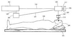
- FIG. 2is a block schematic diagram of a type of system for delivering a laser beam shot pattern to the lens of an eye according to the teachings of the present invention.
- FIG. 2Ais a block schematic diagram of illustrative components forming a portion of a system for delivering a laser beam shot pattern to the lens of an eye according to the teachings of the present invention.
- FIG. 3is a diagram of the anterior surface of a lens normal to the AP axis illustrating a laser shot pattern having a flower like shape which has a contour generally following approximately the last 15% of the fiber length from the end of the fiber.
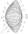
- FIGS. 4A , 4 B, 4 C, 4 D and 4 Eare diagrams representing elevation views of the geometry used for the development of laser shot patterns based upon the structure of the fetal nucleus (three suture branch nucleus) as it is rotated from the posterior view 4 A through and to the anterior view 4 E.
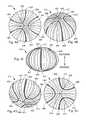
- FIGS. 5A , 5 B, and 5 Care diagrams representing posterior, side and anterior elevation views, respectively, of the geometry used for the development of laser shot patterns based upon the structure of the infantile nucleus (six suture branch nucleus).
- FIGS. 6A , 6 B and 6 Care diagrams representing posterior, side and anterior elevation views, respectively of the geometry used for the development of laser shot patterns based upon the structure of the adolescent nucleus (nine suture branch nucleus).
- FIGS. 7A , 7 B and 7 Care diagrams representing posterior, side and anterior elevation views, respectively of the geometry used for the development of laser shot patterns based upon the structure of the an adult nucleus ( 12 suture branch).
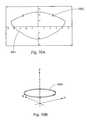
- FIGS. 8 and 8Aare perspective cutout views of an adult lens representing the placement of essentially concentric shells in accordance with the teachings of the present invention.
- FIG. 9is a cross-section drawing of the lens relating to the model developed by Burd.
- FIG. 10is a cross-section drawing of a lens based upon the model developed by Burd.
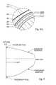
- FIG. 11is a cross-section drawing of a lens based upon the model developed by Burd.
- FIG. 12is a cross-section drawing of a lens based upon the model developed by Burd.
- FIG. 13is a cross-section drawing of a lens showing the placement of a shell laser shot pattern in accordance with the teachings of the present invention.
- FIG. 14is a cross-section drawing of a lens showing the placement of a shell laser shot pattern in accordance with the teachings of the present invention.
- FIG. 15is a cross-section drawing of a lens showing the placement of a partial shell laser shot pattern in accordance with the teachings of the present invention.
- FIG. 16is a cross-section drawing of a lens showing the placement of a partial shell laser shot pattern in accordance with the teachings of the present invention.
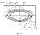
- FIG. 17is a cross-section drawing of a lens showing the placement of a shell laser shot pattern in accordance with the teachings of the present invention.
- FIGS. 18-24are cross-section drawings of a lens showing the placement of a volumetric removal laser shot patterns in accordance with the teachings of the present invention.
- FIG. 25is a cross-section drawing of a lens showing the placement of a cube laser shot pattern in accordance with the teachings of the present invention.
- FIGS. 26-27are cross-section drawings of a lens showing the placement of a gradient index modification laser shot patterns in accordance with the teachings of the present invention.
- FIGS. 28 A, C and Ediagrams depicting laser suture cut shot patterns on the anterior portion of a lens of the present invention.
- FIGS. 28 B, D, and Fare diagrams illustrating the placement of the shot patterns of FIGS. 28 A, C, and E respectively.
- FIG. 29is a diagram illustrating the relative placement of the shot patterns of FIGS. 28 A, C, and E, if performed in the same lens.
- FIGS. 30 A-Dare diagrams of the cross-section of a lens illustrating a capsulorhexis shot pattern of the present invention.
- FIGS. 31 A-Dare diagrams illustrating youthful vs old age gradient index behavior.
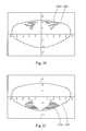
- FIG. 32is a diagram illustrating a primarily horizontal sectional shot pattern.
- FIG. 33is a diagram illustrating a primarily vertical sectional shot pattern.
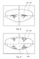
- FIG. 34is diagram illustrating a sectional shot pattern.
- FIG. 35is diagram illustrating a sectional shot pattern.
- FIG. 36is diagram illustrating a sectional shot pattern.
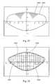
- FIG. 38is diagram illustrating a sectional shot pattern.
- FIG. 39is diagram illustrating a sectional shot pattern.
- FIG. 40is diagram illustrating a sectional shot pattern.
- FIG. 41is a diagram illustrating a primarily vertical sectional shot pattern.
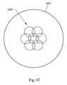
- FIG. 42is a diagram illustrating an overlapping vertical sectional shot pattern.
- the present inventionprovides a system and method for increasing the amplitude of accommodation and/or changing the refractive power and/or enabling the removal of the clear or cataractous lens material of a natural crystalline lens.
- a system for delivering a laser beam shot pattern to the lens of an eyecomprising: a patient support 201 ; a laser 202 ; optics for delivering the laser beam 203 ; a control system for delivering the laser beam to the lens in a particular pattern 204 , which control system 204 is associated with and/or interfaces with the other components of the system as represented by lines 205 ; a means for determining the position of lens with respect to the laser 206 , which means 206 receives an image 211 of the lens of the eye; and a laser patient interface 207 .
- the patient support 201positions the patent's body 208 and head 209 to interface with the optics for delivering the laser beam 203 .
- the laser 202should provide a beam 210 that is of a wavelength that transmits through the cornea, aqueous and lens.
- the beamshould be of a short pulse width, together with the energy and beam size, to produce photodisruption.
- laser shot or shotrefers to a laser beam pulse delivered to a location that results in photodisruption.
- photodisruptionessentially refers to the conversion of matter to a gas by the laser.
- wavelengths of about 300 nm to 2500 nmmay be employed. Pulse widths from about 1 femtosecond to 100 picoseconds may be employed.
- the pulse rate(also referred to as pulse repetition frequency (PRF) and pulses per second measured in Hertz) may be from about 1 KHz to several GHz.
- PRFpulse repetition frequency
- lower pulse ratescorrespond to higher pulse energy in commercial laser devices.
- a wide variety of laser typesmay be used to cause photodisruption of ocular tissues, dependent upon pulse width and energy density. Thus, examples of such lasers would include: the Delmar Photonics Inc.
- Trestles-20which is a Titanium Sapphire (Ti:Sapphire) oscillator having a wavelength range of 780 to 840 nm, less than a 20 femtosecond pulse width, about 100 MHz PRF, with 2.5 nanojoules; the Clark CPA-2161, which is an amplified Ti:Sapphire having a wavelength of 775 nm, less than a 150 femtosecond pulse width, about 3 KHz PRF, with 850 microjoules; the IMRA FCPA (fiber chirped pulse amplification) ⁇ Jewel D series D-400-HR, which is a Yb:fiber oscillator/amplifier having a wavelength of 1045 nm, less than a 1 picosecond pulse width, about 5 MHz PRF, with 100 nanojoules; the Lumera Staccato, which is a Nd:YVO4 having a wavelength of 1064 nm, about 10 picosecond pulse width, about 100 KHz PRF, with 100 microjoules; the Lum
- the optics for delivering the laser beam 203 to the natural lens of the eyeshould be capable of providing a series of shots to the natural lens in a precise and predetermined pattern in the x, y and z dimension.
- the opticsshould also provide a predetermined beam spot size to cause photodisruption with the laser energy reaching the natural lens.
- the opticsmay include, without limitation: an x y scanner; a z focusing device; and, focusing optics.
- the focusing opticsmay be conventional focusing optics, and/or flat field optics and/or telecentric optics, each having corresponding computer controlled focusing, such that calibration in x, y, z dimensions is achieved.
- an x y scannermay be a pair of closed loop galvanometers with position detector feedback.
- Examples of such x y scannerswould be the Cambridge Technology Inc. Model 6450, the SCANLAB hurrySCAN and the AGRES Rhino Scanner.
- Examples of such z focusing deviceswould be the Phsyik International Peizo focus unit Model ESee Z focus control and the SCANLAB varrioSCAN.
- control system for delivering the laser beam 204may be any computer, controller, and/or software hardware combination that is capable of selecting and controlling x y z scanning parameters and laser firing. These components may typically be associated at least in part with circuit boards that interface to the x y scanner, the z focusing device and/or the laser.
- the control systemmay also, but does not necessarily, have the further capabilities of controlling the other components of the system as well as maintaining data, obtaining data and performing calculations.
- the control systemmay contain the programs that direct the laser through one or more laser shot patterns.
- the means for determining the position of the lens with respect to the laser 206should be capable of determining the relative distance with respect to the laser and portions of the lens, which distance is maintained constant by the patient interface 207 .
- this componentwill provide the ability to determine the position of the lens with respect to the scanning coordinates in all three dimensions. This may be accomplished by several methods and apparatus. For example, x y centration of the lens may be accomplished by observing the lens through a co-boresighed camera system and display or by using direct view optics and then manually positioning the patients' eye to a known center.
- the z positionmay then be determined by a range measurement device utilizing optical triangulation or laser and ccd system, such as the Micro-Epsilon opto NCDT 1401 laser sensor and/or the Aculux Laser Ranger LR2-22.
- a 3-dimensional viewing and measurement apparatusmay also be used to determine the x, y and z positions of the lens.
- the Hawk 3 axis non-contact measurement system from Vision Engineeringcould be used to make these determinations.
- an apparatus that can be used to determine the position of the lensis a 3-dimension measurement apparatus. This apparatus would comprise a camera, which can view a reference and the natural lens, and would also include a light source to illuminate the natural lens. Such light source could be a structured light source, such as for example a slit, illumination designed to generate 3-dimensional information based upon geometry.
- a further component of the systemis the laser patient interface 207 .
- This interfaceshould provide that the x, y, z position between the natural lens and the laser remains fixed during the procedure, which includes both the measurement steps of determining the x y z position and the delivery step of delivering the laser to the lens in a shot pattern.
- the interface devicemay contain an optically transparent applanator.
- One example of this interfaceis a suction ring applanator that is fixed against the outer surface of the eye and is then positioned against the laser optical housing, thus fixing the distance between the laiser, the eye and the natural lens.
- the reference marks for the 3-dimensional viewing and measuring apparatusmay also be placed on this applanator.
- a further example of a laser patient interfaceis a device having a lower ring, which has suction capability for affixing the interface to the eye.
- the interfacefurther has a flat bottom, which presses against the eye flattening the eye's shape.
- This flat bottomis constructed of material that transmits the laser beam and also preferably, although not necessarily, transmits optical images of the eye within the visible light spectrum.
- the upper ringhas a structure for engaging with the housing for the laser optics and/or some structure that is of known distance from the laser along the path of the laser beam and fixed with respect to the laser.
- the flat bottomfurther has a reference, which consists of three reference marks. Although three marks are provided in this example to make up the reference, the reference may consist of only a single mark or several marks.
- the interfacemay be a corneal shaped transparent element whereby the cornea is put into direct contact with the interface or contains an interface fluid between.
- FIG. 2Ais a more detailed schematic diagram of a configuration of the system of FIG. 2 .
- the example of FIG. 2Aprovides a laser 202 , laser optics for delivering the laser beam 203 , which optics comprise a beam expander telescope 220 , a z focus mechanism 221 , a beam combiner 222 , an x y scanner 223 , and focusing optics 224 .
- opticscomprise a beam expander telescope 220 , a z focus mechanism 221 , a beam combiner 222 , an x y scanner 223 , and focusing optics 224 .
- FIG. 2Ais a more detailed schematic diagram of a configuration of the system of FIG. 2 .
- FIG. 2Aprovides a laser 202 , laser optics for delivering the laser beam 203 , which optics comprise a beam expander telescope 220 , a z focus mechanism 221 , a beam combiner 222 , an x y scanner 223
- 2Arelay optics 230 , camera optics with zoom and focus 231 , and a ccd camera 232 , which components form a part of a three-dimensional viewing and measuring apparatus. Moreover, these components 230 , 231 and 232 in combination with a light source 233 , the reference mark 212 and the scanner 223 function as a means for determining the position of the lens 206 .
- FIG. 2Autilizes the x y scanner 223 to create stereoscopic images of the lens with only a single ccd camera 232 .
- Optical images 211 of the eye 213 and in particular optical images of the natural lens 103 of the eye 213are conveyed along a path 211 .
- This path 211follows the same path as the laser beam 210 from the natural lens 103 through the laser patient interface 207 , the focusing optics 224 , the x y scanner 223 and the beam combiner 222 .
- 2Afurther comprises: a laser patient interface 207 , with a reference mark 212 ; and a light source 233 , which could be for example uniform illumination, a slit illumination, or other structured light source designed to enhance 3-dimensional accuracy.
- the light sourcein part, provides illumination of the natural lens of the patient's eye for the purposes of determining the 3-dimensional position of the lens.
- a controller and/or computernot shown in FIG. 2A ) for further processing and use in determining 3-dimensional positions of the lens.
- Stereo imagesmay be generated by commanding the scanner to go to and pause at a nominal left position and then electronically trigger the camera and controller to capture and store the left image; then command the scanner/camera/controller similarly to capture and store right image. This sequence may be repeated in a periodic manner.
- These left and right imagescan be processed by the controller to generate the position and shape of the lens.
- the left and right imagescan be displayed using a stereo video monitor.
- Camera images or stereo imagesmay also be used to measure suture geometry and orientation in the patients lens, which can be used to determine the parameters of suture based shot patterns and to align suture based shot patterns to the patients lens suture geometry and orientation.
- the combination illustrated in FIG. 2Aprovides 3-dimensional information that can be used to determine the shape of the lens, including the anterior and posterior surfaces thereof. This information can also be used to visualize the structure of the lens, including sutures. Moreover, the information about the lens obtained from the combination of FIG. 2A can further be used in determining the laser shot pattern and laser shot placement with respect to lens shape and
- FIG. 2 and FIG. 2Aare block schematic diagrams and thus the relative positions and spacing of the components illustrated therein are by way of example. Accordingly, the relative placements of these components with respect to one another may be varied and all or some of their functions and components may be combined.
- FIGS. 4A-Eillustrate the three branched or Y suture geometry in the context of the structures found in the fetal nucleus 415 of the lens.
- these figuresprovide a more detailed view of the structures illustrated as layer 130 , which encompasses layer 122 of FIG. 1A .
- the view of the inner layer of the lensis rotated stepwise from the posterior side FIG. 4A to the anterior side FIG. 4E of the lens.
- this layer of the lenshas three posterior suture lines 401 , 402 , and 403 .
- This layeralso has three anterior suture lines 412 , 413 and 414 .
- the anterior suture linesare longer than the posterior suture lines and these lines are staggered when viewed along the anterior to posterior (AP) axis 411 .
- the lens fibers, which form the layers of the nucleusare shown by lines 404 , it being understood that these are only illustrative lines and that in the actual natural layer of the lens there would be many times more fibers present.
- To aid in illustrating the structure and geometry of this layer of the nucleus representative fibers 405 , 406 , 407 , 408 , 409 and 410have been exaggerated and individually shaded in FIGS. 4 A-E.
- FIGS. 4 A-EThus, as the view of the lens nucleus is rotated from posterior to anterior the positions of these representative fibers, there relationship to each other, and there relationship to the suture lines is illustrated.
- the length of the suture lines for the anterior sideare approximately 75% of the equatorial radius of the layer or shell in which they are found.
- the length of the suture lines for the posterior sideare approximately 85% of the length of the corresponding anterior sutures, i.e, 64% of the equatorial radius of that shell.
- the fetal nucleusis a biconvex shape.
- the anterior and posterior sides of the lenshave different curvatures, with the anterior being flatter. These curvatures generally follow the curvature of the cortex and the outer layer and general shape of the lens.
- the lenscan be viewed as a stratified structure consisting of long crescent fiber cells arranged end to end to form essentially concentric or nested shells.
- the present inventionutilizes this and the further addressed geometry, structure and positioning of the lens layers, fibers and suture lines to provide laser shot patterns for increasing the accommodative amplitude of the lens.
- itis the structure, positioning and geometry of the lens and lens fibers, in contrast to the material properties of the lens and lens fibers, that gives rise to loss of accommodative amplitude.
- these patternsare designed to alter and affect that structure, positioning and/or geometry to increase accommodative amplitude.
- FIGS. 5A-Cillustrate the six branched or star suture geometry in the context of the structure found in the infantile layer of the nucleus 515 of the lens.
- FIGS. 5A-Cprovide a more detailed view of the structures illustrated as layer 124 of FIG. 1A .
- the view of the layer of the lensis rotated from the posterior side FIG. 5A to a side view FIG. 5B to the anterior side FIG. 5C .
- this layer of the nucleushas six posterior suture lines 501 , 502 , 503 , 504 , 505 , and 506 .
- This layer of the nucleusalso has six anterior suture lines 509 , 510 , 511 , 512 , 513 , and 514 .
- the anterior suture linesare longer than the posterior suture lines and these lines are staggered when viewed along the AP axis 508 .
- the lens fibers, which form the layers of the nucleus,are shown by lines 507 , it being understood that these are only illustrative lines and that in the actual natural layer of the lens there would be many times more fibers present.
- the shape of the outer surface of the lensessentially follows the infantile nucleus 515 , which is a biconvex shape.
- the anterior and posterior sides of this layer of the lenshave different curvatures, with the anterior being flatter.
- These curvaturesgenerally follow the curvature of the cortex and the outer layer and general shape of the lens.
- These curvaturesalso generally follow the curvature of the fetal nucleus 415 .
- the lenscan be viewed as a stratified structure consisting of long crescent fiber cells arranged end to end to form essentially concentric or nested shells, with the infantile nucleus 515 having the fetal nucleus 415 nested within it.
- additional fiber layersgrow containing between 6 and 9 sutures.
- FIGS. 6A-Cillustrate the nine branched or star suture geometry in the context of the structure found in the adolescent layer of the nucleus 611 of the lens.
- FIGS. 6A-Cprovide a more detailed view of the structures illustrated as layer 126 of FIG. 1A .
- the view of the layer of the lensis rotated from the posterior side FIG. 6A to a side view FIG. 6B to the anterior side FIG. 6C .
- this layer of the nucleushas nine posterior suture lines 601 , 602 , 603 , 604 , 605 , 606 , 607 , 608 and 609 .
- This layer of the nucleusalso has nine anterior suture lines 612 , 613 , 614 , 615 , 616 , 617 , 618 , 619 and 620 .
- the anterior suture linesare longer than the posterior suture lines and these lines are staggered when viewed along the AP axis 610 .
- the lens fibers, which form the layers of the nucleus,are shown by lines 621 ; it being understood that these are only illustrative lines, and that in the actual natural layer of the lens there would be many times more fibers present.
- the outer surface of the corneafollows the adolescent nucleus 611 , which is a biconvex shape.
- the anterior and posterior sides of this layerhave different curvatures, with the anterior being flatter.
- These curvaturesgenerally follow the curvature of the cortex and the outer layer and general shape of the lens.
- These curvaturesalso generally follow the curvature of the fetal nucleus 415 and the infantile nucleus 515 , which are nested within the adolescent nucleus 611 .
- the lenscan be viewed as a stratified structure consisting of long crescent fiber cells arranged end to end to form essentially concentric or nested shells. As development continues through adulthood, additional fiber layers grow containing between 9 and 12 sutures.
- FIGS. 7A-Cillustrates the twelve branched or star suture geometry in the context of the structure found in the adult layer of the nucleus 713 of the lens.
- FIGS. 7A-Cprovide a more detailed view of the adult layer 128 depicted in FIG. 1A .
- the view of the layer of the lensis rotated from the posterior side FIG. 7A to a side view FIG. 7B to the anterior side FIG. 7C .
- the adult layer of the nucleushas twelve posterior suture lines 701 , 702 , 703 , 704 , 705 , 706 , 707 , 708 , 709 , 710 , 711 , and 712 .
- This layer of the nucleusalso has twelve anterior suture lines 714 - 725 .
- the anterior suture linesare longer than the posterior suture lines and these lines are staggered when viewed along the AP axis 726 .
- the lens fibers, which form the layers of the nucleus,are shown by lines 728 ; it being understood that these are only illustrative lines, and that in the actual natural layer of the lens there would be many times more fibers present.
- the adult nucleus 713is a biconvex shape that follows the outer surface of the lens.
- the anterior and posterior sides of this layerhave different curvatures, with the anterior being flatter.
- These curvaturesfollow the curvature of the cortex and the outer layer and shape of the lens.
- These curvaturesalso generally follow the curvature of the adolescent nucleus 611 , the infantile nucleus 515 and the fetal nucleus 415 and the embryonic nucleus, which are essentially concentric to and nested within the adult nucleus 611 .
- the lenscan be viewed as a stratified structure consisting of long crescent fiber cells arranged end to end to form essentially concentric or nested shells.
- a subsequent adult layer having 15 suturesmay also be present in some individuals after age 40.
- This subsequent adult layerwould be similar to the later adult layer 713 in general structure, with the recognition that the subsequent adult layer would have a geometry having more sutures and would encompass the later adult layer 713 ; and as such, the subsequent adult layer would be the outermost layer of the nucleus and would thus be the layer further from the center of the nucleus and the layer that is youngest in age.
- the present inventionprovides for the delivery of the laser beam in patterns that utilize, or are based at least in part on, the lens suture geometry and/or the curvature of the lens and/or the various layers within the nucleus; and/or the curvatures of the various layers within the nucleus; and/or the suture geometry of the various layers within the nucleus.
- the concept of matching the curvature of the anterior ablations to the specific curvature of the anterior capsule, while having a different curvature for posterior ablations, which in turn match the posterior curvature of the lensis provided.
- Anterior and posterior curvaturescan be based on Kuszak aged lens models, Burd's numeric modeling, Burd et al.
- these laser delivery patternsare based in whole and/or in part on the mathematical modeling and actual observation data regarding the shape of the lens, the shape of the layers of the lens, the suture pattern, and the position of the sutures and/or the geometry of the sutures.
- the present inventionseeks to generally emulate the natural lens geometry, structures and positioning and/or portions thereof, as well as build upon, modify and reposition such naturally occurring parameters through the use of the laser shot patterns described herein.
- laser beam delivery patternsthat cut a series of essentially concentric, i.e., nested, shells in the lens may be employed.
- the shellswould essentially follow the anterior and posterior curvature of the lens.
- creating in the lensa series of cuts which resemble the nucleus layers of FIGS. 4 , 5 , 6 and 7 .
- These cutsmay follow the same geometry, i.e., shape and distance from the center, of these layers or may follow only a part of that geometry.
- the term shellrefers to the lens material and the term shell cut refers to the laser beam delivery pattern and consequently the placement of the laser beam shots in the lens in accordance with that pattern. More or less shell cuts, and thus shells may be utilized. Moreover, the cuts may be such that they in effect create a complete shell, i.e., the shell and shell cuts completely encompass a volume of lens material. The cuts may also be such that less than a complete shell is formed.
- partial shellsby the use of partial shell cuts, may be employed.
- Such partial cutswould for example be only a portion of a shell e.g., the anterior quartile, the anterior half, the posterior quartile, stacked annular rings, staggered annular rings, and/or combinations thereof.
- Such partial shells and shell cutsmay be any portion of a three dimensional form, including ellipsoid, spheroids and combinations thereof as those terms are used in their broadest sense that in general follows the contours of the lens, capsule, cortex, nucleus, and/or the layers of the lens including the layers of the nucleus.
- the use of complete and partial shells and shell cutsmay be used in a single lens.
- the first and second cuts 801 and 803are annular cuts, while the third cut is a complete cut.
- partial shellsare created, by use of partial pie shaped shell cuts. These cuts may be placed in between the suture lines at the various layers of the lens. These partial shells may follow the contour of the lens, i.e., have a curved shape, or they may be flatter and have a more planar shape or be flat.
- a further use of these pie shape shells and shell cutswould be to create these cuts in a suture like manner, but not following the natural suture placement in the lens.
- a suture like pattern of cutsis made in the lens, following the general geometry of the natural lens suture lines, but not their exact position in the lens.
- other shaped cutsmay be employed, such as by way of illustration a series of ellipses, rectangular planes or squares.
- partial shells and/or planar partial shellsare to create a series of overlapping staggered partial shells by using overlapping staggered partial shell cuts. In this way essentially complete and uninterrupted layers of lens material are disrupted creating planar like sections of the lens that can slide one atop the other to thus increase accommodative amplitude.
- These partial shellscan be located directly atop each other, when viewed along the AP axis, or they could be slightly staggered, completely staggered, or any combination thereof.
- linescan also be cut into the lens. These lines can follow the geometry and/or geometry and position of the various natural suture lines.
- a laser shot patternis provided that places shots in the geometry of one or more of the natural suture lines of one or more of the various natural layers of the lens as shown in FIGS. 4 , 5 , 6 , and 7 , as well as in the 15 suture line layer, or it may follow any of the other patterns in the continuum of layers in the lens.
- shot patternscan follow the general geometry of the natural suture lines, i.e., a series of star shapes with the number of legs in each star increasing as their placement moves away from the center of the lens.
- star shaped shot patternsmay follow the precise geometry of the natural suture patterns of the layers of the lens; or it can follow the exact geometry and placement of the sutures, at the same distances as found in the natural lens or as determined by modeling of the natural lens.
- one or more starsmay be cut.
- the length of the lines of the legs of the starmay be the longer, shorter or the same length as the natural suture lines.
- the lengthmay be placed toward the center of the star shape, i.e. the point where the lines join each other, or towards the end of the suture line, i.e., the point furthest on the suture line from the joining point.
- partial star shaped cutscan be used, such as cuts having a “V” shape, or vertical or horizontal or at an angle in between. These linear cuts, discussed above, are in general referred to herein as laser created suture lines. Moreover, laser created suture lines may be grouped together to in effect form a shell or partial shell.
- a shot pattern 301is provided to an anterior portion of a layer 302 of the lens.
- This shot pattern 301has a contour 303 that follows the contour of approximately the last 15% of fiber length of fibers, represented by lines 304 .
- the shell cutresembles the shape of a flower.
- the number of petals in the flower shaped shellshould correspond to the number of suture lines 305 at that growth layer.
- this partial shell cut and/or cutswill have the effect of unbinding the layers and returning the lens to a more youthful increased amplitude of accommodation.
- annular partial shells or planar partial shells in this general areai.e., the general area at or near the ends of the suture lines, may be employed for the same reasons.
- This theoryis put forward for the purposes of providing further teaching and to advancing the art. This theory, however, is not needed to practice the invention; and the invention and the claims herein are not bound by or restricted by or to this theory.
- laser created suture linesincluding star shaped patterns may also be used in conjunction with shells, partial shells and planar partial shells.
- a particular laser shot pattern, or series of shot patternsemploying elements of each of these shapes. These patterns may be based upon the geometry shown in FIGS. 4-7 as well as the 15 suture line geometry discussed herein; they may follow that geometry exactly, in whole or in part; and/or they may follow that geometry, in whole or in part, as well as following the position of that geometry in the lens.
- a maximum of 15 suture linesis known in the natural lens, more than 15 laser created suture lines may be employed.
- the lenshas multiple layers with a continuum of suture lines ranging from 3 to 15 and thus, this invention is not limited to the suture patents of FIGS. 4-7 , but instead covers any number of suture lines from 3 to 15, including fractions thereof.
- a shot pattern that cuts the lens into small cubes, which cubes can then be removed from the lens capsuleis provided.
- the cubescan range in size from a side having a length of about 100 ⁇ m to about 4 mm, with about 500 ⁇ m to 2 mm being a preferred size. Additionally, this invention is not limited to the formation of cubes and other volumetric shapes of similar general size may be employed.
- the laseris also used to create a small opening, capsulorhexis, in the lens anterior surface of the lens capsule for removal of the sectioned cubes. Thus, this procedure may be used to treat cataracts.
- This proceduremay also be used to remove a lens having opacification that has not progressed to the point of being cataractous.
- This proceduremay further be used to remove a natural lens that is clear, but which has lost its ability to accommodate.
- a suitable replacementsuch as an IOL, accommodative IOL, or synthetic lens refilling materials.
- the size and the shape of the capsulorhexisis variable and precisely controlled and preferably is in 2 mm or less diameter for lens refilling applications and about 5 mm for IOLs.
- a further implementation of the procedure to provide a capsulorhexisis to provide only a partially annular cut and thus leave a portion of the capsule attached to the lens creating a hinged flap like structure. Thus, this procedure may be used to treat cataracts.
- volumetric removal of the lenscan be performed to correct refractive errors in the eye, such as myopia, hyperopia and astigmatism.
- the laser shot patternis such that a selected volume and/or shape of lens material is removed by photodisruption from the lens. This removal has the affect of alternating the lens shape and thus reducing and/or correcting the refractive error.
- Volumetric removal of lens tissuecan be preformed in conjunction with the various shot patterns provided for increasing accommodative amplitude. In this manner both presbyopia and refractive error can be addressed by the same shot pattern and/or series of shot patterns.
- the volumetric removal of lens tissuefinds further application in enhancing corrective errors for patients that have had prior corneal laser visions correction, such as LASIK, and/or who have corneas that are too thin or weak to have laser corneal surgery.
- the laser shot patternsgenerally follow the shape of the lens and placement of individual shots with respect to adjacent shots in the pattern are sufficiently close enough to each other, such that when the pattern is complete a sufficiently continuous layer and/or line and/or volume of lens material has been removed; resulting in a structural change affecting accommodative amplitude and/or refractive error and/or the removal of lens material from the capsule.
- Shot spacing of lesser or greater distancesare contemplated herein and including overlap as necessary to obtain the desired results. Shot spacing considerations include gas bubble dissipation, volume removal efficiency, sequencing efficiency, scanner performance, and cleaving efficiency among others.
- the term cleavingmeans to substantially separate the tissue.
- the forgoing shot spacing considerationsare interrelated to a lesser or greater extent and one of skill in the art will know how to evaluate these conditions based upon the teachings of the present disclosure to accomplish the objectives herein.
- the placement of individual shots with respect to adjacent shots in the patternmay in general be such that they are as close as possible, typically limited by the size and time frame of photodisruption physics, which would include among other things gas bubble expansion of the previous shot.
- the time frame of photodisruptive physicsreferrers to the effects that take place surrounding photodisruption, such as plasma formation and expansion, shock waive propagation, and gas bubble expansion and contraction.
- the timing of sequential pulsessuch that they are timed faster than some of, elements of, or all of those effects, can increase volumetric removal and/or cleaving efficiency. Accordingly, we propose using pulse repetition frequencies from 50 MHz to 5 GHz., which could be accomplished by a laser with the following parameters: a mode lock laser of cavity length from 3 meters to 3 cm. Such high PRF lasers can more easily produce multiple pulses overlapping a location allowing for a lower energy per pulse to achieve photodisruption.
- first, second, third, etc.are relative terms and must be viewed in the context in which they are used. They do not relate to timing, unless specifically referred to as such. Thus, a first cut may be made after a second cut.
- cataractsit may be advantageous to shoot from anterior to posterior, because of the inability of the laser to penetrate substantially beyond the cataract.
- Burd modelingwhich model is set forth in Burd et al., Numerical modeling of the accommodating lens, Visions Research 42 (2002) 2235-2251.
- variables Z and Rare defined by the drawing FIG. 9 .
- FIGS. 10 , 11 and 12provide cross sectional views of the lens having an outer surface 1001 , 1101 , 1201 for three ages, 18, 29 and 45-year old respectively, based upon the Burd model and show growth in size along with shape changes with age.
- the units for the axes on these drawings, as well as for FIGS. 13 to 29are in millimeters (mm).
- EXAMPLE 1provides for making nested, lens shaped shell cuts.
- the laser shot patternsare illustrated in FIG. 13 , which provides the outer surface 1301 of a 45-year old lens based upon the Burd model.
- a series of nested or essentially concentric shells and shell cutswhich essentially follow the shape of the lens.
- a first shell cut 1302there is provided a first shell cut 1302 , a second shell cut 1304 , and a third shell cut 1306 .
- These shell cutsform a first shell 1303 and a second shell 1305 .
- Shells or partial shellsare designed to increase flexibility in the lens by decreasing the strength of nested fiber layers by separating the bound layers, which it is theorized would reduce the structural strength and increase deflection for a given load or force.
- the shell cuts in this exampleare positioned approximately such that the third shell cut 1306 is where 3 suture branches begin forming additional branches, or approximately 6 mm lens equatorial diameter, at the boundary of the fetal nucleus, or the lens at birth; the second shell cut 1304 is where the 6 suture branch layer begins forming additional branches at approximately 7.2 mm diameter, or the infantile nucleus or the lens at approximately age 3; and the first shell cut is where the 9 suture branch begins forming additional branches at approximately 9 mm diameter, or at the adolescent nucleus at approximately age 13.
- EXAMPLE 2provides as an alternative to using a 45-year old lens shape from the Burd model, the actual patient lens structural or shape data may be utilized to customize surgery for each patient.
- a 45-year old human cadaver lenswhose shape was measured optically and mathematically fit via the same fifth order function used in the Burd model, yields coefficients unique to the measured lens.
- the outer cross-section shape of this lens and a shot pattern similar to that of Example 1, but which was tailored to the particular lens of this Exampleis illustrated in FIG. 14 .
- an outer surface 1401 of the 45-year old lensThere is further provided a series of nested or essentially concentric shells and shell cuts.
- first shell cut 1402a first shell cut 1402 , a second shell cut 1404 , and a third shell cut 1406 .
- These shell cutsform a first shell 1403 and a second shell 1405 .
- any of the exemplary cuts and shot patternscan be implemented via partial or full shells and/or can be implemented via modeled (the Burd model being just one example) or measured lens data.
- EXAMPLE 3provides a shot pattern for cutting partial shells on the measured 45-year old lens, and having an excluded defined central zone.
- an outer surface 1501 of a 45-year old lensa central zone 1512 , partial cuts 1502 , 1504 , 1506 , 1507 , 1509 and 1511 .
- These partial cuts as shownare part of the same generally annularly shaped.
- cuts 1502 and 1507 , cuts 1504 and 1509 , and cuts 1506 and 1511are the opposite sides respectively of three generally annularly shaped partial.
- EXAMPLE 4provides a shot pattern for cutting partial shells on the measured 45-year old lens, and having both an excluded defined peripheral zone and central zone.
- an outer surface 1601 of a 45-year old lensthere is provided an outer surface 1601 of a 45-year old lens, a central zone 1622 and two peripheral zones 1620 and 1621 .
- partial cuts 1602 , 1604 , 1605 , 1606 , 1607 , 1611 , 1613 , 1615 , 1617 , and 1618as well as, partial shells 1603 , 1608 , 1609 , 1610 , 1612 , 1614 , 1616 and 1619 .
- partial cuts 1603 , 1608 , 1609 , 1610 , 1612 , 1614 , 1616 and 1619As with example 3 and FIG.
- these cutsare viewed in cross section and thus it is understood that they are opposite sides of generally annular ring shaped cuts, which essentially follow the shape of the lens and which encompasses the central zone 1622 . There are thus 5 partial cuts depicted in FIG. 16 .
- EXAMPLE 5provides a laser shot pattern for a finer detailed cutting of the lens to approximate the structural boundaries at 3 , 4 , 5 , 6 , 7 , 8 , 9 suture branches, or the use of six shells.
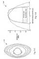
- FIG. 17seven essentially concentric shot patterns 1702 - 1708 , which create seven corresponding shell cuts and which also create six corresponding shells 1709 - 1714 .
- the outer surface 1701 of a 45-year old lens as measuredis also provided in FIG. 17 . While this example provides for the creation of six shells, it is understood that the lens contains thousands of fiber layers and that it may be desirable to utilize much greater than six shells and up to hundreds or even thousands, depending on the resolution of the laser deliver system and laser beam parameters.
- Examples 6-12relate to the volumetric removal of lens material in a predetermined shape, based upon a precise shot pattern. Thus, these examples illustrate how refractive change by shaped volumetric reduction may be accomplished.
- This approachrecognizes a limitation of photodisruption laser beam delivery, i.e., that the gas bubbles created are considerably larger then the resultant material void found after all gas bubble dissipation occurs. This can have the effect of causing material voids to be spaced further apart than ideal for high efficiency volume removal.
- the closest spacing attainabledepending on detailed laser spot size, energy and pulse width, may provide a low, net volumetric removal efficiency, which is the ratio of achieved volume removal to the volume of material treated.
- a simple exampleconsiders a void size equal to the spacing between voids yielding a nominal 50% linear efficiency, which from symmetric geometry has a 25% area efficiency and a corresponding 12.5% volumetric efficiency of void creation.
- an approachis provided whereby the treatment shaped volume is proportionally larger than desired shaped volume removal to compensate for the low volume efficiency.
- void shapeasymmetries, void location, tissue compliance as a function of age, external forces and more, may effect the final volume efficiency and experimental validation of volumetric efficiency may be required.
- EXAMPLE 6provides a shot pattern and volume removal to make a negative refractive change, or reduce the power in the crystalline lens by 3 Diopters, using the Gullstrand-LaGrand optical model, which would require the removal of approximately 180 um centrally tapering to 0 over a 3 mm radius.
- FIG. 18there is provided an outer lens surface 1801 and a shot pattern 1802 for the desired volume removal.
- the shot patternwould have to remove essentially 100% of the shaded region volume which is extremely difficult due to low volume efficiency found in photodisruption laser beam delivery.
- EXAMPLE 7is based upon dealing with low volume removal efficiency and in this example the assumption that we have a volumetric efficiency of 12.5% or 1 ⁇ 8 th we would treat an 8 times larger volume or 1.44 mm thick to compensate for the low volume efficiency, tapering to 0 over the same 3 mm as shown in FIG. 19 , which illustrates a lens outer surface 1901 and a shot pattern 1902 .
- the shape of the shot patternis based upon and essentially follows the shape of the outer surface 1901 of the lens.
- EXAMPLE 8provides a shot pattern to cause a refractive change to increase lens power or reduce hyperopia in patients, where the shot pattern is primarily implemented in the anterior region of the lens. This pattern is illustrated in FIG. 20 , which provides an outer surface 2001 and thus shape of the lens and a shot pattern 2002 .
- EXAMPLE 9provides a shot pattern to cause a refractive change to increase lens power or reduce hyperopia in patients, where the algorithm is primarily implemented in the posterior region of the lens.
- This patternis illustrated in FIG. 21 , which provides an outer surface 2101 and thus shape of the lens and a shot pattern 2102 .
- This examplefurther illustrates a shot pattern having a shape is modified to primarily follow the posterior curve of the lens.
- EXAMPLE 10provides a shot pattern to cause a refractive change to increase lens power or reduce hyperopia in patients, where the shot pattern is primarily implemented in the central region of the lens.
- the shot patternis primarily implemented in the central region of the lens.
- an outer surface 2201 of the lens and a shot pattern 2202which provides a volumetric shape.
- the anterior shape of the lens or posterior shape of the lens or bothcan be utilized to determine the shape of the shot pattern and/or volumetric shape.
- EXAMPLE 11provides two volumetric shot patterns that follow the shape of the lens surface to which they are adjacent.
- the volumetric shapes to be removed from the lensare located in the anterior and posterior regions of the lens and have a surface that follows the anterior and posterior shape of the lens respectively.
- EXAMPLE 12illustrates a manner in which different shot pattern features are combined to address both refractive errors and those to increase flexibility utilizing a plurality of stacked partial shells, which are partially overlapping.
- the placement of the partial shell cutsare adjacent the anterior surface of the lens as shown it FIG. 24 .
- the partial shell cutsmay similarly be placed adjacent the posterior surface of the lens, in which case they should follow the shape of that surface.
- more effective cleavingis obtained.
- the shot pattern in the figures associated with EXAMPLES 6, 7, 8, 9, 10 and 11are shown to cut horizontal partial planes whose extent is defined by a refractive shape. It is to be understood that as an alternative to horizontal planes, vertical partial planes or other orientation cuts whose extent is defined by the refractive shape may be used.
- Examples 13 and 14are directed towards methods and shot patterns for treating and removal of cataracts and/or for clear lens extractions.
- a method for the structural modification of the lens materialto make it easier to remove while potentially increasing the safety of the procedure by eliminating the high frequency ultrasonic energy used in Phaco emulsification today.
- the use of photodisruption cutting in a specific shape patternsis utilized to carve up the lens material into tiny cube like structures small enough to be aspirated away with 1 to 2 mm sized aspiration needles.
- EXAMPLE 13provides a shot pattern to create 0.5 mm sized cubes out of the lens material following the structural shape of a 45-year old Burd Model lens. It is preferred that the patient's actual lens shape can be measured and used.
- an outer surface 2501and thus an outer shape of the lens.
- a shot pattern 2502that creates grid like cuts, the end of which cuts 2503 essentially follows the shape of the lens.
- one shell cut 2504which is integral with the grid like cuts.
- the sequence of laser shots in the pattern in FIG. 25may be executed from posterior to anterior, as in most of the patterns disclosed herein, to obtain more predictable results by reducing the variation caused by shooting through gas bubbles.
- cataractsmay be desirable to shoot cataracts from the anterior to the posterior for the purpose of choosing the lesser of two undesirable effects.
- itis further provided to essentially drill down anterior to posterior, which we call the z axis throughout this document and then move in x/y and drill down again.
- EXAMPLE 14provides for a clear lens extraction.
- the shot pattern of FIG. 25is applied to a clear lens and that lens material is subsequently removed.
- shooting from posterior to anterioris desirable.
- EXAMPLE 15provides for a precision capsulorhexis.
- the creation of precise capsulorhexis for the surgeon to access the lens to remove the lens materialis provided.
- an outer surface 3001and thus an outer shape of the lens.
- a ring shaped band shape cut 3002 and shot patternare provided.
- the figureshows the cross section view of this ring shaped annular band and accordingly provides for two sides 3002 of the ring.
- the ring shaped capsulorhexiscuts of 100 ⁇ m deep, approximately centered on the anterior lens capsule surface and precisely 5 mm in diameter.
- the lens capsuleis approximately 5 to 15 ⁇ m thick, it is desirable for the depth of the cut to be typically between 5 and several hundred um, although there is not much penalty for cutting several millimeters.
- This diametercan be varied between 0.1 mm to 9 mm diameter and the capsulorhexis can be elliptical with the x axis different then the y axis or other shapes.
- a particular IOLmay benefit from and/or may require a particular capsulorhexis shape.
- Examples 16 to 17relate to gradient index modification of the lens.
- Moffat, Atchison and Pope, Vision Research 42 (2002) 1683-1693showed that the natural crystalline lens contains a gradient index of refraction behavior that follows the lens shells structure and dramatically contributes to overall lens power. They also showed that this gradient substantially diminishes, or flattens as the lens ages reducing the optical power of the lens.
- the loss of gradient index with agemost likely explains the so-called Lens Paradox, which presents the conundrum that the ageing lens is known to grow to a steeper curvature shape that should result in higher power, yet the aging lens has similar power to the youthful lens. Essentially it is postulated that the increase in power due to shape changes is offset by the power loss from gradient index loss.
- FIG. 31Examples of the youthful vs old age gradient index behavior is shown in FIG. 31 , which provides data taken from the more recent work from the same group Jones, Atchison, Meder and Pope, Vision Research 45 (2005) 2352-236.
- the old lens 3101has a flat index behavior radially 3102 and the young lens 3103 has continuously diminishing index radially 3104 from approximately 1.42 in the center to 1.38 nearer the outer shells of the lens.
- the photodisruptive laserin the creation of small voids within the lens fiber material which will then fill-in with aqueous humor fluid which has a lower index of refraction and, via area weighting or volume weighting, decrease the net refractive index of a particular region. Accordingly, if different void densities are placed in nested shell volumes, then this would diminish the average index of refraction of essentially concentric regions in a similar manner to the youthful lens.
- EXAMPLE 16provides a gradient index modification, which has different void densities placed in nested volumes, as shown in FIG. 26 .
- a series of nested shot patterns 2602 and a lens outer surface 2601with each pattern creating an incrementally different void density in the lens material. For example, if a nominal 25% weighting efficiency was obtained in the most densely treated region, filling that volume with 1.38 index of aqueous humor, and the remaining region that was 75% lens material of index 1.42, then the average resultant index of refraction would be 0.25*1.38+0.75*1.42 or 1.41, which we see from FIG. 31 , that would restore the gradient from the center to a 2 mm radius, which is most central optical region for visual function.
- FIG. 26shows a distributed regional treatment of increasing density from the center of the lens to the periphery of the lens.
- EXAMPLE 17provides a gradient index modification that is similar to example 16, except that the area where void density is changed is located further from the outer surface of the lens.
- This example and patternis illustrated in FIG. 27 .
- FIG. 27Thus there is provided a series of nested shot patterns 2702 and lens outer surface 2701 , with each pattern creating an incrementally different void density in the lens material.
- this figureshows a distributed regional shell treatment that is primarily confined to the nucleus.
- EXAMPLE 18provides for the cutting in relation to suture lines.
- suture linesouts along either modeled suture lines, according to Kuzak described suture locations as a function of shell geometry with age and shape, or measured suture lines may be used.
- the latterbeing provided by the measuring of patient lens sutures with a CCD camera and aligning suture cuts to the measured locations of suture lines.
- the brightest suture lines and or those with the widest spatial distributionlikely belong to the deepest layers, and perhaps the initial Y suture branches found in the fetal nucleus.
- FIGS. 28 & 29snows three different cutting patterns 2801 , 2802 , 2803 in the anterior portion of the lens that can be done separately or in combination.
- FIGS. 28 A, C & Eshows x-y cuts 2801 , 2802 , 2803 looking down at the anterior side of the lens.
- FIGS. 28 B, D, and Fare schematic representation to illustrate that the star shaped patterns follow the shape of the layer of the lens and do not show the actual cut.
- FIG. 29is the combination of the illustrations in FIGS. 28 B, D, and F to show their relative positions. It is understood that similar suture cuts can be made in the posterior following the posterior shell curves there, based again on either modeled geometry or measured lens data. There is yet further provided cutting 3, 4, 5, 6, 7, 8, 9, 10, 11, 12, 13, 14 and 15 branch sutures per Kuszak., cut separately or in any combination.
- Sectional patternsmay be employed. Such patterns would include the cube pattern of FIG. 25 , variations in the shape and size of this cube pattern, concentric cylinders, radial planes, horizontal planes and vertical planes, partial shells and shells, and combinations thereof. As used to describe these patterns, vertical refers to essentially parallel to the optical axis, i.e., the AP axis.
- These sectional patternsare employed within, or to comprise, a particular shaped volume, such as the shaped volumes in FIGS. 18-25 . Thus, these sectional patterns can be used in shaped volumes that provide for positive or negative refractive corrections. Further, these shaped patterns can be used in shaped volumes that result in shaped structural weakening, which causes shape change and results in a positive or negative refractive correction. Additionally, shaped structural weakening may also result in increased accommodative amplitude.
- these patternscan be employed in conjunction with each other, i.e., vertical and horizontal, or in isolation, i.e., only vertical or horizontal, at various locations in the lens, which locations can range from totally separate, to slightly overlapping, to overlapping. Additionally, by selectively arranging placement and density of these patterns and/or combination of primarily vertical and primarily horizontal patterns, local structure in the lens can be weakened by varying and predetermined amounts, which can result in selective flexibility and shape changes. Thus, through such selective placement and density determinations shaped structural weakening may be accomplished.
- These sectional patternsmay be employed using primarily vertical or primarily horizontal patterns.
- Primarily horizontal patterns 3201 to 3211are illustrated in FIG. 32 with respect to a lens having an outer surface 3212 .
- Primarily vertical patterns 3301 to 3312are illustrated in FIG. 33 with respect to a lens having an outer surface 3313 .
- Primarily vertical patternswhich include vertical cylinders and vertical planes, may provide more complete cleaving than essentially horizontal patterns due to the relative long depth of field of a photo disruption spot compared to the narrow width of the spot.
- Primarily horizontal patterns, such as horizontal planes and shell cuts near the center of the lens, i.e, polesmay provide lesser structural weakening due to less complete cleaving.
- primarily horizontal patterns, such as shells cut to the shape of the lenswill tend to preserve the overall shape of the lens, while still providing some structural weakening to improve flexibility.
- greater structural weakening with less regard to preserving initial shapemay be employed by providing primarily vertical patterns therein. Moreover still greater structural weakening with less regard to preserving initial shape may be employed by providing both primarily vertical and primarily horizontal patterns therein. Further, in determining the particular types of structural patterns to use, greater structural weakening with less regard to preserving initial shape may be employed within the center of the lens, such as the compacted fetal nucleus by providing primarily vertical patterns therein. Moreover still greater structural weakening with less regard to preserving initial shape may be employed within the center of the lens, such as the compacted fetal nucleus by providing both primarily vertical and primarily horizontal patterns therein.
- Optical performance and optical qualityare dependent upon the surface shape and quality of the lens.
- densities and placements of these patternsmay be employed.
- a combination of central patterns and peripheral patternsmay be utilized to maximize structural weakening and control of lens shape.
- patternscan be selected for placement in the center of the lens, such as the fetal and embryonic nucleus, which will result in maximum shaped structural weakening with minimal effect on lens surface shape changes, which surface effect is based essentially upon the placement of the pattern.
- peripheral lens areassuch as the infantile, adolescent and adult nucleus and cortex
- primarily horizontal patternsmay be selected such as to change the lens surface shape in a predetermined manner.
- Examples 19 to 27further illustrate this teaching and provide illustrative ways in which sectional patterns can be implemented to improve accommodative amplitude and/or refractive error.
- EXAMPLE 19provides a plurality of primarily horizontal patterns.
- shell cuts 3201 , 3202 and 3211that are placed in more peripheral lens areas, which are positioned to increase lens flexibility while minimizing the effect on lens shape.
- shell cuts 3203 , 3204 , 3205 , 3206 , 3207 , 3208 , 3209 , and 3210which are placed in more central lens areas, which are positioned to result in structural weakening with minimal effects on lens surface shapes.
- more or less of these types of cutsmay be utilized depending upon the desired structural weakening, shape change and increase in flexibility that is being sought.
- EXAMPLE 20as illustrated in FIG. 33 , provides a plurality of primarily vertical patterns.
- a series of cylindrical tubesplaced away from the central area of the lens, with opposing sides of the tubes being seen as pairs of cuts 3302 - 3303 , 3301 - 3304 , 3310 - 3311 , 3309 - 3312 .
- a series of tubesplaced in more central lens area, with opposing sides of the tubes being seen as pairs of cuts 3306 - 3307 , and 3305 - 3308 .
- FIG. 41there is provided a plurality of primarily vertical patterns in a lens having an outer surface 4113 .
- a series of concentric cylindrical tubeswith opposing sides of the tubes being seen as pairs of cuts 4101 - 4112 , 4102 - 4111 , 4103 - 4110 , 4104 - 4109 , 4105 - 08 , 4106 - 07 .
- the outer surface of the cylinders when viewed in cross sectioncan be arcuate, such as 4109 - 4112 , or straight, such as 4106 - 07 .
- the curvature of the cutsmay follow the curvature of the lens, such as 4101 - 12 ; or the curvature may be counter to the curvature of the lens, such as 4103 - 4110 .
- FIG. 42which is an equatorial view of a lens having an outer surface 4201 , there is provided a plurality of overlapping primarily vertical cylindrical shot patterns 4202 .
- cuts 3203 , 3204 , 3209 , 3210 , 3305 , 3306 , 3307 , and 3308are straight as see in cross section, they may be arcuate, such as cut 3201 . Similarly, the more arcuate cuts may be straighter. Additionally, the curvature of the cuts may follow the curvature of the lens, such as 3201 , 3202 , 3301 , and 3311 ; or the curvature may be counter to the curvature of the lens, such as 3205 , 3207 , 3304 , and 3310 .
- EXAMPLE 21as illustrated in FIG. 34 , provides within an outer lens surface 3401 a combination of primarily vertical and horizontal patterns in a tapered annular volume, which is show in cross section having opposite sides 3402 and 3403 .
- the density of the patternsincreases moving along the AP from anterior to posterior.
- This combination of sectional patternsprovides shaped structural weakening, where the higher density of the pattern provides greater structural weakening. This pattern will provide increased flexibility and increased refractive power.
- EXAMPLE 22provides within an outer lens surface 3501 a first primarily horizontal pattern 3502 - 3503 , which is in the form of an annular partial shell having opposite sides 3502 and 3503 shown in cross section; a second primarily horizontal pattern 3510 - 3511 , which is in the form of an annular partial shell having opposite sides 3510 and 3511 shown in cross section; and, a combination of partial vertical and partial horizontal patterns in a series of layers 3504 - 3505 , 3506 - 3507 , 3508 - 3509 , in an annular shaped volume, with opposite sides shown in cross-section.
- the density of the patternsincreases from 3504 - 3505 to 3506 - 3507 to 3508 - 3509 .
- This combination of sectional patternsprovides shaped structural weakening for increased flexibility and increased refractive power.
- EXAMPLE 23provides in a lens having an outer surface 3601 , three sectional patterns.
- the first pattern 3602 - 3603is a primarily horizontal pattern in a tapered annular volume. This tapered anunular volume is to increase flexibility and increase refractive power by shaped structural weakening.
- the second pattern 3606is a combination of primarily horizontal and vertical sectional patterns and is primarily provided to increased flexibility.
- the third pattern 3604 - 3605is a primarily horizontal pattern in a tapered annular volume, which is provided to increased flexibility and increased refractive power by shaped structural weakening. Additionally, the third pattern may not be made at the time when the first and second patterns are cut in the lens.
- the portion of the lens where the third pattern is positionis reserved for later refractive and/or flexibility corrections and/or fine tunings.
- the posterior 3604 - 3605 , the anterior 3602 - 3603 , and/or the central 3606may be reserved for the later correction.
- EXAMPLE 24as illustrated in FIG. 37 , provides in a lens having an outer surface 3701 , a combination of primarily vertical and horizontal patterns in a tapered annular volume, which is show in cross section having opposite sides 3702 and 3703 .
- the density of the patternsis constant moving along the AP axis from anterior to posterior.
- This combination of sectional patternsprovides shaped structural weakening for increasing flexibility and increasing refractive power.
- EXAMPLE 25as illustrated in FIG. 38 , provides in a lens having an outer surface 3801 , a combination of first primarily horizontal patterns 3802 and a second tapered annular volume of primarily horizontal and primarily vertical patterns 3803 - 3804 .
- This combination of sectional patternsprovides shaped structural weakening for increased flexibility and increased refractive power.
- the first patternis directed primarily toward increasing lens flexibility and the second pattern is directed primarily toward lens shape.
- the central portion of the lensis reserved for later corrections.
- EXAMPLE 26as illustrated in FIG. 39 , provides in a lens having an outer surface 3901 , a combination of primarily vertical and primarily horizontal patterns in a biconvex volume 3902 .
- This patternprovides shaped structural weakening for reducing refractive power and increasing flexibility.
- This patternmay also be used in conjunction with or in place of the other patterns illustrated in the forgoing figures.
- this volumeis anteriorly located, but may also be posteriorly or centrally located.
- EXAMPLE 27as illustrated in FIG. 40 , provides in a lens having an outer surface 4001 , a combination of primarily vertical and primarily horizontal patterns in a biconvex volume 4002 , having a increased density of patterns.
- the pattern densityincreases toward the center of the lens, for the purpose of having increased flexibility in the older central region of the lens, which his subject to the most compaction.
- This patternprovides shaped structural weakening for reducing refractive power and increasing flexibility.
- This patternmay also be used in conjunction with or in place of the other patterns illustrated in the forgoing figures. In Examples 26 and 27, these volumes are anteriorly located, but may also be posteriorly or centrally located.
- a refractive errormay be introduced into the lens, hereinafter referred to as an induced refractive error.
- This induced refractive errorcan be predicted and/or observed.
- This induced refractive errorcan be reduced, prevented, and/or minimized by the predetermined placement of additional laser shots, either as part of the shot pattern for increasing accommodative amplitude or as a separate shot pattern. Additionally, this induced refractive error can be addressed by any technique for correcting refractive error known to those skilled in the art.
- shotsare selected for the shot pattern to simultaneous correct refractive error while increasing accommodative amplitude. Further, these selected shots may provide shaped structural weakening for the purpose of refractive error change.
- these selected shots to correct induced retractive errorinclude modifications to the shape of the pattern, modifications to the placement of the shots, and may further include the same number of shots or a higher or lower number of shots. For determining the selected shots the induced refractive error can be predicted, based upon modeling and/or prior testing and observation.
- the actual change in refraction of the eyemay be determined through observation. Based upon this observed change in refraction a corrective refractive procedure is selected to correct and/or minimize the observed change.
- This corrective refractive proceduremay be a laser shot pattern provided to the lens, such as but not limited to the refractive laser shot patterns provided herein.
- This corrective refractive proceduremay also be laser corrective procedure that is directed towards the cornea, such as laser techniques known to those skilled in the art for treating refractive errors through modification of corneal tissues, such as PRK and LASIK.
- the laser for correcting induced refractive errormay be different from the laser used for the accommodative amplitude procedure.
- Additional corneal refractive proceduresare known to those of skill in the art and may be employed to address induced refractive error; such procedures included but are not limited to radial keratotomy and conductive keretoplasty.
- the observed change in refractionmay be addressed by spectacles and/or contact lens.
- the corrective refractive proceduremay be performed shortly after the procedure to increase accommodative amplitude. However, the corrective refractive procedure may also be provided at longer periods of time after the accommodative amplitude procedure, including, days, weeks, months or longer.
- the correction of induced refractive errormay be further understood by the following by the following illustrative and exemplary teaching.
- the patient's range of accommodationPrior to lens flexibility treatment, the patient's range of accommodation, will extend about a corrected distance vision of 0 diopters. After lens flexibility treatment, the patient's range of accommodation will be substantially increased but the range will now extend negatively from 0 to ⁇ diopters.
- a second lens refractive treatmentis performed to shift the range positively by adding ⁇ diopters of refractive power to the lens. In this way the range of the patient's accommodation extends positively from 0 to ⁇ diopters
- the flexibility power changewill not be ⁇ but instead will be distributed about a mean X flex (which we design to be ⁇ ) with a variance of ⁇ 2 flex .
- the refractive power changewill also not be ⁇ but will be distributed about a mean X ref (which we design to be ⁇ ) with a variance of ⁇ 2 ref .
- This approachminimizes the need for additional refractive correction by reducing the range of accommodation from ⁇ to ⁇ sd total .
- a system for performing these examples, or parts of these examplesmay include by way of illustration and without limitation a laser, an optical system for delivering the laser beam, a scanner, a camera, an illumination source, and an applanator which has reference marks thereon. These components are positioned so that when the eye is illuminated by the illumination source, light will travel from the eye through the applanator to the scanner. In this system the illumination source is movable with respect to the eye to provide varying angles by which the eye can be illuminated.
- such systemmay also include by way of example and without limitation a laser, a system for determining the position and shape of components of an eye, a camera, a controller (which term refers to and includes without limitation processors, microprocessors and/or other such types of computing devices that are known to those of skill in the art to have the capabilities necessary to operate such a system), an illumination source, and an eye interface device.
- the scanneris optically associated with the eye interface device, such that when the eye is illuminated by the illumination source, light will travel from the eye through the eye interface device to the scanner.
- the scanneris further optically associated with the camera, such that the scanner has the capability to provide stereo pairs of images of the eye to the camera.
- the camerais associated with the controller and is capable of providing digital images of the eye to the controller; and, the controller further has the capability to determine, based in part upon the digital images provided from the camera, the shape, position and orientation of components of the eye.
- such systemsmay also include by way of example and without limitation a system for delivering a laser to an eye.
- This systemwould have a laser, a scanner, a camera, an illumination source, an eye interface device, a means for determining the shape and position of components within an eye and a means for directing the delivery of a laser beam from the laser to a precise three dimensional coordinate with respect to the components of the eye, the means for directing the delivery of the laser beam having the capability to direct the beam based at least in part on the determination of the shape and position of components within the eye by the determining means.
Landscapes
- Health & Medical Sciences (AREA)
- Ophthalmology & Optometry (AREA)
- Life Sciences & Earth Sciences (AREA)
- Surgery (AREA)
- Animal Behavior & Ethology (AREA)
- Veterinary Medicine (AREA)
- Engineering & Computer Science (AREA)
- Biomedical Technology (AREA)
- Heart & Thoracic Surgery (AREA)
- General Health & Medical Sciences (AREA)
- Public Health (AREA)
- Nuclear Medicine, Radiotherapy & Molecular Imaging (AREA)
- Vascular Medicine (AREA)
- Optics & Photonics (AREA)
- Physics & Mathematics (AREA)
- Oral & Maxillofacial Surgery (AREA)
- Pathology (AREA)
- Medical Informatics (AREA)
- Molecular Biology (AREA)
- Laser Surgery Devices (AREA)
- Prostheses (AREA)
- Eyeglasses (AREA)
Abstract
Description
Z=aR5+bR4+cR3+dR2+f
| TABLE II | ||||||
| a | b | c | d | f | ||
| Anterior (11-year) | −0.00048433393427 | 0.00528772036011 | −0.01383693844808 | −0.07352941176471 | 2.18 |
| Posterior (11-year) | 0.00300182571400 | −0.02576464843559 | 0.06916082660799 | 0.08928571428571 | −2.13 |
| Anterior (29-year) | −0.00153004454939 | 0.01191111565048 | −0.02032562095557 | −0.07692307692308 | 2.04 |
| Posterior (29-year) | 0.00375558685672 | −0.03036516318799 | 0.06955483582257 | 0.09433962264151 | −2.09 |
| Anterior (45-year) | −0.00026524088453 | 0.00449862869630 | −0.01657250977510 | −0.06578947368421 | 2.42 |
| Posterior (45-year) | 0.00266482873720 | −0.02666997217562 | 0.08467905191557 | 0.06172839506173 | −2.42 |
Claims (8)
Priority Applications (32)
| Application Number | Priority Date | Filing Date | Title |
|---|---|---|---|
| US11/414,838US8262646B2 (en) | 2006-01-20 | 2006-05-01 | System and method for providing the shaped structural weakening of the human lens with a laser |
| PCT/US2007/001312WO2007084602A2 (en) | 2006-01-20 | 2007-01-18 | System and method for providing the shaped structural weakening of the human lens with a laser |
| CN201110314451.9ACN102423275B (en) | 2006-01-20 | 2007-01-19 | System and method for providing the shaped structural weakening of the human lens with a laser |
| PCT/US2007/001486WO2007084694A2 (en) | 2006-01-20 | 2007-01-19 | System and apparatus for delivering a laser beam to the lens of an eye |
| PCT/US2007/001353WO2007084627A2 (en) | 2006-01-20 | 2007-01-19 | System and method for improving the accommodative amplitude and increasing the refractive power of the human lens with a laser |
| CA002637232ACA2637232A1 (en) | 2006-01-20 | 2007-01-19 | System and apparatus for delivering a laser beam to the lens of an eye |
| CN200780010007XACN101404951B (en) | 2006-01-20 | 2007-01-19 | System and apparatus for delivering a laser beam to the lens of an eye |
| CA002637206ACA2637206A1 (en) | 2006-01-20 | 2007-01-19 | System and method for improving the accommodative amplitude and increasing the refractive power of the human lens with a laser |
| EP20172668.4AEP3711719B1 (en) | 2006-01-20 | 2007-01-19 | System for improving the accommodative amplitude and increasing the refractive power of the human lens with a laser |
| EP07718259.0AEP1981426B1 (en) | 2006-01-20 | 2007-01-19 | System for delivering a laser beam to the lens of an eye |
| MX2008009407AMX2008009407A (en) | 2006-01-20 | 2007-01-19 | System and apparatus for delivering a laser beam to the lens of an eye. |
| EP07718200.4AEP1981454B1 (en) | 2006-01-20 | 2007-01-19 | Apparatus for improving the accommodative amplitude and increasing the refractive power of the human lens with a laser |
| CN201110314428.XACN102423274B (en) | 2006-01-20 | 2007-01-19 | System and method for providing the shaped structural weakening of the human lens with a laser |
| AU2007207513AAU2007207513A1 (en) | 2006-01-20 | 2007-01-19 | System and method for improving the accommodative amplitude and increasing the refractive power of the human lens with a laser |
| CN201110205299.0ACN102335061B (en) | 2006-01-20 | 2007-01-19 | System and apparatus for delivering a laser beam to the lens of an eye |
| JP2008551393AJP2009523550A (en) | 2006-01-20 | 2007-01-19 | System and method for increasing the range of adjustment of the human lens and increasing the refractive power with a laser |
| JP2008551425AJP2009523556A (en) | 2006-01-20 | 2007-01-19 | System and apparatus for delivering a laser beam to an eye lens |
| AU2007207399AAU2007207399A1 (en) | 2006-01-20 | 2007-01-19 | System and apparatus for delivering a laser beam to the lens of an eye |
| CN2007800097626ACN101404964B (en) | 2006-01-20 | 2007-01-19 | Systems and methods for improving human lens accommodation and increasing refractive power using laser light |
| MX2008009406AMX2008009406A (en) | 2006-01-20 | 2007-01-19 | System and method for improving the accommodative amplitude and increasing the refractive power of the human lens with a laser. |
| US12/217,285US9545338B2 (en) | 2006-01-20 | 2008-07-02 | System and method for improving the accommodative amplitude and increasing the refractive power of the human lens with a laser |
| US12/217,295US9889043B2 (en) | 2006-01-20 | 2008-07-02 | System and apparatus for delivering a laser beam to the lens of an eye |
| US12/842,870US9375349B2 (en) | 2006-01-20 | 2010-07-23 | System and method for providing laser shot patterns to the lens of an eye |
| US14/224,012US10213340B2 (en) | 2006-01-20 | 2014-03-24 | Methods and systems to provide excluded defined zones for increasing accommodative amplitude |
| US15/130,845US10709610B2 (en) | 2006-01-20 | 2016-04-15 | Laser methods and systems for addressing conditions of the lens |
| US15/404,116US10667950B2 (en) | 2006-01-20 | 2017-01-11 | System and method for improving the accomodative amplitude and increasing the refractive power of the human lens with a laser |
| US15/893,696US11166850B2 (en) | 2006-01-20 | 2018-02-12 | System and apparatus for delivering a laser beam to the lens of an eye |
| US16/284,636US11806283B2 (en) | 2006-01-20 | 2019-02-25 | Methods and systems to provide excluded defined zones for increasing accommodative amplitude |
| US16/889,634US11672705B2 (en) | 2006-01-20 | 2020-06-01 | System for treating the human lens with a laser |
| US16/926,867US20200337902A1 (en) | 2006-01-20 | 2020-07-13 | Laser methods and systems for addressing conditions of the lens |
| US16/926,903US20210137738A1 (en) | 2006-01-20 | 2020-07-13 | Laser methods and systems for addressing conditions of the lens |
| US17/520,764US12303431B2 (en) | 2006-01-20 | 2021-11-08 | System and apparatus for treating the lens of an eye |
Applications Claiming Priority (2)
| Application Number | Priority Date | Filing Date | Title |
|---|---|---|---|
| US11/337,127US10842675B2 (en) | 2006-01-20 | 2006-01-20 | System and method for treating the structure of the human lens with a laser |
| US11/414,838US8262646B2 (en) | 2006-01-20 | 2006-05-01 | System and method for providing the shaped structural weakening of the human lens with a laser |
Related Parent Applications (3)
| Application Number | Title | Priority Date | Filing Date |
|---|---|---|---|
| US11/337,127Continuation-In-PartUS10842675B2 (en) | 2006-01-20 | 2006-01-20 | System and method for treating the structure of the human lens with a laser |
| US11/414,819Continuation-In-PartUS9180051B2 (en) | 2006-01-20 | 2006-05-01 | System and apparatus for treating the lens of an eye |
| US11/414,819ContinuationUS9180051B2 (en) | 2006-01-20 | 2006-05-01 | System and apparatus for treating the lens of an eye |
Related Child Applications (6)
| Application Number | Title | Priority Date | Filing Date |
|---|---|---|---|
| US11/337,127Continuation-In-PartUS10842675B2 (en) | 2006-01-20 | 2006-01-20 | System and method for treating the structure of the human lens with a laser |
| US11/414,819Continuation-In-PartUS9180051B2 (en) | 2006-01-20 | 2006-05-01 | System and apparatus for treating the lens of an eye |
| PCT/US2007/001486Continuation-In-PartWO2007084694A2 (en) | 2006-01-20 | 2007-01-19 | System and apparatus for delivering a laser beam to the lens of an eye |
| PCT/US2007/001353Continuation-In-PartWO2007084627A2 (en) | 2006-01-20 | 2007-01-19 | System and method for improving the accommodative amplitude and increasing the refractive power of the human lens with a laser |
| US12/217,295Continuation-In-PartUS9889043B2 (en) | 2006-01-20 | 2008-07-02 | System and apparatus for delivering a laser beam to the lens of an eye |
| US12/217,285Continuation-In-PartUS9545338B2 (en) | 2006-01-20 | 2008-07-02 | System and method for improving the accommodative amplitude and increasing the refractive power of the human lens with a laser |
Publications (2)
| Publication Number | Publication Date |
|---|---|
| US20070185475A1 US20070185475A1 (en) | 2007-08-09 |
| US8262646B2true US8262646B2 (en) | 2012-09-11 |
Family
ID=38288225
Family Applications (1)
| Application Number | Title | Priority Date | Filing Date |
|---|---|---|---|
| US11/414,838Active2029-03-25US8262646B2 (en) | 2006-01-20 | 2006-05-01 | System and method for providing the shaped structural weakening of the human lens with a laser |
Country Status (7)
| Country | Link |
|---|---|
| US (1) | US8262646B2 (en) |
| JP (2) | JP2009523550A (en) |
| CN (3) | CN102423275B (en) |
| AU (2) | AU2007207513A1 (en) |
| CA (2) | CA2637232A1 (en) |
| MX (2) | MX2008009407A (en) |
| WO (1) | WO2007084602A2 (en) |
Cited By (111)
| Publication number | Priority date | Publication date | Assignee | Title |
|---|---|---|---|---|
| US20080281303A1 (en)* | 2007-03-13 | 2008-11-13 | William Culbertson | Method and apparatus for creating ocular surgical and relaxing incisions |
| US20090131921A1 (en)* | 2007-09-06 | 2009-05-21 | Lensx Lasers, Inc. | Precise Targeting of Surgical Photodisruption |
| US20110022036A1 (en)* | 2009-07-24 | 2011-01-27 | Frey Rudolph W | System and method for performing ladar assisted procedures on the lens of an eye |
| US20110028950A1 (en)* | 2009-07-29 | 2011-02-03 | Lensx Lasers, Inc. | Optical System for Ophthalmic Surgical Laser |
| US20110028951A1 (en)* | 2009-07-29 | 2011-02-03 | Lensx Lasers, Inc. | Optical System with Movable Lens for Ophthalmic Surgical Laser |
| US20110118609A1 (en)* | 2009-11-16 | 2011-05-19 | Lensx Lasers, Inc. | Imaging Surgical Target Tissue by Nonlinear Scanning |
| US20110118713A1 (en)* | 2009-11-16 | 2011-05-19 | Lensx Lasers, Inc. | Variable Stage Optical System For Ophthalmic Surgical Laser |
| US20110202046A1 (en)* | 2010-01-22 | 2011-08-18 | Optimedica Corporation | Method and apparatus for automated placement of scanned laser capsulorhexis incisions |
| US20120172852A1 (en)* | 2010-12-23 | 2012-07-05 | Rowiak Gmbh | Controller for a surgical laser |
| US8382745B2 (en) | 2009-07-24 | 2013-02-26 | Lensar, Inc. | Laser system and method for astigmatic corrections in association with cataract treatment |
| US20130102922A1 (en)* | 2011-10-21 | 2013-04-25 | Optimedica Corporation | Patient interface for ophthalmologic diagnostic and interventional procedures |
| US8459794B2 (en) | 2011-05-02 | 2013-06-11 | Alcon Lensx, Inc. | Image-processor-controlled misalignment-reduction for ophthalmic systems |
| US8480659B2 (en) | 2008-07-25 | 2013-07-09 | Lensar, Inc. | Method and system for removal and replacement of lens material from the lens of an eye |
| US8500723B2 (en) | 2008-07-25 | 2013-08-06 | Lensar, Inc. | Liquid filled index matching device for ophthalmic laser procedures |
| US8556425B2 (en) | 2010-02-01 | 2013-10-15 | Lensar, Inc. | Purkinjie image-based alignment of suction ring in ophthalmic applications |
| USD694890S1 (en) | 2010-10-15 | 2013-12-03 | Lensar, Inc. | Laser system for treatment of the eye |
| USD695408S1 (en) | 2010-10-15 | 2013-12-10 | Lensar, Inc. | Laser system for treatment of the eye |
| US8617146B2 (en) | 2009-07-24 | 2013-12-31 | Lensar, Inc. | Laser system and method for correction of induced astigmatism |
| US8690862B2 (en) | 2005-01-10 | 2014-04-08 | Optimedica Corporation | Apparatus for patterned plasma-mediated laser trephination of the lens capsule and three dimensional phaco-segmentation |
| US8758332B2 (en) | 2009-07-24 | 2014-06-24 | Lensar, Inc. | Laser system and method for performing and sealing corneal incisions in the eye |
| US8801186B2 (en) | 2010-10-15 | 2014-08-12 | Lensar, Inc. | System and method of scan controlled illumination of structures within an eye |
| US8852177B2 (en) | 2012-03-09 | 2014-10-07 | Alcon Lensx, Inc. | Spatio-temporal beam modulator for surgical laser systems |
| US8863749B2 (en) | 2011-10-21 | 2014-10-21 | Optimedica Corporation | Patient interface for ophthalmologic diagnostic and interventional procedures |
| US20140378955A1 (en)* | 2006-01-20 | 2014-12-25 | Lensar, Inc. | Methods and systems to provide excluded defined zones for increasing accommodative amplitude |
| EP2862548A1 (en) | 2013-10-15 | 2015-04-22 | LensAR, Inc. | Iris registration method and system |
| US9023016B2 (en) | 2011-12-19 | 2015-05-05 | Alcon Lensx, Inc. | Image processor for intra-surgical optical coherence tomographic imaging of laser cataract procedures |
| US9066784B2 (en) | 2011-12-19 | 2015-06-30 | Alcon Lensx, Inc. | Intra-surgical optical coherence tomographic imaging of cataract procedures |
| US9180051B2 (en) | 2006-01-20 | 2015-11-10 | Lensar Inc. | System and apparatus for treating the lens of an eye |
| US9237967B2 (en) | 2011-10-21 | 2016-01-19 | Optimedica Corporation | Patient interface for ophthalmologic diagnostic and interventional procedures |
| US9254082B2 (en)* | 2013-07-16 | 2016-02-09 | Riken | Method of measuring the elasticity of crystalline lens, a method of determining whether crystalline lens is presbyopic, an apparatus for measuring the elasticity of crystalline lens and an apparatus for determining whether crystalline lens is presbyopic |
| US9278028B2 (en) | 2010-02-08 | 2016-03-08 | Optimedica Corporation | System and method for plasma-mediated modification of tissue |
| US9351879B2 (en) | 2010-09-02 | 2016-05-31 | Optimedica Corporation | Patient interface for ophthalmologic diagnostic and interventional procedures |
| US9375349B2 (en) | 2006-01-20 | 2016-06-28 | Lensar, Llc | System and method for providing laser shot patterns to the lens of an eye |
| US20160266466A1 (en)* | 2013-11-07 | 2016-09-15 | University Of Maryland, College Park | Waveguides, and systems and methods for forming and using such waveguides |
| US9445946B2 (en) | 2012-11-02 | 2016-09-20 | Optimedica Corporation | Laser eye surgery system |
| US20160346128A1 (en)* | 2008-04-01 | 2016-12-01 | Amo Development, Llc | System and method of iris-pupil contrast enhancement |
| US9545338B2 (en) | 2006-01-20 | 2017-01-17 | Lensar, Llc. | System and method for improving the accommodative amplitude and increasing the refractive power of the human lens with a laser |
| US9549670B2 (en) | 2012-11-02 | 2017-01-24 | Optimedica Corporation | Optical surface identification for laser surgery |
| US9622913B2 (en) | 2011-05-18 | 2017-04-18 | Alcon Lensx, Inc. | Imaging-controlled laser surgical system |
| US9721351B2 (en) | 2014-09-25 | 2017-08-01 | Optimedica Corporation | Methods and systems for corneal topography, blink detection and laser eye surgery |
| WO2017177061A1 (en) | 2016-04-07 | 2017-10-12 | Lensar, Inc. | Patient interface device for laser methods and systems |
| US9833358B2 (en) | 2010-01-08 | 2017-12-05 | Optimedica Corporation | Method and system for modifying eye tissue and intraocular lenses |
| US9849032B2 (en) | 2013-03-13 | 2017-12-26 | Optimedica Corporation | Laser eye surgery system |
| US9889043B2 (en) | 2006-01-20 | 2018-02-13 | Lensar, Inc. | System and apparatus for delivering a laser beam to the lens of an eye |
| US9901503B2 (en) | 2008-03-13 | 2018-02-27 | Optimedica Corporation | Mobile patient bed |
| WO2018049414A1 (en) | 2016-09-12 | 2018-03-15 | Lensar, Inc. | Laser methods and systems for the aligned insertion of devices into a structure of the eye |
| US9918873B2 (en) | 2013-10-08 | 2018-03-20 | Optimedica Corporation | Laser eye surgery system calibration |
| US9987165B2 (en) | 2012-11-02 | 2018-06-05 | Optimedica Corporation | Liquid optical interface for laser eye surgery system |
| US10022270B2 (en) | 2013-03-14 | 2018-07-17 | Optimedica Corporation | Laser capsulovitreotomy |
| US10080684B2 (en) | 2008-03-13 | 2018-09-25 | Optimedica Corporation | System and method for laser corneal incisions for keratoplasty procedures |
| US10085886B2 (en) | 2010-01-08 | 2018-10-02 | Optimedica Corporation | Method and system for modifying eye tissue and intraocular lenses |
| US10105261B2 (en) | 2014-03-24 | 2018-10-23 | Optimedica Corporation | Automated calibration of laser system and tomography system with fluorescent imaging of scan pattern |
| US10123696B2 (en) | 2014-03-26 | 2018-11-13 | Optimedica Corporation | Confocal laser eye surgery system |
| US10143590B2 (en) | 2012-09-07 | 2018-12-04 | Optimedica Corporation | Methods and systems for performing a posterior capsulotomy and for laser eye surgery with a penetrated cornea |
| US10182943B2 (en) | 2012-03-09 | 2019-01-22 | Alcon Lensx, Inc. | Adjustable pupil system for surgical laser systems |
| US10195085B2 (en) | 2014-10-17 | 2019-02-05 | Optimedica Corporation | Vacuum loss detection during laser eye surgery |
| US10219945B2 (en) | 2013-04-17 | 2019-03-05 | Optimedica Corporation | Laser fiducials for axis alignment in cataract surgery |
| US10258507B2 (en) | 2015-07-08 | 2019-04-16 | Optimedica Corporation | Methods and systems for laser scan location verification and laser surgical systems with laser scan location verification |
| US10285860B2 (en) | 2012-11-02 | 2019-05-14 | Optimedica Corporation | Vacuum loss detection during laser eye surgery |
| US10327953B2 (en) | 2013-10-31 | 2019-06-25 | Optimedica Corporation | Laser eye surgery lens fragmentation |
| US10357399B2 (en) | 2015-02-06 | 2019-07-23 | Optimedica Corporation | Closed-loop laser eye surgery treatment |
| US10363173B2 (en) | 2014-02-04 | 2019-07-30 | Optimedica Corporation | Confocal detection to minimize capsulotomy overcut while dynamically running on the capsular surface |
| US10369053B2 (en) | 2013-04-17 | 2019-08-06 | Optimedica Corporation | Corneal topography measurements and fiducial mark incisions in laser surgical procedures |
| US10406032B2 (en) | 2014-12-19 | 2019-09-10 | Optimedica Corporation | Liquid loss detection during laser eye surgery |
| US10441465B2 (en) | 2014-03-26 | 2019-10-15 | Optimedica Corporation | Registration of LOI fiducials with camera |
| US10441463B2 (en) | 2014-03-26 | 2019-10-15 | Optimedica Corporation | Confocal laser eye surgery system and improved confocal bypass assembly |
| US10463541B2 (en) | 2011-03-25 | 2019-11-05 | Lensar, Inc. | System and method for correcting astigmatism using multiple paired arcuate laser generated corneal incisions |
| US10470932B2 (en) | 2013-03-13 | 2019-11-12 | Optimedica Corporation | Free floating patient interface for laser surgery system |
| US10485705B2 (en) | 2015-07-01 | 2019-11-26 | Optimedica Corporation | Sub-nanosecond laser cataract surgery system |
| US10485704B2 (en) | 2013-04-18 | 2019-11-26 | Optimedica Corporation | Corneal topography measurement and alignment of corneal surgical procedures |
| US10492954B2 (en) | 2013-07-29 | 2019-12-03 | Lensar, Inc. | Second pass femtosecond laser for incomplete laser full or partial thickness corneal incisions |
| US10548470B2 (en) | 2015-07-08 | 2020-02-04 | Optimedica Corporation | Image processing method and system for edge detection and laser eye surgery system incorporating the same |
| US10555835B2 (en) | 2016-05-10 | 2020-02-11 | Optimedica Corporation | Laser eye surgery systems and methods of treating vitreous and ocular floaters |
| US10624786B2 (en) | 2012-11-02 | 2020-04-21 | Amo Development, Llc | Monitoring laser pulse energy in a laser eye surgery system |
| US10646116B2 (en) | 2013-07-25 | 2020-05-12 | Amo Development, Llc | In situ determination of refractive index of materials |
| US10667949B2 (en) | 2015-10-21 | 2020-06-02 | Amo Development, Llc | Laser beam calibration and beam quality measurement in laser surgery systems |
| US10675147B2 (en) | 2017-02-06 | 2020-06-09 | Amo Development, Llc | Additive manufacturing inside the human eye |
| US10702209B2 (en) | 2012-10-24 | 2020-07-07 | Amo Development, Llc | Graphical user interface for laser eye surgery system |
| US10709610B2 (en) | 2006-01-20 | 2020-07-14 | Lensar, Inc. | Laser methods and systems for addressing conditions of the lens |
| US10743758B2 (en) | 2015-03-25 | 2020-08-18 | Amo Development, Llc | Multiple depth optical coherence tomography system and method and laser eye surgery system incorporating the same |
| US10772499B2 (en) | 2009-07-25 | 2020-09-15 | Lensar, Inc. | System and method for measuring tilt |
| US10786389B2 (en) | 2016-10-26 | 2020-09-29 | Amo Development, Llc | Ophthalmic laser delivery apparatus using MEMS micromirror arrays for scanning and focusing laser beam |
| USD900316S1 (en) | 2014-12-03 | 2020-10-27 | Amo Development, Llc | Docking assembly |
| US10888459B2 (en) | 2013-03-15 | 2021-01-12 | Amo Development, Llc | Microfemtotomy methods and systems |
| US10940042B2 (en) | 2015-10-23 | 2021-03-09 | The Trustees Of Columbia University In The City Of New York | Laser induced collagen crosslinking in tissue |
| US10973683B2 (en) | 2017-01-31 | 2021-04-13 | Amo Development, Llc | Methods and systems for laser ophthalmic surgery that provide for iris exposures below a predetermined exposure limit |
| WO2021138641A1 (en) | 2020-01-03 | 2021-07-08 | Lensar, Inc. | Integrated systems for predetermined combination laser-phacoemulsification therapies |
| US11083625B2 (en) | 2015-07-01 | 2021-08-10 | Amo Development, Llc | Sub-nanosecond laser surgery system utilizing multiple pulsed laser beams |
| US11134839B2 (en)* | 2018-02-09 | 2021-10-05 | Alcon Inc. | System inverting controller for laser scanning systems |
| US11185226B2 (en) | 2008-07-25 | 2021-11-30 | Lensar, Inc. | System and method for measuring tilt in the crystalline lens for laser phaco fragmentation |
| US11331220B2 (en) | 2014-10-17 | 2022-05-17 | Amo Development, Llc | Automatic patient positioning within a laser eye surgery system |
| US11344450B2 (en) | 2014-02-04 | 2022-05-31 | Amo Development, Llc | System and method for laser corneal incisions for keratoplasty procedures |
| US11351060B2 (en) | 2012-11-02 | 2022-06-07 | Amo Development, Llc | Interface force feedback in a laser eye surgery system |
| US11406536B2 (en) | 2014-02-04 | 2022-08-09 | Amo Development, Llc | Confocal detection to minimize capsulotomy overcut while dynamically running on the capsular surface |
| US11497403B2 (en) | 2016-06-10 | 2022-11-15 | The Trustees Of Columbia University In The City Of New York | Devices, methods, and systems for detection of collagen tissue features |
| US11529230B2 (en) | 2019-04-05 | 2022-12-20 | Amo Groningen B.V. | Systems and methods for correcting power of an intraocular lens using refractive index writing |
| US11564839B2 (en) | 2019-04-05 | 2023-01-31 | Amo Groningen B.V. | Systems and methods for vergence matching of an intraocular lens with refractive index writing |
| US11583389B2 (en) | 2019-04-05 | 2023-02-21 | Amo Groningen B.V. | Systems and methods for correcting photic phenomenon from an intraocular lens and using refractive index writing |
| US11583446B2 (en) | 2013-07-29 | 2023-02-21 | Lensar, Inc. | Patient interface device for ophthalmic laser procedures |
| US11583388B2 (en) | 2019-04-05 | 2023-02-21 | Amo Groningen B.V. | Systems and methods for spectacle independence using refractive index writing with an intraocular lens |
| US11607339B2 (en) | 2018-03-02 | 2023-03-21 | Lensar, Inc. | Laser methods and systems for addressing, mitigating and reversing presbyopia |
| US11666481B1 (en) | 2017-12-01 | 2023-06-06 | The Trustees Of Columbia University In The City Of New York | Diagnosis and treatment of collagen-containing tissues |
| US11678975B2 (en) | 2019-04-05 | 2023-06-20 | Amo Groningen B.V. | Systems and methods for treating ocular disease with an intraocular lens and refractive index writing |
| US11885738B1 (en) | 2013-01-22 | 2024-01-30 | J.A. Woollam Co., Inc. | Reflectometer, spectrophotometer, ellipsometer or polarimeter system including sample imaging system that simultaneously meet the scheimpflug condition and overcomes keystone error |
| US11944574B2 (en) | 2019-04-05 | 2024-04-02 | Amo Groningen B.V. | Systems and methods for multiple layer intraocular lens and using refractive index writing |
| US11963908B2 (en) | 2012-11-02 | 2024-04-23 | Amo Development, Llc | Laser eye surgery system calibration |
| US11992266B2 (en) | 2008-07-25 | 2024-05-28 | Lensar, Inc. | System and method for measuring tilt in the crystalline lens for laser phaco fragmentation |
| US12357509B2 (en) | 2019-04-05 | 2025-07-15 | Amo Groningen B.V. | Systems and methods for improving vision from an intraocular lens in an incorrect position and using refractive index writing |
| US12377622B2 (en) | 2019-04-05 | 2025-08-05 | Amo Groningen B.V. | Systems and methods for vergence matching with an optical profile and using refractive index writing |
| EP4603071A2 (en) | 2015-04-16 | 2025-08-20 | Lensar, Inc. | Laser methods and systems for addressing conditions of the lens |
| US12440383B2 (en) | 2022-08-04 | 2025-10-14 | Amo Development, Llc | Laser eye surgery lens fragmentation |
Families Citing this family (60)
| Publication number | Priority date | Publication date | Assignee | Title |
|---|---|---|---|---|
| US7655002B2 (en) | 1996-03-21 | 2010-02-02 | Second Sight Laser Technologies, Inc. | Lenticular refractive surgery of presbyopia, other refractive errors, and cataract retardation |
| US9649224B2 (en)* | 2005-02-19 | 2017-05-16 | Lenticular Research Group Llc | Apparatus and processes for preventing or delaying onset or progression of age-related cataract |
| US8262646B2 (en) | 2006-01-20 | 2012-09-11 | Lensar, Inc. | System and method for providing the shaped structural weakening of the human lens with a laser |
| MX367099B (en) | 2006-07-11 | 2019-08-05 | Refocus Group Inc | Scleral prosthesis for treating presbyopia and other eye disorders and related devices and methods. |
| US8911496B2 (en) | 2006-07-11 | 2014-12-16 | Refocus Group, Inc. | Scleral prosthesis for treating presbyopia and other eye disorders and related devices and methods |
| US8764736B2 (en)* | 2007-09-05 | 2014-07-01 | Alcon Lensx, Inc. | Laser-induced protection shield in laser surgery |
| WO2009033107A2 (en)* | 2007-09-06 | 2009-03-12 | Lensx Lasers, Inc. | Photodisruptive treatment of crystalline lens |
| US9456925B2 (en) | 2007-09-06 | 2016-10-04 | Alcon Lensx, Inc. | Photodisruptive laser treatment of the crystalline lens |
| WO2009036104A2 (en)* | 2007-09-10 | 2009-03-19 | Lensx Lasers, Inc. | Effective laser photodisruptive surgery in a gravity field |
| JP2010538703A (en)* | 2007-09-10 | 2010-12-16 | アルコン レンゼックス, インコーポレーテッド | Apparatus, system and technique for providing an interface to the eye in laser surgery |
| WO2009039315A2 (en)* | 2007-09-18 | 2009-03-26 | Lensx Lasers, Inc. | Methods and apparatus for laser treatment of the crystalline lens |
| EP2200549A4 (en)* | 2007-09-18 | 2013-01-23 | Alcon Lensx Inc | Methods and apparatus for integrated cataract surgery |
| EP2211802B1 (en)* | 2007-11-02 | 2012-06-27 | Alcon LenSx, Inc. | Apparatus for improved post-operative ocular optical peformance |
| US9108270B2 (en) | 2008-01-02 | 2015-08-18 | Amo Development, Llc | System and method for scanning a pulsed laser beam |
| US9101446B2 (en) | 2008-01-02 | 2015-08-11 | Intralase Corp. | System and method for scanning a pulsed laser beam |
| EP2240108B1 (en) | 2008-01-09 | 2015-04-29 | Alcon LenSx, Inc. | Photodisruptive laser fragmentation of tissue |
| US10182942B2 (en) | 2008-06-05 | 2019-01-22 | Carl Zeiss Meditec Ag | Ophthalmological laser system and operating method |
| DE102008027358A1 (en) | 2008-06-05 | 2009-12-10 | Carl Zeiss Meditec Ag | Ophthalmic laser system and operating procedures |
| US20100082017A1 (en) | 2008-09-26 | 2010-04-01 | Advanced Medical Optics, Inc. | Laser modification of intraocular lens |
| US8496651B2 (en)* | 2009-01-27 | 2013-07-30 | Technolas Perfect Vision Gmbh | System and method for refractive surgery with augmentation by intrastromal corrective procedure |
| DE102009012873B4 (en) | 2009-03-12 | 2021-08-19 | Carl Zeiss Meditec Ag | Ophthalmic laser system and control unit |
| CN102625684B (en) | 2009-07-24 | 2014-12-10 | 能斯雅有限公司 | Laser system and method for: correction of induced astigmatism and astigmatic correction in association with cataract treatment |
| CN102612353B (en)* | 2009-07-29 | 2015-10-07 | 爱尔康蓝斯克斯股份有限公司 | For the optical system of ophthalmic surgical laser |
| BR112012002085A2 (en)* | 2009-07-29 | 2016-05-24 | Alcon Lensx Inc | ophthalmic surgical laser optics |
| KR20120068819A (en)* | 2009-07-29 | 2012-06-27 | 알콘 렌즈엑스 인코포레이티드 | Optical system for ophthalmic surgical laser |
| JP2013500130A (en)* | 2009-07-29 | 2013-01-07 | アルコン レンゼックス, インコーポレーテッド | Optical system for laser for ophthalmic surgery |
| JP2013500768A (en)* | 2009-07-29 | 2013-01-10 | アルコン レンゼックス, インコーポレーテッド | Optical system for laser for ophthalmic surgery |
| US9445889B2 (en) | 2009-09-30 | 2016-09-20 | Abbott Medical Optics Inc. | Capsular membrane implants to increase accommodative amplitude |
| US9278026B2 (en) | 2009-09-30 | 2016-03-08 | Abbott Medical Optics Inc. | Capsular membrane treatments to increase accommodative amplitude |
| US8518028B2 (en)* | 2009-09-30 | 2013-08-27 | Abbott Medical Optics Inc. | Methods for enhancing accommodation of a natural lens of an eye |
| US8265364B2 (en) | 2010-02-05 | 2012-09-11 | Alcon Lensx, Inc. | Gradient search integrated with local imaging in laser surgical systems |
| US8414564B2 (en) | 2010-02-18 | 2013-04-09 | Alcon Lensx, Inc. | Optical coherence tomographic system for ophthalmic surgery |
| US8398236B2 (en) | 2010-06-14 | 2013-03-19 | Alcon Lensx, Inc. | Image-guided docking for ophthalmic surgical systems |
| US9532708B2 (en) | 2010-09-17 | 2017-01-03 | Alcon Lensx, Inc. | Electronically controlled fixation light for ophthalmic imaging systems |
| US9465187B2 (en) | 2010-11-15 | 2016-10-11 | DigitalOptics Corporation MEMS | Thermal despace compensation systems and methods |
| US10716706B2 (en) | 2011-04-07 | 2020-07-21 | Bausch & Lomb Incorporated | System and method for performing lens fragmentation |
| WO2012178054A1 (en) | 2011-06-23 | 2012-12-27 | Amo Development, Llc | Ophthalmic range finding |
| US8398238B1 (en) | 2011-08-26 | 2013-03-19 | Alcon Lensx, Inc. | Imaging-based guidance system for ophthalmic docking using a location-orientation analysis |
| JP5919709B2 (en)* | 2011-09-30 | 2016-05-18 | 株式会社ニデック | Ophthalmic laser surgery device |
| US8986290B2 (en)* | 2011-10-06 | 2015-03-24 | Douglas Patton | Systems and methods for combined femto-phaco cataract surgery |
| US10238541B2 (en) | 2011-10-19 | 2019-03-26 | Iridex Corporation | Short duration pulse grid pattern laser treatment and methods |
| US9278029B2 (en) | 2011-10-19 | 2016-03-08 | Iridex Corporation | Short duration pulse grid pattern laser treatment and methods |
| US9301806B2 (en) | 2011-10-21 | 2016-04-05 | Nusite Technologies Llc | Methods and patterns for increasing amplitude of accommodations in a human lens |
| US9393154B2 (en) | 2011-10-28 | 2016-07-19 | Raymond I Myers | Laser methods for creating an antioxidant sink in the crystalline lens for the maintenance of eye health and physiology and slowing presbyopia development |
| EP2633841B1 (en) | 2012-02-28 | 2017-08-16 | Ziemer Ophthalmic Systems AG | Device for treating eye tissue using pulsed laser beams |
| JP5735719B2 (en)* | 2012-04-02 | 2015-06-17 | コーニンクレッカ フィリップス エヌ ヴェ | Light-based skin treatment device that avoids LIOB in air |
| US10548771B2 (en)* | 2012-09-06 | 2020-02-04 | Carl Zeiss Meditec Ag | Device and procedure to treat presbyopia |
| CN104685393A (en) | 2012-09-24 | 2015-06-03 | 艾瑞黛克斯公司 | Hybrid device identifier |
| JP2014072288A (en)* | 2012-09-28 | 2014-04-21 | Sharp Corp | Method for manufacturing solar cell and solar cell |
| US9592157B2 (en) | 2012-11-09 | 2017-03-14 | Bausch & Lomb Incorporated | System and method for femto-fragmentation of a crystalline lens |
| US10335315B2 (en)* | 2013-02-01 | 2019-07-02 | Alcon Lensx, Inc. | Bi-radial patient interface |
| KR101444757B1 (en)* | 2013-02-19 | 2014-09-30 | 주식회사 루트로닉 | An ophthalmic surgical apparatus, an method for controlling thereof and method for surgery using that |
| KR102007990B1 (en)* | 2013-02-27 | 2019-08-06 | 노바르티스 아게 | Laser apparatus and method for laser processing a target material |
| US9554891B2 (en) | 2013-03-14 | 2017-01-31 | Amo Groningen B.V. | Apparatus, system, and method for providing an implantable ring for altering a shape of the cornea |
| KR101663583B1 (en) | 2014-07-30 | 2016-10-07 | 주식회사 루트로닉 | Ocular treatment apparatus and method for operating that |
| ES2900333T3 (en)* | 2014-09-18 | 2022-03-16 | Light Matter Interaction Inc | Laser device for the treatment of a cataractic lens |
| WO2017023296A1 (en)* | 2015-08-03 | 2017-02-09 | Optimedica Corporation | System for modifying eye tissue and intraocular lenses |
| ES2762489T3 (en)* | 2015-10-13 | 2020-05-25 | Alcon Inc | System to reduce the effect of post-surgical rainbow |
| EP4595868A3 (en)* | 2016-04-29 | 2025-10-01 | Consejo Superior De Investigaciones Científicas - CSIC | Method of estimating a full shape of the crystalline lens from measurements taken by optic imaging techniques and method of estimating an intraocular lens position in a cataract surgery |
| JP2018043034A (en)* | 2017-11-27 | 2018-03-22 | ノバルティス アーゲー | Laser apparatus and method for laser processing a target material |
Citations (414)
| Publication number | Priority date | Publication date | Assignee | Title |
|---|---|---|---|---|
| US3074407A (en) | 1956-09-17 | 1963-01-22 | Marguerite Barr Moon Eye Res F | Surgical devices for keratoplasty and methods thereof |
| US3971382A (en) | 1973-12-11 | 1976-07-27 | Krasnov Mikhail M | Method of non-surgical treatment of cataracts |
| US3982541A (en) | 1974-07-29 | 1976-09-28 | Esperance Jr Francis A L | Eye surgical instrument |
| US4024852A (en) | 1976-02-05 | 1977-05-24 | Esperance Paul M L | Solar energy reflector-collector |
| US4263893A (en) | 1978-10-03 | 1981-04-28 | Consuntrator, Inc. | Solar energy collector construction |
| US4309998A (en) | 1978-06-08 | 1982-01-12 | Aron Rosa Daniele S | Process and apparatus for ophthalmic surgery |
| US4334736A (en) | 1980-05-23 | 1982-06-15 | Herbert M Linton | Wet cornea microscope |
| FR2497087A1 (en) | 1980-12-30 | 1982-07-02 | Essilor Int | Automatic corneal size measuring appts. - has rotating luminous source and magnifying lens providing image detected by photodiodes giving proportional signal over scanned meridians |
| US4381007A (en) | 1981-04-30 | 1983-04-26 | The United States Of America As Represented By The United States Department Of Energy | Multipolar corneal-shaping electrode with flexible removable skirt |
| US4394144A (en) | 1981-09-03 | 1983-07-19 | Kaken Chemical Co., Ltd. | Dehumidifying container |
| US4461294A (en) | 1982-01-20 | 1984-07-24 | Baron Neville A | Apparatus and process for recurving the cornea of an eye |
| US4477159A (en) | 1980-11-06 | 1984-10-16 | Nidek Co., Ltd. | Photocoagulator |
| US4502816A (en) | 1983-06-27 | 1985-03-05 | Creter Vault Corp. | Shoreline breakwater |
| US4517980A (en) | 1981-10-02 | 1985-05-21 | Essilor International Cie Generale D'optique | Ophthalmic surgical laser apparatus |
| US4537193A (en) | 1982-10-28 | 1985-08-27 | Hgm, Inc. | Laser endocoagulator apparatus |
| US4538608A (en) | 1984-03-23 | 1985-09-03 | Esperance Jr Francis A L | Method and apparatus for removing cataractous lens tissue by laser radiation |
| US4554917A (en) | 1982-04-01 | 1985-11-26 | Essilor International Cie Generale D'optique | Laser ophthalmological surgical device |
| US4561436A (en) | 1983-10-28 | 1985-12-31 | Cooper Lasersonics, Inc. | Optical system for surgical ophthalmic laser instrument |
| US4565197A (en) | 1983-11-22 | 1986-01-21 | Lasers For Medicine | Laser ophthalmic surgical system |
| US4573778A (en) | 1983-03-16 | 1986-03-04 | Boston University | Aqueous fluorophotometer |
| US4576160A (en) | 1982-07-15 | 1986-03-18 | Tokyo Kogaku Kikai Kabushiki Kaisha | Phototherapeutic apparatus with spot size regulating means |
| US4579430A (en) | 1982-12-11 | 1986-04-01 | Carl-Zeiss-Stiftung | Method and apparatus for forming an image of the ocular fundus |
| US4580559A (en) | 1984-07-24 | 1986-04-08 | Esperance Francis A L | Indirect ophthalmoscopic photocoagulation delivery system for retinal surgery |
| US4582405A (en) | 1983-09-01 | 1986-04-15 | Carl-Zeiss-Stiftung | Ophthalmological combination instrument for diagnosis and treatment |
| US4583539A (en) | 1982-01-12 | 1986-04-22 | Cornell Research Foundation, Inc. | Laser surgical system |
| US4588505A (en) | 1984-05-07 | 1986-05-13 | Frontier Technology, Inc. | Water scavenger pouch |
| US4601037A (en) | 1984-06-13 | 1986-07-15 | Britt Corporation | Pulsed laser system |
| US4601288A (en) | 1984-02-24 | 1986-07-22 | Myers John D | Laser device and method |
| US4607622A (en) | 1985-04-11 | 1986-08-26 | Charles D. Fritch | Fiber optic ocular endoscope |
| US4628416A (en) | 1985-05-03 | 1986-12-09 | Coopervision, Inc. | Variable spot size illuminator with constant convergence angle |
| US4633866A (en) | 1981-11-23 | 1987-01-06 | Gholam Peyman | Ophthalmic laser surgical method |
| US4638801A (en) | 1983-07-06 | 1987-01-27 | Lasers For Medicine | Laser ophthalmic surgical system |
| US4644948A (en) | 1983-05-27 | 1987-02-24 | Carl-Zeiss-Stiftung | Apparatus for dose measurement upon photocoagulation in the fundus of the eye |
| US4648400A (en) | 1985-05-06 | 1987-03-10 | Rts Laboratories, Inc. | Ophthalmic surgery system |
| US4657013A (en) | 1985-03-25 | 1987-04-14 | Carl-Zeiss-Stiftung | Illuminance dosage device for an operation microscope |
| US4665913A (en) | 1983-11-17 | 1987-05-19 | Lri L.P. | Method for ophthalmological surgery |
| US4669466A (en) | 1985-01-16 | 1987-06-02 | Lri L.P. | Method and apparatus for analysis and correction of abnormal refractive errors of the eye |
| US4669839A (en) | 1983-09-16 | 1987-06-02 | Carl-Zeiss-Stiftung | Optical system for therapeutic use of laser light |
| US4682595A (en) | 1985-03-25 | 1987-07-28 | Carl-Zeiss-Stiftung | Illuminance dosage device |
| US4686979A (en) | 1984-01-09 | 1987-08-18 | The United States Of America As Represented By The United States Department Of Energy | Excimer laser phototherapy for the dissolution of abnormal growth |
| US4686992A (en) | 1985-05-03 | 1987-08-18 | Coopervision, Inc. | Ophthalmic beam director |
| US4702245A (en) | 1983-10-29 | 1987-10-27 | Meditec-Reinhardt Thyzel Gmbh | Pulsed laser for medical applications |
| US4702576A (en) | 1985-09-27 | 1987-10-27 | Cambridge Instruments Inc. | Ocular scattering analyzer |
| US4711541A (en) | 1984-02-02 | 1987-12-08 | Tokyo Kogaku Kikai Kabushiki Kaisha | Slit lamp and accessory device thereof |
| US4711540A (en) | 1984-01-04 | 1987-12-08 | Tokyo Kogaku Kikai Kabushiki Kaisha | Eye disease inspecting instrument |
| US4712543A (en) | 1982-01-20 | 1987-12-15 | Baron Neville A | Process for recurving the cornea of an eye |
| US4715703A (en) | 1982-10-12 | 1987-12-29 | Rodenstock Instrument Corporation | Ocular-fundus analyzer |
| US4718418A (en) | 1983-11-17 | 1988-01-12 | Lri L.P. | Apparatus for ophthalmological surgery |
| US4719912A (en) | 1983-02-28 | 1988-01-19 | Promed Technology, Inc. | Apparatus for controlling the photocoagulation of biological tissue |
| US4724522A (en) | 1986-05-27 | 1988-02-09 | Belgorod Barry M | Method and apparatus for modification of corneal refractive properties |
| US4729373A (en) | 1986-12-18 | 1988-03-08 | Peyman Gholam A | Laser-powered surgical device with a vibrating crystalline tip |
| US4729372A (en) | 1983-11-17 | 1988-03-08 | Lri L.P. | Apparatus for performing ophthalmic laser surgery |
| US4732148A (en) | 1983-11-17 | 1988-03-22 | Lri L.P. | Method for performing ophthalmic laser surgery |
| US4732460A (en) | 1986-07-01 | 1988-03-22 | Coherent, Inc. | Beam selector for a photocoagulator |
| US4736744A (en) | 1985-10-18 | 1988-04-12 | Kowa Company Ltd. | Laser coagulation system |
| US4741612A (en) | 1980-06-27 | 1988-05-03 | Reginald Birngruber | Device for performing a photocoagulation operation on a biological tissue, especially on the fundus of an eye |
| US4744362A (en) | 1985-06-27 | 1988-05-17 | Gruendler Patrik | Device for transplanting the cornea of the human eye |
| US4758081A (en) | 1985-07-18 | 1988-07-19 | Bausch & Lomb Incorporated | Control of laser photocoagulation using Raman radiation |
| US4765336A (en) | 1986-01-20 | 1988-08-23 | Carl-Zeiss-Stiftung | Supplement arrangement for a slit-lamp apparatus for treating the eye by means of laser rays |
| US4770172A (en) | 1983-11-17 | 1988-09-13 | Lri L.P. | Method of laser-sculpture of the optically used portion of the cornea |
| US4770486A (en) | 1985-09-26 | 1988-09-13 | Alcon Laboratories, Inc. | Optical system for powered surgical instrument system |
| US4770162A (en) | 1983-05-26 | 1988-09-13 | Phillips Petroleum Company | Solar energy collecting system |
| US4772116A (en) | 1983-08-31 | 1988-09-20 | Meditec Reinhardt Thyzel Gmbh | Device for the integration of operating light in an ocular examination instrument |
| US4773414A (en) | 1983-11-17 | 1988-09-27 | Lri L.P. | Method of laser-sculpture of the optically used portion of the cornea |
| US4775361A (en) | 1986-04-10 | 1988-10-04 | The General Hospital Corporation | Controlled removal of human stratum corneum by pulsed laser to enhance percutaneous transport |
| US4776687A (en) | 1984-01-12 | 1988-10-11 | Kowa Company, Ltd. | Apparatus for detecting ophthalmic disease |
| US4798204A (en) | 1987-05-13 | 1989-01-17 | Lri L.P. | Method of laser-sculpture of the optically used portion of the cornea |
| US4820264A (en) | 1985-05-01 | 1989-04-11 | Tokyo Kogaku Kikai Kabushiki Kaisha | Infusion instrument |
| US4830483A (en) | 1987-02-07 | 1989-05-16 | Canon Kabushiki Kaisha | Laser applying apparatus |
| US4832043A (en) | 1986-11-27 | 1989-05-23 | Kowa Company Ltd. | Ophthalmic disease detection apparatus |
| US4837857A (en) | 1986-11-06 | 1989-06-06 | Storz Instrument Company | Foot pedal assembly for ophthalmic surgical instrument |
| US4838266A (en) | 1986-09-08 | 1989-06-13 | Koziol Jeffrey E | Lens shaping device using a laser attenuator |
| US4840175A (en) | 1986-12-24 | 1989-06-20 | Peyman Gholam A | Method for modifying corneal curvature |
| US4846172A (en) | 1987-05-26 | 1989-07-11 | Berlin Michael S | Laser-delivery eye-treatment method |
| US4848340A (en) | 1988-02-10 | 1989-07-18 | Intelligent Surgical Lasers | Eyetracker and method of use |
| US4854693A (en) | 1985-05-22 | 1989-08-08 | Kowa Company Ltd. | Ophthalmic disease detection apparatus |
| US4856513A (en) | 1987-03-09 | 1989-08-15 | Summit Technology, Inc. | Laser reprofiling systems and methods |
| US4863261A (en) | 1986-01-21 | 1989-09-05 | Interzeag Ag | Method of and apparatus for measuring the extent of clouding of the lens of a human eye |
| US4862888A (en) | 1983-10-28 | 1989-09-05 | Bausch & Lomb Incorporated | Laser system |
| US4866243A (en) | 1987-04-30 | 1989-09-12 | Canon Kabushiki Kaisha | Laser applying apparatus |
| US4865441A (en) | 1987-07-22 | 1989-09-12 | G. Rodenstock Instruments Gmbh | Apparatus using a laser for treating the eye |
| US4865029A (en) | 1986-04-24 | 1989-09-12 | Eye Research Institute Of Retina Foundation | Endophotocoagulation probe |
| US4870952A (en) | 1983-10-28 | 1989-10-03 | Miquel Martinez | Fiber optic illuminator for use in surgery |
| US4881808A (en) | 1988-02-10 | 1989-11-21 | Intelligent Surgical Lasers | Imaging system for surgical lasers |
| US4883351A (en) | 1982-09-10 | 1989-11-28 | Weiss Jeffrey N | Apparatus for the detection of diabetes and other abnormalities affecting the lens of the eye |
| US4884884A (en) | 1986-06-20 | 1989-12-05 | G. Rodenstock Instrumente Gmbh | Apparatus for treatment of the eye with the use of a laser |
| US4887019A (en) | 1985-09-11 | 1989-12-12 | G. Rodenstock Instruments Gmbh | Device for the generation of a laser beam spot of adjustable size |
| US4887592A (en) | 1987-06-02 | 1989-12-19 | Hanspeter Loertscher | Cornea laser-cutting apparatus |
| US4891043A (en) | 1987-05-28 | 1990-01-02 | Board Of Trustees Of The University Of Illinois | System for selective release of liposome encapsulated material via laser radiation |
| US4900145A (en) | 1987-04-09 | 1990-02-13 | Kowa Company Ltd. | Ophthalmic disease detection apparatus |
| US4900143A (en) | 1988-03-09 | 1990-02-13 | Electro-Optics Laboratory, Inc. | Ophthalmoscope handpiece with laser delivery system |
| US4901718A (en) | 1988-02-02 | 1990-02-20 | Intelligent Surgical Lasers | 3-Dimensional laser beam guidance system |
| US4902124A (en) | 1988-09-06 | 1990-02-20 | Roy Sr Frederick H | Cataract monitoring method and means |
| US4903695A (en) | 1988-11-30 | 1990-02-27 | Lri L.P. | Method and apparatus for performing a keratomileusis or the like operation |
| US4905711A (en) | 1988-03-08 | 1990-03-06 | Taunton Technologies, Inc. | Eye restraining device |
| US4907586A (en)* | 1988-03-31 | 1990-03-13 | Intelligent Surgical Lasers | Method for reshaping the eye |
| US4911711A (en) | 1986-12-05 | 1990-03-27 | Taunton Technologies, Inc. | Sculpture apparatus for correcting curvature of the cornea |
| US4911160A (en) | 1986-04-30 | 1990-03-27 | Meditec Reinhardt Thyzel Gmbh | Apparatus for laser surgery on a patient lying on an operating table |
| US4917486A (en) | 1987-05-20 | 1990-04-17 | Scientific Generics Ltd. | Photocoagulation apparatus |
| US4931053A (en) | 1988-01-27 | 1990-06-05 | L'esperance Medical Technologies, Inc. | Method and apparatus for enhanced vascular or other growth |
| US4941093A (en) | 1985-09-12 | 1990-07-10 | Summit Technology, Inc. | Surface erosion using lasers |
| US4953969A (en) | 1988-07-11 | 1990-09-04 | Fedorov Svjatoslav N | Device for correcting ocular refraction anomalies |
| US4966577A (en) | 1988-03-16 | 1990-10-30 | Allergan, Inc. | Prevention of lens-related tissue growth in the eye |
| EP0397962A1 (en) | 1989-04-28 | 1990-11-22 | Taunton Technologies, Inc. | Topography measuring apparatus |
| US4972836A (en) | 1989-12-18 | 1990-11-27 | General Electric Company | Motion detector for high-resolution magnetic resonance imaging |
| US4973330A (en) | 1985-02-04 | 1990-11-27 | Synthelabo | Surgical apparatus for modifying the curvature of the eye cornea |
| US4976709A (en) | 1988-12-15 | 1990-12-11 | Sand Bruce J | Method for collagen treatment |
| US4988348A (en) | 1989-05-26 | 1991-01-29 | Intelligent Surgical Lasers, Inc. | Method for reshaping the cornea |
| US4994058A (en) | 1986-03-19 | 1991-02-19 | Summit Technology, Inc. | Surface shaping using lasers |
| US5000751A (en) | 1985-06-29 | 1991-03-19 | Aesculap Ag | Apparatus for laser surgery and particularly for the keratotomy of the cornea (III) |
| US5000561A (en) | 1988-10-06 | 1991-03-19 | Lasag Ag | Control arrangement for an apparatus for ophthalmological treatment |
| US5002571A (en) | 1989-02-06 | 1991-03-26 | Donnell Jr Francis E O | Intraocular lens implant and method of locating and adhering within the posterior chamber |
| US5013311A (en) | 1988-10-06 | 1991-05-07 | Lasag Ag | Envelope generator for a power beam |
| US5019074A (en) | 1987-03-09 | 1991-05-28 | Summit Technology, Inc. | Laser reprofiling system employing an erodable mask |
| US5041134A (en) | 1989-08-11 | 1991-08-20 | Donnell Francis E O | Intraocular lens assembly |
| US5049147A (en) | 1989-04-06 | 1991-09-17 | Danon Nissim N | Apparatus for computerized laser surgery |
| US5048946A (en) | 1990-05-15 | 1991-09-17 | Phoenix Laser Systems, Inc. | Spectral division of reflected light in complex optical diagnostic and therapeutic systems |
| US5054907A (en) | 1989-12-22 | 1991-10-08 | Phoenix Laser Systems, Inc. | Ophthalmic diagnostic apparatus and method |
| US5057102A (en) | 1989-05-29 | 1991-10-15 | Kabushiki Kaisha Topcon | Contrast adjustor for aiming laser |
| US5067951A (en) | 1988-09-07 | 1991-11-26 | Carl-Zeiss-Stiftung | Ophthalmologic apparatus |
| WO1991019539A1 (en) | 1990-06-21 | 1991-12-26 | Phoenix Laser Systems, Inc. | Dynamic control of laser energy output |
| US5090798A (en) | 1987-04-27 | 1992-02-25 | Canon Kabushiki Kaisha | Applied intensity distribution controlling apparatus |
| US5092863A (en) | 1990-04-09 | 1992-03-03 | St. Louis University | Ophthalmological surgery apparatus and methods |
| US5098426A (en) | 1989-02-06 | 1992-03-24 | Phoenix Laser Systems, Inc. | Method and apparatus for precision laser surgery |
| US5102409A (en) | 1988-04-22 | 1992-04-07 | Balgorod Barry M | Method and apparatus for modification of corneal refractive properties |
| US5108388A (en) | 1983-12-15 | 1992-04-28 | Visx, Incorporated | Laser surgery method |
| US5108412A (en) | 1988-11-11 | 1992-04-28 | Jorg H. Krumeich | Suction ring for surgical operations on the human eye |
| US5112328A (en) | 1988-01-25 | 1992-05-12 | Refractive Laser Research & Development Program, Ltd. | Method and apparatus for laser surgery |
| US5116114A (en) | 1987-09-30 | 1992-05-26 | Canon Kabushiki Kaisha | Ophthalmologic apparatus |
| US5122135A (en) | 1988-10-06 | 1992-06-16 | Lasag Ag | Apparatus for the surgical treatment of a point situated in an eye |
| US5123902A (en) | 1988-09-13 | 1992-06-23 | Carl-Zeiss-Stiftung | Method and apparatus for performing surgery on tissue wherein a laser beam is applied to the tissue |
| US5128509A (en) | 1990-09-04 | 1992-07-07 | Reliant Laser Corp. | Method and apparatus for transforming and steering laser beams |
| US5133708A (en) | 1988-01-14 | 1992-07-28 | Smith Robert F | Method for controlled corneal ablation |
| US5137530A (en) | 1985-09-27 | 1992-08-11 | Sand Bruce J | Collagen treatment apparatus |
| US5141506A (en) | 1985-10-22 | 1992-08-25 | York Kenneth K | Systems and methods for creating substrate surfaces by photoablation |
| US5147349A (en) | 1988-10-07 | 1992-09-15 | Spectra-Physics, Inc. | Diode laser device for photocoagulation of the retina |
| US5147352A (en) | 1987-07-08 | 1992-09-15 | Alain Azema | Optical system for use in a surgical apparatus |
| US5152055A (en) | 1991-04-26 | 1992-10-06 | At&T Bell Laboratories | Article alignment method |
| US5152759A (en) | 1989-06-07 | 1992-10-06 | University Of Miami, School Of Medicine, Dept. Of Ophthalmology | Noncontact laser microsurgical apparatus |
| US5163934A (en) | 1987-08-05 | 1992-11-17 | Visx, Incorporated | Photorefractive keratectomy |
| US5171242A (en) | 1990-10-26 | 1992-12-15 | Coherent, Inc. | Combination lens system for retinal photocoagulator laser system |
| US5174021A (en) | 1991-05-31 | 1992-12-29 | At&T Bell Laboratories | Device manipulation apparatus and method |
| US5178635A (en) | 1992-05-04 | 1993-01-12 | Allergan, Inc. | Method for determining amount of medication in an implantable device |
| US5188631A (en) | 1983-11-17 | 1993-02-23 | Visx, Incorporated | Method for opthalmological surgery |
| US5194948A (en) | 1991-04-26 | 1993-03-16 | At&T Bell Laboratories | Article alignment method and apparatus |
| US5196027A (en) | 1990-05-02 | 1993-03-23 | Thompson Keith P | Apparatus and process for application and adjustable reprofiling of synthetic lenticules for vision correction |
| US5196006A (en) | 1989-04-25 | 1993-03-23 | Summit Technology, Inc. | Method and apparatus for excision endpoint control |
| US5201730A (en) | 1989-10-24 | 1993-04-13 | Surgical Technologies, Inc. | Tissue manipulator for use in vitreous surgery combining a fiber optic endoilluminator with an infusion/aspiration system |
| US5203353A (en) | 1989-10-24 | 1993-04-20 | Surgical Technologies, Inc. | Method of penetrating and working in the vitreous humor of the eye |
| US5207668A (en) | 1983-11-17 | 1993-05-04 | Visx Incorporated | Method for opthalmological surgery |
| US5213092A (en) | 1991-10-31 | 1993-05-25 | Martin Uram | Aspirating endoscope |
| US5215104A (en) | 1988-08-16 | 1993-06-01 | Steinert Roger F | Method for corneal modification |
| US5217459A (en) | 1991-08-27 | 1993-06-08 | William Kamerling | Method and instrument for performing eye surgery |
| US5219343A (en) | 1983-11-17 | 1993-06-15 | Visx Incorporated | Apparatus for performing ophthalmogolical surgery |
| US5219344A (en) | 1988-06-09 | 1993-06-15 | Visx, Incorporated | Methods and apparatus for laser sculpture of the cornea |
| US5222981A (en) | 1991-08-15 | 1993-06-29 | Werblin Research & Development Corp. | Multi-component intraocular lens |
| US5224942A (en) | 1992-01-27 | 1993-07-06 | Alcon Surgical, Inc. | Surgical method and apparatus utilizing laser energy for removing body tissue |
| US5226903A (en) | 1991-01-30 | 1993-07-13 | Nidek Co., Ltd. | Apparatus for ophthalmic operation using photocoagulation by a laser beam |
| US5246436A (en) | 1991-12-18 | 1993-09-21 | Alcon Surgical, Inc. | Midinfrared laser tissue ablater |
| US5246435A (en) | 1992-02-25 | 1993-09-21 | Intelligent Surgical Lasers | Method for removing cataractous material |
| US5257988A (en) | 1991-07-19 | 1993-11-02 | L'esperance Medical Technologies, Inc. | Apparatus for phacoemulsifying cataractous-lens tissue within a protected environment |
| US5258025A (en) | 1990-11-21 | 1993-11-02 | Fedorov Svjatoslav N | Corrective intraocular lens |
| US5263950A (en) | 1991-07-24 | 1993-11-23 | L'esperance Medical Technologies, Inc. | Phaco-extractor for fragmenting cataractous-lens situs of fragmentation |
| US5263951A (en) | 1989-04-21 | 1993-11-23 | Kerus Medical Systems | Correction of the optical focusing system of the eye using laser thermal keratoplasty |
| US5275593A (en) | 1992-04-30 | 1994-01-04 | Surgical Technologies, Inc. | Ophthalmic surgery probe assembly |
| US5277911A (en) | 1990-08-07 | 1994-01-11 | Mediventures, Inc. | Ablatable mask of polyoxyalkylene polymer and ionic polysaccharide gel for laser reprofiling of the cornea |
| US5279611A (en) | 1992-03-13 | 1994-01-18 | Mcdonnell Peter J | Laser shaping of ocular surfaces using ablation mask formed in situ |
| US5279298A (en) | 1992-11-20 | 1994-01-18 | The Johns Hopkins University | Method and apparatus to identify and treat neovascular membranes in the eye |
| US5282798A (en) | 1992-02-12 | 1994-02-01 | Heraeus Surgical, Inc. | Apparatus for supporting an orbicularly tipped surgical laser fiber |
| US5284477A (en) | 1987-06-25 | 1994-02-08 | International Business Machines Corporation | Device for correcting the shape of an object by laser treatment |
| US5288293A (en) | 1992-09-24 | 1994-02-22 | Donnell Jr Francis E O | In vivo modification of refractive power of an intraocular lens implant |
| US5290272A (en) | 1992-03-16 | 1994-03-01 | Helios Inc. | Method for the joining of ocular tissues using laser light |
| US5295989A (en) | 1991-05-31 | 1994-03-22 | Nidek Co., Ltd. | Light cable for use in an apparatus for ophthalmic operation using a laser beam |
| US5300061A (en) | 1991-08-29 | 1994-04-05 | Surgical Technologies, Inc. | Laser delivery probe having a mechanically free floating sheath |
| US5300020A (en) | 1991-05-31 | 1994-04-05 | Medflex Corporation | Surgically implantable device for glaucoma relief |
| US5300063A (en) | 1991-05-11 | 1994-04-05 | Nidek Co., Ltd. | Ophthalmic laser apparatus |
| US5300062A (en) | 1990-11-16 | 1994-04-05 | Hidek Co., Ltd. | Photocoagulator |
| US5304169A (en) | 1985-09-27 | 1994-04-19 | Laser Biotech, Inc. | Method for collagen shrinkage |
| US5304168A (en) | 1992-01-09 | 1994-04-19 | Pei-Chang Cheng | Real image/red ray pulse and beep type amblyopia curing device |
| US5311224A (en) | 1991-10-04 | 1994-05-10 | Nidek Co., Ltd. | Optical ophthalmic treatment apparatus |
| US5312393A (en) | 1992-12-31 | 1994-05-17 | Douglas Mastel | Ring lighting system for microsurgery |
| US5314422A (en) | 1990-11-14 | 1994-05-24 | Nibatec S.A. | Equipment for the correction of presbyopia by remodelling the corneal surface by means of photo-ablation |
| US5318560A (en) | 1991-11-06 | 1994-06-07 | Surgical Technologies, Inc. | Laser delivery system |
| US5318047A (en) | 1992-01-14 | 1994-06-07 | Keravision Inc. | Method for corneal curvature variation |
| US5324281A (en) | 1987-03-09 | 1994-06-28 | Summit Technology, Inc. | Laser reprofiling system employing a photodecomposable mask |
| US5325134A (en) | 1989-07-28 | 1994-06-28 | Canon Kabushiki Kaisha | Keratometer |
| US5323788A (en) | 1992-09-21 | 1994-06-28 | Keravision | Overlapping split ring device for corneal curvature adjustment |
| US5334190A (en) | 1990-10-16 | 1994-08-02 | Summit Technology, Inc. | Laser thermokeratoplasty methods and apparatus |
| US5336216A (en) | 1991-10-10 | 1994-08-09 | Coherent, Inc. | Apparatus for delivering a defocused laser beam having a sharp-edged cross-section |
| US5336215A (en) | 1993-01-22 | 1994-08-09 | Intelligent Surgical Lasers | Eye stabilizing mechanism for use in ophthalmic laser surgery |
| US5342370A (en) | 1993-03-19 | 1994-08-30 | University Of Miami | Method and apparatus for implanting an artifical meshwork in glaucoma surgery |
| US5342351A (en) | 1992-08-19 | 1994-08-30 | Carl Zeiss-Stiftung | Beam positioning device for an ophthalmological instrument |
| US5347329A (en) | 1991-07-31 | 1994-09-13 | Nidek Co., Ltd. | Apparatus for ophthalmic treatment using a light beam |
| US5346491A (en) | 1991-03-28 | 1994-09-13 | Sony Corporation | Feed device for bipolar electrodes for capsulotomy |
| US5345948A (en) | 1993-04-08 | 1994-09-13 | Donnell Jr Francis E O | Method of performing translactrimal laser dacryocystorhinostomy |
| US5350374A (en) | 1993-03-18 | 1994-09-27 | Smith Robert F | Topography feedback control system for photoablation |
| US5355181A (en) | 1990-08-20 | 1994-10-11 | Sony Corporation | Apparatus for direct display of an image on the retina of the eye using a scanning laser |
| US5354331A (en) | 1992-07-15 | 1994-10-11 | Schachar Ronald A | Treatment of presbyopia and other eye disorders |
| US5356409A (en) | 1991-11-21 | 1994-10-18 | Nibatec S.A. | Equipment for the correction of astygmatism by remodelling the corneal surface by means of photo-ablation |
| US5356407A (en) | 1992-04-30 | 1994-10-18 | Infinitech, Inc. | Ophthalmic surgery probe assembly |
| US5360424A (en) | 1993-06-04 | 1994-11-01 | Summit Technology, Inc. | Tracking system for laser surgery |
| US5364390A (en) | 1988-05-19 | 1994-11-15 | Refractive Laser Research And Development, Inc. | Handpiece and related apparatus for laser surgery and dentistry |
| US5364388A (en) | 1988-04-01 | 1994-11-15 | Koziol Jeffrey E | Beam delivery system for corneal surgery |
| US5368590A (en) | 1992-09-29 | 1994-11-29 | Katsuhiko Mukai | Corneal sponge and method of use |
| US5370641A (en) | 1992-05-22 | 1994-12-06 | O'donnell, Jr.; Francis E. | Laser trabeculodissection |
| US5372595A (en) | 1991-03-13 | 1994-12-13 | Georgetown University | Contact probe for laser cyclophotocoagulation |
| US5376086A (en) | 1993-10-26 | 1994-12-27 | Khoobehi; Bahram | Laser surgical method of sculpting a patient's cornea and associated intermediate controlling mask |
| US5391165A (en) | 1990-08-22 | 1995-02-21 | Phoenix Laser Systems, Inc. | System for scanning a surgical laser beam |
| US5395356A (en) | 1993-06-04 | 1995-03-07 | Summit Technology, Inc. | Correction of presbyopia by photorefractive keratectomy |
| US5403307A (en) | 1987-05-01 | 1995-04-04 | Zelman; Jerry | Apparatus, system, and method for softening and extracting cataractous tissue |
| US5408484A (en) | 1990-05-30 | 1995-04-18 | Weimel; Erich | Switchable energy supply for a laser system |
| US5411501A (en) | 1993-06-04 | 1995-05-02 | Summit Technology, Inc. | Laser reprofiling system for correction of astigmatisms |
| US5412561A (en) | 1992-01-28 | 1995-05-02 | Rosenshein; Joseph S. | Method of analysis of serial visual fields |
| US5413555A (en) | 1993-04-30 | 1995-05-09 | Mcmahan; William H. | Laser delivery system |
| US5423801A (en) | 1986-03-19 | 1995-06-13 | Summit Technology, Inc. | Laser corneal surgery |
| US5423800A (en) | 1992-10-19 | 1995-06-13 | The University Of Miami | Laser scleral buckling method and instruments therefor |
| US5423798A (en) | 1988-04-20 | 1995-06-13 | Crow; Lowell M. | Ophthalmic surgical laser apparatus |
| US5425730A (en) | 1994-02-16 | 1995-06-20 | Luloh; K. P. | Illumination cannula system for vitreous surgery |
| US5425729A (en) | 1985-10-18 | 1995-06-20 | Kowa Company Ltd. | Laser coagulation system |
| US5425727A (en) | 1988-04-01 | 1995-06-20 | Koziol; Jeffrey E. | Beam delivery system and method for corneal surgery |
| US5437657A (en) | 1992-09-02 | 1995-08-01 | Epstein; Robert L. | Instrument for ophthalmological surgery |
| US5437658A (en) | 1992-10-07 | 1995-08-01 | Summit Technology, Incorporated | Method and system for laser thermokeratoplasty of the cornea |
| US5439462A (en) | 1992-02-25 | 1995-08-08 | Intelligent Surgical Lasers | Apparatus for removing cataractous material |
| US5442412A (en) | 1994-04-25 | 1995-08-15 | Autonomous Technologies Corp. | Patient responsive eye fixation target method and system |
| US5441511A (en) | 1990-04-12 | 1995-08-15 | Hanna; Khalil | Keratotome for performing arcuate incisions |
| US5442487A (en) | 1990-04-12 | 1995-08-15 | Nidek Co., Ltd. | Ophthalmic photocoagulating apparatus using a laser diode and a lens system for the apparatus |
| US5441496A (en) | 1993-04-15 | 1995-08-15 | Infinitech, Inc. | Laser delivery system with soft tip |
| US5445633A (en) | 1992-11-07 | 1995-08-29 | Nidek Co., Ltd. | Ablation apparatus for ablating a cornea by laser beam |
| US5461212A (en) | 1993-06-04 | 1995-10-24 | Summit Technology, Inc. | Astigmatic laser ablation of surfaces |
| US5460627A (en) | 1993-05-03 | 1995-10-24 | O'donnell, Jr.; Francis E. | Method of evaluating a laser used in ophthalmological surgery |
| US5462739A (en) | 1991-11-21 | 1995-10-31 | Yeda Research And Development Co., Ltd. | Microdelivery device and method for enhanced drug administration to the eye |
| US5465737A (en) | 1992-07-15 | 1995-11-14 | Schachar; Ronald A. | Treatment of presbyopia and other eye disorders |
| US5470329A (en) | 1992-08-31 | 1995-11-28 | Nidek Co., Ltd. | Operation apparatus for correcting ametropia with laser beam |
| US5474548A (en) | 1993-07-14 | 1995-12-12 | Knopp; Carl F. | Method of establishing a unique machine independent reference frame for the eye |
| US5480396A (en) | 1994-12-09 | 1996-01-02 | Simon; Gabriel | Laser beam ophthalmological surgery method and apparatus |
| US5484432A (en) | 1985-09-27 | 1996-01-16 | Laser Biotech, Inc. | Collagen treatment apparatus |
| US5507740A (en) | 1993-05-03 | 1996-04-16 | O'donnell, Jr.; Francis E. | Corneal topography enhancement device |
| US5514125A (en) | 1994-06-17 | 1996-05-07 | Carl-Zeiss-Stiftung | Applicator for the treatment of an elevated internal ocular pressure by means of laser radiation |
| US5514124A (en) | 1992-08-10 | 1996-05-07 | Alpins; Noel A. | Method of analyzing astigmatism and apparatus for performing corneal surgery |
| US5520679A (en) | 1992-12-03 | 1996-05-28 | Lasersight, Inc. | Ophthalmic surgery method using non-contact scanning laser |
| US5527774A (en) | 1993-07-12 | 1996-06-18 | Girard; Louis J. | Dislocation of cataractous lens by enzymatic zonulolysis |
| US5533997A (en) | 1994-06-29 | 1996-07-09 | Ruiz; Luis A. | Apparatus and method for performing presbyopia corrective surgery |
| US5548352A (en) | 1994-01-19 | 1996-08-20 | Coherent, Inc. | Anti-astigmatic ophthalmic contact lens for use in performing laser surgery |
| US5549597A (en) | 1993-05-07 | 1996-08-27 | Visx Incorporated | In situ astigmatism axis alignment |
| US5556395A (en) | 1993-05-07 | 1996-09-17 | Visx Incorporated | Method and system for laser treatment of refractive error using an offset image of a rotatable mask |
| US5594753A (en) | 1994-04-25 | 1997-01-14 | Autonomous Technology Corporation | Cartridge excimer laser system |
| US5616139A (en) | 1992-11-20 | 1997-04-01 | Shinseiro Okamoto | Method and apparatus for operating a cornea |
| US5620435A (en) | 1995-10-05 | 1997-04-15 | Optomedic Medical Technologies, Ltd. | Eye surgery |
| US5627162A (en) | 1990-01-11 | 1997-05-06 | Gwon; Arlene E. | Methods and means for control of proliferation of remnant cells following surgery |
| US5632742A (en) | 1994-04-25 | 1997-05-27 | Autonomous Technologies Corp. | Eye movement sensing method and system |
| US5656186A (en) | 1994-04-08 | 1997-08-12 | The Regents Of The University Of Michigan | Method for controlling configuration of laser induced breakdown and ablation |
| US5684560A (en) | 1995-05-04 | 1997-11-04 | Johnson & Johnson Vision Products, Inc. | Concentric ring single vision lens designs |
| US5709868A (en) | 1995-09-20 | 1998-01-20 | Perricone; Nicholas V. | Lipoic acid in topical compositions |
| US5722970A (en) | 1991-04-04 | 1998-03-03 | Premier Laser Systems, Inc. | Laser surgical method using transparent probe |
| US5731909A (en) | 1995-05-12 | 1998-03-24 | Schachar; Ronald A. | Method for increasing the power of an elastically deformable lens |
| US5738677A (en) | 1992-04-10 | 1998-04-14 | Premier Laser Systems, Inc. | Apparatus and method for performing eye surgery |
| US5752950A (en) | 1994-04-25 | 1998-05-19 | Autonomous Technologies Corp. | System for automatically inhibiting ophthalmic treatment laser |
| US5773472A (en) | 1993-11-03 | 1998-06-30 | Pharmacia Ab | Method and means for prevention of cataract |
| US5843184A (en) | 1998-01-26 | 1998-12-01 | Cionni; Robert J. | Endocapsular tension ring and method of implanting same |
| US5849006A (en) | 1994-04-25 | 1998-12-15 | Autonomous Technologies Corporation | Laser sculpting method and system |
| US5886768A (en) | 1995-03-15 | 1999-03-23 | Knopp; Carl F. | Apparatus and method of imaging interior structures of the eye |
| US5907908A (en) | 1997-10-01 | 1999-06-01 | Tetra Technologies, Inc. | Dehumidifying pouch |
| US5912915A (en) | 1997-05-19 | 1999-06-15 | Coherent, Inc. | Ultrafast laser with multiply-folded resonant cavity |
| US5919186A (en) | 1986-12-18 | 1999-07-06 | Bath; Patricia E. | Laser apparatus for surgery of cataractous lenses |
| US5980513A (en) | 1994-04-25 | 1999-11-09 | Autonomous Technologies Corp. | Laser beam delivery and eye tracking system |
| US5984916A (en) | 1993-04-20 | 1999-11-16 | Lai; Shui T. | Ophthalmic surgical laser and method |
| US5993441A (en) | 1994-04-08 | 1999-11-30 | Summit Technology, Inc. | Control of photorefractive keratectomy |
| US6007578A (en) | 1997-10-08 | 1999-12-28 | Ras Holding Corp | Scleral prosthesis for treatment of presbyopia and other eye disorders |
| US6013101A (en) | 1994-11-21 | 2000-01-11 | Acuity (Israel) Limited | Accommodating intraocular lens implant |
| US6019472A (en) | 1997-05-12 | 2000-02-01 | Koester; Charles J. | Contact lens element for examination or treatment of ocular tissues |
| US6027494A (en) | 1997-06-06 | 2000-02-22 | Autonomous Technologies Corporation | Ablatement designed for dark adaptability |
| US6050687A (en) | 1999-06-11 | 2000-04-18 | 20/10 Perfect Vision Optische Geraete Gmbh | Method and apparatus for measurement of the refractive properties of the human eye |
| US6059772A (en) | 1995-03-10 | 2000-05-09 | Candela Corporation | Apparatus and method for treating glaucoma using a gonioscopic laser trabecular ablation procedure |
| US6070981A (en) | 1997-11-11 | 2000-06-06 | Kabushiki Kaisha Topcon | Ophthalmologic characteristic measuring apparatus |
| US6099522A (en) | 1989-02-06 | 2000-08-08 | Visx Inc. | Automated laser workstation for high precision surgical and industrial interventions |
| US6132424A (en) | 1998-03-13 | 2000-10-17 | Lasersight Technologies Inc. | Smooth and uniform laser ablation apparatus and method |
| US6186148B1 (en) | 1998-02-04 | 2001-02-13 | Kiyoshi Okada | Prevention of posterior capsular opacification |
| WO2001013838A1 (en) | 1999-08-26 | 2001-03-01 | Asclepion-Meditec Ag | Method and device for treating opaqueness and/or hardening of a closed eye |
| US6197018B1 (en) | 1996-08-12 | 2001-03-06 | O'donnell, Jr. Francis E. | Laser method for restoring accommodative potential |
| US6197056B1 (en) | 1992-07-15 | 2001-03-06 | Ras Holding Corp. | Segmented scleral band for treatment of presbyopia and other eye disorders |
| US6252595B1 (en) | 1996-06-16 | 2001-06-26 | Ati Technologies Inc. | Method and apparatus for a multi-state window |
| US6254595B1 (en) | 1998-10-15 | 2001-07-03 | Intralase Corporation | Corneal aplanation device |
| US6271914B1 (en) | 1996-11-25 | 2001-08-07 | Autonomous Technologies Corporation | Objective measurement and correction of optical systems using wavefront analysis |
| US6275718B1 (en) | 1999-03-23 | 2001-08-14 | Philip Lempert | Method and apparatus for imaging and analysis of ocular tissue |
| US6280435B1 (en) | 1998-03-04 | 2001-08-28 | Visx, Incorporated | Method and systems for laser treatment of presbyopia using offset imaging |
| US20010029363A1 (en) | 1999-05-03 | 2001-10-11 | Lin J. T. | Methods and apparatus for presbyopia correction using ultraviolet and infrared lasers |
| US6312424B1 (en) | 1995-07-25 | 2001-11-06 | Allergan | Method of vision correction |
| US6313165B1 (en) | 1994-03-14 | 2001-11-06 | The Trustees Of Columbia University In The City Of New York | Inhibition of cataracts and other disorders |
| US6312422B1 (en) | 1998-03-30 | 2001-11-06 | Carl Zeiss Jena Gmbh | Process and arrangement for monitoring and controlling the treatment parameters in an ophthalmic treatment device |
| US6319274B1 (en) | 1998-06-22 | 2001-11-20 | John H. Shadduck | Devices and techniques for light-mediated stimulation of trabecular meshwork in glaucoma therapy |
| US6324191B1 (en) | 2000-01-12 | 2001-11-27 | Intralase Corp. | Oscillator with mode control |
| US6322556B1 (en)* | 1991-10-30 | 2001-11-27 | Arlene E. Gwon | Method of laser photoablation of lenticular tissue for the correction of vision problems |
| US6322545B1 (en) | 1998-11-03 | 2001-11-27 | Ras Holding Corp | Device for the treatment of macular degeneration and other eye disorders |
| US6325791B1 (en) | 1997-06-10 | 2001-12-04 | Yutaka Shimoji | Method of using a cordless medical laser to cure composites |
| US6325792B1 (en) | 1991-11-06 | 2001-12-04 | Casimir A. Swinger | Ophthalmic surgical laser and method |
| US6328732B1 (en) | 1996-12-10 | 2001-12-11 | Wavelight Laser Technologies Gmbh | Device for treating bodily substances |
| US20020004658A1 (en) | 1998-04-17 | 2002-01-10 | Audrey Munnerlyn | Multiple beam laser sculpting system and method |
| US6344040B1 (en) | 1999-03-11 | 2002-02-05 | Intralase Corporation | Device and method for removing gas and debris during the photodisruption of stromal tissue |
| US20020025311A1 (en) | 2000-08-16 | 2002-02-28 | Till Jonathan S. | Presbyopia treatment by lens alteration |
| US20020029053A1 (en) | 1999-01-15 | 2002-03-07 | Medjet, Inc. | Refraction correction with custom shaping by inner corneal tissue removal using a microjet beam |
| US6373571B1 (en) | 1999-03-11 | 2002-04-16 | Intralase Corp. | Disposable contact lens for use with an ophthalmic laser system |
| US20020049450A1 (en) | 1996-03-21 | 2002-04-25 | Second Sight Laser Technologies, Inc. | Correction of presbyopia, other refractive errors and cataract retardation |
| USD459806S1 (en) | 2001-04-11 | 2002-07-02 | Intralase Corporation | Patient interface gripper for ophthalmic laser surgery |
| USD459807S1 (en) | 2001-04-11 | 2002-07-02 | Intralase Corporation | Patient interface gripper for ophthalmic laser surgery |
| US6413262B2 (en) | 2000-03-14 | 2002-07-02 | Mototsugu Saishin | Method of crystalline lens replacement |
| US20020103478A1 (en) | 1991-10-30 | 2002-08-01 | Gwon Arlene E. | Method of laser photoablation of lenticular tissue for the correction of vision problems |
| US20020110549A1 (en) | 2000-08-16 | 2002-08-15 | Till Jonathan S. | Presbyopia treatment by lens alteration |
| USD462443S1 (en) | 2001-04-11 | 2002-09-03 | Intralase Corporation | Applanation lens cone for ophthalmic laser surgery |
| USD462442S1 (en) | 2001-04-11 | 2002-09-03 | Intralase Corporation | Suction ring for ophthalmic laser surgery |
| US20020138139A1 (en) | 2001-03-22 | 2002-09-26 | Till Jonathan S. | Presbyopia treatment by scleral compression |
| US20020140903A1 (en) | 2001-03-28 | 2002-10-03 | Ras Holding Corp | System and method for providing an improved test for determining the resolving power of the eye |
| US6460997B1 (en) | 2000-05-08 | 2002-10-08 | Alcon Universal Ltd. | Apparatus and method for objective measurements of optical systems using wavefront analysis |
| US6467906B1 (en) | 1992-08-10 | 2002-10-22 | Noel Ami Alpins | System for evaluating and correcting irregular astigmatism in the eye of a patient |
| US6494910B1 (en) | 1998-12-31 | 2002-12-17 | Societe Medicale De Precision S.M.P. Sa | Device for treating presbyopia or other ocular disorder |
| US6530917B1 (en) | 1999-02-05 | 2003-03-11 | Wavelight Laser Technologie Ag | Device for photorefractive cornea surgery in higher-order visual disorders |
| US20030050629A1 (en) | 2001-09-07 | 2003-03-13 | Kadziauskas Kenneth E | Cataract extraction apparatus and method |
| US20030055412A1 (en) | 1998-10-02 | 2003-03-20 | Scientific Optics, Inc. | Method for diagnosing and improving vision |
| US6544254B1 (en) | 1988-02-24 | 2003-04-08 | Patricia Era Bath | Combination ultrasound and laser method and apparatus for removing cataract lenses |
| US6547394B2 (en) | 1998-10-20 | 2003-04-15 | Victor J. Doherty | Hand-held ophthalmic illuminator |
| US6554825B1 (en) | 2000-05-09 | 2003-04-29 | Laserscope | Variable pulse duration, adjustable wavelength medical laser system |
| US20030109926A1 (en) | 2001-12-10 | 2003-06-12 | Valdemar Portney | Accommodating intraocular lens |
| US6588902B2 (en) | 2000-09-28 | 2003-07-08 | Nidek Co., Ltd. | Ophthalmic apparatus |
| US20030135272A1 (en) | 2002-01-14 | 2003-07-17 | Advanced Medical Optics, Inc. | Accommodating intraocular lens with integral capsular bag ring |
| US20030139737A1 (en) | 2002-01-24 | 2003-07-24 | J.T. Lin | Method and apparatus for treatment of presbyopia by lens relaxation and anterior shift |
| US6623476B2 (en) | 1998-10-15 | 2003-09-23 | Intralase Corp. | Device and method for reducing corneal induced aberrations during ophthalmic laser surgery |
| US20030212387A1 (en) | 2002-03-23 | 2003-11-13 | Intralase Corp. | System and method for improved material processing using a laser beam |
| US6648877B1 (en) | 2000-06-30 | 2003-11-18 | Intralase Corp. | Method for custom corneal corrections |
| US20030220630A1 (en) | 2001-03-30 | 2003-11-27 | Jui-Teng Lin | Methods and systems for treating presbyopia via laser ablation |
| US6693927B1 (en) | 2002-09-13 | 2004-02-17 | Intralase Corp. | Method and apparatus for oscillator start-up control for mode-locked laser |
| US6702853B1 (en) | 2000-10-02 | 2004-03-09 | Gholam A. Peyman | Intraocular lens system utilizing an adhesive and method for implanting the same |
| US20040054359A1 (en) | 2000-10-17 | 2004-03-18 | Ruiz Luis Antonio | Method and apparatus for precision laser surgery |
| US20040070761A1 (en) | 2002-10-11 | 2004-04-15 | Intralase Corp. | Method and system for determining the alignment of a surface of a material in relation to a laser beam |
| US20040143244A1 (en) | 2000-04-19 | 2004-07-22 | Alcon Refractivehorizons, Inc. | Eye registration and astigmatism alignment control systems and method |
| US20040199149A1 (en) | 1996-03-21 | 2004-10-07 | Myers Raymond I. | Lenticular refractive surgery of presbyopia, other refractive errors, and cataract retardation |
| US20040199150A1 (en) | 1991-08-02 | 2004-10-07 | Lai Shui T. | Method and apparatus for laser surgery of the cornea |
| US20040243111A1 (en) | 2003-06-02 | 2004-12-02 | Mark Bendett | Method and apparatus for precision working of material |
| US20040249403A1 (en) | 1992-04-10 | 2004-12-09 | Bryan Loomas | Corneal vacuum centering guide and dissector |
| US6863667B2 (en) | 2001-01-29 | 2005-03-08 | Intralase Corp. | Ocular fixation and stabilization device for ophthalmic surgical applications |
| US20050107775A1 (en) | 2002-03-04 | 2005-05-19 | The Cleveland Clinic Foundation | Method and apparatus for controlling ablation in refractive surgery |
| US6905641B2 (en) | 2000-09-26 | 2005-06-14 | Calhoun Vision, Inc. | Delivery system for post-operative power adjustment of adjustable lens |
| US20050165387A1 (en) | 2004-01-23 | 2005-07-28 | Holger Lubatschowski | Control for a surgical laser |
| US20050197655A1 (en) | 1995-10-27 | 2005-09-08 | Telfair William B. | Method and apparatus for removing corneal tissue with infrared laser radiation and short pulse mid-infrared parametric generator for surgery |
| US20050203492A1 (en) | 2003-12-23 | 2005-09-15 | Nguyen Phuoc K. | Method and system for patient optical fixation |
| US20050243276A1 (en) | 2004-04-20 | 2005-11-03 | Wavetec Vision Systems, Inc. | Integrated surgical microscope and wavefront sensor |
| US20060058682A1 (en) | 2002-06-12 | 2006-03-16 | Miller Donald T | Method and apparatus for improving both lateral and axial resolution in ophthalmoscopy |
| US7077838B2 (en) | 2002-05-30 | 2006-07-18 | Visx, Incorporated | Variable repetition rate firing scheme for refractive laser systems |
| US20060195076A1 (en) | 2005-01-10 | 2006-08-31 | Blumenkranz Mark S | Method and apparatus for patterned plasma-mediated laser trephination of the lens capsule and three dimensional phaco-segmentation |
| US20060192921A1 (en) | 2005-02-25 | 2006-08-31 | Frieder Loesel | Device and method for aligning an eye with a surgical laser |
| US20060215111A1 (en) | 2003-03-31 | 2006-09-28 | Toshifumi Mihashi | Refraction measuring instrument |
| US20060259022A1 (en) | 2000-06-01 | 2006-11-16 | The General Hospital Corporation, A Massachusetts Corporation | Selective photocoagulation |
| US7182759B2 (en) | 2001-09-07 | 2007-02-27 | Advanced Medical Optics, Inc. | Cataract extraction apparatus and method with rapid pulse phaco power |
| US20070078447A1 (en) | 2004-12-17 | 2007-04-05 | Martin Weinacht | Devices and methods for separating layers of materials having different ablation thresholds |
| US20070088352A1 (en)* | 2005-10-14 | 2007-04-19 | Rosen Robert S | Method and system for radio frequency ophthalmological presbyopia surgery |
| US20070093795A1 (en) | 2005-10-21 | 2007-04-26 | Markus Melcher | Cornea contact system |
| US20070093796A1 (en) | 2005-10-24 | 2007-04-26 | Intralase Corp. | Disposable patient interface |
| US20070129693A1 (en) | 2005-11-11 | 2007-06-07 | Hunter Ian W | Controlled needle-free eye injector |
| US7252662B2 (en) | 2004-11-02 | 2007-08-07 | Lenticular Research Group Llc | Apparatus and processes for preventing or delaying one or more symptoms of presbyopia |
| US20070185475A1 (en) | 2006-01-20 | 2007-08-09 | Frey Rudolph W | System and method for providing the shaped structural weakening of the human lens with a laser |
| US20070265603A1 (en) | 2004-08-06 | 2007-11-15 | Roberto Pinelli | Apparatus for correcting presbyopia |
| USRE40002E1 (en) | 1998-11-10 | 2008-01-15 | Surgilight, Inc. | Treatment of presbyopia and other eye disorders using a scanning laser system |
| US7338167B2 (en) | 2003-12-10 | 2008-03-04 | Joslin Diabetes Center, Inc. | Retinal imaging system |
| US20080071254A1 (en) | 2001-01-29 | 2008-03-20 | Advanced Medical Optics, Inc. | Ophthalmic interface apparatus and system and method of interfacing a surgical laser with an eye |
| USRE40184E1 (en) | 1999-05-03 | 2008-03-25 | Surgilight, Inc. | Refractive surgery and presbyopia correction using infrared and ultraviolet lasers |
| US7357504B2 (en) | 2000-11-17 | 2008-04-15 | Haag-Streit Ag | Device and method for examining and/or treating and eye |
| US7364575B2 (en) | 2000-02-14 | 2008-04-29 | Customvis Plc. | Apparatus and procedure for ultraviolet laser ablation |
| US20080111972A1 (en) | 1998-12-10 | 2008-05-15 | Carl Zeiss Meditec Ag | System and method for the non-contacting measurements of the eye |
| US7402159B2 (en) | 2004-03-01 | 2008-07-22 | 20/10 Perfect Vision Optische Geraete Gmbh | System and method for positioning a patient for laser surgery |
| CA2680072A1 (en) | 2007-03-14 | 2008-09-18 | Wavelight Ag | Apparatus for coupling an element to the eye |
| US20080281303A1 (en) | 2007-03-13 | 2008-11-13 | William Culbertson | Method and apparatus for creating ocular surgical and relaxing incisions |
| US20080312675A1 (en) | 2007-06-18 | 2008-12-18 | Advanced Medical Optics, Inc. | System and method for calculating limbal relaxing incisions |
| US20090069794A1 (en) | 2007-09-10 | 2009-03-12 | Kurtz Ronald M | Apparatus, Systems And Techniques For Interfacing With An Eye In Laser Surgery |
| US20090088734A1 (en) | 2007-09-28 | 2009-04-02 | Eos Holdings, Llc | Laser-assisted thermal separation of tissue |
| US20090126870A1 (en) | 2007-11-19 | 2009-05-21 | Advanced Medical Optics, Inc. | Method of making sub-surface photoalterations in a material |
| US20090131921A1 (en) | 2007-09-06 | 2009-05-21 | Lensx Lasers, Inc. | Precise Targeting of Surgical Photodisruption |
| US20090137993A1 (en) | 2007-09-18 | 2009-05-28 | Kurtz Ronald M | Methods and Apparatus for Integrated Cataract Surgery |
| US20090137988A1 (en) | 2007-11-02 | 2009-05-28 | Lensx Lasers, Inc | Methods And Apparatus For Improved Post-Operative Ocular Optical Performance |
| US20090137991A1 (en) | 2007-09-18 | 2009-05-28 | Kurtz Ronald M | Methods and Apparatus for Laser Treatment of the Crystalline Lens |
| US7540613B2 (en) | 2006-08-19 | 2009-06-02 | Severns Matthew L | Device to monitor retinal ischemia |
| US20090157063A1 (en) | 2007-12-17 | 2009-06-18 | Luis Antonio Ruiz | Method patterns for intrastromal refractive surgery |
| US20090161065A1 (en) | 2003-11-19 | 2009-06-25 | Vision Crc Limited | Methods and Apparatuses for Altering Relative Curvature of Field and Positions of Peripheral, Off-Axis Focal Positions |
| US20090171327A1 (en) | 2007-09-06 | 2009-07-02 | Lensx Lasers, Inc. | Photodisruptive Laser Treatment of the Crystalline Lens |
| US20090177189A1 (en) | 2008-01-09 | 2009-07-09 | Ferenc Raksi | Photodisruptive laser fragmentation of tissue |
| US20090244482A1 (en) | 2006-05-31 | 2009-10-01 | Elsner Ann E | Laser scanning digital camera with simplified optics and potential for multiply scattered light imaging |
| US20090281530A1 (en) | 2005-06-13 | 2009-11-12 | Technolas Perfect Vision Gmbh Messerschmittstrasse 1+3 | Method for treating an organic material |
| US20100002837A1 (en) | 2006-12-13 | 2010-01-07 | Oraya Therapeutics, Inc. | Orthovoltage radiotherapy |
| US20100004643A1 (en) | 2006-01-20 | 2010-01-07 | Frey Rudolph W | System and method for improving the accommodative amplitude and increasing the refractive power of the human lens with a laser |
| US20100004641A1 (en) | 2006-01-20 | 2010-01-07 | Frey Rudolph W | System and apparatus for delivering a laser beam to the lens of an eye |
| US20100022994A1 (en) | 2008-07-25 | 2010-01-28 | Frey Rudolph W | Liquid filled index matching device for ophthalmic laser procedures |
| US20100022995A1 (en) | 2008-07-25 | 2010-01-28 | Frey Rudolph W | Method and system for removal and replacement of lens material from the lens of an eye |
| US20100022996A1 (en) | 2008-07-25 | 2010-01-28 | Frey Rudolph W | Method and system for creating a bubble shield for laser lens procedures |
| US20100060855A1 (en) | 2008-09-08 | 2010-03-11 | Graether John M | System and method for axis identification in astigmatic cataract surgery |
| US7766903B2 (en) | 2003-12-24 | 2010-08-03 | The Board Of Trustees Of The Leland Stanford Junior University | Patterned laser treatment of the retina |
| US20100256614A1 (en) | 2009-04-01 | 2010-10-07 | Wavelight Ag | Apparatus for Treating an Eye with Laser Radiation |
| US20100292678A1 (en) | 2006-01-20 | 2010-11-18 | Frey Rudolph W | System and method for providing laser shot patterns to the lens of an eye |
| US20100292676A1 (en) | 2006-11-10 | 2010-11-18 | Lars Michael Larsen | Method and apparatus for non-or minimally disruptive photomanipulation of an eye |
| US7836894B2 (en) | 2003-07-11 | 2010-11-23 | Medizinisches Laserzentrum Luebeck Gmbh | Phototherapy method for irradiating biological tissue with a series of laser pulse sequences |
| US20100312231A1 (en) | 2007-05-07 | 2010-12-09 | Singh Ajoy I | A method and a system for laser photoablation within a lens |
| US20100324542A1 (en) | 2007-11-02 | 2010-12-23 | Kurtz Ronald M | Method to Guide a Cataract Procedure by Corneal Imaging |
| US20110022036A1 (en) | 2009-07-24 | 2011-01-27 | Frey Rudolph W | System and method for performing ladar assisted procedures on the lens of an eye |
| US20110022035A1 (en) | 2009-07-24 | 2011-01-27 | Porter Gerrit N | Liquid holding interface device for ophthalmic laser procedures |
| US20110028950A1 (en) | 2009-07-29 | 2011-02-03 | Lensx Lasers, Inc. | Optical System for Ophthalmic Surgical Laser |
| US20110092965A1 (en) | 2000-05-08 | 2011-04-21 | I Optima Ltd. | Non-penetrating filtration surgery |
| US20110118712A1 (en) | 2008-01-18 | 2011-05-19 | Holger Lubatschowski | Laser correction of vision conditions on the natural eye lens |
| US20110137301A1 (en) | 2002-11-19 | 2011-06-09 | Franco Bartoli | Excimer laser unit and relative control method for performing cornea ablation to reduce presbyopia |
| US20110149240A1 (en) | 2009-11-12 | 2011-06-23 | Noel Ami Alpins | Assessment of topographic semi-meridian parameters for corneal astigmatism analysis and vector planning treatment |
| US20110160710A1 (en) | 2009-07-24 | 2011-06-30 | Frey Rudolph W | Laser system and method for performing and sealing corneal incisions in the eye |
| US20110160711A1 (en) | 2009-07-24 | 2011-06-30 | Naranjo-Tackman Ramon | Laser system and method for correction of induced astigmatism |
| US20110166557A1 (en) | 2009-07-24 | 2011-07-07 | Naranjo-Tackman Ramon | Laser system and method for astigmatic corrections in asssociation with cataract treatment |
| US20110184395A1 (en) | 2009-12-23 | 2011-07-28 | Optimedica Corporation | Method for laser capsulotomy and lens conditioning |
| US20110187995A1 (en) | 2010-02-01 | 2011-08-04 | Lensar, Inc. | Purkinjie image-based alignment of suction ring in ophthalmic applications |
| US20110190739A1 (en) | 2010-01-29 | 2011-08-04 | Lensar, Inc. | Servo controlled docking force device for use in ophthalmic applications |
| US20110190740A1 (en) | 2010-02-01 | 2011-08-04 | Lensar, Inc. | Placido ring measurement of astigmatism axis and laser marking of astigmatism axis |
| US20110292340A1 (en) | 2010-05-31 | 2011-12-01 | Nidek Co., Ltd. | Ophthalmic apparatus |
| WO2012051490A1 (en) | 2010-10-15 | 2012-04-19 | Lensar, Inc. | System and method of scan controlled illumination of structures within an eye |
Family Cites Families (5)
| Publication number | Priority date | Publication date | Assignee | Title |
|---|---|---|---|---|
| JP2932636B2 (en)* | 1990-08-20 | 1999-08-09 | ソニー株式会社 | Direct-view image display device |
| JP3676437B2 (en)* | 1995-07-10 | 2005-07-27 | 新日本石油株式会社 | Reaction tower |
| DE19610298A1 (en)* | 1996-03-15 | 1997-09-18 | Aga Ab | Process for laser cutting metallic workpieces |
| DE19631414A1 (en)* | 1996-08-05 | 1998-02-19 | Daimler Benz Ag | Device for recording the retinal reflex image and superimposing additional images in the eye |
| CN1940067A (en)* | 1999-03-22 | 2007-04-04 | 诺沃奇梅兹有限公司 | Promoters for expressing genes in a fungal cell |
- 2006
- 2006-05-01USUS11/414,838patent/US8262646B2/enactiveActive
- 2007
- 2007-01-18WOPCT/US2007/001312patent/WO2007084602A2/enactiveApplication Filing
- 2007-01-19CACA002637232Apatent/CA2637232A1/ennot_activeAbandoned
- 2007-01-19MXMX2008009407Apatent/MX2008009407A/enunknown
- 2007-01-19JPJP2008551393Apatent/JP2009523550A/enactivePending
- 2007-01-19AUAU2007207513Apatent/AU2007207513A1/ennot_activeAbandoned
- 2007-01-19CNCN201110314451.9Apatent/CN102423275B/enactiveActive
- 2007-01-19JPJP2008551425Apatent/JP2009523556A/enactivePending
- 2007-01-19CACA002637206Apatent/CA2637206A1/ennot_activeAbandoned
- 2007-01-19CNCN201110205299.0Apatent/CN102335061B/enactiveActive
- 2007-01-19AUAU2007207399Apatent/AU2007207399A1/ennot_activeAbandoned
- 2007-01-19CNCN201110314428.XApatent/CN102423274B/enactiveActive
- 2007-01-19MXMX2008009406Apatent/MX2008009406A/enunknown
Patent Citations (467)
| Publication number | Priority date | Publication date | Assignee | Title |
|---|---|---|---|---|
| US3074407A (en) | 1956-09-17 | 1963-01-22 | Marguerite Barr Moon Eye Res F | Surgical devices for keratoplasty and methods thereof |
| US3971382A (en) | 1973-12-11 | 1976-07-27 | Krasnov Mikhail M | Method of non-surgical treatment of cataracts |
| US3982541A (en) | 1974-07-29 | 1976-09-28 | Esperance Jr Francis A L | Eye surgical instrument |
| US4024852A (en) | 1976-02-05 | 1977-05-24 | Esperance Paul M L | Solar energy reflector-collector |
| US4309998A (en) | 1978-06-08 | 1982-01-12 | Aron Rosa Daniele S | Process and apparatus for ophthalmic surgery |
| US4263893A (en) | 1978-10-03 | 1981-04-28 | Consuntrator, Inc. | Solar energy collector construction |
| US4334736A (en) | 1980-05-23 | 1982-06-15 | Herbert M Linton | Wet cornea microscope |
| US4741612A (en) | 1980-06-27 | 1988-05-03 | Reginald Birngruber | Device for performing a photocoagulation operation on a biological tissue, especially on the fundus of an eye |
| US4477159A (en) | 1980-11-06 | 1984-10-16 | Nidek Co., Ltd. | Photocoagulator |
| FR2497087A1 (en) | 1980-12-30 | 1982-07-02 | Essilor Int | Automatic corneal size measuring appts. - has rotating luminous source and magnifying lens providing image detected by photodiodes giving proportional signal over scanned meridians |
| US4381007A (en) | 1981-04-30 | 1983-04-26 | The United States Of America As Represented By The United States Department Of Energy | Multipolar corneal-shaping electrode with flexible removable skirt |
| US4394144A (en) | 1981-09-03 | 1983-07-19 | Kaken Chemical Co., Ltd. | Dehumidifying container |
| US4517980A (en) | 1981-10-02 | 1985-05-21 | Essilor International Cie Generale D'optique | Ophthalmic surgical laser apparatus |
| US4633866A (en) | 1981-11-23 | 1987-01-06 | Gholam Peyman | Ophthalmic laser surgical method |
| US4583539A (en) | 1982-01-12 | 1986-04-22 | Cornell Research Foundation, Inc. | Laser surgical system |
| US4712543A (en) | 1982-01-20 | 1987-12-15 | Baron Neville A | Process for recurving the cornea of an eye |
| US4461294A (en) | 1982-01-20 | 1984-07-24 | Baron Neville A | Apparatus and process for recurving the cornea of an eye |
| US4554917A (en) | 1982-04-01 | 1985-11-26 | Essilor International Cie Generale D'optique | Laser ophthalmological surgical device |
| US4576160A (en) | 1982-07-15 | 1986-03-18 | Tokyo Kogaku Kikai Kabushiki Kaisha | Phototherapeutic apparatus with spot size regulating means |
| US4883351A (en) | 1982-09-10 | 1989-11-28 | Weiss Jeffrey N | Apparatus for the detection of diabetes and other abnormalities affecting the lens of the eye |
| US4715703A (en) | 1982-10-12 | 1987-12-29 | Rodenstock Instrument Corporation | Ocular-fundus analyzer |
| US4537193A (en) | 1982-10-28 | 1985-08-27 | Hgm, Inc. | Laser endocoagulator apparatus |
| US4579430A (en) | 1982-12-11 | 1986-04-01 | Carl-Zeiss-Stiftung | Method and apparatus for forming an image of the ocular fundus |
| US4719912A (en) | 1983-02-28 | 1988-01-19 | Promed Technology, Inc. | Apparatus for controlling the photocoagulation of biological tissue |
| US4573778A (en) | 1983-03-16 | 1986-03-04 | Boston University | Aqueous fluorophotometer |
| US4770162A (en) | 1983-05-26 | 1988-09-13 | Phillips Petroleum Company | Solar energy collecting system |
| US4644948A (en) | 1983-05-27 | 1987-02-24 | Carl-Zeiss-Stiftung | Apparatus for dose measurement upon photocoagulation in the fundus of the eye |
| US4502816A (en) | 1983-06-27 | 1985-03-05 | Creter Vault Corp. | Shoreline breakwater |
| US4638801A (en) | 1983-07-06 | 1987-01-27 | Lasers For Medicine | Laser ophthalmic surgical system |
| US4772116A (en) | 1983-08-31 | 1988-09-20 | Meditec Reinhardt Thyzel Gmbh | Device for the integration of operating light in an ocular examination instrument |
| US4582405A (en) | 1983-09-01 | 1986-04-15 | Carl-Zeiss-Stiftung | Ophthalmological combination instrument for diagnosis and treatment |
| US4669839A (en) | 1983-09-16 | 1987-06-02 | Carl-Zeiss-Stiftung | Optical system for therapeutic use of laser light |
| US4862888A (en) | 1983-10-28 | 1989-09-05 | Bausch & Lomb Incorporated | Laser system |
| US4561436A (en) | 1983-10-28 | 1985-12-31 | Cooper Lasersonics, Inc. | Optical system for surgical ophthalmic laser instrument |
| US4870952A (en) | 1983-10-28 | 1989-10-03 | Miquel Martinez | Fiber optic illuminator for use in surgery |
| US4702245A (en) | 1983-10-29 | 1987-10-27 | Meditec-Reinhardt Thyzel Gmbh | Pulsed laser for medical applications |
| US5207668A (en) | 1983-11-17 | 1993-05-04 | Visx Incorporated | Method for opthalmological surgery |
| US5188631A (en) | 1983-11-17 | 1993-02-23 | Visx, Incorporated | Method for opthalmological surgery |
| US4732148A (en) | 1983-11-17 | 1988-03-22 | Lri L.P. | Method for performing ophthalmic laser surgery |
| US5312320A (en) | 1983-11-17 | 1994-05-17 | Visx, Incorporated | Apparatus for performing ophthalmological surgery |
| US4773414A (en) | 1983-11-17 | 1988-09-27 | Lri L.P. | Method of laser-sculpture of the optically used portion of the cornea |
| US4770172A (en) | 1983-11-17 | 1988-09-13 | Lri L.P. | Method of laser-sculpture of the optically used portion of the cornea |
| US4718418A (en) | 1983-11-17 | 1988-01-12 | Lri L.P. | Apparatus for ophthalmological surgery |
| US4665913A (en) | 1983-11-17 | 1987-05-19 | Lri L.P. | Method for ophthalmological surgery |
| US5219343A (en) | 1983-11-17 | 1993-06-15 | Visx Incorporated | Apparatus for performing ophthalmogolical surgery |
| US4729372A (en) | 1983-11-17 | 1988-03-08 | Lri L.P. | Apparatus for performing ophthalmic laser surgery |
| US4565197A (en) | 1983-11-22 | 1986-01-21 | Lasers For Medicine | Laser ophthalmic surgical system |
| US5108388B1 (en) | 1983-12-15 | 2000-09-19 | Visx Inc | Laser surgery method |
| US5108388A (en) | 1983-12-15 | 1992-04-28 | Visx, Incorporated | Laser surgery method |
| US4711540A (en) | 1984-01-04 | 1987-12-08 | Tokyo Kogaku Kikai Kabushiki Kaisha | Eye disease inspecting instrument |
| US4686979A (en) | 1984-01-09 | 1987-08-18 | The United States Of America As Represented By The United States Department Of Energy | Excimer laser phototherapy for the dissolution of abnormal growth |
| US4776687A (en) | 1984-01-12 | 1988-10-11 | Kowa Company, Ltd. | Apparatus for detecting ophthalmic disease |
| US4711541A (en) | 1984-02-02 | 1987-12-08 | Tokyo Kogaku Kikai Kabushiki Kaisha | Slit lamp and accessory device thereof |
| US4601288A (en) | 1984-02-24 | 1986-07-22 | Myers John D | Laser device and method |
| US4538608A (en) | 1984-03-23 | 1985-09-03 | Esperance Jr Francis A L | Method and apparatus for removing cataractous lens tissue by laser radiation |
| US4588505A (en) | 1984-05-07 | 1986-05-13 | Frontier Technology, Inc. | Water scavenger pouch |
| US4601037A (en) | 1984-06-13 | 1986-07-15 | Britt Corporation | Pulsed laser system |
| US4580559A (en) | 1984-07-24 | 1986-04-08 | Esperance Francis A L | Indirect ophthalmoscopic photocoagulation delivery system for retinal surgery |
| US4669466A (en) | 1985-01-16 | 1987-06-02 | Lri L.P. | Method and apparatus for analysis and correction of abnormal refractive errors of the eye |
| US4721379A (en) | 1985-01-16 | 1988-01-26 | Lri L.P. | Apparatus for analysis and correction of abnormal refractive errors of the eye |
| US4973330A (en) | 1985-02-04 | 1990-11-27 | Synthelabo | Surgical apparatus for modifying the curvature of the eye cornea |
| US4657013A (en) | 1985-03-25 | 1987-04-14 | Carl-Zeiss-Stiftung | Illuminance dosage device for an operation microscope |
| US4682595A (en) | 1985-03-25 | 1987-07-28 | Carl-Zeiss-Stiftung | Illuminance dosage device |
| US4607622A (en) | 1985-04-11 | 1986-08-26 | Charles D. Fritch | Fiber optic ocular endoscope |
| US4820264A (en) | 1985-05-01 | 1989-04-11 | Tokyo Kogaku Kikai Kabushiki Kaisha | Infusion instrument |
| US4686992A (en) | 1985-05-03 | 1987-08-18 | Coopervision, Inc. | Ophthalmic beam director |
| US4628416A (en) | 1985-05-03 | 1986-12-09 | Coopervision, Inc. | Variable spot size illuminator with constant convergence angle |
| US4648400A (en) | 1985-05-06 | 1987-03-10 | Rts Laboratories, Inc. | Ophthalmic surgery system |
| US4854693A (en) | 1985-05-22 | 1989-08-08 | Kowa Company Ltd. | Ophthalmic disease detection apparatus |
| US4744362A (en) | 1985-06-27 | 1988-05-17 | Gruendler Patrik | Device for transplanting the cornea of the human eye |
| US5000751A (en) | 1985-06-29 | 1991-03-19 | Aesculap Ag | Apparatus for laser surgery and particularly for the keratotomy of the cornea (III) |
| US4758081A (en) | 1985-07-18 | 1988-07-19 | Bausch & Lomb Incorporated | Control of laser photocoagulation using Raman radiation |
| US4887019A (en) | 1985-09-11 | 1989-12-12 | G. Rodenstock Instruments Gmbh | Device for the generation of a laser beam spot of adjustable size |
| US4941093A (en) | 1985-09-12 | 1990-07-10 | Summit Technology, Inc. | Surface erosion using lasers |
| US4770486A (en) | 1985-09-26 | 1988-09-13 | Alcon Laboratories, Inc. | Optical system for powered surgical instrument system |
| US5374265A (en) | 1985-09-27 | 1994-12-20 | Laser Biotech, Inc. | Collagen treatment apparatus and method |
| US4702576A (en) | 1985-09-27 | 1987-10-27 | Cambridge Instruments Inc. | Ocular scattering analyzer |
| US5484432A (en) | 1985-09-27 | 1996-01-16 | Laser Biotech, Inc. | Collagen treatment apparatus |
| US5304169A (en) | 1985-09-27 | 1994-04-19 | Laser Biotech, Inc. | Method for collagen shrinkage |
| US5137530A (en) | 1985-09-27 | 1992-08-11 | Sand Bruce J | Collagen treatment apparatus |
| US5618284A (en) | 1985-09-27 | 1997-04-08 | Sunrise Technologies | Collagen treatment apparatus |
| US5425729A (en) | 1985-10-18 | 1995-06-20 | Kowa Company Ltd. | Laser coagulation system |
| US4736744A (en) | 1985-10-18 | 1988-04-12 | Kowa Company Ltd. | Laser coagulation system |
| US5141506A (en) | 1985-10-22 | 1992-08-25 | York Kenneth K | Systems and methods for creating substrate surfaces by photoablation |
| US4765336A (en) | 1986-01-20 | 1988-08-23 | Carl-Zeiss-Stiftung | Supplement arrangement for a slit-lamp apparatus for treating the eye by means of laser rays |
| US4863261A (en) | 1986-01-21 | 1989-09-05 | Interzeag Ag | Method of and apparatus for measuring the extent of clouding of the lens of a human eye |
| US4994058A (en) | 1986-03-19 | 1991-02-19 | Summit Technology, Inc. | Surface shaping using lasers |
| US5423801A (en) | 1986-03-19 | 1995-06-13 | Summit Technology, Inc. | Laser corneal surgery |
| US4775361A (en) | 1986-04-10 | 1988-10-04 | The General Hospital Corporation | Controlled removal of human stratum corneum by pulsed laser to enhance percutaneous transport |
| US4865029A (en) | 1986-04-24 | 1989-09-12 | Eye Research Institute Of Retina Foundation | Endophotocoagulation probe |
| US4911160A (en) | 1986-04-30 | 1990-03-27 | Meditec Reinhardt Thyzel Gmbh | Apparatus for laser surgery on a patient lying on an operating table |
| US4724522A (en) | 1986-05-27 | 1988-02-09 | Belgorod Barry M | Method and apparatus for modification of corneal refractive properties |
| US4884884A (en) | 1986-06-20 | 1989-12-05 | G. Rodenstock Instrumente Gmbh | Apparatus for treatment of the eye with the use of a laser |
| US4732460A (en) | 1986-07-01 | 1988-03-22 | Coherent, Inc. | Beam selector for a photocoagulator |
| US4838266A (en) | 1986-09-08 | 1989-06-13 | Koziol Jeffrey E | Lens shaping device using a laser attenuator |
| US4837857A (en) | 1986-11-06 | 1989-06-06 | Storz Instrument Company | Foot pedal assembly for ophthalmic surgical instrument |
| US4832043A (en) | 1986-11-27 | 1989-05-23 | Kowa Company Ltd. | Ophthalmic disease detection apparatus |
| US4911711A (en) | 1986-12-05 | 1990-03-27 | Taunton Technologies, Inc. | Sculpture apparatus for correcting curvature of the cornea |
| US5919186A (en) | 1986-12-18 | 1999-07-06 | Bath; Patricia E. | Laser apparatus for surgery of cataractous lenses |
| US4729373A (en) | 1986-12-18 | 1988-03-08 | Peyman Gholam A | Laser-powered surgical device with a vibrating crystalline tip |
| US4840175A (en) | 1986-12-24 | 1989-06-20 | Peyman Gholam A | Method for modifying corneal curvature |
| US4830483A (en) | 1987-02-07 | 1989-05-16 | Canon Kabushiki Kaisha | Laser applying apparatus |
| US5324281A (en) | 1987-03-09 | 1994-06-28 | Summit Technology, Inc. | Laser reprofiling system employing a photodecomposable mask |
| US5019074A (en) | 1987-03-09 | 1991-05-28 | Summit Technology, Inc. | Laser reprofiling system employing an erodable mask |
| US4856513A (en) | 1987-03-09 | 1989-08-15 | Summit Technology, Inc. | Laser reprofiling systems and methods |
| US4900145A (en) | 1987-04-09 | 1990-02-13 | Kowa Company Ltd. | Ophthalmic disease detection apparatus |
| US5090798A (en) | 1987-04-27 | 1992-02-25 | Canon Kabushiki Kaisha | Applied intensity distribution controlling apparatus |
| US4866243A (en) | 1987-04-30 | 1989-09-12 | Canon Kabushiki Kaisha | Laser applying apparatus |
| US5403307A (en) | 1987-05-01 | 1995-04-04 | Zelman; Jerry | Apparatus, system, and method for softening and extracting cataractous tissue |
| US4798204A (en) | 1987-05-13 | 1989-01-17 | Lri L.P. | Method of laser-sculpture of the optically used portion of the cornea |
| US4917486A (en) | 1987-05-20 | 1990-04-17 | Scientific Generics Ltd. | Photocoagulation apparatus |
| US4846172A (en) | 1987-05-26 | 1989-07-11 | Berlin Michael S | Laser-delivery eye-treatment method |
| US4891043A (en) | 1987-05-28 | 1990-01-02 | Board Of Trustees Of The University Of Illinois | System for selective release of liposome encapsulated material via laser radiation |
| US4887592A (en) | 1987-06-02 | 1989-12-19 | Hanspeter Loertscher | Cornea laser-cutting apparatus |
| US5284477A (en) | 1987-06-25 | 1994-02-08 | International Business Machines Corporation | Device for correcting the shape of an object by laser treatment |
| US5147352A (en) | 1987-07-08 | 1992-09-15 | Alain Azema | Optical system for use in a surgical apparatus |
| US4865441A (en) | 1987-07-22 | 1989-09-12 | G. Rodenstock Instruments Gmbh | Apparatus using a laser for treating the eye |
| US5163934A (en) | 1987-08-05 | 1992-11-17 | Visx, Incorporated | Photorefractive keratectomy |
| US5116114A (en) | 1987-09-30 | 1992-05-26 | Canon Kabushiki Kaisha | Ophthalmologic apparatus |
| US5133708A (en) | 1988-01-14 | 1992-07-28 | Smith Robert F | Method for controlled corneal ablation |
| US5112328A (en) | 1988-01-25 | 1992-05-12 | Refractive Laser Research & Development Program, Ltd. | Method and apparatus for laser surgery |
| US4951663A (en) | 1988-01-27 | 1990-08-28 | L'esperance Medical Technologies, Inc. | Method for enhanced sterilization of a living-tissue area of prospective surgical invasion |
| US4931053A (en) | 1988-01-27 | 1990-06-05 | L'esperance Medical Technologies, Inc. | Method and apparatus for enhanced vascular or other growth |
| US4901718A (en) | 1988-02-02 | 1990-02-20 | Intelligent Surgical Lasers | 3-Dimensional laser beam guidance system |
| US4881808A (en) | 1988-02-10 | 1989-11-21 | Intelligent Surgical Lasers | Imaging system for surgical lasers |
| US4848340A (en) | 1988-02-10 | 1989-07-18 | Intelligent Surgical Lasers | Eyetracker and method of use |
| US6544254B1 (en) | 1988-02-24 | 2003-04-08 | Patricia Era Bath | Combination ultrasound and laser method and apparatus for removing cataract lenses |
| US4905711A (en) | 1988-03-08 | 1990-03-06 | Taunton Technologies, Inc. | Eye restraining device |
| US4900143A (en) | 1988-03-09 | 1990-02-13 | Electro-Optics Laboratory, Inc. | Ophthalmoscope handpiece with laser delivery system |
| US4966577A (en) | 1988-03-16 | 1990-10-30 | Allergan, Inc. | Prevention of lens-related tissue growth in the eye |
| US4907586A (en)* | 1988-03-31 | 1990-03-13 | Intelligent Surgical Lasers | Method for reshaping the eye |
| US5425727A (en) | 1988-04-01 | 1995-06-20 | Koziol; Jeffrey E. | Beam delivery system and method for corneal surgery |
| US5364388A (en) | 1988-04-01 | 1994-11-15 | Koziol Jeffrey E | Beam delivery system for corneal surgery |
| US5423798A (en) | 1988-04-20 | 1995-06-13 | Crow; Lowell M. | Ophthalmic surgical laser apparatus |
| US5102409A (en) | 1988-04-22 | 1992-04-07 | Balgorod Barry M | Method and apparatus for modification of corneal refractive properties |
| US5364390A (en) | 1988-05-19 | 1994-11-15 | Refractive Laser Research And Development, Inc. | Handpiece and related apparatus for laser surgery and dentistry |
| US5219344A (en) | 1988-06-09 | 1993-06-15 | Visx, Incorporated | Methods and apparatus for laser sculpture of the cornea |
| US4953969A (en) | 1988-07-11 | 1990-09-04 | Fedorov Svjatoslav N | Device for correcting ocular refraction anomalies |
| US5215104A (en) | 1988-08-16 | 1993-06-01 | Steinert Roger F | Method for corneal modification |
| US4902124A (en) | 1988-09-06 | 1990-02-20 | Roy Sr Frederick H | Cataract monitoring method and means |
| US5067951A (en) | 1988-09-07 | 1991-11-26 | Carl-Zeiss-Stiftung | Ophthalmologic apparatus |
| US5123902A (en) | 1988-09-13 | 1992-06-23 | Carl-Zeiss-Stiftung | Method and apparatus for performing surgery on tissue wherein a laser beam is applied to the tissue |
| US5122135A (en) | 1988-10-06 | 1992-06-16 | Lasag Ag | Apparatus for the surgical treatment of a point situated in an eye |
| US5013311A (en) | 1988-10-06 | 1991-05-07 | Lasag Ag | Envelope generator for a power beam |
| US5000561A (en) | 1988-10-06 | 1991-03-19 | Lasag Ag | Control arrangement for an apparatus for ophthalmological treatment |
| US5147349A (en) | 1988-10-07 | 1992-09-15 | Spectra-Physics, Inc. | Diode laser device for photocoagulation of the retina |
| US5108412A (en) | 1988-11-11 | 1992-04-28 | Jorg H. Krumeich | Suction ring for surgical operations on the human eye |
| US4903695A (en) | 1988-11-30 | 1990-02-27 | Lri L.P. | Method and apparatus for performing a keratomileusis or the like operation |
| US4903695C1 (en) | 1988-11-30 | 2001-09-11 | Lri L P | Method and apparatus for performing a keratomileusis or the like operation |
| US4976709A (en) | 1988-12-15 | 1990-12-11 | Sand Bruce J | Method for collagen treatment |
| US20040059321A1 (en) | 1989-02-06 | 2004-03-25 | Visx, Incorporated | Automated laser workstation for high precision surgical and industrial interventions |
| US5098426A (en) | 1989-02-06 | 1992-03-24 | Phoenix Laser Systems, Inc. | Method and apparatus for precision laser surgery |
| US5002571A (en) | 1989-02-06 | 1991-03-26 | Donnell Jr Francis E O | Intraocular lens implant and method of locating and adhering within the posterior chamber |
| US6099522A (en) | 1989-02-06 | 2000-08-08 | Visx Inc. | Automated laser workstation for high precision surgical and industrial interventions |
| US5049147A (en) | 1989-04-06 | 1991-09-17 | Danon Nissim N | Apparatus for computerized laser surgery |
| US5263951A (en) | 1989-04-21 | 1993-11-23 | Kerus Medical Systems | Correction of the optical focusing system of the eye using laser thermal keratoplasty |
| US5348551A (en) | 1989-04-21 | 1994-09-20 | Kerus Medical Systems | Method for correcting refractive disorders |
| US5196006A (en) | 1989-04-25 | 1993-03-23 | Summit Technology, Inc. | Method and apparatus for excision endpoint control |
| EP0397962A1 (en) | 1989-04-28 | 1990-11-22 | Taunton Technologies, Inc. | Topography measuring apparatus |
| US4988348A (en) | 1989-05-26 | 1991-01-29 | Intelligent Surgical Lasers, Inc. | Method for reshaping the cornea |
| US5057102A (en) | 1989-05-29 | 1991-10-15 | Kabushiki Kaisha Topcon | Contrast adjustor for aiming laser |
| US5281211A (en) | 1989-06-07 | 1994-01-25 | University Of Miami, School Of Medicine, Dept. Of Ophthalmology | Noncontact laser microsurgical apparatus |
| US5152759A (en) | 1989-06-07 | 1992-10-06 | University Of Miami, School Of Medicine, Dept. Of Ophthalmology | Noncontact laser microsurgical apparatus |
| US5325134A (en) | 1989-07-28 | 1994-06-28 | Canon Kabushiki Kaisha | Keratometer |
| US5041134A (en) | 1989-08-11 | 1991-08-20 | Donnell Francis E O | Intraocular lens assembly |
| US5203353A (en) | 1989-10-24 | 1993-04-20 | Surgical Technologies, Inc. | Method of penetrating and working in the vitreous humor of the eye |
| US5201730A (en) | 1989-10-24 | 1993-04-13 | Surgical Technologies, Inc. | Tissue manipulator for use in vitreous surgery combining a fiber optic endoilluminator with an infusion/aspiration system |
| US4972836A (en) | 1989-12-18 | 1990-11-27 | General Electric Company | Motion detector for high-resolution magnetic resonance imaging |
| US5054907A (en) | 1989-12-22 | 1991-10-08 | Phoenix Laser Systems, Inc. | Ophthalmic diagnostic apparatus and method |
| US5627162A (en) | 1990-01-11 | 1997-05-06 | Gwon; Arlene E. | Methods and means for control of proliferation of remnant cells following surgery |
| US5092863A (en) | 1990-04-09 | 1992-03-03 | St. Louis University | Ophthalmological surgery apparatus and methods |
| US5442487A (en) | 1990-04-12 | 1995-08-15 | Nidek Co., Ltd. | Ophthalmic photocoagulating apparatus using a laser diode and a lens system for the apparatus |
| US5441511A (en) | 1990-04-12 | 1995-08-15 | Hanna; Khalil | Keratotome for performing arcuate incisions |
| US5196027A (en) | 1990-05-02 | 1993-03-23 | Thompson Keith P | Apparatus and process for application and adjustable reprofiling of synthetic lenticules for vision correction |
| US5048946A (en) | 1990-05-15 | 1991-09-17 | Phoenix Laser Systems, Inc. | Spectral division of reflected light in complex optical diagnostic and therapeutic systems |
| US5408484A (en) | 1990-05-30 | 1995-04-18 | Weimel; Erich | Switchable energy supply for a laser system |
| WO1991019539A1 (en) | 1990-06-21 | 1991-12-26 | Phoenix Laser Systems, Inc. | Dynamic control of laser energy output |
| US5277911A (en) | 1990-08-07 | 1994-01-11 | Mediventures, Inc. | Ablatable mask of polyoxyalkylene polymer and ionic polysaccharide gel for laser reprofiling of the cornea |
| US5355181A (en) | 1990-08-20 | 1994-10-11 | Sony Corporation | Apparatus for direct display of an image on the retina of the eye using a scanning laser |
| US5391165A (en) | 1990-08-22 | 1995-02-21 | Phoenix Laser Systems, Inc. | System for scanning a surgical laser beam |
| US5128509A (en) | 1990-09-04 | 1992-07-07 | Reliant Laser Corp. | Method and apparatus for transforming and steering laser beams |
| US5334190A (en) | 1990-10-16 | 1994-08-02 | Summit Technology, Inc. | Laser thermokeratoplasty methods and apparatus |
| US5171242A (en) | 1990-10-26 | 1992-12-15 | Coherent, Inc. | Combination lens system for retinal photocoagulator laser system |
| US5314422A (en) | 1990-11-14 | 1994-05-24 | Nibatec S.A. | Equipment for the correction of presbyopia by remodelling the corneal surface by means of photo-ablation |
| US5300062A (en) | 1990-11-16 | 1994-04-05 | Hidek Co., Ltd. | Photocoagulator |
| US5258025A (en) | 1990-11-21 | 1993-11-02 | Fedorov Svjatoslav N | Corrective intraocular lens |
| US5226903A (en) | 1991-01-30 | 1993-07-13 | Nidek Co., Ltd. | Apparatus for ophthalmic operation using photocoagulation by a laser beam |
| US5372595A (en) | 1991-03-13 | 1994-12-13 | Georgetown University | Contact probe for laser cyclophotocoagulation |
| US5346491A (en) | 1991-03-28 | 1994-09-13 | Sony Corporation | Feed device for bipolar electrodes for capsulotomy |
| US5722970A (en) | 1991-04-04 | 1998-03-03 | Premier Laser Systems, Inc. | Laser surgical method using transparent probe |
| US5152055A (en) | 1991-04-26 | 1992-10-06 | At&T Bell Laboratories | Article alignment method |
| US5194948A (en) | 1991-04-26 | 1993-03-16 | At&T Bell Laboratories | Article alignment method and apparatus |
| US5300063A (en) | 1991-05-11 | 1994-04-05 | Nidek Co., Ltd. | Ophthalmic laser apparatus |
| US5300020A (en) | 1991-05-31 | 1994-04-05 | Medflex Corporation | Surgically implantable device for glaucoma relief |
| US5295989A (en) | 1991-05-31 | 1994-03-22 | Nidek Co., Ltd. | Light cable for use in an apparatus for ophthalmic operation using a laser beam |
| US5174021A (en) | 1991-05-31 | 1992-12-29 | At&T Bell Laboratories | Device manipulation apparatus and method |
| US5257988A (en) | 1991-07-19 | 1993-11-02 | L'esperance Medical Technologies, Inc. | Apparatus for phacoemulsifying cataractous-lens tissue within a protected environment |
| US5263950A (en) | 1991-07-24 | 1993-11-23 | L'esperance Medical Technologies, Inc. | Phaco-extractor for fragmenting cataractous-lens situs of fragmentation |
| US5347329A (en) | 1991-07-31 | 1994-09-13 | Nidek Co., Ltd. | Apparatus for ophthalmic treatment using a light beam |
| US20040199150A1 (en) | 1991-08-02 | 2004-10-07 | Lai Shui T. | Method and apparatus for laser surgery of the cornea |
| US7220255B2 (en) | 1991-08-02 | 2007-05-22 | Lai Shui T | Method and apparatus for laser surgery of the cornea |
| US5222981A (en) | 1991-08-15 | 1993-06-29 | Werblin Research & Development Corp. | Multi-component intraocular lens |
| US5217459A (en) | 1991-08-27 | 1993-06-08 | William Kamerling | Method and instrument for performing eye surgery |
| US5300061A (en) | 1991-08-29 | 1994-04-05 | Surgical Technologies, Inc. | Laser delivery probe having a mechanically free floating sheath |
| US5311224A (en) | 1991-10-04 | 1994-05-10 | Nidek Co., Ltd. | Optical ophthalmic treatment apparatus |
| US5336216A (en) | 1991-10-10 | 1994-08-09 | Coherent, Inc. | Apparatus for delivering a defocused laser beam having a sharp-edged cross-section |
| US20020103478A1 (en) | 1991-10-30 | 2002-08-01 | Gwon Arlene E. | Method of laser photoablation of lenticular tissue for the correction of vision problems |
| US6322556B1 (en)* | 1991-10-30 | 2001-11-27 | Arlene E. Gwon | Method of laser photoablation of lenticular tissue for the correction of vision problems |
| US5213092A (en) | 1991-10-31 | 1993-05-25 | Martin Uram | Aspirating endoscope |
| US5318560A (en) | 1991-11-06 | 1994-06-07 | Surgical Technologies, Inc. | Laser delivery system |
| US6325792B1 (en) | 1991-11-06 | 2001-12-04 | Casimir A. Swinger | Ophthalmic surgical laser and method |
| US5462739A (en) | 1991-11-21 | 1995-10-31 | Yeda Research And Development Co., Ltd. | Microdelivery device and method for enhanced drug administration to the eye |
| US5356409A (en) | 1991-11-21 | 1994-10-18 | Nibatec S.A. | Equipment for the correction of astygmatism by remodelling the corneal surface by means of photo-ablation |
| US5246436A (en) | 1991-12-18 | 1993-09-21 | Alcon Surgical, Inc. | Midinfrared laser tissue ablater |
| US5304168A (en) | 1992-01-09 | 1994-04-19 | Pei-Chang Cheng | Real image/red ray pulse and beep type amblyopia curing device |
| US5318047A (en) | 1992-01-14 | 1994-06-07 | Keravision Inc. | Method for corneal curvature variation |
| US5224942A (en) | 1992-01-27 | 1993-07-06 | Alcon Surgical, Inc. | Surgical method and apparatus utilizing laser energy for removing body tissue |
| US5412561A (en) | 1992-01-28 | 1995-05-02 | Rosenshein; Joseph S. | Method of analysis of serial visual fields |
| US5282798A (en) | 1992-02-12 | 1994-02-01 | Heraeus Surgical, Inc. | Apparatus for supporting an orbicularly tipped surgical laser fiber |
| US5439462A (en) | 1992-02-25 | 1995-08-08 | Intelligent Surgical Lasers | Apparatus for removing cataractous material |
| US5246435A (en) | 1992-02-25 | 1993-09-21 | Intelligent Surgical Lasers | Method for removing cataractous material |
| US5279611A (en) | 1992-03-13 | 1994-01-18 | Mcdonnell Peter J | Laser shaping of ocular surfaces using ablation mask formed in situ |
| US5290272A (en) | 1992-03-16 | 1994-03-01 | Helios Inc. | Method for the joining of ocular tissues using laser light |
| US20040249403A1 (en) | 1992-04-10 | 2004-12-09 | Bryan Loomas | Corneal vacuum centering guide and dissector |
| US5738677A (en) | 1992-04-10 | 1998-04-14 | Premier Laser Systems, Inc. | Apparatus and method for performing eye surgery |
| US5275593A (en) | 1992-04-30 | 1994-01-04 | Surgical Technologies, Inc. | Ophthalmic surgery probe assembly |
| US5356407A (en) | 1992-04-30 | 1994-10-18 | Infinitech, Inc. | Ophthalmic surgery probe assembly |
| US5178635A (en) | 1992-05-04 | 1993-01-12 | Allergan, Inc. | Method for determining amount of medication in an implantable device |
| US5300114A (en) | 1992-05-04 | 1994-04-05 | Allergan, Inc. | Subconjunctival implants for ocular drug delivery |
| US5476511A (en) | 1992-05-04 | 1995-12-19 | Allergan, Inc. | Subconjunctival implants for ocular drug delivery |
| US5370641A (en) | 1992-05-22 | 1994-12-06 | O'donnell, Jr.; Francis E. | Laser trabeculodissection |
| US5465737A (en) | 1992-07-15 | 1995-11-14 | Schachar; Ronald A. | Treatment of presbyopia and other eye disorders |
| US5489299A (en) | 1992-07-15 | 1996-02-06 | Schachar; Ronald A. | Treatment of presbyopia and other eye disorders |
| US5354331A (en) | 1992-07-15 | 1994-10-11 | Schachar Ronald A | Treatment of presbyopia and other eye disorders |
| US5503165A (en) | 1992-07-15 | 1996-04-02 | Schachar; Ronald A. | Treatment of presbyopia and other eye disorders |
| US5722952A (en) | 1992-07-15 | 1998-03-03 | Schachar; Ronald A. | Treatment of presbyopia and other eye disorders |
| US6197056B1 (en) | 1992-07-15 | 2001-03-06 | Ras Holding Corp. | Segmented scleral band for treatment of presbyopia and other eye disorders |
| US5529076A (en)* | 1992-07-15 | 1996-06-25 | Schachar; Ronald A. | Treatment of presbyopia and other eye disorders |
| US6467906B1 (en) | 1992-08-10 | 2002-10-22 | Noel Ami Alpins | System for evaluating and correcting irregular astigmatism in the eye of a patient |
| US5514124A (en) | 1992-08-10 | 1996-05-07 | Alpins; Noel A. | Method of analyzing astigmatism and apparatus for performing corneal surgery |
| US5342351A (en) | 1992-08-19 | 1994-08-30 | Carl Zeiss-Stiftung | Beam positioning device for an ophthalmological instrument |
| US5470329A (en) | 1992-08-31 | 1995-11-28 | Nidek Co., Ltd. | Operation apparatus for correcting ametropia with laser beam |
| US5437657A (en) | 1992-09-02 | 1995-08-01 | Epstein; Robert L. | Instrument for ophthalmological surgery |
| US5323788A (en) | 1992-09-21 | 1994-06-28 | Keravision | Overlapping split ring device for corneal curvature adjustment |
| US5288293A (en) | 1992-09-24 | 1994-02-22 | Donnell Jr Francis E O | In vivo modification of refractive power of an intraocular lens implant |
| US5368590A (en) | 1992-09-29 | 1994-11-29 | Katsuhiko Mukai | Corneal sponge and method of use |
| US5437658A (en) | 1992-10-07 | 1995-08-01 | Summit Technology, Incorporated | Method and system for laser thermokeratoplasty of the cornea |
| US5423800A (en) | 1992-10-19 | 1995-06-13 | The University Of Miami | Laser scleral buckling method and instruments therefor |
| US5445633A (en) | 1992-11-07 | 1995-08-29 | Nidek Co., Ltd. | Ablation apparatus for ablating a cornea by laser beam |
| US5279298A (en) | 1992-11-20 | 1994-01-18 | The Johns Hopkins University | Method and apparatus to identify and treat neovascular membranes in the eye |
| US5616139A (en) | 1992-11-20 | 1997-04-01 | Shinseiro Okamoto | Method and apparatus for operating a cornea |
| US5520679A (en) | 1992-12-03 | 1996-05-28 | Lasersight, Inc. | Ophthalmic surgery method using non-contact scanning laser |
| US5312393A (en) | 1992-12-31 | 1994-05-17 | Douglas Mastel | Ring lighting system for microsurgery |
| US5336215A (en) | 1993-01-22 | 1994-08-09 | Intelligent Surgical Lasers | Eye stabilizing mechanism for use in ophthalmic laser surgery |
| US5350374A (en) | 1993-03-18 | 1994-09-27 | Smith Robert F | Topography feedback control system for photoablation |
| US5342370A (en) | 1993-03-19 | 1994-08-30 | University Of Miami | Method and apparatus for implanting an artifical meshwork in glaucoma surgery |
| US5573544A (en) | 1993-03-19 | 1996-11-12 | University Of Miami | Artificial meshwork filter for glaucoma surgery |
| US5651782A (en) | 1993-03-19 | 1997-07-29 | University Of Miami | Method and apparatus for implanting an artificial meshwork in glaucoma surgery |
| US5345948A (en) | 1993-04-08 | 1994-09-13 | Donnell Jr Francis E O | Method of performing translactrimal laser dacryocystorhinostomy |
| US5441496A (en) | 1993-04-15 | 1995-08-15 | Infinitech, Inc. | Laser delivery system with soft tip |
| US5984916A (en) | 1993-04-20 | 1999-11-16 | Lai; Shui T. | Ophthalmic surgical laser and method |
| US5413555A (en) | 1993-04-30 | 1995-05-09 | Mcmahan; William H. | Laser delivery system |
| US5460627A (en) | 1993-05-03 | 1995-10-24 | O'donnell, Jr.; Francis E. | Method of evaluating a laser used in ophthalmological surgery |
| US5507740A (en) | 1993-05-03 | 1996-04-16 | O'donnell, Jr.; Francis E. | Corneal topography enhancement device |
| US5549597A (en) | 1993-05-07 | 1996-08-27 | Visx Incorporated | In situ astigmatism axis alignment |
| US5556395A (en) | 1993-05-07 | 1996-09-17 | Visx Incorporated | Method and system for laser treatment of refractive error using an offset image of a rotatable mask |
| US5360424A (en) | 1993-06-04 | 1994-11-01 | Summit Technology, Inc. | Tracking system for laser surgery |
| US5461212A (en) | 1993-06-04 | 1995-10-24 | Summit Technology, Inc. | Astigmatic laser ablation of surfaces |
| US5411501A (en) | 1993-06-04 | 1995-05-02 | Summit Technology, Inc. | Laser reprofiling system for correction of astigmatisms |
| US5395356A (en) | 1993-06-04 | 1995-03-07 | Summit Technology, Inc. | Correction of presbyopia by photorefractive keratectomy |
| US5527774A (en) | 1993-07-12 | 1996-06-18 | Girard; Louis J. | Dislocation of cataractous lens by enzymatic zonulolysis |
| US5474548A (en) | 1993-07-14 | 1995-12-12 | Knopp; Carl F. | Method of establishing a unique machine independent reference frame for the eye |
| US5376086A (en) | 1993-10-26 | 1994-12-27 | Khoobehi; Bahram | Laser surgical method of sculpting a patient's cornea and associated intermediate controlling mask |
| US5773472A (en) | 1993-11-03 | 1998-06-30 | Pharmacia Ab | Method and means for prevention of cataract |
| US5548352A (en) | 1994-01-19 | 1996-08-20 | Coherent, Inc. | Anti-astigmatic ophthalmic contact lens for use in performing laser surgery |
| US5425730A (en) | 1994-02-16 | 1995-06-20 | Luloh; K. P. | Illumination cannula system for vitreous surgery |
| US6313165B1 (en) | 1994-03-14 | 2001-11-06 | The Trustees Of Columbia University In The City Of New York | Inhibition of cataracts and other disorders |
| US5656186A (en) | 1994-04-08 | 1997-08-12 | The Regents Of The University Of Michigan | Method for controlling configuration of laser induced breakdown and ablation |
| US5993441A (en) | 1994-04-08 | 1999-11-30 | Summit Technology, Inc. | Control of photorefractive keratectomy |
| US6626896B2 (en) | 1994-04-25 | 2003-09-30 | Alcon, Inc. | Method of correcting vision |
| US6585726B2 (en) | 1994-04-25 | 2003-07-01 | Alcon, Inc. | Method of maintaining spacing in an ablation pattern |
| US5594753A (en) | 1994-04-25 | 1997-01-14 | Autonomous Technology Corporation | Cartridge excimer laser system |
| US6451008B1 (en) | 1994-04-25 | 2002-09-17 | Alcon, Inc. | Laser beam delivery and eye tracking system |
| US5632742A (en) | 1994-04-25 | 1997-05-27 | Autonomous Technologies Corp. | Eye movement sensing method and system |
| US6315773B1 (en) | 1994-04-25 | 2001-11-13 | Autonomous Technologies Corporation | Eye movement sensing system |
| US5980513A (en) | 1994-04-25 | 1999-11-09 | Autonomous Technologies Corp. | Laser beam delivery and eye tracking system |
| US6302879B1 (en) | 1994-04-25 | 2001-10-16 | Autonomous Technologies Corp. | Laser beam delivery and eye tracking system |
| US6626895B2 (en) | 1994-04-25 | 2003-09-30 | Alcon, Inc. | Laser beam delivery system |
| US6055259A (en) | 1994-04-25 | 2000-04-25 | Autonomous Technologies Corporation | Cartridge excimer laser system |
| US6626894B2 (en) | 1994-04-25 | 2003-09-30 | Alcon, Inc. | Method of ablating a moving eye |
| US5849006A (en) | 1994-04-25 | 1998-12-15 | Autonomous Technologies Corporation | Laser sculpting method and system |
| US5442412A (en) | 1994-04-25 | 1995-08-15 | Autonomous Technologies Corp. | Patient responsive eye fixation target method and system |
| US6626898B2 (en) | 1994-04-25 | 2003-09-30 | Alcon, Inc. | Flying spot laser ablation method |
| US5828686A (en) | 1994-04-25 | 1998-10-27 | Autonomous Technologies Inc. | Cartridge excimer laser system |
| US6626893B2 (en) | 1994-04-25 | 2003-09-30 | Alcon, Inc. | Method of correcting vision |
| US6261220B1 (en) | 1994-04-25 | 2001-07-17 | Autonomous Technologies Corporation | Laser sculpting system |
| US5752950A (en) | 1994-04-25 | 1998-05-19 | Autonomous Technologies Corp. | System for automatically inhibiting ophthalmic treatment laser |
| US6626897B2 (en) | 1994-04-25 | 2003-09-30 | Alcon, Inc. | Method of redirecting an ablating laser beam |
| US5514125A (en) | 1994-06-17 | 1996-05-07 | Carl-Zeiss-Stiftung | Applicator for the treatment of an elevated internal ocular pressure by means of laser radiation |
| US5533997A (en) | 1994-06-29 | 1996-07-09 | Ruiz; Luis A. | Apparatus and method for performing presbyopia corrective surgery |
| US6013101A (en) | 1994-11-21 | 2000-01-11 | Acuity (Israel) Limited | Accommodating intraocular lens implant |
| US5480396A (en) | 1994-12-09 | 1996-01-02 | Simon; Gabriel | Laser beam ophthalmological surgery method and apparatus |
| US6059772A (en) | 1995-03-10 | 2000-05-09 | Candela Corporation | Apparatus and method for treating glaucoma using a gonioscopic laser trabecular ablation procedure |
| US5886768A (en) | 1995-03-15 | 1999-03-23 | Knopp; Carl F. | Apparatus and method of imaging interior structures of the eye |
| US5684560A (en) | 1995-05-04 | 1997-11-04 | Johnson & Johnson Vision Products, Inc. | Concentric ring single vision lens designs |
| US5731909A (en) | 1995-05-12 | 1998-03-24 | Schachar; Ronald A. | Method for increasing the power of an elastically deformable lens |
| US6493151B2 (en) | 1995-05-12 | 2002-12-10 | Ras Holding Corp | Variable focus lens by small changes of the equatorial lens diameter |
| US6312424B1 (en) | 1995-07-25 | 2001-11-06 | Allergan | Method of vision correction |
| US5709868A (en) | 1995-09-20 | 1998-01-20 | Perricone; Nicholas V. | Lipoic acid in topical compositions |
| US5620435A (en) | 1995-10-05 | 1997-04-15 | Optomedic Medical Technologies, Ltd. | Eye surgery |
| US20050197655A1 (en) | 1995-10-27 | 2005-09-08 | Telfair William B. | Method and apparatus for removing corneal tissue with infrared laser radiation and short pulse mid-infrared parametric generator for surgery |
| US20040199149A1 (en) | 1996-03-21 | 2004-10-07 | Myers Raymond I. | Lenticular refractive surgery of presbyopia, other refractive errors, and cataract retardation |
| US20100114079A1 (en) | 1996-03-21 | 2010-05-06 | Second Sight Laser Technologies, Inc. | Lenticular refractive surgery of presbyopia, other refractive errors, and cataract retardation |
| US20020049450A1 (en) | 1996-03-21 | 2002-04-25 | Second Sight Laser Technologies, Inc. | Correction of presbyopia, other refractive errors and cataract retardation |
| US7655002B2 (en) | 1996-03-21 | 2010-02-02 | Second Sight Laser Technologies, Inc. | Lenticular refractive surgery of presbyopia, other refractive errors, and cataract retardation |
| US6252595B1 (en) | 1996-06-16 | 2001-06-26 | Ati Technologies Inc. | Method and apparatus for a multi-state window |
| US6197018B1 (en) | 1996-08-12 | 2001-03-06 | O'donnell, Jr. Francis E. | Laser method for restoring accommodative potential |
| US6271915B1 (en) | 1996-11-25 | 2001-08-07 | Autonomous Technologies Corporation | Objective measurement and correction of optical systems using wavefront analysis |
| US6271914B1 (en) | 1996-11-25 | 2001-08-07 | Autonomous Technologies Corporation | Objective measurement and correction of optical systems using wavefront analysis |
| US6328732B1 (en) | 1996-12-10 | 2001-12-11 | Wavelight Laser Technologies Gmbh | Device for treating bodily substances |
| US6019472A (en) | 1997-05-12 | 2000-02-01 | Koester; Charles J. | Contact lens element for examination or treatment of ocular tissues |
| US5912915A (en) | 1997-05-19 | 1999-06-15 | Coherent, Inc. | Ultrafast laser with multiply-folded resonant cavity |
| US6027494A (en) | 1997-06-06 | 2000-02-22 | Autonomous Technologies Corporation | Ablatement designed for dark adaptability |
| US6190375B1 (en) | 1997-06-06 | 2001-02-20 | Autonomous Technologies Corporation | Corneal tissue ablation designed for dark adaptability |
| US6325791B1 (en) | 1997-06-10 | 2001-12-04 | Yutaka Shimoji | Method of using a cordless medical laser to cure composites |
| US5907908A (en) | 1997-10-01 | 1999-06-01 | Tetra Technologies, Inc. | Dehumidifying pouch |
| US6007578A (en) | 1997-10-08 | 1999-12-28 | Ras Holding Corp | Scleral prosthesis for treatment of presbyopia and other eye disorders |
| US6280468B1 (en) | 1997-10-08 | 2001-08-28 | Ras Holding Corp | Scleral prosthesis for treatment of presbyopia and other eye disorders |
| US6299640B1 (en) | 1997-10-08 | 2001-10-09 | R A S Holding Corp | Scleral prosthesis for treatment of presbyopia and other eye disorders |
| US6070981A (en) | 1997-11-11 | 2000-06-06 | Kabushiki Kaisha Topcon | Ophthalmologic characteristic measuring apparatus |
| US5843184A (en) | 1998-01-26 | 1998-12-01 | Cionni; Robert J. | Endocapsular tension ring and method of implanting same |
| US6186148B1 (en) | 1998-02-04 | 2001-02-13 | Kiyoshi Okada | Prevention of posterior capsular opacification |
| US6280435B1 (en) | 1998-03-04 | 2001-08-28 | Visx, Incorporated | Method and systems for laser treatment of presbyopia using offset imaging |
| US6132424A (en) | 1998-03-13 | 2000-10-17 | Lasersight Technologies Inc. | Smooth and uniform laser ablation apparatus and method |
| US6312422B1 (en) | 1998-03-30 | 2001-11-06 | Carl Zeiss Jena Gmbh | Process and arrangement for monitoring and controlling the treatment parameters in an ophthalmic treatment device |
| US20020004658A1 (en) | 1998-04-17 | 2002-01-10 | Audrey Munnerlyn | Multiple beam laser sculpting system and method |
| US6319274B1 (en) | 1998-06-22 | 2001-11-20 | John H. Shadduck | Devices and techniques for light-mediated stimulation of trabecular meshwork in glaucoma therapy |
| US6669342B2 (en) | 1998-10-02 | 2003-12-30 | Scientific Optics, Inc. | Method for diagnosing and improving vision |
| US20030055412A1 (en) | 1998-10-02 | 2003-03-20 | Scientific Optics, Inc. | Method for diagnosing and improving vision |
| US6254595B1 (en) | 1998-10-15 | 2001-07-03 | Intralase Corporation | Corneal aplanation device |
| US6623476B2 (en) | 1998-10-15 | 2003-09-23 | Intralase Corp. | Device and method for reducing corneal induced aberrations during ophthalmic laser surgery |
| US6547394B2 (en) | 1998-10-20 | 2003-04-15 | Victor J. Doherty | Hand-held ophthalmic illuminator |
| US6322545B1 (en) | 1998-11-03 | 2001-11-27 | Ras Holding Corp | Device for the treatment of macular degeneration and other eye disorders |
| USRE40002E1 (en) | 1998-11-10 | 2008-01-15 | Surgilight, Inc. | Treatment of presbyopia and other eye disorders using a scanning laser system |
| US20080111972A1 (en) | 1998-12-10 | 2008-05-15 | Carl Zeiss Meditec Ag | System and method for the non-contacting measurements of the eye |
| US6494910B1 (en) | 1998-12-31 | 2002-12-17 | Societe Medicale De Precision S.M.P. Sa | Device for treating presbyopia or other ocular disorder |
| US20020029053A1 (en) | 1999-01-15 | 2002-03-07 | Medjet, Inc. | Refraction correction with custom shaping by inner corneal tissue removal using a microjet beam |
| US6530917B1 (en) | 1999-02-05 | 2003-03-11 | Wavelight Laser Technologie Ag | Device for photorefractive cornea surgery in higher-order visual disorders |
| US6344040B1 (en) | 1999-03-11 | 2002-02-05 | Intralase Corporation | Device and method for removing gas and debris during the photodisruption of stromal tissue |
| US6373571B1 (en) | 1999-03-11 | 2002-04-16 | Intralase Corp. | Disposable contact lens for use with an ophthalmic laser system |
| US6676653B2 (en) | 1999-03-11 | 2004-01-13 | Intralase Corp. | Device and method for removing gas and debris during the photodisruption of stromal tissue |
| US6275718B1 (en) | 1999-03-23 | 2001-08-14 | Philip Lempert | Method and apparatus for imaging and analysis of ocular tissue |
| USRE40184E1 (en) | 1999-05-03 | 2008-03-25 | Surgilight, Inc. | Refractive surgery and presbyopia correction using infrared and ultraviolet lasers |
| US20010029363A1 (en) | 1999-05-03 | 2001-10-11 | Lin J. T. | Methods and apparatus for presbyopia correction using ultraviolet and infrared lasers |
| US6050687A (en) | 1999-06-11 | 2000-04-18 | 20/10 Perfect Vision Optische Geraete Gmbh | Method and apparatus for measurement of the refractive properties of the human eye |
| WO2001013838A1 (en) | 1999-08-26 | 2001-03-01 | Asclepion-Meditec Ag | Method and device for treating opaqueness and/or hardening of a closed eye |
| US6726679B1 (en) | 1999-08-26 | 2004-04-27 | Asclepion Meditec Ag | Method and device for treating opaqueness and/or hardening of a closed eye |
| USRE40420E1 (en) | 1999-08-26 | 2008-07-01 | Carl Zeiss Meditec Ag | Method and device for treating opaqueness and/or hardening of a closed eye |
| US6324191B1 (en) | 2000-01-12 | 2001-11-27 | Intralase Corp. | Oscillator with mode control |
| US7364575B2 (en) | 2000-02-14 | 2008-04-29 | Customvis Plc. | Apparatus and procedure for ultraviolet laser ablation |
| US6413262B2 (en) | 2000-03-14 | 2002-07-02 | Mototsugu Saishin | Method of crystalline lens replacement |
| US20040143244A1 (en) | 2000-04-19 | 2004-07-22 | Alcon Refractivehorizons, Inc. | Eye registration and astigmatism alignment control systems and method |
| US6460997B1 (en) | 2000-05-08 | 2002-10-08 | Alcon Universal Ltd. | Apparatus and method for objective measurements of optical systems using wavefront analysis |
| US20110092965A1 (en) | 2000-05-08 | 2011-04-21 | I Optima Ltd. | Non-penetrating filtration surgery |
| US6497483B2 (en) | 2000-05-08 | 2002-12-24 | Alcon, Inc. | Apparatus and method for objective measurement of optical systems using wavefront analysis |
| US6554825B1 (en) | 2000-05-09 | 2003-04-29 | Laserscope | Variable pulse duration, adjustable wavelength medical laser system |
| US20060259022A1 (en) | 2000-06-01 | 2006-11-16 | The General Hospital Corporation, A Massachusetts Corporation | Selective photocoagulation |
| US6648877B1 (en) | 2000-06-30 | 2003-11-18 | Intralase Corp. | Method for custom corneal corrections |
| US20020110549A1 (en) | 2000-08-16 | 2002-08-15 | Till Jonathan S. | Presbyopia treatment by lens alteration |
| US6923955B2 (en) | 2000-08-16 | 2005-08-02 | Newlens, Llc | Presbyopia treatment by lens alteration |
| US20020025311A1 (en) | 2000-08-16 | 2002-02-28 | Till Jonathan S. | Presbyopia treatment by lens alteration |
| US6905641B2 (en) | 2000-09-26 | 2005-06-14 | Calhoun Vision, Inc. | Delivery system for post-operative power adjustment of adjustable lens |
| US6588902B2 (en) | 2000-09-28 | 2003-07-08 | Nidek Co., Ltd. | Ophthalmic apparatus |
| US6702853B1 (en) | 2000-10-02 | 2004-03-09 | Gholam A. Peyman | Intraocular lens system utilizing an adhesive and method for implanting the same |
| US20040054359A1 (en) | 2000-10-17 | 2004-03-18 | Ruiz Luis Antonio | Method and apparatus for precision laser surgery |
| US7357504B2 (en) | 2000-11-17 | 2008-04-15 | Haag-Streit Ag | Device and method for examining and/or treating and eye |
| US6863667B2 (en) | 2001-01-29 | 2005-03-08 | Intralase Corp. | Ocular fixation and stabilization device for ophthalmic surgical applications |
| US20080071254A1 (en) | 2001-01-29 | 2008-03-20 | Advanced Medical Optics, Inc. | Ophthalmic interface apparatus and system and method of interfacing a surgical laser with an eye |
| US20020138139A1 (en) | 2001-03-22 | 2002-09-26 | Till Jonathan S. | Presbyopia treatment by scleral compression |
| US20020140903A1 (en) | 2001-03-28 | 2002-10-03 | Ras Holding Corp | System and method for providing an improved test for determining the resolving power of the eye |
| US20030220630A1 (en) | 2001-03-30 | 2003-11-27 | Jui-Teng Lin | Methods and systems for treating presbyopia via laser ablation |
| USD459806S1 (en) | 2001-04-11 | 2002-07-02 | Intralase Corporation | Patient interface gripper for ophthalmic laser surgery |
| USD459807S1 (en) | 2001-04-11 | 2002-07-02 | Intralase Corporation | Patient interface gripper for ophthalmic laser surgery |
| USD462443S1 (en) | 2001-04-11 | 2002-09-03 | Intralase Corporation | Applanation lens cone for ophthalmic laser surgery |
| USD462442S1 (en) | 2001-04-11 | 2002-09-03 | Intralase Corporation | Suction ring for ophthalmic laser surgery |
| US20030050629A1 (en) | 2001-09-07 | 2003-03-13 | Kadziauskas Kenneth E | Cataract extraction apparatus and method |
| US6962583B2 (en) | 2001-09-07 | 2005-11-08 | Advanced Medical Optics, Inc. | Cataract extraction apparatus and method with rapid pulse phaco power |
| US7182759B2 (en) | 2001-09-07 | 2007-02-27 | Advanced Medical Optics, Inc. | Cataract extraction apparatus and method with rapid pulse phaco power |
| US20030109926A1 (en) | 2001-12-10 | 2003-06-12 | Valdemar Portney | Accommodating intraocular lens |
| US20030135272A1 (en) | 2002-01-14 | 2003-07-17 | Advanced Medical Optics, Inc. | Accommodating intraocular lens with integral capsular bag ring |
| US20030139737A1 (en) | 2002-01-24 | 2003-07-24 | J.T. Lin | Method and apparatus for treatment of presbyopia by lens relaxation and anterior shift |
| US20050107775A1 (en) | 2002-03-04 | 2005-05-19 | The Cleveland Clinic Foundation | Method and apparatus for controlling ablation in refractive surgery |
| US20030212387A1 (en) | 2002-03-23 | 2003-11-13 | Intralase Corp. | System and method for improved material processing using a laser beam |
| US7077838B2 (en) | 2002-05-30 | 2006-07-18 | Visx, Incorporated | Variable repetition rate firing scheme for refractive laser systems |
| US20060058682A1 (en) | 2002-06-12 | 2006-03-16 | Miller Donald T | Method and apparatus for improving both lateral and axial resolution in ophthalmoscopy |
| US6693927B1 (en) | 2002-09-13 | 2004-02-17 | Intralase Corp. | Method and apparatus for oscillator start-up control for mode-locked laser |
| US20040070761A1 (en) | 2002-10-11 | 2004-04-15 | Intralase Corp. | Method and system for determining the alignment of a surface of a material in relation to a laser beam |
| US20110137301A1 (en) | 2002-11-19 | 2011-06-09 | Franco Bartoli | Excimer laser unit and relative control method for performing cornea ablation to reduce presbyopia |
| US20060215111A1 (en) | 2003-03-31 | 2006-09-28 | Toshifumi Mihashi | Refraction measuring instrument |
| US20040243111A1 (en) | 2003-06-02 | 2004-12-02 | Mark Bendett | Method and apparatus for precision working of material |
| US7836894B2 (en) | 2003-07-11 | 2010-11-23 | Medizinisches Laserzentrum Luebeck Gmbh | Phototherapy method for irradiating biological tissue with a series of laser pulse sequences |
| US20090161065A1 (en) | 2003-11-19 | 2009-06-25 | Vision Crc Limited | Methods and Apparatuses for Altering Relative Curvature of Field and Positions of Peripheral, Off-Axis Focal Positions |
| US7338167B2 (en) | 2003-12-10 | 2008-03-04 | Joslin Diabetes Center, Inc. | Retinal imaging system |
| US20050203492A1 (en) | 2003-12-23 | 2005-09-15 | Nguyen Phuoc K. | Method and system for patient optical fixation |
| US7766903B2 (en) | 2003-12-24 | 2010-08-03 | The Board Of Trustees Of The Leland Stanford Junior University | Patterned laser treatment of the retina |
| US20100256615A1 (en) | 2003-12-24 | 2010-10-07 | Blumenkranz Mark S | Patterned Laser Treatment |
| US20050165387A1 (en) | 2004-01-23 | 2005-07-28 | Holger Lubatschowski | Control for a surgical laser |
| WO2005070358A1 (en) | 2004-01-23 | 2005-08-04 | Rowiak Gmbh | Control device for a surgical laser |
| CA2553963A1 (en) | 2004-01-23 | 2005-08-04 | Rowiak Gmbh | Control device for a surgical laser |
| US7402159B2 (en) | 2004-03-01 | 2008-07-22 | 20/10 Perfect Vision Optische Geraete Gmbh | System and method for positioning a patient for laser surgery |
| US20050243276A1 (en) | 2004-04-20 | 2005-11-03 | Wavetec Vision Systems, Inc. | Integrated surgical microscope and wavefront sensor |
| US20070265603A1 (en) | 2004-08-06 | 2007-11-15 | Roberto Pinelli | Apparatus for correcting presbyopia |
| US7252662B2 (en) | 2004-11-02 | 2007-08-07 | Lenticular Research Group Llc | Apparatus and processes for preventing or delaying one or more symptoms of presbyopia |
| US20070078447A1 (en) | 2004-12-17 | 2007-04-05 | Martin Weinacht | Devices and methods for separating layers of materials having different ablation thresholds |
| US20060195076A1 (en) | 2005-01-10 | 2006-08-31 | Blumenkranz Mark S | Method and apparatus for patterned plasma-mediated laser trephination of the lens capsule and three dimensional phaco-segmentation |
| US7390089B2 (en) | 2005-02-25 | 2008-06-24 | 20/10 Perfect Vision Optische Geraete Gmbh | Device and method for aligning an eye with a surgical laser |
| US20060192921A1 (en) | 2005-02-25 | 2006-08-31 | Frieder Loesel | Device and method for aligning an eye with a surgical laser |
| US20090281530A1 (en) | 2005-06-13 | 2009-11-12 | Technolas Perfect Vision Gmbh Messerschmittstrasse 1+3 | Method for treating an organic material |
| US20070088352A1 (en)* | 2005-10-14 | 2007-04-19 | Rosen Robert S | Method and system for radio frequency ophthalmological presbyopia surgery |
| US20070093795A1 (en) | 2005-10-21 | 2007-04-26 | Markus Melcher | Cornea contact system |
| US20070093796A1 (en) | 2005-10-24 | 2007-04-26 | Intralase Corp. | Disposable patient interface |
| US20070129693A1 (en) | 2005-11-11 | 2007-06-07 | Hunter Ian W | Controlled needle-free eye injector |
| US20100292678A1 (en) | 2006-01-20 | 2010-11-18 | Frey Rudolph W | System and method for providing laser shot patterns to the lens of an eye |
| US20070185475A1 (en) | 2006-01-20 | 2007-08-09 | Frey Rudolph W | System and method for providing the shaped structural weakening of the human lens with a laser |
| US20100004641A1 (en) | 2006-01-20 | 2010-01-07 | Frey Rudolph W | System and apparatus for delivering a laser beam to the lens of an eye |
| US20100004643A1 (en) | 2006-01-20 | 2010-01-07 | Frey Rudolph W | System and method for improving the accommodative amplitude and increasing the refractive power of the human lens with a laser |
| US20090244482A1 (en) | 2006-05-31 | 2009-10-01 | Elsner Ann E | Laser scanning digital camera with simplified optics and potential for multiply scattered light imaging |
| US7540613B2 (en) | 2006-08-19 | 2009-06-02 | Severns Matthew L | Device to monitor retinal ischemia |
| US20100292676A1 (en) | 2006-11-10 | 2010-11-18 | Lars Michael Larsen | Method and apparatus for non-or minimally disruptive photomanipulation of an eye |
| US20100002837A1 (en) | 2006-12-13 | 2010-01-07 | Oraya Therapeutics, Inc. | Orthovoltage radiotherapy |
| US20080281413A1 (en) | 2007-03-13 | 2008-11-13 | William Culbertson | Method and apparatus for creating incisions to improve intraocular lens placement |
| US20080281303A1 (en) | 2007-03-13 | 2008-11-13 | William Culbertson | Method and apparatus for creating ocular surgical and relaxing incisions |
| CA2680072A1 (en) | 2007-03-14 | 2008-09-18 | Wavelight Ag | Apparatus for coupling an element to the eye |
| US20100312231A1 (en) | 2007-05-07 | 2010-12-09 | Singh Ajoy I | A method and a system for laser photoablation within a lens |
| US20080312675A1 (en) | 2007-06-18 | 2008-12-18 | Advanced Medical Optics, Inc. | System and method for calculating limbal relaxing incisions |
| US20090131921A1 (en) | 2007-09-06 | 2009-05-21 | Lensx Lasers, Inc. | Precise Targeting of Surgical Photodisruption |
| US20090171327A1 (en) | 2007-09-06 | 2009-07-02 | Lensx Lasers, Inc. | Photodisruptive Laser Treatment of the Crystalline Lens |
| US20090069794A1 (en) | 2007-09-10 | 2009-03-12 | Kurtz Ronald M | Apparatus, Systems And Techniques For Interfacing With An Eye In Laser Surgery |
| US20090137993A1 (en) | 2007-09-18 | 2009-05-28 | Kurtz Ronald M | Methods and Apparatus for Integrated Cataract Surgery |
| US20090137991A1 (en) | 2007-09-18 | 2009-05-28 | Kurtz Ronald M | Methods and Apparatus for Laser Treatment of the Crystalline Lens |
| US20090088734A1 (en) | 2007-09-28 | 2009-04-02 | Eos Holdings, Llc | Laser-assisted thermal separation of tissue |
| US20090137988A1 (en) | 2007-11-02 | 2009-05-28 | Lensx Lasers, Inc | Methods And Apparatus For Improved Post-Operative Ocular Optical Performance |
| US20100324542A1 (en) | 2007-11-02 | 2010-12-23 | Kurtz Ronald M | Method to Guide a Cataract Procedure by Corneal Imaging |
| US20090126870A1 (en) | 2007-11-19 | 2009-05-21 | Advanced Medical Optics, Inc. | Method of making sub-surface photoalterations in a material |
| US7717908B2 (en) | 2007-12-17 | 2010-05-18 | Technolas Perfect Vision Gmbh | Method patterns for intrastromal refractive surgery |
| US20090157063A1 (en) | 2007-12-17 | 2009-06-18 | Luis Antonio Ruiz | Method patterns for intrastromal refractive surgery |
| US20090177189A1 (en) | 2008-01-09 | 2009-07-09 | Ferenc Raksi | Photodisruptive laser fragmentation of tissue |
| US20110118712A1 (en) | 2008-01-18 | 2011-05-19 | Holger Lubatschowski | Laser correction of vision conditions on the natural eye lens |
| US20100042079A1 (en) | 2008-07-25 | 2010-02-18 | Frey Rudolph W | Method and System for Removal and Replacement of Lens Material fron the Lens of an Eye |
| US20100022996A1 (en) | 2008-07-25 | 2010-01-28 | Frey Rudolph W | Method and system for creating a bubble shield for laser lens procedures |
| US20100022995A1 (en) | 2008-07-25 | 2010-01-28 | Frey Rudolph W | Method and system for removal and replacement of lens material from the lens of an eye |
| US20100022994A1 (en) | 2008-07-25 | 2010-01-28 | Frey Rudolph W | Liquid filled index matching device for ophthalmic laser procedures |
| US20100060855A1 (en) | 2008-09-08 | 2010-03-11 | Graether John M | System and method for axis identification in astigmatic cataract surgery |
| US20100256614A1 (en) | 2009-04-01 | 2010-10-07 | Wavelight Ag | Apparatus for Treating an Eye with Laser Radiation |
| US20110166557A1 (en) | 2009-07-24 | 2011-07-07 | Naranjo-Tackman Ramon | Laser system and method for astigmatic corrections in asssociation with cataract treatment |
| US20110022036A1 (en) | 2009-07-24 | 2011-01-27 | Frey Rudolph W | System and method for performing ladar assisted procedures on the lens of an eye |
| US20110022035A1 (en) | 2009-07-24 | 2011-01-27 | Porter Gerrit N | Liquid holding interface device for ophthalmic laser procedures |
| US20110160710A1 (en) | 2009-07-24 | 2011-06-30 | Frey Rudolph W | Laser system and method for performing and sealing corneal incisions in the eye |
| US20110160711A1 (en) | 2009-07-24 | 2011-06-30 | Naranjo-Tackman Ramon | Laser system and method for correction of induced astigmatism |
| US20110028950A1 (en) | 2009-07-29 | 2011-02-03 | Lensx Lasers, Inc. | Optical System for Ophthalmic Surgical Laser |
| US20110149240A1 (en) | 2009-11-12 | 2011-06-23 | Noel Ami Alpins | Assessment of topographic semi-meridian parameters for corneal astigmatism analysis and vector planning treatment |
| US20110184395A1 (en) | 2009-12-23 | 2011-07-28 | Optimedica Corporation | Method for laser capsulotomy and lens conditioning |
| US20110190739A1 (en) | 2010-01-29 | 2011-08-04 | Lensar, Inc. | Servo controlled docking force device for use in ophthalmic applications |
| US20110187995A1 (en) | 2010-02-01 | 2011-08-04 | Lensar, Inc. | Purkinjie image-based alignment of suction ring in ophthalmic applications |
| US20110190740A1 (en) | 2010-02-01 | 2011-08-04 | Lensar, Inc. | Placido ring measurement of astigmatism axis and laser marking of astigmatism axis |
| US20110292340A1 (en) | 2010-05-31 | 2011-12-01 | Nidek Co., Ltd. | Ophthalmic apparatus |
| WO2012051490A1 (en) | 2010-10-15 | 2012-04-19 | Lensar, Inc. | System and method of scan controlled illumination of structures within an eye |
Non-Patent Citations (348)
| Title |
|---|
| "Presbyopia—preconditions", Laser Zentrum Hannover e.., 11 pgs. |
| "Principles of Ultrafast Laser Surgery Femtosecond Laser-Tissue Interaction", copyright © Center for Ultrafast Optical Sciences, Un of Michigan, 3 pgs. |
| Akchurin, Garif et al., "Evaluation of the degree of turbidity if cataract lens and its correlation with retinal visual acuity", SPIE, vol. 3591, 1999, pp. 74-81. |
| Al-Ghoul, K. J. et al., "Distribution and Type of Morphological Damage in Human Nuclear Age-Related Cataracts", Department of Cell Biology and Anatomy, University of North Carolina and Duke University Eye Center, 1996, pp. 237-251. |
| Al-Ghoul, Kristin J. et al., "Structural Evidence of Human Nuclear Fiber Compaction as a Function of Ageing and Cataractogenesis", Exp. Eye Res., vol. 72, 2001, pp. 199-214. |
| Alio, et al., "Crystalline Lens Optical Dysfunction through Aging", Ophthalmology, vol. 112, No. 11, Nov. 2005, pp. 2022-2029. |
| Amann, Josef et al., "Increased Endothelial Cell Density in the Paracentral and Peripheral Regions of the Human Cornea", American Journal of Ophthalmology, 2003, vol. 135, No. 5, pp. 584-590. |
| Amendt, M. Strauss et al., "Modeling of bubble dynamics in relation to medical applications", SPIE, 1997, vol. 2975, pp. 362-373. |
| Ansari, Rafat R. et al., "Measuring lens opacity: combining quasi-elastic light scattering with Scheimpflug imaging system", 1998, SPIE, vol. 3246, pp. 35-42. |
| Anschutz, Till, M.D., "Laser Correction of Hyperopia and Presbyopia", Int Ophtalmol Clin, 1998, vol. 34, No. 4, pp. 107-137. |
| Apple, David J. et al., "Preparation and Study of Human Eyes Obtained Postmortem with the Miyake Posterior Photographic Technique", Ophthalmology, 1990, vol. 97, No. 6, pp. 810-816. |
| Armstrong, Larry "A cataract Breakthrough May Be on the Way", Business Week, Mar. 23, 1998, pp. 90-92. |
| Aston, Adam, "Why Settle for 20/20?", Business Week, Mar. 17, 2003, pp. 95-96. |
| Author unknown, "Statement of the Use of Animals in Opthalmic and Visual Research", The Association for Research in Vision and Opthalmology", Obtained from the Internet at: http"//www.avro.org/aboutavro as of Nov. 18, 2010, 3 pgs. |
| AVRO, "Statement for the Use of Animals in Ophthalmic and Visual Research", The Association for Research in Vision and Ophthalmology, copyright © 2002, obtained from the Internet on Jan. 15, 2005 at: http://www.avro.org/AboutAvro/animalst.asp, 3 pgs. |
| Azzam, Naiel et al., "Long-term lens organ culture system to determine age-related effects of UV irradiation on the eye lens", Experimental Eye Research, 2004, vol. 79, pp. 903-911. |
| Back, Arthur P. et al., "Correction of Presbyopia with Contact Lenses: Comparative Success Rates with Three Systems", Optometry & Vision Science, 1989, vol. 66, No. 8, pp. 518-525. |
| Balaram, Mini et al., Noncontact Specular Microscopy of Human Lens Epithelium, IOVS, 2000, vol. 41, No. 2, pp. 474-481. |
| Barak, Adiel et al., "Anterior capsulotomy using the CO2 laser", SPIE, 1998, vol. 3246, pp. 196-198. |
| Bath, Patricia E. et al., "Endocapsular Excimer Laser Phakoablation Through a 1-mm Incision", Opthalmic Laser Therapy, 1987, vol. 2, No. 4, pp. 245-248. |
| Beers, A. P. A. et al. "Age-Related Changes in the Accommodation Mechanism", Optometry and Vision Science, 1996, vol. 73, No. 4, pp. 235-242. |
| Beers, A. P. A. et al., "In Vivo Determination of the Biomechanical Properties of the Component Elements of the Accommodation Mechanism", Vision Res., 1994, vol. 34, pp. 2897-2905. |
| Bellows, John G., M.D. et al., "B. Cataracta Complicata". |
| Benjamin, William J., "Borish's Clinical Refraction", W.B. Saunders, publishers, copyright 1998, p. 110. |
| Ben-Sira, I. et al., "Clinical method for measurement of light backscattering from the in vivo human lens", Invest. Ophthalmol. Vis. Sci., V1980, ol. 19, No. 4, 1980, pp. 435-437. |
| Bettelheim, Frederick A. et al., "Syneretic Response of Aging Normal Human Lens to Pressure", Investigative Ophthalmology & Visual Science, 2003, vol. 44, No. 1, pp. 258-263. |
| Bigler, Emmanuel, "Depth of field and Scheimpflug's rule: a "minimalist" geometrical approach", ENSMM, France, 2002, pp. 1-17. |
| Billie, J. F. et al., "3D Imaging of the Human Eye Using The laser Tomographic Scanner Lts", Institue of Applied Sciences, University of Heidelburg, 2 pgs. |
| Bito, L.Z. et al., "Age-dependent loss of accommodative amplitude in rhesus monkeys: an animal model for presbyopia", Invest. Ophthalmol. Vis. Sci., 1982, vol. 23, No. 1, pp. 23-31. |
| Bor, Zs. PhD., et al., "Plume Emission, Shock Wave and Surface Wave Formation During Excimer Laser Ablation of the Cornea", Supplement to Retroactive & Corneal Surgery, 1993, vol. 9, pp. S110-5115. |
| Borja, David et al., "Crystalline Lens MTF Measurement During Simulated Accommodation", SPIE, 2005, vol. 5688, pp. 26-32. |
| Borkman, Raymond F. "Evidence for a Free Radical Mechanism in Aging and u.v.-Irradiated Ocular Lenses", Exp. Eye Res., 1977, vol. 25, pp. 303-309. |
| Braham, Lewis, "Eye Surgery: It's Getting Sharper", Business Week, Oct. 18, 2004, pp. 142-143. |
| Breitenfeld, P., et al., Finite Element Method-Simulation of the Human Lens during Accomodation, Proc. SPIE Therapuetic Laser Applications and Laser-Tissue Interactions II, vol. 5863, (2005) 9 pgs. |
| Breitling, Detlef et al., "Fundamental aspects in machining of metals with short and ultrashort laser pulses", SPIE , 2004, vol. 5339, pp. 1-15. |
| Brian, G. et ail., "Cataract Blindness—Challenges for the 21st Century", Bulletin of the World Health Origination, vol. 79, No. 3, 2001, pp. 249-256. |
| Bron, A.J., "The Ageing Lens", Opthalmologics, 2000, vol. 214, pp. 86-104. |
| Brown, Nicholas "The Change in Lens Curvature with Age", Exp. Eye Res., 1974, vol. 19, pp. 175-183. |
| Brown, Nicholas "The Change in Shape and Internal Form of the Lens of the Eye on Accommodation", Exp. Eye Res., 1973, vol. 15, pp. 441-459. |
| Brown, Nicholas, "Dating the onset of cataract", Transactions of the Opthalmological Society of the United Kingdom, 1976, vol. 96, pp. 18-23. |
| Burd, H.J. et al., "Can reliable values of Young's modulus be deduced from Fisher's (1971) spinning lens measurements?", Vision Research, 2005, pp. 1-15. |
| Burd, H.J. et al., "Numerical modeling of the accommodating lens", Vision Research, 2002, vol. 42, pp. 2235-2251. |
| Campbell, Melanie C. W., "Measurement of Refractive Index in an Intact Crystalline Lens", Vision Res., 1984, vol. 24, No. 5, pp. 409-415. |
| Carey, James et al., "Propagation and Characterization of Ultrashort Laser Pulses", Spectroscopy of Systems with Spatially Confined Structures, Ed. Rino Di Bartolo, Kluwer Academic Press, Netherlands, 2003, pp. 1-30. |
| CD-ROM containing seven videos relating to eyes: 1. AG.MOV 2. Glasser.WMV 3. Kuszak & Zoltoski movie1.mov 4. Kuszak & Zoltoski movie2.mov 5. Kuszak & Zoltoski movie3.mov 6. VidepClip1.mov 7. VideoClip2.mov. |
| Charles, M. W. et al., "Dimensions of the Human Eye Relevant to Radiation Protection", Phys. Med. Biol., 1975, vol. 20, No. 2, pp. 2002-2218. |
| Chen, Wei-Li et al., Ultrasound Biomicroscopic Findings in Rabbit Eyes Undergoing Scleral Suction during Lamellar Refractive Surgery, IOVS, 2002, vol. 43, No. 12, pp. 3665-3672. |
| Chinese Office Action for related application No. CN 200780009762.6, dated Mar. 2, 2011, 10 pgs. |
| Chinese Office Action for related application No. CN 200780009762.6, dated Sep. 9, 2010, 9 pgs. |
| Claflin, E. S. et al., "Configuring an electrostatic membrane mirror by least-squares fitting with analytically derived influence functions", J. Opt. Soc. Am. A., 1986, vol. 3, No. 11, pp. 1833-1839. |
| Coleman, D. Jackson et al., "Presbyopia, Accommodation, and the Mature Catenary", Ophthalmology, 2001, vol. 108, No. 9, pp. 1544-1551. |
| Cook, Christopher A. et al., "Aging of the Human Crystalline Lens and Anterior Segment", Vision Res., vol. 34, No. 22, pp. 2945-2954. |
| Costagliola, Ciro et al., "ArF 193 nm Excimer Laser Corneal Surgery as a Possible Risk Factor in Cataractogenesis", Exp. Eye Res.,1994, vol. 58, pp. 453-457. |
| Cotlier, Edward, M.D., "The Lens", Adler's Physiology of the Eye, pp. 268-290. |
| Crawford, Kathryn S. et al., "The Role of the Iris in Accommodation of Rhesus Monkeys", Investigative Ophthalmology & Visual Science, 1990, vol. 31, No. 10, pp. 2185-2190. |
| Croft, Mary Ann et al., "Accommodation and Presbyopia", Int Ophthalmol Clin, vol. 41, pp. 33-46. |
| Croft, Mary Ann et al., "Accommodation and Presbyopia: The Ciliary Neuromuscular View", Opthalmol Clin N Am, 2006, vol. 19, 2006, pp. 13-24. |
| Croft, Mary Ann et al., "The Zonula, Lens, and Circumlental Space in the Normal Iridectomized Rhesus Monkey Eye", IVOS, 2006, vol. 47, No. 3, pp. 1087-1095. |
| Croft, Mary Ann et al., Accommodative Ciliary Body and Lens Function in Rhesus Monkeys, I: Normal Lens, Zonule and Ciliary Process Configuration in the Iridectomized Eye, IOVS, 2006, vol. 47, No. 3, pp. 1076-1086. |
| Cromie, William J., "Laser Makes History's Fastest Holes", The Harvard University Gazette, onatined from the Internet on Nov. 22, 1999 at: http://www.news.harvard.edu/gazette/1999/10.07/laser.html, 6 pgs. |
| Czygan, G. et al., "Mechanical testing of isolated senile human eye lens nuclei", Med. Eng. Phys., 1996, vol. 18, No. 5, pp. 345-349. |
| Datta, Debajyoti, "Tissue Surgery and Subcellular Photodisruption with Femtosecond Laser Pulses", Thesis for Dept. of Physics, Harvard University, 2002, pp. 1-74. |
| Dausinger, Friedrich et al., "Micro-machining with ultrashort laser pulses: From basic understanding to technical applications", SPIE, 2002, No. 5147, pp. 1-10. |
| Dholakia, Sheena A. et al., "Prospective evaluation of phacoemulsification in adults younger than 50 years", J Cataract Refract Surg, 2005, vol. 31, pp. 1327-1333. |
| Douven, Lucen F.A. et al., "Characterisation of Mechanical Behaviour of Human Skin In Vivo", SPIE, 2000, vol. 3914, pp. 618-629. |
| Ehrmann, Klaus et al., "Evaluation of porcine crystalline lenses in comparison with molded polymer gel lenses with an improved ex vivo accommodation simulator", SPIE, 2005, vol. 5688, pp. 240-251. |
| Ehrmann, Klaus et al., "Ex Vivo Accommodation Simulator II-Concept and Preliminary Results", SPIE, 2004, vol. 5314, pp. 48-58. |
| Eisner, Georg, "Eye Surgery—An Introduction to operative technique", Springer-Verlag, Berlin, 1980, pp. 14-19. |
| El-Osta, Austen A.R. et al., "In vitro model for the study of human posterior capsule opacification", J Cataract Refract Surg, 2003, vol. 29, pp. 1593-1600. |
| Erpelding, Todd N. et al., "Bubble-Based Acoustic Radiation Force for Monitoring Intraocular Lens Elasticity", IEEE Ultrasonics Symposium, 2004, pp. 732-735. |
| Fagerholm, P.P., "The Response of the Lens to Trauma", Trans. Ophtal. Soc. U. K., 1982 vol. 102, p. 375. |
| Faraggi, E. et al., "Stress confinement, shock wave formation and laser induced damage", Conference 5695: Optical Interactions with Tissue and Cells XVI, Photonics West, undated, 1 pg. |
| Farxsworth, P.N. et al., "Anterior Zonular Shifts with Age", Exp. Eye Res., 1979, vol. 28, pp. 291-297. |
| Figure 4.2—Optical constants for a "standard eye", undated, 1 pg. |
| Findl, Oliver et al., "Laserinterferometric Assessment of Pilocarpine-Induced Movement of an Accommodating Intraocular Lens-A Randomized Trial", Ophthalmology, 2004, vol. 111, No. 8, pp. 1515-1521. |
| Fisher, R F, "The ciliary body in accommodation", Trans Ophthalmol Soc. UK, 1989, vol. 105,1 pg. |
| Fisher, R. F. et al., "The elastic constants and ultrastructural organization of a basement membrane (lens capsule)", Proc. R. Soc. Lond. B. 1976, vol. 93, pp. 335-358. |
| Fisher, R. F., "The Ciliary Body in Accommodation", Trans. Opthalmol. Soc. U.K. 1986. vol. 105, p. 208. |
| Fisher, R. F., "The Mechanics of Accommodation in Relation to Presbyopia", Eye, 1988, vol. 2, pp. 646-649. |
| Fisher, R.F. et al., "Changes in lens fibres after damage to the lens capsule", Lens Clinic, St. May's Hospital, London, 4 pgs. |
| Fisher, R.F., "Elastic Constants of the Human Lens Capsule", J. Physiol. (1969) vol. 201, pp. 1-19. |
| Fisher, R.F., "Presbyopia and the Changes With Age in the Human Crystalline Lens", J. Physiol., 1973, vol. 228, pp. 765-779. |
| Fisher, R.F., "The Elastic Constants of the Human Lens", J. Physiol. 1971, vol. 212, pp. 147-180. |
| Fisher, R.F., "The Force of Contraction of the Human Ciliary Muscle During Accommodation", J. Physiol., 1977. vol. 270, pp. 51-74. |
| Fisher, RF. "The mechanics of accommodation in relation to presbyopia", Eye, 1988, vol. 2. 1 pg. |
| Fleck, Brian W. et al., "Q-switched Nd:YAG laser disruption of rabbit lens nucleus", laser and Light in Ophthalmology, 1990, vol. 3. No. 3, pp. 227-232. |
| Foster, C. Stephen et al., "Smolin and Thoft's The Cornea: Scientific Foundations and Clinical Practice", The New England Journal of Medicine, 2005, 353:23, 2 pgs. |
| Fujimoto, James et al., "Biomedical Optics", Photonics West, SPIE, 2005, vol. 5686, pp. 23-70. |
| Garner LF et al., "Changes in ocular dimensions and refraction with accommodation", Ophthalmic Physiol. Opt., 1997, vol. 17, 1 pg. |
| Garner, LF et al., "Changes in equivalent and gradient refractive index of the crystalline lens with accommodation", Optom Vis. Sci., 1997, vol. 74,1 pg. |
| Garner, LF et al., "Changes in Equivalent and Gradient Refractive Index of the Crystalline Lens with Accommodation", Optom, Vis. Sci., vol. 72, No. 2, Feb. 1997, pp. 114-119. |
| Garner, LF et al., "Changes in Ocular Dimensions and Refraction with Accommodation", Ophthal. Physiol. Opt., vol. 17, No. 1, 1997, pp. 12-17. |
| Garner, Margaret H. et al., "Selective oxidation of cysteine and methionine in normal and senile cataractous lenses", Proc. Natl. Acad. Sci. USA, 1980, vol. 77, No. 3, pp. 1274-1277. |
| Gattass, Rafael et al., "Femtosecond laser micromaching Applications in Technology and Biology", Photonics West conference Jan. 2005, 78 pgs. |
| Gayen, Tapan K. et al., "Near-infrared laser welding of aortic and skin tissues and microscopic investigation of welding efficacy", SPIE, 2003, vol. 4949, pp. 182-185. |
| Gershenzon, A. et al., "Clinical and Epidemiology-New software for lens retro-illumination digital image analysis", Australian and New Zealand Journal of Ophthalmology, 1999, vol. 27, pp. 170-172. |
| Giblin, Frank J. et al., "Nuclear Light Scattering, Disulfide Formation and Membrane Damage in Lenses of Older Guinea Pigs Treated with Hyperbaric Oxygen", Exp. Eye Res. (1995), vol. 60, pp. 219-235. |
| Gills, James P., "Treating astigmatism at the time of cataract surgery", Current Opinion in Ophthalmology, 2002, vol. 13, p. 2-6. |
| Gimbel, Howard V. et al., "Intrastromal Photorefractive Keratectomy with the Nd:YLF Laser", Int Ophthalmol Clin., 1994, vol. 34. No. 4. pp. 139-145. |
| Glasser, A. and Campbell, MCW, "Biometric, optical and physical changes in the isolated human crystalline end with age in relation to presbyopia", Vision Research, vol. 39, 1999, pp. 1991-2015. |
| Glasser, A. and Campbell, MCW, "On the potential causes of presbyopia", Vision Research, vol. 39, 1999, pp. 1267-1272. |
| Glasser, Adrian et al., "Accommodative Changes in Lens Diameter in Rhesus Monkeys", IOVS, 2006, vol. 47, No. 1, pp. 278-286. |
| Glasser, Adrian et al., "On modeling the causes of presbyopia", Vision Research, 2001, vol. 41, pp. 3083-3087. |
| Glasser, Adrian et al., "Presbyopia and the Optical Changes in the Human Crystalline Lens with Age", Vision Res., 1998, vol. 38, No. 2, pp. 209-229. |
| Glasser, Adrian et al., "Ultrasound Biomicroscopy of the Aging Rhesus Monkey Ciliary Region", Optometry and Vision Science, 2001, vol. 78, No. 6, pp. 417-424. |
| Goodenough, Daniel A., "Lens gap junctions: a structural hypothesis for nonregulated low-resistance intercellular pathways", Invest. Ophthalmol. Visual Sci., 1979, vol. 18, No. 11, pp. 1104-1122. |
| Grace, Jeffery M. et al., "Repetitively Pulsed Ruby Lasers As Light Sources for High-Speed Photography", Optical Engineering, 1988, vol. 37, No. 8, pp. 1-26. |
| Gwon, Arlene et al., "Focal laser photophacoablation of normal and cataractous lenses in rabbits: Preliminary report", J Cataract Refract Surg, 1995, vol. 21, pp. 282-286. |
| Habib, Maged S. et al., "Myopic Intrastromal Photorefractive Keratectomy With the Neodymium-Yttrium Lithium Fluoride Picosecond Laser in the Cat Cornea", Arch Ophthalmol., 1995, vol. 113, pp. 499-505. |
| Hahn, D.W., "Dynamics of Ablation Plume Particles Generated During Excimer Laser Corneal Ablation", Lasers in Surgey and Medicine, 1995, vol. 16, pp. 384-389. |
| Hamaoui, Marie et al., "Ex-vivo testing of crystalline lens substitutes: a pilot study", SPIE, 2000, vol. 3908, pp. 123-130. |
| Hammer, Daniel et al., "Shielding Properties of Laser-Induced Breakdown In Water For Pulse Durations From 5 ns to 125 fs", Applied Optics, 1997, vol. 36, No. 22, pp. 5630-5640. |
| Hammer, Daniel X. et al., "Dual OCT/SLO Imager with Three-Dimensional Tracker", SPIE, 2005, vol. 5688, pp. 33-44. |
| Hanson SSRA, Hasan A, Smith DL, Smith. JB, "The major in vivo modifications of the human water insoluble lens crystalline are disulfide bonds, desmidation, methoinine oxidation and backbone cleavage", Exp. Eye Res., vol. 71, 2000, pp. 195-207. |
| Hara, Tsutomu, M.D. et al., "Complications associated with endocapsular balloon implantation rabbit eyes", J Cataract Refract Surg, 1994, vol. 20. |
| Harding, J. J., "Disulphide Cross-linked Protein of High Molecular Weight in Human Cataractous Lens", Exp. Eye Res., 1973, vol. 17, pp. 377-383. |
| Hartwick, Andrew T. E. et al., "Ephitelial activity of hexokinase and glucose-6-phosphate dehydrogenase in cultured bovine lenses recovering from pharmaceutical-induced optical damage", Molecular Vision, vol. 9, 2003, pp. 594-600. |
| Heisterkamp, Alexander et al., "Nonlinear effects inside corneal tissue after fs-photodisruption", SPIE, 2001, vol. 4433, pp. 55-60. |
| Heisterkamp, Alexander et al., "Pulse energy dependence of subcellular dissection by femtosecond laser pulses", Optics Express, 2005, vol. 13, No. 10, pp. 3690-3696. |
| Helsterkamp, A. et al., "Nanosurgery in live cells using ultrashort laser pulses", Conference 5695: Optical Interactions with Tissue and Cells XVI, Photonics West, undated, 1 pg. |
| Hemenger, Richard P. et al., "Change With Age of the Refractive Index Gradient of the Human Ocular Lens", Investigative Ophthalmology & Visual Science, 1995. vol. 36, No. 3, pp. 703-707. |
| Hermans E. et al., "Estimating the External Force Acting on the Human Eye Lens During Accommodation Using Finite Elements Modeling", presentation on Accommodation & Presbyopia, May 2005, 1 pg. |
| Heys, Karl Robert et al., "Massive increase in the stiffness of the human lens nucleus with age: the basis for presbyopia?", Molecular Vision, 2004, vol. 10, pp. 956-963. |
| Ho, A. et al., "Feasibility of simultaneous correction of ametropia by varying gel refractive index with phaco-ersatz", SPIE, 2001, vol. 4245, pp. 119-128. |
| Hoffman, Richard S. et al., "Refractive lens exchange as a refractive surgery modality", Copyright© Lippincott Williams & Wilkins, pp. 22-28. |
| Holzer, Mike P. et al., "Corneal flap complications in refractive surgery—Part 1: Development of an experimental animal model", J Cataract Refract Surg, 2003, vol. 29, pp. 795-802. |
| Holzer, Mike P. et al., "Corneal flap complications in refractive surgery—Part 2: Postoperative treatments of diffuse lamellar keratitis in an experimental animal model", J Cataract Refract Surg, 2003, vol. 29, pp. 803-807. |
| Horwitz, Joseph, "α-Crystallin can function as a molecular chaperone", Proc. Natl. Acad. Sci. USA, 1992, vol. 89, pp. 10449-10453. |
| Hu, Tian-Sheng et al., "Reversal of Galactose Cataract with Sorbinil in Rats", Investigative Ophthalmology & Visual Science, 1983, vol. 24, pp. 640-644. |
| Huber, G. et al., "Room-temperature 2-pm HO:YAG and 3-pm ER:YAG Lasers", Journal de Physique, 3 pgs. |
| Hunter, David, "First, Gather the Data", New England Journal of Medicine, 354:4, 2006, pp. 329-331. |
| Interalase brochure: The Essential Component of Better Vision. |
| International Search Report and Written Opinion for related application No. PCT/US2010/041286, dated Sep. 14, 2010, 8 pgs. |
| International Search Report and Written Opinion for related application No. PCT/US2010/041324, dated Sep. 23, 2010, 11 pgs. |
| International Search Report and Written Opinion for related application No. PCT/US2010/042582, dated Sep. 20, 2010, 10 pgs. |
| International Search Report and Written Opinion for related application No. PCT/US2010/043117, dated Sep. 10, 2010, 14 pgs. |
| International Search Report and Written Opinion for related application No. PCT/US2010/043255, dated Sep. 16, 2010, 10 pgs. |
| International Search Report and Written Opinion for related application No. PCT/US2011/023117, dated Mar. 25, 2011, 10 pgs. |
| International Search Report and Written Opinion for related application No. PCT/US2011/023159, dated Mar. 16, 2011, 6 pgs. |
| International Search Report and Written Opinion for related application No. PCT/US2011/22859, dated Apr. 4, 2011, 7 pgs. |
| International Search Report and Written Opinion for related application No. PCT/US2012/030059, dated Jul. 13, 2012, 12 pgs. |
| International Search Report and Written Opinion for related application No. PCT/US2012/030247, dated Jul. 9, 2012, 6 pgs. |
| International Search Report and Written Opinion for related application No. PCT/US2012/030259, dated Jul. 13, 2012, 11 pgs. |
| International Search Report dated Jan. 2, 2008 from related PCT application No. PCT/US07/01353, 1 page. |
| International Search Report dated Oct, 23, 2007 from related PCT application No. PCT/US07/01312, 1 page. |
| International Search Report for related application No. PCT/US2011/56279,dated Feb. 1, 2012, 9 pgs. |
| Jacques, Paul F. et al., "Long-term vitamin C supplement use and prevalence of early age-related lens opacities", Am J Clin Nutr,1997, vol. 66, pp. 911-916. |
| Johannesson, Mattias, "Active Range Imaging 2", PhD-Thesis: SIMD architectures for Range and Radar Imaging, Linkoping Studies in Science and Technology, Dissertations No. 399, 2005 pp. 1-34. |
| Jones, C.E. et al., "Refractive index distribution and optical properties of the isolated human lens measured using magnetic resonance imaging (MRI)", Vision Research, 2005, vol. 45, pp. 2352-2366. |
| Juhasz, T. et al. "Time resolved observations of shock waves and cavitatin bubbles generated by femtosecond laser pulses in corneal tissue and water", Lasers in Surgery and Med, vol. 19, 1996, pp. 23-31. |
| Juhasz, T. et al., "Time-resolved Studies of Plama-Mediated Surface Ablation of Soft Biological Tissue with Near-Infrared Picosecond Laser Pulses", 1997, SPIE, vol. 2975, pp. 271-281. |
| Juhasz, Tibor, Ph.D. et al., "Dynamics of Shock Waves and Cavitation Bubbles Generated by Picosecond Laser Pulses in Corneal Tissue and Water", Lasers in Surgery and Medicine, 1994, vol. 15, pp. 91-98. |
| Kasthurirangan, Sanjeev, "Amplitude dependent accommodative dynamics in humans", Vision Research, vol. 43, 2003, pp. 2945-2956. |
| Kasthurirangan, Sanjeev, "Influence of Amplitude and Starting Point on Accommodative Dynamics in Humans", IOVS, 2005, vol. 46, No. 9, pp. 3463-3472. |
| Kaufman, Paul L., M.D., "Accommodation and Presbyopia: Neuromuscular and Biophysical Aspects", Adler's Physiology of the Eye, pp. 391-411. |
| Keeney, Arthur H., M.D., "Intralenticular Foreign Bodies", Arch Ophthal., 1971, vol. 86, pp. 499-501. |
| König, Karsten et al., "Are Femtosecond Lasers Safe for Ophthalmic Applications?", Fraunhofer Institute of Biomedical Technologies, pp. 1-16. |
| König, Karsten et al., "Cornea surgery with nanojoule femtosecond laser pulses", SPIE, 2005. vol. 5688, pp. 288-293. |
| König, Karsten et al., "First in vivo animal studies on intraocular nanosurgery and multiphoton tomography with low-energy 80 MHz near infrared femtosecond laser pulses", SPIE, 2004, vol. 5314, pp. 262-269. |
| Koopmans, Steven A. et al., "Polymer Refilling of Presbyopic Human Lenses in Vitro Restores the Ability to Undergo Accommodative Changes", IOVS, 2003, vol. 44, No. 1,pp. 250-257. |
| Koretz, Jane F. et al., "A Model for Accommodation in the Young Human Eye: The Effects of Lens Elastic Anisotropy on the Mechanism", Vision Res., 1983, vol. 23, No. 12, pp. 1679-1686. |
| Koretz, Jane F. et al., "Accommodation and Presbyopia in the Human Eye—Aging of the Anterior Segment", Vision Res., 1989, vol. 29, No. 12, pp. 1685-169. |
| Koretz, Jane F. et al., "Accommodation and Presbyopia in the Human Eye—Changes in the Anterior Segment and Crystalline Lens With Focus", IOVS, vol. 38, No. 3, 1997, pp. 569-578. |
| Koretz, Jane F. et al., "Analysis of Human Crystalline Lens Curvature as a Function of Accommodative State and Age", Vision Res., 1984, vol. 24, No. 10, pp. 1141-1151. |
| Koretz, Jane F. et al., "How the Human Eye Focuses", Scientific American, 1988, pp. 92-99. |
| Koretz, Jane F. et al., "Model of the Accommodative Mechanism in the Human Eye", Vis. Res., 1982, vol. 22, pp. 917-992. |
| Koretz, Jane F. et al., "Scheimpflug and high-resolution magnetic resonance imaging of the anterior segment: a comparative study", J. Opt. Soc. am. A, vol. 21, No. 3, 2004, pp. 346-354. |
| Koretz, Jane F. et al., "The Zones of Discontinuity in the Human Lens: Development and Distribution with Age", Vision Res., 1994, vol. 34, No. 22, pp. 2955-2962. |
| Krag, Susanne et al., "Biomechanical Characteristics of the Human Anterior Lens Capsule in Relation to Age", Investigative Ophthalmology & Visual Science, 1997, vol. 58, No. 2, pp. 357-362. |
| Krag, Susanne et al., "Mechanical Properties of the Human Posterior Lens Capsule", IOVS, 2003, vol. 44, No. 2, pp. 691-696. |
| Krag, Susanne, "Biomechanical measurements of the lens capsule", Scandinavian University Theses, 1999. |
| Krauss, Joel et al., "Laser Interactions With the Cornea", Survey of Opthamology A453, vol. 31, No. 1, 1986, pp. 37-53. |
| Kronemyer, Bob, "Accommodating IOL? Impossible, Recent Study Seems to Say". Ocular Surgery News, http://www.slackmc.com, Sep. 15, 1996. |
| Krueger et al., "Experimental Increase in Accommadative Potential After Neodymium:Yttrium-Aluminum-Garnet Laser Photodisruption of Paired Cadaver Lenses", Ophthalmology, vol. 108, No. 11, Nov. 2001, pp. 2122-2129. |
| Krueger, R.R., "Surfs Up—Catch a wave with a waterjet", Jrn. Ref. Surg., vol. 14, No. 6, May/Jun. 1998, pp. 280-281. |
| Krueger, Ronald R. et al., "Experimental Increase in Accommodative Potential after Neodymium: Yttrium-Aluminum-Garnet Laser Photodisruption of Paired Cadaver Lenses", Ophthalmology, 2001, vol. 108, No. 11, pp. 2122-2129. |
| Krueger, Ronald R. et al., "First safety study of femtosecond laser photodisruption in animal lenses: Tissue morphology and cataractogenesis", J Cataract Refract Surg, 2005, vol. 31, pp. 2386-2394. |
| Krueger, Ronald R., M.D. et al., "Ultrastructure of Picosecond Laser Intrastromal Photodisruption", Journal of Retractive Surgery, 1996, vol. 12, pp. 607-612. |
| Krueger, Ronald R., M.D., et al., "Nonmechanical Microkeratomes Using Laser and Water Jet Technology". |
| Kuizenga, Dirk J., "Fm-Laser Operatiob of the Nd:YAG Laser", IEEE Journal of Quantum Electronics, 1970, Bol. 6, No. 11, pp. 673-. |
| Kurapkienė, S. et al., "The relationship of ultrasonic and mechanical properties of human nuclear cataract. A pilot study", Ultragarsas, 2005, vol. 54, No. 1, pp. 1392-2114. |
| Kurtz, Ron et al., "Femtosecond Laser Corneal Refractive Surgery", SPIE, vol. 3591, 1999, pp. 209-219. |
| Kurtz, Ron et al., "Ophthalmic Applications of Femtosecond Lasers", SPIE, vol. 3616, 1999, pp. 51-65. |
| Kurtz, Ron M. et al., "Optimal Laser Parameters for Intrastromal Corneal Surgery", SPIE, vol. 3255, 1998, pp. 56-66. |
| Kurtz, Ron M., MD, et al., "Photodisruption in the Human Cornea as a Function of Laser Pulse Width", Journal of Refractive Surgery, 1997, vol. 13, pp. 653-658. |
| Kuszak et al., "Light, scanning and electron micrographs have lead to the following interpretations of secondary fiber formation", 2004, 16 pgs. |
| Kuszak Jer R. et al., "The Structure of the Vertebrate Lens", Chapter 4, pp. 71-118. |
| Kuszak, J. et al., "Gap Junctions of Chick Lens Fiber Cells", Exp. Eye Res. 27, (1978), pp. 495-498. |
| Kuszak, J. et al., "The Surface Morphology of Embryonic and Adult Chick Lens-Fiber Cells", The American Journal of Anatomy, pp. 395-410. |
| Kuszak, J. R. et al., "A Quantitative Analysis of Sutural Contributions to Variability in Back Vertex Distance and Transmittance in Rabbit Lenses as a Function of Development, Growth, and Age", Optometry and Vision Science, vol. 79, No. 3, 2002, pp. 193-204. |
| Kuszak, J. R. et al., "Biochemistry of the Crystalline Lens; Anatomy of Aged and Senile Cataractous Lenses", pp. 564-575. |
| Kuszak, J. R. et al., "Development of lens sutures", Int. J. Dev. Biol., vol. 48, 2004, pp. 889-902. |
| Kuszak, J. R. et al., "Electron Microscope Observations of the Crystalline Lens", Microcsopy Research and Technique, 1996, vol. 33, pp. 441-479. |
| Kuszak, J. R. et al., "Fibre cell organization in crystalline lenses", Experimental Eye Research, vol. 78, 2004, pp. 673-687. |
| Kuszak, J. R. et al., "Lens Optical Quality and Lens Sutures", Investigative Ophthalmology & Visual Science, vol. 32, No. 7, Jun. 1991, pp. 2123-2129. |
| Kuszak, J. R. et al., "Lens Optical Quality is a Direct Function of Lens Sutural Architecture", Investigative Ophthalmology & Visual Science, vol. 32, No. 7, Jun. 1991, pp. 2119-2122. |
| Kuszak, J. R. et al., "Quantitative Analysis of Animal Model Lens Anatomy: Accommodative Range is Related to Fiber Structure and Organization", Dept. of Ophthalmology and Pathology, 26 pgs. |
| Kuszak, J. R. et al., "Suppression of Post-Vitrectomy Lens Changes in the Rabbit by Novel Benzopyranyl Esters and Amides", Exp. Eye Res., vol. 75, 2002, pp. 459-473. |
| Kuszak, J. R. et al., "The Relationship Between Rabbit Lens Optical Quality and Sutural Anatomy after Vitrectomy", Exp. Eye Res., vol. 71, 2000, pp. 267-281. |
| Kuszak, J. R. et al., "The Use of an Ex Vivo Mechanical Stretching Apparatus to Examine Fiber Ultrastructure During Accommodation", Dept. of Ophthalmology, Illinois College of Optometry, 5 pgs. |
| Kuszak, J.R., "Progressively More Complex Star Sutures Formed oin Primate Lenses During Periods of Development, Growth and Aging Are Related to Accommodation", Abstracts Online, obtained from the Internet on Apr. 19, 2006 at: http://www.abstractsonline.com/viewer/viewAbstractPrintFriendly.asp?CKey={C8FDF5D...Apr. 19, 2006, 1 p. |
| Kuszak, JR et al., "The interrelationship of lens anatomy and optical quality II Primate Lenses", Exp. Eye Res., vol. 59, 1994, pp. 521-535. |
| Kuwabara, Toichiro, et al., "Electron Microscopic Study of Galactose-Induced Cataract", Investigative Ophthalmology, vol. 8, No. 2, Apr. 1969, pp. 133-149. |
| Lerman, Sidney, et al., "A Method for Detecting 8-Methoxypsoralen in the Ocular Lens", Science, vol. 197, Sep. 23, 1977, pp. 1287-1288. |
| Lerman, Sidney, et al., "Photosensitization of the lens by 8-meth-oxypsoralen", Invent. Ophthalmol. Visual Sci., vol. 16, No. 11, Nov. 1977, pp. 1066-1068. |
| Lerman, Sidney, et al., "Psoralen-long-wave Ultraviolet Therapy and Human Cataractogenesis", Invent. Ophthalmol. Visual Sci., 1982, vol. 23, No. 6, pp. 801-804. |
| Lerman, Sidney, et al., "Spectroscopic Evaluation and Classification of the Normal, Aging, and Cataractous Lens", Opthl. Res., 8: (1976), pp. 335-353. |
| Lerman, Sidney, M.D., "Photosensitizing Drugs and Their Possible Role in Enhancing Ocular Toxicity", Ophthalmology, Mar. 1986, vol. 93, No. 3, pp. 304-318. |
| L'Esperance, Jr. "Opthalmic Lasers Photocoagulation, Photoradiation and Surgery", 2nd Edition, copyright 1983, The C.V. Mosby Company, pp. 529-538. |
| Lim, Seung Jeong, M.D. et al., "Analysis of zonular-free zone and lens size in relation to axial length of eye with age", J Cataract Refract Surg, vol. 24, Mar. 1998, pp. 390-396. |
| Liu, Xinbing et al., "In vivo plasma-mediated ablation as a function of laser pulsewidth", SPIE, vol. 2975, 1997, pp. 282-288. |
| Loerscher, Hanspeter et al., "Noncontact Trephination of the Cornea Using a Pulsed Hydrogen Floride Laser", American Journal of Ophthalmology, vol. 104, pp. 471-475. |
| Loesel paper graphs, 2 pgs. |
| Loesel, Frieder H. et al., "Laser-Induced Optical Breakdown on Hard and Soft Tissues and Its Dependence on the Pulse Duration: Experiment and Model", IEEE Journal of Quantum Electronics, vol. 32, No. 10, 1996, pp. 1717-1722. |
| Lou, Marjorie F., et al., Protein-Thiol Mixed Disulfides in Human Lens, Academic Press Limited, 1992, pp. 889-896. |
| Lubatschowski, H. et al., "Treatment of Presbyopia by Cutting the Cystaline Lens: A Comparison of FEM Simulation and Ex vivo Studies", Lazer Zentrum Hannover e. V. |
| Lubatschowski, Holger, "Surgical Laser System for the Treatment of Presbyopia", 7th Biotech in Europe Investor Forum, Switzerland, Oct. 2-3, 2007, 9 pgs. |
| Lutze, Margaret et al., "Lenses of Diabetic Patients "Yellow" at an Accelerated Rate Similar to Older Normals", Investigative Opholamology & Visual Science, vol. 32, No. 1, Jan. 1991, pp. 194-199. |
| Maguen, Ezra, et al., "Excimer Laser Ablation of the Human Lens at 308 nm with a Fiber Delivery System", J. Cataract Refract Surg., vol. 15, Jul. 1989, pp. 409-414. |
| Manns, Fabrice et al., "Radius of Curvature and Aspericity of the Anterior and Posterior Surface of Human Cadaver Crystalline Lenses", Science Direct Experimental Eye Research, 78, 2004, pp. 39-51. |
| Marion, II, John E. et al., "Medical Applications of Ultra-Short Pulse Lasers", SPIE, vol. 3616, 1999, pp. 42-50. |
| Masters, B.R., "Three-dimensional Microscopic Tomographic Imaging of the Cataract in a Human Lens In Vivo", Optics Express 332, vol. 3, No. 9, 1998, pp. 332-338. |
| Mathias, R.T. et al., "Physiological Properties of the Normal Lens", Physiological Review, vol. 77, No. 1, 1997, pp. 21-50. |
| Mazur, Eric, "An Introduction to Femtosecond Laser Science", Photonics West conference Jan. 2005, 291 pgs. |
| McBrien NA et al., "Experimental myopia in a diurnal mammal (Sciurus carolinensis) with no accommodative ability", J Physiol., 1993, vol. 469, 1 pg. |
| McBrien, N. A et al., "Experimental Myopia in a Diurnal Mammal (Sciurus carolinesis) with No Accommodative Ability", J. Physiol., vol. 469, 1993, pp. 427-441. |
| McCourt ME et al., "Refractive state, depth of focus and accommodation of the eye of the California ground squirrel (Spermophilus beecheyi)", Vision Res., 1984, vol. 24, 1 pg. |
| McCourt, M. E et al., Refractive State, Depth of Focus, and Accommodation of the Eye of the California ground squirrel (Spermophiliu beecheyi), Vision Res, vol. 24, No. 10, 1984, pp. 1261-1266. |
| McDonald, Marguerita B., et al., "Central Photorefractive Keratectomy for Myopia", Arch Ophthalmol, vol. 108, Jun. 1990, pp. 799-808. |
| Michael, Ralph et al., "Refractive Index of Lens Fiber Membranes in Different Parts of the Crystalline Lens", Proceedings of SPIE, vol. 4611, 2002, pp. 159-164. |
| Moffat, B.A. et al., "Age-Related Changes in Refractive Index Distribution and Power of the Human Lens as Measured by Magnetic Resonance Micro-Imaging In Vitro", Vision Research, vol. 42, 2002, pp. 1683-1693. |
| Mutri, Donald O., et al., "A Video Technique for Phakometry of the Human Crystalline Lens", Investigative Opthalmology, & Visual Science, vol. 33, No. 5, Apr. 1992, pp. 1771-1781. |
| Myers, O.D., Raymond I. et al., "Feasibility of Using Lasers to Retard Cataract Development in the Ocular Lens by Restoring Lens Movement"; undated, pp. 1-22. |
| Myers, Raymond I., "Novel Approaches to Correction of Presbyopia With Laser Modification of the Crystalline Lens", Journal of Refractive Surgery, vol. 14, 1998; pp. 136-139. |
| Nanevicz, Tania M., et al., "Excimer Laser Ablation of the Lens", Arch Opthalmol, vol. 104, Dec. 1986, pp. 1825-1829. |
| Nebel, Achim et al., "Fast Micromachining using Picosecond Lasers", Photonics West conference Jan. 2005, 37 pgs. |
| Neev, Joseph, "Ultrashort Pulse Lasers: A New Tool for Biomedical Applications", SPIE, vol. 3255; pp. 2-7. |
| Nichamin, Louis D., "Treating astigmatism at the time of cataract surgery", Current Opinion in Ophthalmology, 2003, vol. 14, p. 35-38. |
| Oberheide, Uwe et al., "Therapy Monitoring of Laser Cyclophotocoagulation", Proceedings of SPIE, vol. 4611, 2002, pp. 48-53. |
| O'Donnell, Colleen B., et al., "Ablation Smoothness as a Function of Excimer Laser Delivery System", J. Cataract Refract Surg., vol. 22, Jul./Aug. 1996, pp. 682-685. |
| O'Donnell, Colleen B., et al., "Surface Roughness in PMMA is Linearly Related to the Amount of Excimer Laser Ablation", Journal of Refractive Surgery, 1996, vol. 12, pp. 171-174. |
| Optical Radiation and Visual Health, pp. 28-33. |
| Oriowo, Olanrewaju Matthew, "A Study of Ultraviolet Radiation Effects on Procine Crystalline Lens and Development of a New Assay Methodology for UV Cataractogenesis Investigation", A Thesis Presented to the University of Waterloo, 2000, pp. i-xix and 1-218. |
| OSN SuperSite, "Increase in leans stiffness with age may cause presbyopia, study suggests", 2005, 1 pg. |
| Ostrin, Lisa A, "Comparisons Between Pharmacologically and Edinger-Westphal-Stimulated Accommodation in Rhesus Monkeys", Investigative Ophthalmology & Visual Science, 2005, vol. 46, No. 2, pp. 609-617. |
| Ostrin, Lisa A. et al., "Effects of Pirenzepine on Pupil Size and Accommodation in Rhesus Monkeys", Investigative Ophthalmology & Visual Science, Oct. 2004, vol. 45, No. 10, pp. 3620-3628. |
| Ostrin, Lisa A. et al., "The Effects of Phenylephrine on Pupil Diameter and Accommodation in Rhesus Monkeys"; Investigative Ophthalmology & Visual Science, 2004, vol. 45, No. 1, pp. 215-221. |
| Parel, Jean-Marie et al., "Intraocular Implants for the Surgical Correction of Presbyopia"; In Ophthalmic Technologies X, Proceedings of SPIE, vol. 3908, 2000, pp. 115-122. |
| Patel, C.K. et al., "The Ageing Lens", Association of Optometrists, City University, London; undated, www.optometry.co.uk; pp. 27-31. |
| Pau, Hans et al., "The increasing sclerosis of the human lens with age and its relevance to accommodation and presbyopia", Graefe's Arch Clin Exp. Ophthalmol (1991) 229, 294-296. |
| Payne, Peter A. et al., "Ophthalmic Applications of Laser-Generated Ultrasound"; SPIE, vol. 3908, 2000, pp. 13-22. |
| Peterson, Jennifer A. et al., "Intraocular Pressure Measurement in Cynomolgus Monkeys, Tono-Pen Versus Manometry", Investigative Ophthalmology & Visual Science, 1996, vol. 37, No. 6, pp. 1197-1199. |
| Picture of an eye obtained from the Internet on Mar. 28, 2005 at: http://www.opt.uh.edu/research/aglasser/aao/gonioani.gif, 1 pg. |
| Pictures of eyes, 5 pgs. |
| Prokofeva GL et al., Effects of low-intensity infrared laser irradiation on the eye an experimental study, Vestn Oftalmol., 1996, vol. 112, 1 pg. |
| Prokofeva, G. I et al., "Effects of Low-Intensity Infrared Laser Irradiation on the Eye, (An Experimental Study)", Vestn. Oftalmol., vol. 112, No. 1, 1996, pp. 31-32, with English Abstract, 5 pgs. |
| Puliafito, Carmen A., M.D. et al., "High-Speed Photography of Excimer Laser Ablatio of the Cornea", Arch Ophthalmol, vol. 105, Sep. 1987, pp. 1255-1259. |
| Qian, Wen et al., "3 Year Simvastatin Treatment and Lens Nuclear Back Scattering"; J Ophthalmol, vol. 84, 2000, pp. 512-516. |
| Qian, Wen et al., "Universal Opacity Standard for Scheimpflug Photography", Ophthalmic Res, 2000, vol. 32, pp. 292-298. |
| Rafferty, Nancy et al., "Lens Wound Healing and Cataractogenesis in a Pigmented Eye", Exp. Eye Res. (1984) vol. 38, pp. 267-277. |
| Rafferty, NS et al., "Comparative study of actin filament patterns in lens epithelial cells, Are these determined by the mechanisms of lens accommodation?", Curr Eye Res., 1989, vol. 8, 1 pg. |
| Rao, Ch. Mohan et al., "Level of Reduced Nucleotides and Lens Photodamage", National Eye Institute, undated, 1 pg. |
| Riley, Michael V., et al., "The Effects of UV-B Irradiation on the Corneal Endothelium", Eye Research Institute of Okland Universityl, 1987, pp. 1021-1033. |
| Ripken, T. et al., "FEM Simulation of the Human Lens Compared to Ex-Vivo Porcine Lens Cutting Pattern: A Possible Treatment of Presbyopia"; 11 pgs. |
| Ripken, T. et al., "Fs-laser Induced Elasticity Changes to Improve Presbyopic Lens Accommodation", 10 pgs. |
| Ripken, T., et al., First in-vivo studies of presbyopia treatment with ultrashort laser pulses, Proc. SPIE 5142, 137, (2003) 9 pgs. |
| Ripken, T., et al., Investigations for the correction of presbyopia by fs-laser induced cuts, Proc. SPIE 5314, 27, (2004) 9 pgs. |
| Rockwell, B.A. et al., "Safe Use of Ultrashort Lasers"; SPIE, vol. 3616, 1999, pp. 32-39. |
| Roesner, C.A.D. et al., "Light-Matter Interactions on the FEMTOSECOND Time Scale", Department of Physics and Division of Engineering and Applied Sciences, Harvard University, pp. 1-27. |
| Rol, Pascal et al., "An Optomechanical Eye Model for Observation of Lens Photoablation"; SPIE, 1997, vol. 2971, pp. 171-174. |
| Sacks, Zachary S. et al., "Laser Spot Size as a Function of Tissue Depth and Laser Wavelength in Human Sclera", SPIE, 1998, vol. 3255, pp. 67-76. |
| Scammon, Richard J. et al., "Simulations of Shock Waves and Cavitation Bubbles Produced in Water by Picosecond and Nanosecond Laser Pulses", SPIE, 1998, vol. 3254, pp. 264-275. |
| Schachar, Ronald A. MD et al., "Mechanism of Human Accommodation as Analyzed by Nonlinear Finite Element Analysis", Ann Ophthalmol; 2001; vol. 33, No. 2, pp. 103-112. |
| Schachar, Ronald A. MD, PhD., et al., "A Revolutionary Variable Focus Lens", Annals of Ophthalmology, vol. 28, No. 1, Jan./Feb. 1996, pp. 11-18. |
| Schachar, Ronald A., M.D. et al., "Experimental Destruction of Cataractous Lenses by Laser", Ophthalmic Surgery, Surgical Forum, pp. 506-509. |
| Schachar, Ronald A., M.D. et al., "Experimental Support for Schachar's Hypothesis of Accommodation", Ann Ophthalmol, 1993. vol. 25, pp. 404-409. |
| Schachar, Ronald A., M.D., "Cause and Treatment of Presbyopia With a Method for Increasing the Amplitude of Accommodation", Annals of Ophthalmol, 1992, vol. 24, pp. 445-452. |
| Schachar, Ronald A., MD, PhD, "Histology of the Ciliary Muscle-Zonular Conections", Annals of Opthalmology, vol. 28, No. 2, Mar./Apr. 1996, pp. 70-79. |
| Schachar, Ronald A., MD, PhD, "Pathophysiology of Accommodation and Presbyopia, Understanding the Clinical Implications", J. Florida M.A., vol. 81, No. 4, Apr. 1994, pp. 268-271. |
| Schaeffel, Frank, "Kappa and Hirschberg Ratio Measured With an Automated Video Gaze Tracker", Optometry and Vision Science, 2002, vol. 79, No. 5, pp. 329-334. |
| Schaffer, Chris B. et al., "Dynamics of Femtosecond Laser-Induced Breakdown in Water From Femtoseconds to Microseconds", Optics Express, 2002, vol. 10, No. 3, pp. 196-203. |
| Schaffer, Chris B. et al., "Morphology of Femtosecond Laser-Induced Structural Changes in Bulk Transparent Materials", Applied Physics Letters, vol. 84, No. 9, 2004, pp. 1441-1443. |
| Shen, J. et al. "Measurement of the Lens Capsule Contraction Force in the Radial Direction", presentation on Accommodation & Presbyopia, May 2005, 1 pg. |
| Shen, Nan, et al., "Ablation of Cytoskeletal Filaments and Mitochondria in Live Cells Using a Femtosecond Laser Nanoscissor", MCB, 2005, vol. 2, No. 1, pp. 17-25. |
| Shen, Nan, et al., "Photodisruption in Biological Tissues and Single Cells Using Femtosecond Laser Pulses", 2 pgs. |
| Shen, Nan, et al., "Surface and Bulk Photodisruption in Turbid Tissue Using Femtosecond Laser Pulses", Department of Physics and Division of Engineering and Applied Sciences, Harvard University, pp. 1-24. |
| Shen, Nan; "Photodisruption in Biological Tissues Using Femtosecond Laser Pulses", A Thesis Presented to the Department of Physics, Harvard University, 2003, pp. 1-125. |
| Sher, Neal A., MD, "Hyperopic Refractive Surgery", Current Opinion in Ophthalmology, 2001, vol. 12, pp. 304-308. |
| Sivak, Jacob G., "Through the Lens Clearly: Phylogeny and Development, the Proctor Lecture", Opthalmology & Visual Science, 2004, vol. 45, No. 3, pp. 740-747. |
| Sliney, D. H et al., "Medical Lasers and Their Safe Use", Springer Verlag, New York, 1993, pp. 42-50. |
| Slingsby, Christine, "Lens Crystallin Crystal Structures", 3 pgs. |
| Söderberg, Per G., et al., "Angular Dependence of the Intensity of Back Scattered Light From Human Lenses With Nuclear Cataract, Implications for Measurement", SPIE, 2000, vol. 3908, pp. 34-37. |
| Söderberg, Per G., et al., "External Standard for Measurements with the Scheimpflug Slitlamp Microscope", SPIE, 1997, vol. 2971, pp. 8-13. |
| Solomon, Ira Seth, M.D., "Aqueous Humor Dynamics", 17 pgs. |
| Spector, Abraham, "Aging of the Lens and Cataract Formation", Aging and Human Visual Function, pp. 27-43. |
| Srinivasan R. et al., "Excimer Laser Surgery of the Cornea", American Journal of Ophthalmology, vol. 96, 1993, pp. 710-715. |
| Srinivasan, R., "Ablation of Polymers and Biological Tissue by Ultraviolet Lasers", , Oct. 1986, 1 pg. |
| Stitzel, Joel D., et al., "A Nonlinear Finite Element Model of the Eye With Experimental Validation for the Prediction of Globe Rupture", Stapp Car Crash Journal, 2002, vol. 45, 24 pgs. |
| Stitzel, Joel D., et al., "Blunt Trauma of the Aging Eye", Arch Ophthalmol, 2005, vol. 123, pp. 789-794. |
| Strauss, Moshe, et al., "Two-Dimensional Rayleigh Model of Vapor Bubble Evolution", SPIE, 1999, vol. 3601, pp. 212-224. |
| Strenk, Susan A. et al., "Magnetic Resonance Imaging Study of the Effects of Age and Accommodation on the Human Lens Cross-Sectional Area", IOVS, 2004, Vo. 45, No. 2, pp. 539-545. |
| Strenk, Susan A., et al, "Age-Related Changes in Human Ciliary Muscle and Lens: A Magnetic Resonance Imaging Study", Investigative Ophthalmology & Visual Science, 1999, vol. 40, No. 6, pp. 1162-1169. |
| Strenk, Susan A., et al, "The Mechanism of Presbyopia", Progress in Retinal and Eye Research, 2004 vol. 11, pp. 1-15. |
| Sweeney, Matthew H.J., et al., "Movement of Cysteine in Intact Monkey Lenses: The Major Site of Entry is the Germinative Region", Experimental Eye Research, 2003, vol. 77. pp. 245-251. |
| Swegmark, Gunnar, "Studies With Impedance Cyclography on Human Ocular Accommodation At Different Ages", ACTA Opthalmologica, vol. 47, 1969, 1186-1206. |
| Taboada, J. et al., "Response of the Corneal Epithelium to KrF Excimer Laser Pulses", Health Physics, vol. 30, 1981, pp. 677-683. |
| Taboada, J., et al., "Optically Coupled Technique for Photorefractive Surgery of the Cornea", Optics Letters, vol. 15, No. 9, May 1, 1990, pp. 458-460. |
| Tahi, Hassan, et al., "Restoring Accommodation: Surgical Technique and Preliminary Evaluation in Rabbits", SPIE, 1999, vol. 3591, pp. 267-269. |
| Tamm, Svenja, et al., "Age-Related Changes of the Human Ciliary Muscle. A Quantitative Morphometric Study", Mechanisms of Aging and Development, vol. 62, 1992, pp. 209-221. |
| Tang, Daxin; "Influence of Age, Diabetes, and Cataract on Calcium, Lipid-Calcium, and Protein-Calcium Relationships in Human Lenses", Investigative Ophthalmology & Visual Science, 2003, vol. 44, No. 5, pp. 2059-2066. |
| Taylor, Virginia L. et al., "Morphology of the Normal Human Lens", Investigative Ophthalmology & Visual Science, Jun. 1996, vol. 37, No. 7, pp. 1396-1410. |
| Topilow, Harvey W., M.D., "Vitreous Changes in Retinal Branch Vein Occlusion", Arch Ophthalmol, 1987, vol. 105, pp. 1165-165. |
| Trokel, Stephen L., M.D., et al., "Eximer Laser Surgery of the Cornea", American Journal of Opthalmology, vol. 96, No. 6, Dec. 1983, pp. 710-715. |
| Tsai, Philbert S., "All-Optical, In-Situ Histology of Neuronal Tissue with Femtosecond Laser Pulses", Imaging in Neuroscience and Development, CSHL Press, 12 pgs. |
| Tsubota, Kazuo, "Application of Erbium: YAG Laser in Ocular Ablation", Ophthalmologica, 1990, 200:pp. 117-122. |
| Unpublished U.S. Appl. No. 13/273,653, filed Oct. 14, 2011 (31 pgs). |
| Unpublished U.S. Appl. No. 13/427,130, filed Mar. 22, 2012 (34 pgs). |
| Unpublished U.S. Appl. No. 13/427,149, filed Mar. 22, 2012 (29 pgs). |
| Unpublished U.S. Appl. No. 13/427,319, filed Mar. 22, 2012 (32 pgs). |
| Unpublished U.S. Appl. No. 13/435,103, filed Mar. 30, 2012 (72 pgs). |
| Van Alphen GW et al., "Elasticity of tissues involved in accommodation", Vision Res., 1991, vol. 31, 1 pg. |
| Van Alphen, G.W.H.M., et al., "Elasticity of Tissues Involved in Accommodation", Vision Res., vol. 31, No. 7/8, 1991, pp. 1417-1438. |
| Venugopalan, V., et al., "The Thermodynamic Response of Soft Biological Tissues to Ultraviolet Laser Irradiation", Biophysical Journal, vol. 60, Oct. 1995, pp. 1258-1271. |
| Vilupuru, Abhiram S., "Optical and Biometric Relationships of the Isolated Pig Crystalline Lens", Ophthal. Physiol. Opt., 2001, vol. 21, No. 4, pp. 296-311. |
| Vilupuru, Abhiram S., "Spatially Variant Changes in Lens Power During Ocular Accommodation in a Rhesus Monkey Eye", Journal of Vision, 2004, vol. 4, pp. 299-309. |
| Vogel, Alfred et al., "Interaction of Laser-Produced Cavitation Bubbles With an Elastic Tissue Model", SPIE, 2001, vol. 4257, pp. 167-177. |
| Vogel, Alfred, et al., "Interaction of Laser-Produced Cavitation Bubbles With an Elastic Tissue Model", SPIE, 2001, vol. 4257, pp. 167-177. |
| Vogel, Alfred, et al., "Laser-Induced Breakdown in the Eye At Pulse Durations From 80 ns to 100 fs", SPIE, 1998, vol. 3255, pp. 34-49. |
| Vogel, Alfred, et al., "Numerical Simulation of Optical Breakdown for Cellular Surgery At Nanosecond to Femtosecond Time Scales", SPIE, 2001, vol. 4433, pp. 70-80. |
| Vogel, Alfred, et al; "Kinetics of Phase Transitions in Pulsed IR Laser Ablation of Biological Tissues", SPIE, 2003, vol. 4961, pp. 66-74. |
| Vogel, Alfred, PhD., et al., "Factors Determining the Refractive Effects of Intrastromal Photorefractive Keratectomy with the Picosecond Laser",J. Cataract Refract Surg., vol. 23, Nov. 1997, pp. 1301-1310. |
| Vrensen, G. F. J. M., "Aging of the human eye lens—A morphological point of view", Comp. Biochem. Physiol., vol. 111A, 519-532, 1995. |
| Wang, B. et al., "In-vivo animal studies on intraocular nanosurgery with low-energy 80 MHZ near infrared femtosecond laser pulses", Conference 5695: Optical Interactions with Tissue and Cells XVI, Photonics West, undated, 1 pg. Presentation Materials. |
| Waring III, George O., M.D., "Presbyopia and Accommodative Intraocular Lenses—the Next Frontier in Refractive Surgery?", Refractive & Corneal Surgery, vol. 8, Nov./Dec. 1992, pp. 421-423. |
| Weale, Robert D., SC., "Presbyopia Toward the End of the 20th Century", Survey of Opthalmology, vol. 34, No. 1, Jul.-Aug. 1989, pp. 15-29. |
| Werblin, Theodore P., M.D., "Should We Consider Clear Lens Extraction for Routine Refractive Surgery?", Refractive & Corneal Surgery, vol. 8, Nov./Dec. 1992, pp. 480-481. |
| Werner, Liliana, MD, et al., "Capsular Bag Opacification After Experimental Implantation of a New Accommodating Intraocular Lens in Rabbit Eyes", J Cataract Refract Surg, 2004, vol. 30, pp. 1114-1123. |
| Werner, Liliana, MD. et al., "Posterior Capsule Opacification in Rabbit Eyes Implanted With 1-Piece and 3-Piece Hydrophobic Acrylic Intraocular Lenses", J Cataract Refract Surg, 2005, vol. 31, pp. 805-811. |
| Wyatt, Harry J., "Application of a Simple Mechanical Model of Accommodation to the Aging Eye", Eye Res., 1993, vol. 33, No. 5/6, pp. 731-738. |
| Ziebarth, Nöel, et al; "Non-contact Optical Measurement of Lens Capsule Thickness During Simulated Accommodation", SPIE, 2005, vol. 5688. pp. 19-25. |
| Zuclich, J.A., et al., "Ocular Effects of Penetrating IR Laser Wavelengths", Reprinted from Proceedings of Laser-Tissue Interaction VI, Feb. 6-9, 1995, SPIE—The International Society for Optical Engineering, vol. 2391, 1995, pp. 111-125. |
| Zuclich, Joseph A. et al., "A comparison of laser-induced retinal damage from infrared wavelengths to that from visible wavelengths", Lasers and Light, 1997, vol. 8, No. 1, pp. 15-29. |
| Zuclich, Joseph A. et al., "In Situ Measurements of Lens Fluorescence and its Interference With Visual Function", Investigative Ophthalmology & Visual Science, vol. 33, No. 2, 1993, pp. 410-415. |
| Zuclich, Joseph A., "Research on the Ocular Effects of Laser Radiation", Technology Incorporated: Life Sciences Division, Abstracts. |
| Zuclich, Joseph A., "Ultraviolet-Induced Photochemical Damage in Ocular Tissues", Health Physics, vol. 56, No. 5, May 1989, pp. 671-681. |
| Zuclich, Joseph A., "Workshop on Long-Term Visual Health Risks of Optical Radiation—Thermal Cataracts Induced by UV Laser Radiation", Workshop Report, Cataract Working Group. |
| Zuclich, Joseph A., et al., "Rapid Noninvasive Optical Characterization of the Human Lens", Lasers in the Life Sciences, 6(1), 1994, pp. 39-53. |
| Zuclich, Joseph, "In Vivo Measurements of Optical Properties of the Ocular Lens", Reprinted from Proceedings of Ultraviolet Radiation Hazards, Jan. 26-27, 1994, SPIE—The International Society for Optical Engineering, vol. 2134B Ultraviolet Radiation Hazards, 1994, pp. 99-112. |
Cited By (300)
| Publication number | Priority date | Publication date | Assignee | Title |
|---|---|---|---|---|
| US9480601B2 (en) | 2005-01-10 | 2016-11-01 | Optimedica Corporation | Apparatus for patterned plasma-mediated laser ophthalmic surgery |
| US12102565B2 (en) | 2005-01-10 | 2024-10-01 | Amo Development, Llc | Apparatus for patterned plasma-mediated laser ophthalmic surgery |
| US9474649B2 (en) | 2005-01-10 | 2016-10-25 | Optimedica Corporation | Apparatus for patterned plasma-mediated laser ophthalmic surgery |
| US11998486B2 (en) | 2005-01-10 | 2024-06-04 | Amo Development, Llc | Apparatus for patterned plasma-mediated laser ophthalmic surgery |
| US8709001B2 (en) | 2005-01-10 | 2014-04-29 | Optimedica Corporation | Method and apparatus for patterned plasma-mediated laser trephination of the lens capsule and three dimensional phaco-segmentation |
| US9119704B2 (en) | 2005-01-10 | 2015-09-01 | Optimedica Corporation | Method and apparatus for patterned plasma-mediated laser trephination of the lens capsule and three dimensional phaco-segmentation |
| US8690862B2 (en) | 2005-01-10 | 2014-04-08 | Optimedica Corporation | Apparatus for patterned plasma-mediated laser trephination of the lens capsule and three dimensional phaco-segmentation |
| US10130510B2 (en) | 2005-01-10 | 2018-11-20 | Optimedica Corporation | Apparatus for patterned plasma-mediated laser ophthalmic surgery |
| US9095415B2 (en) | 2005-01-10 | 2015-08-04 | Optimedica Corporation | Method and apparatus for patterned plasma-mediated laser trephination of the lens capsule and three dimensional phaco-segmentation |
| US9693905B2 (en) | 2005-01-10 | 2017-07-04 | Optimedica Corporation | Apparatus for patterned plasma-mediated laser ophthalmic surgery |
| US9101448B2 (en) | 2005-01-10 | 2015-08-11 | Optimedica Corporation | Method and apparatus for patterned plasma-mediated laser trephination of the lens capsule and three dimensional phaco-segmentation |
| US9693904B2 (en) | 2005-01-10 | 2017-07-04 | Optimedica Corporation | Apparatus for patterned plasma-mediated laser ophthalmic surgery |
| US9107732B2 (en) | 2005-01-10 | 2015-08-18 | Optimedica Corporation | Method and apparatus for patterned plasma-mediated laser trephination of the lens capsule and three dimensional phaco-segmentation |
| US10874553B2 (en) | 2005-01-10 | 2020-12-29 | Amo Development, Llc | Apparatus for patterned plasma-mediated laser ophthalmic surgery |
| US11364147B2 (en) | 2005-01-10 | 2022-06-21 | Amo Development, Llc | Apparatus for patterned plasma-mediated laser ophthalmic surgery |
| US9271870B2 (en) | 2005-01-10 | 2016-03-01 | Optimedica Corporation | Apparatus for patterned plasma-mediated laser ophthalmic surgery |
| US9750640B2 (en) | 2005-01-10 | 2017-09-05 | Optimedica Corporation | Apparatus for patterned plasma-mediated laser ophthalmic surgery |
| US9119703B2 (en) | 2005-01-10 | 2015-09-01 | Optimedica Corporation | Method and apparatus for patterned plasma-mediated laser trephination of the lens capsule and three dimensional phaco-segmentation |
| US9474648B2 (en) | 2005-01-10 | 2016-10-25 | Optimedica Corporation | Apparatus for patterned plasma-mediated laser ophthalmic surgery |
| US9125725B2 (en) | 2005-01-10 | 2015-09-08 | Optimedica Corporation | Method and apparatus for patterned plasma-mediated laser trephination of the lens capsule and three dimensional phaco-segmentation |
| US9693903B2 (en) | 2005-01-10 | 2017-07-04 | Optimedica Corporation | Apparatus for patterned plasma-mediated laser ophthalmic surgery |
| US11806283B2 (en)* | 2006-01-20 | 2023-11-07 | Lensar, Inc. | Methods and systems to provide excluded defined zones for increasing accommodative amplitude |
| US20210137738A1 (en)* | 2006-01-20 | 2021-05-13 | Lensar, Inc. | Laser methods and systems for addressing conditions of the lens |
| US10667950B2 (en)* | 2006-01-20 | 2020-06-02 | Lensar, Inc. | System and method for improving the accomodative amplitude and increasing the refractive power of the human lens with a laser |
| US20190290489A1 (en)* | 2006-01-20 | 2019-09-26 | Lensar, Inc. | Methods and systems to provide excluded defined zones for increasing accommodative amplitude |
| US11273075B2 (en) | 2006-01-20 | 2022-03-15 | Lensar, Inc. | System and apparatus for treating the lens of an eye |
| US10709610B2 (en) | 2006-01-20 | 2020-07-14 | Lensar, Inc. | Laser methods and systems for addressing conditions of the lens |
| US11672705B2 (en)* | 2006-01-20 | 2023-06-13 | Lensar, Inc. | System for treating the human lens with a laser |
| US9375349B2 (en) | 2006-01-20 | 2016-06-28 | Lensar, Llc | System and method for providing laser shot patterns to the lens of an eye |
| US9889043B2 (en) | 2006-01-20 | 2018-02-13 | Lensar, Inc. | System and apparatus for delivering a laser beam to the lens of an eye |
| US9180051B2 (en) | 2006-01-20 | 2015-11-10 | Lensar Inc. | System and apparatus for treating the lens of an eye |
| US12303431B2 (en) | 2006-01-20 | 2025-05-20 | Lensar, Inc. | System and apparatus for treating the lens of an eye |
| US12226347B2 (en) | 2006-01-20 | 2025-02-18 | Lensar, Inc. | System and apparatus for treating the lens of an eye |
| US10213340B2 (en)* | 2006-01-20 | 2019-02-26 | Lensar, Inc. | Methods and systems to provide excluded defined zones for increasing accommodative amplitude |
| US10842675B2 (en) | 2006-01-20 | 2020-11-24 | Lensar, Inc. | System and method for treating the structure of the human lens with a laser |
| US20140378955A1 (en)* | 2006-01-20 | 2014-12-25 | Lensar, Inc. | Methods and systems to provide excluded defined zones for increasing accommodative amplitude |
| US9545338B2 (en) | 2006-01-20 | 2017-01-17 | Lensar, Llc. | System and method for improving the accommodative amplitude and increasing the refractive power of the human lens with a laser |
| US10548716B2 (en) | 2007-03-13 | 2020-02-04 | Optimedica Corporation | Method for creating incision to improve intraocular lens placement |
| US20080281303A1 (en)* | 2007-03-13 | 2008-11-13 | William Culbertson | Method and apparatus for creating ocular surgical and relaxing incisions |
| US10034795B2 (en) | 2007-03-13 | 2018-07-31 | Optimedica Corporation | Intraocular lens |
| US20100137850A1 (en)* | 2007-03-13 | 2010-06-03 | William Culbertson | Method For Patterned Plasma-Mediated Modification Of The Crystalline Lens |
| US10195017B2 (en) | 2007-03-13 | 2019-02-05 | Optimedica Corporation | Method for creating incision to improve intraocular lens placement |
| US9968439B2 (en) | 2007-03-13 | 2018-05-15 | Optimedica Corporation | Method for patterned plasma-mediated modification of the crystalline lens |
| US8968375B2 (en) | 2007-03-13 | 2015-03-03 | Optimedica Corporation | Method for patterned plasma-mediated modification of the crystalline lens |
| US11839536B2 (en) | 2007-03-13 | 2023-12-12 | Amo Development, Llc | Method for patterned plasma-mediated modification of the crystalline lens |
| US11826245B2 (en) | 2007-03-13 | 2023-11-28 | Amo Development, Llc | Method for patterned plasma-mediated modification of the crystalline lens |
| US9526608B2 (en) | 2007-03-13 | 2016-12-27 | Optimedica Corporation | Apparatus for creating incisions to improve intraocular lens placement |
| US11759310B2 (en) | 2007-03-13 | 2023-09-19 | Amo Development, Llc | Method for creating incision to improve intraocular lens placement |
| US9820848B2 (en) | 2007-03-13 | 2017-11-21 | Optimedica Corporation | Method for creating incision to improve intraocular lens placement |
| US9795472B2 (en) | 2007-03-13 | 2017-10-24 | Optimedica Corporation | Method for creating incision to improve intraocular lens placement |
| US10376356B2 (en) | 2007-03-13 | 2019-08-13 | Optimedica Corporation | Method and apparatus for creating ocular surgical and relaxing incisions |
| US11701221B2 (en) | 2007-03-13 | 2023-07-18 | Amo Development, Llc | Intraocular lens |
| US10405970B2 (en) | 2007-03-13 | 2019-09-10 | Optimedica Corporation | Method and apparatus for creating ocular surgical and relaxing incisions |
| US11654015B2 (en) | 2007-03-13 | 2023-05-23 | Amo Development, Llc | Intraocular lens |
| US11612478B2 (en) | 2007-03-13 | 2023-03-28 | Amo Development, Llc | Apparatus for creating incisions to improve intraocular lens placement |
| US10548715B2 (en) | 2007-03-13 | 2020-02-04 | Optimedica Corporation | Apparatus for creating incisions to improve intraocular lens placement |
| US11931243B2 (en) | 2007-03-13 | 2024-03-19 | Amo Development, Llc | Method and apparatus for creating ocular surgical and relaxing incisions |
| US9233024B2 (en) | 2007-03-13 | 2016-01-12 | Optimedica Corporation | Method and apparatus for creating ocular surgical and relaxing incisions |
| US9233023B2 (en) | 2007-03-13 | 2016-01-12 | Optimedica Corporation | Method and apparatus for creating ocular surgical and relaxing incisions |
| US9782253B2 (en) | 2007-03-13 | 2017-10-10 | Optimedica Corporation | Method for patterned plasma-mediated modification of the crystalline lens |
| US10639140B2 (en) | 2007-03-13 | 2020-05-05 | Amo Development, Llc | Method for patterned plasma-mediated modification of the crystalline lens |
| US20110184392A1 (en)* | 2007-03-13 | 2011-07-28 | William Culbertson | Method for patterned plasma-mediated modification of the crystalline lens |
| US10709548B2 (en) | 2007-03-13 | 2020-07-14 | Amo Development, Llc | Method and apparatus for creating ocular surgical and relaxing incisions |
| US10729538B2 (en) | 2007-03-13 | 2020-08-04 | Amo Development, Llc | Method for patterned plasma-mediated modification of the crystalline lens |
| US9364317B2 (en) | 2007-03-13 | 2016-06-14 | Optimedica Corporation | Method for creating incisions to improve intraocular lens placement |
| US10736733B2 (en) | 2007-03-13 | 2020-08-11 | Amo Development, Llc | Intraocular lens |
| US9402715B2 (en) | 2007-03-13 | 2016-08-02 | Optimedica Corporation | Method for patterned plasma-mediated modification of the crystalline lens |
| US20100137983A1 (en)* | 2007-03-13 | 2010-06-03 | William Culbertson | Intraocular Lens |
| US10925720B2 (en) | 2007-03-13 | 2021-02-23 | Amo Development, Llc | Method and apparatus for creating ocular surgical and relaxing incisions |
| US10828149B2 (en) | 2007-03-13 | 2020-11-10 | Amo Development, Llc | Method for patterned plasma-mediated modification of the crystalline lens |
| US9662198B2 (en) | 2007-03-13 | 2017-05-30 | Optimedica Corporation | Method for creating incisions to improve intraocular lens placement |
| US9408749B2 (en) | 2007-09-06 | 2016-08-09 | Alcon Lensx, Inc. | Precise targeting of surgical photodisruption |
| US9044303B2 (en) | 2007-09-06 | 2015-06-02 | Alcon Lensx, Inc. | Precise targeting of surgical photodisruption |
| US8764737B2 (en)* | 2007-09-06 | 2014-07-01 | Alcon Lensx, Inc. | Precise targeting of surgical photodisruption |
| US20090131921A1 (en)* | 2007-09-06 | 2009-05-21 | Lensx Lasers, Inc. | Precise Targeting of Surgical Photodisruption |
| US10426685B2 (en) | 2008-03-13 | 2019-10-01 | Optimedica Corporation | Mobile patient bed |
| US9901503B2 (en) | 2008-03-13 | 2018-02-27 | Optimedica Corporation | Mobile patient bed |
| US10849789B2 (en) | 2008-03-13 | 2020-12-01 | Amo Development, Llc | Methods and systems for ophthalmic measurements and laser surgery and methods and systems for surgical planning based thereon |
| US10080684B2 (en) | 2008-03-13 | 2018-09-25 | Optimedica Corporation | System and method for laser corneal incisions for keratoplasty procedures |
| US10687980B2 (en)* | 2008-04-01 | 2020-06-23 | Amo Development, Llc | System and method of iris-pupil contrast enhancement |
| US20160346128A1 (en)* | 2008-04-01 | 2016-12-01 | Amo Development, Llc | System and method of iris-pupil contrast enhancement |
| US8708491B2 (en) | 2008-07-25 | 2014-04-29 | Lensar, Inc. | Method and system for measuring an eye |
| US8480659B2 (en) | 2008-07-25 | 2013-07-09 | Lensar, Inc. | Method and system for removal and replacement of lens material from the lens of an eye |
| US8500723B2 (en) | 2008-07-25 | 2013-08-06 | Lensar, Inc. | Liquid filled index matching device for ophthalmic laser procedures |
| US11992266B2 (en) | 2008-07-25 | 2024-05-28 | Lensar, Inc. | System and method for measuring tilt in the crystalline lens for laser phaco fragmentation |
| US11185226B2 (en) | 2008-07-25 | 2021-11-30 | Lensar, Inc. | System and method for measuring tilt in the crystalline lens for laser phaco fragmentation |
| US8758332B2 (en) | 2009-07-24 | 2014-06-24 | Lensar, Inc. | Laser system and method for performing and sealing corneal incisions in the eye |
| US8465478B2 (en)* | 2009-07-24 | 2013-06-18 | Lensar, Inc. | System and method for performing LADAR assisted procedures on the lens of an eye |
| US8382745B2 (en) | 2009-07-24 | 2013-02-26 | Lensar, Inc. | Laser system and method for astigmatic corrections in association with cataract treatment |
| US8617146B2 (en) | 2009-07-24 | 2013-12-31 | Lensar, Inc. | Laser system and method for correction of induced astigmatism |
| US20110022036A1 (en)* | 2009-07-24 | 2011-01-27 | Frey Rudolph W | System and method for performing ladar assisted procedures on the lens of an eye |
| US10772499B2 (en) | 2009-07-25 | 2020-09-15 | Lensar, Inc. | System and method for measuring tilt |
| US20110028950A1 (en)* | 2009-07-29 | 2011-02-03 | Lensx Lasers, Inc. | Optical System for Ophthalmic Surgical Laser |
| US20110028949A1 (en)* | 2009-07-29 | 2011-02-03 | Lensx Lasers, Inc. | Optical System for Ophthalmic Surgical Laser |
| US20110028954A1 (en)* | 2009-07-29 | 2011-02-03 | Lensx Lasers, Inc. | Optical System for Ophthalmic Surgical Laser |
| US20110028952A1 (en)* | 2009-07-29 | 2011-02-03 | Lensx Lasers, Inc. | Optical System with Multiple Scanners for Ophthalmic Surgical Laser |
| US8500725B2 (en) | 2009-07-29 | 2013-08-06 | Alcon Lensx, Inc. | Optical system for ophthalmic surgical laser |
| US9504608B2 (en) | 2009-07-29 | 2016-11-29 | Alcon Lensx, Inc. | Optical system with movable lens for ophthalmic surgical laser |
| US8679100B2 (en) | 2009-07-29 | 2014-03-25 | Alcon Lensx, Inc. | Optical system with multiple scanners for ophthalmic surgical laser |
| US20110028951A1 (en)* | 2009-07-29 | 2011-02-03 | Lensx Lasers, Inc. | Optical System with Movable Lens for Ophthalmic Surgical Laser |
| US8419721B2 (en) | 2009-07-29 | 2013-04-16 | Alcon Lensx, Inc. | Optical system for ophthalmic surgical laser |
| US20110028948A1 (en)* | 2009-07-29 | 2011-02-03 | Lensx Lasers, Inc. | Optical System for Ophthalmic Surgical Laser |
| US20110028957A1 (en)* | 2009-07-29 | 2011-02-03 | Lensx Lasers, Inc. | Optical System for Ophthalmic Surgical Laser |
| US20110028955A1 (en)* | 2009-07-29 | 2011-02-03 | Lensx Lasers, Inc. | Optical System For Ophthalmic Surgical Laser |
| US8920407B2 (en) | 2009-07-29 | 2014-12-30 | Alcon Lensx, Inc. | Optical system for ophthalmic surgical laser |
| US20110118609A1 (en)* | 2009-11-16 | 2011-05-19 | Lensx Lasers, Inc. | Imaging Surgical Target Tissue by Nonlinear Scanning |
| US20110118713A1 (en)* | 2009-11-16 | 2011-05-19 | Lensx Lasers, Inc. | Variable Stage Optical System For Ophthalmic Surgical Laser |
| US8506559B2 (en) | 2009-11-16 | 2013-08-13 | Alcon Lensx, Inc. | Variable stage optical system for ophthalmic surgical laser |
| US9492322B2 (en) | 2009-11-16 | 2016-11-15 | Alcon Lensx, Inc. | Imaging surgical target tissue by nonlinear scanning |
| US10085886B2 (en) | 2010-01-08 | 2018-10-02 | Optimedica Corporation | Method and system for modifying eye tissue and intraocular lenses |
| US10864114B2 (en) | 2010-01-08 | 2020-12-15 | Amo Development, Llc | Method and system for modifying eye tissue and intraocular lenses |
| US9833358B2 (en) | 2010-01-08 | 2017-12-05 | Optimedica Corporation | Method and system for modifying eye tissue and intraocular lenses |
| US11058583B2 (en) | 2010-01-08 | 2021-07-13 | Amo Development, Llc | Method and system for modifying eye tissue and intraocular lenses |
| US10758416B2 (en) | 2010-01-08 | 2020-09-01 | Amo Development, Llc | Method and system for modifying eye tissue and intraocular lenses |
| US9495743B2 (en) | 2010-01-22 | 2016-11-15 | Optimedica Corporation | Method and apparatus for automated placement of scanned laser capsulorhexis incisions |
| US8845625B2 (en) | 2010-01-22 | 2014-09-30 | Optimedica Corporation | Method and apparatus for automated placement of scanned laser capsulorhexis incisions |
| US10449091B2 (en) | 2010-01-22 | 2019-10-22 | Optimedica Corporation | Method and apparatus for automated placement of scanned laser capsulorhexis incisions |
| US20110202046A1 (en)* | 2010-01-22 | 2011-08-18 | Optimedica Corporation | Method and apparatus for automated placement of scanned laser capsulorhexis incisions |
| US8556425B2 (en) | 2010-02-01 | 2013-10-15 | Lensar, Inc. | Purkinjie image-based alignment of suction ring in ophthalmic applications |
| US10478341B2 (en) | 2010-02-08 | 2019-11-19 | Optimedica Corporation | System and method for plasma-mediated modification of tissue |
| US9278028B2 (en) | 2010-02-08 | 2016-03-08 | Optimedica Corporation | System and method for plasma-mediated modification of tissue |
| US9351879B2 (en) | 2010-09-02 | 2016-05-31 | Optimedica Corporation | Patient interface for ophthalmologic diagnostic and interventional procedures |
| US10004639B2 (en) | 2010-09-02 | 2018-06-26 | Optimedica Corporation | Patient interface for ophthalmologic diagnostic and interventional procedures |
| US10456296B2 (en) | 2010-09-02 | 2019-10-29 | Optimedica Corporation | Patient interface for ophthalmologic diagnostic and interventional procedures |
| USD695408S1 (en) | 2010-10-15 | 2013-12-10 | Lensar, Inc. | Laser system for treatment of the eye |
| US11019999B2 (en) | 2010-10-15 | 2021-06-01 | Lensar, Inc. | System and method of illumination of structures within an eye |
| USD694890S1 (en) | 2010-10-15 | 2013-12-03 | Lensar, Inc. | Laser system for treatment of the eye |
| US8801186B2 (en) | 2010-10-15 | 2014-08-12 | Lensar, Inc. | System and method of scan controlled illumination of structures within an eye |
| US12004812B2 (en) | 2010-10-15 | 2024-06-11 | Lensar, Inc. | System and method of illumination of structures within an eye |
| US10799392B2 (en)* | 2010-12-23 | 2020-10-13 | Rowiak Gmbh | Controller for a surgical laser |
| US20120172852A1 (en)* | 2010-12-23 | 2012-07-05 | Rowiak Gmbh | Controller for a surgical laser |
| US10463541B2 (en) | 2011-03-25 | 2019-11-05 | Lensar, Inc. | System and method for correcting astigmatism using multiple paired arcuate laser generated corneal incisions |
| US8459794B2 (en) | 2011-05-02 | 2013-06-11 | Alcon Lensx, Inc. | Image-processor-controlled misalignment-reduction for ophthalmic systems |
| US9622913B2 (en) | 2011-05-18 | 2017-04-18 | Alcon Lensx, Inc. | Imaging-controlled laser surgical system |
| US9044302B2 (en)* | 2011-10-21 | 2015-06-02 | Optimedica Corp. | Patient interface for ophthalmologic diagnostic and interventional procedures |
| US9451880B2 (en) | 2011-10-21 | 2016-09-27 | Optimedica Corporation | Patient interface for ophthalmologic diagnostic and interventional procedures |
| US10206818B2 (en) | 2011-10-21 | 2019-02-19 | Optimedica Corporation | Patient interface for ophthalmologic diagnostic and interventional procedures |
| US10660794B2 (en) | 2011-10-21 | 2020-05-26 | Amo Development, Llc | Patient interface for ophthalmologic diagnostic and interventional procedures |
| US10434013B2 (en)* | 2011-10-21 | 2019-10-08 | Optimedica Corporation | Patient interface for ophthalmologic diagnostic and interventional procedures |
| US9636257B2 (en) | 2011-10-21 | 2017-05-02 | Optimedica Corporation | Patient interface for ophthalmologic diagnostic and interventional procedures |
| US9642748B2 (en) | 2011-10-21 | 2017-05-09 | Optimedica Corporation | Patient interface for ophthalmologic diagnostic and interventional procedures |
| US8863749B2 (en) | 2011-10-21 | 2014-10-21 | Optimedica Corporation | Patient interface for ophthalmologic diagnostic and interventional procedures |
| US9987166B2 (en) | 2011-10-21 | 2018-06-05 | Optimedica Corporation | Patient interface for ophthalmologic diagnostic and interventional procedures |
| US9968486B2 (en)* | 2011-10-21 | 2018-05-15 | Optimedica Corporation | Patient interface for opthalmologic diagnostic and interventional procedures |
| US9895263B2 (en) | 2011-10-21 | 2018-02-20 | Optimedica Corporation | Patient interface for ophthalmologic diagnostic and interventional procedures |
| US9237967B2 (en) | 2011-10-21 | 2016-01-19 | Optimedica Corporation | Patient interface for ophthalmologic diagnostic and interventional procedures |
| US20130102922A1 (en)* | 2011-10-21 | 2013-04-25 | Optimedica Corporation | Patient interface for ophthalmologic diagnostic and interventional procedures |
| US10905592B2 (en) | 2011-10-21 | 2021-02-02 | Amo Development, Llc | Patient interface for ophthalmologic diagnostic and interventional procedures |
| US11602459B2 (en) | 2011-10-21 | 2023-03-14 | Amo Development, Llc | Patient interface for ophthalmologic diagnostic and interventional procedures |
| US20150190277A1 (en)* | 2011-10-21 | 2015-07-09 | Optimedica Corporation | Patient Interface for Opthalmologic Diagnostic and Interventional Procedures |
| US9789004B2 (en) | 2011-10-21 | 2017-10-17 | Optimedica Corporation | Patient interface for ophthalmologic diagnostic and interventional procedures |
| US9066784B2 (en) | 2011-12-19 | 2015-06-30 | Alcon Lensx, Inc. | Intra-surgical optical coherence tomographic imaging of cataract procedures |
| US9023016B2 (en) | 2011-12-19 | 2015-05-05 | Alcon Lensx, Inc. | Image processor for intra-surgical optical coherence tomographic imaging of laser cataract procedures |
| US9456926B2 (en) | 2011-12-19 | 2016-10-04 | Alcon Lensx, Inc. | Intra-surgical optical coherence tomographic imaging of cataract procedures |
| US9456927B2 (en) | 2011-12-19 | 2016-10-04 | Alcon Lensx, Inc. | Image processor for intra-surgical optical coherence tomographic imaging of laser cataract procedures |
| US8852177B2 (en) | 2012-03-09 | 2014-10-07 | Alcon Lensx, Inc. | Spatio-temporal beam modulator for surgical laser systems |
| US10182943B2 (en) | 2012-03-09 | 2019-01-22 | Alcon Lensx, Inc. | Adjustable pupil system for surgical laser systems |
| US10143590B2 (en) | 2012-09-07 | 2018-12-04 | Optimedica Corporation | Methods and systems for performing a posterior capsulotomy and for laser eye surgery with a penetrated cornea |
| US11376158B2 (en) | 2012-09-07 | 2022-07-05 | Amo Development, Llc | Methods and systems for performing a posterior capsulotomy and for laser eye surgery with a penetrated cornea |
| US11730369B2 (en) | 2012-10-24 | 2023-08-22 | Amo Development, Llc | Graphical user interface for laser eye surgery system |
| US10702209B2 (en) | 2012-10-24 | 2020-07-07 | Amo Development, Llc | Graphical user interface for laser eye surgery system |
| US11752037B2 (en) | 2012-11-02 | 2023-09-12 | Amo Development, Llc | Optical surface identification for laser eye surgery |
| US10314479B2 (en) | 2012-11-02 | 2019-06-11 | Optimedica Corporation | Laser eye surgery system |
| US11351060B2 (en) | 2012-11-02 | 2022-06-07 | Amo Development, Llc | Interface force feedback in a laser eye surgery system |
| US11426068B2 (en) | 2012-11-02 | 2022-08-30 | Amo Development, Llc | Laser eye surgery system |
| US10561530B2 (en) | 2012-11-02 | 2020-02-18 | Optimedica Corporation | Liquid optical interface for laser eye surgery system |
| US11540945B2 (en) | 2012-11-02 | 2023-01-03 | Amo Development, Llc | Optical surface identification for laser eye surgery |
| US11559432B2 (en) | 2012-11-02 | 2023-01-24 | Amo Development, Llc | Liquid optical interface for laser eye surgery system |
| US11564837B2 (en) | 2012-11-02 | 2023-01-31 | Amo Development, Llc | Monitoring laser pulse energy in a laser eye surgery system |
| US10463539B2 (en) | 2012-11-02 | 2019-11-05 | Optimedica Corporation | Liquid optical interface for laser eye surgery system |
| US10441464B2 (en) | 2012-11-02 | 2019-10-15 | Optimedica Corporation | Optical surface identification for laser eye surgery |
| US11744736B2 (en) | 2012-11-02 | 2023-09-05 | Amo Development, Llc | Vacuum loss detection during laser eye surgery |
| US10624786B2 (en) | 2012-11-02 | 2020-04-21 | Amo Development, Llc | Monitoring laser pulse energy in a laser eye surgery system |
| US10285860B2 (en) | 2012-11-02 | 2019-05-14 | Optimedica Corporation | Vacuum loss detection during laser eye surgery |
| US9549670B2 (en) | 2012-11-02 | 2017-01-24 | Optimedica Corporation | Optical surface identification for laser surgery |
| US11000414B2 (en) | 2012-11-02 | 2021-05-11 | Amo Development, Llc | Vacuum loss detection during laser eye surgery |
| US11963908B2 (en) | 2012-11-02 | 2024-04-23 | Amo Development, Llc | Laser eye surgery system calibration |
| US9987165B2 (en) | 2012-11-02 | 2018-06-05 | Optimedica Corporation | Liquid optical interface for laser eye surgery system |
| US12053416B2 (en) | 2012-11-02 | 2024-08-06 | Amo Development, Llc | Optical surface identification for laser eye surgery |
| US12083046B2 (en) | 2012-11-02 | 2024-09-10 | Amo Development, Llc | Interface force feedback in a laser eye surgery system |
| US12102566B2 (en) | 2012-11-02 | 2024-10-01 | Amo Development, Llc | Liquid optical interface for laser eye surgery system |
| US12251340B2 (en) | 2012-11-02 | 2025-03-18 | Amo Development, Llc | Vacuum loss detection during laser eye surgery |
| US12208037B2 (en) | 2012-11-02 | 2025-01-28 | Amo Development, Llc | Monitoring laser pulse energy in a laser eye surgery system |
| US9445946B2 (en) | 2012-11-02 | 2016-09-20 | Optimedica Corporation | Laser eye surgery system |
| US11885738B1 (en) | 2013-01-22 | 2024-01-30 | J.A. Woollam Co., Inc. | Reflectometer, spectrophotometer, ellipsometer or polarimeter system including sample imaging system that simultaneously meet the scheimpflug condition and overcomes keystone error |
| US11534340B2 (en) | 2013-03-13 | 2022-12-27 | Amo Development, Llc | Free floating patient interface for laser surgery system |
| US10751217B2 (en) | 2013-03-13 | 2020-08-25 | Amo Development, Llc | Free floating patient interface for laser surgery system |
| US10470932B2 (en) | 2013-03-13 | 2019-11-12 | Optimedica Corporation | Free floating patient interface for laser surgery system |
| US10736779B2 (en) | 2013-03-13 | 2020-08-11 | Amo Development, Llc | Laser eye surgery system |
| US11478380B2 (en) | 2013-03-13 | 2022-10-25 | Amo Development, Llc | Laser eye surgery system |
| US9849033B2 (en) | 2013-03-13 | 2017-12-26 | Optimedica Corporation | Laser eye surgery system |
| US11759361B2 (en) | 2013-03-13 | 2023-09-19 | Amo Development, Llc | Free floating patient interface for laser surgery system |
| US10736780B2 (en) | 2013-03-13 | 2020-08-11 | Amo Development, Llc | Laser eye surgery system |
| US11857462B2 (en) | 2013-03-13 | 2024-01-02 | Amo Development, Llc | Laser eye surgery system |
| US12213919B2 (en) | 2013-03-13 | 2025-02-04 | Amo Development, Llc | Free floating patient interface for laser surgery system |
| US9849032B2 (en) | 2013-03-13 | 2017-12-26 | Optimedica Corporation | Laser eye surgery system |
| US10610410B2 (en) | 2013-03-14 | 2020-04-07 | Amo Development, Llc | Laser capsulovitreotomy |
| US10022270B2 (en) | 2013-03-14 | 2018-07-17 | Optimedica Corporation | Laser capsulovitreotomy |
| US10888459B2 (en) | 2013-03-15 | 2021-01-12 | Amo Development, Llc | Microfemtotomy methods and systems |
| US11554043B2 (en) | 2013-03-15 | 2023-01-17 | Amo Development, Llc | Microfemtotomy methods and systems |
| US10219945B2 (en) | 2013-04-17 | 2019-03-05 | Optimedica Corporation | Laser fiducials for axis alignment in cataract surgery |
| US12220352B2 (en) | 2013-04-17 | 2025-02-11 | Amo Development, Llc | Laser fiducials for axis alignment in cataract surgery |
| US11446180B2 (en) | 2013-04-17 | 2022-09-20 | Amo Development, Llc | Laser fiducials for axis alignment in cataract surgery |
| US10369053B2 (en) | 2013-04-17 | 2019-08-06 | Optimedica Corporation | Corneal topography measurements and fiducial mark incisions in laser surgical procedures |
| US11337857B2 (en) | 2013-04-17 | 2022-05-24 | Amo Development, Llc | Corneal topography measurements and fiducial mark incisions in laser surgical procedures |
| US11419763B2 (en) | 2013-04-18 | 2022-08-23 | Amo Development, Llc | Corneal topography measurement and alignment of corneal surgical procedures |
| US11872162B2 (en) | 2013-04-18 | 2024-01-16 | Amo Development, Llc | Corneal topography measurement and alignment of corneal surgical procedures |
| US10485704B2 (en) | 2013-04-18 | 2019-11-26 | Optimedica Corporation | Corneal topography measurement and alignment of corneal surgical procedures |
| US9254082B2 (en)* | 2013-07-16 | 2016-02-09 | Riken | Method of measuring the elasticity of crystalline lens, a method of determining whether crystalline lens is presbyopic, an apparatus for measuring the elasticity of crystalline lens and an apparatus for determining whether crystalline lens is presbyopic |
| US11911104B2 (en) | 2013-07-25 | 2024-02-27 | Amo Development, Llc | In situ determination of refractive index of materials |
| US11672419B2 (en) | 2013-07-25 | 2023-06-13 | Amo Development, Llc | Methods and systems for opthalmic measurements and laser surgery and methods and systems for surgical planning based thereon |
| US10646116B2 (en) | 2013-07-25 | 2020-05-12 | Amo Development, Llc | In situ determination of refractive index of materials |
| US11583446B2 (en) | 2013-07-29 | 2023-02-21 | Lensar, Inc. | Patient interface device for ophthalmic laser procedures |
| US10492954B2 (en) | 2013-07-29 | 2019-12-03 | Lensar, Inc. | Second pass femtosecond laser for incomplete laser full or partial thickness corneal incisions |
| US12239578B2 (en) | 2013-07-29 | 2025-03-04 | Lensar, Inc. | Second pass femtosecond laser for incomplete laser full or partial thickness corneal incisions |
| US12178752B2 (en) | 2013-10-08 | 2024-12-31 | Amo Development, Llc | Laser eye surgery system calibration |
| US10251784B2 (en) | 2013-10-08 | 2019-04-09 | Optimedica Corporation | Laser eye surgery system calibration |
| US9918873B2 (en) | 2013-10-08 | 2018-03-20 | Optimedica Corporation | Laser eye surgery system calibration |
| EP4578429A2 (en) | 2013-10-15 | 2025-07-02 | Lensar, Inc. | Iris registration method and system |
| EP4049636A1 (en) | 2013-10-15 | 2022-08-31 | Lensar, Inc. | Iris registration method and system |
| US12070419B2 (en) | 2013-10-15 | 2024-08-27 | Lensar, Inc. | Iris registration method and system |
| EP4049637A1 (en) | 2013-10-15 | 2022-08-31 | Lensar, Inc. | Iris registration method and system |
| EP2862548A1 (en) | 2013-10-15 | 2015-04-22 | LensAR, Inc. | Iris registration method and system |
| EP4285879A2 (en) | 2013-10-15 | 2023-12-06 | Lensar, Inc. | Iris registration method and system |
| US10327953B2 (en) | 2013-10-31 | 2019-06-25 | Optimedica Corporation | Laser eye surgery lens fragmentation |
| US20160266466A1 (en)* | 2013-11-07 | 2016-09-15 | University Of Maryland, College Park | Waveguides, and systems and methods for forming and using such waveguides |
| US10363173B2 (en) | 2014-02-04 | 2019-07-30 | Optimedica Corporation | Confocal detection to minimize capsulotomy overcut while dynamically running on the capsular surface |
| US11406536B2 (en) | 2014-02-04 | 2022-08-09 | Amo Development, Llc | Confocal detection to minimize capsulotomy overcut while dynamically running on the capsular surface |
| US11344450B2 (en) | 2014-02-04 | 2022-05-31 | Amo Development, Llc | System and method for laser corneal incisions for keratoplasty procedures |
| US12083047B2 (en) | 2014-02-04 | 2024-09-10 | Amo Development, Llc | System and method for laser corneal incisions for keratoplasty procedures |
| US10105261B2 (en) | 2014-03-24 | 2018-10-23 | Optimedica Corporation | Automated calibration of laser system and tomography system with fluorescent imaging of scan pattern |
| US11020273B2 (en) | 2014-03-24 | 2021-06-01 | Amo Development, Llc | Automated calibration of laser system and tomography system with fluorescent imaging of scan pattern |
| US11660230B2 (en) | 2014-03-24 | 2023-05-30 | Amo Development, Llc | Automated calibration of laser system and tomography system with fluorescent imaging of scan pattern |
| US12150899B2 (en) | 2014-03-24 | 2024-11-26 | Amo Development, Llc | Automated calibration of laser system and tomography system with fluorescent imaging of scan pattern |
| US10123696B2 (en) | 2014-03-26 | 2018-11-13 | Optimedica Corporation | Confocal laser eye surgery system |
| US10441465B2 (en) | 2014-03-26 | 2019-10-15 | Optimedica Corporation | Registration of LOI fiducials with camera |
| US11963909B2 (en) | 2014-03-26 | 2024-04-23 | Amo Development, Llc | Registration of LOI fiducials with camera |
| US12042228B2 (en) | 2014-03-26 | 2024-07-23 | Amo Development, Llc | Confocal laser eye surgery system |
| US10441463B2 (en) | 2014-03-26 | 2019-10-15 | Optimedica Corporation | Confocal laser eye surgery system and improved confocal bypass assembly |
| US11229357B2 (en) | 2014-03-26 | 2022-01-25 | Amo Development, Llc | Confocal laser eye surgery system |
| US9996938B2 (en) | 2014-09-25 | 2018-06-12 | Optimedica Corporation | Methods and systems for corneal topography, blink detection and laser eye surgery |
| US9721351B2 (en) | 2014-09-25 | 2017-08-01 | Optimedica Corporation | Methods and systems for corneal topography, blink detection and laser eye surgery |
| US10706560B2 (en) | 2014-09-25 | 2020-07-07 | Amo Development, Llc | Methods and systems for corneal topography, blink detection and laser eye surgery |
| US11406537B2 (en) | 2014-10-17 | 2022-08-09 | Amo Development, Llc | Laser eye surgery lens fragmentation |
| US10195085B2 (en) | 2014-10-17 | 2019-02-05 | Optimedica Corporation | Vacuum loss detection during laser eye surgery |
| US11865048B2 (en) | 2014-10-17 | 2024-01-09 | Amo Development, Llc | Vacuum loss detection during laser eye surgery |
| US11076993B2 (en) | 2014-10-17 | 2021-08-03 | Amo Development, Llc | Vacuum loss detection during laser eye surgery |
| US11331220B2 (en) | 2014-10-17 | 2022-05-17 | Amo Development, Llc | Automatic patient positioning within a laser eye surgery system |
| USD900316S1 (en) | 2014-12-03 | 2020-10-27 | Amo Development, Llc | Docking assembly |
| US11253396B2 (en) | 2014-12-19 | 2022-02-22 | Amo Development, Llc | Liquid loss detection during laser eye surgery |
| US10406032B2 (en) | 2014-12-19 | 2019-09-10 | Optimedica Corporation | Liquid loss detection during laser eye surgery |
| US12036149B2 (en) | 2014-12-19 | 2024-07-16 | Amo Development, Llc | Liquid loss detection during laser eye surgery |
| US11026841B2 (en) | 2015-02-06 | 2021-06-08 | Amo Development, Llc | Closed-loop laser eye surgery treatment |
| US10357399B2 (en) | 2015-02-06 | 2019-07-23 | Optimedica Corporation | Closed-loop laser eye surgery treatment |
| US11883329B2 (en) | 2015-02-06 | 2024-01-30 | Amo Development, Llc | Closed-loop laser eye surgery treatment |
| US10743758B2 (en) | 2015-03-25 | 2020-08-18 | Amo Development, Llc | Multiple depth optical coherence tomography system and method and laser eye surgery system incorporating the same |
| EP4603071A2 (en) | 2015-04-16 | 2025-08-20 | Lensar, Inc. | Laser methods and systems for addressing conditions of the lens |
| US11020274B2 (en) | 2015-07-01 | 2021-06-01 | Amo Development, Llc | Sub-nanosecond laser cataract surgery system |
| US11083625B2 (en) | 2015-07-01 | 2021-08-10 | Amo Development, Llc | Sub-nanosecond laser surgery system utilizing multiple pulsed laser beams |
| US10485705B2 (en) | 2015-07-01 | 2019-11-26 | Optimedica Corporation | Sub-nanosecond laser cataract surgery system |
| US12403037B2 (en) | 2015-07-01 | 2025-09-02 | Amo Development, Llc | Sub-nanosecond laser surgery system utilizing multiple pulsed laser beams |
| US10258507B2 (en) | 2015-07-08 | 2019-04-16 | Optimedica Corporation | Methods and systems for laser scan location verification and laser surgical systems with laser scan location verification |
| US10758417B2 (en) | 2015-07-08 | 2020-09-01 | Amo Development, Llc | Methods and systems for laser scan location verification and laser surgical systems with laser scan location verification |
| US10548470B2 (en) | 2015-07-08 | 2020-02-04 | Optimedica Corporation | Image processing method and system for edge detection and laser eye surgery system incorporating the same |
| US11291361B2 (en) | 2015-07-08 | 2022-04-05 | Amo Development, Inc. | Image processing method and system for edge detection and laser eye surgery system incorporating the same |
| US10667949B2 (en) | 2015-10-21 | 2020-06-02 | Amo Development, Llc | Laser beam calibration and beam quality measurement in laser surgery systems |
| US11896526B2 (en) | 2015-10-21 | 2024-02-13 | Amo Development, Llc | Laser beam calibration and beam quality measurement in laser surgery systems |
| US10940042B2 (en) | 2015-10-23 | 2021-03-09 | The Trustees Of Columbia University In The City Of New York | Laser induced collagen crosslinking in tissue |
| US11957622B2 (en) | 2015-10-23 | 2024-04-16 | The Trustees Of Columbia University In The City Of New York | Laser induced collagen crosslinking in tissue |
| US11559433B2 (en) | 2015-10-23 | 2023-01-24 | The Trustees Of Columbia University In The City Of New York | Laser induced collagen crosslinking in tissue |
| WO2017177061A1 (en) | 2016-04-07 | 2017-10-12 | Lensar, Inc. | Patient interface device for laser methods and systems |
| EP4179992A1 (en) | 2016-04-07 | 2023-05-17 | Lensar, Inc. | Patient interface device for laser methods and systems |
| US10555835B2 (en) | 2016-05-10 | 2020-02-11 | Optimedica Corporation | Laser eye surgery systems and methods of treating vitreous and ocular floaters |
| US11497403B2 (en) | 2016-06-10 | 2022-11-15 | The Trustees Of Columbia University In The City Of New York | Devices, methods, and systems for detection of collagen tissue features |
| WO2018049414A1 (en) | 2016-09-12 | 2018-03-15 | Lensar, Inc. | Laser methods and systems for the aligned insertion of devices into a structure of the eye |
| US11786403B2 (en) | 2016-10-26 | 2023-10-17 | Amo Development, Llc | Ophthalmic laser delivery apparatus using mems micromirror arrays for scanning and focusing laser beam |
| US10786389B2 (en) | 2016-10-26 | 2020-09-29 | Amo Development, Llc | Ophthalmic laser delivery apparatus using MEMS micromirror arrays for scanning and focusing laser beam |
| US10973683B2 (en) | 2017-01-31 | 2021-04-13 | Amo Development, Llc | Methods and systems for laser ophthalmic surgery that provide for iris exposures below a predetermined exposure limit |
| US11865042B2 (en) | 2017-01-31 | 2024-01-09 | Amo Development, Llc | Methods and systems for laser ophthalmic surgery that provide for iris exposures below a predetermined exposure limit |
| US10675147B2 (en) | 2017-02-06 | 2020-06-09 | Amo Development, Llc | Additive manufacturing inside the human eye |
| US11246703B2 (en) | 2017-02-06 | 2022-02-15 | Amo Development, Llc | Additive manufacturing inside the human eye |
| US11666481B1 (en) | 2017-12-01 | 2023-06-06 | The Trustees Of Columbia University In The City Of New York | Diagnosis and treatment of collagen-containing tissues |
| US11134839B2 (en)* | 2018-02-09 | 2021-10-05 | Alcon Inc. | System inverting controller for laser scanning systems |
| US11607339B2 (en) | 2018-03-02 | 2023-03-21 | Lensar, Inc. | Laser methods and systems for addressing, mitigating and reversing presbyopia |
| US11583388B2 (en) | 2019-04-05 | 2023-02-21 | Amo Groningen B.V. | Systems and methods for spectacle independence using refractive index writing with an intraocular lens |
| US12357509B2 (en) | 2019-04-05 | 2025-07-15 | Amo Groningen B.V. | Systems and methods for improving vision from an intraocular lens in an incorrect position and using refractive index writing |
| US12409028B2 (en) | 2019-04-05 | 2025-09-09 | Amo Groningen B.V. | Systems and methods for correcting photic phenomenon from an intraocular lens and using refractive index writing |
| US11529230B2 (en) | 2019-04-05 | 2022-12-20 | Amo Groningen B.V. | Systems and methods for correcting power of an intraocular lens using refractive index writing |
| US11944574B2 (en) | 2019-04-05 | 2024-04-02 | Amo Groningen B.V. | Systems and methods for multiple layer intraocular lens and using refractive index writing |
| US11583389B2 (en) | 2019-04-05 | 2023-02-21 | Amo Groningen B.V. | Systems and methods for correcting photic phenomenon from an intraocular lens and using refractive index writing |
| US11678975B2 (en) | 2019-04-05 | 2023-06-20 | Amo Groningen B.V. | Systems and methods for treating ocular disease with an intraocular lens and refractive index writing |
| US12377622B2 (en) | 2019-04-05 | 2025-08-05 | Amo Groningen B.V. | Systems and methods for vergence matching with an optical profile and using refractive index writing |
| US11564839B2 (en) | 2019-04-05 | 2023-01-31 | Amo Groningen B.V. | Systems and methods for vergence matching of an intraocular lens with refractive index writing |
| US12357449B2 (en) | 2019-04-05 | 2025-07-15 | Amo Groningen B.V. | Systems and methods for treating ocular disease with an intraocular lens and refractive index writing |
| US11931296B2 (en) | 2019-04-05 | 2024-03-19 | Amo Groningen B.V. | Systems and methods for vergence matching of an intraocular lens with refractive index writing |
| EP4218695A1 (en) | 2020-01-03 | 2023-08-02 | Lensar, Inc. | Integrated systems for predetermined combination laser-phacoemulsification therapies |
| WO2021138641A1 (en) | 2020-01-03 | 2021-07-08 | Lensar, Inc. | Integrated systems for predetermined combination laser-phacoemulsification therapies |
| EP4389045A2 (en) | 2020-01-03 | 2024-06-26 | Lensar, Inc. | Methods and systems for combined sonic and laser applications for the eye |
| EP4218694A1 (en) | 2020-01-03 | 2023-08-02 | Lensar, Inc. | Methods and systems for combined sonic and laser applications for the eye |
| US12440383B2 (en) | 2022-08-04 | 2025-10-14 | Amo Development, Llc | Laser eye surgery lens fragmentation |
Also Published As
| Publication number | Publication date |
|---|---|
| MX2008009407A (en) | 2008-11-14 |
| CA2637206A1 (en) | 2007-07-26 |
| MX2008009406A (en) | 2008-11-18 |
| US20070185475A1 (en) | 2007-08-09 |
| AU2007207513A1 (en) | 2007-07-26 |
| WO2007084602A2 (en) | 2007-07-26 |
| AU2007207399A1 (en) | 2007-07-26 |
| CN102335061A (en) | 2012-02-01 |
| CA2637232A1 (en) | 2007-07-26 |
| JP2009523556A (en) | 2009-06-25 |
| CN102423275A (en) | 2012-04-25 |
| JP2009523550A (en) | 2009-06-25 |
| CN102423274A (en) | 2012-04-25 |
| CN102335061B (en) | 2015-05-13 |
| CN102423274B (en) | 2014-06-04 |
| CN102423275B (en) | 2014-06-04 |
| WO2007084602A3 (en) | 2007-12-21 |
Similar Documents
| Publication | Publication Date | Title |
|---|---|---|
| US11672705B2 (en) | System for treating the human lens with a laser | |
| US12226347B2 (en) | System and apparatus for treating the lens of an eye | |
| US8262646B2 (en) | System and method for providing the shaped structural weakening of the human lens with a laser | |
| US12303431B2 (en) | System and apparatus for treating the lens of an eye | |
| EP1981454B1 (en) | Apparatus for improving the accommodative amplitude and increasing the refractive power of the human lens with a laser | |
| US11806283B2 (en) | Methods and systems to provide excluded defined zones for increasing accommodative amplitude | |
| WO2007084579A2 (en) | System and apparatus for treating the lens of an eye | |
| US20240325200A1 (en) | Methods and systems to provide excluded defined zones for increasing accommodative amplitude |
Legal Events
| Date | Code | Title | Description |
|---|---|---|---|
| AS | Assignment | Owner name:LASERSOFT VISION, INC., FLORIDA Free format text:ASSIGNMENT OF ASSIGNORS INTEREST;ASSIGNORS:FREY, RUDOLPH W.;GRAY, GARY P.;PAPE, DENNIS R.;AND OTHERS;REEL/FRAME:018065/0827;SIGNING DATES FROM 20060621 TO 20060628 Owner name:LASERSOFT VISION, INC., FLORIDA Free format text:ASSIGNMENT OF ASSIGNORS INTEREST;ASSIGNORS:FREY, RUDOLPH W.;GRAY, GARY P.;PAPE, DENNIS R.;AND OTHERS;SIGNING DATES FROM 20060621 TO 20060628;REEL/FRAME:018065/0827 | |
| AS | Assignment | Owner name:LENSAR, INC., FLORIDA Free format text:CHANGE OF NAME;ASSIGNOR:LASERSOFT VISION, INC.;REEL/FRAME:019976/0612 Effective date:20070531 | |
| STCF | Information on status: patent grant | Free format text:PATENTED CASE | |
| AS | Assignment | Owner name:PDL BIOPHARMA, INC., NEVADA Free format text:SECURITY AGREEMENT;ASSIGNOR:LENSAR, INC.;REEL/FRAME:031324/0917 Effective date:20131001 | |
| CC | Certificate of correction | ||
| AS | Assignment | Owner name:PDL BIOPHARMA, INC., NEVADA Free format text:SECURITY INTEREST;ASSIGNOR:LION BUYER, LLC;REEL/FRAME:037301/0363 Effective date:20151215 | |
| AS | Assignment | Owner name:LENSAR, LLC, FLORIDA Free format text:CHANGE OF NAME;ASSIGNOR:LION BUYER, LLC;REEL/FRAME:037330/0169 Effective date:20151216 Owner name:LION BUYER, LLC, FLORIDA Free format text:ASSIGNMENT OF ASSIGNORS INTEREST;ASSIGNOR:LENSAR, INC.;REEL/FRAME:037328/0670 Effective date:20151115 | |
| FPAY | Fee payment | Year of fee payment:4 | |
| AS | Assignment | Owner name:PDL BIOPHARMA, INC., NEVADA Free format text:SECURITY INTEREST;ASSIGNOR:LENSAR, INC.;REEL/FRAME:040996/0190 Effective date:20161215 | |
| AS | Assignment | Owner name:LENSAR, INC., FLORIDA Free format text:ASSIGNMENT OF ASSIGNORS INTEREST;ASSIGNOR:LENSAR, LLC;REEL/FRAME:041258/0863 Effective date:20161216 | |
| AS | Assignment | Owner name:PDL BIOPHARMA, INC., NEVADA Free format text:SECURITY INTEREST;ASSIGNOR:LENSAR, INC.;REEL/FRAME:041773/0190 Effective date:20170110 | |
| AS | Assignment | Owner name:PDL BIOPHARMA, INC., NEVADA Free format text:SECURITY INTEREST;ASSIGNOR:LENSAR, INC.;REEL/FRAME:042446/0763 Effective date:20170511 | |
| MAFP | Maintenance fee payment | Free format text:PAYMENT OF MAINTENANCE FEE, 8TH YR, SMALL ENTITY (ORIGINAL EVENT CODE: M2552); ENTITY STATUS OF PATENT OWNER: SMALL ENTITY Year of fee payment:8 | |
| AS | Assignment | Owner name:LENSAR, INC., FLORIDA Free format text:RELEASE OF SECURITY INTEREST IN INTELLECTUAL PROPERTY COLLATERAL (RF 042446/0763);ASSIGNOR:PDL BIOPHARMA, INC.;REEL/FRAME:053197/0084 Effective date:20200713 | |
| MAFP | Maintenance fee payment | Free format text:PAYMENT OF MAINTENANCE FEE, 12TH YR, SMALL ENTITY (ORIGINAL EVENT CODE: M2553); ENTITY STATUS OF PATENT OWNER: SMALL ENTITY Year of fee payment:12 |