US8249317B2 - Methods and systems for compensating for changes in anatomy of radiotherapy patients - Google Patents
Methods and systems for compensating for changes in anatomy of radiotherapy patientsDownload PDFInfo
- Publication number
- US8249317B2 US8249317B2US12/176,785US17678508AUS8249317B2US 8249317 B2US8249317 B2US 8249317B2US 17678508 AUS17678508 AUS 17678508AUS 8249317 B2US8249317 B2US 8249317B2
- Authority
- US
- United States
- Prior art keywords
- images
- portal
- image
- treatment
- treatment parameters
- Prior art date
- Legal status (The legal status is an assumption and is not a legal conclusion. Google has not performed a legal analysis and makes no representation as to the accuracy of the status listed.)
- Active, expires
Links
Images


Classifications
- A—HUMAN NECESSITIES
- A61—MEDICAL OR VETERINARY SCIENCE; HYGIENE
- A61N—ELECTROTHERAPY; MAGNETOTHERAPY; RADIATION THERAPY; ULTRASOUND THERAPY
- A61N5/00—Radiation therapy
- A61N5/10—X-ray therapy; Gamma-ray therapy; Particle-irradiation therapy
- A61N5/1048—Monitoring, verifying, controlling systems and methods
- A61N5/1049—Monitoring, verifying, controlling systems and methods for verifying the position of the patient with respect to the radiation beam
- A—HUMAN NECESSITIES
- A61—MEDICAL OR VETERINARY SCIENCE; HYGIENE
- A61B—DIAGNOSIS; SURGERY; IDENTIFICATION
- A61B6/00—Apparatus or devices for radiation diagnosis; Apparatus or devices for radiation diagnosis combined with radiation therapy equipment
- A61B6/58—Testing, adjusting or calibrating thereof
- A61B6/582—Calibration
- A61B6/583—Calibration using calibration phantoms
- G—PHYSICS
- G06—COMPUTING OR CALCULATING; COUNTING
- G06T—IMAGE DATA PROCESSING OR GENERATION, IN GENERAL
- G06T7/00—Image analysis
- G06T7/30—Determination of transform parameters for the alignment of images, i.e. image registration
- A—HUMAN NECESSITIES
- A61—MEDICAL OR VETERINARY SCIENCE; HYGIENE
- A61N—ELECTROTHERAPY; MAGNETOTHERAPY; RADIATION THERAPY; ULTRASOUND THERAPY
- A61N5/00—Radiation therapy
- A61N5/10—X-ray therapy; Gamma-ray therapy; Particle-irradiation therapy
- A61N5/1048—Monitoring, verifying, controlling systems and methods
- A61N5/1049—Monitoring, verifying, controlling systems and methods for verifying the position of the patient with respect to the radiation beam
- A61N2005/1054—Monitoring, verifying, controlling systems and methods for verifying the position of the patient with respect to the radiation beam using a portal imaging system
- A—HUMAN NECESSITIES
- A61—MEDICAL OR VETERINARY SCIENCE; HYGIENE
- A61N—ELECTROTHERAPY; MAGNETOTHERAPY; RADIATION THERAPY; ULTRASOUND THERAPY
- A61N5/00—Radiation therapy
- A61N5/10—X-ray therapy; Gamma-ray therapy; Particle-irradiation therapy
- A61N5/1048—Monitoring, verifying, controlling systems and methods
- A61N5/1049—Monitoring, verifying, controlling systems and methods for verifying the position of the patient with respect to the radiation beam
- A61N2005/1058—Monitoring, verifying, controlling systems and methods for verifying the position of the patient with respect to the radiation beam using ultrasound imaging
- A—HUMAN NECESSITIES
- A61—MEDICAL OR VETERINARY SCIENCE; HYGIENE
- A61N—ELECTROTHERAPY; MAGNETOTHERAPY; RADIATION THERAPY; ULTRASOUND THERAPY
- A61N5/00—Radiation therapy
- A61N5/10—X-ray therapy; Gamma-ray therapy; Particle-irradiation therapy
- A61N5/1048—Monitoring, verifying, controlling systems and methods
- A61N5/1064—Monitoring, verifying, controlling systems and methods for adjusting radiation treatment in response to monitoring
- A61N5/1065—Beam adjustment
- G—PHYSICS
- G06—COMPUTING OR CALCULATING; COUNTING
- G06T—IMAGE DATA PROCESSING OR GENERATION, IN GENERAL
- G06T2207/00—Indexing scheme for image analysis or image enhancement
- G06T2207/10—Image acquisition modality
- G06T2207/10072—Tomographic images
- G—PHYSICS
- G06—COMPUTING OR CALCULATING; COUNTING
- G06T—IMAGE DATA PROCESSING OR GENERATION, IN GENERAL
- G06T2207/00—Indexing scheme for image analysis or image enhancement
- G06T2207/10—Image acquisition modality
- G06T2207/10116—X-ray image
- G—PHYSICS
- G06—COMPUTING OR CALCULATING; COUNTING
- G06T—IMAGE DATA PROCESSING OR GENERATION, IN GENERAL
- G06T2207/00—Indexing scheme for image analysis or image enhancement
- G06T2207/30—Subject of image; Context of image processing
- G06T2207/30004—Biomedical image processing
Definitions
- This inventionrelates to methods and systems for using imaging to guide radiotherapy treatments.
- Radiation therapyrelies on devising a treatment plan, which includes the arrangement of therapeutic radiation beams, patient positioning relative to the beams, beam energies, apertures, and doses, and other factors.
- the treatment planis based on a three-dimensional (3D) computed tomography (CT) dataset acquired prior to the first treatment session.
- CT scansprovide a good physical map of electron density within the patient, they also have limitations.
- CT scansare devoid of functional information about tumors and provide poor soft-tissue contrast for some organs.
- other secondary imagesmay also be acquired, using modalities such as positron-emission tomography (PET), magnetic resonance imaging (MRI) or ultrasound.
- PETfor example, gives functional information about tumor metabolism, and MRI and ultrasound give superior soft-tissue contrast for some organs.
- IGRTimage-guided radiotherapy
- ARTis not generally undertaken because re-planning is time-consuming and must be validated and approved for each session. Instead, current clinical practice commonly corrects for physiological changes by shifting the patient (using the treatment couch, for example) in order to best align the target anatomy to the planned location. This is accomplished by comparing the target structure position to its position on a reference image acquired during planning.
- IGRTIGRT
- portal imagingi.e., using the treatment beam to acquire images with either film or a two-dimensional (2D) electronic portal imaging detector (EPID). Due to the high energy (megavolt range) of the treatment beam, image quality is generally inferior to diagnostic (kilovolt range) x-ray images, and provides little or no soft-tissue contrast.
- EIDelectronic portal imaging detector
- portal imagescan be effective, however, for localization of bony anatomy, air pockets, and imaging skin surface.
- One advantage to using the IGRT approachis that the information is inherently acquired by and related to the treatment beam.
- DDRsdigitally reconstructed radiographs
- EPIDshave been developed not only for electronic record-keeping, but also to make the acquisition more rapid, and to allow online corrections to patient position prior to each treatment fraction. Further, the introduction of flat-panel detectors has improved image quality of EPIDs such that it is comparable to conventional film-based imaging.
- 2D structuresare extracted from CT contours and displayed on the DRRs on a console.
- EPID imagesare acquired from (typically) two angles, such as anterior-posterior and lateral, and the DRR overlays are shown superimposed on the port films. The operator then moves the overlays such that they fit the anatomy as seen on the EPIDs, and the amount of shift is calculated. This allows the therapist to displace the couch to compensate for any discrepancies.
- portal imagesdo not show soft-tissue contrast
- one practice facilitating IGRTis to implant, in the treated organ (e.g., prostate), gold seeds that may be identified on the portal images. By comparing these positions to those on the planning CT, shifts can be executed to correct for organ motion.
- the use of seedsis invasive, however, and the resulting images do not give a complete picture of the organ and surrounding anatomy.
- Another approachincludes placing a conventional or cone-beam CT scanner in the treatment room. These scanners generate 3D images, and can either be of diagnostic quality or can use the high-energy treatment beam to produce the 3D images (referred to as megavoltage CT). These images provide a good geometric image of the patient, are similar in nature to the planning CT, and have some soft-tissue contrast which can be used to perform IGRT. For some sites such as prostate, however, fiducial marker seeds are typically used because the soft-tissue contrast is still unacceptable.
- Ultrasoundhas also been used for IGRT, as it provides good soft-tissue contrast.
- Two or more ultrasound imagesare referenced to a 3D coordinate system, and are either used individually or are reconstructed to a full 3D image dataset. Patient displacements can then be determined from the ultrasound images. Although they can provide excellent soft-tissue contrast for organs such as the prostate, uterus or breast tumor cavities and do not require fiducial markers, ultrasound images do not give bony anatomy, or a complete anatomical image of the patient.
- the present inventionprovides systems and methods for improving imaging for IGRT.
- Embodiments of the inventionrely on the integration and use of multiple modalities in the treatment room, registered to a common coordinate system, in order to modify treatment parameters.
- the inventioncombines portal images and 3D ultrasound to determine a patient shift to be applied during radiotherapy treatment.
- a set of baseline radiotherapy treatment parametersare established, and, during therapy, two or more registered images of the area under treatment are taken, with at least some of the images being portal images taken from at least one beam direction, and at least one of the images being a non-x-ray-based 3D image.
- the imagesare then registered to a coordinate system associated with a treatment device.
- the non-x-ray-based 3D imageis segmented to form a 3D surface, which is projected onto the plane of each portal image, thereby enhancing the portal images with data from the projected surface.
- the baseline treatment parametersare then updated based on the registered images. In this fashion, new portal images are produced which incorporate the anatomy extracted from the ultrasound.
- a system for identifying changes to patient treatment parameters during delivery of radiotherapyincludes a first and second register and a processor.
- the first registerstores a set of baseline treatment parameters
- the second registerstores the images of an anatomical region to be treated.
- the imagesare obtained using different imaging modalities, such that at least some of the images are portal images from at least one beam direction, and at least one of the images is a non-x-ray-based 3D image.
- the processoris configured to register the images to a coordinate system associated with a treatment device, segment the non-x-ray-based 3D image to form a 3D surface, project the surface onto the plane of each portal image, enhance the portal images with data from the projected surface, and determine modifications to the baseline treatment parameters based on the registered images.
- the present inventionis not limited to the above applications, but encompasses the use of more than one image in the treatment room, registering these two or more images, and using anatomy from at least two of the images to modify treatment parameters.
- FIG. 1schematically shows the combination of a 3D ultrasound image and a portal image.
- FIG. 2schematically shows the creation of a 2D projection contour onto a portal image, created from a 3D surface of an organ obtained from a 3D ultrasound image.
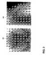
- FIG. 3shows portal images enhanced in accordance with the present invention.
- FIG. 4schematically illustrates a system in accordance with an embodiment of the present invention.
- the beam 100 of a linear accelerator(not shown), at a known gantry angle, is used in combination with a portal imager 105 to form a 2D image of the patient.
- the gantry anglemay then be changed, and another image acquired.
- the portal imager 105is preferably an EPID, producing digital images using the treatment beam 100 .
- the portal imagesmay be stored in a computer.
- a 3D ultrasound image 110is also acquired, before or after the portal images but within as close a time frame as possible so that the patient does not move significantly.
- the portal and ultrasound imagesmay be calibrated to a common coordinate system 115 whose origin coincides with the mechanical isocenter of the linear accelerator.
- This coordinate system 115may be identified using perpendicular lasers passing through the origin.
- Systems and methods for calibrating 3D ultrasound images to such as coordinate systemare known in the art.
- relevant anatomy in the 3D ultrasound imageis contoured (either manually or using an automatic segmentation algorithm) to form one or more 3D surfaces, each corresponding to a separate anatomical region.
- the bladder and prostatecan be contoured separately, even though they may appear in the same image set.
- the surface(s)are then projected onto the 2D portal images in the direction of the beam (i.e., along a beam's eye view).
- a 3D surfacecan be projected into the portalimage by various methods.
- a 3D surface 200is projected onto the 2D image plane 205 (which coincides with the portal and ultrasound images) to produce a 2D surface projection outline 210 .
- Thiscan be done, for example, by tracing a ray from the beam source 215 to a given pixel 220 in the image 205 . If the ray passes through the surface 200 , then the pixel 220 is considered within the projection outline 210 , otherwise it is considered outside the outline. This procedure may be repeated for all the pixels (or a reasonable subset consistent with resolution requirements) of the image 205 .
- the filled pixelsthen represent the projection 210 of the surface 200 .
- the outline of the filled pixelscan be extracted and the projection contour 210 outlined.
- adjustments to various imaging parametersmay be needed to correctly calibrate the 3D surface 200 to the image plane 205 .
- the scale of the portal imagemay require modification, the center of the portal image may need to be moved relative to the source 215 , and/or rotations of the image may be needed relative to the room coordinate system 115 .
- all imagesmay be scaled such that distances are measured in relation to the isocenter of the linear accelerator, i.e., the origin of the room coordinate system 115 . Accordingly, knowing the pixel size of the portal image is not enough, since the distance between the plane of the portal imaging detector (the medium on which the portal image is recorded) and the origin of the room coordinate system affects the scaling for a particular image.
- the image calibration parameterscan be calculated by detector calibration, image pre-calibration or image self-calibration.
- the imaging detectoris itself calibrated such that its parameters are known. For example, since the physical pixel size of the detector elements is known, and the image receptor can be calibrated to be at a known physical distance from the beam source, the scaling at isocenter may be readily computed. If the distance from the source changes, the pixels can be scaled to account for the new distance.
- the center pixel of the detectoris also calibrated to be at a known offset from the central axis of the beam—either the offset is permanently fixed or the detector electronics can determine an offset value.
- the rotation of the detectoris accurately fixed such that it is always aligned with the room coordinate system. Even with frequent detector calibration, there are likely to be deviations from ideal and drifts over time, thus requiring further calibration.
- an object(or “phantom”) of known geometry may be imaged with the detector.
- the phantomis a plate having an arrangement of some number (e.g., four) radio-opaque markers at known distances relative to the center of the plate.
- An image of the plateis acquired using the portal imaging device, and the markers identified on the image.
- the relationship of the markers on the image relative to their known positions on the phantomcan be used to calculate the calibration parameters.
- each gantry anglehas its own calibration parameters, since, for example, the detector may sag as the gantry is rotated. As a result, the central axis of the beam may not always pass exactly through the same point as the gantry is rotated, and therefore the calibration parameters should be checked periodically to identify any drift.
- the outline of the radiation fieldis detected and the outline edges are compared to the expected beam aperture, which may be rectangular or, in some instances, an irregular shape extracted from the treatment plan. Comparing the expected shapes to the detected shapes, the calibration parameters can be determined for the image.
- the state of the detectoris known relative to the room coordinate system, i.e., the detector can be moved in any configuration and an accurate calibration can still be computed.
- graticulea radio-opaque grid placed in the head of the linear accelerator
- similar 2D imagesmay acquired using diagnostic-energy x-ray tubes mounted in the treatment room.
- each acquired portal imagecan be enhanced by augmenting it with an image of the soft-tissue anatomy extracted from the 3D ultrasound scan, as shown in the portal images of FIG. 3 .
- Image 300was acquired at an anterio-posterior beam direction, while the image 305 was acquired with a lateral beam direction.
- the prostateis not visible in either, but the bony anatomy of the pelvis is clearly visible.
- the prostate surface, as contoured on ultrasound images,is projected as a white line superimposed on the portal images, as indicated at 310 and 315 .
- the ultrasound-enhanced portal imagescan be used to calculate patient shifts or other changes in treatment parameters, thereby permitting treatment delivery to account for changes in anatomy that deviate from the plan.
- Portal imagesare typically compared to digitally DRRs. These are simulated projections through the CT dataset, from the planned beam angles (or other more convenient angles), to form 2D images for each beam. By observing differences between the portal images and the DRRs, the treatment couch can be shifted to improve patient alignment with the beam(s). If the portal images have been enhanced using the above methods, the DRR may also be enhanced using ultrasound imaging. If, for example, a 3D ultrasound image is acquired during the planning CT session (e.g., as described in co-pending patent application Ser. No.
- the contours obtained from the ultrasound imagecan be projected onto the DRRs, thereby allowing for direct comparison between ultrasound-enhanced portal images and ultrasound-enhanced DRRs.
- Enhancing a DRR with the ultrasound contour obtained at time of simulationis done in the same fashion as described above with respect to the enhancement of portal images, except typically the DRRs need not be calibrated since their geometric parameters are typically known.
- CT and ultrasound imagescan be acquired in succession prior to a patient treatment; each is calibrated to the room coordinates of the linear accelerator.
- the ultrasound-derived anatomical contoursmay then be superimposed onto the CT image and the treatment parameters modified to better align with anatomy imaged by both modalities.
- the bladder, rectum and bony anatomycan be identified on the CT, while the prostate can be better identified on the ultrasound.
- one or more organscan be identified on images obtained using both modalities, but some organ edges are better revealed by one modality than by the other.
- beam shapes, angles, energies, patient position, etc.can be modified to account for the observed anatomy, which may differ from the planning anatomy.
- FIG. 4schematically depicts a hardware embodiment of the invention realized as a system 400 for modifying treatment parameters based on multimodal images.
- the system 400comprises a register 405 and a processor 415 .
- the register 405which may be any suitably organized data storage facility (e.g., partitions in RAM, etc.), receives images from a plurality of imagers, collectively indicated at 420 , which reflect different imaging modalities.
- Imagers 420may include one or more of an MRI, CT/PET scanner, ultrasound device, or x-ray device.
- the imagesare stored on a data-storage device separate from the imager (e.g., a database, microfiche, etc.) and sent to the system 400 .
- the register 405may receive the images through conventional data ports and may also include circuitry for receiving analog image data and analog-to-digital conversion circuitry for digitizing the image data.
- the register 405provides the images to the processor 415 , which implements the functionality of the present invention in hardware or software, or a combination of both on a general-purpose computer.
- processor 415registers the images and creates an enhanced image, which may be displayed on a device 430 .
- the processor 415thereupon computes patient shifts or other changes in treatment parameters, which are communicated to the controller 435 of a treatment device such as a linear accelerator.
- the controller 435causes appropriate adjustments to be made based on the modified treatment parameters.
- a uservia an input device 425 , may influence, approve, override or revise the modifications to the treatment parameters based on his or her review of the composite image on device 430 .
- the programming for processor 415may be written in any one of a number of high-level languages, such as FORTRAN, PASCAL, C, C++, C#, Java, Tcl, or BASIC. Further, the program can be written in a script, macro, or functionality embedded in commercially available software, such as EXCEL or VISUAL BASIC. Additionally, the software can be implemented in an assembly language directed to a microprocessor resident on a computer. For example, the software can be implemented in Intel 80 ⁇ 86 assembly language if it is configured to run on an IBM PC or PC clone. The software may be embedded on an article of manufacture including, but not limited to, “computer-readable program means” such as a floppy disk, a hard disk, an optical disk, a magnetic tape, a PROM, an EPROM, or CD-ROM.
- computer-readable program meanssuch as a floppy disk, a hard disk, an optical disk, a magnetic tape, a PROM, an EPROM, or CD-ROM.
Landscapes
- Engineering & Computer Science (AREA)
- Health & Medical Sciences (AREA)
- Life Sciences & Earth Sciences (AREA)
- Biomedical Technology (AREA)
- Pathology (AREA)
- Radiology & Medical Imaging (AREA)
- Nuclear Medicine, Radiotherapy & Molecular Imaging (AREA)
- Animal Behavior & Ethology (AREA)
- General Health & Medical Sciences (AREA)
- Public Health (AREA)
- Veterinary Medicine (AREA)
- Physics & Mathematics (AREA)
- Medical Informatics (AREA)
- Biophysics (AREA)
- High Energy & Nuclear Physics (AREA)
- Optics & Photonics (AREA)
- Heart & Thoracic Surgery (AREA)
- Molecular Biology (AREA)
- Surgery (AREA)
- Computer Vision & Pattern Recognition (AREA)
- General Physics & Mathematics (AREA)
- Theoretical Computer Science (AREA)
- Radiation-Therapy Devices (AREA)
- Ultra Sonic Daignosis Equipment (AREA)
Abstract
Description
This application claims priority to and the benefit of U.S. provisional patent application Ser. No. 60/951,005, filed Jul. 20, 2007, the disclosure of which is incorporated herein by reference in its entirety.
This invention relates to methods and systems for using imaging to guide radiotherapy treatments.
Radiation therapy relies on devising a treatment plan, which includes the arrangement of therapeutic radiation beams, patient positioning relative to the beams, beam energies, apertures, and doses, and other factors. Typically, the treatment plan is based on a three-dimensional (3D) computed tomography (CT) dataset acquired prior to the first treatment session. Although CT scans provide a good physical map of electron density within the patient, they also have limitations. For example, CT scans are devoid of functional information about tumors and provide poor soft-tissue contrast for some organs. To circumvent these limitations, other secondary images may also be acquired, using modalities such as positron-emission tomography (PET), magnetic resonance imaging (MRI) or ultrasound. PET, for example, gives functional information about tumor metabolism, and MRI and ultrasound give superior soft-tissue contrast for some organs.
After the treatment plan is developed, the patient is positioned in the treatment room using external skin markings and radiation is delivered according to the treatment plan. This is typically repeated for a number of sessions, for example, once a day for 30 sessions. During this time period, however, a patient's internal anatomy may change. For example, it is known that the prostate can change positions significantly depending on rectal and bladder filling. In an attempt to provide more accurate delivery of radiation therapy, image-guided radiotherapy (IGRT) has become more common. Using IGRT, an image is acquired prior to each session and used to correct the treatment plan for anatomical changes. In principle, a completely new treatment plan can be generated prior to each treatment session—a technique known as adaptive radiotherapy (ART). Although effective, ART is not generally undertaken because re-planning is time-consuming and must be validated and approved for each session. Instead, current clinical practice commonly corrects for physiological changes by shifting the patient (using the treatment couch, for example) in order to best align the target anatomy to the planned location. This is accomplished by comparing the target structure position to its position on a reference image acquired during planning.
One common technique for implementing IGRT is portal imaging, i.e., using the treatment beam to acquire images with either film or a two-dimensional (2D) electronic portal imaging detector (EPID). Due to the high energy (megavolt range) of the treatment beam, image quality is generally inferior to diagnostic (kilovolt range) x-ray images, and provides little or no soft-tissue contrast. Portal images can be effective, however, for localization of bony anatomy, air pockets, and imaging skin surface. One advantage to using the IGRT approach is that the information is inherently acquired by and related to the treatment beam. To ensure the treatment position is correct relative to bony anatomy, the portal images are compared to digitally reconstructed radiographs (DRRs), which are the reconstruction of a 2D projection radiograph from a given beam direction, calculated from the planning CT dataset. EPIDs have been developed not only for electronic record-keeping, but also to make the acquisition more rapid, and to allow online corrections to patient position prior to each treatment fraction. Further, the introduction of flat-panel detectors has improved image quality of EPIDs such that it is comparable to conventional film-based imaging.
Software has been developed to enable rapid displacement calculations using localization images. Typically, 2D structures, called overlays, are extracted from CT contours and displayed on the DRRs on a console. EPID images are acquired from (typically) two angles, such as anterior-posterior and lateral, and the DRR overlays are shown superimposed on the port films. The operator then moves the overlays such that they fit the anatomy as seen on the EPIDs, and the amount of shift is calculated. This allows the therapist to displace the couch to compensate for any discrepancies.
Since portal images do not show soft-tissue contrast, one practice facilitating IGRT is to implant, in the treated organ (e.g., prostate), gold seeds that may be identified on the portal images. By comparing these positions to those on the planning CT, shifts can be executed to correct for organ motion. The use of seeds is invasive, however, and the resulting images do not give a complete picture of the organ and surrounding anatomy.
Another approach includes placing a conventional or cone-beam CT scanner in the treatment room. These scanners generate 3D images, and can either be of diagnostic quality or can use the high-energy treatment beam to produce the 3D images (referred to as megavoltage CT). These images provide a good geometric image of the patient, are similar in nature to the planning CT, and have some soft-tissue contrast which can be used to perform IGRT. For some sites such as prostate, however, fiducial marker seeds are typically used because the soft-tissue contrast is still unacceptable.
Ultrasound has also been used for IGRT, as it provides good soft-tissue contrast. Two or more ultrasound images are referenced to a 3D coordinate system, and are either used individually or are reconstructed to a full 3D image dataset. Patient displacements can then be determined from the ultrasound images. Although they can provide excellent soft-tissue contrast for organs such as the prostate, uterus or breast tumor cavities and do not require fiducial markers, ultrasound images do not give bony anatomy, or a complete anatomical image of the patient.
There has been research and development into the use of in-treatment-room MRI and PET for IGRT, but technical hurdles remain before this technology becomes commercially available.
The present invention provides systems and methods for improving imaging for IGRT. Embodiments of the invention rely on the integration and use of multiple modalities in the treatment room, registered to a common coordinate system, in order to modify treatment parameters.
In one aspect, the invention combines portal images and 3D ultrasound to determine a patient shift to be applied during radiotherapy treatment. A set of baseline radiotherapy treatment parameters are established, and, during therapy, two or more registered images of the area under treatment are taken, with at least some of the images being portal images taken from at least one beam direction, and at least one of the images being a non-x-ray-based 3D image. The images are then registered to a coordinate system associated with a treatment device. The non-x-ray-based 3D image is segmented to form a 3D surface, which is projected onto the plane of each portal image, thereby enhancing the portal images with data from the projected surface. The baseline treatment parameters are then updated based on the registered images. In this fashion, new portal images are produced which incorporate the anatomy extracted from the ultrasound.
In another aspect a system for identifying changes to patient treatment parameters during delivery of radiotherapy includes a first and second register and a processor. The first register stores a set of baseline treatment parameters, and the second register stores the images of an anatomical region to be treated. The images are obtained using different imaging modalities, such that at least some of the images are portal images from at least one beam direction, and at least one of the images is a non-x-ray-based 3D image. The processor is configured to register the images to a coordinate system associated with a treatment device, segment the non-x-ray-based 3D image to form a 3D surface, project the surface onto the plane of each portal image, enhance the portal images with data from the projected surface, and determine modifications to the baseline treatment parameters based on the registered images.
The present invention is not limited to the above applications, but encompasses the use of more than one image in the treatment room, registering these two or more images, and using anatomy from at least two of the images to modify treatment parameters.
In the drawings, like reference characters generally refer to the same parts throughout the different views. Also, the drawings are not necessarily to scale, emphasis instead is generally being placed upon illustrating the principles of the invention.
InFIG. 1 , which illustrates an embodiment of the invention, thebeam 100 of a linear accelerator (not shown), at a known gantry angle, is used in combination with aportal imager 105 to form a 2D image of the patient. The gantry angle may then be changed, and another image acquired. Typically at least two images are acquired from different directions using theportal imager 105. Theportal imager 105 is preferably an EPID, producing digital images using thetreatment beam 100. The portal images may be stored in a computer. A3D ultrasound image 110 is also acquired, before or after the portal images but within as close a time frame as possible so that the patient does not move significantly. The portal and ultrasound images may be calibrated to a common coordinatesystem 115 whose origin coincides with the mechanical isocenter of the linear accelerator. This coordinatesystem 115 may be identified using perpendicular lasers passing through the origin. Systems and methods for calibrating 3D ultrasound images to such as coordinate system are known in the art.
Still referring toFIG. 1 , relevant anatomy in the 3D ultrasound image is contoured (either manually or using an automatic segmentation algorithm) to form one or more 3D surfaces, each corresponding to a separate anatomical region. For example, the bladder and prostate can be contoured separately, even though they may appear in the same image set. The surface(s) are then projected onto the 2D portal images in the direction of the beam (i.e., along a beam's eye view).
In particular, from a given angle corresponding to a single portal image, a 3D surface can be projected into the portalimage by various methods. Referring toFIG. 2 , a3D surface 200 is projected onto the 2D image plane205 (which coincides with the portal and ultrasound images) to produce a 2Dsurface projection outline 210. This can be done, for example, by tracing a ray from thebeam source 215 to a givenpixel 220 in theimage 205. If the ray passes through thesurface 200, then thepixel 220 is considered within theprojection outline 210, otherwise it is considered outside the outline. This procedure may be repeated for all the pixels (or a reasonable subset consistent with resolution requirements) of theimage 205. The filled pixels then represent theprojection 210 of thesurface 200. The outline of the filled pixels can be extracted and theprojection contour 210 outlined.
In some cases, adjustments to various imaging parameters may be needed to correctly calibrate the3D surface 200 to theimage plane 205. For example, the scale of the portal image may require modification, the center of the portal image may need to be moved relative to thesource 215, and/or rotations of the image may be needed relative to the room coordinatesystem 115. In principle, all images may be scaled such that distances are measured in relation to the isocenter of the linear accelerator, i.e., the origin of the room coordinatesystem 115. Accordingly, knowing the pixel size of the portal image is not enough, since the distance between the plane of the portal imaging detector (the medium on which the portal image is recorded) and the origin of the room coordinate system affects the scaling for a particular image. The image calibration parameters can be calculated by detector calibration, image pre-calibration or image self-calibration.
For detector calibration, the imaging detector is itself calibrated such that its parameters are known. For example, since the physical pixel size of the detector elements is known, and the image receptor can be calibrated to be at a known physical distance from the beam source, the scaling at isocenter may be readily computed. If the distance from the source changes, the pixels can be scaled to account for the new distance. The center pixel of the detector is also calibrated to be at a known offset from the central axis of the beam—either the offset is permanently fixed or the detector electronics can determine an offset value. The rotation of the detector is accurately fixed such that it is always aligned with the room coordinate system. Even with frequent detector calibration, there are likely to be deviations from ideal and drifts over time, thus requiring further calibration.
To assist with calibration of the detector prior to imaging, an object (or “phantom”) of known geometry may be imaged with the detector. In some embodiments, the phantom is a plate having an arrangement of some number (e.g., four) radio-opaque markers at known distances relative to the center of the plate. An image of the plate is acquired using the portal imaging device, and the markers identified on the image. The relationship of the markers on the image relative to their known positions on the phantom can be used to calculate the calibration parameters. In principle, each gantry angle has its own calibration parameters, since, for example, the detector may sag as the gantry is rotated. As a result, the central axis of the beam may not always pass exactly through the same point as the gantry is rotated, and therefore the calibration parameters should be checked periodically to identify any drift.
For image self-calibration, the outline of the radiation field is detected and the outline edges are compared to the expected beam aperture, which may be rectangular or, in some instances, an irregular shape extracted from the treatment plan. Comparing the expected shapes to the detected shapes, the calibration parameters can be determined for the image. One advantage of this approach is that the state of the detector is known relative to the room coordinate system, i.e., the detector can be moved in any configuration and an accurate calibration can still be computed.
Other approaches can also be used to calibrate the portal images, such as using a graticule (a radio-opaque grid placed in the head of the linear accelerator), which appears in the portal image. Furthermore, instead of using portal images, similar 2D images may acquired using diagnostic-energy x-ray tubes mounted in the treatment room.
With the ability to project the surface contour onto a calibrated portal image, each acquired portal image can be enhanced by augmenting it with an image of the soft-tissue anatomy extracted from the 3D ultrasound scan, as shown in the portal images ofFIG. 3 .Image 300 was acquired at an anterio-posterior beam direction, while theimage 305 was acquired with a lateral beam direction. The prostate is not visible in either, but the bony anatomy of the pelvis is clearly visible. The prostate surface, as contoured on ultrasound images, is projected as a white line superimposed on the portal images, as indicated at310 and315.
The ultrasound-enhanced portal images, once created, can be used to calculate patient shifts or other changes in treatment parameters, thereby permitting treatment delivery to account for changes in anatomy that deviate from the plan.
Portal images are typically compared to digitally DRRs. These are simulated projections through the CT dataset, from the planned beam angles (or other more convenient angles), to form 2D images for each beam. By observing differences between the portal images and the DRRs, the treatment couch can be shifted to improve patient alignment with the beam(s). If the portal images have been enhanced using the above methods, the DRR may also be enhanced using ultrasound imaging. If, for example, a 3D ultrasound image is acquired during the planning CT session (e.g., as described in co-pending patent application Ser. No. 10/343,336, which is incorporated in its entirety herein by reference), the contours obtained from the ultrasound image can be projected onto the DRRs, thereby allowing for direct comparison between ultrasound-enhanced portal images and ultrasound-enhanced DRRs. Enhancing a DRR with the ultrasound contour obtained at time of simulation is done in the same fashion as described above with respect to the enhancement of portal images, except typically the DRRs need not be calibrated since their geometric parameters are typically known.
While the invention has been described particularly in relation to using both portal images and ultrasound for IGRT, the invention also extends to matching the coordinate systems of any two or more imaging modalities, and using images obtained using these modalities to modify treatment parameters. For example, CT and ultrasound images can be acquired in succession prior to a patient treatment; each is calibrated to the room coordinates of the linear accelerator. The ultrasound-derived anatomical contours may then be superimposed onto the CT image and the treatment parameters modified to better align with anatomy imaged by both modalities. For example, the bladder, rectum and bony anatomy can be identified on the CT, while the prostate can be better identified on the ultrasound. In other instances, one or more organs can be identified on images obtained using both modalities, but some organ edges are better revealed by one modality than by the other. After the anatomy is identified using the multimodality images, beam shapes, angles, energies, patient position, etc. can be modified to account for the observed anatomy, which may differ from the planning anatomy.
Theregister 405, which may be any suitably organized data storage facility (e.g., partitions in RAM, etc.), receives images from a plurality of imagers, collectively indicated at420, which reflect different imaging modalities.Imagers 420 may include one or more of an MRI, CT/PET scanner, ultrasound device, or x-ray device. In some embodiments, the images are stored on a data-storage device separate from the imager (e.g., a database, microfiche, etc.) and sent to thesystem 400. Theregister 405 may receive the images through conventional data ports and may also include circuitry for receiving analog image data and analog-to-digital conversion circuitry for digitizing the image data.
Theregister 405 provides the images to theprocessor 415, which implements the functionality of the present invention in hardware or software, or a combination of both on a general-purpose computer. In particular,processor 415 registers the images and creates an enhanced image, which may be displayed on adevice 430. Theprocessor 415 thereupon computes patient shifts or other changes in treatment parameters, which are communicated to thecontroller 435 of a treatment device such as a linear accelerator. Thecontroller 435, in turn, causes appropriate adjustments to be made based on the modified treatment parameters.
Alternatively or in addition, a user, via aninput device 425, may influence, approve, override or revise the modifications to the treatment parameters based on his or her review of the composite image ondevice 430.
The programming forprocessor 415 may be written in any one of a number of high-level languages, such as FORTRAN, PASCAL, C, C++, C#, Java, Tcl, or BASIC. Further, the program can be written in a script, macro, or functionality embedded in commercially available software, such as EXCEL or VISUAL BASIC. Additionally, the software can be implemented in an assembly language directed to a microprocessor resident on a computer. For example, the software can be implemented in Intel 80×86 assembly language if it is configured to run on an IBM PC or PC clone. The software may be embedded on an article of manufacture including, but not limited to, “computer-readable program means” such as a floppy disk, a hard disk, an optical disk, a magnetic tape, a PROM, an EPROM, or CD-ROM.
While the invention has been particularly shown and described with reference to specific embodiments, it should be understood by those skilled in the art that various changes in form and detail may be made therein without departing from the spirit and scope of the invention as defined by the appended claims. The scope of the invention is thus indicated by the appended claims and all changes which come within the meaning and range of equivalency of the claims are therefore intended to be embraced.
Claims (5)
1. A method of modifying treatment parameters for a patient undergoing radiotherapy, the method comprising the steps of:
(a) establishing a set of baseline treatment parameters during a treatment planning session prior to a treatment delivery session;
(b) during the treatment delivery session obtaining, using different imaging modalities, at least two registered images of an anatomical region to be treated wherein at least some of the images are portal images from at least one beam direction, and at least one of the images is a non-x-ray-based 3D image;
(c) registering the at least two images to a coordinate system associated with a treatment device;
(d) segmenting the non-x-ray-based 3D image to form a 3D surface;
(e) projecting the surface onto the plane of each portal image;
(f) enhancing the portal images with data from the projected surface, wherein the enhancing comprises identifying one or more anatomical regions based on the portal images, identifying one or more different anatomical regions based on the projected surface other than those identified based on the portal images, and superimposing the identified different anatomical regions onto the portal images; and
(g) modifying the baseline treatment parameters based on the enhanced portal images.
2. The method ofclaim 1 further comprising:
enhancing one or more digitally reconstructed images with data from the projected surface; and
modifying the baseline treatment parameters based on a comparison between the enhanced portal images and the enhanced digitally reconstructed images.
3. A system for identifying changes to patient treatment parameters, the system comprising:
(a) a first register for storing a set of baseline treatment parameters obtained during a treatment planning session prior to a treatment delivery session;
(b) a second register for storing at least two images of an anatomical region to be treated, each image being obtained during the treatment delivery session using different imaging modalities wherein at least some of the images are portal images from at least one beam direction, and at least one of the images is a non-x-ray-based 3D image; and
(c) a processor configured to retrieve and execute stored computer programming instructions to:
(i) register the at least two images to a coordinate system associated with a treatment device;
(ii) segment the non-x-ray-based 3D image to form a 3D surface;
(iii) project the surface onto the plane of each portal image;
(iv) enhance the portal images with data from the projected surface, wherein the enhancing comprises identifying one or more anatomical regions based on the portal images, identifying one or more different anatomical regions based on the projected surface other than those identified based on the portal images, and superimposing the identified different anatomical regions onto the portal images; and
(ii) determine modifications to the baseline treatment parameters based on the enhanced portal images.
4. The system ofclaim 3 further comprising a controller for affecting the modifications based on the modified treatment parameters.
5. The system ofclaim 3 wherein the modifications comprise positional changes to the patient.
Priority Applications (1)
| Application Number | Priority Date | Filing Date | Title |
|---|---|---|---|
| US12/176,785US8249317B2 (en) | 2007-07-20 | 2008-07-21 | Methods and systems for compensating for changes in anatomy of radiotherapy patients |
Applications Claiming Priority (2)
| Application Number | Priority Date | Filing Date | Title |
|---|---|---|---|
| US95100507P | 2007-07-20 | 2007-07-20 | |
| US12/176,785US8249317B2 (en) | 2007-07-20 | 2008-07-21 | Methods and systems for compensating for changes in anatomy of radiotherapy patients |
Publications (2)
| Publication Number | Publication Date |
|---|---|
| US20090022383A1 US20090022383A1 (en) | 2009-01-22 |
| US8249317B2true US8249317B2 (en) | 2012-08-21 |
Family
ID=40264887
Family Applications (1)
| Application Number | Title | Priority Date | Filing Date |
|---|---|---|---|
| US12/176,785Active2031-01-14US8249317B2 (en) | 2007-07-20 | 2008-07-21 | Methods and systems for compensating for changes in anatomy of radiotherapy patients |
Country Status (4)
| Country | Link |
|---|---|
| US (1) | US8249317B2 (en) |
| EP (1) | EP2175931B1 (en) |
| CA (1) | CA2693351C (en) |
| WO (1) | WO2009012577A1 (en) |
Cited By (6)
| Publication number | Priority date | Publication date | Assignee | Title |
|---|---|---|---|---|
| US20110190629A1 (en)* | 2008-09-30 | 2011-08-04 | Mediri Gmbh | 3D Motion Detection and Correction By Object Tracking in Ultrasound Images |
| US20140016759A1 (en)* | 2012-07-13 | 2014-01-16 | The Chinese University Of Hong Kong | Compound 6d-offset simulating phantom and quality assurance program for precision image-guided radiotherapy and radiosurgery |
| CN106408509A (en)* | 2016-04-29 | 2017-02-15 | 上海联影医疗科技有限公司 | Registration method and apparatus |
| US9950194B2 (en) | 2014-09-09 | 2018-04-24 | Mevion Medical Systems, Inc. | Patient positioning system |
| US9974977B2 (en) | 2014-10-27 | 2018-05-22 | Elekta, Inc. | Image guidance for radiation therapy |
| WO2021026634A1 (en)* | 2019-08-13 | 2021-02-18 | Elekta Ltd. | Adaptive radiation therapy using composite imaging slices |
Families Citing this family (20)
| Publication number | Priority date | Publication date | Assignee | Title |
|---|---|---|---|---|
| US6693099B2 (en) | 2000-10-17 | 2004-02-17 | The Procter & Gamble Company | Substituted piperazine compounds optionally containing a quinolyl moiety for treating multidrug resistance |
| TW200950749A (en)* | 2008-06-12 | 2009-12-16 | Univ Ishou | Interactive medical imaging alignment system applied to radiotherapy program |
| US8331531B2 (en)* | 2009-03-13 | 2012-12-11 | The Board Of Trustees Of The Leland Stanford Junior University | Configurations for integrated MRI-linear accelerators |
| US20110172526A1 (en)* | 2010-01-12 | 2011-07-14 | Martin Lachaine | Feature Tracking Using Ultrasound |
| DE102010020350B4 (en)* | 2010-05-12 | 2017-02-23 | Siemens Healthcare Gmbh | Method for positioning the focus of a gradient field and treatment device |
| DE102011079561B4 (en) | 2011-07-21 | 2018-10-18 | Siemens Healthcare Gmbh | Method and X-ray device for timely presentation of a moving section of a body, computer program and data carrier |
| US20130229495A1 (en)* | 2012-03-01 | 2013-09-05 | Ali-Reza Bani-Hashemi | Method for calibrating an imaging system |
| WO2014096993A1 (en) | 2012-12-17 | 2014-06-26 | Koninklijke Philips N.V. | Real-time adaptive dose computation radiation therapy |
| WO2014120423A1 (en)* | 2013-01-31 | 2014-08-07 | The Board Of Trustees Of The Leland Stanford Junior University | A novel epid dosimetry method and system for radiation therapy |
| JP6568077B2 (en) | 2013-09-30 | 2019-08-28 | コーニンクレッカ フィリップス エヌ ヴェKoninklijke Philips N.V. | Coordinate system alignment of externally irradiated radiotherapy and magnetic resonance imaging systems |
| CN113842566B (en) | 2013-12-31 | 2025-09-05 | 威斯康星州医药大学股份有限公司 | Adaptive replanning based on multimodal imaging |
| US9844358B2 (en)* | 2014-06-04 | 2017-12-19 | Varian Medical Systems, Inc. | Imaging-based self-adjusting radiation therapy systems, devices, and methods |
| CN108064396B (en)* | 2017-05-27 | 2021-04-30 | 上海联影医疗科技股份有限公司 | System and method for compensating for couch subsidence in image-guided radiation therapy |
| US11491349B2 (en)* | 2017-07-19 | 2022-11-08 | Sino-israeli Health Alliance International Medical Technology Co., Ltd. | Patient irradiation treatment plan verification system and method |
| GB2565119A (en)* | 2017-08-02 | 2019-02-06 | Vision Rt Ltd | Method of calibrating a patient monitoring system for use with a radiotherapy treatment apparatus |
| JP6611833B2 (en)* | 2018-01-16 | 2019-11-27 | キヤノン株式会社 | Radiation imaging system, camera control device and control method thereof |
| CN108619621B (en)* | 2018-05-23 | 2020-08-21 | 郑向鹏 | System for be used for accurate location of tumour patient radiation therapy and pendulum position |
| BR112021003450A2 (en)* | 2018-08-24 | 2021-05-18 | Medical Beam Laboratories, Llc | patient positioning system, radiation beam delivery apparatus, method of delivering a therapeutic dose of radiation to a human or animal patient, and patient positioning device |
| CN112785632B (en)* | 2021-02-13 | 2024-05-24 | 常州市第二人民医院 | Cross-modal automatic registration method for DR and DRR images in image-guided radiotherapy based on EPID |
| CN119868839B (en)* | 2025-03-26 | 2025-07-22 | 东北大学 | A quality control method based on electronic portal imaging system and maximum tissue phantom ratio |
Citations (167)
| Publication number | Priority date | Publication date | Assignee | Title |
|---|---|---|---|---|
| US3082322A (en) | 1958-11-28 | 1963-03-19 | Westinghouse Electric Corp | Therapy unit |
| US3777124A (en) | 1970-11-27 | 1973-12-04 | Varian Associates | Computer assisted radiation therapy machine |
| US3987281A (en) | 1974-07-29 | 1976-10-19 | The United States Of America As Represented By The Department Of Health, Education And Welfare | Method of radiation therapy treatment planning |
| US3991310A (en) | 1970-08-03 | 1976-11-09 | Morrison Richard A | Biplane radiographic localization of target center for radiotherapy |
| US4118631A (en) | 1976-03-30 | 1978-10-03 | Emi Limited | Radiographic apparatus |
| US4618978A (en) | 1983-10-21 | 1986-10-21 | Cosman Eric R | Means for localizing target coordinates in a body relative to a guidance system reference frame in any arbitrary plane as viewed by a tomographic image through the body |
| US4882741A (en) | 1987-10-28 | 1989-11-21 | U.S. Philips Corporation | Multileaf collimator and related apparatus |
| US4923459A (en) | 1987-09-14 | 1990-05-08 | Kabushiki Kaisha Toshiba | Stereotactics apparatus |
| US4943990A (en) | 1987-12-11 | 1990-07-24 | Bbc Brown Boveri Ag | Therapy simulator |
| US5039867A (en) | 1987-08-24 | 1991-08-13 | Mitsubishi Denki Kabushiki Kaisha | Therapeutic apparatus |
| US5080100A (en) | 1988-10-04 | 1992-01-14 | Cgr Mev | System and method for measuring and/or checking the position of a patient in a radio-therapy machine |
| US5086401A (en) | 1990-05-11 | 1992-02-04 | International Business Machines Corporation | Image-directed robotic system for precise robotic surgery including redundant consistency checking |
| US5099846A (en) | 1988-12-23 | 1992-03-31 | Hardy Tyrone L | Method and apparatus for video presentation from a variety of scanner imaging sources |
| US5107839A (en) | 1990-05-04 | 1992-04-28 | Pavel V. Houdek | Computer controlled stereotaxic radiotherapy system and method |
| US5117829A (en) | 1989-03-31 | 1992-06-02 | Loma Linda University Medical Center | Patient alignment system and procedure for radiation treatment |
| US5138647A (en) | 1990-08-03 | 1992-08-11 | Siemens Medical Laboratories, Inc. | Portal imaging device |
| US5207223A (en) | 1990-10-19 | 1993-05-04 | Accuray, Inc. | Apparatus for and method of performing stereotaxic surgery |
| US5222499A (en) | 1989-11-15 | 1993-06-29 | Allen George S | Method and apparatus for imaging the anatomy |
| US5233990A (en) | 1992-01-13 | 1993-08-10 | Gideon Barnea | Method and apparatus for diagnostic imaging in radiation therapy |
| US5291889A (en) | 1991-05-23 | 1994-03-08 | Vanguard Imaging Ltd. | Apparatus and method for spatially positioning images |
| US5295483A (en) | 1990-05-11 | 1994-03-22 | Christopher Nowacki | Locating target in human body |
| US5301674A (en) | 1992-03-27 | 1994-04-12 | Diasonics, Inc. | Method and apparatus for focusing transmission and reception of ultrasonic beams |
| US5379642A (en) | 1993-07-19 | 1995-01-10 | Diasonics Ultrasound, Inc. | Method and apparatus for performing imaging |
| US5389101A (en) | 1992-04-21 | 1995-02-14 | University Of Utah | Apparatus and method for photogrammetric surgical localization |
| US5391139A (en) | 1992-09-03 | 1995-02-21 | William Beaumont Hospital | Real time radiation treatment planning system |
| US5397329A (en) | 1987-11-10 | 1995-03-14 | Allen; George S. | Fiducial implant and system of such implants |
| US5408101A (en) | 1992-07-06 | 1995-04-18 | Telaire Systems, Inc. | Laser assisted quasi-blackbody radiation source |
| US5411026A (en) | 1993-10-08 | 1995-05-02 | Nomos Corporation | Method and apparatus for lesion position verification |
| US5438991A (en) | 1993-10-18 | 1995-08-08 | William Beaumont Hospital | Method and apparatus for controlling a radiation treatment field |
| US5442675A (en) | 1992-03-19 | 1995-08-15 | Wisconsin Alumni Research Foundation | Dynamic collimator for radiation therapy |
| US5446548A (en) | 1993-10-08 | 1995-08-29 | Siemens Medical Systems, Inc. | Patient positioning and monitoring system |
| US5447154A (en) | 1992-07-31 | 1995-09-05 | Universite Joseph Fourier | Method for determining the position of an organ |
| US5483961A (en) | 1993-03-19 | 1996-01-16 | Kelly; Patrick J. | Magnetic field digitizer for stereotactic surgery |
| US5511549A (en) | 1995-02-13 | 1996-04-30 | Loma Linda Medical Center | Normalizing and calibrating therapeutic radiation delivery systems |
| US5524627A (en) | 1994-08-23 | 1996-06-11 | Sonotron Ltd. | Ultrasonic imaging system |
| US5531227A (en) | 1994-01-28 | 1996-07-02 | Schneider Medical Technologies, Inc. | Imaging device and method |
| US5531520A (en) | 1994-09-01 | 1996-07-02 | Massachusetts Institute Of Technology | System and method of registration of three-dimensional data sets including anatomical body data |
| US5553618A (en) | 1993-03-12 | 1996-09-10 | Kabushiki Kaisha Toshiba | Method and apparatus for ultrasound medical treatment |
| US5591983A (en) | 1995-06-30 | 1997-01-07 | Siemens Medical Systems, Inc. | Multiple layer multileaf collimator |
| US5603318A (en) | 1992-04-21 | 1997-02-18 | University Of Utah Research Foundation | Apparatus and method for photogrammetric surgical localization |
| US5609485A (en) | 1994-10-03 | 1997-03-11 | Medsim, Ltd. | Medical reproduction system |
| US5645066A (en) | 1996-04-26 | 1997-07-08 | Advanced Technology Laboratories, Inc. | Medical ultrasonic diagnostic imaging system with scanning guide for three dimensional imaging |
| US5673300A (en) | 1996-06-11 | 1997-09-30 | Wisconsin Alumni Research Foundation | Method of registering a radiation treatment plan to a patient |
| US5690108A (en) | 1994-11-28 | 1997-11-25 | Chakeres; Donald W. | Interventional medicine apparatus |
| US5715166A (en) | 1992-03-02 | 1998-02-03 | General Motors Corporation | Apparatus for the registration of three-dimensional shapes |
| US5734384A (en) | 1991-11-29 | 1998-03-31 | Picker International, Inc. | Cross-referenced sectioning and reprojection of diagnostic image volumes |
| US5740225A (en) | 1995-12-07 | 1998-04-14 | Kabushiki Kaisha Toshiba | Radiation therapy planning method and its system and apparatus |
| US5754623A (en) | 1994-03-25 | 1998-05-19 | Kabushiki Kaisha Toshiba | Radiotherapy system |
| US5757881A (en) | 1997-01-06 | 1998-05-26 | Siemens Business Communication Systems, Inc. | Redundant field-defining arrays for a radiation system |
| US5778043A (en) | 1996-09-20 | 1998-07-07 | Cosman; Eric R. | Radiation beam control system |
| US5810007A (en) | 1995-07-26 | 1998-09-22 | Associates Of The Joint Center For Radiation Therapy, Inc. | Ultrasound localization and image fusion for the treatment of prostate cancer |
| US5851183A (en) | 1990-10-19 | 1998-12-22 | St. Louis University | System for indicating the position of a surgical probe within a head on an image of the head |
| US5859891A (en) | 1997-03-07 | 1999-01-12 | Hibbard; Lyn | Autosegmentation/autocontouring system and method for use with three-dimensional radiation therapy treatment planning |
| US5871445A (en) | 1993-04-26 | 1999-02-16 | St. Louis University | System for indicating the position of a surgical probe within a head on an image of the head |
| US5952577A (en) | 1997-07-21 | 1999-09-14 | Sonotron Ltd. | Ultrasonic imaging system |
| US5991703A (en) | 1997-08-15 | 1999-11-23 | The Institute Of Physical And Chemical Research | Method of synthesizing measurement data of free-form surface |
| US6019724A (en) | 1995-02-22 | 2000-02-01 | Gronningsaeter; Aage | Method for ultrasound guidance during clinical procedures |
| US6038283A (en) | 1996-10-24 | 2000-03-14 | Nomos Corporation | Planning method and apparatus for radiation dosimetry |
| US6094508A (en) | 1997-12-08 | 2000-07-25 | Intel Corporation | Perceptual thresholding for gradient-based local edge detection |
| US6106470A (en) | 1998-03-20 | 2000-08-22 | General Electric Company | Method and appartus for calculating distance between ultrasound images using sum of absolute differences |
| US6112341A (en) | 1994-09-08 | 2000-09-05 | Itt Manufacturing Enterprises, Inc. | Rotary pulsing valve |
| US6117081A (en) | 1998-10-01 | 2000-09-12 | Atl Ultrasound, Inc. | Method for correcting blurring of spatially compounded ultrasonic diagnostic images |
| US6118848A (en) | 1998-01-14 | 2000-09-12 | Reiffel; Leonard | System to stabilize an irradiated internal target |
| US6119033A (en) | 1997-03-04 | 2000-09-12 | Biotrack, Inc. | Method of monitoring a location of an area of interest within a patient during a medical procedure |
| US6122341A (en) | 1992-06-12 | 2000-09-19 | Butler; William E. | System for determining target positions in the body observed in CT image data |
| US6129670A (en) | 1997-11-24 | 2000-10-10 | Burdette Medical Systems | Real time brachytherapy spatial registration and visualization system |
| US6138495A (en) | 1997-12-31 | 2000-10-31 | Ultraguide Ltd. | Calibration method and apparatus for calibrating position sensors on scanning transducers |
| US6144875A (en) | 1999-03-16 | 2000-11-07 | Accuray Incorporated | Apparatus and method for compensating for respiratory and patient motion during treatment |
| FR2778574B1 (en) | 1998-05-13 | 2000-12-08 | Technomed Medical Systems | METHOD FOR MEASURING THE EFFECT OF TREATMENT ON TISSUE |
| US6198957B1 (en) | 1997-12-19 | 2001-03-06 | Varian, Inc. | Radiotherapy machine including magnetic resonance imaging system |
| US6259943B1 (en) | 1995-02-16 | 2001-07-10 | Sherwood Services Ag | Frameless to frame-based registration system |
| US6269143B1 (en) | 1998-08-31 | 2001-07-31 | Shimadzu Corporation | Radiotherapy planning system |
| US6285805B1 (en) | 1999-01-25 | 2001-09-04 | International Business Machines Corp. | System and method for finding the distance from a moving query point to the closest point on one or more convex or non-convex shapes |
| US6292578B1 (en) | 1998-02-18 | 2001-09-18 | International Business Machines Corporation | System and method for restoring, describing and graphically displaying noise-corrupted boundaries in tomography images |
| US6307914B1 (en) | 1998-03-12 | 2001-10-23 | Mitsubishi Denki Kabushiki Kaisha | Moving body pursuit irradiating device and positioning method using this device |
| US20010035871A1 (en) | 2000-03-30 | 2001-11-01 | Johannes Bieger | System and method for generating an image |
| US6314310B1 (en) | 1997-02-14 | 2001-11-06 | Biosense, Inc. | X-ray guided surgical location system with extended mapping volume |
| US6325758B1 (en) | 1997-10-27 | 2001-12-04 | Nomos Corporation | Method and apparatus for target position verification |
| US20010049475A1 (en) | 2000-01-31 | 2001-12-06 | Bucholz Richard D. | System combining proton beam irradiation and magnetic resonance imaging |
| US6345114B1 (en) | 1995-06-14 | 2002-02-05 | Wisconsin Alumni Research Foundation | Method and apparatus for calibration of radiation therapy equipment and verification of radiation treatment |
| US20020018588A1 (en) | 2000-03-30 | 2002-02-14 | Jochen Kusch | System and method for generating an image dataset |
| US6385286B1 (en) | 1998-08-06 | 2002-05-07 | Wisconsin Alumni Research Foundation | Delivery modification system for radiation therapy |
| US6385288B1 (en) | 2001-01-19 | 2002-05-07 | Mitsubishi Denki Kabushiki Kaisha | Radiotherapy apparatus with independent rotation mechanisms |
| US6390982B1 (en) | 1999-07-23 | 2002-05-21 | Univ Florida | Ultrasonic guidance of target structures for medical procedures |
| US20020065461A1 (en) | 1991-01-28 | 2002-05-30 | Cosman Eric R. | Surgical positioning system |
| US20020082494A1 (en) | 2000-12-27 | 2002-06-27 | Ge Medical Systems Global Technology Company, Llc | Multi-plane graphic prescription interface and method |
| US20020087101A1 (en) | 2000-01-04 | 2002-07-04 | Barrick Earl Frederick | System and method for automatic shape registration and instrument tracking |
| US6423009B1 (en) | 1996-11-29 | 2002-07-23 | Life Imaging Systems, Inc. | System, employing three-dimensional ultrasonographic imaging, for assisting in guiding and placing medical instruments |
| US6438202B1 (en) | 1998-08-06 | 2002-08-20 | Wisconsin Alumni Research Foundation | Method using post-patient radiation monitor to verify entrance radiation and dose in a radiation therapy machine |
| US20020122530A1 (en) | 2001-03-05 | 2002-09-05 | Stephan Erbel | Method for producing or updating radiotherapy plan |
| US6459769B1 (en) | 1999-05-03 | 2002-10-01 | Sherwood Services Ag | Movable miniature multi-leaf collimator |
| US20020156375A1 (en) | 1999-10-28 | 2002-10-24 | Paul Kessman | Navigation information overlay onto ultrasound imagery |
| US20020176541A1 (en) | 2001-05-22 | 2002-11-28 | Mario Schubert | Registering image information |
| US20020183610A1 (en) | 1994-10-07 | 2002-12-05 | Saint Louis University And Surgical Navigation Technologies, Inc. | Bone navigation system |
| US20030018232A1 (en) | 2000-06-05 | 2003-01-23 | Mentor Corporation | Automated implantation system for radioisotope seeds |
| US6511430B1 (en) | 1998-08-19 | 2003-01-28 | University Health Network | Use of high frequency ultrasound imaging to detect and monitor the process of apoptosis in living tissues, ex-vivo tissues and cell-culture |
| US6516046B1 (en) | 1999-11-04 | 2003-02-04 | Brainlab Ag | Exact patient positioning by compairing reconstructed x-ray images and linac x-ray images |
| US20030028401A1 (en) | 2001-07-17 | 2003-02-06 | Leon Kaufman | Customizable lung report generator |
| US6535574B1 (en) | 2001-11-01 | 2003-03-18 | Siemens Medical Solutions Usa, Inc. | Patient positioning system employing surface photogrammetry and portal imaging |
| US6546073B1 (en) | 1999-11-05 | 2003-04-08 | Georgia Tech Research Corporation | Systems and methods for global optimization of treatment planning for external beam radiation therapy |
| US6553152B1 (en) | 1996-07-10 | 2003-04-22 | Surgical Navigation Technologies, Inc. | Method and apparatus for image registration |
| US6560311B1 (en) | 1998-08-06 | 2003-05-06 | Wisconsin Alumni Research Foundation | Method for preparing a radiation therapy plan |
| WO2003039370A1 (en) | 2001-11-05 | 2003-05-15 | Computerized Medical Systems, Inc. | Apparatus and method for registration, guidance, and targeting of external beam radiation therapy |
| US6567684B1 (en)* | 2000-11-08 | 2003-05-20 | Regents Of The University Of Michigan | Imaging system, computer, program product and method for detecting changes in rates of water diffusion in a tissue using magnetic resonance imaging (MRI) |
| US6585651B2 (en) | 1999-04-20 | 2003-07-01 | Synthes Ag Chur | Method and device for percutaneous determination of points associated with the surface of an organ |
| US6591127B1 (en) | 1999-03-15 | 2003-07-08 | General Electric Company | Integrated multi-modality imaging system and method |
| US6600810B1 (en) | 1998-08-10 | 2003-07-29 | Siemens Medical Solutions Usa, Inc. | Multiple layer multileaf collimator design to improve resolution and reduce leakage |
| US20030144813A1 (en) | 2002-01-31 | 2003-07-31 | Canon Kabushiki Kaisha | Position and orientation determination method and apparatus and storage medium |
| US20030153825A1 (en) | 2002-02-12 | 2003-08-14 | Science & Engineering Associates, Inc. | Cancer detection and adaptive dose optimization treatment system |
| US6621889B1 (en) | 1998-10-23 | 2003-09-16 | Varian Medical Systems, Inc. | Method and system for predictive physiological gating of radiation therapy |
| US20030182072A1 (en) | 2002-03-19 | 2003-09-25 | Canon Kabushiki Kaisha | Sensor calibration apparatus, sensor calibration method, program, storage medium, information processing method, and information processing apparatus |
| US6628983B1 (en) | 2000-10-25 | 2003-09-30 | Koninklijke Philips Electronics N.V. | Nuclear imaging systems and methods with feature-enhanced transmission imaging |
| US6631284B2 (en) | 1999-10-14 | 2003-10-07 | Cti Pet Systems, Inc. | Combined PET and X-ray CT tomograph |
| US6636622B2 (en) | 1997-10-15 | 2003-10-21 | Wisconsin Alumni Research Foundation | Method and apparatus for calibration of radiation therapy equipment and verification of radiation treatment |
| US6641539B2 (en) | 1999-12-08 | 2003-11-04 | Olympus Optical Co., Ltd. | Ultrasonic probe for operation under microscope |
| US6661870B2 (en) | 2001-03-09 | 2003-12-09 | Tomotherapy Incorporated | Fluence adjustment for improving delivery to voxels without reoptimization |
| US20030231790A1 (en) | 2002-05-02 | 2003-12-18 | Bottema Murk Jan | Method and system for computer aided detection of cancer |
| US20040015176A1 (en) | 1994-06-20 | 2004-01-22 | Cosman Eric R. | Stereotactic localizer system with dental impression |
| US20040015075A1 (en) | 2000-08-21 | 2004-01-22 | Yoav Kimchy | Radioactive emission detector equipped with a position tracking system and utilization thereof with medical systems and in medical procedures |
| US6683985B1 (en) | 1997-04-25 | 2004-01-27 | Riken | Method of discriminating shape of free-form curved surface |
| US6690965B1 (en) | 1998-10-23 | 2004-02-10 | Varian Medical Systems, Inc. | Method and system for physiological gating of radiation therapy |
| US20040034301A1 (en) | 2000-08-01 | 2004-02-19 | Tony Falco | Method and apparatus for lesion localization, definition and verification |
| US6714627B1 (en) | 1998-08-28 | 2004-03-30 | Elekta Ab | Collimator for radiotherapy apparatus |
| US6725079B2 (en) | 2001-06-20 | 2004-04-20 | Odin Medical Technologies, Ltd. | Dual pointer device and method for surgical navigation |
| US6728424B1 (en) | 2000-09-15 | 2004-04-27 | Koninklijke Philips Electronics, N.V. | Imaging registration system and method using likelihood maximization |
| US6731970B2 (en) | 2000-07-07 | 2004-05-04 | Brainlab Ag | Method for breath compensation in radiation therapy |
| US20040092815A1 (en) | 2002-11-12 | 2004-05-13 | Achim Schweikard | Method and apparatus for tracking an internal target region without an implanted fiducial |
| EP1426806A2 (en) | 1997-09-26 | 2004-06-09 | Z-Kat, Inc. | Microscope calibration |
| US6750873B1 (en) | 2000-06-27 | 2004-06-15 | International Business Machines Corporation | High quality texture reconstruction from multiple scans |
| US6754374B1 (en) | 1998-12-16 | 2004-06-22 | Surgical Navigation Technologies, Inc. | Method and apparatus for processing images with regions representing target objects |
| US20040146137A1 (en) | 2001-05-16 | 2004-07-29 | Herbert Bruder | Method for computer tomography and computer tomography device for carrying out the method |
| US6785409B1 (en) | 2000-10-24 | 2004-08-31 | Koninklijke Philips Electronics, N.V. | Segmentation method and apparatus for medical images using diffusion propagation, pixel classification, and mathematical morphology |
| US20040176925A1 (en) | 2003-01-10 | 2004-09-09 | Canon Kabushiki Kaisha | Position/orientation measurement method, and position/orientation measurement apparatus |
| US20040184646A1 (en) | 2003-03-19 | 2004-09-23 | Fuji Photo Film Co., Ltd. | Method, apparatus, and program for judging images |
| US6804548B2 (en) | 2000-12-14 | 2004-10-12 | Mitsubishi Denki Kabushiki Kaisha | Irradiation system and its irradiation target movement monitoring method, and irradiation target position recognizing method |
| US20040252870A1 (en) | 2000-04-11 | 2004-12-16 | Reeves Anthony P. | System and method for three-dimensional image rendering and analysis |
| US20040260142A1 (en) | 2003-06-18 | 2004-12-23 | Lovoi Paul A. | Method for intraoperative radiation treatment of breast cancer |
| US20050020917A1 (en) | 2002-10-07 | 2005-01-27 | Scherch John David | Method and apparatus for target position verification |
| US6914959B2 (en) | 2001-08-09 | 2005-07-05 | Analogic Corporation | Combined radiation therapy and imaging system and method |
| US6915008B2 (en) | 2001-03-08 | 2005-07-05 | Point Grey Research Inc. | Method and apparatus for multi-nodal, three-dimensional imaging |
| US20050180544A1 (en) | 2004-02-17 | 2005-08-18 | Frank Sauer | System and method for patient positioning for radiotherapy in the presence of respiratory motion |
| US20050251029A1 (en) | 2004-04-21 | 2005-11-10 | Ali Khamene | Radiation therapy treatment plan |
| US6980679B2 (en) | 1998-10-23 | 2005-12-27 | Varian Medical System Technologies, Inc. | Method and system for monitoring breathing activity of a subject |
| JP2006000220A (en) | 2004-06-15 | 2006-01-05 | Ishikawajima Harima Heavy Ind Co Ltd | Multi-leaf collimator |
| US20060020195A1 (en) | 2004-07-20 | 2006-01-26 | Tony Falco | Verifying lesion characteristics using beam shapes |
| US20060074292A1 (en) | 2004-09-30 | 2006-04-06 | Accuray, Inc. | Dynamic tracking of moving targets |
| US20060093205A1 (en) | 2004-10-29 | 2006-05-04 | Bryll Robert K | System and method for automatically recovering video tools in a vision system |
| US20060120608A1 (en) | 2004-11-22 | 2006-06-08 | Jiebo Luo | Detecting and classifying lesions in ultrasound images |
| US7095823B2 (en) | 2003-07-08 | 2006-08-22 | Elekta Ab (Publ) | Multi-leaf collimator |
| US20060241443A1 (en) | 2004-11-22 | 2006-10-26 | Whitmore Willet F Iii | Real time ultrasound monitoring of the motion of internal structures during respiration for control of therapy delivery |
| US20060285641A1 (en) | 2005-06-16 | 2006-12-21 | Nomos Corporation | System, tracker, and program product to facilitate and verify proper target alignment for radiation delivery, and related methods |
| US20060293583A1 (en)* | 2005-06-27 | 2006-12-28 | Saracen Michael J | Method for automatic anatomy-specific treatment planning protocols based on historical integration of previously accepted plans |
| US20070015991A1 (en) | 2005-06-29 | 2007-01-18 | Dongshan Fu | Dynamic tracking of soft tissue targets with ultrasound images, without using fiducial markers |
| US20070038058A1 (en) | 2005-08-11 | 2007-02-15 | West Jay B | Patient tracking using a virtual image |
| US20070055090A1 (en) | 2004-08-12 | 2007-03-08 | Navotek Medical Ltd. | Medical Treatment System and Method |
| CA2621741A1 (en) | 2005-09-06 | 2007-03-15 | Resonant Medical Inc. | System and method for patient setup for radiotherapy treatment |
| US7333644B2 (en) | 2003-03-11 | 2008-02-19 | Siemens Medical Solutions Usa, Inc. | Systems and methods for providing automatic 3D lesion segmentation and measurements |
| US7343030B2 (en) | 2003-08-05 | 2008-03-11 | Imquant, Inc. | Dynamic tumor treatment system |
| US20080064953A1 (en) | 2006-09-13 | 2008-03-13 | Tony Falco | Incorporating Internal Anatomy In Clinical Radiotherapy Setups |
| US7430321B2 (en) | 2004-09-09 | 2008-09-30 | Siemens Medical Solutions Usa, Inc. | System and method for volumetric tumor segmentation using joint space-intensity likelihood ratio test |
| US20080292194A1 (en) | 2005-04-27 | 2008-11-27 | Mark Schmidt | Method and System for Automatic Detection and Segmentation of Tumors and Associated Edema (Swelling) in Magnetic Resonance (Mri) Images |
| US20090003523A1 (en) | 2007-06-29 | 2009-01-01 | Accuray Incorporated | Non-collocated imaging and treatment in image-guided radiation treatment systems |
| US20090093716A1 (en) | 2007-10-04 | 2009-04-09 | General Electric Company | Method and apparatus for evaluation of labor with ultrasound |
| US20090110145A1 (en) | 2007-10-25 | 2009-04-30 | Tomotherapy Incorporated | Method for adapting fractionation of a radiation therapy dose |
| US7801349B2 (en) | 2005-06-20 | 2010-09-21 | Accuray Incorporated | Automatic generation of an envelope of constraint points for inverse planning |
| US20110069815A1 (en) | 2009-09-22 | 2011-03-24 | Varian Medical Systems International Ag | Method and Apparatus to Facilitate Supplementing a Dose-Volume Histogram Constraint Using an Adaptive Dose-Volume Histogram Constraint |
| EP1757228B1 (en) | 1999-03-23 | 2011-06-08 | Medtronic Surgical Navigation Technologies | Navigational guidance via computer-assisted fluoroscopic imaging |
Family Cites Families (2)
| Publication number | Priority date | Publication date | Assignee | Title |
|---|---|---|---|---|
| US5784431A (en)* | 1996-10-29 | 1998-07-21 | University Of Pittsburgh Of The Commonwealth System Of Higher Education | Apparatus for matching X-ray images with reference images |
| US7453984B2 (en)* | 2006-01-19 | 2008-11-18 | Carestream Health, Inc. | Real-time target confirmation for radiation therapy |
- 2008
- 2008-07-21CACA2693351Apatent/CA2693351C/enactiveActive
- 2008-07-21USUS12/176,785patent/US8249317B2/enactiveActive
- 2008-07-21WOPCT/CA2008/001339patent/WO2009012577A1/enactiveApplication Filing
- 2008-07-21EPEP08783253.1Apatent/EP2175931B1/enactiveActive
Patent Citations (188)
| Publication number | Priority date | Publication date | Assignee | Title |
|---|---|---|---|---|
| US3082322A (en) | 1958-11-28 | 1963-03-19 | Westinghouse Electric Corp | Therapy unit |
| US3991310A (en) | 1970-08-03 | 1976-11-09 | Morrison Richard A | Biplane radiographic localization of target center for radiotherapy |
| US3777124A (en) | 1970-11-27 | 1973-12-04 | Varian Associates | Computer assisted radiation therapy machine |
| US3987281A (en) | 1974-07-29 | 1976-10-19 | The United States Of America As Represented By The Department Of Health, Education And Welfare | Method of radiation therapy treatment planning |
| US4118631A (en) | 1976-03-30 | 1978-10-03 | Emi Limited | Radiographic apparatus |
| US4618978A (en) | 1983-10-21 | 1986-10-21 | Cosman Eric R | Means for localizing target coordinates in a body relative to a guidance system reference frame in any arbitrary plane as viewed by a tomographic image through the body |
| US5039867A (en) | 1987-08-24 | 1991-08-13 | Mitsubishi Denki Kabushiki Kaisha | Therapeutic apparatus |
| US4923459A (en) | 1987-09-14 | 1990-05-08 | Kabushiki Kaisha Toshiba | Stereotactics apparatus |
| US4882741A (en) | 1987-10-28 | 1989-11-21 | U.S. Philips Corporation | Multileaf collimator and related apparatus |
| US5397329A (en) | 1987-11-10 | 1995-03-14 | Allen; George S. | Fiducial implant and system of such implants |
| US4943990A (en) | 1987-12-11 | 1990-07-24 | Bbc Brown Boveri Ag | Therapy simulator |
| US5080100A (en) | 1988-10-04 | 1992-01-14 | Cgr Mev | System and method for measuring and/or checking the position of a patient in a radio-therapy machine |
| US5099846A (en) | 1988-12-23 | 1992-03-31 | Hardy Tyrone L | Method and apparatus for video presentation from a variety of scanner imaging sources |
| US5117829A (en) | 1989-03-31 | 1992-06-02 | Loma Linda University Medical Center | Patient alignment system and procedure for radiation treatment |
| US5222499A (en) | 1989-11-15 | 1993-06-29 | Allen George S | Method and apparatus for imaging the anatomy |
| US5107839A (en) | 1990-05-04 | 1992-04-28 | Pavel V. Houdek | Computer controlled stereotaxic radiotherapy system and method |
| US5086401A (en) | 1990-05-11 | 1992-02-04 | International Business Machines Corporation | Image-directed robotic system for precise robotic surgery including redundant consistency checking |
| US5295483A (en) | 1990-05-11 | 1994-03-22 | Christopher Nowacki | Locating target in human body |
| US5299288A (en) | 1990-05-11 | 1994-03-29 | International Business Machines Corporation | Image-directed robotic system for precise robotic surgery including redundant consistency checking |
| US5138647A (en) | 1990-08-03 | 1992-08-11 | Siemens Medical Laboratories, Inc. | Portal imaging device |
| US5207223A (en) | 1990-10-19 | 1993-05-04 | Accuray, Inc. | Apparatus for and method of performing stereotaxic surgery |
| US5851183A (en) | 1990-10-19 | 1998-12-22 | St. Louis University | System for indicating the position of a surgical probe within a head on an image of the head |
| US6662036B2 (en) | 1991-01-28 | 2003-12-09 | Sherwood Services Ag | Surgical positioning system |
| US20020188194A1 (en) | 1991-01-28 | 2002-12-12 | Sherwood Services Ag | Surgical positioning system |
| US6405072B1 (en) | 1991-01-28 | 2002-06-11 | Sherwood Services Ag | Apparatus and method for determining a location of an anatomical target with reference to a medical apparatus |
| US20020065461A1 (en) | 1991-01-28 | 2002-05-30 | Cosman Eric R. | Surgical positioning system |
| US5291889A (en) | 1991-05-23 | 1994-03-08 | Vanguard Imaging Ltd. | Apparatus and method for spatially positioning images |
| US5734384A (en) | 1991-11-29 | 1998-03-31 | Picker International, Inc. | Cross-referenced sectioning and reprojection of diagnostic image volumes |
| US5233990A (en) | 1992-01-13 | 1993-08-10 | Gideon Barnea | Method and apparatus for diagnostic imaging in radiation therapy |
| US5715166A (en) | 1992-03-02 | 1998-02-03 | General Motors Corporation | Apparatus for the registration of three-dimensional shapes |
| US5442675A (en) | 1992-03-19 | 1995-08-15 | Wisconsin Alumni Research Foundation | Dynamic collimator for radiation therapy |
| US5301674A (en) | 1992-03-27 | 1994-04-12 | Diasonics, Inc. | Method and apparatus for focusing transmission and reception of ultrasonic beams |
| US5603318A (en) | 1992-04-21 | 1997-02-18 | University Of Utah Research Foundation | Apparatus and method for photogrammetric surgical localization |
| US6146390A (en) | 1992-04-21 | 2000-11-14 | Sofamor Danek Holdings, Inc. | Apparatus and method for photogrammetric surgical localization |
| US5836954A (en) | 1992-04-21 | 1998-11-17 | University Of Utah Research Foundation | Apparatus and method for photogrammetric surgical localization |
| US6491702B2 (en) | 1992-04-21 | 2002-12-10 | Sofamor Danek Holdings, Inc. | Apparatus and method for photogrammetric surgical localization |
| US5389101A (en) | 1992-04-21 | 1995-02-14 | University Of Utah | Apparatus and method for photogrammetric surgical localization |
| US6122341A (en) | 1992-06-12 | 2000-09-19 | Butler; William E. | System for determining target positions in the body observed in CT image data |
| US6359959B1 (en) | 1992-06-12 | 2002-03-19 | Sherwood Services Ag | System for determining target positions in the body observed in CT image data |
| US5408101A (en) | 1992-07-06 | 1995-04-18 | Telaire Systems, Inc. | Laser assisted quasi-blackbody radiation source |
| US5447154A (en) | 1992-07-31 | 1995-09-05 | Universite Joseph Fourier | Method for determining the position of an organ |
| US5391139A (en) | 1992-09-03 | 1995-02-21 | William Beaumont Hospital | Real time radiation treatment planning system |
| US5553618A (en) | 1993-03-12 | 1996-09-10 | Kabushiki Kaisha Toshiba | Method and apparatus for ultrasound medical treatment |
| US5483961A (en) | 1993-03-19 | 1996-01-16 | Kelly; Patrick J. | Magnetic field digitizer for stereotactic surgery |
| US5871445A (en) | 1993-04-26 | 1999-02-16 | St. Louis University | System for indicating the position of a surgical probe within a head on an image of the head |
| US5379642A (en) | 1993-07-19 | 1995-01-10 | Diasonics Ultrasound, Inc. | Method and apparatus for performing imaging |
| EP0647457B1 (en) | 1993-10-08 | 2002-01-02 | Nomos Corporation | Method and apparatus for lesion position verification |
| US5411026A (en) | 1993-10-08 | 1995-05-02 | Nomos Corporation | Method and apparatus for lesion position verification |
| US5446548A (en) | 1993-10-08 | 1995-08-29 | Siemens Medical Systems, Inc. | Patient positioning and monitoring system |
| US5438991A (en) | 1993-10-18 | 1995-08-08 | William Beaumont Hospital | Method and apparatus for controlling a radiation treatment field |
| US5531227A (en) | 1994-01-28 | 1996-07-02 | Schneider Medical Technologies, Inc. | Imaging device and method |
| US5754623A (en) | 1994-03-25 | 1998-05-19 | Kabushiki Kaisha Toshiba | Radiotherapy system |
| US20040015176A1 (en) | 1994-06-20 | 2004-01-22 | Cosman Eric R. | Stereotactic localizer system with dental impression |
| US5524627A (en) | 1994-08-23 | 1996-06-11 | Sonotron Ltd. | Ultrasonic imaging system |
| US5531520A (en) | 1994-09-01 | 1996-07-02 | Massachusetts Institute Of Technology | System and method of registration of three-dimensional data sets including anatomical body data |
| US6112341A (en) | 1994-09-08 | 2000-09-05 | Itt Manufacturing Enterprises, Inc. | Rotary pulsing valve |
| US5609485A (en) | 1994-10-03 | 1997-03-11 | Medsim, Ltd. | Medical reproduction system |
| US20020183610A1 (en) | 1994-10-07 | 2002-12-05 | Saint Louis University And Surgical Navigation Technologies, Inc. | Bone navigation system |
| US5690108A (en) | 1994-11-28 | 1997-11-25 | Chakeres; Donald W. | Interventional medicine apparatus |
| US5511549A (en) | 1995-02-13 | 1996-04-30 | Loma Linda Medical Center | Normalizing and calibrating therapeutic radiation delivery systems |
| US6259943B1 (en) | 1995-02-16 | 2001-07-10 | Sherwood Services Ag | Frameless to frame-based registration system |
| US6019724A (en) | 1995-02-22 | 2000-02-01 | Gronningsaeter; Aage | Method for ultrasound guidance during clinical procedures |
| US6345114B1 (en) | 1995-06-14 | 2002-02-05 | Wisconsin Alumni Research Foundation | Method and apparatus for calibration of radiation therapy equipment and verification of radiation treatment |
| US5591983A (en) | 1995-06-30 | 1997-01-07 | Siemens Medical Systems, Inc. | Multiple layer multileaf collimator |
| US5810007A (en) | 1995-07-26 | 1998-09-22 | Associates Of The Joint Center For Radiation Therapy, Inc. | Ultrasound localization and image fusion for the treatment of prostate cancer |
| US6208883B1 (en) | 1995-07-26 | 2001-03-27 | Associates Of The Joint Center For Radiation Therapy, Inc. | Ultrasound localization and image fusion for the treatment of prostate cancer |
| US5740225A (en) | 1995-12-07 | 1998-04-14 | Kabushiki Kaisha Toshiba | Radiation therapy planning method and its system and apparatus |
| US5645066A (en) | 1996-04-26 | 1997-07-08 | Advanced Technology Laboratories, Inc. | Medical ultrasonic diagnostic imaging system with scanning guide for three dimensional imaging |
| US5673300A (en) | 1996-06-11 | 1997-09-30 | Wisconsin Alumni Research Foundation | Method of registering a radiation treatment plan to a patient |
| US6553152B1 (en) | 1996-07-10 | 2003-04-22 | Surgical Navigation Technologies, Inc. | Method and apparatus for image registration |
| US5778043A (en) | 1996-09-20 | 1998-07-07 | Cosman; Eric R. | Radiation beam control system |
| US6038283A (en) | 1996-10-24 | 2000-03-14 | Nomos Corporation | Planning method and apparatus for radiation dosimetry |
| US6423009B1 (en) | 1996-11-29 | 2002-07-23 | Life Imaging Systems, Inc. | System, employing three-dimensional ultrasonographic imaging, for assisting in guiding and placing medical instruments |
| US5757881A (en) | 1997-01-06 | 1998-05-26 | Siemens Business Communication Systems, Inc. | Redundant field-defining arrays for a radiation system |
| US6314310B1 (en) | 1997-02-14 | 2001-11-06 | Biosense, Inc. | X-ray guided surgical location system with extended mapping volume |
| US6119033A (en) | 1997-03-04 | 2000-09-12 | Biotrack, Inc. | Method of monitoring a location of an area of interest within a patient during a medical procedure |
| US5859891A (en) | 1997-03-07 | 1999-01-12 | Hibbard; Lyn | Autosegmentation/autocontouring system and method for use with three-dimensional radiation therapy treatment planning |
| US6683985B1 (en) | 1997-04-25 | 2004-01-27 | Riken | Method of discriminating shape of free-form curved surface |
| US5952577A (en) | 1997-07-21 | 1999-09-14 | Sonotron Ltd. | Ultrasonic imaging system |
| US5991703A (en) | 1997-08-15 | 1999-11-23 | The Institute Of Physical And Chemical Research | Method of synthesizing measurement data of free-form surface |
| EP1426806A2 (en) | 1997-09-26 | 2004-06-09 | Z-Kat, Inc. | Microscope calibration |
| US6636622B2 (en) | 1997-10-15 | 2003-10-21 | Wisconsin Alumni Research Foundation | Method and apparatus for calibration of radiation therapy equipment and verification of radiation treatment |
| US6325758B1 (en) | 1997-10-27 | 2001-12-04 | Nomos Corporation | Method and apparatus for target position verification |
| US6129670A (en) | 1997-11-24 | 2000-10-10 | Burdette Medical Systems | Real time brachytherapy spatial registration and visualization system |
| US6094508A (en) | 1997-12-08 | 2000-07-25 | Intel Corporation | Perceptual thresholding for gradient-based local edge detection |
| US6198957B1 (en) | 1997-12-19 | 2001-03-06 | Varian, Inc. | Radiotherapy machine including magnetic resonance imaging system |
| US6366798B2 (en) | 1997-12-19 | 2002-04-02 | Varian, Inc. | Radiotherapy machine including magnetic resonance imaging system |
| US6138495A (en) | 1997-12-31 | 2000-10-31 | Ultraguide Ltd. | Calibration method and apparatus for calibrating position sensors on scanning transducers |
| US6118848A (en) | 1998-01-14 | 2000-09-12 | Reiffel; Leonard | System to stabilize an irradiated internal target |
| US6292578B1 (en) | 1998-02-18 | 2001-09-18 | International Business Machines Corporation | System and method for restoring, describing and graphically displaying noise-corrupted boundaries in tomography images |
| US6307914B1 (en) | 1998-03-12 | 2001-10-23 | Mitsubishi Denki Kabushiki Kaisha | Moving body pursuit irradiating device and positioning method using this device |
| US6106470A (en) | 1998-03-20 | 2000-08-22 | General Electric Company | Method and appartus for calculating distance between ultrasound images using sum of absolute differences |
| FR2778574B1 (en) | 1998-05-13 | 2000-12-08 | Technomed Medical Systems | METHOD FOR MEASURING THE EFFECT OF TREATMENT ON TISSUE |
| US6438202B1 (en) | 1998-08-06 | 2002-08-20 | Wisconsin Alumni Research Foundation | Method using post-patient radiation monitor to verify entrance radiation and dose in a radiation therapy machine |
| US6560311B1 (en) | 1998-08-06 | 2003-05-06 | Wisconsin Alumni Research Foundation | Method for preparing a radiation therapy plan |
| US6385286B1 (en) | 1998-08-06 | 2002-05-07 | Wisconsin Alumni Research Foundation | Delivery modification system for radiation therapy |
| US6600810B1 (en) | 1998-08-10 | 2003-07-29 | Siemens Medical Solutions Usa, Inc. | Multiple layer multileaf collimator design to improve resolution and reduce leakage |
| US6511430B1 (en) | 1998-08-19 | 2003-01-28 | University Health Network | Use of high frequency ultrasound imaging to detect and monitor the process of apoptosis in living tissues, ex-vivo tissues and cell-culture |
| US6714627B1 (en) | 1998-08-28 | 2004-03-30 | Elekta Ab | Collimator for radiotherapy apparatus |
| US6269143B1 (en) | 1998-08-31 | 2001-07-31 | Shimadzu Corporation | Radiotherapy planning system |
| US6117081A (en) | 1998-10-01 | 2000-09-12 | Atl Ultrasound, Inc. | Method for correcting blurring of spatially compounded ultrasonic diagnostic images |
| US6690965B1 (en) | 1998-10-23 | 2004-02-10 | Varian Medical Systems, Inc. | Method and system for physiological gating of radiation therapy |
| US6980679B2 (en) | 1998-10-23 | 2005-12-27 | Varian Medical System Technologies, Inc. | Method and system for monitoring breathing activity of a subject |
| US6621889B1 (en) | 1998-10-23 | 2003-09-16 | Varian Medical Systems, Inc. | Method and system for predictive physiological gating of radiation therapy |
| US6754374B1 (en) | 1998-12-16 | 2004-06-22 | Surgical Navigation Technologies, Inc. | Method and apparatus for processing images with regions representing target objects |
| US6285805B1 (en) | 1999-01-25 | 2001-09-04 | International Business Machines Corp. | System and method for finding the distance from a moving query point to the closest point on one or more convex or non-convex shapes |
| US6591127B1 (en) | 1999-03-15 | 2003-07-08 | General Electric Company | Integrated multi-modality imaging system and method |
| US6144875A (en) | 1999-03-16 | 2000-11-07 | Accuray Incorporated | Apparatus and method for compensating for respiratory and patient motion during treatment |
| EP1757228B1 (en) | 1999-03-23 | 2011-06-08 | Medtronic Surgical Navigation Technologies | Navigational guidance via computer-assisted fluoroscopic imaging |
| US6585651B2 (en) | 1999-04-20 | 2003-07-01 | Synthes Ag Chur | Method and device for percutaneous determination of points associated with the surface of an organ |
| US6459769B1 (en) | 1999-05-03 | 2002-10-01 | Sherwood Services Ag | Movable miniature multi-leaf collimator |
| US6390982B1 (en) | 1999-07-23 | 2002-05-21 | Univ Florida | Ultrasonic guidance of target structures for medical procedures |
| US6631284B2 (en) | 1999-10-14 | 2003-10-07 | Cti Pet Systems, Inc. | Combined PET and X-ray CT tomograph |
| US6968224B2 (en) | 1999-10-28 | 2005-11-22 | Surgical Navigation Technologies, Inc. | Method of detecting organ matter shift in a patient |
| US20020156375A1 (en) | 1999-10-28 | 2002-10-24 | Paul Kessman | Navigation information overlay onto ultrasound imagery |
| US6669635B2 (en) | 1999-10-28 | 2003-12-30 | Surgical Navigation Technologies, Inc. | Navigation information overlay onto ultrasound imagery |
| US6516046B1 (en) | 1999-11-04 | 2003-02-04 | Brainlab Ag | Exact patient positioning by compairing reconstructed x-ray images and linac x-ray images |
| US6546073B1 (en) | 1999-11-05 | 2003-04-08 | Georgia Tech Research Corporation | Systems and methods for global optimization of treatment planning for external beam radiation therapy |
| US6641539B2 (en) | 1999-12-08 | 2003-11-04 | Olympus Optical Co., Ltd. | Ultrasonic probe for operation under microscope |
| US20020087101A1 (en) | 2000-01-04 | 2002-07-04 | Barrick Earl Frederick | System and method for automatic shape registration and instrument tracking |
| US20010049475A1 (en) | 2000-01-31 | 2001-12-06 | Bucholz Richard D. | System combining proton beam irradiation and magnetic resonance imaging |
| US20010035871A1 (en) | 2000-03-30 | 2001-11-01 | Johannes Bieger | System and method for generating an image |
| US20020018588A1 (en) | 2000-03-30 | 2002-02-14 | Jochen Kusch | System and method for generating an image dataset |
| US20040252870A1 (en) | 2000-04-11 | 2004-12-16 | Reeves Anthony P. | System and method for three-dimensional image rendering and analysis |
| US20030018232A1 (en) | 2000-06-05 | 2003-01-23 | Mentor Corporation | Automated implantation system for radioisotope seeds |
| US6750873B1 (en) | 2000-06-27 | 2004-06-15 | International Business Machines Corporation | High quality texture reconstruction from multiple scans |
| US6731970B2 (en) | 2000-07-07 | 2004-05-04 | Brainlab Ag | Method for breath compensation in radiation therapy |
| US20040034301A1 (en) | 2000-08-01 | 2004-02-19 | Tony Falco | Method and apparatus for lesion localization, definition and verification |
| EP1304960B1 (en) | 2000-08-01 | 2005-06-08 | Tony Falco | Method and apparatus for lesion localization, definition and verification |
| CA2416887C (en) | 2000-08-01 | 2008-10-07 | Tony Falco | Method and apparatus for lesion localization, definition and verification |
| US20040015075A1 (en) | 2000-08-21 | 2004-01-22 | Yoav Kimchy | Radioactive emission detector equipped with a position tracking system and utilization thereof with medical systems and in medical procedures |
| US6728424B1 (en) | 2000-09-15 | 2004-04-27 | Koninklijke Philips Electronics, N.V. | Imaging registration system and method using likelihood maximization |
| US6785409B1 (en) | 2000-10-24 | 2004-08-31 | Koninklijke Philips Electronics, N.V. | Segmentation method and apparatus for medical images using diffusion propagation, pixel classification, and mathematical morphology |
| US6628983B1 (en) | 2000-10-25 | 2003-09-30 | Koninklijke Philips Electronics N.V. | Nuclear imaging systems and methods with feature-enhanced transmission imaging |
| US6567684B1 (en)* | 2000-11-08 | 2003-05-20 | Regents Of The University Of Michigan | Imaging system, computer, program product and method for detecting changes in rates of water diffusion in a tissue using magnetic resonance imaging (MRI) |
| US6804548B2 (en) | 2000-12-14 | 2004-10-12 | Mitsubishi Denki Kabushiki Kaisha | Irradiation system and its irradiation target movement monitoring method, and irradiation target position recognizing method |
| US20020082494A1 (en) | 2000-12-27 | 2002-06-27 | Ge Medical Systems Global Technology Company, Llc | Multi-plane graphic prescription interface and method |
| US6385288B1 (en) | 2001-01-19 | 2002-05-07 | Mitsubishi Denki Kabushiki Kaisha | Radiotherapy apparatus with independent rotation mechanisms |
| US20020122530A1 (en) | 2001-03-05 | 2002-09-05 | Stephan Erbel | Method for producing or updating radiotherapy plan |
| US6915008B2 (en) | 2001-03-08 | 2005-07-05 | Point Grey Research Inc. | Method and apparatus for multi-nodal, three-dimensional imaging |
| US6661870B2 (en) | 2001-03-09 | 2003-12-09 | Tomotherapy Incorporated | Fluence adjustment for improving delivery to voxels without reoptimization |
| US20040146137A1 (en) | 2001-05-16 | 2004-07-29 | Herbert Bruder | Method for computer tomography and computer tomography device for carrying out the method |
| US20020176541A1 (en) | 2001-05-22 | 2002-11-28 | Mario Schubert | Registering image information |
| US6725079B2 (en) | 2001-06-20 | 2004-04-20 | Odin Medical Technologies, Ltd. | Dual pointer device and method for surgical navigation |
| US20030028401A1 (en) | 2001-07-17 | 2003-02-06 | Leon Kaufman | Customizable lung report generator |
| US6914959B2 (en) | 2001-08-09 | 2005-07-05 | Analogic Corporation | Combined radiation therapy and imaging system and method |
| US6535574B1 (en) | 2001-11-01 | 2003-03-18 | Siemens Medical Solutions Usa, Inc. | Patient positioning system employing surface photogrammetry and portal imaging |
| US20030112922A1 (en) | 2001-11-05 | 2003-06-19 | Computerized Medical Systems, Inc. | Apparatus and method for registration, guidance and targeting of external beam radiation therapy |
| WO2003039370A1 (en) | 2001-11-05 | 2003-05-15 | Computerized Medical Systems, Inc. | Apparatus and method for registration, guidance, and targeting of external beam radiation therapy |
| US7438685B2 (en) | 2001-11-05 | 2008-10-21 | Computerized Medical Systems, Inc. | Apparatus and method for registration, guidance and targeting of external beam radiation therapy |
| US20030144813A1 (en) | 2002-01-31 | 2003-07-31 | Canon Kabushiki Kaisha | Position and orientation determination method and apparatus and storage medium |
| US20030153825A1 (en) | 2002-02-12 | 2003-08-14 | Science & Engineering Associates, Inc. | Cancer detection and adaptive dose optimization treatment system |
| US20030182072A1 (en) | 2002-03-19 | 2003-09-25 | Canon Kabushiki Kaisha | Sensor calibration apparatus, sensor calibration method, program, storage medium, information processing method, and information processing apparatus |
| US20030231790A1 (en) | 2002-05-02 | 2003-12-18 | Bottema Murk Jan | Method and system for computer aided detection of cancer |
| US20050020917A1 (en) | 2002-10-07 | 2005-01-27 | Scherch John David | Method and apparatus for target position verification |
| US20040092815A1 (en) | 2002-11-12 | 2004-05-13 | Achim Schweikard | Method and apparatus for tracking an internal target region without an implanted fiducial |
| US7260426B2 (en) | 2002-11-12 | 2007-08-21 | Accuray Incorporated | Method and apparatus for tracking an internal target region without an implanted fiducial |
| US20040176925A1 (en) | 2003-01-10 | 2004-09-09 | Canon Kabushiki Kaisha | Position/orientation measurement method, and position/orientation measurement apparatus |
| US7092109B2 (en) | 2003-01-10 | 2006-08-15 | Canon Kabushiki Kaisha | Position/orientation measurement method, and position/orientation measurement apparatus |
| US7333644B2 (en) | 2003-03-11 | 2008-02-19 | Siemens Medical Solutions Usa, Inc. | Systems and methods for providing automatic 3D lesion segmentation and measurements |
| US20040184646A1 (en) | 2003-03-19 | 2004-09-23 | Fuji Photo Film Co., Ltd. | Method, apparatus, and program for judging images |
| US20040260142A1 (en) | 2003-06-18 | 2004-12-23 | Lovoi Paul A. | Method for intraoperative radiation treatment of breast cancer |
| US7095823B2 (en) | 2003-07-08 | 2006-08-22 | Elekta Ab (Publ) | Multi-leaf collimator |
| US7343030B2 (en) | 2003-08-05 | 2008-03-11 | Imquant, Inc. | Dynamic tumor treatment system |
| US20050180544A1 (en) | 2004-02-17 | 2005-08-18 | Frank Sauer | System and method for patient positioning for radiotherapy in the presence of respiratory motion |
| US20050251029A1 (en) | 2004-04-21 | 2005-11-10 | Ali Khamene | Radiation therapy treatment plan |
| JP2006000220A (en) | 2004-06-15 | 2006-01-05 | Ishikawajima Harima Heavy Ind Co Ltd | Multi-leaf collimator |
| US20060020195A1 (en) | 2004-07-20 | 2006-01-26 | Tony Falco | Verifying lesion characteristics using beam shapes |
| US20070055090A1 (en) | 2004-08-12 | 2007-03-08 | Navotek Medical Ltd. | Medical Treatment System and Method |
| US7430321B2 (en) | 2004-09-09 | 2008-09-30 | Siemens Medical Solutions Usa, Inc. | System and method for volumetric tumor segmentation using joint space-intensity likelihood ratio test |
| US20080039713A1 (en) | 2004-09-30 | 2008-02-14 | Euan Thomson | Dynamic tracking of moving targets |
| US20060074292A1 (en) | 2004-09-30 | 2006-04-06 | Accuray, Inc. | Dynamic tracking of moving targets |
| US20060093205A1 (en) | 2004-10-29 | 2006-05-04 | Bryll Robert K | System and method for automatically recovering video tools in a vision system |
| US20060120608A1 (en) | 2004-11-22 | 2006-06-08 | Jiebo Luo | Detecting and classifying lesions in ultrasound images |
| US20060241443A1 (en) | 2004-11-22 | 2006-10-26 | Whitmore Willet F Iii | Real time ultrasound monitoring of the motion of internal structures during respiration for control of therapy delivery |
| US20080292194A1 (en) | 2005-04-27 | 2008-11-27 | Mark Schmidt | Method and System for Automatic Detection and Segmentation of Tumors and Associated Edema (Swelling) in Magnetic Resonance (Mri) Images |
| US20060285641A1 (en) | 2005-06-16 | 2006-12-21 | Nomos Corporation | System, tracker, and program product to facilitate and verify proper target alignment for radiation delivery, and related methods |
| US7801349B2 (en) | 2005-06-20 | 2010-09-21 | Accuray Incorporated | Automatic generation of an envelope of constraint points for inverse planning |
| US20060293583A1 (en)* | 2005-06-27 | 2006-12-28 | Saracen Michael J | Method for automatic anatomy-specific treatment planning protocols based on historical integration of previously accepted plans |
| US20070015991A1 (en) | 2005-06-29 | 2007-01-18 | Dongshan Fu | Dynamic tracking of soft tissue targets with ultrasound images, without using fiducial markers |
| US20070038058A1 (en) | 2005-08-11 | 2007-02-15 | West Jay B | Patient tracking using a virtual image |
| CA2621741A1 (en) | 2005-09-06 | 2007-03-15 | Resonant Medical Inc. | System and method for patient setup for radiotherapy treatment |
| US20080219405A1 (en) | 2005-09-06 | 2008-09-11 | Tony Falco | System and method for patient setup for radiotherapy treatment |
| US20080064953A1 (en) | 2006-09-13 | 2008-03-13 | Tony Falco | Incorporating Internal Anatomy In Clinical Radiotherapy Setups |
| US20090003523A1 (en) | 2007-06-29 | 2009-01-01 | Accuray Incorporated | Non-collocated imaging and treatment in image-guided radiation treatment systems |
| US20090093716A1 (en) | 2007-10-04 | 2009-04-09 | General Electric Company | Method and apparatus for evaluation of labor with ultrasound |
| US20090110145A1 (en) | 2007-10-25 | 2009-04-30 | Tomotherapy Incorporated | Method for adapting fractionation of a radiation therapy dose |
| US20110069815A1 (en) | 2009-09-22 | 2011-03-24 | Varian Medical Systems International Ag | Method and Apparatus to Facilitate Supplementing a Dose-Volume Histogram Constraint Using an Adaptive Dose-Volume Histogram Constraint |
Non-Patent Citations (92)
| Title |
|---|
| . |
| <http://www.acmp.org/meetings/hershey—2001/highlights/benedict.pdf>. |
| <http://www.emoryradiationoncology.org/high-technology.htm>. |
| <http://www.gemedicalsystems.com/patient/see—treat/positioning.html>. |
| <http://www.ucsf.edu/jpouliot/Course/chapter5.htm>. |
| <http://www.ucsf.edu/jpouliot/Course/conformal—radiation—therapy.htm>. |
| <http://www.ucsf.edu/jpouliot/Course/Lesson22.htm>. |
| <http://www.varian.com/pinf/imr000c.html>. |
| Aoki, Y. et al. An Integrated Radiotherapy Treatment System and its Clinical Application, Radiation Medicine, vol. 5, No. 4, pp. 131-141, 1987. |
| Barratt, Dean C., "Accuracy of an Electromagnetic Three-Dimensional Ultrasound System for Carotid Artery Imaging" from Ultrasound in Medicine and Biology, vol. 27, No. 10, 2001, pp. 1421-1425. |
| Besl et al., A Method for Registration of 3d Shapes, IEEE Transactions on Pattern Analysis and Machine Intelligence 14(2):239-256 (1992). |
| Bijhold, J. et al. Fast evaluation of patient set-up during radiotherapy by aligning features in portal and simulator images, Phys. Med. Biol., 1999, vol. 36, No. 12, pp. 1665-1679. |
| Bijhold, J. Three-dimensional verification of patient placement during radiotherapy using portal images, Med. Phys. 20 (2), Pt. 1, Mar./Apr. 1993. pp. 347-356. |
| Boctor, et al., A Rapid Calibration Method for Registration and 3D Tracking of Ultrasound Images Using Spatial Localizer, Proceedings of the SPIE (2003). |
| Booth, Modelling the impact of treatment uncertainties in radiotherapy, University of Adelaide, Mar. 2002), Section 2.4 (<http://thesis.library.adelaide.edu.au/uploads/approved/adt-SUA20020816.175301/public/03chapter2.pdf>. |
| Boyer, A. A review of electronic portal imaging devices (EPIDs), Med. Phys. 19 (1), Jan./Feb. 1992 pp. 1. |
| Brujic et al., Analysis of Free-Form Surface Registration, International Conference on Image Processing, pp. 393-396 (1996). |
| Brunie L. et al. Pre-and intra-irradiation multimodal image registration: principles and first experiments, Radiotherapy and Oncology 29 (1993) pp. 244-252. |
| Christensen G. E., Inverse consistent registration with object boundary constraints, Biomedical Imaging: Macro to Nano, 2004, IEEE International Symposium on Arlington, VA, USA Apr. 15-18, 2004, Piscataway, NJ, USA, IEEE (4 pages). |
| Claim Chart for Claim 10 of US Patent No. 5,447,154. |
| Cuadra, M.B. et al., Atlas-based Segmentation of pathological MR brain images using a model of lesion growth; Medical Imaging IEEE Transactions on, vol. 23, No. 10, pp. 1301-1314, Oct. 2004. |
| Cuisenaire, O., <http://www.tele.uci.ac.be/PEOPLE/OC/these/node74.html,> Posted Oct. 5, 1999, Downloaded from the Internet on Aug. 10, 2004. |
| Cuisenaire, O., <http://www.tele.ucl.ac.be/PEOPLE/OC/these/node75.html,> Posted Oct. 5, 1999, Downloaded from the Internet on Aug. 10, 2004. |
| Cuisenaire, O., http://www.tele.ucl.ac.be/PEOPLE/OC/these/node12.html, Posted Oct. 5, 1999, Downloaded from the Internet on Aug. 10, 2004. |
| Cuisenaire, O., http://www.tele.ucl.ac.be/PEOPLE/OC/these/node74.html, Posted Oct. 5, 1999, Downloaded from the Internet on Aug. 10, 2004. |
| Cuisenaire, O., http://www.tele.ucl.ac.be/PEOPLE/OC/these/node75.html, Posted Oct. 5, 1999, Downloaded from the Internet on Aug. 10, 2004. |
| Cuisenaire, O., Posted Oct. 5, 1999, Downloaded from the Internet on Aug. 10, 2004. |
| Czarnota G.J. et al. Ultrasound imaging of apoptosis: high-resolution non-invasive monitoring of programmed cell death in vitro, in situ and in vivo, British Journal of Cancer (1999) 81(3), pp. 520-527. |
| Dubois et al. Intraobserver and Interobserver Variability of MR Imaging- and CT-derived Prostate Volumes after Transperineal Interstitial Permanent Prostate Brachytherapy, Radiology. 207(3):785-9 (1998). |
| Eggert et al., Simultaneous Registration of Multiple Range Views for Reverse Engineering, International Conference of Pattern Recognition, pp. 243-247 (1996). |
| Hanks et al., Clinical and Biochemical Evidence of Control of Prostate Cancer at 5 Years After External Beam Radiation, The Journal of Urology, vol. 154, 456-459 (1995). |
| Hanks, et al. , Three Dimensional Conformal External Beam Treatment of Prostate Cancer . |
| Hanks, et al. , Three Dimensional Conformal External Beam Treatment of Prostate Cancer <http://prostate-help.org/download/pilgrim/10rad.pdf>. |
| Haralick et al., Pose Estimation From Corresponding Data Point, IEEE Transactions on Systems, Man, and Cybernetics, 19(6):1426-1446 (1989). |
| Holupka, et al., (1996), "Ultrasound Image Fusion for External Beam Radiotherapy for Prostate Cancer," J. Radiation Oncology Biol. Phys., vol. 35, No. 5, pp. 975-984. |
| Hua et al., Development of a Semi-Automatic Alignment Tool for Accelerated Localization of the Prostate, Int. J. Radiation Oncology Biol. Phys., 55(3):811-823 (2003). |
| International Preliminary Report on Patentability for International Application No. PCT/CA2005/001106 dated Jan. 23, 2007. |
| International Preliminary Report on Patentability for PCT/CA2005/001428 dated Oct. 3, 2007 (1 page). |
| International Search Report and Written Opinion for PCT/CA2009/000750, mailed Sep. 18, 2009 (8 pages). |
| International Search Report for PCT/CA2005/001106 dated Nov. 15, 2005. |
| International Search Report for PCT/CA2005/001428 dated Nov. 16, 2005. |
| International Search Report for PCT/CA2005/01105 dated Oct. 27, 2005. |
| International Search Report for PCT/CA2006/001289 dated Oct. 30, 2006 (3 pages). |
| International Search Report for PCT/CA2006/001461 dated Nov. 30, 2006 (5 pages). |
| International Search Report for PCT/CA2007/000898 dated Jul. 12, 2007 (3 pages). |
| International Search Report for PCT/CA2007/001626 dated Jan. 3, 2008 (4 pages). |
| Jiang et al., A New Approach to 3-d Registration of Multimodality Medical Images by Surface Matching, SPIE vol. 1808 Visualization in Biomedical Computing,, pp. 196-213 (1992). |
| Krempien et al., Daily patient set-up control in radiation therapy by coded light projection, 3 pages. |
| Le Verre, C. et al. Intensity-Based Registration of Portal Images for Patient Positioning in Radiotherapy, 2005. |
| Leszczynski K W et al., "An Image Registration scheme applied to verification of radiation therapy" British Journal of Radiology British Inst. Radiol UK [Online] vol. 71, No. 844, Apr. 1998, ISSN: 0007-1285, retrieved from the Internet: url:http://bjr.birjournals.org/cgi/reprint/71/844/413.pdf. [retrieved on Nov. 10, 2009]. |
| Lizzi, Frederic, et al., "Ultrasonic Spectrum Analysis of Tissue Assays and Therapy Evaluation," International Journal of Imaging Systems and Technology, Wiley and Sons, New York, vol. 8, No. 1, (Jan. 1, 1997), pp. 3-10. |
| Maurer C R et al., Registration of 3-D Images Using Weighted Geometrical Features, IEEE Transactions on Medical Imaging, IEEE Service Center, Piscataway, NJ, US vol. 15, No. 6, Dec. 1, 1996 (14 pages). |
| Meertens, H. et al. A method for the measurement of field placement errors in digital portal images, Phys. Med. Biol., 1990, vol. 35, No. 3, pp. 299-323. |
| Mencarelli, et al., "A Dosimetric Method to derive optimal couch corrections in the presence of anatomical deformations for H & N cancer," abstract, 2011, 2 pages. |
| Michalski et al., Three-Dimensional Conformal Radiation Therapy (3DCRT) for Prostate Cancer, Radiation Oncology Center, Mallinckrodt Institute of Radiology, Washington University Medical Center, St. Louis, Missouri (1996) . |
| Michalski et al., Three-Dimensional Conformal Radiation Therapy (3DCRT) for Prostate Cancer, Radiation Oncology Center, Mallinckrodt Institute of Radiology, Washington University Medical Center, St. Louis, Missouri (1996) <http://www.phoenix5.org/Infolink/Michalski/#3>. |
| Nagel, et al., "Online dose-guided setup correction protocol for hypo fractionated lung radiotherapy," abstract, 2009, 1 page. |
| Paskalev et al., Daily Target Localization for Prostate Patients based on 3-D Image Correlation, Phys. Med. Biol., vol. 49, pp. 931-939 (2004). |
| Pennec et al,. A Framework for Uncertainty and Validation of 3-D Registration Methods Based on Points and Frames, International Journal of Computer Vision 25(3), 203-229 (1997). |
| Pito, A Registration Aid, International Conference on Recent Advanced in 3D Digital Imaging and Modelling, pp. 85-92 (1997). |
| Pollack et al., Conventional vs. Conformal Radiotherapy for Prostate Cancer: Preliminary Results of Dosimetry and Acute Toxicity, Int. J. Radiation Oncology Biol. Phys., 34(3):555-564. |
| Reinstein, L. et al. Radiotherapy Portal Imaging Quality, Report of AAPM Task Group No. 28, American Association of Physicists in Medicine by the American Institute of Physics, New York, 1988. |
| Robb, Three-Dimensional Visualization in Medicine and Biology. Book Chapter in: Handbook of Medical Imaging: Processing and Analysis, ed. Isaac N. Bankman, Academic Press, San Diego, CA, Chapter 42, pp. 685-671 (2000). |
| Robinson, Advances in Multi-Modal Data Analysis: The ANALYZE Software Environment, , 5 pages. Downloaded on Aug. 10, 2004. |
| Robinson, Advances in Multi-Modal Data Analysis: The ANALYZE Software Environment, <http://www.ii.metu.edu.tr/˜med-ii/makaleler/analyze—sw—enve.pdf>, 5 pages. Downloaded on Aug. 10, 2004. |
| Search Report for European Application No. 08783253.1 mailed Dec. 30, 2011 (7 pages). |
| Search Report for European Patent Application No. 06790638.8, mailed Apr. 23, 2010 (7 pages). |
| Simpson, R.G. et al. A 4-MV CT scanner for radiation therapy: The prototype system. Med. Phys. 9(4), Jul./Aug. 1982, pp. 574-579. |
| Soffen E.M. et al. Conformal static field radiation therapy treatment of early prostate cancer versus non-conformal techniques: A reduction in acute morbidity. Int J Radiat Oncol Biol Phys, 24: 485-488 (1992). |
| Supplementary European Search Report dated Oct. 25, 2010 by Examiner Pau Montes (5 pages). |
| Supplementary European Search Report dated Oct. 30, 2008 for European Patent Application No. 05788508.9/PCT/CA2005001428. |
| Supplementary European Search Report for PCT/CA2005001106-RNM-003PC-dated Nov. 10, 2009, 6 pages. |
| Supplementary European Search Report for PCT/CA2005001106—RNM-003PC—dated Nov. 10, 2009, 6 pages. |
| Supplementary European Search Report, for PCT Application No. PCT/CA2005001135, dated Feb. 27, 2009 (12 pages). |
| Supplementary Partial European Search Report for EP Application No. 5763463, dated Nov. 30, 2009, 7 pages. |
| Swindell, W. et al. Computed tomography with a linear accelerator with radiotheraphy applications, Med. Phys. 10(4), Jul./Aug. 1983, pp. 416-420. |
| Thayananthan, A. et al., , pp. 1-8. Downloaded from the Internet on Aug. 10, 2004. |
| Thayananthan, A. et al., <http://mi.eng.cam.ac.uk/˜bdrs2/papers/thayananthan—cvpr03.pdf>, pp. 1-8. Downloaded from the Internet on Aug. 10, 2004. |
| Tome et al., Commissioning and Quality Assurance of an Optically Guided Three-dimensional Ultrasound Target Localization System for Radiotherapy, Med. Phys., 29(8):1781-1788 (2002). |
| Troccaz, J. et al. Conformal external radiotherapy of prostatic carcinoma: requirements and experimental results, Radiotherapy and Oncology 29 (1993) pp. 176-183. |
| Troccaz., J et al. Patient Setup Optimization for External Conformal Radiotherapy, Journal of Image Guided Surgery, 1, pp. 113-120 (1995). |
| Van de Geijn, J. et al. A Graticule for Evaluation of Megavolt X Ray Port Films, Radiation Oncology Biology Physics, Nov. 1982, vol. 8, No. 11 pp. 1999-2000. |
| Written Opinion for PCT/CA2005/001106 dated Nov. 15, 2005. |
| Written Opinion of the International Search report for PCT/CA2005/001105 dated Oct. 27, 2005. |
| Written Opinion of the International Searching Authority for International Application No. PCT/CA2005/001106 dated Oct. 25, 2005. |
| Written Opinion of the International Searching Authority for PCT/CA2005/001428 dated Nov. 8, 2005 (6 pages). |
| Written Opinion of the International Searching Authority for PCT/CA2006/001289 dated Oct. 30, 2006 (6 pages). |
| Written Opinion of the International Searching Authority for PCT/CA2006/001461 dated Dec. 8, 2006 (5 pages). |
| Written Opinion of the International Searching Authority for PCT/CA2007/000898 dated Jul. 23, 2007 (6 pages). |
| Written Opinion of the International Searching Authority for PCT/CA2007/001626 dated Dec. 21, 2007 (7 pages). |
| Zhang, Iterative Point Matching for Registration of Free-Form Curves and Surfaces, International Journal of Computer Vision, 13(2):119-152 (1994). |
| Zitova, B. et al., Image Registration Methods: A survey, Image and Vision Computing, Elsevier, Guildford, GB, vol. 21, No. 11, Oct. 1, 2003 (24 pages). |
Cited By (12)
| Publication number | Priority date | Publication date | Assignee | Title |
|---|---|---|---|---|
| US20110190629A1 (en)* | 2008-09-30 | 2011-08-04 | Mediri Gmbh | 3D Motion Detection and Correction By Object Tracking in Ultrasound Images |
| US8348846B2 (en)* | 2008-09-30 | 2013-01-08 | Mediri Gmbh | 3D motion detection and correction by object tracking in ultrasound images |
| US20140016759A1 (en)* | 2012-07-13 | 2014-01-16 | The Chinese University Of Hong Kong | Compound 6d-offset simulating phantom and quality assurance program for precision image-guided radiotherapy and radiosurgery |
| US8845191B2 (en)* | 2012-07-13 | 2014-09-30 | The Chinese University Of Hong Kong | Compound 6D-offset simulating phantom and quality assurance program for precision image-guided radiotherapy and radiosurgery |
| US9950194B2 (en) | 2014-09-09 | 2018-04-24 | Mevion Medical Systems, Inc. | Patient positioning system |
| US9974977B2 (en) | 2014-10-27 | 2018-05-22 | Elekta, Inc. | Image guidance for radiation therapy |
| US10300305B2 (en) | 2014-10-27 | 2019-05-28 | Elekta, Inc. | Image guidance for radiation therapy |
| CN106408509A (en)* | 2016-04-29 | 2017-02-15 | 上海联影医疗科技有限公司 | Registration method and apparatus |
| WO2021026634A1 (en)* | 2019-08-13 | 2021-02-18 | Elekta Ltd. | Adaptive radiation therapy using composite imaging slices |
| US11040221B2 (en) | 2019-08-13 | 2021-06-22 | Elekta Ltd. | Adaptive radiation therapy using composite imaging slices |
| US11103729B2 (en) | 2019-08-13 | 2021-08-31 | Elekta ltd | Automatic gating with an MR linac |
| US11602646B2 (en) | 2019-08-13 | 2023-03-14 | Elekta, LTD | Automatic gating with an MR linac |
Also Published As
| Publication number | Publication date |
|---|---|
| WO2009012577A1 (en) | 2009-01-29 |
| EP2175931A1 (en) | 2010-04-21 |
| US20090022383A1 (en) | 2009-01-22 |
| CA2693351C (en) | 2017-06-13 |
| CA2693351A1 (en) | 2009-01-29 |
| EP2175931B1 (en) | 2016-09-07 |
| EP2175931A4 (en) | 2012-02-01 |
Similar Documents
| Publication | Publication Date | Title |
|---|---|---|
| US8249317B2 (en) | Methods and systems for compensating for changes in anatomy of radiotherapy patients | |
| US7634304B2 (en) | Method and apparatus for lesion localization, definition and verification | |
| US8406851B2 (en) | Patient tracking using a virtual image | |
| CN1672651B (en) | System and method for patient positioning for radiotherapy in the presence of respiratory motion | |
| Van Herk | Different styles of image-guided radiotherapy | |
| CN101952837B (en) | Use of a single X-ray image in a quality-assured stereoscopic imaging pair for tracking | |
| US7729744B2 (en) | Verifying lesion characteristics using beam shapes | |
| US7672705B2 (en) | Weighted surface-to-surface mapping | |
| JP2004530467A (en) | System and method for fusion matching and reprojecting incomplete data | |
| WO2002061680A2 (en) | Surface imaging | |
| JP4614957B2 (en) | Use of magnetic resonance imaging to locate anatomical targets | |
| Elmirad | Intra-fraction tumor motion monitoring in arbitrary gantry angles during radiotherapy treatments | |
| Law | Image guidance in radiation therapy |
Legal Events
| Date | Code | Title | Description |
|---|---|---|---|
| AS | Assignment | Owner name:RESONANT MEDICAL, INC., CANADA Free format text:ASSIGNMENT OF ASSIGNORS INTEREST;ASSIGNORS:FALCO, TONY;LACHAINE, MARTIN;REEL/FRAME:021508/0469 Effective date:20070914 | |
| AS | Assignment | Owner name:ELEKTA LTD., CANADA Free format text:CHANGE OF NAME;ASSIGNOR:RESONANT MEDICAL, INC.;REEL/FRAME:028103/0568 Effective date:20110501 | |
| STCF | Information on status: patent grant | Free format text:PATENTED CASE | |
| FPAY | Fee payment | Year of fee payment:4 | |
| MAFP | Maintenance fee payment | Free format text:PAYMENT OF MAINTENANCE FEE, 8TH YEAR, LARGE ENTITY (ORIGINAL EVENT CODE: M1552); ENTITY STATUS OF PATENT OWNER: LARGE ENTITY Year of fee payment:8 | |
| MAFP | Maintenance fee payment | Free format text:PAYMENT OF MAINTENANCE FEE, 12TH YEAR, LARGE ENTITY (ORIGINAL EVENT CODE: M1553); ENTITY STATUS OF PATENT OWNER: LARGE ENTITY Year of fee payment:12 |