US8246948B2 - Stimulation of cartilage formation using reduced pressure treatment - Google Patents
Stimulation of cartilage formation using reduced pressure treatmentDownload PDFInfo
- Publication number
- US8246948B2 US8246948B2US13/108,796US201113108796AUS8246948B2US 8246948 B2US8246948 B2US 8246948B2US 201113108796 AUS201113108796 AUS 201113108796AUS 8246948 B2US8246948 B2US 8246948B2
- Authority
- US
- United States
- Prior art keywords
- periosteum
- cartilage
- reduced pressure
- tissue site
- scaffold
- Prior art date
- Legal status (The legal status is an assumption and is not a legal conclusion. Google has not performed a legal analysis and makes no representation as to the accuracy of the status listed.)
- Active
Links
Images

Classifications
- A—HUMAN NECESSITIES
- A61—MEDICAL OR VETERINARY SCIENCE; HYGIENE
- A61L—METHODS OR APPARATUS FOR STERILISING MATERIALS OR OBJECTS IN GENERAL; DISINFECTION, STERILISATION OR DEODORISATION OF AIR; CHEMICAL ASPECTS OF BANDAGES, DRESSINGS, ABSORBENT PADS OR SURGICAL ARTICLES; MATERIALS FOR BANDAGES, DRESSINGS, ABSORBENT PADS OR SURGICAL ARTICLES
- A61L27/00—Materials for grafts or prostheses or for coating grafts or prostheses
- A61L27/36—Materials for grafts or prostheses or for coating grafts or prostheses containing ingredients of undetermined constitution or reaction products thereof, e.g. transplant tissue, natural bone, extracellular matrix
- A61L27/38—Materials for grafts or prostheses or for coating grafts or prostheses containing ingredients of undetermined constitution or reaction products thereof, e.g. transplant tissue, natural bone, extracellular matrix containing added animal cells
- A61L27/3804—Materials for grafts or prostheses or for coating grafts or prostheses containing ingredients of undetermined constitution or reaction products thereof, e.g. transplant tissue, natural bone, extracellular matrix containing added animal cells characterised by specific cells or progenitors thereof, e.g. fibroblasts, connective tissue cells, kidney cells
- A61L27/3817—Cartilage-forming cells, e.g. pre-chondrocytes
- A—HUMAN NECESSITIES
- A61—MEDICAL OR VETERINARY SCIENCE; HYGIENE
- A61M—DEVICES FOR INTRODUCING MEDIA INTO, OR ONTO, THE BODY; DEVICES FOR TRANSDUCING BODY MEDIA OR FOR TAKING MEDIA FROM THE BODY; DEVICES FOR PRODUCING OR ENDING SLEEP OR STUPOR
- A61M5/00—Devices for bringing media into the body in a subcutaneous, intra-vascular or intramuscular way; Accessories therefor, e.g. filling or cleaning devices, arm-rests
- A61M5/14—Infusion devices, e.g. infusing by gravity; Blood infusion; Accessories therefor
- A61M5/142—Pressure infusion, e.g. using pumps
- A—HUMAN NECESSITIES
- A61—MEDICAL OR VETERINARY SCIENCE; HYGIENE
- A61K—PREPARATIONS FOR MEDICAL, DENTAL OR TOILETRY PURPOSES
- A61K35/00—Medicinal preparations containing materials or reaction products thereof with undetermined constitution
- A61K35/12—Materials from mammals; Compositions comprising non-specified tissues or cells; Compositions comprising non-embryonic stem cells; Genetically modified cells
- A61K35/32—Bones; Osteocytes; Osteoblasts; Tendons; Tenocytes; Teeth; Odontoblasts; Cartilage; Chondrocytes; Synovial membrane
- A—HUMAN NECESSITIES
- A61—MEDICAL OR VETERINARY SCIENCE; HYGIENE
- A61K—PREPARATIONS FOR MEDICAL, DENTAL OR TOILETRY PURPOSES
- A61K38/00—Medicinal preparations containing peptides
- A61K38/16—Peptides having more than 20 amino acids; Gastrins; Somatostatins; Melanotropins; Derivatives thereof
- A61K38/17—Peptides having more than 20 amino acids; Gastrins; Somatostatins; Melanotropins; Derivatives thereof from animals; from humans
- A61K38/18—Growth factors; Growth regulators
- A—HUMAN NECESSITIES
- A61—MEDICAL OR VETERINARY SCIENCE; HYGIENE
- A61K—PREPARATIONS FOR MEDICAL, DENTAL OR TOILETRY PURPOSES
- A61K38/00—Medicinal preparations containing peptides
- A61K38/16—Peptides having more than 20 amino acids; Gastrins; Somatostatins; Melanotropins; Derivatives thereof
- A61K38/17—Peptides having more than 20 amino acids; Gastrins; Somatostatins; Melanotropins; Derivatives thereof from animals; from humans
- A61K38/22—Hormones
- A61K38/30—Insulin-like growth factors, i.e. somatomedins, e.g. IGF-1, IGF-2
- A—HUMAN NECESSITIES
- A61—MEDICAL OR VETERINARY SCIENCE; HYGIENE
- A61L—METHODS OR APPARATUS FOR STERILISING MATERIALS OR OBJECTS IN GENERAL; DISINFECTION, STERILISATION OR DEODORISATION OF AIR; CHEMICAL ASPECTS OF BANDAGES, DRESSINGS, ABSORBENT PADS OR SURGICAL ARTICLES; MATERIALS FOR BANDAGES, DRESSINGS, ABSORBENT PADS OR SURGICAL ARTICLES
- A61L27/00—Materials for grafts or prostheses or for coating grafts or prostheses
- A61L27/36—Materials for grafts or prostheses or for coating grafts or prostheses containing ingredients of undetermined constitution or reaction products thereof, e.g. transplant tissue, natural bone, extracellular matrix
- A61L27/38—Materials for grafts or prostheses or for coating grafts or prostheses containing ingredients of undetermined constitution or reaction products thereof, e.g. transplant tissue, natural bone, extracellular matrix containing added animal cells
- A61L27/3839—Materials for grafts or prostheses or for coating grafts or prostheses containing ingredients of undetermined constitution or reaction products thereof, e.g. transplant tissue, natural bone, extracellular matrix containing added animal cells characterised by the site of application in the body
- A61L27/3843—Connective tissue
- A61L27/3852—Cartilage, e.g. meniscus
- A—HUMAN NECESSITIES
- A61—MEDICAL OR VETERINARY SCIENCE; HYGIENE
- A61L—METHODS OR APPARATUS FOR STERILISING MATERIALS OR OBJECTS IN GENERAL; DISINFECTION, STERILISATION OR DEODORISATION OF AIR; CHEMICAL ASPECTS OF BANDAGES, DRESSINGS, ABSORBENT PADS OR SURGICAL ARTICLES; MATERIALS FOR BANDAGES, DRESSINGS, ABSORBENT PADS OR SURGICAL ARTICLES
- A61L27/00—Materials for grafts or prostheses or for coating grafts or prostheses
- A61L27/36—Materials for grafts or prostheses or for coating grafts or prostheses containing ingredients of undetermined constitution or reaction products thereof, e.g. transplant tissue, natural bone, extracellular matrix
- A61L27/38—Materials for grafts or prostheses or for coating grafts or prostheses containing ingredients of undetermined constitution or reaction products thereof, e.g. transplant tissue, natural bone, extracellular matrix containing added animal cells
- A61L27/3895—Materials for grafts or prostheses or for coating grafts or prostheses containing ingredients of undetermined constitution or reaction products thereof, e.g. transplant tissue, natural bone, extracellular matrix containing added animal cells using specific culture conditions, e.g. stimulating differentiation of stem cells, pulsatile flow conditions
- A—HUMAN NECESSITIES
- A61—MEDICAL OR VETERINARY SCIENCE; HYGIENE
- A61L—METHODS OR APPARATUS FOR STERILISING MATERIALS OR OBJECTS IN GENERAL; DISINFECTION, STERILISATION OR DEODORISATION OF AIR; CHEMICAL ASPECTS OF BANDAGES, DRESSINGS, ABSORBENT PADS OR SURGICAL ARTICLES; MATERIALS FOR BANDAGES, DRESSINGS, ABSORBENT PADS OR SURGICAL ARTICLES
- A61L27/00—Materials for grafts or prostheses or for coating grafts or prostheses
- A61L27/40—Composite materials, i.e. containing one material dispersed in a matrix of the same or different material
- A61L27/44—Composite materials, i.e. containing one material dispersed in a matrix of the same or different material having a macromolecular matrix
- A61L27/48—Composite materials, i.e. containing one material dispersed in a matrix of the same or different material having a macromolecular matrix with macromolecular fillers
- A—HUMAN NECESSITIES
- A61—MEDICAL OR VETERINARY SCIENCE; HYGIENE
- A61M—DEVICES FOR INTRODUCING MEDIA INTO, OR ONTO, THE BODY; DEVICES FOR TRANSDUCING BODY MEDIA OR FOR TAKING MEDIA FROM THE BODY; DEVICES FOR PRODUCING OR ENDING SLEEP OR STUPOR
- A61M1/00—Suction or pumping devices for medical purposes; Devices for carrying-off, for treatment of, or for carrying-over, body-liquids; Drainage systems
- A—HUMAN NECESSITIES
- A61—MEDICAL OR VETERINARY SCIENCE; HYGIENE
- A61P—SPECIFIC THERAPEUTIC ACTIVITY OF CHEMICAL COMPOUNDS OR MEDICINAL PREPARATIONS
- A61P19/00—Drugs for skeletal disorders
- A—HUMAN NECESSITIES
- A61—MEDICAL OR VETERINARY SCIENCE; HYGIENE
- A61P—SPECIFIC THERAPEUTIC ACTIVITY OF CHEMICAL COMPOUNDS OR MEDICINAL PREPARATIONS
- A61P19/00—Drugs for skeletal disorders
- A61P19/02—Drugs for skeletal disorders for joint disorders, e.g. arthritis, arthrosis
- A—HUMAN NECESSITIES
- A61—MEDICAL OR VETERINARY SCIENCE; HYGIENE
- A61L—METHODS OR APPARATUS FOR STERILISING MATERIALS OR OBJECTS IN GENERAL; DISINFECTION, STERILISATION OR DEODORISATION OF AIR; CHEMICAL ASPECTS OF BANDAGES, DRESSINGS, ABSORBENT PADS OR SURGICAL ARTICLES; MATERIALS FOR BANDAGES, DRESSINGS, ABSORBENT PADS OR SURGICAL ARTICLES
- A61L2430/00—Materials or treatment for tissue regeneration
- A61L2430/06—Materials or treatment for tissue regeneration for cartilage reconstruction, e.g. meniscus
Definitions
- the present inventionrelates generally to tissue treatment systems and in particular to methods for stimulating cartilage formation.
- the porous padis generally sized to fit the existing wound, placed in contact with the wound, and then periodically replaced with smaller pieces of foam as the wound begins to heal and becomes smaller.
- the porous padoften is incorporated into a dressing having other components that facilitate treatment. While reduced pressure therapy has been used to treat soft tissue injuries, it has not been used to promote, for example, cartilage regeneration.
- the jointcan be totally destroyed, leading the patient to major replacement surgery with a total prosthesis, or to disability.
- the complications of cartilage injuryare multifold. For example, injured cartilage tends to cause additional damage to articulations and the articular surfaces. Damage to articular surfaces is linked to bone spur development, which further limits joint movement.
- cartilageis the main structural support of various parts of the body, such as ears and the nose. As such, a lack of cartilage from injury may also result in a cosmetic defect. Thus, in sum, damaged and degraded cartilage results in a reduced quality of life.
- Cartilageis primarily composed of collagen fibers, proteoglycans and elastin fibers that form an extracellular matrix.
- the matrixis formed by specialized cells called chondrocytes. Chondrocytes are one of the few cell types that can survive with a minimal blood supply.
- chondrocytesare one of the few cell types that can survive with a minimal blood supply.
- the lack of an adequate blood supply to the chondrocytesresults in an inability to regenerate new chondrocytes, a process that requires an increased amount of nutrients and access through the blood stream to other cells and proteins.
- Full thickness articular cartilage damage or osteochondral lesionsmay allow for normal inflammatory response, but then result in repair with functionally inferior fibrocartilage formation.
- a method of inducing new cartilage growth from periosteumincludes positioning a foam manifold in contact with the periosteum. The method further includes positioning a drape over the foam manifold and the periosteum to create a sealed space between the drape and the periosteum. A reduced pressure is applied to the sealed space.
- a method of stimulating cartilage formation at a tissue site having periosteumincludes applying a foam manifold to the periosteum and applying reduced pressure to the foam manifold and the tissue site for a time sufficient to cause new cartilage formation at the tissue site.
- a system for stimulating cartilage formation at a tissue siteincludes a foam manifold positioned in contact with periosteum at the tissue site.
- a drapeis positioned over the foam manifold and the tissue site to create a sealed space between the drape and the tissue site.
- a reduced pressure sourceis in fluid communication with the sealed space.
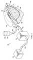
- FIG. 1is an illustrative embodiment of a reduced-pressure therapy system for treating tissue
- FIG. 2is a flow chart illustrating a method of administering a reduced pressure therapy to a tissue site requiring cartilage regeneration according to an illustrative embodiment
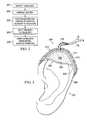
- FIG. 3illustrates use of a mold to facilitate administration of reduced pressure therapy to induce connective tissue regeneration according to an illustrative embodiment
- FIGS. 4A-4Cillustrate histological sections demonstrating the results of reduced pressure therapy for cartilage regeneration according to an illustrative embodiment.
- reduced pressuregenerally refers to a pressure less than the ambient pressure at a tissue site that is being subjected to treatment. In most cases, this reduced pressure will be less than the atmospheric pressure at which the patient is located. Alternatively, the reduced pressure may be less than a hydrostatic pressure associated with tissue at the tissue site.
- vacuumand “negative pressure” may be used to describe the pressure applied to the tissue site, the actual pressure reduction applied to the tissue site may be significantly less than the pressure reduction normally associated with a complete vacuum.
- Reduced pressuremay initially generate fluid flow in the area of the tissue site. As the hydrostatic pressure around the tissue site approaches the desired reduced pressure, the flow may subside, and the reduced pressure is then maintained. Unless otherwise indicated, values of pressure stated herein are gauge pressures. Similarly, references to increases in reduced pressure typically refer to a decrease in absolute pressure, while decreases in reduced pressure typically refer to an increase in absolute pressure.
- tissue siterefers to the location of a wound or defect on or within any tissue, including but not limited to, bone tissue, adipose tissue, muscle tissue, neural tissue, dermal tissue, vascular tissue, connective tissue, cartilage, tendons, or ligaments.
- tissue sitemay further refer to areas of any tissue that are not necessarily wounded or defective, but are instead areas in which it is desired to add or promote the growth of additional tissue. For example, reduced pressure tissue treatment may be used in certain tissue areas to grow additional tissue that may be harvested and transplanted to another tissue location.
- tissue site 102an illustrative embodiment of a system 100 for applying reduced-pressure therapy to a tissue site 102 is shown.
- the illustrative embodiments of the system 100apply reduced-pressure therapy to a wound 106 at the tissue site 102 which includes, for example, cartilage that needs to be repaired by regeneration.
- the tissue site 102may be the bodily tissue of any human, animal, or other organism, including bone tissue, adipose tissue, muscle tissue, dermal tissue, vascular tissue, connective tissue, cartilage, tendons, ligaments, or any other tissue.
- Treatment of tissue site 102may include removal of fluids, e.g., ascites or exudate, delivery of fluids, e.g., saline or materials such as growth factors, and delivery of reduced pressure, for facilitating the growth of cartilage.
- the cartilage wound 106 on the tissue site 102may be due a variety of causes, including trauma, surgery, wear, arthritis, cancer, etc., or may be congenital.
- the system 100comprises a reduced pressure dressing 110 , which includes a manifold 111 adapted to distribute the reduced pressure to the tissue site 102 and a scaffold 112 adapted for placement adjacent the wound 106 , and a drape 114 at least partially covering the reduced pressure dressing 110 to provide a seal covering the wound 106 at the tissue site 102 .
- a chondrocyte or chondrocyte precursorcan be placed directly on the tissue site 102 or be contained within a scaffold 112 that is applied to the tissue site 102 .
- the system 100further comprises a canister 115 with a filter (not shown) and a reduced pressure source 116 , wherein the canister 115 is in fluid communication with the reduced pressure dressing 110 via a conduit 118 and is also in fluid communication with the reduced pressure source 116 via a conduit 119 .
- the reduced pressure source 116is adapted to supply reduced pressure to the manifold 111 and the scaffold 112 which distribute the reduced pressure to the tissue site 102 when in operation.
- the conduit 118may fluidly communicate with the reduced pressure dressing 110 through a tubing adapter 120 to provide the reduced pressure through the drape 114 to the manifold 111 .
- the reduced pressure dressing 110may be constructed from multiple layers or materials in addition to or in lieu of the manifold 111 , the scaffold 112 , and the drape 114 . Some of these layers may be bioabsorbable while others are not.
- the manifold 111may include a bioabsorbable material adjacent to a bio-inert material or a bioabsorbable material that degrades more slowly (as the terms are defined below), such that the reduced pressure dressing 110 may be removed and replaced without removal of any absorbable scaffold 112 , that supports tissue growth, from the wound 106 .
- the canister 115may be a fluid reservoir, or collection member, to filter and hold exudates and other fluids removed from the tissue site 102 .
- the canister 115may include other devices (not shown) including the following non-limiting examples: a pressure-feedback device, a volume detection system, a blood detection system, an infection detection system, a flow monitoring system, and a temperature monitoring system. Some of these devices may be formed integral with the reduce-pressure source 116 .
- a reduced-pressure port on the reduced-pressure source 116may include a filter member that includes one or more filters, e.g., an odor filter.
- the reduced-pressure source 116may be any device for supplying a reduced pressure, such as a vacuum pump, wall suction, or other source. While the amount and nature of reduced pressure applied to a tissue site will typically vary according to the application, the reduced pressure will typically be between ⁇ 5 mm Hg and ⁇ 500 mm Hg and more typically between ⁇ 100 mm Hg and ⁇ 300 mm Hg. The particular protocol used in reduced pressure treatment depends upon the location of the tissue site 102 , the reduced pressure dressing 110 , or pharmacological agents being utilized. Additionally, reduced pressure may be a substantially continuous or cyclical application such that it oscillates the pressure over time.
- the reduced pressure source 116may include sensors, processing units, alarm indicators, memory, databases, software, display units, and user interfaces that further facilitate the application of reduced pressure treatment to the tissue site 102 .
- a sensor or switch(not shown) may be disposed at or near the reduced pressure source 116 to determine a source pressure generated by the reduced pressure source 116 .
- the sensormay communicate with a processing unit that monitors and controls the reduced pressure but is delivered by the reduced pressure source 116 .
- the cartilagemay be any type of cartilage.
- hyaline cartilageis the most common type of cartilage in the body and characteristically contains collagen type II fibers in its extracellular matrix.
- Hyaline cartilageis found in articular joints, costal cartilage (ribs), nose, larynx, and growth plate.
- Another type of cartilageis elastic cartilage found in ear, trachea and epiglottis.
- the third type of cartilaginous tissue, fibrocartilageis present in the pubic symphysis, intervertebral disc, parts of the articular joints, menisci and in sites connecting tendons or ligaments to bones.
- There also exist various combinations or intermediates of these types of cartilagesuch as the epiphyseal cartilage in the growth or cartilage plate.
- the manifold 111 of the reduced pressure dressing 110is adapted to contact the scaffold 112 or portions of the tissue site 102 .
- the manifold 111may be partially or fully in contact with the tissue site 102 being treated by the reduced pressure dressing 110 .
- the manifold 111may partially or fully fill the wound.
- the manifold 111may be any size, shape, or thickness depending on a variety of factors, such as a type of treatment being implemented or the nature and size of the tissue site 102 .
- the size and shape of the manifold 111may be customized by a user to cover a particular portion of the scaffold 112 and/or the tissue site 102 .
- the manifold 111may have, for example, a square shape, or may be shaped as a circle, polygon, an irregular shape, or any other shape.
- the manifold 111is a foam material that distributes reduced pressure to the scaffold 112 and the tissue site 102 when the manifold 111 is in contact with, or near, the scaffold 112 .
- Foam materialmay be either hydrophobic or hydrophilic.
- the manifold 111is an open-cell, articulated polyurethane foam such as GranuFoam®dressing available from Kinetic Concepts, Inc. of San Antonio, Tex.
- the manifold 111is made from a hydrophilic material, where the manifold 111 functions to wick fluid away from the tissue site 102 , while continuing to provide reduced pressure to the scaffold 112 and the tissue site 102 as a manifold.
- the wicking properties of the manifold 111can draw fluid away from the scaffold 112 and the tissue site 102 by capillary flow or other wicking mechanisms.
- An example of hydrophilic foamis a polyvinyl alcohol, open-cell foam such as V.A.C. WhiteFoam® dressing available from Kinetic Concepts, Inc. of San Antonio, Tex.
- Other hydrophilic foamsmay include those made from polyether. Additional foams that may exhibit hydrophilic characteristics include hydrophobic foams that have been treated or were coated to provide hydrophilicity.
- the manifold 111is constructed from a bioabsorbable material, natural or synthetic, that does not have to be removed from the tissue site 102 following use of the reduced pressure dressing 110 .
- Bioabsorbable materialis material that is capable of being absorbed in the body or removed from the body by excretion or metabolic functions; prior to absorption, the bioabsorbable material may be chemically, enzymatically, or otherwise degraded in vivo into simpler chemical species.
- Suitable bioresorbable materialsmay include, without limitation, a polymeric blend of polylactic acid (PLA) and polyglycolic acid (PGA).
- the polymeric blendmay also include without limitation polycarbonates, polyfumarates, and caprolactones.
- the manifold 111may further serve as a scaffold for new cell growth, or may be used in conjunction with the scaffold 112 to promote cell-growth.
- the manifold 111may further promote granulation at the tissue site 102 as reduced pressure is applied through the reduced pressure dressing 110 .
- any or all of the surfaces of the manifold 111may have an uneven, course, or jaded profile that causes microstrains and stresses at the scaffold 112 and the tissue site 102 when reduced pressure is applied through the manifold 111 . These microstrains and stresses have been shown to increase new tissue growth.
- the scaffold 112may be placed adjacent to, in contact with, or substantially over the tissue site 102 to promote the growth of the cartilage in the wound 106 . As indicated above, the scaffold 112 may also function as a manifold when transferring reduced pressure to the tissue site 102 .
- the scaffold 112is a three-dimensional porous structure that provides a template for cell growth of the cartilage 108 within the wound 106 .
- Nonlimiting examples of scaffold materialsinclude calcium phosphate, collagen, PLA/PGA, hydroxyapatite, carbonates, and processed allograft materials.
- the scaffold 112may also assist in delivering fluids to, or removing fluids from, the tissue site 102 .
- the scaffold 112may further comprise a distribution surface 122 that is positioned adjacent to the wound 106 to facilitate fluid flow, chondrocyte migration, and the like for moving a fluid and other material to or from the tissue site 102 to the pores in the scaffold 112 .
- the scaffold 112is flexible to conform to the shape or contour of the wound 106 at the tissue site 102 .
- the design of the scaffold 112may also serve to prevent cartilage overgrowth. The shape and flexibility of the scaffold 112 may be selected without undue experimentation depending on the type of cartilage being treated in the location of the cartilage in the body treated.
- a chondrocyte or chondrocyte precursormay be grafted, or otherwise applied, to the tissue site 102 or the scaffold 112 to facilitate the growth of the cartilage 108 .
- An example of a chondrocyte precursoris a mesenchymal stem cell.
- the source of the chondrocyte or chondrocyte precursormay be an osteochondral graft, autologous to the patient, or comprising allograft, xenograft, or artificially prepared tissue.
- the tissue sourcemay be chondrocytic cell cultures, such as chondrocyte or stem cell cultures which have been prepared through ex vivo cell culture methods, with or without additional growth factors. For examples of cell culture methods, see, e.g., U.S.
- the tissuemay also be harvested by traditional non-cell culture based means, using techniques such as mosaicplasty, in which cartilage is harvested using commercially available instruments such as Acufex7, COR System, or Arthrex7 Osteochondral Autograft Transfer System. Further, the tissue harvested may be applied directly to the scaffold 112 , or may be cultured beforehand.
- the cells, chondrocyte, or chondrocyte precursormay be transfected, either transiently or stably, to further comprise a recombinant nucleic acid.
- nucleic acidsinclude those that encode a protein, such as a cytokine, an enzyme, or a regulatory protein; a regulatory nucleic acid such as a promoter that causes a native protein to be overexpressed or silenced (e.g., to inhibit cancer initiation or growth); an miRNA or another RNAi molecule; an antisense molecule; a marker to assist in monitoring tissue formation; etc.
- a chondrocyte or chondrocyte precursorcomprising an appropriate recombinant nucleic acid for any particular application.
- Other cellsmay also be seeded onto the scaffold 112 and/or placed on or into the tissue site 102 to stimulate the growth of cartilage.
- Non-limiting examplesinclude fibroblasts, immune cells, stem cells that are not a chondrocyte precursor, etc.
- attachment of the cells to the scaffold 112may be enhanced by coating the scaffold 112 with compounds such as basement membrane components, agar, agarose, gelatin, gum arabic, collagen types I, II, III, IV, and V, fibronectin, laminin, glycosaminoglycans, polyvinyl alcohol, mixtures thereof, and other hydrophilic and peptide attachment materials known to those skilled in the art of cell culture.
- the cellsare seeded onto the scaffold 112 , and the scaffold 112 is incubated before the scaffold 112 is applied to the tissue site 102 .
- a cytokinemay be applied to the tissue site 102 or in the scaffold 112 as indicated above.
- a cytokineis a protein that affects cellular growth, proliferation or differentiation, including growth factors and hormones.
- the cytokineis one that can encourage cartilage growth.
- Nonlimiting examplesinclude bone morphogenic protein (BMP)-2, BMP-6, BMP-7, transforming growth factor- ⁇ (TGF- ⁇ ), insulin-like growth factor (IGF), platelet-derived growth factor (PDGF), or cartilage-derived retinoic acid sensitive protein (CD-RAP).
- BMPbone morphogenic protein
- TGF- ⁇transforming growth factor- ⁇
- IGFinsulin-like growth factor
- PDGFplatelet-derived growth factor
- CD-RAPcartilage-derived retinoic acid sensitive protein
- the cytokinemay be synthetic or naturally produced, or produced naturally or transgenically by cells placed at the tissue site 102 or in the scaffold 112 .
- the scaffold 112may include, without limitation, calcium phosphate, collagen, PLA/PGA, hydroxyapatite, carbonates, and/or processed allograft materials.
- the scaffold 112may be used to release at least one therapeutic or prophylactic agent to the tissue site 102 by binding at least one therapeutic or prophylactic agent to the surface of the scaffold 112 .
- an antibioticmay also be applied to the scaffold 112 , which is then released to the tissue site 102 .
- the scaffold 112is a porous material that includes a plurality of open chambers or “pores” that are connected by flow channels to allow fluid communication between the pores.
- the size, shape, or interconnectivity of the poresmay be uniform, random, or patterned, and may be altered to enhance or control cartilage formation, response, repair, or host integration. Further, the size, shape, or interconnectivity of the pores in the scaffold 112 may be altered to enhance or control the integration of newly formed cartilage 108 with surrounding healthy tissue at the tissue site 102 .
- the scaffold 112has a high void-fraction (i.e., a high content of air). It is desired in some embodiments that the pores are designed to allow the attachment of infiltrating cells to induce new cartilage formation.
- the pores and flow chambersmay be seeded with chondrocytes or other cell types in advance to promote cartilage formation.
- the flow channels in the scaffold 112also facilitate distribution of fluids provided to and removed from the tissue site 102 , including the transfer of reduced pressure to the tissue site 102 .
- the scaffold 112is made primarily of an open pore material that includes a plurality of pores fluidly connected to adjacent pores, where a plurality of flow channels is formed by and between the open pores of the material.
- the variations in size and shape of the poresresults in variations in flow channels and can be used to alter flow characteristics of fluid through the material.
- the scaffold 112 pore sizeranges between 25 ⁇ m and 500 ⁇ m. In other embodiments, the pore size is between 50 ⁇ m and 250 ⁇ m. In additional embodiments, the pore size is between 50 ⁇ m and 150 ⁇ m.
- the scaffold 112may be formed of any biocompatible material, i.e. a material that does not elicit any undesirable local or systemic effects in vivo.
- a biocompatible scaffold 112should also have the mechanical and biochemical properties that provide adequate support for tissue growth and cell proliferation.
- the materialscan be characterized with respect to mechanical properties such as tensile strength using an Instron tester, molecular weight by gel permeation chromatography (GPC), glass transition temperature by differential scanning calorimetry (DSC) and bond structure by infrared (IR) spectroscopy.
- the materialmay also be characterized with respect to toxicology by, for example, mutagen tests, e.g. involving an Ames assay or an in vitro teratogenicity assay, or biochemical, cell, or implantation studies in animals for immunogenicity, inflammation, release or degradation.
- the scaffold 112is formed of a bio-inert material, i.e., a material that does not elicit any response in vivo and does not bioabsorb or otherwise degrade in vivo.
- the scaffold 112is formed of a bioabsorbable material as that term in defined above. Regardless of whether the scaffold 112 is bioabsorbable or bio-inert when it contacts the tissue site 102 , the scaffold 112 may also be biocompatible. If the scaffold 112 is made of bioabsorbable materials, the materials may be designed to degrade within a desired time frame. In one embodiment, the desired degradation time frame is six to twelve weeks. In another embodiment, the desired degradation time frame is between three months and one year.
- the desired degradation timeis greater than a year.
- scaffolds 112 made of bioabsorbable materialsmay degrade in a manner related to the molecular weights of the materials used to make the scaffold 112 . In those embodiments, scaffolds 112 comprising a higher molecular weight material often retain structural integrity for longer periods of time than scaffolds 112 comprising lower molecular weight materials.
- the scaffold 112may be formed by melt-spinning, extrusion, casting, or other techniques well known in the polymer processing area.
- Preferred solvents, if used,are those which are removed by the processing or which are biocompatible in the amounts remaining after processing.
- Examples of polymers which can be used to form scaffolds 112include natural and synthetic polymers.
- Synthetic polymersthat may be used include, but are not limited to, bioabsorbable polymers such as polylactic acid (PLA), polyglycolic acid (PGA), polylactic-coglycolide acid (PLGA), and other polyhydroxyacids, polycaprolactones, polycarbonates, polyamides, polyanhydrides, polyamino acids, polyortho esters, polyacetals, degradable polycyanoacrylates and degradable polyurethanes, as well as a polylactide-coglycolide (PLAGA) polymer or a polyethylene glycol-PLAGA copolymer.
- bioabsorbable polymerssuch as polylactic acid (PLA), polyglycolic acid (PGA), polylactic-coglycolide acid (PLGA), and other polyhydroxyacids, polycaprolactones, polycarbonates, polyamides, polyanhydrides, polyamino acids, polyortho esters, polyacetals, degradable polycyanoacrylates and degradable
- natural polymersinclude, but are not limited to, proteins such as albumin, collagen, fibrin, and synthetic polyamino acids, and polysaccharides such as alginate, heparin, and other naturally occurring biodegradable polymers of sugar units.
- the polymeric blendmay also include without limitation polycarbonates, polyfumarates, and capralactones.
- the bioabsorbable scaffold 112is made of PLA, PGA or PLA/PGA copolymers.
- PLA polymersmay be prepared from the cyclic esters of lactic acids. Both L (+) and D ( ⁇ ) forms of lactic acid can be used to prepare the PLA polymers, as well as the optically inactive DL-lactic acid mixture of D ( ⁇ ) and L (+) lactic acids.
- PGAis the homopolymer of glycolic acid (hydroxyacetic acid). Typically, in the conversion of glycolic acid to polyglycolic acid, glycolic acid is initially reacted with itself to form the cyclic ester glycolide, which in the presence of heat and a catalyst is converted to a high molecular weight linear-chain polymer.
- the scaffold 112may be felted mats, liquids, gels, foams, or any other biocompatible material that provides fluid communication through a plurality of channels in three dimensions.
- the drape 114covers the reduced pressure dressing 110 and serves as a semi-permeable barrier to transmission of fluids such as liquids, air, and other gases.
- the drape 114which in some embodiments provides structural support for the reduced pressure dressing 110 , may be coupled to the reduced pressure dressing 110 or the manifold 111 using any technique, including via an adhesive.
- the term “coupled”includes coupling via a separate object and includes direct coupling.
- the term “coupled”also encompasses two or more components that are continuous with one another by virtue of each of the components being formed from the same piece of material.
- the term “coupled”may include chemical, such as via a chemical bond, mechanical, thermal, or electrical coupling.
- the drape 114is not a separate, attached structure, but instead the manifold 111 itself may include a lining of impermeable materials that functions the same as the drape 114 .
- the drapemay be any material that provides a pneumatic or fluid seal.
- the drapemay, for example, be an impermeable or semi-permeable elastomeric material.
- “Elastomeric”means having the properties of an elastomer. It generally refers to a polymeric material that has rubber-like properties. More specifically, most elastomers have elongation rates greater than 100% and a significant amount of resilience. The resilience of a material refers to the material's ability to recover from an elastic deformation.
- Nonlimiting examples of elastomersinclude natural rubbers, polyisoprene, styrene butadiene rubber, chloroprene rubber, polybutadiene, nitrile rubber, butyl rubber, ethylene propylene rubber, ethylene propylene diene monomer, chlorosulfonated polyethylene, polysulfide rubber, polyurethane, EVA film, co-polyester, and silicones.
- drape materialsinclude a silicone drape, 3M Tegaderm® drape, acrylic drape such as one available from Avery Dennison, or an incise drape.
- the system 100is used to stimulate formation of cartilage at the tissue site 102 .
- a caretakercan apply a chondrocyte or chondrocyte precursor to the tissue site 102 or the reduced pressure dressing 110 , and then apply reduced pressure to the tissue site 102 via the manifold 111 and the scaffold 112 for a time sufficient to cause new cartilage formation at the tissue site 102 .
- the application of reduced pressurecan result in the flexible drape 114 compressing and conforming to the surface of the tissue site 102 as air is removed from within the space between the drape 114 and the tissue site 102 .
- the system 100may be used to cosmetically alter tissue having cartilage, such as a nose or ear.
- Cartilagemay also be harvested on one mammal and then transplanted to another mammal, e.g., growing a nose or ear on a mouse for transplantation to a human.
- the system 100may also be applied to a cartilage wound 106 and used to at least partially fill the wound 106 .
- the system 100may also allow effective control of fixation, temperature, pressure (and its associated gradients for vital gases such as oxygen), osmotic forces, oncotic forces, and the addition or removal of various nutrients and pharmacological agents. Still further, the devices to apply reduced pressure in the current system and methodology may be enabled to transfer elements for the manipulation of gas and liquid pathways by preprogrammed, coordinated influx and efflux cycles. Such cycles would be designed to maintain the desired integrity and stability of the system while still allowing variations in multiple forces, flows, and concentrations within tolerated ranges.
- the system 100may also be configured to deliver fluid, liquids or gas, to the tissue site 102 .
- a fluid supply 124 for delivering a fluid 125 to the tissue site 102fluidly communicates with the reduced pressure dressing 110 by a conduit 126 that may be connected directly to the reduced pressure dressing 110 (not shown) or indirectly via the conduit 118 which requires the use of valves 127 and 128 for controlling the delivery of reduced pressure from the reduced pressure source 116 and/or fluid 125 from the fluid supply 124 , respectively.
- the fluid supply 124may be separate from, attached to, or integrated within the reduced-pressure source 116 .
- the fluid supply 124enables treatment procedures to infuse the tissue site 102 with fluids to flush contaminants, counter infection, or promote tissue growth in the wound 106 .
- the fluid supply 124can be used to deliver various irrigation fluids, growth factors, antibiotics, anesthetics, antibacterial agents, antiviral agents, cell-growth promotion agents, or chemically active agents to the tissue site 102 .
- the fluid supply 124can also be used to deliver gaseous fluids to the tissue site 102 for a similar purpose including, for example, the delivery of sterile air in small quantities to promote and maintain the therapeutic effect at the tissue site 102 with or without the reduced pressure being maintained.
- the tissue area of interestis identified, for example, by a caretaker (step 201 ). If the tissue site is located underneath the skin of a patient, i.e., not in direct line of sight, the caretaker may identify the tissue site by use of imaging equipment and techniques, such as MRI imaging. At this time, the caretaker would then determine the best path through the patient's body to reach the tissue area which would cause the least damage to healthy, normal tissues.
- the manifoldis then delivered to the tissue site (step 202 ). Further, depending upon the embodiment, conduits to deliver reduced pressure, fluids, gases, or air may be connected before or after the manifold is delivered to the tissue site. If the tissue site is located underneath the skin of the patient, the manifold may be delivered to the tissue site by insertion into the body through the skin of the patient and through any interstitial tissue.
- the tissue sitehas insufficient space to insert a manifold.
- a devicemay be inserted that creates a void.
- this devicemay be an inflatable device. Once a void is prepared, the manifold may then be delivered.
- the main distribution surface of the manifoldis then positioned adjacent to the tissue site (step 203 ).
- a reduced pressureis then applied to the tissue site (step 204 ).
- the reduced pressuremay be applied continuously or in an intermittent fashion. Further, it is contemplated that the reduced pressure may be alternated with delivery of fluids, air, or agents that promote healing or regeneration as previously discussed.
- the length and force of the reduced-pressure therapymay depend upon various factors determined appropriate by a caretaker, such as previous experience, connective tissue regeneration rate, and the like.
- the manifoldmay be removed upon partial or complete regeneration of the cartilage (step 205 ).
- the open pores or flow channels of the scaffoldmay be designed to promote a certain connective tissue growth in a particular three-dimensional shape.
- the scaffoldmay be designed to promote cartilage growth on the surface of the body such as, for example, an ear.
- FIG. 3an illustrative embodiment of a system 300 for applying reduced-pressure therapy to an ear 301 at a tissue site 302 on the top of the ear 301 is shown. This illustrative embodiment of the system 300 applies reduced-pressure therapy to a missing section of the ear 301 , or cartilage wound 306 , to regenerate the missing cartilage.
- the cartilage wound 306 of the tissue site 302may have been due to any cause including, for example, trauma, surgery, or cancer.
- the system 300comprises a reduced pressure dressing 303 which includes a manifold 311 adapted to distribute the reduced pressure to the tissue site 302 and a scaffold 312 adapted for placement adjacent the cartilage wound 306 , and a drape 314 at least partially covering the reduced pressure dressing 303 to provide an airtight seal covering the cartilage wound 306 at the tissue site 302 .
- the remaining components of the system 300include the same components comprising the system 100 described above including, for example, the tube adapter 120 fluidly coupling the conduit 118 to the reduced pressure dressing 303 . All the components of the system 300 described above operate in a fashion similar to the components of the system 100 .
- the scaffold 312may be placed adjacent to, in contact with, or substantially over the tissue site 302 to promote the growth of the cartilage in the cartilage wound 306 .
- the scaffold 312is a three-dimensional porous structure that provides a template for cell growth of the cartilage within the wound 306 .
- the shape and flexibility of the scaffold 312may be selected based on the desired shape of the ear 301 as indicated by the dashed line on the reduced pressure dressing 303 .
- a mold(not shown) may be used to form the scaffold 312 into the desired shape. Once the mold is created to fit the ear 301 at the tissue site 302 with the missing portion, it can be used to form the scaffold 312 into the three-dimensional shape desired to repair the cartilage wound 306 .
- the scaffold 312may contain chondrocytes or a coping may be applied directly to the cartilage wound 306 .
- the drape 314is positioned to cover the reduced pressure dressing 303 as described in detail above. Reduced pressure therapy can then be applied by use of the reduced-pressure source (not shown) via the conduits 118 fluidly coupled to the reduced pressure dressing 303 .
- the moldmay be positioned over the cartilage wound 306 creating a void that may be filled with a fluid containing chondrocytes that is delivered by a fluid supply (not shown) via the conduit 118 or other independent supply of fluid.
- a fluid supplynot shown
- the fluidhardens to form the three-dimensional scaffold 312 that assumes the desired shape for the regenerated cartilage at the tissue site 302 .
- the scaffoldmay also be seeded with chondrocytes after hardening.
- Cartilage formationwas observed in response to the application of reduced pressure therapy to the surface of intact cranial periosteal membranes. These observations are of significance in that cartilage formation in response to a therapy is unique and of great interest in the field of tissue engineering. These formations were observed in the absence of scaffold materials and only with the application of reduced pressure. No cartilage formation was observed in controls not subjected to reduced pressure.
- Cartilage degeneration caused by congenital abnormalities or disease and traumais of great clinical consequence. Because of the lack of blood supply and subsequent wound-healing response, damage to cartilage generally results in an incomplete repair by the body. Full-thickness articular cartilage damage, or osteochondral lesions, allow for the normal inflammatory response, but result in inferior fibrocartilage formation. Surgical intervention is often the only option. Treatments for repair of cartilage damage are often less than satisfactory, and rarely restore full function or return the tissue to its native normal state. This Example demonstrates the induction of new cartilage from periosteum using GranuFoam® and reduced pressure treatment.
- a foam manifold and reduced pressurewere evaluated for their ability to induce the periosteum to synthesize new cartilage.
- the intact, undamaged crania of rabbitswere exposed.
- a GranuFoam® (KCI Licensing, Inc., San Antonio Tex.) foam dressingwas applied to the bone. With some treatments, the foam-covered bone was also subjected to reduced pressure. After treatment, the treated bone was subjected to paraffin embedding, sectioning, and staining to evaluate the effect of the treatment on new bone formation.
- FIG. 4Ashows a na ⁇ ve, undamaged periosteum in rabbit cranium.
- the dotsdenote the demarcation between the cortical bone and the thin layer of the periosteum.
- FIGS. 4B and 4Cshow that, with the use of GranuFoam® and reduced pressure ( ⁇ 125 mm Hg), extensive cartilage tissues was induced overlying the periosteum.
Landscapes
- Health & Medical Sciences (AREA)
- Life Sciences & Earth Sciences (AREA)
- Engineering & Computer Science (AREA)
- Chemical & Material Sciences (AREA)
- Biomedical Technology (AREA)
- Veterinary Medicine (AREA)
- Public Health (AREA)
- General Health & Medical Sciences (AREA)
- Animal Behavior & Ethology (AREA)
- Medicinal Chemistry (AREA)
- Epidemiology (AREA)
- Zoology (AREA)
- Cell Biology (AREA)
- Chemical Kinetics & Catalysis (AREA)
- Transplantation (AREA)
- Pharmacology & Pharmacy (AREA)
- Oral & Maxillofacial Surgery (AREA)
- Dermatology (AREA)
- Botany (AREA)
- Immunology (AREA)
- Bioinformatics & Cheminformatics (AREA)
- Rheumatology (AREA)
- Proteomics, Peptides & Aminoacids (AREA)
- Gastroenterology & Hepatology (AREA)
- Developmental Biology & Embryology (AREA)
- Vascular Medicine (AREA)
- Orthopedic Medicine & Surgery (AREA)
- Urology & Nephrology (AREA)
- Virology (AREA)
- Diabetes (AREA)
- Molecular Biology (AREA)
- Endocrinology (AREA)
- Biotechnology (AREA)
- Heart & Thoracic Surgery (AREA)
- Anesthesiology (AREA)
- General Chemical & Material Sciences (AREA)
- Organic Chemistry (AREA)
- Nuclear Medicine, Radiotherapy & Molecular Imaging (AREA)
- Hematology (AREA)
- Physical Education & Sports Medicine (AREA)
Abstract
Description
Claims (18)
Priority Applications (1)
| Application Number | Priority Date | Filing Date | Title |
|---|---|---|---|
| US13/108,796US8246948B2 (en) | 2008-06-26 | 2011-05-16 | Stimulation of cartilage formation using reduced pressure treatment |
Applications Claiming Priority (3)
| Application Number | Priority Date | Filing Date | Title |
|---|---|---|---|
| US7602808P | 2008-06-26 | 2008-06-26 | |
| US12/491,445US8197806B2 (en) | 2008-06-26 | 2009-06-25 | Stimulation of cartilage formation using reduced pressure treatment |
| US13/108,796US8246948B2 (en) | 2008-06-26 | 2011-05-16 | Stimulation of cartilage formation using reduced pressure treatment |
Related Parent Applications (1)
| Application Number | Title | Priority Date | Filing Date |
|---|---|---|---|
| US12/491,445ContinuationUS8197806B2 (en) | 2008-06-26 | 2009-06-25 | Stimulation of cartilage formation using reduced pressure treatment |
Publications (2)
| Publication Number | Publication Date |
|---|---|
| US20110218504A1 US20110218504A1 (en) | 2011-09-08 |
| US8246948B2true US8246948B2 (en) | 2012-08-21 |
Family
ID=41328471
Family Applications (2)
| Application Number | Title | Priority Date | Filing Date |
|---|---|---|---|
| US12/491,445Active2030-04-19US8197806B2 (en) | 2008-06-26 | 2009-06-25 | Stimulation of cartilage formation using reduced pressure treatment |
| US13/108,796ActiveUS8246948B2 (en) | 2008-06-26 | 2011-05-16 | Stimulation of cartilage formation using reduced pressure treatment |
Family Applications Before (1)
| Application Number | Title | Priority Date | Filing Date |
|---|---|---|---|
| US12/491,445Active2030-04-19US8197806B2 (en) | 2008-06-26 | 2009-06-25 | Stimulation of cartilage formation using reduced pressure treatment |
Country Status (11)
| Country | Link |
|---|---|
| US (2) | US8197806B2 (en) |
| EP (1) | EP2288396B1 (en) |
| JP (2) | JP5538380B2 (en) |
| KR (1) | KR20110022706A (en) |
| CN (2) | CN103655040A (en) |
| AU (1) | AU2009262163B2 (en) |
| CA (1) | CA2726283C (en) |
| MX (1) | MX2010014304A (en) |
| RU (1) | RU2011101665A (en) |
| TW (1) | TW201006447A (en) |
| WO (1) | WO2009158480A2 (en) |
Cited By (2)
| Publication number | Priority date | Publication date | Assignee | Title |
|---|---|---|---|---|
| US9474883B2 (en) | 2012-12-06 | 2016-10-25 | Ic Surgical, Inc. | Adaptable wound drainage system |
| US10792403B2 (en) | 2015-01-08 | 2020-10-06 | Arthrex, Inc. | Suction swab for surgical use |
Families Citing this family (17)
| Publication number | Priority date | Publication date | Assignee | Title |
|---|---|---|---|---|
| WO2008137115A1 (en) | 2007-05-03 | 2008-11-13 | The Brigham And Women's Hospital, Inc. | Multipotent stem cells and uses thereof |
| EP3851131A1 (en) | 2011-05-31 | 2021-07-21 | LifeCell Corporation | Adipose tissue matrices |
| AU2012323963B2 (en) | 2011-10-13 | 2017-10-12 | Solventum Intellectual Properties Company | Stimulation of cartilage repair using reduced pressure treatment |
| AU2013289045B2 (en) | 2012-07-13 | 2017-02-16 | Lifecell Corporation | Methods for improved treatment of adipose tissue |
| US9370536B2 (en) | 2012-09-26 | 2016-06-21 | Lifecell Corporation | Processed adipose tissue |
| US20140336557A1 (en)* | 2013-05-10 | 2014-11-13 | Biovation Ii, Llc | Biopolymer multi-layer multi-functional medical dressing and method of making same |
| US10188861B2 (en)* | 2016-03-29 | 2019-01-29 | Warsaw Orthopedic, Inc. | Bioabsorbable or partially-bioabsorbable bone growth stimulator system and method for manufacturing a bioabsorbable or partially-bioabsorbable bone-regeneration stimulator system |
| CN106421918A (en)* | 2016-11-08 | 2017-02-22 | 华南生物医药研究院 | Chondrocyte composition |
| WO2018213408A1 (en)* | 2017-05-17 | 2018-11-22 | Advanced Aesthetic Technologies, Inc. | Agaroid structures and related methods of use and manufacture |
| JP7297739B2 (en) | 2017-10-18 | 2023-06-26 | ライフセル コーポレーション | Adipose tissue products and manufacturing methods |
| US11123375B2 (en) | 2017-10-18 | 2021-09-21 | Lifecell Corporation | Methods of treating tissue voids following removal of implantable infusion ports using adipose tissue products |
| US11246994B2 (en) | 2017-10-19 | 2022-02-15 | Lifecell Corporation | Methods for introduction of flowable acellular tissue matrix products into a hand |
| AU2018351314A1 (en) | 2017-10-19 | 2020-03-19 | Lifecell Corporation | Flowable acellular tissue matrix products and methods of production |
| WO2019241562A1 (en)* | 2018-06-14 | 2019-12-19 | University Of Miami | Methods of implanting cells |
| AU2020267589B2 (en) | 2019-05-08 | 2024-01-04 | Musculoskeletal Transplant Foundation | Tissue derived porous matrices and methods for making and using same |
| EP3976127B1 (en) | 2019-05-30 | 2025-09-24 | LifeCell Corporation | Biologic breast implant |
| CN112370098B (en)* | 2020-10-20 | 2022-02-25 | 广东施泰宝医疗科技有限公司 | Tendon suture system for joint replacement |
Citations (157)
| Publication number | Priority date | Publication date | Assignee | Title |
|---|---|---|---|---|
| US1355846A (en) | 1920-02-06 | 1920-10-19 | David A Rannells | Medical appliance |
| US2547758A (en) | 1949-01-05 | 1951-04-03 | Wilmer B Keeling | Instrument for treating the male urethra |
| US2632443A (en) | 1949-04-18 | 1953-03-24 | Eleanor P Lesher | Surgical dressing |
| GB692578A (en) | 1949-09-13 | 1953-06-10 | Minnesota Mining & Mfg | Improvements in or relating to drape sheets for surgical use |
| US2682873A (en) | 1952-07-30 | 1954-07-06 | Johnson & Johnson | General purpose protective dressing |
| US2910763A (en) | 1955-08-17 | 1959-11-03 | Du Pont | Felt-like products |
| US2969057A (en) | 1957-11-04 | 1961-01-24 | Brady Co W H | Nematodic swab |
| US3066672A (en) | 1960-09-27 | 1962-12-04 | Jr William H Crosby | Method and apparatus for serial sampling of intestinal juice |
| US3367332A (en) | 1965-08-27 | 1968-02-06 | Gen Electric | Product and process for establishing a sterile area of skin |
| US3520300A (en) | 1967-03-15 | 1970-07-14 | Amp Inc | Surgical sponge and suction device |
| US3568675A (en) | 1968-08-30 | 1971-03-09 | Clyde B Harvey | Fistula and penetrating wound dressing |
| US3648692A (en) | 1970-12-07 | 1972-03-14 | Parke Davis & Co | Medical-surgical dressing for burns and the like |
| US3682180A (en) | 1970-06-08 | 1972-08-08 | Coilform Co Inc | Drain clip for surgical drain |
| US3826254A (en) | 1973-02-26 | 1974-07-30 | Verco Ind | Needle or catheter retaining appliance |
| DE2640413A1 (en) | 1976-09-08 | 1978-03-09 | Wolf Gmbh Richard | CATHETER MONITORING DEVICE |
| US4080970A (en) | 1976-11-17 | 1978-03-28 | Miller Thomas J | Post-operative combination dressing and internal drain tube with external shield and tube connector |
| US4096853A (en) | 1975-06-21 | 1978-06-27 | Hoechst Aktiengesellschaft | Device for the introduction of contrast medium into an anus praeter |
| US4139004A (en) | 1977-02-17 | 1979-02-13 | Gonzalez Jr Harry | Bandage apparatus for treating burns |
| US4165748A (en) | 1977-11-07 | 1979-08-28 | Johnson Melissa C | Catheter tube holder |
| US4184510A (en) | 1977-03-15 | 1980-01-22 | Fibra-Sonics, Inc. | Valued device for controlling vacuum in surgery |
| US4233969A (en) | 1976-11-11 | 1980-11-18 | Lock Peter M | Wound dressing materials |
| US4245630A (en) | 1976-10-08 | 1981-01-20 | T. J. Smith & Nephew, Ltd. | Tearable composite strip of materials |
| US4256109A (en) | 1978-07-10 | 1981-03-17 | Nichols Robert L | Shut off valve for medical suction apparatus |
| US4261363A (en) | 1979-11-09 | 1981-04-14 | C. R. Bard, Inc. | Retention clips for body fluid drains |
| US4275721A (en) | 1978-11-28 | 1981-06-30 | Landstingens Inkopscentral Lic, Ekonomisk Forening | Vein catheter bandage |
| US4284079A (en) | 1979-06-28 | 1981-08-18 | Adair Edwin Lloyd | Method for applying a male incontinence device |
| US4297995A (en) | 1980-06-03 | 1981-11-03 | Key Pharmaceuticals, Inc. | Bandage containing attachment post |
| US4333468A (en) | 1980-08-18 | 1982-06-08 | Geist Robert W | Mesentery tube holder apparatus |
| US4373519A (en) | 1981-06-26 | 1983-02-15 | Minnesota Mining And Manufacturing Company | Composite wound dressing |
| US4382441A (en) | 1978-12-06 | 1983-05-10 | Svedman Paul | Device for treating tissues, for example skin |
| US4392853A (en) | 1981-03-16 | 1983-07-12 | Rudolph Muto | Sterile assembly for protecting and fastening an indwelling device |
| US4392858A (en) | 1981-07-16 | 1983-07-12 | Sherwood Medical Company | Wound drainage device |
| US4419097A (en) | 1981-07-31 | 1983-12-06 | Rexar Industries, Inc. | Attachment for catheter tube |
| EP0100148A1 (en) | 1982-07-06 | 1984-02-08 | Dow Corning Limited | Medical-surgical dressing and a process for the production thereof |
| US4465485A (en) | 1981-03-06 | 1984-08-14 | Becton, Dickinson And Company | Suction canister with unitary shut-off valve and filter features |
| EP0117632A2 (en) | 1983-01-27 | 1984-09-05 | Johnson & Johnson Products Inc. | Adhesive film dressing |
| US4475909A (en) | 1982-05-06 | 1984-10-09 | Eisenberg Melvin I | Male urinary device and method for applying the device |
| US4480638A (en) | 1980-03-11 | 1984-11-06 | Eduard Schmid | Cushion for holding an element of grafted skin |
| US4525374A (en) | 1984-02-27 | 1985-06-25 | Manresa, Inc. | Treating hydrophobic filters to render them hydrophilic |
| US4525166A (en) | 1981-11-21 | 1985-06-25 | Intermedicat Gmbh | Rolled flexible medical suction drainage device |
| US4540412A (en) | 1983-07-14 | 1985-09-10 | The Kendall Company | Device for moist heat therapy |
| US4543100A (en) | 1983-11-01 | 1985-09-24 | Brodsky Stuart A | Catheter and drain tube retainer |
| US4548202A (en) | 1983-06-20 | 1985-10-22 | Ethicon, Inc. | Mesh tissue fasteners |
| US4551139A (en) | 1982-02-08 | 1985-11-05 | Marion Laboratories, Inc. | Method and apparatus for burn wound treatment |
| EP0161865A2 (en) | 1984-05-03 | 1985-11-21 | Smith and Nephew Associated Companies p.l.c. | Adhesive wound dressing |
| US4569348A (en) | 1980-02-22 | 1986-02-11 | Velcro Usa Inc. | Catheter tube holder strap |
| US4605399A (en) | 1984-12-04 | 1986-08-12 | Complex, Inc. | Transdermal infusion device |
| US4608041A (en) | 1981-10-14 | 1986-08-26 | Frese Nielsen | Device for treatment of wounds in body tissue of patients by exposure to jets of gas |
| US4640688A (en) | 1985-08-23 | 1987-02-03 | Mentor Corporation | Urine collection catheter |
| US4655754A (en) | 1984-11-09 | 1987-04-07 | Stryker Corporation | Vacuum wound drainage system and lipids baffle therefor |
| US4664662A (en) | 1984-08-02 | 1987-05-12 | Smith And Nephew Associated Companies Plc | Wound dressing |
| US4710165A (en) | 1985-09-16 | 1987-12-01 | Mcneil Charles B | Wearable, variable rate suction/collection device |
| US4733659A (en) | 1986-01-17 | 1988-03-29 | Seton Company | Foam bandage |
| GB2195255A (en) | 1986-09-30 | 1988-04-07 | Vacutec Uk Limited | Method and apparatus for vacuum treatment of an epidermal surface |
| US4743232A (en) | 1986-10-06 | 1988-05-10 | The Clinipad Corporation | Package assembly for plastic film bandage |
| GB2197789A (en) | 1986-11-28 | 1988-06-02 | Smiths Industries Plc | Anti-foaming disinfectants used in surgical suction apparatus |
| US4758220A (en) | 1985-09-26 | 1988-07-19 | Alcon Laboratories, Inc. | Surgical cassette proximity sensing and latching apparatus |
| US4787888A (en) | 1987-06-01 | 1988-11-29 | University Of Connecticut | Disposable piezoelectric polymer bandage for percutaneous delivery of drugs and method for such percutaneous delivery (a) |
| US4826494A (en) | 1984-11-09 | 1989-05-02 | Stryker Corporation | Vacuum wound drainage system |
| US4838883A (en) | 1986-03-07 | 1989-06-13 | Nissho Corporation | Urine-collecting device |
| US4840187A (en) | 1986-09-11 | 1989-06-20 | Bard Limited | Sheath applicator |
| US4863449A (en) | 1987-07-06 | 1989-09-05 | Hollister Incorporated | Adhesive-lined elastic condom cathether |
| US4872450A (en) | 1984-08-17 | 1989-10-10 | Austad Eric D | Wound dressing and method of forming same |
| US4878901A (en) | 1986-10-10 | 1989-11-07 | Sachse Hans Ernst | Condom catheter, a urethral catheter for the prevention of ascending infections |
| GB2220357A (en) | 1988-05-28 | 1990-01-10 | Smiths Industries Plc | Medico-surgical containers |
| US4897081A (en) | 1984-05-25 | 1990-01-30 | Thermedics Inc. | Percutaneous access device |
| US4906233A (en) | 1986-05-29 | 1990-03-06 | Terumo Kabushiki Kaisha | Method of securing a catheter body to a human skin surface |
| US4906240A (en) | 1988-02-01 | 1990-03-06 | Matrix Medica, Inc. | Adhesive-faced porous absorbent sheet and method of making same |
| US4919654A (en) | 1988-08-03 | 1990-04-24 | Kalt Medical Corporation | IV clamp with membrane |
| CA2005436A1 (en) | 1988-12-13 | 1990-06-13 | Glenda G. Kalt | Transparent tracheostomy tube dressing |
| US4941882A (en) | 1987-03-14 | 1990-07-17 | Smith And Nephew Associated Companies, P.L.C. | Adhesive dressing for retaining a cannula on the skin |
| US4953565A (en) | 1986-11-26 | 1990-09-04 | Shunro Tachibana | Endermic application kits for external medicines |
| US4969880A (en) | 1989-04-03 | 1990-11-13 | Zamierowski David S | Wound dressing and treatment method |
| US4985019A (en) | 1988-03-11 | 1991-01-15 | Michelson Gary K | X-ray marker |
| GB2235877A (en) | 1989-09-18 | 1991-03-20 | Antonio Talluri | Closed wound suction apparatus |
| US5037397A (en) | 1985-05-03 | 1991-08-06 | Medical Distributors, Inc. | Universal clamp |
| US5053050A (en) | 1988-04-29 | 1991-10-01 | Samuel Itay | Compositions for repair of cartilage and bone |
| US5086170A (en) | 1989-01-16 | 1992-02-04 | Roussel Uclaf | Process for the preparation of azabicyclo compounds |
| US5092858A (en) | 1990-03-20 | 1992-03-03 | Becton, Dickinson And Company | Liquid gelling agent distributor device |
| US5100396A (en) | 1989-04-03 | 1992-03-31 | Zamierowski David S | Fluidic connection system and method |
| US5134994A (en) | 1990-02-12 | 1992-08-04 | Say Sam L | Field aspirator in a soft pack with externally mounted container |
| US5149331A (en) | 1991-05-03 | 1992-09-22 | Ariel Ferdman | Method and device for wound closure |
| US5167613A (en) | 1992-03-23 | 1992-12-01 | The Kendall Company | Composite vented wound dressing |
| US5176663A (en) | 1987-12-02 | 1993-01-05 | Pal Svedman | Dressing having pad with compressibility limiting elements |
| US5215522A (en) | 1984-07-23 | 1993-06-01 | Ballard Medical Products | Single use medical aspirating device and method |
| US5232453A (en) | 1989-07-14 | 1993-08-03 | E. R. Squibb & Sons, Inc. | Catheter holder |
| US5261893A (en) | 1989-04-03 | 1993-11-16 | Zamierowski David S | Fastening system and method |
| US5278100A (en) | 1991-11-08 | 1994-01-11 | Micron Technology, Inc. | Chemical vapor deposition technique for depositing titanium silicide on semiconductor wafers |
| US5279550A (en) | 1991-12-19 | 1994-01-18 | Gish Biomedical, Inc. | Orthopedic autotransfusion system |
| US5298015A (en) | 1989-07-11 | 1994-03-29 | Nippon Zeon Co., Ltd. | Wound dressing having a porous structure |
| US5342376A (en) | 1993-05-03 | 1994-08-30 | Dermagraphics, Inc. | Inserting device for a barbed tissue connector |
| US5344415A (en) | 1993-06-15 | 1994-09-06 | Deroyal Industries, Inc. | Sterile system for dressing vascular access site |
| DE4306478A1 (en) | 1993-03-02 | 1994-09-08 | Wolfgang Dr Wagner | Drainage device, in particular pleural drainage device, and drainage method |
| US5358494A (en) | 1989-07-11 | 1994-10-25 | Svedman Paul | Irrigation dressing |
| US5437651A (en) | 1993-09-01 | 1995-08-01 | Research Medical, Inc. | Medical suction apparatus |
| US5437622A (en) | 1992-04-29 | 1995-08-01 | Laboratoire Hydrex (Sa) | Transparent adhesive dressing with reinforced starter cuts |
| DE29504378U1 (en) | 1995-03-15 | 1995-09-14 | MTG Medizinisch, technische Gerätebau GmbH, 66299 Friedrichsthal | Electronically controlled low-vacuum pump for chest and wound drainage |
| US5527293A (en) | 1989-04-03 | 1996-06-18 | Kinetic Concepts, Inc. | Fastening system and method |
| US5549584A (en) | 1994-02-14 | 1996-08-27 | The Kendall Company | Apparatus for removing fluid from a wound |
| US5556375A (en) | 1994-06-16 | 1996-09-17 | Hercules Incorporated | Wound dressing having a fenestrated base layer |
| US5607388A (en) | 1994-06-16 | 1997-03-04 | Hercules Incorporated | Multi-purpose wound dressing |
| US5636643A (en)* | 1991-11-14 | 1997-06-10 | Wake Forest University | Wound treatment employing reduced pressure |
| US5645081A (en) | 1991-11-14 | 1997-07-08 | Wake Forest University | Method of treating tissue damage and apparatus for same |
| US5723331A (en) | 1994-05-05 | 1998-03-03 | Genzyme Corporation | Methods and compositions for the repair of articular cartilage defects in mammals |
| US5786219A (en) | 1996-10-28 | 1998-07-28 | Molecular Probes, Inc. | Microspheres with fluorescent spherical zones |
| US5811094A (en) | 1990-11-16 | 1998-09-22 | Osiris Therapeutics, Inc. | Connective tissue regeneration using human mesenchymal stem cell preparations |
| WO1998046164A1 (en) | 1997-04-11 | 1998-10-22 | Usbiomaterials Corporation | Biodegradable implant material comprising bioactive ceramic |
| WO1998053768A1 (en) | 1997-05-30 | 1998-12-03 | Osteobiologics, Inc. | Fiber-reinforced, porous, biodegradable implant device |
| GB2333965A (en) | 1997-09-12 | 1999-08-11 | Kci Medical Ltd | Surgical drape |
| US6071267A (en) | 1998-02-06 | 2000-06-06 | Kinetic Concepts, Inc. | Medical patient fluid management interface system and method |
| US6135116A (en) | 1997-07-28 | 2000-10-24 | Kci Licensing, Inc. | Therapeutic method for treating ulcers |
| US6241747B1 (en) | 1993-05-03 | 2001-06-05 | Quill Medical, Inc. | Barbed Bodily tissue connector |
| US6287316B1 (en) | 1999-03-26 | 2001-09-11 | Ethicon, Inc. | Knitted surgical mesh |
| US20020077661A1 (en) | 2000-12-20 | 2002-06-20 | Vahid Saadat | Multi-barbed device for retaining tissue in apposition and methods of use |
| CA2216752C (en) | 1996-01-16 | 2002-07-09 | Matrix Biotechnologies, Inc. | Cartilage repair unit |
| US20020115951A1 (en) | 2001-02-22 | 2002-08-22 | Core Products International, Inc. | Ankle brace providing upper and lower ankle adjustment |
| US20020120185A1 (en) | 2000-05-26 | 2002-08-29 | Kci Licensing, Inc. | System for combined transcutaneous blood gas monitoring and vacuum assisted wound closure |
| US20020143286A1 (en) | 2001-03-05 | 2002-10-03 | Kci Licensing, Inc. | Vacuum assisted wound treatment apparatus and infection identification system and method |
| US6488643B1 (en) | 1998-10-08 | 2002-12-03 | Kci Licensing, Inc. | Wound healing foot wrap |
| US6491693B1 (en) | 1999-12-07 | 2002-12-10 | Michael Lytinas | Method of promoting osteogenesis by application of a vacuum to affected bone areas, and device for same |
| US6493568B1 (en) | 1994-07-19 | 2002-12-10 | Kci Licensing, Inc. | Patient interface system |
| AU755496B2 (en) | 1997-09-12 | 2002-12-12 | Kci Licensing, Inc. | Surgical drape and suction head for wound treatment |
| US20030225347A1 (en) | 2002-06-03 | 2003-12-04 | Argenta Louis C. | Directed tissue growth employing reduced pressure |
| US6695823B1 (en) | 1999-04-09 | 2004-02-24 | Kci Licensing, Inc. | Wound therapy device |
| US6727224B1 (en) | 1999-02-01 | 2004-04-27 | Genetics Institute, Llc. | Methods and compositions for healing and repair of articular cartilage |
| US20040134502A1 (en) | 2002-03-22 | 2004-07-15 | Shuichi Mizuno | Method for in situ repair of injured, damaged, diseased or aged articular cartilage |
| US6763836B2 (en) | 1998-06-02 | 2004-07-20 | Arthrocare Corporation | Methods for electrosurgical tendon vascularization |
| US6767334B1 (en) | 1998-12-23 | 2004-07-27 | Kci Licensing, Inc. | Method and apparatus for wound treatment |
| EP1466633A1 (en) | 2003-04-02 | 2004-10-13 | Lifescan, Inc. | Composite scaffolds seeded with mammalian cells |
| US20040225296A1 (en) | 1994-01-26 | 2004-11-11 | Kyphon Inc. | Devices and methods using an expandable body with internal restraint for compressing cancellous bone |
| US6840962B1 (en) | 1995-05-01 | 2005-01-11 | Massachusetts Institute Of Technology | Tissue engineered tendons and ligaments |
| US6936037B2 (en) | 2002-12-31 | 2005-08-30 | Kci Licensing, Inc. | Tissue closure treatment system, patient interface and method |
| US6949525B2 (en) | 2001-11-13 | 2005-09-27 | Alcon, Inc. | Use of a mixture of sodium hyaluronate and chondroitin sulfate for the treatment of osteoarthritis |
| US6958149B2 (en) | 1998-10-06 | 2005-10-25 | Stryker Corporation | Repair of larynx, trachea, and other fibrocartilaginous tissues |
| US6982298B2 (en) | 2003-01-10 | 2006-01-03 | The Cleveland Clinic Foundation | Hydroxyphenyl cross-linked macromolecular network and applications thereof |
| US6994702B1 (en) | 1999-04-06 | 2006-02-07 | Kci Licensing, Inc. | Vacuum assisted closure pad with adaptation for phototherapy |
| US20060036331A1 (en) | 2004-03-05 | 2006-02-16 | Lu Helen H | Polymer-ceramic-hydrogel composite scaffold for osteochondral repair |
| US7004915B2 (en) | 2001-08-24 | 2006-02-28 | Kci Licensing, Inc. | Negative pressure assisted tissue treatment system |
| US7070584B2 (en) | 2001-02-20 | 2006-07-04 | Kci Licensing, Inc. | Biocompatible wound dressing |
| US20060154367A1 (en) | 2003-06-27 | 2006-07-13 | Ethicon, Incorporated | Cartilage and bone repair and regeneration using postpartum-derived cells |
| US7077832B2 (en) | 1997-05-27 | 2006-07-18 | Kci Licensing, Inc. | Process and device for application of active substances to a wound surface |
| US7108683B2 (en) | 2001-04-30 | 2006-09-19 | Kci Licensing, Inc | Wound therapy and tissue management system and method with fluid differentiation |
| US20070021697A1 (en) | 2004-07-26 | 2007-01-25 | Kci Licensing, Inc. | System and method for use of agent in combination with subatmospheric tissue treatment |
| US20070021698A1 (en) | 1997-05-27 | 2007-01-25 | Kci Licensing, Inc. | Process and device for application of active substances to a wound surface |
| US7169151B1 (en) | 2003-04-10 | 2007-01-30 | Kci Licensing, Inc. | Bone regeneration device for long bones, and method of use |
| US7186224B2 (en) | 2003-04-28 | 2007-03-06 | Scimed Life Systems, Inc. | Side attaching guidewire torque device |
| US7214202B1 (en) | 1997-07-28 | 2007-05-08 | Kci Licensing, Inc. | Therapeutic apparatus for treating ulcers |
| US20070185426A1 (en) | 2001-02-16 | 2007-08-09 | Kci Licensing, Inc. | Biocompatible wound dressing |
| US7279612B1 (en) | 1999-04-22 | 2007-10-09 | Kci Licensing, Inc. | Wound treatment apparatus employing reduced pressure |
| US7316672B1 (en) | 1995-11-14 | 2008-01-08 | Kci Licensing, Inc. | Portable wound treatment apparatus |
| US20080033324A1 (en) | 2006-03-14 | 2008-02-07 | Cornet Douglas A | System for administering reduced pressure treatment having a manifold with a primary flow passage and a blockage prevention member |
| US7346945B2 (en) | 1996-11-18 | 2008-03-25 | Kci Licensing, Inc. | Bariatric treatment system and related methods |
| US7351250B2 (en) | 2002-08-21 | 2008-04-01 | Kci Licensing, Inc. | Circumferential medical closure device and method |
| JP4129536B2 (en) | 2000-02-24 | 2008-08-06 | ヴェネテック インターナショナル,インコーポレイテッド | Highly compatible catheter anchoring system |
| US20080269674A1 (en) | 2007-04-25 | 2008-10-30 | Biomet Sports Medicine, Inc. | Localized Cartilage Defect Therapy |
| US20080275409A1 (en) | 2007-05-01 | 2008-11-06 | The Brigham And Women's Hospital, Inc. | Wound healing device |
| US20090005796A1 (en) | 2007-06-29 | 2009-01-01 | Swain Larry D | Activation of bone and cartilage formation |
Family Cites Families (9)
| Publication number | Priority date | Publication date | Assignee | Title |
|---|---|---|---|---|
| FR2389355B2 (en)* | 1977-05-03 | 1980-04-18 | Applic Gaz Sa | |
| US4420638A (en)* | 1982-03-02 | 1983-12-13 | E. I. Du Pont De Nemours And Company | Fluorinated ether-ketones |
| MY102407A (en) | 1986-11-14 | 1992-06-17 | Aruze Corp | Roulette playing device |
| ATE359836T1 (en)* | 2001-09-24 | 2007-05-15 | Millenium Biologix Inc | POROUS CERAMIC COMPOSITE BONE IMPLANTS |
| US20090232784A1 (en)* | 2005-03-10 | 2009-09-17 | Dale Feldman | Endothelial predecessor cell seeded wound healing scaffold |
| US7608066B2 (en)* | 2005-08-08 | 2009-10-27 | Innovative Therapies, Inc. | Wound irrigation device pressure monitoring and control system |
| US7837673B2 (en)* | 2005-08-08 | 2010-11-23 | Innovative Therapies, Inc. | Wound irrigation device |
| US20070219585A1 (en)* | 2006-03-14 | 2007-09-20 | Cornet Douglas A | System for administering reduced pressure treatment having a manifold with a primary flow passage and a blockage prevention member |
| NZ589814A (en)* | 2006-03-20 | 2012-03-30 | Tigenix Nv | Methods to maintain, improve and restore the cartilage phenotype of chondrocytes |
- 2009
- 2009-06-25EPEP09771012.3Apatent/EP2288396B1/enactiveActive
- 2009-06-25RURU2011101665/14Apatent/RU2011101665A/ennot_activeApplication Discontinuation
- 2009-06-25MXMX2010014304Apatent/MX2010014304A/ennot_activeApplication Discontinuation
- 2009-06-25JPJP2011516652Apatent/JP5538380B2/enactiveActive
- 2009-06-25AUAU2009262163Apatent/AU2009262163B2/ennot_activeCeased
- 2009-06-25CNCN201310464870.XApatent/CN103655040A/enactivePending
- 2009-06-25KRKR1020117001803Apatent/KR20110022706A/ennot_activeWithdrawn
- 2009-06-25USUS12/491,445patent/US8197806B2/enactiveActive
- 2009-06-25CNCN200980122557XApatent/CN102065929B/enactiveActive
- 2009-06-25WOPCT/US2009/048628patent/WO2009158480A2/enactiveApplication Filing
- 2009-06-25CACA2726283Apatent/CA2726283C/ennot_activeExpired - Fee Related
- 2009-06-26TWTW098121673Apatent/TW201006447A/enunknown
- 2011
- 2011-05-16USUS13/108,796patent/US8246948B2/enactiveActive
- 2013
- 2013-08-02JPJP2013161242Apatent/JP2014000424A/enactivePending
Patent Citations (168)
| Publication number | Priority date | Publication date | Assignee | Title |
|---|---|---|---|---|
| US1355846A (en) | 1920-02-06 | 1920-10-19 | David A Rannells | Medical appliance |
| US2547758A (en) | 1949-01-05 | 1951-04-03 | Wilmer B Keeling | Instrument for treating the male urethra |
| US2632443A (en) | 1949-04-18 | 1953-03-24 | Eleanor P Lesher | Surgical dressing |
| GB692578A (en) | 1949-09-13 | 1953-06-10 | Minnesota Mining & Mfg | Improvements in or relating to drape sheets for surgical use |
| US2682873A (en) | 1952-07-30 | 1954-07-06 | Johnson & Johnson | General purpose protective dressing |
| US2910763A (en) | 1955-08-17 | 1959-11-03 | Du Pont | Felt-like products |
| US2969057A (en) | 1957-11-04 | 1961-01-24 | Brady Co W H | Nematodic swab |
| US3066672A (en) | 1960-09-27 | 1962-12-04 | Jr William H Crosby | Method and apparatus for serial sampling of intestinal juice |
| US3367332A (en) | 1965-08-27 | 1968-02-06 | Gen Electric | Product and process for establishing a sterile area of skin |
| US3520300A (en) | 1967-03-15 | 1970-07-14 | Amp Inc | Surgical sponge and suction device |
| US3568675A (en) | 1968-08-30 | 1971-03-09 | Clyde B Harvey | Fistula and penetrating wound dressing |
| US3682180A (en) | 1970-06-08 | 1972-08-08 | Coilform Co Inc | Drain clip for surgical drain |
| US3648692A (en) | 1970-12-07 | 1972-03-14 | Parke Davis & Co | Medical-surgical dressing for burns and the like |
| US3826254A (en) | 1973-02-26 | 1974-07-30 | Verco Ind | Needle or catheter retaining appliance |
| US4096853A (en) | 1975-06-21 | 1978-06-27 | Hoechst Aktiengesellschaft | Device for the introduction of contrast medium into an anus praeter |
| DE2640413A1 (en) | 1976-09-08 | 1978-03-09 | Wolf Gmbh Richard | CATHETER MONITORING DEVICE |
| US4245630A (en) | 1976-10-08 | 1981-01-20 | T. J. Smith & Nephew, Ltd. | Tearable composite strip of materials |
| US4233969A (en) | 1976-11-11 | 1980-11-18 | Lock Peter M | Wound dressing materials |
| US4080970A (en) | 1976-11-17 | 1978-03-28 | Miller Thomas J | Post-operative combination dressing and internal drain tube with external shield and tube connector |
| US4139004A (en) | 1977-02-17 | 1979-02-13 | Gonzalez Jr Harry | Bandage apparatus for treating burns |
| US4184510A (en) | 1977-03-15 | 1980-01-22 | Fibra-Sonics, Inc. | Valued device for controlling vacuum in surgery |
| US4165748A (en) | 1977-11-07 | 1979-08-28 | Johnson Melissa C | Catheter tube holder |
| US4256109A (en) | 1978-07-10 | 1981-03-17 | Nichols Robert L | Shut off valve for medical suction apparatus |
| US4275721A (en) | 1978-11-28 | 1981-06-30 | Landstingens Inkopscentral Lic, Ekonomisk Forening | Vein catheter bandage |
| US4382441A (en) | 1978-12-06 | 1983-05-10 | Svedman Paul | Device for treating tissues, for example skin |
| US4284079A (en) | 1979-06-28 | 1981-08-18 | Adair Edwin Lloyd | Method for applying a male incontinence device |
| US4261363A (en) | 1979-11-09 | 1981-04-14 | C. R. Bard, Inc. | Retention clips for body fluid drains |
| US4569348A (en) | 1980-02-22 | 1986-02-11 | Velcro Usa Inc. | Catheter tube holder strap |
| US4480638A (en) | 1980-03-11 | 1984-11-06 | Eduard Schmid | Cushion for holding an element of grafted skin |
| US4297995A (en) | 1980-06-03 | 1981-11-03 | Key Pharmaceuticals, Inc. | Bandage containing attachment post |
| US4333468A (en) | 1980-08-18 | 1982-06-08 | Geist Robert W | Mesentery tube holder apparatus |
| US4465485A (en) | 1981-03-06 | 1984-08-14 | Becton, Dickinson And Company | Suction canister with unitary shut-off valve and filter features |
| US4392853A (en) | 1981-03-16 | 1983-07-12 | Rudolph Muto | Sterile assembly for protecting and fastening an indwelling device |
| US4373519A (en) | 1981-06-26 | 1983-02-15 | Minnesota Mining And Manufacturing Company | Composite wound dressing |
| US4392858A (en) | 1981-07-16 | 1983-07-12 | Sherwood Medical Company | Wound drainage device |
| US4419097A (en) | 1981-07-31 | 1983-12-06 | Rexar Industries, Inc. | Attachment for catheter tube |
| US4608041A (en) | 1981-10-14 | 1986-08-26 | Frese Nielsen | Device for treatment of wounds in body tissue of patients by exposure to jets of gas |
| US4525166A (en) | 1981-11-21 | 1985-06-25 | Intermedicat Gmbh | Rolled flexible medical suction drainage device |
| US4551139A (en) | 1982-02-08 | 1985-11-05 | Marion Laboratories, Inc. | Method and apparatus for burn wound treatment |
| US4475909A (en) | 1982-05-06 | 1984-10-09 | Eisenberg Melvin I | Male urinary device and method for applying the device |
| EP0100148A1 (en) | 1982-07-06 | 1984-02-08 | Dow Corning Limited | Medical-surgical dressing and a process for the production thereof |
| EP0117632A2 (en) | 1983-01-27 | 1984-09-05 | Johnson & Johnson Products Inc. | Adhesive film dressing |
| US4548202A (en) | 1983-06-20 | 1985-10-22 | Ethicon, Inc. | Mesh tissue fasteners |
| US4540412A (en) | 1983-07-14 | 1985-09-10 | The Kendall Company | Device for moist heat therapy |
| US4543100A (en) | 1983-11-01 | 1985-09-24 | Brodsky Stuart A | Catheter and drain tube retainer |
| US4525374A (en) | 1984-02-27 | 1985-06-25 | Manresa, Inc. | Treating hydrophobic filters to render them hydrophilic |
| EP0161865A2 (en) | 1984-05-03 | 1985-11-21 | Smith and Nephew Associated Companies p.l.c. | Adhesive wound dressing |
| US4897081A (en) | 1984-05-25 | 1990-01-30 | Thermedics Inc. | Percutaneous access device |
| US5215522A (en) | 1984-07-23 | 1993-06-01 | Ballard Medical Products | Single use medical aspirating device and method |
| US4664662A (en) | 1984-08-02 | 1987-05-12 | Smith And Nephew Associated Companies Plc | Wound dressing |
| US4872450A (en) | 1984-08-17 | 1989-10-10 | Austad Eric D | Wound dressing and method of forming same |
| US4826494A (en) | 1984-11-09 | 1989-05-02 | Stryker Corporation | Vacuum wound drainage system |
| US4655754A (en) | 1984-11-09 | 1987-04-07 | Stryker Corporation | Vacuum wound drainage system and lipids baffle therefor |
| US4605399A (en) | 1984-12-04 | 1986-08-12 | Complex, Inc. | Transdermal infusion device |
| US5037397A (en) | 1985-05-03 | 1991-08-06 | Medical Distributors, Inc. | Universal clamp |
| US4640688A (en) | 1985-08-23 | 1987-02-03 | Mentor Corporation | Urine collection catheter |
| US4710165A (en) | 1985-09-16 | 1987-12-01 | Mcneil Charles B | Wearable, variable rate suction/collection device |
| US4758220A (en) | 1985-09-26 | 1988-07-19 | Alcon Laboratories, Inc. | Surgical cassette proximity sensing and latching apparatus |
| US4733659A (en) | 1986-01-17 | 1988-03-29 | Seton Company | Foam bandage |
| US4838883A (en) | 1986-03-07 | 1989-06-13 | Nissho Corporation | Urine-collecting device |
| US4906233A (en) | 1986-05-29 | 1990-03-06 | Terumo Kabushiki Kaisha | Method of securing a catheter body to a human skin surface |
| US4840187A (en) | 1986-09-11 | 1989-06-20 | Bard Limited | Sheath applicator |
| GB2195255A (en) | 1986-09-30 | 1988-04-07 | Vacutec Uk Limited | Method and apparatus for vacuum treatment of an epidermal surface |
| US4743232A (en) | 1986-10-06 | 1988-05-10 | The Clinipad Corporation | Package assembly for plastic film bandage |
| US4878901A (en) | 1986-10-10 | 1989-11-07 | Sachse Hans Ernst | Condom catheter, a urethral catheter for the prevention of ascending infections |
| US4953565A (en) | 1986-11-26 | 1990-09-04 | Shunro Tachibana | Endermic application kits for external medicines |
| GB2197789A (en) | 1986-11-28 | 1988-06-02 | Smiths Industries Plc | Anti-foaming disinfectants used in surgical suction apparatus |
| US4941882A (en) | 1987-03-14 | 1990-07-17 | Smith And Nephew Associated Companies, P.L.C. | Adhesive dressing for retaining a cannula on the skin |
| US4787888A (en) | 1987-06-01 | 1988-11-29 | University Of Connecticut | Disposable piezoelectric polymer bandage for percutaneous delivery of drugs and method for such percutaneous delivery (a) |
| US4863449A (en) | 1987-07-06 | 1989-09-05 | Hollister Incorporated | Adhesive-lined elastic condom cathether |
| US5176663A (en) | 1987-12-02 | 1993-01-05 | Pal Svedman | Dressing having pad with compressibility limiting elements |
| US4906240A (en) | 1988-02-01 | 1990-03-06 | Matrix Medica, Inc. | Adhesive-faced porous absorbent sheet and method of making same |
| US4985019A (en) | 1988-03-11 | 1991-01-15 | Michelson Gary K | X-ray marker |
| US5053050A (en) | 1988-04-29 | 1991-10-01 | Samuel Itay | Compositions for repair of cartilage and bone |
| EP0358302A2 (en) | 1988-05-28 | 1990-03-14 | Smiths Industries Public Limited Company | Medico-surgical suction container |
| GB2220357A (en) | 1988-05-28 | 1990-01-10 | Smiths Industries Plc | Medico-surgical containers |
| US4919654A (en) | 1988-08-03 | 1990-04-24 | Kalt Medical Corporation | IV clamp with membrane |
| CA2005436A1 (en) | 1988-12-13 | 1990-06-13 | Glenda G. Kalt | Transparent tracheostomy tube dressing |
| US5086170A (en) | 1989-01-16 | 1992-02-04 | Roussel Uclaf | Process for the preparation of azabicyclo compounds |
| US4969880A (en) | 1989-04-03 | 1990-11-13 | Zamierowski David S | Wound dressing and treatment method |
| US5527293A (en) | 1989-04-03 | 1996-06-18 | Kinetic Concepts, Inc. | Fastening system and method |
| US5100396A (en) | 1989-04-03 | 1992-03-31 | Zamierowski David S | Fluidic connection system and method |
| US5261893A (en) | 1989-04-03 | 1993-11-16 | Zamierowski David S | Fastening system and method |
| US5298015A (en) | 1989-07-11 | 1994-03-29 | Nippon Zeon Co., Ltd. | Wound dressing having a porous structure |
| US5358494A (en) | 1989-07-11 | 1994-10-25 | Svedman Paul | Irrigation dressing |
| US5232453A (en) | 1989-07-14 | 1993-08-03 | E. R. Squibb & Sons, Inc. | Catheter holder |
| GB2235877A (en) | 1989-09-18 | 1991-03-20 | Antonio Talluri | Closed wound suction apparatus |
| US5134994A (en) | 1990-02-12 | 1992-08-04 | Say Sam L | Field aspirator in a soft pack with externally mounted container |
| US5092858A (en) | 1990-03-20 | 1992-03-03 | Becton, Dickinson And Company | Liquid gelling agent distributor device |
| US5811094A (en) | 1990-11-16 | 1998-09-22 | Osiris Therapeutics, Inc. | Connective tissue regeneration using human mesenchymal stem cell preparations |
| US5149331A (en) | 1991-05-03 | 1992-09-22 | Ariel Ferdman | Method and device for wound closure |
| US5278100A (en) | 1991-11-08 | 1994-01-11 | Micron Technology, Inc. | Chemical vapor deposition technique for depositing titanium silicide on semiconductor wafers |
| US5645081A (en) | 1991-11-14 | 1997-07-08 | Wake Forest University | Method of treating tissue damage and apparatus for same |
| US5636643A (en)* | 1991-11-14 | 1997-06-10 | Wake Forest University | Wound treatment employing reduced pressure |
| US5279550A (en) | 1991-12-19 | 1994-01-18 | Gish Biomedical, Inc. | Orthopedic autotransfusion system |
| US5167613A (en) | 1992-03-23 | 1992-12-01 | The Kendall Company | Composite vented wound dressing |
| US5437622A (en) | 1992-04-29 | 1995-08-01 | Laboratoire Hydrex (Sa) | Transparent adhesive dressing with reinforced starter cuts |
| DE4306478A1 (en) | 1993-03-02 | 1994-09-08 | Wolfgang Dr Wagner | Drainage device, in particular pleural drainage device, and drainage method |
| US6241747B1 (en) | 1993-05-03 | 2001-06-05 | Quill Medical, Inc. | Barbed Bodily tissue connector |
| US5342376A (en) | 1993-05-03 | 1994-08-30 | Dermagraphics, Inc. | Inserting device for a barbed tissue connector |
| US5344415A (en) | 1993-06-15 | 1994-09-06 | Deroyal Industries, Inc. | Sterile system for dressing vascular access site |
| US5437651A (en) | 1993-09-01 | 1995-08-01 | Research Medical, Inc. | Medical suction apparatus |
| US20040225296A1 (en) | 1994-01-26 | 2004-11-11 | Kyphon Inc. | Devices and methods using an expandable body with internal restraint for compressing cancellous bone |
| US5549584A (en) | 1994-02-14 | 1996-08-27 | The Kendall Company | Apparatus for removing fluid from a wound |
| US5723331A (en) | 1994-05-05 | 1998-03-03 | Genzyme Corporation | Methods and compositions for the repair of articular cartilage defects in mammals |
| US5607388A (en) | 1994-06-16 | 1997-03-04 | Hercules Incorporated | Multi-purpose wound dressing |
| US5556375A (en) | 1994-06-16 | 1996-09-17 | Hercules Incorporated | Wound dressing having a fenestrated base layer |
| US6493568B1 (en) | 1994-07-19 | 2002-12-10 | Kci Licensing, Inc. | Patient interface system |
| DE29504378U1 (en) | 1995-03-15 | 1995-09-14 | MTG Medizinisch, technische Gerätebau GmbH, 66299 Friedrichsthal | Electronically controlled low-vacuum pump for chest and wound drainage |
| US6840962B1 (en) | 1995-05-01 | 2005-01-11 | Massachusetts Institute Of Technology | Tissue engineered tendons and ligaments |
| US7316672B1 (en) | 1995-11-14 | 2008-01-08 | Kci Licensing, Inc. | Portable wound treatment apparatus |
| CA2216752C (en) | 1996-01-16 | 2002-07-09 | Matrix Biotechnologies, Inc. | Cartilage repair unit |
| US5786219A (en) | 1996-10-28 | 1998-07-28 | Molecular Probes, Inc. | Microspheres with fluorescent spherical zones |
| US7346945B2 (en) | 1996-11-18 | 2008-03-25 | Kci Licensing, Inc. | Bariatric treatment system and related methods |
| WO1998046164A1 (en) | 1997-04-11 | 1998-10-22 | Usbiomaterials Corporation | Biodegradable implant material comprising bioactive ceramic |
| US7077832B2 (en) | 1997-05-27 | 2006-07-18 | Kci Licensing, Inc. | Process and device for application of active substances to a wound surface |
| US20070021698A1 (en) | 1997-05-27 | 2007-01-25 | Kci Licensing, Inc. | Process and device for application of active substances to a wound surface |
| WO1998053768A1 (en) | 1997-05-30 | 1998-12-03 | Osteobiologics, Inc. | Fiber-reinforced, porous, biodegradable implant device |
| US6135116A (en) | 1997-07-28 | 2000-10-24 | Kci Licensing, Inc. | Therapeutic method for treating ulcers |
| US7214202B1 (en) | 1997-07-28 | 2007-05-08 | Kci Licensing, Inc. | Therapeutic apparatus for treating ulcers |
| US6345623B1 (en) | 1997-09-12 | 2002-02-12 | Keith Patrick Heaton | Surgical drape and suction head for wound treatment |
| US6814079B2 (en) | 1997-09-12 | 2004-11-09 | Kci Licensing, Inc. | Surgical drape and suction head for wound treatment |
| EP1018967B1 (en) | 1997-09-12 | 2004-08-18 | KCI Licensing, Inc. | Suction head for wound treatment and combination with a surgical drape |
| GB2329127B (en) | 1997-09-12 | 2000-08-16 | Kci Medical Ltd | Surgical drape and suction head for wound treatment |
| AU745271B2 (en) | 1997-09-12 | 2002-03-14 | Kci Licensing, Inc. | Surgical drape and suction head for wound treatment |
| AU755496B2 (en) | 1997-09-12 | 2002-12-12 | Kci Licensing, Inc. | Surgical drape and suction head for wound treatment |
| US6553998B2 (en) | 1997-09-12 | 2003-04-29 | Kci Licensing, Inc. | Surgical drape and suction head for wound treatment |
| GB2333965A (en) | 1997-09-12 | 1999-08-11 | Kci Medical Ltd | Surgical drape |
| US6071267A (en) | 1998-02-06 | 2000-06-06 | Kinetic Concepts, Inc. | Medical patient fluid management interface system and method |
| US6763836B2 (en) | 1998-06-02 | 2004-07-20 | Arthrocare Corporation | Methods for electrosurgical tendon vascularization |
| US6958149B2 (en) | 1998-10-06 | 2005-10-25 | Stryker Corporation | Repair of larynx, trachea, and other fibrocartilaginous tissues |
| US6488643B1 (en) | 1998-10-08 | 2002-12-03 | Kci Licensing, Inc. | Wound healing foot wrap |
| US6767334B1 (en) | 1998-12-23 | 2004-07-27 | Kci Licensing, Inc. | Method and apparatus for wound treatment |
| US6727224B1 (en) | 1999-02-01 | 2004-04-27 | Genetics Institute, Llc. | Methods and compositions for healing and repair of articular cartilage |
| US6287316B1 (en) | 1999-03-26 | 2001-09-11 | Ethicon, Inc. | Knitted surgical mesh |
| US6994702B1 (en) | 1999-04-06 | 2006-02-07 | Kci Licensing, Inc. | Vacuum assisted closure pad with adaptation for phototherapy |
| US6695823B1 (en) | 1999-04-09 | 2004-02-24 | Kci Licensing, Inc. | Wound therapy device |
| US7279612B1 (en) | 1999-04-22 | 2007-10-09 | Kci Licensing, Inc. | Wound treatment apparatus employing reduced pressure |
| US6491693B1 (en) | 1999-12-07 | 2002-12-10 | Michael Lytinas | Method of promoting osteogenesis by application of a vacuum to affected bone areas, and device for same |
| JP4129536B2 (en) | 2000-02-24 | 2008-08-06 | ヴェネテック インターナショナル,インコーポレイテッド | Highly compatible catheter anchoring system |
| US20020120185A1 (en) | 2000-05-26 | 2002-08-29 | Kci Licensing, Inc. | System for combined transcutaneous blood gas monitoring and vacuum assisted wound closure |
| US6856821B2 (en) | 2000-05-26 | 2005-02-15 | Kci Licensing, Inc. | System for combined transcutaneous blood gas monitoring and vacuum assisted wound closure |
| US20020077661A1 (en) | 2000-12-20 | 2002-06-20 | Vahid Saadat | Multi-barbed device for retaining tissue in apposition and methods of use |
| US20070185426A1 (en) | 2001-02-16 | 2007-08-09 | Kci Licensing, Inc. | Biocompatible wound dressing |
| US7070584B2 (en) | 2001-02-20 | 2006-07-04 | Kci Licensing, Inc. | Biocompatible wound dressing |
| US20020115951A1 (en) | 2001-02-22 | 2002-08-22 | Core Products International, Inc. | Ankle brace providing upper and lower ankle adjustment |
| US20020143286A1 (en) | 2001-03-05 | 2002-10-03 | Kci Licensing, Inc. | Vacuum assisted wound treatment apparatus and infection identification system and method |
| US7108683B2 (en) | 2001-04-30 | 2006-09-19 | Kci Licensing, Inc | Wound therapy and tissue management system and method with fluid differentiation |
| US7004915B2 (en) | 2001-08-24 | 2006-02-28 | Kci Licensing, Inc. | Negative pressure assisted tissue treatment system |
| US6949525B2 (en) | 2001-11-13 | 2005-09-27 | Alcon, Inc. | Use of a mixture of sodium hyaluronate and chondroitin sulfate for the treatment of osteoarthritis |
| US7468192B2 (en) | 2002-03-22 | 2008-12-23 | Histogenics Corporation | Method for repair of cartilage lesions |
| US20040134502A1 (en) | 2002-03-22 | 2004-07-15 | Shuichi Mizuno | Method for in situ repair of injured, damaged, diseased or aged articular cartilage |
| US20030225347A1 (en) | 2002-06-03 | 2003-12-04 | Argenta Louis C. | Directed tissue growth employing reduced pressure |
| US7351250B2 (en) | 2002-08-21 | 2008-04-01 | Kci Licensing, Inc. | Circumferential medical closure device and method |
| US6936037B2 (en) | 2002-12-31 | 2005-08-30 | Kci Licensing, Inc. | Tissue closure treatment system, patient interface and method |
| US6951553B2 (en) | 2002-12-31 | 2005-10-04 | Kci Licensing, Inc | Tissue closure treatment system and method with externally-applied patient interface |
| US6982298B2 (en) | 2003-01-10 | 2006-01-03 | The Cleveland Clinic Foundation | Hydroxyphenyl cross-linked macromolecular network and applications thereof |
| EP1466633A1 (en) | 2003-04-02 | 2004-10-13 | Lifescan, Inc. | Composite scaffolds seeded with mammalian cells |
| US7169151B1 (en) | 2003-04-10 | 2007-01-30 | Kci Licensing, Inc. | Bone regeneration device for long bones, and method of use |
| US20070123895A1 (en) | 2003-04-10 | 2007-05-31 | Kci Licensing, Inc. | Bone regeneration device for bones, and method of use |
| US7186224B2 (en) | 2003-04-28 | 2007-03-06 | Scimed Life Systems, Inc. | Side attaching guidewire torque device |
| US20060154367A1 (en) | 2003-06-27 | 2006-07-13 | Ethicon, Incorporated | Cartilage and bone repair and regeneration using postpartum-derived cells |
| US20060036331A1 (en) | 2004-03-05 | 2006-02-16 | Lu Helen H | Polymer-ceramic-hydrogel composite scaffold for osteochondral repair |
| US20070021697A1 (en) | 2004-07-26 | 2007-01-25 | Kci Licensing, Inc. | System and method for use of agent in combination with subatmospheric tissue treatment |
| US20080033324A1 (en) | 2006-03-14 | 2008-02-07 | Cornet Douglas A | System for administering reduced pressure treatment having a manifold with a primary flow passage and a blockage prevention member |
| US20080269674A1 (en) | 2007-04-25 | 2008-10-30 | Biomet Sports Medicine, Inc. | Localized Cartilage Defect Therapy |
| US20080275409A1 (en) | 2007-05-01 | 2008-11-06 | The Brigham And Women's Hospital, Inc. | Wound healing device |
| US20090005796A1 (en) | 2007-06-29 | 2009-01-01 | Swain Larry D | Activation of bone and cartilage formation |
Non-Patent Citations (48)
| Title |
|---|
| A.A. Safronov, Dissertation Abstract, Vacuum Therapy of Trophic Ulcers of the Lower Leg with Simultaneous Autoplasty of the Skin (Central Scientific Research Institute of Traumatology and Orthopedics, Moscow, U.S.S.R. 1967) (copy and certified translation). |
| Arnljots, Björn et al.: "Irrigation Treatment in Split-Thickness Skin Grafting of Intractable Leg Ulcers", Scand J. Plast Reconstr. Surg., No. 19, 1985, pp. 211-213. |
| Azimzadeh et al. Hematology and Cell Therapy 38(4):331-343, 1997. |
| C.E. Tennants, "The Use of Hypermia in the Postoperative Treatment of Lesions of the Extremities and Thorax," Journal of the American Medical Association 64 (1915), pp. 1548-1549. |
| Chariker, Mark E., M.D., et al; "Effective Management of incisional and cutaneous fistulae with closed suction wound drainage"; Contemporary Surgery, vol. 34, Jun. 1989, pp. 59-63. |
| Chinn, Steven D. et al.: "Closed Wound Suction Drainage", The Journal of Foot Surgery, vol. 24, No. 1, 1985, pp. 76-81. |
| D.E. Tribble, An Improved Sump Drain-Irrigation Device of Simple Construction, Archives of Surgery 105 (1972) pp. 511-513. |
| Dattilo, Philip P., Jr., et al., "Medical Textiles: Application of an Absorbable Barbed Bi-directional Surgical Suture"; Journal of Textile and Textile and Apparel, Technology and Management, vol. 2, Issue 2, Spring 2002, pp. 1-5. |
| Davydov, Yu. A., et al., "Concepts for the Clinical-Biological Management of the Wound Process in the Treatment of Purulent Wounds by Means of Vacuum Therapy"; Vestnik Khirurgi, Jul. 7, 1980, pp. 132-136, and 8 page English translation thereof. |
| Davydov, Yu. A., et al., "Vacuum Therapy in the Treatment of Purulent Lactation Mastitis"; Vestnik Khirurgi, May 14, 1986, pp. 66-70, and 9 page English translation thereof. |
| Davydov, Yu. A., et al; "Bacteriological and Cytological Assessment of Vacuum Therapy for Purulent Wounds"; Vestnik Khirurgi, Oct. 1988, pp. 48-52, and 8 page English translation thereof. |
| Egnell Minor, Instruction Book, First Edition, 300 7502, Feb. 1975, pp. 24. |
| Egnell Minor: Addition to the Users Manual Concerning Overflow Protection-Concerns all Egnell Pumps, Feb. 3, 1983, pp. 2. |
| F.E Johnson, "An Improved Technique for Skin Graft Placement Using a Suction Drain," Surgery, Gynecology, and Obstetrics 159 (1984), pp. 584-585. |
| G. {hacek over (Z)}ivadinovic, V. ukic, Maksimovic, . Radak, and P. Pe{hacek over (s)}ka, "Vacuum Therapy in the Treatment of Peripheral Blood Vessels," Timok Medical Journal 11 (1986), pp. 161-164 (copy and certified translation). |
| G. {hacek over (Z)}ivadinović, V. ukić, Maksimović, . Radak, and P. Pe{hacek over (s)}ka, "Vacuum Therapy in the Treatment of Peripheral Blood Vessels," Timok Medical Journal 11 (1986), pp. 161-164 (copy and certified translation). |
| George V. Letsou, MD., et al., "Stimulation of Adenylate Cyclase Activity in Cultured Endothelial Cells Subjected to Cyclic Stretch"; Journal of Cardiovascular Surgery, 31, 1990, pp. 634-639. |
| Gould and Auchincloss. Immunology Today 20(2):77-82, 1999. |
| International Search Report and Written Opinion date mailed Aug. 16, 2010; PCT International Application No. PCT/US2009/069031. |
| International Search Report and Written Opinion date mailed May 6, 2010; PCT International Application No. PCT/US2009/048628. |
| International Search Report for PCT International Application PCT/GB95/01983; Nov. 23, 1995. |
| James H. Blackburn, II, MD, et al; "Negative-Pressure Dressings as a Bolster for Skin Grafts"; Annals of Plastic Surgery, vol. 40, No. 5, May 1998, pp. 453-457. |
| John Masters; "Reliable, Inexpensive and Simple Suction Dressing"; Letter to the Editor, British Journal of Plastic Surgery, 1998, vol. 51 (3), p. 267; Elsevier Science/The British Association of Plastic Surgeons, UK. |
| K.F. Jeter, T.E. Tintle, and M. Chariker, Managing Draining Wounds and Fistulae: "New and Established Methods," Chronic Wound Care, edited by D. Krasner (Health Management Publications, Inc., King of Prussia, PA 1990), pp. 240-246. |
| Kostyuchenok, B.M., et al; "Vacuum Treatment in the Surgical Management of Purulent Wounds"; Vestnik Khirurgi, Sep. 1986, pp. 18-21 and 6 page English translation thereof. |
| Louis C. Argenta, MD and Michael J. Morykwas, PhD; "Vacuum-Assisted Closure: A New Method for Wound Control and Treatment; Clinical Experience"; Annals of Plastice Surgery, vol. 38, No. 6, Jun. 1997; pp. 563-576. |
| M. Schein, R. Saadia J.R. Jamieson, and G.A.G Decker, "The 'Sandwich Technique' in the Management of the Open Abdomen," British Journal of Surgery 73 (1986), pp. 369-370. |
| M.J. Morykwas, L.C. Argenta, E.I. Shelton-Brown, and W. McGuirt, "Vacuum-Assisted Closure: A New Method for Wound Control and Treatment: Animal Studies and Basic Foundation," Annals of Plastic Surgery 38 (1997), pp. 553-562 (Morykwas I). |
| N.A. Bagautdinov, "Variant of External Vacuum Aspiration in the Treatment of Purulent Diseases of the Soft Tissues," Current Problems in Modern Clinical Surgery: Interdepartmental Collection, edited by V. Ye Volkov et al. (Chuvashia State University, Cheboksary, U.S.S.R 1986);pp. 94-96 copy and certified translation). |
| Orringer, Jay, et al., "Management of Wounds in Patients with Complex Enterocutaneous Fistulas"; Surgery, Gynecology & Obstetrics, Jul. 1987, vol. 165, pp. 79-80. |
| PCT International Examination and Search Report, PCT International Application PCT/GB96/02802; Jan. 15, 1998 & Apr. 29, 1997. |
| PCT International Search Report for PCT International Application PCT/GB98/02713; Jan. 8, 1999. |
| PCT Written Opinion, PCT International Application PCT/GB96/02802; Sep. 3, 1997. |
| PCT Written Opinion; PCT International Application PCT/GB98/02713; Jun. 8, 1999. |
| Response filed Sep. 8, 2011 for U.S. Appl. No. 12/491,445. |
| Restriction Requirement date mailed Aug. 12, 2011 for U.S. Appl. No. 12/491,445. |
| S.E. Greer, et al "The Use of Subatmospheric Pressure Dressing Therapy to Close Lymphocutaneous Fistulas of the Groin" British Journal of Plastic Surgery (2000), 53, pp. 484-487. |
| Selections from W. Meyer and V. Schmieden, Bier's Hyperemic Treatment in Surgery, Medicine, and the Specialties: A Manual of Its Practical Application, (W.B. Saunders Co., Philadelphia, PA 1909), pp. 17-25, 44-64, 90-96, 167-170, and 210-211. |
| Susan Mendez-Eastmen, RN; "When Wounds Won't Heal" RN Jan. 1996, vol. 61 (1); Medical Economics Company, Inc., Montvale, NJ, USA; pp. 20-24. |
| Svedman, P. et al., "A Dressing System Providing Fluid Supply and Suction Drainage Used for Continuous or Intermittent Irrigation", Annals of Plastic Surgery, vol. 17, No. 2, Aug. 1986, pp. 125-133. |
| Svedman, P.: "A Dressing Allowing Continuous Treatment of a Biosurface", IRCS Medical Science: Biomedical Technology, Clinical Medicine, Surgery and Transplantation, vol. 7, 1979, p. 221. |
| Svedman, P.: "Irrigation Treatment of Leg Ulcers", The Lancet, Sep. 3, 1983, pp. 532-534. |
| Sylvester et al. Arch Surg. 139:93-99, 2004.* |
| V.A. Kuznetsov & N.A. Bagautdinov, "Vacuum and Vacuum-Sorption Treatment of Open Septic Wounds," in II All-Union Conference on Wounds and Wound Infections: Presentation Abstracts, edited by B.M. Kostyuchenok et al. (Moscow, U.S.S.R. Oct. 28-29, 1986) pp. 91-92 ("Bagautdinov II"). |
| V.A. Solovev et al., Guidelines, The Method of Treatment of Immature External Fistulas in the Upper Gastrointestinal Tract, editor-in-chief Prov. V.I. Parahonyak (S.M. Kirov Gorky State Medical Institute, Gorky, U.S.S.R. 1987) ("Solovev Guidelines"). |
| V.A. Solovev, Dissertation Abstract, Treatment and Prevention of Suture Failures after Gastric Resection (S.M. Kirov Gorky State Medical Institute, Gorky, U.S.S.R 1988) ("Solovev Abstract"), pp. 1-23. |
| V.A.C.® Therapy Clinical Guidelines: A Reference Source for Clinicians (Jul. 2007), pp. 1-88. |
| Yusupov. Yu. N., et al; "Active Wound Drainage", Vestnik Khirurgi, vol. 138, Issue 4, 1987, and 7 page English translation thereof. |
Cited By (2)
| Publication number | Priority date | Publication date | Assignee | Title |
|---|---|---|---|---|
| US9474883B2 (en) | 2012-12-06 | 2016-10-25 | Ic Surgical, Inc. | Adaptable wound drainage system |
| US10792403B2 (en) | 2015-01-08 | 2020-10-06 | Arthrex, Inc. | Suction swab for surgical use |
Also Published As
| Publication number | Publication date |
|---|---|
| CN103655040A (en) | 2014-03-26 |
| JP5538380B2 (en) | 2014-07-02 |
| JP2014000424A (en) | 2014-01-09 |
| CA2726283C (en) | 2017-10-03 |
| JP2011526185A (en) | 2011-10-06 |
| EP2288396B1 (en) | 2016-06-15 |
| MX2010014304A (en) | 2011-02-15 |
| US20090326423A1 (en) | 2009-12-31 |
| TW201006447A (en) | 2010-02-16 |
| KR20110022706A (en) | 2011-03-07 |
| WO2009158480A2 (en) | 2009-12-30 |
| AU2009262163B2 (en) | 2015-07-09 |
| AU2009262163A1 (en) | 2009-12-30 |
| RU2011101665A (en) | 2012-08-10 |
| CN102065929A (en) | 2011-05-18 |
| CA2726283A1 (en) | 2009-12-30 |
| EP2288396A2 (en) | 2011-03-02 |
| CN102065929B (en) | 2013-11-06 |
| US20110218504A1 (en) | 2011-09-08 |
| US8197806B2 (en) | 2012-06-12 |
| WO2009158480A3 (en) | 2010-06-17 |
Similar Documents
| Publication | Publication Date | Title |
|---|---|---|
| US8246948B2 (en) | Stimulation of cartilage formation using reduced pressure treatment | |
| US20190091387A1 (en) | Tissue Roll Scaffolds | |
| US10052132B2 (en) | Reduced pressure augmentation of microfracture procedures for cartilage repair | |
| AU2018200282A1 (en) | Stimulation of cartilage formation using reduced pressure treatment | |
| AU2015203147A1 (en) | Stimulation of cartilage formation using reduced pressure treatment | |
| HK1157260A (en) | Stimulation of cartilage formation using reduced pressure treatment and chondrocytes |
Legal Events
| Date | Code | Title | Description |
|---|---|---|---|
| STCF | Information on status: patent grant | Free format text:PATENTED CASE | |
| FEPP | Fee payment procedure | Free format text:PAYOR NUMBER ASSIGNED (ORIGINAL EVENT CODE: ASPN); ENTITY STATUS OF PATENT OWNER: LARGE ENTITY | |
| FEPP | Fee payment procedure | Free format text:PAYER NUMBER DE-ASSIGNED (ORIGINAL EVENT CODE: RMPN); ENTITY STATUS OF PATENT OWNER: LARGE ENTITY Free format text:PAYOR NUMBER ASSIGNED (ORIGINAL EVENT CODE: ASPN); ENTITY STATUS OF PATENT OWNER: LARGE ENTITY | |
| FPAY | Fee payment | Year of fee payment:4 | |
| AS | Assignment | Owner name:WILMINGTON TRUST, NATIONAL ASSOCIATION, AS COLLATERAL AGENT, MINNESOTA Free format text:SECOND LIEN SECURITY AGREEMENT;ASSIGNORS:KCI USA, INC.;LIFECELL CORPORATION;KCI LICENSING, INC.;REEL/FRAME:040098/0268 Effective date:20160920 Owner name:WILMINGTON TRUST, NATIONAL ASSOCIATION, AS COLLATE Free format text:SECOND LIEN SECURITY AGREEMENT;ASSIGNORS:KCI USA, INC.;LIFECELL CORPORATION;KCI LICENSING, INC.;REEL/FRAME:040098/0268 Effective date:20160920 | |
| AS | Assignment | Owner name:WILMINGTON TRUST, NATIONAL ASSOCIATION, AS COLLATERAL AGENT, MINNESOTA Free format text:LIMITED THIRD LIEN INTELLECTUAL PROPERTY SECURITY AGREEMENT;ASSIGNORS:KCI USA, INC.;LIFECELL CORPORATION;KCI LICENSING, INC.;REEL/FRAME:040291/0237 Effective date:20161006 Owner name:WILMINGTON TRUST, NATIONAL ASSOCIATION, AS COLLATE Free format text:LIMITED THIRD LIEN INTELLECTUAL PROPERTY SECURITY AGREEMENT;ASSIGNORS:KCI USA, INC.;LIFECELL CORPORATION;KCI LICENSING, INC.;REEL/FRAME:040291/0237 Effective date:20161006 | |
| AS | Assignment | Owner name:KCI USA, INC., TEXAS Free format text:RELEASE OF SECURITY INTEREST REEL/FRAME 040098/0268;ASSIGNOR:WILMINGTON TRUST;REEL/FRAME:041666/0320 Effective date:20170203 | |
| AS | Assignment | Owner name:KCI LICENSING, INC., TEXAS Free format text:RELEASE OF SECURITY INTEREST IN PATENTS;ASSIGNOR:WILMINGTON TRUST, NATIONAL ASSOCIATION;REEL/FRAME:050966/0547 Effective date:20191011 Owner name:KCI USA, INC., TEXAS Free format text:RELEASE OF SECURITY INTEREST IN PATENTS;ASSIGNOR:WILMINGTON TRUST, NATIONAL ASSOCIATION;REEL/FRAME:050966/0547 Effective date:20191011 | |
| MAFP | Maintenance fee payment | Free format text:PAYMENT OF MAINTENANCE FEE, 8TH YEAR, LARGE ENTITY (ORIGINAL EVENT CODE: M1552); ENTITY STATUS OF PATENT OWNER: LARGE ENTITY Year of fee payment:8 | |
| AS | Assignment | Owner name:3M INNOVATIVE PROPERTIES COMPANY, MINNESOTA Free format text:ASSIGNMENT OF ASSIGNORS INTEREST;ASSIGNOR:KCI LICENSING, INC.;REEL/FRAME:064790/0085 Effective date:20230830 | |
| MAFP | Maintenance fee payment | Free format text:PAYMENT OF MAINTENANCE FEE, 12TH YEAR, LARGE ENTITY (ORIGINAL EVENT CODE: M1553); ENTITY STATUS OF PATENT OWNER: LARGE ENTITY Year of fee payment:12 | |
| AS | Assignment | Owner name:SOLVENTUM INTELLECTUAL PROPERTIES COMPANY, MINNESOTA Free format text:ASSIGNMENT OF ASSIGNORS INTEREST;ASSIGNOR:3M INNOVATIVE PROPERTIES COMPANY;REEL/FRAME:066432/0345 Effective date:20240201 |