US8246947B2 - Methods of using adipose tissue-derived cells in augmenting autologous fat transfer - Google Patents
Methods of using adipose tissue-derived cells in augmenting autologous fat transferDownload PDFInfo
- Publication number
- US8246947B2 US8246947B2US12/694,103US69410310AUS8246947B2US 8246947 B2US8246947 B2US 8246947B2US 69410310 AUS69410310 AUS 69410310AUS 8246947 B2US8246947 B2US 8246947B2
- Authority
- US
- United States
- Prior art keywords
- tissue
- adipose
- cells
- disaggregated
- adipose tissue
- Prior art date
- Legal status (The legal status is an assumption and is not a legal conclusion. Google has not performed a legal analysis and makes no representation as to the accuracy of the status listed.)
- Expired - Fee Related
Links
- 210000000577adipose tissueAnatomy0.000titleclaimsabstractdescription209
- 238000000034methodMethods0.000titleclaimsabstractdescription162
- 238000012546transferMethods0.000titleclaimsdescription46
- 230000003190augmentative effectEffects0.000titleclaimsdescription13
- 210000004027cellAnatomy0.000claimsabstractdescription507
- 238000012545processingMethods0.000claimsabstractdescription171
- 239000000203mixtureSubstances0.000claimsabstractdescription118
- 210000000130stem cellAnatomy0.000claimsabstractdescription96
- 210000004872soft tissueAnatomy0.000claimsabstractdescription28
- 210000000481breastAnatomy0.000claimsabstractdescription27
- 238000002156mixingMethods0.000claimsabstractdescription11
- 210000001519tissueAnatomy0.000claimsdescription174
- 239000012634fragmentSubstances0.000claimsdescription34
- 239000000654additiveSubstances0.000claimsdescription23
- 210000001789adipocyteAnatomy0.000claimsdescription21
- 230000037361pathwayEffects0.000claimsdescription20
- 230000003511endothelial effectEffects0.000claimsdescription16
- 210000002808connective tissueAnatomy0.000claimsdescription11
- 239000003018immunosuppressive agentSubstances0.000claimsdescription10
- 206010029113NeovascularisationDiseases0.000claimsdescription7
- 239000002870angiogenesis inducing agentSubstances0.000claimsdescription7
- 238000001727in vivoMethods0.000claimsdescription7
- 229940124589immunosuppressive drugDrugs0.000claimsdescription6
- 230000001623arteriogenic effectEffects0.000claimsdescription5
- 238000012258culturingMethods0.000claimsdescription5
- 230000017074necrotic cell deathEffects0.000claimsdescription4
- 230000000996additive effectEffects0.000claimsdescription3
- 230000004069differentiationEffects0.000claimsdescription2
- 230000003416augmentationEffects0.000abstractdescription27
- 230000007547defectEffects0.000abstractdescription16
- 206010046543Urinary incontinenceDiseases0.000abstractdescription7
- 230000001172regenerating effectEffects0.000description236
- 239000012530fluidSubstances0.000description81
- 239000000243solutionSubstances0.000description56
- FAPWRFPIFSIZLT-UHFFFAOYSA-MSodium chlorideChemical compound[Na+].[Cl-]FAPWRFPIFSIZLT-UHFFFAOYSA-M0.000description50
- 239000011780sodium chlorideSubstances0.000description50
- 239000011148porous materialSubstances0.000description44
- 239000000463materialSubstances0.000description43
- 239000003795chemical substances by applicationSubstances0.000description37
- 238000005406washingMethods0.000description34
- 238000001914filtrationMethods0.000description31
- 241000699670Mus sp.Species0.000description29
- 230000008569processEffects0.000description26
- 108090000623proteins and genesProteins0.000description25
- 239000002699waste materialSubstances0.000description25
- 239000002245particleSubstances0.000description23
- 102000029816CollagenaseHuman genes0.000description21
- 108060005980CollagenaseProteins0.000description21
- 229960002424collagenaseDrugs0.000description21
- 230000006870functionEffects0.000description21
- 239000003102growth factorSubstances0.000description21
- 239000008188pelletSubstances0.000description21
- 239000007943implantSubstances0.000description20
- 108010073929Vascular Endothelial Growth Factor AProteins0.000description19
- 102100039037Vascular endothelial growth factor AHuman genes0.000description19
- 239000002537cosmeticSubstances0.000description19
- 108010019530Vascular Endothelial Growth FactorsProteins0.000description18
- 208000027418Wounds and injuryDiseases0.000description18
- 230000033115angiogenesisEffects0.000description18
- 102000008186CollagenHuman genes0.000description17
- 108010035532CollagenProteins0.000description17
- 229920001436collagenPolymers0.000description17
- 239000012510hollow fiberSubstances0.000description17
- 230000001965increasing effectEffects0.000description17
- 206010052428WoundDiseases0.000description16
- 102000004169proteins and genesHuman genes0.000description16
- 230000002491angiogenic effectEffects0.000description15
- 239000002243precursorSubstances0.000description15
- 230000001225therapeutic effectEffects0.000description15
- 230000008901benefitEffects0.000description14
- 230000000694effectsEffects0.000description14
- 210000002889endothelial cellAnatomy0.000description14
- 239000007788liquidSubstances0.000description14
- 230000001976improved effectEffects0.000description13
- 102100035194Placenta growth factorHuman genes0.000description12
- 238000013459approachMethods0.000description12
- 238000003306harvestingMethods0.000description12
- 241001465754MetazoaSpecies0.000description11
- 210000004204blood vesselAnatomy0.000description11
- 238000005119centrifugationMethods0.000description11
- 238000000926separation methodMethods0.000description11
- 101000595923Homo sapiens Placenta growth factorProteins0.000description10
- 208000037265diseases, disorders, signs and symptomsDiseases0.000description10
- 238000002513implantationMethods0.000description10
- 230000007246mechanismEffects0.000description10
- 238000012544monitoring processMethods0.000description10
- 210000004369bloodAnatomy0.000description9
- 239000008280bloodSubstances0.000description9
- 239000012141concentrateSubstances0.000description9
- 230000005484gravityEffects0.000description9
- 238000007443liposuctionMethods0.000description9
- 210000003141lower extremityAnatomy0.000description9
- 210000000689upper legAnatomy0.000description9
- 102000004190EnzymesHuman genes0.000description8
- 108090000790EnzymesProteins0.000description8
- 229940088598enzymeDrugs0.000description8
- 208000014674injuryDiseases0.000description8
- 230000014759maintenance of locationEffects0.000description8
- 239000012528membraneSubstances0.000description8
- 238000001356surgical procedureMethods0.000description8
- 230000004083survival effectEffects0.000description8
- 239000000725suspensionSubstances0.000description8
- 238000011282treatmentMethods0.000description8
- 230000035899viabilityEffects0.000description8
- 239000003153chemical reaction reagentSubstances0.000description7
- 230000006378damageEffects0.000description7
- 230000003247decreasing effectEffects0.000description7
- 238000002347injectionMethods0.000description7
- 239000007924injectionSubstances0.000description7
- 230000000302ischemic effectEffects0.000description7
- 230000009467reductionEffects0.000description7
- 238000004062sedimentationMethods0.000description7
- 239000006228supernatantSubstances0.000description7
- 230000002792vascularEffects0.000description7
- 102000004127CytokinesHuman genes0.000description6
- 108090000695CytokinesProteins0.000description6
- -1LeptinProteins0.000description6
- 238000013019agitationMethods0.000description6
- 208000035475disorderDiseases0.000description6
- 150000002632lipidsChemical class0.000description6
- 239000002609mediumSubstances0.000description6
- 238000012986modificationMethods0.000description6
- 230000004048modificationEffects0.000description6
- 239000000523sampleSubstances0.000description6
- 210000005070sphincterAnatomy0.000description6
- 238000009987spinningMethods0.000description6
- 238000002054transplantationMethods0.000description6
- 239000013598vectorSubstances0.000description6
- 230000002745absorbentEffects0.000description5
- 239000002250absorbentSubstances0.000description5
- 230000008859changeEffects0.000description5
- 239000000356contaminantSubstances0.000description5
- 238000011161developmentMethods0.000description5
- 230000018109developmental processEffects0.000description5
- 239000003814drugSubstances0.000description5
- 230000012010growthEffects0.000description5
- 230000000004hemodynamic effectEffects0.000description5
- 238000000338in vitroMethods0.000description5
- 208000028867ischemiaDiseases0.000description5
- 210000002414legAnatomy0.000description5
- 230000007774longtermEffects0.000description5
- 238000004519manufacturing processMethods0.000description5
- 206010033675panniculitisDiseases0.000description5
- 239000004033plasticSubstances0.000description5
- 229920003023plasticPolymers0.000description5
- 230000001737promoting effectEffects0.000description5
- 230000008439repair processEffects0.000description5
- 230000010410reperfusionEffects0.000description5
- 239000007790solid phaseSubstances0.000description5
- 210000004003subcutaneous fatAnatomy0.000description5
- 210000005166vasculatureAnatomy0.000description5
- 230000037303wrinklesEffects0.000description5
- UCTWMZQNUQWSLP-VIFPVBQESA-N(R)-adrenalineChemical compoundCNC[C@H](O)C1=CC=C(O)C(O)=C1UCTWMZQNUQWSLP-VIFPVBQESA-N0.000description4
- 229930182837(R)-adrenalineNatural products0.000description4
- 102100029647Apoptosis-associated speck-like protein containing a CARDHuman genes0.000description4
- 108091003079Bovine Serum AlbuminProteins0.000description4
- 108090000379Fibroblast growth factor 2Proteins0.000description4
- 101001001487Homo sapiens Phosphatidylinositol-glycan biosynthesis class F proteinProteins0.000description4
- NNJVILVZKWQKPM-UHFFFAOYSA-NLidocaineChemical compoundCCN(CC)CC(=O)NC1=C(C)C=CC=C1CNNJVILVZKWQKPM-UHFFFAOYSA-N0.000description4
- 241000700159RattusSpecies0.000description4
- 230000004913activationEffects0.000description4
- QVGXLLKOCUKJST-UHFFFAOYSA-Natomic oxygenChemical compound[O]QVGXLLKOCUKJST-UHFFFAOYSA-N0.000description4
- 230000017531blood circulationEffects0.000description4
- 210000004271bone marrow stromal cellAnatomy0.000description4
- 239000000872bufferSubstances0.000description4
- 238000012937correctionMethods0.000description4
- 230000007423decreaseEffects0.000description4
- 210000004207dermisAnatomy0.000description4
- 206010012601diabetes mellitusDiseases0.000description4
- 201000010099diseaseDiseases0.000description4
- 229920001971elastomerPolymers0.000description4
- 238000005516engineering processMethods0.000description4
- 229960005139epinephrineDrugs0.000description4
- 210000003743erythrocyteAnatomy0.000description4
- 230000001815facial effectEffects0.000description4
- 230000023597hemostasisEffects0.000description4
- 229940125721immunosuppressive agentDrugs0.000description4
- 238000002955isolationMethods0.000description4
- 229960004194lidocaineDrugs0.000description4
- 230000003287optical effectEffects0.000description4
- 229910052760oxygenInorganic materials0.000description4
- 239000001301oxygenSubstances0.000description4
- 229920000728polyesterPolymers0.000description4
- 239000005060rubberSubstances0.000description4
- 238000007920subcutaneous administrationMethods0.000description4
- 238000010361transductionMethods0.000description4
- 230000026683transductionEffects0.000description4
- 230000032258transportEffects0.000description4
- 210000003708urethraAnatomy0.000description4
- 102000008076Angiogenic ProteinsHuman genes0.000description3
- 108010074415Angiogenic ProteinsProteins0.000description3
- 208000004434CalcinosisDiseases0.000description3
- 102100024785Fibroblast growth factor 2Human genes0.000description3
- 108010017213Granulocyte-Macrophage Colony-Stimulating FactorProteins0.000description3
- 102100039620Granulocyte-macrophage colony-stimulating factorHuman genes0.000description3
- 102000007651Macrophage Colony-Stimulating FactorHuman genes0.000description3
- 108010046938Macrophage Colony-Stimulating FactorProteins0.000description3
- 241000699666Mus <mouse, genus>Species0.000description3
- 238000011789NOD SCID mouseMethods0.000description3
- 206010028851NecrosisDiseases0.000description3
- 241001111421PannusSpecies0.000description3
- 238000012274Preoperative evaluationMethods0.000description3
- 210000003815abdominal wallAnatomy0.000description3
- 230000027746artery morphogenesisEffects0.000description3
- 230000000712assemblyEffects0.000description3
- 238000000429assemblyMethods0.000description3
- 230000002146bilateral effectEffects0.000description3
- 230000015572biosynthetic processEffects0.000description3
- 210000001217buttockAnatomy0.000description3
- 230000002308calcificationEffects0.000description3
- 230000024245cell differentiationEffects0.000description3
- 230000001413cellular effectEffects0.000description3
- 230000007012clinical effectEffects0.000description3
- 238000005345coagulationMethods0.000description3
- 230000015271coagulationEffects0.000description3
- 238000013479data entryMethods0.000description3
- 230000029087digestionEffects0.000description3
- 238000006073displacement reactionMethods0.000description3
- 230000002500effect on skinEffects0.000description3
- 239000013613expression plasmidSubstances0.000description3
- 210000003414extremityAnatomy0.000description3
- 210000001105femoral arteryAnatomy0.000description3
- 239000012091fetal bovine serumSubstances0.000description3
- 210000002950fibroblastAnatomy0.000description3
- 239000001963growth mediumSubstances0.000description3
- 230000036512infertilityEffects0.000description3
- 238000003780insertionMethods0.000description3
- 230000037431insertionEffects0.000description3
- 201000002818limb ischemiaDiseases0.000description3
- 230000033001locomotionEffects0.000description3
- 210000004698lymphocyteAnatomy0.000description3
- 239000003550markerSubstances0.000description3
- 230000001404mediated effectEffects0.000description3
- 210000003205muscleAnatomy0.000description3
- 230000036961partial effectEffects0.000description3
- 239000004417polycarbonateSubstances0.000description3
- 229920000515polycarbonatePolymers0.000description3
- 210000000229preadipocyteAnatomy0.000description3
- 238000000746purificationMethods0.000description3
- 238000011084recoveryMethods0.000description3
- 230000002829reductive effectEffects0.000description3
- 210000004761scalpAnatomy0.000description3
- 238000010186stainingMethods0.000description3
- ZSJLQEPLLKMAKR-GKHCUFPYSA-NstreptozocinChemical compoundO=NN(C)C(=O)N[C@H]1[C@@H](O)O[C@H](CO)[C@@H](O)[C@@H]1OZSJLQEPLLKMAKR-GKHCUFPYSA-N0.000description3
- 230000002459sustained effectEffects0.000description3
- 238000002560therapeutic procedureMethods0.000description3
- MZOFCQQQCNRIBI-VMXHOPILSA-N(3s)-4-[[(2s)-1-[[(2s)-1-[[(1s)-1-carboxy-2-hydroxyethyl]amino]-4-methyl-1-oxopentan-2-yl]amino]-5-(diaminomethylideneamino)-1-oxopentan-2-yl]amino]-3-[[2-[[(2s)-2,6-diaminohexanoyl]amino]acetyl]amino]-4-oxobutanoic acidChemical compoundOC[C@@H](C(O)=O)NC(=O)[C@H](CC(C)C)NC(=O)[C@H](CCCN=C(N)N)NC(=O)[C@H](CC(O)=O)NC(=O)CNC(=O)[C@@H](N)CCCCNMZOFCQQQCNRIBI-VMXHOPILSA-N0.000description2
- OPIFSICVWOWJMJ-AEOCFKNESA-N5-bromo-4-chloro-3-indolyl beta-D-galactosideChemical compoundO[C@@H]1[C@@H](O)[C@@H](O)[C@@H](CO)O[C@H]1OC1=CNC2=CC=C(Br)C(Cl)=C12OPIFSICVWOWJMJ-AEOCFKNESA-N0.000description2
- 108010006533ATP-Binding Cassette TransportersProteins0.000description2
- 102000005416ATP-Binding Cassette TransportersHuman genes0.000description2
- 102100034278Annexin A6Human genes0.000description2
- 108090000656Annexin A6Proteins0.000description2
- 102100021943C-C motif chemokine 2Human genes0.000description2
- 101710155857C-C motif chemokine 2Proteins0.000description2
- 102100036170C-X-C motif chemokine 9Human genes0.000description2
- 101710085500C-X-C motif chemokine 9Proteins0.000description2
- 241000702421DependoparvovirusSpecies0.000description2
- 239000006144Dulbecco’s modified Eagle's mediumSubstances0.000description2
- 102100023688EotaxinHuman genes0.000description2
- 101710139422EotaxinProteins0.000description2
- 208000034347Faecal incontinenceDiseases0.000description2
- 102000018233Fibroblast Growth FactorHuman genes0.000description2
- 108050007372Fibroblast Growth FactorProteins0.000description2
- WSFSSNUMVMOOMR-UHFFFAOYSA-NFormaldehydeChemical compoundO=CWSFSSNUMVMOOMR-UHFFFAOYSA-N0.000description2
- 108010017080Granulocyte Colony-Stimulating FactorProteins0.000description2
- 102000004269Granulocyte Colony-Stimulating FactorHuman genes0.000description2
- WZUVPPKBWHMQCE-UHFFFAOYSA-NHaematoxylinChemical compoundC12=CC(O)=C(O)C=C2CC2(O)C1C1=CC=C(O)C(O)=C1OC2WZUVPPKBWHMQCE-UHFFFAOYSA-N0.000description2
- 206010021639IncontinenceDiseases0.000description2
- 102000048143Insulin-Like Growth Factor IIHuman genes0.000description2
- 108090001117Insulin-Like Growth Factor IIProteins0.000description2
- 102000013462Interleukin-12Human genes0.000description2
- 108010065805Interleukin-12Proteins0.000description2
- 102000003816Interleukin-13Human genes0.000description2
- 108090000176Interleukin-13Proteins0.000description2
- 108090001005Interleukin-6Proteins0.000description2
- 102000004889Interleukin-6Human genes0.000description2
- 108010002335Interleukin-9Proteins0.000description2
- 102000000585Interleukin-9Human genes0.000description2
- 102100039364Metalloproteinase inhibitor 1Human genes0.000description2
- 102100026262Metalloproteinase inhibitor 2Human genes0.000description2
- 108010082093Placenta Growth FactorProteins0.000description2
- 102100024616Platelet endothelial cell adhesion moleculeHuman genes0.000description2
- 102100030304Platelet factor 4Human genes0.000description2
- 239000004695Polyether sulfoneSubstances0.000description2
- 235000011449RosaNutrition0.000description2
- ZSJLQEPLLKMAKR-UHFFFAOYSA-NStreptozotocinNatural productsO=NN(C)C(=O)NC1C(O)OC(CO)C(O)C1OZSJLQEPLLKMAKR-UHFFFAOYSA-N0.000description2
- 206010066218Stress Urinary IncontinenceDiseases0.000description2
- 210000001744T-lymphocyteAnatomy0.000description2
- 108010031374Tissue Inhibitor of Metalloproteinase-1Proteins0.000description2
- 108010031372Tissue Inhibitor of Metalloproteinase-2Proteins0.000description2
- 108700019146TransgenesProteins0.000description2
- 108060008682Tumor Necrosis FactorProteins0.000description2
- 102000000852Tumor Necrosis Factor-alphaHuman genes0.000description2
- MCMNRKCIXSYSNV-UHFFFAOYSA-NZirconium dioxideChemical compoundO=[Zr]=OMCMNRKCIXSYSNV-UHFFFAOYSA-N0.000description2
- NIXOWILDQLNWCW-UHFFFAOYSA-Nacrylic acid groupChemical groupC(C=C)(=O)ONIXOWILDQLNWCW-UHFFFAOYSA-N0.000description2
- 229920000122acrylonitrile butadiene styrenePolymers0.000description2
- 239000004676acrylonitrile butadiene styreneSubstances0.000description2
- 230000010398acute inflammatory responseEffects0.000description2
- 238000004458analytical methodMethods0.000description2
- 239000003242anti bacterial agentSubstances0.000description2
- 229940088710antibiotic agentDrugs0.000description2
- 239000004599antimicrobialSubstances0.000description2
- 230000009286beneficial effectEffects0.000description2
- 239000000560biocompatible materialSubstances0.000description2
- 210000001124body fluidAnatomy0.000description2
- 210000000988bone and boneAnatomy0.000description2
- 244000309466calfSpecies0.000description2
- 238000004113cell cultureMethods0.000description2
- 239000006143cell culture mediumSubstances0.000description2
- 230000010261cell growthEffects0.000description2
- 230000006037cell lysisEffects0.000description2
- 239000002771cell markerSubstances0.000description2
- 238000002659cell therapyMethods0.000description2
- 230000003833cell viabilityEffects0.000description2
- 150000001875compoundsChemical class0.000description2
- 238000010276constructionMethods0.000description2
- 230000009089cytolysisEffects0.000description2
- 230000001419dependent effectEffects0.000description2
- 238000001514detection methodMethods0.000description2
- 238000010790dilutionMethods0.000description2
- 239000012895dilutionSubstances0.000description2
- 229940079593drugDrugs0.000description2
- 238000000605extractionMethods0.000description2
- 239000000835fiberSubstances0.000description2
- 229940126864fibroblast growth factorDrugs0.000description2
- 239000000945fillerSubstances0.000description2
- 238000005194fractionationMethods0.000description2
- 208000021302gastroesophageal reflux diseaseDiseases0.000description2
- 230000002962histologic effectEffects0.000description2
- 238000001802infusionMethods0.000description2
- 239000003112inhibitorSubstances0.000description2
- 230000003834intracellular effectEffects0.000description2
- 238000001990intravenous administrationMethods0.000description2
- 210000002510keratinocyteAnatomy0.000description2
- 101150066555lacZ geneProteins0.000description2
- 239000002502liposomeSubstances0.000description2
- 210000002540macrophageAnatomy0.000description2
- 238000009607mammographyMethods0.000description2
- 238000005259measurementMethods0.000description2
- 239000012188paraffin waxSubstances0.000description2
- 230000010412perfusionEffects0.000description2
- 210000003668pericyteAnatomy0.000description2
- 230000002572peristaltic effectEffects0.000description2
- 229920006393polyether sulfonePolymers0.000description2
- 229920001296polysiloxanePolymers0.000description2
- 238000004321preservationMethods0.000description2
- 230000037452primingEffects0.000description2
- 239000000047productSubstances0.000description2
- 239000002510pyrogenSubstances0.000description2
- 108020003175receptorsProteins0.000description2
- 102000005962receptorsHuman genes0.000description2
- 238000002278reconstructive surgeryMethods0.000description2
- 230000008929regenerationEffects0.000description2
- 238000011069regeneration methodMethods0.000description2
- 238000011160researchMethods0.000description2
- 231100000241scarToxicity0.000description2
- 210000003625skullAnatomy0.000description2
- 210000000329smooth muscle myocyteAnatomy0.000description2
- 230000000638stimulationEffects0.000description2
- 229960001052streptozocinDrugs0.000description2
- 229920003048styrene butadiene rubberPolymers0.000description2
- 239000000126substanceSubstances0.000description2
- 239000013589supplementSubstances0.000description2
- 230000008733traumaEffects0.000description2
- 210000003556vascular endothelial cellAnatomy0.000description2
- 238000012800visualizationMethods0.000description2
- 230000001755vocal effectEffects0.000description2
- WNWVKZTYMQWFHE-UHFFFAOYSA-N4-ethylmorpholineChemical compound[CH2]CN1CCOCC1WNWVKZTYMQWFHE-UHFFFAOYSA-N0.000description1
- 206010060954Abdominal HerniaDiseases0.000description1
- 108010059616ActivinsProteins0.000description1
- 208000003918Acute Kidney Tubular NecrosisDiseases0.000description1
- 206010067484Adverse reactionDiseases0.000description1
- 102000009088Angiopoietin-1Human genes0.000description1
- 108010048154Angiopoietin-1Proteins0.000description1
- 102000009075Angiopoietin-2Human genes0.000description1
- 108010048036Angiopoietin-2Proteins0.000description1
- 206010003694AtrophyDiseases0.000description1
- 230000020955B cell costimulationEffects0.000description1
- 108010049931Bone Morphogenetic Protein 2Proteins0.000description1
- 108010049870Bone Morphogenetic Protein 7Proteins0.000description1
- 102100024506Bone morphogenetic protein 2Human genes0.000description1
- 102100024505Bone morphogenetic protein 4Human genes0.000description1
- 102100022544Bone morphogenetic protein 7Human genes0.000description1
- 201000006474Brain IschemiaDiseases0.000description1
- 241001631457CannulaSpecies0.000description1
- 206010065929Cardiovascular insufficiencyDiseases0.000description1
- 102000011727CaspasesHuman genes0.000description1
- 108010076667CaspasesProteins0.000description1
- 208000032544CicatrixDiseases0.000description1
- 206010069729Collateral circulationDiseases0.000description1
- 208000032170Congenital AbnormalitiesDiseases0.000description1
- 229930105110Cyclosporin ANatural products0.000description1
- PMATZTZNYRCHOR-CGLBZJNRSA-NCyclosporin AChemical groupCC[C@@H]1NC(=O)[C@H]([C@H](O)[C@H](C)C\C=C\C)N(C)C(=O)[C@H](C(C)C)N(C)C(=O)[C@H](CC(C)C)N(C)C(=O)[C@H](CC(C)C)N(C)C(=O)[C@@H](C)NC(=O)[C@H](C)NC(=O)[C@H](CC(C)C)N(C)C(=O)[C@H](C(C)C)NC(=O)[C@H](CC(C)C)N(C)C(=O)CN(C)C1=OPMATZTZNYRCHOR-CGLBZJNRSA-N0.000description1
- 108010036949CyclosporineProteins0.000description1
- 102100039498Cytotoxic T-lymphocyte protein 4Human genes0.000description1
- 102000016911DeoxyribonucleasesHuman genes0.000description1
- 108010053770DeoxyribonucleasesProteins0.000description1
- 208000007342Diabetic NephropathiesDiseases0.000description1
- 208000032131Diabetic NeuropathiesDiseases0.000description1
- 206010012689Diabetic retinopathyDiseases0.000description1
- 238000012286ELISA AssayMethods0.000description1
- 102100033902Endothelin-1Human genes0.000description1
- 101800004490Endothelin-1Proteins0.000description1
- 208000010228Erectile DysfunctionDiseases0.000description1
- 241000509992EremiasSpecies0.000description1
- 102000003951ErythropoietinHuman genes0.000description1
- 108090000394ErythropoietinProteins0.000description1
- 108010054218Factor VIIIProteins0.000description1
- 102000001690Factor VIIIHuman genes0.000description1
- 108010080379Fibrin Tissue AdhesiveProteins0.000description1
- 102000003974Fibroblast growth factor 2Human genes0.000description1
- 108090000378Fibroblast growth factor 3Proteins0.000description1
- 102100028043Fibroblast growth factor 3Human genes0.000description1
- 102100028072Fibroblast growth factor 4Human genes0.000description1
- 108090000380Fibroblast growth factor 5Proteins0.000description1
- 102100028073Fibroblast growth factor 5Human genes0.000description1
- 108090000382Fibroblast growth factor 6Proteins0.000description1
- 102100028075Fibroblast growth factor 6Human genes0.000description1
- 108090000368Fibroblast growth factor 8Proteins0.000description1
- 102100037362FibronectinHuman genes0.000description1
- 108010067306FibronectinsProteins0.000description1
- 108010044091GlobulinsProteins0.000description1
- 102000006395GlobulinsHuman genes0.000description1
- 201000003200Goldenhar SyndromeDiseases0.000description1
- 102100031573Hematopoietic progenitor cell antigen CD34Human genes0.000description1
- 241000282412HomoSpecies0.000description1
- 101000762379Homo sapiens Bone morphogenetic protein 4Proteins0.000description1
- 101000823298Homo sapiens Broad substrate specificity ATP-binding cassette transporter ABCG2Proteins0.000description1
- 101000889276Homo sapiens Cytotoxic T-lymphocyte protein 4Proteins0.000description1
- 101001060274Homo sapiens Fibroblast growth factor 4Proteins0.000description1
- 101000777663Homo sapiens Hematopoietic progenitor cell antigen CD34Proteins0.000description1
- 101000582950Homo sapiens Platelet factor 4Proteins0.000description1
- 101000738771Homo sapiens Receptor-type tyrosine-protein phosphatase CProteins0.000description1
- 108010003272Hyaluronate lyaseProteins0.000description1
- 102000001974HyaluronidasesHuman genes0.000description1
- 208000031226HyperlipidaemiaDiseases0.000description1
- 108700009884HypoadiponectinemiaProteins0.000description1
- 206010070511Hypoxic-ischaemic encephalopathyDiseases0.000description1
- 102100026818Inhibin beta E chainHuman genes0.000description1
- 108090000723Insulin-Like Growth Factor IProteins0.000description1
- 102000004218Insulin-Like Growth Factor IHuman genes0.000description1
- 206010048858Ischaemic cardiomyopathyDiseases0.000description1
- 208000032382Ischaemic strokeDiseases0.000description1
- 241000713666LentivirusSpecies0.000description1
- 108010052014LiberaseProteins0.000description1
- 102000004882LipaseHuman genes0.000description1
- 108090001060LipaseProteins0.000description1
- 239000004367LipaseSubstances0.000description1
- 206010062315LipohypertrophyDiseases0.000description1
- 206010054805MacroangiopathyDiseases0.000description1
- 241000124008MammaliaSpecies0.000description1
- 101100102907Mus musculus Wdtc1 geneProteins0.000description1
- 102000002111NeuropilinHuman genes0.000description1
- 108050009450NeuropilinProteins0.000description1
- 102000035092Neutral proteasesHuman genes0.000description1
- 108091005507Neutral proteasesProteins0.000description1
- 102000004264OsteopontinHuman genes0.000description1
- 108010081689OsteopontinProteins0.000description1
- 229910019142PO4Inorganic materials0.000description1
- 239000002033PVDF binderSubstances0.000description1
- 102000057297Pepsin AHuman genes0.000description1
- 108090000284Pepsin AProteins0.000description1
- 208000018262Peripheral vascular diseaseDiseases0.000description1
- 108090000778Platelet factor 4Proteins0.000description1
- 102100040681Platelet-derived growth factor CHuman genes0.000description1
- 239000004698PolyethyleneSubstances0.000description1
- 239000004743PolypropyleneSubstances0.000description1
- 241000288906PrimatesSpecies0.000description1
- 208000012287ProlapseDiseases0.000description1
- 108010029485Protein IsoformsProteins0.000description1
- 102000001708Protein IsoformsHuman genes0.000description1
- 206010056658PseudocystDiseases0.000description1
- 241000700157Rattus norvegicusSpecies0.000description1
- 102100037422Receptor-type tyrosine-protein phosphatase CHuman genes0.000description1
- 206010038540Renal tubular necrosisDiseases0.000description1
- 206010040047SepsisDiseases0.000description1
- 230000020385T cell costimulationEffects0.000description1
- 102000004887Transforming Growth Factor betaHuman genes0.000description1
- 108090001012Transforming Growth Factor betaProteins0.000description1
- 102000004142TrypsinHuman genes0.000description1
- 108090000631TrypsinProteins0.000description1
- 206010067584Type 1 diabetes mellitusDiseases0.000description1
- 108010057266Type A Botulinum ToxinsProteins0.000description1
- 108010041865Ulex europaeus lectinsProteins0.000description1
- 102000008790VE-cadherinHuman genes0.000description1
- 108010073925Vascular Endothelial Growth Factor BProteins0.000description1
- 108010073923Vascular Endothelial Growth Factor CProteins0.000description1
- 108010073919Vascular Endothelial Growth Factor DProteins0.000description1
- 102100038217Vascular endothelial growth factor BHuman genes0.000description1
- 102100038232Vascular endothelial growth factor CHuman genes0.000description1
- 102100038234Vascular endothelial growth factor DHuman genes0.000description1
- 208000005248Vocal Cord ParalysisDiseases0.000description1
- 210000001015abdomenAnatomy0.000description1
- 108010022164acetyl-LDLProteins0.000description1
- XECAHXYUAAWDEL-UHFFFAOYSA-Nacrylonitrile butadiene styreneChemical compoundC=CC=C.C=CC#N.C=CC1=CC=CC=C1XECAHXYUAAWDEL-UHFFFAOYSA-N0.000description1
- 239000000488activinSubstances0.000description1
- 206010000891acute myocardial infarctionDiseases0.000description1
- 230000001070adhesive effectEffects0.000description1
- 230000006838adverse reactionEffects0.000description1
- 238000004220aggregationMethods0.000description1
- 230000004075alterationEffects0.000description1
- 210000003484anatomyAnatomy0.000description1
- 238000010171animal modelMethods0.000description1
- 230000002424anti-apoptotic effectEffects0.000description1
- 230000001494anti-thymocyte effectEffects0.000description1
- 239000003146anticoagulant agentSubstances0.000description1
- 229940127219anticoagulant drugDrugs0.000description1
- 239000003429antifungal agentSubstances0.000description1
- 239000000427antigenSubstances0.000description1
- 108091007433antigensProteins0.000description1
- 102000036639antigensHuman genes0.000description1
- 238000002617apheresisMethods0.000description1
- 210000000617armAnatomy0.000description1
- 238000003556assayMethods0.000description1
- 230000037444atrophyEffects0.000description1
- 210000003719b-lymphocyteAnatomy0.000description1
- 230000003115biocidal effectEffects0.000description1
- 239000012620biological materialSubstances0.000description1
- 230000008512biological responseEffects0.000description1
- 210000000601blood cellAnatomy0.000description1
- 229940089093botoxDrugs0.000description1
- 239000007853buffer solutionSubstances0.000description1
- DQXBYHZEEUGOBF-UHFFFAOYSA-Nbut-3-enoic acid;etheneChemical compoundC=C.OC(=O)CC=CDQXBYHZEEUGOBF-UHFFFAOYSA-N0.000description1
- 108010018828cadherin 5Proteins0.000description1
- 239000012876carrier materialSubstances0.000description1
- 125000002091cationic groupChemical group0.000description1
- 238000013354cell bankingMethods0.000description1
- 239000012578cell culture reagentSubstances0.000description1
- 230000030833cell deathEffects0.000description1
- 239000001913celluloseSubstances0.000description1
- 229920002678cellulosePolymers0.000description1
- 239000000919ceramicSubstances0.000description1
- 238000009643clonogenic assayMethods0.000description1
- 231100000096clonogenic assayToxicity0.000description1
- 230000002301combined effectEffects0.000description1
- 238000004891communicationMethods0.000description1
- 238000007906compressionMethods0.000description1
- 230000006835compressionEffects0.000description1
- 230000001010compromised effectEffects0.000description1
- 238000012790confirmationMethods0.000description1
- 238000001816coolingMethods0.000description1
- 238000002316cosmetic surgeryMethods0.000description1
- 230000008878couplingEffects0.000description1
- 238000010168coupling processMethods0.000description1
- 238000005859coupling reactionMethods0.000description1
- 230000037416cystogenesisEffects0.000description1
- 238000002574cystoscopyMethods0.000description1
- 210000000805cytoplasmAnatomy0.000description1
- 230000006735deficitEffects0.000description1
- 230000003111delayed effectEffects0.000description1
- 238000009795derivationMethods0.000description1
- 230000001627detrimental effectEffects0.000description1
- 208000033679diabetic kidney diseaseDiseases0.000description1
- 238000002405diagnostic procedureMethods0.000description1
- 230000003292diminished effectEffects0.000description1
- 208000037765diseases and disordersDiseases0.000description1
- 238000002224dissectionMethods0.000description1
- 231100000673dose–response relationshipToxicity0.000description1
- 239000008151electrolyte solutionSubstances0.000description1
- 230000008030eliminationEffects0.000description1
- 238000003379elimination reactionMethods0.000description1
- 238000010828elutionMethods0.000description1
- 230000002124endocrineEffects0.000description1
- 230000008519endogenous mechanismEffects0.000description1
- 230000009762endothelial cell differentiationEffects0.000description1
- 230000006862enzymatic digestionEffects0.000description1
- 229940105423erythropoietinDrugs0.000description1
- 150000002148estersChemical class0.000description1
- 239000005038ethylene vinyl acetateSubstances0.000description1
- 238000011156evaluationMethods0.000description1
- 238000002474experimental methodMethods0.000description1
- 210000005071external anal sphincterAnatomy0.000description1
- 229960000301factor viiiDrugs0.000description1
- 239000012894fetal calf serumSubstances0.000description1
- 238000011049fillingMethods0.000description1
- 239000012467final productSubstances0.000description1
- MHMNJMPURVTYEJ-UHFFFAOYSA-Nfluorescein-5-isothiocyanateChemical compoundO1C(=O)C2=CC(N=C=S)=CC=C2C21C1=CC=C(O)C=C1OC1=CC(O)=CC=C21MHMNJMPURVTYEJ-UHFFFAOYSA-N0.000description1
- 238000009472formulationMethods0.000description1
- 238000001476gene deliveryMethods0.000description1
- 210000004392genitaliaAnatomy0.000description1
- 208000024693gingival diseaseDiseases0.000description1
- 239000008187granular materialSubstances0.000description1
- 230000003394haemopoietic effectEffects0.000description1
- 238000010438heat treatmentMethods0.000description1
- 208000017918hemifacial microsomiaDiseases0.000description1
- 238000010231histologic analysisMethods0.000description1
- 102000056858human ABCG2Human genes0.000description1
- 210000005260human cellAnatomy0.000description1
- 229960002773hyaluronidaseDrugs0.000description1
- 230000003345hyperglycaemic effectEffects0.000description1
- 201000001421hyperglycemiaDiseases0.000description1
- 210000003090iliac arteryAnatomy0.000description1
- 238000003384imaging methodMethods0.000description1
- 230000036737immune functionEffects0.000description1
- 238000002991immunohistochemical analysisMethods0.000description1
- 201000001881impotenceDiseases0.000description1
- 230000006872improvementEffects0.000description1
- 238000011065in-situ storageMethods0.000description1
- 238000011534incubationMethods0.000description1
- 230000001939inductive effectEffects0.000description1
- 208000015181infectious diseaseDiseases0.000description1
- 230000004054inflammatory processEffects0.000description1
- 230000010354integrationEffects0.000description1
- 238000007918intramuscular administrationMethods0.000description1
- 208000023569ischemic bowel diseaseDiseases0.000description1
- 238000011813knockout mouse modelMethods0.000description1
- 235000019421lipaseNutrition0.000description1
- 229940040461lipaseDrugs0.000description1
- 230000004807localizationEffects0.000description1
- 210000000111lower esophageal sphincterAnatomy0.000description1
- 239000000314lubricantSubstances0.000description1
- 210000004705lumbosacral regionAnatomy0.000description1
- 239000003287lymphocyte surface markerSubstances0.000description1
- 238000002595magnetic resonance imagingMethods0.000description1
- 230000003211malignant effectEffects0.000description1
- 239000011159matrix materialSubstances0.000description1
- 238000002483medicationMethods0.000description1
- 238000013508migrationMethods0.000description1
- 230000005012migrationEffects0.000description1
- 230000003278mimic effectEffects0.000description1
- 239000003607modifierSubstances0.000description1
- 229950007856mofetilDrugs0.000description1
- 238000003032molecular dockingMethods0.000description1
- 239000003068molecular probeSubstances0.000description1
- 210000002894multi-fate stem cellAnatomy0.000description1
- 208000029744multiple organ dysfunction syndromeDiseases0.000description1
- 239000013642negative controlSubstances0.000description1
- 230000007823neuropathyEffects0.000description1
- 150000004767nitridesChemical class0.000description1
- 238000010899nucleationMethods0.000description1
- 210000000056organAnatomy0.000description1
- 230000003076paracrineEffects0.000description1
- 229940111202pepsinDrugs0.000description1
- 238000005325percolationMethods0.000description1
- 239000008177pharmaceutical agentSubstances0.000description1
- 239000012071phaseSubstances0.000description1
- RGCLLPNLLBQHPF-HJWRWDBZSA-NphosphamidonChemical compoundCCN(CC)C(=O)C(\Cl)=C(/C)OP(=O)(OC)OCRGCLLPNLLBQHPF-HJWRWDBZSA-N0.000description1
- NBIIXXVUZAFLBC-UHFFFAOYSA-KphosphateChemical compound[O-]P([O-])([O-])=ONBIIXXVUZAFLBC-UHFFFAOYSA-K0.000description1
- 239000010452phosphateSubstances0.000description1
- 230000000704physical effectEffects0.000description1
- 238000011548physical evaluationMethods0.000description1
- 230000008288physiological mechanismEffects0.000description1
- 239000013612plasmidSubstances0.000description1
- 108010017992platelet-derived growth factor CProteins0.000description1
- 102000005162pleiotrophinHuman genes0.000description1
- 238000005498polishingMethods0.000description1
- 229920001200poly(ethylene-vinyl acetate)Polymers0.000description1
- 229920002492poly(sulfone)Polymers0.000description1
- 229920000573polyethylenePolymers0.000description1
- 229920000642polymerPolymers0.000description1
- 229920001155polypropylenePolymers0.000description1
- 239000004810polytetrafluoroethyleneSubstances0.000description1
- 229920001343polytetrafluoroethylenePolymers0.000description1
- 229920002981polyvinylidene fluoridePolymers0.000description1
- 238000012805post-processingMethods0.000description1
- OXCMYAYHXIHQOA-UHFFFAOYSA-Npotassium;[2-butyl-5-chloro-3-[[4-[2-(1,2,4-triaza-3-azanidacyclopenta-1,4-dien-5-yl)phenyl]phenyl]methyl]imidazol-4-yl]methanolChemical compound[K+].CCCCC1=NC(Cl)=C(CO)N1CC1=CC=C(C=2C(=CC=CC=2)C2=N[N-]N=N2)C=C1OXCMYAYHXIHQOA-UHFFFAOYSA-N0.000description1
- 238000011045prefiltrationMethods0.000description1
- 230000001763pro-adipogenic effectEffects0.000description1
- 108090000765processed proteins & peptidesProteins0.000description1
- 230000002062proliferating effectEffects0.000description1
- 238000005086pumpingMethods0.000description1
- 238000003908quality control methodMethods0.000description1
- 230000005855radiationEffects0.000description1
- 238000003259recombinant expressionMethods0.000description1
- 238000007634remodelingMethods0.000description1
- 230000001177retroviral effectEffects0.000description1
- 230000000250revascularizationEffects0.000description1
- 230000037390scarringEffects0.000description1
- 230000037387scarsEffects0.000description1
- 238000007789sealingMethods0.000description1
- 230000003248secreting effectEffects0.000description1
- 239000013049sedimentSubstances0.000description1
- 210000002966serumAnatomy0.000description1
- 230000011664signalingEffects0.000description1
- 208000020431spinal cord injuryDiseases0.000description1
- 230000002269spontaneous effectEffects0.000description1
- 238000003860storageMethods0.000description1
- 210000002536stromal cellAnatomy0.000description1
- 238000006467substitution reactionMethods0.000description1
- 239000013595supernatant sampleSubstances0.000description1
- 230000009469supplementationEffects0.000description1
- 230000008093supporting effectEffects0.000description1
- 230000003319supportive effectEffects0.000description1
- 238000011477surgical interventionMethods0.000description1
- 208000024891symptomDiseases0.000description1
- 238000012360testing methodMethods0.000description1
- ZRKFYGHZFMAOKI-QMGMOQQFSA-NtgfbetaChemical compoundC([C@H](NC(=O)[C@H](C(C)C)NC(=O)CNC(=O)[C@H](CCC(O)=O)NC(=O)[C@H](CCCNC(N)=N)NC(=O)[C@H](CC(N)=O)NC(=O)[C@H](CC(C)C)NC(=O)[C@H]([C@@H](C)O)NC(=O)[C@H](CCC(O)=O)NC(=O)[C@H]([C@@H](C)O)NC(=O)[C@H](CC(C)C)NC(=O)CNC(=O)[C@H](C)NC(=O)[C@H](CO)NC(=O)[C@H](CCC(N)=O)NC(=O)[C@@H](NC(=O)[C@H](C)NC(=O)[C@H](C)NC(=O)[C@@H](NC(=O)[C@H](CC(C)C)NC(=O)[C@@H](N)CCSC)C(C)C)[C@@H](C)CC)C(=O)N[C@@H]([C@@H](C)O)C(=O)N[C@@H](C(C)C)C(=O)N[C@@H](CC=1C=CC=CC=1)C(=O)N[C@@H](C)C(=O)N1[C@@H](CCC1)C(=O)N[C@@H]([C@@H](C)O)C(=O)N[C@@H](CC(N)=O)C(=O)N[C@@H](CCC(O)=O)C(=O)N[C@@H](C)C(=O)N[C@@H](CC=1C=CC=CC=1)C(=O)N[C@@H](CCCNC(N)=N)C(=O)N[C@@H](C)C(=O)N[C@@H](CC(C)C)C(=O)N1[C@@H](CCC1)C(=O)N1[C@@H](CCC1)C(=O)N[C@@H](CCCNC(N)=N)C(=O)N[C@@H](CCC(O)=O)C(=O)N[C@@H](CCCNC(N)=N)C(=O)N[C@@H](CO)C(=O)N[C@@H](CCCNC(N)=N)C(=O)N[C@@H](CC(C)C)C(=O)N[C@@H](CC(C)C)C(O)=O)C1=CC=C(O)C=C1ZRKFYGHZFMAOKI-QMGMOQQFSA-N0.000description1
- 229940124597therapeutic agentDrugs0.000description1
- 230000007838tissue remodelingEffects0.000description1
- 239000002407tissue scaffoldSubstances0.000description1
- 238000002627tracheal intubationMethods0.000description1
- 238000001890transfectionMethods0.000description1
- 230000001052transient effectEffects0.000description1
- 230000000472traumatic effectEffects0.000description1
- 238000013024troubleshootingMethods0.000description1
- 239000012588trypsinSubstances0.000description1
- 229960001322trypsinDrugs0.000description1
- 208000001072type 2 diabetes mellitusDiseases0.000description1
- 238000002604ultrasonographyMethods0.000description1
- 241001430294unidentified retrovirusSpecies0.000description1
- 238000011144upstream manufacturingMethods0.000description1
- 238000002562urinalysisMethods0.000description1
- 230000002485urinary effectEffects0.000description1
- 230000003202urodynamic effectEffects0.000description1
- 239000003981vehicleSubstances0.000description1
- 210000003462veinAnatomy0.000description1
- 230000007998vessel formationEffects0.000description1
- 239000013603viral vectorSubstances0.000description1
- 230000003612virological effectEffects0.000description1
- 230000009278visceral effectEffects0.000description1
- 210000001260vocal cordAnatomy0.000description1
- 108010047303von Willebrand FactorProteins0.000description1
- 102100036537von Willebrand factorHuman genes0.000description1
- 229960001134von willebrand factorDrugs0.000description1
- 238000003260vortexingMethods0.000description1
Images


Classifications
- A—HUMAN NECESSITIES
- A61—MEDICAL OR VETERINARY SCIENCE; HYGIENE
- A61K—PREPARATIONS FOR MEDICAL, DENTAL OR TOILETRY PURPOSES
- A61K35/00—Medicinal preparations containing materials or reaction products thereof with undetermined constitution
- A61K35/12—Materials from mammals; Compositions comprising non-specified tissues or cells; Compositions comprising non-embryonic stem cells; Genetically modified cells
- A61K35/44—Vessels; Vascular smooth muscle cells; Endothelial cells; Endothelial progenitor cells
- A—HUMAN NECESSITIES
- A61—MEDICAL OR VETERINARY SCIENCE; HYGIENE
- A61K—PREPARATIONS FOR MEDICAL, DENTAL OR TOILETRY PURPOSES
- A61K35/00—Medicinal preparations containing materials or reaction products thereof with undetermined constitution
- A61K35/12—Materials from mammals; Compositions comprising non-specified tissues or cells; Compositions comprising non-embryonic stem cells; Genetically modified cells
- A61K35/28—Bone marrow; Haematopoietic stem cells; Mesenchymal stem cells of any origin, e.g. adipose-derived stem cells
- A—HUMAN NECESSITIES
- A61—MEDICAL OR VETERINARY SCIENCE; HYGIENE
- A61L—METHODS OR APPARATUS FOR STERILISING MATERIALS OR OBJECTS IN GENERAL; DISINFECTION, STERILISATION OR DEODORISATION OF AIR; CHEMICAL ASPECTS OF BANDAGES, DRESSINGS, ABSORBENT PADS OR SURGICAL ARTICLES; MATERIALS FOR BANDAGES, DRESSINGS, ABSORBENT PADS OR SURGICAL ARTICLES
- A61L27/00—Materials for grafts or prostheses or for coating grafts or prostheses
- A61L27/36—Materials for grafts or prostheses or for coating grafts or prostheses containing ingredients of undetermined constitution or reaction products thereof, e.g. transplant tissue, natural bone, extracellular matrix
- A61L27/3604—Materials for grafts or prostheses or for coating grafts or prostheses containing ingredients of undetermined constitution or reaction products thereof, e.g. transplant tissue, natural bone, extracellular matrix characterised by the human or animal origin of the biological material, e.g. hair, fascia, fish scales, silk, shellac, pericardium, pleura, renal tissue, amniotic membrane, parenchymal tissue, fetal tissue, muscle tissue, fat tissue, enamel
- A—HUMAN NECESSITIES
- A61—MEDICAL OR VETERINARY SCIENCE; HYGIENE
- A61L—METHODS OR APPARATUS FOR STERILISING MATERIALS OR OBJECTS IN GENERAL; DISINFECTION, STERILISATION OR DEODORISATION OF AIR; CHEMICAL ASPECTS OF BANDAGES, DRESSINGS, ABSORBENT PADS OR SURGICAL ARTICLES; MATERIALS FOR BANDAGES, DRESSINGS, ABSORBENT PADS OR SURGICAL ARTICLES
- A61L27/00—Materials for grafts or prostheses or for coating grafts or prostheses
- A61L27/36—Materials for grafts or prostheses or for coating grafts or prostheses containing ingredients of undetermined constitution or reaction products thereof, e.g. transplant tissue, natural bone, extracellular matrix
- A61L27/38—Materials for grafts or prostheses or for coating grafts or prostheses containing ingredients of undetermined constitution or reaction products thereof, e.g. transplant tissue, natural bone, extracellular matrix containing added animal cells
- A61L27/3804—Materials for grafts or prostheses or for coating grafts or prostheses containing ingredients of undetermined constitution or reaction products thereof, e.g. transplant tissue, natural bone, extracellular matrix containing added animal cells characterised by specific cells or progenitors thereof, e.g. fibroblasts, connective tissue cells, kidney cells
- A61L27/3834—Cells able to produce different cell types, e.g. hematopoietic stem cells, mesenchymal stem cells, marrow stromal cells, embryonic stem cells
- A—HUMAN NECESSITIES
- A61—MEDICAL OR VETERINARY SCIENCE; HYGIENE
- A61L—METHODS OR APPARATUS FOR STERILISING MATERIALS OR OBJECTS IN GENERAL; DISINFECTION, STERILISATION OR DEODORISATION OF AIR; CHEMICAL ASPECTS OF BANDAGES, DRESSINGS, ABSORBENT PADS OR SURGICAL ARTICLES; MATERIALS FOR BANDAGES, DRESSINGS, ABSORBENT PADS OR SURGICAL ARTICLES
- A61L27/00—Materials for grafts or prostheses or for coating grafts or prostheses
- A61L27/36—Materials for grafts or prostheses or for coating grafts or prostheses containing ingredients of undetermined constitution or reaction products thereof, e.g. transplant tissue, natural bone, extracellular matrix
- A61L27/38—Materials for grafts or prostheses or for coating grafts or prostheses containing ingredients of undetermined constitution or reaction products thereof, e.g. transplant tissue, natural bone, extracellular matrix containing added animal cells
- A61L27/3886—Materials for grafts or prostheses or for coating grafts or prostheses containing ingredients of undetermined constitution or reaction products thereof, e.g. transplant tissue, natural bone, extracellular matrix containing added animal cells comprising two or more cell types
- A—HUMAN NECESSITIES
- A61—MEDICAL OR VETERINARY SCIENCE; HYGIENE
- A61L—METHODS OR APPARATUS FOR STERILISING MATERIALS OR OBJECTS IN GENERAL; DISINFECTION, STERILISATION OR DEODORISATION OF AIR; CHEMICAL ASPECTS OF BANDAGES, DRESSINGS, ABSORBENT PADS OR SURGICAL ARTICLES; MATERIALS FOR BANDAGES, DRESSINGS, ABSORBENT PADS OR SURGICAL ARTICLES
- A61L27/00—Materials for grafts or prostheses or for coating grafts or prostheses
- A61L27/50—Materials characterised by their function or physical properties, e.g. injectable or lubricating compositions, shape-memory materials, surface modified materials
- A61L27/54—Biologically active materials, e.g. therapeutic substances
- A—HUMAN NECESSITIES
- A61—MEDICAL OR VETERINARY SCIENCE; HYGIENE
- A61M—DEVICES FOR INTRODUCING MEDIA INTO, OR ONTO, THE BODY; DEVICES FOR TRANSDUCING BODY MEDIA OR FOR TAKING MEDIA FROM THE BODY; DEVICES FOR PRODUCING OR ENDING SLEEP OR STUPOR
- A61M1/00—Suction or pumping devices for medical purposes; Devices for carrying-off, for treatment of, or for carrying-over, body-liquids; Drainage systems
- A61M1/89—Suction aspects of liposuction
- A61M1/892—Suction aspects of liposuction with treatment of the collected fat
- A61M1/893—Suction aspects of liposuction with treatment of the collected fat with extraction of specific components, e.g. of stem cells
- A—HUMAN NECESSITIES
- A61—MEDICAL OR VETERINARY SCIENCE; HYGIENE
- A61M—DEVICES FOR INTRODUCING MEDIA INTO, OR ONTO, THE BODY; DEVICES FOR TRANSDUCING BODY MEDIA OR FOR TAKING MEDIA FROM THE BODY; DEVICES FOR PRODUCING OR ENDING SLEEP OR STUPOR
- A61M1/00—Suction or pumping devices for medical purposes; Devices for carrying-off, for treatment of, or for carrying-over, body-liquids; Drainage systems
- A61M1/89—Suction aspects of liposuction
- A61M1/895—Suction aspects of liposuction with means for reinjection of collected fat
- A—HUMAN NECESSITIES
- A61—MEDICAL OR VETERINARY SCIENCE; HYGIENE
- A61P—SPECIFIC THERAPEUTIC ACTIVITY OF CHEMICAL COMPOUNDS OR MEDICINAL PREPARATIONS
- A61P1/00—Drugs for disorders of the alimentary tract or the digestive system
- A—HUMAN NECESSITIES
- A61—MEDICAL OR VETERINARY SCIENCE; HYGIENE
- A61P—SPECIFIC THERAPEUTIC ACTIVITY OF CHEMICAL COMPOUNDS OR MEDICINAL PREPARATIONS
- A61P17/00—Drugs for dermatological disorders
- A—HUMAN NECESSITIES
- A61—MEDICAL OR VETERINARY SCIENCE; HYGIENE
- A61P—SPECIFIC THERAPEUTIC ACTIVITY OF CHEMICAL COMPOUNDS OR MEDICINAL PREPARATIONS
- A61P3/00—Drugs for disorders of the metabolism
- A61P3/06—Antihyperlipidemics
- A—HUMAN NECESSITIES
- A61—MEDICAL OR VETERINARY SCIENCE; HYGIENE
- A61P—SPECIFIC THERAPEUTIC ACTIVITY OF CHEMICAL COMPOUNDS OR MEDICINAL PREPARATIONS
- A61P43/00—Drugs for specific purposes, not provided for in groups A61P1/00-A61P41/00
- A—HUMAN NECESSITIES
- A61—MEDICAL OR VETERINARY SCIENCE; HYGIENE
- A61P—SPECIFIC THERAPEUTIC ACTIVITY OF CHEMICAL COMPOUNDS OR MEDICINAL PREPARATIONS
- A61P9/00—Drugs for disorders of the cardiovascular system
- C—CHEMISTRY; METALLURGY
- C12—BIOCHEMISTRY; BEER; SPIRITS; WINE; VINEGAR; MICROBIOLOGY; ENZYMOLOGY; MUTATION OR GENETIC ENGINEERING
- C12N—MICROORGANISMS OR ENZYMES; COMPOSITIONS THEREOF; PROPAGATING, PRESERVING, OR MAINTAINING MICROORGANISMS; MUTATION OR GENETIC ENGINEERING; CULTURE MEDIA
- C12N5/00—Undifferentiated human, animal or plant cells, e.g. cell lines; Tissues; Cultivation or maintenance thereof; Culture media therefor
- C12N5/06—Animal cells or tissues; Human cells or tissues
- C12N5/0602—Vertebrate cells
- C12N5/0652—Cells of skeletal and connective tissues; Mesenchyme
- C12N5/0662—Stem cells
- C12N5/0667—Adipose-derived stem cells [ADSC]; Adipose stromal stem cells
- C—CHEMISTRY; METALLURGY
- C12—BIOCHEMISTRY; BEER; SPIRITS; WINE; VINEGAR; MICROBIOLOGY; ENZYMOLOGY; MUTATION OR GENETIC ENGINEERING
- C12N—MICROORGANISMS OR ENZYMES; COMPOSITIONS THEREOF; PROPAGATING, PRESERVING, OR MAINTAINING MICROORGANISMS; MUTATION OR GENETIC ENGINEERING; CULTURE MEDIA
- C12N5/00—Undifferentiated human, animal or plant cells, e.g. cell lines; Tissues; Cultivation or maintenance thereof; Culture media therefor
- C12N5/06—Animal cells or tissues; Human cells or tissues
- C12N5/0602—Vertebrate cells
- C12N5/069—Vascular Endothelial cells
- A—HUMAN NECESSITIES
- A61—MEDICAL OR VETERINARY SCIENCE; HYGIENE
- A61L—METHODS OR APPARATUS FOR STERILISING MATERIALS OR OBJECTS IN GENERAL; DISINFECTION, STERILISATION OR DEODORISATION OF AIR; CHEMICAL ASPECTS OF BANDAGES, DRESSINGS, ABSORBENT PADS OR SURGICAL ARTICLES; MATERIALS FOR BANDAGES, DRESSINGS, ABSORBENT PADS OR SURGICAL ARTICLES
- A61L2300/00—Biologically active materials used in bandages, wound dressings, absorbent pads or medical devices
- A61L2300/40—Biologically active materials used in bandages, wound dressings, absorbent pads or medical devices characterised by a specific therapeutic activity or mode of action
- A61L2300/412—Tissue-regenerating or healing or proliferative agents
- A61L2300/414—Growth factors
- A—HUMAN NECESSITIES
- A61—MEDICAL OR VETERINARY SCIENCE; HYGIENE
- A61L—METHODS OR APPARATUS FOR STERILISING MATERIALS OR OBJECTS IN GENERAL; DISINFECTION, STERILISATION OR DEODORISATION OF AIR; CHEMICAL ASPECTS OF BANDAGES, DRESSINGS, ABSORBENT PADS OR SURGICAL ARTICLES; MATERIALS FOR BANDAGES, DRESSINGS, ABSORBENT PADS OR SURGICAL ARTICLES
- A61L2300/00—Biologically active materials used in bandages, wound dressings, absorbent pads or medical devices
- A61L2300/40—Biologically active materials used in bandages, wound dressings, absorbent pads or medical devices characterised by a specific therapeutic activity or mode of action
- A61L2300/416—Anti-neoplastic or anti-proliferative or anti-restenosis or anti-angiogenic agents, e.g. paclitaxel, sirolimus
- A—HUMAN NECESSITIES
- A61—MEDICAL OR VETERINARY SCIENCE; HYGIENE
- A61L—METHODS OR APPARATUS FOR STERILISING MATERIALS OR OBJECTS IN GENERAL; DISINFECTION, STERILISATION OR DEODORISATION OF AIR; CHEMICAL ASPECTS OF BANDAGES, DRESSINGS, ABSORBENT PADS OR SURGICAL ARTICLES; MATERIALS FOR BANDAGES, DRESSINGS, ABSORBENT PADS OR SURGICAL ARTICLES
- A61L2300/00—Biologically active materials used in bandages, wound dressings, absorbent pads or medical devices
- A61L2300/40—Biologically active materials used in bandages, wound dressings, absorbent pads or medical devices characterised by a specific therapeutic activity or mode of action
- A61L2300/426—Immunomodulating agents, i.e. cytokines, interleukins, interferons
- A—HUMAN NECESSITIES
- A61—MEDICAL OR VETERINARY SCIENCE; HYGIENE
- A61L—METHODS OR APPARATUS FOR STERILISING MATERIALS OR OBJECTS IN GENERAL; DISINFECTION, STERILISATION OR DEODORISATION OF AIR; CHEMICAL ASPECTS OF BANDAGES, DRESSINGS, ABSORBENT PADS OR SURGICAL ARTICLES; MATERIALS FOR BANDAGES, DRESSINGS, ABSORBENT PADS OR SURGICAL ARTICLES
- A61L2430/00—Materials or treatment for tissue regeneration
- A61L2430/34—Materials or treatment for tissue regeneration for soft tissue reconstruction
- C—CHEMISTRY; METALLURGY
- C12—BIOCHEMISTRY; BEER; SPIRITS; WINE; VINEGAR; MICROBIOLOGY; ENZYMOLOGY; MUTATION OR GENETIC ENGINEERING
- C12N—MICROORGANISMS OR ENZYMES; COMPOSITIONS THEREOF; PROPAGATING, PRESERVING, OR MAINTAINING MICROORGANISMS; MUTATION OR GENETIC ENGINEERING; CULTURE MEDIA
- C12N2506/00—Differentiation of animal cells from one lineage to another; Differentiation of pluripotent cells
- C12N2506/13—Differentiation of animal cells from one lineage to another; Differentiation of pluripotent cells from connective tissue cells, from mesenchymal cells
- C12N2506/1346—Differentiation of animal cells from one lineage to another; Differentiation of pluripotent cells from connective tissue cells, from mesenchymal cells from mesenchymal stem cells
- C12N2506/1384—Differentiation of animal cells from one lineage to another; Differentiation of pluripotent cells from connective tissue cells, from mesenchymal cells from mesenchymal stem cells from adipose-derived stem cells [ADSC], from adipose stromal stem cells
Definitions
- This inventiongenerally relates to cells derived from adipose tissue, and more particularly, to adipose-derived regenerative cells (e.g., stem and/or progenitor cells), methods of using adipose-derived regenerative cells, compositions containing adipose-derived regenerative cells, and systems for preparing and using adipose-derived regenerative cells which are used to augment fat transfer.
- adipose-derived regenerative cellse.g., stem and/or progenitor cells
- Fat transferis a relatively common cosmetic, therapeutic and structural procedure involving the harvest of adipose tissue (fat) from one location and re-implantation in another location (Coleman 1995; Coleman 2001). While being largely used for repair of small cosmetic defects such as facial folds, wrinkles, pock marks and divots, fat transfer has also been used cosmetically in breast augmentation and reconstruction (Bircoll and Novack 1987; Dixon 1988). Augmentation of the buttocks has also been performed using fat transfer approaches (Cardenas-Camarena, Lacouture et al. 1999; de Pedroza 2000; Peren, Gomez et al. 2000).
- a number of groupshave looked at ways to supplement the graft in such a way as to improve long-term survival and retention.
- One grouphas reported results using a serum-free cell culture medium to enhance graft survival in an animal model (Ullmann, Hyams et al. 1998) while others have shown that augmenting transferred tissue with growth factors can enhance graft viability in another model system (Eppley, Snyders et al. 1992; Yuksel, Weinfeld et al. 2000; Yuksel, Weinfeld et al. 2000).
- the present inventionis based, at least in part, on the discovery that the adipose derived regenerative cells (e.g., endothelial precursor cells) of the present invention are capable of providing angiogenic support and long-term production of both vascular endothelial cells and adipocytes. Accordingly, the present invention provides methods of augmenting fat transfer, e.g., autologous fat transfer. The present invention also provides rapid and reliable devices, systems and methods for preparing adult regenerative cells from adipose tissue with increased yield, consistency and purity with a diminished or non-existent need for post-extraction manipulation.
- the adipose derived regenerative cellse.g., endothelial precursor cells
- the present inventionprovides methods of augmenting fat transfer, e.g., autologous fat transfer.
- the present inventionalso provides rapid and reliable devices, systems and methods for preparing adult regenerative cells from adipose tissue with increased yield, consistency and purity with a diminished or non-
- the present inventionfurther provides compositions, methods, and systems for using cells derived from adipose tissue that may be admixed with intact adipose tissue and placed directly into a recipient along with such additives necessary to promote, engender, or support a therapeutic, structural, or cosmetic benefit.
- the regenerative cells prepared according to this disclosureare prepared and subsequently mixed with intact (non-disaggregated or non-processed) adipose tissue fragments to form a composition.
- the compositioncomprises a mixture of adipose tissue and regenerative cells.
- the compositionmay be implanted into the recipient to provide an autologous soft tissue filler for correction of contour defects (wrinkles, “divots,” pockmarks, and larger deficits) or for providing support to damaged structures such as the urethra.
- the compositionmay also be administered to breast regions in connection with breast augmentation procedures and soft tissue defects.
- the adipose tissue processingoccurs in a system that maintains a closed, sterile fluid/tissue pathway. This is achieved by use of a pre-assembled, linked set of closed, sterile containers and tubing allowing for transfer of tissue and fluid elements within a closed pathway.
- the systemmay be linked to a processing device which can automate the addition of reagents, temperature, and timing of processing thus relieving operators of the need to manually manage the process.
- the entire procedure from tissue extraction through processing and placement into the recipientwould all be performed in the same facility, indeed, even within the same room of the patient undergoing the procedure.
- a method of treating a patientincludes steps of: a) providing a tissue removal system; b) removing adipose tissue from a patient using the tissue removal system, the adipose tissue having a concentration of regenerative cells; c) processing at least a part of the adipose tissue to obtain a concentration of regenerative cells other than the concentration of regenerative cells of the adipose tissue before processing; and d) administering the regenerative cells to a patient without removing the regenerative cells from the tissue removal system before being administered to the patient To thereby treat the patient.
- a method of treating a patientincludes: a) providing an adipose tissue removal system; b) removing adipose tissue from a patient using the adipose tissue removal system, the adipose tissue having a concentration of regenerative cells; c) processing the adipose tissue to increase the concentration of regenerative cells in the adipose tissue; d) mixing the adipose tissue having the concentrated regenerative cells with another unit portion of adipose tissue; and e) administering the adipose tissue with the increased concentration of regenerative cells to a patient to thereby treat the patient.
- a patientis treated for soft-tissue defects.
- the breast of a patientis treated.
- the patientis treated for urinary incontinence.
- the methods of treatment disclosed hereinmay be used to treat any cosmetic or non-cosmetic disorder which requires fat transfer both autologous and non-autologous.
- the regenerative cells used to treatare patient are stem cells or progenitor cells.
- the regenerative cellsare endothelial progenitor cells.
- the regenerative cellsare any population of regenerative cells as described herein. Additionally, the regenerative cell population used in the methods of treatment encompassed by the invention may be a homogenous or heterogeneous population of cells.
- the regenerative cellsare placed into the recipient in combination with other cells, tissue, tissue fragments, or other stimulators of cell growth and/or differentiation.
- the regenerative cellsmay be combined with growth factors and/or cytokines, e.g., angiogenic or arteriogenic growth factors.
- the regenerative cellsmay also be combined with immunosuppressive drugs. These additives may be administered during or after the regenerative cells have been concentrated using the systems and methods of the invention.
- the regenerative cellsare directed to other targets such as implant materials, surgical devices, cell culturing devices or purification devices, prior to placement into the recipient.
- the cells, with any of the abovementioned additivesare placed into the person from whom they were obtained in the context of a single operative procedure with the intention of deriving a therapeutic, structural, or cosmetic benefit to the recipient.
- FIG. 1is an illustration of a system for separating regenerative cells from tissue which includes one filter assembly.
- FIG. 2is an illustration of a system similar to FIG. 1 having a plurality of filter assemblies in a serial configuration.
- FIG. 3is an illustration of a system similar to FIG. 1 having a plurality of filter assemblies in a parallel configuration.
- FIG. 4is an illustration of a system for separating regenerative cells from tissue which includes a centrifuge chamber.
- FIG. 5is a sectional view of a collection chamber including a prefixed filter utilized in a system for separating regenerative cells from tissue.
- FIG. 6is a sectional view of a processing chamber of a system for separating regenerative cells from tissue utilizing a percolative filtration system.
- FIG. 7is a sectional view of a processing chamber of a system for separating regenerative cells utilizing a centrifuge device for concentrating the regenerative cells.
- FIG. 8is another sectional view of the processing chamber of FIG. 7 .
- FIGS. 9A , 9 B and 9 Cillustrate an elutriation component in use with the system of the invention.
- FIG. 10is an illustration of a system for separating regenerative cells from tissue utilizing vacuum pressure to move fluids through the system.
- a vacuum systemcan be constructed by applying a vacuum pump or vacuum source to the outlet of the system, controlled at a predetermined rate to pull tissue and fluid through, using a system of stopcocks, vents, and clamps to control the direction and timing of the flow.
- FIG. 11is an illustration of a system for separating regenerative cells from tissue utilizing positive pressure to move fluids through the system.
- a positive pressure systemuses a mechanical means such as a peristaltic pump to push or propel the fluid and tissue through the system at a determined rate, using valves, stopcocks, vents, and clamps to control the direction and timing of the flow.
- FIG. 12Aillustrates a filtration process in which the feed stream of fluid flows tangentially to the pores of the filter.
- FIG. 12Billustrates a filtration process in which the feed stream of fluid flows perpendicular to the pores of the filter.
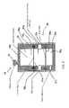
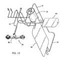
- FIG. 13is an illustration of an exemplary disposable set for a system of the invention.
- FIG. 14is an illustration of an exemplary re-usable component for a system of the invention.
- FIG. 15is an illustration of an exemplary device of the invention assembled using a disposable set similar to FIG. 13 and a re-usable component similar to FIG. 14 .
- FIGS. 16A and 16Bdepict the expression of VEGF ( 5 A) and PIGF ( 5 B) protein by cultured adipose derived stem cells.
- FIG. 17depicts detection of endothelial progenitor cells within adipose derived stem cell populations.
- FIGS. 18A and 18Bdepict the in vitro development of vascular structures in both normal ( 7 A) and streptozotocin-treated ( 7 B) mice.
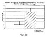
- FIG. 19depicts the increased average restoration of blood flow in hindlimb ischemia mice treated with adipose derived stem cell compared to a negative control.
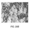
- FIGS. 20A and 20Bshows that increasing adipose derived stem cell dose improves graft survival and angiogenesis ( 20 A) and depicts the retention of adipose tissue architecture in histologic specimen ( 20 B).
- the present inventionprovides methods for augmenting autologous fat transfer using adipose derived regenerative cells (“ADCs”).
- ADCsadipose derived regenerative cells
- the adipose derived regenerative cells of the invention(1) express angiogenic growth factors and cytokines, including PIGF, VEGF, bFGF, IGF-II, Eotaxin, G-CSF, GM-CSF, IL-12 p40/p70, EL-12 p70, IL-13, IL-6, IL-9, Leptin, MCP-1, M-CSF, MIG, PF4, TIMP-1, TIMP-2, TNF- ⁇ and Thrombopoetin, (2) comprise endothelial progenitor cells (EPC) which have a well-established function in blood vessel formation, (3) develop into blood vessels in vitro, and (4) support ischemic tissue survival in vivo.
- EPCendothelial progenitor cells
- regenerative cellrefers to any cells obtained using the systems and methods of the present invention which cause or contribute to complete or partial regeneration, restoration, or substitution of structure or function of an organ, tissue, or physiologic unit or system to thereby provide a therapeutic, structural or cosmetic benefit.
- regenerative cellsinclude: ASCs, endothelial cells, endothelial precursor cells, endothelial progenitor cells, macrophages, fibroblasts, pericytes, smooth muscle cells, preadipocytes, differentiated or de-differentiated adipocytes, keratinocytes, unipotent and multipotent progenitor and precursor cells (and their progeny), and lymphocytes.
- the regenerative cellsmay provide a therapeutic, structural or cosmetic benefit
- ASCs and/or their progenymay incorporate into newly generated bone, muscle, or other structural or functional tissue and thereby cause or contribute to a therapeutic, structural or cosmetic improvement.
- endothelial cells or endothelial precursor or progenitor cells and their progenymay incorporate into existing, newly generated, repaired, or expanded blood vessels to thereby cause or contribute to a therapeutic, structural or cosmetic benefit.
- regenerative cellsmay provide a therapeutic, structural or cosmetic benefit
- expressing and/or secreting moleculese.g., growth factors, that promote creation, retention, restoration, and/or regeneration of structure or function of a given tissue or tissue component.
- regenerative cellsmay express and/or secrete molecules which result in enhanced growth of tissues or cells that then participate directly or indirectly in improved structure or function.
- Regenerative cellsmay express and/or secrete growth factors or cytokines, including, for example, Vascular Endothelial Growth Factor (VEGF), Placental Growth factor (PlGF), and their isoforms, which may perform one or more of the following functions: stimulate development of new blood vessels, i.e., promote angiogenesis; improve oxygen supply of pre-existent small blood vessels (collaterals) by expanding their blood carrying capacity; induce mobilization of regenerative cells from sites distant from the site of injury to thereby enhance the homing and migration of such cells to the site of injury; stimulate the growth and/or promote the survival of cells within a site of injury thereby promoting retention of function or structure; deliver molecules with anti-apoptotic properties thereby reducing the rate or likelihood of cell death and permanent loss of function; and interact with endogenous regenerative cells and/or other physiological mechanisms.
- VEGFVascular Endothelial Growth Factor
- PlGFPlacental Growth factor
- isoformswhich may perform one or more of the following functions: stimulate
- the regenerative cellsmay be used in their ‘native’ form as present in or extracted from the tissue using the systems and methods of the present invention or they may be modified by stimulation or priming with growth factors or other biologic response modifiers, by gene transfer (transient or stable transfer), by further sub-fractionation of the resultant population on the basis or physical properties (for example size or density), differential adherence to a solid phase material, expression of cell surface or intracellular molecules, cell culture or other ex vivo or in vivo manipulation, modification, or fractionation as further described herein.
- the regenerative cellsmay also be used in combination with other cells or devices such as synthetic or biologic scaffolds, materials or devices that deliver factors, drugs, chemicals or other agents that modify or enhance the relevant characteristics of the cells as further described herein.
- regenerative cell compositionrefers to the composition of cells typically present in a volume of liquid after a tissue, e.g., adipose tissue, is washed and at least partially disaggregated.
- a regenerative cell composition of the inventioncomprises multiple different types of regenerative cells, including ASCs, endothelial cells, endothelial precursor cells, endothelial progenitor cells, macrophages, fibroblasts, pericytes, smooth muscle cells, preadipocytes, differentiated or de-differentiated adipocytes, keratinocytes, unipotent and multipotent progenitor and precursor cells (and their progeny), and lymphocytes.
- the regenerative cell compositionmay also contain one or more contaminants, such as collagen, which may be present in the tissue fragments, or residual collagenase or other enzyme or agent employed in or resulting from the tissue disaggregation process described herein.
- regenerative medicinerefers to any therapeutic, structural or cosmetic benefit that is derived from the placement, either directly or indirectly, of regenerative cells into a subject.
- fat transferis a form of regenerative medicine and is intended to include all procedures whereby surplus fat cells are removed from one area of a body and re-implanted into another area of a body. Fat transfer includes both autologous and non-autologous fat transfer. The phrase “autologous fat transfer” is intended to include all procedures whereby the fat removal and re-implantation are performed on the same subject.
- Exemplary cosmetic fat transfer proceduresinclude fat grafts or implants to the lips, nasolabials (mouth to nose folds), wrinkles and other facial folds (depressions around the eyes, between the brows, as well as on the rest of the face), undereyes, cheeks, chin, temples, breasts, thighs, calves, arms, abdomen, buttocks as well as any other area of the body.
- Cosmetic fat transfer proceduresmay be combined with other cosmetic applications such as facial implants, blepheroplasty, brow lifts, face lifts, neck lift, botox applications, chemical peels and laser resurfacing.
- Non-cosmetic fat transfer proceduresinclude implants to treat sphincter disorders, including fat implants in gastroesophageal, urethral and rectal sphincters. Fat transfer procedures may also be used to treat trauma (e.g., radiation) or disease induced soft tissue defects (e.g., abdominal hernia), hemifacial microsomia, vocal cord injury and lumbar spine disorders. Fat transfer procedures may also be used to treat adipose-related diseases or disorders, including but not limited to dyslipidimia, hypoadiponectinemia, hyperlipidemia, lipatrophy and lipohypertrophy.
- stem cellrefers to a multipotent regenerative cell with the potential to differentiate into a variety of other cell types, which perform one or more specific functions and have the ability to self-renew. Some of the stem cells disclosed herein may be pluripotent.
- progenitor cellrefers to a multipotent regenerative cell with the potential to differentiate into more than one cell type. “Progenitor cell”, as used herein, also refers to a unipotent regenerative cell with the potential to differentiate into only a single cell type, which performs one or more specific functions and has limited or no ability to self-renew.
- endothelial progenitor cellrefers to a multipotent or unipotent cell with the potential to differentiate into vascular endothelial cells.
- precursor cellrefers to a unipotent regenerative cell with the potential to differentiate into one cell type. Precursor cells and their progeny may retain extensive proliferative capacity, e.g., lymphocytes and endothelial cells, which can proliferate under appropriate conditions.
- angiogenesisrefers to the process by which new blood vessels are generated from existing vasculature and tissue (Folkman, 1995).
- the phrase “repair or remodeling”refers to the reformation of existing vasculature.
- the alleviation of tissue ischemiais critically dependent upon angiogenesis.
- the spontaneous growth of new blood vesselsprovides collateral circulation in and around an ischemic area, improves blood flow, and alleviates the symptoms caused by the ischemia.
- Angiogenesis mediated diseases and disordersinclude acute myocardial infarction, ischemic cardiomyopathy, peripheral vascular disease, ischemic stroke, acute tubular necrosis, ischemic wounds-including AFT, sepsis, ischemic bowel disease, diabetic retinopathy, neuropathy and nephropathy, vasculitidies, ischemic encephalopathy, erectile dysfunction-physiologic, ischemic or traumatic spinal cord injuries, multiple organ system failure, ischemic gum disease, and transplant related ischemia.
- angiogenic factorrefers to any known protein, peptide or other agent capable of promoting growth of new blood vessels from existing vasculature (“angiogenesis”).
- Suitable angiogenic factors for use in the inventioninclude, but are not limited to, Placenta Growth Factor (Luttun et al., 2002), Macrophage Colony Stimulating Factor (Aharinejad et al., 1995), Granulocyte Macrophage Colony Stimulating Factor (Buschmann et al., 2003), Vascular Endothelial Growth Factor (VEGF)-A, VEGF-A, VEGF-B, VEGF-C, VEGF-D, VEGF-E (Mints et al., 2002), neuropilin (Wang et al., 2003), fibroblast growth factor (FGF)-1, FGF-2(bFGF), FGF-3, FGF4, FGF-5, FGFGF
- Angiogenic factorscan act independently, or in combination with one another. When in combination, angiogenic factors can also act synergistically, whereby the combined effect of the factors is greater than the sum of the effects of the individual factors taken separately.
- the term “angiogenic factor” or “angiogenic protein”also encompasses functional analogues of such factors. Functional analogues include, for example, functional portions of the factors. Functional analogues also include anti-idiotypic antibodies which bind to the receptors of the factors and, thus, mimic the activity of the factors in promoting angiogenesis and/or tissue remodeling. Methods for generating such anti-idiotypic antibodies are well known in the art and are described, for example, in WO 97/23510, the contents of which are incorporated by reference herein.
- Angiogenic factors used in the present inventioncan be produced or obtained from any suitable source.
- the factorscan be purified from their native sources, or produced synthetically or by recombinant expression.
- the factorscan be administered to patients as a protein composition.
- the factorscan be administered in the form of an expression plasmid encoding the factors.
- suitable expression plasmidsis well known in the art.
- Suitable vectors for constructing expression plasmidsinclude, for example, adenoviral vectors, retroviral vectors, adeno-associated viral vectors, RNA vectors, liposomes, cationic lipids, lentiviral vectors and transposons.
- stem cell numberor “stem cell frequency” refers to the number of colonies observed in a clonogenic assay in which adipose derived cells (ADC) are plated at low cell density ( ⁇ 10,000 cells/well) and grown in growth medium supporting MSC growth (for example, DMEM/F12 medium supplemented with 10% fetal calf serum, 5% horse serum, and antibiotic/antimycotic agents. Cells are grown for two weeks after which cultures are stained with hematoxylin and colonies of more than 50 cells are counted as CFU-F.
- ADCadipose derived cells
- Stem cell frequencyis calculated as the number of CFU-F observed per 100 nucleated cells plated (for example; 15 colonies counted in a plate initiated with 1,000 nucleated ADC cells gives a stem cell frequency of 1.5%). Stem cell number is calculated as stem cell frequency multiplied by the total number of nucleated ADC cells obtained.
- a high percentage (.about.100%) of CFU-F grown from ADC cellsexpress the cell surface molecule CD 105 which is also expressed by marrow-derived stem cells (Barry et al., 1999). CD105 is also expressed by adipose tissue-derived stem cells (Zuk et al., 2002).
- Adipose tissuerefers to fat including the connective tissue that stores fat. Adipose tissue contains multiple regenerative cell types, including ASCs and endothelial progenitor and precursor cells.
- unit of adipose tissuerefers to a discrete or measurable amount of adipose tissue.
- a unit of adipose tissuemay be measured by determining the weight and/or volume of the unit. Based on the data identified above, a unit of processed adipose tissue, as removed from a patient, has a cellular component in which at least 0.1% of the cellular component is stem cells; that is, it has a stem cell frequency, determined as described above, of at least 0.1%.
- a unit of adipose tissuemay refer to the entire amount of adipose tissue removed from a patient, or an amount that is less than the entire amount of adipose tissue removed from a patient.
- a unit of adipose tissuemay be combined with another unit of adipose tissue to form a unit of adipose tissue that has a weight or volume that is the sum of the individual units.
- portionrefers to an amount of a material that is less than a whole.
- a minor portionrefers to an amount that is less than 50%, and a major portion refers to an amount greater than 50%.
- a unit of adipose tissue that is less than the entire amount of adipose tissue removed from a patientis a portion of the removed adipose tissue.
- processed lipoaspiraterefers to adipose tissue that has been processed to separate the active cellular component (e.g., the component containing stem and progenitor cells) from the mature adipocytes and connective tissue. This fraction is referred to herein as “adipose-derived cells” or “ADC.”
- ADCrefers to the pellet of regenerative cells obtained by washing and separating the cells from the adipose tissue. The pellet is typically obtained by centrifuging a suspension of cells so that the cells aggregate at the bottom of a centrifuge chamber or cell concentrator.
- the terms “administering,” “introducing,” “delivering,” “placement” and “transplanting”are used interchangeably herein and refer to the placement of the ADC of the invention into a subject by a method or route which results in at least partial localization of the ADC at a desired site.
- the ADCcan be administered by any appropriate route which results in delivery to a desired location in the subject where at least a portion of the cells or components of the cells remain viable.
- the period of viability of the cells after administration to a subjectcan be as short as a few hours, e.g., twenty-four hours, to a few days, to as long as several years.
- the term “subject”includes warm-blooded animals, preferably mammals, including humans.
- the subjectis a primate.
- the subjectis a human.
- regenerative cellse.g., stem and progenitor cells
- the system of the present inventionmay be used for all such tissues.
- Adipose tissueis an especially rich source of regenerative cells. Accordingly, the system of the present invention is illustrated herein using adipose tissue as a source of regenerative cells by way of example only and not limitation.
- Adipose tissuecan be obtained by any method known to a person of ordinary skill in the art.
- adipose tissuemay be removed from a patient by liposuction (syringe or power assisted) or by lipectomy, e.g., suction-assisted lipoplasty, ultrasound-assisted lipoplasty, and excisional lipectomy or combinations thereof.
- the adipose tissueis removed and collected and processed by a system of the invention described herein for the purpose of separating and concentrating regenerative cells.
- the amount of tissue collecteddepends on numerous factors, including the body mass index and age of the donor, the time available for collection, the availability of accessible adipose tissue harvest sites, concomitant and pre-existing medications and conditions (such as anticoagulant therapy), and the clinical purpose for which the tissue is being collected.
- the stem cell percentage of 100 ml of adipose tissue extracted from a lean individualis greater than that extracted from an obese donor (Table 1). This likely reflects a dilutive effect of the increased fat content in the obese individual. Therefore, it may be desirable, in accordance with one aspect of the invention, to obtain larger amounts of tissue from overweight donors compared to the amounts that would be withdrawn from leaner patients. This observation also indicates that the utility of this invention is not limited to individuals with large amounts of adipose tissue.
- the system of the present inventiongenerates a heterogenous plurality of adipose derived regenerative cells which may be used for research and/or therapeutic purposes.
- the cellsare suitable for placement or re-infusion within the body of the recipient.
- the cellsmay be used for research, e.g., the cells can be used to establish stem or progenitor cell lines which can survive for extended periods of time and be used for further study.
- a system 10 of the present inventionis generally comprised of one or more of a tissue collection chamber 20 , a processing chamber 30 , a waste chamber 40 , an output chamber 50 and a sample chamber 60 .
- the various chambersare coupled together via one or more conduits 12 such that fluids containing biological material may pass from one chamber to another while maintaining a closed, sterile fluid/tissue pathway.
- the conduitsmay comprise rigid or flexible bodies referred to interchangeably herein as lumens or tubing.
- the conduitsare in the form of flexible tubing, such as polyethylene tubing conventionally used in clinical settings.
- the tubingsmay be constructed of silicone.
- the flexile tubing usedshould be capable of withstanding negative pressure to reduce the likelihood of collapse.
- the flexible tubing usedshould also be capable of withstanding positive pressure which is generated by, for example, a positive displacement pump, which may be used in the system.
- All the chambers of the systemmay be comprised of one or more ports, e.g., outlet 22 or inlet 21 ports, which accept standard IV, syringe and suction tubing connections.
- the portsmay be a sealed port such as a rubber septum closed syringe needle access port 51 .
- the inlet portsmay be coupled to one or more cannulas (not shown) by way of conduits.
- a tissue inlet port 21may be coupled to an integrated single use liposuction cannula and the conduit may be a flexible tubing.
- the conduitsare generally positioned to provide fluid passageways from one chamber of the system to another.
- the conduits and portsmay be coupled to, for example, a suction device (not shown) which may be manually or automatically operated.
- the suction devicemay be, e.g., a syringe or an electric pump.
- the suction deviceshould be capable of providing sufficient negative pressure to aspirate tissue from a patient.
- any suitable suction device known to one of ordinary skill in the art, e.g., a surgeon,may be used.
- the conduits 12may further comprise one or more clamps (not shown) to control the flow of material among various components of the system.
- the clampsare useful for maintaining the sterility of the system by effectively sealing different regions of the system.
- the conduits 12may comprise one or more valves 14 that control the flow of material through the system.
- the valves 14are identified as open circles in the Figures.
- the valvesmay be electromechanical pinch valves.
- the valvesmay be pneumatic valves.
- the valvesmay be hydraulic valves or mechanical valves.
- Such valvesare preferably activated by a control system which may be coupled to levers. The levers may be manually manipulated such that the levers are activated.
- control systemmay be coupled to the levers as well as a processing device which may activate the valves at pre-determined activation conditions.
- activation of the valvesmay be partially automated and partially subject to the user's preference such that the process may be optimized.
- certain valvesmay be activated manually and others automatically through the processing device.
- the valves 14may also be used in conjunction with one or more pumps, e.g., peristaltic pumps 34 or positive displacement pumps (not shown).
- the conduits 12 and/or the valves 14may also be comprised of sensors 29 , e.g., optical sensors, ultrasonic sensors, pressure sensors or other forms of monitors known in the art that are capable of distinguishing among the various fluid components and fluid levels that flow through the system.
- the sensors 29may be optical sensors.
- the systemmay also include a plurality of filters 36 .
- the filtersmay be within a chamber of the system 28 . Different chambers within the system may be comprised of different filters.
- the filtersare effective to separate the regenerative cells, e.g., stem cells and/or progenitor cells, from undesirable cells and disaggregation agents that may be used in accordance with the system.
- a filter assembly 36includes a hollow fiber filtration device.
- a filter assembly 36includes a percolative filtration device, which may or may not be used with a sedimentation process.
- the filter assembly 36comprises a centrifugation device, which may or may not be used with an elutriation device and process.
- the systemcomprises a combination of these filtering devices.
- the filtration functions of the present inventioncan be two-fold, with some filters removing things from the final concentration such as collagen, free lipid, free adipocytes and residual collagenase, and with other filters being used to concentrate the final product.
- the filters of the systemmay be comprised of a plurality of pores ranging in diameters and/or length from 20 to 800 ⁇ m.
- the collection chamber 20has a prefixed filter 28 with a plurality of pores ranging from 80 to 400 ⁇ m.
- the collection chamber 20has a prefixed filter 28 with a plurality of 265 ⁇ m pores.
- the filtersmay be detachable and/or disposable.
- the systemmay also be comprised of one or more temperature control devices (not shown) that are positioned to adjust the temperature of the material contained within one or more chambers of the system.
- the temperature control devicemay be a heater, a cooler or both, i.e., it may be able to switch between a heater and a cooler.
- the temperature devicemay adjust the temperature of any of the material passing through the system, including the tissue, the disaggregation agents, the resuspension agents, the rinsing agents, the washing agents or the additives. For example, heating of adipose tissue facilitates disaggregation whereas the cooling of the regenerative cell output is desirable to maintain viability. Also, if pre-warmed reagents are needed for optimal tissue processing, the role of the temperature device would be to maintain the pre-determined temperature rather than to increase or decrease the temperature.
- all ports and valvesmay comprise a closure that maintains the sealed configuration of the system.
- the closuremay be a membrane that is impermeable to fluid, air and other contaminants or it may be any other suitable closure known in the art.
- all ports of the systemmay be designed such that they can accommodate syringes, needles or other devices for withdrawing the materials in the chambers without compromising the sterility of the system.
- tissuemay be extracted from a patient via any art recognized method.
- the aspirated tissueis transferred to the collection chamber 20 via a conduit such as 12 a where it is rinsed and digested.
- the aspirated tissuetypically enters the collection chamber 20 via a sealed entry port, such as a rubber septum closed syringe needle access port (not shown on collection chamber).
- the collection chamber 20may be comprised of a plurality of flexible or rigid canisters or cylinders or combinations thereof.
- the collection chamber 20may be comprised of one or more rigid canisters of varying sizes.
- the collection chamber 20may also be comprised of one or more flexible bags.
- the bagis preferably provided with a support, such as in internal or external frame, that helps reduce the likelihood that the bag will collapse upon the application of suction to the bag.
- the collection chamber 20is sized to hold the requisite amount of saline to appropriately rinse and digest the tissue prior to the wash and concentrate stage of the process performed in the processing chamber 30 .
- the volume of tissue or fluid present in the collection chamber 20is easily ascertainable to the naked eye.
- a suitable collection chamberhas the capacity to hold 800 ml of lipoaspirate and 1200 ml of saline. Accordingly, in one embodiment, the collection chamber 20 has a capacity of at least 2 liters. In another embodiment, to separate and concentrate red blood cells from blood, the collection chamber 20 has a capacity of at least 1.5 liters.
- the size of the collection chamber 20will vary depending on the type and amount of tissue collected from the patient. The collection chamber 20 may be sized to hold as little as about 5 ml to up to about 2 liters of tissue. For smaller tissue volumes, e.g., 5 mls to 100 mls, the tissue may be gathered in a syringe prior to transfer to the collection chamber 20 .
- the collection chamber 20may be constructed using any suitable biocompatible material that can be sterilized.
- the collection chamber 20is constructed of disposable material that meets biocompatibility requirements for intravascular contact as described in the ISO 10993 standard.
- polycarbonate acrylic or ABSmay be used.
- the fluid path of the collection chamber 20is preferably pyrogen free, i.e., suitable for blood use without danger of disease transmittal.
- the collection chamber 20is constructed of a material that allows the user to visually determine the approximate volume of tissue present in the chamber.
- the volume of tissue and/or fluid in the collection chamber 20is determined by automated sensors 29 .
- the collection chamber 20is preferably designed such that the automated system can determine the volume of tissue and/or fluid within the chamber with a reasonable degree of accuracy. In a preferred embodiment, the system senses the volume within the collection chamber with an accuracy of plus or minus fifteen percent.
- the collection chamber 20is in the form of a rigid chamber, for example, a chamber constructed of a medical grade polycarbonate containing a roughly conical prefixed filter 28 of medical grade polyester with a mesh size of 265 ⁇ m.
- the rigid tissue collection containermay have a size of approximately eight inches high and approximately five inches in diameter; the wall thickness may be about 0.125 inches.
- the interior of the cylindermay be accessed through, for example, one or more ports for suction tubing, one or more ports with tubing for connection through sterile docking technology, and/or one or more ports for needle puncture access through a rubber septum.
- the prefixed filter 28 in the interior of the collection chamber 20is preferably structured to retain adipose tissue and to pass non-adipose tissue as, for example, the tissues are removed from the patient. More specifically, the filter 28 may allow passage of free lipid, blood, and saline, while retaining fragments of adipose tissue during, or in another embodiment after, the initial harvesting of the adipose tissue.
- the filter 28includes a plurality of pores, of either the same or different sizes, but ranging in size from about 20 ⁇ m to 5 mm. In a preferred embodiment, the filter 28 includes a plurality of 400 ⁇ m pores.
- the filter 28is a medical grade polyester mesh of around 200 ⁇ m thickness with a pore size of around 265 ⁇ m and around 47% open area. This material holds the tissue during rinsing but allows cells to pass out through the mesh following tissue disaggregation. Thus, when the tissues are aspirated from the patient, non-adipose tissue may be separated from adipose tissue.
- the same functionalitycould be achieved with different materials, mesh size, and the number and type of ports. For example, mesh pore sizes smaller than 100 ⁇ m or as large as several thousand microns would achieve the same purpose of allowing passage of saline and blood cells while retaining adipose tissue aggregates and fragments. Similarly, the same purpose could be achieved by use of an alternative rigid plastic material, or by many other modifications that would be known to those skilled in the art
- the collection chamber 20may be further comprised of a means for washing the tissue as well as a means for mixing and/or disaggregating the tissue.
- the tissuemay be washed, mixed or disaggregated by agitation to maximize cell viability and to minimize the amount of free lipid released.
- the tissueis agitated by rotating the entire collection chamber 20 through an arc of varying degrees (e.g., through an arc of about 45 degrees to about 90 degrees) at varying speeds, e.g., about 30 revolutions per minute.
- the tissueis agitated by rotating the entire collection chamber 20 , wherein the collection chamber 20 is comprised of one or more paddles or protrusions rigidly attached to an inside surface of the collection chamber, through an arc of varying degrees (e.g., through an arc of about 45 degrees to about 90 degrees) at varying speeds, e.g., about 30 revolutions per minute.
- the inside surface of the collection chamber 20 to which the paddles 25 a or protrusions are rigidly attachedis a prefixed filter 28 .
- the inside surface of the collection chamber 20 to which the paddles 25 a or protrusions are rigidly attachedis a filter cage 27 of a prefixed filter 28 .
- the rotation of the collection chamber 20may be accomplished by a drive mechanism attached to or in proximity with the collection chamber 20 .
- the drive mechanismmay be a simple belt or gear or other drive mechanism known in the art.
- the speed of the rotationmay be, for example, 30 revolutions per minute. Generally, higher speeds have been found to generate larger volumes of free lipids and may not be optimal.
- the tissueis agitated by placing a rotatable shaft 25 inside the collection chamber 20 , wherein the rotatable shaft is comprised of one or more paddles 25 a or protrusions rigidly attached to the rotatable shaft 25 which pass through the mixture as the shaft is being rotated.
- the rotatable shaft 25 with rigidly attached 25 a paddlesmay be rested on the bottom of the collection chamber 20 . This may be accomplished, for example, by placing the paddle-like device into a spinning magnetic field (e.g., magnetic stirrer). Alternatively, agitating of the tissue may be accomplished using a simple agitator known in the art, i.e. a device implementing shaking up and down without rotation.
- a spinning magnetic fielde.g., magnetic stirrer
- agitating of the tissuemay be accomplished using a simple agitator known in the art, i.e. a device implementing shaking up and down without rotation.
- An exemplary collection chamber 20 of the system, illustrated in FIG. 5is comprised of a vacuum line 11 which may be used to evacuate air from the chamber which allows the user to remove tissue with a user supplied cannula; an inlet port 21 ; an outlet port 22 for draining or removing waste; and a rotatable shaft 25 with a paddle-like device wherein the one or more paddles 25 a are rigidly attached to a filter cage 27 of a prefixed filter 28 for tissue agitation using a magnetic stirrer (not shown).
- the system 10may also be comprised of one or more washing solution sources 22 .
- the washing solution sourcemay comprise a source of saline 23 , and a source of a tissue disaggregation agent 24 , such as collagenase.
- the washing solutionmay be any solution known to one of skill in the art, including saline or any other buffered or unbuffered electrolyte solution.
- Disaggregation agentsthat may be used include neutral proteases, collagenase, trypsin, lipase, hyaluronidase, deoxyribonuclease, members of the Blendzyme enzyme mixture family, e.g., Liberase H1, pepsin and/or combinations thereof.
- the types of tissue being processedwill dictate the types or combinations of washing solutions used.
- the washing solutionsuch as saline
- the washing solutionmay be delivered to the collection chamber 20 before the adipose tissue is extracted, or may be delivered to the collection chamber 20 concurrently with the adipose tissue.
- the washing solution and the extracted adipose tissuemay be mixed by any means including the methods described above.
- the containers for the saline 23 and/or the disaggregation agents 24may be any suitable container that can hold their contents in a sterile manner, e.g., a collapsible bag, such as an IV bag used in clinical settings. These containers may have conduits 12 , such as conduit 12 e , coupled to the collection chamber 20 so that the saline and/or the disaggregation agent may be delivered to the interior of the collection chamber 20 .
- the saline and/or disaggregation agentmay be delivered to the interior of the collection chamber 20 through any art-recognized manner, including simple gravity pressure applied to the outside of the containers for the saline 23 and/or the disaggregation agents 24 or by placement of a positive displacement pump on the conduits, e.g., conduit 12 d in FIG. 4 .
- the tissue and/or fluid within the collection chambershould be maintained at a temperature ranging from 30 degrees Celsius to 40 degrees Celsius. In a preferred embodiment, the temperature of the suspension inside the collection chamber is maintained at 37 degrees Celsius. In certain embodiments, if the surgical procedure or therapeutic application needs to be delayed, the selected tissue may be stored in the collection chamber for later use. The tissue may be stored at or about room temperature or at about 4 degrees Celsius for up to 96 hours.
- the collection chamber 20allows for differentiation of buoyant and non-buoyant liquid within the chamber.
- the collection chambermay be comprised of sensors 29 which can detect when the interface between the buoyant and non-buoyant liquids has been reached.
- the sensor 29may be an optical sensor which may be capable of detecting a change in the light refraction of the effluent which is flowing in the outlet fluid passageway of the collection chamber and sending a signal to the processing device of the system to thereby activate or de-activate one or more pumps and/or valves in accordance with the processing device associated with the system.
- the collection chamber 20is preferably comprised of an outlet port 22 at the lowest point of the chamber such that blood and other non-buoyant components of the tissue may be drained to the waste container. Accordingly, the collection chamber 20 may be coupled to one or more waste containers 40 via one or more conduits 12 described herein to permit waste from the collection chamber to be drained or removed from the system.
- the drainingmay be passive or active.
- the non-buoyant components described abovecould be drained using gravity, by applying positive or negative pressure, by use of pumps 34 or by use of vents 32 .
- the collection chambermay be positioned such that the outlet ports 22 are positioned at, or near, the bottom of the collection chamber.
- the collection chamberis preferably positioned in this orientation to allow adipose tissue fragments to float, e.g. from 15 seconds to several minutes or longer.
- the processing device of the systemcalculates various parameters, e.g., the volume of saline required for washing the tissue and the time required for washing the tissue, based on information initially entered by the user (e.g., volume of tissue being processed). Based on the control logic of the processing device, certain valves and/or pumps are activated or de-activated such that waste from the collection chamber 20 is removed from the system. Sensors 29 such as optical sensors may be placed such that they are capable of signaling the processing device of the system to proceed with the next step in the tissue processing.
- the collection chamber 20is comprised of a closed fluid pathway that allows saline and reagents to be added to the tissue in an aseptic manner. Accordingly, the collection chamber 20 is further comprised of conduits 12 , e.g., flexible or rigid conduits, that are appropriately sized to allow for free passage of tissue and liquid.
- the conduits 12are in the form of tubing.
- the conduits 12can vary in size depending on whether passage of fluid or tissue is desired.
- the conduits 12may also vary in size depending on the amount of tissue or fluid that is cycled through the system.
- the conduitsmay have a diameter ranging from about 0.060 to about 0.750 inches and for the passage of tissue, the conduits may have a diameter ranging from 0.312 to 0.750 inches.
- the size of the conduitsis selected to balance the volume the conduits can accommodate and the time required to transport the tissue or fluids through said conduits.
- the foregoing parametersi.e., volume and time for transport, must be identified such that the appropriate signals can be transmitted to the processing device of the system. This allows the device to move accurate volumes of liquid and tissue from one chamber to another.
- the collection chamber 20also allows for removal of the washed adipose tissue to a processing chamber 30 . Accordingly, the collection chamber 20 must be connected to the necessary tubing 12 , valves 14 and pump 34 for the movement and storage of washed adipose tissue.
- the collection chamber 20typically includes one or more ports 21 for permitting the washing solution to be delivered to the interior of the chamber, and one or more ports 22 for permitting waste and other materials to be directed out from the collection chamber 20 .
- the collection chambermay include one or more sealed entry ports as described herein.
- the collection chamber 20may also include one or more caps (not shown), such as a top cap and a bottom cap to further ensure that the system remains sterile while washing solution is delivered into the collection chamber and/or waste is transported out.
- the ports 21may be provided on the caps of the collection chamber or on a sidewall of the collection chamber.
- the process of washing with fresh wash solutionmay be repeated until the residual content of non-buoyant contaminants in the solution reaches a pre-determined level.
- the remaining material in the collection chamber 20which comprises the buoyant material of the mixture described above, including adipose tissue fragments, may be washed one or more additional times until the amount of undesired material is reduced to a desired pre-determined level.
- One method of determining the end point of the washingis to measure the amount of red blood cells in the tissue solution. This can be accomplished by measuring the light absorbed on the 540 nm wavelength. In a preferred embodiment, a range between about 0.546 and about 0.842 is deemed acceptable.
- a tissue disaggregation agentmay be delivered to the collection chamber 20 to digest the remaining adipose tissue components.
- salinesuch as saline delivered from a saline source 23 as described above, may be added to the adipose tissue along with or immediately followed by addition of collagenase, such as collagenase delivered from a collagenase source 24 as described above.
- collagenasesuch as collagenase delivered from a collagenase source 24 as described above.
- the washed adipose tissue and the tissue disaggregation agentmay then be agitated in manners similar to the agitation methods described above, until the washed adipose tissue is disaggregated.
- the washed adipose tissue and the tissue disaggregation agentmay be agitated by rotating the entire collection chamber through an arc of approximately 90 degrees, by having a shaft which contains one or more paddles which pass through the solution as the shaft is being rotated, and/or by rotating the entire collection chamber which contains paddles or protrusions on the inside surface of the collection chamber.
- the adipose tissue fragmentsare mixed with a collagenase-containing enzyme solution at or around 37.degree. C. for about 20-60 minutes. In other embodiments, a higher concentration of collagenase or similar agent may be added to decrease the digestion time.
- the collection chamber 20may then be placed in an upright position so that the outlet ports 22 are located at the bottom of the collection chamber for a period of time sufficient to allow buoyant cells and tissue fragments to float. Typically, the time may ranges from about 15 seconds to several minutes but other times may be implemented in modified embodiments.
- the adipose tissuemay either be partially disaggregated, or completely disaggregated.
- a portion of washed adipose tissuemay be removed and set aside in a sample container prior to any digestion.
- harvested adipose tissueis partially disaggregated to concentrate cells before being reintroduced back into the patient.
- the adipose tissueis mixed with a tissue disaggregation agent for a period of time generally less than about 20 minutes. A portion of the partially disaggregated tissue may then be removed from the collection chamber, and the remaining partially disaggregated tissue may be further disaggregated by mixing the adipose tissue with a tissue disaggregation agent for another 40 minutes.
- the adipose derived cellsare to be used as an essentially pure population of regenerative cells, the adipose tissue may be fully disaggregated.
- additivesmay be added to the various containers as needed to enhance the results.
- additivesinclude agents that optimize washing and disaggregation, additives that enhance the viability of the active cell population during processing, anti-microbial agents (e.g., antibiotics), additives that lyse adipocytes and/or red blood cells, or additives that enrich for cell populations of interest (by differential adherence to solid phase moieties or to otherwise promote the substantial reduction or enrichment of cell populations).
- anti-microbial agentse.g., antibiotics
- additives that lyse adipocytes and/or red blood cellsor additives that enrich for cell populations of interest (by differential adherence to solid phase moieties or to otherwise promote the substantial reduction or enrichment of cell populations).
- Other possible additivesinclude those that promote recovery and viability of regenerative cells (for example, caspase inhibitors) or which reduce the likelihood of adverse reaction on infusion or emplacement (for example, inhibitors of re-aggregation of cells or connective tissue).
- the non-buoyant fraction of the resulting mixture of washed adipose tissue fragments and tissue disaggregation agentswill contain regenerative cells, e.g., stem cells and other adipose derived progenitor cells.
- the non-buoyant fraction containing the regenerative cellswill be transferred to the processing chamber 30 wherein the regenerative cells of interest, such as the adipose derived stem cells, will be separated from other cells and materials present in the non-buoyant fraction of the mixture.
- the processing chamber 30is positioned within the system such that the rinsed and digested tissue suspension moves from the collection chamber 20 to the processing chamber 30 by way of tubing 12 , valves 14 and pump 34 .
- the processing chamberis sized to accommodate tissue/fluid mixtures ranging from 10 mL to 1.2 L. In a preferred embodiment, the processing chamber is sized to accommodate 800 mLs. In certain embodiments, the entire regenerative cell composition from the collection chamber 20 is directed to the processing chamber 30 .
- the regenerative cell composition of the present inventionis a composition of cells typically present in a volume of liquid after a tissue, e.g., adipose tissue, is washed and at least partially disaggregated.
- the regenerative cell composition that is transferred from the collection chamber 20 after being mixed with a tissue disaggregation agentcomprises multiple different types of cells, including stem cells, progenitor cells, endothelial precursor cells, adipocytes and other regenerative cells described herein.
- the regenerative cell compositionmay also contain one or more contaminants, such as collagen and other connective tissue proteins and fragments thereof, which were present in the adipose tissue fragments, or residual collagenase from the tissue disaggregation process.
- the processing chamber 30may be constructed using any suitable biocompatible material that can be sterilized.
- the processing chamber 30is constructed of disposable material that meets biocompatibility requirements for intravascular contact, as described in the ISO 10993 standard.
- the fluid path of the disposable processing chamberis pyrogen free.
- the processing chambermay be in the form of a plastic bag, such as those conventionally used in processing blood in blood banks; or in other embodiments, it may be structurally rigid ( FIG. 6 ).
- the processing chamber 30may be similar to the processing chamber disclosed in commonly owned U.S. application Ser. No. 10/316,127, filed Dec. 7, 2001 and U.S. application Ser. No. 10/325,728, filed Dec. 20, 2002, the contents of which in their entirety are hereby incorporated by reference.
- the regenerative cell composition from the collection chamber 20is introduced into the processing chamber 30 where the solution can be filtered to separate and/or concentrate a particular regenerative cell composition.
- Cell filtrationis a method of separating particular components and cells from other different components or types of cells.
- the regenerative cell composition of the inventioncomprises multiple different types of cells, including stem cells, progenitor cells and adipocytes, as well as one or more contaminants, such as collagen, which was present in the adipose tissue fragments, or residual collagenase from the tissue disaggregation process.
- the filters 36 present in the processing chamber 30may allow for separation and concentration of a particular subpopulation of regenerative cells, e.g., stem cells or endothelial progenitors cells etc.
- Some variables which are associated with filtration of cells from a liquidinclude, but are not limited to, pore size of the filter media, geometry (shape) of the pore, surface area of the filter, flow direction of the solution being filtered, trans-membrane pressure, dilution of the particular cell population, particulate size and shape as well as cell size and cell viability.
- the particular cells that are desired to be separated or filteredare typically adipose derived stem cells.
- the particular cellsmay include adipose derived progenitor cells, such as endothelial precursor cells, alone or in combination with the stem cells.
- the regenerative cell compositionmay be directed through a filter assembly, such as filter assembly 36 .
- the filter assembly 36comprises a plurality of filters which are structured to perform different functions and separate the regenerative cell composition into distinct parts or components.
- one of the filtersmay be configured to separate collagen from the regenerative cell composition
- one of the filtersmay be configured to separate adipocytes and/or lipid components from the regenerative cell composition
- one of the filtersmay be configured to separate residual enzymes, such as the tissue disaggregation agent, from the regenerative cell composition.
- one of the filtersis capable of performing two functions, such as separating collagen and the tissue disaggregation agent from the composition.
- the plurality of filtersare typically serially arranged; however, at least a portion of the filters may be arranged in parallel, as well.
- a serial arrangement of the filters of the filter assembly 36is shown in FIG. 2 .
- a parallel arrangement of the filters of the filter assembly 36is shown in FIG. 3 .
- the filter assembly 36comprises a first filter, a second filter, and a third filter.
- the first filteris configured to remove collagen particles present in the regenerative cell composition. These collagen particles are typically approximately 0.1 microns in diameter and can be up to 20 microns long. The collagen particles may be of varying sizes depending on the digestion. They also may be fibrils, meaning they have twists and turns. Any of the filters described herein may be made from polyethersulfone, polyester, PTFE, polypropylene, PVDF, or possibly cellulose. There are two possibilities for filtering the collagen. One is to try to remove the larger particles first, letting the cells go through, which would require for example a filter probably in the 10 micron range.
- the second methodis to use a smaller size filter, such as 4.5 micron, with the intent that the collagen would be well digested, so as to trap the cells, and let the collagen pass through. This would require a means to float the cells back off the filter. There may also be a possibility of implementing a filter which would attract and hold the collagen fibers.
- the second filteris configured to remove free immature adipocytes which are not buoyant in the regenerative cell composition.
- the second filtercan be constructed of polyester and have a pore size between about 30 and about 50 microns with a preferred pore size being about 40 microns.
- placement of such a devicemay be in a first, rather than second, position to facilitate an initial removal of larger cells and particles.
- the third filteris configured to remove the unused or residual collagenase or other tissue disaggregation agent present in the composition.
- the collagenasemay degenerate over time.
- the third filtercomprises a plurality of pores having a diameter, or length less than 1 ⁇ m.
- the poresmay have diameters that are smaller than 1 ⁇ m. In other embodiments, the pores have diameters between 10 kD and 5 microns.
- the third filtermay be configured to concentrate the regenerative cell population into a small volume of saline or other washing solution, as discussed herein.
- the final filteris the hollow fiber unit. It is not necessary for any of the filters to be of the hollow fiber type.
- the hollow fiber unitis used for the final filter in a preferred implementation because it is the most efficient in removing the collagenase with the smallest detrimental effect to the regenerative cells.
- the three filtersare in separate housings. It is feasible to have the first and second filters combined into one housing if a hollow fiber unit is used for the third filter. If the final filter is not a hollow fiber set-up then all three filters can be contained in one housing.
- the filters of the filter assembly 36may be located in the processing chamber 30 or may be provided as components separate from the processing chamber 30 .
- the filters of the filter assembly 36may be provided in multiple processing chambers or in an inline fashion.
- the conduits or tubingmay act as a processing chamber or chambers.
- the processing chambercan be reduced in size such that it becomes the inside volume of the conduits which connect the filters. This type of system will function correctly if the volume of tissue solution is sized appropriately.
- the conduitsmay act as the processing chamber by containing the fluid with cells as it is being run through the filters. Care may be taken to minimize the volume of the conduits so that cells/tissue are not unnecessarily lost in the process of priming and running the system.
- the regenerative cell compositioncontaining the washed cells and residual collagen, adipocytes, and/or undigested tissue disaggregation agent, may be directed through the first filter to remove at least a portion of and preferably substantially all of the collagen particles from the composition so that fewer, and preferably no, collagen particles are present in the filtered solution.
- the filtered regenerative cell composition containing the adipocytes and/or undigested tissue disaggregation agentmay then be directed through the second filter to remove at least a portion of and preferably substantially all of the free adipocytes from the filtered regenerative cell composition.
- the twice filtered regenerative cell compositioncontaining the undigested tissue disaggregation agent, may be directed through the third filter, such as a hollow fiber filtration device, as discussed herein, to remove or reduce the undigested tissue disaggregation agent from the regenerative cell composition.
- the third filtersuch as a hollow fiber filtration device, as discussed herein, to remove or reduce the undigested tissue disaggregation agent from the regenerative cell composition.
- the thrice-filtered regenerative cell composition(i.e., the composition remaining after being passed through the first, second, and third filters) may then be directed to multiple outlets, which may include a portion of the processing chamber 30 comprising multiple outlets. These outlets can serve to maintain the necessary pressure, as well as to provide connections via conduits to other containers which may include the collection chamber 20 , the output chamber 50 , and/or the waste container 40 .
- a filter of the filter assembly 36comprises a hollow-fiber filtration member.
- the filtercomprises a collection of hollow tubes formed with the filter media.
- filter mediawhich can be used with the disclosed system 10 include polysulfone, polyethersulfone or a mixed ester material, and the like.
- These hollow fibers or hollow tubes of filter mediamay be contained in a cylindrical cartridge of the filter assembly 36 .
- the individual tubes or fibers of filter mediatypically have an inside diameter which ranges from about 0.1 mm to about 1 mm with a preferred value being about 0.5 mm.
- the diameter and length of a suitable cylindrical cartridgewill determine the number of individual tubes of filter media which can be placed inside the cartridge.
- a suitable hollow fiber filter cartridgeis the FiberFlo®.
- Pore sizes of the filter mediacan range between about 10 kiloDaltons and about 5 microns with a preferred pore size being about 0.5 microns.
- each hollow tubehas a body with a first end, a second end, and a lumen located in the body and extending between the first end and second end.
- the body of each hollow tubeincludes a plurality of pores.
- the poresare generally oriented in the body so that a regenerative cell composition is filtered by flowing through the lumen of the body, and the products to be filtered tangentially pass through the pores, as shown in FIG. 12A .
- the smaller particles in the liquidpass tangentially through the pores relative the flow of fluid through the lumen of the body.
- the composition with the regenerative cellspasses through the lumen of each hollow tube when the composition is being filtered.
- the flow of the compositionis tangential to the pores of the body of each hollow tube.
- the efficiency of filtration of the stem cellsmay be enhanced relative to other filtration techniques.
- the pores of the filter mediaare placed in such a manner that the filter is orientated perpendicular to the flow of the fluid so that the Filter media blocks the path of the fluid being filtered, as illustrated in FIG. 12B .
- the particles which are being filtered out of the regenerative cell compositione.g., the stem cells, tend to build up on one side of the filter and block the flow of the fluid through the pores. This blockage can reduce the efficiency of the filter.
- the cellsare constantly compressed by the pressure of the fluid flow as well as the weight of the cells accumulating on the upstream side of the filter. This can lead to increased lysis of stem cells.
- both large cells and small particlescan be undesirably directed against the filter media as the fluid is passed through the pores. Consequently, larger products in the liquid such as cells may block the pores, thereby decreasing the filtering effect and increasing an occurrence of cell rupture or injury.
- the fluid which is being filteredflows inside the lumen of the hollow tube.
- the portion of the fluid which has the ability to pass through the pores of the body of the filterdoes so with the aid of the positive pressure of the fluid on the inside of the body as well as a negative pressure which is applied on the outside of the body.
- the cellstypically are not subjected to the pressure of the fluid flow or the weight of other cells, and therefore, the shear forces on the stem cells are reduced
- the efficiency and effectiveness of the filtrationcan be enhanced by the reduction in clogging rates and the reduction in regenerative cell lysis.
- filtrationis enhanced by generating a vacuum on the outside of the hollow tube filter media. Due to the size of the regenerative cells, e.g., stem cells or progenitor cells, these cells typically cannot pass through the pores of the body and therefore remain on the inside of the hollow tube filter (e.g., in the lumens of the tubes) and are directed back to the processing chamber 30 via a conduit between the filter and the processing chamber, or to the output chamber 50 .
- the regenerative cellse.g., stem cells or progenitor cells
- the hollow fiber filterhas about a 0.05 micron pore size, and contains approximately 550 cm 2 surface area of filter media.
- An individual media tubetypically has a diameter of about 0.5 mm.
- approximately 120 ml of additional salinemay be added to the composition.
- the processing or filter timemay be approximately 8 minutes.
- the differential of the pressures on either side of the body of the hollow fiber tubee.g., the pressure inside the lumen of the body, and outside the body
- the trans-membrane pressurecan range from about 1 mmHg to about 500 mmHg with a preferred pressure being about 200 mmHg.
- the average nucleated cell recovery and viability using hollow fiber filtrationcan be approximately 80% of viable cells.
- the amount of collagenase which is typically removed in such a systemequates to a three log reduction. For example if the initial concentration of collagenase in the regenerative cell composition which is transferred from the collection chamber to the processing chamber is 0.078 U/ml the collagenase concentration of the final regenerative cell composition would be 0.00078 U/ml.
- the collagenaseis removed in the hollow fiber filter, and the hollow fiber filter corresponds to the third filter discussed above.
- FIGS. 1-3Processing chambers illustrating one or more cell filtration methods described above are shown in the Figures, particularly FIGS. 1-3 .
- a pumpmay be provided, such as pump 34 .
- vent and pressure sensorssuch as vent 32 , and pressure sensor 39 , may be provided in line with the processing chamber 30 and the filter assembly 36 . Fittings for the output chamber 50 may also be provided.
- These optional componentsmay be provided between the processing chamber 30 and the filter assembly 36 so that liquid contained in the processing chamber 30 may flow to one or more of these optional components before flowing through the filter assembly 36 .
- liquidmay flow through the pump 34 before it is passed to the filter assembly 36 .
- liquidmay pass through the pressure sensor 39 before passing through the filter assembly to obtain a pre-filter liquid pressure in the system.
- one or more of these componentsmay also be provided as an element of the processing chamber 30 , such as the vent 32 as illustrated in FIG. 6 .
- the pressure sensor 39is in line to determine the pressure of the regenerative cell composition which is generated by the pump 34 as it enters the filtering chamber of the filter assembly 36 .
- This constructioncan facilitate monitoring of the trans-membrane pressure across the filter membrane.
- Additional saline or other buffer and washing solutioncan be added to the regenerative cell composition to assist in the removal of unwanted proteins as the composition is being filtered through the filter assembly 36 . This repeated washing can be performed multiple times to enhance the purity of the regenerative cell composition.
- the salinecan be added at any step as deemed necessary to enhance filtration.
- the unwanted proteins and saline or other washing solutionis removed in the following manner.
- the composition with the regenerative cells, as well as collagen and connective tissue particles or fragments, adipocytes, and collagenaseis cycled through a series of filters until a minimum volume is reached.
- the minimum volumeis a function of the total hold up volume of the system and some predetermined constant.
- the hold up volumeis the volume of liquid which is contained in the tubing and conduits if all of the processing chambers are empty. In one embodiment, the minimum volume is 15 ml.
- a predetermined volume of washing solutionis introduced into the system to be mixed with the regenerative cell composition.
- This mixture of washing solution and the regenerative cell compositionis then cycled through the filters until the minimum volume is reached again.
- This cyclecan be repeated multiple times to enhance the purity of the regenerative cells, or in other words, to increase the ratio of regenerative cells in the composition to the other materials in the composition. See FIGS. 10 and 11 .
- an output chamber 50such as an output bag, may be connected to an outlet port of the processing chamber 30 and/or the filter assembly 36 , depending on the specific embodiment.
- a ventsuch as the vent 32 , may then be opened to facilitate the output of the concentrated regenerative cells.
- this determination of when a minimum concentration has been reachedis made empirically after experiments have been run and programmed into the electronic controls of the device.
- the determinationcan be an input into the process of what is desired to yield, i.e., how many stem/progenitor cells are desired, or range of cell concentration. Based on scientific data, a predefined amount of adipose tissue needs to be obtained and placed into the system to achieve the desired output. With the vent 32 open, a pump, such as the pump 34 , can function to transfer the concentrated regenerative cells into the output bag.
- the output bag 50is similar to an empty blood bag which has a tube with a fitting on one end. In a sterile fashion, the fitting on the output bag may be attached to the outlet port, and the concentrated regenerative cells may be transferred to the output bag.
- a vacuum pump 26may be provided in the system 10 to change the pressure in the system, among other things.
- the vacuum pump 26may be coupled to the collection chamber 20 via a conduit, such as conduit 12 b , to cause a decrease in pressure within the collection chamber 20 .
- Vacuum pump 26may also be coupled to the processing chamber 30 by way of a conduit, such as conduit 12 g .
- a conduitsuch as conduit 12 g .
- two separate vacuum pumps or sourcesmay be implemented, or a single one may be implemented by using valves which direct the vacuum pull to the different conduits that need it at specific points in the process.
- vacuum pump 26may be coupled to the waste container 40 via a conduit, such as conduit 12 f.
- the pressure generated by the vacuum pump 26can be used to direct the flow of fluids, including the regenerative cells, through the conduits 12 .
- This pressurecan be supplied in multiple directions, for example, by automatically or manually controlling the position of one or more valves 14 in the system 10 .
- the system 10can be made to function properly with the use of positive pressure or through the use of negative pressure, or combinations thereof.
- the regenerative cellscan be pulled through the first and second filters described above into a soft sided container which is connected to the third filter.
- the soft-sided containercan be in line (serial) connected ahead of the third filter.
- the final output chambermay be a soft sided container which is on the other side (e.g., the downstream side) of the third filter.
- pressureis used to move the regenerative cells from one soft sided container to a second soft sided container through the filter.
- the filtration of the stem cells and/or adipose derived progenitor cellsmay be accomplished using a combination of percolative filtration and sedimentation.
- a tissue regenerative cell compositione.g., the composition containing the stem cells and/or adipose derived progenitor cells
- variables which are associated with percolative filtration of cells from a regenerative cell compositioninclude, but are not limited to, pore size of the filter media, pore geometry or shape, surface area of the filter, flow direction of the regenerative cell composition being filtered, flow rate of the infused saline, trans-membrane pressure, dilution of the cell population, cell size and viability.
- the processing chamber 30uses a filter assembly 36 which implements percolative filtration and sedimentation to separate and concentrate the regenerative cells.
- the processing chamber 30is defined as a generally cylindrical body having a sidewall 30 a , a top surface 30 b , and a bottom surface 30 c , as shown in FIG. 6 .
- a sterile vent 32is provided in the top surface 30 b.
- the processing chamber 30is illustrated as including a filter assembly 36 , which includes two filters, such as large pore filter 36 a , and small pore filter 36 b .
- the pore sizes of the filters 36 a and 36 btypically are in a range between about 0.05 microns and about 10 microns.
- the large pore filter 36 amay comprise pores with a diameter of about 5 and the small pore filter 36 b may comprise pores with a diameter of about 1-3 ⁇ m.
- the filtershave a surface area of about 785 mm 2 .
- Filters 36 a and 36 bdivide an interior of the processing chamber 30 to include a first chamber 37 a , a second chamber 37 b , and a third chamber 37 c .
- first chamber 37 ais located between second chamber 37 b and third chamber 37 c .
- first chamber 37 ais shown as being the region of the processing chamber 30 having an inlet port 31 a and an outlet port 31 b .
- the illustrated processing chamber 30includes a plurality of ports providing communication paths from an exterior of the processing chamber 30 to the interior of the processing chamber 30 , such as ports 31 a , 31 b , and 31 c .
- the ports 31 a , 31 b , and 31 care illustrated as being disposed in the sidewall 30 a of a body of the processing chamber 30 . However, the ports 31 a , 31 b , and 31 c could be positioned in other regions, as well.
- Port 31 ais illustrated as a sample inlet port, which is constructed to be coupled to a conduit so that a composition containing regenerative cells can be passed into the interior of the processing chamber 30 .
- Port 31 bis illustrated as an outlet port constructed to be coupled to a conduit so that the separated and concentrated cells may be removed from the interior of the processing chamber 30 .
- Port 31 cis illustrated as an inlet port constructed to be coupled to a conduit for delivery of a fresh washing solution, such as saline into the interior of the processing chamber 30 .
- the regenerative cellsmay be introduced into the central chamber 37 a via inlet port 31 a .
- Saline or other bufferis introduced into the bottom chamber 37 b through inlet port 31 c .
- the salinemay be directed through the regenerative cell composition in chamber 37 a at a rate of about 10 ml/min.
- the flow rate of the salineis such that it counteracts the force of gravity.
- the flow of salinegives the cells in the chamber the ability to separate based on the density of the cells.
- the larger cells in the compositionwill settle to the bottom of the central chamber 37 a , and the smaller cells and proteins will be carried away through the second filter 36 b into the top chamber 37 c .
- This filteringis accomplished by adjusting the flow rate of the saline such that the larger cells are rolled in place which allows the smaller particles to be liberated and carried off with the saline.
- the sterile vent 32is included in the chamber 30 to ensure that the correct pressure gradient is maintained in the three chambers within the processing unit.
- the upper chamber 37 ccan comprise an absorbent media 33 .
- the purpose of the absorbent mediais to trap the unwanted proteins in the solution to ensure that they do not cross the filter media back into the processing solution, if, for example, the saline flow rate decreases.
- An absorbent mediacan be a type of filter material that is absorbent, or attracts materials or components to be filtered out.
- An outflow portcan be added above the top filter to help draw off the waste. Another embodiment of this may be to apply a gentle vacuum from the top to help pull off waste.
- Absorbent mediacan be implemented when, as in the illustrated embodiment, the flow rates are relatively small. Excess saline and proteins are then carried
- the composition containing the separated cellsmay be concentrated, as discussed herein.
- the compositionmay be further concentrated after it has been removed from chamber 37 a through outlet port 31 b , or while it is in the chamber 37 a .
- the concentration of cells in the compositionis increased in the following manner. After the cells have been sufficiently separated the filters, such as filters 36 a and 36 b , may be moved towards each other. This movement has the effect of reducing the volume between the two filters (e.g., the volume of chamber 37 a ).
- a vibrating membermay also be provided in connection with the processing chamber 30 to facilitate concentrating of the cells in the composition.
- the vibrating membermay be coupled to the filter 36 b (e.g., the small pore filter). Vibrating can reduce an incidence of cells becoming trapped in the filters. The reduction in volume of the composition allows the excess saline to be removed as waste and the cells to be concentrated in a smaller volume.
- the concentration of the regenerative cellsis accomplished in the following manner.
- the regenerative cell compositioncan be transferred to another chamber (not shown) which uses gravity to filter out the excess saline.
- the sedimentationcan occur at the same time as the percolation. This sedimentation may be accomplished by introducing the composition on top of a filter which has a pore size ranging from about 10 kD to about 2 microns. In one embodiment, a suitable filter has a pore size of about 1 micron. The force of gravity will allow the saline and smaller particles to be passed through the filter while preventing the cells in the composition to flow through the filter.
- the regenerative cell compositionmay be agitated to remove the cells from the filter and, subsequently, the concentrated regenerative cells may be transferred to the output bag.
- the smaller particlescan be drawn off as waste through an outlet.
- the processing chamberis comprised of a centrifuge chamber or a cell concentrator ( FIGS. 4 , 7 and 8 ) that facilitates separation of the regenerative cells from the regenerative cell composition.
- the cell concentratormay be a centrifuge device or part of a centrifuge device that may separate regenerative cells from the regenerative cell composition based on, e.g., size or density ( FIGS. 7 and 8 ).
- the cell concentratormay also be a spinning membrane filter.
- Centrifugationis recognized in the art as a means for separating and concentrating solutions having multiple components of varying densities. This is done by imparting a centripetal force on the solution which is, for example, higher than gravity. The imparted force causes the tissue solution to separate based on the cell and or particle density. After the cells have been sufficiently concentrated the excess saline and proteins may be removed.
- Some of the variables which are associated with centrifugation separation of the cells from the solutioninclude, but are not limited to, speed of the rotor, distance of the solution from the center of rotation, and time.
- the centrifuge devicemay be a component of the processing chamber 30 , or may be separate from the processing chamber.
- the centrifuge devicemay also be partially within the processing chamber and partially separate from the processing chamber (see FIGS. 14 and 15 ).
- the centrifuge devicecauses a container containing the cell solution, e.g., an output chamber 50 , to spin around an axis thereby increasing the force on the cells in the solution to be greater than gravity.
- the denser or heavier materials in the solutiontypically settle to one end of the output chamber 50 to form a pellet.
- the pelletmay then be re-suspended to obtain a solution with a desired concentration of cells and/or a desired volumes of cells and medium.
- the processing chamber 30itself may be in the form of a centrifuge chamber or cell concentrator.
- a processing chamberis constructed to separate and concentrate cells using both centrifugal and gravitational forces.
- centrifugal forcedirects the denser components of the regenerative cell composition, e.g., the regenerative cells, towards the outermost ends of the centrifuge chamber.
- gravitational forcehelps the regenerative cells to remain in the outermost ends of the centrifuge chamber and form a cell pellet. Accordingly, the unwanted components of the regenerative cell composition, i.e., the waste, can be removed without disturbing the cell pellet.
- centrifugal elutriationmay also be applied.
- the cellsmay be separated based on the individual cell sedimentation rate such that the directional (e.g., outward) force applied by centrifugation causes cells and solutes to sediment at different rates.
- the sedimentation rate of the target cell populationis opposed by an opposite (e.g., inward) flow rate applied by pumping solution in the opposite direction to the centrifugal force.
- the counterflowis adjusted so that the cells and particles within the solution are separated.
- Elutriationhas been applied in many instances of cell separation (Inoue, Carsten et al. 1981; Hayner, Braun et al. 1984; Noga 1999) and the principles and practices used to optimize flow and centrifugal parameters can be applied herein in light of the present disclosure by one skilled in the art.
- FIG. 9illustrates principles associated with an elutriation implementation in accordance with the present invention.
- the elutriation embodimentcan be similar to a centrifugation implementation to the extent that a force is applied to the solution using a spinning rotor.
- Some of the variables which are associated with the presently embodied elutriation separationinclude, but are not limited to, the size and shape of the spinning chamber, the diameter of the rotor, the speed of the rotor, the diameter of the counter flow tubing, the flow rate of the counter flow, as well as the size and density of the particles and cells which are to be removed-from solution.
- the regenerative cellscan be separated based on individual cell densities.
- the regenerative cell compositione.g., the solution containing the regenerative cells and the collagenase
- the solutionis added to the chamber
- additional salineis added to the chamber at a predetermined flow rate.
- the flow rate of the salinecan be predetermined as a function of the speed of the rotor, the cell diameter, and the chamber constant which has been established empirically. The flow rate will be controlled for example with a device similar to an IV pump.
- a purpose of the additional salineis to provide a condition inside the rotor chamber where the larger particles will move to one side of the chamber and the smaller particles will move to the other, as illustrated in FIG. 9B .
- the flowis adjusted so that, in this application, the smaller particles will exit the chamber and move to a waste container, as shown in FIG. 9C .
- This movementresults in the solution in the rotor chamber having a substantially homogenous population of cells, such as stem cells.
- the counter flowis stopped.
- the cells inside the chamberwill then form a concentrated pellet on the outside wall of the chamber.
- the counter flowis reversed and the cell pellet is transferred to the output bag.
- An exemplary processing chamber 30 in the form of a centrifuge chamber or cell concentrator, shown in FIGS. 7 and 8 ,is comprised of a rotating seal 30 . 1 comprising an outer housing 30 . 2 , one or more seals 30 . 3 , one or more bearings 30 . 4 and an attachment point 30 . 6 for attaching the processing chamber to the centrifuge device of the system; one or more fluid paths 30 . 5 in the form of conduits extending out from the rotating seal and ending in a centrifuge chamber on each end which is in the form of an output chamber 50 housed in a frame 53 wherein the frame is comprised of one or more ports 52 and one or more handles to manually re-position the output chamber 50 .
- the processing chamber 30 in the form of a centrifuge chamberis comprised of one or more fluid paths 30 . 5 which lead into and out of the various components of the processing chamber, e.g., the output chambers 50 .
- one fluid path 30 . 6radiates outward from the central axis of the processing chamber 30 and terminates near the outer ends of the processing chamber 30 , i.e., within the centrifuge chambers which house the output chambers 50 .
- Such a fluid pathmay be used to, for example, transport the regenerative cell composition from a collection chamber 20 to the processing chamber 30 .
- the fluid pathmay also be used to re-suspend the cell pellet that is formed after centrifugation.
- the processing chambermay also comprise a fluid path 30 . 5 which terminates at the bottom central portion of the processing chamber. Such a fluid path may be used to remove supernatant or waste from the output chambers 50 .
- the processing chambercomprises a fluid path which terminates at the bottom central portion of the processing chamber which is used to remove supernatant or waste generated by the processing chamber 30 itself.
- the processing chamber 30has two fluid paths. Both fluid paths go through the top of the processing chamber (i.e., the center of the shaft in the rotating seal). One fluid path continues straight down to the bottom of the processing chamber and the other splits into two and extends to the outer ends of the processing chamber, i.e., towards the centrifuge chambers which house the output chambers.
- the processing chamber 30has the same general shape but one of the fluid paths is moved or changed. In this embodiment, one fluid path continues straight from the shaft in the rotating seal to the bottom of the processing chamber. The second fluid path, however, splits outside the processing chamber and then connects to the outer ends of the processing chamber. In this embodiment, large output volumes may be generated as the fluid path may be used to add additives and re-suspension solutions to the centrifuge chamber and/or the output chambers directly.
- FIGS. 4 , 7 - 9 and 14 - 15Processing chambers comprising a centrifugation device described above are shown in FIGS. 4 , 7 - 9 and 14 - 15 .
- a pump 34 and one or more valves 14may be provided between the collection chamber 20 and the processing chamber 30 .
- the valves 14are electromechanical valves.
- sensors, such as pressure sensor 29may be provided in line with the processing chamber 30 and the collection chamber 36 .
- the regenerative cell compositionmay be pumped from the collection chamber 20 along a pathway through the rotating seal network 30 . 1 comprising an outer housing 30 . 2 , one or more seals 30 . 3 (e.g., lip seals), and one or more bearings 30 . 4 .
- the rotating seal network 30 . 1which includes a rotating shaft is further comprised of two or more bearings 30 . 4 , three or more lip seals 30 . 3 , and an outer housing 30 . 2 .
- the bearings 30 . 4further comprise an outer and inner shaft (not shown) referred to herein as races. These races may be separated by precision ground spheres.
- the races and spheres comprising the bearingsare preferably fabricated with material suitable for contact with bodily fluid, or are coated with material suitable for contact with bodily fluid.
- the races and spheresare fabricated using, for example, silicone nitride or zirconia.
- the three lip sealsare comprised of a circular “U” shaped channel (not shown) as well as a circular spring (not shown).
- the circular “U” shaped channelis preferably fabricated using flexible material such that a leakage proof junction with the rotating shaft of the rotating seal network 30 . 1 is formed.
- the lip sealsare preferably oriented in a manner such that pressure from the regenerative cell composition flowing through the processing chamber causes the seal assembly to tighten its junction with the rotating shaft by way of increased tension.
- the sealsmay be secured in position by way of one or more circular clips (not shown) which are capable of expanding and/or collapsing as needed in order to engage a groove in the outer housing 30 . 2 of the rotating seal network 30 . 1 .
- the rotating seal network 30 . 1is preferably designed such that multiple fluid pathways, e.g., fluid pathway 30 . 5 , can be maintained in a sterile condition and can be accessed while the centrifuge chamber of the processing chamber is spinning. Accordingly, an advantage of this embodiment is that all areas of the processing chamber illustrated in FIGS. 7 and 8 can be accessed at any given time during the separation and concentration phase of the system. Finally, the heat generated by or near the rotating seal network 30 . 1 must be controlled to prevent lysis of the cells in the solution which is being moved through the passage. This may be accomplished by, for example, selecting a hard material for constructing the rotating shaft, polishing the area of the rotating shaft which comes in contact with the seals and minimizing contact between the rotating shaft and the seal.
- the rotating seal network 30 . 1is comprised of a single rubber seal 30 . 3 and an air gasket (not shown). This seal and gasket provide a tortuous path for any biologic matter which could compromise the sterility of the system.
- the rotating seal network 30 . 1is comprised of multiple spring loaded seals 30 . 3 which isolate the individual fluid paths. The seals 30 . 3 are fabricated of a material which can be sterilized as well as seal the rotating shaft without lubricant.
- the rotating seal network 30 . 1is compromised of a pair of ceramic disks (not shown) which create the different fluid paths and can withstand the rotation of the system and not cause cell lysis.
- the fluid pathwayis flexible and is allowed to wind and unwind with respect to the processing chamber. This is accomplished by having the flexible fluid pathway rotate one revolution for every two revolutions of the processing chamber 30 . This eliminates the need for a rotating seal altogether.
- the processing chamber 30is designed such that the fluid path enters through the axis of rotation of the rotating seal network 30 . 1 and then divides into a minimum of two fluid pathways 30 . 5 each of which lead to opposite extremes of the processing chamber 30 towards the output chambers 50 .
- the processing chamber 30is comprised of two or more output chambers 50 as shown in FIGS. 7 and 8 . These output chambers 50 are positioned such that they are in one orientation during processing 30 . 7 and another orientation for retrieval of concentrated regenerative cells 30 . 8 .
- the two positions of the output chamber 50may be manually manipulated through a handle 53 which protrudes out of the processing chamber 30 .
- the compositionis subjected to a load of, for example, approximately 400 times the force of gravity for a period of approximately 5 minutes.
- the output chamber 50is constructed such that the outer extremes of the chamber form a small reservoir for the dense particles and cells.
- the output chamber 50retains the dense particles in what is termed a ‘cell pellet’, while allowing the lighter supernatant to be removed through the second fluid path (not shown).
- the output chamberis further constructed such that the supernatant may be removed without disturbing the cell pellet. This may be accomplished via fluid pathway which is controlled with valves 14 and a pump 34 that help remove the supernatant.
- the second fluid pathis along the axis of rotation of the rotating seal network 30 . 1 . This fluid path travels from the low point in the center of the processing chamber 30 through the rotating seal to the waste container 40 .
- the cell pelletcomprises the concentrated regenerative cells of the invention.
- additional solutions and/or other additivesmay be added to the processing chamber 30 from the collection chamber 20 in the manner described above to thereby re-suspend the cell pellet. Re-suspension of the cell pellet in this manner allows for further washing of the regenerative cells to remove unwanted proteins and chemical compounds as well as increasing the flow of oxygen to the cells.
- the resulting suspensionmay be subjected to another load of approximately 400 times the force of gravity for another period of approximately 5 minutes.
- a final wash in the manner described abovemay be performed with saline or some other appropriate buffer solution.
- the final pellet present in the output chamber 50may then be retrieved using an appropriate syringe after the output chamber 50 is positioned in the orientation appropriate for cell removal.
- the final pelletmay be automatically moved to a container in the in the output chamber 50 which may be removed and stored or used as needed.
- This containermay be in any appropriate form or size.
- the containermay be a syringe.
- the final pelletis removed in an aseptic manner.
- an output container 50may be automatically heat sealed and isolated from the other components of the processing chamber for subsequent retrieval and use in proprietary therapeutic applications as described herein.
- the pressure sensor 29is in line to determine the pressure of the regenerative cell composition which is generated by the pump 34 as it enters the processing chamber 30 .
- Additional saline or other buffer and washing solutioncan be added to the regenerative cell composition to assist in the removal of unwanted proteins as the solution is being processed in the processing chamber 30 . This repeated washing can be performed multiple times to enhance the purity of the regenerative cell solution.
- the salinecan be added at any step as deemed necessary to enhance processing.
- the processing chamber 30 or the output chamber 50may include one or more ports, e.g., ports 51 or 52 .
- ports 51 or 52may be designed to direct the regenerative cells, or a portion thereof, to other targets such as implant materials (e.g., scaffolds or bone fragments), surgical devices, cell culturing devices or purification devices.
- the processing chamber 30 or the output chamber 50may additionally comprise a device to mix the regenerative cells and additives. Mixing may be achieved by any means known to those skilled in the art including but not limited to agitation, rocking, inversion or by compression pulsed or moving rollers.
- the portsmay be also be used to add one or more additives, e.g., growth factors, re-suspension fluids, cell culture reagents, cell expansion reagents, cell preservation reagents or cell modification reagents including agents that transfer genes to the cells.
- additivesinclude agents that optimize washing and disaggregation, additives that enhance the viability of the active cell population during processing, anti-microbial agents (e.g., antibiotics), additives that lyse adipocytes and/or red blood cells, or additives that enrich for cell populations of interest (by differential adherence to solid phase moieties or to otherwise promote the substantial reduction or enrichment of cell populations).
- any suitable method for separating the particular regenerative cell typemay be employed, such as the use of cell-specific antibodies that recognize and bind antigens present on, for example, stem cells or progenitor cells, e.g., endothelial precursor cells. These include both positive selection (selecting the target cells), negative selection (selective removal of unwanted cells), or combinations thereof. Intracellular markers such as enzymes may also be used in selection using molecules which fluoresce when acted upon by specific enzymes.
- a solid phase material with adhesive properties selected to allow for differential adherence and/or elution of a particular population of regenerative cells within the final cell pelletcould be inserted into the output chamber of the system.
- An alternate embodiment of this differential adherence approachwould include use of antibodies and/or combinations of antibodies recognizing surface molecules differentially expressed on target regenerative cells and unwanted cells. Selection on the basis of expression of specific cell surface markers (or combinations thereof) is another commonly applied technique in which antibodies are attached (directly or indirectly) to a solid phase support structure (Geiselhart et al., 1996; Formanek et al., 1998; Graepler et al., 1998; Kobari et al., 2001; Mohr et al., 2001).
- the cell pelletcould be re-suspended, layered over (or under) a fluid material formed into a continuous or discontinuous density gradient and placed in a centrifuge for separation of cell populations on the basis of cell density.
- continuous flow approachessuch as apheresis (Smith, 1997), and elutriation (with or without counter-current) (Lasch et al., 2000) (Ito and Shinomiya, 2001) may also be employed.
- At least a portion of the separated adipose derived cellsmay be cryopreserved, as described in U.S. patent application Ser. No. 10/242,094, entitled PRESERVATION OF NON EMBRYONIC CELLS FROM NON HEMATOPOIETIC TISSUES, filed Sep. 12, 2002, which claims the benefit of U.S. Provisional Patent Application 60/322,070 filed Sep. 14, 2001, which is commonly assigned, and the contents of which in their entireties are expressly incorporated herein by reference.
- the entire systemis automated.
- the systemhas both automated and manual components.
- the systemmay be comprised of one or more disposable components mounted on a re-usable hardware component or module.
- the automated systems of the inventionprovide screen displays (see FIG. 16 ) that prompt proper operation of the system.
- the automated systemsmay also provide a screen that provides status of the procedure and/or the step by step instructions as to the proper setup of the disposable components of the system.
- the screenmay also indicate problems or failures in the system if they occur and provide “troubleshooting” guidance if appropriate.
- the screen that allows the user to interface with the systemis a touch screen.
- the partial and fully automated systemsmay include a processing device (e.g., microprocessor or personal computer) and associated software programs that provide the control logic for the system to operate and to automate one or more steps of the process based on the user's selection.
- the processing devicemay be operably linked to one or more components or steps of the system.
- steps amenable to such automationinclude, but are not limited to, controlling the ingress and egress of fluids and tissues along particular tubing paths by controlling pumps and valves of the system or processing device; controlling the proper sequence and/or direction of activation; detecting blockages with pressure sensors; mixing mechanisms, measuring the amount of tissue and/or fluid to be moved along a particular pathway using volumetric mechanisms; maintaining temperatures of the various components using heat control devices; washing and concentrating the cell, and integrating the process with timing and software mechanisms.
- the automated systemmay also include pressure sensors for detection of blockages and similar safety and quality control mechanisms.
- softwarecan control the parameters of the process to allow production of a cell population prepared to specific operator-defined parameters.
- the processing devicecan control centrifuge speeds based on the tissue type being processed and/or the cell population or sub-population being harvested.
- the automation device or devicesimprove the performance of the procedures, and provide automatic harvesting of adipose tissue and processing of the adipose tissue for administration to a patient.
- the processing devicemay also include standard parallel or serial ports or other means of communicating with other computers or networks. Accordingly, the processing device can be a stand alone unit or be associated with another device.
- one or more aspects of the systemmay be user-programmable via software residing in the processing device.
- the processing devicemay have one or more pre-programmed software programs in Read Only Memory (ROM).
- ROMRead Only Memory
- the processing devicemay have pre-programmed software tailored for processing blood, another program for processing adipose tissue to obtain small volumes of regenerative cells and another program for processing adipose tissue to obtain larger volumes of regenerative cells.
- the processing devicemay also have pre-programmed software which provides the user with appropriate parameters to optimize the process based on the user's input of relevant information such as the amount of regenerative cells required, the type of tissue being processed, the type of post-processing manipulation required, the type of therapeutic application, etc.
- the softwaremay allow for automated collection of “run data” including, for example, the lot numbers of disposable components, temperature and volume measurements, tissue volume and cell number parameters, dose of enzyme applied, incubation time, operator identity, date and time, patient identity, etc.
- a bar code reading systemwould be integrated to permit data entry of these variables (for example disposable set lot number and expiration date, lot number and expiration date of the Collagenase, patient/sample identifiers, etc.) into the processing device as part of documentation of processing. This would reduce the opportunity for data entry errors.
- a bar code reading systemcould be easily incorporated into the processing device using a USB or other interface port and system known to the art. In this way the device would provide integrated control of the data entry and documentation of the process. A print-out report of these parameters would be part of the user-defined parameters of a programmed operation of the system. Naturally this would require integration of a printer component (hardware and driver) or printer driver in software plus an interface output connector for a printer (e.g., a USB port) in the hardware of the device.
- the systemis a single integrated system that does not require either user intervention to perform the various steps of the separation and concentration process or separate devices.
- the systemcan be run in fully automatic mode without user input.
- the systemcan also be run in semi-automatic mode during which the system goes through certain steps without user intervention but requires user intervention before certain processes can occur.
- the systemis a single integrated system that displays instructions to guide the user to perform predetermined operations at predetermined times. For example, the processing device may prompt users through the steps necessary for proper insertion of tubing, chambers and other components of the system. Accordingly, the user can ensure that the proper sequence of operations is being performed.
- Such a systemcan additionally require confirmation of each operational step by the user to prevent inadvertent activation or termination of steps in the process.
- the systemmay initiate automated testing to confirm correct insertion of tubing, chambers, absence of blockages etc.
- the system of the present inventionis capable of being programmed to perform multiple separation and concentration processes through automated control of tissue flow through the system. This feature may be important, for example, during surgery on a patient where tissue that would otherwise be lost is collected into the system, and regenerative cells from the tissue are separated and concentrated and returned to the patient.
- components of the systemmay be disposable (referred to herein as “disposable set(s)”), such that portions of the system can be disposed of after a single use.
- This implementationcan help ensure that any surface which comes in contact with the patient's tissue will be disposed of properly after being used.
- An exemplary disposable setis illustrated in FIG. 13 .
- the disposable components of the systemare pre-sterilized and packaged so as to be usable “off the shelf” that are easy to use and easy to load and that eliminate the need for many tubing connections and complex routing of tubing connections.
- Such disposable componentsare relatively inexpensive to manufacture, and therefore, do not create a substantial expense due to their disposal.
- the disposable system(referred to interchangeably herein as “disposable set(s)”) comprises, consists essentially of, or consists of, the collection chamber 20 , the processing chamber 30 , the waste chamber 40 , the output chamber 50 , the filter assemblies 36 , the sample bag 60 and the associated conduits 12 or tubing.
- the collection chamber 20 and the processing chamber 30are connected by way of conduits 12 that are housed in a rigid frame.
- the rotating seal network ( FIGS. 7 & 8 ) of a processing chamber 30may also be housed in the same rigid frame.
- the various chambers and containers of the disposable setare comprised of the necessary interfaces that are capable of communicating with the processing device of the system such that the pumps, valves, sensors and other devices that automate the system are appropriately activated or de-activated as needed without user intervention.
- the interfacesalso reduce the time and expertise required to set up the system and also reduce errors by indicating how to properly set up the system and alerting the user in the event of an erroneous setup.
- the disposable setsmay further comprise one or more needles or syringes suitable for obtaining adipose or other tissue from the patient and returning regenerative cells to the patient.
- the type number and variety of the needles and syringes includedwill depend on the type and amount of tissue being processed.
- the disposable setsmay further comprise one or more rigid or flexible containers to hold washing fluids and other processing reagents used in the system.
- the disposable setsmay comprise containers to hold saline, enzymes and any other treatment or replacement fluids required for the procedure.
- suitable washing solutions, re-suspension fluids, additives, agents or transplant materialsmay be provided with the disposable sets for use in conjunction with the systems and methods of the invention.
- the re-usable component of the systemcomprises, consists essentially of, or consists of the agitation mechanism for the collection chamber, the pump, and assorted sensors which activate valves and pump controls, the centrifuge motor, the rotating frame of the centrifuge motor, the user interface screen and USB ports and other associated devices.
- An exemplary re-usable componentis illustrated in FIG. 14 .
- the re-usable hardwaremay be used with a variety of disposable sets.
- the re-usable hardwarecan be used with disposable sets for separating and concentrating regenerative cells from a wide variety of tissues as described herein.
- a disposable set for use in the systemis comprised of a collection chamber 20 which can accommodate about 800 mL of tissue; a processing chamber 30 which can process the regenerative cell composition generated by about 800 mL of tissue washed and digested in the collection chamber 20 ; an output chamber 50 which can accommodate at least 0.5 mL of regenerative cells; and a waster container 40 which can accommodate about 10 L of waste.
- the hardware deviceis no larger than 24′′L ⁇ 18′′W ⁇ 36′′H. Alternative dimensions of the various components of the disposable sets as well as the hardware device may be constructed as needed and are intended to be encompassed by the present invention without limitation.
- the disposable components of the systemare easy to place on the device.
- An illustration of a disposable set utilized assembled together with a corresponding re-usable componentis illustrated in FIG. 15 .
- the systemis preferably designed such that it can detect an improperly loaded disposable component.
- the components of each disposable setmay have color-guided marks to properly align and insert the tubing, chambers etc. into appropriate places in the system.
- the system disclosed hereinis a portable unit.
- the portable unitmay be able to be moved from one location where adipose tissue harvesting has occurred, to another location for adipose tissue harvesting.
- the portable unitis suitable for harvesting and processing of adipose tissue by a patient's bedside.
- a portable unitmay be part of a system which can be moved from patient to patient. Accordingly, the portable unit may be on wheels which lock in place and, thus, can be easily placed and used in a convenient location in a stable and secure position throughout the procedure.
- the portable unitis designed for set-up and operation on a flat surface such as a table top.
- the portable unitmay also be enclosed in a housing unit.
- the portable unitmay further be comprised of hangers, hooks, labels, scales and other devices to assist in the procedure. All of the herein described re-usable components of the system such as the centrifuge, processing device, display screen may be mounted on the portable unit of the system.
- adipose tissuemay be processed using any combination of the components of the system, equipment and/or supplies described herein.
- the regenerative cells obtained by the foregoing methodsmay be mixed with adipose tissue fragments and administered to a patient without further processing, or may be administered to a patient after being mixed with other tissues, cells, implants or devices.
- the regenerative cellsare mixed with one or more units of adipose tissue that has not been similarly processed.
- a compositioncomprising adipose tissue with an enhanced concentration of regenerative cells may be administered to the patient.
- the volumes of the various units of adipose tissuemay be different. For example, one volume may be at least 25% greater than the volume of another unit of adipose tissue.
- one volumemay be at least 50%, such as at least 100%, and even 150% or more greater than the volume of another unit of adipose tissue.
- the desired compositionmay be obtained by mixing a first unit of adipose tissue with the concentrated regenerative cell population, which may be a cell pellet containing the regenerative cells, with one or more other units of adipose tissue.
- these other unitswill not have an increased concentration of regenerative cells, or in other words, will have a regenerative cell concentration less than that contained in the first unit of adipose tissue.
- one of the unitsis cryopreserved material that contains, for example, an increased concentration of regenerative cells.
- the concentrated cellsmay be loaded into a delivery device, such as a syringe, for placement into the recipient by either subcutaneous, intramuscular, or other technique allowing delivery of the cell/tissue admixture to the target site within the patient, for example, the periurethral region, the subcutaneous space beneath a wrinkle, or within the breast.
- a delivery devicesuch as a syringe
- cellsmay be placed into the patient by any means known to persons of ordinary skill in the art.
- Preferred embodimentsinclude placement by needle or catheter, or by direct surgical implantation.
- the cell and tissue admixturecould be applied in association with additives such as a preformed matrix.
- the active cell populationmay be applied alone or in combination with other cells, tissue, tissue fragments, growth factors such as VEGF and other known angiogenic or arteriogenic growth factors, biologically active or inert compounds, resorbable plastic scaffolds, or other additive intended to enhance the delivery, efficacy, tolerability, or function of the population.
- the cell populationmay also be modified by insertion of DNA or by placement in cell culture in such a way as to change, enhance, or supplement the function of the cells for derivation of a structural or therapeutic purpose.
- gene transfer techniques for stem cellsare known by persons of ordinary skill in the art, as disclosed in (Morizono et al., 2003; Mosca et al., 2000), and may include viral transfection techniques, and more specifically, adeno-associated virus gene transfer techniques, as disclosed in (Walther and Stein, 2000) and (Athanasopoulos et al., 2000).
- Non-viral based techniquesmay also be performed as disclosed in (Muramatsu et al., 1998).
- the cellscould be combined with a gene encoding growth factors, e.g., angiogenic growth factor(s) which would allow cells to act as their own source of the growth factor.
- Addition of the gene (or combination of genes)could be by any technology known in the art including but not limited to adenoviral transduction, “gene guns,” liposome-mediated transduction, and retrovirus or lentivirus-mediated transduction, plasmid, adeno-associated virus.
- Cellscould be implanted along with a carrier material bearing gene delivery vehicle capable of releasing and/or presenting genes to the cells over time such that transduction can continue or be initiated in situ.
- immunosuppressive agentsmay be administered to the patient receiving the cells and/or tissue to reduce, and preferably prevent, rejection of the transplant.
- immunosuppressive drug or agentis intended to include pharmaceutical agents which inhibit or interfere with normal immune function.
- immunosuppressive agents suitable with the methods disclosed hereininclude agents that inhibit T-cell/B-cell costimulation pathways, such as agents that interfere with the coupling of T-cells and B-cells via the CTLA4 and B7 pathways, as disclosed in U.S. Pat. Pub. No. 20020182211.
- a preferred immunosuppressive agentis cyclosporine A.
- the immunosuppressive drugis administered with at least one other therapeutic agent.
- the immunosuppressive drugis administered in a formulation which is compatible with the route of administration and is administered to a subject at a dosage sufficient to achieve the desired therapeutic effect.
- the immunosuppressive drugis administered transiently for a sufficient time to induce tolerance to the regenerative cells of the invention.
- the cellsare administered to a patient with one or more cellular differentiation agents, such as cytokines and growth factors.
- cellular differentiation agentssuch as cytokines and growth factors.
- cytokines and growth factorsinclude cytokines and growth factors.
- various cell differentiation agentsare disclosed in (Gimble et al., 1995; Lennon et al., 1995; Majumdar et al., 1998; Caplan and Goldberg, 1999; Ohgushi and Caplan, 1999; Pittenger et al., 1999; Caplan and Bruder, 2001; Fukuda, 2001; Worster et al., 2001; Zuk et al., 2001).
- the cell populationcould be placed into the recipient and surrounded by a resorbable plastic sheath or other materials such as that manufactured by MacroPore Biosurgery, Inc. (e.g., U.S. Pat. Nos. 6,269,716; 5,919,234; 6,673,362; 6,635,064; 6,653,146; 6,391,059; 6,343,531; 6,280,473).
- a resorbable plastic sheathor other materials such as that manufactured by MacroPore Biosurgery, Inc.
- This approachcould be used in reconstructive surgery in which the sheath could be pre-molded to the final form desired allowing the tissue to be molded in vivo to the desired shape.
- the beneficial effectmight be enhanced by supplementation with additional components such as pro-adipogenic or angiogenic protein growth factors or biological or artificial scaffolds.
- adipose tissue enhanced with regenerative cellsmay improve neovascularization and diminish necrosis in the implant, thereby resulting in improved engraftment and a reduction in the risk of liponecrotic pseudocyst formation.
- the regenerative cell-enhanced adipose tissuemay be used to correct soft tissue defects and the like, as described above. The addition of this concentrated regenerative cell population to the normal adipose tissue graft may improve the longevity of the graft through providing a supportive microenvironment.
- compositions described hereinmay also be used to provide structural support of the lower esophageal sphincter as well as the external anal sphincter to treat gastroesophageal reflux disease (GERD) and fecal incontinence (Bernardi, Favetta et al. 1998), respectively.
- a person who is deemed a candidate for conventional augmentation mammoplastyis a candidate for breast augmentation by adipose tissue-derived cell augmented autologous fat transfer.
- these methodsmay apply to the population of persons seeking a small/moderate enlargement, shape change or contour alteration of a breast or breasts, which may not be technically possible or aesthetically acceptable with existing implant technology.
- Candidates for soft tissue augmentationare similarly candidates for autologous fat transfer procedures using cell enhanced autologous adipose tissue.
- soft tissue augmentation proceduresinclude, but are not limited to: contour deformities of the face including but not limited to facial (e.g., glabellar, nasolabial) folds, perioral lines, marionette lines, dermal divots; buttocks; calves; genitals; retro-orbital, and plantar fat pad.
- a person who is deemed a candidate for urethral bulking injectionis also a candidate for autologous fat transplantation with the cell-enhanced adipose tissue disclosed herein.
- These proceduresmay include transurethral as well as periurethral injection in females, as well as transurethral or antegrade injection in males.
- Pre-operative evaluationtypically includes routine history and physical examination in addition to thorough informed consent disclosing all relevant risks and benefits of the procedure.
- the patientAfter identification of a patient candidate, the patient typically undergoes adipose tissue collection.
- the patient's habitusmay be examined for a site suitable for adipose tissue collection.
- the proceduremay be performed at bedside or in an operating suite with hemodynamic monitoring appropriate to the patient's clinical status.
- Some preferred harvest site(s)will be characterized by: potential space(s) limited by normal anatomical structures, no major vascular or visceral structures at risk for damage, and ease of access. While virgin harvest sites are typically preferred, a previous harvest site does not preclude additional adipose tissue harvest. These preferred sites include but are not limited to the following: lateral and medial thigh regions of bilateral lower extremities, anterior abdominal wall pannus, and bilateral flank regions. These procedures may frequently be performed concomitantly with liposculpture.
- the site of adipose tissue collectionmay also be determined by the patient's aesthetic expectations as well as the safety profile as determined by the physician.
- the area to be harvestedis injected subcutaneously, for example, with a standard tumescent fluid solution, which may or may not contain a combination of lidocaine, saline, and/or epinephrine in different standardized dosing regimens.
- a standard tumescent fluid solutionwhich may or may not contain a combination of lidocaine, saline, and/or epinephrine in different standardized dosing regimens.
- 11-blade scalpelor other standard blade
- a small puncture woundis made in order to transverse the dermis near the harvest area.
- the bladeis rotated, such as being turned 360 degrees, to complete the wound.
- a blunt tip 14-guage (or appropriately sized) cannulamay then be inserted into the subcutaneous adipose tissue plane.
- the cannulamay be connected to a power assisted suction device or to a syringe for manual aspiration.
- the cannulais then moved throughout the plane to disrupt the connective tissue architecture.
- the volume of aspirate obtainedmay range from about 0 cc to about 1500 cc.
- a fraction or portion of adipose tissue collected in this mannerwill be processed for isolation and concentration of regenerative cells using the methods described herein.
- the remainder of the adipose tissuemay be processed for re-implantation into the patient according to the currently accepted standard of care.
- the patientmay have adipose removed through a lipectomy procedure. After removal of adipose tissue, hemostasis will be achieved with standard surgical techniques and the wound closed primarily.
- adipose tissue-derived cellswhich have been cryopreserved in an adipose tissue-derived cell banking facility, may be used.
- the volume of adipose tissue collectedwill typically vary from about 1 cc to about 1500 cc. Preferred methods of tissue collection will be to follow accepted quality care standards.
- the volume of fat removedwill vary from patient to patient and may depend on a number of factors including, but not limited to: amount of adipose tissue required for augmentation mammoplasty, aesthetic expectations, age, body habitus, coagulation profile, hemodynamic stability, co-morbidities, and physician preference.
- the patientmay be prepared to undergo adipose tissue transplantation in connection with augmentation mammoplasty, soft tissue augmentation, and/or treatment of urinary incontinence.
- adipose tissue transplantationin connection with augmentation mammoplasty, soft tissue augmentation, and/or treatment of urinary incontinence.
- Some issues surrounding transplantationinclude timing, cell dose, route, method, location, and monitoring.
- the regenerative cell-enhanced adipose tissueis administered to the patient at the time of the transplantation procedures. These methods do not exclude the need for multiple injections of material over time. Ultimately, the timing used will follow quality care standards. In additional embodiments, an alternative timing regimen may exist if the cells to be applied are subject to modification, purification, stimulation, or other manipulation, as discussed above.
- the cell dose to be delivered to an individual patientwill typically be determined from the cell yield after adipose tissue processing. All of the cells harvested may not be required for the particular procedures, and remaining portions of cells may be cryopreserved as described herein. In one embodiment, the minimum number of cells to be delivered to the patient is expected to be 5.5 ⁇ 10 5 per 50 cc of transplanted fat. However, this value can be expected to change by orders of magnitude to achieve the desired effect.
- the injection of additional augmented adipose tissue (over-correction)is not an uncommon practice, as a percentage of the volume injected is expected to regress with time.
- the methods disclosed hereindo not exclude the need for a series of doses, more cells may be administered to the patient than indicated above.
- the route of deliverymay include open delivery through a standard 14-guage blunt tip cannula inserted into the breast tissue through a axillary, periareolar, inframammary
- the route of deliverymay include open delivery through a standard 14-guage blunt tip cannula inserted into the soft tissue through an appropriately placed incision.
- the route of deliverymay include direct injection at the bladder neck and proximal urethra through cystoscopic visualization.
- the transplantmay be delivered via an antegrade route.
- the cell-enhanced tissuemay be delivered through an intravenous route to be accessed via currently accepted methods.
- controlling the directional flow of transplanted materialmay be achieved through endocrine and paracrine trafficking resulting from the inflammatory process initiated by surgical intervention.
- the routes discussed hereindo not exclude the use of multiple routes to achieve the desired clinical effect, or umbilical incision.
- cell-enhanced tissuemay be delivered through a transaxillary endoscopic subpectoral approach.
- regenerative cells obtained from adipose processingare mixed with fat to be transplanted in the ratio described above. This mixing may occur through automated means (e.g., device-controlled agitation or centrifugation) or through manual methods (e.g., luer-locked syringes, vortexing).
- the regenerative cell enhanced adipose tissueis preferably administered in a tear like fashion to maximize the surface area to volume ratio.
- regenerative cellsmay be resuspended in an artificial or natural medium or tissue scaffold, which is then inserted into the implant region, such as the breast region, and/or the intrinsic sphincter region to bring about the desired effect.
- the combined cells and tissueare injected following the process generally referred to as the ‘Coleman Technique’ (Coleman 1991; Coleman 1995; Coleman 2001).
- Adipose tissue collectionmay take place in any appropriate clinical setting, such as the following locations: clinic, clinical office, emergency department, hospital ward, intensive care unit, operating room, catheterization suite, and radiologic suite.
- Augmentation mammoplastyis typically performed in an outpatient setting, but may be performed in the in-patient environment.
- Urethral bulking injectionis typically performed in an outpatient setting, but may be performed in the in-patient environment.
- the effect of regenerative cell augmented autologous fat transfermay be manifested by one or more of the following clinical measures: increased breast size, altered breast shape, altered breast contour, sustained engraftment, decreased rate of liponecrotic cyst formation, improved patient satisfaction and decreased use of implantable foreign material.
- the effect of regenerative cell augmented autologous fat transfermay be manifested by one or more of the following clinical measures: improved soft tissue shape, improved tissue function, improved soft tissue contour, sustained engraftment, improved patient quality of life and decreased use of implantable foreign material.
- the effect of regenerative cell augmented autologous fat transfer for sphincter supportmay be manifested by one or more of the following clinical measures: decreased frequency of incontinence, sustained engraftment, improved patient quality of life and decreased use of implantable foreign material.
- the effect of cellular therapytypically takes place over the course of days to weeks. However, a beneficial effect may be observed as early as several hours and may persist for years.
- Patient monitoring prior to, during, and after the delivery of transplanted adipose tissuemay include, but is not limited to, the following: coagulation studies, oxygen saturation, hemodynamic monitoring, and wound status. Patients may be advised that they should receive pre-operative diagnostic procedures, such as mammographies, as there may be concerns that calcifications that form may distort the ability to detect malignant calcification of the breast. However, the advice may be optional because the use of magnetic resonance imaging may overcome this limitation. Additional monitoring will be specific to the desired clinical effect.
- Patient monitoring prior to, during, and after the delivery of transplanted adipose tissuemay include the following: urinalysis, pelvic examination, cystoscopy, and urodynamic evaluation, coagulation studies, oxygen saturation, hemodynamic monitoring, and wound status. Additional monitoring will be specific to the desired clinical effect.
- VEGFVascular Endothelial Growth Factor
- PlGF and VEGF expression by the regenerative cells of the present inventionwas examined using an ELISA assay (R&D Systems, Minneapolis, Minn.) using adipose derived regenerative cells from three donors.
- One donorhad a history of hyperglycemia and Type 2 diabetes (a condition highly associated with microvascular and macrovascular disease).
- Regenerative cells from each donorwere plated at 1,000 cells/cm 2 in DMEM/F-12 medium supplemented with 10% FCS and 5% HS and grown until confluent. Supernatant samples were taken and assayed for expression of PIGF and VEGF protein.
- FIGS. 16A and 16Bthe results demonstrate robust expression of both VEGF ( FIG. 16A ) and PIGF ( FIG. 16B ) by the adipose derived regenerative cells of the invention.
- the relative quantity of angiogenic related cytokines secreted by cultured regenerative cells from normal adult micewas measured.
- the regenerative cellswere cultured in alpha-MEM with 10% FBS to five days beyond cell confluence, at which time the cell culture medium was harvested and evaluated by antibody array analysis (RayBio® Mouse Cytokine Antibody Array II (RayBiotech, Inc.).
- VEGFVascular Endothelial Growth Factor
- bFGFVascular Endothelial Growth Factor
- IGF-IIVascular Endothelial Growth Factor
- EotaxinG-CSF
- GM-CSFGM-CSF
- IL-12 p40/p70IL-12 p40/p70
- EL-12 p70IL-13
- IL-6IL-9
- LeptinLeptin
- ADCContains Cell Populations that Participate in Angiogenesis
- EPCsendothelial progenitor cells
- human adipose derived regenerative cellswere evaluated for EPC cell surface markers, e.g., CD-34.
- ADCswere isolated by enzymatic digestion of human subcutaneous adipose tissue. ADCs (100 ul) were incubated in phosphate saline buffer (PBS) containing 0.2% fetal bovine serum (FBS), and incubated for 20 to 30 minutes at 4.degree. C. with fluorescently labeled antibodies directed towards the human endothelial markers CD-31 (differentiated endothelial cell marker) and CD-34 (EPC marker), as well as human ABCG2 (ATP binding cassette transporter), which is selectively expressed on multipotent cells. After washing, cells were analyzed on a FACSAria Sorter (Beckton Dickenson—Immunocytometry).
- PBSphosphate saline buffer
- FBSfetal bovine serum
- ADCswere plated onto fibronectin-coated plates and cultured in endothelial cell medium for three days to remove mature endothelial cells. Nonadherent cells were removed and re-plated. After 14 days, colonies were identified by staining with FITC-conjugated Ulex europaeus Agglutinin-1 (Vector Labs, Burlingame, Calif.) and D11-labeled acetylated LDL (Molecular Probes, Eugene, Oreg.). As shown in FIG. 17 , the results indicate an EPC frequency of approximately 500 EPC/10 6 ADC cells.
- EPCs within the adipose tissue derived regenerative cellsindicates that these cells can participate directly in development of new blood vessels and enhance angiogenesis and reperfusion.
- An art-recognized assay for angiogenesisis one in which endothelial cells grown on a feeder layer of fibroblasts develop a complex network of CD31-positive tubes reminiscent of a nascent capillary network (Donovan et al., 2001). Since adipose derived regenerative cells contain endothelial cells, EPCs and other stromal cell precursors, we tested the ability of these regenerative cells to form capillary-like structures in the absence of a feeder layer. Regenerative cells obtained from inguinal fat pads of normal mice developed capillary networks two weeks after culture ( FIG. 18A ). Notably, regenerative cells from hyperglycemic mice with streptozotocin (STZ)-induced Type 1 diabetes eight weeks following administration of STZ formed equivalent capillary networks as those formed by cells from normal mice ( FIG. 18B ).
- STZstreptozotocin
- adipose derived regenerative cellswere cultured in complete culture medium (a-MEM supplemented with 10% FCS) and no additional growth factors. These regenerative cells also formed capillary networks. Furthermore, the vascular structures formed stained positive for the endothelial cell markers CD31, CD34, VE-cadherin and von Willebrand factor/Factor VIII, but not the pan-lymphocyte marker, CD45.
- regenerative cells from young vs. elderly miceTo compare the ability of regenerative cells from young vs. elderly mice to form capillary networks, regenerative cells from normal young and elderly mice (aged 1, 12, or 18 months) were cultured for 2 weeks in complete culture medium ( ⁇ -MEM supplemented with 10% FCS) and no additional growth factors. Equivalent capillary-like networks were observed in cultures of regenerative cells from all donors (not shown).
- adipose derived regenerative cells from normal and diabetic, as well as young and elderly patientscan form vascular structures consistent with the formation of nascent capillary networks. Accordingly, the regenerative cells of the invention may be used to treat angiogenic insufficiencies.
- Pre-operative blood flow valueswere determined for both hind limbs as described below.
- the vasculature of anesthetized micewas tied off with a 4-0 silk ligature at the following sites: 1) iliac artery proximal to its bifurcation, 2) just distal to the origin of deep femoral artery, 3) just proximal to branching of the superficial femoral artery. After ligation, the vasculature was removed en bloc. Prior to wound closure, grossly observable collaterals branching from the ligated femoral arteries were also ligated.
- 129S micewere injected with 5 ⁇ 10 6 syngeneic mouse adipose derived regenerative cells and NOD SCID mice were injected with human adipose derived regenerative cells through the tail vein.
- Flowwas imaged immediately after surgery and at intervals following treatment using a Laser Doppler Flow Imager (Moor Instruments Inc., Wilmington, Del.). Measurements, taken three times per week for 24 days, were normalized to the pre-operative value for that limb and presented relative to the control (unoperated) limb.
- miceare immunodeficient animals, lacking the ability to ignite an acute inflammatory response. For these mice, this surgical approach generates severe ischemia such that two thirds of untreated animals lost hind limb structures below the site of femoral excision. No cell-treated animal lost any structures above the toe. Yet, for immunocompetent 129S mice, no untreated animals lost any structures above the phalanges and displayed an endogenous ability to partially regenerate reperfusion. This could be due to the intrinsic angiogenesis associated with an acute inflammatory response. This may explain why reperfusion was less extreme when comparing the treated versus control animals of different strains.
- mice treated with adipose derived regenerative cellsshowed significantly improved perfusion as compared to untreated mice of both strains.
- blood flowwas restored to 50 ⁇ 11% in NOD-SCID mice treated with human regenerative cells, as compared to 10 ⁇ 10% in untreated mice (p ⁇ 0.05).
- immunocompetent 129S miceexhibited 80 ⁇ 12% restoration of flow at day 14 , as compared to 56 ⁇ 4% in untreated mice
- micegross dissection of mice revealed the appearance of collateral vessels in the hind limbs of mice treated with regenerative cells, but not in those from untreated mice or in the healthy limbs of any mice.
- Transplant of autologous adipose tissueis a relatively common procedure in plastic and reconstructive surgery ⁇ Fulton, 1998; Shiffman, 2001 ⁇ .
- this procedureis limited by the fact that the adipose tissue fragments are transferred without a vascular supply and, as a result, graft survival is dependent upon neovascularization (Coleman, 1995; Eppley et al., 1990).
- the transplanted tissuerepresents an ischemic tissue.
- a study in Fisher ratswas performed in which adipose tissue fragments were transplanted into the subcutaneous space over the muscles of the outer thigh.
- the right legwas transplanted with 0.2 g of adipose tissue fragments alone, the left leg with 0.2 g of adipose tissue fragments supplemented by addition of adipose derived stem cells at three different doses (1.7 ⁇ 10 5-1.3 ⁇ 10 6 cells/graft; three animals per dose); in this way the contralateral leg acted as a control.
- Animalswere then maintained for one month after which the animals were euthanized and the grafts recovered, weighed, fixed in formalin and embedded in paraffin for histologic analysis.
- the resultsshow minimal retention of grafted tissue in the control leg and a dose-dependent increase in retention of graft weight in the treated leg.
- immunohistochemical analysis of the graftsshowed considerable neoangiogenesis and perfusion in the adipose derived stem cell treated grafts ( FIG. 20B , arrows). This was also associated with retention of adipose tissue morphology ( FIG. 20B ).
- Examples 1-5demonstrate that the adipose derived regenerative cells of the invention secrete angiogenic and arteriogenic growth factors; form nascent capillary networks in vitro; enhance survival of fat grafts; and enhance ischemic reperfusion.
- the regenerative cells of the inventionare capable of promoting angiogenesis and arteriogenesis, and may be functional in treating multiple diseases with underlying circulatory insufficiencies.
- ADCsadipose derived regenerative cells
- VEGFR1Vascular Endothelial Growth factor 1
- FIG. 19shows the results of staining in which the implant was shown to contain numerous circular structures comprised of cells that contained blue granules (arrows) and which were also VEGFR1-positive (dark coloration over cell cytoplasm). This data confirms the ability of processed lipoaspirate to induce the formation of new blood vessels within the implant.
- Extracted adipose tissue fragments from inbred Wistar ratswere mixed with adipose derived regenerative cells in accordance with the methods disclosed herein. This composition was then implanted subcutaneously into the thigh and under the scalp of recipient rats. As controls an equal number of animals received adipose tissue alone (no ADCs) under the scalp while animals receiving an implant in the thigh had the contralateral thigh implanted with adipose tissue alone. Grafts were harvested one month post-implantation.
- FIG. 20show a trend of increasing graft weight of thigh implants with increasing dose of adipose derived regenerative cells. Histologic examination of the implants showed improved vascularity of grafts supplemented with ADC. A similar correlation was observed with scalp implants albeit with lower overall retention due to the low vascularity of the dorsal skull in these rats.
- Pre-operative evaluation of the patientincludes routine history and physical examination in addition to thorough informed consent disclosing all relevant risks and benefits of the procedure.
- Adipose tissueis selected to be harvested from the lateral and medial thigh regions of the patient.
- the area to be harvestedis injected subcutaneously with a standard tumescent fluid solution, which may or may not contain a combination of lidocaine, saline, and/or epinephrine in different standardized dosing regimens.
- a small puncture woundis made in order to transverse the dermis.
- the bladeis turned 360 degrees to complete the wound.
- a blunt tip 14-guage (or appropriately sized) cannulais then inserted into the subcutaneous adipose tissue plane.
- the cannulamay be connected to a power assisted suction device or to a syringe for manual aspiration.
- the cannulais then moved throughout the plane to disrupt the connective tissue architecture.
- the volume of aspirate obtainedis between 700 cc and 1000 cc.
- a fraction of adipose tissue collected in this manneris processed for isolation and concentration of adipose tissue-derived regenerative cells using the methods described above.
- the remainder of the adipose tissueis processed for re-implantation into the breast.
- the patientmay have adipose tissue removed through a lipectomy procedure.
- adipose tissueAfter removal of adipose tissue, hemostasis of the patient is achieved with standard surgical techniques and the wound closed primarily.
- the collection of adipose tissueoccurs about 1-2 hours prior to augmentation mammoplasty in a clinical office. However, the timing of collection is expected to vary and will depend on quality care standards. Ultimately, the practitioner responsible for administering care to the patient will determine the timing of collection.
- the regenerative cells obtained from the adipose tissue processingare mixed with a unit of adipose tissue (approximately 100-300 cc) to be transplanted in the ratio described above.
- tissue processingis complete, the patient is prepared to undergo augmentation mammoplasty.
- the cell dose delivered to the patientis determined from the cell yield after adipose tissue processing.
- Approximately 5.5 ⁇ 10 5 cells per 50 cc of autologous fatis transplanted into the breast.
- the compositionis delivered through a standard 14-guage blunt tip cannula inserted into the breast tissue through a periareolar incision.
- the regenerative cell enhanced adipose tissueis administered in a tear like fashion to increase the surface area to volume ratio.
- the patientis monitored, and approximately 7 days after the procedure, the transplant appears to have been successfully engrafted with the effects of the cellular therapy becoming apparent to the attending physician.
- a patientpresents a desire for soft tissue augmentation, in particular, treatment of dermal divots.
- a physicianevaluates the patient by conducting a history and physical evaluation and determines that the patient is a candidate for autologous fat transfer.
- the patientundergoes adipose tissue collection.
- the patient's habitusis examined for a site suitable for adipose tissue collection.
- the proceduremay be performed at the patient's bedside.
- Adipose tissueis harvested from the patient's anterior abdominal wall pannus.
- the area to be harvestedis injected subcutaneously with a standard tumescent fluid solution, which may or may not contain a combination of lidocaine, saline, and/or epinephrine in different standardized dosing regimens.
- a small puncture woundis made in order to transverse the dermis.
- the bladeis turned 360 degrees to complete the wound.
- a blunt tip 14-guage (or appropriately sized) cannulais then inserted into the subcutaneous adipose tissue plane.
- the cannulamay be connected to a power assisted suction device or to a syringe for manual aspiration.
- the cannulais then moved throughout the plane to disrupt the connective tissue architecture.
- the volume of aspirate obtainedranges from about 400 cc to about 800 cc.
- a fraction of adipose tissue collected in this manneris processed for isolation and concentration of cells using the methods disclosed above. The remainder is processed for re-implantation into the soft tissue.
- hemostasis of the patientis achieved with standard surgical techniques and the wound closed primarily. The collection may take place 1-2 hours prior to soft tissue augmentation.
- Regenerative cells obtained from the adipose tissue processingare mixed with a unit of adipose tissue to be transplanted in a manner and ratio as described above.
- tissue processingis complete, the patient is prepared to undergo soft-tissue augmentation.
- the cell dose delivered to the patientis determined from the cell yield after adipose tissue processing.
- Approximately 5.5 ⁇ 10 5 per 50 cc of autologous fatis transplanted into the soft tissue.
- the compositionis delivered through a standard 14-guage blunt tip cannula inserted into the soft tissue through an appropriately placed incision.
- the regenerative cell enhanced adipose tissueis preferably administered in a tear like fashion to increase the surface area to volume ratio.
- the patientis monitored prior to, during, and after the delivery of transplanted adipose tissue. After 2 days, the patient notices an almost complete elimination of the dermal divots.
- a person experiencing urinary incontinencerequests treatment from a physician.
- Pre-operative evaluation of the patientincludes routine history and physical examination in addition to thorough informed consent disclosing all relevant risks and benefits of the procedure.
- Adipose tissueis harvested from the patient's lateral and medial thigh regions of bilateral lower extremities, and anterior abdominal wall pannus. The area to be harvested is injected subcutaneously with a standard tumescent fluid solution, which may or may not contain a combination of lidocaine, saline, and/or epinephrine in different standardized dosing regimens.
- a small puncture woundis made in order to transverse the dermis.
- the bladeis turned 360 degrees to complete the wound.
- a blunt tip 14-guage (or appropriately sized) cannulais then inserted into the subcutaneous adipose tissue plane.
- the cannulamay be connected to a power assisted suction device or to a syringe for manual aspiration.
- the cannulais then moved throughout the plane to disrupt the connective tissue architecture.
- the volume of aspirate obtainedis about 1200 cc.
- a fraction of adipose tissue collected in this manneris processed for isolation and concentration of regenerative cells using the methods described above.
- adipose tissueis processed for re-implantation in proximity of the patient's bladder neck and proximal urethra.
- the patientmay have adipose removed through a lipectomy procedure. After removal of adipose tissue, hemostasis is achieved with standard surgical techniques and the wound closed primarily. The collection of adipose tissue occurs about 1-2 hours prior to the procedure.
- the regenerative cells obtained from the adipose tissue processingare mixed with a unit of adipose tissue to be transplanted in the ratio described above. After tissue processing is complete, the patient is prepared to undergo transplantation. The cell dose delivered to the patient is determined inter alia from the cell yield after adipose tissue processing. Approximately 5.5 ⁇ 10 5 cells per 50 cc of autologous fat is transplanted in proximity of the patient's bladder neck and proximal urethra through cystoscopic visualization.
- the patientis monitored after the procedure. Approximately three days after the transplant, the patient experiences a decreased frequency of incontinence. Approximately one month after the procedure, the patient indicates that his quality of life has improved. The physician evaluates the engrafted tissue and determines that the long-term engraftment was successful.
Landscapes
- Health & Medical Sciences (AREA)
- Life Sciences & Earth Sciences (AREA)
- Engineering & Computer Science (AREA)
- Biomedical Technology (AREA)
- Chemical & Material Sciences (AREA)
- General Health & Medical Sciences (AREA)
- Medicinal Chemistry (AREA)
- Cell Biology (AREA)
- Animal Behavior & Ethology (AREA)
- Public Health (AREA)
- Veterinary Medicine (AREA)
- Zoology (AREA)
- Organic Chemistry (AREA)
- Bioinformatics & Cheminformatics (AREA)
- Developmental Biology & Embryology (AREA)
- Epidemiology (AREA)
- Biotechnology (AREA)
- Chemical Kinetics & Catalysis (AREA)
- Dermatology (AREA)
- Pharmacology & Pharmacy (AREA)
- Oral & Maxillofacial Surgery (AREA)
- Genetics & Genomics (AREA)
- Transplantation (AREA)
- Wood Science & Technology (AREA)
- Hematology (AREA)
- Immunology (AREA)
- Botany (AREA)
- Vascular Medicine (AREA)
- General Chemical & Material Sciences (AREA)
- Heart & Thoracic Surgery (AREA)
- Nuclear Medicine, Radiotherapy & Molecular Imaging (AREA)
- Urology & Nephrology (AREA)
- Virology (AREA)
- General Engineering & Computer Science (AREA)
- Molecular Biology (AREA)
- Biochemistry (AREA)
- Microbiology (AREA)
- Surgery (AREA)
- Rheumatology (AREA)
- Anesthesiology (AREA)
Abstract
Description
| TABLE 1 |
| Effect of Body Mass Index on Tissue and Cell Yield |
| Amount of Tissue | ||
| Body Mass Index Status | Obtained (g) | Total Cell Yield (×107) |
| Normal | 641 ± 142 | 2.1 ± 0.4 |
| Obese | 1,225 ± 173 | 2.4 ± 0.5 |
| p value | 0.03 | 0.6 |
Claims (23)
Priority Applications (2)
| Application Number | Priority Date | Filing Date | Title |
|---|---|---|---|
| US12/694,103US8246947B2 (en) | 2001-12-07 | 2010-01-26 | Methods of using adipose tissue-derived cells in augmenting autologous fat transfer |
| US13/588,989US8771678B2 (en) | 2001-12-07 | 2012-08-17 | Methods of using adipose tissue-derived cells in augmenting autologous fat transfer |
Applications Claiming Priority (5)
| Application Number | Priority Date | Filing Date | Title |
|---|---|---|---|
| US33885601P | 2001-12-07 | 2001-12-07 | |
| US10/316,127US20030161816A1 (en) | 2001-12-07 | 2002-12-09 | Systems and methods for treating patients with processed lipoaspirate cells |
| US47941803P | 2003-06-18 | 2003-06-18 | |
| US10/871,503US7651684B2 (en) | 2001-12-07 | 2004-06-18 | Methods of using adipose tissue-derived cells in augmenting autologous fat transfer |
| US12/694,103US8246947B2 (en) | 2001-12-07 | 2010-01-26 | Methods of using adipose tissue-derived cells in augmenting autologous fat transfer |
Related Parent Applications (1)
| Application Number | Title | Priority Date | Filing Date |
|---|---|---|---|
| US10/871,503DivisionUS7651684B2 (en) | 2001-12-07 | 2004-06-18 | Methods of using adipose tissue-derived cells in augmenting autologous fat transfer |
Related Child Applications (1)
| Application Number | Title | Priority Date | Filing Date |
|---|---|---|---|
| US13/588,989ContinuationUS8771678B2 (en) | 2001-12-07 | 2012-08-17 | Methods of using adipose tissue-derived cells in augmenting autologous fat transfer |
Publications (2)
| Publication Number | Publication Date |
|---|---|
| US20100233139A1 US20100233139A1 (en) | 2010-09-16 |
| US8246947B2true US8246947B2 (en) | 2012-08-21 |
Family
ID=43382550
Family Applications (3)
| Application Number | Title | Priority Date | Filing Date |
|---|---|---|---|
| US10/871,503Active2025-12-04US7651684B2 (en) | 2001-12-07 | 2004-06-18 | Methods of using adipose tissue-derived cells in augmenting autologous fat transfer |
| US12/694,103Expired - Fee RelatedUS8246947B2 (en) | 2001-12-07 | 2010-01-26 | Methods of using adipose tissue-derived cells in augmenting autologous fat transfer |
| US13/588,989Expired - LifetimeUS8771678B2 (en) | 2001-12-07 | 2012-08-17 | Methods of using adipose tissue-derived cells in augmenting autologous fat transfer |
Family Applications Before (1)
| Application Number | Title | Priority Date | Filing Date |
|---|---|---|---|
| US10/871,503Active2025-12-04US7651684B2 (en) | 2001-12-07 | 2004-06-18 | Methods of using adipose tissue-derived cells in augmenting autologous fat transfer |
Family Applications After (1)
| Application Number | Title | Priority Date | Filing Date |
|---|---|---|---|
| US13/588,989Expired - LifetimeUS8771678B2 (en) | 2001-12-07 | 2012-08-17 | Methods of using adipose tissue-derived cells in augmenting autologous fat transfer |
Country Status (1)
| Country | Link |
|---|---|
| US (3) | US7651684B2 (en) |
Cited By (33)
| Publication number | Priority date | Publication date | Assignee | Title |
|---|---|---|---|---|
| US20100279405A1 (en)* | 2009-05-01 | 2010-11-04 | Alvin Peterson | Systems, methods and compositions for optimizing tissue and cell enriched grafts |
| US20110206646A1 (en)* | 2008-08-19 | 2011-08-25 | Zeni Alfonso | Methods of using adipose tissue-derived cells in the treatment of the lymphatic system and malignant disease |
| US20120209381A1 (en)* | 2009-03-27 | 2012-08-16 | Allergan, Inc. | Bioerodible matrix for tissue involvement |
| US20120308536A1 (en)* | 2001-12-07 | 2012-12-06 | Cytori Therapeutics, Inc. | Methods of using adipose tissue-derived cells in augmenting autologous fat transfer |
| US8691216B2 (en) | 2001-12-07 | 2014-04-08 | Cytori Therapeutics, Inc. | Methods of using regenerative cells to promote wound healing |
| US8697057B2 (en) | 2010-08-19 | 2014-04-15 | Allergan, Inc. | Compositions and soft tissue replacement methods |
| US8697056B2 (en) | 2010-08-19 | 2014-04-15 | Allergan, Inc. | Compositions and soft tissue replacement methods |
| US8703118B2 (en) | 2007-10-09 | 2014-04-22 | Allergan, Inc. | Crossed-linked hyaluronic acid and collagen and uses thereof |
| US8741281B2 (en) | 2010-08-19 | 2014-06-03 | Allergan, Inc. | Compositions and soft tissue replacement methods |
| US8883499B2 (en)* | 2001-12-07 | 2014-11-11 | Cytori Therapeutics, Inc. | Systems and methods for isolating and using clinically safe adipose derived regenerative cells |
| US8883139B2 (en) | 2010-08-19 | 2014-11-11 | Allergan Inc. | Compositions and soft tissue replacement methods |
| US8889123B2 (en) | 2010-08-19 | 2014-11-18 | Allergan, Inc. | Compositions and soft tissue replacement methods |
| US8894992B2 (en) | 2010-08-19 | 2014-11-25 | Allergan, Inc. | Compositions and soft tissue replacement methods |
| US8900571B2 (en) | 2010-08-19 | 2014-12-02 | Allergan, Inc. | Compositions and soft tissue replacement methods |
| US8926963B2 (en) | 2010-08-19 | 2015-01-06 | Allergan, Inc. | Compositions and soft tissue replacement methods |
| US9005605B2 (en) | 2010-08-19 | 2015-04-14 | Allergan, Inc. | Compositions and soft tissue replacement methods |
| US9005606B2 (en) | 2009-04-20 | 2015-04-14 | Allergan, Inc. | Silk fibroin hydrogels and uses thereof |
| US9149422B2 (en) | 2011-06-03 | 2015-10-06 | Allergan, Inc. | Dermal filler compositions including antioxidants |
| US9198937B2 (en) | 2001-12-07 | 2015-12-01 | Cytori Therapeutics, Inc. | Adipose-derived regenerative cells for treating liver injury |
| US9308070B2 (en) | 2008-12-15 | 2016-04-12 | Allergan, Inc. | Pliable silk medical device |
| US9463203B2 (en) | 2001-12-07 | 2016-10-11 | Cytori Therapeutics, Inc. | Methods of using regenerative cells in the treatment of cartilage defects |
| US9480718B2 (en) | 2001-12-07 | 2016-11-01 | Cytori Therapeutics, Inc. | Methods of using adipose-derived regenerative cells in the treatment of peripheral vascular disease and related disorders |
| US20170000969A1 (en)* | 2010-05-20 | 2017-01-05 | Lipogems International S.P.A. | Device and kit for preparing tissue, particularly adipose tissue, for transplantation from lobular fat extracted by liposuction |
| US9597395B2 (en) | 2001-12-07 | 2017-03-21 | Cytori Therapeutics, Inc. | Methods of using adipose tissue-derived cells in the treatment of cardiovascular conditions |
| US9662422B2 (en) | 2011-09-06 | 2017-05-30 | Allergan, Inc. | Crosslinked hyaluronic acid-collagen gels for improving tissue graft viability and soft tissue augmentation |
| US9795711B2 (en) | 2011-09-06 | 2017-10-24 | Allergan, Inc. | Hyaluronic acid-collagen matrices for dermal filling and volumizing applications |
| US10092600B2 (en) | 2013-07-30 | 2018-10-09 | Musculoskeletal Transplant Foundation | Method of preparing an adipose tissue derived matrix |
| US10196611B2 (en) | 2015-02-07 | 2019-02-05 | Julian HENLEY | Device and surgical system for rapid aseptic isolation and concentration of autologous fat derived stem cells |
| US10589268B2 (en) | 2016-06-08 | 2020-03-17 | The Regents Of The University Of California | Method and device for processing tissues and cells |
| US10792338B2 (en) | 2007-08-16 | 2020-10-06 | Remedor Biomed Ltd. | Erythropoietin and fibronectin compositions for therapeutic and cosmetic applications |
| US10912864B2 (en) | 2015-07-24 | 2021-02-09 | Musculoskeletal Transplant Foundation | Acellular soft tissue-derived matrices and methods for preparing same |
| US10960114B2 (en)* | 2015-10-14 | 2021-03-30 | Mario GOISIS | Fat filtration device |
| US11052175B2 (en) | 2015-08-19 | 2021-07-06 | Musculoskeletal Transplant Foundation | Cartilage-derived implants and methods of making and using same |
Families Citing this family (118)
| Publication number | Priority date | Publication date | Assignee | Title |
|---|---|---|---|---|
| US7595043B2 (en)* | 2001-12-07 | 2009-09-29 | Cytori Therapeutics, Inc. | Method for processing and using adipose-derived stem cells |
| US8404229B2 (en)* | 2001-12-07 | 2013-03-26 | Cytori Therapeutics, Inc. | Methods of using adipose derived stem cells to treat acute tubular necrosis |
| JP5138384B2 (en)* | 2005-01-10 | 2013-02-06 | サイトリ セラピューティクス インコーポレイテッド | Apparatus and method for monitoring, managing and servicing medical devices |
| US7794449B2 (en)* | 2005-03-23 | 2010-09-14 | Shippert Ronald D | Tissue transplantation method and apparatus |
| US7780649B2 (en) | 2005-03-23 | 2010-08-24 | Shippert Ronald D | Tissue transplantation method and apparatus |
| US8622997B2 (en) | 2005-03-23 | 2014-01-07 | Ronald D. Shippert | Tissue transfer method and apparatus |
| US9581942B1 (en) | 2005-03-23 | 2017-02-28 | Shippert Enterprises, Llc | Tissue transfer method and apparatus |
| US7789872B2 (en)* | 2005-03-23 | 2010-09-07 | Shippert Ronald D | Tissue transplantation method and apparatus |
| US8062286B2 (en) | 2005-03-23 | 2011-11-22 | Shippert Ronald D | Tissue transplantation method and apparatus |
| JP4907908B2 (en)* | 2005-06-29 | 2012-04-04 | ルネサスエレクトロニクス株式会社 | Driving circuit and display device |
| KR20130066707A (en)* | 2005-09-21 | 2013-06-20 | 다스크 테크날러지, 엘엘씨 | Methods and compositions for organ and tissue functionality |
| US20070213717A1 (en)* | 2006-02-14 | 2007-09-13 | Sdgi Holdings, Inc. | Biological fusion in the vertebral column |
| US20070213718A1 (en)* | 2006-02-14 | 2007-09-13 | Sdgi Holdings, Inc. | Treatment of the vertebral column |
| US7520888B2 (en)* | 2006-02-14 | 2009-04-21 | Warsaw Orthopedic, Inc. | Treatment of the vertebral column |
| US20070227547A1 (en)* | 2006-02-14 | 2007-10-04 | Sdgi Holdings, Inc. | Treatment of the vertebral column |
| US20070224173A1 (en)* | 2006-03-21 | 2007-09-27 | Koullick Edouard A | Nonexpansion Protocol for Autologous Cell-Based Therapies |
| US20070232660A1 (en)* | 2006-04-04 | 2007-10-04 | Allergan, Inc. | Therapeutic and delivery methods of prostaglandin ep4 agonists |
| US8066691B2 (en)* | 2006-04-21 | 2011-11-29 | Khouri Roger K | Method and system for preparing soft tissue for grafting, enhancing grafting results, and grafting autologous fat to soft tissue such as the breast |
| KR100757265B1 (en) | 2006-04-27 | 2007-09-10 | (주) 차바이오텍 | Urinary incontinence treatment composition comprising umbilical cord blood-derived stem cells |
| US20090304644A1 (en)* | 2006-05-30 | 2009-12-10 | Cytori Therapeutics, Inc. | Systems and methods for manipulation of regenerative cells separated and concentrated from adipose tissue |
| WO2008013863A2 (en)* | 2006-07-26 | 2008-01-31 | Cytori Therapeutics, Inc. | Generation of adipose tissue and adipocytes |
| US8574567B2 (en) | 2007-05-03 | 2013-11-05 | The Brigham And Women's Hospital, Inc. | Multipotent stem cells and uses thereof |
| WO2008137115A1 (en) | 2007-05-03 | 2008-11-13 | The Brigham And Women's Hospital, Inc. | Multipotent stem cells and uses thereof |
| ATE487441T1 (en)* | 2007-06-01 | 2010-11-15 | Allergan Inc | DEVICE FOR GENERATING TENSILE-INDUCED GROWTH OF BIOLOGICAL TISSUE |
| MX2010000394A (en)* | 2007-11-30 | 2010-05-13 | Rnl Bio Co Ltd | Cellular therapeutic agent for incontinence of urine comprising stem cells originated from decidua or adipose. |
| US7934434B2 (en)* | 2007-11-30 | 2011-05-03 | Pharmaceutical Product Development, Inc. | Apparatus and method for analysis of dry powder inhaler products |
| US20090181104A1 (en)* | 2007-12-14 | 2009-07-16 | Gino Rigotti | Breast reconstruction or augmentation using computer-modeled deposition of processed adipose tissue |
| US20090209456A1 (en)* | 2008-02-19 | 2009-08-20 | Iliana Sweis | Compositions and methods for improving facial and body aesthetics |
| ES2829971T3 (en) | 2008-09-02 | 2021-06-02 | Tautona Group Lp | Hyaluronic acid threads and / or derivatives thereof, methods to manufacture them and uses thereof |
| US20100112696A1 (en)* | 2008-11-03 | 2010-05-06 | Baxter International Inc. | Apparatus And Methods For Processing Tissue To Release Cells |
| US8309343B2 (en) | 2008-12-01 | 2012-11-13 | Baxter International Inc. | Apparatus and method for processing biological material |
| CN101474427B (en)* | 2009-01-07 | 2013-04-17 | 中山大学中山眼科中心 | Construction system and method of tissue engineering product |
| WO2010117919A1 (en)* | 2009-04-08 | 2010-10-14 | Mallinckrodt Inc. | Vacuum assist syringe filling |
| US8540078B2 (en)* | 2009-06-16 | 2013-09-24 | Biomet Biologics, Llc | Liquid separation from adipose tissue |
| US8790519B2 (en)* | 2009-06-16 | 2014-07-29 | Biomet Biologics, Llc | Liquid separation from adipose tissue |
| US20110027239A1 (en)* | 2009-07-29 | 2011-02-03 | Tissue Genesis, Inc. | Adipose-derived stromal cells (asc) as delivery tool for treatment of cancer |
| US8465471B2 (en)* | 2009-08-05 | 2013-06-18 | Rocin Laboratories, Inc. | Endoscopically-guided electro-cauterizing power-assisted fat aspiration system for aspirating visceral fat tissue within the abdomen of a patient |
| US8348929B2 (en) | 2009-08-05 | 2013-01-08 | Rocin Laboratories, Inc. | Endoscopically-guided tissue aspiration system for safely removing fat tissue from a patient |
| US20110213336A1 (en) | 2009-08-05 | 2011-09-01 | Cucin Robert L | Method of and apparatus for sampling, processing and collecting tissue and reinjecting the same into human patients |
| SE0951009A1 (en) | 2009-12-22 | 2011-06-23 | Device and method for purification and enrichment of biological sample | |
| DK2521535T3 (en) | 2010-01-05 | 2018-06-25 | Cell Construct I Llc | BIOMATERIALS MANUFACTURED FROM HUMAN HAIR |
| US20110172180A1 (en) | 2010-01-13 | 2011-07-14 | Allergan Industrie. Sas | Heat stable hyaluronic acid compositions for dermatological use |
| CA2790867A1 (en)* | 2010-02-23 | 2011-09-01 | Sebana Medical Ltd. | Methods and compositions for enhancing fat graft survival |
| PL2550027T5 (en) | 2010-03-22 | 2019-07-31 | Allergan, Inc. | Polysaccharide and protein-polysaccharide cross-linked hydrogels for soft tissue augmentation |
| AT509664B1 (en) | 2010-04-01 | 2012-04-15 | Karl Georg Ddr Heinrich | METHOD FOR STABILIZING FAT TISSUE |
| US8790321B2 (en)* | 2010-04-21 | 2014-07-29 | Genesis Medical Devices, LLC | Apparatus, system, and method for harvesting improved bone graft material with reamer-irrigator-aspirator (RIA) device |
| US10130736B1 (en) | 2010-05-14 | 2018-11-20 | Musculoskeletal Transplant Foundation | Tissue-derived tissuegenic implants, and methods of fabricating and using same |
| US8883210B1 (en) | 2010-05-14 | 2014-11-11 | Musculoskeletal Transplant Foundation | Tissue-derived tissuegenic implants, and methods of fabricating and using same |
| US9352003B1 (en) | 2010-05-14 | 2016-05-31 | Musculoskeletal Transplant Foundation | Tissue-derived tissuegenic implants, and methods of fabricating and using same |
| AU2011258295B2 (en) | 2010-05-25 | 2015-01-29 | Cook Biotech Incorporated | Methods, substrates, and systems useful for cell seeding of medical grafts |
| EP2590695B1 (en) | 2010-07-09 | 2024-11-20 | GID BIO, Inc. | Apparatus and methods relating to collecting and processing human biological material containing adipose |
| US9296984B2 (en) | 2010-07-09 | 2016-03-29 | The Gid Group, Inc. | Tissue processing apparatus and method for processing adipose tissue |
| US9206387B2 (en) | 2010-07-09 | 2015-12-08 | The Gid Group, Inc. | Method and apparatus for processing adipose tissue |
| WO2013106655A1 (en) | 2012-01-11 | 2013-07-18 | The Gid Group, Inc. | Method for processing adipose tissue and processing apparatus |
| WO2012019103A2 (en)* | 2010-08-06 | 2012-02-09 | The General Hospital Corporation D/B/A | System and apparatus for cell treatment |
| US9669200B2 (en) | 2010-08-06 | 2017-06-06 | Boston Scientific Scimed, Inc. | Systems and methods for the treatment of pelvic disorders including magnetic particulates |
| US9180172B2 (en) | 2010-12-15 | 2015-11-10 | Ams Research Corporation | Treatment of Peyronies disease |
| US8979797B2 (en) | 2010-12-16 | 2015-03-17 | Ams Research Corporation | High pressure delivery system and method for treating pelvic disorder using large molecule therapeutics |
| US8440440B2 (en) | 2010-12-27 | 2013-05-14 | Intellicell Biosciences Inc. | Ultrasonic cavitation derived stromal or mesenchymal vascular extracts and cells derived therefrom obtained from adipose tissue and use thereof |
| US9314568B2 (en) | 2011-02-11 | 2016-04-19 | Lifecell Corporation | Devices and methods for tissue transfer |
| US8632498B2 (en) | 2011-02-11 | 2014-01-21 | TauTona Group Research and Development Company, L.L.C. | Tissue transfer systems |
| AU2012217630A1 (en) | 2011-02-17 | 2013-09-05 | Allergan, Inc. | Compositions and improved soft tissue replacement methods |
| WO2013105997A2 (en) | 2011-02-23 | 2013-07-18 | Allergan, Inc. | Compositions and improved soft tissue replacement methods |
| US8887770B1 (en) | 2011-03-17 | 2014-11-18 | Ronald D. Shippert | Vessel fill control method and apparatus |
| US8834928B1 (en) | 2011-05-16 | 2014-09-16 | Musculoskeletal Transplant Foundation | Tissue-derived tissugenic implants, and methods of fabricating and using same |
| US20130096081A1 (en) | 2011-06-03 | 2013-04-18 | Allergan, Inc. | Dermal filler compositions |
| US9393263B2 (en) | 2011-06-03 | 2016-07-19 | Allergan, Inc. | Dermal filler compositions including antioxidants |
| US9408797B2 (en) | 2011-06-03 | 2016-08-09 | Allergan, Inc. | Dermal filler compositions for fine line treatment |
| US10154664B2 (en) | 2011-10-06 | 2018-12-18 | David K Moscatello | Systems and methods for the digestion of adipose tissue samples obtained from a client for cryopreservation |
| ES2689698T3 (en)* | 2012-01-13 | 2018-11-15 | Lifecell Corporation | Breast prostheses and breast prosthesis manufacturing methods |
| EP2802308A1 (en)* | 2012-01-13 | 2014-11-19 | Allergan, Inc. | Crosslinked hyaluronic acid-collagen gels for improving tissue graft viability and soft tissue augmentation |
| EP4122441A1 (en) | 2012-01-13 | 2023-01-25 | Allergan, Inc. | Crosslinked hyaluronic acid-collagen gels for improving tissue graft viability and soft tissue augmentation |
| WO2013123270A1 (en) | 2012-02-16 | 2013-08-22 | Allergan, Inc. | Compositions and improved soft tissue replacement methods |
| EP2814527A1 (en) | 2012-02-16 | 2014-12-24 | Allergan, Inc. | Compositions and improved soft tissue replacement methods |
| WO2013123274A1 (en) | 2012-02-16 | 2013-08-22 | Allergan, Inc. | Compositions and improved soft tissue replacement methods |
| EP2814510A1 (en) | 2012-02-16 | 2014-12-24 | Allergan, Inc. | Compositions and improved soft tissue replacement methods |
| BR102012007438A2 (en)* | 2012-03-26 | 2013-11-19 | Bio Biotecnologia Em Reproducao Animal Ltda | PROCEDURE FOR PROCEDURING THROUGH CELL BIOLOGY FOR USE IN ANIMALS |
| BR112014029655A2 (en) | 2012-05-30 | 2017-06-27 | Lifecell Corp | tissue preparation device, and method for preparing tissue |
| AU2013289045B2 (en) | 2012-07-13 | 2017-02-16 | Lifecell Corporation | Methods for improved treatment of adipose tissue |
| BR112015004003B1 (en) | 2012-09-06 | 2020-05-19 | The Gid Group Inc | apparatus for processing human biological material containing fibrous tissue and method for processing adipose tissue |
| US11821824B2 (en) | 2012-09-20 | 2023-11-21 | Tissue Genesis International Llc | Cell separation apparatus |
| US20140081237A1 (en)* | 2012-09-20 | 2014-03-20 | Tissue Genesis, Inc. | Hand-held micro-liposuction adipose harvester, processor, and cell concentrator |
| US9358255B2 (en)* | 2012-10-08 | 2016-06-07 | Spinesmith Partners, L.P. | Methods and devices for obtaining and providing combination cell therapy |
| US9468709B2 (en) | 2012-11-12 | 2016-10-18 | Shippert Enterprises, Llc | Syringe fill method and apparatus |
| US9867939B2 (en) | 2013-03-12 | 2018-01-16 | Allergan, Inc. | Adipose tissue combinations, devices, and uses thereof |
| US20140350516A1 (en) | 2013-05-23 | 2014-11-27 | Allergan, Inc. | Mechanical syringe accessory |
| DK3013379T3 (en)* | 2013-06-26 | 2017-05-22 | Stemform Aps | Lipo-filling with ex vivo expanded, from adipose tissue stem cells, to cosmetic breast filling or to face filling and / or rejuvenation |
| US10336980B2 (en) | 2013-09-05 | 2019-07-02 | The Gid Group, Inc. | Tissue processing apparatus and method for processing adipose tissue |
| US9248384B2 (en) | 2013-10-02 | 2016-02-02 | Allergan, Inc. | Fat processing system |
| US10093902B2 (en) | 2014-03-17 | 2018-10-09 | University Of Massachusetts | Human adipose tissue white and ‘brown-on-white’ progenitors for reconstructive and metabolic therapies |
| WO2015143165A1 (en)* | 2014-03-20 | 2015-09-24 | Tissue Genesis, Inc. | Hand-held adipose processor and cell concentrator |
| US10092711B2 (en) | 2014-05-02 | 2018-10-09 | Lifecell Corporation | Injection sensor with feedback mechanism |
| US10029048B2 (en) | 2014-05-13 | 2018-07-24 | Allergan, Inc. | High force injection devices |
| US10772997B2 (en) | 2014-05-15 | 2020-09-15 | Ronald D. Shippert | Tissue parcelization method and apparatus |
| US20170239402A1 (en)* | 2014-09-04 | 2017-08-24 | Werd, Llc | System for determining components of matter removed from a living body and related methods |
| EP3620184A1 (en) | 2014-09-30 | 2020-03-11 | Allergan Industrie, SAS | Stable hydrogel compositions including additives |
| AU2016217792B2 (en) | 2015-02-13 | 2020-07-02 | Allergan Industrie, Sas | Implants for sculpting, augmenting or correcting facial features such as the chin |
| EP3258979A1 (en) | 2015-02-19 | 2017-12-27 | Lifecell Corporation | Tissue processing device and associated systems and methods |
| EP3268063A4 (en) | 2015-03-10 | 2018-10-31 | Allergan Pharmaceuticals Holdings (Ireland) Unlimited Company | Multiple needle injector |
| CA2986702C (en) | 2015-05-21 | 2023-04-04 | David Wang | Modified demineralized cortical bone fibers |
| US10188777B2 (en) | 2015-08-20 | 2019-01-29 | Aurastem Llc | Liposuction device and system and use thereof |
| ES2775597T3 (en) | 2015-10-21 | 2020-07-27 | Lifecell Corp | Systems and methods for tube management |
| AU2016342012A1 (en) | 2015-10-21 | 2018-05-10 | Lifecell Corporation | Systems and methods for medical device control |
| AU2016378566A1 (en) | 2015-12-22 | 2018-06-28 | Lifecell Corporation | Syringe filling device for fat transfer |
| EP3872164B1 (en)* | 2016-03-17 | 2023-10-04 | Synova Life Sciences, Inc. | Isolating stem cells from adipose tissue |
| CN109310827B (en) | 2016-04-08 | 2021-09-07 | 阿勒根公司 | Suction and Injection Devices |
| US20170312403A1 (en) | 2016-04-27 | 2017-11-02 | Sarah Elizabeth Hagarty | Simple Closed Loop System for Direct Harvest and Transfer for High Volume Fat Grafting |
| WO2018044791A1 (en) | 2016-08-30 | 2018-03-08 | Lifecell Corporation | Systems and methods for medical device control |
| USD851777S1 (en) | 2017-01-30 | 2019-06-18 | Lifecell Corporation | Canister-type device for tissue processing |
| JP6970279B2 (en) | 2017-04-28 | 2021-11-24 | オーラステム エルエルシーAurastem Llc | Micro Liposuction Needle Device and Its Use |
| US11732233B2 (en) | 2017-07-18 | 2023-08-22 | Gid Bio, Inc. | Adipose tissue digestion system and tissue processing method |
| EP3787702B1 (en) | 2018-05-03 | 2024-08-07 | CollPlant Ltd. | Dermal fillers and applications thereof |
| CA3122024A1 (en) | 2018-12-03 | 2020-06-11 | Ross Ward | Indirect ultrasonic cavitation-derived perivascular cells and methods of use thereof |
| WO2020227095A1 (en) | 2019-05-03 | 2020-11-12 | Lifecell Corporation | Breast treatment device |
| GB2587257B (en)* | 2020-03-09 | 2021-09-15 | Alma Lasers Ltd | Lipoaspirate processing |
| WO2022266343A1 (en)* | 2021-06-16 | 2022-12-22 | The Methodist Hospital System | Closed-loop adipose transplant systems and kits, controllers, and methods related thereto |
| JP2024029563A (en)* | 2022-08-22 | 2024-03-06 | キヤノンメディカルシステムズ株式会社 | Medical information processing device, medical information processing method and program |
| WO2025166434A1 (en)* | 2024-02-06 | 2025-08-14 | Silimed Indústria De Implantes, Ltda. | Systems and methods for performing an autologous adipose tissue transplant |
Citations (99)
| Publication number | Priority date | Publication date | Assignee | Title |
|---|---|---|---|---|
| US4000275A (en) | 1969-11-24 | 1976-12-28 | Eli Lilly And Company | Immunosuppressants |
| US4418691A (en) | 1981-10-26 | 1983-12-06 | Massachusetts Institute Of Technology | Method of promoting the regeneration of tissue at a wound |
| JPS5990649A (en) | 1982-11-12 | 1984-05-25 | Hitachi Koki Co Ltd | Germfree type continuous centrifugal separation system |
| US4458678A (en) | 1981-10-26 | 1984-07-10 | Massachusetts Institute Of Technology | Cell-seeding procedures involving fibrous lattices |
| US4734269A (en) | 1985-06-11 | 1988-03-29 | American Hospital Supply Corporation | Venous reservoir bag with integral high-efficiency bubble removal system |
| US4820626A (en) | 1985-06-06 | 1989-04-11 | Thomas Jefferson University | Method of treating a synthetic or naturally occuring surface with microvascular endothelial cells, and the treated surface itself |
| US4834703A (en) | 1987-11-23 | 1989-05-30 | Dubrul Will R | Liposuction filter and lipoplasty device |
| US4883755A (en) | 1987-10-28 | 1989-11-28 | Thomas Jefferson University | Method of reendothelializing vascular linings |
| US4897185A (en) | 1988-10-06 | 1990-01-30 | Cobe Laboratories, Inc. | Cell processing apparatus and method |
| US4963489A (en) | 1987-04-14 | 1990-10-16 | Marrow-Tech, Inc. | Three-dimensional cell and tissue culture system |
| EP0418979A2 (en) | 1989-09-21 | 1991-03-27 | Michele Dr. Zocchi | Method and apparatus for producing human autologous collagen |
| US5035708A (en) | 1985-06-06 | 1991-07-30 | Thomas Jefferson University | Endothelial cell procurement and deposition kit |
| EP0446450A1 (en) | 1990-02-09 | 1991-09-18 | Thomas Jefferson University | Device for collecting and processing fat tissue to produce endothelial cell product |
| EP0448770A1 (en) | 1990-03-28 | 1991-10-02 | Katsuya Takasu | Method for separating collagen |
| US5079160A (en) | 1987-06-08 | 1992-01-07 | Lacy Paul E | Method to isolate clusters of cell subtypes from organs |
| US5092883A (en) | 1988-12-28 | 1992-03-03 | Eppley Barry L | Method for promoting soft connective tissue growth and repair in mammals |
| US5143063A (en) | 1988-02-09 | 1992-09-01 | Fellner Donald G | Method of removing adipose tissue from the body |
| US5158867A (en) | 1987-08-21 | 1992-10-27 | Cryolife Inc. | Method for cryopreserving blood vessels |
| EP0512769A2 (en) | 1991-05-03 | 1992-11-11 | Becton, Dickinson and Company | Device and method for collecting and processing fat tissue and procuring microvessel endothelial cells |
| EP0515726A1 (en) | 1991-05-28 | 1992-12-02 | Katsuya Takasu | Collagen gathering apparatus |
| US5226914A (en) | 1990-11-16 | 1993-07-13 | Caplan Arnold I | Method for treating connective tissue disorders |
| US5261612A (en) | 1991-10-09 | 1993-11-16 | Newman-Ftaiha, Inc. | Method and apparatus for extracting injectable collagen from adipose tissue |
| EP0570331A1 (en) | 1992-05-11 | 1993-11-18 | SULZER Medizinaltechnik AG | Method and apparatus to make vascular prostheses |
| US5312380A (en) | 1985-06-06 | 1994-05-17 | Thomas Jefferson University | Endothelial cell procurement and deposition kit |
| US5372945A (en) | 1985-06-06 | 1994-12-13 | Alchas; Paul G. | Device and method for collecting and processing fat tissue and procuring microvessel endothelial cells to produce endothelial cell product |
| US5409833A (en) | 1993-07-01 | 1995-04-25 | Baxter International Inc. | Microvessel cell isolation apparatus |
| US5436135A (en) | 1985-09-02 | 1995-07-25 | Pasteur Merieux Serums Et Vaccins | New preparation of placenta collagen, their extraction method and their applications |
| US5470307A (en) | 1994-03-16 | 1995-11-28 | Lindall; Arnold W. | Catheter system for controllably releasing a therapeutic agent at a remote tissue site |
| US5486359A (en) | 1990-11-16 | 1996-01-23 | Osiris Therapeutics, Inc. | Human mesenchymal stem cells |
| US5591625A (en) | 1993-11-24 | 1997-01-07 | Case Western Reserve University | Transduced mesenchymal stem cells |
| US5654186A (en) | 1993-02-26 | 1997-08-05 | The Picower Institute For Medical Research | Blood-borne mesenchymal cells |
| US5686289A (en) | 1993-10-08 | 1997-11-11 | The University Of Michigan | Method and compositions of a bioartificial kidney suitable for use in vivo or ex vivo |
| US5686262A (en) | 1993-06-16 | 1997-11-11 | Ranpak Corporation | Recycle process for the production of low-cost soluble collagen |
| US5688531A (en) | 1994-12-23 | 1997-11-18 | Ramot University Authority For Applied Research And Industrial Development, Ltd. | Method for regulating bone forming cells |
| US5728739A (en) | 1994-08-02 | 1998-03-17 | Centre International De Recherches Dermatologiques Galderma | Stimulating the differentiation of preadipocytic cells and therapies based thereon |
| US5736396A (en) | 1995-01-24 | 1998-04-07 | Case Western Reserve University | Lineage-directed induction of human mesenchymal stem cell differentiation |
| US5744360A (en) | 1994-07-01 | 1998-04-28 | Baxter International Inc. | Methods for harvesting adipose tissue containing autologous microvascular endothelial cells |
| US5783408A (en) | 1995-06-07 | 1998-07-21 | Hamilton; Bradford S. | Method for screening potential anti-obesity agents |
| US5786207A (en) | 1997-05-28 | 1998-07-28 | University Of Pittsburgh | Tissue dissociating system and method |
| US5811094A (en) | 1990-11-16 | 1998-09-22 | Osiris Therapeutics, Inc. | Connective tissue regeneration using human mesenchymal stem cell preparations |
| US5817050A (en) | 1997-05-29 | 1998-10-06 | Klein; Jeffrey A. | Liposuction cannula |
| US5827740A (en) | 1996-07-30 | 1998-10-27 | Osiris Therapeutics, Inc. | Adipogenic differentiation of human mesenchymal stem cells |
| US5827735A (en) | 1992-06-22 | 1998-10-27 | Morphogen Pharmaceuticals, Inc. | Pluripotent mesenchymal stem cells and methods of use thereof |
| US5837235A (en) | 1994-07-08 | 1998-11-17 | Sulzer Medizinaltechnik Ag | Process for regenerating bone and cartilage |
| US5837444A (en) | 1996-07-16 | 1998-11-17 | Shah; Kumarpal A. | Islet cell transplantation machine for diabetes cure |
| US5837539A (en) | 1990-11-16 | 1998-11-17 | Osiris Therapeutics, Inc. | Monoclonal antibodies for human mesenchymal stem cells |
| US5851832A (en) | 1991-07-08 | 1998-12-22 | Neurospheres, Ltd. | In vitro growth and proliferation of multipotent neural stem cells and their progeny |
| US5869037A (en) | 1996-06-26 | 1999-02-09 | Cornell Research Foundation, Inc. | Adenoviral-mediated gene transfer to adipocytes |
| US5906934A (en) | 1995-03-14 | 1999-05-25 | Morphogen Pharmaceuticals, Inc. | Mesenchymal stem cells for cartilage repair |
| US5908784A (en) | 1995-11-16 | 1999-06-01 | Case Western Reserve University | In vitro chondrogenic induction of human mesenchymal stem cells |
| US5916743A (en) | 1990-09-13 | 1999-06-29 | Baxter International Inc. | Continuous process for the separation of biologic components from heterogeneous cell populations |
| US5952215A (en) | 1996-12-06 | 1999-09-14 | Roche Diagnostics Corporation | Enzyme composition for tissue dissociation |
| US5968356A (en) | 1995-06-07 | 1999-10-19 | Pacfab, Inc. | System for hepatocyte cell isolation and collection |
| US5980887A (en) | 1996-11-08 | 1999-11-09 | St. Elizabeth's Medical Center Of Boston | Methods for enhancing angiogenesis with endothelial progenitor cells |
| US6010696A (en) | 1990-11-16 | 2000-01-04 | Osiris Therapeutics, Inc. | Enhancing hematopoietic progenitor cell engraftment using mesenchymal stem cells |
| US6020196A (en) | 1996-05-09 | 2000-02-01 | Baxter International Inc. | Devices for harvesting and homogenizing adipose tissue containing autologous endothelial cells |
| US6030836A (en) | 1998-06-08 | 2000-02-29 | Osiris Therapeutics, Inc. | Vitro maintenance of hematopoietic stem cells |
| EP0987325A1 (en) | 1997-01-24 | 2000-03-22 | ASAHI MEDICAL Co., Ltd. | Method for separating cells |
| EP1011752A2 (en) | 1997-05-20 | 2000-06-28 | Zymequest, Inc. | Cell processing systems |
| US6086582A (en) | 1997-03-13 | 2000-07-11 | Altman; Peter A. | Cardiac drug delivery system |
| US6090121A (en) | 1998-12-02 | 2000-07-18 | Weber; Paul J. | Highly flexible, reinforced swan neck liposuction cannulas |
| US6139757A (en) | 1996-03-28 | 2000-10-31 | Terumo Kabushiki Kaisha | Method of separating cells from blood using a filter having a changeable porosity |
| EP1077254A2 (en) | 1999-08-19 | 2001-02-21 | Zen Bio, Inc. | Multiple mesodermal lineage differentiation potentials for adipose tissue-derived stromal cells and uses thereof |
| US6197325B1 (en) | 1990-11-27 | 2001-03-06 | The American National Red Cross | Supplemented and unsupplemented tissue sealants, methods of their production and use |
| US6200606B1 (en) | 1996-01-16 | 2001-03-13 | Depuy Orthopaedics, Inc. | Isolation of precursor cells from hematopoietic and nonhematopoietic tissues and their use in vivo bone and cartilage regeneration |
| US6206873B1 (en) | 1996-02-13 | 2001-03-27 | El. En. S.P.A. | Device and method for eliminating adipose layers by means of laser energy |
| US20010000802A1 (en) | 1998-04-30 | 2001-05-03 | Medtronic, Inc. | Implantable system with drug-eluting cells for on-demand local drug delivery |
| US6261549B1 (en) | 1997-07-03 | 2001-07-17 | Osiris Therapeutics, Inc. | Human mesenchymal stem cells from peripheral blood |
| US6316247B1 (en) | 1999-06-15 | 2001-11-13 | University Of Pittsburgh | System and method for refining liposuctioned adipose tissue |
| US6368356B1 (en) | 1996-07-11 | 2002-04-09 | Scimed Life Systems, Inc. | Medical devices comprising hydrogel polymers having improved mechanical properties |
| US6387369B1 (en) | 1997-07-14 | 2002-05-14 | Osiris Therapeutics, Inc. | Cardiac muscle regeneration using mesenchymal stem cells |
| US20020103542A1 (en) | 2000-09-18 | 2002-08-01 | Bilbo Patrick R. | Methods for treating a patient using a bioengineered flat sheet graft prostheses |
| US6429013B1 (en) | 1999-08-19 | 2002-08-06 | Artecel Science, Inc. | Use of adipose tissue-derived stromal cells for chondrocyte differentiation and cartilage repair |
| US20020111694A1 (en) | 2000-12-06 | 2002-08-15 | Bioti As | Medical prosthetic devices and implants having improved biocompatibility |
| US20030014126A1 (en) | 2001-06-28 | 2003-01-16 | Patel Umesh H. | Graft prosthesis devices containing renal capsule collagen |
| US20030069168A1 (en) | 1998-09-14 | 2003-04-10 | Qiang Xu | Immunosuppressive agents |
| US20030082152A1 (en) | 1999-03-10 | 2003-05-01 | Hedrick Marc H. | Adipose-derived stem cells and lattices |
| US20030100105A1 (en) | 2001-11-27 | 2003-05-29 | Ramon E. Poo | Apparatus and method for isolating cells from organs |
| US6576464B2 (en) | 2000-11-27 | 2003-06-10 | Geron Corporation | Methods for providing differentiated stem cells |
| US6589728B2 (en) | 1998-11-18 | 2003-07-08 | California Institute Of Technology | Methods for isolation and activation of, and control of differentiation from, stem and progenitor cells |
| US20030152558A1 (en) | 2001-11-09 | 2003-08-14 | Christopher Luft | Methods and compositions for the use of stromal cells to support embryonic and adult stem cells |
| US20030161816A1 (en) | 2001-12-07 | 2003-08-28 | Fraser John K. | Systems and methods for treating patients with processed lipoaspirate cells |
| US20030161817A1 (en) | 2001-03-28 | 2003-08-28 | Young Henry E. | Pluripotent embryonic-like stem cells, compositions, methods and uses thereof |
| US6623959B2 (en) | 2001-06-13 | 2003-09-23 | Ethicon, Inc. | Devices and methods for cell harvesting |
| US20030212024A1 (en) | 2000-05-12 | 2003-11-13 | Keating Mark T | Compositions and methods for cell dedifferentiation and tissue regeneration |
| US20030211085A1 (en) | 2000-03-16 | 2003-11-13 | Sanberg Paul R. | Cell therapy for chronic stroke |
| US20030211602A1 (en) | 2000-04-28 | 2003-11-13 | Anthony Atala | Isolation of mesenchymal stem cells and use thereof |
| US20040067218A1 (en) | 2001-01-10 | 2004-04-08 | Louis Casteilla | Extramedullary adipose tissue cells and use thereof for regenerating hematopoietic and muscular tissue |
| US6777231B1 (en) | 1999-03-10 | 2004-08-17 | The Regents Of The University Of California | Adipose-derived stem cells and lattices |
| JP2004272762A (en) | 2003-03-11 | 2004-09-30 | Topcon Corp | Medical network system, information processing device, billing method, program therefor, and information recording medium recording this program |
| US6804558B2 (en) | 1999-07-07 | 2004-10-12 | Medtronic, Inc. | System and method of communicating between an implantable medical device and a remote computer system or health care provider |
| US20050084961A1 (en) | 2001-12-07 | 2005-04-21 | Hedrick Marc H. | Systems and methods for separating and concentrating regenerative cells from tissue |
| US7078230B2 (en) | 2000-02-26 | 2006-07-18 | Artecel, Inc. | Adipose tissue-derived stromal cell that expresses characteristics of a neuronal cell |
| EP1712616A1 (en) | 2003-12-25 | 2006-10-18 | Kanazawa University Technology Licensing Organization Ltd. | Induction of myocardial cell with the use of mammalian bone marrow cell or cord blood-origin cell and fat tissue |
| US7585670B2 (en)* | 2001-12-07 | 2009-09-08 | Cytori Therapeutics, Inc. | Automated methods for isolating and using clinically safe adipose derived regenerative cells |
| US7595043B2 (en)* | 2001-12-07 | 2009-09-29 | Cytori Therapeutics, Inc. | Method for processing and using adipose-derived stem cells |
| US7651684B2 (en)* | 2001-12-07 | 2010-01-26 | Cytori Therapeutics, Inc. | Methods of using adipose tissue-derived cells in augmenting autologous fat transfer |
| US7767452B2 (en) | 1997-02-20 | 2010-08-03 | Kleinsek Don A | Tissue treatments with adipocyte cells |
| US7771716B2 (en)* | 2001-12-07 | 2010-08-10 | Cytori Therapeutics, Inc. | Methods of using regenerative cells in the treatment of musculoskeletal disorders |
Family Cites Families (58)
| Publication number | Priority date | Publication date | Assignee | Title |
|---|---|---|---|---|
| US5034135A (en) | 1982-12-13 | 1991-07-23 | William F. McLaughlin | Blood fractionation system and method |
| US4699788A (en) | 1984-08-20 | 1987-10-13 | Trustees Of Boston University | Angiogenic factor methods of extraction and method for producing angiogenesis |
| EP0244447A1 (en) | 1985-10-25 | 1987-11-11 | ATTIKIOUZEL, Yianni | Warning system for rear end vehicle collisions |
| ES2004281A6 (en) | 1986-04-04 | 1988-12-16 | Univ Jefferson | Method of treating a synthetic naturally occurring surface with a collagen laminate to support microvascular endothelial cell growth, and the surface itself |
| US5087244A (en) | 1989-01-31 | 1992-02-11 | C. R. Bard, Inc. | Catheter and method for locally applying medication to the wall of a blood vessel or other body lumen |
| US5521087A (en) | 1989-05-10 | 1996-05-28 | Massachusetts Institute Of Technology | Method for producing oriented connective tissue cells in a ligament configuration |
| US5234608A (en) | 1990-12-11 | 1993-08-10 | Baxter International Inc. | Systems and methods for processing cellular rich suspensions |
| US5336178A (en) | 1992-11-02 | 1994-08-09 | Localmed, Inc. | Intravascular catheter with infusion array |
| CA2140455A1 (en) | 1993-05-28 | 1994-12-08 | William C. Lake | Continuous centrifugation process for the separation of biologic components from heterogeneous cell populations |
| US5599335A (en) | 1994-03-29 | 1997-02-04 | The Procter & Gamble Company | Absorbent members for body fluids having good wet integrity and relatively high concentrations of hydrogel-forming absorbent polymer |
| US6238908B1 (en) | 1995-06-07 | 2001-05-29 | Aastrom Biosciences, Inc. | Apparatus and method for maintaining and growth biological cells |
| US5653689A (en) | 1995-09-30 | 1997-08-05 | Abacus Design & Development, Inc. | Infusion catheter |
| EP0877795A4 (en) | 1996-01-16 | 2003-03-05 | Depuy Orthopaedics Inc | Isolation of precursor cells from hematopoietic and non-hematopoietic tissues and their use |
| US5830714A (en) | 1996-04-17 | 1998-11-03 | Molecular Biology Resources, Inc. | Biologically active fragment of bacillus stearothermophilus DNA polymerase |
| AU2730497A (en) | 1996-04-17 | 1997-11-07 | Case Western Reserve University | Cryopreservation and extensive subculturing of human mesenchymal stem cells |
| DE69739540D1 (en) | 1996-04-19 | 2009-10-01 | Osiris Therapeutics Inc | THE RECONSTRUCTION AND REINFORCEMENT OF BONES BY MEANS OF MESENCHYMAL STEM CELLS |
| JP2000508922A (en) | 1996-04-26 | 2000-07-18 | ケース ウエスターン リザーブ ユニバーシティ | Skin regeneration using mesenchymal stem cells |
| US5785965A (en) | 1996-05-15 | 1998-07-28 | The Board Of Trustees Of The Leland Stanford Junior Univ. | VEGF gene transfer into endothelial cells for vascular prosthesis |
| CA2271758A1 (en) | 1996-11-15 | 1998-05-22 | Osiris Therapeutics, Inc. | Msc-megakaryocyte precursor composition and method of isolating mscs associated with isolated megakaryocytes by isolating megakaryocytes |
| WO1998022574A2 (en) | 1996-11-19 | 1998-05-28 | Roche Diagnostics Gmbh | Type i recombinant clostridium histolyticum collagenase and its use for isolating cells and cell aggregates |
| ES2251746T3 (en) | 1996-12-06 | 2006-05-01 | Osiris Therapeutics, Inc. | IMPROVED CONDROGENIC DIFFERENTIATION OF HUMAN MESENQUIMATOSE PRECURSOR CELLS. |
| US6436639B1 (en) | 1997-02-18 | 2002-08-20 | Tanox, Inc. | Bak promoter expression system |
| EP2110431A1 (en) | 1997-05-13 | 2009-10-21 | Osiris Therapeutics, Inc. | Cartilage regeneration using human mesenchymal stem cells |
| US6451207B1 (en) | 1997-06-04 | 2002-09-17 | Dexter Magnetic Technologies, Inc. | Magnetic cell separation device |
| US6077987A (en) | 1997-09-04 | 2000-06-20 | North Shore-Long Island Jewish Research Institute | Genetic engineering of cells to enhance healing and tissue regeneration |
| WO1999011771A1 (en) | 1997-09-04 | 1999-03-11 | Science Research Laboratory, Inc. | Cell separation using electric fields |
| JP4222726B2 (en) | 1997-12-02 | 2009-02-12 | ゼン‐バイオ,インコーポレイテッド | Generation and use of osteoblasts |
| US6251295B1 (en) | 1998-01-08 | 2001-06-26 | Nexell Therapeutics Inc. | Method for recirculation washing of blood cells |
| US6497823B1 (en) | 1998-01-23 | 2002-12-24 | Pall Corporation | Method for processing a biological fluid |
| CA2323073C (en) | 1998-03-13 | 2010-06-22 | Osiris Therapeutics, Inc. | Uses for human non-autologous mesenchymal stem cells |
| US6001642A (en) | 1998-06-29 | 1999-12-14 | Wyle Laboratories, Inc. Life Sciences | Bioreactor and cell culturing processes using the bioreactor |
| DE19841835C2 (en) | 1998-09-12 | 2003-05-28 | Fresenius Ag | Centrifuge chamber for a cell separator |
| JP4014350B2 (en) | 1999-03-12 | 2007-11-28 | 株式会社神鋼環境ソリューション | Yeast storage agitation tank, method for producing fermented foods such as beer using the agitation tank, and agitation blades provided in the agitation tank |
| JP4195250B2 (en) | 1999-03-12 | 2008-12-10 | 株式会社神鋼環境ソリューション | Method for producing fermented foods such as beer |
| US6764820B2 (en) | 1999-03-26 | 2004-07-20 | Ludwig Institute For Cancer Research | Screening for lymphatic disorders involving the FLT4 receptor tyrosine kinase (VEGFR-3) |
| CN1134537C (en) | 1999-09-08 | 2004-01-14 | 何清华 | Separation, extracorporeal culture, preparation and application of human primitive mesenchymal stem cell population |
| JP3524835B2 (en) | 2000-02-25 | 2004-05-10 | 財団法人地球環境産業技術研究機構 | Photosynthetic culture device and culture method |
| US20070111935A1 (en) | 2000-04-06 | 2007-05-17 | Franco Wayne P | Combination growth factor therapy and cell therapy for treatment of acute and chronic diseases of the organs |
| AU6456501A (en) | 2000-05-03 | 2001-11-12 | Ludwig Inst Cancer Res | A method for activating only the vascular endothelial growth factor receptor-3 and uses thereof |
| US7094874B2 (en) | 2000-05-26 | 2006-08-22 | Bristol-Myers Squibb Co. | Soluble CTLA4 mutant molecules |
| US6517526B1 (en) | 2000-12-29 | 2003-02-11 | Yehuda Tamari | Container for lyophilizing biological products |
| US20030054331A1 (en) | 2001-09-14 | 2003-03-20 | Stemsource, Inc. | Preservation of non embryonic cells from non hematopoietic tissues |
| US20050008626A1 (en) | 2001-12-07 | 2005-01-13 | Fraser John K. | Methods of using adipose tissue-derived cells in the treatment of cardiovascular conditions |
| US8404229B2 (en) | 2001-12-07 | 2013-03-26 | Cytori Therapeutics, Inc. | Methods of using adipose derived stem cells to treat acute tubular necrosis |
| US8105580B2 (en) | 2001-12-07 | 2012-01-31 | Cytori Therapeutics, Inc. | Methods of using adipose derived stem cells to promote wound healing |
| EP1467746A4 (en) | 2001-12-20 | 2006-10-04 | Macropore Inc | Systems and methods for treating patients with collagen-rich material extracted from adipose tissue |
| CN1653080A (en) | 2002-03-07 | 2005-08-10 | 路德维格癌症研究院 | Endothelial Cell Genes of Lymphatic and Blood Vessels |
| US20050271636A1 (en) | 2002-08-09 | 2005-12-08 | St. Jude Children's Research Hospital, Inc. | Diagnostic and therapeutic uses for prox 1 |
| US20040197304A1 (en) | 2003-04-01 | 2004-10-07 | The Procter & Gamble Company And Alimentary Health, Ltd. | Methods of determining efficacy of treatments of inflammatory diseases of the bowel |
| FR2859381B1 (en) | 2003-09-05 | 2008-07-25 | Centre Nat Rech Scient | USE OF CELLS FROM ADIPOSE TISSUES TO INDUCE THE FORMATION OF A FUNCTIONAL VASCULAR NETWORK |
| SG146691A1 (en) | 2003-10-08 | 2008-10-30 | Vet Stem Inc | Methods of preparing and using stem cell compositions and kits comprising the same |
| JP4217262B2 (en) | 2003-11-04 | 2009-01-28 | 株式会社バイオマスター | Method and system for preparing stem cells from adipose tissue |
| US20060025338A1 (en) | 2004-03-08 | 2006-02-02 | Ludwig Institute For Cancer Research | Compositions and methods for treatment of lymphatic and venous vessel arterialization |
| US8053248B2 (en) | 2004-08-13 | 2011-11-08 | The United States Of America As Represented By The Secretary Of The Army | Support system for flexible lyophilization containers |
| US20090104159A1 (en) | 2005-02-10 | 2009-04-23 | Felipe Prosper | Vascular/Lymphatic Endothelial Cells |
| CN102600548A (en) | 2006-02-09 | 2012-07-25 | 阿德兰斯研究学院有限公司 | Apparatus and methods for delivering fluid and material to a subject |
| WO2008013863A2 (en) | 2006-07-26 | 2008-01-31 | Cytori Therapeutics, Inc. | Generation of adipose tissue and adipocytes |
| WO2010124235A1 (en) | 2009-04-23 | 2010-10-28 | Cytori Therapeutics, Inc. | Use adipose tissue-derived regenerative cells in the modulation of inflammation in the pancreas and in the kidney |
- 2004
- 2004-06-18USUS10/871,503patent/US7651684B2/enactiveActive
- 2010
- 2010-01-26USUS12/694,103patent/US8246947B2/ennot_activeExpired - Fee Related
- 2012
- 2012-08-17USUS13/588,989patent/US8771678B2/ennot_activeExpired - Lifetime
Patent Citations (112)
| Publication number | Priority date | Publication date | Assignee | Title |
|---|---|---|---|---|
| US4000275A (en) | 1969-11-24 | 1976-12-28 | Eli Lilly And Company | Immunosuppressants |
| US4418691A (en) | 1981-10-26 | 1983-12-06 | Massachusetts Institute Of Technology | Method of promoting the regeneration of tissue at a wound |
| US4458678A (en) | 1981-10-26 | 1984-07-10 | Massachusetts Institute Of Technology | Cell-seeding procedures involving fibrous lattices |
| JPS5990649A (en) | 1982-11-12 | 1984-05-25 | Hitachi Koki Co Ltd | Germfree type continuous centrifugal separation system |
| US5372945A (en) | 1985-06-06 | 1994-12-13 | Alchas; Paul G. | Device and method for collecting and processing fat tissue and procuring microvessel endothelial cells to produce endothelial cell product |
| US4820626A (en) | 1985-06-06 | 1989-04-11 | Thomas Jefferson University | Method of treating a synthetic or naturally occuring surface with microvascular endothelial cells, and the treated surface itself |
| US5312380A (en) | 1985-06-06 | 1994-05-17 | Thomas Jefferson University | Endothelial cell procurement and deposition kit |
| US5035708A (en) | 1985-06-06 | 1991-07-30 | Thomas Jefferson University | Endothelial cell procurement and deposition kit |
| US4734269A (en) | 1985-06-11 | 1988-03-29 | American Hospital Supply Corporation | Venous reservoir bag with integral high-efficiency bubble removal system |
| US5436135A (en) | 1985-09-02 | 1995-07-25 | Pasteur Merieux Serums Et Vaccins | New preparation of placenta collagen, their extraction method and their applications |
| US4963489A (en) | 1987-04-14 | 1990-10-16 | Marrow-Tech, Inc. | Three-dimensional cell and tissue culture system |
| US5079160A (en) | 1987-06-08 | 1992-01-07 | Lacy Paul E | Method to isolate clusters of cell subtypes from organs |
| US5158867A (en) | 1987-08-21 | 1992-10-27 | Cryolife Inc. | Method for cryopreserving blood vessels |
| US4883755A (en) | 1987-10-28 | 1989-11-28 | Thomas Jefferson University | Method of reendothelializing vascular linings |
| US4834703A (en) | 1987-11-23 | 1989-05-30 | Dubrul Will R | Liposuction filter and lipoplasty device |
| US5143063A (en) | 1988-02-09 | 1992-09-01 | Fellner Donald G | Method of removing adipose tissue from the body |
| US4897185A (en) | 1988-10-06 | 1990-01-30 | Cobe Laboratories, Inc. | Cell processing apparatus and method |
| US5092883A (en) | 1988-12-28 | 1992-03-03 | Eppley Barry L | Method for promoting soft connective tissue growth and repair in mammals |
| EP0418979A2 (en) | 1989-09-21 | 1991-03-27 | Michele Dr. Zocchi | Method and apparatus for producing human autologous collagen |
| EP0446450A1 (en) | 1990-02-09 | 1991-09-18 | Thomas Jefferson University | Device for collecting and processing fat tissue to produce endothelial cell product |
| EP0448770A1 (en) | 1990-03-28 | 1991-10-02 | Katsuya Takasu | Method for separating collagen |
| US5916743A (en) | 1990-09-13 | 1999-06-29 | Baxter International Inc. | Continuous process for the separation of biologic components from heterogeneous cell populations |
| US20010009757A1 (en) | 1990-09-13 | 2001-07-26 | Bischof Daniel F. | Apparatus for the separation of biologic components from heterogeneous cell populations |
| US5811094A (en) | 1990-11-16 | 1998-09-22 | Osiris Therapeutics, Inc. | Connective tissue regeneration using human mesenchymal stem cell preparations |
| US5486359A (en) | 1990-11-16 | 1996-01-23 | Osiris Therapeutics, Inc. | Human mesenchymal stem cells |
| US5837539A (en) | 1990-11-16 | 1998-11-17 | Osiris Therapeutics, Inc. | Monoclonal antibodies for human mesenchymal stem cells |
| US6010696A (en) | 1990-11-16 | 2000-01-04 | Osiris Therapeutics, Inc. | Enhancing hematopoietic progenitor cell engraftment using mesenchymal stem cells |
| US5226914A (en) | 1990-11-16 | 1993-07-13 | Caplan Arnold I | Method for treating connective tissue disorders |
| US6197325B1 (en) | 1990-11-27 | 2001-03-06 | The American National Red Cross | Supplemented and unsupplemented tissue sealants, methods of their production and use |
| EP0512769A2 (en) | 1991-05-03 | 1992-11-11 | Becton, Dickinson and Company | Device and method for collecting and processing fat tissue and procuring microvessel endothelial cells |
| EP0515726A1 (en) | 1991-05-28 | 1992-12-02 | Katsuya Takasu | Collagen gathering apparatus |
| US5851832A (en) | 1991-07-08 | 1998-12-22 | Neurospheres, Ltd. | In vitro growth and proliferation of multipotent neural stem cells and their progeny |
| US5261612A (en) | 1991-10-09 | 1993-11-16 | Newman-Ftaiha, Inc. | Method and apparatus for extracting injectable collagen from adipose tissue |
| EP0570331A1 (en) | 1992-05-11 | 1993-11-18 | SULZER Medizinaltechnik AG | Method and apparatus to make vascular prostheses |
| US5827735A (en) | 1992-06-22 | 1998-10-27 | Morphogen Pharmaceuticals, Inc. | Pluripotent mesenchymal stem cells and methods of use thereof |
| US5654186A (en) | 1993-02-26 | 1997-08-05 | The Picower Institute For Medical Research | Blood-borne mesenchymal cells |
| US5686262A (en) | 1993-06-16 | 1997-11-11 | Ranpak Corporation | Recycle process for the production of low-cost soluble collagen |
| US5409833A (en) | 1993-07-01 | 1995-04-25 | Baxter International Inc. | Microvessel cell isolation apparatus |
| US5686289A (en) | 1993-10-08 | 1997-11-11 | The University Of Michigan | Method and compositions of a bioartificial kidney suitable for use in vivo or ex vivo |
| US5591625A (en) | 1993-11-24 | 1997-01-07 | Case Western Reserve University | Transduced mesenchymal stem cells |
| US5470307A (en) | 1994-03-16 | 1995-11-28 | Lindall; Arnold W. | Catheter system for controllably releasing a therapeutic agent at a remote tissue site |
| US5744360A (en) | 1994-07-01 | 1998-04-28 | Baxter International Inc. | Methods for harvesting adipose tissue containing autologous microvascular endothelial cells |
| US5837235A (en) | 1994-07-08 | 1998-11-17 | Sulzer Medizinaltechnik Ag | Process for regenerating bone and cartilage |
| US5854292A (en) | 1994-08-02 | 1998-12-29 | Centre International De Recherches Dermatologiques Galderma | Stimulating the differentiation of preadipocytic cells and therapies based thereon |
| US5827897A (en) | 1994-08-02 | 1998-10-27 | Centre International De Recherches Dermatologiques Galderma | Stimulating the differentiation of predipocytic cells and therapies based thereon |
| US5728739A (en) | 1994-08-02 | 1998-03-17 | Centre International De Recherches Dermatologiques Galderma | Stimulating the differentiation of preadipocytic cells and therapies based thereon |
| US5688531A (en) | 1994-12-23 | 1997-11-18 | Ramot University Authority For Applied Research And Industrial Development, Ltd. | Method for regulating bone forming cells |
| US5736396A (en) | 1995-01-24 | 1998-04-07 | Case Western Reserve University | Lineage-directed induction of human mesenchymal stem cell differentiation |
| US5906934A (en) | 1995-03-14 | 1999-05-25 | Morphogen Pharmaceuticals, Inc. | Mesenchymal stem cells for cartilage repair |
| US5783408A (en) | 1995-06-07 | 1998-07-21 | Hamilton; Bradford S. | Method for screening potential anti-obesity agents |
| US5968356A (en) | 1995-06-07 | 1999-10-19 | Pacfab, Inc. | System for hepatocyte cell isolation and collection |
| US5908784A (en) | 1995-11-16 | 1999-06-01 | Case Western Reserve University | In vitro chondrogenic induction of human mesenchymal stem cells |
| US6200606B1 (en) | 1996-01-16 | 2001-03-13 | Depuy Orthopaedics, Inc. | Isolation of precursor cells from hematopoietic and nonhematopoietic tissues and their use in vivo bone and cartilage regeneration |
| US6206873B1 (en) | 1996-02-13 | 2001-03-27 | El. En. S.P.A. | Device and method for eliminating adipose layers by means of laser energy |
| US6139757A (en) | 1996-03-28 | 2000-10-31 | Terumo Kabushiki Kaisha | Method of separating cells from blood using a filter having a changeable porosity |
| US6020196A (en) | 1996-05-09 | 2000-02-01 | Baxter International Inc. | Devices for harvesting and homogenizing adipose tissue containing autologous endothelial cells |
| US5869037A (en) | 1996-06-26 | 1999-02-09 | Cornell Research Foundation, Inc. | Adenoviral-mediated gene transfer to adipocytes |
| US6368356B1 (en) | 1996-07-11 | 2002-04-09 | Scimed Life Systems, Inc. | Medical devices comprising hydrogel polymers having improved mechanical properties |
| US5837444A (en) | 1996-07-16 | 1998-11-17 | Shah; Kumarpal A. | Islet cell transplantation machine for diabetes cure |
| US6322784B1 (en) | 1996-07-30 | 2001-11-27 | Osiris Therapeutics, Inc. | Adipogenic differentiation of human mesenchymal stem cells |
| US5827740A (en) | 1996-07-30 | 1998-10-27 | Osiris Therapeutics, Inc. | Adipogenic differentiation of human mesenchymal stem cells |
| US5980887A (en) | 1996-11-08 | 1999-11-09 | St. Elizabeth's Medical Center Of Boston | Methods for enhancing angiogenesis with endothelial progenitor cells |
| US5952215A (en) | 1996-12-06 | 1999-09-14 | Roche Diagnostics Corporation | Enzyme composition for tissue dissociation |
| EP0987325A1 (en) | 1997-01-24 | 2000-03-22 | ASAHI MEDICAL Co., Ltd. | Method for separating cells |
| US7767452B2 (en) | 1997-02-20 | 2010-08-03 | Kleinsek Don A | Tissue treatments with adipocyte cells |
| US6086582A (en) | 1997-03-13 | 2000-07-11 | Altman; Peter A. | Cardiac drug delivery system |
| EP1011752A2 (en) | 1997-05-20 | 2000-06-28 | Zymequest, Inc. | Cell processing systems |
| US5786207A (en) | 1997-05-28 | 1998-07-28 | University Of Pittsburgh | Tissue dissociating system and method |
| US5817050A (en) | 1997-05-29 | 1998-10-06 | Klein; Jeffrey A. | Liposuction cannula |
| US6261549B1 (en) | 1997-07-03 | 2001-07-17 | Osiris Therapeutics, Inc. | Human mesenchymal stem cells from peripheral blood |
| US6387369B1 (en) | 1997-07-14 | 2002-05-14 | Osiris Therapeutics, Inc. | Cardiac muscle regeneration using mesenchymal stem cells |
| US20010000802A1 (en) | 1998-04-30 | 2001-05-03 | Medtronic, Inc. | Implantable system with drug-eluting cells for on-demand local drug delivery |
| US6030836A (en) | 1998-06-08 | 2000-02-29 | Osiris Therapeutics, Inc. | Vitro maintenance of hematopoietic stem cells |
| US20030069168A1 (en) | 1998-09-14 | 2003-04-10 | Qiang Xu | Immunosuppressive agents |
| US6589728B2 (en) | 1998-11-18 | 2003-07-08 | California Institute Of Technology | Methods for isolation and activation of, and control of differentiation from, stem and progenitor cells |
| US6090121A (en) | 1998-12-02 | 2000-07-18 | Weber; Paul J. | Highly flexible, reinforced swan neck liposuction cannulas |
| US20030082152A1 (en) | 1999-03-10 | 2003-05-01 | Hedrick Marc H. | Adipose-derived stem cells and lattices |
| US6777231B1 (en) | 1999-03-10 | 2004-08-17 | The Regents Of The University Of California | Adipose-derived stem cells and lattices |
| US6316247B1 (en) | 1999-06-15 | 2001-11-13 | University Of Pittsburgh | System and method for refining liposuctioned adipose tissue |
| US6804558B2 (en) | 1999-07-07 | 2004-10-12 | Medtronic, Inc. | System and method of communicating between an implantable medical device and a remote computer system or health care provider |
| US6429013B1 (en) | 1999-08-19 | 2002-08-06 | Artecel Science, Inc. | Use of adipose tissue-derived stromal cells for chondrocyte differentiation and cartilage repair |
| EP1077254A2 (en) | 1999-08-19 | 2001-02-21 | Zen Bio, Inc. | Multiple mesodermal lineage differentiation potentials for adipose tissue-derived stromal cells and uses thereof |
| US7078230B2 (en) | 2000-02-26 | 2006-07-18 | Artecel, Inc. | Adipose tissue-derived stromal cell that expresses characteristics of a neuronal cell |
| US20030211085A1 (en) | 2000-03-16 | 2003-11-13 | Sanberg Paul R. | Cell therapy for chronic stroke |
| US20030211602A1 (en) | 2000-04-28 | 2003-11-13 | Anthony Atala | Isolation of mesenchymal stem cells and use thereof |
| US20030212024A1 (en) | 2000-05-12 | 2003-11-13 | Keating Mark T | Compositions and methods for cell dedifferentiation and tissue regeneration |
| US20020103542A1 (en) | 2000-09-18 | 2002-08-01 | Bilbo Patrick R. | Methods for treating a patient using a bioengineered flat sheet graft prostheses |
| US6576464B2 (en) | 2000-11-27 | 2003-06-10 | Geron Corporation | Methods for providing differentiated stem cells |
| US20020111694A1 (en) | 2000-12-06 | 2002-08-15 | Bioti As | Medical prosthetic devices and implants having improved biocompatibility |
| US20040067218A1 (en) | 2001-01-10 | 2004-04-08 | Louis Casteilla | Extramedullary adipose tissue cells and use thereof for regenerating hematopoietic and muscular tissue |
| US20030161817A1 (en) | 2001-03-28 | 2003-08-28 | Young Henry E. | Pluripotent embryonic-like stem cells, compositions, methods and uses thereof |
| US6623959B2 (en) | 2001-06-13 | 2003-09-23 | Ethicon, Inc. | Devices and methods for cell harvesting |
| US20030014126A1 (en) | 2001-06-28 | 2003-01-16 | Patel Umesh H. | Graft prosthesis devices containing renal capsule collagen |
| US20030152558A1 (en) | 2001-11-09 | 2003-08-14 | Christopher Luft | Methods and compositions for the use of stromal cells to support embryonic and adult stem cells |
| US20030100105A1 (en) | 2001-11-27 | 2003-05-29 | Ramon E. Poo | Apparatus and method for isolating cells from organs |
| US7651684B2 (en)* | 2001-12-07 | 2010-01-26 | Cytori Therapeutics, Inc. | Methods of using adipose tissue-derived cells in augmenting autologous fat transfer |
| US7585670B2 (en)* | 2001-12-07 | 2009-09-08 | Cytori Therapeutics, Inc. | Automated methods for isolating and using clinically safe adipose derived regenerative cells |
| US7901672B2 (en)* | 2001-12-07 | 2011-03-08 | Cytori Therapeutics, Inc. | Methods of making enhanced, autologous fat grafts |
| US7887795B2 (en)* | 2001-12-07 | 2011-02-15 | Cytori Therapeutics, Inc. | Methods of making enhanced, autologous fat grafts |
| US7390484B2 (en)* | 2001-12-07 | 2008-06-24 | Cytori Therapeutics, Inc. | Self-contained adipose derived stem cell processing unit |
| US7429488B2 (en)* | 2001-12-07 | 2008-09-30 | Cytori Therapeutics, Inc. | Method for processing lipoaspirate cells |
| US7473420B2 (en)* | 2001-12-07 | 2009-01-06 | Cytori Therapeutics, Inc. | Systems and methods for treating patients with processed lipoaspirate cells |
| US7501115B2 (en)* | 2001-12-07 | 2009-03-10 | Cytori Therapeutics, Inc. | Systems and methods for treating patients with processed lipoaspirate cells |
| US20030161816A1 (en) | 2001-12-07 | 2003-08-28 | Fraser John K. | Systems and methods for treating patients with processed lipoaspirate cells |
| US7595043B2 (en)* | 2001-12-07 | 2009-09-29 | Cytori Therapeutics, Inc. | Method for processing and using adipose-derived stem cells |
| US7514075B2 (en)* | 2001-12-07 | 2009-04-07 | Cytori Therapeutics, Inc. | Systems and methods for separating and concentrating adipose derived stem cells from tissue |
| US7771716B2 (en)* | 2001-12-07 | 2010-08-10 | Cytori Therapeutics, Inc. | Methods of using regenerative cells in the treatment of musculoskeletal disorders |
| US7687059B2 (en)* | 2001-12-07 | 2010-03-30 | Cytori Therapeutics, Inc. | Systems and methods for treating patients with processed lipoaspirate cells |
| US20050084961A1 (en) | 2001-12-07 | 2005-04-21 | Hedrick Marc H. | Systems and methods for separating and concentrating regenerative cells from tissue |
| JP2004272762A (en) | 2003-03-11 | 2004-09-30 | Topcon Corp | Medical network system, information processing device, billing method, program therefor, and information recording medium recording this program |
| US20070212676A1 (en) | 2003-12-25 | 2007-09-13 | Kanazawa University Technology Licensing Organization Ltd. | Induction of Myocardial Cell From Mammalian Bone Marrow Cell or Cord Blood-Derived Cell and Fat Tissue |
| EP1712616A1 (en) | 2003-12-25 | 2006-10-18 | Kanazawa University Technology Licensing Organization Ltd. | Induction of myocardial cell with the use of mammalian bone marrow cell or cord blood-origin cell and fat tissue |
Non-Patent Citations (507)
| Title |
|---|
| {hacek over (S)}mahel, J., "Aspiration lipectomy and adipose tissue injection: pathophysiologic commentary," European Journal of Plastic Surgery, 14:126-31, 1991. |
| Abbate, A., Biondi-Zoccai, G.G. and Baldi, A. (2002) "Pathophysiologic role of myocardial apoptosis in post-infarction left ventricular remodeling" J Cell Physiol 193, 145-53. |
| Aharinejad, S., Mars, S.C., Jr., Bock P., Mason-Savas, A., MacKay, C.A. Larson, E.K., Jackson, M.E., Luftensteiner, M. and Weisbauer, E. (1995) "CSF-1 treatment promotes angiogenesis in the metaphysics of osteopetrotic (toothless, tl) rats" Bone 16, 315-3. |
| Ahrens, Patricia Buckley et al., "Stage-Related Capacity for Limb Chondrogenesis in Cell Culture," Developmental Biology, 1977, 60:69-82. |
| Ailhaud, et al., 1983, "Hormonal requirements for growth and differentiation of OB17 preadipocyte cells in vitro," Diabete & Metabolisme, vol. 9:125-133. |
| Ailhaud, et al., 1985, "Lipoprotiene lipase et differenciation adipocytaire," Reprod. Nutr. Develop., vol. 25:153-158. |
| Alameddine, Hala S. et al., "Regeneration of Skeletal Muscle Fibers from Autologous Satellite Cells Multiplied in Vitro. An Experimental Model for Testing Cultured Cell Myogenicity," Muscle & Nerve, 1989, 12:544-55. |
| Alhadlaq et al. "Engineered adipose tissue from human mesenchymal stem cells maintains predefined shape and dimension: implications in soft tissue augmentation and reconstruction." Tissue Eng 11, 556-566 (2005). |
| Angele, P. et al., "Engineering of Osteochondral Tissue with Bone Marrow Mesenchymal Progenitor Cells in a Derivatized Hyaluronan-Gelatin Composite Sponge," Tissue Engineering, 1999, 5:545-53. |
| Aragona, F., L. D'Urso et al (1998) "Immunologic aspects of bovine injectable collagen in humans. A review" Eur Urol 33(2): 129-33. |
| Arvidsson, A., Collin, T., Kirk, D., Kokaia, Z., and Lindvall, O. (2002) "Neuronal replacement from endogenous precursors in the adult brain after stroke" Nat Med 8, 963-70. |
| Asken, S. (1990) "Microliposuction and autologous fat transplantation for aesthetic enhancement of the aging face" J Dermatol Surg Oncol 16(10): 965-72. |
| Asou, Y., Rittling, S.R., Yoshitake, H., Tsuji, K., Shinomiya, K., Nifuji, A., Denhardt, D.T., and Noda, M. (2001) "Osteopontin facilitates angiogenesis, accumulation of osteoclasts, and resorption in ectopic bone" Endocrinology 142, 1325-1332. |
| Assady, S., Maor, G., Amit, M., Itskovitz-Eldor, J., Skorecki, K.L., and Tzukerman, M. (2001) "Insulin production by human embryonic stem cells" Diabetes 50, 1691-7. |
| Athanasopoulos, T., Fabb, S., and Dickson, G. (2000) "Gene therapy vectors based on adeno-associated virus: characteristics and applications to acquired and inherited diseases (review)" Int J Mol Med 6, 363-75. |
| ATTC Preservation Methods: Freezing and Free-Drying, ATCC, 2nd Edition, 1991. |
| Avital, I., D. lnderbitzin, et al. (2001) "Isolation, characterization and transplantation of bone marrow-derived hepatocyte stem cells" Biochem Biophys Res Commun 288(1): 156-64. |
| Badiavas, et al. "Participation of bone marrow derived cells in cutaneous would healing." Journal of Cellular Physiology. 196(2): 245-250 (2003). |
| Bagnato, A. and Spinella, F. (2003) "Emerging role of endothelin-1 in tumor angiogenesis" Trends Endocrinol Metab 14, 44-50. |
| Bailey, A. J. et al., "Age-Related Changes in the Biochemical Properties of Human Cancellous Bone Collagen: Relationship to Bone Strength," Calcified Tissue International, 1999, 65:203-10. |
| Banfi, A., Bianchi, G., Galotto, M., Cancedda, R., and Quarto, R. (2001) "Bone marrow stromal damage after chemo/radiotherapy: occurrence, consequences and possibilities of treatment" Leuk Lymphoma 42, 863-70. |
| Barghorn, A. et al., "a-Smooth Muscle Actin Distribution in the Pulmonary Vasculature Comparing Hypoplastic and Normal Fetal Lungs," Pediatric Pathology & Laboratory Medicine, 1998, 18:5-22. |
| Barry, F.P., Boynton, R.E., Haynesworth, S., Murphy, J.M , and Zaia, J. (1999) "The monoclonal antibody SH-2, raised against human mesenchymal stem cells, recognizes an epitope on endoglin (CD105)" Biochem Biophys Res Commun 265, 134-9. |
| Bartynski, J.M., S. Marion et al. (1990) "Histopathologic evaluation of adipose autografts in a rabit ear model" Otolaryngol Hea Neck Surg 102(4): 314-21. |
| Bastard, J. P. et al., "A Mini-Liposuction Technique Adapted to the Study of Human Adipocyte Glucose Transport System," Diabetologia, 36(Suppl. 1):A135, 1993. |
| Baylink, David J., "Glucocorticoid-Induced Osteoporosis," The New England Journal of Medicine, 1983, 309:306-8. |
| Becerra, José et al., "Demineralized Bone Matrix Mediates Differentiation of Bone Marrow Stromal Cells in Vitro: Effect of Age of Cell Donor," Journal of Bone and Mineral Research, 1996, 11:1703-14. |
| Beecken, W.D., Kramer, W., and Jonas, D. (2000) "New molecular mediators in tumor angiogenesis" J Cell Mol Med 4, 262-269. |
| Beiser, Ian H. and Irvin O. Kanat, "Subchondral Bone Drilling: A Treatment for Cartilage Defects," Journal of Foot Surgery, 1990, 29:595-601. |
| Bennett, JH, et al., 1991 J. Cell Sci. "Adipocytic cells cultured from marrow have osteogenic potential," 99(Pt1):131-139. |
| Beresford, et al., 1986 Endo. "1,25- Dihydroxyvitamin D3 and Human Bone-Derived Cells in Vitro: Effects on Alkaline Phosphatase, Type I Collagen and Proliferation," 119:1776-1785. |
| Bergeon, M.T. (1967) "Collagen: a review" J Okla State Med Assoc 60(6): 330-2. |
| Bernlohr, David A. et al., "Tissue Specific Expression of p422 protein , A putative Lipid Carrier, In Mouse Adipocytes," Biochem. Biophys. Res. Comun. 1985 132:850-855. |
| Bianco et al., Apr. 2008, Mesenchymal stem cells: revisiting history, concepts, and assays, Cell Stem Cell, 2:313-319. |
| Bickenbach, J.R. and Dunnwald, M. (2000) "Epidermal stern cells: characteristics and use in tissue engineering and gene therapy" Adv Dermatol 16, 159-83. |
| Bjornson, et al., 1999 Science "Turning Brain into Blood: A Hematopoetic Fate Adopted by Adult Neural Stem Cells in Vivo," 283:534-537. |
| Björntrop, et al. "Isolation and characterization of cells from rat adipose tissue developing into adipocytes." J. Lipid Res. 19:316-324 (1978). |
| Block, C.A., C.S. Cooper (2003) "Long-term Efficacy of periurethral collagen injection for the treatment of urinary incontinence secondary to myelomeningocele" J Urol 169(1): 327-329. |
| Boering, G. and A.J. Huffstadt (1967) "The use of derma-fat grafts in the face" Br J Plast Surg 20(2): 172-8. |
| Boerner, C.F. (1988) "Allergic response to a porcine collagen corneal shield. Case report" Arch Opthalmol 106(2): 171. |
| Bond et al., 1999, "Human Subcutaneouspreadipocytes Differentiate Into osteoblasts," FASEB Journal 13:600A. |
| Bonner-Weir, S. and Sharma, A. (2002) "Pancreatic stem cells" J Pathol 197, 519-526. |
| Boskey, et al., 1985, "The Effect of Osteocalcin on In Vitro Lipid-Induced Hydroxyapatite Formation and Seeded Hydroxyapatite Growth," Calc. Tiss. Int. 37:75. |
| Botta, M., Manetti, F., and Corelli, F. (2000) "Fibroblast growth factors and their inhibitors" Curr. Pharm. Des 6, 1897-1924. |
| Breen, Ellen C. et al., "TGFb Alters Growth and Differentiation Related Gene Expression in Proliferating Osteoblasts In Vitro, Preventing Development of the Mature Bone Phenotype," Journal of Cellular Physiology, 1994, 160:323-35. |
| Bruder, et al., 1997 J. Cell Biochem. "Growth Kinetics, Self-Renewal, and the Osteogenic Potential of Purified Human Mesenchymal Stem Cells During Extensive Subcultivation and Following Cryopreservation," 64:278-294. |
| Bruder, Scott P. et al., "Bone Regeneration by Implantation of Purified, Culture-Expanded Human Mesenchymal Stem Cells," Journal of Orthopaedic Research, 1998, 16:155-62. |
| Bulleid, J.J., D.C. John et al (2000) "Recombinant expression systems for the production of collagen" Biochem Soc Trans 28(4): 350-3. |
| Burres, S. (2001) "Soft-tissue augmentation with fascian" Clin Plast Surg 28(1): 101-10. |
| Buschmann, I.R. Busch, H.J., Mies, G., and Hossmann, K.A. (2003) "Therapeutic induction of arteriogenesis in hypoperfused rat brain via granulocyte-macrophage colony-stimulating factor" Circulation 108, 610-615. |
| Butler-Browne, et al., 1990 Anat. Embryol. (Berl) "Myosin heavy and light chain expression during human skeletal muscle development and precocious muscle maturation induced by thyroid hormone," 181:513-522. |
| Butnariu-Ephrat, Miriam et al., "Resurfacing of Goat Articular Cartilage by Chondrocytes Derived From Bone Marrow," Clinical Orthopaedics and Related Research, 1996, 330:234-43. |
| Campion, Dennis R., "The Muscle Satellite Cell: A Review," Internationals Review of Cytology, 1984, 87:225-51. |
| Caplan, A.I. and Bruder, S.P. (2001) "Mesenchymal stem cells: building blocks for molecular medicine in the 21st century" Trends Mol Med 7, 259-64. |
| Caplan, Arnold I., "Mesenchymal Stem Cells," Journal of Orthopaedic Research, 1991, 9:641-50. |
| Caplan, Arnold I., "The Mesengenic Process," Clinics in Plastic Surgery, 21:429-35, 1994. |
| Carano, R.A. and Filvaroff, E.H. (2003) "Angiogenesis and bone repair" Drug Discov. Today 8, 980-989. |
| Carmeliet, P. (2000) "Mechamisms of angiogenesis and arteriogenesis" Nat Med 6, 389-395. |
| Carmeliet, P. and A. Luttun (2001) "The emerging role of the bone marrow-derived stem cells in (therapeutic angiogenesis" Thromb Haemost 86(1): 289-97. |
| Carpandena, C.A. "Collagen alterations in adipose autograft's." Aesthetic Plastic Surgery vol. 18, 11-15 (1994). |
| Carranza-Bencano, A. et al., "Comparative Study of the Reconstruction of Articular Cartilage Defects with Free Costal Perichondrial Grafts and Free Tibial Periosteal Grafts: An Experimental Study on Rabbits," Calcified Tissue International, 1999, 65:402-7. |
| Casteilla et al., Apr. 26, 2011, Adipose-derived stromal cells: their identity and uses in clinical trials, an update, World J. Stem Cells, 3(4):25-33. |
| Castro, et al. "Failure of Bone Marrow Cells to Transdifferentiate into Neural Cells in Vivo." Science. 297:1299 (2002). |
| Castro, et al. "Response to Comment on 'Failure of Bone Marrow Cells to Transdifferentiate into Neural Cells in Vivo."' Science. 299:1184c (2003). |
| Castro-Malaspina, H., W. Ebell, et al. (1984) "Human bone marrow fibroblast colony-forming units (CFU-F"Prog Clin Ciol Res 154: 209-36. |
| Cheifetz, S. et al., "Endoglin Is a Component of the Transforming Growth Factor-beta Receptor System in Human Endothelial Cells," J. Biol. Chem., 1992 267:19027-19030. |
| Cheifetz, S. et al., "Endoglin Is a Component of the Transforming Growth Factor-β Receptor System in Human Endothelial Cells," J. Biol. Chem., 1992 267:19027-19030. |
| Chen, J. et al. Intravenous Administration of Human Bone Marrow Stromal Cells Induces Angiogenesis in the Ischemic Boundary Zone After Stroke in Rats, Circulation Research, Apr. 2003, vol. 92, pp. 692-699. |
| Chen, J. et al. Intravenous Bone Marrow Stromal Cell Therapy Reduces Apoptosis and Promotes Endogenous Cell Proliferation After Stroke in Femal Rat, J. Neuroscience Research, Sep. 2003, vol. 73 pp. 778-786. |
| Chen, Theresa L. et al., "1alpha,25-Dihydroxyvitamin D3 Receptors in Cultured Rat osteoblast-like Cells," J. Biol. Chem. 1983 258:4350-4355. |
| Chen, Theresa L. et al., "1α,25-Dihydroxyvitamin D3 Receptors in Cultured Rat osteoblast-like Cells," J. Biol. Chem. 1983 258:4350-4355. |
| Chen, Xiaoli et al., "Differentiation-dependent expression of obese (ob) gene by preadipocytes and adipocytes in primary cultures of porcine stromal-vascular cells," Biochimica et Biophysica Acta, 1997, 1359:136-42. |
| Cheng S-L., et al., 1994 Endo "Differentiation of Human Bone Marrow Osteogenic Stromal Cells in Vitro: Induction of the Osteoblast Phenotype by Dexamethasone," 134: 277-286. |
| Cho,S.W. et aL "Engineering of volume-stable adipose tissues." Biomaterials 26, 3577-3585 (2005). |
| Cho,S.W. et al. "Enhancement of adipose tissue formation by implantation of adipogenic-differentiated preadipocytes." Biochem Biophys Res Commun 345, 588-594 (2006). |
| Choi et al. "Adipose tissue engineering using mesenchymal stem cells attached to injectable PLGA spheres." Biomaterials 26, 5855-5863 (2005). |
| Choi,Y.S. et al. "Adipogenic differentiation of adipose tissue derived adult stem cells in nude mouse." Biochem Biophys Res Commun 345, 631-637 (2006). |
| Chyun, et al., 1984 Endo. "Cortisol Decreases Bone Formation by Inhibiting Periosteal Cell Proliferation," 114:477-480. |
| Civin, C.I., Strauss, L.C., Fackler, M.J., Trischmann, T.M. Wiley, J.M., and Loken, M.R. (1990) "Positive stem cell selection-basic science" Prog Clin Biol Res 333, 387-401. |
| Clarke, D. and Frisen, J. (2001) "Differentiation potential of adult stem cells" Curr Opin Genet Dev 11, 575-80. |
| Clavijo-Alvarez,J.A. et al. "A novel perfluoroelastomer seeded with adipose-derived stem cells for soft-tissue repair." Plast Reconstr Surg 118, 1132-1142 (2006). |
| Coleman III, et al. "Autologous Collagen? Lipocytic Dermal Augmentation. A Histopathologic Study". J. Dermatol Surg Oncol. vol. 19, 1032-1040 (1993). |
| Coleman, S.R. (1995) "Long-term survival of fat transplants: controlled demonstrations" Aesthetic Plast Surg 19(5): 421-5. |
| Coleman, S.R. (2001). "Structural fat grafts: the ideal filler?" Clin Plast Surg 28(1): 111-9. |
| Coleman, W.P., 3rd (1991) "Autologous fat transplantation" Plast Reconstr Surg 88(4): 736. |
| Commons, G.W., Halperin, B., and Chang, C.C. (2001) "Large-volume liposuction: a review of 631 consecutive cases over 12 years" Plast Reconstr Surg 108, 1753-63. |
| Conget, PA and JJ Minguell 1999 J. Cell. Physiol"Phenotypical and Functional Properties of Human Bone Marrow Mesenchymal Progenitor Cells," 181:67-73. |
| Connolly, J.F. (1998) "Clinical use of marrow osteoprogenitor cells to stimulate osteogenesis" Clin Orthop(355 Suppl): S257-66. |
| Considine, et al., "Paracrine stimulation of preadipocyte-erriched cell cultures by mature adipocytes," American Journal of Physiology 1996 270(5) E895-E899. |
| Cooper, et al., 1999 J. Endocrinol. "Glucocorticoid activity inactivity and the osteoblast," 163: 159-164. |
| Crandall, David L. et al., "Identification of Estrogen Receptor b RNA in Human Breast and Abdominal Subcutaneous Adipose Tissue," Biochemical and Biophysical Research Communications, 248:523-6, 1998. |
| Crevensten et al. "Intervertebral Disc Cell Therapy for Regeneration: Mesenchymal Stem Cell Implantation in Rat Intervertebral Discs." Annals of Biomedical Engineering. 32(3):430-434 (2004). |
| Cronin,K.J. et al. "New murine model of spontaneous autologous tissue engineering, combining an arteriovenous pedicle with matrix materials." Plast Reconstr Surg 113, 260-269 (2004). |
| Dani, et al., "Differentiation of embryonic stem cells into adipocytes in vitro,"J.Cell Sci. 1997 110, 1279-1285. |
| Davis, P.F. and Z.M. Mackie (1981) "A simple procedure for the separation of insoluble collagen and elastin" Anal Biochem 115(1): 11-7. |
| De Ugarte, D.A., Ashjian, P.H., Elbarbary, A., and Hedrick, M.H. (2003) "Future of fat as raw material for tissue regeneration" Ann Plast Surg 50, 215-9. |
| Deng, Weiwen et al., "In Vitro Differentiation of Human Marrow Stromal Cells into Early Progenitors of Neural Cells by Conditions That Increase Intracellular Cyclic AMP," Biochemical and Biophysical Research Communications, 2001, 282:148-52. |
| Dengler T et al. 2002. Stem Cell Therapy for the Infarctec Heart ("Cellular Cardiomyoplasty"), Herz 27:598-610. |
| Denker, A.E., et al., 1995 Differentiation "Formation of cartilage-like spheroids by micromass cultures of murine C3H101/2 cells upon treatment with transforming growth factor-b1," 59: 25-34. |
| Denker, et al., 1999 Differentiation "Chondrogenic differentiation of murine C3H10T1/2 multipotential mesenchymal cells: I. Stimulation by bone morphogenetic protein-2 in high-density micromass cultures," 64:67-76. |
| Dennis, James E. et al., "A Quadripotential Mesenchymal Progenitor Cell Isolated from the Marrow of an Adult Mouse," Journal of Bone and Mineral Research, 1999, 14:700-9. |
| Dias, Peter et al., "The Molecular Basis of Skeletal Muscle Differentiation," Seminars in Diagnostic Pathology, 1994, 11:3-14. |
| Diefenderfer, David L. and Carl T. Brighton, "Microvascular Pericytes Express Aggrecan Message Which is Regulated by BMP-2," Biochemical and Biophysical Research Communications, 2000, 269:172-8. |
| Dimri, et, al., 1995 Proc. Natl. Acad. Sci. USA "A biomarker that identifies a senescent human cells in culture and in aging skin in vivo," 92: 9363-9367. |
| D'Ippolito, G., Schiller, P.C., Ricordi, C., Roos, B.A., and Howard, G.A. (1999) "Age-related osteogenic potential of mesenchymal stromal stem cells from human vertebral bone marrow" J Bone Miner Res 14, 1115-22. |
| Donovan, D., Brown, N.J., Bishop, E.T. and Lewis, C.E. (2001) "Comparison of three in vitro human ‘angiogenesis’ assays with capillaries formed in vivo" Angiogenesis 4, 113-121. |
| Dragoo et al. "Tissue-engineered cartilage and bone using stem cells from human infrapatellar fat pads." The Journal of Bone and Joint Surgery. 85(5):740-747 (2003). |
| Ducy, et, al., 1997 Cell"Osf2/Cbfa1: A Transcriptional Activator of Osteoblast Differentiation," 89:747-754. |
| Eisenberg, Shlomo, "High density lipoprotein metabolism," Journal of Lipid Research, 1984, 25:1017-58. |
| Engleholm, S.A., Spang-Thomsen, M., Brunner N., Nohr, I., and Vindelov, L.L. (1985) "Disaggregation of human solid tumors by combined mechanical and enzymatic methods" Br J Cancer 51, 93-8. |
| Enomoto, Hirayuki et al., "Cbfal Is a Positive Regulatory Factor in Chondrocyte Maturation," J. Biol. Chem. 2000 275:8695-8702. |
| Entenmann, et al., "Relationship between replication and differentiation cultured human adipocyte precursor cells," American Phys.Soc. 1996 270,C1011-C1016. |
| Eppley, B.L., Smith, P.G., Sadove, A.M., and Delvino, J.J. (1990) "Experimental effects of graft revascularization and consistency on cervicofacial fat transplant survival" J Oral Maxillofac Surg 48, 54-62. |
| Eremia, S. and N. Newman (2000). "Long-term follow-up after autologous fat grafting: analysis of results from 116 patients followed at least 12 months after receiving the last of a minimum of two treatments" Dermatol Surg 26(12): 1150-8. |
| Erickson et al. "Chondrogenic potential of adipose tissue derived stromal cells in vitro and in vivo." Biochemical and Biophysical Research Communications. 290(2):763-769 (2002). |
| Ersek, Robert A. "Transplantation of Purified Autologous Fat: A 3-Year Follow-Up is Disappointing." Plast. Reconst. Surg. 87(2):219-228 (1991). |
| Eschenhagen, T., Didie, M., Muzel, FI, Schubert, P., Schneiderbanger, K., and Zimmermann, W.H. (2002) "3D engineered hear tissue for replacement therapy" Basic Res Cardiol 97 Suppl 1, 1146-1152. |
| Eslami Varzaneh, et al., "Extracellular Matrix Components Secreted by Microvascular Endothelial Cells Stimulate Preadipocyte Differentiation In Vitro," Metabolism 1994 43 (7), 906-912. |
| European Search Report for European Application No. 07124088.1 dated Apr. 25, 2008. |
| European Search Report for European Application No. 10184623.6 dated May 26, 2011. |
| Ex Parte Reexamination Communication dated Dec. 30, 2009. |
| Examination Report for European Application No. 05754073.4 dated Nov. 30, 2009. |
| Examination Report for European Patent Application No. 4756641.9 dated Jan. 19, 2007. |
| Extended European Search Report for European Application No. 10183690.6 dated Aug. 22, 2011. |
| Extended European Search Report for European Application No. 10183737.5 dated Aug. 22, 2011. |
| Fain et al. "Comparison of the Release of Adipolines by Adipose Tissue, Adipose Tissue Matrix, and Adipocytes from Visceral and Subcutaneous Abdominal Adipose Tissues of Obese Humans." Endocrinology. 145(5):2273-2282, at 2278, Col. 2 (2004). |
| Fajas, Lluis, et al., "Transcriptional control of adipogenesis," Current Opinion in Cell Biology, 1998, 10:165-73. |
| Falla, N., Van V., Bierkens, J., Borremans, B., Schoeters, G. and Van Gorp, J. (1993) "Characterization of a 5-flurorouracil-enriched osteoprogenitor population of the murine bone marrow" Blood 82, 3580-91. |
| Farndale, Richard W. et al., "Improved quantitation and discrimination of sulphated glycosaminoglycans by use of dimethylene blue," Biochimica et Biophysica Acta, 1986, 883:173-7. |
| Ferrari G., et al., 1998 Science "Muscle Regeneration by Bone Marrow-Derived Myogenic Progenitors," 279: 1528-1530. |
| Folkman, J. (1995) "Angiogenesis in cancer, vascular, rheumatoid and other disease" Nat Med 1, 27-31. |
| Ford, C.N., P.A. Staskowski et al. (1995) "Autologous collagen vocal fold injection: a preliminary clinical study" Laryngoscope 105(9 Pt 1): 944-8. |
| Fortier, Lisa, et al., 2000, "Isolation and chondrocytic differentiation of equine bone marrow-derived mesenchymal stem cells," Am. J. Vet. Res. 59:1182-1187. |
| Fraser et al. "Adult Stem Cell Therapy for the Heart." The International Journal of Biochemistry & Cell Biology. 36(4):658-666 (2004). |
| Fraser JK. Adipose Tissue: Challenging the Marrow Monopoly. Cytotherapy. 4(6):509-510 (2002). |
| Frederikson and McKay 1988 J. Neurosci. "Proliferation and Differentiation of Rat Neuroepithelial Precursor Cells in vivo," 8:1144-1151. |
| Fridman, et al., 1992 Int. J. Cancer "Malignant Transformation of NIH-3T3 Cells After Subcutaneous co-Injection With a Reconstituted Basement Membrane (Matrigel)," 51(5), 740-44. |
| Fukuda, K. (2001). "Development of regenerative cardiomyocytes from mesenchymal stem cells for cardiovascular tissue engineering" Artif Organs 25(3): 187-93. |
| Fülöp, Csaba et al., "Expression of Alternatively Spliced Epidermal Growth Factor-like Domains in Aggrecans of Different Species," The Journal of Biological Chemistry, 1993, 268:17377-83. |
| Fulton et al., "Fat Grafting" Fundamentals of Cosmetic Surgery. 19(3):523-530 (Jul. 2001). |
| Ganey et al. "A potential role for cell-based therapeutics in the treatment of intervertebral disc herniation." Eur Spine J. 11(Supp1.2):S206-214 (2002). |
| Ganey et al. "Intervertebral Disc Repair Using Adipose Tissue-Derived Stem and Regenerative Cells." 34(21):2297-2304 (2009). |
| Garcia-Olmo et al., "Autologous Stem Cell Transplantation for Treatment of Rectovaginal Fistula in Perianal Crohn's Disease: A New Cell-Based Therapy", Int. J Colorectal Dis (2003), 18:451-454. |
| Gaustad, K.G., Boquest, A.C., Anderson, B.E., Gerdes, A.M., and Collas, P. (2004) "Differentiation of human adipose tissue stem cells using extracts of rat cardiomyocytes" Biochem. Biophys. Res Commun. 314, 420-427. |
| Geiselhart, A., Neu, S., Buchholz, F., Lang, P., Niethammer, D., and Handgretinger, R. (1996) "Postive selection of CD56+ lymphocytes by magnetic cell sorting" Nat Immun. 15, 227-233. |
| Gimble et al. "Adipose-Derived Adult Stem Cells: Isolation, Characterization, and Differentiation Potential." Cytotherapy. 5(5):362-369 (2003). |
| Gimble, Jeffery M. et al., "Adipose tissue-derived therapeutics," Expert Opin. Biol., 2003, 3(5)705-713. |
| Glowacki, J., "Influence of Age on Human Marrow," Calcified Tissue International, 1995, 56(Supp. 1):S50-1. |
| Greenberg, A.W. and Hammer, D.A. (2001) "Cell separation mediated by differential rolling adhesion" Biotechnol Bioeng 73 111-24. |
| Grigoradis A., et al., 1988 J. Cell Biol. "Differentiation of Muscle, Fat, Cartilage, and Bone from Progenitor Cells Present in a Bone-derived Clonal Cell Population: Effect of Dexamethasone," 106: 2139-2151. |
| Grigoriadis, Agamemnon E. et al., "Analysis of chondroprogenitor frequency and cartilage differentiation in a novel family of clonal chondrogenic rat cell lines," Differentiation, 1996, 60:299-307. |
| Groutz, A., J.G. Blavias et al (2000) "Outcome results of transurethral collagen injection for female stress incontinence: assessment by urinary incontinence score" J Urol 164(6): 2006-9. |
| Guerrerosantos, J., A. Gonzalez-Mendoza, et al. (1996). "Long-term survival of free fat grafts in muscle: an experimental study in rats." Aesthetic Plast Surg 20(5): 403-8. |
| Guerriero, V and JR Florini 1980 Endocrinology "Dexamethasone Effects on Myoblast Proliferation and differentiation," 106:1198-1202. |
| Haab, F., P.E. Zimmern et al (1997) Urinary stress incontinence due to intrinsic sphincteric deficiency: experience with fat and collagen periurethral in ections: J Urol 157(4): 1283-6. |
| Hagege, A.A., Carrion, C., Menasche, P., Vilquin, J.T., Duboc, D., Marolleau, J.P., Desnos, M., and Bruneval, P. (2003) "Viability and differentiation of autologous skeletal myoblast grafts in ischaemic cardiomyopathy" Lancet 361, 491-2. |
| Hak et al., "Toxic effects of DMSO on cultured beating heart cells at temperatures above zero," Cryobiology, 1973, 10:244-250. |
| Hall, BK 1981 "Intracellular and extracellular control of differentiation of cartilage and bone," Histochem. J. 13:599-614. |
| Hamano et al. The induction of angiogenesis by the implantation of autologous bone marrow cells: A novel and simple therapeutic method. Surgery. 130(1):44-54 (2001). |
| Hamel, M., T. Shaarawy et al (2001) "Deep sclerectomy with collagen implant in patients with glaucoma and high myopia" J Cataract Refract Surg 27(9): 1410-7. |
| Hardingham, Tim et al., "Studies on the Synthesis, Secretion and Assembly of Proteoglycan Aggregates by Chondrocytes," Matrices and Cell Differentiation, 1984, 151:17-29. |
| Hauner et al. "Cultures of Human Adipose Precursor Cells." Methods in Molecular Biology. 155(1):239-247 (2001). |
| Hauner H. et al., "Glucocorticoids and Insulin Promote the Differentiation of Human Adipocyte Precursor Cells into Fat Cells," Journal of Clinical Endocrinology and Metabolism, 64:832-5, 1987. |
| Hauner, et al., "Endothelin-1 Inhibits the Adipose Differentiation of Cultured Human Adipocyte Precursor Cells," Metabolism 1994 43(2) pp. 227-232. |
| Hauner, Hans et al., "Promoting Effect of Glucocorticoids on the Differentiation of Human Adipocyte Precursor Cells Cultured in a Chemically Defined Medium," Journal of Clinical Investigation, 84:1663-70, 1989. |
| Hausman, et al., "The Influence of Extracellular Matrix Substrata on Preadipocyte Development in Serum-Free Cultures of Stromal-Vascular Cells," J. Anim.Sci. 1996 74(9), 2117-2128. |
| Haynesworth, S. E. et al., "Cell Surface Antigen on Human Marrow-Derived Mesenchymal Cells are Detected by Monoclonal Antibodies," Bone, 1992, 13:69-80. |
| Hemmrich,K. et al. "Implantation of preadipocyte-loaded hyaluronic acid-based scaffolds into nude mice to evaluate potential for soft tissue engineering." Biomaterials 26, 7025-7037 (2005). |
| Hemstreet, G.P. 3, Enoch, P.G., and Pretlow, T.G. 2 (1980) "Tissue disaggregation of human renal cell carcinoma with further isopyknic and isokinetic gradient purification" Cancer Res 40, 1043-9. |
| Herman, Ira M. and Patricia D'Amore, "Microvascular Pericytes Contain Muscle and Nonmuscle Actins," J. Cell Biol. 1985 101:43-52. |
| Hess, D.C. et al. Hematopoietic Origin of Microglial and Perivascular Cells in Brain, Experimental Neurology, Apr. 2004, vol. 186, pp. 134-144. |
| Hironori et al. The Japan Endocrine Society Journal. 80(1).90 (Apr. 2004). |
| Hong et al. "Adipose tissue engineering by human adipose-derived stromal cells." Cells Tissues Organs 183, 133-140 (2006). |
| Horwitz et al., 2005, Clarification of the nomenclature for MSC: The International Society for Cellular Therapy position statement, 7(5):393-395. |
| Horwitz, E. M., D.J. Prockop, et al. (1999). "Transplantability and therapeutic effects of bone marrow-derived mesenchymal cells in children with osteogenesis imperfecta." Nat Med 5(3): 309-13. |
| Hsiung, M. W., P. Woo et al (2000) "Fat augmentation for glottic insufficiency" Laryngoscope 110(6): 1026-33. |
| Huang, J.I., S.R. Beanes, et al. (2002) "Rat extramedullary adipose tissue as a source of osteochondrogenic progenitor cells" Plast Reconstr Surg 109(3): 1033-41; discussion 1042-3. |
| Huibregtse, Barbara, et al., 1998, "Effect of Age and Sampling Site on the Chondro-Osteogenic Potential of Rabbit Marrow-derived Mesenchymal Progenitor Cells," Journal of Orthopaedic Research. 18:18-24. |
| Hui-Ling et al., "Increased expression of G in mouse embryo stem cells promotes terminal differentiation to adipocytes," American Physiological Society 1993 265(6), C1729-C1735. |
| Hur et al. Akt Is a Key Modulator of Endothelial Progenitor Cell Trafficking in Ischemic Muscle. Stem Cells. 25:1769-1778 (2007). |
| Huss, Ralf, "Isolation of Primary and Immortalized CD34- Hematopoietic and Mesenchymal Stem Cells from Various Sources," Stem Cells, 2000, 18:1-9. |
| Hutley, L.J., A.G. Herington, et al. (2001) "Human adipose tissue endothelial cells promote preadipocyte proliferation" Am J. Physiol Endocrinol Metab 281(5): E1037-44. |
| International Preliminary Report on Patentability for International Patent Application No. PCT/US04/21483 dated Jan. 18, 2007, containing Written Opinion dated Apr. 13, 2006. |
| International Preliminary Report on Patentability for International Patent Application No. PCT/US04/21549 dated Jan. 18, 2007, containing Written Opinion dated Feb. 7, 2006. |
| International Preliminary Report on Patentability for PCT/US04/21415 dated Jan. 18, 2007, containing Written Opinion dated Dec. 30, 2005. |
| International Search Report for International Application No. PCT/US04/21419 dated Jul. 3, 2008. |
| International Search Report for International Application No. PCT/US05/18605 dated Jul. 3, 2008. |
| International Search Report for International Patent Application No. PCT/US02/29207 dated Dec. 17, 2002. |
| International Search Report for International Patent Application No. PCT/US02/39465 dated Jun. 22, 2006. |
| International Search Report for International Patent Application No. PCT/US02/40921 dated Jul. 30, 2003. |
| International Search Report for International Patent Application No. PCT/US04/20594 dated Apr. 6, 2005. |
| International Search Report for International Patent Application No. PCT/US04/21391 dated Apr. 4, 2005. |
| International Search Report for International Patent Application No. PCT/US04/21417 dated Apr. 12, 2005. |
| International Search Report for International Patent Application No. PCT/US04/21418 dated Dec. 22, 2005. |
| International Search Report for International Patent Application No. PCT/US04/21480 dated Apr. 4, 2005. |
| International Search Report for International Patent Application No. PCT/US2004/005117 dated Apr. 6, 2006. |
| International Search Report for International Patent Application No. PCT/US2005/001267 dated Apr. 28, 2006. |
| International Search Report for International Patent Application No. PCT/US2005/046296 dated Jun. 26, 2007. |
| International Search Report for International Patent Application No. PCT/US2006/021017 dated Oct. 20, 2006. |
| International Search Report for International Patent Application No. PCT/US2006/040221 dated Feb. 27, 2007. |
| Iwasaki, Motoki et al., "Regulation of Proliferation and Osteochondrogenic Differentiation of Periosteum-Derived Cells by Transforming Growth Factor-b and Basic Fibroblast Growth Factor," Journal of Bone and Joint Surgery, 1995, 77A:543-54. |
| Jackson et al. Regeneration of ischemic cardiac muscle and vascular endothelium by adult stem cells. Journal Clinical Investigation. 107(11): 1395-1402 (2001). |
| Jaiswal, et al., 1997 "Osteogenic Differentiation of Purified, Culture-Expanded Human Mesenchymal Stem Cells In Vitro," J. Cell Biochem. 64:295-312. |
| Jiang, Y., Jahagirdar, B.N., Reinhardt, R.L., Schwartz, R.E., Keene, C.D., Ortiz-Gonzalez, X.R., Reyes, M., Lenvik, T., Lund, T., Blackstad, M., Du, J., Aldrich, S., Lisberg, A., Low, W.C., Largaespada, D.A., and Verfaillie, C.M. (2002a) "pluripotency of mesenchymal stem cells derived from adult marrow" Nature 418, 41-9. |
| Jiang, Y., Vaessen, B., Lenvik, T., Blackstad, M., Reyes, M., and Verfaillie, C.M. (2002b)"Multipotent progenitor cells can be isolated from postnatal murine bone marrow, muscle, and brain" Exp Hematol 30, 896-904. |
| Johnson, P. R. et al., "Uncontrolled adipocyte proliferation is not the primary lesion in the genetically-obese Zucker rat," International Journal of Obesity, 5:563-70, 1981. |
| Johnstone B., et al., 1998 "In Vitro Chondrogenesis of Bone Marrow-Derived Mesenchymal Progenitor Cells," Exp. Cell Res. 238: 265-272. |
| Joyner, C.J., Triffitt, J., Puddle, B., and Athanasou, N.A. (1999) "Development of a monoclonal antibody to the aP2 protein to identify adipocyte precursors in tumours of adipose differentiation" Pathol. Res Pract. 195, 461-466. |
| Jurgens et al. "Effect of tissue-harvesting site on yield of stem cells derived from adipose tissue: implications for cell-based therapies." Cell Tiss. Res. 332:415-426 (2008). |
| Kale et al. "Bone marrow stem cells contribute to repair of the ischemically injured renal tubule" J. Clinical Investigation, vol. 112, No. 1 42-49 (Jul. 2003). |
| Kamer, F.M. and M.M. Churukian (1984) "Clinical use of injectable collagen. A three-year retrospective review" Arch Otolaryngol 110(2): 93-8. |
| Kamihata et al. "Improvement of collateral perfusion and regional function by implantation of peripheral blood mononuclear cells into ischemic hibernating myocardium." Thromb Vascular Biology. 22:1804-1810 (2002). |
| Kang et al. "Improvement of neurological deficits by intracerebral transplantation of human adipose tissue-derived stromal cells after cerebral ischemia in rats." Experimental Neurology. 183(2):355-366 (2003). |
| Kang et al. "Interactions between human adipose stromal cells and mouse neural stem cells in vitro." Developmental Brain Research. 145(1): 141-149 (2003). |
| Kania, et al., 1990 "The Drosophila segmentation gene runt encodes a novel nuclear regulatory protein that is also expressed in the developing nervous system," Genes Dev. 4:1701-1713. |
| Karlsson et al., "Long-term storage of tissues by cryopreservation: critical issues," Biomaterials 1996, 17(3):243-256. |
| Katz, A.J., Hedrick, M.H., Llull, R., and Futrell, J.W. (2001) "A novel device for the simple and efficient refinement of liposuctioned tissue" Plast Reconstr Surg 107, No. 2, 595-597. |
| Katz, Adam J. et al., "Emerging Approaches to the Tissue Engineering of Fat," Clinics in Plastic Surgery, 1999, 26:587-603. |
| Katz, B.E., Bruck, M.C. and Coleman, W. P. 3 (2001b) "The benefits of powered liposuction versus traditional liposuction: a paired comparison analysis" Dermatol Surg 27, 863-7. |
| Kehlen, A. et al., 2000 J. Cell Biochem. "Increased Lymphocytic Aminopeptidase N/CD13 Promoter Activity After Cell-Cells Contact," 80:115-123. |
| Kern, P.A., A. Knedler, et al. (1983) Isolation and culture of microvascular endolthellium from human adipose tissue: J Clin Invest 71(6): 1822-9. |
| Killinger, D. W. et al., "Influence of Adipose Tissue Distribution on the Biological Activity of Androgens," Annals New York Academy of Sciences, 595:199-211, 1990. |
| Killinger, Donald W. et al., "The Relationship Between Aromatase Activity and Body Fat Distribution," Steroids, 50:61-72, 1987. |
| Kim, et al. "Wound healing effect of adipose-derived stem cells: A critical role of secretory factors on human dermal fibroblasts." Journal of Dermatological Science. 48(1): 15-24 (2007). |
| Kimura et al. "Adipose tissue engineering based on human preadipocytes combined with gelatin microspheres containing basic fibroblast growth factor." Biomaterials 24, 2513-2521 (2003). |
| Kirsch, Thorsten and Klaus von der Mark, "Remodelling of collagen types I, II and X and calcification of human fetal cartilage," Bone and Mineral, 1992, 18:107-17. |
| Klein, A.W. (2001) "Skin filling. Collagen and other injectables of the skin" Dermatol Clin 19(3): 491-508, ix. |
| Kosher, RA, et al., 1986 J. Cell Biol. "Collagen Gene Expression During Limb Cartilage Differentiation," 102:1151-1156. |
| Kosher, Robert A. and Michael Solursh, "Widespread Distribution of Type II Collagen during Embryonic Chick Development," Developmental Biology, 1989, 131:558-66. |
| Koufman, J.A. (1991) "Lipoinjection for vocal cord paralysis" Laryngoscope 101(12 Pt 1): 1385. |
| Lafontan, M. et al., "Réflexions sur une nouvelle approche de chirurgie plastique réparatrice: la réimplantation de fragments de tissu adipeux prélevés par iposuccion," Ann. Chur. Plast. Esthet., 34:77-81, 1989. |
| Lam, Anson and Ronald Moy, "The Potential for Fat Transplantation," J. Dermatol. Surg. Oncol., 18:432-4, 1992. |
| Lamouille, S., Mallet, C., Feige, J.J., and Bailly, S. (2002) "Activin receptor-like kinase 1 is implicated in the maturation phase of angiogenesis" Blood 100, 4495-4501. |
| Lanier, L.L. et al, 1991 J. Immunol. "Molecular and Functional Analysis of Human Natural Killer Cell-Associated Neural Cells Adhesion Molecule (N-Cam/CD56),"146:4421-4426. |
| Lasch, J., Kullertz, G., and Opalka, J.R. (2000) "Separation of erthrocytes into age-related fractions by density or size? Counterflow centrifugation" Clin Chem Lab Med 38, 629-632. |
| Latoni, J.D., D.M. Marshall et al (2000 "Overgrowth of fat autotransplanted for correction of localized steroid-induced atrophy" Plast Reconstr Surg 106(7): 1566-9. |
| Lawson-Smith, M.J. and McGeachie, J.K. 1998 J. Anat. "The identification of myogenic cells in skeletal muscle, with emphasis on the use of tritiated thymidine autoradiography and desmin antibodies," 192:161-171. |
| Lazarus, Hillard M. et al., "Human Bone Marrow-Derived Mesenchymal (Stromal) Progenitor Cells (MPCs) Cannot Be Recovered from Peripheral Blood Progenitor Cell Collections," Journal of Hematotherapy, 1997, 6:447-55. |
| Leboy, et al., 1991 J. Cell Physiol. "Dexamethasone Induction of Osteoblast mRNAs in Rat Marrow Stromal Cell Cultures," 146:370-378. |
| Leboy, Phoebe S. et al., "Ascorbic Acid Induces Alkaline Phosphatase, Type X Collagen, and Calcium Deposition in Cultured Chick Chondrocytes," The Journal of Biological Chemistry, 1989, 264:17281-6. |
| Lecoeur, L. and J. P. Ouhayoun, "In vitro induction of osteogenic differentiation from non-osteogenic mesenchymal cells," Biomaterials, 18:989-93, 1997. |
| Lee, J. H., Z. !Ilic, et al. (1996) "Cell kinetics of repair after allyl alcohol-induced liver necrosis in mice" Int J Exp Pathol 77(2): 63-72. |
| Lee, P.E., R.C. Knug, et al. (2001) "Periurethral autologous fat injection as treatment for female stress urinary incontinence: a randomized double-blind controlled trial" J Urol 165(1): 153-8. |
| Lehner, M. and Holter, W. (2002) "Endotoxin-free purification of monocytes for dendritic cell generation via discontinuous density gradient centrifugation based on diluted Ficoll-Paque Plus" Int Arch Allergy Immunol 128, 73-76. |
| Lendahl, et al., 1990 Cell "CNS Stem Cells Express a New Class of Intermediate Filament Protein," 60:585-595. |
| Lennon, Donald P. et al., "Human and Animal Mesenchymal Progenitor Cells from Bone Marrow: Identification of Serum for Optimal Selection and Proliferation," In Vitro Cell. Dev. Biol.—Animal, 1996, 32:602-11. |
| Lenoir, N. 2000 Science "Europe Confronts The Embryonic Stem Cell Research Challenge," 287:1425-1427. |
| Lev, Robert and S. S. Spicer, "Specific Staining of Sulphate Groups with Alcian Blue at Low pH," J. Histochem. Cytochem., 1964, 12:309-10. |
| Lin, et al. "Hematopoietic Stem Cells Contribute to the Regeneration of Renal Tubules After Renal Ischmia-Reperfusion Injury in Mice." Journal of the American Society of Nephrology. 14: 1188-1199 (2003). |
| Linsenmayer, Thomas et al., 1998, "Type X Collagen: A Hypertrophic Cartilage-Specific Molecule," Pathol. Immunopathol. 7:14-19. |
| Liu, S.H., R.S. Yang et al (1995) "Collagen in tendon, ligament and bone healing. A current review" Clin Orthop (318): 265-78. |
| Long, Michael W. et al., "Age-Related Phenotypic Alterations in Populations of Purified Human Bone Precursor Cells," The Journals of Gerontology, 1999, 54A:B54-62. |
| Lucas, P. A. et al., "Isolation of Putative Mesenchymal Stem Cells from Rat Embryonic and Adult Skeletal Muscle," In Vitro Cell Dev. Biol., 1992, 28:154A. |
| Lucas, Paul A. et al., "Mesenchymal Stem Cells From Granulation Tissue," J. Cell Biochem, 1993 17E:122, R212. |
| Lumelsky, N., et al. 2001 Science "Differentiation of Embryonic Stem Cells to Insulin-Secreting Structures Similar to Pancreatic Islets," 292:1389-1394. |
| Lund et al. "Granulocyte colony-stimulating factor mobilized CFU-F can be found in the peripheral blood but have limited expansion potential." Haematologica. 93(6):908-912 (2008). |
| Luskey, B.D., Lim, B., Apperley, J.F., Orkin, S.H., and Williams, D.A. (1990) "Gene transfer into murine hematopoietic stem cells and bone marrow stromal cells" Ann NY Acad Sci 612, 398-406. |
| MacDougald, Ormond A. and M. Daniel Lane, "Transcriptional Regulation of Gene Expression During Adipocyte Differentiation," Annu. Rev. Biochem., 1995, 64:345-73. |
| Mainwaring, G. and Rowley, A.F. (1985) "Separation of leucocytes in the dogfish (Scyliorhinus canicula) using density gradient centrifugation and differential adhesion to glass coverslips" Cell Tissue Res 241, 283-90. |
| Majeska, Robert J. and Gideon A. Rodan, "The Effect of 1,25(OH)2D3 on Alkaline Phosphates in Osteoblastic Osteosarcoma Cells," J. Biol. Chem. 1982 257:3362-3365. |
| Majumdar, M.K., Thiede, M.A., Mosca, J.D., Moorman, M , and Gerson, S.L. (1998) "Phenotypic and functional comparison of cultures of marrow-derived mesenchymal stem cells (MSCs) and stromal cells" J Cell Physiol 176, 57-66. |
| Malaval, et al., 1994 J. Cell. Physiol. "Cellular Expression of Bone-Related Proteins During In Vitro Ostegenesis in Rat Bone Marrow Stromal Cell Culture," 158:555-572. |
| Manduca, et al., 1992 Eur. J. Cell Biol. "Chondrogenic differentiation in chick embryo osteoblast cultures," 57:193-201. |
| Marko, et al., "Isolation of a Preadipocyte Cell Line from Rat Bone Marrow and Differentiation to Adipocytes," Endocrinology 1995 136(10), 4582-4588. |
| Martin, et al., 1999 Exp. Cell Res. "Mammalian Chondrocytes Expanded in the Presence of Fibroblast Growth Factor 2 Maintain the Ability to Differentiate and Regenerate Three-Dimensional Cartilaginous Tissue," 253:681-688. |
| Martinez-Estrada et al. "Human adipose tissue as a source of Flk-1 <+> cells: new method of differentiation and expansion." Cardiovascular Research. 65(2):328-333 (2005). |
| Masuda et al. "Novel strategy for soft tissue augmentation based on transplantation of fragmented omentum and preadipocytes." Tissue Eng 10, 1672-1683 (2004). |
| Masuda,et al. "Photocured, styrenated gelatin-based microspheres for de novo adipogenesis through corelease of basic fibroblast growth factor, insulin, and insulin-like growth factor I." Tissue Eng 10, 523-535 (2004). |
| Megeney, et al., 1996 Genes Dev. "MyoD is required for myogenic stem cell function in adult skeletal muscle," 10:1173-1183. |
| Mezey, et al. "Comment on ‘Failure of Bone Marrow Cells to Transdifferentiate into Neural Cells in Vivo."’ Science. 299:1184b (2003). |
| Miller, J.J. and J.C. Poop (2002) "Fat hypertrophy after autologous fat transfer" Opthal Plast Reconstr Surg 18(3): 228-31. |
| Mills, J.D., Fischer, D., and Villeneuve, F.S. (2000) "Coronary collateral development during chronic ischemia: serial assessment using harmonic myocardial contrast echocardiography" J Am Coll Cardiol 36(2):618-24. |
| Miranville et al. "Human adipose tissue-derived stem cells improve blood flow in the ischemic mouse hind-limb" Circulation, vol. 108, No. 17, Supp. IV, 164 (Oct. 2003). |
| Miranville et al. "Human adipose tissue-derived stem cells improve postnatal neovascularization." International Journal of Obesity. 28(Suppl 1):S100 (May 2004). |
| Miranville, et al. "Improvement of postnatal neovascularization by human adipose tissue-derived stem cells." Circulation, American Heart Association. 110(3):349-355 (2004). |
| Mizuno, H., P.A. Zuk, et al. (2002) "Myogenic differentiation by human processed lipoaspirate cells" Plast Reconstr Surg 109(1): 199-209; discussion 210-1. |
| Molkentin and Olson 1996 Curr. Opin. Genet. Dev. "Defining the regulatory networks for muscle development," 6:445-453. |
| Monteiro, P., Antunes, A., Goncalves, L.M., and Providencia, L.A. (2003) "Long-term clincal impact of coronary-collateral vessels after acute myocardial infarction" Rev. Port. Cardiol 22, 1051-1061. |
| Morizono, K., De Ugarte, D.A., Zhu, M., Zuk, P., Elbarbary, A., Ashjian, P., Benhaim, P. Chen, I.S., and Hedrick, M.H. (2003) "Multilineage cells from adipose tissue as gene delivery vehicles" Hum Gene Ther 14, 59-66. |
| Mullen, Richard J. et al., "NeuN, a neuronal specific nuclear protein in vertebrates," Development, 1992, 116:201-11. |
| Muller et al. "Selection of ventricular-like cardiomyocytes from ES cells in vitro." The FASEB Journal. 14:2540-2548 (2000). |
| Mullins, R.J., C. Richards et al. (1996) "allergic reactions to oral, surgical and topical bovine collagen. Anaphylactic risk for surgeons" Aust N Z J Ophthalmol 24(3): 257-60. |
| Mundlos, et al., 1997 Cell "Mutations Involving the Transcription Factor CBFA12 Cause Cleidocranial Dysplasia," 89:773-779. |
| Muramatsu, T., Nakamura, A., and Park, H.M. (1998) "In vivo electroportion: a powerful and convenient means of nonviral gene transfer to tissues of living animals (Review)" Int J Mol Med 1, 55-62. |
| Murayama, T., O.M. Tepper, et al. (2002) "Determination of bone marrow-derived endothelial progenitor cells significance in angiogenic growth factor-induced neovascularization in vivo" Exp Hematol 30(8): 967-72. |
| Murry CE et al. 2004. Hematopoietic stem cells do not transdifferentiate into cardiac myocytes in myocardial infarcts. Nature 428: 664-668. |
| Murry, C.E., Wiseman, R.W., Schwartz, S.M., and Hauschka, S.D. (1996) skeletal myoblast transplantation for repair of myocardial necrosis: J Clin Invest 98, 2512-23. |
| Muschler, G.F., Nitto, H., Boehm, C.A., and Easley, K.A. (2001) "Age-and gender-related changes in the cellularity of human bone marrow and the prevalence of osteoblastic progenitors" J Orthop Res 19(1), 117-25. |
| Myllyharju, J. (2000) "Recombinant collagen trimers from insect cells and yeast" Methods Mol Biol 139: 39-48. |
| Nagle, R. B. et al., "Factor VII-Associated Antigen in Human Lymphatic Endothelium," Lymphology, 1987, 20:20-4. |
| Nagy, J.A., Dvorak, A. M., and Dvorak, H.F. (2003) VEGF-A (164/165) and PIGF: roles in angiogenesis and arteriogenesis: Trends Cardiovasc Med 13, 169-175. |
| Nakahara, H. et al., "Bone and Cartilage Formation in Diffusion Chambers by Subcultured Cells Derived from the Periosteum," Bone, 1990, 11:181-8. |
| Nakajima, I. et al., 1998, "Adipose tissue extracellar matrix newly organized by adipocytes during differentiation," Differentiation 63:193-200. |
| Nakano, Hirotaka et al., "RT-PCR Suggests Human Skeletal Muscle Origin of Alveolar Soft-Part Sarcoma," Oncology, 2000, 58:319-23. |
| Nambu et al. Japanese Society for Biomaterials, Conference Proceedings. 25:96 (2003). |
| Nathan, Suresh et al. "Cell-Based Therapy in the Repair of Osteochrondral Defects: A Novel Use for Adipose Tissue", Tissue Engineering, vol. 9, No. 4, 2003. |
| Nehls, A. and D Drenckhahn 1991 J. Cell Biol. "Heterogene ty of Microvascular Pericytes for Smooth Muscle Type Alpha-Actin," 113:147-154. |
| Nerem, R.M. and Ensely, A.E. (2004) "The tissue engineering of blood vessels and the heart" Am J Transplant 4 Supp 6, 36-42. |
| Nguyen, A., K.A. Pasyk et al. (1990) "Comparative study of survival of autologous adipose tissue taken and transplanted by different techniques" Plast Reconstr Surg 85(3): 378-86; discussion 387-9. |
| Nishimori, M. Yamada, Y., Hoshi, K., Akiyama, Y., Hosi, Y. Morishima, Y., Tsuchida, M., Fukuhara, S., and Kodera,Y. (2002) "Health-related quality of life of unrelated bone marrow donors in Japan" Blood 99(6), 1995-2001. |
| Notice of Assignment of Reexamination Request dated Nov. 12, 2009. |
| Notice of Failure to Comply with Ex Parte Reexamination Request Filing Requirements dated Oct. 22, 2009. |
| Notice of Reexamination Request Filing Date (Third Party Requester) dated Nov. 12, 2009. |
| Novakofski, Jan E., "Primary Cell Culture of Adipose Tissue," Biology of the Adipocyte: Research Approaches, Van Nostrand Reinhold Company, NY, 1987 160-97. |
| Odorico, J.S., Kaufman, D.S., and Thomson, J.A. (2001) "Multilineage differentiation from human embryonic stem cells lines" Stem Cells 19, 193-204. |
| O'Driscoll, Shawn W., "Current Concepts Review: The Healing and Regeneration of Articular Cartilage," Journal of Bone and Joint Surgery, 1998, 80A:1795-812. |
| Office Actions in U.S. Appl. No. 10/242,094, filed Sep. 12, 2002. |
| Office Actions in U.S. Appl. No. 10/325,728, filed Dec. 20, 2002. |
| Office Actions in U.S. Appl. No. 10/614,392, filed Jul. 7, 2003. |
| Office Actions in U.S. Appl. No. 10/614,431, filed Jul. 7, 2003. |
| Office Actions in U.S. Appl. No. 10/614,643, filed Jul. 7, 2003. |
| Office Actions in U.S. Appl. No. 10/614,644, filed Jul. 7, 2003. |
| Office Actions in U.S. Appl. No. 10/614,648, filed Jul. 7, 2003. |
| Office Actions in U.S. Appl. No. 10/783,957, filed Feb. 20, 2004. |
| Office Actions in U.S. Appl. No. 10/877,822, filed Jun. 25, 2004. |
| Office Actions in U.S. Appl. No. 10/884,637, filed Jul. 1, 2004. |
| Office Actions in U.S. Appl. No. 10/884,638, filed Jul. 2, 2004. |
| Office Actions in U.S. Appl. No. 10/884,639, filed Jul. 1, 2004. |
| Office Actions in U.S. Appl. No. 10/884,860, filed Jul. 1, 2004. |
| Office Actions in U.S. Appl. No. 10/884,861, filed Jul. 1, 2004. |
| Office Actions in U.S. Appl. No. 10/884,871, filed Jul. 1, 2004. |
| Office Actions in U.S. Appl. No. 10/885,293, filed Jul. 1, 2004. |
| Office Actions in U.S. Appl. No. 10/885,294, filed Jul. 1, 2004. |
| Office Actions in U.S. Appl. No. 11/138,083, filed May 25, 2005. |
| Office Actions in U.S. Appl. No. 11/229,028, filed Sep. 15, 2005. |
| Office Actions in U.S. Appl. No. 11/317,422, filed Dec. 22, 2005. |
| Office Actions in U.S. Appl. No. 11/584,202, filed Oct. 20, 2006. |
| Office Actions in U.S. Appl. No. 11/813,579, filed Jul. 9, 2007. |
| Ohgushi, H. and Caplan, A.I. (1999) "Stem cell technology and bioceramics: from cell to gene engineering" J Biomed Mater Res 48, 913-27. |
| Olson, E. N. et al., "Know Your Neighbors: Three Phenotypes in Null Mutants of the Myogenic bHLH Gene MRF4," Cell, 1996, 85:1-4. |
| Ooi, K., M.P. Lacy et al (1991) "salt-soluble collagen and elastin in the human aorta and pulmonary artery" Exp Mol Pathol 55(1): 25-9. |
| Orlic, D., J. Kajstura, et al. (2001) "Bone marrow cells regenerate infarcted myocardium" Nature 410(6829): 701-5. |
| Orlic, D., J. Kajstura, et al. (2001) "Transplanted adult bone marrow cells repair myocardial infarcts in mice" Ann N Y Acad Sci 938: 221-9, discussion 229-30. |
| Pairault, Jacques and Howard Green, "A study of the adipose conversion of suspended 3T3 cells by using glycerophosphate dehydrogenase as differentiation marker," Proc. Natl. Acad. Sci. USA, 1979, 76:5138-42. |
| Palma, P.C., C.L. Riccetto, et al. (1997) "Repeated lipoinjections for stress urinary incontinence" J Endourol 11(1): 67-70. |
| Park, S. R. et al., "Interconversion Potential of Clone Human Marrow Adipocytes in Vitro," Bone, 1999, 24:549-54. |
| Partial European Search Report for European Application No. 10183850.6 dated Jun. 17, 2011. |
| Patrick et al. "Long-term implantation of preadipocyte-seeded PLGA scaffolds." Tissue Eng. 8(2):283-93 (2002). |
| Patrick et al. "Preadipocyte Seeded PLGA Scaffolds for Adipose Tissue Engineering." Tissue Eng. 5(2): 139-151 (1999). |
| Paul S.R., et al., 1991 Blood "Stromal Cell-Associated Hematopoiesis: Immortalization and Characterization of Primate Bone Marrow-Derived Stromal Cell Line," 77: 1723-33. |
| Pedersen, S. B. et al., "Identification of oestrogen receptors and oestrogen receptor mRNA in human adipose tissue," European Journal of Clinical Investigation, 26:262-9, 1996. |
| Pera, M.F., Reubinoff, B., and Trounson, A. (2000) "Human embryonic stem cells" J Cell Sci 113 (Pt 1) 5-10. |
| Periasamy, Muthu et al., "Regulation of myosin heavy-chain gene expression during skeletal-muscle hypertrophy," Biochem. J. 1989 257:691-698. |
| Perin et al. "Transendocardial, autologous bone marrow cell transplantation for severe, chronic ischemic heart failure." Circulation. 107(18):2294-2302 (2003). |
| Pettengell et al. "Peripheral Blood Progenitor Cell Transportation in Lymphoma and Leukemia Using a Single Apheresis." Blood. 82:3770-3777 (1993). |
| Pettersson, Per et al., "Adipocyte Precursor Cells in Obese and Nonobese Humans," Metabolism, 34:808-12, 1985. |
| Pettersson, Per et al., "Cells in Human Adipose Tissue Developing into Adipocytes," Acta Med Scand, 1984, 215:447-51. |
| Pierelli, Luca et al., "CD34+/CD105+ cells are enriched in primitive circulating progenitors residing in the G0 phase of the cell cycle and contain all bone marrow and cord blood CD34+/CD38 low/-precursors," British Journal of Haematology, 2000, 108:610-2. |
| Piersma et al. "Migration of fibroblastoid stromal cells in murine blood." Cell Tissue Kinet. 18:589-595 (1985). |
| Pittenger, M.F., A.M. Mackay, et al. (1999) "Multilineage potential of adult human mesenchymal stem cells" Science 284(5411): 143-7. |
| Planat-Benard et al. "Spontaneous Cardiomyocyte Differentiation from Adipose Tissue Stroma Cells." Circulation Research. 94(2):223-229 (2004). |
| Planat-Bernard, et al. "Plasticity of Human Adipose Lineage Cells toward Endothelial Cells Physiological and Therapeutic Perspectives." Circulation, American Heart Association. 109(5):656-663 (2004). |
| Pollard, A. et al., "Controlled Conversion of an Immortalized Mesodermal progenitor Cell Towards osteogenic, Chondrogenic, or Adipogenic Pathways," J. Cell Biol. 1995 130;1461-1472. |
| Price, Paul A. and Sharon A. Baukol, "1,25-Dihydroxyvitamin D3 Increases Synthesis of the Vitamin K-dependent Bone Protein by Osteosarcoma Cells," The Journal of Biological Chemistry, 1980, 255:11660-3. |
| Price, Paul A. et al., "Matrix GLA Protein, A New Y-Carboxyglutamic Acid-Containing Protein Which is Associated With The Organic Matrix of Bone," Biochem. Biophys. Res. Commun., 1983 117:765-771. |
| Price, Paul A., "GLA-Containing Proteins of Bone," Connective Tissue Research, 1989, 21:51-60. |
| Probst, M. et al., "Homologous bladder augmentation in dog with the bladder acellular matrix graft," BJU International, 2000, 85:362-71. |
| Prockop D.J. 1997 Science "Marrow Stromal Cells as Stem Cells for Nonhematopoietic Tissues," 276: 71-74. |
| Prockop, D.J., S.A. Azizi, et al. (2000) Potential use of marrow stromal cells as therapeutic vectors for diseases of the central nervous system: Prog Brain Res 128:293-7. |
| Purna, S.K. and M. Babu (2000) "Collagen based dressings-a review" Burns 26(1): 54-62. |
| Qian, X., Jin, L., and Lloyd, R.V. (1998) Percoll Density Gradient-Enriched Populations of Rat Pituitary Cells: Interleukin 6 Secretion, Proliferative Activity, and Nitric Oxide Synthase Expression: Endocr. Pathol. 9, 339-346. |
| Quirici, N., Soligo, D., Bossolasco, P., Servida, F., Lumini, C., and Deliliers, G.L. (2002) "Isolation of bone marrow mesenchymal stem cells by anti-nerve growth factor receptor antiobidies" Exp Hematol 30, 783-91. |
| Rajnoch, C., Chachques, J.C., Berrebi, A., Bruneval, P., Benoit, M.O., and Carpentier, A. (2001) "Cellular therapy reverses myocardial dysfunction" J Thorac Cardiovasc Surg 121(5), 871-8. |
| Ramsay, T. G. et al., "Pre-Adipocyte Proliferation and Differentiation in Response to Hormone Supplementation of Decapitated Fetal Pig Sera," J. Anim. Sci., 64:735-44, 1987. |
| Rando, et al., 1995 Exp. Cell Res. "The Fate of Myoblasts Following Transportation into Mature Muscle," 220:383-389. |
| Rando, Thomas A. and Helen M. Blau, "Primary Mouse Myoblast Purification, Characterization, and Transplantation for Cell-mediated Gene Therapy," J. Cell Biol 1994 125:1275-1287. |
| Rangappa, S., Fen, C., Lee, E.H., Bongso, A., and Wei, E.S. (2003) "Transformation of adult mesenchymal stem cells isolated from the fatty tissue into cardiomyocytes" Ann Thorac. Surg 75, 775-779. |
| Rehman, et al. "Angiogenic potential of subcutaneous adipose stromal cells for autologous cell therapy." Journal of the American College of Cardiology. 41(6)(Suppl A): 308A (Mar. 19, 2003). |
| Rehman, et al. "Secretion of angiogenic and antiapoptotic factors by human adipose stromal cells." Circulation. 109(10)1292-1298 (2004). |
| Reinecke H et al. 2002. Skeletal muscle stem cells do not transdifferentiate into cardiomyocytes after cardiac grafting. J. Mol. Cell. Cardiol. 34: 241-249. |
| Remacle, M., G. Lawson et al (1999) "Correcting vocal fold immobility by autologous collagen injection for voice rehabilitation. A short-term study." Ann Otol Rhinol Laryngol 108(8): 788-83. |
| Remme, W.J. (2000) "Overview of the relationship between ischemia and congestive heart failure" Clin Cardiol 23, 4-8. |
| Request for Ex Parte Reexamination dated Oct. 14, 2009. |
| Response to Notice of Failure to Comply with Ex Parte Reexamination Request Filing Requirements dated Nov. 4, 2009. |
| Response to Notice of Failure to Comply with Ex Parte Reexamination Request Filing Requirements dated Oct. 23, 2009. |
| Reyes, M., Lund, T., Lenvik, T., Aguiar, D., Koodie, L., and Verfaillie, C.M. (2001) "Purification and ex vivo expansion of postnatal human marrow mesodermal progenitor cells" Blood 98, 2615-2625. |
| Richardson, J. B. et al., "Repair of human articular cartilage after implantation of autologous chondrocytes," The Journal of Bone and Joint Surgery, 1999, 81:1064-8 ). |
| Rickard, David J. et al., "Isolation and Characterization of Osteoblast Precursor Cells from Human Bone Marrow," Journal of Bone and Mineral Research, 1996, 11:312-24. |
| Rodriguez et al. "The human adipose tissue is a source of multipotent stem cells." Biochimie. 87(1):125-128 (2005). |
| Rubens, F. D. et al., "Tissue Factor Expression by Cells Used for Sodding of Prosthetic Vascular Grafts," Journal of Surgical Research, 72:22-8, 1997. |
| Russell, S.W., Doe, W.F., Hoskins, R.G. and Cochrane, C G. (1976) "inflammatory cells in solid murine neoplasms. I. Tumor disaggregation and identification of constituent inflammatory cells" Int J Cancer 18, 322-30. |
| Saalbach, A., et al., 1997 Cell and Tiss. Res. "The Fibroblast-specific MAb AS02: a novel tool for detection and elimination of human fibroblasts," 290:593-599. |
| Safford et al. "In vivo engraftment and differentiation of murine adipose derived stromal cells" Blood, vol. 100, No. 11, 731a, (Nov. 2002). |
| Safford, Kristine M. et al., "Neurogenic differentiation of murine and human adipose-derived stromal cells," Biochemical and Biophysical Research Communications, 2002, 371-379. |
| Saito et al. "Transcoronary implantation of bone marrow stromal cells ameliorates cardiac function after myorcardial infarction." The Journal of Thoracic and Cardiovascular Surgery. 126(1) (2003). |
| Sanchez-Ramos, et al., 2000 "Adult Bone Marrow Stromal Cells Differentiate into Neural Cells in Vitro," Exp. Neurol. 164:247-256. |
| Sarnat, Harvey B. et al., "Neuronal nuclear antigen (NeuN): a marker of neuronal maturation in the early human fetal nervous system," Brain & Development, '998, 20:88-94. |
| Sattler et al. "Liporecycling: a technique for facial rejuvination and body contouring" Dermantol. Surg. vol. 26, No. 12, 1140-1144 (Dec. 2000). |
| Savitz et al. "Cell Transplantation for stroke." Annals of Neurology. 52(3):266-275 (2002). |
| Scherberich, A. and A. Beretz (2000) "Culture of vascular cells in tridimensional (3-D) collagen: a methodological review" Therapie 55(1): 35-41. |
| Schoeller et al. "Histomorphologic and volumetric analysis of implanted autologous preadipocyte cultures suspended in fibrin glue: a potential new source for tissue augmentation." Aesthetic Plastic Surgery. 25(1):57-63 (2001). |
| Scholz, D., Cai, W.J., and Schaper, W. (2001) "Arteriogenesis, a new concept of vascular adaptation in occlusive disease" Angiogenesis 4, 247-257. |
| Scholz, D., Elasaesser, H., Sauer, A., Friedrich, C., Luttun, A., Carmeliet, P., and Schaper, W. (2003) "Bone marrow transplantation abolishes inhibition of arteriogenesis in placenta growth factor (PIGF)—mice" J Mol Cell Cardiol 35, 177-184. |
| Scholz,D., Ziegelhoeffer, T., Helisch, A., Wagner, S., Friedrich, C., Podzuweit, T. and Schaper, W. (2002) "Contribution of arteriogenesis and angiogenesis to postocculsive hindlimb perfusion in mice" J Mol Cell Cardiol 34, 775-787. |
| Schwartzmann, M. (2000) "Use of collagen membranes for guided bone regeneration: a review" Implant Dent 9(1): 63-6. |
| Schweitzer, C.M., Van Der, Schoot, Ce, Drager, A.M., Van der Valk, P., Zevenbergen, A., Hooibrink, B., Westra, A.H., and Langenhuijsen, M.M. (1995) "Isolation and culture of human bone marrow endothelial cells" Exp Hematol 23, 41-8. |
| Sclafani, A.P. and T. Romo, 3rd (2001) "Collagen, human collagen and fat: the search for a three-dimensional soft tissue filler" Facial Plast Surg 17(1): 79-85. |
| Sclafani, A.P., T. Romo, 3rd et al. (2002) "Rejuvenation of the aging lip with an injectable acellular dermal graft (cymetra)" Arch Facial Plast Surg 4(4): 252-7. |
| Scott, Douglas M. et al., "Collagen Synthesis in Cultured Osteoblast-like Cells," Archives of Biochemistry and Biophysics, 1980, 201:384-91. |
| Seale and Rudnicki 2000 Dev. Biol. "A New Look at the Origin, Function, and "Stem-Cell" Status of Muscle Satellite Cells," 218:115-124. |
| Sekiya, I., Larson, B.L., Smith, J.R., Pochampally, R., Cui, J.G., and Prockop, D.J. (2002) "Expansion of human adult stem cells from bone marrow stroma: conditions that maximize the yields of early progenitors and evaluate their quality" Stem Cells 20, 5. |
| Sergeant, P., Blackstone, E., and Meyns, B. (1997) "Early and late outcome after CABG in patients with evolving myocardial infarction" Eur J Cardiothorac. Surg 11, 848-856. |
| Shalhoub, Victoria et al., "Downregulation of Cell Growth and Cell Cycle Regulated Genes during Chick Osteoblast Differentiation with the Reciprocal Expression of Histone Gene Variants," Biochemistry, 1989, 28:5318-22. |
| Shi, Q., S. Rafil, et al. (1998) "Evidence for circulating bone marrow-derived endothelial cells" Blood 92(2): 362-7. |
| Shigematsu, S., Yamauchi, K., Nakajima, K., Iijima, S., Aizawa, T., and Hashizume, K. (1999) "IGF-1 regulates migration and angiogenesis of human endothelial cells" Endocr. J 46 Suppl, S59-S62. |
| Shillabeer, et al., "A novel method for studying preadipocyte differentiation in vitro," Intl. J. Obesity 1996 20(Supp. 3), S77-S83. |
| Shore, J.W. (2000) "Injectable lyophilized particulate human fascia lata (Fascian) for lip, perioral and glabellar enhancement" Opthal Plast Reconstr Surg 16(1): 23-7. |
| Shukunami C., et. al., 1996 Journ. Of Cell Bio. "Chrondrogenic Differentiation of Clonal Mouse Embryonic Cell Line ATDC5 In Vitro: Differentiation-dependent Gene Expression of Parathyroid Hormone (PTH)/PTH-related Peptide Receptor," 133:2:457-468. |
| Shukunami, C., et al., 1998 Exp. Cell Res. "Sequential Progression of the Differentiation Program by Bone Morphogenetic Protein-2 in Chondrogenic Cell Line ATDC5," 241:1-11. |
| Siffert, Robert S., "The Role of Alkaline Phosphatase in Osteogenesis," The Journal of Experimental Medicine, 1951, 93:415-26. |
| Silberstein, L., et al., 1986 Cell "Developmental Progression of Myosin Gene Expression in Cultured Muscle Cells," 46:1075-1081. |
| Silver, F.H. and G. Pins (1992) "Cell growth on collagen: a review of tissue engineering using scaffolds containing extracellular matrix" J Long Term Eff Med Implants 2(1): 67-80. |
| Sivan-Loukianova et al. "CD34+ Blood cells accelerate vascularization and healing of diabetic mouse skin wounds" J. Vascular Research, vol. 40, No. 40, No. 4 368-377 (Jul.-Aug. 2003). |
| Smith et al., 2000, "Mesenchymal Stem Cells Derived From Bone Marrow and Human Adipose Tissue Exhibit Multilineage Potential," Journal of Investigative Medicine, 95A. |
| Smith, J.W. (1997) "Apheresis techniques and cellular immunomodulation" Ther. Apher. 1, 203-206. |
| Smits, G., Holzgreve, W., and Hahn, S. (2000) "An examination of different Percoll density gradients and magnetic activated cell sorting (MACS) for the enrichment of fetal erythroblasts from maternal blood" Arch. Cynecol. Obstet. 263, 160-163. |
| Soda, et al., 1983, "Adipocyte stem cell: A brief review," Int. J. of Cell Cloning, 1:79-84. |
| Sodian, R., Lemke, T., Fritsche, C., Hoerstrup, S.P, Fu, P , Potapov, E.V., Hausmann, H., and Hetzer, R. (2002) "Tissue-engineering bioreactors: a new combinec cell-seeding and perfusion system for vascular tissue engineering" Tissue Eng 8, 863-870. |
| Sommer et al. "Current Concepts of Fat Graft Survival: Histology of Aspirated Adipose Tissue and Review of the Literature." Dermatologic Surgery. 26(12):1159-1166 (2000). |
| Sorisky et al., "From preadipocyte to Adipocyte: Differentiation-Directed Signals of Insulin from the Cell Surface to the Nucleus," Critical Review in Clinical Laboratory Sciences 1999 36(1), 1-34. |
| Speranza, M.L. and G. Valentini (1986) "A simple procedure for the purification of neutral salt soluble type I collagen from skin" Ital J Biochem 35(1): 42-8. |
| Springhorn, Jeremy P. et al., "Human Capillary Endothelial Cells from Abdominal Wall Adipose Tissue: Isolation Using an Anti-Pecam Antibody," In Vitro Cellular & Developmental Biology—Animal, 31:473-81, 1995. |
| Stamm, C., Westphal, B., Kleine, H.D., Petzsch, M., Kittner, C., Klinge, H., Schumichen, C., Nienaber, C.A., Freund, M. and Steinhoffm G. (2003) "Autologous bone-marrow stem-cell transplantation for myocardial regeneration" Lancet 4, 45-46. |
| Stashower et al., 1999, "Stromal progenitor cells present within liposuction and reduction abdominoplasty fat for autologous transfer to aged skin," Dermatologic Surgery, 25:12:945-949. |
| Stosich et al. "Adipose tissue engineering from human adult stem cells: clinical implications in plastic and reconstructive surgery." Plast Reconstr Surg 119, 71-83 (2007). |
| Strauer, B.E., M. Brehm, et al. (2002) "Repair of infarcted myocardium by autologous intracoronary mononuclear bone marrow cell transplantation in humans" Circulation 106(15): 1913-8. |
| Strutt et al., 1996, "Growth and differentiation of human adipose stromal cells in culture," methods in Molecular Medicine: Human Cell Culture Protools, 41-51. |
| Suga, S., et al., 1996,"Eur. J. Cell Biol."Intracellular localization of antigens recognized by anti-vimentin monoclonal antibodies (mAbs): Cross-reactivities of anti-vimentin mAbs with other cellular components 70:84-91. |
| Supplemental European Search Report for European Application No. 04756626.0 dated Sep. 2, 2009. |
| Supplemental European Search Report for European Application No. 04776784.3 dated Nov. 5, 2009. |
| Supplemental European Search Report for European Application No. 04777546.5 dated Jun. 10, 2009. |
| Supplemental European Search Report for European Application No. 05754073.4 dated Aug. 7, 2009. |
| Supplemental European Search Report for European Patent Application No. 04713403.6 dated Jul. 11, 2007. |
| Supplemental European Search Report for European Patent Application No. 04756607.0 dated Nov. 14, 2007. |
| Supplemental European Search Report for European Patent Application No. 04756623.7 dated Oct. 10, 2007. |
| Supplemental European Search Report for European Patent Application No. 2805565.5 dated Jul. 4, 2007. |
| Supplemental European Search Report for European Patent Application No. 4756641.9 dated Oct. 18, 2006. |
| Supplemental European Search Report for European Patent Application No. 4777155.5 dated Aug. 4, 2006. |
| Supplemental European Search Report for European Patent Application No. 4777586.1 dated Aug. 3, 2007. |
| Supplemental European Search Report for European Patent Application No. EP 02805648 dated Sep. 5, 2006. |
| Supplemental Partial European Search Report for European Patent Application No. 04777586.1 dated Jun. 5, 2007. |
| Supplemental Partial European Search Report for European Patent Application No. 2805565.5 dated Mar. 6, 2007. |
| Syrjälä, M. et al., "A flow cytometric assay of CD34-postitive cell populations in the bone marrow," British Journal of Haematology, 1994, 88:679-84. |
| Tabata,Y. et al. "De novo formation of adipose tissue by controlled release of basic fibroblast growth factor." Tissue Eng. 6:6279-289 (2000). |
| Tacchetti, C, et al., 1992 Exp Cell Res. "Cell Condensation in Chondrogenic Differentiation,". 200:26-33. |
| Tacchetti, C. et al., "In Vitro Morphogenesis of Chick Embryo Hypertrophic Cartilage," The Journal of Cell Biology, 1987, 105:999-1006. |
| Takahashi, T., C. Kalka, et al. (1999) "Ischemia and cytokine-induced mobilization of bone marrow-derived endothelial progentiro cells for neovascularization" Nat Med 5(4): 434-8. |
| Takasaki, et al (1995) "Human type VI collagen: purification from human subcutaneous fat tissue and an immunohistochemical study of morphea and systemic sclerosis" J Dermatol 22(7): 480-5. |
| Tapscott, et al., 1988 Science "MyoD1: A Nuclear Phosphoprotein Requiring a Myc Homology Region to Convert Fibroblasts to Myoblasts," 242:405-411. |
| Tavassoli et al., 1981, "The Nature of Fibroblasts Derived From Adipose Tissue In-Vitro," Clinical Research, 29:5:871A. |
| Tavassoli, Mehdi, "In Vivo Development of Adipose Tissue Following Implantation of Lipid-Depleted Cultured Adipocyte," Experimental Cell Research, 137:55-62, 1982. |
| Thomas, E.D. (1994) "Stem Cell Transplantation: Past, Present and Future" Stem Cells 12: 539-544. |
| Thornell, et al., 1984 J. Neurol. Sci. "Development of Fiber Types in Human Fetal Muscle," 66:107-115. |
| Toma, J.G., Akhavan, M., Fernandes, K.J., Barnabe-Heider, F., Sadikot, A., Kaplan, D.R. and Miller, F.D. (2001) "Isolation of multipotent adult stem cells from the dermis of mammalian skin" Nat Cell Biol 3, 778-84.2. |
| Tontonoz, Peter et al., "mPPARg2: tissue-specific regulator of an adipocyte enhancer," Genes & Development, 1994, 8:1224-34. |
| Torio-Padron et al. "Engineering of adipose tissue by injection of human preadipocytes in fibrin." Aesthetic Plast Surg 31, 285-293 (2007)3. |
| Tosh, et al. "Conversion of pancreatic cells to hepatocytes." Biochem Soc Trans 30:51-55 (2002). |
| Totonoz, et al., 1995 Nucl Acid Res "mPPARg2: tissue-specific regulator of an adipocyte enhancer." |
| Trayhurn, P. and Margaret Ashwell, "Control of white and brown adipose tissues by the autonomic nervous system," The Proceedings of the Nutrition Society, 1987, 46:135-42. |
| Tsonis and Goetinck 1990 Exp. Cell Res. "Cell Density Dependent Effect of a Tumor Promoter on Proliferation and Chondrogenesis of Limb Bud Mesenchymal Cells," 190:247-253. |
| Twentyman, P.R. and Yuhas, J.M. (1980) "Use of bacterial leutral protease for disaggregation of mouse tumours and multicellular tumor spheroids" Cancer Lett 9, 225-8. |
| Uitto, J. (1971) "Collagen biosynthesis in human skin. A review with emphasis on scleroderma" Ann Clin Res 3(5): 250-8. |
| Urban et al. "Degeneration of the intervertebral disc." Arthritis Research & Therapy. 5(3):120-130 (2003). |
| Urbich et al. "Endothelial Progenitor Cells." Trends in Cardiovascular Medicine. 14(8):318-322 (2004). |
| Van et al., 1978, "Complete Differentiation of Adipocyte Precursors," Cell Tissue, 195:317-329. |
| Van Merris, V., Meyer, E., Dosogne, H., and Burvenich, C (2001) "Separation of bovine bone marrow into maturation-related myeloid cell fractions" Vet. Immunol. Immunopathol. 83, 11-17. |
| Vandenburgh, Herman H. and Patricia Karlisch, "Longitudinal Growth of Skeletal Myotubes In Vitro in a New Horizontal Mechanical Cell Stimulator," In Vitro Cellular & Developmental Biology, 1989, 25:607-16. |
| Vassaux, et al., "Proliferation and differentiation of Rat Adipose Precursor Cells in Chemically Defined Medium: Differential Action of Anti-Adipogenic Agents," Journal of Cellular Physiology 1994 161(2), 249-256. |
| von der Mark, et al., 1977 Nature "Relationship between cell shape and type of collagen synthesised as chondrocytes lose their cartilage phenotype in culture," 267:531-532. |
| von Heimburg, D. et al. "Human preadipocytes seeded on freeze-dried collagen scaffolds investigated in vitro and in vivo." Biomaterials 22, 429-38 (2001). |
| Wabitsch, et al., "Biological Effects of Human Growth Hormone in Rat Adipocyte Precursor Cells and Newly Differentiated Adipocytes in primary Culture," Metabolism 1996 vol. 45, No. 1 pp. 34-42. |
| Wakitani, Shigeyuki et al., "Mesenchymal Cell-Based Repair of Large, Full-Thickness Defects of Articular Cartilage," The Journal of Bone and Joint Surgery, 1994, 76A:579-92. |
| Wakitani, Shigeyuki et al., "Myogenic Cells Derived from Rat Bone Marrow Mesenchymal Stem Cells Exposed to 5-Azacytidine," Muscle & Nerve, 1995, 18:1417-26. |
| Walther, W. and Stein, U. (2000) Viral vectors for gene transfer: a review of their use in the treatment of human diseases: Drugs 609, 249-71. |
| Walton et al. "De novo adipose formation in a vascularized engineered construct." Microsurgery 24, 378-384 (2004). |
| Wang, L., Zeng, H., Wang, P., Soker, S., and Mukhopadhyay, D. (2003) "Neuropilin-1 mediated vascular permeability factor/vascular endothelial growth factor-dependent endothelial cell migration" J Biol Chem 278, 48848-48860. |
| Weiner, Francis R. et al. "Regulation of collagen Gene Expression in 3T3-L1 Cells. Effects of Adipocyte Differentiation and Tumor necrosis Factor a," Biochem 1989 28:4094-4099. |
| Weintraub, et al., 1991 Science "The myoD Gene Family: Nodal Point During Specification of the Muscle Cell Lineage," 251:761-766. |
| Weintraub, Harold et al. "Tissue-specific gene activation by MyoD: determination of specificity by cis-acting repression elements," Genes & Development, 1994, 8:2203-11. |
| Werlich, T., K.J. Stiller, et al. (1999) "Experimental studies on the stem cell concept of liver regeneration II" Exp Toxicol Pathol 51(1): 93-8. |
| Williams, Irene H. and S. Efthimios Polakis, "Differentiation of 3T3-L1 Fibroblasts to Adipocytes, the Effect of Indomethacin, Prostaglandin E1 and Cyclic AMP on the Process of Differentiation" Biochem Biophys. Res.Commun. 1977 77:175-186. |
| Williams, John T. et al., "Cells Isolated from Adult Human Skeletal Muscle Capable of Differentiating into Multiple Mesodermal Phenotypes," The American Surgeon, 65:22-6, 1999. |
| Williams, S.K., McKenney, S. and Jarrell, B.E. (1995) "Ccllagenase lot selection and purification for adipose tissue digestion" Cell Transplant 4, 281-9. |
| Williams, Stuart K. et al., "Liposuction-derived human fat used for vascular graft sodding contains endothelial cells and not mesothelial cells as the major cell type," Journal of Vascular Surgery, 19:916-23, 1994. |
| Wise, Leigh S. and Howard Green, "Participation of One Isozyme of Cytosolic Glycerophosphate Dehydrogenase in the Adipose Conversion of 3T3 Cells," J. Biol. Chem. 1979 254:273-275. |
| Wlodarski, Krzysztof H., "Section III. Basic Science and Pathology. Properties and Origin of Osteoblasts," Clinical Orthopaedics and Related Research, 252:276-93, 1990. |
| Wollert et al. "Intracoronary autologous bone-marrow cell transfer after myocardial infarction: the BOOST randomised controlled clinical trial." The Lancet Limited. 364(9429):141-148 (2004). |
| Woodbury, et al., 2000 J. Neurosci. Res. Science "Adult Rat and Human Bone Marrow Stromal cells Differentiate Into Neurons," 61:364-370 Young, 2000 Science "A Time for Restraint," 287:1424. |
| Wu et al. "Preparation and assessment of glutaraldehyde-crosslinked collagen-chitosan hydrogels for adipose tissue engineering." J Biomed Mater Res A 81, 59-65 (2007). |
| Xiong, B., Gong, L.L., Zhang, F., Hu, M.B. and Yuan, H.Y. (2002) "TGF beta1 expression and angiogenesis in colorectal cancer tissue" World J Gastroenterol. 8, 496-498. |
| Yavorkovsky, L., E. Lai, et al. (1995) "Participation of small intraportal stem cells in the restitutive response of the liver to periportal necrosis induced by allyl alochol" Hepatology 21(6): 1702-12. |
| Ye, Q., Zund, G., Benedikt, P., Jockenhoevel, S., Hoerstrup, S.P., Sakyama, S., Hubbell, J.A. and Turina, M. (2000) "Fibrin gel a three dimensional matrix in cardiovascular tissue engineering" Eur J Cardiothorac Surg 17, 587-91. |
| Yin, L., D. Lynch, et al. (1999) "Participation of different cell types in the restitutive response of the rat liver to periportal injury induced by allyl alcohol" J Hepatol 31(3): 497-507. |
| Yokoyama, T., N. Yoshimural et al (2001) "Persistence and survival of autologous muscle derived cells versus bovine collagen as potential treatment of stress urinary incontinence" J Urol 165(1): 271-6. |
| Yoo, Jung U. and Brian Johnstone, "The Role of Osteochondral Progenitor Cells in Fracture Repair," Clinical Orthopaedics and Related Research, 1998, 355S:S73-81 ). |
| Yoon, Kyonggeun et al., "Characterization of the Rat osteocalcin Gene: Stimulation of Promoter Activity by 1,25-Dihydroxyvitamin D3," Biochem. 1988 27:8521-8526. |
| Young et al., "Mesenchymal Stem Cells Reside Within the Connective Tissues of Many Organs," Developmental Dynamics 1995 202(2), 137-144. |
| Young, "A Time for Restraint", 2000 Science 287:1424. |
| Young, Henry E. et al., "Human Pluripotent and Progenitor Cells Display Cell Surface Cluster Differentiation Markers CD10, CD13, CD56, and MHC Class-I," Proc. Soc. Exp. Biol. Med., 1999, 221:63-71. |
| Zalin, RJ 1987 Exp. Cell Res. "The Role of Hormones and Prostanoids in the in Vitro Proliferation and differentiation of Human Myoblasts," 172:265-281. |
| Zezulak, Kathleen M. and Howard Green, "Specificity of Gene Expression in Adipocytes," Molecular and Cellular Biology, 1985, 5:419-21. |
| Zimmerman, W.H., Diddle, N., Wasmeier, G.H. Nixdorff, U., Hess, A., Meinychenko, I., Boy, O., Neuhuber, W.L., Weyand, M., and Eschenhagen, T. (2002) "Cardiac grafting of engineered heart tissue in syngenic rats" Circulation 106, 1151-1157. |
| Zimmermann, W.H., Melnychenko, I., and Eschenhagen, T. (2004) "Engineered heart tisue for regeneration of diseased hearts" Biomaterials 25, 1639-1647. |
| Zohar, R. et al., "Analysis of intracellular osteopontin as a marker of osteoblastic cell differentiation and mesenchymal cell migration," European Journal of Oral Sciences, 1998, 106(Supp. 1):401-7. |
| Zuk, Patricia A. et al., "Human Adipose Tissue Is a Source of Multipotent Stem Cells," Molecular Biology of the Cell, 2002, 13:4279-4295. |
| Zuk, Patricia Z. et al., "Multilineage Cells from Human Adipose Tissue: Implication for Cell-Based Therapies," Tissue Engineering, Apr. 2001, 7:211-28. |
| Zvaifler, et al., 2000, "Mesenchymal precursor cells in the blood of normal individuals," Arthritis Res. 2:477-488. |
Cited By (62)
| Publication number | Priority date | Publication date | Assignee | Title |
|---|---|---|---|---|
| US9597395B2 (en) | 2001-12-07 | 2017-03-21 | Cytori Therapeutics, Inc. | Methods of using adipose tissue-derived cells in the treatment of cardiovascular conditions |
| US8771678B2 (en)* | 2001-12-07 | 2014-07-08 | Cytori Therapeutics, Inc. | Methods of using adipose tissue-derived cells in augmenting autologous fat transfer |
| US9198937B2 (en) | 2001-12-07 | 2015-12-01 | Cytori Therapeutics, Inc. | Adipose-derived regenerative cells for treating liver injury |
| US20120308536A1 (en)* | 2001-12-07 | 2012-12-06 | Cytori Therapeutics, Inc. | Methods of using adipose tissue-derived cells in augmenting autologous fat transfer |
| US8691216B2 (en) | 2001-12-07 | 2014-04-08 | Cytori Therapeutics, Inc. | Methods of using regenerative cells to promote wound healing |
| US9511094B2 (en) | 2001-12-07 | 2016-12-06 | Cytori Therapeutics, Inc. | Methods of using regenerative cells in the treatment of stroke and related diseases and disorders |
| US9511096B2 (en) | 2001-12-07 | 2016-12-06 | Cytori Therapeutics, Inc. | Methods of using regenerative cells to treat an ischemic wound |
| US9463203B2 (en) | 2001-12-07 | 2016-10-11 | Cytori Therapeutics, Inc. | Methods of using regenerative cells in the treatment of cartilage defects |
| US9480718B2 (en) | 2001-12-07 | 2016-11-01 | Cytori Therapeutics, Inc. | Methods of using adipose-derived regenerative cells in the treatment of peripheral vascular disease and related disorders |
| US9504716B2 (en) | 2001-12-07 | 2016-11-29 | Cytori Therapeutics, Inc. | Methods of using adipose derived regenerative cells to promote restoration of intevertebral disc |
| US9872877B2 (en) | 2001-12-07 | 2018-01-23 | Cytori Therapeutics, Inc. | Methods of using regenerative cells to promote epithelialization or neodermis formation |
| US9492483B2 (en) | 2001-12-07 | 2016-11-15 | Cytori Therapeutics, Inc. | Methods of using regenerative cells to treat a burn |
| US9849149B2 (en) | 2001-12-07 | 2017-12-26 | Cytori Therapeutics, Inc. | Methods of using regenerative cells in the treatment of erectile dysfunction |
| US8883499B2 (en)* | 2001-12-07 | 2014-11-11 | Cytori Therapeutics, Inc. | Systems and methods for isolating and using clinically safe adipose derived regenerative cells |
| US10792338B2 (en) | 2007-08-16 | 2020-10-06 | Remedor Biomed Ltd. | Erythropoietin and fibronectin compositions for therapeutic and cosmetic applications |
| US8703118B2 (en) | 2007-10-09 | 2014-04-22 | Allergan, Inc. | Crossed-linked hyaluronic acid and collagen and uses thereof |
| US8784801B2 (en) | 2008-08-19 | 2014-07-22 | Cytori Therapeutics, Inc. | Methods of using adipose tissue-derived cells in the treatment of the lymphatic system and malignant disease |
| US20110206646A1 (en)* | 2008-08-19 | 2011-08-25 | Zeni Alfonso | Methods of using adipose tissue-derived cells in the treatment of the lymphatic system and malignant disease |
| US9486484B2 (en) | 2008-08-19 | 2016-11-08 | Cytori Therapeutics, Inc. | Methods of using adipose tissue-derived cells in the treatment of the lymphatic system and malignant disease |
| US9308070B2 (en) | 2008-12-15 | 2016-04-12 | Allergan, Inc. | Pliable silk medical device |
| US10898607B2 (en) | 2009-03-27 | 2021-01-26 | Allergan, Inc. | Bioerodible matrix for tissue involvement |
| US20120209381A1 (en)* | 2009-03-27 | 2012-08-16 | Allergan, Inc. | Bioerodible matrix for tissue involvement |
| US9150668B2 (en) | 2009-04-20 | 2015-10-06 | Allergan, Inc. | Silk fibroin hydrogels and uses thereof |
| US9005606B2 (en) | 2009-04-20 | 2015-04-14 | Allergan, Inc. | Silk fibroin hydrogels and uses thereof |
| US9133431B2 (en) | 2009-05-01 | 2015-09-15 | Bimini Technologies Llc | Systems, methods and compositions for optimizing tissue and cell enriched grafts |
| US20100279405A1 (en)* | 2009-05-01 | 2010-11-04 | Alvin Peterson | Systems, methods and compositions for optimizing tissue and cell enriched grafts |
| US10286177B2 (en)* | 2010-05-20 | 2019-05-14 | Lipogems International S.P.A. | Device and kit for preparing tissue, particularly adipose tissue, for transplantation from lobular fat extracted by liposuction |
| US20170000969A1 (en)* | 2010-05-20 | 2017-01-05 | Lipogems International S.P.A. | Device and kit for preparing tissue, particularly adipose tissue, for transplantation from lobular fat extracted by liposuction |
| US10286178B2 (en) | 2010-05-20 | 2019-05-14 | Lipogems International S.P.A. | Method for preparing tissue, particularly adipose tissue, for transplantation from lobular fat extracted by liposuction |
| US10478587B2 (en) | 2010-05-20 | 2019-11-19 | Lipogems International S.P.A. | Device and method for preparing tissue, particularly adipose tissue |
| US11389616B2 (en) | 2010-05-20 | 2022-07-19 | Lipogems International S.P.A. | Method for preparing tissue, particularly adipose tissue, for transplantation from lobular fat extracted by liposuction |
| US8894992B2 (en) | 2010-08-19 | 2014-11-25 | Allergan, Inc. | Compositions and soft tissue replacement methods |
| US8883139B2 (en) | 2010-08-19 | 2014-11-11 | Allergan Inc. | Compositions and soft tissue replacement methods |
| US9005605B2 (en) | 2010-08-19 | 2015-04-14 | Allergan, Inc. | Compositions and soft tissue replacement methods |
| US8697059B2 (en) | 2010-08-19 | 2014-04-15 | Allergan, Inc. | Compositions and soft tissue replacement methods |
| US8697056B2 (en) | 2010-08-19 | 2014-04-15 | Allergan, Inc. | Compositions and soft tissue replacement methods |
| US8926963B2 (en) | 2010-08-19 | 2015-01-06 | Allergan, Inc. | Compositions and soft tissue replacement methods |
| US8900571B2 (en) | 2010-08-19 | 2014-12-02 | Allergan, Inc. | Compositions and soft tissue replacement methods |
| US8697057B2 (en) | 2010-08-19 | 2014-04-15 | Allergan, Inc. | Compositions and soft tissue replacement methods |
| US8741281B2 (en) | 2010-08-19 | 2014-06-03 | Allergan, Inc. | Compositions and soft tissue replacement methods |
| US8889123B2 (en) | 2010-08-19 | 2014-11-18 | Allergan, Inc. | Compositions and soft tissue replacement methods |
| US9149422B2 (en) | 2011-06-03 | 2015-10-06 | Allergan, Inc. | Dermal filler compositions including antioxidants |
| US9795711B2 (en) | 2011-09-06 | 2017-10-24 | Allergan, Inc. | Hyaluronic acid-collagen matrices for dermal filling and volumizing applications |
| US10434214B2 (en) | 2011-09-06 | 2019-10-08 | Allergan, Inc. | Hyaluronic acid-collagen matrices for dermal filling and volumizing applications |
| US11833269B2 (en) | 2011-09-06 | 2023-12-05 | Allergan, Inc. | Hyaluronic acid-collagen matrices for dermal filling and volumizing applications |
| US11844878B2 (en) | 2011-09-06 | 2023-12-19 | Allergan, Inc. | Crosslinked hyaluronic acid-collagen gels for improving tissue graft viability and soft tissue augmentation |
| US9782517B2 (en) | 2011-09-06 | 2017-10-10 | Allergan, Inc. | Crosslinked hyaluronic acid-collagen gels for improving tissue graft viability and soft tissue augmentation |
| US9662422B2 (en) | 2011-09-06 | 2017-05-30 | Allergan, Inc. | Crosslinked hyaluronic acid-collagen gels for improving tissue graft viability and soft tissue augmentation |
| US10596201B2 (en) | 2013-07-30 | 2020-03-24 | Musculoskeletal Transplant Foundation | Delipidated, decellularized adipose tissue matrix |
| US11191788B2 (en) | 2013-07-30 | 2021-12-07 | Musculoskeletal Transplant Foundation | Acellular soft tissue-derived matrices and methods for preparing same |
| US10092600B2 (en) | 2013-07-30 | 2018-10-09 | Musculoskeletal Transplant Foundation | Method of preparing an adipose tissue derived matrix |
| US11779610B2 (en) | 2013-07-30 | 2023-10-10 | Musculoskeletal Transplant Foundation | Acellular soft tissue-derived matrices and methods for using same |
| US10196611B2 (en) | 2015-02-07 | 2019-02-05 | Julian HENLEY | Device and surgical system for rapid aseptic isolation and concentration of autologous fat derived stem cells |
| US11524093B2 (en) | 2015-07-24 | 2022-12-13 | Musculoskeletal Transplant Foundation | Acellular soft tissue-derived matrices and methods for preparing same |
| US10912864B2 (en) | 2015-07-24 | 2021-02-09 | Musculoskeletal Transplant Foundation | Acellular soft tissue-derived matrices and methods for preparing same |
| US11806443B2 (en) | 2015-08-19 | 2023-11-07 | Musculoskeletal Transplant Foundation | Cartilage-derived implants and methods of making and using same |
| US11052175B2 (en) | 2015-08-19 | 2021-07-06 | Musculoskeletal Transplant Foundation | Cartilage-derived implants and methods of making and using same |
| US11938245B2 (en) | 2015-08-19 | 2024-03-26 | Musculoskeletal Transplant Foundation | Cartilage-derived implants and methods of making and using same |
| US10960114B2 (en)* | 2015-10-14 | 2021-03-30 | Mario GOISIS | Fat filtration device |
| US10589268B2 (en) | 2016-06-08 | 2020-03-17 | The Regents Of The University Of California | Method and device for processing tissues and cells |
| US11130127B2 (en) | 2016-06-08 | 2021-09-28 | The Regents Of The University Of California | Method and device for processing tissues and cells |
| US12201978B2 (en) | 2016-06-08 | 2025-01-21 | The Regents Of The University Of California | Method and device for processing tissues and cells |
Also Published As
| Publication number | Publication date |
|---|---|
| US20100233139A1 (en) | 2010-09-16 |
| US20120308536A1 (en) | 2012-12-06 |
| US7651684B2 (en) | 2010-01-26 |
| US20050025755A1 (en) | 2005-02-03 |
| US8771678B2 (en) | 2014-07-08 |
Similar Documents
| Publication | Publication Date | Title |
|---|---|---|
| US8771678B2 (en) | Methods of using adipose tissue-derived cells in augmenting autologous fat transfer | |
| EP1638507B1 (en) | Methods of using adipose tissue-derived cells in augmenting autologous fat transfer | |
| US9872877B2 (en) | Methods of using regenerative cells to promote epithelialization or neodermis formation | |
| US9849149B2 (en) | Methods of using regenerative cells in the treatment of erectile dysfunction | |
| EP1670315B1 (en) | Methods of using adipose derived regenerative cells in the treatment of peripheral vascular disease | |
| EP1778833B1 (en) | Methods of using regenerative cells to promote wound healing | |
| EP1776126B1 (en) | Methods of using regenerative cells in the treatment of stroke and related diseases and disorders | |
| MXPA05013967A (en) | Methods of using adipose tissue-derived cells in augmenting autologous fat transfer |
Legal Events
| Date | Code | Title | Description |
|---|---|---|---|
| STCF | Information on status: patent grant | Free format text:PATENTED CASE | |
| AS | Assignment | Owner name:GENERAL ELECTRIC CAPITAL CORPORATION, MARYLAND Free format text:SECURITY AGREEMENT;ASSIGNOR:CYTORI THERAPEUTICS INC.;REEL/FRAME:029395/0660 Effective date:20100611 | |
| AS | Assignment | Owner name:CYTORI THERAPEUTICS, INC., CALIFORNIA Free format text:RELEASE BY SECURED PARTY;ASSIGNOR:GENERAL ELECTRIC CAPITAL CORPORATION;REEL/FRAME:030734/0778 Effective date:20130628 | |
| FPAY | Fee payment | Year of fee payment:4 | |
| AS | Assignment | Owner name:OXFORD FINANCE LLC, AS COLLATERAL AGENT, VIRGINIA Free format text:SECURITY INTEREST;ASSIGNOR:CYTORI THERAPEUTICS, INC.;REEL/FRAME:043952/0150 Effective date:20170920 | |
| AS | Assignment | Owner name:CYTORI THERAPEUTICS, INC., CALIFORNIA Free format text:RELEASE BY SECURED PARTY;ASSIGNOR:OXFORD FINANCE LLC;REEL/FRAME:049011/0347 Effective date:20190423 Owner name:LOREM VASCULAR PTE. LTD., CALIFORNIA Free format text:ASSIGNMENT OF ASSIGNORS INTEREST;ASSIGNOR:CYTORI THERAPEUTICS, INC.;REEL/FRAME:049651/0001 Effective date:20190424 | |
| AS | Assignment | Owner name:LOREM VASCULAR PTE. LTD., AUSTRALIA Free format text:ASSIGNMENT OF ASSIGNORS INTEREST;ASSIGNOR:CYTORI THERAPEUTICS, INC.;REEL/FRAME:049313/0434 Effective date:20190424 | |
| AS | Assignment | Owner name:LOREM VASCULAR PTE. LTD., SINGAPORE Free format text:CORRECTIVE ASSIGNMENT TO CORRECT THE CORRECT ASSIGNEE'S ADDRESS PREVIOUSLY RECORDED AT REEL: 049313 FRAME: 0434. ASSIGNOR(S) HEREBY CONFIRMS THE ASSIGNMENT;ASSIGNOR:CYTORI THERAPEUTICS, INC.;REEL/FRAME:049942/0204 Effective date:20190424 | |
| FEPP | Fee payment procedure | Free format text:MAINTENANCE FEE REMINDER MAILED (ORIGINAL EVENT CODE: REM.); ENTITY STATUS OF PATENT OWNER: SMALL ENTITY | |
| LAPS | Lapse for failure to pay maintenance fees | Free format text:PATENT EXPIRED FOR FAILURE TO PAY MAINTENANCE FEES (ORIGINAL EVENT CODE: EXP.); ENTITY STATUS OF PATENT OWNER: SMALL ENTITY | |
| STCH | Information on status: patent discontinuation | Free format text:PATENT EXPIRED DUE TO NONPAYMENT OF MAINTENANCE FEES UNDER 37 CFR 1.362 |