US8233718B2 - System and method for identifying a vascular border - Google Patents
System and method for identifying a vascular borderDownload PDFInfo
- Publication number
- US8233718B2 US8233718B2US13/155,196US201113155196AUS8233718B2US 8233718 B2US8233718 B2US 8233718B2US 201113155196 AUS201113155196 AUS 201113155196AUS 8233718 B2US8233718 B2US 8233718B2
- Authority
- US
- United States
- Prior art keywords
- border
- image
- factor
- ivus
- blood
- Prior art date
- Legal status (The legal status is an assumption and is not a legal conclusion. Google has not performed a legal analysis and makes no representation as to the accuracy of the status listed.)
- Expired - Fee Related
Links
Images


Classifications
- G—PHYSICS
- G06—COMPUTING OR CALCULATING; COUNTING
- G06T—IMAGE DATA PROCESSING OR GENERATION, IN GENERAL
- G06T7/00—Image analysis
- G06T7/0002—Inspection of images, e.g. flaw detection
- G06T7/0012—Biomedical image inspection
- A—HUMAN NECESSITIES
- A61—MEDICAL OR VETERINARY SCIENCE; HYGIENE
- A61B—DIAGNOSIS; SURGERY; IDENTIFICATION
- A61B5/00—Measuring for diagnostic purposes; Identification of persons
- A61B5/02—Detecting, measuring or recording for evaluating the cardiovascular system, e.g. pulse, heart rate, blood pressure or blood flow
- A61B5/02007—Evaluating blood vessel condition, e.g. elasticity, compliance
- A—HUMAN NECESSITIES
- A61—MEDICAL OR VETERINARY SCIENCE; HYGIENE
- A61B—DIAGNOSIS; SURGERY; IDENTIFICATION
- A61B8/00—Diagnosis using ultrasonic, sonic or infrasonic waves
- A61B8/12—Diagnosis using ultrasonic, sonic or infrasonic waves in body cavities or body tracts, e.g. by using catheters
- G—PHYSICS
- G06—COMPUTING OR CALCULATING; COUNTING
- G06T—IMAGE DATA PROCESSING OR GENERATION, IN GENERAL
- G06T7/00—Image analysis
- G06T7/10—Segmentation; Edge detection
- G06T7/12—Edge-based segmentation
- G—PHYSICS
- G06—COMPUTING OR CALCULATING; COUNTING
- G06T—IMAGE DATA PROCESSING OR GENERATION, IN GENERAL
- G06T2207/00—Indexing scheme for image analysis or image enhancement
- G06T2207/10—Image acquisition modality
- G06T2207/10132—Ultrasound image
- G—PHYSICS
- G06—COMPUTING OR CALCULATING; COUNTING
- G06T—IMAGE DATA PROCESSING OR GENERATION, IN GENERAL
- G06T2207/00—Indexing scheme for image analysis or image enhancement
- G06T2207/20—Special algorithmic details
- G06T2207/20092—Interactive image processing based on input by user
- G06T2207/20101—Interactive definition of point of interest, landmark or seed
- G—PHYSICS
- G06—COMPUTING OR CALCULATING; COUNTING
- G06T—IMAGE DATA PROCESSING OR GENERATION, IN GENERAL
- G06T2207/00—Indexing scheme for image analysis or image enhancement
- G06T2207/30—Subject of image; Context of image processing
- G06T2207/30004—Biomedical image processing
- G06T2207/30101—Blood vessel; Artery; Vein; Vascular
Definitions
- the present inventionrelates to vascular borders, or more particularly, to a system and method of using a first vascular image (or control points located therein) to identify a border on a second vascular image.
- the present inventionrelates to medical imaging arts. It finds particular application to a system and method of, identifying a border in an intra-vascular ultrasound (IVUS) image. It should be appreciated that while the present invention is described in terms of identifying a luminal and medial-adventitial border on an IVUS image, the present invention is not so limited. Thus, for example, identifying any border (or boundary) in any vascular image is within the spirit and scope of the present invention.
- IVUSintra-vascular ultrasound
- Ultrasonic imaging of portions of a patient's bodyprovides a useful tool in various areas of medical practice for determining, the best type and course of treatment.
- Imaging of the coronary vessels of a patient by ultrasonic techniquescan provide physicians with valuable information.
- the image datamay show the extent of a stenosis in a patient, reveal progression of disease, help determine whether procedures such as angioplasty or atherectomy are indicated or whether more invasive procedures may be warranted.
- an ultrasonic transduceris attached to the end of a catheter that is carefully maneuvered through a patient's body to a point of interest such as within a blood vessel.
- the transducermay be a single-element crystal or probe that is mechanically scanned or rotated back and forth to cover a sector over a selected angular range.
- Acoustic signalsare then transmitted and echoes (or backscatter) from these acoustic signals are received.
- the backscatter datacan be used to identify the type or density of a scanned tissue.
- an image of the blood vesseli.e., an IVUS image
- This imageis then visually analyzed by a cardiologist to assess the vessel components and plaque content.
- a typical analysisincludes determining the size of the lumen and amount of plaque in the vessel. This is performed by generating an image of the vessel (e.g., an IVUS image) and manually drawing contoured boundaries on the image where the clinician believes the luminal and the medial-adventitial borders are located. This is a very time consuming process. Furthermore, this process is made more difficult when multiple images are being analyzed (e.g., to recreate a 3D vascular image, etc.) or the images are of poor quality (e.g., making the boundaries more difficult to see). Thus, it would advantageous to have a system and method of identifying a border on a vascular image that overcomes at least one of these drawbacks.
- the present inventionprovides a system and method of using a first vascular image, or more particularly a plurality of control points located thereon, to identify a border on a second vascular image.
- Embodiments of the present inventionoperate in accordance with an intra-vascular ultrasound (IVUS) device and a computing device electrically connected thereto.
- an IVUS consoleis electrically connected to a computing device and a transducer via a catheter.
- the transduceris inserted into a blood vessel of a patient and used to gather IVUS data (i.e., blood-vessel data, or data that can be used to identify the shape of a blood vessel, its density, its composition, etc.).
- the IVUS datais then provided to (or acquired by) the IVUS console, where it is used to produce an IVUS image of the vessel.
- the computing deviceincludes a plurality of applications operating thereon—i.e., a border-detection application, an extrapolation application, and an active-contour application. These applications are used to (i) identify a border and control points on a first IVUS image (i.e., any IVUS image), (ii) extrapolate the control points to a second IVUS image (i.e., another IVUS image), (iii) identify a border on the second IVUS image, and (iv) adjust the border on the second IVUS image in accordance with at least one factor.
- a border-detection applicationi.e., any IVUS image
- extrapolate the control pointsi.e., another IVUS image
- identify a border on the second IVUS imagei.e., another IVUS image
- adjust the border on the second IVUS imagein accordance with at least one factor.
- the border-detection applicationis adapted to identify a border on a vascular image (e.g., an IVUS image). In one embodiment of the present invention, this is accomplished by analyzing the IVUS image, or IVUS data that corresponds to the IVUS image, to determine certain gradients located therein. This is because borders of vascular objects can be identified by a change in pixel color (e.g., light-to-dark, dark-to-light, shade1-to-shade2, etc). Once the border is identified, the border-detection application is used to identify at least one control point (i.e., a starting-control point) on the identified border.
- a vascular imagee.g., an IVUS image
- thisis accomplished by analyzing the IVUS image, or IVUS data that corresponds to the IVUS image, to determine certain gradients located therein. This is because borders of vascular objects can be identified by a change in pixel color (e.g., light-to-dark, dark-to-light,
- the extrapolation applicationis then used to identify at least one control point (i.e., an additional control point) on at least one other IVUS image. In a preferred embodiment of the present invention, this is done by extrapolating the previously identified control point (i.e., the starting-control point) to at least one other IVUS image. Once the control point(s) is extrapolated, the extrapolating application is adapted to identify (or approximate) a border that passes through the extrapolated point(s).
- the active-contour applicationis then used to adjust the approximated border (i.e., the border passing through the extrapolated point(s)) to more closely match the actual border of the vascular object.
- the active-contour applicationmay consider, or take into account at least (i) image gradients (i.e., gradient factor), (ii) the proximity of the border to each extrapolated point (i.e., continuity or control-point factor), and/or (iii) border curvature or smoothness (i.e., curvature or boundary factor).
- the gradient factorcan be used to adjust the border if the neighboring pixels (as opposed to the pixels of the border) include border characteristics (e.g., a dark-to-light transition, etc.).
- the borderis adjusted if the neighboring pixels include border-like characteristics (or at least more so than the pixels forming the border), then the border is adjusted.
- the continuity factor and the curvature factorcan be used to ensure that the border passes through each extrapolated point and does not include any sharp transitions (e.g., corners, etc.), respectively.
- the active-contour applicationis further adapted to adjust related borders on adjacent images if the boarder is manually adjusted.
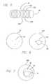
- FIG. 2illustrates and exemplary intra-vascular ultrasound (IVUS) image.
- IVUSintra-vascular ultrasound
- FIG. 3illustrates a plurality of borders that can be identified in an IVUS image.
- FIG. 4illustrates a plurality of control points on one of the borders depicted in FIG. 3 .
- FIG. 5illustrates how a plurality of 2D vascular images can be used to generate a 3D vascular image.
- FIG. 6illustrates how the control points from a first image (e.g., the image depicted in FIG. 4 ) can be extrapolated onto a second image.
- a first imagee.g., the image depicted in FIG. 4
- FIG. 7illustrates a vascular image including a luminal boundary, a medial-adventitial boundary, and a plaque component located therebetween.
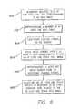
- FIG. 8illustrates a method of identifying a border of a vascular object in accordance with one embodiment of the present invention.
- the present inventionprovides a system and method of using a first vascular image, or more particularly a plurality of control points located thereon, to identify a border on a second vascular image.
- a first vascular imageor more particularly a plurality of control points located thereon, to identify a border on a second vascular image.
- FIG. 1illustrates a vascular-border-identification system 10 in accordance with one embodiment of the present invention.
- an IVUS console 110is electrically connected to a computing device 120 and a transducer 114 via a catheter 112 .
- the transducer 114is inserted into a blood vessel of a patient (not shown) and used to gather IVUS data (i.e., blood-vessel data, or data that can be used to identify the shape of a blood vessel, its density, its composition, etc.).
- IVUS datai.e., blood-vessel data, or data that can be used to identify the shape of a blood vessel, its density, its composition, etc.
- the IVUS datais then provided to (or acquired by) the IVUS console 110 , where it is used to produce an IVUS image of the vessel.
- IVUS datais typically gathered in segments, either through a rotating transducer or an array of circumferentially positioned transducers, where each segment represents an angular portion of an IVUS image.
- ittakes a plurality of segments (or a set of IVUS data) to image an entire cross-section of a vascular object.
- multiple sets of IVUS dataare typically gathered from multiple locations within a vascular object (e.g., by moving the transducer linearly through the vessel). These multiple sets of data can then be used to create a plurality of two-dimensional (2D) images or one three-dimensional (3D) image.
- thermographic devicese.g., an optical coherence tomography (OCT) console
- OCToptical coherence tomography
- MRI devicese.g., MRI devices
- any vascular imaging devicesgenerally known to those skilled in the art.
- computing device depicted in FIG. 1includes, but its not limited to, personal computers or any other data-processing devices (general purpose or application specific) that are generally known to those skilled in the art.
- the IVUS data(or multiple sets thereof) is then provided to (or acquired by) the computing device 120 .
- the computing device 120includes a plurality of applications operating thereon—i.e., a border-detection application 122 , an extrapolation application 124 , and an active-contour application 126 . These applications are used to (i) identify a border and control points on a first IVUS image (i.e., any IVUS image), (ii) extrapolate the control points to a second IVUS image (i.e., another IVUS image), (iii) identify a border on the second IVUS image, and (iv) adjust the border on the second IVUS image.
- FIG. 1the number and/or location of the applications depicted in FIG. 1 are not intended to limit the present invention, but are merely provided to illustrate the environment in which the present invention operates. Thus, for example, using a single application to perform the application functions, as discussed herein, or remotely locating at least one of the applications (in whole or in part) is within the spirit and scope of the present invention. It should further be appreciated that, while the present invention is discussed in terms of singularities (e.g., identifying a border on one IVUS image, extrapolating control points to another IVUS image, etc.), the present invention is not so limited.
- the present inventionis particularly useful if it is used on a plurality of IVUS images (e.g., identifying borders on every fifth IVUS image, extrapolating control points from the fifth IVUS image to the next four IVUS images, etc.).
- first and secondare used broadly to identify any two IVUS images.
- second IVUS imagemay be used to identify an IVUS image distinct from a first IVUS image (as opposed to the second IVUS image in a series of IVUS images).
- Vascular objectsinclude several identifiable borders.
- the luminal borderdemarcates the blood-intima interface and the medial-adventitial border demarcates the external elastic membrane (the boundary between the media and adventitia).
- the plaque-media complexwhich is located there between, can be analyzed and/or calculated. It should be appreciated that the present invention is not limited to the identification of any particular border, and includes all vascular boundaries generally known to those skilled in the art.
- the border-detection application 122is adapted to identify a border on a vascular image (e.g., an IVUS image). In one embodiment of the present invention, this is performed by analyzing the IVUS image, or IVUS data that corresponds the IVUS image, to determine certain gradients located therein. This is because borders of vascular objects can be identified by a change in pixel color (e.g., light-to-dark, dark-to-light, shade1-to-shade2, etc).
- FIG. 2illustrates an exemplary IVUS image 20 of a vascular object.
- the cathetercan be identified by the first light-to-dark transition (or gradient).
- the catheter borderis further identified in FIG. 3 (i.e., 330 ).
- the next dark-to-light transitionor gradient
- the luminal borderi.e., see FIG. 3 , 320 .
- the medial-adventitial bordercan then be identified by going outward from the luminal border until the next dark-to-light transition (or gradient) is found (see FIG. 3 , 310 ).
- the IVUS imageis constructed using gray-scales, it may be necessary to utilize an algorithm and/or at least one threshold value to identify precisely where the image changes from light to dark (or vice versa).
- the present inventionis not limited to any particular algorithm for identifying the aforementioned transitions, and includes all algorithms (and/or threshold values) generally known to those skilled in the art.
- the border-detection algorithmis further adapted to identify at least one control point on the border.
- the border-detection algorithmcan be used to identify a plurality of control points 22 on the luminal border 320 .
- the location and number of control points depicted in FIG. 4are not intended to limit the present invention, and are merely provided to illustrate the environment in which the present invention may operate.
- the border-detection application 122is adapted to identify a border using user-identified control points. Such an embodiment is discussed in detail in U.S. Pat. No. 6,381,350, which issued Apr. 30, 2002, and is incorporated herein, in its entirety, by reference.
- the extrapolation application 124is used to identify at least one control point on at least one other IVUS image. In a preferred embodiment of the present invention, this is done by extrapolating the previously identified control points to at least one other IVUS image.
- multiple 2D imagescan be produced. For example, as illustrated in FIG. 5 , multiple 2D images (e.g., 20 , 52 a - 52 d , etc.) are used to produce a 3D image of a tubular (e.g., vascular) object 50 .
- FIG. 6illustrates how an identified control point can be extrapolated to another IVUS image.
- the control points that were illustrated in FIG. 4i.e., 22
- another IVUS imagee.g., 52 d
- the control pointsare extrapolated using Cartesian coordinates.
- FIG. 6illustrates control points being extrapolated to an adjacent image, the present invention is not so limited.
- extracting control points to additional imagesis within the spirit and scope of the present invention.
- the extrapolating applicationis further adapted to identify (or approximate) a border based on the extrapolated points.
- the extrapolated points 62may be connected using a plurality of lines 64 , where the lines are either straight or curved (not shown).
- the extrapolating applicationis adapted to use an algorithm (e.g., a cubic-interpolation algorithm, etc.) to identify line shape.
- the active-contour application 126is then used to adjust the border to more closely match the actual border of the vascular object.
- the active-contour application 126may consider or take into account at least (i) image gradients (i.e., gradient data), (ii) the proximity of the border to each extrapolated point (i.e., continuity or control-point factor), and/or (iii) border curvature or smoothness (i.e., curvature or boundary factor).
- image gradientsi.e., gradient data
- the proximity of the border to each extrapolated pointi.e., continuity or control-point factor
- border curvature or smoothnessi.e., curvature or boundary factor.
- the bordercan be adjusted if the neighboring pixels (as opposed to the pixels of the border) include border characteristics (e.g., a dark-to-light transition, etc.).
- the bordercan be adjusted so that it passes through each extrapolated point.
- the bordercan be adjusted to prevent sharp transitions (e.g., corners, etc.).
- the continuity and curvature factorsare also used to connect related borders on adjacent images. It should be appreciated that if multiple factors are being considered, then individual factors may be weighted more heavily than others. This becomes important if the factors produce different results (e.g., the gradient factor suggests adjusting the border away from an extrapolated point, etc.). It should further be appreciated that the active-contour application may also be used to adjust the border identified by the border-detection application.
- the present inventionis not limited to the use of the aforementioned factors for border optimization, and that the use of additional factors (e.g., frequency factor, etc.) to adjust (or optimize) a border is within the spirit and scope of the present invention.
- additional factorse.g., frequency factor, etc.
- the adjusted bordersare configured to be manually manipulated. In other words, at least one point on the border can be selected and manually moved to a new location. The active-contour application is then used (as previously discussed) to reconstruct the border accordingly.
- the active-contour applicationis further adapted to adjust related borders in adjacent images. This is done by fitting a geometrical model (e.g., a tensor product B-spline, etc.) over the surface of a plurality of related borders (e.g., as identified on multiple IVUS images). A plurality of points on the geometrical model are then parameterized and formulated into a constrained least-squares system of equations. If a point on the border is manually moved, the active-contour application can utilize these equations to calculate a resulting surface (or mesh of control points). The affected borders (e.g., adjacent borders) can then be adjusted accordingly.
- a geometrical modele.g., a tensor product B-spline, etc.
- the aforementioned processcan be repeated to identify additional borders.
- multiple borderse.g., luminal and medial-adventitial borders
- the multiple bordercan then be imaged (in either 2D or 3D) and analyzed by either a skilled practitioner or a computer algorithm.
- the luminal border 74 and the medial-adventitial border 76can be used (by either a clinician or an algorithm) to identify the plaque-media complex 78 of a vascular object.
- FIG. 8One method of identify a border on a vascular image is illustrated in FIG. 8 .
- step 810multiple sets of IVUS data are acquired, where each set of IVUS data corresponds to a 2D IVUS image.
- a borderis approximated in one IVUS image (e.g., using gradient data, etc.).
- Control points on the approximated borderare then identified at step 814 .
- these control pointsare then used to identify additional control points on additional 2D IVUS images (e.g., via extrapolation, etc.).
- These additional control pointsare then used to approximate at least one other border at step 818 , which is then adjusted at step 820 .
- the borderis adjusted in accordance with at least gradient data.
Landscapes
- Health & Medical Sciences (AREA)
- Engineering & Computer Science (AREA)
- Life Sciences & Earth Sciences (AREA)
- Physics & Mathematics (AREA)
- General Health & Medical Sciences (AREA)
- Medical Informatics (AREA)
- Animal Behavior & Ethology (AREA)
- Heart & Thoracic Surgery (AREA)
- Veterinary Medicine (AREA)
- General Physics & Mathematics (AREA)
- Theoretical Computer Science (AREA)
- Radiology & Medical Imaging (AREA)
- Biophysics (AREA)
- Pathology (AREA)
- Biomedical Technology (AREA)
- Computer Vision & Pattern Recognition (AREA)
- Molecular Biology (AREA)
- Surgery (AREA)
- Nuclear Medicine, Radiotherapy & Molecular Imaging (AREA)
- Public Health (AREA)
- Quality & Reliability (AREA)
- Vascular Medicine (AREA)
- Cardiology (AREA)
- Physiology (AREA)
- Ultra Sonic Daignosis Equipment (AREA)
- Apparatus For Radiation Diagnosis (AREA)
Abstract
Description
This application is a continuation of U.S. patent application Ser. No. 12/102,661 filed on Apr. 14, 2008, now U.S. Pat. No. 7,978,916 which is a continuation of U.S. patent application Ser. No. 10/649,473 filed on Aug. 26, 2003, now U.S. Pat. No. 7,359,554, which claims the benefit pursuant to 35 U.S.C. §119(e) of U.S. Provisional Patent Application Numbers 60/406,148, 60/406,183, 60/406,184, 60/406,185, 60/406,234, and 60/406,254, all of which were filed Aug. 26, 2002, and all are incorporated herein, in their entirety, by reference.
1. Field of the Invention
The present invention relates to vascular borders, or more particularly, to a system and method of using a first vascular image (or control points located therein) to identify a border on a second vascular image.
2. Description of Related Art
The present invention relates to medical imaging arts. It finds particular application to a system and method of, identifying a border in an intra-vascular ultrasound (IVUS) image. It should be appreciated that while the present invention is described in terms of identifying a luminal and medial-adventitial border on an IVUS image, the present invention is not so limited. Thus, for example, identifying any border (or boundary) in any vascular image is within the spirit and scope of the present invention.
Ultrasonic imaging of portions of a patient's body provides a useful tool in various areas of medical practice for determining, the best type and course of treatment. Imaging of the coronary vessels of a patient by ultrasonic techniques can provide physicians with valuable information. For example, the image data may show the extent of a stenosis in a patient, reveal progression of disease, help determine whether procedures such as angioplasty or atherectomy are indicated or whether more invasive procedures may be warranted.
In a typical ultrasound imaging system, an ultrasonic transducer is attached to the end of a catheter that is carefully maneuvered through a patient's body to a point of interest such as within a blood vessel. The transducer may be a single-element crystal or probe that is mechanically scanned or rotated back and forth to cover a sector over a selected angular range. Acoustic signals are then transmitted and echoes (or backscatter) from these acoustic signals are received. The backscatter data can be used to identify the type or density of a scanned tissue. As the probe is swept through the sector, many acoustic lines are processed building up a sector-shaped image of the patient. After the data is collected, an image of the blood vessel (i.e., an IVUS image) is reconstructed using well-known techniques. This image is then visually analyzed by a cardiologist to assess the vessel components and plaque content.
A typical analysis includes determining the size of the lumen and amount of plaque in the vessel. This is performed by generating an image of the vessel (e.g., an IVUS image) and manually drawing contoured boundaries on the image where the clinician believes the luminal and the medial-adventitial borders are located. This is a very time consuming process. Furthermore, this process is made more difficult when multiple images are being analyzed (e.g., to recreate a 3D vascular image, etc.) or the images are of poor quality (e.g., making the boundaries more difficult to see). Thus, it would advantageous to have a system and method of identifying a border on a vascular image that overcomes at least one of these drawbacks.
The present invention provides a system and method of using a first vascular image, or more particularly a plurality of control points located thereon, to identify a border on a second vascular image. Embodiments of the present invention operate in accordance with an intra-vascular ultrasound (IVUS) device and a computing device electrically connected thereto. Specifically, in one embodiment of the present invention, an IVUS console is electrically connected to a computing device and a transducer via a catheter. The transducer is inserted into a blood vessel of a patient and used to gather IVUS data (i.e., blood-vessel data, or data that can be used to identify the shape of a blood vessel, its density, its composition, etc.). The IVUS data is then provided to (or acquired by) the IVUS console, where it is used to produce an IVUS image of the vessel.
The IVUS data (or multiple sets thereof) is then provided to (or acquired by) the computing device. In one embodiment of the present invention, the computing device includes a plurality of applications operating thereon—i.e., a border-detection application, an extrapolation application, and an active-contour application. These applications are used to (i) identify a border and control points on a first IVUS image (i.e., any IVUS image), (ii) extrapolate the control points to a second IVUS image (i.e., another IVUS image), (iii) identify a border on the second IVUS image, and (iv) adjust the border on the second IVUS image in accordance with at least one factor.
Specifically, the border-detection application is adapted to identify a border on a vascular image (e.g., an IVUS image). In one embodiment of the present invention, this is accomplished by analyzing the IVUS image, or IVUS data that corresponds to the IVUS image, to determine certain gradients located therein. This is because borders of vascular objects can be identified by a change in pixel color (e.g., light-to-dark, dark-to-light, shade1-to-shade2, etc). Once the border is identified, the border-detection application is used to identify at least one control point (i.e., a starting-control point) on the identified border. The extrapolation application is then used to identify at least one control point (i.e., an additional control point) on at least one other IVUS image. In a preferred embodiment of the present invention, this is done by extrapolating the previously identified control point (i.e., the starting-control point) to at least one other IVUS image. Once the control point(s) is extrapolated, the extrapolating application is adapted to identify (or approximate) a border that passes through the extrapolated point(s).
The active-contour application is then used to adjust the approximated border (i.e., the border passing through the extrapolated point(s)) to more closely match the actual border of the vascular object. In doing so, the active-contour application may consider, or take into account at least (i) image gradients (i.e., gradient factor), (ii) the proximity of the border to each extrapolated point (i.e., continuity or control-point factor), and/or (iii) border curvature or smoothness (i.e., curvature or boundary factor). Specifically, the gradient factor can be used to adjust the border if the neighboring pixels (as opposed to the pixels of the border) include border characteristics (e.g., a dark-to-light transition, etc.). In other words, if the neighboring pixels include border-like characteristics (or at least more so than the pixels forming the border), then the border is adjusted. The continuity factor and the curvature factor can be used to ensure that the border passes through each extrapolated point and does not include any sharp transitions (e.g., corners, etc.), respectively. In one embodiment of the present invention, the active-contour application is further adapted to adjust related borders on adjacent images if the boarder is manually adjusted.
A more complete understanding of the system and method of identifying a border on an IVUS image will be afforded to those skilled in the art, as well as a realization of additional advantages and objects thereof, by a consideration of the following detailed description of the preferred embodiment. Reference will be made to the appended sheets of drawings which will first be described briefly.
The present invention provides a system and method of using a first vascular image, or more particularly a plurality of control points located thereon, to identify a border on a second vascular image. In the detailed description that follows, like element numerals are used to describe like elements illustrated in one or more figures.
Embodiments of the present invention operate in accordance with an intra-vascular ultrasound (IVUS) device and a computing device electrically connected thereto.FIG. 1 illustrates a vascular-border-identification system 10 in accordance with one embodiment of the present invention. Specifically, anIVUS console 110 is electrically connected to acomputing device 120 and atransducer 114 via acatheter 112. Thetransducer 114 is inserted into a blood vessel of a patient (not shown) and used to gather IVUS data (i.e., blood-vessel data, or data that can be used to identify the shape of a blood vessel, its density, its composition, etc.). The IVUS data is then provided to (or acquired by) theIVUS console 110, where it is used to produce an IVUS image of the vessel.
More particularly, IVUS data is typically gathered in segments, either through a rotating transducer or an array of circumferentially positioned transducers, where each segment represents an angular portion of an IVUS image. Thus, it takes a plurality of segments (or a set of IVUS data) to image an entire cross-section of a vascular object. Furthermore, multiple sets of IVUS data are typically gathered from multiple locations within a vascular object (e.g., by moving the transducer linearly through the vessel). These multiple sets of data can then be used to create a plurality of two-dimensional (2D) images or one three-dimensional (3D) image. It should be appreciated that the present invention is not limited to the use of an IVUS device (or the acquisition of IVUS data), and may further include using thermographic devices, optical devices (e.g., an optical coherence tomography (OCT) console), MRI devices, or any vascular imaging devices generally known to those skilled in the art. It should further be appreciated that the computing device depicted inFIG. 1 includes, but its not limited to, personal computers or any other data-processing devices (general purpose or application specific) that are generally known to those skilled in the art.
The IVUS data (or multiple sets thereof) is then provided to (or acquired by) thecomputing device 120. In one embodiment of the present invention, thecomputing device 120 includes a plurality of applications operating thereon—i.e., a border-detection application 122, anextrapolation application 124, and an active-contour application 126. These applications are used to (i) identify a border and control points on a first IVUS image (i.e., any IVUS image), (ii) extrapolate the control points to a second IVUS image (i.e., another IVUS image), (iii) identify a border on the second IVUS image, and (iv) adjust the border on the second IVUS image. It should be appreciated that the number and/or location of the applications depicted inFIG. 1 are not intended to limit the present invention, but are merely provided to illustrate the environment in which the present invention operates. Thus, for example, using a single application to perform the application functions, as discussed herein, or remotely locating at least one of the applications (in whole or in part) is within the spirit and scope of the present invention. It should further be appreciated that, while the present invention is discussed in terms of singularities (e.g., identifying a border on one IVUS image, extrapolating control points to another IVUS image, etc.), the present invention is not so limited. In fact, the present invention is particularly useful if it is used on a plurality of IVUS images (e.g., identifying borders on every fifth IVUS image, extrapolating control points from the fifth IVUS image to the next four IVUS images, etc.). It should also be appreciated that the terms “first” and “second,” as those terms are used herein, are used broadly to identify any two IVUS images. Thus, the phrase “second IVUS image” may be used to identify an IVUS image distinct from a first IVUS image (as opposed to the second IVUS image in a series of IVUS images).
Vascular objects include several identifiable borders. For example, the luminal border demarcates the blood-intima interface and the medial-adventitial border demarcates the external elastic membrane (the boundary between the media and adventitia). By identifying these borders, the plaque-media complex, which is located there between, can be analyzed and/or calculated. It should be appreciated that the present invention is not limited to the identification of any particular border, and includes all vascular boundaries generally known to those skilled in the art.
Referring back toFIG. 1 , the border-detection application 122 is adapted to identify a border on a vascular image (e.g., an IVUS image). In one embodiment of the present invention, this is performed by analyzing the IVUS image, or IVUS data that corresponds the IVUS image, to determine certain gradients located therein. This is because borders of vascular objects can be identified by a change in pixel color (e.g., light-to-dark, dark-to-light, shade1-to-shade2, etc).
For example,FIG. 2 illustrates anexemplary IVUS image 20 of a vascular object. Starting from the center and working outward, the catheter can be identified by the first light-to-dark transition (or gradient). The catheter border is further identified inFIG. 3 (i.e.,330). Referring back toFIG. 2 , and continuing outward, the next dark-to-light transition (or gradient) identifies the luminal border (i.e., seeFIG. 3 ,320). The medial-adventitial border can then be identified by going outward from the luminal border until the next dark-to-light transition (or gradient) is found (seeFIG. 3 ,310). It should be appreciated that because the IVUS image is constructed using gray-scales, it may be necessary to utilize an algorithm and/or at least one threshold value to identify precisely where the image changes from light to dark (or vice versa). However, it should further be appreciated that the present invention is not limited to any particular algorithm for identifying the aforementioned transitions, and includes all algorithms (and/or threshold values) generally known to those skilled in the art.
Once the border is identified, the border-detection algorithm is further adapted to identify at least one control point on the border. For example, with reference toFIGS. 3 and 4 , the border-detection algorithm can be used to identify a plurality of control points22 on theluminal border 320. It should be appreciated that the location and number of control points depicted inFIG. 4 are not intended to limit the present invention, and are merely provided to illustrate the environment in which the present invention may operate. In an alternate embodiment, the border-detection application 122 is adapted to identify a border using user-identified control points. Such an embodiment is discussed in detail in U.S. Pat. No. 6,381,350, which issued Apr. 30, 2002, and is incorporated herein, in its entirety, by reference.
Referring back toFIG. 1 , once the border and control point(s) are identified on a first vascular image, theextrapolation application 124 is used to identify at least one control point on at least one other IVUS image. In a preferred embodiment of the present invention, this is done by extrapolating the previously identified control points to at least one other IVUS image. By doing this, multiple 2D images (or at least one 3D image) can be produced. For example, as illustrated inFIG. 5 , multiple 2D images (e.g.,20,52a-52d, etc.) are used to produce a 3D image of a tubular (e.g., vascular)object 50.
Once the control points are extrapolated, the extrapolating application is further adapted to identify (or approximate) a border based on the extrapolated points. For example, as shown inFIG. 6 , the extrapolated points62 may be connected using a plurality oflines 64, where the lines are either straight or curved (not shown). In another embodiment of the present invention, the extrapolating application is adapted to use an algorithm (e.g., a cubic-interpolation algorithm, etc.) to identify line shape.
Referring back toFIG. 1 , the active-contour application 126 is then used to adjust the border to more closely match the actual border of the vascular object. In doing so, the active-contour application 126 may consider or take into account at least (i) image gradients (i.e., gradient data), (ii) the proximity of the border to each extrapolated point (i.e., continuity or control-point factor), and/or (iii) border curvature or smoothness (i.e., curvature or boundary factor). Specifically, by considering gradient data (or a gradient factor), the border can be adjusted if the neighboring pixels (as opposed to the pixels of the border) include border characteristics (e.g., a dark-to-light transition, etc.). By considering a continuity or control-point factor, the border can be adjusted so that it passes through each extrapolated point. Furthermore, by considering a curvature or boundary factor, the border can be adjusted to prevent sharp transitions (e.g., corners, etc.). In one embodiment of the present invention, the continuity and curvature factors are also used to connect related borders on adjacent images. It should be appreciated that if multiple factors are being considered, then individual factors may be weighted more heavily than others. This becomes important if the factors produce different results (e.g., the gradient factor suggests adjusting the border away from an extrapolated point, etc.). It should further be appreciated that the active-contour application may also be used to adjust the border identified by the border-detection application. It should also be appreciated that the present invention is not limited to the use of the aforementioned factors for border optimization, and that the use of additional factors (e.g., frequency factor, etc.) to adjust (or optimize) a border is within the spirit and scope of the present invention.
In one embodiment of the present invention, the adjusted borders are configured to be manually manipulated. In other words, at least one point on the border can be selected and manually moved to a new location. The active-contour application is then used (as previously discussed) to reconstruct the border accordingly. In another embodiment of the present invention, the active-contour application is further adapted to adjust related borders in adjacent images. This is done by fitting a geometrical model (e.g., a tensor product B-spline, etc.) over the surface of a plurality of related borders (e.g., as identified on multiple IVUS images). A plurality of points on the geometrical model are then parameterized and formulated into a constrained least-squares system of equations. If a point on the border is manually moved, the active-contour application can utilize these equations to calculate a resulting surface (or mesh of control points). The affected borders (e.g., adjacent borders) can then be adjusted accordingly.
Once the border has been sufficiently adjusted, the aforementioned process can be repeated to identify additional borders. In an alternate embodiment of the present invention, multiple borders (e.g., luminal and medial-adventitial borders) are identified concurrently. The multiple border can then be imaged (in either 2D or 3D) and analyzed by either a skilled practitioner or a computer algorithm. For example, as illustrated inFIG. 7 , theluminal border 74 and the medial-adventitial border 76 can be used (by either a clinician or an algorithm) to identify the plaque-media complex 78 of a vascular object.
One method of identify a border on a vascular image is illustrated inFIG. 8 . Specifically, instep 810, multiple sets of IVUS data are acquired, where each set of IVUS data corresponds to a 2D IVUS image. Atstep 812, a border is approximated in one IVUS image (e.g., using gradient data, etc.). Control points on the approximated border are then identified atstep 814. Atstep 816, these control points are then used to identify additional control points on additional 2D IVUS images (e.g., via extrapolation, etc.). These additional control points are then used to approximate at least one other border atstep 818, which is then adjusted atstep 820. In one embodiment, the border is adjusted in accordance with at least gradient data.
Having thus described a preferred embodiment of a system and method of identifying a border on a vascular image, it should be apparent to those skilled in the art that certain advantages of the system have been achieved. It should also be appreciated that various modifications, adaptations, and alternative embodiments thereof may be made within the scope and spirit of the present invention. The invention is further defined by the following claims.
Claims (20)
1. A method of identifying a border of a vascular object, comprising:
acquiring multiple sets of blood-vessel data related to a vascular object, each set of blood-vessel data corresponding to an image of at least a portion of the vascular object;
using a first set of blood-vessel data to approximate a border on a first image of the vascular object;
identifying at least one control point on the border on the first image;
extrapolating the at least one identified control point to a second set of blood-vessel data to define at least one other control point on a second image;
using the at least one other control point to approximate at least one other border on the second image; and
adjusting a position of the at least one other border on the second image in accordance with at least one factor selected from the group of factors consisting of: a gradient factor, a continuity factor, and a curvature factor.
2. The method ofclaim 1 , wherein the step of acquiring multiple sets of blood-vessel data comprises acquiring multiple sets of intra-vascular ultrasound (IVUS) data, where each set corresponds to an IVUS image of the vascular object.
3. The method ofclaim 1 , wherein the step of using the first set of blood-vessel data to approximate a border on the first image comprises identifying gradients in the first image and using the identified gradients to approximate the border on the first image of the vascular object.
4. The method ofclaim 1 , wherein the second set of blood-vessel data is an adjacent the first set of blood-vessel data such that the second image is an image adjacent the first image.
5. The method ofclaim 1 , wherein the step adjusting the position of the at least one other border comprises adjusting the position of the at least one other border in accordance with at least two factors selected from the group of factors consisting of: a gradient factor, a continuity factor, and a curvature factor.
6. The method ofclaim 5 , wherein one of the at least two factors is weighted more heavily than another of the at least two factors.
7. The method ofclaim 1 , wherein the step of adjusting the position of the at least one other border comprises adjusting the at least one other border in accordance with a continuity factor, the continuity factor representing an amount of continuity between adjacent control points on the at least one other border on the second image.
8. The method ofclaim 1 , wherein the step of adjusting the position of the at least one other border comprises adjusting the at least one other border in accordance with a curvature factor, the curvature factor representing an amount of continuity between adjacent portions of the at least one other border.
9. The method ofclaim 1 , further comprising:
adjusting the position of the at least one other border on the second image in response to an adjustment in the position of the border on the first image.
10. The method ofclaim 9 , wherein the step of adjusting the position of the at least one other border on the second image in response to the adjustment in the position of the border on the first image is performed automatically.
11. The method ofclaim 10 , wherein the position of the border on the first image is adjusted manually by a user.
12. A border-identification system comprising:
a computing device adapted to acquire multiple sets of blood-vessel data related to a vascular object, each set of blood vessel data corresponding to an image of at least a portion of the vascular object;
a border-detection application operating on the computing device, the border-detection application configured to:
approximate a border on a first image of the vascular object using a first set of blood-vessel data;
identify at least one control point on the border on the first image;
extrapolate the at least one identified control point to a second set of blood-vessel data to define at least one other control point on a second image;
approximate at least one other border on the second image using the at least one other control point; and
adjust a position of the at least one other border on the second image in accordance with at least one factor selected from the group of factors consisting of: a gradient factor, a continuity factor, and a curvature factor.
13. The border-identification system ofclaim 12 , wherein the computing device is configured to be in communication with a data-gathering device to acquire the multiple sets of blood-vessel data.
14. The border-identification system ofclaim 13 , wherein the data-gathering device is configured to generate the multiple sets of blood-vessel data.
15. The border-identification system ofclaim 14 , wherein the data-gathering device comprises an intra-vascular ultrasound (IVUS) system.
16. The border-identification system ofclaim 12 , wherein the border detection application is configured to adjust the position of the at least one other border on the second image with at least two factors selected from the group of factors consisting of: a gradient factor, a continuity factor, and a curvature factor.
17. The border-identification system ofclaim 16 , wherein one of the at least two factors is weighted more heavily than another of the at least two factors.
18. The border-identification system ofclaim 12 , wherein the border detection application is configured to adjust the position of the at least one other border on the second image in response to an adjustment in the position of the border on the first image.
19. The border-identification system ofclaim 18 , wherein the border detection application is configured to automatically adjust the position of the at least one other border on the second image in response to the adjustment in the position of the border on the first image.
20. The border-identification system ofclaim 19 , wherein the border detection application is configured to automatically adjust the position of the at least one other border on the second image in response to a manual adjustment in the position of the border on the first image.
Priority Applications (2)
| Application Number | Priority Date | Filing Date | Title |
|---|---|---|---|
| US13/155,196US8233718B2 (en) | 2002-08-26 | 2011-06-07 | System and method for identifying a vascular border |
| US13/563,299US8630492B2 (en) | 2002-08-26 | 2012-07-31 | System and method for identifying a vascular border |
Applications Claiming Priority (9)
| Application Number | Priority Date | Filing Date | Title |
|---|---|---|---|
| US40614802P | 2002-08-26 | 2002-08-26 | |
| US40618302P | 2002-08-26 | 2002-08-26 | |
| US40618502P | 2002-08-26 | 2002-08-26 | |
| US40618402P | 2002-08-26 | 2002-08-26 | |
| US40623402P | 2002-08-26 | 2002-08-26 | |
| US40625402P | 2002-08-26 | 2002-08-26 | |
| US10/649,473US7359554B2 (en) | 2002-08-26 | 2003-08-26 | System and method for identifying a vascular border |
| US12/102,661US7978916B2 (en) | 2002-08-26 | 2008-04-14 | System and method for identifying a vascular border |
| US13/155,196US8233718B2 (en) | 2002-08-26 | 2011-06-07 | System and method for identifying a vascular border |
Related Parent Applications (1)
| Application Number | Title | Priority Date | Filing Date |
|---|---|---|---|
| US12/102,661ContinuationUS7978916B2 (en) | 2002-08-26 | 2008-04-14 | System and method for identifying a vascular border |
Related Child Applications (1)
| Application Number | Title | Priority Date | Filing Date |
|---|---|---|---|
| US13/563,299ContinuationUS8630492B2 (en) | 2002-08-26 | 2012-07-31 | System and method for identifying a vascular border |
Publications (2)
| Publication Number | Publication Date |
|---|---|
| US20110235892A1 US20110235892A1 (en) | 2011-09-29 |
| US8233718B2true US8233718B2 (en) | 2012-07-31 |
Family
ID=31892409
Family Applications (4)
| Application Number | Title | Priority Date | Filing Date |
|---|---|---|---|
| US10/649,473Expired - Fee RelatedUS7359554B2 (en) | 2002-08-26 | 2003-08-26 | System and method for identifying a vascular border |
| US12/102,661Expired - Fee RelatedUS7978916B2 (en) | 2002-08-26 | 2008-04-14 | System and method for identifying a vascular border |
| US13/155,196Expired - Fee RelatedUS8233718B2 (en) | 2002-08-26 | 2011-06-07 | System and method for identifying a vascular border |
| US13/563,299Expired - Fee RelatedUS8630492B2 (en) | 2002-08-26 | 2012-07-31 | System and method for identifying a vascular border |
Family Applications Before (2)
| Application Number | Title | Priority Date | Filing Date |
|---|---|---|---|
| US10/649,473Expired - Fee RelatedUS7359554B2 (en) | 2002-08-26 | 2003-08-26 | System and method for identifying a vascular border |
| US12/102,661Expired - Fee RelatedUS7978916B2 (en) | 2002-08-26 | 2008-04-14 | System and method for identifying a vascular border |
Family Applications After (1)
| Application Number | Title | Priority Date | Filing Date |
|---|---|---|---|
| US13/563,299Expired - Fee RelatedUS8630492B2 (en) | 2002-08-26 | 2012-07-31 | System and method for identifying a vascular border |
Country Status (1)
| Country | Link |
|---|---|
| US (4) | US7359554B2 (en) |
Cited By (66)
| Publication number | Priority date | Publication date | Assignee | Title |
|---|---|---|---|---|
| US20110033098A1 (en)* | 2009-08-07 | 2011-02-10 | Medinol Ltd. | Method and system for stabilizing a series of intravascular ultrasound images and extracting vessel lumen from the images |
| US8670603B2 (en) | 2007-03-08 | 2014-03-11 | Sync-Rx, Ltd. | Apparatus and methods for masking a portion of a moving image stream |
| US8700130B2 (en) | 2007-03-08 | 2014-04-15 | Sync-Rx, Ltd. | Stepwise advancement of a medical tool |
| US8855744B2 (en) | 2008-11-18 | 2014-10-07 | Sync-Rx, Ltd. | Displaying a device within an endoluminal image stack |
| US9095313B2 (en) | 2008-11-18 | 2015-08-04 | Sync-Rx, Ltd. | Accounting for non-uniform longitudinal motion during movement of an endoluminal imaging probe |
| US9101286B2 (en) | 2008-11-18 | 2015-08-11 | Sync-Rx, Ltd. | Apparatus and methods for determining a dimension of a portion of a stack of endoluminal data points |
| US9144394B2 (en) | 2008-11-18 | 2015-09-29 | Sync-Rx, Ltd. | Apparatus and methods for determining a plurality of local calibration factors for an image |
| US9286673B2 (en) | 2012-10-05 | 2016-03-15 | Volcano Corporation | Systems for correcting distortions in a medical image and methods of use thereof |
| US9292918B2 (en) | 2012-10-05 | 2016-03-22 | Volcano Corporation | Methods and systems for transforming luminal images |
| US9301687B2 (en) | 2013-03-13 | 2016-04-05 | Volcano Corporation | System and method for OCT depth calibration |
| US9305334B2 (en) | 2007-03-08 | 2016-04-05 | Sync-Rx, Ltd. | Luminal background cleaning |
| US9307926B2 (en) | 2012-10-05 | 2016-04-12 | Volcano Corporation | Automatic stent detection |
| US9324141B2 (en) | 2012-10-05 | 2016-04-26 | Volcano Corporation | Removal of A-scan streaking artifact |
| US9360630B2 (en) | 2011-08-31 | 2016-06-07 | Volcano Corporation | Optical-electrical rotary joint and methods of use |
| US9367965B2 (en) | 2012-10-05 | 2016-06-14 | Volcano Corporation | Systems and methods for generating images of tissue |
| US9375164B2 (en) | 2007-03-08 | 2016-06-28 | Sync-Rx, Ltd. | Co-use of endoluminal data and extraluminal imaging |
| US9383263B2 (en) | 2012-12-21 | 2016-07-05 | Volcano Corporation | Systems and methods for narrowing a wavelength emission of light |
| US9478940B2 (en) | 2012-10-05 | 2016-10-25 | Volcano Corporation | Systems and methods for amplifying light |
| US9486143B2 (en) | 2012-12-21 | 2016-11-08 | Volcano Corporation | Intravascular forward imaging device |
| US9596993B2 (en) | 2007-07-12 | 2017-03-21 | Volcano Corporation | Automatic calibration systems and methods of use |
| US9612105B2 (en) | 2012-12-21 | 2017-04-04 | Volcano Corporation | Polarization sensitive optical coherence tomography system |
| US9622706B2 (en) | 2007-07-12 | 2017-04-18 | Volcano Corporation | Catheter for in vivo imaging |
| US9629571B2 (en) | 2007-03-08 | 2017-04-25 | Sync-Rx, Ltd. | Co-use of endoluminal data and extraluminal imaging |
| US9709379B2 (en) | 2012-12-20 | 2017-07-18 | Volcano Corporation | Optical coherence tomography system that is reconfigurable between different imaging modes |
| US9730613B2 (en) | 2012-12-20 | 2017-08-15 | Volcano Corporation | Locating intravascular images |
| US9770172B2 (en) | 2013-03-07 | 2017-09-26 | Volcano Corporation | Multimodal segmentation in intravascular images |
| US9855384B2 (en) | 2007-03-08 | 2018-01-02 | Sync-Rx, Ltd. | Automatic enhancement of an image stream of a moving organ and displaying as a movie |
| US9858668B2 (en) | 2012-10-05 | 2018-01-02 | Volcano Corporation | Guidewire artifact removal in images |
| US9867530B2 (en) | 2006-08-14 | 2018-01-16 | Volcano Corporation | Telescopic side port catheter device with imaging system and method for accessing side branch occlusions |
| US9888969B2 (en) | 2007-03-08 | 2018-02-13 | Sync-Rx Ltd. | Automatic quantitative vessel analysis |
| US9974509B2 (en) | 2008-11-18 | 2018-05-22 | Sync-Rx Ltd. | Image super enhancement |
| US10058284B2 (en) | 2012-12-21 | 2018-08-28 | Volcano Corporation | Simultaneous imaging, monitoring, and therapy |
| US10070827B2 (en) | 2012-10-05 | 2018-09-11 | Volcano Corporation | Automatic image playback |
| US10166003B2 (en) | 2012-12-21 | 2019-01-01 | Volcano Corporation | Ultrasound imaging with variable line density |
| US10191220B2 (en) | 2012-12-21 | 2019-01-29 | Volcano Corporation | Power-efficient optical circuit |
| US10219780B2 (en) | 2007-07-12 | 2019-03-05 | Volcano Corporation | OCT-IVUS catheter for concurrent luminal imaging |
| US10219887B2 (en) | 2013-03-14 | 2019-03-05 | Volcano Corporation | Filters with echogenic characteristics |
| US10226597B2 (en) | 2013-03-07 | 2019-03-12 | Volcano Corporation | Guidewire with centering mechanism |
| US10238367B2 (en) | 2012-12-13 | 2019-03-26 | Volcano Corporation | Devices, systems, and methods for targeted cannulation |
| US10292677B2 (en) | 2013-03-14 | 2019-05-21 | Volcano Corporation | Endoluminal filter having enhanced echogenic properties |
| US10332228B2 (en) | 2012-12-21 | 2019-06-25 | Volcano Corporation | System and method for graphical processing of medical data |
| US10362962B2 (en) | 2008-11-18 | 2019-07-30 | Synx-Rx, Ltd. | Accounting for skipped imaging locations during movement of an endoluminal imaging probe |
| US10413317B2 (en) | 2012-12-21 | 2019-09-17 | Volcano Corporation | System and method for catheter steering and operation |
| US10420530B2 (en) | 2012-12-21 | 2019-09-24 | Volcano Corporation | System and method for multipath processing of image signals |
| US10426590B2 (en) | 2013-03-14 | 2019-10-01 | Volcano Corporation | Filters with echogenic characteristics |
| US10568586B2 (en) | 2012-10-05 | 2020-02-25 | Volcano Corporation | Systems for indicating parameters in an imaging data set and methods of use |
| US10595820B2 (en) | 2012-12-20 | 2020-03-24 | Philips Image Guided Therapy Corporation | Smooth transition catheters |
| US10638939B2 (en) | 2013-03-12 | 2020-05-05 | Philips Image Guided Therapy Corporation | Systems and methods for diagnosing coronary microvascular disease |
| US10716528B2 (en) | 2007-03-08 | 2020-07-21 | Sync-Rx, Ltd. | Automatic display of previously-acquired endoluminal images |
| US10724082B2 (en) | 2012-10-22 | 2020-07-28 | Bio-Rad Laboratories, Inc. | Methods for analyzing DNA |
| US10748289B2 (en) | 2012-06-26 | 2020-08-18 | Sync-Rx, Ltd | Coregistration of endoluminal data points with values of a luminal-flow-related index |
| US10758207B2 (en) | 2013-03-13 | 2020-09-01 | Philips Image Guided Therapy Corporation | Systems and methods for producing an image from a rotational intravascular ultrasound device |
| US10942022B2 (en) | 2012-12-20 | 2021-03-09 | Philips Image Guided Therapy Corporation | Manual calibration of imaging system |
| US10939826B2 (en) | 2012-12-20 | 2021-03-09 | Philips Image Guided Therapy Corporation | Aspirating and removing biological material |
| US10993694B2 (en) | 2012-12-21 | 2021-05-04 | Philips Image Guided Therapy Corporation | Rotational ultrasound imaging catheter with extended catheter body telescope |
| US11026591B2 (en) | 2013-03-13 | 2021-06-08 | Philips Image Guided Therapy Corporation | Intravascular pressure sensor calibration |
| US11040140B2 (en) | 2010-12-31 | 2021-06-22 | Philips Image Guided Therapy Corporation | Deep vein thrombosis therapeutic methods |
| US11064903B2 (en) | 2008-11-18 | 2021-07-20 | Sync-Rx, Ltd | Apparatus and methods for mapping a sequence of images to a roadmap image |
| US11064964B2 (en) | 2007-03-08 | 2021-07-20 | Sync-Rx, Ltd | Determining a characteristic of a lumen by measuring velocity of a contrast agent |
| US11141063B2 (en) | 2010-12-23 | 2021-10-12 | Philips Image Guided Therapy Corporation | Integrated system architectures and methods of use |
| US11154313B2 (en) | 2013-03-12 | 2021-10-26 | The Volcano Corporation | Vibrating guidewire torquer and methods of use |
| US11197651B2 (en) | 2007-03-08 | 2021-12-14 | Sync-Rx, Ltd. | Identification and presentation of device-to-vessel relative motion |
| US11272845B2 (en) | 2012-10-05 | 2022-03-15 | Philips Image Guided Therapy Corporation | System and method for instant and automatic border detection |
| US11406498B2 (en) | 2012-12-20 | 2022-08-09 | Philips Image Guided Therapy Corporation | Implant delivery system and implants |
| US12201477B2 (en) | 2012-10-05 | 2025-01-21 | Philips Image Guided Therapy Corporation | Methods and systems for establishing parameters for three-dimensional imaging |
| US12343198B2 (en) | 2013-03-14 | 2025-07-01 | Philips Image Guided Therapy Corporation | Delivery catheter having imaging capabilities |
Families Citing this family (145)
| Publication number | Priority date | Publication date | Assignee | Title |
|---|---|---|---|---|
| US7359554B2 (en)* | 2002-08-26 | 2008-04-15 | Cleveland Clinic Foundation | System and method for identifying a vascular border |
| US6835177B2 (en)* | 2002-11-06 | 2004-12-28 | Sonosite, Inc. | Ultrasonic blood vessel measurement apparatus and method |
| US7727153B2 (en)* | 2003-04-07 | 2010-06-01 | Sonosite, Inc. | Ultrasonic blood vessel measurement apparatus and method |
| EP1654704A2 (en)* | 2003-07-21 | 2006-05-10 | Paieon Inc. | Method and system for identifying an optimal image within a series of images that depict a moving organ |
| US7215802B2 (en)* | 2004-03-04 | 2007-05-08 | The Cleveland Clinic Foundation | System and method for vascular border detection |
| US7678052B2 (en)* | 2004-04-13 | 2010-03-16 | General Electric Company | Method and apparatus for detecting anatomic structures |
| US7831081B2 (en)* | 2005-08-15 | 2010-11-09 | Boston Scientific Scimed, Inc. | Border detection in medical image analysis |
| DE102005050344A1 (en)* | 2005-10-20 | 2007-05-03 | Siemens Ag | Cryocatheter for medical investigation and treatment equipment for e.g. diagnosis and treatment of heart infarcts, has image capture device that maps region of vessel around balloon arranged near catheter tip |
| US8135453B2 (en)* | 2005-12-07 | 2012-03-13 | Siemens Corporation | Method and apparatus for ear canal surface modeling using optical coherence tomography imaging |
| US7945121B2 (en)* | 2006-08-29 | 2011-05-17 | Ati Technologies Ulc | Method and apparatus for interpolating image information |
| US7935060B2 (en)* | 2006-11-08 | 2011-05-03 | Lightlab Imaging, Inc. | Opto-acoustic imaging devices and methods |
| WO2009023626A1 (en)* | 2007-08-10 | 2009-02-19 | The Trustees Of Columbia University In The City Of New York | Systems and methods for tissue characterization and border detection |
| US8781555B2 (en) | 2007-11-26 | 2014-07-15 | C. R. Bard, Inc. | System for placement of a catheter including a signal-generating stylet |
| ES2465915T3 (en) | 2007-11-26 | 2014-06-09 | C.R. Bard, Inc. | Integrated system for intravascular catheter placement |
| US9521961B2 (en) | 2007-11-26 | 2016-12-20 | C. R. Bard, Inc. | Systems and methods for guiding a medical instrument |
| US8388851B2 (en) | 2008-01-08 | 2013-03-05 | Micron Technology, Inc. | Capacitor forming methods |
| US9125562B2 (en) | 2009-07-01 | 2015-09-08 | Avinger, Inc. | Catheter-based off-axis optical coherence tomography imaging system |
| US9788790B2 (en) | 2009-05-28 | 2017-10-17 | Avinger, Inc. | Optical coherence tomography for biological imaging |
| US9572494B2 (en)* | 2008-08-12 | 2017-02-21 | New Jersy Institute of Technology | Method and apparatus for multi-spectral imaging and analysis of skin lesions and biological tissues |
| US9532724B2 (en) | 2009-06-12 | 2017-01-03 | Bard Access Systems, Inc. | Apparatus and method for catheter navigation using endovascular energy mapping |
| WO2011003006A2 (en) | 2009-07-01 | 2011-01-06 | Avinger, Inc. | Atherectomy catheter with laterally-displaceable tip |
| EP2742858B1 (en) | 2009-09-23 | 2024-06-05 | Light-Lab Imaging Inc. | Lumen morphology and vascular resistance measurements data collection systems, apparatus and methods |
| US12426789B2 (en) | 2009-09-23 | 2025-09-30 | Lightlab Imaging, Inc. | Blood vessel lumen morphology and minimum lumen area measurements data collection by intravascular imaging systems for stenosis or stent planning |
| EP2912999B1 (en) | 2010-05-28 | 2022-06-29 | C. R. Bard, Inc. | Apparatus for use with needle insertion guidance system |
| EP2637555B1 (en) | 2010-11-08 | 2021-09-15 | Conavi Medical Inc. | Systems for improved visualization during minimally invasive procedures |
| US20130023912A1 (en) | 2010-12-31 | 2013-01-24 | Volcano Corporation | Multiple Sclerosis Therapeutic Methods Using Therapeutic Cutting Devices and Systems |
| US9949754B2 (en) | 2011-03-28 | 2018-04-24 | Avinger, Inc. | Occlusion-crossing devices |
| EP2691038B1 (en) | 2011-03-28 | 2016-07-20 | Avinger, Inc. | Occlusion-crossing devices, imaging, and atherectomy devices |
| WO2012133878A1 (en) | 2011-03-31 | 2012-10-04 | オリンパスメディカルシステムズ株式会社 | Ultrasound observation device, method for operating ultrasound observation device, and program for operating ultrasound observation device |
| US9295447B2 (en) | 2011-08-17 | 2016-03-29 | Volcano Corporation | Systems and methods for identifying vascular borders |
| US9076680B2 (en) | 2011-10-18 | 2015-07-07 | Micron Technology, Inc. | Integrated circuitry, methods of forming capacitors, and methods of forming integrated circuitry comprising an array of capacitors and circuitry peripheral to the array |
| US8831321B1 (en) | 2011-11-07 | 2014-09-09 | Lightlab Imaging, Inc. | Side branch detection methods, systems and devices |
| EP2849660B1 (en) | 2012-05-14 | 2021-08-25 | Avinger, Inc. | Atherectomy catheter drive assemblies |
| WO2013179859A1 (en) | 2012-05-30 | 2013-12-05 | オリンパスメディカルシステムズ株式会社 | Ultrasonic observation device, ultrasonic observation device operation method, and ultrasonic observation device operation program |
| WO2014022556A1 (en)* | 2012-08-02 | 2014-02-06 | Volcano Corporation | Method for seam elimination and reconstruction of coplanar images from intravascular ultrasonic data |
| EP2919658B1 (en) | 2012-11-19 | 2024-03-20 | Lightlab Imaging, Inc. | Interface devices, systems and methods for multimodal probes |
| EP2931115B1 (en) | 2012-12-12 | 2017-07-26 | Lightlab Imaging, Inc. | Apparatus for automated determination of a lumen contour of a blood vessel |
| WO2014097104A1 (en)* | 2012-12-17 | 2014-06-26 | Koninklijke Philips N.V. | Segmentation of breast lesions in ultrasound images |
| US10398413B2 (en)* | 2012-12-21 | 2019-09-03 | Volcano Corporation | Method for multi-frequency imaging and composite image display using high-bandwidth transducer outputs |
| US10368836B2 (en)* | 2012-12-26 | 2019-08-06 | Volcano Corporation | Gesture-based interface for a multi-modality medical imaging system |
| US10642953B2 (en) | 2012-12-26 | 2020-05-05 | Philips Image Guided Therapy Corporation | Data labeling and indexing in a multi-modality medical imaging system |
| US9779483B2 (en)* | 2012-12-26 | 2017-10-03 | Volcano Corporation | Measurement and enhancement in a multi-modality medical imaging system |
| US10799209B2 (en) | 2012-12-26 | 2020-10-13 | Philips Image Guided Therapy Corporation | Measurement navigation in a multi-modality medical imaging system |
| JP5642910B1 (en)* | 2013-01-23 | 2014-12-17 | オリンパスメディカルシステムズ株式会社 | Ultrasonic observation apparatus, operation method of ultrasonic observation apparatus, and operation program of ultrasonic observation apparatus |
| WO2014151808A1 (en)* | 2013-03-14 | 2014-09-25 | Volcano Corporation | Parallelized tree-based pattern recognition for tissue characterization |
| US9833221B2 (en) | 2013-03-15 | 2017-12-05 | Lightlab Imaging, Inc. | Apparatus and method of image registration |
| US11096717B2 (en) | 2013-03-15 | 2021-08-24 | Avinger, Inc. | Tissue collection device for catheter |
| WO2014143064A1 (en) | 2013-03-15 | 2014-09-18 | Avinger, Inc. | Chronic total occlusion crossing devices with imaging |
| EP3019096B1 (en)* | 2013-07-08 | 2023-07-05 | Avinger, Inc. | System for identification of elastic lamina to guide interventional therapy |
| WO2015108941A1 (en) | 2014-01-14 | 2015-07-23 | Volcano Corporation | Devices and methods for forming vascular access |
| US11260160B2 (en) | 2014-01-14 | 2022-03-01 | Philips Image Guided Therapy Corporation | Systems and methods for improving an AV access site |
| US10874409B2 (en) | 2014-01-14 | 2020-12-29 | Philips Image Guided Therapy Corporation | Methods and systems for clearing thrombus from a vascular access site |
| EP3094241B1 (en) | 2014-01-14 | 2018-07-04 | Volcano Corporation | Systems and methods for evaluating hemodialysis arteriovenous fistula maturation |
| US20150297097A1 (en) | 2014-01-14 | 2015-10-22 | Volcano Corporation | Vascular access evaluation and treatment |
| US10357277B2 (en) | 2014-07-08 | 2019-07-23 | Avinger, Inc. | High speed chronic total occlusion crossing devices |
| EP3166479B1 (en) | 2014-07-11 | 2024-01-03 | Koninklijke Philips N.V. | Devices and systems for treatment of vessels |
| WO2016092390A1 (en) | 2014-12-08 | 2016-06-16 | Koninklijke Philips N.V. | Interactive physiologic data and intravascular imaging data and associated devices, systems, and methods |
| JP6789944B2 (en) | 2014-12-08 | 2020-11-25 | コーニンクレッカ フィリップス エヌ ヴェKoninklijke Philips N.V. | Interactive cardiac test data and related devices, systems, and methods |
| JP6751092B2 (en) | 2014-12-10 | 2020-09-02 | コーニンクレッカ フィリップス エヌ ヴェKoninklijke Philips N.V. | Device, system and method for predicting in-stent restenosis |
| US9629553B2 (en)* | 2014-12-19 | 2017-04-25 | Volcano Corporation | Seam elimination and motion compensation in imaging data |
| US10105107B2 (en) | 2015-01-08 | 2018-10-23 | St. Jude Medical International Holding S.À R.L. | Medical system having combined and synergized data output from multiple independent inputs |
| EP3258863B1 (en) | 2015-02-20 | 2020-09-16 | Koninklijke Philips N.V. | Atherectomy apparatus with imaging |
| US10109058B2 (en) | 2015-05-17 | 2018-10-23 | Lightlab Imaging, Inc. | Intravascular imaging system interfaces and stent detection methods |
| US10222956B2 (en) | 2015-05-17 | 2019-03-05 | Lightlab Imaging, Inc. | Intravascular imaging user interface systems and methods |
| US10646198B2 (en) | 2015-05-17 | 2020-05-12 | Lightlab Imaging, Inc. | Intravascular imaging and guide catheter detection methods and systems |
| US9996921B2 (en) | 2015-05-17 | 2018-06-12 | LIGHTLAB IMAGING, lNC. | Detection of metal stent struts |
| WO2016207762A1 (en) | 2015-06-25 | 2016-12-29 | Koninklijke Philips N.V. | Interactive intravascular procedure training and associated devices, systems, and methods |
| US10568520B2 (en) | 2015-07-13 | 2020-02-25 | Avinger, Inc. | Micro-molded anamorphic reflector lens for image guided therapeutic/diagnostic catheters |
| EP3324830B1 (en) | 2015-07-25 | 2023-01-04 | Lightlab Imaging, Inc. | Intravascular data visualization method and device |
| WO2017046628A1 (en) | 2015-09-15 | 2017-03-23 | Koninklijke Philips N.V. | Device and method for using ivus data to characterize and evaluate a vascular graft condition |
| ES2851548T3 (en) | 2015-11-23 | 2021-09-07 | Lightlab Imaging Inc | Shadow detection and validation in intravascular images |
| EP3435892B1 (en) | 2016-04-01 | 2024-04-03 | Avinger, Inc. | Atherectomy catheter with serrated cutter |
| JP7027331B2 (en) | 2016-04-14 | 2022-03-01 | ライトラボ・イメージング・インコーポレーテッド | Identification of blood vessel branches |
| US10631754B2 (en) | 2016-05-16 | 2020-04-28 | Lightlab Imaging, Inc. | Intravascular absorbable stent detection and diagnostic methods and systems |
| US11344327B2 (en) | 2016-06-03 | 2022-05-31 | Avinger, Inc. | Catheter device with detachable distal end |
| WO2018006041A1 (en) | 2016-06-30 | 2018-01-04 | Avinger, Inc. | Atherectomy catheter with shapeable distal tip |
| US11020563B2 (en) | 2016-07-14 | 2021-06-01 | C. R. Bard, Inc. | Automated catheter-to-vessel size comparison tool and related methods |
| EP3532866A1 (en) | 2016-10-28 | 2019-09-04 | PPG Industries Ohio, Inc. | Coatings for increasing near-infrared detection distances |
| US10842589B2 (en) | 2017-03-21 | 2020-11-24 | Canon U.S.A., Inc. | Method for displaying an anatomical image of a coronary artery on a graphical user interface |
| JP7054411B2 (en) | 2017-07-26 | 2022-04-13 | キヤノン ユーエスエイ,インコーポレイテッド | Methods for assessing cardiac motion using angiographic images |
| US11883235B2 (en) | 2017-08-15 | 2024-01-30 | Philips Image Guided Therapy Corporation | Phased array imaging and therapy intraluminal ultrasound device |
| US11819360B2 (en) | 2017-08-15 | 2023-11-21 | Koninklijke Philips N.V. | Intraluminal rotational ultrasound for diagnostic imaging and therapy |
| WO2019034500A1 (en) | 2017-08-15 | 2019-02-21 | Koninklijke Philips N.V. | Frequency-tunable intraluminal ultrasound device |
| JP7069294B2 (en) | 2017-08-15 | 2022-05-17 | コーニンクレッカ フィリップス エヌ ヴェ | Intraluminal ultrasound device for diagnostic imaging and treatment |
| EP3668410B1 (en) | 2017-08-15 | 2023-01-04 | Koninklijke Philips N.V. | Intracardiac therapeutic and diagnostic ultrasound device |
| EP3668597A1 (en) | 2017-08-16 | 2020-06-24 | Koninklijke Philips N.V. | Disposable therapeutic ultrasound device |
| US11571129B2 (en) | 2017-10-03 | 2023-02-07 | Canon U.S.A., Inc. | Detecting and displaying stent expansion |
| US10621748B2 (en) | 2017-10-03 | 2020-04-14 | Canon U.S.A., Inc. | Detecting and displaying stent expansion |
| WO2019174971A1 (en) | 2018-03-14 | 2019-09-19 | Koninklijke Philips N.V. | Alternative anatomical borders of blood vessels and associated devices, systems, and methods |
| US12167867B2 (en) | 2018-04-19 | 2024-12-17 | Avinger, Inc. | Occlusion-crossing devices |
| JP7075371B2 (en) | 2018-05-03 | 2022-05-25 | キヤノン ユーエスエイ,インコーポレイテッド | Devices, systems, and methods for highlighting areas of interest across multiple imaging modality |
| US11382516B2 (en) | 2018-06-08 | 2022-07-12 | Canon U.S.A., Inc. | Apparatuses, methods, and storage mediums for lumen and artifacts detection in one or more images, such as in optical coherence tomography images |
| US10992079B2 (en) | 2018-10-16 | 2021-04-27 | Bard Access Systems, Inc. | Safety-equipped connection systems and methods thereof for establishing electrical connections |
| JP7378484B2 (en) | 2018-10-26 | 2023-11-13 | コーニンクレッカ フィリップス エヌ ヴェ | Intraluminal ultrasound imaging with automatic and assisted labels and bookmarks |
| EP3870057B1 (en) | 2018-10-26 | 2025-02-12 | Koninklijke Philips N.V. | Disease specific and treatment type specific control of intraluminal ultrasound imaging |
| CN112996445B (en) | 2018-10-26 | 2025-01-07 | 皇家飞利浦有限公司 | Velocity determination for intraluminal ultrasound imaging and associated devices, systems and methods |
| CN113518588B (en) | 2018-10-26 | 2024-08-09 | 皇家飞利浦有限公司 | Intraluminal ultrasound direction guidance and associated devices, systems, and methods |
| EP3870063A1 (en) | 2018-10-26 | 2021-09-01 | Koninklijke Philips N.V. | Intraluminal ultrasound navigation guidance and associated devices, systems, and methods |
| US11596384B2 (en) | 2018-10-26 | 2023-03-07 | Philips Image Guided Therapy Corporation | Intraluminal ultrasound vessel border selection and associated devices, systems, and methods |
| EP3870068A1 (en) | 2018-10-26 | 2021-09-01 | Koninklijke Philips N.V. | Graphical longitudinal display for intraluminal ultrasound imaging and associated devices, systems, and methods |
| KR20210087991A (en) | 2018-11-13 | 2021-07-13 | 피피지 인더스트리즈 오하이오 인코포레이티드 | How to detect hidden patterns |
| US11561329B2 (en) | 2019-01-07 | 2023-01-24 | Ppg Industries Ohio, Inc. | Near infrared control coating, articles formed therefrom, and methods of making the same |
| US20220061802A1 (en)* | 2019-01-15 | 2022-03-03 | Koninklijke Philips N.V. | Systems and methods for identifying features sensed by a vascular device |
| WO2020159984A1 (en) | 2019-01-30 | 2020-08-06 | Canon U.S.A., Inc. | Apparatuses, systems, methods and storage mediums for performance of co-registration |
| US12109056B2 (en) | 2019-09-17 | 2024-10-08 | Canon U.S.A., Inc. | Constructing or reconstructing 3D structure(s) |
| US11759166B2 (en) | 2019-09-20 | 2023-09-19 | Bard Access Systems, Inc. | Automatic vessel detection tools and methods |
| US12178640B2 (en) | 2019-10-08 | 2024-12-31 | Philips Image Guided Therapy Corporation | Visualization of reflectors in intraluminal ultrasound images and associated systems, methods, and devices |
| US20220395333A1 (en) | 2019-11-06 | 2022-12-15 | Philips Image Guided Therapy Corporation | Co-registration of intravascular data and multi-segment vasculature, and associated devices, systems, and methods |
| US11963740B2 (en) | 2019-12-05 | 2024-04-23 | Canon U.S.A., Inc. | Lumen, stent, and/or artifact detection in one or more images, such as in optical coherence tomography images |
| WO2021115958A1 (en) | 2019-12-10 | 2021-06-17 | Koninklijke Philips N.V. | Intraluminal image-based vessel diameter determination and associated devices, systems, and methods |
| WO2021140042A1 (en) | 2020-01-06 | 2021-07-15 | Koninklijke Philips N.V. | Intraluminal imaging based detection and visualization of intraluminal treatment anomalies |
| EP4117534A1 (en) | 2020-03-10 | 2023-01-18 | Koninklijke Philips N.V. | Intraluminal image visualization with adaptive scaling and associated systems, methods, and devices |
| US11877810B2 (en) | 2020-07-21 | 2024-01-23 | Bard Access Systems, Inc. | System, method and apparatus for magnetic tracking of ultrasound probe and generation of 3D visualization thereof |
| EP4185209A1 (en) | 2020-08-04 | 2023-05-31 | Bard Access Systems, Inc. | System and method for optimized medical component insertion monitoring and imaging enhancement |
| WO2022035760A1 (en) | 2020-08-10 | 2022-02-17 | Bard Access Systems, Inc. | System and method for generating vessel representations in mixed reality/virtual reality |
| US11890139B2 (en) | 2020-09-03 | 2024-02-06 | Bard Access Systems, Inc. | Portable ultrasound systems |
| US11992363B2 (en) | 2020-09-08 | 2024-05-28 | Bard Access Systems, Inc. | Dynamically adjusting ultrasound-imaging systems and methods thereof |
| CN216257185U (en) | 2020-09-10 | 2022-04-12 | 巴德阿克塞斯系统股份有限公司 | Ultrasound Probes and Ultrasound Systems |
| WO2022067101A1 (en) | 2020-09-25 | 2022-03-31 | Bard Access Systems, Inc. | Minimum catheter length tool |
| WO2022072727A2 (en) | 2020-10-02 | 2022-04-07 | Bard Access Systems, Inc. | Ultrasound systems and methods for sustained spatial attention |
| EP4228516A1 (en) | 2020-10-15 | 2023-08-23 | Bard Access Systems, Inc. | Ultrasound imaging system for generation of a three-dimensional ultrasound image |
| CN216933458U (en) | 2020-11-24 | 2022-07-12 | 巴德阿克塞斯系统股份有限公司 | Object recognition and needle guidance system |
| CN114569156A (en) | 2020-12-01 | 2022-06-03 | 巴德阿克塞斯系统股份有限公司 | Ultrasound imaging system and method for identifying one or more of a plurality of blood vessels |
| CN114569155A (en) | 2020-12-01 | 2022-06-03 | 巴德阿克塞斯系统股份有限公司 | Ultrasound imaging system and method for obtaining ultrasound image by the same |
| EP4258997A1 (en) | 2020-12-14 | 2023-10-18 | Bard Access Systems, Inc. | Securement of hands-free ultrasound probe |
| JPWO2022209657A1 (en)* | 2021-03-30 | 2022-10-06 | ||
| CN217960146U (en) | 2021-04-15 | 2022-12-06 | 巴德阿克塞斯系统股份有限公司 | Ultrasound imaging system |
| US11779221B2 (en) | 2021-06-18 | 2023-10-10 | Canon U.S.A., Inc. | Apparatus, method and storage medium for lumen curve simplification for editing in one or more images, such as in optical coherence tomography images |
| CN118042992A (en) | 2021-09-30 | 2024-05-14 | 皇家飞利浦有限公司 | Intraluminal ultrasound vessel segment identification and related devices, systems, and methods |
| CN116058873A (en) | 2021-11-03 | 2023-05-05 | 巴德阿克塞斯系统股份有限公司 | Interoperation optimization function through Doppler and image-based vessel discrimination |
| US20230181140A1 (en) | 2021-12-11 | 2023-06-15 | Philips Image Guided Therapy Corporation | Registration of intraluminal physiological data to longitudinal image body lumen using extraluminal imaging data |
| US20230190227A1 (en) | 2021-12-16 | 2023-06-22 | Philips Image Guided Therapy Corporation | Plaque burden indication on longitudinal intraluminal image and x-ray image |
| EP4447816A1 (en) | 2021-12-17 | 2024-10-23 | Koninklijke Philips N.V. | Systems, devices, and methods for coregistration of intravascular data to enhanced stent deployment x-ray images |
| CN118414127A (en) | 2021-12-17 | 2024-07-30 | 皇家飞利浦有限公司 | Controlling laser atherectomy by co-registered intravascular imaging |
| EP4449430A1 (en) | 2021-12-17 | 2024-10-23 | Philips Image Guided Therapy Corporation | Intravascular imaging assessment of stent deployment and associated systems, devices, and methods |
| CN118541093A (en) | 2021-12-22 | 2024-08-23 | 皇家飞利浦有限公司 | Intraluminal imaging with deep breath confirmation reference and target image frames |
| WO2023118080A1 (en) | 2021-12-22 | 2023-06-29 | Koninklijke Philips N.V. | Intravascular ultrasound imaging for calcium detection and analysis |
| EP4201342A1 (en) | 2021-12-22 | 2023-06-28 | Koninklijke Philips N.V. | Intravascular ultrasound imaging for calcium detection and analysis |
| US20230190215A1 (en) | 2021-12-22 | 2023-06-22 | Philips Image Guided Therapy Corporation | Co-registration of intraluminal data to no contrast x-ray image frame and associated systems, device and methods |
| CN118829999A (en) | 2022-03-08 | 2024-10-22 | 皇家飞利浦有限公司 | Intravascular ultrasound imaging with contour generation and editing for circular and non-circular vessel boundaries |
| US12433567B2 (en) | 2022-03-16 | 2025-10-07 | Bard Access Systems, Inc. | Ultrasound imaging system |
| US12102481B2 (en) | 2022-06-03 | 2024-10-01 | Bard Access Systems, Inc. | Ultrasound probe with smart accessory |
| WO2023247467A1 (en) | 2022-06-24 | 2023-12-28 | Koninklijke Philips N.V. | Intraluminal ultrasound imaging with automatic detection of target and reference regions |
| US12137989B2 (en) | 2022-07-08 | 2024-11-12 | Bard Access Systems, Inc. | Systems and methods for intelligent ultrasound probe guidance |
| WO2024120659A1 (en) | 2022-12-07 | 2024-06-13 | Koninklijke Philips N.V. | Registration of intraluminal physiological data to longitudinal image of body lumen using extraluminal imaging data |
Citations (35)
| Publication number | Priority date | Publication date | Assignee | Title |
|---|---|---|---|---|
| US4228804A (en) | 1978-02-28 | 1980-10-21 | Case Western Reserve University | Diagnostic ultrasonography utilizing frequency spectrum analysis presented in terms of B-scan color patterns or X-Y graph displays |
| US4511984A (en) | 1981-09-19 | 1985-04-16 | Tokyo Shibaura Denki Kabushiki Kaisha | Ultrasound diagnostic apparatus |
| US4561019A (en) | 1983-05-16 | 1985-12-24 | Riverside Research Institute | Frequency diversity for image enhancement |
| US4575799A (en) | 1983-03-23 | 1986-03-11 | Fujitsu Limited | Ultrasonic living body tissue characterization system |
| US4858124A (en) | 1984-08-15 | 1989-08-15 | Riverside Research Institute | Method for enhancement of ultrasonic image data |
| US5016641A (en)* | 1989-11-13 | 1991-05-21 | Advanced Technology Laboratories, Inc. | Spectral interpolation of ultrasound Doppler signal |
| US5235984A (en) | 1992-03-30 | 1993-08-17 | Hewlett-Packard Company | On-line acoustic densitometry tool for use with an ultrasonic imaging system |
| US5285786A (en)* | 1991-06-12 | 1994-02-15 | Kabushiki Kaisha Toshiba | Apparatus and method for radiographic diagnosis |
| US5363850A (en) | 1994-01-26 | 1994-11-15 | Cardiovascular Imaging Systems, Inc. | Method for recognition and reduction of blood speckle in blood vessel imaging system |
| US5417215A (en) | 1994-02-04 | 1995-05-23 | Long Island Jewish Medical Center | Method of tissue characterization by ultrasound |
| US5445155A (en) | 1991-03-13 | 1995-08-29 | Scimed Life Systems Incorporated | Intravascular imaging apparatus and methods for use and manufacture |
| US5559901A (en) | 1993-06-29 | 1996-09-24 | U.S. Philips Corporation | Method and device for determining a contour in a space of image parameter values |
| US5724972A (en) | 1996-05-02 | 1998-03-10 | Acuson Corporation | Method and apparatus for distributed focus control with slope tracking |
| US5876343A (en) | 1997-09-23 | 1999-03-02 | Scimed Life Systems, Inc. | Methods and apparatus for blood speckle detection in an intravascular ultrasound imaging system |
| US5885218A (en) | 1997-11-07 | 1999-03-23 | Scimed Life Systems, Inc. | Method and apparatus for spatial filtering in an intravascular ultrasound imaging system |
| US5938607A (en) | 1996-09-25 | 1999-08-17 | Atl Ultrasound, Inc. | Ultrasonic diagnostic imaging system with access to reference image library |
| US5957138A (en) | 1997-08-25 | 1999-09-28 | Diasonics Ultrasound, Inc. | Method and apparatus for three-dimensional flow lumen imaging |
| US6095976A (en) | 1997-06-19 | 2000-08-01 | Medinol Ltd. | Method for enhancing an image derived from reflected ultrasound signals produced by an ultrasound transmitter and detector inserted in a bodily lumen |
| US6106465A (en) | 1997-08-22 | 2000-08-22 | Acuson Corporation | Ultrasonic method and system for boundary detection of an object of interest in an ultrasound image |
| US6106460A (en) | 1998-03-26 | 2000-08-22 | Scimed Life Systems, Inc. | Interface for controlling the display of images of diagnostic or therapeutic instruments in interior body regions and related data |
| US6120445A (en) | 1998-10-02 | 2000-09-19 | Scimed Life Systems, Inc. | Method and apparatus for adaptive cross-sectional area computation of IVUS objects using their statistical signatures |
| US6200268B1 (en) | 1999-09-10 | 2001-03-13 | The Cleveland Clinic Foundation | Vascular plaque characterization |
| US6217517B1 (en) | 1998-09-28 | 2001-04-17 | Scimed Life Systems, Inc. | Ultrasound imaging with zoom having independent processing channels |
| US6238342B1 (en) | 1998-05-26 | 2001-05-29 | Riverside Research Institute | Ultrasonic tissue-type classification and imaging methods and apparatus |
| US6306089B1 (en) | 1999-09-24 | 2001-10-23 | Atl Ultrasound, Inc. | Ultrasonic diagnostic imaging system with customized measurements and calculations |
| US6324420B1 (en)* | 1997-04-02 | 2001-11-27 | Canon Kabushiki Kaisha | Blood vessel tracking apparatus |
| US6335980B1 (en) | 1997-07-25 | 2002-01-01 | Arch Development Corporation | Method and system for the segmentation of lung regions in lateral chest radiographs |
| US6381350B1 (en) | 1999-07-02 | 2002-04-30 | The Cleveland Clinic Foundation | Intravascular ultrasonic analysis using active contour method and system |
| US6454715B2 (en) | 2000-04-11 | 2002-09-24 | Scimed Life Systems, Inc. | Methods and apparatus for blood speckle detection in an intravascular ultrasound imaging system |
| US6544187B2 (en) | 1999-03-31 | 2003-04-08 | Mayo Foundation For Medical Education And Research | Parametric imaging ultrasound catheter |
| US6561980B1 (en) | 2000-05-23 | 2003-05-13 | Alpha Intervention Technology, Inc | Automatic segmentation of prostate, rectum and urethra in ultrasound imaging |
| US6569104B2 (en)* | 1998-07-16 | 2003-05-27 | Canon Kabushiki Kaisha | Blood vessel detecting apparatus |
| US6785409B1 (en) | 2000-10-24 | 2004-08-31 | Koninklijke Philips Electronics, N.V. | Segmentation method and apparatus for medical images using diffusion propagation, pixel classification, and mathematical morphology |
| US7027630B2 (en)* | 2000-12-22 | 2006-04-11 | Koninklijke Philips Electronics, N.V. | Method of analyzing a data set comprising a volumetric representation of an object to be examined |
| US7359554B2 (en) | 2002-08-26 | 2008-04-15 | Cleveland Clinic Foundation | System and method for identifying a vascular border |
Family Cites Families (5)
| Publication number | Priority date | Publication date | Assignee | Title |
|---|---|---|---|---|
| US5217456A (en)* | 1992-02-24 | 1993-06-08 | Pdt Cardiovascular, Inc. | Device and method for intra-vascular optical radial imaging |
| EP0633548B1 (en) | 1993-06-29 | 2002-01-30 | Koninklijke Philips Electronics N.V. | Method and device for determining a contour in a space having a density distribution |
| EP0745211B1 (en)* | 1994-01-24 | 2003-05-14 | Svg Lithography Systems, Inc. | Grating-grating interferometric alignment system |
| US6148095A (en) | 1997-09-08 | 2000-11-14 | University Of Iowa Research Foundation | Apparatus and method for determining three-dimensional representations of tortuous vessels |
| US6659981B2 (en)* | 2000-12-08 | 2003-12-09 | Medtronic, Inc. | Medical device delivery catheter with distal locator |
- 2003
- 2003-08-26USUS10/649,473patent/US7359554B2/ennot_activeExpired - Fee Related
- 2008
- 2008-04-14USUS12/102,661patent/US7978916B2/ennot_activeExpired - Fee Related
- 2011
- 2011-06-07USUS13/155,196patent/US8233718B2/ennot_activeExpired - Fee Related
- 2012
- 2012-07-31USUS13/563,299patent/US8630492B2/ennot_activeExpired - Fee Related
Patent Citations (43)
| Publication number | Priority date | Publication date | Assignee | Title |
|---|---|---|---|---|
| US4228804A (en) | 1978-02-28 | 1980-10-21 | Case Western Reserve University | Diagnostic ultrasonography utilizing frequency spectrum analysis presented in terms of B-scan color patterns or X-Y graph displays |
| US4511984A (en) | 1981-09-19 | 1985-04-16 | Tokyo Shibaura Denki Kabushiki Kaisha | Ultrasound diagnostic apparatus |
| US4575799A (en) | 1983-03-23 | 1986-03-11 | Fujitsu Limited | Ultrasonic living body tissue characterization system |
| US4561019A (en) | 1983-05-16 | 1985-12-24 | Riverside Research Institute | Frequency diversity for image enhancement |
| US4858124A (en) | 1984-08-15 | 1989-08-15 | Riverside Research Institute | Method for enhancement of ultrasonic image data |
| US5016641A (en)* | 1989-11-13 | 1991-05-21 | Advanced Technology Laboratories, Inc. | Spectral interpolation of ultrasound Doppler signal |
| US5445155A (en) | 1991-03-13 | 1995-08-29 | Scimed Life Systems Incorporated | Intravascular imaging apparatus and methods for use and manufacture |
| US5285786A (en)* | 1991-06-12 | 1994-02-15 | Kabushiki Kaisha Toshiba | Apparatus and method for radiographic diagnosis |
| US5235984A (en) | 1992-03-30 | 1993-08-17 | Hewlett-Packard Company | On-line acoustic densitometry tool for use with an ultrasonic imaging system |
| US5559901A (en) | 1993-06-29 | 1996-09-24 | U.S. Philips Corporation | Method and device for determining a contour in a space of image parameter values |
| US5363850A (en) | 1994-01-26 | 1994-11-15 | Cardiovascular Imaging Systems, Inc. | Method for recognition and reduction of blood speckle in blood vessel imaging system |
| US5417215A (en) | 1994-02-04 | 1995-05-23 | Long Island Jewish Medical Center | Method of tissue characterization by ultrasound |
| US5724972A (en) | 1996-05-02 | 1998-03-10 | Acuson Corporation | Method and apparatus for distributed focus control with slope tracking |
| US5938607A (en) | 1996-09-25 | 1999-08-17 | Atl Ultrasound, Inc. | Ultrasonic diagnostic imaging system with access to reference image library |
| US6324420B1 (en)* | 1997-04-02 | 2001-11-27 | Canon Kabushiki Kaisha | Blood vessel tracking apparatus |
| US6095976A (en) | 1997-06-19 | 2000-08-01 | Medinol Ltd. | Method for enhancing an image derived from reflected ultrasound signals produced by an ultrasound transmitter and detector inserted in a bodily lumen |
| US6152878A (en) | 1997-06-19 | 2000-11-28 | Medinol Ltd. | Intravascular ultrasound enhanced image and signal processing |
| US6335980B1 (en) | 1997-07-25 | 2002-01-01 | Arch Development Corporation | Method and system for the segmentation of lung regions in lateral chest radiographs |
| US6106465A (en) | 1997-08-22 | 2000-08-22 | Acuson Corporation | Ultrasonic method and system for boundary detection of an object of interest in an ultrasound image |
| US5957138A (en) | 1997-08-25 | 1999-09-28 | Diasonics Ultrasound, Inc. | Method and apparatus for three-dimensional flow lumen imaging |
| US5876343A (en) | 1997-09-23 | 1999-03-02 | Scimed Life Systems, Inc. | Methods and apparatus for blood speckle detection in an intravascular ultrasound imaging system |
| US6050946A (en) | 1997-09-23 | 2000-04-18 | Scimed Life Systems, Inc. | Methods and apparatus for blood speckle detection in an intravascular ultrasound imaging system |
| US6254541B1 (en) | 1997-09-23 | 2001-07-03 | Scimed Life Systems, Inc. | Methods and apparatus for blood speckle detection in an intravascular ultrasound imaging system |
| US5885218A (en) | 1997-11-07 | 1999-03-23 | Scimed Life Systems, Inc. | Method and apparatus for spatial filtering in an intravascular ultrasound imaging system |
| US6106460A (en) | 1998-03-26 | 2000-08-22 | Scimed Life Systems, Inc. | Interface for controlling the display of images of diagnostic or therapeutic instruments in interior body regions and related data |
| US20030028118A1 (en) | 1998-03-26 | 2003-02-06 | Boston Scientific Corporation | Interactive systems and methods for controlling the use of diagnostic or therapeutic instruments in interior body regions |
| US6238342B1 (en) | 1998-05-26 | 2001-05-29 | Riverside Research Institute | Ultrasonic tissue-type classification and imaging methods and apparatus |
| US6569104B2 (en)* | 1998-07-16 | 2003-05-27 | Canon Kabushiki Kaisha | Blood vessel detecting apparatus |
| US6217517B1 (en) | 1998-09-28 | 2001-04-17 | Scimed Life Systems, Inc. | Ultrasound imaging with zoom having independent processing channels |
| US6287259B1 (en) | 1998-10-02 | 2001-09-11 | Scimed Life Systems, Inc. | Systems and methods for evaluating objects within an ultrasound image |
| US20030092993A1 (en) | 1998-10-02 | 2003-05-15 | Scimed Life Systems, Inc. | Systems and methods for evaluating objects within an ultrasound image |
| US20010014774A1 (en) | 1998-10-02 | 2001-08-16 | Sorin Grunwald | Systems and methods for evaluating objects within an ultrasound image |
| US6514202B2 (en) | 1998-10-02 | 2003-02-04 | Scimed Life Systems, Inc. | Systems and methods for evaluating objects within an ultrasound image |
| US6120445A (en) | 1998-10-02 | 2000-09-19 | Scimed Life Systems, Inc. | Method and apparatus for adaptive cross-sectional area computation of IVUS objects using their statistical signatures |
| US6544187B2 (en) | 1999-03-31 | 2003-04-08 | Mayo Foundation For Medical Education And Research | Parametric imaging ultrasound catheter |
| US6381350B1 (en) | 1999-07-02 | 2002-04-30 | The Cleveland Clinic Foundation | Intravascular ultrasonic analysis using active contour method and system |
| US6200268B1 (en) | 1999-09-10 | 2001-03-13 | The Cleveland Clinic Foundation | Vascular plaque characterization |
| US6306089B1 (en) | 1999-09-24 | 2001-10-23 | Atl Ultrasound, Inc. | Ultrasonic diagnostic imaging system with customized measurements and calculations |
| US6454715B2 (en) | 2000-04-11 | 2002-09-24 | Scimed Life Systems, Inc. | Methods and apparatus for blood speckle detection in an intravascular ultrasound imaging system |
| US6561980B1 (en) | 2000-05-23 | 2003-05-13 | Alpha Intervention Technology, Inc | Automatic segmentation of prostate, rectum and urethra in ultrasound imaging |
| US6785409B1 (en) | 2000-10-24 | 2004-08-31 | Koninklijke Philips Electronics, N.V. | Segmentation method and apparatus for medical images using diffusion propagation, pixel classification, and mathematical morphology |
| US7027630B2 (en)* | 2000-12-22 | 2006-04-11 | Koninklijke Philips Electronics, N.V. | Method of analyzing a data set comprising a volumetric representation of an object to be examined |
| US7359554B2 (en) | 2002-08-26 | 2008-04-15 | Cleveland Clinic Foundation | System and method for identifying a vascular border |
Non-Patent Citations (16)
| Title |
|---|
| Bruining, N., Von Birgelen, C., De Feyter. P., Ligthart. J., Serruys, P., Roelandt, J., "Dynamic Imaging of Coronary Stent Structures: An ECG-Gated Three-Dimensional Intracoronary Ultrasound Study in Humans", Ultrasound in Medicine and Biology. 1998, pp. 631-637, vol. 24, No. 5, Elsevier, New York, U.S.A. |
| Chen. C.. Lu, H., Hsiao, A., "A Dual-Snake Model of High Penetrability for Ultrasound Image Boundary Extraction", Ultrasound in Medicine and Biology, 2001, pp. 1651-1665, vol. 27. No. 12, Elsevier, New York, U.S.A. |
| Iwase, M., Kondo, T., Hasegawa, K,, Kimura. M., Matsuyama. H., Watanabe. Y,, Hishida. H., "Three-Dimensional Echocardiography by Semi-Automatic Border Detection In Assessment of Left Ventricular Volume and Ejection Fraction: Comparison with Magnetic Resonance Imaging", Journal of Cardiology, 1997, pp. 97-105, vol. 30, No. 2, Japanese College of Cardiology. Tokyo. Japan. |
| Klingensmith, J. and Vince, p., "B-Spline Methods for interactive Segmentation and Modeling of Lumen and Vessel Surfaces in Three-Dimensional Intravascular Ultrasound," Computerized Medical Imaging and Graphics 26, 2002, pp. 429-438, Elsevier Science Ltd.. U.S.A. |
| Klingensmith, J., Shekhar, R., Vince. D., "Evaluation of Three-Dimensional Segmentation Algorithms for the Identification of Luminal and Medial-Adventitial Borders in Intravascular Ultrasound Images", IEEE Transactions on . Medical Imaging, 2000, pp. 996-1011, vol. 19, No. 10, Institute of Electrical and Electronics Engineers, New York, U.S.A. |
| Klingensmith, J., Shekhar, R., Vince. D., "Evaluation of Three-Dimensional Segmentation Algorithms for the Identification of Luminal and Medial-Adventitial Borders in Intravascular Ultrasound Images", IEEE Transactions on • Medical Imaging, 2000, pp. 996-1011, vol. 19, No. 10, Institute of Electrical and Electronics Engineers, New York, U.S.A. |
| Klingensmith, J., Vince, D., Kuban, B., Shekhar, R., Tuzcu, E.. Nissen, S., Cornhill, J., "Assessment of Coronary Compensatory Enlargement by Three-Dimensional Intravascular Ultrasound", International Journal of Cardiac Imaging. 2000. pp. 87-96, vol. 16, No. 2. Nijhoff, Boston. U.S.A. |
| Ko, C., Mao. C., Sun, Y., Chang, S., "A Fully Automated Identification of Coronary Borders from the Tree Structure of Coronary Angiograms", International Journal of Bio-Medical Computing, 1995, pp. 193-208, vol. 39, No. 2. Elsevier, Barking, England. |
| Kovalski, G., Beyar, R.., Shofti, R., Azhari. H., "Three-Dimensional Automatic Quantitative Analysis of Intravascular Ultrasound Images", Ultrasound in Medicine and Biology, 2000, pp. 527-537, vol. 26, No. 4, Elsevier, New York, U.S.A. |
| Lobregt, S., Viergever, M., "A Discrete Dynamic Contour Model", IEEE Transactions on Medical Imaging. 1995. pp. 12-24, vol. 14, No. 1. Institute of Electrical and Electronics Engineers. New York, U.S.A. |
| Nair, A., Kuban, B., Obuchowski, N., and Vince, D., "Assessing Spectral Algorithms to Predict Atherosclerotic Plaque Composition With Normalized and Raw Intravascular Ultrasound Data", Ultrasound in Medicine and Biology. 2001. pp. 1319-1331. vol. 27. No. 10. Bsevier, U.S.A. |
| Nair, A., Kuban, B., Tuzcu, E., Schoenhagen, P., Nissen, S., and Vince. D., "Coronary Plaque Classification With Intravascular Ultrasound Radiofrequency Data Analysis", Circulation, 2002, pp. 2200-2206; 106, American Heart Association, U.S.A. |
| Sebbahi, A., Herment, A., De Cesare, A., Mousseaux, E., "Multimodality Cardiovascular Image Segmentation Using a Deformable Contour Model", Computerized Medical Imaging and Graphics, 1997, pp. 79-89, vol. 21. No. 2, Pergamon Press. New York, U.S.A. |
| Tsutsui, H., Ziada, K., Schoenhagen, P., Iyisoy, A., Magyar, W., Crowe, T., Klingensmith, J., Vince, D.. Rincon, G., Hobbs, R., Yamagishi, M., Nissen, S., Tuzcu, E., "Lumen Loss in Transplant Coronary Artery Disease Is a Biphasic Process Involving Early Intimal Thickening and Late Constrictive Remodeling: Results from a 5-Year Serial Intravascular Ultrasound Study", Circulation, 2001, pp. 653-657, vol. 104, No. 6, American Heart Association, Dallas. U.S.A. |
| Von Birgelen, C., De Vrey, E., Mintz, G., Nicosia, A., Bruining, N., Li, W, Slager, C., Roelandt, J., Serruys, P., De Feyter, P., "ECG-Gated Three-Dimensional Intravascular Ultrasound: Feasibility and Reporoducibility of the Automated Analysis of Coronary Lumen and Atherosclerotic Plaque Dimensions in Humans", Circulation, 1997. pp. 2944-2952, vol. 96. No. 9, American Heart Association, Dallas. U.S.A. |
| Von Birgelen, C., Di Mario. C., Li, W., Schuurbiers, J., Slager, C.. De Feyter, P., Roelandt, J., Serruys, P., "Morphometric Analysis in Three-Dimensional Intracoronary Ultrasound: An In Vitro and In Vivo Study Performed with a Novel System for the Contour Detection of Lumen and Plaque", American Heart Journal, 1996, pp. 516-527. vol. 132. No. 3, Elsevier, St. Louis, U.S.A. |
Cited By (91)
| Publication number | Priority date | Publication date | Assignee | Title |
|---|---|---|---|---|
| US9867530B2 (en) | 2006-08-14 | 2018-01-16 | Volcano Corporation | Telescopic side port catheter device with imaging system and method for accessing side branch occlusions |
| US10716528B2 (en) | 2007-03-08 | 2020-07-21 | Sync-Rx, Ltd. | Automatic display of previously-acquired endoluminal images |
| US9008754B2 (en) | 2007-03-08 | 2015-04-14 | Sync-Rx, Ltd. | Automatic correction and utilization of a vascular roadmap comprising a tool |
| US8693756B2 (en) | 2007-03-08 | 2014-04-08 | Sync-Rx, Ltd. | Automatic reduction of interfering elements from an image stream of a moving organ |
| US8700130B2 (en) | 2007-03-08 | 2014-04-15 | Sync-Rx, Ltd. | Stepwise advancement of a medical tool |
| US8781193B2 (en) | 2007-03-08 | 2014-07-15 | Sync-Rx, Ltd. | Automatic quantitative vessel analysis |
| US10307061B2 (en) | 2007-03-08 | 2019-06-04 | Sync-Rx, Ltd. | Automatic tracking of a tool upon a vascular roadmap |
| US9629571B2 (en) | 2007-03-08 | 2017-04-25 | Sync-Rx, Ltd. | Co-use of endoluminal data and extraluminal imaging |
| US9008367B2 (en) | 2007-03-08 | 2015-04-14 | Sync-Rx, Ltd. | Apparatus and methods for reducing visibility of a periphery of an image stream |
| US9014453B2 (en) | 2007-03-08 | 2015-04-21 | Sync-Rx, Ltd. | Automatic angiogram detection |
| US10226178B2 (en) | 2007-03-08 | 2019-03-12 | Sync-Rx Ltd. | Automatic reduction of visibility of portions of an image |
| US8670603B2 (en) | 2007-03-08 | 2014-03-11 | Sync-Rx, Ltd. | Apparatus and methods for masking a portion of a moving image stream |
| US11064964B2 (en) | 2007-03-08 | 2021-07-20 | Sync-Rx, Ltd | Determining a characteristic of a lumen by measuring velocity of a contrast agent |
| US9305334B2 (en) | 2007-03-08 | 2016-04-05 | Sync-Rx, Ltd. | Luminal background cleaning |
| US11179038B2 (en) | 2007-03-08 | 2021-11-23 | Sync-Rx, Ltd | Automatic stabilization of a frames of image stream of a moving organ having intracardiac or intravascular tool in the organ that is displayed in movie format |
| US11197651B2 (en) | 2007-03-08 | 2021-12-14 | Sync-Rx, Ltd. | Identification and presentation of device-to-vessel relative motion |
| US9968256B2 (en) | 2007-03-08 | 2018-05-15 | Sync-Rx Ltd. | Automatic identification of a tool |
| US9216065B2 (en) | 2007-03-08 | 2015-12-22 | Sync-Rx, Ltd. | Forming and displaying a composite image |
| US9888969B2 (en) | 2007-03-08 | 2018-02-13 | Sync-Rx Ltd. | Automatic quantitative vessel analysis |
| US9308052B2 (en) | 2007-03-08 | 2016-04-12 | Sync-Rx, Ltd. | Pre-deployment positioning of an implantable device within a moving organ |
| US10499814B2 (en) | 2007-03-08 | 2019-12-10 | Sync-Rx, Ltd. | Automatic generation and utilization of a vascular roadmap |
| US9855384B2 (en) | 2007-03-08 | 2018-01-02 | Sync-Rx, Ltd. | Automatic enhancement of an image stream of a moving organ and displaying as a movie |
| US9717415B2 (en) | 2007-03-08 | 2017-08-01 | Sync-Rx, Ltd. | Automatic quantitative vessel analysis at the location of an automatically-detected tool |
| US9375164B2 (en) | 2007-03-08 | 2016-06-28 | Sync-Rx, Ltd. | Co-use of endoluminal data and extraluminal imaging |
| US12053317B2 (en) | 2007-03-08 | 2024-08-06 | Sync-Rx Ltd. | Determining a characteristic of a lumen by measuring velocity of a contrast agent |
| US10219780B2 (en) | 2007-07-12 | 2019-03-05 | Volcano Corporation | OCT-IVUS catheter for concurrent luminal imaging |
| US9622706B2 (en) | 2007-07-12 | 2017-04-18 | Volcano Corporation | Catheter for in vivo imaging |
| US9596993B2 (en) | 2007-07-12 | 2017-03-21 | Volcano Corporation | Automatic calibration systems and methods of use |
| US11350906B2 (en) | 2007-07-12 | 2022-06-07 | Philips Image Guided Therapy Corporation | OCT-IVUS catheter for concurrent luminal imaging |
| US11064903B2 (en) | 2008-11-18 | 2021-07-20 | Sync-Rx, Ltd | Apparatus and methods for mapping a sequence of images to a roadmap image |
| US8855744B2 (en) | 2008-11-18 | 2014-10-07 | Sync-Rx, Ltd. | Displaying a device within an endoluminal image stack |
| US10362962B2 (en) | 2008-11-18 | 2019-07-30 | Synx-Rx, Ltd. | Accounting for skipped imaging locations during movement of an endoluminal imaging probe |
| US9095313B2 (en) | 2008-11-18 | 2015-08-04 | Sync-Rx, Ltd. | Accounting for non-uniform longitudinal motion during movement of an endoluminal imaging probe |
| US9101286B2 (en) | 2008-11-18 | 2015-08-11 | Sync-Rx, Ltd. | Apparatus and methods for determining a dimension of a portion of a stack of endoluminal data points |
| US11883149B2 (en) | 2008-11-18 | 2024-01-30 | Sync-Rx Ltd. | Apparatus and methods for mapping a sequence of images to a roadmap image |
| US9144394B2 (en) | 2008-11-18 | 2015-09-29 | Sync-Rx, Ltd. | Apparatus and methods for determining a plurality of local calibration factors for an image |
| US9974509B2 (en) | 2008-11-18 | 2018-05-22 | Sync-Rx Ltd. | Image super enhancement |
| US8483488B2 (en)* | 2009-08-07 | 2013-07-09 | Medinol Ltd. | Method and system for stabilizing a series of intravascular ultrasound images and extracting vessel lumen from the images |
| US20110033098A1 (en)* | 2009-08-07 | 2011-02-10 | Medinol Ltd. | Method and system for stabilizing a series of intravascular ultrasound images and extracting vessel lumen from the images |
| US11141063B2 (en) | 2010-12-23 | 2021-10-12 | Philips Image Guided Therapy Corporation | Integrated system architectures and methods of use |
| US11040140B2 (en) | 2010-12-31 | 2021-06-22 | Philips Image Guided Therapy Corporation | Deep vein thrombosis therapeutic methods |
| US9360630B2 (en) | 2011-08-31 | 2016-06-07 | Volcano Corporation | Optical-electrical rotary joint and methods of use |
| US10748289B2 (en) | 2012-06-26 | 2020-08-18 | Sync-Rx, Ltd | Coregistration of endoluminal data points with values of a luminal-flow-related index |
| US10984531B2 (en) | 2012-06-26 | 2021-04-20 | Sync-Rx, Ltd. | Determining a luminal-flow-related index using blood velocity determination |
| US10070827B2 (en) | 2012-10-05 | 2018-09-11 | Volcano Corporation | Automatic image playback |
| US9292918B2 (en) | 2012-10-05 | 2016-03-22 | Volcano Corporation | Methods and systems for transforming luminal images |
| US9324141B2 (en) | 2012-10-05 | 2016-04-26 | Volcano Corporation | Removal of A-scan streaking artifact |
| US9478940B2 (en) | 2012-10-05 | 2016-10-25 | Volcano Corporation | Systems and methods for amplifying light |
| US9286673B2 (en) | 2012-10-05 | 2016-03-15 | Volcano Corporation | Systems for correcting distortions in a medical image and methods of use thereof |
| US10568586B2 (en) | 2012-10-05 | 2020-02-25 | Volcano Corporation | Systems for indicating parameters in an imaging data set and methods of use |
| US11890117B2 (en) | 2012-10-05 | 2024-02-06 | Philips Image Guided Therapy Corporation | Systems for indicating parameters in an imaging data set and methods of use |
| US11510632B2 (en) | 2012-10-05 | 2022-11-29 | Philips Image Guided Therapy Corporation | Systems for indicating parameters in an imaging data set and methods of use |
| US9367965B2 (en) | 2012-10-05 | 2016-06-14 | Volcano Corporation | Systems and methods for generating images of tissue |
| US11864870B2 (en) | 2012-10-05 | 2024-01-09 | Philips Image Guided Therapy Corporation | System and method for instant and automatic border detection |
| US11272845B2 (en) | 2012-10-05 | 2022-03-15 | Philips Image Guided Therapy Corporation | System and method for instant and automatic border detection |
| US9858668B2 (en) | 2012-10-05 | 2018-01-02 | Volcano Corporation | Guidewire artifact removal in images |
| US12201477B2 (en) | 2012-10-05 | 2025-01-21 | Philips Image Guided Therapy Corporation | Methods and systems for establishing parameters for three-dimensional imaging |
| US9307926B2 (en) | 2012-10-05 | 2016-04-12 | Volcano Corporation | Automatic stent detection |
| US10724082B2 (en) | 2012-10-22 | 2020-07-28 | Bio-Rad Laboratories, Inc. | Methods for analyzing DNA |
| US10238367B2 (en) | 2012-12-13 | 2019-03-26 | Volcano Corporation | Devices, systems, and methods for targeted cannulation |
| US10942022B2 (en) | 2012-12-20 | 2021-03-09 | Philips Image Guided Therapy Corporation | Manual calibration of imaging system |
| US11141131B2 (en) | 2012-12-20 | 2021-10-12 | Philips Image Guided Therapy Corporation | Smooth transition catheters |
| US10595820B2 (en) | 2012-12-20 | 2020-03-24 | Philips Image Guided Therapy Corporation | Smooth transition catheters |
| US11892289B2 (en) | 2012-12-20 | 2024-02-06 | Philips Image Guided Therapy Corporation | Manual calibration of imaging system |
| US9709379B2 (en) | 2012-12-20 | 2017-07-18 | Volcano Corporation | Optical coherence tomography system that is reconfigurable between different imaging modes |
| US9730613B2 (en) | 2012-12-20 | 2017-08-15 | Volcano Corporation | Locating intravascular images |
| US10939826B2 (en) | 2012-12-20 | 2021-03-09 | Philips Image Guided Therapy Corporation | Aspirating and removing biological material |
| US11406498B2 (en) | 2012-12-20 | 2022-08-09 | Philips Image Guided Therapy Corporation | Implant delivery system and implants |
| US10993694B2 (en) | 2012-12-21 | 2021-05-04 | Philips Image Guided Therapy Corporation | Rotational ultrasound imaging catheter with extended catheter body telescope |
| US10413317B2 (en) | 2012-12-21 | 2019-09-17 | Volcano Corporation | System and method for catheter steering and operation |
| US10332228B2 (en) | 2012-12-21 | 2019-06-25 | Volcano Corporation | System and method for graphical processing of medical data |
| US9383263B2 (en) | 2012-12-21 | 2016-07-05 | Volcano Corporation | Systems and methods for narrowing a wavelength emission of light |
| US9612105B2 (en) | 2012-12-21 | 2017-04-04 | Volcano Corporation | Polarization sensitive optical coherence tomography system |
| US11786213B2 (en) | 2012-12-21 | 2023-10-17 | Philips Image Guided Therapy Corporation | System and method for multipath processing of image signals |
| US9486143B2 (en) | 2012-12-21 | 2016-11-08 | Volcano Corporation | Intravascular forward imaging device |
| US10420530B2 (en) | 2012-12-21 | 2019-09-24 | Volcano Corporation | System and method for multipath processing of image signals |
| US10191220B2 (en) | 2012-12-21 | 2019-01-29 | Volcano Corporation | Power-efficient optical circuit |
| US10166003B2 (en) | 2012-12-21 | 2019-01-01 | Volcano Corporation | Ultrasound imaging with variable line density |
| US11253225B2 (en) | 2012-12-21 | 2022-02-22 | Philips Image Guided Therapy Corporation | System and method for multipath processing of image signals |
| US10058284B2 (en) | 2012-12-21 | 2018-08-28 | Volcano Corporation | Simultaneous imaging, monitoring, and therapy |
| US10226597B2 (en) | 2013-03-07 | 2019-03-12 | Volcano Corporation | Guidewire with centering mechanism |
| US9770172B2 (en) | 2013-03-07 | 2017-09-26 | Volcano Corporation | Multimodal segmentation in intravascular images |
| US11154313B2 (en) | 2013-03-12 | 2021-10-26 | The Volcano Corporation | Vibrating guidewire torquer and methods of use |
| US10638939B2 (en) | 2013-03-12 | 2020-05-05 | Philips Image Guided Therapy Corporation | Systems and methods for diagnosing coronary microvascular disease |
| US10758207B2 (en) | 2013-03-13 | 2020-09-01 | Philips Image Guided Therapy Corporation | Systems and methods for producing an image from a rotational intravascular ultrasound device |
| US11026591B2 (en) | 2013-03-13 | 2021-06-08 | Philips Image Guided Therapy Corporation | Intravascular pressure sensor calibration |
| US9301687B2 (en) | 2013-03-13 | 2016-04-05 | Volcano Corporation | System and method for OCT depth calibration |
| US10426590B2 (en) | 2013-03-14 | 2019-10-01 | Volcano Corporation | Filters with echogenic characteristics |
| US10219887B2 (en) | 2013-03-14 | 2019-03-05 | Volcano Corporation | Filters with echogenic characteristics |
| US10292677B2 (en) | 2013-03-14 | 2019-05-21 | Volcano Corporation | Endoluminal filter having enhanced echogenic properties |
| US12343198B2 (en) | 2013-03-14 | 2025-07-01 | Philips Image Guided Therapy Corporation | Delivery catheter having imaging capabilities |
Also Published As
| Publication number | Publication date |
|---|---|
| US20110235892A1 (en) | 2011-09-29 |
| US20080287795A1 (en) | 2008-11-20 |
| US20130028497A1 (en) | 2013-01-31 |
| US7359554B2 (en) | 2008-04-15 |
| US7978916B2 (en) | 2011-07-12 |
| US20040037455A1 (en) | 2004-02-26 |
| US8630492B2 (en) | 2014-01-14 |
Similar Documents
| Publication | Publication Date | Title |
|---|---|---|
| US8233718B2 (en) | System and method for identifying a vascular border | |
| EP1534137B1 (en) | System and method for identifying a vascular border | |
| EP1732461B1 (en) | System for vascular border detection | |
| Rohling et al. | Automatic registration of 3-D ultrasound images | |
| AU776527B2 (en) | Intravascular ultrasonic analysis using active contour method and system | |
| Nelson et al. | Three-dimensional ultrasound imaging | |
| Golemati et al. | Using the Hough transform to segment ultrasound images of longitudinal and transverse sections of the carotid artery | |
| EP0961135B1 (en) | Method and apparatus for ultrasound image acquisition | |
| Kovalski et al. | Three-dimensional automatic quantitative analysis of intravascular ultrasound images | |
| Adam et al. | Semiautomated Border Tracking of Cine Echocardiographic Ventnrcular Images | |
| JP2007526083A5 (en) | ||
| KR102643899B1 (en) | Abdominal aortic aneurysm quantitative analysis system and method using 3D ultrasound images | |
| KR20010014492A (en) | Automated segmentation method for 3-dimensional ultrasound | |
| JP2014161734A (en) | Method and apparatus for matching medical images | |
| JP2006510412A (en) | Ultrasound device for estimating arterial parameters | |
| CN111354006A (en) | Method and device for tracing target tissue in ultrasonic image | |
| Chandrasekaran et al. | Three-dimensional volumetric ultrasound imaging of arterial pathology from two-dimensional intravascular ultrasound: an in vitro study | |
| Martens et al. | The EchoPAC-3D software for 3D image analysis | |
| JP7680325B2 (en) | COMPUTER PROGRAM, INFORMATION PROCESSING APPARATUS, AND INFORMATION PROCESSING METHOD | |
| Leotta et al. | Display of spatially-registered Doppler spectral waveforms and three-dimensional vein graft geometry | |
| INTRAVASCULAR | Adithya G. Gangidi and Chi Hau Chen | |
| Kitney et al. | 3D characterization of the arterial wall using intravascular ultrasound | |
| Gill | Development, characterization and optimization of a deformable model based approach for carotid artery lumen and plaque surface segmentation. |
Legal Events
| Date | Code | Title | Description |
|---|---|---|---|
| AS | Assignment | Owner name:THE CLEVELAND CLINIC FOUNDATION, OHIO Free format text:ASSIGNMENT OF ASSIGNORS INTEREST;ASSIGNORS:KLINGENSMITH, JON D.;VINCE, D. GEOFFREY;NAIR, ANUJA;AND OTHERS;REEL/FRAME:026406/0115 Effective date:20030826 | |
| STCF | Information on status: patent grant | Free format text:PATENTED CASE | |
| FPAY | Fee payment | Year of fee payment:4 | |
| FEPP | Fee payment procedure | Free format text:MAINTENANCE FEE REMINDER MAILED (ORIGINAL EVENT CODE: REM.); ENTITY STATUS OF PATENT OWNER: LARGE ENTITY | |
| LAPS | Lapse for failure to pay maintenance fees | Free format text:PATENT EXPIRED FOR FAILURE TO PAY MAINTENANCE FEES (ORIGINAL EVENT CODE: EXP.); ENTITY STATUS OF PATENT OWNER: LARGE ENTITY | |
| STCH | Information on status: patent discontinuation | Free format text:PATENT EXPIRED DUE TO NONPAYMENT OF MAINTENANCE FEES UNDER 37 CFR 1.362 | |
| FP | Lapsed due to failure to pay maintenance fee | Effective date:20200731 |