US8231603B2 - Irreversible electroporation and tissue regeneration - Google Patents
Irreversible electroporation and tissue regenerationDownload PDFInfo
- Publication number
- US8231603B2 US8231603B2US12/703,355US70335510AUS8231603B2US 8231603 B2US8231603 B2US 8231603B2US 70335510 AUS70335510 AUS 70335510AUS 8231603 B2US8231603 B2US 8231603B2
- Authority
- US
- United States
- Prior art keywords
- tissue
- target region
- regenerative
- cells
- regenerative material
- Prior art date
- Legal status (The legal status is an assumption and is not a legal conclusion. Google has not performed a legal analysis and makes no representation as to the accuracy of the status listed.)
- Active, expires
Links
- 238000004520electroporationMethods0.000titledescription21
- 230000002427irreversible effectEffects0.000titledescription18
- 230000017423tissue regenerationEffects0.000titledescription7
- 239000000463materialSubstances0.000claimsabstractdescription169
- 230000001172regenerating effectEffects0.000claimsabstractdescription117
- 238000000034methodMethods0.000claimsabstractdescription73
- 238000011282treatmentMethods0.000claimsabstractdescription24
- 210000001519tissueAnatomy0.000claimsdescription107
- 210000004027cellAnatomy0.000claimsdescription79
- 238000002679ablationMethods0.000claimsdescription78
- 210000004185liverAnatomy0.000claimsdescription38
- 210000004072lungAnatomy0.000claimsdescription35
- 230000001413cellular effectEffects0.000claimsdescription12
- 230000000694effectsEffects0.000claimsdescription12
- 108090000623proteins and genesProteins0.000claimsdescription10
- 239000002243precursorSubstances0.000claimsdescription8
- 239000003795chemical substances by applicationSubstances0.000claimsdescription7
- 150000002500ionsChemical class0.000claimsdescription7
- 235000000346sugarNutrition0.000claimsdescription7
- 210000002889endothelial cellAnatomy0.000claimsdescription6
- 239000005556hormoneSubstances0.000claimsdescription6
- 229940088597hormoneDrugs0.000claimsdescription6
- 230000000144pharmacologic effectEffects0.000claimsdescription6
- XLYOFNOQVPJJNP-UHFFFAOYSA-NwaterSubstancesOXLYOFNOQVPJJNP-UHFFFAOYSA-N0.000claimsdescription6
- 102000005789Vascular Endothelial Growth FactorsHuman genes0.000claimsdescription5
- 108010019530Vascular Endothelial Growth FactorsProteins0.000claimsdescription5
- 102000004169proteins and genesHuman genes0.000claimsdescription5
- 230000001467vasoreactive effectEffects0.000claimsdescription5
- 102000007547LamininHuman genes0.000claimsdescription4
- 108010085895LamininProteins0.000claimsdescription4
- 230000001079digestive effectEffects0.000claimsdescription4
- 230000002124endocrineEffects0.000claimsdescription4
- 230000001926lymphatic effectEffects0.000claimsdescription4
- 230000003211malignant effectEffects0.000claimsdescription4
- 230000003387muscularEffects0.000claimsdescription4
- 230000001613neoplastic effectEffects0.000claimsdescription4
- 230000003076paracrineEffects0.000claimsdescription4
- 230000001855preneoplastic effectEffects0.000claimsdescription4
- 230000001850reproductive effectEffects0.000claimsdescription4
- 210000004872soft tissueAnatomy0.000claimsdescription4
- 210000002536stromal cellAnatomy0.000claimsdescription4
- 238000001356surgical procedureMethods0.000claimsdescription4
- 230000002485urinary effectEffects0.000claimsdescription4
- KIUKXJAPPMFGSW-DNGZLQJQSA-N(2S,3S,4S,5R,6R)-6-[(2S,3R,4R,5S,6R)-3-Acetamido-2-[(2S,3S,4R,5R,6R)-6-[(2R,3R,4R,5S,6R)-3-acetamido-2,5-dihydroxy-6-(hydroxymethyl)oxan-4-yl]oxy-2-carboxy-4,5-dihydroxyoxan-3-yl]oxy-5-hydroxy-6-(hydroxymethyl)oxan-4-yl]oxy-3,4,5-trihydroxyoxane-2-carboxylic acidChemical compoundCC(=O)N[C@H]1[C@H](O)O[C@H](CO)[C@@H](O)[C@@H]1O[C@H]1[C@H](O)[C@@H](O)[C@H](O[C@H]2[C@@H]([C@@H](O[C@H]3[C@@H]([C@@H](O)[C@H](O)[C@H](O3)C(O)=O)O)[C@H](O)[C@@H](CO)O2)NC(C)=O)[C@@H](C(O)=O)O1KIUKXJAPPMFGSW-DNGZLQJQSA-N0.000claimsdescription3
- 102000008186CollagenHuman genes0.000claimsdescription3
- 108010035532CollagenProteins0.000claimsdescription3
- 102000004127CytokinesHuman genes0.000claimsdescription3
- 108090000695CytokinesProteins0.000claimsdescription3
- 102000016942ElastinHuman genes0.000claimsdescription3
- 108010014258ElastinProteins0.000claimsdescription3
- 102000004190EnzymesHuman genes0.000claimsdescription3
- 108090000790EnzymesProteins0.000claimsdescription3
- 108010067306FibronectinsProteins0.000claimsdescription3
- 102000035195PeptidasesHuman genes0.000claimsdescription3
- 108091005804PeptidasesProteins0.000claimsdescription3
- 239000004365ProteaseSubstances0.000claimsdescription3
- 108010067787ProteoglycansProteins0.000claimsdescription3
- 102000016611ProteoglycansHuman genes0.000claimsdescription3
- 230000000735allogeneic effectEffects0.000claimsdescription3
- 239000002260anti-inflammatory agentSubstances0.000claimsdescription3
- 229940121363anti-inflammatory agentDrugs0.000claimsdescription3
- 210000000988bone and boneAnatomy0.000claimsdescription3
- 150000001720carbohydratesChemical class0.000claimsdescription3
- 235000014633carbohydratesNutrition0.000claimsdescription3
- 229920001436collagenPolymers0.000claimsdescription3
- 229920002549elastinPolymers0.000claimsdescription3
- 150000004676glycansChemical class0.000claimsdescription3
- 229920002674hyaluronanPolymers0.000claimsdescription3
- 229960003160hyaluronic acidDrugs0.000claimsdescription3
- 102000006495integrinsHuman genes0.000claimsdescription3
- 108010044426integrinsProteins0.000claimsdescription3
- 150000002632lipidsChemical class0.000claimsdescription3
- 210000000496pancreasAnatomy0.000claimsdescription3
- 229920001282polysaccharidePolymers0.000claimsdescription3
- 239000005017polysaccharideSubstances0.000claimsdescription3
- 235000018102proteinsNutrition0.000claimsdescription3
- 210000000329smooth muscle myocyteAnatomy0.000claimsdescription3
- 150000003431steroidsChemical class0.000claimsdescription3
- 150000008163sugarsChemical class0.000claimsdescription3
- 210000004504adult stem cellAnatomy0.000claimsdescription2
- 210000002919epithelial cellAnatomy0.000claimsdescription2
- 210000002307prostateAnatomy0.000claimsdescription2
- 210000003556vascular endothelial cellAnatomy0.000claimsdescription2
- 238000003780insertionMethods0.000claims2
- 230000037431insertionEffects0.000claims2
- 102000016359FibronectinsHuman genes0.000claims1
- 239000000523sampleSubstances0.000description79
- 210000000130stem cellAnatomy0.000description31
- 230000008929regenerationEffects0.000description26
- 238000011069regeneration methodMethods0.000description26
- 238000001802infusionMethods0.000description20
- 238000012546transferMethods0.000description14
- 238000005516engineering processMethods0.000description13
- 230000008439repair processEffects0.000description12
- 230000008901benefitEffects0.000description11
- 210000004204blood vesselAnatomy0.000description11
- 238000011161developmentMethods0.000description7
- 230000018109developmental processEffects0.000description7
- 230000033001locomotionEffects0.000description7
- 230000001939inductive effectEffects0.000description6
- 238000009413insulationMethods0.000description6
- 210000000056organAnatomy0.000description6
- 125000006850spacer groupChemical group0.000description6
- 230000001965increasing effectEffects0.000description5
- 239000000243solutionSubstances0.000description5
- KDCGOANMDULRCW-UHFFFAOYSA-N7H-purineChemical compoundN1=CNC2=NC=NC2=C1KDCGOANMDULRCW-UHFFFAOYSA-N0.000description4
- -1Asb5Proteins0.000description4
- 206010028980NeoplasmDiseases0.000description4
- OIRDTQYFTABQOQ-KQYNXXCUSA-NadenosineChemical compoundC1=NC=2C(N)=NC=NC=2N1[C@@H]1O[C@H](CO)[C@@H](O)[C@H]1OOIRDTQYFTABQOQ-KQYNXXCUSA-N0.000description4
- 210000001671embryonic stem cellAnatomy0.000description4
- 210000002257embryonic structureAnatomy0.000description4
- 230000012010growthEffects0.000description4
- 239000010410layerSubstances0.000description4
- 210000001161mammalian embryoAnatomy0.000description4
- 210000005036nerveAnatomy0.000description4
- 238000011084recoveryMethods0.000description4
- 102100038083EndosialinHuman genes0.000description3
- 108010069196Neural Cell Adhesion MoleculesProteins0.000description3
- 102100023616Neural cell adhesion molecule L1-like proteinHuman genes0.000description3
- 102000001708Protein IsoformsHuman genes0.000description3
- 108010029485Protein IsoformsProteins0.000description3
- 230000002411adverseEffects0.000description3
- 230000030833cell deathEffects0.000description3
- 230000008878couplingEffects0.000description3
- 238000010168coupling processMethods0.000description3
- 238000005859coupling reactionMethods0.000description3
- 230000006378damageEffects0.000description3
- 239000012530fluidSubstances0.000description3
- 239000007788liquidSubstances0.000description3
- 210000005229liver cellAnatomy0.000description3
- 210000004940nucleusAnatomy0.000description3
- 239000007787solidSubstances0.000description3
- 238000010374somatic cell nuclear transferMethods0.000description3
- 230000035882stressEffects0.000description3
- 238000002560therapeutic procedureMethods0.000description3
- 108091032973(ribonucleotides)n+mProteins0.000description2
- ASJSAQIRZKANQN-CRCLSJGQSA-N2-deoxy-D-riboseChemical compoundOC[C@@H](O)[C@@H](O)CC=OASJSAQIRZKANQN-CRCLSJGQSA-N0.000description2
- ZKHQWZAMYRWXGA-KQYNXXCUSA-NAdenosine triphosphateChemical compoundC1=NC=2C(N)=NC=NC=2N1[C@@H]1O[C@H](COP(O)(=O)OP(O)(=O)OP(O)(O)=O)[C@@H](O)[C@H]1OZKHQWZAMYRWXGA-KQYNXXCUSA-N0.000description2
- ZKHQWZAMYRWXGA-UHFFFAOYSA-NAdenosine triphosphateNatural productsC1=NC=2C(N)=NC=NC=2N1C1OC(COP(O)(=O)OP(O)(=O)OP(O)(O)=O)C(O)C1OZKHQWZAMYRWXGA-UHFFFAOYSA-N0.000description2
- 101100481403Bos taurus TIE1 geneProteins0.000description2
- 239000002126C01EB10 - AdenosineSubstances0.000description2
- 108050007957CadherinProteins0.000description2
- 102000000905CadherinHuman genes0.000description2
- HMFHBZSHGGEWLO-SOOFDHNKSA-ND-ribofuranoseChemical compoundOC[C@H]1OC(O)[C@H](O)[C@@H]1OHMFHBZSHGGEWLO-SOOFDHNKSA-N0.000description2
- 108020004414DNAProteins0.000description2
- 102400000308Fetal antigen 1Human genes0.000description2
- 101800000656Fetal antigen 1Proteins0.000description2
- 102100037362FibronectinHuman genes0.000description2
- 101000884275Homo sapiens EndosialinProteins0.000description2
- 102100027754Mast/stem cell growth factor receptor KitHuman genes0.000description2
- 102100023472P-selectinHuman genes0.000description2
- NBIIXXVUZAFLBC-UHFFFAOYSA-LPhosphate ion(2-)Chemical compoundOP([O-])([O-])=ONBIIXXVUZAFLBC-UHFFFAOYSA-L0.000description2
- 102100024616Platelet endothelial cell adhesion moleculeHuman genes0.000description2
- CZPWVGJYEJSRLH-UHFFFAOYSA-NPyrimidineChemical compoundC1=CN=CN=C1CZPWVGJYEJSRLH-UHFFFAOYSA-N0.000description2
- PYMYPHUHKUWMLA-LMVFSUKVSA-NRiboseNatural productsOC[C@@H](O)[C@@H](O)[C@@H](O)C=OPYMYPHUHKUWMLA-LMVFSUKVSA-N0.000description2
- 102100026966ThrombomodulinHuman genes0.000description2
- 238000011298ablation treatmentMethods0.000description2
- 229960005305adenosineDrugs0.000description2
- 229960001456adenosine triphosphateDrugs0.000description2
- HMFHBZSHGGEWLO-UHFFFAOYSA-Nalpha-D-Furanose-RiboseNatural productsOCC1OC(O)C(O)C1OHMFHBZSHGGEWLO-UHFFFAOYSA-N0.000description2
- 150000001413amino acidsChemical class0.000description2
- 210000004102animal cellAnatomy0.000description2
- 239000011324beadSubstances0.000description2
- 230000033228biological regulationEffects0.000description2
- 210000002449bone cellAnatomy0.000description2
- 210000002808connective tissueAnatomy0.000description2
- 238000004720dielectrophoresisMethods0.000description2
- 230000005684electric fieldEffects0.000description2
- 230000002708enhancing effectEffects0.000description2
- 239000007789gasSubstances0.000description2
- 230000002068genetic effectEffects0.000description2
- 238000010438heat treatmentMethods0.000description2
- 230000003284homeostatic effectEffects0.000description2
- 230000013632homeostatic processEffects0.000description2
- 210000005265lung cellAnatomy0.000description2
- 230000007246mechanismEffects0.000description2
- 230000001404mediated effectEffects0.000description2
- 210000004379membraneAnatomy0.000description2
- 210000003205muscleAnatomy0.000description2
- 230000017074necrotic cell deathEffects0.000description2
- 239000002777nucleosideSubstances0.000description2
- 125000003835nucleoside groupChemical group0.000description2
- 238000002355open surgical procedureMethods0.000description2
- 230000003287optical effectEffects0.000description2
- 210000004789organ systemAnatomy0.000description2
- 230000036515potencyEffects0.000description2
- 230000008569processEffects0.000description2
- 238000011160researchMethods0.000description2
- 230000004044responseEffects0.000description2
- 210000001057smooth muscle myoblastAnatomy0.000description2
- 210000001082somatic cellAnatomy0.000description2
- LAQPKDLYOBZWBT-NYLDSJSYSA-N(2s,4s,5r,6r)-5-acetamido-2-{[(2s,3r,4s,5s,6r)-2-{[(2r,3r,4r,5r)-5-acetamido-1,2-dihydroxy-6-oxo-4-{[(2s,3s,4r,5s,6s)-3,4,5-trihydroxy-6-methyloxan-2-yl]oxy}hexan-3-yl]oxy}-3,5-dihydroxy-6-(hydroxymethyl)oxan-4-yl]oxy}-4-hydroxy-6-[(1r,2r)-1,2,3-trihydroxChemical compoundO[C@H]1[C@H](O)[C@H](O)[C@H](C)O[C@H]1O[C@H]([C@@H](NC(C)=O)C=O)[C@@H]([C@H](O)CO)O[C@H]1[C@H](O)[C@@H](O[C@]2(O[C@H]([C@H](NC(C)=O)[C@@H](O)C2)[C@H](O)[C@H](O)CO)C(O)=O)[C@@H](O)[C@@H](CO)O1LAQPKDLYOBZWBT-NYLDSJSYSA-N0.000description1
- 1021000224645'-nucleotidaseHuman genes0.000description1
- 102000002260Alkaline PhosphataseHuman genes0.000description1
- 108020004774Alkaline PhosphataseProteins0.000description1
- 102100022749Aminopeptidase NHuman genes0.000description1
- 102100022014Angiopoietin-1 receptorHuman genes0.000description1
- 101000993093Arabidopsis thaliana Heat stress transcription factor B-2aProteins0.000description1
- 241000283690Bos taurusSpecies0.000description1
- 102100032912CD44 antigenHuman genes0.000description1
- 102100025222CD63 antigenHuman genes0.000description1
- 102100024155Cadherin-11Human genes0.000description1
- 102000016843Calbindin 2Human genes0.000description1
- 108010028326Calbindin 2Proteins0.000description1
- OYPRJOBELJOOCE-UHFFFAOYSA-NCalciumChemical compound[Ca]OYPRJOBELJOOCE-UHFFFAOYSA-N0.000description1
- 241000700199Cavia porcellusSpecies0.000description1
- 108010072135Cell Adhesion Molecule-1Proteins0.000description1
- 102100024649Cell adhesion molecule 1Human genes0.000description1
- 241000699800CricetinaeSpecies0.000description1
- 102100036912DesminHuman genes0.000description1
- 108010044052DesminProteins0.000description1
- 101710144543EndosialinProteins0.000description1
- 102100021185Guanine nucleotide-binding protein-like 3Human genes0.000description1
- 101710147092Guanine nucleotide-binding protein-like 3Proteins0.000description1
- 102100032606Heat shock factor protein 1Human genes0.000description1
- 101710190344Heat shock factor protein 1Proteins0.000description1
- 102100031573Hematopoietic progenitor cell antigen CD34Human genes0.000description1
- 102000006752Hepatocyte Nuclear Factor 4Human genes0.000description1
- 102000008088Hepatocyte Nuclear FactorsHuman genes0.000description1
- 108010049606Hepatocyte Nuclear FactorsProteins0.000description1
- 101150094793Hes3 geneProteins0.000description1
- 101150029234Hes5 geneProteins0.000description1
- 241000282412HomoSpecies0.000description1
- 101000678236Homo sapiens 5'-nucleotidaseProteins0.000description1
- 101000757160Homo sapiens Aminopeptidase NProteins0.000description1
- 101000753291Homo sapiens Angiopoietin-1 receptorProteins0.000description1
- 101000868273Homo sapiens CD44 antigenProteins0.000description1
- 101000934368Homo sapiens CD63 antigenProteins0.000description1
- 101000777663Homo sapiens Hematopoietic progenitor cell antigen CD34Proteins0.000description1
- 101001078158Homo sapiens Integrin alpha-1Proteins0.000description1
- 101000935043Homo sapiens Integrin beta-1Proteins0.000description1
- 101001008874Homo sapiens Mast/stem cell growth factor receptor KitProteins0.000description1
- 101000868422Homo sapiens Sushi, nidogen and EGF-like domain-containing protein 1Proteins0.000description1
- 101000800116Homo sapiens Thy-1 membrane glycoproteinProteins0.000description1
- 101000843556Homo sapiens Transcription factor HES-1Proteins0.000description1
- 101000851030Homo sapiens Vascular endothelial growth factor receptor 3Proteins0.000description1
- DGAQECJNVWCQMB-PUAWFVPOSA-MIlexoside XXIXChemical compoundC[C@@H]1CC[C@@]2(CC[C@@]3(C(=CC[C@H]4[C@]3(CC[C@@H]5[C@@]4(CC[C@@H](C5(C)C)OS(=O)(=O)[O-])C)C)[C@@H]2[C@]1(C)O)C)C(=O)O[C@H]6[C@@H]([C@H]([C@@H]([C@H](O6)CO)O)O)O.[Na+]DGAQECJNVWCQMB-PUAWFVPOSA-M0.000description1
- 102100025323Integrin alpha-1Human genes0.000description1
- 102100025304Integrin beta-1Human genes0.000description1
- 108010064593Intercellular Adhesion Molecule-1Proteins0.000description1
- 102100037877Intercellular adhesion molecule 1Human genes0.000description1
- FYYHWMGAXLPEAU-UHFFFAOYSA-NMagnesiumChemical compound[Mg]FYYHWMGAXLPEAU-UHFFFAOYSA-N0.000description1
- 241000124008MammaliaSpecies0.000description1
- 241001465754MetazoaSpecies0.000description1
- 241000699666Mus <mouse, genus>Species0.000description1
- 101100013973Mus musculus Gata4 geneProteins0.000description1
- 101100396074Mus musculus Hoxc10 geneProteins0.000description1
- 101100351033Mus musculus Pax7 geneProteins0.000description1
- 101000868424Mus musculus Sushi, nidogen and EGF-like domain-containing protein 1Proteins0.000description1
- 101100369076Mus musculus Tdgf1 geneProteins0.000description1
- 102100038380Myogenic factor 5Human genes0.000description1
- 101710099061Myogenic factor 5Proteins0.000description1
- 102000003729NeprilysinHuman genes0.000description1
- 108090000028NeprilysinProteins0.000description1
- 102000008730NestinHuman genes0.000description1
- 108010088225NestinProteins0.000description1
- 102100028749NeuritinHuman genes0.000description1
- 101710189685NeuritinProteins0.000description1
- 102100021852Neuronal cell adhesion moleculeHuman genes0.000description1
- 101710130688Neuronal cell adhesion moleculeProteins0.000description1
- 241000283973Oryctolagus cuniculusSpecies0.000description1
- 108010035766P-SelectinProteins0.000description1
- 102000007354PAX6 Transcription FactorHuman genes0.000description1
- 108010032788PAX6 Transcription FactorProteins0.000description1
- 241000009328PerroSpecies0.000description1
- 108010069381Platelet Endothelial Cell Adhesion Molecule-1Proteins0.000description1
- ZLMJMSJWJFRBEC-UHFFFAOYSA-NPotassiumChemical compound[K]ZLMJMSJWJFRBEC-UHFFFAOYSA-N0.000description1
- 241000700159RattusSpecies0.000description1
- 101100016889Rattus norvegicus Hes2 geneProteins0.000description1
- 102100032853Sushi, nidogen and EGF-like domain-containing protein 1Human genes0.000description1
- 108010017842TelomeraseProteins0.000description1
- 102100038126TenascinHuman genes0.000description1
- 108010008125TenascinProteins0.000description1
- 108010079274ThrombomodulinProteins0.000description1
- 102100033523Thy-1 membrane glycoproteinHuman genes0.000description1
- 101001023030Toxoplasma gondii Myosin-DProteins0.000description1
- 102100030798Transcription factor HES-1Human genes0.000description1
- 108010053096Vascular Endothelial Growth Factor Receptor-1Proteins0.000description1
- 102100033178Vascular endothelial growth factor receptor 1Human genes0.000description1
- 102100033179Vascular endothelial growth factor receptor 3Human genes0.000description1
- 102000013127VimentinHuman genes0.000description1
- 108010065472VimentinProteins0.000description1
- 230000001594aberrant effectEffects0.000description1
- 238000010317ablation therapyMethods0.000description1
- 230000032683agingEffects0.000description1
- 239000000556agonistSubstances0.000description1
- 239000005557antagonistSubstances0.000description1
- 238000013459approachMethods0.000description1
- 238000003491arrayMethods0.000description1
- 230000001746atrial effectEffects0.000description1
- CXQCLLQQYTUUKJ-ALWAHNIESA-Nbeta-D-GalpNAc-(1->4)-[alpha-Neup5Ac-(2->8)-alpha-Neup5Ac-(2->3)]-beta-D-Galp-(1->4)-beta-D-Glcp-(1<->1')-Cer(d18:1/18:0)Chemical compoundO[C@@H]1[C@@H](O)[C@H](OC[C@H](NC(=O)CCCCCCCCCCCCCCCCC)[C@H](O)\C=C\CCCCCCCCCCCCC)O[C@H](CO)[C@H]1O[C@H]1[C@H](O)[C@@H](O[C@]2(O[C@H]([C@H](NC(C)=O)[C@@H](O)C2)[C@H](O)[C@@H](CO)O[C@]2(O[C@H]([C@H](NC(C)=O)[C@@H](O)C2)[C@H](O)[C@H](O)CO)C(O)=O)C(O)=O)[C@@H](O[C@H]2[C@@H]([C@@H](O)[C@@H](O)[C@@H](CO)O2)NC(C)=O)[C@@H](CO)O1CXQCLLQQYTUUKJ-ALWAHNIESA-N0.000description1
- 210000000013bile ductAnatomy0.000description1
- 230000015572biosynthetic processEffects0.000description1
- 238000001815biotherapyMethods0.000description1
- 230000000740bleeding effectEffects0.000description1
- 210000004369bloodAnatomy0.000description1
- 239000008280bloodSubstances0.000description1
- 230000017531blood circulationEffects0.000description1
- 210000001185bone marrowAnatomy0.000description1
- 210000004556brainAnatomy0.000description1
- 239000011575calciumSubstances0.000description1
- 229910052791calciumInorganic materials0.000description1
- 230000010261cell growthEffects0.000description1
- 210000000170cell membraneAnatomy0.000description1
- 230000008859changeEffects0.000description1
- 238000006243chemical reactionMethods0.000description1
- 239000003153chemical reaction reagentSubstances0.000description1
- 230000015271coagulationEffects0.000description1
- 238000005345coagulationMethods0.000description1
- 238000000576coating methodMethods0.000description1
- 230000034994deathEffects0.000description1
- 230000007423decreaseEffects0.000description1
- 230000003247decreasing effectEffects0.000description1
- 230000007547defectEffects0.000description1
- 210000005045desminAnatomy0.000description1
- 230000001066destructive effectEffects0.000description1
- 229920001971elastomerPolymers0.000description1
- 238000005485electric heatingMethods0.000description1
- 230000004076epigenetic alterationEffects0.000description1
- 230000001973epigenetic effectEffects0.000description1
- 238000002474experimental methodMethods0.000description1
- 238000005755formation reactionMethods0.000description1
- 230000014509gene expressionEffects0.000description1
- 230000004077genetic alterationEffects0.000description1
- 231100000118genetic alterationToxicity0.000description1
- 230000005484gravityEffects0.000description1
- 239000003102growth factorSubstances0.000description1
- 108091008634hepatocyte nuclear factors 4Proteins0.000description1
- 210000000987immune systemAnatomy0.000description1
- 238000000338in vitroMethods0.000description1
- 238000011065in-situ storageMethods0.000description1
- 238000002347injectionMethods0.000description1
- 239000007924injectionSubstances0.000description1
- 230000001788irregularEffects0.000description1
- 238000002955isolationMethods0.000description1
- 238000012830laparoscopic surgical procedureMethods0.000description1
- 239000003446ligandSubstances0.000description1
- 230000007774longtermEffects0.000description1
- 239000011777magnesiumSubstances0.000description1
- 229910052749magnesiumInorganic materials0.000description1
- 239000003550markerSubstances0.000description1
- 239000012528membraneSubstances0.000description1
- 239000000203mixtureSubstances0.000description1
- 230000004048modificationEffects0.000description1
- 238000012986modificationMethods0.000description1
- 238000012544monitoring processMethods0.000description1
- 210000005055nestinAnatomy0.000description1
- 230000001537neural effectEffects0.000description1
- 208000015122neurodegenerative diseaseDiseases0.000description1
- 238000005457optimizationMethods0.000description1
- 108010000953osteoblast cadherinProteins0.000description1
- 230000036961partial effectEffects0.000description1
- 239000002245particleSubstances0.000description1
- 230000001575pathological effectEffects0.000description1
- 230000001991pathophysiological effectEffects0.000description1
- 230000007310pathophysiologyEffects0.000description1
- 230000008823permeabilizationEffects0.000description1
- 230000035479physiological effects, processes and functionsEffects0.000description1
- 230000035790physiological processes and functionsEffects0.000description1
- 210000001778pluripotent stem cellAnatomy0.000description1
- 229920000728polyesterPolymers0.000description1
- 239000011591potassiumSubstances0.000description1
- 229910052700potassiumInorganic materials0.000description1
- 230000009696proliferative responseEffects0.000description1
- 238000007674radiofrequency ablationMethods0.000description1
- 230000002829reductive effectEffects0.000description1
- 238000009256replacement therapyMethods0.000description1
- 239000002356single layerSubstances0.000description1
- 239000011734sodiumSubstances0.000description1
- 229910052708sodiumInorganic materials0.000description1
- 241000894007speciesSpecies0.000description1
- 230000004083survival effectEffects0.000description1
- 230000008685targetingEffects0.000description1
- 238000012360testing methodMethods0.000description1
- 238000011277treatment modalityMethods0.000description1
- 210000004291uterusAnatomy0.000description1
- 230000035899viabilityEffects0.000description1
- 210000005048vimentinAnatomy0.000description1
- 238000012800visualizationMethods0.000description1
Images


Classifications
- A—HUMAN NECESSITIES
- A61—MEDICAL OR VETERINARY SCIENCE; HYGIENE
- A61N—ELECTROTHERAPY; MAGNETOTHERAPY; RADIATION THERAPY; ULTRASOUND THERAPY
- A61N1/00—Electrotherapy; Circuits therefor
- A61N1/18—Applying electric currents by contact electrodes
- A61N1/32—Applying electric currents by contact electrodes alternating or intermittent currents
- A61N1/327—Applying electric currents by contact electrodes alternating or intermittent currents for enhancing the absorption properties of tissue, e.g. by electroporation
- A—HUMAN NECESSITIES
- A61—MEDICAL OR VETERINARY SCIENCE; HYGIENE
- A61B—DIAGNOSIS; SURGERY; IDENTIFICATION
- A61B18/00—Surgical instruments, devices or methods for transferring non-mechanical forms of energy to or from the body
- A61B18/04—Surgical instruments, devices or methods for transferring non-mechanical forms of energy to or from the body by heating
- A61B18/12—Surgical instruments, devices or methods for transferring non-mechanical forms of energy to or from the body by heating by passing a current through the tissue to be heated, e.g. high-frequency current
- A61B18/14—Probes or electrodes therefor
- A61B18/148—Probes or electrodes therefor having a short, rigid shaft for accessing the inner body transcutaneously, e.g. for neurosurgery or arthroscopy
- A—HUMAN NECESSITIES
- A61—MEDICAL OR VETERINARY SCIENCE; HYGIENE
- A61B—DIAGNOSIS; SURGERY; IDENTIFICATION
- A61B18/00—Surgical instruments, devices or methods for transferring non-mechanical forms of energy to or from the body
- A61B2018/00571—Surgical instruments, devices or methods for transferring non-mechanical forms of energy to or from the body for achieving a particular surgical effect
- A61B2018/00613—Irreversible electroporation
- A—HUMAN NECESSITIES
- A61—MEDICAL OR VETERINARY SCIENCE; HYGIENE
- A61N—ELECTROTHERAPY; MAGNETOTHERAPY; RADIATION THERAPY; ULTRASOUND THERAPY
- A61N1/00—Electrotherapy; Circuits therefor
- A61N1/18—Applying electric currents by contact electrodes
- A61N1/20—Applying electric currents by contact electrodes continuous direct currents
- A61N1/30—Apparatus for iontophoresis, i.e. transfer of media in ionic state by an electromotoric force into the body, or cataphoresis
- A61N1/303—Constructional details
- A61N1/306—Arrangements where at least part of the apparatus is introduced into the body
Definitions
- the present inventionrelates generally to advancements in medical treatment. More specifically, this present invention is related to effectively treating a target region of tissue with Irreversible Electroporation (IRE), followed by introduction of regenerative materials leading to regrowth, restructuring, and cellular repopulation of the treated region.
- IREIrreversible Electroporation
- Tissue ablationis a medically necessary activity with destructive effects leading to cellular death within a target region (also herein called target tissue). Historically this endeavor has included a series of methods, each with varying degrees of effectiveness and subsequent levels of unintended consequences including adverse effects to surrounding tissue. Depending on the method used for tissue ablation and any underlying pathophysiology related to the medical treatment, the patient may have remaining tissue that is damaged, disorganized, and in need of repair. This is due to the fact that ablation techniques used historically have been nonselective in that they mediate cell death with methods such as extreme heat or cold. These methods will non-selectively and adversely affect blood vessels, nerves, and connective structures adjacent to the ablation zone.
- Disruption of the nerveslocally impedes the body's natural ability to sense and regulate homeostatic and repair processes at and surrounding the ablation region.
- Disruption of the blood vesselsprevents removal of debris and detritus. This also prevents or impedes repair systems, prevents homing of immune system components, and generally prevents normal blood flow that could carry factors such as hormones to the area.
- reconstruction of the blood vessels and internal liningsbecome retarded as redeployment of cellular materials is inefficient or even impossible. Therefore historical ablation treatments do not leave tissue in an optimal state for self-repair in regenerating the region.
- IREirreversible electroporation
- IREoffers the advantage of being a nonthermal ablation technique, which avoids some of the adverse consequences associated with temperature changes of ablative techniques such as radiofrequency (RF) ablation, microwave ablation, or even cryoablation.
- RFradiofrequency
- IREhas been shown to have sparing effects on structural components, leaving blood vessels and connective structures intact. This provides the advantage of providing a scaffold system which could then be utilized to increase the rate of reconstruction in the rebuilding process of recovery following ablation.
- IREhas been applied to the treatment of tissue using ablation, and this technology has the distinct advantage of inducing cell necrosis without raising the temperature of the ablation zone. More specifically IRE is a technology where electrical pulses in the range of nanoseconds to milliseconds are applied to tissue to produce cellular necrosis and irreversible cell membrane permeabilization. More precisely, IRE treatment acts by creating defects in the cell membrane that are nanoscale in size and that lead to a disruption of homeostasis while sparing connective and scaffolding structure and tissue.
- IREregrowth, regeneration, and cellular repopulation in a treated region that far surpasses current treatment modalities.
- the reason for thisstarts with the fact that the IRE treatment leaves structures such as blood vessels and nerves intact and ends with the fact that there are technologies that can take advantage of that fact for increased regeneration capabilities.
- the remaining vessels and connective tissuesare structures that provide a scaffold that can be built upon. These vessels can also act as a conduit for new materials, while remaining nerves can act to assist monitoring and mediating of the local conditions. Meanwhile, the introduction of regenerative materials to these locations can take advantage of the remaining foundation to advance regeneration.
- stem cellsOne example of a component of regenerative materials that would work synergistically with the IRE technology would be stem cells.
- a stem cellcan be defined as a cell capable of producing unaltered daughter cells continuously, and a cell that is also capable of producing daughter cells that have differentiated characteristics.
- stem cells producing progeny that are to have separate or distinguished fateswill have undergone asymmetric division while those daughter cells having the same fate have undergone symmetric division.
- stem cellsin a regenerative process involves the ability of a small number of cells to repopulate an area since the dividing cells have less potential for exhaustion on division.
- stem cell categorieswhich can be grossly broken into totipotent, pluripotent, multipotent, and unipotent, which are indicated here with respective decreasing plasticity or potency.
- a second advantage of utilizing stem cellsis that the cells can differentiate into one or more cell types depending on the milieu of factors in the host niche environment. The power of this capacity can potentially be utilized as an astonishing regenerative tool of medicine that could combat tissue injury, lead to treatments for degenerative diseases, and the normal decline of aging. This concept has been addressed in the following two papers, hereby incorporated by reference:
- Stem cellscould also be used for therapies for progressive blindness, neurological disorders including stroke, Parkinson's disease, and multiple sclerosis, and also holds potential for treatment of heart disease. This concept has been discussed in the following three papers, hereby incorporated by reference:
- This inventionallows for the combined use of nonthermal ablation of undesired tissue through IRE with the introduction of regenerative materials that will allow the regrowth of tissue following ablation.
- the proposed method and apparatusmatches these needs and allows for an increased opportunity for regrowth in tissues through the introduction of regenerative materials that may include stem cells.
- the proposed method and apparatusalso provides for a treatment that can be used widely; in tissues that naturally regenerate (to enhance the effectiveness and rate of regeneration), in tissues without significant natural regenerative powers, and in those with pathophysiological factors that may otherwise impede regenerations.
- the present inventionprovides among other things a method and apparatus to advance medical treatment outcomes through the utilization of regenerative therapies following targeted nonthermal tissue ablation to return tissue of a treated region more rapidly and effectively to a non-pathological, normal, homeostatic state.
- Nonthermal IRE ablationinvolves ablation where the primary method of cellular disruption leading to death is mediated via electroporation (rather than factors such as effects of or responses to heating). In certain embodiments, depending on the parameters mentioned (including time that the resulting temperature occurs), cellular death can be mediated via nonthermal IRE up to approximately 50° C.
- a parametercan also be a voltage, amperage, pulse number, timing of pulses, or duration between pulses, or a combination of at least one of voltage, amperage, pulse number, timing of pulses, or duration between pulses.
- a device and methodthat can provide ablative and regenerative therapies in a single method or apparatus, or in a simplified series of effective applications of regenerative materials so as to increase the effectiveness of treatments, provide components for cellular rebuilding and introduce factors inducing proliferative response and regrowth to advance objectives for patient recovery.
- a method and device for treating tissuewherein the device has a channel for release of materials or factors in a device also capable of electroporation.
- the above and other purposesmay be achieved using a method to nonthermally ablate tissue using irreversible electroporation and to introduce regenerative materials into the ablated area.
- This methodprovides, among other things, a patient with a potentially decreased recovery time through increased efficiency of tissue regrowth and reformation.
- Regenerative materialscan be released through the same probe (or same device) that is used in ablation, thus leading to ablation directly followed by introduction of regenerative materials.
- Regenerative materialscan also be released using a separate device such as syringe or second probe.
- Regenerative materialsof various qualities: those that are totipotent, pluripotent, multipotent, and unipotent (cells), as well as those that are autogeneic, isogeneic, allogeneic, and xenogeneic.
- Regenerative materialscan also include a variety of cells obtained through a variety of mechanism, including: embryonic stem cells, adult stem cells, vascular endothelial cell precursors and mesodermal stromal cells. These cells may be obtained from the use of magnetic beads, optical sensors, electric fields, as well as dielectrophoresis. Cells within the regenerative materials also may have a variety of distinct markers, protein expressions, or genetic compositions. This variety allows for multiple purposes to be effectively met.
- the methodcan be used when the target tissue either actually is one of the following tissues or is within the following tissues: digestive, skeletal, muscular, nervous, endocrine, circulatory, reproductive, integumentary, lymphatic, urinary, and soft tissue.
- the methodcan be used to target tissue of or within a vessel, a liver, or lung tissue.
- the methodcan also be used singly or in combination in tissues that are in the pancreas, prostate, uterus, and brain.
- the methodcan also be used to target singly or in combination tissues that are benign, malignant, cancerous, neoplastic, preneoplastic, or tumorous.
- the above and other purposesmay be achieved by applying materials subsequent to ablation that will enhance the regenerative properties of remaining tissue.
- the remaining tissuethat is still a target region (just in a different state of physiology or viability) may also be called a treated region.
- This treated regioncan then be altered so as to bring about regeneration of that remaining tissue. This can involve direct application of regenerative materials, or can first involve a release of factors to rebalance any altered conditions as a result of the ablation.
- VEGFvascular endothelial growth factor
- cytokinescytokines
- anti-inflammatory agentswater, ions, hormones, paracrine agents, pharmacological mediators and vasoreactive elements.
- the materialsmay need to be applied acutely or chronically, from one time to many times.
- regenerative materialssuch as stem cells are released at any given time in the ablation therapy, from multiple sources singly or in combination and simultaneously or nonsimultaneously.
- regenerative materialsthat can be used to reestablish normal linings and membranes and cellular networks.
- Thiscan involve direct application of a variety of regenerative materials released singly or in combination, in whole, in part, or precursors of DNA, RNA, proteins, carbohydrates, sugars, lipids, enzymes, proteases, steroids, amino acids, purine bases, pyrimidine bases, deoxyribose sugar, ribose sugar, nucleosides, adenosine-triphosphate, and adenosine biphosphate, polysaccharides, proteoglycans, hyaluronic acid, collagen, fibronectin, elastin, laminin, and integrins.
- the regenerative materialcould also include singly or in combination smooth muscle cells, epithelial cells, endothelial cells, liver cells, lung cells, pancreatic cells, and bone cells.
- at least one cellcan be the same cell type as the primary cell type of the target region.
- the primary cell typewould refer either to the most predominant cell in number or area or the cell type providing that area with its anatomical name (such as a liver cell in a liver).
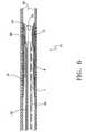
- FIG. 1Adepicts a perspective view of an IRE power source coupled to an energy delivery device that in this depiction is a bipolar probe utilized in the current invention. Also shown is a container for regenerative materials and an infusion pump for movement of materials.
- FIG. 1Bdepicts an enlarged side-view of the distal portion of the shaft of the bipolar probe.
- the shaftis shown as one example of a shaft, particularly a needle.
- FIG. 1Cdepicts an enlarged cross-sectional view of a portion of the needle of the bipolar probe utilized in the current invention.
- FIG. 2Adepicts a cross-sectional view of a liver with a target region of tissue within the liver, where a needle of a bipolar probe coupled to an IRE power source has been inserted into the target region of tissue within the liver and where the safety zone of ablation surrounding the target region in the liver as well as interstitial space and a skin surface are shown for perspective.
- FIG. 2Bis a cross-sectional view of the liver from FIG. 2A at a later time point, where IRE ablation has been performed, and regenerative materials are being released into the region that has been ablated with IRE energy within the liver through a channel in the needle of the bipolar probe.
- FIG. 3Ais a cross-sectional view of the liver from FIG. 2B at a later time point, where the released regenerative materials have settled within the total region that was ablated with IRE energy within the liver at the start of the regenerative process.
- FIG. 3Bis a cross-sectional view of the liver from 3 A at a later time point, when the regenerative process has been completed and the liver has been restored.
- FIG. 4Adepicts a cross-sectional view of a lung with a target region of tissue within the lung, where a needle of a bipolar probe coupled to an IRE power source has been inserted into the target region of tissue within the lung and where the safety zone of ablation surrounding the target region in the lung as well as interstitial space and a skin surface are shown for perspective.
- FIG. 4Bis a cross-sectional view of the lung from FIG. 4A at a later time point, where IRE ablation has been performed, and regenerative materials are being released into the region that has been ablated with IRE energy within the lung through a channel in the needle of the bipolar probe.
- FIG. 5Ais a cross-sectional view of a lung from FIG. 4B at a later time point, where the released regenerative materials have settled within the total region that was ablated with IRE energy within the lung at the start of the regenerative process.
- FIG. 5Bis a cross-sectional view of the lung from 5 A at a later time point, when the regenerative process has been completed and the lung has been restored.
- FIG. 6depicts a cross-sectional view of a blood vessel containing a blockage and the needle of a bipolar probe within the vessel.
- FIG. 7Aillustrates a cross-sectional view of a blood vessel immediately after ablation of a blockage by IRE treatment using a bipolar probe shown within the vessel.
- FIG. 7Billustrates a cross-sectional view of a blood vessel, at a later time point from FIG. 7A , after ablation of a blockage by IRE treatment using a bipolar probe shown within the vessel, where regenerative materials are being released from a channel in the bipolar probe.
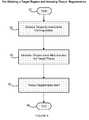
- FIG. 8depicts a flowchart showing a method of ablating a target region and inducing tissue regeneration.
- FIG. 9depicts a flowchart showing a method of ablating a target region within a tissue such as a liver, as well as the introduction of materials capable of inducing tissue regeneration such as a regenerative solution that contained stem cells.
- Target regionscan include or be within digestive, skeletal, muscular, nervous, endocrine, circulatory, reproductive, integumentary, lymphatic, urinary, and soft tissue.
- the targetsmay also include benign or malignant, cancerous, neoplastic, preneoplastic, or tumors as stand-alone targets or targets found within another tissue (such as an organ or organ system).
- Ablationcan be performed in each of laparoscopic, percutaneous, and open surgical procedures.
- Regenerationrefers to at least a partial restoration of an organ or tissue or new growth by an organism of organs and tissues that have been lost, removed, or injured. Regeneration can occur through several mechanisms, including but not limited to regrowth, restructuring, and cellular repopulation.
- Regrowthrefers to growing, developing, and gradually increasing in size, number, value, or strength.
- Restructuringrefers to a change in cell type, organ or tissue shape, pattern, cell type, connectivity, or arrangement than was originally present.
- Cellular repopulationrefers to development of an area starting from a group of cells that can be exogenous from another part of the body or introduced in medical or experimental procedures to cause a specific effect of growth in a damaged area. Any of the processes or regeneration can be brought about or enhanced via introduction of synthetics, exogenous materials mimicking internal, natural, agents, and can be brought about by pharmacological reagents including agonists or antagonists to enhance regeneration.
- Treatment positionrefers to a position such as, but not limited to, a position from the skin surface of a patient to the most distal edge of a target region where the energy delivery device is capable of treatment of a tissue to cause irreversible electroporation.
- Various treatment positionsinclude placement such that irreversible electroporation occurs in a target region with at least a portion of the energy delivery device placed within the target region; also, an additional position includes positioning at least a portion of the energy delivery device such that it touches the surface of the target region.
- Yet other treatment positionsinclude positioning at least a portion of the energy delivery device such that it is adjacent, or near to the target region.
- an energy delivery device 1is depicted in FIG. 1A as a bipolar probe, including the handle 3 of the bipolar probe, shaft 5 (shown here as a needle 5 ), a proximal electrode 7 , a distal electrode 11 , an electrode spacer 9 , a tip 13 of the bipolar probe shown here in this embodiment as a three faced trocar tip, and a probe connector 15 of the bipolar probe.
- the term energy delivery device and probewill be used interchangeably, with specific information regarding the type of probe being added to clarify monopolar, bipolar, and array types of energy delivery devices.
- the probeis coupled to a nonthermal power source 17 , which has a positive and negative connector 19 for the bipolar probe 1 .
- the energy delivery devicemay be in the form of probes that are multiple monopolar, bipolar, or array formations; in embodiments using a bipolar as well as the array approach, there can be more than 1 anode or cathode on a given needle 5 of a probe.
- the monopolar embodimentcan be used with two monopolar probes, one monopolar probe and a pad as known in the art, or in combination with bipolar probes or arrays of probes. Each of monopolar, bipolar, and array devices can be utilized together.
- various portions of the probeare flexible or semi-flexible or articulating.
- the needleis of various flexibilities and may be articulating.
- the IRE power sourcecan be a generator or other energy source and can be connected to a catheter that allows flexible entry into a lumen. This allows for utilization of the optimal probe for a given medical procedure.
- FIG. 1Aalso shows the following: a container 101 having a sealable cap 115 for introducing and removing material, an infusion pump 103 , a first material transfer tube 113 between a container 101 and an infusion pump 103 , and a second material transfer tube 105 and a third material transfer tube 111 that allow transfer of materials between the infusion pump 103 and the handle 3 of the bipolar probe.
- the second material transfer tube 105has an end with a first fitting 107 that allows coupling to a second fitting 109 on one end of the third material transfer tube 111 .
- the container 101represents any source of materials for introduction through the energy delivery device.
- the containercan store one or more regenerative materials transiently, long-term, or permanently.
- the containercan be programmable such that it stores materials at various temperatures, and can have or be coupled to a temperature controller to maintain regenerative material at a selected temperature.
- the containercan also have an internal portion that rotates or otherwise changes position so as to ensure materials stay in solution or do not adhere to the bottom or sides surface through gravity and other adhering forces.
- the containercan have multiple regions each containing one or more regenerative materials that can be released singly or in combination to the target region of tissue through the needle 5 of the probe through one or more lumens and one or more couplings.
- the containercan also be programmable regarding pressure or pH levels, and can have internal sensors so as to allow regulation of water volume or viscosity.
- the containeris capable of containing any regenerative materials described herein.
- the sealable cap 115allows for placing material within the container 101 and for removing material from container 101 .
- the capcan comprise; one or more electrical or mechanical pieces that acts as a door to provide for moving of the materials; this can include a door, sealable shaft, rubber or plastic pieces allowing a syringe or container or hand to be placed within the interior of the container to replace or remove materials.
- the infusion pump 103provides for movement of the materials to the energy delivery device.
- the infusion pump 103is capable of moving materials including but not limited to liquids, gases, semi-solids, and combinations of materials of various states from gas to liquid to solid.
- the infusion pump 103is capable of moving any regenerative materials mentioned in this specification.
- the infusion pump 103moves stem cells.
- the infusion pump 103can be programmable directly or remotely through any wireless system known in the art, and can deliver materials at any rate, including from introduction through drips to high pressure release of fluid.
- the programmable partcan provide for regulation of volume or pressure of release.
- the infusion pump 103can be powered via battery, or plug in to any wall outlet known in the art, from a generator, or can be powered from a diversion of power to the handle 3 of the probe.
- the infusion pump 103can be programmed so as to release multiple regenerative materials.
- the infusion pump 103can also be programmed to release one or more regenerative materials at various timepoints or the same or varying volumes.
- the infusion pump 103is capable of being a source for or storing regenerative materials (or both storing and being a source of) and in certain embodiments the infusion pump and container store materials. In various embodiments the infusion pump and container are contained in a unit that is part of the handle 3 .
- the first material transfer tube 113allows movement of material between the container 101 and the infusion pump 103 .
- the infusion pump 103is located on the handle 3 of the probe.
- the container 101 and the infusion pump 103are one unit and there is no material transfer tube 113 .
- the first fitting 107allows coupling to the second fitting 109 and sealably couples one end of the second material transfer tube 105 to the second fitting 109 located on one end of the third material transfer tube 111 and provides for material movement through the handle 3 toward tissue through the needle 5 of the probe.
- the second fitting 109can couple with the first fitting 107 .
- Second fitting 109can also couple directly to a syringe or multiple syringes.
- the containeris a syringe or a series of syringes; in various embodiments the second fitting 109 couples directly to the container 101 and in other embodiments there is no infusion pump.
- manual power of the syringe plungerprovides for movement of materials through the energy delivery device.
- the syringeis coated with a material on the interior to enhance survival or activity of regenerative material; the syringe can also be shaped or have a diameter such as to limit cellular shear stress.
- the material transfer tubes( 113 , 105 , 111 ) can each be made of any material allowing for transfer of materials.
- the tubeshave coatings that prevent sticking of materials to the walls.
- the diameteris large enough to minimize shear stress on inserted cells.
- FIG. 1Bdepicts an enlarged side-view of the distal portion of the needle of the bipolar probe. Shown are the proximal 7 and distal 11 electrodes separated by the electrode spacer 9 , as well as a tip 13 of the bipolar probe shown here in this embodiment as a three faced trocar tip.
- a channel 21is illustrated which in one embodiment is hollow and allows for the movement of materials including liquids.
- the needleis any shaft capable of delivery of materials through the probe that is also capable of delivery of voltage. In certain embodiments the needle is capable of or adapted for movement of regenerative material.
- FIG. 1Bdepicts an example embodiment with a channel shown as a single opening at the end of the probe, this is only one embodiment of many possible.
- the single channelcould be centered within the needle of the probe, or could be placed nearer to one edge, and the end of the opening could be completely open or could be partially or fully covered with a solid, permeable, or semi-permeable covering, with or without micropores that allow for efficient release of regenerative materials for a given tissue.
- there are a series of channelsallowing for simultaneous or non-simultaneous, single or multiple releases of regenerative materials.
- the needlehas a series of apertures at various points along its length so as to allow release of fluids and small particles.
- the release in all stated examples hereincan be either active or passive release of regenerative materials.
- the IRE power sourcecan be coupled to a catheter that can be used for ablation as well as for release of regenerative materials, and in various embodiments the catheter has a series of apertures along its length to release regenerative materials actively or passively.
- FIGS. 1A and 1Bshow a channel for release of regenerative material from the needle of the probe, this is but one example of one configuration.
- the probecan be designed so as to be loaded with regenerative materials through one or more openings in the handle or the needle.
- the openingallows for the loading and releasing of regenerative materials in a straight line from the point of loading to the release point, so as to minimize turbulence and shear stress on any released cells or other materials.
- there is a loadingwhere there is an angle of greater than zero degrees from the point of loading to the point of release, such as an embodiment where there is an opening designed to receive materials from a syringe that can be coupled to the handle in a Y-shape.
- FIG. 1Bshows a single channel in a needle of a probe as a release point for regenerative material
- the ablation and release of regenerative materialscan be performed using an ablation probe singly or in combination with single or multiple catheters, syringes, or additional probes in cases including percutaneous, laparoscopic, and open surgery.
- FIG. 1Cis an enlarged cross-section of the distal portion of the needle of the bipolar probe from Section A-A of FIG. 1B .
- the proximal 7 and distal 11 electrodeswhich are separated by a portion of insulative material reaching the outer surface of the needle and which is the electrode spacer 9 .
- the channel 21 within the probeas well as the outer insulation 23 and inner insulation 25 .
- one or both of the outer and inner insulative materialsis composed at least in part of polyester shrink material in single or multiple layers. The channel allows for the passage of materials such as fluids.
- the channelcan be used to release regenerative materials following ablation.
- the channelcan also be used to release factors to optimize the environment prior to the introduction of the regenerative materials.
- Optimizingrefers to affecting the treated region in a way that either returns the region to a homeostatic condition or otherwise improves the likelihood, rate, or efficiency of regenerative materials in causing of effecting regeneration. This could involve active or passive rebalancing of tissue levels of materials following ablation, involving singly or in combination adding or altering the levels of water, ions, or factors such as hormones, paracrine agents and paracrine-type agents, and pharmacological mediators including vasoreactive elements.
- the introduced factorscan be natural or synthetic and in certain cases can involve the introduction of a layer of cells. Optimization can be brought about through the introduction of factors.
- factorsmay be released singly or in combination including growth factors (to, in some cases, increase the growth of cells or in some cases to increase the growth rate of certain cells or all cells and in other cases to prevent the growth of certain cell types that may inhibit regeneration or lead to aberrant or undesirable regeneration) such as VEGF, cytokines, and anti-inflammatory agents. In certain embodiments these factors may increase the chance of successful regeneration.
- ion levelsare altered singly or in combination, such as sodium, potassium, magnesium and calcium levels.
- the factors or ionsare released with one or multiple cell types before, after, or in a simultaneous release with the cells to advance regeneration.
- the regenerative materialincludes cells and factors that aim to restore tissue, membranes, or matrices. This can involve direct application of a variety of regenerative materials released singly or in combination, in whole, in part, or precursors of DNA, RNA, proteins, carbohydrates, sugars, lipids, enzymes, proteases, steroids, amino acids, purine bases, pyrimidine bases, deoxyribose sugar, ribose sugar, nucleosides, adenosine-triphosphate, and adenosine biphosphate, polysaccharides, proteoglycans, hyaluronic acid, collagen, fibronectin, elastin, laminin, and integrins.
- the regenerative materialcould also include singly or in combination smooth muscle cells, eplithelial cells, endothelial cells, liver cells, lung cells,
- FIG. 2Aillustrates the ablation and regeneration concerning a target region in a liver. Specifically, this depicts a cross-sectional view of a liver 37 with a target region of tissue 45 within the liver, where a needle 5 of a bipolar probe coupled to an IRE power source has been inserted into the target region 45 of tissue within the liver. There is a safety zone of ablation 47 surrounding the target region in the liver, though it is a very small layer and is shown here not necessarily to scale for ease of visualization. In addition, interstitial space 43 and a skin surface 41 outside the liver are shown for perspective.
- FIG. 2Bdepicts the liver from FIG. 2A at a later time point once ablation has been performed. Structures shown include a liver 37 , blood vessels 39 within the liver, a target region of tissue 45 within a liver, and a safety zone of ablation 47 surrounding the target region. Also shown is a needle of a bipolar probe 5 , and regenerative materials 35 released from the probe into the previously ablated region.
- regenerative materials released in FIG. 2Bdepict regeneration in relation to a liver, though this is only one example among various applications of this technology.
- release of regenerative materialsinvolves ablation of target regions that can include or be within digestive, skeletal, muscular, nervous, endocrine, circulatory, reproductive, integumentary, lymphatic, urinary, and soft tissue.
- the targetsmay also include benign or malignant cancerous, neoplastic, preneoplastic, or tumors as stand-alone targets or targets found within another tissue (such as an organ or organ system).
- released regenerative materialsinclude stem cells that range from totipotent, to pluripotent, to multipotent, and to unipotent.
- the stem cells utilizedinclude cell lines currently available commercially. These stem cell lines may be human or other animal cell lines, which may or may not be genetically altered, or may be chimeras or may be released with factors enhancing regeneration obtained or derived from humans or nonhuman animals or both.
- released regenerative materialsinclude a cell line available from ATCC (Manassas, Va.). An example embodiment would utilize a cell line with ATCC number SCRC-2002 with designation hESC BG01V of the cell type Embryonic Stem Cell.
- Cell lines from any of the following: BresaGen, Inc.Cell lines with provider codes such as Hesbgn-01, Hesbgn-02, Hesbgn-03, Hesbgn-04), Cellartis AB (Cell lines with provider codes such as Sahlgrenska 1, Sahlgrenska 2), ES Cell International (Cell lines with provider codes such as HES-1, HES-2, HES-3, HES-4, HES-5, HES-6), Technion-Israel Institute of Technology (Cell lines with provider codes such as I 3, I 3.2, I 3.3, I 4, I 6, I 6.2, J 3, J 3.2), University of California at San Francisco (Cell lines with provider codes such as HSF-1, HSF-6), as well as the Wisconsin Alumni Research Foundation (Cell lines with provider codes such as H1, H7, H9, H13, H14). Additional embodiments utilize cell lines from the National Stem Cell Bank. Yet an additional embodiment utilizes cells with at least one of the genetic code for or expressed marker of SSEA
- FIG. 3Ais a cross-sectional view of the liver from FIG. 2B at a later time point, where the released regenerative materials have settled within the total region that was ablated 49 with IRE energy within the liver at the start of the regenerative process.
- FIG. 3Ashows the liver 37 with vessels 39 , the total region ablated 49 , as well as interstitial space 43 , and a skin surface 41 surrounding the liver.
- FIG. 3Bis a cross-sectional view of the liver from 3 A at a later time point, when the regenerative process has been completed and the liver has been restored. This illustrates the liver 37 , vessel 39 , the interstitial space 43 outside the liver, and a skin surface 41 .
- FIG. 4Athis illustrates the ablation and regeneration concerning a target region in a lung.
- FIG. 4Adepicts a cross-sectional view of a lung 51 with a target region 59 of tissue within the lung, where a needle 5 of a bipolar probe coupled to an IRE power source has been inserted into the target region of tissue 59 within the lung and a safety zone of ablation 61 surrounding the target region in the lung. Also shown for perspective are branches of airways 57 as well as interstitial space 55 and a skin surface 53 .
- FIG. 4Bis a cross-sectional view of the lung from FIG. 4A at a later time point, where IRE ablation has been performed, and regenerative materials are being released into the region that has been ablated with IRE energy within the lung through a channel in the needle of the bipolar probe.
- a lung 51with a target region 59 of tissue within the lung, where a needle 5 of a bipolar probe coupled to an IRE power source has been inserted into the target region of tissue 59 within the lung and a safety zone of ablation 61 surrounding the target region in the lung is shown.
- branches of airways 57as well as interstitial space 55 and a skin surface 53 .
- release of the regenerative materials 35 from a channel in the needle of the bipolar probeis shown.
- FIG. 4Bdepict regeneration in relation to a lung, though this is only one example among various applications of this technology.
- the technologycan be applied to release of regenerative materials in any of the ablation targets described in this application.
- the released materialswill include stem cells.
- stem cellsthere are various sources of stem cells that are contemplated within this technology.
- There are varied sources of stem cellswith a variety of methods being developed largely in response to concerns of the use of embryos by scientists in endeavors for developing stem cell lines (which in such a case would be an embryonic stem cell line).
- stem cell lineswhich in such a case would be an embryonic stem cell line.
- the resultshave been an increased number of sources of stem cells and methods of producing the cells and cell lines. To this point, there are several methods and sources of stem cells.
- released regenerative materialsinclude cells of variable potencies that have been dedifferentiated.
- the cells usedinvolve cells dedifferentiated via genetic alterations.
- the cells usedhave been dedifferentiated through epigenetic alterations.
- the cells usedhave been dedifferentiated through exposure to external factors, ex vivo or in situ or in vitro, or through a combination of these.
- Additional embodiments of cells that may be utilized within the regenerative materials releasedinclude any or a combination of the following: stem cells derived from dead embryos in what has been termed the Landry-Zucker proposal by those in the art, stem cells derived from an embryo that lead to the destruction of the embryo, stem cells from living human embryos without harming the developmental capabilities of such embryos, and stem cells isolated or obtained through use of somatic cell nuclear transfer (SCNT).
- stem cells derived from dead embryosin what has been termed the Landry-Zucker proposal by those in the art
- stem cells derived from an embryo that lead to the destruction of the embryostem cells from living human embryos without harming the developmental capabilities of such embryos
- SCNTsomatic cell nuclear transfer
- Additional embodimentsinclude use of stem cells derived from a constructed biological artifact in a modification of SCNT method of removing an egg's nucleus and replacing it with a somatic cell nucleus in what is known as altered nuclear transfer (ANT), thereby altering the somatic cell nucleus before transfer so that the result is an artifact without essential attributes of a human embryo.
- Still additional embodimentsinvolve the use of multipotent or pluripotent adult human stem cells.
- variations in stem cell usemay involve animal cells of various potencies for release as part of the regenerative materials. In certain embodiments, cells will be isolated from a given individual for reintroduction as part of the regenerative material into that same individual.
- cells or factors to be utilized as part of or in conjunction with the release of regeneration materialsinclude cells or factors isolated using one or more of one of magnetic beads, optical sensors, electric fields, and dielectrophoresis.
- FIG. 5Ais a cross-sectional view of a lung from FIG. 4B at a later time point, where the released regenerative materials have settled within the total region that was ablated 63 with IRE energy within the lung at the start of the regenerative process.
- FIG. 5Ashows a lung 51 with branches of airways 57 , as well as the interstitial space 55 and skin surface 53 outside the lung.
- FIG. 5Bis a cross-sectional view of the lung from 5 A at a later time point, when the regenerative process has been completed and the lung has been restored. Shown is the lung 51 , the branches of airways 57 , and interstitial space 55 and skin surface 53 outside the lung.
- FIG. 6depicted is an enlarged cross-sectional view of a portion of the needle of the bipolar probe utilized in the current invention.
- a vessel 31including an endothelial cell layer 27 of a vessel, the lumen 33 of the vessel, and a blockage 29 within the vessel.
- Depicted within the vesselis the distal portion of a needle of a bipolar probe that could be utilized with IRE ablation.
- proximal 7 and distal 11 electrodesseparated by the electrode spacer 9 .
- the channel 21 within the probeas well as outer insulation 23 and inner insulation 25 .
- the tip 13 of the probeis shown to have a rounded or curved or dulled ending to ensure less damage to the vessel.
- the endcould be shard or dull or rounded or curved or padded to ensure proper treatment depending on the specific characteristics of the target region.
- FIG. 7Aillustrates a cross-sectional view of a blood vessel 31 immediately after ablation of a blockage 29 by IRE treatment using a bipolar probe shown within the vessel.
- FIG. 7Arepresents a later time point of the image shown in FIG. 6 , where the blockage 29 has been ablated and the reduced material remaining will be resorbed and removed by normal physiological processes within the vessel of the patient.
- the endothelial cell layer of a vessel 27 and the lumen of the vessel 33Depicted within the vessel is the distal portion of a needle of a bipolar probe.
- the proximal 7 and distal 11 electrodesseparated by the electrode spacer 9 .
- the channel 21 within the probeas well as outer insulation 23 and inner insulation 25 , as well as the tip 13 of the probe.
- FIG. 7Ashows a bipolar probe for ablation purposes in this particular embodiment within this vessel
- monopolar probeswith various applications in various tissues including but not limited to applications within a vessel (including applications in any regions indicated in this application such as example embodiments including liver as well as lung, or cancerous or tumorous tissues).
- one embodiment for ablationwould include two monopolar probes spaced 10 mm apart, with an exposed length of up to 20 mm.
- Another embodimentincludes a voltage of up to 2000 volts, with pulses of 100 microseconds in length being applied to a target region of tissue.
- An additional embodimentwould involve 90 pulses provided in pulse-trains of 10, with an interval between pulses of 250 milliseconds and a time between pulse-trains of 2 seconds.
- the total number of pulses and pulse trains in various embodimentsvaries based on the effectiveness of the treatment for a given tissue.
- the ablation zoneinvolves two monopolar probes ablating a zone of approximately 22 mm ⁇ 18 mm ⁇ 12 mm, though it is understood that the ablation size and shape varies with placement of the probes and probe types, and that this is an advantage of this invention.
- Two single probesmay also be configured so as to involve other ablation areas, including: ablation of an area of approximately 30 mm ⁇ 25 mm ⁇ 17 mm, including exposed electrode lengths of approximately 20 mm and a spacing of 15 mm. Such an embodiment could involve a voltage of 2500 v. It is understood that the ablation size and shape varies with placement of the probes and probe types, and that this is an advantage of this invention.
- one embodimentwould include a voltage of up to 2700 v.
- An additional embodimentwould involve 90 pulses provided in pulse-trains of 10, with an interval between pulses of 250 milliseconds and a time between pulse-trains of 2 seconds.
- an IRE generatoris used as an IRE power source, utilizing a standard wall outlet of 110 volts (v) or 230 v with a manually adjustable power supply depending on voltage.
- the generatorwould have a minimum voltage of 100 v to 3000 v and be adjustable at 100 v intervals.
- the ablation pulse appliedwould be between 20 and 100 microseconds in length, and be adjustable at 10 microsecond intervals.
- a preferred embodimentwould include a generator programmable so as to operate between 2 and 50 amps, with test ranges involving even a lower maximum when appropriate.
- a preferable embodiment of an IRE generatorwould include 2 to 6 positive and negative connectors, though it is understood that this is simply a preferred embodiment and that the invention would pertain to additional embodiments understood in the art and necessary for optimal configurations for ablation.
- IRE ablationcan be performed with variations described in the following reference previously incorporated by reference: U.S. Patent Application Publication Number US 2007/0043345A1, “Tissue Ablation with Irreversible Electroporation,” application Ser. No. 10/571,162. Certain embodiments involve pulses between 5 microseconds and 62,000 milliseconds, while others involve pulses of 75 microseconds to 20,000 milliseconds. In certain embodiments electrodes are spaced from 100 Volts per centimeter (V/cm) to 7,000 V/cm, while in other embodiments the spacing is 200 to 2000 V/cm as well as from 300 V/cm to 1000 V/cm. The number of pulses to be used in IRE ablation can vary.
- the number of pulsesis from 1 to 15 pulses.
- groups of 1 to 15 pulsesare also called pulse-trains
- the gap of time between groups of pulsesis 0.5 second to 10 seconds.
- Pulsescan be delivered using probes, needles, and electrodes each of varying lengths suitable for use in percutaneous, laparoscopic, and open surgical procedures. Electrodes can be made of various materials known in the art and be of different sizes and shapes and be spaced at various distances from one another. Specific embodiments can be square, oval, rectangular, circular or irregular.
- Certain embodimentshave the distance between two electrodes from 0.5 to 10 centimenters (cm), while others have from 1 to 5 cm, and yet others embodiments have from 2-3 cm.
- the electrode surface areacan vary, and in specific embodiments the electrodes are from 0.1 to 5 square cm, and in others, from 1 to 2 square cm. The embodiments described are simply certain embodiments and are not a complete description of embodiments.
- FIG. 7Bis a later time point of the images seen in FIG. 7A , including a vessel 31 with a lumen 33 , and inside the lumen is the distal portion of a needle of a bipolar probe.
- the proximal 7 and distal 11 electrodescan be seen, as well as the electrode spacer 9 .
- FIG. 7Billustrates introduction of regenerative materials 35 into the lumen 33 of the vessel at the site of ablation.
- the regenerative materials released into the vesselcontain precursor cells capable of developing into a given cell type intended for reintroduction or regrowth or population development or redevelopment in cell number or size.
- precursor cellscapable of developing into a given cell type intended for reintroduction or regrowth or population development or redevelopment in cell number or size.
- the introduced regenerative materialsinclude endothelial cell precursors or precursors from tissues such as but not limited to blood, bone, or muscle (such as satellite cells as well as mesodermal-stromal cells).
- tissuessuch as but not limited to blood, bone, or muscle (such as satellite cells as well as mesodermal-stromal cells).
- tissue specific stem cellsat various places in the body, including satellite cells in muscle (as has been described in Rando T. A. Stem Cells, Ageing and the Quest for Immortality . Nature. Vol.
- mesodermal stromal cellsthat are bone-marrow derived (Described in the following reference hereby incorporated by reference: Hermann A., Maisel M., Storch A., Epigenetic Conversion of Human Adult Bone Mesodermal Stromal Cells into Neuroectodermal Cell Types for Replacement Therapy of Neurodegenerative Disorders, 6(7) Expert Opinion on Biological Therapy 6(7):653 (2006))
- the introduced regenerative materialincludes one or more isolated cells containing the gene for, capable of expressing, expressing, or differentially expressing singly or in combination VE Cadherin (CD144), VonWillibrand Factor, thrombomodulin (CD141), PAL-E, PECAM-1 (CD31), CD146, VEGF Receptor-1 (FLT-1), VEGF Receptor-2, VEGF Receptor-3, TIE-1 (C-Terminus), TIE-1 (N-terminus), TIE-2, CD34, ICAM-1 (CD54), P-Selectin (CD62P), and Anti-Endoglin (CD105).
- Embodimentsinclude cells with various homologues of the listed materials as well as other known RNA splice variations and isoforms.
- the introduced regenerative materialincludes one or more of cells containing the gene for, capable of expressing, expressing, or differentially expressing singly or in combination neural cell adhesion molecule (N-CAM), fetal antigen 1 (FA1), Pax7, Asb5, IgSF4, Hoxc10, Myf5, Neuritin, Klra18, as well as MyoD target genes (such as Pw1, Dapk2, Sytl2, and NLRR1).
- N-CAMneural cell adhesion molecule
- F1fetal antigen 1
- Pax7Asb5, IgSF4, Hoxc10, Myf5, Neuritin, Klra18, as well as MyoD target genes (such as Pw1, Dapk2, Sytl2, and NLRR1).
- Embodimentsinclude cells with various homologues of the listed materials as well as other known RNA splice variations and isoforms.
- the introduced regenerative materialincludes one or more of cells containing the gene for, capable of expressing, or expressing, or differentially expressing singly or in combination STRO-1, HOP-26 (CD63), CD49a and SB-10 (CD166), CD13, CD29, CD44, CD73, CD90, Cadherin-11, Calretinin, CD10, CD117, Desmin, Endoglyx-1, Endosialin (TEM1, CD248), Fibroblast-Activation Protein (FAP), Laminin gamma2 chain, Neural Ganglioside GD2, Nucleostemin, Snep (stromal nidogen extracellular matrix protein), and Tenascin.
- Embodimentsinclude cells with various homologues of the listed materials as well as other known RNA splice variations and isoforms.
- the introduced regenerative materialincludes one or more of cells containing the gene for, capable of expressing, expressing, or differentially expressing singly or in combination known pluripotent stem cell markers as: Alkaline Phosphatase, Alfa-fetoprotein (AFP), Bone-Morphogenic Protein-4, Brachyury, Cluster Designation 30 (CD30), Cripto (TDGF-1), GATA-4 gene, GCTM-2, genesis, Germ Nuclear Factor, Hepatocyte Nuclear Factor-4 (HNF-4), Nestin, Neuronal Cell Adhesion Molecule (N-CAM), Oct-4, Pax-6, Stage Specific Embryonic Antigen-3 (SSEA-3), Stage Specific Embryonic Antigen-3 (SSEA-4), Stem Cell Factor (SCFor C-Kit Ligand), Telomerase, TRA-1-60, TRA-1-81, and Vimentin.
- pluripotent stem cell markersas: Alkaline Phosphatase, Alfa-fetoprotein (AFP), Bone-Morpho
- Introduced regenerative materialsinclude in certain embodiments single or multiple infusions or injections that include sources of materials from autogeneic, isogeneic, allogeneic, and xenogeneic sources, and can include of one or more of cell types from one or more species such as mouse, rat, guinea pig, hamster, rabbit, dog, cow, as well as horse. Additional embodiments include additional mammals that are known in the art and which are routinely used for isolation of cellular tissue for the development of cell lines and for uses in research and medical procedures.
- FIG. 8depicts a flowchart showing a method of ablating a target region and inducing tissue regeneration.
- tissueis destroyed by irreversible electroporation 93 .
- Regenerative materialsare introduced into the area of the target tissue 95 , the tissue regenerates itself 97 and the ablation and regeneration comes to an end 99 .
- FIG. 9depicts a flowchart showing a method of ablating a target region within a tissue such as a liver, as well as the introduction of materials capable of inducing tissue regeneration such as a regenerative solution that contained stem cells.
- the target regionis located 67 .
- a tumoris indicated as the target region within a liver, this is only one example and various embodiments may include any herein described target region.
- the probeis coupled to the IRE power source, the probe is inserted into the target region, and IRE parameters are set 69 .
- IRE electroporation and ablation of the target regionis undertaken 75 .
- the environment at the site of ablationis then optimized for the introduction of regenerative materials 79 .
- thismay involve actively or passively rebalancing tissue levels of materials by adding water, ions, or factors such as hormones, paracrine-type agents, or pharmacological mediators such as vasoreactive elements.
- the regenerative materialsare added (in this embodiment stem cells) 81 and the question is asked as to whether optimal levels of regenerative materials have been reached 83 . If the optimal level has not been reached, additional materials will be added. Once the optimal level of regenerative material has been reached the probe is withdrawn 89 , using thermal energy in track ablation in an example embodiment.
- the effect of irreversible electroporationcan be brought about using a power source of Direct Current (DC).
- DCDirect Current
- one or more power sourcescan be used so as to effect irreversible electroporation in a target region and singly or in combination also effect radiofrequency ablation, thermal electric heating, and traditional heating methods with electrodes using direct current or alternating current.
- These combinations of treatmentcan be used for additional energy output to have an effect on the target region or on a track to be ablated when the probe is withdrawn to ensure no cells are drawn from the target region towards the patient skin surface as well as allowing coagulation when desired to prevent bleeding.
Landscapes
- Health & Medical Sciences (AREA)
- Life Sciences & Earth Sciences (AREA)
- Engineering & Computer Science (AREA)
- Veterinary Medicine (AREA)
- Surgery (AREA)
- Nuclear Medicine, Radiotherapy & Molecular Imaging (AREA)
- Biomedical Technology (AREA)
- Animal Behavior & Ethology (AREA)
- General Health & Medical Sciences (AREA)
- Public Health (AREA)
- Biophysics (AREA)
- Neurology (AREA)
- Neurosurgery (AREA)
- Physics & Mathematics (AREA)
- Plasma & Fusion (AREA)
- Otolaryngology (AREA)
- Radiology & Medical Imaging (AREA)
- Heart & Thoracic Surgery (AREA)
- Medical Informatics (AREA)
- Molecular Biology (AREA)
- Surgical Instruments (AREA)
- Materials For Medical Uses (AREA)
Abstract
Description
- Smith A.,A Glossary for Stem Cell Biology. Nature Vol. 441(7097):1060-61 (2006).
- Morrison S. J., Kimble J.,Asymmetric and Symmetric Stem Cell Divisions in Development and Cancer,441(7097) Nature 1068, 1068-74 (2006).
- Rando T. A.Stem Cells, Ageing and the Quest for Immortality. Nature. Vol. 441(7097):1080-1086 (2006).
- Ioannidou E.,Therapeutic modulation of growth factors and cytokines in regenerative medicine,12(19) Current Pharmaceutical Design. Vol. 12(19):2397 (2006).
- Lindvall O., Kokaia Z.,Stem cells for the treatment of neurological disorders. Nature. Vol. 441(7097): 1094-1096 (2006).
- Couzin J.,A Shot of Bone Marrow Can Help the Heart. Science. Vol 313: 1715-1716 (2006).
- Srivastava D., Ivey K. N.,Potential of Stem-Cell-Based Therapies for Heart Disease. Nature. Vol. 441(7097): 1097-1099 (2006).
Claims (21)
Priority Applications (2)
| Application Number | Priority Date | Filing Date | Title |
|---|---|---|---|
| US12/703,355US8231603B2 (en) | 2009-02-10 | 2010-02-10 | Irreversible electroporation and tissue regeneration |
| US13/556,510US8562588B2 (en) | 2009-02-10 | 2012-07-24 | Irreversible electroporation and tissue regeneration |
Applications Claiming Priority (2)
| Application Number | Priority Date | Filing Date | Title |
|---|---|---|---|
| US15130509P | 2009-02-10 | 2009-02-10 | |
| US12/703,355US8231603B2 (en) | 2009-02-10 | 2010-02-10 | Irreversible electroporation and tissue regeneration |
Related Child Applications (1)
| Application Number | Title | Priority Date | Filing Date |
|---|---|---|---|
| US13/556,510ContinuationUS8562588B2 (en) | 2009-02-10 | 2012-07-24 | Irreversible electroporation and tissue regeneration |
Publications (2)
| Publication Number | Publication Date |
|---|---|
| US20100204638A1 US20100204638A1 (en) | 2010-08-12 |
| US8231603B2true US8231603B2 (en) | 2012-07-31 |
Family
ID=42541004
Family Applications (2)
| Application Number | Title | Priority Date | Filing Date |
|---|---|---|---|
| US12/703,355Active2030-12-06US8231603B2 (en) | 2009-02-10 | 2010-02-10 | Irreversible electroporation and tissue regeneration |
| US13/556,510ActiveUS8562588B2 (en) | 2009-02-10 | 2012-07-24 | Irreversible electroporation and tissue regeneration |
Family Applications After (1)
| Application Number | Title | Priority Date | Filing Date |
|---|---|---|---|
| US13/556,510ActiveUS8562588B2 (en) | 2009-02-10 | 2012-07-24 | Irreversible electroporation and tissue regeneration |
Country Status (2)
| Country | Link |
|---|---|
| US (2) | US8231603B2 (en) |
| WO (1) | WO2010093692A2 (en) |
Cited By (41)
| Publication number | Priority date | Publication date | Assignee | Title |
|---|---|---|---|---|
| US20120289888A1 (en)* | 2009-02-10 | 2012-11-15 | Hobbs Eamonn P | Irreversible Electroporation and Tissue Regeneration |
| US9598691B2 (en) | 2008-04-29 | 2017-03-21 | Virginia Tech Intellectual Properties, Inc. | Irreversible electroporation to create tissue scaffolds |
| US9757196B2 (en) | 2011-09-28 | 2017-09-12 | Angiodynamics, Inc. | Multiple treatment zone ablation probe |
| US9867652B2 (en) | 2008-04-29 | 2018-01-16 | Virginia Tech Intellectual Properties, Inc. | Irreversible electroporation using tissue vasculature to treat aberrant cell masses or create tissue scaffolds |
| US9888956B2 (en) | 2013-01-22 | 2018-02-13 | Angiodynamics, Inc. | Integrated pump and generator device and method of use |
| US9895189B2 (en) | 2009-06-19 | 2018-02-20 | Angiodynamics, Inc. | Methods of sterilization and treating infection using irreversible electroporation |
| US10117707B2 (en) | 2008-04-29 | 2018-11-06 | Virginia Tech Intellectual Properties, Inc. | System and method for estimating tissue heating of a target ablation zone for electrical-energy based therapies |
| US10154874B2 (en)* | 2008-04-29 | 2018-12-18 | Virginia Tech Intellectual Properties, Inc. | Immunotherapeutic methods using irreversible electroporation |
| US10154869B2 (en) | 2013-08-02 | 2018-12-18 | Gary M. Onik | System and method for creating radio-frequency energy electrical membrane breakdown for tissue ablation |
| US10238447B2 (en) | 2008-04-29 | 2019-03-26 | Virginia Tech Intellectual Properties, Inc. | System and method for ablating a tissue site by electroporation with real-time monitoring of treatment progress |
| US10245105B2 (en) | 2008-04-29 | 2019-04-02 | Virginia Tech Intellectual Properties, Inc. | Electroporation with cooling to treat tissue |
| US10272178B2 (en) | 2008-04-29 | 2019-04-30 | Virginia Tech Intellectual Properties Inc. | Methods for blood-brain barrier disruption using electrical energy |
| US10292755B2 (en) | 2009-04-09 | 2019-05-21 | Virginia Tech Intellectual Properties, Inc. | High frequency electroporation for cancer therapy |
| US10471254B2 (en) | 2014-05-12 | 2019-11-12 | Virginia Tech Intellectual Properties, Inc. | Selective modulation of intracellular effects of cells using pulsed electric fields |
| US10470822B2 (en) | 2008-04-29 | 2019-11-12 | Virginia Tech Intellectual Properties, Inc. | System and method for estimating a treatment volume for administering electrical-energy based therapies |
| US10694972B2 (en) | 2014-12-15 | 2020-06-30 | Virginia Tech Intellectual Properties, Inc. | Devices, systems, and methods for real-time monitoring of electrophysical effects during tissue treatment |
| US10702337B2 (en) | 2016-06-27 | 2020-07-07 | Galary, Inc. | Methods, apparatuses, and systems for the treatment of pulmonary disorders |
| US10702326B2 (en) | 2011-07-15 | 2020-07-07 | Virginia Tech Intellectual Properties, Inc. | Device and method for electroporation based treatment of stenosis of a tubular body part |
| US10835311B2 (en) | 2014-10-24 | 2020-11-17 | Northwestern University | Electroporation apparatus and method of using same for ablation of an arbitrary volume |
| US10849678B2 (en) | 2013-12-05 | 2020-12-01 | Immunsys, Inc. | Cancer immunotherapy by radiofrequency electrical membrane breakdown (RF-EMB) |
| US11141216B2 (en) | 2015-01-30 | 2021-10-12 | Immunsys, Inc. | Radio-frequency electrical membrane breakdown for the treatment of high risk and recurrent prostate cancer, unresectable pancreatic cancer, tumors of the breast, melanoma or other skin malignancies, sarcoma, soft tissue tumors, ductal carcinoma, neoplasia, and intra and extra luminal abnormal tissue |
| US11254926B2 (en) | 2008-04-29 | 2022-02-22 | Virginia Tech Intellectual Properties, Inc. | Devices and methods for high frequency electroporation |
| US11272979B2 (en) | 2008-04-29 | 2022-03-15 | Virginia Tech Intellectual Properties, Inc. | System and method for estimating tissue heating of a target ablation zone for electrical-energy based therapies |
| US11311329B2 (en) | 2018-03-13 | 2022-04-26 | Virginia Tech Intellectual Properties, Inc. | Treatment planning for immunotherapy based treatments using non-thermal ablation techniques |
| US11382681B2 (en) | 2009-04-09 | 2022-07-12 | Virginia Tech Intellectual Properties, Inc. | Device and methods for delivery of high frequency electrical pulses for non-thermal ablation |
| US11453873B2 (en) | 2008-04-29 | 2022-09-27 | Virginia Tech Intellectual Properties, Inc. | Methods for delivery of biphasic electrical pulses for non-thermal ablation |
| US11497544B2 (en) | 2016-01-15 | 2022-11-15 | Immunsys, Inc. | Immunologic treatment of cancer |
| US11607537B2 (en) | 2017-12-05 | 2023-03-21 | Virginia Tech Intellectual Properties, Inc. | Method for treating neurological disorders, including tumors, with electroporation |
| US11638603B2 (en) | 2009-04-09 | 2023-05-02 | Virginia Tech Intellectual Properties, Inc. | Selective modulation of intracellular effects of cells using pulsed electric fields |
| US11707629B2 (en) | 2009-05-28 | 2023-07-25 | Angiodynamics, Inc. | System and method for synchronizing energy delivery to the cardiac rhythm |
| US11723710B2 (en) | 2016-11-17 | 2023-08-15 | Angiodynamics, Inc. | Techniques for irreversible electroporation using a single-pole tine-style internal device communicating with an external surface electrode |
| US11925405B2 (en) | 2018-03-13 | 2024-03-12 | Virginia Tech Intellectual Properties, Inc. | Treatment planning system for immunotherapy enhancement via non-thermal ablation |
| US11931096B2 (en) | 2010-10-13 | 2024-03-19 | Angiodynamics, Inc. | System and method for electrically ablating tissue of a patient |
| US11938317B2 (en) | 2017-12-26 | 2024-03-26 | Galvanize Therapeutics, Inc. | Optimization of energy delivery for various applications |
| US11950835B2 (en) | 2019-06-28 | 2024-04-09 | Virginia Tech Intellectual Properties, Inc. | Cycled pulsing to mitigate thermal damage for multi-electrode irreversible electroporation therapy |
| US12102376B2 (en) | 2012-02-08 | 2024-10-01 | Angiodynamics, Inc. | System and method for increasing a target zone for electrical ablation |
| US12114911B2 (en) | 2014-08-28 | 2024-10-15 | Angiodynamics, Inc. | System and method for ablating a tissue site by electroporation with real-time pulse monitoring |
| US12201349B2 (en) | 2009-04-03 | 2025-01-21 | Angiodynamics, Inc. | Congestive obstruction pulmonary disease (COPD) |
| US12214189B2 (en) | 2019-07-24 | 2025-02-04 | Virginia Tech Intellectual Properties, Inc. | Fourier analysis spectroscopy for monitoring tissue impedance changes and treatment outcome during electroporation-based-therapies |
| US12390262B2 (en) | 2018-03-13 | 2025-08-19 | Virginia Tech Intellectual Properties, Inc. | Treatment planning system for immunotherapy enhancement via non-thermal ablation |
| US12403305B2 (en) | 2016-06-27 | 2025-09-02 | Galvanize Therapeutics, Inc. | Immunostimulation in the treatment of viral infection |
Families Citing this family (45)
| Publication number | Priority date | Publication date | Assignee | Title |
|---|---|---|---|---|
| US8926606B2 (en) | 2009-04-09 | 2015-01-06 | Virginia Tech Intellectual Properties, Inc. | Integration of very short electric pulses for minimally to noninvasive electroporation |
| US20100152725A1 (en)* | 2008-12-12 | 2010-06-17 | Angiodynamics, Inc. | Method and system for tissue treatment utilizing irreversible electroporation and thermal track coagulation |
| CN102686147B (en) | 2009-11-05 | 2016-01-20 | 格雷特巴奇有限公司 | waveguide neural interface device |
| US20110112520A1 (en)* | 2009-11-11 | 2011-05-12 | Invasix Corporation | Method and device for fat treatment |
| US9956391B2 (en) | 2011-12-12 | 2018-05-01 | Pulse Biosciences, Inc. | Electric pulse generators |
| US20130218143A1 (en)* | 2012-02-20 | 2013-08-22 | Tyco Healthcare Group Lp | Combined Thermal Therapy and Hydrogel with Embedded Stem Cell Treatment |
| CA2881462C (en) | 2012-08-09 | 2020-07-14 | University Of Iowa Research Foundation | Catheters, catheter systems, and methods for puncturing through a tissue structure |
| WO2015089505A2 (en) | 2013-12-13 | 2015-06-18 | The Trustees Of The University Of Pennsylvania | Coaxial ablation probe and method and system for real-time monitoring of ablation therapy |
| EP3091921B1 (en) | 2014-01-06 | 2019-06-19 | Farapulse, Inc. | Apparatus for renal denervation ablation |
| TWM487746U (en)* | 2014-03-14 | 2014-10-11 | Contour Optik Inc | Electrode |
| TWM488322U (en) | 2014-03-14 | 2014-10-21 | Contour Optik Inc | Transcranial direct current stimulation device |
| EP3495018B1 (en) | 2014-05-07 | 2023-09-06 | Farapulse, Inc. | Apparatus for selective tissue ablation |
| WO2015175944A1 (en) | 2014-05-16 | 2015-11-19 | Gary Long | Methods and apparatus for multi-catheter tissue ablation |
| EP3154463B1 (en) | 2014-06-12 | 2019-03-27 | Farapulse, Inc. | Apparatus for rapid and selective transurethral tissue ablation |
| WO2015192018A1 (en) | 2014-06-12 | 2015-12-17 | Iowa Approach Inc. | Method and apparatus for rapid and selective tissue ablation with cooling |
| EP3206613B1 (en) | 2014-10-14 | 2019-07-03 | Farapulse, Inc. | Apparatus for rapid and safe pulmonary vein cardiac ablation |
| CN115737104A (en) | 2015-03-31 | 2023-03-07 | 昂科赛克医疗公司 | Systems and methods for improved tissue sensing-based electroporation |
| JP7495778B2 (en) | 2015-10-07 | 2024-06-05 | メイヨ・ファウンデーション・フォー・メディカル・エデュケーション・アンド・リサーチ | Electroporation for the treatment of obesity or diabetes |
| US20170189097A1 (en) | 2016-01-05 | 2017-07-06 | Iowa Approach Inc. | Systems, apparatuses and methods for delivery of ablative energy to tissue |
| US12144541B2 (en) | 2016-01-05 | 2024-11-19 | Boston Scientific Scimed, Inc. | Systems, apparatuses and methods for delivery of ablative energy to tissue |
| US10172673B2 (en) | 2016-01-05 | 2019-01-08 | Farapulse, Inc. | Systems devices, and methods for delivery of pulsed electric field ablative energy to endocardial tissue |
| US10130423B1 (en) | 2017-07-06 | 2018-11-20 | Farapulse, Inc. | Systems, devices, and methods for focal ablation |
| US10660702B2 (en) | 2016-01-05 | 2020-05-26 | Farapulse, Inc. | Systems, devices, and methods for focal ablation |
| EP3471631A4 (en) | 2016-06-16 | 2020-03-04 | Farapulse, Inc. | GUIDE WIRE DISTRIBUTION SYSTEMS, APPARATUSES AND METHODS |
| US9987081B1 (en) | 2017-04-27 | 2018-06-05 | Iowa Approach, Inc. | Systems, devices, and methods for signal generation |
| US10617867B2 (en) | 2017-04-28 | 2020-04-14 | Farapulse, Inc. | Systems, devices, and methods for delivery of pulsed electric field ablative energy to esophageal tissue |
| JP7586706B2 (en) | 2017-09-12 | 2024-11-19 | ボストン サイエンティフィック サイムド,インコーポレイテッド | Systems, devices and methods for focal ventricular ablation - Patents.com |
| EP3749238B1 (en) | 2018-02-08 | 2023-08-16 | Farapulse, Inc. | Apparatus for controlled delivery of pulsed electric field ablative energy to tissue |
| US20190336198A1 (en) | 2018-05-03 | 2019-11-07 | Farapulse, Inc. | Systems, devices, and methods for ablation using surgical clamps |
| CN112087978B (en) | 2018-05-07 | 2023-01-17 | 波士顿科学医学有限公司 | epicardial ablation catheter |
| WO2019217433A1 (en) | 2018-05-07 | 2019-11-14 | Farapulse, Inc. | Systems, apparatuses and methods for delivery of ablative energy to tissue |
| CN119074196A (en) | 2018-05-07 | 2024-12-06 | 波士顿科学医学有限公司 | Systems, devices and methods for filtering high voltage noise induced by pulsed electric field ablation |
| US10687892B2 (en) | 2018-09-20 | 2020-06-23 | Farapulse, Inc. | Systems, apparatuses, and methods for delivery of pulsed electric field ablative energy to endocardial tissue |
| US11071860B2 (en)* | 2019-02-06 | 2021-07-27 | Oncosec Medical Incorporated | Systems and methods for detecting fault conditions in electroporation therapy |
| US10625080B1 (en) | 2019-09-17 | 2020-04-21 | Farapulse, Inc. | Systems, apparatuses, and methods for detecting ectopic electrocardiogram signals during pulsed electric field ablation |
| EP4048179A1 (en) | 2019-10-21 | 2022-08-31 | Endogenex, Inc. | Devices, systems, and methods for pulsed electric field treatment of the duodenum |
| US11065047B2 (en) | 2019-11-20 | 2021-07-20 | Farapulse, Inc. | Systems, apparatuses, and methods for protecting electronic components from high power noise induced by high voltage pulses |
| US11497541B2 (en) | 2019-11-20 | 2022-11-15 | Boston Scientific Scimed, Inc. | Systems, apparatuses, and methods for protecting electronic components from high power noise induced by high voltage pulses |
| US10842572B1 (en) | 2019-11-25 | 2020-11-24 | Farapulse, Inc. | Methods, systems, and apparatuses for tracking ablation devices and generating lesion lines |
| US11903638B2 (en) | 2019-12-11 | 2024-02-20 | Biosense Webster (Israel) Ltd. | Regulating delivery of irreversible electroporation pulses according to transferred energy |
| WO2022020478A1 (en) | 2020-07-24 | 2022-01-27 | Boston Scientific Scimed Inc | Electric field application for single shot cardiac ablation by irreversible electroporation |
| US12310652B2 (en) | 2020-07-24 | 2025-05-27 | Boston Scientific Scimed, Inc. | Hybrid electroporation ablation catheter |
| US12076071B2 (en) | 2020-08-14 | 2024-09-03 | Kardium Inc. | Systems and methods for treating tissue with pulsed field ablation |
| WO2022072385A2 (en) | 2020-09-30 | 2022-04-07 | Boston Scientific Scimed Inc | Pretreatment waveform for irreversible electroporation |
| JP2024504184A (en) | 2021-01-27 | 2024-01-30 | ボストン サイエンティフィック サイムド,インコーポレイテッド | Voltage-controlled pulse sequence for irreversible electroporation ablation |
Citations (138)
| Publication number | Priority date | Publication date | Assignee | Title |
|---|---|---|---|---|
| US1653819A (en) | 1926-08-07 | 1927-12-27 | Northcott Ephraim | Electrotherapeutical apparatus |
| DE863111C (en) | 1951-07-03 | 1953-01-15 | Walter Hallegger | Instrument for transcutaneous and subcutaneous heating and iontophoresis and method of its use |
| US4016886A (en) | 1974-11-26 | 1977-04-12 | The United States Of America As Represented By The United States Energy Research And Development Administration | Method for localizing heating in tumor tissue |
| US4226246A (en) | 1977-05-27 | 1980-10-07 | Carba Societe Anonyme | Apparatus for maintaining the negative potential of human, animal, and plant cells |
| US4262672A (en) | 1978-01-02 | 1981-04-21 | Horst Kief | Acupuncture instrument |
| US4407943A (en) | 1976-12-16 | 1983-10-04 | Millipore Corporation | Immobilized antibody or antigen for immunoassay |
| US4810963A (en) | 1984-04-03 | 1989-03-07 | Public Health Laboratory Service Board | Method for investigating the condition of a bacterial suspension through frequency profile of electrical admittance |
| US4907601A (en) | 1988-06-15 | 1990-03-13 | Etama Ag | Electrotherapy arrangement |
| US4946793A (en) | 1986-05-09 | 1990-08-07 | Electropore, Inc. | Impedance matching for instrumentation which electrically alters vesicle membranes |
| US5019034A (en) | 1988-01-21 | 1991-05-28 | Massachusetts Institute Of Technology | Control of transport of molecules across tissue using electroporation |
| DE4000893A1 (en) | 1990-01-15 | 1991-07-18 | Bosch Gmbh Robert | Multichannel appts. for electro-simulation - provides several current circuits for patient with electrodes applying pulse signals |
| US5052391A (en) | 1990-10-22 | 1991-10-01 | R.F.P., Inc. | High frequency high intensity transcutaneous electrical nerve stimulator and method of treatment |
| US5058605A (en) | 1989-02-22 | 1991-10-22 | Ceske Vysoke Uceni Technicke | Method and device for the controlled local, non-invasive application of dc pulses to human and animal tissues |
| US5098843A (en) | 1987-06-04 | 1992-03-24 | Calvin Noel M | Apparatus for the high efficiency transformation of living cells |
| US5134070A (en) | 1990-06-04 | 1992-07-28 | Casnig Dael R | Method and device for cell cultivation on electrodes |
| US5173158A (en) | 1991-07-22 | 1992-12-22 | Schmukler Robert E | Apparatus and methods for electroporation and electrofusion |
| US5193537A (en) | 1990-06-12 | 1993-03-16 | Zmd Corporation | Method and apparatus for transcutaneous electrical cardiac pacing |
| US5273525A (en) | 1992-08-13 | 1993-12-28 | Btx Inc. | Injection and electroporation apparatus for drug and gene delivery |
| US5318563A (en) | 1992-06-04 | 1994-06-07 | Valley Forge Scientific Corporation | Bipolar RF generator |
| US5328451A (en) | 1991-08-15 | 1994-07-12 | Board Of Regents, The University Of Texas System | Iontophoretic device and method for killing bacteria and other microbes |
| US5389069A (en) | 1988-01-21 | 1995-02-14 | Massachusetts Institute Of Technology | Method and apparatus for in vivo electroporation of remote cells and tissue |
| US5403311A (en) | 1993-03-29 | 1995-04-04 | Boston Scientific Corporation | Electro-coagulation and ablation and other electrotherapeutic treatments of body tissue |
| US5425752A (en) | 1991-11-25 | 1995-06-20 | Vu'nguyen; Dung D. | Method of direct electrical myostimulation using acupuncture needles |
| US5439440A (en) | 1993-04-01 | 1995-08-08 | Genetronics, Inc. | Electroporation system with voltage control feedback for clinical applications |
| US5458625A (en) | 1994-05-04 | 1995-10-17 | Kendall; Donald E. | Transcutaneous nerve stimulation device and method for using same |
| US5533999A (en) | 1993-08-23 | 1996-07-09 | Refractec, Inc. | Method and apparatus for modifications of visual acuity by thermal means |
| US5536240A (en) | 1992-08-12 | 1996-07-16 | Vidamed, Inc. | Medical probe device and method |
| US5575811A (en) | 1993-07-08 | 1996-11-19 | Urologix, Inc. | Benign prostatic hyperplasia treatment catheter with urethral cooling |
| EP0378132B1 (en) | 1989-01-09 | 1996-11-27 | Cit Ionofor, S.L. | A device for the administration of medication by iontopheresis for local - regional treatment. |
| US5626146A (en) | 1992-12-18 | 1997-05-06 | British Technology Group Limited | Electrical impedance tomography |
| US5634899A (en) | 1993-08-20 | 1997-06-03 | Cortrak Medical, Inc. | Simultaneous cardiac pacing and local drug delivery method |
| US5674267A (en) | 1993-03-30 | 1997-10-07 | Centre National De La Recherche Scientifique | Electric pulse applicator using pairs of needle electrodes for the treatment of biological tissue |
| US5702359A (en) | 1995-06-06 | 1997-12-30 | Genetronics, Inc. | Needle electrodes for mediated delivery of drugs and genes |
| US5720921A (en) | 1995-03-10 | 1998-02-24 | Entremed, Inc. | Flow electroporation chamber and method |
| US5778894A (en) | 1996-04-18 | 1998-07-14 | Elizabeth Arden Co. | Method for reducing human body cellulite by treatment with pulsed electromagnetic energy |
| US5782882A (en) | 1995-11-30 | 1998-07-21 | Hewlett-Packard Company | System and method for administering transcutaneous cardiac pacing with transcutaneous electrical nerve stimulation |
| US5807395A (en)* | 1993-08-27 | 1998-09-15 | Medtronic, Inc. | Method and apparatus for RF ablation and hyperthermia |
| US5810762A (en) | 1995-04-10 | 1998-09-22 | Genetronics, Inc. | Electroporation system with voltage control feedback for clinical applications |
| US5836905A (en) | 1994-06-20 | 1998-11-17 | Lemelson; Jerome H. | Apparatus and methods for gene therapy |
| US5843026A (en) | 1992-08-12 | 1998-12-01 | Vidamed, Inc. | BPH ablation method and apparatus |
| US5873849A (en) | 1997-04-24 | 1999-02-23 | Ichor Medical Systems, Inc. | Electrodes and electrode arrays for generating electroporation inducing electrical fields |
| US5919142A (en) | 1995-06-22 | 1999-07-06 | Btg International Limited | Electrical impedance tomography method and apparatus |
| US5947889A (en) | 1995-01-17 | 1999-09-07 | Hehrlein; Christoph | Balloon catheter used to prevent re-stenosis after angioplasty and process for producing a balloon catheter |
| US5983131A (en) | 1995-08-11 | 1999-11-09 | Massachusetts Institute Of Technology | Apparatus and method for electroporation of tissue |
| US5991697A (en) | 1996-12-31 | 1999-11-23 | The Regents Of The University Of California | Method and apparatus for optical Doppler tomographic imaging of fluid flow velocity in highly scattering media |
| US5999847A (en) | 1997-10-21 | 1999-12-07 | Elstrom; John A. | Apparatus and method for delivery of surgical and therapeutic agents |
| US6009347A (en) | 1998-01-27 | 1999-12-28 | Genetronics, Inc. | Electroporation apparatus with connective electrode template |
| US6010613A (en) | 1995-12-08 | 2000-01-04 | Cyto Pulse Sciences, Inc. | Method of treating materials with pulsed electrical fields |
| US6016452A (en) | 1996-03-19 | 2000-01-18 | Kasevich; Raymond S. | Dynamic heating method and radio frequency thermal treatment |
| US6041252A (en) | 1995-06-07 | 2000-03-21 | Ichor Medical Systems Inc. | Drug delivery system and method |
| US6055453A (en) | 1997-08-01 | 2000-04-25 | Genetronics, Inc. | Apparatus for addressing needle array electrodes for electroporation therapy |
| US6085115A (en) | 1997-05-22 | 2000-07-04 | Massachusetts Institite Of Technology | Biopotential measurement including electroporation of tissue surface |
| US6090106A (en) | 1996-01-09 | 2000-07-18 | Gyrus Medical Limited | Electrosurgical instrument |
| US6102885A (en) | 1996-08-08 | 2000-08-15 | Bass; Lawrence S. | Device for suction-assisted lipectomy and method of using same |
| US6106521A (en) | 1996-08-16 | 2000-08-22 | United States Surgical Corporation | Apparatus for thermal treatment of tissue |
| US6109270A (en) | 1997-02-04 | 2000-08-29 | The United States Of America As Represented By The Administrator Of The National Aeronautics And Space Administration | Multimodality instrument for tissue characterization |
| US6122599A (en) | 1998-02-13 | 2000-09-19 | Mehta; Shailesh | Apparatus and method for analyzing particles |
| US6132419A (en) | 1992-05-22 | 2000-10-17 | Genetronics, Inc. | Electroporetic gene and drug therapy |
| US6159163A (en) | 1998-05-07 | 2000-12-12 | Cedars-Sinai Medical Center | System for attenuating pain during bone marrow aspiration and method |
| US6208893B1 (en) | 1998-01-27 | 2001-03-27 | Genetronics, Inc. | Electroporation apparatus with connective electrode template |
| US6212433B1 (en) | 1998-07-28 | 2001-04-03 | Radiotherapeutics Corporation | Method for treating tumors near the surface of an organ |
| US6210402B1 (en) | 1995-11-22 | 2001-04-03 | Arthrocare Corporation | Methods for electrosurgical dermatological treatment |
| US6216034B1 (en) | 1997-08-01 | 2001-04-10 | Genetronics, Inc. | Method of programming an array of needle electrodes for electroporation therapy of tissue |
| US6219577B1 (en) | 1998-04-14 | 2001-04-17 | Global Vascular Concepts, Inc. | Iontophoresis, electroporation and combination catheters for local drug delivery to arteries and other body tissues |
| US6241702B1 (en) | 1992-08-12 | 2001-06-05 | Vidamed, Inc. | Radio frequency ablation device for treatment of the prostate |
| US6261831B1 (en) | 1999-03-26 | 2001-07-17 | The United States Of America As Represented By The Secretary Of The Air Force | Ultra-wide band RF-enhanced chemotherapy for cancer treatmeat |
| US6300108B1 (en) | 1999-07-21 | 2001-10-09 | The Regents Of The University Of California | Controlled electroporation and mass transfer across cell membranes |
| US20010044596A1 (en) | 2000-05-10 | 2001-11-22 | Ali Jaafar | Apparatus and method for treatment of vascular restenosis by electroporation |
| US6326177B1 (en) | 1999-08-04 | 2001-12-04 | Eastern Virginia Medical School Of The Medical College Of Hampton Roads | Method and apparatus for intracellular electro-manipulation |
| US20020010491A1 (en) | 1999-08-04 | 2002-01-24 | Schoenbach Karl H. | Method and apparatus for intracellular electro-manipulation |
| US6347247B1 (en) | 1998-05-08 | 2002-02-12 | Genetronics Inc. | Electrically induced vessel vasodilation |
| US6349233B1 (en) | 1993-02-22 | 2002-02-19 | Angeion Corporation | Neuro-stimulation to control pain during cardioversion defibrillation |
| US6351674B2 (en) | 1998-11-23 | 2002-02-26 | Synaptic Corporation | Method for inducing electroanesthesia using high frequency, high intensity transcutaneous electrical nerve stimulation |
| US20020055731A1 (en) | 1997-10-24 | 2002-05-09 | Anthony Atala | Methods for promoting cell transfection in vivo |
| US6387671B1 (en) | 1999-07-21 | 2002-05-14 | The Regents Of The University Of California | Electrical impedance tomography to control electroporation |
| US6403348B1 (en) | 1999-07-21 | 2002-06-11 | The Regents Of The University Of California | Controlled electroporation and mass transfer across cell membranes |
| US20020077676A1 (en) | 1999-04-09 | 2002-06-20 | Schroeppel Edward A. | Implantable device and method for the electrical treatment of cancer |
| US20020099323A1 (en) | 1998-07-13 | 2002-07-25 | Nagendu B. Dev | Skin and muscle-targeted gene therapy by pulsed electrical field |
| US20020138117A1 (en) | 2000-06-21 | 2002-09-26 | Son Young Tae | Apparatus and method for selectively removing a body fat mass in human body |
| US6470211B1 (en) | 1997-06-03 | 2002-10-22 | Uab Research Foundation | Method and apparatus for treating cardiac arrhythmia |
| US6493592B1 (en) | 1999-12-01 | 2002-12-10 | Vertis Neuroscience, Inc. | Percutaneous electrical therapy system with electrode position maintenance |
| US20020193831A1 (en) | 2001-04-26 | 2002-12-19 | Smith Edward Dewey | Method and apparatus for the treatment of cosmetic skin conditions |
| US6500173B2 (en) | 1992-01-07 | 2002-12-31 | Ronald A. Underwood | Methods for electrosurgical spine surgery |
| US20030009110A1 (en) | 2001-07-06 | 2003-01-09 | Hosheng Tu | Device for tumor diagnosis and methods thereof |
| US6526320B2 (en) | 1998-11-16 | 2003-02-25 | United States Surgical Corporation | Apparatus for thermal treatment of tissue |
| US20030060856A1 (en) | 2001-08-13 | 2003-03-27 | Victor Chornenky | Apparatus and method for treatment of benign prostatic hyperplasia |
| US20030088189A1 (en) | 2001-11-05 | 2003-05-08 | Hosheng Tu | Apparatus and methods for monitoring tissue impedance |
| US20030105454A1 (en) | 1990-12-14 | 2003-06-05 | Cucin Robert L. | Power-assisted liposuction instrument with cauterizing cannula assembly |
| US20030130711A1 (en) | 2001-09-28 | 2003-07-10 | Pearson Robert M. | Impedance controlled tissue ablation apparatus and method |
| US6607529B1 (en) | 1995-06-19 | 2003-08-19 | Medtronic Vidamed, Inc. | Electrosurgical device |
| US6611706B2 (en) | 1998-11-09 | 2003-08-26 | Transpharma Ltd. | Monopolar and bipolar current application for transdermal drug delivery and analyte extraction |
| US6613211B1 (en) | 1999-08-27 | 2003-09-02 | Aclara Biosciences, Inc. | Capillary electrokinesis based cellular assays |
| US20030170898A1 (en) | 2001-12-04 | 2003-09-11 | Gundersen Martin A. | Method for intracellular modifications within living cells using pulsed electric fields |
| US6627421B1 (en) | 1999-04-13 | 2003-09-30 | Imarx Therapeutics, Inc. | Methods and systems for applying multi-mode energy to biological samples |
| US20030208200A1 (en) | 2002-05-03 | 2003-11-06 | Palanker Daniel V. | Method and apparatus for plasma-mediated thermo-electrical ablation |
| US6653091B1 (en) | 1998-09-30 | 2003-11-25 | Cyngnus, Inc. | Method and device for predicting physiological values |
| US20030225360A1 (en) | 2002-03-11 | 2003-12-04 | Jonathan Eppstein | Transdermal drug delivery patch system, method of making same and method of using same |
| US6669691B1 (en) | 2000-07-18 | 2003-12-30 | Scimed Life Systems, Inc. | Epicardial myocardial revascularization and denervation methods and apparatus |
| US20040019371A1 (en) | 2001-02-08 | 2004-01-29 | Ali Jaafar | Apparatus and method for reducing subcutaneous fat deposits, virtual face lift and body sculpturing by electroporation |
| US6692493B2 (en) | 1998-02-11 | 2004-02-17 | Cosman Company, Inc. | Method for performing intraurethral radio-frequency urethral enlargement |
| US6697670B2 (en) | 2001-08-17 | 2004-02-24 | Minnesota Medical Physics, Llc | Apparatus and method for reducing subcutaneous fat deposits by electroporation with improved comfort of patients |
| US6702808B1 (en) | 2000-09-28 | 2004-03-09 | Syneron Medical Ltd. | Device and method for treating skin |
| US20040059389A1 (en) | 2002-08-13 | 2004-03-25 | Chornenky Victor I. | Apparatus and method for the treatment of benign prostatic hyperplasia |
| US20040146877A1 (en) | 2001-04-12 | 2004-07-29 | Diss James K.J. | Diagnosis and treatment of cancer:I |
| US20040153057A1 (en) | 1998-11-20 | 2004-08-05 | Arthrocare Corporation | Electrosurgical apparatus and methods for ablating tissue |
| US6795728B2 (en) | 2001-08-17 | 2004-09-21 | Minnesota Medical Physics, Llc | Apparatus and method for reducing subcutaneous fat deposits by electroporation |
| US6801804B2 (en) | 2002-05-03 | 2004-10-05 | Aciont, Inc. | Device and method for monitoring and controlling electrical resistance at a tissue site undergoing iontophoresis |
| US20040243107A1 (en) | 2001-10-01 | 2004-12-02 | Macoviak John A | Methods and devices for treating atrial fibrilation |
| US20040267189A1 (en) | 2001-10-24 | 2004-12-30 | Daniela Mavor | Device and method for controlled delivery of active substance into the skin |
| US20050010259A1 (en) | 2002-09-06 | 2005-01-13 | Medtronic, Inc. | Method, system and device for treating disorders of the pelvic floor by delivering drugs to various nerves or tissues |
| WO2004037341A3 (en) | 2002-05-07 | 2005-01-27 | Edward A Schroeppel | Method and device for treating concer with electrical therapy in conjunction with chemotherapeutic agents and radiation therapy |
| US20050043726A1 (en) | 2001-03-07 | 2005-02-24 | Mchale Anthony Patrick | Device II |
| US20050049541A1 (en) | 2001-10-12 | 2005-03-03 | Francine Behar | Device for medicine delivery by intraocular iontophoresis or electroporation |
| EP0935482B1 (en) | 1996-07-18 | 2005-05-04 | Radinvent AB | An apparatus for treating tumoral diseases (cancer) |
| US6912417B1 (en) | 2002-04-05 | 2005-06-28 | Ichor Medical Systmes, Inc. | Method and apparatus for delivery of therapeutic agents |
| US20050165393A1 (en) | 1996-12-31 | 2005-07-28 | Eppstein Jonathan A. | Microporation of tissue for delivery of bioactive agents |
| US20050171523A1 (en) | 2003-12-24 | 2005-08-04 | The Regents Of The University Of California | Irreversible electroporation to control bleeding |
| US6927049B2 (en) | 1999-07-21 | 2005-08-09 | The Regents Of The University Of California | Cell viability detection using electrical measurements |
| US6962587B2 (en) | 2000-07-25 | 2005-11-08 | Rita Medical Systems, Inc. | Method for detecting and treating tumors using localized impedance measurement |
| US20050261672A1 (en) | 2004-05-18 | 2005-11-24 | Mark Deem | Systems and methods for selective denervation of heart dysrhythmias |
| US6972014B2 (en) | 2003-01-04 | 2005-12-06 | Endocare, Inc. | Open system heat exchange catheters and methods of use |
| US20050288730A1 (en) | 2002-04-08 | 2005-12-29 | Mark Deem | Methods and apparatus for renal neuromodulation |
| US20060015147A1 (en) | 1998-03-31 | 2006-01-19 | Aditus Medical Ab. | Apparatus for controlling the generation of electric fields |
| US20060025760A1 (en) | 2002-05-06 | 2006-02-02 | Podhajsky Ronald J | Blood detector for controlling anesu and method therefor |
| US20060079883A1 (en) | 2004-10-13 | 2006-04-13 | Ahmed Elmouelhi | Transurethral needle ablation system |
| US7053063B2 (en) | 1999-07-21 | 2006-05-30 | The Regents Of The University Of California | Controlled electroporation and mass transfer across cell membranes in tissue |
| US7063698B2 (en) | 2002-06-14 | 2006-06-20 | Ncontact Surgical, Inc. | Vacuum coagulation probes |
| US20060212078A1 (en) | 2002-04-08 | 2006-09-21 | Ardian, Inc. | Methods and apparatus for treating congestive heart failure |
| US20060264752A1 (en) | 2005-04-27 | 2006-11-23 | The Regents Of The University Of California | Electroporation controlled with real time imaging |
| US20070093789A1 (en) | 2005-09-30 | 2007-04-26 | Transcutaneous Technologies Inc. | Iontophoresis apparatus and method for delivery of angiogenic factors to enhance healing of injured tissue |
| US7211083B2 (en) | 2003-03-17 | 2007-05-01 | Minnesota Medical Physics, Llc | Apparatus and method for hair removal by electroporation |
| US20080052786A1 (en) | 2006-08-24 | 2008-02-28 | Pei-Cheng Lin | Animal Model of Prostate Cancer and Use Thereof |
| US20080269838A1 (en) | 2000-02-23 | 2008-10-30 | The Trustees Of The University Of Pennsylvania | Regulation of genes via application of specific and selective electrical and electromagnetic signals |
| US7565208B2 (en)* | 2004-03-25 | 2009-07-21 | Boston Scientific Scimed, Inc. | Catheter with sensor tips, tool and device and methods of use of same |
| US20090269317A1 (en) | 2008-04-29 | 2009-10-29 | Davalos Rafael V | Irreversible electroporation to create tissue scaffolds |
| US20090281477A1 (en)* | 2008-05-09 | 2009-11-12 | Angiodynamics, Inc. | Electroporation device and method |
| US20100030211A1 (en) | 2008-04-29 | 2010-02-04 | Rafael Davalos | Irreversible electroporation to treat aberrant cell masses |
| US8109926B2 (en)* | 2006-09-14 | 2012-02-07 | Lazure Scientific, Inc. | Ablation probe with deployable electrodes |
Family Cites Families (4)
| Publication number | Priority date | Publication date | Assignee | Title |
|---|---|---|---|---|
| DE69328682T2 (en)* | 1992-06-19 | 2000-12-28 | The Regents Of The University Of California, Oakland | Lipids for epidermal moisturizing and restoring barrier function |
| US6090016A (en)* | 1998-11-18 | 2000-07-18 | Kuo; Hai Pin | Collapsible treader with enhanced stability |
| JP4676042B2 (en)* | 1999-10-01 | 2011-04-27 | 帝國製薬株式会社 | Topical analgesic / anti-inflammatory patch containing felbinac |
| US8231603B2 (en)* | 2009-02-10 | 2012-07-31 | Angiodynamics, Inc. | Irreversible electroporation and tissue regeneration |
- 2010
- 2010-02-10USUS12/703,355patent/US8231603B2/enactiveActive
- 2010-02-10WOPCT/US2010/023752patent/WO2010093692A2/enactiveApplication Filing
- 2012
- 2012-07-24USUS13/556,510patent/US8562588B2/enactiveActive
Patent Citations (158)
| Publication number | Priority date | Publication date | Assignee | Title |
|---|---|---|---|---|
| US1653819A (en) | 1926-08-07 | 1927-12-27 | Northcott Ephraim | Electrotherapeutical apparatus |
| DE863111C (en) | 1951-07-03 | 1953-01-15 | Walter Hallegger | Instrument for transcutaneous and subcutaneous heating and iontophoresis and method of its use |
| US4016886A (en) | 1974-11-26 | 1977-04-12 | The United States Of America As Represented By The United States Energy Research And Development Administration | Method for localizing heating in tumor tissue |
| US4407943A (en) | 1976-12-16 | 1983-10-04 | Millipore Corporation | Immobilized antibody or antigen for immunoassay |
| US4226246A (en) | 1977-05-27 | 1980-10-07 | Carba Societe Anonyme | Apparatus for maintaining the negative potential of human, animal, and plant cells |
| US4262672A (en) | 1978-01-02 | 1981-04-21 | Horst Kief | Acupuncture instrument |
| US4810963A (en) | 1984-04-03 | 1989-03-07 | Public Health Laboratory Service Board | Method for investigating the condition of a bacterial suspension through frequency profile of electrical admittance |
| US4946793A (en) | 1986-05-09 | 1990-08-07 | Electropore, Inc. | Impedance matching for instrumentation which electrically alters vesicle membranes |
| US5098843A (en) | 1987-06-04 | 1992-03-24 | Calvin Noel M | Apparatus for the high efficiency transformation of living cells |
| US5019034A (en) | 1988-01-21 | 1991-05-28 | Massachusetts Institute Of Technology | Control of transport of molecules across tissue using electroporation |
| US5389069A (en) | 1988-01-21 | 1995-02-14 | Massachusetts Institute Of Technology | Method and apparatus for in vivo electroporation of remote cells and tissue |
| US5019034B1 (en) | 1988-01-21 | 1995-08-15 | Massachusetts Inst Technology | Control of transport of molecules across tissue using electroporation |
| US4907601A (en) | 1988-06-15 | 1990-03-13 | Etama Ag | Electrotherapy arrangement |
| EP0378132B1 (en) | 1989-01-09 | 1996-11-27 | Cit Ionofor, S.L. | A device for the administration of medication by iontopheresis for local - regional treatment. |
| US5058605A (en) | 1989-02-22 | 1991-10-22 | Ceske Vysoke Uceni Technicke | Method and device for the controlled local, non-invasive application of dc pulses to human and animal tissues |
| DE4000893A1 (en) | 1990-01-15 | 1991-07-18 | Bosch Gmbh Robert | Multichannel appts. for electro-simulation - provides several current circuits for patient with electrodes applying pulse signals |
| US5134070A (en) | 1990-06-04 | 1992-07-28 | Casnig Dael R | Method and device for cell cultivation on electrodes |
| US5193537A (en) | 1990-06-12 | 1993-03-16 | Zmd Corporation | Method and apparatus for transcutaneous electrical cardiac pacing |
| US5052391A (en) | 1990-10-22 | 1991-10-01 | R.F.P., Inc. | High frequency high intensity transcutaneous electrical nerve stimulator and method of treatment |
| US20030105454A1 (en) | 1990-12-14 | 2003-06-05 | Cucin Robert L. | Power-assisted liposuction instrument with cauterizing cannula assembly |
| US5173158A (en) | 1991-07-22 | 1992-12-22 | Schmukler Robert E | Apparatus and methods for electroporation and electrofusion |
| US5283194A (en) | 1991-07-22 | 1994-02-01 | Schmukler Robert E | Apparatus and methods for electroporation and electrofusion |
| US5328451A (en) | 1991-08-15 | 1994-07-12 | Board Of Regents, The University Of Texas System | Iontophoretic device and method for killing bacteria and other microbes |
| US5425752A (en) | 1991-11-25 | 1995-06-20 | Vu'nguyen; Dung D. | Method of direct electrical myostimulation using acupuncture needles |
| US6500173B2 (en) | 1992-01-07 | 2002-12-31 | Ronald A. Underwood | Methods for electrosurgical spine surgery |
| US6132419A (en) | 1992-05-22 | 2000-10-17 | Genetronics, Inc. | Electroporetic gene and drug therapy |
| US5318563A (en) | 1992-06-04 | 1994-06-07 | Valley Forge Scientific Corporation | Bipolar RF generator |
| US6241702B1 (en) | 1992-08-12 | 2001-06-05 | Vidamed, Inc. | Radio frequency ablation device for treatment of the prostate |
| US5536240A (en) | 1992-08-12 | 1996-07-16 | Vidamed, Inc. | Medical probe device and method |
| US5843026A (en) | 1992-08-12 | 1998-12-01 | Vidamed, Inc. | BPH ablation method and apparatus |
| US5800378A (en) | 1992-08-12 | 1998-09-01 | Vidamed, Inc. | Medical probe device and method |
| US5273525A (en) | 1992-08-13 | 1993-12-28 | Btx Inc. | Injection and electroporation apparatus for drug and gene delivery |
| US5626146A (en) | 1992-12-18 | 1997-05-06 | British Technology Group Limited | Electrical impedance tomography |
| US6349233B1 (en) | 1993-02-22 | 2002-02-19 | Angeion Corporation | Neuro-stimulation to control pain during cardioversion defibrillation |
| US5403311A (en) | 1993-03-29 | 1995-04-04 | Boston Scientific Corporation | Electro-coagulation and ablation and other electrotherapeutic treatments of body tissue |
| US5674267A (en) | 1993-03-30 | 1997-10-07 | Centre National De La Recherche Scientifique | Electric pulse applicator using pairs of needle electrodes for the treatment of biological tissue |
| US5439440A (en) | 1993-04-01 | 1995-08-08 | Genetronics, Inc. | Electroporation system with voltage control feedback for clinical applications |
| US5575811A (en) | 1993-07-08 | 1996-11-19 | Urologix, Inc. | Benign prostatic hyperplasia treatment catheter with urethral cooling |
| US5634899A (en) | 1993-08-20 | 1997-06-03 | Cortrak Medical, Inc. | Simultaneous cardiac pacing and local drug delivery method |
| US5533999A (en) | 1993-08-23 | 1996-07-09 | Refractec, Inc. | Method and apparatus for modifications of visual acuity by thermal means |
| US5807395A (en)* | 1993-08-27 | 1998-09-15 | Medtronic, Inc. | Method and apparatus for RF ablation and hyperthermia |
| US5458625A (en) | 1994-05-04 | 1995-10-17 | Kendall; Donald E. | Transcutaneous nerve stimulation device and method for using same |
| US5836905A (en) | 1994-06-20 | 1998-11-17 | Lemelson; Jerome H. | Apparatus and methods for gene therapy |
| US5947889A (en) | 1995-01-17 | 1999-09-07 | Hehrlein; Christoph | Balloon catheter used to prevent re-stenosis after angioplasty and process for producing a balloon catheter |
| US5720921A (en) | 1995-03-10 | 1998-02-24 | Entremed, Inc. | Flow electroporation chamber and method |
| US5810762A (en) | 1995-04-10 | 1998-09-22 | Genetronics, Inc. | Electroporation system with voltage control feedback for clinical applications |
| US5702359A (en) | 1995-06-06 | 1997-12-30 | Genetronics, Inc. | Needle electrodes for mediated delivery of drugs and genes |
| US6041252A (en) | 1995-06-07 | 2000-03-21 | Ichor Medical Systems Inc. | Drug delivery system and method |
| US6607529B1 (en) | 1995-06-19 | 2003-08-19 | Medtronic Vidamed, Inc. | Electrosurgical device |
| US5919142A (en) | 1995-06-22 | 1999-07-06 | Btg International Limited | Electrical impedance tomography method and apparatus |
| US5983131A (en) | 1995-08-11 | 1999-11-09 | Massachusetts Institute Of Technology | Apparatus and method for electroporation of tissue |
| US6210402B1 (en) | 1995-11-22 | 2001-04-03 | Arthrocare Corporation | Methods for electrosurgical dermatological treatment |
| US5782882A (en) | 1995-11-30 | 1998-07-21 | Hewlett-Packard Company | System and method for administering transcutaneous cardiac pacing with transcutaneous electrical nerve stimulation |
| US6010613A (en) | 1995-12-08 | 2000-01-04 | Cyto Pulse Sciences, Inc. | Method of treating materials with pulsed electrical fields |
| US6090106A (en) | 1996-01-09 | 2000-07-18 | Gyrus Medical Limited | Electrosurgical instrument |
| US6016452A (en) | 1996-03-19 | 2000-01-18 | Kasevich; Raymond S. | Dynamic heating method and radio frequency thermal treatment |
| US5778894A (en) | 1996-04-18 | 1998-07-14 | Elizabeth Arden Co. | Method for reducing human body cellulite by treatment with pulsed electromagnetic energy |
| EP0935482B1 (en) | 1996-07-18 | 2005-05-04 | Radinvent AB | An apparatus for treating tumoral diseases (cancer) |
| US6102885A (en) | 1996-08-08 | 2000-08-15 | Bass; Lawrence S. | Device for suction-assisted lipectomy and method of using same |
| US6106521A (en) | 1996-08-16 | 2000-08-22 | United States Surgical Corporation | Apparatus for thermal treatment of tissue |
| US20050165393A1 (en) | 1996-12-31 | 2005-07-28 | Eppstein Jonathan A. | Microporation of tissue for delivery of bioactive agents |
| US5991697A (en) | 1996-12-31 | 1999-11-23 | The Regents Of The University Of California | Method and apparatus for optical Doppler tomographic imaging of fluid flow velocity in highly scattering media |
| US6109270A (en) | 1997-02-04 | 2000-08-29 | The United States Of America As Represented By The Administrator Of The National Aeronautics And Space Administration | Multimodality instrument for tissue characterization |
| US5873849A (en) | 1997-04-24 | 1999-02-23 | Ichor Medical Systems, Inc. | Electrodes and electrode arrays for generating electroporation inducing electrical fields |
| US6278895B1 (en) | 1997-04-24 | 2001-08-21 | Ichor Medical Systems, Inc. | Electrodes and electrode arrays for generating electroporation inducing electrical fields |
| US6085115A (en) | 1997-05-22 | 2000-07-04 | Massachusetts Institite Of Technology | Biopotential measurement including electroporation of tissue surface |
| US6470211B1 (en) | 1997-06-03 | 2002-10-22 | Uab Research Foundation | Method and apparatus for treating cardiac arrhythmia |
| US6055453A (en) | 1997-08-01 | 2000-04-25 | Genetronics, Inc. | Apparatus for addressing needle array electrodes for electroporation therapy |
| US6216034B1 (en) | 1997-08-01 | 2001-04-10 | Genetronics, Inc. | Method of programming an array of needle electrodes for electroporation therapy of tissue |
| US6068650A (en) | 1997-08-01 | 2000-05-30 | Gentronics Inc. | Method of Selectively applying needle array configurations |
| US5999847A (en) | 1997-10-21 | 1999-12-07 | Elstrom; John A. | Apparatus and method for delivery of surgical and therapeutic agents |
| US20020055731A1 (en) | 1997-10-24 | 2002-05-09 | Anthony Atala | Methods for promoting cell transfection in vivo |
| US6208893B1 (en) | 1998-01-27 | 2001-03-27 | Genetronics, Inc. | Electroporation apparatus with connective electrode template |
| US6009347A (en) | 1998-01-27 | 1999-12-28 | Genetronics, Inc. | Electroporation apparatus with connective electrode template |
| US6692493B2 (en) | 1998-02-11 | 2004-02-17 | Cosman Company, Inc. | Method for performing intraurethral radio-frequency urethral enlargement |
| US6122599A (en) | 1998-02-13 | 2000-09-19 | Mehta; Shailesh | Apparatus and method for analyzing particles |
| US20070118069A1 (en) | 1998-03-31 | 2007-05-24 | Aditus Medical Ab | Apparatus for controlling the generation of electric fields |
| US20060015147A1 (en) | 1998-03-31 | 2006-01-19 | Aditus Medical Ab. | Apparatus for controlling the generation of electric fields |
| US6219577B1 (en) | 1998-04-14 | 2001-04-17 | Global Vascular Concepts, Inc. | Iontophoresis, electroporation and combination catheters for local drug delivery to arteries and other body tissues |
| US6159163A (en) | 1998-05-07 | 2000-12-12 | Cedars-Sinai Medical Center | System for attenuating pain during bone marrow aspiration and method |
| US6347247B1 (en) | 1998-05-08 | 2002-02-12 | Genetronics Inc. | Electrically induced vessel vasodilation |
| US6865416B2 (en) | 1998-05-08 | 2005-03-08 | Genetronics, Inc. | Electrically induced vessel vasodilation |
| US6697669B2 (en) | 1998-07-13 | 2004-02-24 | Genetronics, Inc. | Skin and muscle-targeted gene therapy by pulsed electrical field |
| US20020099323A1 (en) | 1998-07-13 | 2002-07-25 | Nagendu B. Dev | Skin and muscle-targeted gene therapy by pulsed electrical field |
| US6212433B1 (en) | 1998-07-28 | 2001-04-03 | Radiotherapeutics Corporation | Method for treating tumors near the surface of an organ |
| US6653091B1 (en) | 1998-09-30 | 2003-11-25 | Cyngnus, Inc. | Method and device for predicting physiological values |
| US6611706B2 (en) | 1998-11-09 | 2003-08-26 | Transpharma Ltd. | Monopolar and bipolar current application for transdermal drug delivery and analyte extraction |
| US6526320B2 (en) | 1998-11-16 | 2003-02-25 | United States Surgical Corporation | Apparatus for thermal treatment of tissue |
| US20040153057A1 (en) | 1998-11-20 | 2004-08-05 | Arthrocare Corporation | Electrosurgical apparatus and methods for ablating tissue |
| US6351674B2 (en) | 1998-11-23 | 2002-02-26 | Synaptic Corporation | Method for inducing electroanesthesia using high frequency, high intensity transcutaneous electrical nerve stimulation |
| US6261831B1 (en) | 1999-03-26 | 2001-07-17 | The United States Of America As Represented By The Secretary Of The Air Force | Ultra-wide band RF-enhanced chemotherapy for cancer treatmeat |
| US20020077676A1 (en) | 1999-04-09 | 2002-06-20 | Schroeppel Edward A. | Implantable device and method for the electrical treatment of cancer |
| US6627421B1 (en) | 1999-04-13 | 2003-09-30 | Imarx Therapeutics, Inc. | Methods and systems for applying multi-mode energy to biological samples |
| US7053063B2 (en) | 1999-07-21 | 2006-05-30 | The Regents Of The University Of California | Controlled electroporation and mass transfer across cell membranes in tissue |
| US6927049B2 (en) | 1999-07-21 | 2005-08-09 | The Regents Of The University Of California | Cell viability detection using electrical measurements |
| US6387671B1 (en) | 1999-07-21 | 2002-05-14 | The Regents Of The University Of California | Electrical impedance tomography to control electroporation |
| US6482619B1 (en) | 1999-07-21 | 2002-11-19 | The Regents Of The University Of California | Cell/tissue analysis via controlled electroporation |
| US6300108B1 (en) | 1999-07-21 | 2001-10-09 | The Regents Of The University Of California | Controlled electroporation and mass transfer across cell membranes |
| US6562604B2 (en) | 1999-07-21 | 2003-05-13 | The Regents Of The University Of California | Controlled electroporation and mass transfer across cell membranes |
| US6403348B1 (en) | 1999-07-21 | 2002-06-11 | The Regents Of The University Of California | Controlled electroporation and mass transfer across cell membranes |
| US20060121610A1 (en) | 1999-07-21 | 2006-06-08 | The Regents Of The University Of California | Controlled electroporation and mass transfer across cell membranes |
| US20020010491A1 (en) | 1999-08-04 | 2002-01-24 | Schoenbach Karl H. | Method and apparatus for intracellular electro-manipulation |
| US6326177B1 (en) | 1999-08-04 | 2001-12-04 | Eastern Virginia Medical School Of The Medical College Of Hampton Roads | Method and apparatus for intracellular electro-manipulation |
| US6613211B1 (en) | 1999-08-27 | 2003-09-02 | Aclara Biosciences, Inc. | Capillary electrokinesis based cellular assays |
| US6493592B1 (en) | 1999-12-01 | 2002-12-10 | Vertis Neuroscience, Inc. | Percutaneous electrical therapy system with electrode position maintenance |
| US20080269838A1 (en) | 2000-02-23 | 2008-10-30 | The Trustees Of The University Of Pennsylvania | Regulation of genes via application of specific and selective electrical and electromagnetic signals |
| US20010044596A1 (en) | 2000-05-10 | 2001-11-22 | Ali Jaafar | Apparatus and method for treatment of vascular restenosis by electroporation |
| US20020138117A1 (en) | 2000-06-21 | 2002-09-26 | Son Young Tae | Apparatus and method for selectively removing a body fat mass in human body |
| US6669691B1 (en) | 2000-07-18 | 2003-12-30 | Scimed Life Systems, Inc. | Epicardial myocardial revascularization and denervation methods and apparatus |
| US6962587B2 (en) | 2000-07-25 | 2005-11-08 | Rita Medical Systems, Inc. | Method for detecting and treating tumors using localized impedance measurement |
| US20050182462A1 (en) | 2000-08-17 | 2005-08-18 | Chornenky Victor I. | Apparatus and method for reducing subcutaneous fat deposits, virtual face lift and body sculpturing by electroporation |
| US6702808B1 (en) | 2000-09-28 | 2004-03-09 | Syneron Medical Ltd. | Device and method for treating skin |
| US6892099B2 (en) | 2001-02-08 | 2005-05-10 | Minnesota Medical Physics, Llc | Apparatus and method for reducing subcutaneous fat deposits, virtual face lift and body sculpturing by electroporation |
| US20040019371A1 (en) | 2001-02-08 | 2004-01-29 | Ali Jaafar | Apparatus and method for reducing subcutaneous fat deposits, virtual face lift and body sculpturing by electroporation |
| US20050043726A1 (en) | 2001-03-07 | 2005-02-24 | Mchale Anthony Patrick | Device II |
| US20040146877A1 (en) | 2001-04-12 | 2004-07-29 | Diss James K.J. | Diagnosis and treatment of cancer:I |
| US20020193831A1 (en) | 2001-04-26 | 2002-12-19 | Smith Edward Dewey | Method and apparatus for the treatment of cosmetic skin conditions |
| US20030009110A1 (en) | 2001-07-06 | 2003-01-09 | Hosheng Tu | Device for tumor diagnosis and methods thereof |
| US20030060856A1 (en) | 2001-08-13 | 2003-03-27 | Victor Chornenky | Apparatus and method for treatment of benign prostatic hyperplasia |
| US20060217703A1 (en) | 2001-08-13 | 2006-09-28 | Chornenky Victor I | Apparatus and method for treatment of benign prostatic hyperplasia |
| US6994706B2 (en) | 2001-08-13 | 2006-02-07 | Minnesota Medical Physics, Llc | Apparatus and method for treatment of benign prostatic hyperplasia |
| US6795728B2 (en) | 2001-08-17 | 2004-09-21 | Minnesota Medical Physics, Llc | Apparatus and method for reducing subcutaneous fat deposits by electroporation |
| US6697670B2 (en) | 2001-08-17 | 2004-02-24 | Minnesota Medical Physics, Llc | Apparatus and method for reducing subcutaneous fat deposits by electroporation with improved comfort of patients |
| US20030130711A1 (en) | 2001-09-28 | 2003-07-10 | Pearson Robert M. | Impedance controlled tissue ablation apparatus and method |
| US20040243107A1 (en) | 2001-10-01 | 2004-12-02 | Macoviak John A | Methods and devices for treating atrial fibrilation |
| US20050049541A1 (en) | 2001-10-12 | 2005-03-03 | Francine Behar | Device for medicine delivery by intraocular iontophoresis or electroporation |
| US20040267189A1 (en) | 2001-10-24 | 2004-12-30 | Daniela Mavor | Device and method for controlled delivery of active substance into the skin |
| US20030088189A1 (en) | 2001-11-05 | 2003-05-08 | Hosheng Tu | Apparatus and methods for monitoring tissue impedance |
| US20030170898A1 (en) | 2001-12-04 | 2003-09-11 | Gundersen Martin A. | Method for intracellular modifications within living cells using pulsed electric fields |
| US20030225360A1 (en) | 2002-03-11 | 2003-12-04 | Jonathan Eppstein | Transdermal drug delivery patch system, method of making same and method of using same |
| US6912417B1 (en) | 2002-04-05 | 2005-06-28 | Ichor Medical Systmes, Inc. | Method and apparatus for delivery of therapeutic agents |
| US20060212078A1 (en) | 2002-04-08 | 2006-09-21 | Ardian, Inc. | Methods and apparatus for treating congestive heart failure |
| US20050288730A1 (en) | 2002-04-08 | 2005-12-29 | Mark Deem | Methods and apparatus for renal neuromodulation |
| US20030208200A1 (en) | 2002-05-03 | 2003-11-06 | Palanker Daniel V. | Method and apparatus for plasma-mediated thermo-electrical ablation |
| US6801804B2 (en) | 2002-05-03 | 2004-10-05 | Aciont, Inc. | Device and method for monitoring and controlling electrical resistance at a tissue site undergoing iontophoresis |
| US20060025760A1 (en) | 2002-05-06 | 2006-02-02 | Podhajsky Ronald J | Blood detector for controlling anesu and method therefor |
| WO2004037341A3 (en) | 2002-05-07 | 2005-01-27 | Edward A Schroeppel | Method and device for treating concer with electrical therapy in conjunction with chemotherapeutic agents and radiation therapy |
| US7063698B2 (en) | 2002-06-14 | 2006-06-20 | Ncontact Surgical, Inc. | Vacuum coagulation probes |
| US20040059389A1 (en) | 2002-08-13 | 2004-03-25 | Chornenky Victor I. | Apparatus and method for the treatment of benign prostatic hyperplasia |
| US7130697B2 (en) | 2002-08-13 | 2006-10-31 | Minnesota Medical Physics Llc | Apparatus and method for the treatment of benign prostatic hyperplasia |
| US20050010259A1 (en) | 2002-09-06 | 2005-01-13 | Medtronic, Inc. | Method, system and device for treating disorders of the pelvic floor by delivering drugs to various nerves or tissues |
| US6972014B2 (en) | 2003-01-04 | 2005-12-06 | Endocare, Inc. | Open system heat exchange catheters and methods of use |
| US7211083B2 (en) | 2003-03-17 | 2007-05-01 | Minnesota Medical Physics, Llc | Apparatus and method for hair removal by electroporation |
| US7267676B2 (en) | 2003-03-17 | 2007-09-11 | Minnesota Medical Physics Llc | Method for hair removal by electroporation |
| US20050171523A1 (en) | 2003-12-24 | 2005-08-04 | The Regents Of The University Of California | Irreversible electroporation to control bleeding |
| US20070043345A1 (en) | 2003-12-24 | 2007-02-22 | Rafael Davalos | Tissue ablation with irreversible electroporation |
| US20050171574A1 (en) | 2003-12-24 | 2005-08-04 | The Regents Of The University Of California | Electroporation to interrupt blood flow |
| US7565208B2 (en)* | 2004-03-25 | 2009-07-21 | Boston Scientific Scimed, Inc. | Catheter with sensor tips, tool and device and methods of use of same |
| US20050261672A1 (en) | 2004-05-18 | 2005-11-24 | Mark Deem | Systems and methods for selective denervation of heart dysrhythmias |
| US20060079883A1 (en) | 2004-10-13 | 2006-04-13 | Ahmed Elmouelhi | Transurethral needle ablation system |
| US20060264752A1 (en) | 2005-04-27 | 2006-11-23 | The Regents Of The University Of California | Electroporation controlled with real time imaging |
| US20070093789A1 (en) | 2005-09-30 | 2007-04-26 | Transcutaneous Technologies Inc. | Iontophoresis apparatus and method for delivery of angiogenic factors to enhance healing of injured tissue |
| US20080052786A1 (en) | 2006-08-24 | 2008-02-28 | Pei-Cheng Lin | Animal Model of Prostate Cancer and Use Thereof |
| US8109926B2 (en)* | 2006-09-14 | 2012-02-07 | Lazure Scientific, Inc. | Ablation probe with deployable electrodes |
| US20090269317A1 (en) | 2008-04-29 | 2009-10-29 | Davalos Rafael V | Irreversible electroporation to create tissue scaffolds |
| WO2009134876A1 (en) | 2008-04-29 | 2009-11-05 | Virginia Tech Intellectual Properties, Inc. | Irreversible electroporation to create tissue scaffolds |
| US20100030211A1 (en) | 2008-04-29 | 2010-02-04 | Rafael Davalos | Irreversible electroporation to treat aberrant cell masses |
| US20090281477A1 (en)* | 2008-05-09 | 2009-11-12 | Angiodynamics, Inc. | Electroporation device and method |
Non-Patent Citations (93)
| Title |
|---|
| Amasha, et al., Quantitative Assessment of Impedance Tomography for Temperature Measurements in Microwave Hyperthermia, Clin. Phys. Physiol. Meas., 1998, Suppl. A, 49-53. |
| Andreason, Electroporation as a Technique for the Transfer of Macromolecules into Mammalian Cell Lines, J. Tiss. Cult. Meth., 15:56-62, 1993. |
| Baker, et al., Calcium-Dependent Exocytosis in Bovine Adrenal Medullary Cells with Leaky Plasma Membranes, Nature, vol. 276, pp. 620-622, 1978. |
| Barber, Electrical Impedance Tomography Applied Potential Tomography, Advances in Biomedical Engineering, Beneken and Thevenin, eds., IOS Press, 1993. |
| Beebe, S.J., et al., Nanosecond pulsed electric field (nsPEF) effects on cells and tissues: apoptosis induction and tumor growth inhibition. PPPS-2001 Pulsed Power Plasma Science 2001, 28th IEEE International Conference on Plasma Science and 13th IEEE International Pulsed Power Conference, Digest of Technical Papers (Cat. No. 01CH37251). IEEE, Part vol. 1, 2001, pp. 211-215, vol. I, Piscataway, NJ, USA. |
| Blad, et al., Impedance Spectra of Tumour Tissue in Comparison with Normal Tissue; a Possible Clinical Application for Electrical Impedance Tomography, Physiol. Meas. 17 (1996) A105-A115. |
| BPH Management Strategies: Improving Patient Satisfaction, Urology Times, May 2001, vol. 29, Supplement 1. |
| Brown, et al., Blood Flow Imaging Using Electrical Impedance Tomography, Clin. Phys. Physiol. Meas., 1992, vol. 13, Suppl. A, 175-179. |
| Brown, S.G., Phototherapy of tumors. World J. Surgery, 1983. 7: p. 700-9. |
| Chandrasekar, et al., Transurethral Needle Ablation of the Prostate (TUNA)-a Propsective Study, Six Year Follow Up, (Abstract), Presented at 2001 National Meeting, Anaheim, CA, Jun. 5, 2001. |
| Chandrasekar, et al., Transurethral Needle Ablation of the Prostate (TUNA)—a Propsective Study, Six Year Follow Up, (Abstract), Presented at 2001 National Meeting, Anaheim, CA, Jun. 5, 2001. |
| Coates, C.W.,et al., "The Electrical Discharge of the Electric Eel, Electrophorous Electricus," Zoologica, 1937, 22(1), pp. 1-32. |
| Cook, et al., ACT3: A High-Speed, High-Precision Electrical Impedance Tomograph, IEEE Transactions on Biomedical Engineering, vol. 41, No. 8, Aug. 1994. |
| Cowley, Good News for Boomers, Newsweek, Dec. 30, 1996/Jan. 6, 1997. |
| Cox, et al., Surgical Treatment of Atrial Fibrillation: A Review, Europace (2004) 5, S20-S-29. |
| Crowley, Electrical Breakdown of Biomolecular Lipid Membranes as an Electromechanical Instability, Biophysical Journal, vol. 13, pp. 711-724, 1973. |
| Davalos, et al ., Theoretical Analysis of the Thermal Effects During In Vivo Tissue Electroporation, Bioelectrochemistry, vol. 61, pp. 99-107, 2003. |
| Davalos, et al., A Feasibility Study for Electrical Impedance Tomography as a Means to Monitor T issue Electroporation for Molecular Medicine, IEEE Transactions on Biomedical Engineering, vol. 49, No. 4, Apr. 2002. |
| Davalos, et al., Tissue Ablation with Irreversible Electroporation, Annals of Biomedical Engineering, vol. 33, No. 2, Feb. 2005. |
| Davalos, Real-Time Imaging for Molecular Medicine through Electrical Impedance Tomography of Electroporation, Dissertation for Ph.D. in Engineering-Mechanical Engineering, Graduate Division of University of California, Berkeley, 2002. |
| Dean, Nonviral Gene Transfer to Skeletal, Smooth, and Cardiac Muscle in Living Animals, Am J. Physiol Cell Physiol 289: 233-245, 2005. |
| Dev, et al., Medical Applications of Electroporation, IEEE Transactions of Plasma Science, vol. 28, No. 1, pp. 206-223, Feb. 2000. |
| Dev, et al., Sustained Local Delivery of Heparin to the Rabbit Arterial Wall with an Electroporation Catheter, Catheterization and Cardiovascular Diagnosis, Nov. 1998, vol. 45, No. 3, pp. 337-343. |
| Duraiswami, et al., Boundary Element Techniques for Efficient 2-D and 3-D Electrical Impedance Tomography, Chemical Engineering Science, vol. 52, No. 13, pp. 2185-2196, 1997. |
| Duraiswami, et al., Efficient 2D and 3D Electrical Impedance Tomography Using Dual Reciprocity Boundary Element Techniques, Engineering Analysis with Boundary Elements 22, (1998) 13-31. |
| Duraiswami, et al., Solution of Electrical Impedance Tomography Equations Using Boundary Element Methods, Boundary Element Technology XII, 1997, pp. 226-237. |
| Edd, J., et al., In-Vivo Results of a New Focal Tissue Ablation Technique: Irreversible Electroporaton, IEEE Trans. Biomed. Eng. 53 (2006) p. 1409-1415. |
| Erez, et al., Controlled Destruction and Temperature Distributions in Biological Tissues Subjected to Monoactive Electrocoagulation, Transactions of the ASME: Journal of Mechanical Design, vol. 102, Feb. 1980. |
| Foster, R.S., et al., High-intensity focused ultrasound in the treatment of prostatic disease. Eur. Urol., 1993. 23: 44-7). |
| Fox, et al., Sampling Conductivity Images via MCMC, Mathematics Department, Auckland University, New Zealand, May 1997. |
| Gauger, et al., A Study of Dielectric Membrane Breakdown in the Fucus Egg, J. Membrane Biol., vol. 48, No. 3, pp. 249-264, 1979. |
| Gehl, et al., In Vivo Electroporation of Skeletal Muscle: Threshold, Efficacy and Relation to Electric Field Distribution, Biochimica et Biphysica Acta 1428, 1999, pp. 233-240. |
| Gençer, et al., Electrical Impedance Tomography: Induced-Current Imaging Achieved with a Multiple Coil System, IEEE Transactions on Biomedical Engineering, vol. 43, No. 2, Feb. 1996. |
| Gilbert, et al., Novel Electrode Designs for Electrochemotherapy, Biochimica et Biophysica Acta 1334, 1997, pp. 9-14. |
| Gilbert, et al., The Use of Ultrasound Imaging for Monitoring Cryosurgery, Proceedings 6th Annual Conference, IEEE Engineering in Medicine and Biology, 107-111, 1984. |
| Glidewell, et al., The Use of Magnetic Resonance Imaging Data and the Inclusion of Anisotropic Regions in Electrical Impedance Tomography, Biomed, Sci. Instrum. 1993; 29: 251-7. |
| Gothelf, et al., Electrochemotherapy: Results of Cancer Treatment Using Enhanced Delivery of Bleomycin by Electroporation, Cancer Treatment Reviews 2003: 29: 371-387. |
| Griffiths, et al., A Dual-Frequency Electrical Impedance Tomography System, Phys. Med. Biol., 1989, vol. 34, No. 10, pp. 1465-1476. |
| Griffiths, The Importance of Phase Measurement in Electrical Impedance Tomography, Phys. Med. Biol., 1987, vol. 32, No. 11, pp. 1435-1444. |
| Griffiths, Tissue Spectroscopy with Electrical Impedance Tomography: Computer Simulations, IEEE Transactions on Biomedical Engineering, vol. 42, No. 9, Sep. 1995. |
| Gumerov, et al., The Dipole Approximation Method and Its Coupling with the Regular Boundary Element Method for Efficient Electrical Impedance Tomography, Boundary Element Technology XIII, 1999. |
| Hapala, Breaking the Barrier: Methods for Reversible Permeabilization of Cellular Membranes, Critical Reviews in Biotechnology, 17(2): 105-122, 1997. |
| Heller, et al., Clinical Applications of Electrochemotherapy, Advanced Drug Delivery Reviews, vol. 35, pp. 119-129, 1999. |
| Ho, et al., Electroporation of Cell Membranes: A Review, Critical Reviews in Biotechnology, 16(4): 349-362, 1996. |
| Holder, et al., Assessment and Calibration of a Low-Frequency System for Electrical Impedance Tomography (EIT), Optimized for Use in Imaging Brain Function in Ambulant Human Subjects, Annals of the New York Academy of Science, vol. 873, Issue 1, Electrical BI, pp. 512-519, 1999. |
| Huang, et al., Micro-Electroporation: Improving the Efficiency and Understanding of Electrical Permeabilization of Cells, Biomedical Microdevices, vol. 2, pp. 145-150, 1999. |
| Hughes, et al., An Analysis of Studies Comparing Electrical Impedance Tomography with X-Ray Videofluoroscopy in the Assessment of Swallowing, Physiol. Meas. 15, 1994, pp. A199-A209. |
| Issa, et al., The TUNA Procedure for BPH: Review of the Technology: The TUNA Procedure for BPH: Basic Procedure and Clinical Results, Reprinted from Infections in Urology, Jul./Aug. 1998 and Sep./Oct. 1998. |
| Ivanu{hacek over (s)}a, et al., MRI Macromolecular Contrast Agents as Indicators of Changed Tumor Blood Flow, Radiol. Oncol. 2001; 35(2): 139-47. |
| Jaroszeski, et al., In Vivo Gene Delivery by Electroporation, Advanced Drug Delivery Review, vol. 35, pp. 131-137, 1999. |
| Kinosita, et al., Hemolysis of Human Erythrocytes by a Transient Electric Field, Proc. Natl. Acad. Sci. USA, vol. 74, No. 5, pp. 1923-1927, 1977. |
| Liu, et al., Measurement of Pharyngeal Transit Time by Electrical Impedance Tomography, Clin. Phys. Physiol. Meas., 1992, vol. 13, Suppl. A, pp. 197-200. |
| Lundqvist, et al., Altering the Biochemical State of Individual Cultured Cells and Organelles with Ultramicroelectrodes, Proc. Natl. Acad. Sci. USA, vol. 95, pp. 10356-10360, Sep. 1998. |
| Lurquin, Gene Transfer by Electroporation, Molecular Biotechnology, vol. 7, 1997. |
| Lynn, et al., A New Method for the Generation and Use of Focused Ultrasound in Experimental Biology, The Journal of General Physiology, vol. 26, 179-193, 1942. |
| Miklav{hacek over (c)}i{hacek over (c)}, et al., A Validated Model of an in Vivo Electric Field Distribution in Tissues for Electrochemotherapy and for DNA Electrotransfer for Gene Therapy, Biochimica et Biophysica Acta 1523 (2000), pp. 73-83. |
| Miklav{hacek over (c)}i{hacek over (c)}, et al., The Importance of Electric Field Distribution for Effective in Vivo Electroporation of Tissues, Biophysical Journal, vol. 74, May 1998, pp. 2152-2158. |
| Miller, L., et al., Cancer cells ablation with irreversible electroporation, Technology in Cancer Research and Treatment 4 (2005) 699-706. |
| Mir, et al., Effective Treatment of Cutaneous and Subcutaneous Malignant Tumours by Electrochemotherapy, British Journal of Cancer, vol. 77, No. 12, pp. 2336-2342, 1998. |
| Mir, et al., Electrochemotherapy Potentiation of Antitumour Effect of Bleomycin by Local Electric Pulses, European Journal of Cancer, vol. 27, No. 1, pp. 68-72, 1991. |
| Mir, et al., Electrochemotherapy, a Novel Antitumor Treatment: First Clinical Trial, C.R. Acad. Sci. Paris, Ser. III, vol. 313, pp. 613-618, 1991. |
| Mir, L.M. and Orlowski, S., The basis of electrochemotherapy, in Electrochemotherapy, electrogenetherapy, and transdermal drug delivery: electrically mediated delivery of molecules to cells, M.J. Jaroszeski, R. Heller, R. Gilbert, Editors, 2000, Humana Press, p. 99-118. |
| Mir, L.M., et al., Electric Pulse-Mediated Gene Delivery to Various Animal Tissues, in Advances in Genetics, Academic Press, 2005, p. 83-114. |
| Mir, Therapeutic Perspectives of In Vivo Cell Electropermeabilization, Bioelectrochemistry, vol. 53, pp. 1-10, 2000. |
| Narayan, et al., Establishment and Characterization of a Human Primary Prostatic Adenocarcinoma Cell Line (ND-1), The Journal of Urology, vol. 148, 1600-1604, Nov. 1992. |
| Naslund, Cost-Effectiveness of Minimally Invasive Treatments and Transurethral Resection (TURP) in Benign Prostatic Hyperplasia (BPH), (Abstract), Presented at 2001 AUA National Meeting,, Anaheim, CA, Jun. 5, 2001. |
| Naslund, Michael J., Transurethral Needle Ablation of the Prostate, Urology, vol. 50, No. 2, Aug. 1997. |
| Neumann, et al., Gene Transfer into Mouse Lyoma Cells by Electroporation in High Electric Fields, J. Embo., vol. 1, No. 7, pp. 841-845, 1982. |
| Neumann, et al., Permeability Changes Induced by Electric Impulses in Vesicular Membranes, J. Membrane Biol., vol. 10, pp. 279-290, 1972. |
| Okino, et al., Effects of High-Voltage Electrical Impulse and an Anticancer Drug on In Vivo Growing Tumors, Japanese Journal of Cancer Research, vol. 78, pp. 1319-1321, 1987. |
| Onik, et al., Sonographic Monitoring of Hepatic Cryosurgery in an Experimental Animal Model, AJR American J. of Roentgenology, vol. 144, pp. 1043-1047, May 1985. |
| Onik, et al., Ultrasonic Characteristics of Frozen Liver, Cryobiology, vol. 21, pp. 321-328, 1984. |
| Organ, L.W., Electrophysiological principles of radiofrequency lesion making, Apply. Neurophysiol., 1976. 39: p. 69-76. |
| Piñero, et al., Apoptotic and Necrotic Cell Death Are Both Induced by Electroporation in HL60 Human Promyeloid Leukaemia Cells, Apoptosis, vol. 2, No. 3, 330-336, Aug. 1997. |
| Precision Office TUNA System, When Patient Satisfaction is Your Goal. |
| Rols, M.P., et al., Highly Efficient Transfection of Mammalian Cells by Electric Field Pulses: Application to Large Volumes of Cell Culture by Using a Flow System, Eur. J. Biochem. 1992, 206, pp. 115-121. |
| Rubinsky, B., ed, Cryosurgery. Annu Rev. Biomed. Eng. vol. 2 2000. 157-187. |
| Schmukler, Impedance Spectroscopy of Biological Cells, downloaded from IEEE Xplore website. |
| Sersa, et al., Reduced Blood Flow and Oxygenation in SA-1 Tumours after Electrochemotherapy with Cisplatin, British Journal of Cancer, 87, 1047-1054, 2002. |
| Sersa, et al., Tumour Blood Flow Modifying Effects of Electrochemotherapy: a Potential Vascular Targeted Mechanism, Radiol. Oncol., 37(1): 43-8, 2003. |
| Sharma, et al., Poloxamer 188 Decreases Susceptibility of Artificial Lipid Membranes to Electroporation, Biophysical Journal, vol. 71, No. 6, pp. 3229-3241, Dec. 1996. |
| Shiina, S., et al, Percutaneous ethanol injection therapy for hepatocellular carcinoma: results in 146 patients. AJR, 1993, 160: p. 1023-8. |
| The International Search Report in PCT/US10/23752 which was mailed on Jan. 3, 2011. |
| Thompson, et al., To determine whether the temperature of 2% lignocaine gel affects the initial discomfort which may be associated with its instillation into the male urethra, BJU International (1999), 84, 1035-1037. |
| TUNA-Suggested Local Anesthesia Guidelines. |
| TUNA—Suggested Local Anesthesia Guidelines. |
| Vidamed, Inc., Transurethral Needle Ablation (TUNA): Highlights from Worldwide Clinical Studies, Vidamed's Office TUNA System. |
| Weaver, Electroporation: A General Phenomenon for Manipulating Cells and Tissues, Journal of Cellular Biochemistry, 51: 426-435, 1993. |
| Weaver, et al., Theory of Electroporation: A Review, Bioelectrochemistry and Bioenergetics, vol. 41, pp. 136-160, 1996. |
| Zimmermann, et al., Dielectric Breakdown of Cell Membranes, Biophysical Journal, vol. 14, No. 11, pp. 881-899, 1974. |
| Zlotta, et al., Long-Term Evaluation of Transurethral Needle Ablation of the Prostate (TUNA) for Treatment of Benign Prostatic Hyperplasia (BPH): Clinical Outcome After 5 Years. (Abstract) Presented at 2001 AUA National Meeting, Anaheim, CA-Jun. 5, 2001. |
| Zlotta, et al., Long-Term Evaluation of Transurethral Needle Ablation of the Prostate (TUNA) for Treatment of Benign Prostatic Hyperplasia (BPH): Clinical Outcome After 5 Years. (Abstract) Presented at 2001 AUA National Meeting, Anaheim, CA—Jun. 5, 2001. |
| Zlotta, et al., Possible Mechanisms of Action of Transurethral Needle Ablation of the Prostate on Benign Prostatic Hyperplasia Symptoms: a Neurohistochemical Study, Reprinted from Journal of Urology, vol. 157, No. 3, Mar. 1997, pp. 894-899. |
Cited By (69)
| Publication number | Priority date | Publication date | Assignee | Title |
|---|---|---|---|---|
| US11974800B2 (en) | 2008-04-29 | 2024-05-07 | Virginia Tech Intellectual Properties, Inc. | Irreversible electroporation using tissue vasculature to treat aberrant cell masses or create tissue scaffolds |
| US10245098B2 (en) | 2008-04-29 | 2019-04-02 | Virginia Tech Intellectual Properties, Inc. | Acute blood-brain barrier disruption using electrical energy based therapy |
| US9598691B2 (en) | 2008-04-29 | 2017-03-21 | Virginia Tech Intellectual Properties, Inc. | Irreversible electroporation to create tissue scaffolds |
| US11737810B2 (en) | 2008-04-29 | 2023-08-29 | Virginia Tech Intellectual Properties, Inc. | Immunotherapeutic methods using electroporation |
| US9867652B2 (en) | 2008-04-29 | 2018-01-16 | Virginia Tech Intellectual Properties, Inc. | Irreversible electroporation using tissue vasculature to treat aberrant cell masses or create tissue scaffolds |
| US11890046B2 (en) | 2008-04-29 | 2024-02-06 | Virginia Tech Intellectual Properties, Inc. | System and method for ablating a tissue site by electroporation with real-time monitoring of treatment progress |
| US11655466B2 (en) | 2008-04-29 | 2023-05-23 | Virginia Tech Intellectual Properties, Inc. | Methods of reducing adverse effects of non-thermal ablation |
| US10117707B2 (en) | 2008-04-29 | 2018-11-06 | Virginia Tech Intellectual Properties, Inc. | System and method for estimating tissue heating of a target ablation zone for electrical-energy based therapies |
| US10154874B2 (en)* | 2008-04-29 | 2018-12-18 | Virginia Tech Intellectual Properties, Inc. | Immunotherapeutic methods using irreversible electroporation |
| US11952568B2 (en) | 2008-04-29 | 2024-04-09 | Virginia Tech Intellectual Properties, Inc. | Device and methods for delivery of biphasic electrical pulses for non-thermal ablation |
| US10238447B2 (en) | 2008-04-29 | 2019-03-26 | Virginia Tech Intellectual Properties, Inc. | System and method for ablating a tissue site by electroporation with real-time monitoring of treatment progress |
| US10245105B2 (en) | 2008-04-29 | 2019-04-02 | Virginia Tech Intellectual Properties, Inc. | Electroporation with cooling to treat tissue |
| US10828085B2 (en) | 2008-04-29 | 2020-11-10 | Virginia Tech Intellectual Properties, Inc. | Immunotherapeutic methods using irreversible electroporation |
| US10272178B2 (en) | 2008-04-29 | 2019-04-30 | Virginia Tech Intellectual Properties Inc. | Methods for blood-brain barrier disruption using electrical energy |
| US10286108B2 (en) | 2008-04-29 | 2019-05-14 | Virginia Tech Intellectual Properties, Inc. | Irreversible electroporation to create tissue scaffolds |
| US11607271B2 (en) | 2008-04-29 | 2023-03-21 | Virginia Tech Intellectual Properties, Inc. | System and method for estimating a treatment volume for administering electrical-energy based therapies |
| US11453873B2 (en) | 2008-04-29 | 2022-09-27 | Virginia Tech Intellectual Properties, Inc. | Methods for delivery of biphasic electrical pulses for non-thermal ablation |
| US12390268B2 (en) | 2008-04-29 | 2025-08-19 | Virginia Tech Intellectual Properties, Inc. | System and method for estimating tissue heating of a target ablation zone for electrical-energy based therapies |
| US10470822B2 (en) | 2008-04-29 | 2019-11-12 | Virginia Tech Intellectual Properties, Inc. | System and method for estimating a treatment volume for administering electrical-energy based therapies |
| US10537379B2 (en) | 2008-04-29 | 2020-01-21 | Virginia Tech Intellectual Properties, Inc. | Irreversible electroporation using tissue vasculature to treat aberrant cell masses or create tissue scaffolds |
| US12059197B2 (en) | 2008-04-29 | 2024-08-13 | Virginia Tech Intellectual Properties, Inc. | Blood-brain barrier disruption using reversible or irreversible electroporation |
| US10828086B2 (en) | 2008-04-29 | 2020-11-10 | Virginia Tech Intellectual Properties, Inc. | Immunotherapeutic methods using irreversible electroporation |
| US11254926B2 (en) | 2008-04-29 | 2022-02-22 | Virginia Tech Intellectual Properties, Inc. | Devices and methods for high frequency electroporation |
| US10959772B2 (en) | 2008-04-29 | 2021-03-30 | Virginia Tech Intellectual Properties, Inc. | Blood-brain barrier disruption using electrical energy |
| US11272979B2 (en) | 2008-04-29 | 2022-03-15 | Virginia Tech Intellectual Properties, Inc. | System and method for estimating tissue heating of a target ablation zone for electrical-energy based therapies |
| US12173280B2 (en) | 2008-04-29 | 2024-12-24 | Virginia Tech Intellectual Properties, Inc. | Methods of reducing adverse effects of non-thermal ablation |
| US8562588B2 (en)* | 2009-02-10 | 2013-10-22 | Angiodynamics, Inc. | Irreversible electroporation and tissue regeneration |
| US20120289888A1 (en)* | 2009-02-10 | 2012-11-15 | Hobbs Eamonn P | Irreversible Electroporation and Tissue Regeneration |
| US12201349B2 (en) | 2009-04-03 | 2025-01-21 | Angiodynamics, Inc. | Congestive obstruction pulmonary disease (COPD) |
| US11638603B2 (en) | 2009-04-09 | 2023-05-02 | Virginia Tech Intellectual Properties, Inc. | Selective modulation of intracellular effects of cells using pulsed electric fields |
| US10448989B2 (en) | 2009-04-09 | 2019-10-22 | Virginia Tech Intellectual Properties, Inc. | High-frequency electroporation for cancer therapy |
| US10292755B2 (en) | 2009-04-09 | 2019-05-21 | Virginia Tech Intellectual Properties, Inc. | High frequency electroporation for cancer therapy |
| US11382681B2 (en) | 2009-04-09 | 2022-07-12 | Virginia Tech Intellectual Properties, Inc. | Device and methods for delivery of high frequency electrical pulses for non-thermal ablation |
| US11707629B2 (en) | 2009-05-28 | 2023-07-25 | Angiodynamics, Inc. | System and method for synchronizing energy delivery to the cardiac rhythm |
| US9895189B2 (en) | 2009-06-19 | 2018-02-20 | Angiodynamics, Inc. | Methods of sterilization and treating infection using irreversible electroporation |
| US11931096B2 (en) | 2010-10-13 | 2024-03-19 | Angiodynamics, Inc. | System and method for electrically ablating tissue of a patient |
| US10702326B2 (en) | 2011-07-15 | 2020-07-07 | Virginia Tech Intellectual Properties, Inc. | Device and method for electroporation based treatment of stenosis of a tubular body part |
| US12232792B2 (en) | 2011-07-15 | 2025-02-25 | Virginia Tech Intellectual Properties, Inc. | Device and method for electroporation based treatment |
| US11779395B2 (en) | 2011-09-28 | 2023-10-10 | Angiodynamics, Inc. | Multiple treatment zone ablation probe |
| US9757196B2 (en) | 2011-09-28 | 2017-09-12 | Angiodynamics, Inc. | Multiple treatment zone ablation probe |
| US12102376B2 (en) | 2012-02-08 | 2024-10-01 | Angiodynamics, Inc. | System and method for increasing a target zone for electrical ablation |
| US9888956B2 (en) | 2013-01-22 | 2018-02-13 | Angiodynamics, Inc. | Integrated pump and generator device and method of use |
| US11957405B2 (en) | 2013-06-13 | 2024-04-16 | Angiodynamics, Inc. | Methods of sterilization and treating infection using irreversible electroporation |
| US10154869B2 (en) | 2013-08-02 | 2018-12-18 | Gary M. Onik | System and method for creating radio-frequency energy electrical membrane breakdown for tissue ablation |
| US10849678B2 (en) | 2013-12-05 | 2020-12-01 | Immunsys, Inc. | Cancer immunotherapy by radiofrequency electrical membrane breakdown (RF-EMB) |
| US12295640B2 (en) | 2013-12-05 | 2025-05-13 | Immunsys, Inc. | Cancer immunotherapy by radiofrequency electrical membrane breakdown (RF-EMB) |
| US11696797B2 (en) | 2013-12-05 | 2023-07-11 | Immunsys, Inc. | Cancer immunotherapy by radiofrequency electrical membrane breakdown (RF-EMB) |
| US11406820B2 (en) | 2014-05-12 | 2022-08-09 | Virginia Tech Intellectual Properties, Inc. | Selective modulation of intracellular effects of cells using pulsed electric fields |
| US10471254B2 (en) | 2014-05-12 | 2019-11-12 | Virginia Tech Intellectual Properties, Inc. | Selective modulation of intracellular effects of cells using pulsed electric fields |
| US12114911B2 (en) | 2014-08-28 | 2024-10-15 | Angiodynamics, Inc. | System and method for ablating a tissue site by electroporation with real-time pulse monitoring |
| US10835311B2 (en) | 2014-10-24 | 2020-11-17 | Northwestern University | Electroporation apparatus and method of using same for ablation of an arbitrary volume |
| US11903690B2 (en) | 2014-12-15 | 2024-02-20 | Virginia Tech Intellectual Properties, Inc. | Devices, systems, and methods for real-time monitoring of electrophysical effects during tissue treatment |
| US10694972B2 (en) | 2014-12-15 | 2020-06-30 | Virginia Tech Intellectual Properties, Inc. | Devices, systems, and methods for real-time monitoring of electrophysical effects during tissue treatment |
| US11141216B2 (en) | 2015-01-30 | 2021-10-12 | Immunsys, Inc. | Radio-frequency electrical membrane breakdown for the treatment of high risk and recurrent prostate cancer, unresectable pancreatic cancer, tumors of the breast, melanoma or other skin malignancies, sarcoma, soft tissue tumors, ductal carcinoma, neoplasia, and intra and extra luminal abnormal tissue |
| US11612426B2 (en) | 2016-01-15 | 2023-03-28 | Immunsys, Inc. | Immunologic treatment of cancer |
| US11497544B2 (en) | 2016-01-15 | 2022-11-15 | Immunsys, Inc. | Immunologic treatment of cancer |
| US10702337B2 (en) | 2016-06-27 | 2020-07-07 | Galary, Inc. | Methods, apparatuses, and systems for the treatment of pulmonary disorders |
| US11369433B2 (en) | 2016-06-27 | 2022-06-28 | Galvanize Therapeutics, Inc. | Methods, apparatuses, and systems for the treatment of pulmonary disorders |
| US10939958B2 (en) | 2016-06-27 | 2021-03-09 | Galary, Inc. | Methods, apparatuses, and systems for the treatment of pulmonary disorders |
| US12349967B2 (en) | 2016-06-27 | 2025-07-08 | Galvanize Therapeutics, Inc. | Methods, apparatuses, and systems for the treatment of pulmonary disorders |
| US12403305B2 (en) | 2016-06-27 | 2025-09-02 | Galvanize Therapeutics, Inc. | Immunostimulation in the treatment of viral infection |
| US11723710B2 (en) | 2016-11-17 | 2023-08-15 | Angiodynamics, Inc. | Techniques for irreversible electroporation using a single-pole tine-style internal device communicating with an external surface electrode |
| US11607537B2 (en) | 2017-12-05 | 2023-03-21 | Virginia Tech Intellectual Properties, Inc. | Method for treating neurological disorders, including tumors, with electroporation |
| US11938317B2 (en) | 2017-12-26 | 2024-03-26 | Galvanize Therapeutics, Inc. | Optimization of energy delivery for various applications |
| US11311329B2 (en) | 2018-03-13 | 2022-04-26 | Virginia Tech Intellectual Properties, Inc. | Treatment planning for immunotherapy based treatments using non-thermal ablation techniques |
| US12390262B2 (en) | 2018-03-13 | 2025-08-19 | Virginia Tech Intellectual Properties, Inc. | Treatment planning system for immunotherapy enhancement via non-thermal ablation |
| US11925405B2 (en) | 2018-03-13 | 2024-03-12 | Virginia Tech Intellectual Properties, Inc. | Treatment planning system for immunotherapy enhancement via non-thermal ablation |
| US11950835B2 (en) | 2019-06-28 | 2024-04-09 | Virginia Tech Intellectual Properties, Inc. | Cycled pulsing to mitigate thermal damage for multi-electrode irreversible electroporation therapy |
| US12214189B2 (en) | 2019-07-24 | 2025-02-04 | Virginia Tech Intellectual Properties, Inc. | Fourier analysis spectroscopy for monitoring tissue impedance changes and treatment outcome during electroporation-based-therapies |
Also Published As
| Publication number | Publication date |
|---|---|
| US20100204638A1 (en) | 2010-08-12 |
| WO2010093692A2 (en) | 2010-08-19 |
| US20120289888A1 (en) | 2012-11-15 |
| US8562588B2 (en) | 2013-10-22 |
| WO2010093692A3 (en) | 2011-02-24 |
Similar Documents
| Publication | Publication Date | Title |
|---|---|---|
| US8231603B2 (en) | Irreversible electroporation and tissue regeneration | |
| CN103252016B (en) | Combined thermal therapy and hydrogel with embedded stem cell treatment | |
| US11974800B2 (en) | Irreversible electroporation using tissue vasculature to treat aberrant cell masses or create tissue scaffolds | |
| US10272178B2 (en) | Methods for blood-brain barrier disruption using electrical energy | |
| US10286108B2 (en) | Irreversible electroporation to create tissue scaffolds | |
| EP1850779B1 (en) | Electro-surgical needle apparatus | |
| WO2012071526A2 (en) | Irreversible electroporation using tissue vasculature to treat aberrant cell masses or create tissue scaffolds | |
| WO2019164650A1 (en) | Focal pulsed field ablation devices and methods | |
| EP1567223A1 (en) | Cardiac stimulation system and method | |
| JP2002535100A (en) | Delivery of macromolecules into cells | |
| AU2015225914A1 (en) | Microneedle based cell delivery | |
| US8738125B1 (en) | Devices and methods for delivering molecules to the heart with electric fields | |
| US20160287855A1 (en) | Surgical methods/devices for tissue injury removal by tattooing of autologous stem cells | |
| Zhang et al. | Microneedle systems: cell, exosome, and nucleic acid based strategies | |
| Sadiq et al. | An overview: investigation of electroporation and sonoporation techniques | |
| Golberg | Emerging electroporation-based technologies for wound care | |
| JP2023125051A (en) | Medical equipment for injecting cell suspension into target site | |
| RU139710U1 (en) | BIPOLAR ELECTRODE FOR DESTRUCTION OF BIOLOGICAL FABRIC | |
| EP4061222A1 (en) | Interstitial fluid removal device | |
| Genovese | Electricity and Stem Cells: Old Signal for New Therapies |
Legal Events
| Date | Code | Title | Description |
|---|---|---|---|
| AS | Assignment | Owner name:ANGIODYNAMICS, INC., NEW YORK Free format text:ASSIGNMENT OF ASSIGNORS INTEREST;ASSIGNORS:HOBBS, EAMONN P.;LOVEWELL, JAMES G.;PEARSON, ROBERT M.;SIGNING DATES FROM 20100205 TO 20100216;REEL/FRAME:023952/0991 | |
| AS | Assignment | Owner name:JPMORGAN CHASE BANK, N.A., AS ADMINISTRATIVE AGENT Free format text:SECURITY AGREEMENT;ASSIGNOR:ANGIODYNAMICS, INC.;REEL/FRAME:028260/0329 Effective date:20120522 | |
| STCF | Information on status: patent grant | Free format text:PATENTED CASE | |
| AS | Assignment | Owner name:JPMORGAN CHASE BANK, N.A., AS ADMINISTRATIVE AGENT, ILLINOIS Free format text:SECURITY AGREEMENT;ASSIGNOR:ANGIODYNAMICS, INC.;REEL/FRAME:031315/0720 Effective date:20130919 Owner name:ANGIODYNAMICS, INC., NEW YORK Free format text:RELEASE BY SECURED PARTY;ASSIGNOR:JPMORGAN CHASE BANK N.A., AS ADMINISTRATIVE AGENT;REEL/FRAME:031315/0361 Effective date:20130919 Owner name:JPMORGAN CHASE BANK, N.A., AS ADMINISTRATIVE AGENT Free format text:SECURITY AGREEMENT;ASSIGNOR:ANGIODYNAMICS, INC.;REEL/FRAME:031315/0720 Effective date:20130919 | |
| FPAY | Fee payment | Year of fee payment:4 | |
| AS | Assignment | Owner name:JPMORGAN CHASE BANK, N.A., AS ADMINISTRATIVE AGENT, ILLINOIS Free format text:SECURITY INTEREST;ASSIGNOR:ANGIODYNAMICS, INC.;REEL/FRAME:040613/0049 Effective date:20161107 Owner name:JPMORGAN CHASE BANK, N.A., AS ADMINISTRATIVE AGENT Free format text:SECURITY INTEREST;ASSIGNOR:ANGIODYNAMICS, INC.;REEL/FRAME:040613/0049 Effective date:20161107 | |
| AS | Assignment | Owner name:ANGIODYNAMICS, INC., NEW YORK Free format text:RELEASE BY SECURED PARTY;ASSIGNOR:JPMORGAN CHASE BANK, N.A., AS ADMINISTRATIVE AGENT;REEL/FRAME:040688/0540 Effective date:20161107 | |
| AS | Assignment | Owner name:JPMORGAN CHASE BANK, N.A., AS ADMINISTRATIVE AGENT Free format text:CONFIRMATORY GRANT OF SECURITY INTEREST IN UNITED STATES PATENTS;ASSIGNOR:ANGIODYNAMICS, INC.;REEL/FRAME:049371/0657 Effective date:20190603 Owner name:JPMORGAN CHASE BANK, N.A., AS ADMINISTRATIVE AGENT, ILLINOIS Free format text:CONFIRMATORY GRANT OF SECURITY INTEREST IN UNITED STATES PATENTS;ASSIGNOR:ANGIODYNAMICS, INC.;REEL/FRAME:049371/0657 Effective date:20190603 | |
| MAFP | Maintenance fee payment | Free format text:PAYMENT OF MAINTENANCE FEE, 8TH YEAR, LARGE ENTITY (ORIGINAL EVENT CODE: M1552); ENTITY STATUS OF PATENT OWNER: LARGE ENTITY Year of fee payment:8 | |
| AS | Assignment | Owner name:JPMORGAN CHASE BANK, N.A., AS ADMINISTRATIVE AGENT, ILLINOIS Free format text:SECURITY INTEREST;ASSIGNOR:ANGIODYNAMICS, INC.;REEL/FRAME:061360/0668 Effective date:20220830 Owner name:ANGIODYNAMICS, INC., NEW YORK Free format text:RELEASE BY SECURED PARTY;ASSIGNOR:JPMORGAN CHASE BANK, N.A., AS ADMINISTRATIVE AGENT;REEL/FRAME:061363/0446 Effective date:20220830 | |
| AS | Assignment | Owner name:ANGIODYNAMICS, INC., NEW YORK Free format text:RELEASE BY SECURED PARTY;ASSIGNOR:JPMORGAN CHASE BANK, N.A., AS ADMINISTRATIVE AGENT;REEL/FRAME:063940/0362 Effective date:20230608 | |
| MAFP | Maintenance fee payment | Free format text:PAYMENT OF MAINTENANCE FEE, 12TH YEAR, LARGE ENTITY (ORIGINAL EVENT CODE: M1553); ENTITY STATUS OF PATENT OWNER: LARGE ENTITY Year of fee payment:12 |