US8224640B2 - Method and system for computational modeling of the aorta and heart - Google Patents
Method and system for computational modeling of the aorta and heartDownload PDFInfo
- Publication number
- US8224640B2 US8224640B2US12/825,905US82590510AUS8224640B2US 8224640 B2US8224640 B2US 8224640B2US 82590510 AUS82590510 AUS 82590510AUS 8224640 B2US8224640 B2US 8224640B2
- Authority
- US
- United States
- Prior art keywords
- model
- aortic wall
- parameters
- patient specific
- fsi
- Prior art date
- Legal status (The legal status is an assumption and is not a legal conclusion. Google has not performed a legal analysis and makes no representation as to the accuracy of the status listed.)
- Active, expires
Links
Images


Classifications
- G—PHYSICS
- G06—COMPUTING OR CALCULATING; COUNTING
- G06T—IMAGE DATA PROCESSING OR GENERATION, IN GENERAL
- G06T17/00—Three dimensional [3D] modelling, e.g. data description of 3D objects
- G—PHYSICS
- G06—COMPUTING OR CALCULATING; COUNTING
- G06T—IMAGE DATA PROCESSING OR GENERATION, IN GENERAL
- G06T7/00—Image analysis
- G06T7/0002—Inspection of images, e.g. flaw detection
- G06T7/0012—Biomedical image inspection
- G—PHYSICS
- G16—INFORMATION AND COMMUNICATION TECHNOLOGY [ICT] SPECIALLY ADAPTED FOR SPECIFIC APPLICATION FIELDS
- G16H—HEALTHCARE INFORMATICS, i.e. INFORMATION AND COMMUNICATION TECHNOLOGY [ICT] SPECIALLY ADAPTED FOR THE HANDLING OR PROCESSING OF MEDICAL OR HEALTHCARE DATA
- G16H50/00—ICT specially adapted for medical diagnosis, medical simulation or medical data mining; ICT specially adapted for detecting, monitoring or modelling epidemics or pandemics
- G16H50/50—ICT specially adapted for medical diagnosis, medical simulation or medical data mining; ICT specially adapted for detecting, monitoring or modelling epidemics or pandemics for simulation or modelling of medical disorders
- G—PHYSICS
- G06—COMPUTING OR CALCULATING; COUNTING
- G06T—IMAGE DATA PROCESSING OR GENERATION, IN GENERAL
- G06T2207/00—Indexing scheme for image analysis or image enhancement
- G06T2207/10—Image acquisition modality
- G06T2207/10072—Tomographic images
- G—PHYSICS
- G06—COMPUTING OR CALCULATING; COUNTING
- G06T—IMAGE DATA PROCESSING OR GENERATION, IN GENERAL
- G06T2207/00—Indexing scheme for image analysis or image enhancement
- G06T2207/30—Subject of image; Context of image processing
- G06T2207/30004—Biomedical image processing
- G06T2207/30048—Heart; Cardiac
- G—PHYSICS
- G16—INFORMATION AND COMMUNICATION TECHNOLOGY [ICT] SPECIALLY ADAPTED FOR SPECIFIC APPLICATION FIELDS
- G16H—HEALTHCARE INFORMATICS, i.e. INFORMATION AND COMMUNICATION TECHNOLOGY [ICT] SPECIALLY ADAPTED FOR THE HANDLING OR PROCESSING OF MEDICAL OR HEALTHCARE DATA
- G16H50/00—ICT specially adapted for medical diagnosis, medical simulation or medical data mining; ICT specially adapted for detecting, monitoring or modelling epidemics or pandemics
- G16H50/20—ICT specially adapted for medical diagnosis, medical simulation or medical data mining; ICT specially adapted for detecting, monitoring or modelling epidemics or pandemics for computer-aided diagnosis, e.g. based on medical expert systems
Definitions
- the present inventionrelates to modeling the heart in medical images, and more particularly, to modeling the heart using personalized 4D anatomical heart model of the full cardiac system estimated from volumetric image sequences for decision support in diagnosis and treatment of cardiac disease.
- Cardiac diseaseis the leading cause of death for men and women in the United States and accounts no less than 30% of deaths worldwide.
- medical advances in recent yearshave provided important improvements in the diagnosis and treatment of complex cardiac diseases such as valvular disease, thoracic aortic aneurysm, and Tetralogy of Fallot, the incidence of premature morbidity and mortality is still large.
- These problemsare due, at least in part, to a lack of accurate estimates (in-vivo and in-vitro) of patient-specific parameters that accurately characterize the heart and aortic anatomy, physiology and hemodynamics.
- early disease prediction and progression modelsare often based on generic data, rendering them ineffective for therapeutic interventions on individual patients.
- the present inventionprovides a method and system for decision support in treatment and prognosis of cardiac disease using a personalized anatomic model of the heart generated from volumetric image data.
- Embodiments of the present inventionestimate patient specific material properties of the aortic wall using a non-linear, anisotropic, parametric, constitutive model for the mechanical properties of the aortic wall.
- a multi-component patient specific 4D geometric model of the heart and aortais estimated from a sequence of volumetric cardiac imaging data.
- a patient specific 4D computational modelis generated based on one or more of personalized geometry, material properties, fluid boundary conditions, and flow velocity measurements in the 4D geometric model.
- Patient specific material properties of the aortic wallare estimated based on the 4D geometrical model and the 4D computational model.
- a patient specific biomechanical modelcan be generated based on Fluid Structure Interaction (FSI) simulations using the 4D computational model and the estimated material properties of the aortic wall.
- FSIFluid Structure Interaction
- Patient specific clinical parameterscan be extracted based on the 4D geometric model and the FSI simulations.
- Disease progression modeling and risk stratificationcan be performed based on the patient specific clinical parameters.
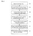
- FIG. 1illustrates a method for multi-component heart and aorta modeling and cardiac disease decision support according to an embodiment of the present invention
- FIG. 2illustrates an example of the multi-component hear and aorta modeling and decision support method of FIG. 1 according to an embodiment of the present invention
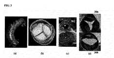
- FIG. 3illustrates exemplary patient-specific models generated for cardiac components from received image data
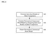
- FIG. 4illustrates a method for generating a 4D personalized geometric model of the heart according to an embodiment of the present invention
- FIG. 5illustrates exemplary models of the heart chambers and the aortic valve
- FIGS. 6A and 6Billustrate integrating individual models to generate a personalized anatomical heart model
- FIG. 7illustrates exemplary results of a multi-component, patient specific heart models
- FIG. 8illustrates a method for estimating parameters that characterize the material properties of the aorta wall according to an embodiment of the present invention
- FIG. 9illustrates an exemplary wall shear stress distribution in the aorta, resulting from a patient-specific simulation
- FIG. 10illustrates exemplary results of a CFD based FSI simulation for an aortic valve
- FIG. 11illustrates disease progression trajectories for coarctation
- FIG. 12is a high-level block diagram of a computer capable of implementing the present invention.
- the present inventionrelates to generating a 4D personalized anatomical model of the heart from a sequence of volumetric data, such as computed tomography (CT), magnetic resonance imaging (MRI), and echocardiography data.
- a sequence of volumetric datasuch as computed tomography (CT), magnetic resonance imaging (MRI), and echocardiography data.
- CTcomputed tomography
- MRImagnetic resonance imaging
- echocardiography dataSuch sequences of volumetric data also referred to herein as 4D image data or 4D images, are sequences taken over a period of time to cover one or more cardiac cycles, in which each frame is a 3D image (volume).
- Embodiments of the present inventionare described herein to give a visual understanding of the heart modeling method.
- a digital imageis often composed of digital representations of one or more objects (or shapes).
- the digital representation of an objectis often described herein in terms of identifying and manipulating the objects.
- Such manipulationsare virtual manipulations accomplished in the memory or other circuitry/hardware of a computer system.
- Embodiments of the present inventionare directed to generating a comprehensive and personalized computational model of the full heart and aorta from high resolution CT, MR, and rotational X-ray imaging, in order to guide decision support for patient evaluation, risk stratification, procedure planning, and timing of surgical intervention. Subtle, but critical interconnections are estimated between the aorta and the heart's components and disease progression models are derived. In order for the underlying hemodynamic analysis to generate patient-specific parameters to be subsequently used as discriminative features for the decision support process, embodiments of the present invention include the following components:
- FIG. 1illustrates a method for multi-component heart and aorta modeling and cardiac disease decision support according to an embodiment of the present invention.
- the method of FIG. 1transforms image data representing a coronary region of a patient into an anatomical model of the heart and uses the heart model for decision support in diagnosing and treating cardiac disease.
- FIG. 2illustrates an example of the multi-component hear and aorta modeling and decision support method of FIG. 1 according to an embodiment of the present invention.
- 4D image datais received.
- the sequence of volumetric image datacan be a sequence of 3D images (volumes) acquired over a certain time period.
- 4D image data3D+time
- 4D image data3D+time
- 4D image datacan be received using various medical imaging modalities.
- 4D CT data, 4D echocardiography, and 4D magnetic resonance (MR) image datacan be received, as well as other types of image data.
- the image datacan be received directly from one or more image acquisition devices, such as a CT scanner, an ultrasound device, or an MR scanner. It is also possible that previously stored image data be loaded, for example from a memory or storage of a computer system or some other computer readable storage medium.
- 4D MR datais received at step 202 .
- a multi-component patient-specific 4D geometric modelis estimated from the received 4D image data.
- a 4D geometric modelis generated from the received image data for each of multiple cardiac components, such as the aorta, aortic valve, mitral valve, tricuspid valve, pulmonary valve, and left and right ventricles and atria.
- cardiac componentssuch as the aorta, aortic valve, mitral valve, tricuspid valve, pulmonary valve, and left and right ventricles and atria.
- patient specific 4D geometric heart modelsare generated for each cardiac component.
- FIG. 3illustrates exemplary patient-specific models generated for cardiac components from received image data.
- image (a)shows a patient-specific model of the aorta and the ostia derived from CT data.
- Image (b)shows a patient specific model of the aortic valve generated from transesophageal echocardiogram (TEE) data.
- Image (c)shows a patient specific model of coupled aortic ( 302 ) and mitral ( 304 ) valves generated from TEE data.
- Image (d)shows a patient specific model of pathological aortic valves. In particular, image (d) shows a stenotic aortic valve 306 and a bicuspid aortic valve 308 .
- the multi-component 4D geometric modelgives the morphology of the patient's heart and can be used to determine morphological (dimensions) and dynamic parameters for any component of the heart.
- the patient specific 4D geometric modelcan be used to measure the left ventricle (LV) volume and ejection fraction (EF), inter-chamber synchronicity analysis, aortic and mitral valve analysis, etc.
- FIG. 4illustrates a method for generating a 4D personalized geometric model of the heart according to an embodiment of the present invention.
- the method of FIG. 4transforms image data representing a coronary region of a patient to generate a personalized geometric model of the heart for that patient.
- the method of FIG. 4can be used to implement step 104 of the method of FIG. 2 .
- an individual modelis generated from the received image data for each of a plurality of heart components.
- modelsare generated for the heart chambers: left ventricle (LV) (endocardium and epicardium), right ventricle (RV), left atrium (LA) and right atrium (RA); valves: mitral valve and aortic valve; and main vessels: aorta and pulmonary trunk. All of these portions of the heart are referred to herein collectively as the “heart components”.
- a physiological model of the heart componentis estimated in each frame of the 4D image data using a discriminative database-guide estimation/detection technique.
- the physiological model of each anatomic structureis constructed offline prior to generating the personalized heart model for a particular patient.
- Each physiological modelis generated based on a mathematical representation of the corresponding heart component in a set of annotated training data.
- the physiological model for each heart componentcan be generated using mean shapes of the heart component in a set of annotated training data.
- United States Patent Application Publication No. 2008/0101676which is incorporated herein by reference, describes a generating a four-chamber physiological heart model and fitting the heart model to image data.
- the heart modelis a 3D mesh and initial meshes for each chamber are generated using mean shapes of the chambers in annotated training data.
- United States Patent Application No. 2009/0123050which is incorporated herein by reference, describes a 4D physiological model of the aortic valve.
- a physiological modelcan similarly be generated offline for each of the heart components based on a set of annotated training data.
- the parameters of the physiological modelare estimated to fit the image using a discriminative machine-learning technique based on a large database of annotated training images.
- MSLmarginal space learning
- MSLProbabilistic Boosting Trees
- learning-based boundary detectioncan be performed on the individual heart component model in each image to refine the estimated model parameters.
- the boundary of each estimated modelcan be refined using the learning-based boundary detection to increase the accuracy of the physiological model estimation for each heart component.
- FIG. 5illustrates exemplary models of the heart chambers and the aortic valve.
- image 510shows the LV endocardium 501 , the LV epicardium 502 , the RV 503 , the LA 504 , and the RA 505 in diastole
- image 520shows the LV endocardium 501 , the LV epicardium 502 , the RV 503 , the LA 504 , and the RA 505 in systole.
- Image 530shows the aortic valve 506 in diastole and image 540 shows the aortic valve 506 in systole.
- a 4D personalized anatomical model of the heartis generated by integrating the individual models generated for each of the heart components.
- Each of the individual heart component models resulting from step 402is a mesh made of a certain number of points.
- mesh point correspondencesare established between connecting or overlapping models. The mesh point correspondences allow the models to be correctly aligned with respect to each other. It is possible to establish mesh point correspondence between models by re-sampling the models. For example, United States Patent Application Publication No.
- 2008/0262814which is incorporated herein by reference, describes various re-sampling methods to establish mesh point correspondence between models of the four heart chambers in order to correctly align the heart chamber models. It is to be understood that the techniques described in United States Patent Application Publication No. 2008/0262814 can be extended to establish mesh point correspondence between the individual heart component models described herein.
- FIGS. 6A and 6Billustrate integrating individual models to generate a personalized anatomical heart model.
- the images of FIG. 6Aillustrate a possible order for integrating the heart component models according to an embodiment of the present invention.
- image 602shows an LV endocardium model 601 .
- Image 604shows the integration of the mitral valve model 603 .
- Image 606shows the integration of the LV epicardium model 605 .
- Image 608shows the integration of the aortic valve model 607 and the aortic root model 609 .
- Image 610shows the integration of the RV model 611 .
- Image 612shows the integration of the LA model 613 .
- Image 614shows the integration of the RA model 615 .
- Image 616shows the integration of the aorta model 617 .
- Image 618shows the integration of the pulmonary trunk model 619 .
- images 620 and 622show 3D renderings of the resulting personalized anatomical heart model fit to image data. It is to be understood that although FIGS. 6A and 6B , illustrate the integration of the heart component models for a 3D volume, the heart component models can be similarly integrated in each frame of a 4D image sequence.
- the 4D personalized anatomical heart modelis output.
- the 4D personalized anatomical heart modelcan be output by storing the 4D personalized anatomical heart model to a memory, storage, or computer readable medium.
- the 4D personalized anatomical heart modelcan also be output by displaying the 4D personalized anatomical heart model or printing an image of the 4D personalized anatomical heart model.
- the output 4D personalized anatomical heart modelcan be used for further medical image processing.
- the 4D personalized anatomical heart modelcan be used to estimate various morphological and functional measurements, of the heart.
- the 4D personalized anatomic heart modelcan also be used to simulate blood flow or blood-tissue interaction. FIG.
- image (a)shows left and right ventricles and the aortic root derived from MR data.
- Image (b)shows the left endocardium and epicardium, right ventricle, left and right atria, and the aortic root derived from CT data.
- Image (c)shows all of the above components plus the aorta derived from CT data.
- a patient-specific 4D computational model based on personalized geometry, material properties, fluid boundary conditions, and flow velocity measurements (e.g., velocity encoded contrast MR and echo Doppler) in the 4D geometric modelis generated. For example, measurements of a chamber's volume and a valve's opening area computed over a full cardiac cycle enable for the characterization of the hemodynamics. Blood quantity, pressure and velocity can be directly estimated, for each of the four chambers, from the fitted 4D personalized anatomical heart model. The integration of Doppler echocardiogram or velocity encoded contrast MR velocity measurements further enhances the robustness of the blood parameter computation. Referring to FIG. 2 , at step 206 , the patient-specific 4D mesh is generated and velocity encoded contrast MR is generated at the aortic and mitral valve.
- flow velocity measurementse.g., velocity encoded contrast MR and echo Doppler
- patient-specific material properties of the aortic wallare estimated.
- an accurate representationis needed of the material properties of the aortic wall, in conjunction with measured velocity boundary conditions at the inlet and outlet of the aortic valve, measured velocity boundary conditions at the outlet of the aorta, and blood pressure measurements.
- a parameter estimation procedureis used. Since the aortic wall is a three-layered structure with inhomogeneous mechanical properties, a non-linear, hyperelastic constitutive multi-fiber model can be used for the aortic wall. Under this formulation, the aortic wall is modeled as a four-fiber family model, which takes into consideration the effect of the isotropic elastin behavior in addition to the anisotropic collagen behavior.
- c, c 1 (k) , c 2 (k) ⁇ 0are material parameters.
- ⁇ (k)is the stretch of the k th fiber family, given by:
- ⁇ ( k )( ⁇ ⁇ ⁇ sin ⁇ ⁇ ⁇ ( k ) ) 2 + ( ⁇ z ⁇ cos ⁇ ⁇ ⁇ ( k ) ) 2 , ( 4 ) with ⁇ (k) being the orientation and ⁇ ⁇ , ⁇ z being the circumferential and axial stretches, respectively.
- the unknown coefficients c, c 1 (k) , c 2 (k)are determined using a parameter estimation procedure in conjunction with Fluid Structure Interaction (FSI) based simulations.
- the time-resolved geometric models obtained in step 104are used for determining the actual (measured) deformation of the aortic wall due to pulsatile blood flow during the cardiac cycle.
- a parametric model of the material propertyis then used together with the time resolved in-flow out-flow velocity profile measured by 3D CINE MRI acquisition to perform a fully couple two-way FSI based simulation for the blood flow across the aortic wall and the length of the aorta.
- the parameter estimation procedureis modeled as an optimization problem where the underlying objective function is the residue (or error) between the actual (measured) deformation of the aortic wall and the simulated deformation of the aortic wall.
- the objective functioncan be expressed as:
- r(c)denotes the residue
- d i FSI (c) and d i MRIdenote the simulated and measured deformation vectors of the wall, respectively, at the N sample points (indexed by i).
- FIG. 8illustrates a method for estimating parameters that characterize the material properties of the aorta wall according to an embodiment of the present invention.
- the method of FIG. 8is used to estimate the parameters c, c 1 (k) , c 2 (k) described above.
- the method of FIG. 8can be used to implement step 108 of FIG. 1 .
- an actual deformation of the aortic wallis measured in the patient-specific 4D geometric model.
- the actual deformation of the aortic wall due to pulsatile blood flow during the cardiac cycleis measured in the 4D geometric model. This deformation may be measured at multiple sampling points resulting in a measured deformation vector.
- initial estimates for the parameterscan be selected from the literature in order to speed up the optimization process. For example, average values of a large population can be used for the initial parameter values.
- a constitutive material model of the aortic wallis constructed with the current parameters.
- the constitutive material model of the aortic wallhas a strain energy determined as described above in Equations (1)-(4) based on the current parameters.
- an FSI based simulationis performed using the constitutive material model of the aortic wall with the current parameters.
- the constitutive material modelcan be used together with the time resolved in-flow out-flow velocity profile measured by 3D CINE MRI acquisition to perform a fully couple two-way FSI based simulation for the blood flow across the aortic wall and the length of the aorta.
- the FSI simulationresults in a simulated blood flow and simulated deformation of the aortic wall.
- the objective functionis evaluated based on the FSI based simulation results. For example, the residue (error) between the current simulated deformation and the measured deformation can be calculated (Equation (5)).
- Equation (5)the residue (error) between the current simulated deformation and the measured deformation can be calculated (Equation (5)).
- the objective function described above in Equation (5)compares the simulated and measures deformations of the aortic wall, the present invention is not limited thereto. It is also possible to compare velocity vectors from simulated flow resulting from the FSI based simulation with velocity vectors determined from the patient-specific model.
- step 812it is determined whether the objective function has converged.
- the objective functionhas converged when the calculated residue (error) is less than a predetermined threshold value. If it is determined that the objective function has not converged, the method proceeds to step 814 . If it is determined that the objective function has converged, the method proceeds to step 816 .
- new parametersare determined using an optimization algorithm. For example, new parameters can be determined by minimization of the objective function (with respect to the material parameter vector c) using a non-linear gradient based optimization algorithm, such as the well-known Levenberg-Marquardt algorithm, which utilizes gradient information to obtain a locally optimal solution.
- a non-linear gradient based optimization algorithmsuch as the well-known Levenberg-Marquardt algorithm, which utilizes gradient information to obtain a locally optimal solution.
- the optimal material parametersare output. Accordingly, when it is determined that the objective function has converged, the current parameters are determined to be the optimum material parameters for the aortic wall, and the optimal material parameters are output.
- the optimum material parametersgive an accurate representation of the material properties of the aortic wall and can be used in patient-specific simulations for determining hemodynamics and wall mechanics.
- This parameter estimation for estimating parameters that characterize material properties of the aortic wallcan subsequently be used to resolve the mechanical properties of the vascular wall material on a fine mesh, and to generate a ‘map’ of the varying material properties along the aorta.
- a patient specific biomechanical modelis generated based on Fluid Structure Interaction (FSI) simulations using the 4D computational model and the estimated patient-specific material properties of the aortic wall.
- FSIFluid Structure Interaction
- a detailed simulation of the blood flow pattern of the patient, as well as the interaction of the blood with the anatomical structures of the heart,can be obtained by combining the above described measurements with established biomechanical and hemodynamics models, and finite element methods.
- the blood flow and tissue interactioncan be simulated using the parameters measured in the computational model. This enables the computation of path, pressure, and velocity of the blood on a particle level with a desired granularity.
- CFDcomputational fluid dynamics
- the computational modelis used as a tool for non-invasive assessment of surgical procedures on specific patients and for analyzing the effect of surgery on key parameters like wall shear stress and displacement. This is achieved by appropriately modifying the patient-specific structure model (e.g., to reflect surgical changes) together with the boundary conditions, and then simulating the blood flow in the simulated post-operative heart and aorta.
- operational modelscan also be used to simulate physical stress and exertion conditions, to analyze its effect on the key hemodynamic attributes, and to incorporate it into the risk progression model to generally reflect the activity of daily living.
- a coupled FSI simulation of the left ventricle, aortic valve, and the aortacan be performed with patient-specific velocity boundary conditions and time-resolved geometry (from 3D-time resolved PC MRI, Ct, and Rotational X-ray imaging).
- FSI methodscan be tailored to the particular heart parts, as some parts perform an active role (e.g., ventricle) or a mixed passive/active role (e.g., valves, arteries). Exemplary FSI methods for various heart parts are described below.
- Aorta(or other blood vessels): FSI models that include both fluid and structure equations fully coupled together through a set of boundary conditions, such as equal displacement, equal traction, and no-slip condition.
- An Arbitrary Lagrangian Eulerian (ALE) formulationcan be used for the coupled problem. This allows advanced bio-mechanical measurements including but not limited to wall shear stress, elasticity or stiffness, Von-Mises stress, flow pathlines, streamlines, and vorticity throughout the length of the aorta.
- FIG. 9illustrates an exemplary wall shear stress distribution in the aorta, resulting from a patient-specific simulation.
- Heart ChambersHemodynamic model based on Navier-Stokes equations and rigid structure assumption driven by moving boundary conditions associated with i) heart walls contractions/displacement during both diastolic and systolic cycled and ii) dynamic blood flow velocity/pressure boundary conditions at the flow entry and exit points.
- FIG. 10illustrates exemplary results of a CFD based FSI simulation for an aortic valve.
- step 210CFD-based FSI simulation is performed on the patient-specific computational models.
- patient-specific clinical parametersare extracted based on the patient-specific model and the FSI simulations.
- phenotypic, anatomic and hemodynamic parametersare derived from the patient-specific model and the simulations.
- the personalized modelsenable direct quantification of morphological, dynamical, and bio-mechanical characteristics including: dilation of the entire length of the thoracic aorta including the aortic annulus, and sino-tubular junction, aortic arch, proximal and distal descending aorta; chamber size and mass; vessel wall thickness; luminal dilation and aortic compliance and stiffness by calculation of relations between change in segmental aortic diameters or volumes and central blood pressure.
- CFD simulations on the patient-specific anatomic modelsgenerate hemodynamic parameters that characterize the complex flow fields including turbulence, jets, and recirculation. These simulations can further be used to derive the wall shear stress, blood velocity flow field, wall displacement, wall Von Mises stress (tensile), and turbulence intensity (vorticity).
- morphological and dynamic parameters of the heart and aortaare extracted, as well as hemodynamic parameters including wall shear stress, tensile stress, compliance and stiffness, turbulence intensity, aortic dilation, and aortic pressure. Additional patient information (e.g., clinical, history, and genetic information) can also be input and used as parameters.
- patient-specific disease progression and risk stratificationis performed based on the patient-specific phenotypic, anatomic, and hemodynamic parameters derived from the patient-specific 4D model and the FSI simulations.
- the disease progression and risk stratificationare performed using a trained decision-support model.
- multi-level Markov Cycle tree modelscan be used for both disease progression and risk stratification as a function of time. Individual patients are characterized and stratified based on their patient-specific dynamic heart model. Markov models for decision analysis provide a rich framework for integrating available patient information, stimulating disease progression based on time-dependent patient risk, and providing statistics of expected outcomes based on alternatives. According to a possible implementation, the models can be studies under various conditions, such as normal operation, after simulated surgical intervention, and under simulated stress conditions.
- This stratificationis reflected by differences in transition rates among disease states, as well as transition rates from each disease state to critical states. It can be performed using methods of cluster analysis and automated subgroup discovery. Traditional statistical methods, such as multivariate regression and significance tests, can be combined with modern machine learning methods, such as Support Vector Machines, SCR's, Probabilistic Boosting Trees, and Bayesian Networks. Predictive models enable the identification of individual risk factors, as well as combinations of characteristics that together are most strongly associated with patient outcomes, even if individually they are not predicative. As illustrated in FIG. 2 , at step 214 , decision support is performed based on the extracted parameters using a disease progression model.
- the disease progression modelcan reflect unique genetic disease signatures, physical stress and exertion, and various possible surgical outcomes. Heart specific disease progression and risk stratification is described in greater detail below.
- Disease progressioncan be modeled in terms of the continuous evolution and temporal fluctuations of the anatomic, morphological, hemodynamic and phenotype parameters extracted at step 110 .
- the condition of an individual patientcan be characterized in terms of the instantaneous values of variables that will constitute the present state of an individual patient (x(t)).
- Each patientis represented as a distinct point in a state-space, a high-dimensional space spanned by the various attributes of the patients, which completely defines the state.
- the state vectorcontains both continuous and discrete state variables, some of which are inherently inter-related with one another due to the various biological, physical, and physiological constraints. These are observed, measured, extracted from imaging studies, or simulated under patient-specific framework, as described above.
- State dynamicswhich represents continuous progression of the disease and discontinuous discrete events of intervention therapies. This is represented as follows:
- FIG. 11illustrates disease progression trajectories for coarctation.
- the trajectories 1102 , 1104 , and 1106represents disease trajectories under different interventions/treatments.
- each trajectory 1102 , 1104 , and 1106begins in coarctation and ends with the patient being healthy.
- FIG. 11also shows complications or dissection, aneurysm, and recoarctation that the trajectories 1102 , 1104 , and 1106 can pass through.
- a non-linear similarity measureis learned and then used to distinguish specific patient groups.
- each patient profileis represented as a multi-dimensional vector of features. From the comprehensive set of quantities, we isolate individual patients in classes, specific to the clinical use cases for a particular cardiovascular disease. The probability that classifies patients in clinical relevant disease progression clusters is learned from equivalence constraints, able to capture statistics from heterogeneous input measurements. Non-linear regression is applied to estimate the probability, which models a similarity measure between a pair of two patient profiles. During clinical decision-making, the profile of the subject patient is compared to each individual in the training population, while the k-Nearest Neighbor algorithm applied on the similarity scores performs the classification.
- Risk stratificationinvolves characterizing the risk for intra- or post-procedural complications for individual patients.
- Our proposed methodologyinvolves applying a non-linear similarity measure to distinguish between two classes: low-risk patients and high-risk patients.
- the classificationis performed separately for each type of complication associated with the particular cardiovascular disease of interest (e.g. in case of coarctation, the three complications are dissection, aneurysm and recoarctation).
- To distinguish between low- and high profileswe integrate the measurements obtained during the clinical evaluation at stage (morphology, dynamics, hemodynamics, phenotype and material properties). Additionally, the feature vector used for classification contains parameters of specific therapies to be applied (e.g. surgery, percutaneous implant etc).
- the similarity measureis learned from patients that are pre-classified in low or high risk, based on their follow-up studies.
- Computer 1202contains a processor 1204 , which controls the overall operation of the computer 1202 by executing computer program instructions which define such operation.
- the computer program instructionsmay be stored in a storage device 1212 (e.g., magnetic disk) and loaded into memory 1210 when execution of the computer program instructions is desired.
- At least one image acquisition device 1220such as a CT scanning device, ultrasound device, MR scanning device, etc., can be connected to the computer 1202 to input the 3D volumes to the computer 1202 . It is possible to implement the image acquisition device 1220 and the computer 1202 as one device. It is also possible that the image acquisition device 1220 and the computer 1202 communicate wirelessly through a network.
- the computer 1202also includes one or more network interfaces 1206 for communicating with other devices via a network.
- the computer 1202also includes other input/output devices 1208 that enable user interaction with the computer 1202 (e.g., display, keyboard, mouse, speakers, buttons, etc.). Such input/output devices 1208 may be used in conjunction with a set of computer programs as an annotation tool to annotate volumes received from the image acquisition device 1220 .
- input/output devices 1208may be used in conjunction with a set of computer programs as an annotation tool to annotate volumes received from the image acquisition device 1220 .
- FIG. 12is a high level representation of some of the components of such a computer for illustrative purposes.
Landscapes
- Engineering & Computer Science (AREA)
- Physics & Mathematics (AREA)
- Health & Medical Sciences (AREA)
- Medical Informatics (AREA)
- Public Health (AREA)
- General Physics & Mathematics (AREA)
- General Health & Medical Sciences (AREA)
- Theoretical Computer Science (AREA)
- Databases & Information Systems (AREA)
- Quality & Reliability (AREA)
- Computer Vision & Pattern Recognition (AREA)
- Biomedical Technology (AREA)
- Data Mining & Analysis (AREA)
- Radiology & Medical Imaging (AREA)
- Pathology (AREA)
- Nuclear Medicine, Radiotherapy & Molecular Imaging (AREA)
- Epidemiology (AREA)
- Primary Health Care (AREA)
- Computer Graphics (AREA)
- Geometry (AREA)
- Software Systems (AREA)
- Apparatus For Radiation Diagnosis (AREA)
- Magnetic Resonance Imaging Apparatus (AREA)
Abstract
Description
ψ=ψiso+ψaniso, (1)
where ψ, the strain energy is composed of an anisotropic part ψanisodue to the multiple fiber families, together with an isotropic neo-hookean term ψiso, as shown below:
ψiso=c(I1−3) (2)
where I1is the first invariant of the right Cauchy-Green tensor C (i.e., I1=trace(C)) and c, c1(k), c2(k)≦0 are material parameters. λ(k)is the stretch of the kthfiber family, given by:
with α(k)being the orientation and λθ, λzbeing the circumferential and axial stretches, respectively.
where r(c) denotes the residue, and diFSI(c) and diMRIdenote the simulated and measured deformation vectors of the wall, respectively, at the N sample points (indexed by i). The unknown parameters (characterizing the material properties of the aortic wall) constitute the parameter vector c=[c, c1(k), c2(k)] for k=1, 2, 3, and 4.
Claims (25)
Priority Applications (1)
| Application Number | Priority Date | Filing Date | Title |
|---|---|---|---|
| US12/825,905US8224640B2 (en) | 2009-09-08 | 2010-06-29 | Method and system for computational modeling of the aorta and heart |
Applications Claiming Priority (2)
| Application Number | Priority Date | Filing Date | Title |
|---|---|---|---|
| US24039709P | 2009-09-08 | 2009-09-08 | |
| US12/825,905US8224640B2 (en) | 2009-09-08 | 2010-06-29 | Method and system for computational modeling of the aorta and heart |
Publications (2)
| Publication Number | Publication Date |
|---|---|
| US20110060576A1 US20110060576A1 (en) | 2011-03-10 |
| US8224640B2true US8224640B2 (en) | 2012-07-17 |
Family
ID=43648394
Family Applications (1)
| Application Number | Title | Priority Date | Filing Date |
|---|---|---|---|
| US12/825,905Active2030-08-17US8224640B2 (en) | 2009-09-08 | 2010-06-29 | Method and system for computational modeling of the aorta and heart |
Country Status (1)
| Country | Link |
|---|---|
| US (1) | US8224640B2 (en) |
Cited By (19)
| Publication number | Priority date | Publication date | Assignee | Title |
|---|---|---|---|---|
| US20120084064A1 (en)* | 2010-09-29 | 2012-04-05 | Nutech Ventures, Inc. | Model-based systems and methods for analyzing and predicting outcomes of vascular interventions and reconstructions |
| US20130046168A1 (en)* | 2011-08-17 | 2013-02-21 | Lei Sui | Method and system of characterization of carotid plaque |
| US20130185231A1 (en)* | 2012-01-17 | 2013-07-18 | International Business Machines Corporation | Predicting diagnosis of a patient |
| WO2014182505A1 (en)* | 2013-05-10 | 2014-11-13 | Stenomics, Inc. | Modeling and simulation system for optimizing prosthetic heart valve treatment |
| US9092743B2 (en) | 2013-10-23 | 2015-07-28 | Stenomics, Inc. | Machine learning system for assessing heart valves and surrounding cardiovascular tracts |
| US9585632B2 (en) | 2014-04-23 | 2017-03-07 | Siemens Medical Solutions Usa, Inc. | Estimation of a mechanical property of anatomy from medical scan data |
| US9629563B2 (en) | 2013-09-04 | 2017-04-25 | Siemens Healthcare Gmbh | Method and system for functional assessment of renal artery stenosis from medical images |
| US10617314B1 (en) | 2019-06-10 | 2020-04-14 | Vektor Medical, Inc. | Heart graphic display system |
| RU2725917C1 (en)* | 2019-09-11 | 2020-07-07 | Федеральное государственное бюджетное научное учреждение "Научно-исследовательский институт комплексных проблем сердечно-сосудистых заболеваний" ФГБНУ "Научно-исследовательский институт комплексных проблем сердечно-сосудистых заболеваний" | Method for numerical modelling of transcatheter implantation of patient's heart valve |
| US10709347B1 (en) | 2019-06-10 | 2020-07-14 | Vektor Medical, Inc. | Heart graphic display system |
| US10860754B2 (en) | 2018-04-26 | 2020-12-08 | Vektor Medical, Inc. | Calibration of simulated cardiograms |
| US10856816B2 (en) | 2018-04-26 | 2020-12-08 | Vektor Medical, Inc. | Machine learning using simulated cardiograms |
| US10952794B2 (en) | 2018-11-13 | 2021-03-23 | Vektor Medical, Inc. | Augmentation of images with source locations |
| US11259871B2 (en) | 2018-04-26 | 2022-03-01 | Vektor Medical, Inc. | Identify ablation pattern for use in an ablation |
| US11338131B1 (en) | 2021-05-05 | 2022-05-24 | Vektor Medical, Inc. | Guiding implantation of an energy delivery component in a body |
| US11534224B1 (en) | 2021-12-02 | 2022-12-27 | Vektor Medical, Inc. | Interactive ablation workflow system |
| US11896432B2 (en) | 2021-08-09 | 2024-02-13 | Vektor Medical, Inc. | Machine learning for identifying characteristics of a reentrant circuit |
| US11974853B2 (en) | 2020-10-30 | 2024-05-07 | Vektor Medical, Inc. | Heart graphic display system |
| US20240346652A1 (en)* | 2018-01-24 | 2024-10-17 | Pie Medical Imaging B.V. | Flow analysis in 4d mr image data |
Families Citing this family (45)
| Publication number | Priority date | Publication date | Assignee | Title |
|---|---|---|---|---|
| US8200466B2 (en) | 2008-07-21 | 2012-06-12 | The Board Of Trustees Of The Leland Stanford Junior University | Method for tuning patient-specific cardiovascular simulations |
| US9405886B2 (en) | 2009-03-17 | 2016-08-02 | The Board Of Trustees Of The Leland Stanford Junior University | Method for determining cardiovascular information |
| US20110081054A1 (en)* | 2009-10-02 | 2011-04-07 | Harris Corporation | Medical image analysis system for displaying anatomical images subject to deformation and related methods |
| US20110161013A1 (en)* | 2010-05-18 | 2011-06-30 | Saeed Ranjbar | Method for determining heat boundary value conditions of red blood cells in the neighborhood of myocardium |
| US8682626B2 (en) | 2010-07-21 | 2014-03-25 | Siemens Aktiengesellschaft | Method and system for comprehensive patient-specific modeling of the heart |
| US8315812B2 (en) | 2010-08-12 | 2012-11-20 | Heartflow, Inc. | Method and system for patient-specific modeling of blood flow |
| US9119540B2 (en) | 2010-09-16 | 2015-09-01 | Siemens Aktiengesellschaft | Method and system for non-invasive assessment of coronary artery disease |
| US9141763B2 (en) | 2011-02-07 | 2015-09-22 | Siemens Aktiengesellschaft | Method and system for patient-specific computational modeling and simulation for coupled hemodynamic analysis of cerebral vessels |
| US9824302B2 (en)* | 2011-03-09 | 2017-11-21 | Siemens Healthcare Gmbh | Method and system for model-based fusion of multi-modal volumetric images |
| DE102011006501B4 (en) | 2011-03-31 | 2021-12-23 | Siemens Healthcare Gmbh | Method, image processing device and computed tomography system for obtaining a 4D image data set of an examination subject, as well as computer program product with program code sections for executing such a method |
| US10162932B2 (en) | 2011-11-10 | 2018-12-25 | Siemens Healthcare Gmbh | Method and system for multi-scale anatomical and functional modeling of coronary circulation |
| US8983809B2 (en) | 2011-12-06 | 2015-03-17 | Siemens Aktiengesellschaft | Method and system for patient-specific hemodynamic assessment of virtual stent implantation |
| US9155470B2 (en) | 2012-01-24 | 2015-10-13 | Siemens Aktiengesellschaft | Method and system for model based fusion on pre-operative computed tomography and intra-operative fluoroscopy using transesophageal echocardiography |
| US10311978B2 (en)* | 2012-01-30 | 2019-06-04 | Siemens Healthcare Gmbh | Method and system for patient specific planning of cardiac therapies on preoperative clinical data and medical images |
| US9883941B2 (en) | 2012-06-19 | 2018-02-06 | Boston Scientific Scimed, Inc. | Replacement heart valve |
| KR101768526B1 (en)* | 2012-07-27 | 2017-08-17 | 삼성전자주식회사 | Method and apparatus for creating model of patient specified target organ based on blood vessel structure |
| US10433740B2 (en) | 2012-09-12 | 2019-10-08 | Heartflow, Inc. | Systems and methods for estimating ischemia and blood flow characteristics from vessel geometry and physiology |
| US10398386B2 (en)* | 2012-09-12 | 2019-09-03 | Heartflow, Inc. | Systems and methods for estimating blood flow characteristics from vessel geometry and physiology |
| FR2996667B1 (en)* | 2012-10-05 | 2015-12-11 | Olea Medical | SYSTEM AND METHOD FOR ESTIMATING A QUANTITY OF INTEREST IN A CINEMATIC SYSTEM BY CONTRAST AGENT TOMOGRAPHY |
| US10485517B2 (en) | 2013-02-15 | 2019-11-26 | The Johns Hopkins University | Computational flow dynamics based method for estimating thromboembolic risk in patients with myocardial infarction |
| US20160034665A1 (en)* | 2013-03-14 | 2016-02-04 | Cardioart Technologies Ltd. | System and method for personalized hemodynamics modeling and monitoring |
| US10130332B2 (en) | 2013-08-06 | 2018-11-20 | Samsung Electronics Co., Ltd. | Method and apparatus of diagnosing cardiac diseases based on modeling of cardiac motion |
| US9805463B2 (en)* | 2013-08-27 | 2017-10-31 | Heartflow, Inc. | Systems and methods for predicting location, onset, and/or change of coronary lesions |
| CN105474219B (en)* | 2013-08-28 | 2019-10-18 | 西门子公司 | Systems and methods for estimating physiological cardiac measurements from medical images and clinical data |
| US9058692B1 (en) | 2014-04-16 | 2015-06-16 | Heartflow, Inc. | Systems and methods for image-based object modeling using multiple image acquisitions or reconstructions |
| US9449145B2 (en)* | 2014-04-22 | 2016-09-20 | Heartflow, Inc. | Systems and methods for virtual contrast agent simulation and computational fluid dynamics (CFD) to compute functional significance of stenoses |
| US10779743B2 (en) | 2014-05-06 | 2020-09-22 | Peacs B.V. | Estimating distribution, fluctuation and/or movement of electrical activity through a heart tissue |
| US11172860B2 (en) | 2014-05-06 | 2021-11-16 | Peacs Investments B.V. | Estimating distribution fluctuation and/or movement of electrical activity through a heart tissue |
| CA2949449C (en)* | 2014-05-23 | 2021-05-25 | Dacadoo Ag | Automated health data acquisition, processing and communication system |
| EP3166032B1 (en)* | 2014-07-03 | 2018-10-03 | Fujitsu Limited | Biometric simulation device, method for controlling biometric simulation device, and program for controlling biometric simulation device |
| CN107077731B (en)* | 2014-10-22 | 2021-08-03 | 皇家飞利浦有限公司 | Visualization of imaging uncertainty |
| US9349178B1 (en)* | 2014-11-24 | 2016-05-24 | Siemens Aktiengesellschaft | Synthetic data-driven hemodynamic determination in medical imaging |
| US10716513B2 (en) | 2015-04-17 | 2020-07-21 | Heartflow, Inc. | Systems and methods for cardiovascular blood flow and musculoskeletal modeling for predicting device failure or clinical events |
| US11289207B2 (en)* | 2015-07-09 | 2022-03-29 | Peacs Investments B.V. | System for visualizing heart activation |
| US10438355B2 (en)* | 2015-11-10 | 2019-10-08 | General Electric Company | System and method for estimating arterial pulse wave velocity |
| CA3006102A1 (en) | 2015-11-24 | 2017-06-01 | Dacadoo Ag | Automated health data acquisition, processing and communication system and method |
| EP3228245B1 (en) | 2016-04-05 | 2021-05-26 | Siemens Healthcare GmbH | Determining arterial wall property with blood flow model |
| US11458320B2 (en) | 2016-09-06 | 2022-10-04 | Peacs Investments B.V. | Method of cardiac resynchronization therapy |
| EP3555778B1 (en)* | 2016-12-15 | 2020-10-21 | Sintef TTO AS | Method and process for providing a subject-specific computational model used for decision support and making diagnosis of cardiovascular diseases |
| CN109758228A (en)* | 2019-02-28 | 2019-05-17 | 四川大学华西医院 | Optimal stent placement method for transjugular portosystemic shunt surgery based on three-dimensional reconstruction |
| CN110991103B (en)* | 2019-11-21 | 2024-04-02 | 哈尔滨理工大学 | Method for establishing super-elastic model containing interaction of fibers and matrix |
| US11728040B2 (en)* | 2020-01-29 | 2023-08-15 | Tata Consultancy Services Limited | Neuromodulation based adaptive controller for mitral stenosis |
| CN113648059B (en)* | 2021-08-26 | 2023-09-29 | 上海联影医疗科技股份有限公司 | Surgical planning assessment methods, computer equipment and storage media |
| CN118037994B (en)* | 2024-04-15 | 2024-06-21 | 法琛堂(昆明)医疗科技有限公司 | Heart three-dimensional structure reconstruction method and system |
| CN118964984B (en)* | 2024-07-26 | 2025-09-23 | 杭州中谦科技有限公司 | Method and system for building interactive medical simulation scenarios based on AI artificial intelligence |
Citations (10)
| Publication number | Priority date | Publication date | Assignee | Title |
|---|---|---|---|---|
| US20050281447A1 (en)* | 2004-03-02 | 2005-12-22 | Romain Moreau-Gobard | System and method for detecting the aortic valve using a model-based segmentation technique |
| US20060004275A1 (en) | 2004-06-30 | 2006-01-05 | Vija A H | Systems and methods for localized image registration and fusion |
| US20060004274A1 (en) | 2004-06-30 | 2006-01-05 | Hawman Eric G | Fusing nuclear medical images with a second imaging modality |
| US7117026B2 (en) | 2002-06-12 | 2006-10-03 | Koninklijke Philips Electronics N.V. | Physiological model based non-rigid image registration |
| US20080101676A1 (en) | 2006-09-28 | 2008-05-01 | Siemens Corporate Research, Inc. | System and Method For Segmenting Chambers Of A Heart In A Three Dimensional Image |
| US20080262814A1 (en) | 2007-04-23 | 2008-10-23 | Yefeng Zheng | Method and system for generating a four-chamber heart model |
| US7450780B2 (en) | 2003-09-08 | 2008-11-11 | Siemens Medical Solutions, Usa, Inc. | Similarity measures |
| US20090123050A1 (en) | 2007-11-02 | 2009-05-14 | Razvan Ionasec | Method and system for automatic quantification of aortic valve function from 4D computed tomography data using a physiological model |
| US20100070249A1 (en)* | 2008-09-18 | 2010-03-18 | Siemens Corporate Research, Inc. | Method and System for Generating a Personalized Anatomical Heart Model |
| US20100280352A1 (en)* | 2009-05-01 | 2010-11-04 | Siemens Corporation | Method and System for Multi-Component Heart and Aorta Modeling for Decision Support in Cardiac Disease |
- 2010
- 2010-06-29USUS12/825,905patent/US8224640B2/enactiveActive
Patent Citations (10)
| Publication number | Priority date | Publication date | Assignee | Title |
|---|---|---|---|---|
| US7117026B2 (en) | 2002-06-12 | 2006-10-03 | Koninklijke Philips Electronics N.V. | Physiological model based non-rigid image registration |
| US7450780B2 (en) | 2003-09-08 | 2008-11-11 | Siemens Medical Solutions, Usa, Inc. | Similarity measures |
| US20050281447A1 (en)* | 2004-03-02 | 2005-12-22 | Romain Moreau-Gobard | System and method for detecting the aortic valve using a model-based segmentation technique |
| US20060004275A1 (en) | 2004-06-30 | 2006-01-05 | Vija A H | Systems and methods for localized image registration and fusion |
| US20060004274A1 (en) | 2004-06-30 | 2006-01-05 | Hawman Eric G | Fusing nuclear medical images with a second imaging modality |
| US20080101676A1 (en) | 2006-09-28 | 2008-05-01 | Siemens Corporate Research, Inc. | System and Method For Segmenting Chambers Of A Heart In A Three Dimensional Image |
| US20080262814A1 (en) | 2007-04-23 | 2008-10-23 | Yefeng Zheng | Method and system for generating a four-chamber heart model |
| US20090123050A1 (en) | 2007-11-02 | 2009-05-14 | Razvan Ionasec | Method and system for automatic quantification of aortic valve function from 4D computed tomography data using a physiological model |
| US20100070249A1 (en)* | 2008-09-18 | 2010-03-18 | Siemens Corporate Research, Inc. | Method and System for Generating a Personalized Anatomical Heart Model |
| US20100280352A1 (en)* | 2009-05-01 | 2010-11-04 | Siemens Corporation | Method and System for Multi-Component Heart and Aorta Modeling for Decision Support in Cardiac Disease |
Non-Patent Citations (10)
| Title |
|---|
| De Hart et al. Journal of Biomechanics (2003) vol. 36, pp. 103-112.* |
| Deparis et al. (A Domain Decomposition Framework for Fluid-Structure Interaction Problems in Computational Fluid Dynamics (2004) Part I, pp. 41-58).* |
| Gerbeau et al. (2005) Computers and Structures, vol. 83:155-165.* |
| Ionasec, Razvan Ioan et al., "Dynamic Model-Driven Quantitative and Visual Evaluation of the Aortic Valve from 4D CT", Int'l Conference on Medical Image Computing and Computer-Assisted Intervention, 11(Pt 1), 2008. |
| Scotti et al. (2007) Computers and Structures, vol. 85:1097-1113.* |
| Sermesant et al. Medical Image Analysis (2006) vol. 10, pp. 642-656.* |
| Wang et al. (1999) IEEE Engineering in Medicine and Biology, Nov./Dec.: 33-39.* |
| Yang, Lin et al., "3D UltraSound Tracking of the Left Ventricles Using One-Step Forward Prediction and Data Fusion of Collaborative Trackers", CVPR, 2008. |
| Zhang et al. (2007) Comput. Methods Appl. Mech. Engrg., vol. 196:2943-2959.* |
| Zheng, Yefeng, et al., "Four-Chamber Heart Modeling and Automatic Segmentation for 3D Cardiac CT Volumes Using Marginal Space Learning and Steerable Features", IEEE Transactions on Medical Imaging, 27(11), Nov. 2008. |
Cited By (45)
| Publication number | Priority date | Publication date | Assignee | Title |
|---|---|---|---|---|
| US20120084064A1 (en)* | 2010-09-29 | 2012-04-05 | Nutech Ventures, Inc. | Model-based systems and methods for analyzing and predicting outcomes of vascular interventions and reconstructions |
| US20130046168A1 (en)* | 2011-08-17 | 2013-02-21 | Lei Sui | Method and system of characterization of carotid plaque |
| US20130185231A1 (en)* | 2012-01-17 | 2013-07-18 | International Business Machines Corporation | Predicting diagnosis of a patient |
| US8996428B2 (en)* | 2012-01-17 | 2015-03-31 | International Business Machines Corporation | Predicting diagnosis of a patient |
| WO2014182505A1 (en)* | 2013-05-10 | 2014-11-13 | Stenomics, Inc. | Modeling and simulation system for optimizing prosthetic heart valve treatment |
| US9135381B2 (en) | 2013-05-10 | 2015-09-15 | Stenomics, Inc. | Modeling and simulation system for optimizing prosthetic heart valve treatment |
| US11315690B2 (en) | 2013-05-10 | 2022-04-26 | Stenomics, Inc. | Modeling and simulation system for optimizing prosthetic heart valve treatment |
| US10497476B2 (en) | 2013-05-10 | 2019-12-03 | Stenomics, Inc. | Modeling and simulation system for optimizing prosthetic heart valve treatment |
| US9629563B2 (en) | 2013-09-04 | 2017-04-25 | Siemens Healthcare Gmbh | Method and system for functional assessment of renal artery stenosis from medical images |
| US10943698B2 (en) | 2013-10-23 | 2021-03-09 | Stenomics, Inc. | Machine learning system for assessing heart valves and surrounding cardiovascular tracts |
| US9953272B2 (en) | 2013-10-23 | 2018-04-24 | Stenomics, Inc. | Machine learning system for assessing heart valves and surrounding cardiovascular tracts |
| US11024425B2 (en) | 2013-10-23 | 2021-06-01 | Stenomics, Inc. | Machine learning system for assessing heart valves and surrounding cardiovascular tracts |
| US11024426B2 (en) | 2013-10-23 | 2021-06-01 | Stenomics, Inc. | Machine learning system for assessing heart valves and surrounding cardiovascular tracts |
| US9424531B2 (en) | 2013-10-23 | 2016-08-23 | Stenomics, Inc. | Machine learning system for assessing heart valves and surrounding cardiovascular tracts |
| US9092743B2 (en) | 2013-10-23 | 2015-07-28 | Stenomics, Inc. | Machine learning system for assessing heart valves and surrounding cardiovascular tracts |
| US10762442B2 (en) | 2013-10-23 | 2020-09-01 | Stenomics, Inc. | Machine learning system for assessing heart valves and surrounding cardiovascular tracts |
| US9585632B2 (en) | 2014-04-23 | 2017-03-07 | Siemens Medical Solutions Usa, Inc. | Estimation of a mechanical property of anatomy from medical scan data |
| US20240346652A1 (en)* | 2018-01-24 | 2024-10-17 | Pie Medical Imaging B.V. | Flow analysis in 4d mr image data |
| US11253206B2 (en) | 2018-04-26 | 2022-02-22 | Vektor Medical, Inc. | Display of an electrical force generated by an electrical source within a body |
| US11504073B2 (en) | 2018-04-26 | 2022-11-22 | Vektor Medical, Inc. | Machine learning using clinical and simulated data |
| US10856816B2 (en) | 2018-04-26 | 2020-12-08 | Vektor Medical, Inc. | Machine learning using simulated cardiograms |
| US10860754B2 (en) | 2018-04-26 | 2020-12-08 | Vektor Medical, Inc. | Calibration of simulated cardiograms |
| US12064215B2 (en) | 2018-04-26 | 2024-08-20 | Vektor Medical, Inc. | Classification relating to atrial fibrillation based on electrocardiogram and non-electrocardiogram features |
| US11259871B2 (en) | 2018-04-26 | 2022-03-01 | Vektor Medical, Inc. | Identify ablation pattern for use in an ablation |
| US11259756B2 (en) | 2018-04-26 | 2022-03-01 | Vektor Medical, Inc. | Machine learning using clinical and simulated data |
| US12076119B2 (en) | 2018-04-26 | 2024-09-03 | Vektor Medical, Inc. | Bootstrapping a simulation-based electromagnetic output of a different anatomy |
| US12390113B2 (en) | 2018-04-26 | 2025-08-19 | Vektor Medical, Inc. | User interface for presenting simulated anatomies of an electromagnetic source |
| US11344263B2 (en) | 2018-04-26 | 2022-05-31 | Vektor Medical, Inc. | Bootstrapping a simulation-based electromagnetic output of a different anatomy |
| US11806080B2 (en) | 2018-04-26 | 2023-11-07 | Vektor Medical, Inc. | Identify ablation pattern for use in an ablation |
| US11622732B2 (en) | 2018-04-26 | 2023-04-11 | Vektor Medical, Inc. | Identifying an attribute of an electromagnetic source configuration by matching simulated and patient data |
| US11576624B2 (en) | 2018-04-26 | 2023-02-14 | Vektor Medical, Inc. | Generating approximations of cardiograms from different source configurations |
| US11547369B2 (en) | 2018-04-26 | 2023-01-10 | Vektor Medical, Inc. | Machine learning using clinical and simulated data |
| US11564641B2 (en) | 2018-04-26 | 2023-01-31 | Vektor Medical, Inc. | Generating simulated anatomies of an electromagnetic source |
| US12048488B2 (en) | 2018-11-13 | 2024-07-30 | Vektor Medical, Inc. | Augmentation of images with source locations |
| US10952794B2 (en) | 2018-11-13 | 2021-03-23 | Vektor Medical, Inc. | Augmentation of images with source locations |
| US10617314B1 (en) | 2019-06-10 | 2020-04-14 | Vektor Medical, Inc. | Heart graphic display system |
| US11638546B2 (en) | 2019-06-10 | 2023-05-02 | Vektor Medical, Inc. | Heart graphic display system |
| US11490845B2 (en) | 2019-06-10 | 2022-11-08 | Vektor Medical, Inc. | Heart graphic display system |
| US11957471B2 (en) | 2019-06-10 | 2024-04-16 | Vektor Medical, Inc. | Heart graphic display system |
| US10709347B1 (en) | 2019-06-10 | 2020-07-14 | Vektor Medical, Inc. | Heart graphic display system |
| RU2725917C1 (en)* | 2019-09-11 | 2020-07-07 | Федеральное государственное бюджетное научное учреждение "Научно-исследовательский институт комплексных проблем сердечно-сосудистых заболеваний" ФГБНУ "Научно-исследовательский институт комплексных проблем сердечно-сосудистых заболеваний" | Method for numerical modelling of transcatheter implantation of patient's heart valve |
| US11974853B2 (en) | 2020-10-30 | 2024-05-07 | Vektor Medical, Inc. | Heart graphic display system |
| US11338131B1 (en) | 2021-05-05 | 2022-05-24 | Vektor Medical, Inc. | Guiding implantation of an energy delivery component in a body |
| US11896432B2 (en) | 2021-08-09 | 2024-02-13 | Vektor Medical, Inc. | Machine learning for identifying characteristics of a reentrant circuit |
| US11534224B1 (en) | 2021-12-02 | 2022-12-27 | Vektor Medical, Inc. | Interactive ablation workflow system |
Also Published As
| Publication number | Publication date |
|---|---|
| US20110060576A1 (en) | 2011-03-10 |
Similar Documents
| Publication | Publication Date | Title |
|---|---|---|
| US8224640B2 (en) | Method and system for computational modeling of the aorta and heart | |
| US8527251B2 (en) | Method and system for multi-component heart and aorta modeling for decision support in cardiac disease | |
| US10162932B2 (en) | Method and system for multi-scale anatomical and functional modeling of coronary circulation | |
| US20210241914A1 (en) | Machine learning system for assessing heart valves and surrounding cardiovascular tracts | |
| US11664125B2 (en) | System and method for deep learning based cardiac electrophysiology model personalization | |
| US9405996B2 (en) | Method and system for generating a personalized anatomical heart model | |
| US8009887B2 (en) | Method and system for automatic quantification of aortic valve function from 4D computed tomography data using a physiological model | |
| US8682626B2 (en) | Method and system for comprehensive patient-specific modeling of the heart | |
| US8218845B2 (en) | Dynamic pulmonary trunk modeling in computed tomography and magnetic resonance imaging based on the detection of bounding boxes, anatomical landmarks, and ribs of a pulmonary artery | |
| US9585632B2 (en) | Estimation of a mechanical property of anatomy from medical scan data | |
| US10496729B2 (en) | Method and system for image-based estimation of multi-physics parameters and their uncertainty for patient-specific simulation of organ function | |
| EP3555778B1 (en) | Method and process for providing a subject-specific computational model used for decision support and making diagnosis of cardiovascular diseases | |
| US9848856B2 (en) | Valve modeling with dense chordae from medical scan data | |
| US20180174068A1 (en) | Method and process for providing a subject-specific computational model used for treatment of cardiovascular diseases | |
| US9462952B2 (en) | System and method for estimating artery compliance and resistance from 4D cardiac images and pressure measurements | |
| Soni et al. | A comprehensive review on CFD simulations of left ventricle hemodynamics: numerical methods, experimental validation techniques, and emerging trends | |
| Xu | Anatomical and functional analysis of cardiac MRI scans using deep learning | |
| Baličević et al. | Atlas construction for cardiac velocity profiles segmentation using a lumped computational model of circulatory system | |
| Liffey | From physiology to clinics: clinical applications of multiscale modelling of the heart | |
| Gdoura et al. | Informatics in Medicine Unlocked |
Legal Events
| Date | Code | Title | Description |
|---|---|---|---|
| AS | Assignment | Owner name:SIEMENS CORPORATION, NEW JERSEY Free format text:ASSIGNMENT OF ASSIGNORS INTEREST;ASSIGNORS:SHARMA, PUNEET;GEORGESCU, BOGDAN;IONASEC, RAZVAN IOAN;AND OTHERS;SIGNING DATES FROM 20100622 TO 20100625;REEL/FRAME:024609/0781 | |
| AS | Assignment | Owner name:SIEMENS AKTIENGESELLSCHAFT, GERMANY Free format text:ASSIGNMENT OF ASSIGNORS INTEREST;ASSIGNOR:SIEMENS CORPORATION;REEL/FRAME:025774/0578 Effective date:20110125 | |
| STCF | Information on status: patent grant | Free format text:PATENTED CASE | |
| FPAY | Fee payment | Year of fee payment:4 | |
| AS | Assignment | Owner name:SIEMENS HEALTHCARE GMBH, GERMANY Free format text:ASSIGNMENT OF ASSIGNORS INTEREST;ASSIGNOR:SIEMENS AKTIENGESELLSCHAFT;REEL/FRAME:039271/0561 Effective date:20160610 | |
| MAFP | Maintenance fee payment | Free format text:PAYMENT OF MAINTENANCE FEE, 8TH YEAR, LARGE ENTITY (ORIGINAL EVENT CODE: M1552); ENTITY STATUS OF PATENT OWNER: LARGE ENTITY Year of fee payment:8 | |
| AS | Assignment | Owner name:SIEMENS HEALTHINEERS AG, GERMANY Free format text:ASSIGNMENT OF ASSIGNORS INTEREST;ASSIGNOR:SIEMENS HEALTHCARE GMBH;REEL/FRAME:066088/0256 Effective date:20231219 | |
| MAFP | Maintenance fee payment | Free format text:PAYMENT OF MAINTENANCE FEE, 12TH YEAR, LARGE ENTITY (ORIGINAL EVENT CODE: M1553); ENTITY STATUS OF PATENT OWNER: LARGE ENTITY Year of fee payment:12 | |
| AS | Assignment | Owner name:SIEMENS HEALTHINEERS AG, GERMANY Free format text:CHANGE OF NAME;ASSIGNOR:SIEMENS HEALTHCARE GMBH;REEL/FRAME:066244/0428 Effective date:20231201 | |
| AS | Assignment | Owner name:SIEMENS HEALTHINEERS AG, GERMANY Free format text:CORRECTIVE ASSIGNMENT TO CORRECT THE ASSIGNEE PREVIOUSLY RECORDED AT REEL: 066088 FRAME: 0256. ASSIGNOR(S) HEREBY CONFIRMS THE ASSIGNMENT;ASSIGNOR:SIEMENS HEALTHCARE GMBH;REEL/FRAME:071178/0246 Effective date:20231219 |