US8213693B1 - System and method to track and navigate a tool through an imaged subject - Google Patents
System and method to track and navigate a tool through an imaged subjectDownload PDFInfo
- Publication number
- US8213693B1 US8213693B1US11/860,368US86036807AUS8213693B1US 8213693 B1US8213693 B1US 8213693B1US 86036807 AUS86036807 AUS 86036807AUS 8213693 B1US8213693 B1US 8213693B1
- Authority
- US
- United States
- Prior art keywords
- relative
- transducer
- coordinate system
- tracking
- image
- Prior art date
- Legal status (The legal status is an assumption and is not a legal conclusion. Google has not performed a legal analysis and makes no representation as to the accuracy of the status listed.)
- Active, expires
Links
Images


Classifications
- A—HUMAN NECESSITIES
- A61—MEDICAL OR VETERINARY SCIENCE; HYGIENE
- A61B—DIAGNOSIS; SURGERY; IDENTIFICATION
- A61B34/00—Computer-aided surgery; Manipulators or robots specially adapted for use in surgery
- A61B34/20—Surgical navigation systems; Devices for tracking or guiding surgical instruments, e.g. for frameless stereotaxis
- A—HUMAN NECESSITIES
- A61—MEDICAL OR VETERINARY SCIENCE; HYGIENE
- A61B—DIAGNOSIS; SURGERY; IDENTIFICATION
- A61B17/00—Surgical instruments, devices or methods
- A61B17/00234—Surgical instruments, devices or methods for minimally invasive surgery
- A61B2017/00292—Surgical instruments, devices or methods for minimally invasive surgery mounted on or guided by flexible, e.g. catheter-like, means
- A61B2017/003—Steerable
- A—HUMAN NECESSITIES
- A61—MEDICAL OR VETERINARY SCIENCE; HYGIENE
- A61B—DIAGNOSIS; SURGERY; IDENTIFICATION
- A61B17/00—Surgical instruments, devices or methods
- A61B2017/00681—Aspects not otherwise provided for
- A61B2017/00694—Aspects not otherwise provided for with means correcting for movement of or for synchronisation with the body
- A61B2017/00703—Aspects not otherwise provided for with means correcting for movement of or for synchronisation with the body correcting for movement of heart, e.g. ECG-triggered
- A—HUMAN NECESSITIES
- A61—MEDICAL OR VETERINARY SCIENCE; HYGIENE
- A61B—DIAGNOSIS; SURGERY; IDENTIFICATION
- A61B18/00—Surgical instruments, devices or methods for transferring non-mechanical forms of energy to or from the body
- A61B2018/00315—Surgical instruments, devices or methods for transferring non-mechanical forms of energy to or from the body for treatment of particular body parts
- A61B2018/00345—Vascular system
- A61B2018/00351—Heart
- A—HUMAN NECESSITIES
- A61—MEDICAL OR VETERINARY SCIENCE; HYGIENE
- A61B—DIAGNOSIS; SURGERY; IDENTIFICATION
- A61B18/00—Surgical instruments, devices or methods for transferring non-mechanical forms of energy to or from the body
- A61B2018/00571—Surgical instruments, devices or methods for transferring non-mechanical forms of energy to or from the body for achieving a particular surgical effect
- A61B2018/00577—Ablation
- A—HUMAN NECESSITIES
- A61—MEDICAL OR VETERINARY SCIENCE; HYGIENE
- A61B—DIAGNOSIS; SURGERY; IDENTIFICATION
- A61B90/00—Instruments, implements or accessories specially adapted for surgery or diagnosis and not covered by any of the groups A61B1/00 - A61B50/00, e.g. for luxation treatment or for protecting wound edges
- A61B90/36—Image-producing devices or illumination devices not otherwise provided for
- A61B90/37—Surgical systems with images on a monitor during operation
- A61B2090/378—Surgical systems with images on a monitor during operation using ultrasound
- A61B2090/3782—Surgical systems with images on a monitor during operation using ultrasound transmitter or receiver in catheter or minimal invasive instrument
- A61B2090/3784—Surgical systems with images on a monitor during operation using ultrasound transmitter or receiver in catheter or minimal invasive instrument both receiver and transmitter being in the instrument or receiver being also transmitter
Definitions
- FIG. 6illustrates a schematic diagram of another embodiment of a frame of fiducials that includes a docking station to receive the ICE catheter or other tool of FIG. 1 .
- the output device 240is capable of illustrating two-dimensional, three-dimensional image and/or four-dimensional image data through shading, coloring, and/or the like.
- Examples of the output device 240include a cathode ray monitor, a liquid crystal display (LCD) monitor, a touch-screen monitor, a plasma monitor, or the like or combination thereof.
- LCDliquid crystal display
- An embodiment of an electrophysiological system(s) 460 is connected in combination with the ICE imaging system 126is to track or monitor the cardiac cycle or respiratory cycle of imaged subject 110 correlated to the image data or three-dimensional models acquired or generated by the ICE imaging system 126 .
- An embodiment of a catheter steering system 500is generally operable to steer or drive movement and changes in direction of the ICE catheter 130 and attached transducer array 132 and transmitter 200 through the imaged subject 110 .
Landscapes
- Health & Medical Sciences (AREA)
- Surgery (AREA)
- Engineering & Computer Science (AREA)
- Life Sciences & Earth Sciences (AREA)
- Medical Informatics (AREA)
- Robotics (AREA)
- Biomedical Technology (AREA)
- Heart & Thoracic Surgery (AREA)
- Nuclear Medicine, Radiotherapy & Molecular Imaging (AREA)
- Molecular Biology (AREA)
- Animal Behavior & Ethology (AREA)
- General Health & Medical Sciences (AREA)
- Public Health (AREA)
- Veterinary Medicine (AREA)
- Apparatus For Radiation Diagnosis (AREA)
- Ultra Sonic Daignosis Equipment (AREA)
- Magnetic Resonance Imaging Apparatus (AREA)
Abstract
Description
This application claims priority to Provisional Application No. 60/938,385 filed on May 16, 2007, and is hereby incorporated herein by reference in its entirety.
The subject matter herein generally relates to a medical imaging, and more specifically, to a system and method to navigate a tool through an imaged subject.
Image-guided surgery is a developing technology that generally provides a surgeon with a virtual roadmap into a patient's anatomy. This virtual roadmap allows the surgeon to reduce the size of entry or incision into the patient, which can minimize pain and trauma to the patient and result in shorter hospital stays. Examples of image-guided procedures include laparoscopic surgery, thoracoscopic surgery, endoscopic surgery, etc. Types of medical imaging systems, for example, computerized tomography (CT), magnetic resonance imaging (MRI), positron emission tomography (PET), ultrasound (US), radiological machines, etc., can be useful in providing static image guiding assistance to medical procedures. The above-described imaging systems can provide two-dimensional or three-dimensional images that can be displayed to provide a surgeon or clinician with an illustrative map of an area of interest of a patient's body.
When performing a medical procedure, it is desired to calibrate or align the acquired image data of the imaged subject with the tracked tool so as to navigate through the imaged subject. Yet, the sensors to the track the tool and the detectors to acquire the image data may not be precisely located due to manufacturing variation.
There is a need for a system to track and navigate the position and movement of a surgical instrument or tool (e.g., a catheter) simultaneously relative to real-time generated images or models of the patient's anatomy. Generally, as a surgeon moves the medical instrument with respect to the patient's anatomy, virtual images of the instrument or object are displayed simultaneously relative to real-time acquired image data represented in the model of the patient's anatomy. The system and method of tracking should be able to readily track the spatial relationship of the medical instruments or objects traveling through an operating space of patient. The system and method should be able to compensate for manufacturing in the assembly of the sensors of the tracking system and the assembly of the detectors in the imaging system.
The above-mentioned need is addressed by the embodiments of the subject matter described herein in the following description.
According to one embodiment of the subject matter described herein, a system to navigate in an area of interest of an imaged subject in relation to an acquired image of the imaged subject is provided. The system includes an intracardiac echocardiography (ICE) imaging system having a transducer operable to acquire image data so as to create a four-dimensional image model of the imaged subject. The model is defined in spatial relation and orientation relative to an image coordinate system. The system also includes a tracking system operable to track movement and orientation of the transducer through the imaged subject relative to a tracking coordinate system. The system also includes a controller electrically connected in communication with the imaging system and the tracking system. The controller includes a processor operable to execute a plurality of program instructions stored in a memory, the plurality of program instructions in combination with the processor operable to register the image coordinate system with the tracking coordinate system; and to calibrate the image coordinate system and the tracking coordinate system relative to a common reference having a plurality of fiducials of known spatial relation.
According to yet another embodiment of the subject matter described herein, a method of navigating in an area of interest of an imaged subject is provided. The method comprises the acts of generating a four-dimensional model of the region of interest of the imaged subject with an intracardiac echocardiography (ICE) imaging system, the four-dimensional model including image data arranged in spatial relation and orientation relative to an image coordinate system and correlated relative to a time of acquisition; tracking movement and orientation of the transducer traveling through the imaged subject relative to a tracking coordinate system; registering the image coordinate system relative to the tracking coordinate system; and calibrating the image coordinate system and the tracking coordinate system relative to a common reference frame comprising a plurality of fiducials of known spatial relation.
According to yet another embodiment, a frame to calibrate an imaging system relative to a tracking system is provided. The imaging system includes a transducer operable to acquire image data of an imaged subject. The frame comprises a series of fiducials of known spatial relation relative to one another; and an adapter configured to receive the transducer of the imaging system. The adapter is configured to rotate with respect to the frame.
Systems and methods of varying scope are described herein. In addition to the aspects and advantages described in this summary, further aspects will become apparent by reference to the drawings and with reference to the detailed description that follows.
In the following detailed description, reference is made to the accompanying drawings that form a part hereof, and in which is shown by way of illustration specific embodiments, which may be practiced. These embodiments are described in sufficient detail to enable those skilled in the art to practice the embodiments, and it is to be understood that other embodiments may be utilized and that logical, mechanical, electrical and other changes may be made without departing from the scope of the embodiments. The following detailed description is, therefore, not to be taken in a limiting sense.
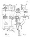
Theimage acquiring system 115 is generally operable to generate a two-dimensional, three-dimensional, or four-dimensional image data corresponding to an area of interest of theimaged subject 110. Examples of theimage acquiring system 115 can include, but is not limited to, computed tomography (CT), magnetic resonance imaging (MRI), x-ray or radiation, positron emission tomography (PET), computerized tomosynthesis (CT), ultrasound (US), angiographic, fluoroscopic, and the like or combination thereof. Theimage acquiring system 115 can be operable to generate static images acquired by static imaging detectors (e.g., CT systems, MRI systems, etc.) prior to a medical procedure, or real-time images acquired with real-time imaging detectors (e.g., angioplastic systems, laparoscopic systems, endoscopic systems, etc.) during the medical procedure. Thus, the types of images can be diagnostic or interventional.
An exemplaryimage acquiring system 115 includes a real-time, intracardiac echocardiography (ICE)imaging system 126 that employs ultrasound to acquire image data of the patient's anatomy and to merge acquired image data to generate a three-dimensional model of the patient's anatomy relative to time, generating herein referred to as a four-dimensional model or image. In accordance with another embodiment, theimage acquiring system 115 is operable to fuse or combine acquired image data using above-described ICEimaging system 126 with pre-acquired image data or image models (e.g., two- or three-dimensional reconstructed image models) generated by another type ofsupplemental imaging system 128, examples of which are described above (e.g., CT, MRI, PET, etc.).
The tool orobject 105 can be a surgical tool, navigational tool, a guidewire, a catheter, an endoscopic tool, a laparoscopic tool, ultrasound probe, pointer, aspirator, coil, or the like employed in a medical procedure (e.g., ablation of tissue). Yet, the type oftool 105 can vary.
Referring toFIG. 3 , an embodiment of thetool 105 operable to acquire intracardiac echocardiography (ICE) image data of the imaged subject110 (SeeFIG. 1 ) includes anICE catheter 130. The illustrated embodiment of theICE catheter 130 includes atransducer array 132, amicromotor 134, a drive shaft or othermechanical connection 136 between themicromotor 134 and thetransducer array 132, aninterconnect 138, and acatheter housing 140.
According to the depicted embodiment, themicromotor 134 via thedrive shaft 136 generally rotates thetransducer array 132. The rotational motion of thetransducer array 132, is controlled by amotor control 142 of themicromotor 134. Theinterconnect 138 generally refers to, for example, cables and other connections coupling so as to receive and/or transmit signals between thetransducer array 132 with the ICE imaging system (shown inFIG. 1 )126. An embodiment of theinterconnect 138 is configured to reduce its respective torque load on thetransducer array 132 and themicromotor 134.
Still referring toFIG. 2 , an embodiment of the catheter housing140 generally encloses thetransducer array 132, themicromotor 134, thedrive shaft 136, theinterconnect 138, and themotor control 142. The catheter housing is generally of a material, size, and shape adaptable to internal imaging applications and insertion into regions of interest of theimaged subject 110. At least a portion of the catheter housing140 that intersects the ultrasound imaging volume or scanning direction is comprised of acoustically transparent (e.g., low attenuation and scattering, acoustic impedance near that of the blood and tissue (Z˜1.5M Rayl) material. An embodiment of the space between thetransducer array 132 and thehousing 140 is filled with acoustic coupling fluid (e.g., water) having an acoustic impedance and sound velocity near those of blood and tissue (e.g., Z˜1.5M Rayl, V˜1540 m/sec).
An embodiment of thetransducer array 132 is a 64-element one-dimensional array having 0.110 mm azimuth pitch, 2.5 mm elevation, and 6.5 MHz center frequency. The elements of thetransducer array 132 are electronically phased in order to acquire a sector image parallel to thelongitudinal axis 144 of thecatheter housing 140. In operation, themicromotor 134 mechanically rotates thetransducer array 132 about thelongitudinal axis 144. The rotatingtransducer array 132 captures a plurality of two-dimensional images for transmission to the ICE imaging system126 (shown inFIG. 1 ). TheICE imaging system 126 is generally operable to assemble the sequence or succession of acquired two-dimensional images so as to generally produce or generate a three-dimensional image or reconstructed model of the imaged subject110.
The rate of rotation of thetransducer array 132 about thelongitudinal axis 144 of theICE catheter 130 is generally regulated by themotor control 142 via themicromotor 132. For example, themotor control 142 instructs the micromotor134 to rotate thetransducer array 132 relatively slowly to produce a three-dimensional reconstructed image. In contrast, themotor control 142 instructs the micromotor134 to rotate thetransducer array 132 relatively faster to produce a real-time three-dimensional reconstructed image, referred to as a four-dimensional image correlated to a general instantaneous time. Themotor control 142 is also generally operable to vary the direction of rotation sous to generally create an oscillatory motion of thetransducer array 132. In this manner, the torque load associated with theinterconnect 138 is reduced such that thetransducer array 132 can focus on imaging specific regions within the range of motion about thelongitudinal axis 144.
Referring now toFIGS. 1 and 3 , thetracking system 125 is generally operable to track or detect the position of thetool 105 and theICE catheter 130 relative to the acquired image generated by theimage acquiring system 115. As illustrated inFIG. 3 , an embodiment of thetracking system 125 includes an array or series of sensors or trackingelements FIG. 1 ). Yet, it should be understood that the number of trackingelements tracking elements dynamic references more receivers 190. The number and combination of transmitters and receivers can vary. Either thetransmitters receiver 190 can define the reference of the spatial relation. An embodiment of thereceiver 190 is detachably connected at and moves with a table in support of the imaged subject110.
Referring now toFIGS. 1 ,2 and3, an embodiment of thetool 105 andICE catheter 130 includes atracking element 200 of thetracking system 125 in communication or coupled with thereceiver 190. As shown inFIG. 2 , an embodiment of thetransmitter 200 generally includes a series of coils that define the orientation or alignment of theICE catheter 130 about a rotational axis (generally aligned along the longitudinal axis144) of theICE catheter 130. Referring toFIG. 3 , thetransmitter 200 is located integrally with theICE catheter 130 and is generally operable to generate or transmit amagnetic field 205 to be detected by thereceiver 190 of thetracking system 125. In response to passing through themagnetic field 205, thereceiver 190 generates a signal representative of a spatial relation and orientation relative to thetransmitter 200. Yet, it should be understood that the type or mode of coupling, link or communication (e.g., RF signal, infrared light, magnetic field, etc.) operable to measure the spatial relation varies. The spatial relation and orientation of thetransmitter 200 is mechanically defined and known in relation relative to a feature (e.g., a tip) of theICE catheter 130. Thereby, thetracking system 125 is operable to track the position and orientation of theICE catheter 130 navigating through the imaged subject110. Alternatively, thereceiver 190 can be attached at theICE catheter 130 and in communication to measure a spatial relation withtransmitters ICE catheter 130.
Alternatively, thetransmitters receiver 190 of thetracking system 125 and which defines an orientation of theICE catheter 130. An embodiment of thereceiver 190 includes at least one conductive loop operable to generate an electric signal indicative of spatial relation and orientation relative to the magnetic field generated by thetransmitters
Still referringFIGS. 1 ,2 and3, a controller orworkstation computer 210 is generally connected in communication with the imaging system115 (e.g., theICE imaging system 126 and static imaging system128) and thetracking system 125. An embodiment of thecontroller 210 includes aprocessor 220 in communication with amemory 225. Theprocessor 220 can be arranged independent of or integrated with thememory 225. Theprocessor 220 is generally operable to execute the program instructions representative of acts described herein and stored in thememory 225. Theprocessor 220 can also be capable of receiving input data or information or communicating output data. Examples of theprocessor 220 can include a central processing unit of a desktop computer, a microprocessor, a microcontroller, or programmable logic controller (PLC), or the like or combination thereof.
An embodiment of thememory 225 generally comprises one or more computer-readable mediums such as a hard disk, a floppy disk, CD, CD-ROM, DVD, compact storage medium, flash memory, random access memory, read-only memory, programmable read-only memory, memory stick, or the like or combination thereof. Thememory 225 is operable to store the plurality of program instructions for execution by theprocessor 220. Thememory 225 is also operable to store data generated or received by thecontroller 210.
Thecontroller 210 further includes or is in communication with aninput device 230 andoutput device 240. Theinput device 230 is generally operable to receive and communicate information data from user to thecontroller 210. Theinput device 230 can include a mouse device, pointer, keyboard, touch screen, microphone, or other like device capable of receiving a user directive. Theoutput device 240 is generally operable to illustrate output data for viewing by the user. An embodiment of theoutput device 240 is operable to simultaneously illustrate or fuse static or real-time image data generated by the image acquiring system115 (e.g., theICE imaging system 126 and static imaging system128) with tracking data generated by thetracking system 125. Theoutput device 240 is capable of illustrating two-dimensional, three-dimensional image and/or four-dimensional image data through shading, coloring, and/or the like. Examples of theoutput device 240 include a cathode ray monitor, a liquid crystal display (LCD) monitor, a touch-screen monitor, a plasma monitor, or the like or combination thereof.
Having provided a description of the general construction of thesystem 100, the following is a description of a method300 (seeFIG. 4 ) of operation of thesystem 100 in relation to the imaged subject110. Although an exemplary embodiment of themethod 300 is discussed below, it should be understood that one or more acts or steps comprising themethod 300 can be omitted or added. It should also be understood that one or more of the acts can be performed simultaneously or at least substantially simultaneously, and the sequence of the acts can vary. Furthermore, it is embodied that at least several of the following acts can be represented as a series of modules of computer-readable program instructions to be stored in thememory 225 of thecontroller 210 for execution by theprocessor 220.
Referring now toFIG. 3 and for sake of example, assume that the spatial relation and orientation of the image data acquired by thetransducer array 132 of theICE imaging system 126 is defined by an image coordinatesystem 320 referenced in predetermined spatial relation and orientation relative to the transducer array132 (SeeFIG. 2 ) at theICE catheter 130. The image coordinatesystem 320 generally defines the spatial relation of voxels or pixels of image data relative to one another in the generated image frames or models generated by theICE imaging system 126 in three-dimensions relative to time (i.e., four-dimensional image). Also, for sake of example, assume thetracking system 125 utilizes a tracking coordinatesystem 325 to define tracking spatial relation and orientation and movement of thetracking elements system 325 references the orientation and spatial relation of thetransmitter 200 at theICE catheter 130 relative to the receiver orreference 190 of thetracking system 125. Although these coordinatesystems systems 320 and325 (e.g., polar, etc.) can vary. In addition, the location and orientation of the coordinatesystems transmitter 200 relative to the ultrasonic transducer array132 (SeeFIG. 2 ) is known or preoperatively measured.
An embodiment of theoffline calibrating act 310 includes pre-operatively applying or performing a rigid body transformation algorithm before acquiring images of the imaged subject110 with theICE imaging system 126. Referring now toFIG. 5 , this embodiment of the calibratingact 310 includes acquiring image data of one or moreultrasonic fiducials 330 included in a phantom orultrasonic lucent frame 335, in place of the imaged subject110, with theICE imaging system 126. An embodiment of thefiducials 330 are located at a predetermined spatial relationship and orientation with respect to one another. The arrangement, shape (e.g., cylinder, etc.) and dimensions of thefiducials 330 and theframe 335 can vary. The locations of thefiducials 330 and/orframe 335 are optimized to cover a field ofview 337 of theICE imaging system 126. The material of the ultrasound fiducials330 is compatible to be detected with the ultrasound imaging technology. An embodiment of theframe 335 includes a docking area or station338 configured to receive theICE catheter 130 in a desired position or location and orientation relative to thefiducials 330 andframe 335.
The calibratingact 310 includes aligning acquired image data (e.g., grayscale, contrast, etc. of the pixels or voxels) of thefiducials 330 in theframe 335 as acquired by thetransducer array 132 of theICE catheter 130 and communicated to theICE imaging system 126, with the physical or mechanical dimensions and orientation of theframe 335. The calibratingact 310 further includes measuring the mechanical spatial relation and orientation of thefiducials 330 in theframe 325 relative toultrasonic transducer array 132 of theICE imaging system 126 that acquires the image data of thefiducials 330. The manufacturer may provide predetermined measurements of the parameters that define the mechanical spatial relation and orientation of thefiducials 330 relative to theframe 335. Alternatively, the spatial relation and orientation of thefiducials 330 can be measured optically in accordance to conventional techniques associated with using a dual camera system. The predetermined spatial relationship can be communicated directly or indirectly and stored at thecontroller 210.
Referring toFIG. 3 , with thetracking element 200 at theICE catheter 130, one of theother tracking elements frame 335 and defines a reference or world coordinatesystem 340 that may or may not be the same as coordinatesystems controller 210 calibrates the positions and orientations of the fiducials330 (FIG. 5 ) with the world coordinatesystem 340. Yet another of thetracking elements fiducials 330. Thecontroller 210 measures the spatial relation to calculate the positions and orientation of thetracking elements FIG. 5 ).
Referring toFIG. 5 , the calibrating act or step310 also includes acquiring image data of thefiducials 330 in theframe 335 with thetransducer array 132 of theICE imaging system 126. Thecontroller 210 is operable to detect the location of the pixels or voxels having image data of thefiducials 330 in the generated image frames, and to measure the spatial relation and orientation of the pixels or voxels with image data of thefiducials 330 relative to one another.
Knowing the spatial relation of thetracking elements fiducials 330 as measured by thetracking system 125, and knowing the mechanical spatial relation and orientation of thefiducials 330 relative to theframe 335, thecontroller 210 is operable to automatically register the spatial relation and orientation of thefiducials 330 relative to the tracking coordinatesystem 325, and hence relative to the image coordinatesystem 320.
According to the above-described description, thecontroller 210 is operable to calibrate or to adjust calibration of the image coordinatesystem 320, the tracking coordinatesystem 325 and the world coordinatesystem 340 and registration relative to one another.
In yet another embodiment, the fiducials or markers orlandmarks 330 can be integrated to include an additional tracking elements (transmitters or receivers or combination similar to trackingelements tracking system 125.
At least a portion of the above-describedcalibration act 310 can be represented as program instructions for execution by theprocessor 220 of thecontroller 210. Execution of the program instructions by theprocessor 220 can be triggered or controlled by a graphic user interface at theoutput device 140. The positions and orientations of thetracking elements fiducials 330 can be denoted as T(f1 to wcs), T(f2 relative to wcs) through to T(fn relative to wcs), where T(fn relative to wcs) representative a position of each of the fiducials330 (n) relative to the world coordinatesystem 340 as measured with thetracking system 125. Thecontroller 210 processes the three-dimensional image data acquired with theICE imaging system 126 to calculate the pixel or voxel position of thefiducials 330 illustrated in the three-dimensional data, denoted as T(fn relative ice), representative of the position and orientation of thefiducials 330 relative to the image coordinatesystem 320 that defines the three-dimensional model generated by theICE imaging system 126. The position of thetransmitter 200 andtransducer array 132 at theICE catheter 130, denoted as T(scs relative to wcs). Thecontroller 210 executes the calibration through the following transformation algorithms:
T(firelative towcs)=T(firelative toscs)T(scsrelative towcs), and
T(firelative toscs)=T(firelative towcs)[T(scsrelative towcs)].inv
where (fi) refers to the index (f1, . . . fn) offiducials 330, and T[scs relative to wcs].inv refers to the inverse transformation of T(scs relative to wcs). Thecontroller 210 aligns the image frame generated by theICE imaging system 126 with the position of thetransmitter 200 at theICE catheter 130 andframe 335 through the following transformations:
T(firelative toscs)=T(firelative to ice)T(ice relative toscs), and
T(ice relative toscs)=T(firelative toscs)[T(firelative to ice)].inv
where (fi) refers to (f1, . . . fn) index offiducials 330, and [T(fi relative to ice)].inv denotes the inverse transformation of T(fi relative to ice). The calibration information denoted by T(ice relative to scs) can be stored in the memory or other computer-readible medium of thecontroller 210 or to theICE imaging system 126.
T(firelative towcs)=T(firelative toscs)T(scsrelative towcs), and
T(firelative toscs)=T(firelative towcs)[T(scsrelative towcs)].inv
where (fi) refers to the index (f1, . . . fn) of
T(firelative toscs)=T(firelative to ice)T(ice relative toscs), and
T(ice relative toscs)=T(firelative toscs)[T(firelative to ice)].inv
where (fi) refers to (f1, . . . fn) index of
As thetracking element 200 andtransducer array 132 move with theICE catheter 130 through the imaged subject110, thetracking element 200 is linked in electromagnetic communication so as to allow the tracking system to track a location or movement of thetracking element 200 and attachedtransducer array 132 of theICE catheter 130 relative to theother tracking elements system 325 for communication via a wireless or wired connection to thecontroller 200. Based on the signals from all or some of thetracking elements controller 210 automatically continuously or periodically updates this measured spatial relation to track movement of thetransducer array 132 at theICE catheter 130 relative to the imaged subject110 and acquired data represented in the four-dimensional model generated by theICE imaging system 126.
Thecontroller 210 is operable to track movement of thetool 105 orICE catheter 130 via thetracking system 125 in accordance with known mathematical algorithms programmed as program instructions of a software for execution by theprocessor 220 of thecontroller 200. An exemplary navigation software is INSTATRAK® as manufactured by the GENERAL ELECTRIC® Corporation, and NAVIVISION® as manufactured by SIEMENS® and BRAINLAB®.
Referring back toFIG. 1 , having described calibration of theICE imaging system 126 with thetracking system 125, act350 of themethod 300 and can be further extended to calibrating theICE imaging system 126 andtracking system 125 with other components of thesystem 100, including anablation catheter system 450, an electrophysiological system(s) (e.g., cardiac monitoring system, respiratory monitoring system, etc.)460, and a steering system500 of theICE catheter 130.
An embodiment of theablation system 450 having anablation catheter 452 that is operable to work in combination with theICE imaging system 126 to ablate or end electrical activity of tissue. An embodiment of an electrophysiological system(s)460 is connected in combination with theICE imaging system 126 is to track or monitor the cardiac cycle or respiratory cycle of imaged subject110 correlated to the image data or three-dimensional models acquired or generated by theICE imaging system 126. An embodiment of a catheter steering system500 is generally operable to steer or drive movement and changes in direction of theICE catheter 130 and attachedtransducer array 132 andtransmitter 200 through the imaged subject110. The steering system500 can also be operable to drive rotation of themotor 134 in rotating or moving orientation of thetransducer array 132 about therotational axis 144. This embodiment of extending the calibrating act to a control system500 is generally similar to the calibrating act described above directed to theICE imaging system 126 with thetracking system 125.
For example, assume the spatial movement and orientation as recognized by the steering system500 is defined by a steering coordinate system505 (SeeFIG. 1 ). Referring now toFIG. 6 , the ICE catheter130 (SeeFIGS. 1 ,2 and3) can be placed at a docking station508 (similar to the docking station338 described above) on aframe 510 offiducials 515. Thedocking station 508 includes anadapter 520 configured to receive theICE catheter 130 orother tool 105. Theadapter 520 of thedocking station 508 is configured to rotate, or to allow rotation of theICE catheter 130, about the longitudinal axis144 (SeeFIG. 2 ) with respect to a remainder of theframe 510. A tracking element orsensor tracking system 125 is attached at theframe 510 to define the reference or world coordinatesystem 525, similar to the world coordinatesystem 340 described above. Alignment of theICE imaging system 126 and the steering control system500 includes measuring the angular displacement between the mechanical steering angle (e.g., as referenced by540 inFIG. 2 ) of themotor 140 at theICE catheter 130 or other drive device of theICE catheter 130 relative to the orientation of thetransmitter 200 at theICE catheter 130. It should be understood that the reference (e.g.,longitudinal axis 144, axis of motor, pivot point, etc.) and measured displacement (rotational, linear, pivot angle, etc.) tracked and adjusted by the steering system500 could vary.
An embodiment of thisextended calibrating act 350 can be represented in modules of computer readable programming instructions that can be launched from a graphic user interface at the output device. For example, assume the steering system500 is operable to drive rotation of theICE catheter 130 ormotor 140 ortransducer array 132 attached thereto among a plurality of angular positions herein referred to as T(mcs.a1 relative to wcs), T(mcs.a2 relative to wcs) . . . to T(mcs.an relative to wcs), where a1, . . . an represent an index of angular positions, and “mcs” refers to the mechanical or steering coordinatesystem 505 of the steering system500. The extended calibrating act includes acquiring orientation data or information of theICE catheter 130 at corresponding angular positions denoted as T(scs.a1 relative to wcs), T(scs.a2 relative to wcs) . . . to T(scs.an relative to wcs), where “scs” refers to theframe 510. The displacement or alignment T between theICE imaging system 126 and the steering system500 can be estimated through the following algorithms:
T(mcsrelative toscs)=average[T(mcs.airelative towcs)−T(scs.airelative towcs)],
where i=1, 2, . . . n, and where T(mcs relative to scs) represents the calibration information that is operable to be stored in the memory of thecontroller 210 or other computer readable medium and to communicate to theICE imaging system 126.
T(mcsrelative toscs)=average[T(mcs.airelative towcs)−T(scs.airelative towcs)],
where i=1, 2, . . . n, and where T(mcs relative to scs) represents the calibration information that is operable to be stored in the memory of the
A technical effect of the above-describedsystem 100 andmethod 300 described above is an ability to calibrate a four dimensional,ICE catheter system 126 with atracking system 125 via aframe fiducials controller 210. Thecontroller 210 is operable to perform registration of the coordinatesystems fiducials ICE imaging system 126. Another technical effect of thesystem 100 andmethod 300 described above is an ability to calibrate theICE imaging system 126 andtracking system 115 with the steering system500 of theICE catheter 130 orother tools 105. Thesystem 100 also provides for tracking the position and orientation of thetransducer array 132 at theICE catheter 130, enhances superposition of the image data generated by theICE imaging system 126 with other models generated by other imaging systems128 (e.g., MRI, CT, PET, X-ray, Fluoroscopy, etc.), provides for navigation of theICE catheter 130 orother tools 105 associated with ablation in combination with acquiring imaging data with theICE imaging system 126, and tracking movement and orientation of theICE catheter 130 orother tools 105 and synchronizing their movement with electrophysiological signals (e.g., respiratory cycle, cardiac cycle, etc.) as tracked by the electrophysiological system(s)460.
This written description uses examples to disclose the invention, including the best mode, and also to enable any person skilled in the art to make and use the invention. The patentable scope of the invention is defined by the claims, and may include other examples that occur to those skilled in the art. Such other examples are intended to be within the scope of the claims if they have structural elements that do not differ from the literal language of the claims, or if they include equivalent structural elements with insubstantial differences from the literal languages of the claims.
Claims (19)
1. A system to navigate in an area of interest of an imaged subject in relation to an acquired image of the imaged subject, comprising:
an intracardiac echocardiography (ICE) imaging system having a transducer operable to acquire image data so as to create a four-dimensional image model of the imaged subject, the model defined in spatial relation and orientation relative to an image coordinate system;
a tracking system operable to track movement and orientation of the transducer through the imaged subject relative to a tracking coordinate system; and
a controller electrically connected in communication with the imaging system and the tracking system, the controller having a processor operable to execute a plurality of program instructions stored in a memory, the plurality of program instructions in combination with the processor operable to:
register the image coordinate system with the tracking coordinate system; and
calibrate the image coordinate system and the tracking coordinate system relative to a common reference having a plurality of fiducials of known spatial relation.
2. The system ofclaim 1 , wherein the ICE imaging system includes a steering control system operable to drive movement of a tool relative to steering coordinate system, wherein the controller includes instructions to calibrate the steering coordinate system relative to the image coordinate system and to the tracking coordinate system.
3. The system ofclaim 2 , wherein the common reference includes a docking station to receive the tool at a known location and orientation relative to the plurality of fiducials.
4. The system ofclaim 3 , wherein the docking station includes an adapter configured such the tool can rotate along a longitudinal axis of the tool relative to the frame.
5. The system ofclaim 2 , wherein the tool is part of a transducer operable to acquire the image data of the imaged subject.
6. The system ofclaim 5 , wherein the transducer employs ultrasound to acquire the image data.
7. The system ofclaim 5 , wherein a motor rotates the transducer about a longitudinal axis of the transducer.
8. The system ofclaim 7 , wherein the motor changes a direction of rotation of the transducer about the longitudinal axis.
9. A method of navigating in an area of interest of an imaged subject, the method comprising the acts of:
generating a four-dimensional model of the region of interest of the imaged subject with an intracardiac echocardiography (ICE) imaging system, the four-dimensional model including image data arranged in spatial relation and orientation relative to an image coordinate system and correlated relative to a time of acquisition;
tracking movement and orientation of the transducer traveling through the imaged subject relative to a tracking coordinate system;
registering the image coordinate system relative to the tracking coordinate system; and
calibrating the image coordinate system and the tracking coordinate system relative to a common reference frame comprising a plurality of fiducials of known spatial relation.
10. The method ofclaim 9 , further including the act of:
directing movement of the transducer via a steering control system relative to steering coordinate system; and
calibrating the steering coordinate system relative to the image coordinate system and to the tracking coordinate system.
11. The method ofclaim 9 , wherein the common reference includes a docking station configured to receive the transducer at a known location and orientation relative to the plurality of fiducials.
12. The method ofclaim 11 , further including the act of:
rotating the transducer along a longitudinal axis of the transducer relative to the frame.
13. The method ofclaim 9 , wherein the transducer is part of a tool traveling through the imaged subject.
14. The method ofclaim 9 , wherein the transducer employs ultrasound to acquire the image data, and further including the act of acquiring ultrasound image data of the plurality of fiducials.
15. The method ofclaim 14 , wherein a motor rotates the transducer about a longitudinal axis of the transducer.
16. The method ofclaim 15 , wherein the motor changes a direction of rotation of the transducer about the longitudinal axis.
17. The method ofclaim 9 , further including the act of:
calculating the location and orientation of the plurality of fiducials relative to the common reference frame.
18. A frame to calibrate an imaging system relative to a tracking system, the imaging system including a transducer operable to acquire image data of an imaged subject, comprising:
a plurality of fiducials of known spatial relation relative to one another; and an adapter configured to receive the transducer of the imaging system, the adapter configured to rotate with respect to the frame, wherein the imaging system is an intracardiac echocardiography (ICE) imaging system, the transducer employs ultrasound to acquire image data of the imaged subject, and the plurality of fiducials are compatible to be detected with ultrasound.
19. The frame ofclaim 18 , wherein the adapter is configured such that the transducer rotates about a longitudinal axis of the transducer.
Priority Applications (1)
| Application Number | Priority Date | Filing Date | Title |
|---|---|---|---|
| US11/860,368US8213693B1 (en) | 2007-05-16 | 2007-09-24 | System and method to track and navigate a tool through an imaged subject |
Applications Claiming Priority (2)
| Application Number | Priority Date | Filing Date | Title |
|---|---|---|---|
| US93838507P | 2007-05-16 | 2007-05-16 | |
| US11/860,368US8213693B1 (en) | 2007-05-16 | 2007-09-24 | System and method to track and navigate a tool through an imaged subject |
Publications (1)
| Publication Number | Publication Date |
|---|---|
| US8213693B1true US8213693B1 (en) | 2012-07-03 |
Family
ID=46320221
Family Applications (1)
| Application Number | Title | Priority Date | Filing Date |
|---|---|---|---|
| US11/860,368Active2031-03-27US8213693B1 (en) | 2007-05-16 | 2007-09-24 | System and method to track and navigate a tool through an imaged subject |
Country Status (1)
| Country | Link |
|---|---|
| US (1) | US8213693B1 (en) |
Cited By (69)
| Publication number | Priority date | Publication date | Assignee | Title |
|---|---|---|---|---|
| US20100104162A1 (en)* | 2008-10-23 | 2010-04-29 | Immersion Corporation | Systems And Methods For Ultrasound Simulation Using Depth Peeling |
| US20100168557A1 (en)* | 2008-12-30 | 2010-07-01 | Deno D Curtis | Multi-electrode ablation sensing catheter and system |
| US20110160593A1 (en)* | 2008-12-30 | 2011-06-30 | Deno D Curtis | Intracardiac imaging system utilizing a multipurpose catheter |
| US20140276002A1 (en)* | 2013-03-15 | 2014-09-18 | The Cleveland Clinic Foundation | Method and system to facilitate intraoperative positioning and guidance |
| US8948476B2 (en) | 2010-12-20 | 2015-02-03 | St. Jude Medical, Atrial Fibrillation Division, Inc. | Determination of cardiac geometry responsive to doppler based imaging of blood flow characteristics |
| US9076212B2 (en) | 2006-05-19 | 2015-07-07 | The Queen's Medical Center | Motion tracking system for real time adaptive imaging and spectroscopy |
| US9305365B2 (en) | 2013-01-24 | 2016-04-05 | Kineticor, Inc. | Systems, devices, and methods for tracking moving targets |
| US9510763B2 (en) | 2011-05-03 | 2016-12-06 | Medtronic, Inc. | Assessing intra-cardiac activation patterns and electrical dyssynchrony |
| US9586052B2 (en) | 2014-08-15 | 2017-03-07 | Medtronic, Inc. | Systems and methods for evaluating cardiac therapy |
| US9586050B2 (en) | 2014-08-15 | 2017-03-07 | Medtronic, Inc. | Systems and methods for configuration of atrioventricular interval |
| US9591982B2 (en) | 2014-07-31 | 2017-03-14 | Medtronic, Inc. | Systems and methods for evaluating cardiac therapy |
| US9606209B2 (en) | 2011-08-26 | 2017-03-28 | Kineticor, Inc. | Methods, systems, and devices for intra-scan motion correction |
| US9610118B2 (en) | 2008-12-31 | 2017-04-04 | St. Jude Medical, Atrial Fibrillation Division, Inc. | Method and apparatus for the cancellation of motion artifacts in medical interventional navigation |
| US9717461B2 (en) | 2013-01-24 | 2017-08-01 | Kineticor, Inc. | Systems, devices, and methods for tracking and compensating for patient motion during a medical imaging scan |
| US9734589B2 (en) | 2014-07-23 | 2017-08-15 | Kineticor, Inc. | Systems, devices, and methods for tracking and compensating for patient motion during a medical imaging scan |
| US9764143B2 (en) | 2014-08-15 | 2017-09-19 | Medtronic, Inc. | Systems and methods for configuration of interventricular interval |
| US9776009B2 (en) | 2014-03-20 | 2017-10-03 | Medtronic, Inc. | Non-invasive detection of phrenic nerve stimulation |
| US9782141B2 (en) | 2013-02-01 | 2017-10-10 | Kineticor, Inc. | Motion tracking system for real time adaptive motion compensation in biomedical imaging |
| US9877789B2 (en) | 2013-06-12 | 2018-01-30 | Medtronic, Inc. | Implantable electrode location selection |
| US9924884B2 (en) | 2013-04-30 | 2018-03-27 | Medtronic, Inc. | Systems, methods, and interfaces for identifying effective electrodes |
| US9943247B2 (en) | 2015-07-28 | 2018-04-17 | The University Of Hawai'i | Systems, devices, and methods for detecting false movements for motion correction during a medical imaging scan |
| US9980786B2 (en) | 2016-07-19 | 2018-05-29 | Shifamed Holdings, Llc | Medical devices and methods of use |
| US9986928B2 (en) | 2013-12-09 | 2018-06-05 | Medtronic, Inc. | Noninvasive cardiac therapy evaluation |
| US10004462B2 (en) | 2014-03-24 | 2018-06-26 | Kineticor, Inc. | Systems, methods, and devices for removing prospective motion correction from medical imaging scans |
| US10064567B2 (en) | 2013-04-30 | 2018-09-04 | Medtronic, Inc. | Systems, methods, and interfaces for identifying optimal electrical vectors |
| US10070971B2 (en) | 2016-01-22 | 2018-09-11 | Warsaw Orthopedic, Inc. | Surgical instrument and method |
| US10251555B2 (en) | 2013-06-12 | 2019-04-09 | Medtronic, Inc. | Implantable electrode location selection |
| US10285668B2 (en) | 2014-02-05 | 2019-05-14 | Verathon Inc. | Ultrasonic data collection |
| US10327708B2 (en) | 2013-01-24 | 2019-06-25 | Kineticor, Inc. | Systems, devices, and methods for tracking and compensating for patient motion during a medical imaging scan |
| US10402990B2 (en)* | 2014-03-21 | 2019-09-03 | Koninklijke Philips N.V. | Medical viewing system with a viewing plane determination |
| US10433746B2 (en) | 2017-12-22 | 2019-10-08 | Regents Of The University Of Minnesota | Systems and methods for anterior and posterior electrode signal analysis |
| US10492705B2 (en) | 2017-12-22 | 2019-12-03 | Regents Of The University Of Minnesota | Anterior and posterior electrode signals |
| US10532213B2 (en) | 2017-03-03 | 2020-01-14 | Medtronic, Inc. | Criteria for determination of local tissue latency near pacing electrode |
| US10537306B2 (en) | 2017-03-30 | 2020-01-21 | Shifamed Holdings, Llc | Medical tool positioning devices, systems, and methods of use and manufacture |
| US10617318B2 (en) | 2018-02-27 | 2020-04-14 | Medtronic, Inc. | Mapping electrical activity on a model heart |
| WO2020102389A1 (en) | 2018-11-13 | 2020-05-22 | Shifamed Holdings, Llc | Medical tool positioning devices, systems, and methods of use and manufacture |
| US10668290B2 (en) | 2018-03-01 | 2020-06-02 | Medtronic, Inc. | Delivery of pacing therapy by a cardiac pacing device |
| US10716515B2 (en) | 2015-11-23 | 2020-07-21 | Kineticor, Inc. | Systems, devices, and methods for tracking and compensating for patient motion during a medical imaging scan |
| US10773085B2 (en) | 2017-03-15 | 2020-09-15 | Medtronic, Inc. | QRS offset and onset determination |
| US10780279B2 (en) | 2016-02-26 | 2020-09-22 | Medtronic, Inc. | Methods and systems of optimizing right ventricular only pacing for patients with respect to an atrial event and left ventricular event |
| US10780281B2 (en) | 2018-03-23 | 2020-09-22 | Medtronic, Inc. | Evaluation of ventricle from atrium pacing therapy |
| US10786167B2 (en) | 2017-12-22 | 2020-09-29 | Medtronic, Inc. | Ectopic beat-compensated electrical heterogeneity information |
| US10799703B2 (en) | 2017-12-22 | 2020-10-13 | Medtronic, Inc. | Evaluation of his bundle pacing therapy |
| US10918863B2 (en) | 2017-07-28 | 2021-02-16 | Medtronic, Inc. | Generating activation times |
| US10918870B2 (en) | 2018-03-07 | 2021-02-16 | Medtronic, Inc. | Atrial lead placement for treatment of atrial dyssynchrony |
| US10940321B2 (en) | 2018-06-01 | 2021-03-09 | Medtronic, Inc. | Systems, methods, and interfaces for use in cardiac evaluation |
| US10987691B2 (en)* | 2018-01-03 | 2021-04-27 | Ttns Inc. | Method and apparatus for controlling pattern-width of coating liquid dispensed from a nozzle |
| US11132801B2 (en) | 2018-02-02 | 2021-09-28 | Centerline Biomedical, Inc. | Segmentation of three-dimensional images containing anatomic structures |
| US11150776B2 (en) | 2018-02-02 | 2021-10-19 | Centerline Biomedical, Inc. | Graphical user interface for marking anatomic structures |
| US11219769B2 (en) | 2016-02-26 | 2022-01-11 | Medtronic, Inc. | Noninvasive methods and systems of determining the extent of tissue capture from cardiac pacing |
| US11224484B2 (en) | 2018-01-12 | 2022-01-18 | Globus Medical Inc. | Surgical sensor anchor system |
| US11241751B2 (en) | 2016-06-30 | 2022-02-08 | General Electric Company | Drilling tool for use in machining a conductive work piece |
| US11253178B2 (en) | 2015-01-29 | 2022-02-22 | Medtronic, Inc. | Noninvasive assessment of cardiac resynchronization therapy |
| US11285312B2 (en) | 2018-03-29 | 2022-03-29 | Medtronic, Inc. | Left ventricular assist device adjustment and evaluation |
| US11304641B2 (en) | 2018-06-01 | 2022-04-19 | Medtronic, Inc. | Systems, methods, and interfaces for use in cardiac evaluation |
| US11393110B2 (en) | 2019-04-04 | 2022-07-19 | Centerline Biomedical, Inc. | Spatial registration of tracking system with an image using two-dimensional image projections |
| US11419539B2 (en) | 2017-12-22 | 2022-08-23 | Regents Of The University Of Minnesota | QRS onset and offset times and cycle selection using anterior and posterior electrode signals |
| US11471678B2 (en) | 2017-07-28 | 2022-10-18 | Medtronic, Inc. | Cardiac cycle selection |
| US11497889B2 (en) | 2018-08-23 | 2022-11-15 | Nuvera Medical, Inc. | Medical tool positioning devices, systems, and methods of use and manufacture |
| US11497431B2 (en) | 2019-10-09 | 2022-11-15 | Medtronic, Inc. | Systems and methods for configuring cardiac therapy |
| US11538574B2 (en) | 2019-04-04 | 2022-12-27 | Centerline Biomedical, Inc. | Registration of spatial tracking system with augmented reality display |
| US11547858B2 (en) | 2019-03-29 | 2023-01-10 | Medtronic, Inc. | Systems, methods, and devices for adaptive cardiac therapy |
| US11642533B2 (en) | 2019-11-04 | 2023-05-09 | Medtronic, Inc. | Systems and methods for evaluating cardiac therapy |
| US11697025B2 (en) | 2019-03-29 | 2023-07-11 | Medtronic, Inc. | Cardiac conduction system capture |
| US11813464B2 (en) | 2020-07-31 | 2023-11-14 | Medtronic, Inc. | Cardiac conduction system evaluation |
| US12023503B2 (en) | 2020-07-30 | 2024-07-02 | Medtronic, Inc. | ECG belt systems to interoperate with IMDs |
| US12201843B2 (en) | 2019-10-09 | 2025-01-21 | Medtronic, Inc. | Synchronizing external electrical activity |
| US12280260B2 (en) | 2020-12-02 | 2025-04-22 | Medtronic, Inc. | Evaluation and adjustment of left bundle branch (LBB) pacing therapy |
| US12383183B2 (en) | 2020-01-30 | 2025-08-12 | Medtronic, Inc. | Disturbance detection and removal in cardiac signals |
Citations (47)
| Publication number | Priority date | Publication date | Assignee | Title |
|---|---|---|---|---|
| US4706681A (en) | 1984-07-26 | 1987-11-17 | Telectronics N.V. | Cardiac ultrasonically marked leads and method for used same |
| US4821731A (en) | 1986-04-25 | 1989-04-18 | Intra-Sonix, Inc. | Acoustic image system and method |
| US4896673A (en) | 1988-07-15 | 1990-01-30 | Medstone International, Inc. | Method and apparatus for stone localization using ultrasound imaging |
| US5135001A (en) | 1990-12-05 | 1992-08-04 | C. R. Bard, Inc. | Ultrasound sheath for medical diagnostic instruments |
| US5840025A (en)* | 1993-07-20 | 1998-11-24 | Biosense, Inc. | Apparatus and method for treating cardiac arrhythmias |
| US5928248A (en)* | 1997-02-14 | 1999-07-27 | Biosense, Inc. | Guided deployment of stents |
| US5938602A (en) | 1996-06-11 | 1999-08-17 | Roke Manor Research Limited | Catheter tracking system and method |
| US20020005719A1 (en) | 1998-08-02 | 2002-01-17 | Super Dimension Ltd . | Intrabody navigation and imaging system for medical applications |
| US20020026118A1 (en) | 2000-08-18 | 2002-02-28 | Assaf Govari | Three-dimensional reconstruction using ultrasound |
| US6368285B1 (en)* | 1999-09-21 | 2002-04-09 | Biosense, Inc. | Method and apparatus for mapping a chamber of a heart |
| US6490474B1 (en) | 1997-08-01 | 2002-12-03 | Cardiac Pathways Corporation | System and method for electrode localization using ultrasound |
| US20030013958A1 (en) | 2001-07-10 | 2003-01-16 | Assaf Govari | Location sensing with real-time ultrasound imaging |
| US6514249B1 (en) | 1997-07-08 | 2003-02-04 | Atrionix, Inc. | Positioning system and method for orienting an ablation element within a pulmonary vein ostium |
| US20030074011A1 (en) | 1998-09-24 | 2003-04-17 | Super Dimension Ltd. | System and method of recording and displaying in context of an image a location of at least one point-of-interest in a body during an intra-body medical procedure |
| US6618612B1 (en)* | 1996-02-15 | 2003-09-09 | Biosense, Inc. | Independently positionable transducers for location system |
| US20030208102A1 (en) | 2002-05-03 | 2003-11-06 | Super Dimension Ltd. | Multiple-electrode catheter assembly and method of operating such a catheter assembly |
| US6669635B2 (en) | 1999-10-28 | 2003-12-30 | Surgical Navigation Technologies, Inc. | Navigation information overlay onto ultrasound imagery |
| US6719700B1 (en)* | 2002-12-13 | 2004-04-13 | Scimed Life Systems, Inc. | Ultrasound ranging for localization of imaging transducer |
| US20040102769A1 (en) | 2002-11-26 | 2004-05-27 | Yitzhack Schwartz | Ultrasound pulmonary vein isolation |
| US20040138548A1 (en) | 2003-01-13 | 2004-07-15 | Mediguide Ltd. | Method and system for registering a medical situation associated with a first coordinate system, in second coordinate system using an MPS system |
| US6776402B2 (en) | 2000-11-20 | 2004-08-17 | Honda Giken Kogyo Kabushiki Kaisha | Liquid-encapsulated damper mount and hydraulic damper mounting structure in suspension of automobile |
| US20040162550A1 (en) | 2003-02-19 | 2004-08-19 | Assaf Govari | Externally-applied high intensity focused ultrasound (HIFU) for pulmonary vein isolation |
| US20040162507A1 (en) | 2003-02-19 | 2004-08-19 | Assaf Govari | Externally-applied high intensity focused ultrasound (HIFU) for therapeutic treatment |
| US20040254458A1 (en) | 2003-05-29 | 2004-12-16 | Assaf Govari | Ultrasound catheter calibration system |
| US6892091B1 (en)* | 2000-02-18 | 2005-05-10 | Biosense, Inc. | Catheter, method and apparatus for generating an electrical map of a chamber of the heart |
| US20050197557A1 (en) | 2004-03-08 | 2005-09-08 | Mediguide Ltd. | Automatic guidewire maneuvering system and method |
| US20060241445A1 (en) | 2005-04-26 | 2006-10-26 | Altmann Andres C | Three-dimensional cardial imaging using ultrasound contour reconstruction |
| US20060253024A1 (en) | 2005-04-26 | 2006-11-09 | Altmann Andres C | Software product for three-dimensional cardiac imaging using ultrasound contour reconstruction |
| US20060253029A1 (en) | 2005-04-26 | 2006-11-09 | Altmann Andres C | Display of two-dimensional ultrasound fan |
| US20060253031A1 (en) | 2005-04-26 | 2006-11-09 | Altmann Andres C | Registration of ultrasound data with pre-acquired image |
| US20060253032A1 (en) | 2005-04-26 | 2006-11-09 | Altmann Andres C | Display of catheter tip with beam direction for ultrasound system |
| US20070167821A1 (en) | 2005-11-30 | 2007-07-19 | Warren Lee | Rotatable transducer array for volumetric ultrasound |
| US7327872B2 (en)* | 2004-10-13 | 2008-02-05 | General Electric Company | Method and system for registering 3D models of anatomical regions with projection images of the same |
| US7366562B2 (en)* | 2003-10-17 | 2008-04-29 | Medtronic Navigation, Inc. | Method and apparatus for surgical navigation |
| US7499743B2 (en)* | 2002-03-15 | 2009-03-03 | General Electric Company | Method and system for registration of 3D images within an interventional system |
| US7505810B2 (en)* | 2006-06-13 | 2009-03-17 | Rhythmia Medical, Inc. | Non-contact cardiac mapping, including preprocessing |
| US7515954B2 (en)* | 2006-06-13 | 2009-04-07 | Rhythmia Medical, Inc. | Non-contact cardiac mapping, including moving catheter and multi-beat integration |
| US7599730B2 (en)* | 2002-11-19 | 2009-10-06 | Medtronic Navigation, Inc. | Navigation system for cardiac therapies |
| US7660623B2 (en)* | 2003-01-30 | 2010-02-09 | Medtronic Navigation, Inc. | Six degree of freedom alignment display for medical procedures |
| US7697972B2 (en)* | 2002-11-19 | 2010-04-13 | Medtronic Navigation, Inc. | Navigation system for cardiac therapies |
| US7713210B2 (en)* | 2004-11-23 | 2010-05-11 | St. Jude Medical, Atrial Fibrillation Division, Inc. | Method and apparatus for localizing an ultrasound catheter |
| US7729752B2 (en)* | 2006-06-13 | 2010-06-01 | Rhythmia Medical, Inc. | Non-contact cardiac mapping, including resolution map |
| US7751868B2 (en)* | 2004-11-12 | 2010-07-06 | Philips Electronics Ltd | Integrated skin-mounted multifunction device for use in image-guided surgery |
| US7787951B1 (en)* | 2003-12-24 | 2010-08-31 | Pacesetter, Inc. | System and method for determining optimal stimulation sites based on ECG information |
| US7912258B2 (en)* | 2005-09-27 | 2011-03-22 | Vanderbilt University | Method and apparatus for standardizing ultrasonography training using image to physical space registration of tomographic volumes from tracked ultrasound |
| US7918793B2 (en)* | 2005-10-28 | 2011-04-05 | Biosense Webster, Inc. | Synchronization of ultrasound imaging data with electrical mapping |
| US20110190629A1 (en)* | 2008-09-30 | 2011-08-04 | Mediri Gmbh | 3D Motion Detection and Correction By Object Tracking in Ultrasound Images |
- 2007
- 2007-09-24USUS11/860,368patent/US8213693B1/enactiveActive
Patent Citations (57)
| Publication number | Priority date | Publication date | Assignee | Title |
|---|---|---|---|---|
| US4706681A (en) | 1984-07-26 | 1987-11-17 | Telectronics N.V. | Cardiac ultrasonically marked leads and method for used same |
| US4821731A (en) | 1986-04-25 | 1989-04-18 | Intra-Sonix, Inc. | Acoustic image system and method |
| US4896673A (en) | 1988-07-15 | 1990-01-30 | Medstone International, Inc. | Method and apparatus for stone localization using ultrasound imaging |
| US5135001A (en) | 1990-12-05 | 1992-08-04 | C. R. Bard, Inc. | Ultrasound sheath for medical diagnostic instruments |
| US5840025A (en)* | 1993-07-20 | 1998-11-24 | Biosense, Inc. | Apparatus and method for treating cardiac arrhythmias |
| US6618612B1 (en)* | 1996-02-15 | 2003-09-09 | Biosense, Inc. | Independently positionable transducers for location system |
| US5938602A (en) | 1996-06-11 | 1999-08-17 | Roke Manor Research Limited | Catheter tracking system and method |
| US5928248A (en)* | 1997-02-14 | 1999-07-27 | Biosense, Inc. | Guided deployment of stents |
| US6514249B1 (en) | 1997-07-08 | 2003-02-04 | Atrionix, Inc. | Positioning system and method for orienting an ablation element within a pulmonary vein ostium |
| US6490474B1 (en) | 1997-08-01 | 2002-12-03 | Cardiac Pathways Corporation | System and method for electrode localization using ultrasound |
| US20020005719A1 (en) | 1998-08-02 | 2002-01-17 | Super Dimension Ltd . | Intrabody navigation and imaging system for medical applications |
| US20020042571A1 (en) | 1998-08-02 | 2002-04-11 | Super Dimension Ltd. | Navigable catheter |
| US20030074011A1 (en) | 1998-09-24 | 2003-04-17 | Super Dimension Ltd. | System and method of recording and displaying in context of an image a location of at least one point-of-interest in a body during an intra-body medical procedure |
| US6368285B1 (en)* | 1999-09-21 | 2002-04-09 | Biosense, Inc. | Method and apparatus for mapping a chamber of a heart |
| US6669635B2 (en) | 1999-10-28 | 2003-12-30 | Surgical Navigation Technologies, Inc. | Navigation information overlay onto ultrasound imagery |
| US6892091B1 (en)* | 2000-02-18 | 2005-05-10 | Biosense, Inc. | Catheter, method and apparatus for generating an electrical map of a chamber of the heart |
| US20020026118A1 (en) | 2000-08-18 | 2002-02-28 | Assaf Govari | Three-dimensional reconstruction using ultrasound |
| US6716166B2 (en) | 2000-08-18 | 2004-04-06 | Biosense, Inc. | Three-dimensional reconstruction using ultrasound |
| US6776402B2 (en) | 2000-11-20 | 2004-08-17 | Honda Giken Kogyo Kabushiki Kaisha | Liquid-encapsulated damper mount and hydraulic damper mounting structure in suspension of automobile |
| US20030013958A1 (en) | 2001-07-10 | 2003-01-16 | Assaf Govari | Location sensing with real-time ultrasound imaging |
| US6773402B2 (en) | 2001-07-10 | 2004-08-10 | Biosense, Inc. | Location sensing with real-time ultrasound imaging |
| US7499743B2 (en)* | 2002-03-15 | 2009-03-03 | General Electric Company | Method and system for registration of 3D images within an interventional system |
| US20030208102A1 (en) | 2002-05-03 | 2003-11-06 | Super Dimension Ltd. | Multiple-electrode catheter assembly and method of operating such a catheter assembly |
| US7697972B2 (en)* | 2002-11-19 | 2010-04-13 | Medtronic Navigation, Inc. | Navigation system for cardiac therapies |
| US7599730B2 (en)* | 2002-11-19 | 2009-10-06 | Medtronic Navigation, Inc. | Navigation system for cardiac therapies |
| US20040102769A1 (en) | 2002-11-26 | 2004-05-27 | Yitzhack Schwartz | Ultrasound pulmonary vein isolation |
| US7156816B2 (en) | 2002-11-26 | 2007-01-02 | Biosense, Inc. | Ultrasound pulmonary vein isolation |
| US6719700B1 (en)* | 2002-12-13 | 2004-04-13 | Scimed Life Systems, Inc. | Ultrasound ranging for localization of imaging transducer |
| US20040138548A1 (en) | 2003-01-13 | 2004-07-15 | Mediguide Ltd. | Method and system for registering a medical situation associated with a first coordinate system, in second coordinate system using an MPS system |
| US7660623B2 (en)* | 2003-01-30 | 2010-02-09 | Medtronic Navigation, Inc. | Six degree of freedom alignment display for medical procedures |
| US20040162550A1 (en) | 2003-02-19 | 2004-08-19 | Assaf Govari | Externally-applied high intensity focused ultrasound (HIFU) for pulmonary vein isolation |
| US20040162507A1 (en) | 2003-02-19 | 2004-08-19 | Assaf Govari | Externally-applied high intensity focused ultrasound (HIFU) for therapeutic treatment |
| US7090639B2 (en) | 2003-05-29 | 2006-08-15 | Biosense, Inc. | Ultrasound catheter calibration system |
| US20040254458A1 (en) | 2003-05-29 | 2004-12-16 | Assaf Govari | Ultrasound catheter calibration system |
| US7366562B2 (en)* | 2003-10-17 | 2008-04-29 | Medtronic Navigation, Inc. | Method and apparatus for surgical navigation |
| US7787951B1 (en)* | 2003-12-24 | 2010-08-31 | Pacesetter, Inc. | System and method for determining optimal stimulation sites based on ECG information |
| US20050197557A1 (en) | 2004-03-08 | 2005-09-08 | Mediguide Ltd. | Automatic guidewire maneuvering system and method |
| US7327872B2 (en)* | 2004-10-13 | 2008-02-05 | General Electric Company | Method and system for registering 3D models of anatomical regions with projection images of the same |
| US7751868B2 (en)* | 2004-11-12 | 2010-07-06 | Philips Electronics Ltd | Integrated skin-mounted multifunction device for use in image-guided surgery |
| US7713210B2 (en)* | 2004-11-23 | 2010-05-11 | St. Jude Medical, Atrial Fibrillation Division, Inc. | Method and apparatus for localizing an ultrasound catheter |
| US20060253029A1 (en) | 2005-04-26 | 2006-11-09 | Altmann Andres C | Display of two-dimensional ultrasound fan |
| US20060253031A1 (en) | 2005-04-26 | 2006-11-09 | Altmann Andres C | Registration of ultrasound data with pre-acquired image |
| US20060253024A1 (en) | 2005-04-26 | 2006-11-09 | Altmann Andres C | Software product for three-dimensional cardiac imaging using ultrasound contour reconstruction |
| US20060253032A1 (en) | 2005-04-26 | 2006-11-09 | Altmann Andres C | Display of catheter tip with beam direction for ultrasound system |
| US20060241445A1 (en) | 2005-04-26 | 2006-10-26 | Altmann Andres C | Three-dimensional cardial imaging using ultrasound contour reconstruction |
| US20060253030A1 (en) | 2005-04-26 | 2006-11-09 | Altmann Andres C | Registration of electro-anatomical map with pre-acquired image using ultrasound |
| US7912258B2 (en)* | 2005-09-27 | 2011-03-22 | Vanderbilt University | Method and apparatus for standardizing ultrasonography training using image to physical space registration of tomographic volumes from tracked ultrasound |
| US7918793B2 (en)* | 2005-10-28 | 2011-04-05 | Biosense Webster, Inc. | Synchronization of ultrasound imaging data with electrical mapping |
| US20070167821A1 (en) | 2005-11-30 | 2007-07-19 | Warren Lee | Rotatable transducer array for volumetric ultrasound |
| US7729752B2 (en)* | 2006-06-13 | 2010-06-01 | Rhythmia Medical, Inc. | Non-contact cardiac mapping, including resolution map |
| US7505810B2 (en)* | 2006-06-13 | 2009-03-17 | Rhythmia Medical, Inc. | Non-contact cardiac mapping, including preprocessing |
| US7515954B2 (en)* | 2006-06-13 | 2009-04-07 | Rhythmia Medical, Inc. | Non-contact cardiac mapping, including moving catheter and multi-beat integration |
| US7930018B2 (en)* | 2006-06-13 | 2011-04-19 | Rhythmia Medical, Inc. | Cardiac mapping, including moving catheter and multi-beat integration |
| US7937136B2 (en)* | 2006-06-13 | 2011-05-03 | Rhythmia Medical, Inc. | Cardiac mapping, including resolution map |
| US7953475B2 (en)* | 2006-06-13 | 2011-05-31 | Rhythmia Medical, Inc. | Preprocessing for cardiac mapping |
| US7957791B2 (en)* | 2006-06-13 | 2011-06-07 | Rhythmin Medical, Inc. | Multi-beat integration for cardiac mapping |
| US20110190629A1 (en)* | 2008-09-30 | 2011-08-04 | Mediri Gmbh | 3D Motion Detection and Correction By Object Tracking in Ultrasound Images |
Non-Patent Citations (7)
| Title |
|---|
| "Catheter Ablation", Cleveland Clinic-Heart & Vascular Institute, http:/www.clevelandclinic.org/heartcenter/pub/guide/tests/procedures/ablation.htm, Apr. 2005. |
| Huang, X. et al, "Dynamic 3D Ultrasound and MR Image Registration of the Beating Heart", MICAI, LNCS 3750, pp. 171-178, 2005. |
| Kanckstedt, C. et al, "Semi-automated 3-dimensional intracardiac echocardiography: development and initial clinical experience of a new system to guide ablation procedures", Heart Rhythm, 3 (12), pp. 1453-1459, 2006. |
| Leotta, D. F. et al, "Three-Dimensional Ultrasound Imaging Using Multiple Magnetic Tracking Systems and Miniature Magnetic Sensors", IEEE on Ultrasonics Symposium, pp. 1415-1418, 1995. |
| Martin, R. et al, "A Miniature Position and Orientation Locator for Three Dimensional Echocardiography", IEEE Proceedings on Computer in Cardiology, pp. 25-28, 1993. |
| Pagoulatos, N. et al, "Interactive 3-D Registration of Ultrasound and Magnetic Resonance Images Based on a Magnetic Position Sensor", IEE on Info. Tech. In Biomedicine, vol. 3, No. 4, 1999. |
| Proulx, T.L. et al, "Advances in Catheter-Based Ultrasound Imaging", IEEE International Ultrasonics Symposium Proceedings, 2005. |
Cited By (100)
| Publication number | Priority date | Publication date | Assignee | Title |
|---|---|---|---|---|
| US9867549B2 (en) | 2006-05-19 | 2018-01-16 | The Queen's Medical Center | Motion tracking system for real time adaptive imaging and spectroscopy |
| US9076212B2 (en) | 2006-05-19 | 2015-07-07 | The Queen's Medical Center | Motion tracking system for real time adaptive imaging and spectroscopy |
| US9138175B2 (en) | 2006-05-19 | 2015-09-22 | The Queen's Medical Center | Motion tracking system for real time adaptive imaging and spectroscopy |
| US10869611B2 (en) | 2006-05-19 | 2020-12-22 | The Queen's Medical Center | Motion tracking system for real time adaptive imaging and spectroscopy |
| US20100104162A1 (en)* | 2008-10-23 | 2010-04-29 | Immersion Corporation | Systems And Methods For Ultrasound Simulation Using Depth Peeling |
| US8428326B2 (en)* | 2008-10-23 | 2013-04-23 | Immersion Corporation | Systems and methods for ultrasound simulation using depth peeling |
| US20100168557A1 (en)* | 2008-12-30 | 2010-07-01 | Deno D Curtis | Multi-electrode ablation sensing catheter and system |
| US20110160593A1 (en)* | 2008-12-30 | 2011-06-30 | Deno D Curtis | Intracardiac imaging system utilizing a multipurpose catheter |
| US10206652B2 (en) | 2008-12-30 | 2019-02-19 | St. Jude Medical, Atrial Fibrillation Division, Inc. | Intracardiac imaging system utilizing a multipurpose catheter |
| US8900150B2 (en)* | 2008-12-30 | 2014-12-02 | St. Jude Medical, Atrial Fibrillation Division, Inc. | Intracardiac imaging system utilizing a multipurpose catheter |
| US9610118B2 (en) | 2008-12-31 | 2017-04-04 | St. Jude Medical, Atrial Fibrillation Division, Inc. | Method and apparatus for the cancellation of motion artifacts in medical interventional navigation |
| US8948476B2 (en) | 2010-12-20 | 2015-02-03 | St. Jude Medical, Atrial Fibrillation Division, Inc. | Determination of cardiac geometry responsive to doppler based imaging of blood flow characteristics |
| US11027135B2 (en) | 2011-05-03 | 2021-06-08 | Medtronic, Inc. | Assessing intra-cardiac activation patterns |
| US9510763B2 (en) | 2011-05-03 | 2016-12-06 | Medtronic, Inc. | Assessing intra-cardiac activation patterns and electrical dyssynchrony |
| US9974457B2 (en) | 2011-05-03 | 2018-05-22 | Medtronic, Inc. | Assessing intra-cardiac activation patterns |
| US9962097B2 (en) | 2011-05-03 | 2018-05-08 | Medtronic, Inc. | Assessing intra-cardiac activation patterns and electrical dyssynchrony |
| US12133984B2 (en) | 2011-05-03 | 2024-11-05 | Medtronic, Inc. | Assessing intra-cardiac activation patterns |
| US10663553B2 (en) | 2011-08-26 | 2020-05-26 | Kineticor, Inc. | Methods, systems, and devices for intra-scan motion correction |
| US9606209B2 (en) | 2011-08-26 | 2017-03-28 | Kineticor, Inc. | Methods, systems, and devices for intra-scan motion correction |
| US9607377B2 (en) | 2013-01-24 | 2017-03-28 | Kineticor, Inc. | Systems, devices, and methods for tracking moving targets |
| US9717461B2 (en) | 2013-01-24 | 2017-08-01 | Kineticor, Inc. | Systems, devices, and methods for tracking and compensating for patient motion during a medical imaging scan |
| US9305365B2 (en) | 2013-01-24 | 2016-04-05 | Kineticor, Inc. | Systems, devices, and methods for tracking moving targets |
| US10339654B2 (en) | 2013-01-24 | 2019-07-02 | Kineticor, Inc. | Systems, devices, and methods for tracking moving targets |
| US9779502B1 (en) | 2013-01-24 | 2017-10-03 | Kineticor, Inc. | Systems, devices, and methods for tracking moving targets |
| US10327708B2 (en) | 2013-01-24 | 2019-06-25 | Kineticor, Inc. | Systems, devices, and methods for tracking and compensating for patient motion during a medical imaging scan |
| US9782141B2 (en) | 2013-02-01 | 2017-10-10 | Kineticor, Inc. | Motion tracking system for real time adaptive motion compensation in biomedical imaging |
| US10653381B2 (en) | 2013-02-01 | 2020-05-19 | Kineticor, Inc. | Motion tracking system for real time adaptive motion compensation in biomedical imaging |
| US20140276002A1 (en)* | 2013-03-15 | 2014-09-18 | The Cleveland Clinic Foundation | Method and system to facilitate intraoperative positioning and guidance |
| US10799145B2 (en)* | 2013-03-15 | 2020-10-13 | The Cleveland Clinic Foundation | Method and system to facilitate intraoperative positioning and guidance |
| US12048523B2 (en) | 2013-03-15 | 2024-07-30 | The Cleveland Clinic Foundation | Method and system to facilitate intraoperative positioning and guidance |
| US10064567B2 (en) | 2013-04-30 | 2018-09-04 | Medtronic, Inc. | Systems, methods, and interfaces for identifying optimal electrical vectors |
| US9931048B2 (en) | 2013-04-30 | 2018-04-03 | Medtronic, Inc. | Systems, methods, and interfaces for identifying effective electrodes |
| US9924884B2 (en) | 2013-04-30 | 2018-03-27 | Medtronic, Inc. | Systems, methods, and interfaces for identifying effective electrodes |
| US11648406B2 (en) | 2013-04-30 | 2023-05-16 | Medtronic, Inc. | Systems, methods, and interfaces for identifying effective electrodes |
| US10251555B2 (en) | 2013-06-12 | 2019-04-09 | Medtronic, Inc. | Implantable electrode location selection |
| US9877789B2 (en) | 2013-06-12 | 2018-01-30 | Medtronic, Inc. | Implantable electrode location selection |
| US11456062B2 (en) | 2013-12-09 | 2022-09-27 | Medtronic, Inc. | Noninvasive cardiac therapy evaluation |
| US9993172B2 (en) | 2013-12-09 | 2018-06-12 | Medtronic, Inc. | Noninvasive cardiac therapy evaluation |
| US10206601B2 (en) | 2013-12-09 | 2019-02-19 | Medtronic, Inc. | Noninvasive cardiac therapy evaluation |
| US9986928B2 (en) | 2013-12-09 | 2018-06-05 | Medtronic, Inc. | Noninvasive cardiac therapy evaluation |
| US10285668B2 (en) | 2014-02-05 | 2019-05-14 | Verathon Inc. | Ultrasonic data collection |
| US9776009B2 (en) | 2014-03-20 | 2017-10-03 | Medtronic, Inc. | Non-invasive detection of phrenic nerve stimulation |
| US10402990B2 (en)* | 2014-03-21 | 2019-09-03 | Koninklijke Philips N.V. | Medical viewing system with a viewing plane determination |
| US10004462B2 (en) | 2014-03-24 | 2018-06-26 | Kineticor, Inc. | Systems, methods, and devices for removing prospective motion correction from medical imaging scans |
| US10438349B2 (en) | 2014-07-23 | 2019-10-08 | Kineticor, Inc. | Systems, devices, and methods for tracking and compensating for patient motion during a medical imaging scan |
| US11100636B2 (en) | 2014-07-23 | 2021-08-24 | Kineticor, Inc. | Systems, devices, and methods for tracking and compensating for patient motion during a medical imaging scan |
| US9734589B2 (en) | 2014-07-23 | 2017-08-15 | Kineticor, Inc. | Systems, devices, and methods for tracking and compensating for patient motion during a medical imaging scan |
| US9591982B2 (en) | 2014-07-31 | 2017-03-14 | Medtronic, Inc. | Systems and methods for evaluating cardiac therapy |
| US9764143B2 (en) | 2014-08-15 | 2017-09-19 | Medtronic, Inc. | Systems and methods for configuration of interventricular interval |
| US9586050B2 (en) | 2014-08-15 | 2017-03-07 | Medtronic, Inc. | Systems and methods for configuration of atrioventricular interval |
| US9586052B2 (en) | 2014-08-15 | 2017-03-07 | Medtronic, Inc. | Systems and methods for evaluating cardiac therapy |
| US11253178B2 (en) | 2015-01-29 | 2022-02-22 | Medtronic, Inc. | Noninvasive assessment of cardiac resynchronization therapy |
| US10660541B2 (en) | 2015-07-28 | 2020-05-26 | The University Of Hawai'i | Systems, devices, and methods for detecting false movements for motion correction during a medical imaging scan |
| US9943247B2 (en) | 2015-07-28 | 2018-04-17 | The University Of Hawai'i | Systems, devices, and methods for detecting false movements for motion correction during a medical imaging scan |
| US10716515B2 (en) | 2015-11-23 | 2020-07-21 | Kineticor, Inc. | Systems, devices, and methods for tracking and compensating for patient motion during a medical imaging scan |
| US10070971B2 (en) | 2016-01-22 | 2018-09-11 | Warsaw Orthopedic, Inc. | Surgical instrument and method |
| US10780279B2 (en) | 2016-02-26 | 2020-09-22 | Medtronic, Inc. | Methods and systems of optimizing right ventricular only pacing for patients with respect to an atrial event and left ventricular event |
| US11219769B2 (en) | 2016-02-26 | 2022-01-11 | Medtronic, Inc. | Noninvasive methods and systems of determining the extent of tissue capture from cardiac pacing |
| US11241751B2 (en) | 2016-06-30 | 2022-02-08 | General Electric Company | Drilling tool for use in machining a conductive work piece |
| US9980786B2 (en) | 2016-07-19 | 2018-05-29 | Shifamed Holdings, Llc | Medical devices and methods of use |
| US10532213B2 (en) | 2017-03-03 | 2020-01-14 | Medtronic, Inc. | Criteria for determination of local tissue latency near pacing electrode |
| US10773085B2 (en) | 2017-03-15 | 2020-09-15 | Medtronic, Inc. | QRS offset and onset determination |
| US12268554B2 (en) | 2017-03-30 | 2025-04-08 | Nuvera Medical, Inc. | Medical tool positioning devices, systems, and methods of use and manufacture |
| US11617564B2 (en) | 2017-03-30 | 2023-04-04 | Nuvera Medical, Inc. | Medical tool positioning devices, systems, and methods of use and manufacture |
| US10537306B2 (en) | 2017-03-30 | 2020-01-21 | Shifamed Holdings, Llc | Medical tool positioning devices, systems, and methods of use and manufacture |
| US11931205B2 (en) | 2017-03-30 | 2024-03-19 | Nuvera Medical, Inc. | Medical tool positioning devices, systems, and methods of use and manufacture |
| US11471678B2 (en) | 2017-07-28 | 2022-10-18 | Medtronic, Inc. | Cardiac cycle selection |
| US10918863B2 (en) | 2017-07-28 | 2021-02-16 | Medtronic, Inc. | Generating activation times |
| US10433746B2 (en) | 2017-12-22 | 2019-10-08 | Regents Of The University Of Minnesota | Systems and methods for anterior and posterior electrode signal analysis |
| US10799703B2 (en) | 2017-12-22 | 2020-10-13 | Medtronic, Inc. | Evaluation of his bundle pacing therapy |
| US10492705B2 (en) | 2017-12-22 | 2019-12-03 | Regents Of The University Of Minnesota | Anterior and posterior electrode signals |
| US10786167B2 (en) | 2017-12-22 | 2020-09-29 | Medtronic, Inc. | Ectopic beat-compensated electrical heterogeneity information |
| US11419539B2 (en) | 2017-12-22 | 2022-08-23 | Regents Of The University Of Minnesota | QRS onset and offset times and cycle selection using anterior and posterior electrode signals |
| US10987691B2 (en)* | 2018-01-03 | 2021-04-27 | Ttns Inc. | Method and apparatus for controlling pattern-width of coating liquid dispensed from a nozzle |
| US11224484B2 (en) | 2018-01-12 | 2022-01-18 | Globus Medical Inc. | Surgical sensor anchor system |
| US11132801B2 (en) | 2018-02-02 | 2021-09-28 | Centerline Biomedical, Inc. | Segmentation of three-dimensional images containing anatomic structures |
| US11604556B2 (en) | 2018-02-02 | 2023-03-14 | Centerline Biomedical, Inc. | Graphical user interface for marking anatomic structures |
| US11150776B2 (en) | 2018-02-02 | 2021-10-19 | Centerline Biomedical, Inc. | Graphical user interface for marking anatomic structures |
| US10617318B2 (en) | 2018-02-27 | 2020-04-14 | Medtronic, Inc. | Mapping electrical activity on a model heart |
| US10668290B2 (en) | 2018-03-01 | 2020-06-02 | Medtronic, Inc. | Delivery of pacing therapy by a cardiac pacing device |
| US10918870B2 (en) | 2018-03-07 | 2021-02-16 | Medtronic, Inc. | Atrial lead placement for treatment of atrial dyssynchrony |
| US10780281B2 (en) | 2018-03-23 | 2020-09-22 | Medtronic, Inc. | Evaluation of ventricle from atrium pacing therapy |
| US11285312B2 (en) | 2018-03-29 | 2022-03-29 | Medtronic, Inc. | Left ventricular assist device adjustment and evaluation |
| US11304641B2 (en) | 2018-06-01 | 2022-04-19 | Medtronic, Inc. | Systems, methods, and interfaces for use in cardiac evaluation |
| US12357215B2 (en) | 2018-06-01 | 2025-07-15 | Medtronic, Inc. | Systems, methods, and interfaces for use in cardiac evaluation |
| US10940321B2 (en) | 2018-06-01 | 2021-03-09 | Medtronic, Inc. | Systems, methods, and interfaces for use in cardiac evaluation |
| US11497889B2 (en) | 2018-08-23 | 2022-11-15 | Nuvera Medical, Inc. | Medical tool positioning devices, systems, and methods of use and manufacture |
| WO2020102389A1 (en) | 2018-11-13 | 2020-05-22 | Shifamed Holdings, Llc | Medical tool positioning devices, systems, and methods of use and manufacture |
| US11697025B2 (en) | 2019-03-29 | 2023-07-11 | Medtronic, Inc. | Cardiac conduction system capture |
| US11547858B2 (en) | 2019-03-29 | 2023-01-10 | Medtronic, Inc. | Systems, methods, and devices for adaptive cardiac therapy |
| US11393110B2 (en) | 2019-04-04 | 2022-07-19 | Centerline Biomedical, Inc. | Spatial registration of tracking system with an image using two-dimensional image projections |
| US12119101B2 (en) | 2019-04-04 | 2024-10-15 | Centerline Biomedical, Inc. | Registration of spatial tracking system with augmented reality display |
| US11538574B2 (en) | 2019-04-04 | 2022-12-27 | Centerline Biomedical, Inc. | Registration of spatial tracking system with augmented reality display |
| US11497431B2 (en) | 2019-10-09 | 2022-11-15 | Medtronic, Inc. | Systems and methods for configuring cardiac therapy |
| US12201843B2 (en) | 2019-10-09 | 2025-01-21 | Medtronic, Inc. | Synchronizing external electrical activity |
| US11642533B2 (en) | 2019-11-04 | 2023-05-09 | Medtronic, Inc. | Systems and methods for evaluating cardiac therapy |
| US12383183B2 (en) | 2020-01-30 | 2025-08-12 | Medtronic, Inc. | Disturbance detection and removal in cardiac signals |
| US12023503B2 (en) | 2020-07-30 | 2024-07-02 | Medtronic, Inc. | ECG belt systems to interoperate with IMDs |
| US11813464B2 (en) | 2020-07-31 | 2023-11-14 | Medtronic, Inc. | Cardiac conduction system evaluation |
| US12280260B2 (en) | 2020-12-02 | 2025-04-22 | Medtronic, Inc. | Evaluation and adjustment of left bundle branch (LBB) pacing therapy |
Similar Documents
| Publication | Publication Date | Title |
|---|---|---|
| US8213693B1 (en) | System and method to track and navigate a tool through an imaged subject | |
| US8989842B2 (en) | System and method to register a tracking system with intracardiac echocardiography (ICE) imaging system | |
| US8527032B2 (en) | Imaging system and method of delivery of an instrument to an imaged subject | |
| US8364242B2 (en) | System and method of combining ultrasound image acquisition with fluoroscopic image acquisition | |
| US8428690B2 (en) | Intracardiac echocardiography image reconstruction in combination with position tracking system | |
| US7940972B2 (en) | System and method of extended field of view image acquisition of an imaged subject | |
| US20080287783A1 (en) | System and method of tracking delivery of an imaging probe | |
| US20080287805A1 (en) | System and method to guide an instrument through an imaged subject | |
| CN109069217B (en) | System and method for pose estimation and calibration of fluoroscopic imaging systems in image-guided surgery | |
| US7671887B2 (en) | System and method of navigating a medical instrument | |
| US9579161B2 (en) | Method and apparatus for tracking a patient | |
| JP5207795B2 (en) | System and method for navigating an object being imaged | |
| US9055883B2 (en) | Surgical navigation system with a trackable ultrasound catheter | |
| JP2020028718A (en) | Virtual image with optical shape sensing device perspective | |
| JP5328137B2 (en) | User interface system that displays the representation of tools or buried plants | |
| JP4920371B2 (en) | Orientation control of catheter for ultrasonic imaging | |
| US20090118620A1 (en) | System and method for tracking an ultrasound catheter | |
| US10674891B2 (en) | Method for assisting navigation of an endoscopic device | |
| CN109715054B (en) | Visualization of image objects related to an instrument in an extracorporeal image | |
| AU2008249201B2 (en) | Flashlight view of an anatomical structure | |
| CN100591282C (en) | System for guiding a medical device inside a patient | |
| US20100030063A1 (en) | System and method for tracking an instrument | |
| US20110087091A1 (en) | Method and apparatus for collection of cardiac geometry based on optical or magnetic tracking | |
| CA2967000A1 (en) | System for tracking and imaging a treatment probe | |
| Kim et al. | Robot for ultrasound-guided prostate imaging and intervention |
Legal Events
| Date | Code | Title | Description |
|---|---|---|---|
| AS | Assignment | Owner name:GENERAL ELECTRIC COMPANY, NEW YORK Free format text:ASSIGNMENT OF ASSIGNORS INTEREST;ASSIGNOR:LI, DUN ALEX;REEL/FRAME:019886/0540 Effective date:20070824 | |
| STCF | Information on status: patent grant | Free format text:PATENTED CASE | |
| FPAY | Fee payment | Year of fee payment:4 | |
| MAFP | Maintenance fee payment | Free format text:PAYMENT OF MAINTENANCE FEE, 8TH YEAR, LARGE ENTITY (ORIGINAL EVENT CODE: M1552); ENTITY STATUS OF PATENT OWNER: LARGE ENTITY Year of fee payment:8 | |
| MAFP | Maintenance fee payment | Free format text:PAYMENT OF MAINTENANCE FEE, 12TH YEAR, LARGE ENTITY (ORIGINAL EVENT CODE: M1553); ENTITY STATUS OF PATENT OWNER: LARGE ENTITY Year of fee payment:12 | |
| AS | Assignment | Owner name:GE PRECISION HEALTHCARE LLC, WISCONSIN Free format text:NUNC PRO TUNC ASSIGNMENT;ASSIGNOR:GENERAL ELECTRIC COMPANY;REEL/FRAME:071225/0218 Effective date:20250505 |