US8211142B2 - Method for hybrid gastro-jejunostomy - Google Patents
Method for hybrid gastro-jejunostomyDownload PDFInfo
- Publication number
- US8211142B2 US8211142B2US11/277,289US27728906AUS8211142B2US 8211142 B2US8211142 B2US 8211142B2US 27728906 AUS27728906 AUS 27728906AUS 8211142 B2US8211142 B2US 8211142B2
- Authority
- US
- United States
- Prior art keywords
- stomach
- fastener
- jejunum
- opening
- applier device
- Prior art date
- Legal status (The legal status is an assumption and is not a legal conclusion. Google has not performed a legal analysis and makes no representation as to the accuracy of the status listed.)
- Expired - Fee Related, expires
Links
- 238000000034methodMethods0.000titleclaimsabstractdescription61
- 230000003872anastomosisEffects0.000claimsabstractdescription24
- 238000005304joiningMethods0.000claimsabstractdescription11
- 210000002784stomachAnatomy0.000claimsdescription52
- 210000001630jejunumAnatomy0.000claimsdescription36
- 238000005520cutting processMethods0.000claimsdescription12
- 210000003238esophagusAnatomy0.000claimsdescription5
- 230000000284resting effectEffects0.000claimsdescription5
- 238000012830laparoscopic surgical procedureMethods0.000claimsdescription3
- 239000003550markerSubstances0.000claimsdescription3
- 210000001519tissueAnatomy0.000description71
- 230000015572biosynthetic processEffects0.000description11
- 208000008589ObesityDiseases0.000description6
- 230000000694effectsEffects0.000description6
- 238000003780insertionMethods0.000description6
- 230000037431insertionEffects0.000description6
- 238000001356surgical procedureMethods0.000description6
- 208000012696congenital leptin deficiencyDiseases0.000description5
- 208000001022morbid obesityDiseases0.000description5
- 238000013459approachMethods0.000description4
- 239000012530fluidSubstances0.000description4
- 230000002496gastric effectEffects0.000description4
- 210000000813small intestineAnatomy0.000description4
- 238000004140cleaningMethods0.000description3
- 230000008901benefitEffects0.000description2
- 230000037406food intakeEffects0.000description2
- 235000012631food intakeNutrition0.000description2
- 230000001965increasing effectEffects0.000description2
- 238000012986modificationMethods0.000description2
- 230000004048modificationEffects0.000description2
- 208000019693Lung diseaseDiseases0.000description1
- 206010025476MalabsorptionDiseases0.000description1
- 208000004155Malabsorption SyndromesDiseases0.000description1
- 206010061902Pancreatic neoplasmDiseases0.000description1
- 208000006011StrokeDiseases0.000description1
- 238000007681bariatric surgeryMethods0.000description1
- 230000003542behavioural effectEffects0.000description1
- 210000000941bileAnatomy0.000description1
- 210000001953common bile ductAnatomy0.000description1
- 238000009223counselingMethods0.000description1
- 230000001419dependent effectEffects0.000description1
- 206010012601diabetes mellitusDiseases0.000description1
- 235000005911dietNutrition0.000description1
- 230000000378dietary effectEffects0.000description1
- 210000001198duodenumAnatomy0.000description1
- 238000011846endoscopic investigationMethods0.000description1
- 230000002708enhancing effectEffects0.000description1
- 210000001035gastrointestinal tractAnatomy0.000description1
- 208000019622heart diseaseDiseases0.000description1
- 238000005286illuminationMethods0.000description1
- 238000002513implantationMethods0.000description1
- 238000002347injectionMethods0.000description1
- 239000007924injectionSubstances0.000description1
- 230000007794irritationEffects0.000description1
- 230000007774longtermEffects0.000description1
- 208000015486malignant pancreatic neoplasmDiseases0.000description1
- 238000004519manufacturing processMethods0.000description1
- 238000012544monitoring processMethods0.000description1
- 235000020824obesityNutrition0.000description1
- 201000002528pancreatic cancerDiseases0.000description1
- 208000008443pancreatic carcinomaDiseases0.000description1
- 230000000144pharmacologic effectEffects0.000description1
- 238000002360preparation methodMethods0.000description1
- 210000000664rectumAnatomy0.000description1
- 230000036186satietyEffects0.000description1
- 235000019627satietyNutrition0.000description1
- 239000000243solutionSubstances0.000description1
- 210000001562sternumAnatomy0.000description1
- 238000002560therapeutic procedureMethods0.000description1
Images


Classifications
- A—HUMAN NECESSITIES
- A61—MEDICAL OR VETERINARY SCIENCE; HYGIENE
- A61B—DIAGNOSIS; SURGERY; IDENTIFICATION
- A61B17/00—Surgical instruments, devices or methods
- A61B17/11—Surgical instruments, devices or methods for performing anastomosis; Buttons for anastomosis
- A—HUMAN NECESSITIES
- A61—MEDICAL OR VETERINARY SCIENCE; HYGIENE
- A61B—DIAGNOSIS; SURGERY; IDENTIFICATION
- A61B17/00—Surgical instruments, devices or methods
- A61B17/11—Surgical instruments, devices or methods for performing anastomosis; Buttons for anastomosis
- A61B17/1114—Surgical instruments, devices or methods for performing anastomosis; Buttons for anastomosis of the digestive tract, e.g. bowels or oesophagus
- A—HUMAN NECESSITIES
- A61—MEDICAL OR VETERINARY SCIENCE; HYGIENE
- A61B—DIAGNOSIS; SURGERY; IDENTIFICATION
- A61B17/00—Surgical instruments, devices or methods
- A61B17/11—Surgical instruments, devices or methods for performing anastomosis; Buttons for anastomosis
- A61B2017/1103—Approximator
- A—HUMAN NECESSITIES
- A61—MEDICAL OR VETERINARY SCIENCE; HYGIENE
- A61B—DIAGNOSIS; SURGERY; IDENTIFICATION
- A61B17/00—Surgical instruments, devices or methods
- A61B17/11—Surgical instruments, devices or methods for performing anastomosis; Buttons for anastomosis
- A61B2017/1139—Side-to-side connections, e.g. shunt or X-connections
Definitions
- the present inventionrelates to methods for joining one piece of tissue to another piece of tissue.
- morbid obesityThe percentage of the world population suffering from morbid obesity is steadily increasing. Severely obese persons are susceptible to increased risk of heart disease, stroke, diabetes, pulmonary disease, and accidents. Because of the effect of morbid obesity on the life of the patient, methods of treating morbid obesity are being researched.
- Surgical treatments for morbid obesityhave been increasingly used with greater success. These approaches may be generalized as those that reduce the effective size of the stomach, limiting the amount of food intake, and those that create malabsorption of the food that is eaten.
- AGBadjustable gastric bands
- a fluid conduitcommunicates between an inwardly presented fluid bladder of the AGB to a fluid injection port subcutaneously placed in front of the patient's sternum.
- a syringe needlemay then inject or withdraw fluid as desired to adjust the AGB.
- a method for joining tissueincludes inserting an applier device having an actuation portion into a first body lumen through a natural body orifice, forming a first opening in a first piece of tissue within the first lumen and a second opening in a second piece of tissue defining a portion of a second lumen adjacent to the first piece of tissue, and inserting the applier device through the first and second openings such that the actuation portion is between the first and second pieces of tissue.
- the methodcan further include deploying a fastener into the first and second pieces of tissue through the actuation portion of the applier device to join the first and second pieces of tissue to form an anastomosis between the first and second lumens.
- the applier devicecan be inserted endoscopically into the natural body orifice, and in one embodiment where the first piece of tissue can be part of a stomach and the second piece of tissue can be part of a jejunum, the device can endoscopically access the stomach through the esophagus.
- the first and second openingscan be formed using a variety of techniques.
- the distal end of the applier devicecan be used as a marker to facilitate formation of the first opening.
- the first and second openingscan then be formed via a laparoscopic surgical procedure, or alternatively the first opening can be formed using a cutting member associated with a distal end of the applier device and the second opening can be formed using a cutting element that accesses a site of the second opening by a laparoscopic port.
- the first and second openingscan be expanded using a tapered distal end of the applier device or at least one grasping element.
- the techniques used to position the actuation portion between the first and second pieces of tissue and to deploy a fastener thereincan vary depending upon the configuration of the applier device, and in particular, the configuration of the actuation portion of the applier device and the fastener.
- the actuation portion of the applier devicecan include at least one fastener having proximal and distal ring members joined by a connecting element, with the proximal and distal ring members each having a plurality of arms extending therefrom that are adapted to engage tissue.
- positioning the actuation portioncan include positioning the distal ring member of the fastener such that it is adjacent to the second piece of tissue and positioning the proximal ring member of the fastener such that it is adjacent to the first piece of tissue. This allows the connecting element to extend between the first and second pieces of tissue.
- the fastenercan be deployed by actuating the actuation portion such that the plurality of arms of the proximal and distal ring members move from a first resting position to a second actuated position such that the plurality of arms engage and hold together the first and second pieces of tissue.
- the plurality of arms of the proximal and distal ring memberscan be independently or simultaneously actuated to engage the first and second pieces of tissue.
- the fastenercan also optionally be disengaged from a catch located on the actuation portion that holds the fastener on the actuation portion prior to any deployment into tissue.
- a method for forming an anastomosiscan include endoscopically inserting an applier device through a natural body orifice, through a lumen and through a first opening formed in a first piece of tissue and a second opening formed in a second piece of tissue, and positioning an actuation portion of the applier device between the first and second pieces of tissue.
- the methodcan also include deploying a fastener into the first and second pieces of tissue through the actuation portion of the device to form an anastomosis therebetween.
- FIG. 1Ais a schematic illustrating the formation of a first opening in a stomach
- FIG. 1Bis a schematic illustrating the insertion of an actuation portion of an applier device through the first opening of FIG. 1A and a second opening formed in a jejunum;
- FIG. 1Cis a schematic illustrating the deployment of a fastener into a distal portion of the jejunum
- FIG. 1Dis a schematic illustrating the retraction of the applier device from the jejunum
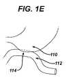
- FIG. 1Eis a schematic illustrating an anastomosis formed between the stomach and the jejunum as a result of the method FIGS. 1A-1D ;
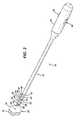
- FIG. 2is perspective view of one embodiment of an applier device having a fastener for use with the method of FIGS. 1A-1D ;
- FIG. 3is a perspective view of the fastener of FIG. 2 ;
- FIG. 4is a perspective view of the applier device and fastener of FIG. 2 after actuation, where the fastener is deployed into tissue;
- FIG. 5is a perspective view of the fastener of FIG. 3 after deployment into tissue
- FIG. 6is another perspective view of the fastener of FIG. 3 after deployment into tissue.
- the present inventiongenerally provides methods for joining one piece of tissue to another piece of tissue.
- the methodcan include inserting an applier device having an actuation portion into a first body lumen through a natural body orifice, forming a first opening in a first piece of tissue within the first lumen and a second opening in a second piece of tissue defining a portion of a second lumen adjacent to the first piece of tissue, and inserting the applier device through the first and second openings such that the actuation portion is between the first and second piece of tissue.
- the methodcan further include deploying a fastener into the first and second pieces of tissue through the actuation portion of the applier device, thereby joining the first and second pieces of tissue to form an anastomosis between the first and second lumens.
- the applier devicecan be inserted through a variety of lumens, such as natural body lumens, and the lumens can be accessed through natural body orifices, such as the esophagus and the rectum, or through a surgically-created portal, during a variety of medical procedures which require tissues to be joined.
- such medical proceduresinclude a Roux-en-Y procedure or other bariatric procedures which can require the joining of the jejunum to a stomach or stomach part, or bypass procedures for bypassing a cancerous or non-cancerous obstruction(s) in tissue, such as the duodenum.
- This methodis particularly advantageous in that it minimizes the number of access lumens formed in a patient by relying on natural orifices and/or a single created lumen or portal, such as a laparoscopic portal, for the insertion of the applier device.
- FIGS. 1A-1Eillustrate one exemplary embodiment of a method for forming an anastomosis 114 between a part of a jejunum 112 and a part of a stomach 110 .
- an applier device 126can be inserted transorally, and guided through the esophagus into the stomach 110 .
- the applier device 126can then be positioned at a site adjacent a wall of the stomach 110 , and the tip of the applier device 126 can optionally form a slight protrusion (not shown) on the distal wall thereof.
- This protrusioncan serve as a marker that allows a surgeon to identify the location of the applier device 126 within the stomach 110 , such that the surgeon can form a first opening 116 in the stomach 110 from which the applier device 126 can exit the stomach 110 .
- traditional monitoring and/or tracking techniquessuch as radioopaque bands located on the distal tip of the applier device, can be used to facilitate locating and/or tracking of the applier device and formation of the first opening.
- the first opening 116can be formed using a variety of techniques, and in one embodiment, the first opening can be formed from within the stomach 110 using a cutting member (not shown), such as a blade, that is located on the distal end of the applier device 126 .
- a cutting membersuch as a blade
- the first opening 116can be formed using at least one cutting element (cutting elements 120 a , 120 b are shown), such as scissors, that can be inserted by a laparoscopic surgical technique to a location adjacent to the position of the applier device 126 external to the stomach 110 .
- the first opening 116can also be formed using a combination of above-mentioned laparoscopic and endoscopic techniques.
- the first opening 116can be expanded to allow the applier device 126 , and in particular the actuation portion thereof, to be inserted therethrough and directed towards the jejunum 112 .
- Any technique known in the artcan be used to expand the tissue of the stomach 110 , and exemplary techniques can include the use of a tapered element located on the distal end of the applier device, the use of graspers, or some other element that is adapted to hold open at least a portion of the first opening to enlarge it, and combinations thereof.
- This insertion techniqueis particularly advantageous in that it does not require the formation of a new incision to allow the applier device 126 to access the jejunum 112 .
- the laparoscopic port used to form the first opening 116can be left in position for the formation of a second opening 124 in the jejunum 112 , as will be discussed in more detail below.
- a second opening 124 for receiving the applier device 126can be formed in the jejunum 112 .
- the second opening 124can be formed using techniques similar to those noted above with respect to the formation of the first opening 116 (e.g., a cutting member on the distal end of the applier device, using a cutting element(s) that accesses the site through a laparoscopic port, or combinations thereof).
- the second opening 124can be formed using a cutting element(s) that accesses the site through the same laparoscopic port that was used to form the first opening 116 .
- the applier device 126can be inserted into and through the second opening 124 such that at least a portion of the actuation portion is positioned between the stomach 110 and the jejunum 112 to facilitate the placement of a fastener within the tissues 110 , 112 .
- the second opening 124can be expanded to facilitate insertion of the applier device 126 therein using a distal end (not shown) of the applier device 126 , and additionally or alternatively, two graspers 122 a , 122 b that are positioned on opposed sides of the second opening 124 .
- the actuation portioncan be actuated to effect the deployment of a fastener into the tissues 110 , 112 .
- a variety of techniquescan be used to actuate the actuation portion of the applier device 126 to deploy the fastener, and those techniques can depend upon the types of applier devices and fasteners used.
- the actuation portion of the applier devicecan include at least one fastener having proximal and distal ring members joined by a connecting element, with the proximal and distal ring members each having a plurality of arms extending therefrom that are adapted to engage tissue.
- the actuation portioncan also optionally include a catch for holding the fastener thereto prior to deployment into tissue.
- actuationcan cause the fastener to optionally be released from the catch and the plurality of arms formed on the proximal and distal ring members (not shown) to move from a first resting position to a second actuated position such that they engage and hold together opposed tissues of the stomach 110 and the jejunum 112 .
- the plurality of armscan be independently or simultaneously actuated with respect to one another, in an exemplary embodiment, the plurality of arms on the distal ring member can be actuated prior to the actuation of the plurality of arms on the proximal ring member.
- the plurality of arms of the distal end of the fastenercan engage the tissue of the jejunum 112 , as shown in FIG.
- the applier device 126can be moved proximally such that it is retracted within the esophagus to cause the jejunum tissue 112 to contact the stomach tissue 110 , as shown in FIG. 1D .
- the proximal ring membercan then be deployed into stomach tissue 110 that is located opposite to the already-engaged tissue of the jejunum 112 , such that the plurality of arms of the fastener engage the tissue of the stomach 110 , causing an anastomosis 114 to form between the stomach 110 and the jejunum 112 ( FIG. 1E ).
- the applier device 126can be extracted from the stomach 110 transorally, and any laparoscopic instruments can be removed from the surgical site. The site can then optionally be leak tested to ensure that the anastomosis is sound. Over time, the tissue walls can permanently heal together and the fastener can be passed out of the digestive tract, especially if the fastener is biofragmentable.
- the applier device 10includes an elongate implement portion 12 having proximal and distal ends that is dimensionally sized for insertion through a natural or created orifice.
- the proximal endcan include a handle 54 and the distal end can include an actuation portion 24 adapted to hold a fastener 30 .
- the distal endcan also have a distal tip 18 that can be adapted to pierce through an opening 20 at an anastomosis site 22 in tissue 14 , 16 to facilitate the positioning of the actuation portion 24 within the tissue 14 , 16 .
- the elongate implement portion 12can have virtually any configuration, and in an exemplary embodiment, the elongate implement portion 12 can be flexible such that it can be endoscopically inserted into and through an orifice.
- the handle 54can be adapted to effect actuation of the fastener, and as shown the applier device 10 includes controls for effecting the actuation of the actuation portion 24 to cause deployment of the fastener 30 into tissue 14 , 16 .
- the controlscan include a first slide control 58 and a second slide control 60 .
- the handle 54can further include controls to effect illumination of the distal tip so that actuation of at least a portion of the fastener 30 in a lumen can be proximally viewed using an optics unit.
- the optics unitmay be part of an endoscope or the applier device. While the illustrated applier device can be manually positioned and actuated, one skilled in the art will appreciate that the applier device 10 can also adapted to be remotely positioned and actuated.
- the fastener 30has three rings, a proximal ring 32 , a center ring 34 , and a distal ring 36 , that are cylindrically aligned with one another.
- the proximal ring 32is longitudinally attached to the center ring 34 by proximal arms 38 , which in turn is longitudinally attached to the distal ring 36 by distal arms 40 .
- Each proximal and distal arm 38 , 40is bisected respectively by a hinged joint 42 , 44 that defines an inner arm segment 46 , 48 that is hingedly attached to the center ring 34 , and an outer arm segment 50 , 52 that is also hingedly attached to the respective proximal or distal ring 32 , 36 .
- the fastener 30can have a variety of shapes and sizes, however, in its unactuated state as shown in FIG. 3 , the fastener 30 has a substantially cylindrical configuration.
- the relative lengths of the inner arm segments 46 , 48 to the outer arm segments 50 , 52can be selected to angularly contact the tissue when the fastener 30 is deployed, and as illustrated in FIGS.
- the relationship between the proximal and distal rings 32 , 36resembles a cantilevered contact with the inner arm segments 46 , 48 actuating to an approximately parallel relationship to the tissue walls 14 , 16 .
- the fastener 30can also optionally include a locking mechanism that is adapted to maintain the position of the proximal and distal arms relative to one another once the fastener is deployed into tissue.
- One exemplary locking mechanismcan include at least one hook that is connected to the distal ring of the fastener that can latch to the center ring or the proximal ring upon actuation thereof to maintain the distal ring in an actuated position, and/or another hook that is connected to the proximal ring that can latch to the center ring or the distal ring upon actuation thereof to maintain the proximal ring in an actuated position.
- the two slide controls 58 , 60 on the handle 54can be withdrawn proximally to effect actuation of the actuation portion 24 , which causes the proximal and distal rings 32 , 36 to move from a first resting position to a second actuated position relative to the center ring 34 .
- the proximal and distal arms 38 , 40hinge outwardly from the longitudinal axis of the fastener 30 , creating a hollow rivet or hourglass shape for apposing tissue walls 14 , 16 .
- the center ring 34sits at a tissue junction between the lumens, and the distal and proximal rings 32 , 36 can engage the opposed tissues 14 , 16 .
- the rings 32 - 38can also be latched or locked to one another when actuated, as a result of the locking mechanism, to cause the fastener 30 to be held in the actuated position with bent arms 38 , 40 opposing the tissue 14 , 16 , as shown in FIG. 6 .
- the proximal arms 38can be staggered, as shown in FIG. 6 , from distal arms 40 to create a tortuous path for the compressed tissue. Alternatively, in other embodiments, the arms 38 , 40 can be aligned to directly mate to each other.
- the proximal ring 36is stationary with respect to the applier device 10
- the device 10can also include a third control so that each of the three rings can be positioned independently from the rest, further enhancing the ability to actuate either the distal or the proximal arms 40 , 38 .
- the center ring 34can be stationary with respect to the applier device 10 , with controls effective to move the proximal and distal rings 32 , 36 inwardly to the center ring 34 .
- the applier devicecan further include a variety of other features known in the art and not disclosed herein, such as, by way of non-limiting example, a catch mechanism for holding the fastener to the device, and in particular to the actuation portion thereof, to prevent accidental deployment.
- FIGS. 2-6illustrate one exemplary applier device and fastener that can be used to join a first piece of tissue to a second piece of tissue
- a variety of applier devices and fastenerscan be used to form an anastomosis in accordance with the method disclosed herein, such as those disclosed in commonly-owned U.S. application Ser. No. 10/675,091, filed Sep. 30, 2003, and entitled “Unfolding Anastomosis Device;” U.S. application Ser. No. 10/674,371, filed Sep. 30, 2003, and entitled “Anastomosis Wire Ring Device;” and U.S. application Ser. No. 10/675,497, filed Sep. 30, 2003, and entitled “Single Lumen Anastomosis Applier for Self-Deploying Fastener,” all of which are incorporated by reference herein.
- Applier devicescan be designed to be disposed after a single use, or they can be designed to be used multiple times. In either case, however, the device can be reconditioned for reuse after at least one use. Reconditioning can include any combination of the steps of disassembly of the device, followed by cleaning or replacement of particular pieces, and subsequent reassembly.
- the applier devices that can be used hereincan be reconditioned after the device has been used in a medical procedure.
- the devicecan be disassembled, and any number of the particular pieces (e.g., the fasteners, the actuation portion, and the distal tip) can be selectively replaced or removed in any combination.
- the fastenerscan be replaced by adding a new fastener cartridge to the actuation portion or by replacing the actuation portion with a fully loaded actuation portion.
- the devicecan be reassembled for subsequent use either at a reconditioning facility, or by a surgical team immediately prior to a surgical procedure.
- reconditioning of an applier devicecan utilize a variety of techniques for disassembly, cleaning/replacement, and reassembly. Use of such techniques, and the resulting reconditioned applier device, are all within the scope of the present application.
Landscapes
- Health & Medical Sciences (AREA)
- Life Sciences & Earth Sciences (AREA)
- Surgery (AREA)
- Heart & Thoracic Surgery (AREA)
- Engineering & Computer Science (AREA)
- Biomedical Technology (AREA)
- Nuclear Medicine, Radiotherapy & Molecular Imaging (AREA)
- Medical Informatics (AREA)
- Molecular Biology (AREA)
- Animal Behavior & Ethology (AREA)
- General Health & Medical Sciences (AREA)
- Public Health (AREA)
- Veterinary Medicine (AREA)
- Physiology (AREA)
- Surgical Instruments (AREA)
Abstract
Description
Claims (16)
Priority Applications (4)
| Application Number | Priority Date | Filing Date | Title |
|---|---|---|---|
| US11/277,289US8211142B2 (en) | 2003-09-30 | 2006-03-23 | Method for hybrid gastro-jejunostomy |
| AU2007201158AAU2007201158B2 (en) | 2006-03-23 | 2007-03-16 | Method for hybrid gastro-jejunostomy |
| BRPI0701287BRPI0701287A (en) | 2006-03-23 | 2007-03-23 | method for hybrid gastrojenunostomy |
| MX2007003572AMX2007003572A (en) | 2006-03-23 | 2007-03-23 | Method for hybrid gastro-jejunostomy. |
Applications Claiming Priority (3)
| Application Number | Priority Date | Filing Date | Title |
|---|---|---|---|
| US10/675,705US20050070935A1 (en) | 2003-09-30 | 2003-09-30 | Single lumen access deployable ring for intralumenal anastomosis |
| US10/675,077US7452363B2 (en) | 2003-09-30 | 2003-09-30 | Applier for fastener for single lumen access anastomosis |
| US11/277,289US8211142B2 (en) | 2003-09-30 | 2006-03-23 | Method for hybrid gastro-jejunostomy |
Related Parent Applications (2)
| Application Number | Title | Priority Date | Filing Date |
|---|---|---|---|
| US10/675,705Continuation-In-PartUS20050070935A1 (en) | 2003-09-30 | 2003-09-30 | Single lumen access deployable ring for intralumenal anastomosis |
| US10/675,077Continuation-In-PartUS7452363B2 (en) | 2003-09-30 | 2003-09-30 | Applier for fastener for single lumen access anastomosis |
Publications (2)
| Publication Number | Publication Date |
|---|---|
| US20060217748A1 US20060217748A1 (en) | 2006-09-28 |
| US8211142B2true US8211142B2 (en) | 2012-07-03 |
Family
ID=37036162
Family Applications (1)
| Application Number | Title | Priority Date | Filing Date |
|---|---|---|---|
| US11/277,289Expired - Fee RelatedUS8211142B2 (en) | 2003-09-30 | 2006-03-23 | Method for hybrid gastro-jejunostomy |
Country Status (1)
| Country | Link |
|---|---|
| US (1) | US8211142B2 (en) |
Families Citing this family (25)
| Publication number | Priority date | Publication date | Assignee | Title |
|---|---|---|---|---|
| US20040143342A1 (en)* | 2003-01-16 | 2004-07-22 | Stack Richard S. | Satiation pouches and methods of use |
| US7452363B2 (en)* | 2003-09-30 | 2008-11-18 | Ethicon Endo-Surgery, Inc. | Applier for fastener for single lumen access anastomosis |
| US8211142B2 (en) | 2003-09-30 | 2012-07-03 | Ortiz Mark S | Method for hybrid gastro-jejunostomy |
| US8425539B2 (en) | 2004-04-12 | 2013-04-23 | Xlumena, Inc. | Luminal structure anchoring devices and methods |
| US12303105B2 (en) | 2004-04-12 | 2025-05-20 | Boston Scientific Scimed, Inc. | Luminal structure anchoring devices and methods |
| JP5111112B2 (en) | 2004-12-08 | 2012-12-26 | エックスルミナ, インコーポレイテッド | Device for performing needle-guided therapy |
| US8784437B2 (en) | 2005-06-09 | 2014-07-22 | Xlumena, Inc. | Methods and devices for endosonography-guided fundoplexy |
| US8777967B2 (en) | 2005-06-09 | 2014-07-15 | Xlumena, Inc. | Methods and devices for anchoring to tissue |
| US8029522B2 (en)* | 2005-08-05 | 2011-10-04 | Ethicon Endo-Surgery, Inc. | Method and apparatus for sealing a gastric opening |
| US7625392B2 (en)* | 2006-02-03 | 2009-12-01 | James Coleman | Wound closure devices and methods |
| US20070191871A1 (en)* | 2006-02-10 | 2007-08-16 | Endogastric Solutions, Inc. | Transesophageal gastric reduction method and device for reducing the size of a previously formed gastric reduction pouch |
| WO2008115922A1 (en) | 2007-03-19 | 2008-09-25 | Michael Brenzel | Methods and apparatus for occlusion of body lumens |
| EP2178445B1 (en)* | 2007-07-17 | 2012-10-03 | Cook Medical Technologies LLC | Rivet introduction system |
| US9301761B2 (en) | 2007-10-22 | 2016-04-05 | James E. Coleman | Anastomosis devices and methods |
| US8454632B2 (en) | 2008-05-12 | 2013-06-04 | Xlumena, Inc. | Tissue anchor for securing tissue layers |
| US8197498B2 (en)* | 2008-11-06 | 2012-06-12 | Trinitas Ventures Ltd. | Gastric bypass devices and procedures |
| EP2414024A4 (en)* | 2009-03-30 | 2012-10-03 | Jointech Med Ltd | Synovial shunts |
| US9364259B2 (en) | 2009-04-21 | 2016-06-14 | Xlumena, Inc. | System and method for delivering expanding trocar through a sheath |
| JP5535313B2 (en) | 2009-05-29 | 2014-07-02 | エックスルミナ, インコーポレイテッド | Device and method for deploying a stent across adjacent tissue layers |
| WO2012129234A1 (en) | 2011-03-21 | 2012-09-27 | Endo Pharmaceuticals Inc. | Urethral anastomosis device and method |
| US9247930B2 (en) | 2011-12-21 | 2016-02-02 | James E. Coleman | Devices and methods for occluding or promoting fluid flow |
| JP6360042B2 (en) | 2012-05-17 | 2018-07-18 | ボストン サイエンティフィック サイムド,インコーポレイテッドBoston Scientific Scimed,Inc. | Method and device for access across adjacent tissue layers |
| ES2980140T3 (en) | 2013-02-21 | 2024-09-30 | Boston Scient Scimed Inc | Devices for forming an anastomosis |
| EP3360488B1 (en) | 2014-05-28 | 2020-03-18 | Boston Scientific Scimed, Inc. | Catheter with radiofrequency cutting tip and heated balloon |
| US12251168B2 (en) | 2021-05-28 | 2025-03-18 | Medos International Sarl | Systems, methods, and devices for localized tracking of a vertebral body or other anatomic structure |
Citations (108)
| Publication number | Priority date | Publication date | Assignee | Title |
|---|---|---|---|---|
| US2004014A (en) | 1931-08-05 | 1935-06-04 | Alfred F Sanford | Refrigerating apparatus |
| US2004013A (en) | 1934-07-11 | 1935-06-04 | Clarence E Reed | Antifriction bearing assembly for drills |
| US2004172A (en) | 1934-07-31 | 1935-06-11 | Everett F Niday | Starting block |
| US4841888A (en) | 1984-09-11 | 1989-06-27 | Mills Timothy N | Sewing machine |
| US5041129A (en) | 1990-07-02 | 1991-08-20 | Acufex Microsurgical, Inc. | Slotted suture anchor and method of anchoring a suture |
| US5080663A (en) | 1990-09-26 | 1992-01-14 | Univerity College London | Sewing device |
| US5217486A (en) | 1992-02-18 | 1993-06-08 | Mitek Surgical Products, Inc. | Suture anchor and installation tool |
| US5269809A (en) | 1990-07-02 | 1993-12-14 | American Cyanamid Company | Locking mechanism for use with a slotted suture anchor |
| US5376101A (en) | 1992-10-09 | 1994-12-27 | The United States Surgical Corporation | Suture retaining clip |
| US5398670A (en) | 1993-08-31 | 1995-03-21 | Ethicon, Inc. | Lumen traversing device |
| WO1995019140A1 (en) | 1994-01-13 | 1995-07-20 | Suturtek, Inc. | Suturing instrument with thread management |
| US5462558A (en) | 1994-08-29 | 1995-10-31 | United States Surgical Corporation | Suture clip applier |
| US5464415A (en) | 1994-03-15 | 1995-11-07 | Chen; Te-Chuan | Sutureless intestinal anastomosis gun |
| US5503635A (en) | 1993-11-12 | 1996-04-02 | United States Surgical Corporation | Apparatus and method for performing compressional anastomoses |
| US5514159A (en) | 1994-09-13 | 1996-05-07 | United States Surgical Corporation | Guillotine suture clip |
| US5531678A (en)* | 1994-09-19 | 1996-07-02 | Abbott Laboratories | Method of using a spring-loaded reciprocable stylet holder to eject a T-fastener |
| US5540705A (en) | 1995-05-19 | 1996-07-30 | Suturtek, Inc. | Suturing instrument with thread management |
| US5571119A (en) | 1993-10-25 | 1996-11-05 | Children's Medical Center Corporation | Retractable suture needle with self-contained driver |
| US5584861A (en) | 1992-09-04 | 1996-12-17 | University College London | Device for use in securing a thread |
| US5649938A (en) | 1992-04-08 | 1997-07-22 | American Cyanamid Co. | Surgical purse string suturing instrument and method |
| US5676670A (en) | 1996-06-14 | 1997-10-14 | Beth Israel Deaconess Medical Center | Catheter apparatus and method for creating a vascular bypass in-vivo |
| US5709693A (en) | 1996-02-20 | 1998-01-20 | Cardiothoracic System, Inc. | Stitcher |
| US5713910A (en) | 1992-09-04 | 1998-02-03 | Laurus Medical Corporation | Needle guidance system for endoscopic suture device |
| US5755730A (en) | 1994-03-23 | 1998-05-26 | University College London | Device for use in cutting threads |
| US5797920A (en) | 1996-06-14 | 1998-08-25 | Beth Israel Deaconess Medical Center | Catheter apparatus and method using a shape-memory alloy cuff for creating a bypass graft in-vivo |
| US5814071A (en) | 1994-11-10 | 1998-09-29 | Innovasive Devices, Inc. | Suture anchor assembly and methods |
| US5843088A (en) | 1991-12-23 | 1998-12-01 | Ela Medical Sa | Tool and method for installation of a ventricular cannulation device |
| US5853422A (en)* | 1996-03-22 | 1998-12-29 | Scimed Life Systems, Inc. | Apparatus and method for closing a septal defect |
| US5860992A (en) | 1996-01-31 | 1999-01-19 | Heartport, Inc. | Endoscopic suturing devices and methods |
| US5868760A (en) | 1994-12-07 | 1999-02-09 | Mcguckin, Jr.; James F. | Method and apparatus for endolumenally resectioning tissue |
| US5887594A (en) | 1997-09-22 | 1999-03-30 | Beth Israel Deaconess Medical Center Inc. | Methods and devices for gastroesophageal reflux reduction |
| US5899921A (en) | 1997-07-25 | 1999-05-04 | Innovasive Devices, Inc. | Connector device and method for surgically joining and securing flexible tissue repair members |
| US5954731A (en) | 1997-07-29 | 1999-09-21 | Yoon; Inbae | Surgical instrument with multiple rotatably mounted spreadable end effectors |
| US6007544A (en) | 1996-06-14 | 1999-12-28 | Beth Israel Deaconess Medical Center | Catheter apparatus having an improved shape-memory alloy cuff and inflatable on-demand balloon for creating a bypass graft in-vivo |
| US6010515A (en) | 1993-09-03 | 2000-01-04 | University College London | Device for use in tying knots |
| US6036694A (en) | 1998-08-03 | 2000-03-14 | Innovasive Devices, Inc. | Self-tensioning soft tissue fixation device and method |
| WO2001010312A1 (en) | 1999-08-10 | 2001-02-15 | Innovasive Devices, Inc. | Self-locking suture anchor |
| US6200329B1 (en) | 1998-08-31 | 2001-03-13 | Smith & Nephew, Inc. | Suture collet |
| WO2001066001A2 (en) | 2000-03-03 | 2001-09-13 | C.R. Bard, Inc. | Suture clips, delivery devices and methods |
| US20010023352A1 (en) | 1992-09-04 | 2001-09-20 | Gordon Norman S. | Suturing instruments and methods of use |
| WO2001089393A1 (en) | 2000-05-19 | 2001-11-29 | C.R. Bard, Inc. | Tissue capturing and suturing device and method |
| US6346111B1 (en) | 1992-09-04 | 2002-02-12 | Scimed Life Systems, Inc. | Suturing instruments and methods of use |
| US6379368B1 (en) | 1999-05-13 | 2002-04-30 | Cardia, Inc. | Occlusion device with non-thrombogenic properties |
| US20020107530A1 (en) | 2001-02-02 | 2002-08-08 | Sauer Jude S. | System for endoscopic suturing |
| US6443962B1 (en) | 1997-09-11 | 2002-09-03 | Benny Gaber | Stitching tool |
| US6454778B2 (en) | 1998-03-20 | 2002-09-24 | Scimed Life Systems, Inc. | Endoscopic suture systems |
| US6461320B1 (en) | 1998-08-12 | 2002-10-08 | Cardica, Inc. | Method and system for attaching a graft to a blood vessel |
| WO2002096327A2 (en) | 2001-05-30 | 2002-12-05 | Satiety, Inc. | Obesity treatment tools and methods |
| US6494888B1 (en) | 1999-06-22 | 2002-12-17 | Ndo Surgical, Inc. | Tissue reconfiguration |
| US20020193809A1 (en) | 2001-06-14 | 2002-12-19 | Meade John C. | Apparatus and method for surgical suturing with thread management |
| US6500195B2 (en) | 1993-05-14 | 2002-12-31 | Peter M. Bonutti | Method and apparatus for anchoring a suture |
| WO2003000142A2 (en) | 2001-06-20 | 2003-01-03 | Park Medical, Llc. | Anastomotic device |
| US6506196B1 (en) | 1999-06-22 | 2003-01-14 | Ndo Surgical, Inc. | Device and method for correction of a painful body defect |
| US6524328B2 (en) | 2001-04-12 | 2003-02-25 | Scion International, Inc. | Suture lock, lock applicator and method therefor |
| US6540789B1 (en) | 2000-06-15 | 2003-04-01 | Scimed Life Systems, Inc. | Method for treating morbid obesity |
| US6543456B1 (en)* | 2002-05-31 | 2003-04-08 | Ethicon Endo-Surgery, Inc. | Method for minimally invasive surgery in the digestive system |
| US20030083674A1 (en) | 2001-10-04 | 2003-05-01 | Gibbens George H. | Cycling suturing and knot-tying device |
| US6558400B2 (en) | 2001-05-30 | 2003-05-06 | Satiety, Inc. | Obesity treatment tools and methods |
| US6572629B2 (en) | 2000-08-17 | 2003-06-03 | Johns Hopkins University | Gastric reduction endoscopy |
| US20030109900A1 (en) | 2000-09-15 | 2003-06-12 | Jonathan Martinek | Knotless tissue anchor |
| US20030120292A1 (en)* | 2001-06-20 | 2003-06-26 | Park Medical, Llc | Anastomotic device |
| RU2208400C2 (en) | 2001-10-08 | 2003-07-20 | Робак Анатолий Николаевич | Apparatus for application of compression circular anastomoses |
| US20030171760A1 (en) | 2000-05-19 | 2003-09-11 | Gambale Richard A | Tissue capturing and suturing device and method |
| US20030181924A1 (en) | 2002-01-30 | 2003-09-25 | Olympus Optical Co., Ltd. | Endoscopic suturing system |
| US20030191481A1 (en) | 2000-03-31 | 2003-10-09 | John Nguyen | Multiple bias surgical fastener |
| US6632227B2 (en) | 2001-08-24 | 2003-10-14 | Scimed Life Systems, Inc. | Endoscopic resection devices |
| US20030225312A1 (en) | 2002-03-18 | 2003-12-04 | Anthony Kalloo | Endoscopic system for treating inside of body cavity |
| US20030229296A1 (en) | 2002-03-18 | 2003-12-11 | Olympus Optical Co., Ltd. | Guide tube |
| US6663639B1 (en) | 1999-06-22 | 2003-12-16 | Ndo Surgical, Inc. | Methods and devices for tissue reconfiguration |
| US20030233104A1 (en) | 2002-06-12 | 2003-12-18 | Scimed Life Systems, Inc. | Suturing instrument with deflectable head |
| US20040006351A1 (en) | 2002-07-02 | 2004-01-08 | Jamy Gannoe | Method and device for use in tissue approximation and fixation |
| US20040034369A1 (en) | 2001-02-02 | 2004-02-19 | Sauer Jude S. | System for endoscopic suturing |
| US6699263B2 (en)* | 2002-04-05 | 2004-03-02 | Cook Incorporated | Sliding suture anchor |
| WO2004021894A1 (en) | 2002-09-09 | 2004-03-18 | Brian Kelleher | Device and method for endoluminal therapy |
| US6709441B2 (en) | 1998-02-13 | 2004-03-23 | Heartport, Inc. | Devices and methods for performing vascular anastomosis |
| US6712836B1 (en) | 1999-05-13 | 2004-03-30 | St. Jude Medical Atg, Inc. | Apparatus and methods for closing septal defects and occluding blood flow |
| US6719763B2 (en) | 2000-09-29 | 2004-04-13 | Olympus Optical Co., Ltd. | Endoscopic suturing device |
| US20040082963A1 (en) | 2002-10-23 | 2004-04-29 | Jamy Gannoe | Method and device for use in endoscopic organ procedures |
| US20040098050A1 (en) | 2002-11-19 | 2004-05-20 | Opus Medical, Inc. | Devices and methods for repairing soft tissue |
| US6746460B2 (en) | 2002-08-07 | 2004-06-08 | Satiety, Inc. | Intra-gastric fastening devices |
| US20040122473A1 (en) | 2002-12-11 | 2004-06-24 | Ewers Richard C. | Delivery systems and methods for gastric reduction |
| US6755843B2 (en) | 2000-09-29 | 2004-06-29 | Olympus Optical Co., Ltd. | Endoscopic suturing device |
| US20040147958A1 (en) | 2002-12-11 | 2004-07-29 | Usgi Medical | Apparatus and methods for forming and securing gastrointestinal tissue folds |
| US20040162568A1 (en) | 1999-06-25 | 2004-08-19 | Usgi Medical | Apparatus and methods for forming and securing gastrointestinal tissue folds |
| US20040194790A1 (en) | 1999-06-22 | 2004-10-07 | Ndo Surgical, Inc. | Tissue reconfiguration |
| US20040210243A1 (en) | 2003-04-16 | 2004-10-21 | Jamy Gannoe | Method and devices for modifying the function of a body organ |
| US20040215058A1 (en) | 2002-09-06 | 2004-10-28 | Zirps Christopher T | Endoscopic accessory mounting adaptor |
| US6821858B2 (en) | 2000-05-01 | 2004-11-23 | Seiko Epson Corporation | Semiconductor devices and methods for manufacturing the same |
| US6835200B2 (en) | 1999-06-22 | 2004-12-28 | Ndo Surgical. Inc. | Method and devices for tissue reconfiguration |
| US20050015101A1 (en) | 2001-10-04 | 2005-01-20 | Gibbens George H. | Leverage locking reversible cyclic suturing and knot-tying device |
| US20050033319A1 (en) | 2003-05-16 | 2005-02-10 | Gambale Richard A. | Single intubation, multi-stitch endoscopic suturing system |
| US6869395B2 (en) | 2000-05-15 | 2005-03-22 | C. R. Bard, Inc. | Endoscopic accessory attachment mechanism |
| US20050070934A1 (en) | 2003-09-30 | 2005-03-31 | Tanaka Don A. | Anastomosis wire ring device |
| US20050070926A1 (en) | 2003-09-30 | 2005-03-31 | Ortiz Mark S. | Applier for fastener for single lumen access anastomosis |
| US20050070931A1 (en) | 2003-08-06 | 2005-03-31 | Rhodemann Li | Method and apparatus for creating a restriction in the stomach or other anatomical structure |
| US20050070939A1 (en) | 2003-09-30 | 2005-03-31 | Jean Beaupre | Unfolding anastomosis ring device |
| US20050070935A1 (en) | 2003-09-30 | 2005-03-31 | Ortiz Mark S. | Single lumen access deployable ring for intralumenal anastomosis |
| US20050070921A1 (en) | 2003-09-30 | 2005-03-31 | Ortiz Mark S. | Single lumen anastomosis applier for self-deploying fastener |
| US20050075654A1 (en) | 2003-10-06 | 2005-04-07 | Brian Kelleher | Methods and devices for soft tissue securement |
| US6908427B2 (en) | 2002-12-30 | 2005-06-21 | PARÉ Surgical, Inc. | Flexible endoscope capsule |
| US20050143760A1 (en) | 2001-05-01 | 2005-06-30 | Imran Mir A. | Endoscopic gastric constriction device |
| US20050143762A1 (en) | 2003-09-15 | 2005-06-30 | Paraschac Joseph F. | Suture locking device and methods |
| US20050149067A1 (en) | 2002-01-30 | 2005-07-07 | Olympus Corporation | Endoscopic suturing system |
| US20050192601A1 (en) | 2004-02-27 | 2005-09-01 | Demarais Denise M. | Methods and devices for reducing hollow organ volume |
| US20050192599A1 (en) | 2004-02-13 | 2005-09-01 | Demarais Denise M. | Methods for reducing hollow organ volume |
| US20050203488A1 (en) | 2004-03-09 | 2005-09-15 | Usgi Medical Inc. | Apparatus and methods for mapping out endoluminal gastrointestinal surgery |
| US6949116B2 (en) | 1996-05-08 | 2005-09-27 | Carag Ag | Device for plugging an opening such as in a wall of a hollow or tubular organ including biodegradable elements |
| US20060217748A1 (en) | 2003-09-30 | 2006-09-28 | Ethicon Endo-Surgery, Inc. | Method for Hybrid Gastro-Jejunostomy |
- 2006
- 2006-03-23USUS11/277,289patent/US8211142B2/ennot_activeExpired - Fee Related
Patent Citations (129)
| Publication number | Priority date | Publication date | Assignee | Title |
|---|---|---|---|---|
| US2004014A (en) | 1931-08-05 | 1935-06-04 | Alfred F Sanford | Refrigerating apparatus |
| US2004013A (en) | 1934-07-11 | 1935-06-04 | Clarence E Reed | Antifriction bearing assembly for drills |
| US2004172A (en) | 1934-07-31 | 1935-06-11 | Everett F Niday | Starting block |
| US4841888A (en) | 1984-09-11 | 1989-06-27 | Mills Timothy N | Sewing machine |
| US5041129A (en) | 1990-07-02 | 1991-08-20 | Acufex Microsurgical, Inc. | Slotted suture anchor and method of anchoring a suture |
| US5269809A (en) | 1990-07-02 | 1993-12-14 | American Cyanamid Company | Locking mechanism for use with a slotted suture anchor |
| US5080663A (en) | 1990-09-26 | 1992-01-14 | Univerity College London | Sewing device |
| US5843088A (en) | 1991-12-23 | 1998-12-01 | Ela Medical Sa | Tool and method for installation of a ventricular cannulation device |
| US5217486A (en) | 1992-02-18 | 1993-06-08 | Mitek Surgical Products, Inc. | Suture anchor and installation tool |
| US5649938A (en) | 1992-04-08 | 1997-07-22 | American Cyanamid Co. | Surgical purse string suturing instrument and method |
| US20040059350A1 (en) | 1992-09-04 | 2004-03-25 | Scimed Life Systems, Inc. | Suturing instruments and methods of use |
| US20010023352A1 (en) | 1992-09-04 | 2001-09-20 | Gordon Norman S. | Suturing instruments and methods of use |
| US5713910A (en) | 1992-09-04 | 1998-02-03 | Laurus Medical Corporation | Needle guidance system for endoscopic suture device |
| US6346111B1 (en) | 1992-09-04 | 2002-02-12 | Scimed Life Systems, Inc. | Suturing instruments and methods of use |
| US6358259B1 (en) | 1992-09-04 | 2002-03-19 | University College London | Device for use in tying knots |
| US5584861A (en) | 1992-09-04 | 1996-12-17 | University College London | Device for use in securing a thread |
| US5376101A (en) | 1992-10-09 | 1994-12-27 | The United States Surgical Corporation | Suture retaining clip |
| US6500195B2 (en) | 1993-05-14 | 2002-12-31 | Peter M. Bonutti | Method and apparatus for anchoring a suture |
| US5398670A (en) | 1993-08-31 | 1995-03-21 | Ethicon, Inc. | Lumen traversing device |
| US6010515A (en) | 1993-09-03 | 2000-01-04 | University College London | Device for use in tying knots |
| US5571119A (en) | 1993-10-25 | 1996-11-05 | Children's Medical Center Corporation | Retractable suture needle with self-contained driver |
| US5503635A (en) | 1993-11-12 | 1996-04-02 | United States Surgical Corporation | Apparatus and method for performing compressional anastomoses |
| US5437681A (en) | 1994-01-13 | 1995-08-01 | Suturtek Inc. | Suturing instrument with thread management |
| WO1995019140A1 (en) | 1994-01-13 | 1995-07-20 | Suturtek, Inc. | Suturing instrument with thread management |
| US5464415A (en) | 1994-03-15 | 1995-11-07 | Chen; Te-Chuan | Sutureless intestinal anastomosis gun |
| US5755730A (en) | 1994-03-23 | 1998-05-26 | University College London | Device for use in cutting threads |
| US5462558A (en) | 1994-08-29 | 1995-10-31 | United States Surgical Corporation | Suture clip applier |
| US5514159A (en) | 1994-09-13 | 1996-05-07 | United States Surgical Corporation | Guillotine suture clip |
| US5531678A (en)* | 1994-09-19 | 1996-07-02 | Abbott Laboratories | Method of using a spring-loaded reciprocable stylet holder to eject a T-fastener |
| US5814071A (en) | 1994-11-10 | 1998-09-29 | Innovasive Devices, Inc. | Suture anchor assembly and methods |
| US5868760A (en) | 1994-12-07 | 1999-02-09 | Mcguckin, Jr.; James F. | Method and apparatus for endolumenally resectioning tissue |
| US5540705A (en) | 1995-05-19 | 1996-07-30 | Suturtek, Inc. | Suturing instrument with thread management |
| US5860992A (en) | 1996-01-31 | 1999-01-19 | Heartport, Inc. | Endoscopic suturing devices and methods |
| US5709693A (en) | 1996-02-20 | 1998-01-20 | Cardiothoracic System, Inc. | Stitcher |
| US6312446B1 (en) | 1996-03-22 | 2001-11-06 | Scimed Life Systems, Inc. | Apparatus and method for closing a septal defect |
| US5853422A (en)* | 1996-03-22 | 1998-12-29 | Scimed Life Systems, Inc. | Apparatus and method for closing a septal defect |
| US6949116B2 (en) | 1996-05-08 | 2005-09-27 | Carag Ag | Device for plugging an opening such as in a wall of a hollow or tubular organ including biodegradable elements |
| US6007544A (en) | 1996-06-14 | 1999-12-28 | Beth Israel Deaconess Medical Center | Catheter apparatus having an improved shape-memory alloy cuff and inflatable on-demand balloon for creating a bypass graft in-vivo |
| US5676670A (en) | 1996-06-14 | 1997-10-14 | Beth Israel Deaconess Medical Center | Catheter apparatus and method for creating a vascular bypass in-vivo |
| US5797920A (en) | 1996-06-14 | 1998-08-25 | Beth Israel Deaconess Medical Center | Catheter apparatus and method using a shape-memory alloy cuff for creating a bypass graft in-vivo |
| US5902321A (en) | 1997-07-25 | 1999-05-11 | Innovasive Devices, Inc. | Device and method for delivering a connector for surgically joining and securing flexible tissue repair members |
| US5899921A (en) | 1997-07-25 | 1999-05-04 | Innovasive Devices, Inc. | Connector device and method for surgically joining and securing flexible tissue repair members |
| US5954731A (en) | 1997-07-29 | 1999-09-21 | Yoon; Inbae | Surgical instrument with multiple rotatably mounted spreadable end effectors |
| US6443962B1 (en) | 1997-09-11 | 2002-09-03 | Benny Gaber | Stitching tool |
| US5887594A (en) | 1997-09-22 | 1999-03-30 | Beth Israel Deaconess Medical Center Inc. | Methods and devices for gastroesophageal reflux reduction |
| US6709441B2 (en) | 1998-02-13 | 2004-03-23 | Heartport, Inc. | Devices and methods for performing vascular anastomosis |
| US20040002720A1 (en) | 1998-03-20 | 2004-01-01 | Scimed Life Systems, Inc. | Endoscopic suture systems |
| US6454778B2 (en) | 1998-03-20 | 2002-09-24 | Scimed Life Systems, Inc. | Endoscopic suture systems |
| US6036694A (en) | 1998-08-03 | 2000-03-14 | Innovasive Devices, Inc. | Self-tensioning soft tissue fixation device and method |
| US7004949B2 (en) | 1998-08-12 | 2006-02-28 | Cardica, Inc. | Method and system for attaching a graft to a blood vessel |
| US6461320B1 (en) | 1998-08-12 | 2002-10-08 | Cardica, Inc. | Method and system for attaching a graft to a blood vessel |
| US6200329B1 (en) | 1998-08-31 | 2001-03-13 | Smith & Nephew, Inc. | Suture collet |
| US6379368B1 (en) | 1999-05-13 | 2002-04-30 | Cardia, Inc. | Occlusion device with non-thrombogenic properties |
| US6712836B1 (en) | 1999-05-13 | 2004-03-30 | St. Jude Medical Atg, Inc. | Apparatus and methods for closing septal defects and occluding blood flow |
| US6494888B1 (en) | 1999-06-22 | 2002-12-17 | Ndo Surgical, Inc. | Tissue reconfiguration |
| US20040193184A1 (en) | 1999-06-22 | 2004-09-30 | Ndo Surgical, Inc., A Massachusetts Corporation | Methods and devices for tissue reconfiguration |
| US6773441B1 (en) | 1999-06-22 | 2004-08-10 | Ndo Surgical, Inc. | Methods and devices for tissue reconfiguration |
| US6506196B1 (en) | 1999-06-22 | 2003-01-14 | Ndo Surgical, Inc. | Device and method for correction of a painful body defect |
| US6663639B1 (en) | 1999-06-22 | 2003-12-16 | Ndo Surgical, Inc. | Methods and devices for tissue reconfiguration |
| US20040194790A1 (en) | 1999-06-22 | 2004-10-07 | Ndo Surgical, Inc. | Tissue reconfiguration |
| US6835200B2 (en) | 1999-06-22 | 2004-12-28 | Ndo Surgical. Inc. | Method and devices for tissue reconfiguration |
| US20040162568A1 (en) | 1999-06-25 | 2004-08-19 | Usgi Medical | Apparatus and methods for forming and securing gastrointestinal tissue folds |
| US20050075653A1 (en) | 1999-06-25 | 2005-04-07 | Usgi Medical Inc. | Apparatus and methods for forming and securing gastrointestinal tissue folds |
| WO2001010312A1 (en) | 1999-08-10 | 2001-02-15 | Innovasive Devices, Inc. | Self-locking suture anchor |
| WO2001066001A2 (en) | 2000-03-03 | 2001-09-13 | C.R. Bard, Inc. | Suture clips, delivery devices and methods |
| US20030191481A1 (en) | 2000-03-31 | 2003-10-09 | John Nguyen | Multiple bias surgical fastener |
| US6821858B2 (en) | 2000-05-01 | 2004-11-23 | Seiko Epson Corporation | Semiconductor devices and methods for manufacturing the same |
| US6869395B2 (en) | 2000-05-15 | 2005-03-22 | C. R. Bard, Inc. | Endoscopic accessory attachment mechanism |
| US20030171760A1 (en) | 2000-05-19 | 2003-09-11 | Gambale Richard A | Tissue capturing and suturing device and method |
| WO2001089393A1 (en) | 2000-05-19 | 2001-11-29 | C.R. Bard, Inc. | Tissue capturing and suturing device and method |
| US6540789B1 (en) | 2000-06-15 | 2003-04-01 | Scimed Life Systems, Inc. | Method for treating morbid obesity |
| US6572629B2 (en) | 2000-08-17 | 2003-06-03 | Johns Hopkins University | Gastric reduction endoscopy |
| US20030109900A1 (en) | 2000-09-15 | 2003-06-12 | Jonathan Martinek | Knotless tissue anchor |
| US6755843B2 (en) | 2000-09-29 | 2004-06-29 | Olympus Optical Co., Ltd. | Endoscopic suturing device |
| US6719763B2 (en) | 2000-09-29 | 2004-04-13 | Olympus Optical Co., Ltd. | Endoscopic suturing device |
| US20040034369A1 (en) | 2001-02-02 | 2004-02-19 | Sauer Jude S. | System for endoscopic suturing |
| US20020107530A1 (en) | 2001-02-02 | 2002-08-08 | Sauer Jude S. | System for endoscopic suturing |
| US20050165419A1 (en) | 2001-02-02 | 2005-07-28 | Sauer Jude S. | System for endoscopic suturing |
| US6524328B2 (en) | 2001-04-12 | 2003-02-25 | Scion International, Inc. | Suture lock, lock applicator and method therefor |
| US20050143760A1 (en) | 2001-05-01 | 2005-06-30 | Imran Mir A. | Endoscopic gastric constriction device |
| US20040122452A1 (en) | 2001-05-30 | 2004-06-24 | Satiety, Inc. | Obesity treatment tools and methods |
| US6558400B2 (en) | 2001-05-30 | 2003-05-06 | Satiety, Inc. | Obesity treatment tools and methods |
| US20030120265A1 (en) | 2001-05-30 | 2003-06-26 | Deem Mark E. | Obesity treatment tools and methods |
| US20040122453A1 (en) | 2001-05-30 | 2004-06-24 | Satiety, Inc. | Obesity treatment tools and methods |
| US20040024386A1 (en) | 2001-05-30 | 2004-02-05 | Deem Mark E. | Obesity treatment tools and methods |
| WO2002096327A2 (en) | 2001-05-30 | 2002-12-05 | Satiety, Inc. | Obesity treatment tools and methods |
| US20020193809A1 (en) | 2001-06-14 | 2002-12-19 | Meade John C. | Apparatus and method for surgical suturing with thread management |
| US20030032967A1 (en) | 2001-06-20 | 2003-02-13 | Park Medical, Llc | Anastomotic device |
| US20030120292A1 (en)* | 2001-06-20 | 2003-06-26 | Park Medical, Llc | Anastomotic device |
| WO2003000142A2 (en) | 2001-06-20 | 2003-01-03 | Park Medical, Llc. | Anastomotic device |
| US6632227B2 (en) | 2001-08-24 | 2003-10-14 | Scimed Life Systems, Inc. | Endoscopic resection devices |
| US20030083674A1 (en) | 2001-10-04 | 2003-05-01 | Gibbens George H. | Cycling suturing and knot-tying device |
| US20050015101A1 (en) | 2001-10-04 | 2005-01-20 | Gibbens George H. | Leverage locking reversible cyclic suturing and knot-tying device |
| RU2208400C2 (en) | 2001-10-08 | 2003-07-20 | Робак Анатолий Николаевич | Apparatus for application of compression circular anastomoses |
| US20030181924A1 (en) | 2002-01-30 | 2003-09-25 | Olympus Optical Co., Ltd. | Endoscopic suturing system |
| US20050149067A1 (en) | 2002-01-30 | 2005-07-07 | Olympus Corporation | Endoscopic suturing system |
| US20030225312A1 (en) | 2002-03-18 | 2003-12-04 | Anthony Kalloo | Endoscopic system for treating inside of body cavity |
| US20030229296A1 (en) | 2002-03-18 | 2003-12-11 | Olympus Optical Co., Ltd. | Guide tube |
| US6699263B2 (en)* | 2002-04-05 | 2004-03-02 | Cook Incorporated | Sliding suture anchor |
| US6543456B1 (en)* | 2002-05-31 | 2003-04-08 | Ethicon Endo-Surgery, Inc. | Method for minimally invasive surgery in the digestive system |
| US20030233104A1 (en) | 2002-06-12 | 2003-12-18 | Scimed Life Systems, Inc. | Suturing instrument with deflectable head |
| US6955643B2 (en) | 2002-06-12 | 2005-10-18 | Boston Scientific Scimed, Inc. | Endoscopic suture instrument |
| US20030233108A1 (en) | 2002-06-12 | 2003-12-18 | Scimed Life Systems, Inc. | Endoscopic suture instrument |
| US6773440B2 (en) | 2002-07-02 | 2004-08-10 | Satiety, Inc. | Method and device for use in tissue approximation and fixation |
| US20040006351A1 (en) | 2002-07-02 | 2004-01-08 | Jamy Gannoe | Method and device for use in tissue approximation and fixation |
| US6746460B2 (en) | 2002-08-07 | 2004-06-08 | Satiety, Inc. | Intra-gastric fastening devices |
| US20040215058A1 (en) | 2002-09-06 | 2004-10-28 | Zirps Christopher T | Endoscopic accessory mounting adaptor |
| US20050055038A1 (en) | 2002-09-09 | 2005-03-10 | Brian Kelleher | Device and method for endoluminal therapy |
| WO2004021894A1 (en) | 2002-09-09 | 2004-03-18 | Brian Kelleher | Device and method for endoluminal therapy |
| US20040082963A1 (en) | 2002-10-23 | 2004-04-29 | Jamy Gannoe | Method and device for use in endoscopic organ procedures |
| US20040098050A1 (en) | 2002-11-19 | 2004-05-20 | Opus Medical, Inc. | Devices and methods for repairing soft tissue |
| US20040122473A1 (en) | 2002-12-11 | 2004-06-24 | Ewers Richard C. | Delivery systems and methods for gastric reduction |
| US20040147958A1 (en) | 2002-12-11 | 2004-07-29 | Usgi Medical | Apparatus and methods for forming and securing gastrointestinal tissue folds |
| US6908427B2 (en) | 2002-12-30 | 2005-06-21 | PARÉ Surgical, Inc. | Flexible endoscope capsule |
| US20040210243A1 (en) | 2003-04-16 | 2004-10-21 | Jamy Gannoe | Method and devices for modifying the function of a body organ |
| US20050033319A1 (en) | 2003-05-16 | 2005-02-10 | Gambale Richard A. | Single intubation, multi-stitch endoscopic suturing system |
| US20050070931A1 (en) | 2003-08-06 | 2005-03-31 | Rhodemann Li | Method and apparatus for creating a restriction in the stomach or other anatomical structure |
| US20050143762A1 (en) | 2003-09-15 | 2005-06-30 | Paraschac Joseph F. | Suture locking device and methods |
| US20050070939A1 (en) | 2003-09-30 | 2005-03-31 | Jean Beaupre | Unfolding anastomosis ring device |
| US20050070926A1 (en) | 2003-09-30 | 2005-03-31 | Ortiz Mark S. | Applier for fastener for single lumen access anastomosis |
| US20050070934A1 (en) | 2003-09-30 | 2005-03-31 | Tanaka Don A. | Anastomosis wire ring device |
| US20060217748A1 (en) | 2003-09-30 | 2006-09-28 | Ethicon Endo-Surgery, Inc. | Method for Hybrid Gastro-Jejunostomy |
| US20050070921A1 (en) | 2003-09-30 | 2005-03-31 | Ortiz Mark S. | Single lumen anastomosis applier for self-deploying fastener |
| US20050070935A1 (en) | 2003-09-30 | 2005-03-31 | Ortiz Mark S. | Single lumen access deployable ring for intralumenal anastomosis |
| US20050075654A1 (en) | 2003-10-06 | 2005-04-07 | Brian Kelleher | Methods and devices for soft tissue securement |
| WO2005034729A3 (en) | 2003-10-06 | 2007-07-12 | Brian Kelleher | Methods and devices for soft tissue securement |
| US20050192599A1 (en) | 2004-02-13 | 2005-09-01 | Demarais Denise M. | Methods for reducing hollow organ volume |
| US20050192601A1 (en) | 2004-02-27 | 2005-09-01 | Demarais Denise M. | Methods and devices for reducing hollow organ volume |
| US20050203488A1 (en) | 2004-03-09 | 2005-09-15 | Usgi Medical Inc. | Apparatus and methods for mapping out endoluminal gastrointestinal surgery |
Non-Patent Citations (9)
| Title |
|---|
| EPO Communication dor Application No. 04256046.6, dated Oct. 17, 2006. cited by other. |
| EPO Communication for Application No. 04256046.6, dated Dec. 27, 2005. cited by other. |
| EPO Search Report for Application No. 04256018.5, dated Dec. 22, 2004. cited by other. |
| EPO Search Report for Application No. 04256046.6, dated Feb. 9, 2005. cited by other. |
| USPTO Final Rejection for U.S. Appl. No. 10/675,705, dated Jan. 24, 2007. cited by other. |
| USPTO Non-Final Rejection for U.S. Appl. No. 10/675,705, dated Aug. 7, 2006. cited by other. |
| USPTO Non-Final Rejection for U.S. Appl. No. 10/675,705, dated Feb. 27, 2006. cited by other. |
| USPTO Non-Final Rejection for U.S. Appl. No. 10/675,705, dated May 4, 2007. cited by other. |
| USPTO Office Action for U.S. Appl. No. 10/675,705, dated Jan. 10, 2006. cited by other. |
Also Published As
| Publication number | Publication date |
|---|---|
| US20060217748A1 (en) | 2006-09-28 |
Similar Documents
| Publication | Publication Date | Title |
|---|---|---|
| US8211142B2 (en) | Method for hybrid gastro-jejunostomy | |
| US7452363B2 (en) | Applier for fastener for single lumen access anastomosis | |
| JP4531514B2 (en) | Single lumen placement ring for interluminal anastomosis | |
| AU2011202357B2 (en) | Unfolding anastomosis ring device | |
| US7871418B2 (en) | Applier for fastener for single lumen access anastomosis | |
| US7229428B2 (en) | Method and device for use in endoscopic organ procedures | |
| JP4658557B2 (en) | Single lumen anastomosis applier for self-positioning fasteners | |
| EP1908418B1 (en) | An anastomotic applier | |
| EP1908419B1 (en) | A locking device for an anastomotic device | |
| AU2007201158B2 (en) | Method for hybrid gastro-jejunostomy |
Legal Events
| Date | Code | Title | Description |
|---|---|---|---|
| AS | Assignment | Owner name:ETHICON ENDO-SURGERY, INC., OHIO Free format text:ASSIGNMENT OF ASSIGNORS INTEREST;ASSIGNOR:ORTIZ, MARK S.;REEL/FRAME:017599/0734 Effective date:20060403 | |
| ZAAA | Notice of allowance and fees due | Free format text:ORIGINAL CODE: NOA | |
| ZAAB | Notice of allowance mailed | Free format text:ORIGINAL CODE: MN/=. | |
| STCF | Information on status: patent grant | Free format text:PATENTED CASE | |
| FPAY | Fee payment | Year of fee payment:4 | |
| MAFP | Maintenance fee payment | Free format text:PAYMENT OF MAINTENANCE FEE, 8TH YEAR, LARGE ENTITY (ORIGINAL EVENT CODE: M1552); ENTITY STATUS OF PATENT OWNER: LARGE ENTITY Year of fee payment:8 | |
| FEPP | Fee payment procedure | Free format text:MAINTENANCE FEE REMINDER MAILED (ORIGINAL EVENT CODE: REM.); ENTITY STATUS OF PATENT OWNER: LARGE ENTITY | |
| LAPS | Lapse for failure to pay maintenance fees | Free format text:PATENT EXPIRED FOR FAILURE TO PAY MAINTENANCE FEES (ORIGINAL EVENT CODE: EXP.); ENTITY STATUS OF PATENT OWNER: LARGE ENTITY | |
| STCH | Information on status: patent discontinuation | Free format text:PATENT EXPIRED DUE TO NONPAYMENT OF MAINTENANCE FEES UNDER 37 CFR 1.362 | |
| FP | Lapsed due to failure to pay maintenance fee | Effective date:20240703 |