US8192461B2 - Methods for facilitating closure of a bodily opening using one or more tacking devices - Google Patents
Methods for facilitating closure of a bodily opening using one or more tacking devicesDownload PDFInfo
- Publication number
- US8192461B2 US8192461B2US12/557,204US55720409AUS8192461B2US 8192461 B2US8192461 B2US 8192461B2US 55720409 AUS55720409 AUS 55720409AUS 8192461 B2US8192461 B2US 8192461B2
- Authority
- US
- United States
- Prior art keywords
- tacking device
- tacking
- tissue
- proximal
- loop portion
- Prior art date
- Legal status (The legal status is an assumption and is not a legal conclusion. Google has not performed a legal analysis and makes no representation as to the accuracy of the status listed.)
- Active, expires
Links
- 238000000034methodMethods0.000titleclaimsabstractdescription67
- 238000003780insertionMethods0.000claimsdescription57
- 230000037431insertionEffects0.000claimsdescription57
- 210000001519tissueAnatomy0.000description59
- 239000000463materialSubstances0.000description25
- 230000007246mechanismEffects0.000description14
- 206010060954Abdominal HerniaDiseases0.000description10
- 208000035091Ventral HerniaDiseases0.000description8
- 230000008901benefitEffects0.000description8
- 210000004876tela submucosaAnatomy0.000description8
- 229910001000nickel titaniumInorganic materials0.000description6
- 210000003815abdominal wallAnatomy0.000description5
- 108010037362Extracellular Matrix ProteinsProteins0.000description4
- 102000010834Extracellular Matrix ProteinsHuman genes0.000description4
- 239000000853adhesiveSubstances0.000description4
- 230000001070adhesive effectEffects0.000description4
- 210000002744extracellular matrixAnatomy0.000description4
- 238000010438heat treatmentMethods0.000description4
- 239000012781shape memory materialSubstances0.000description4
- 239000010935stainless steelSubstances0.000description4
- 229910001220stainless steelInorganic materials0.000description4
- 210000002784stomachAnatomy0.000description4
- 230000009466transformationEffects0.000description4
- 210000003484anatomyAnatomy0.000description3
- 230000008878couplingEffects0.000description3
- 238000010168coupling processMethods0.000description3
- 238000005859coupling reactionMethods0.000description3
- 238000009558endoscopic ultrasoundMethods0.000description3
- 230000000968intestinal effectEffects0.000description3
- 230000033001locomotionEffects0.000description3
- 229910000734martensiteInorganic materials0.000description3
- 230000003446memory effectEffects0.000description3
- HLXZNVUGXRDIFK-UHFFFAOYSA-Nnickel titaniumChemical compound[Ti].[Ti].[Ti].[Ti].[Ti].[Ti].[Ti].[Ti].[Ti].[Ti].[Ti].[Ni].[Ni].[Ni].[Ni].[Ni].[Ni].[Ni].[Ni].[Ni].[Ni].[Ni].[Ni].[Ni].[Ni]HLXZNVUGXRDIFK-UHFFFAOYSA-N0.000description3
- 210000000056organAnatomy0.000description3
- -1polypropylenePolymers0.000description3
- 230000008439repair processEffects0.000description3
- 238000001356surgical procedureMethods0.000description3
- 230000009278visceral effectEffects0.000description3
- 206010019909HerniaDiseases0.000description2
- 229910045601alloyInorganic materials0.000description2
- 239000000956alloySubstances0.000description2
- 230000003872anastomosisEffects0.000description2
- 210000002469basement membraneAnatomy0.000description2
- 210000001124body fluidAnatomy0.000description2
- 238000001816coolingMethods0.000description2
- 230000000694effectsEffects0.000description2
- 238000002594fluoroscopyMethods0.000description2
- 230000002401inhibitory effectEffects0.000description2
- 229910052751metalInorganic materials0.000description2
- 239000002184metalSubstances0.000description2
- 150000002739metalsChemical class0.000description2
- 230000000149penetrating effectEffects0.000description2
- BASFCYQUMIYNBI-UHFFFAOYSA-NplatinumChemical compound[Pt]BASFCYQUMIYNBI-UHFFFAOYSA-N0.000description2
- 229920000642polymerPolymers0.000description2
- 210000004872soft tissueAnatomy0.000description2
- 229910000679solderInorganic materials0.000description2
- 238000007794visualization techniqueMethods0.000description2
- 229910000684Cobalt-chromeInorganic materials0.000description1
- 108010035532CollagenProteins0.000description1
- 102000008186CollagenHuman genes0.000description1
- 206010011224CoughDiseases0.000description1
- 241001465754MetazoaSpecies0.000description1
- 239000004677NylonSubstances0.000description1
- 239000004952PolyamideSubstances0.000description1
- 239000004698PolyethyleneSubstances0.000description1
- 239000004743PolypropyleneSubstances0.000description1
- RTAQQCXQSZGOHL-UHFFFAOYSA-NTitaniumChemical compound[Ti]RTAQQCXQSZGOHL-UHFFFAOYSA-N0.000description1
- 210000000683abdominal cavityAnatomy0.000description1
- 230000003187abdominal effectEffects0.000description1
- 210000000579abdominal fatAnatomy0.000description1
- 230000009471actionEffects0.000description1
- 230000002491angiogenic effectEffects0.000description1
- 229910001566austeniteInorganic materials0.000description1
- 238000005452bendingMethods0.000description1
- 239000012620biological materialSubstances0.000description1
- 230000002201biotropic effectEffects0.000description1
- 210000004204blood vesselAnatomy0.000description1
- 239000002775capsuleSubstances0.000description1
- 210000004027cellAnatomy0.000description1
- 230000001413cellular effectEffects0.000description1
- 230000008859changeEffects0.000description1
- 239000010952cobalt-chromeSubstances0.000description1
- 229920001436collagenPolymers0.000description1
- 239000002131composite materialSubstances0.000description1
- 230000006835compressionEffects0.000description1
- 238000007906compressionMethods0.000description1
- 230000008473connective tissue growthEffects0.000description1
- 238000010276constructionMethods0.000description1
- 230000004069differentiationEffects0.000description1
- 210000001951dura materAnatomy0.000description1
- 230000002500effect on skinEffects0.000description1
- 238000001839endoscopyMethods0.000description1
- 230000008472epithelial growthEffects0.000description1
- 210000000109fascia lataAnatomy0.000description1
- 239000012530fluidSubstances0.000description1
- PCHJSUWPFVWCPO-UHFFFAOYSA-NgoldChemical compound[Au]PCHJSUWPFVWCPO-UHFFFAOYSA-N0.000description1
- 229910052737goldInorganic materials0.000description1
- 239000010931goldSubstances0.000description1
- 238000003384imaging methodMethods0.000description1
- 230000009545invasionEffects0.000description1
- 210000002429large intestineAnatomy0.000description1
- 210000004185liverAnatomy0.000description1
- 239000011159matrix materialSubstances0.000description1
- 210000004379membraneAnatomy0.000description1
- 239000012528membraneSubstances0.000description1
- 239000007769metal materialSubstances0.000description1
- 230000027939micturitionEffects0.000description1
- 229920001778nylonPolymers0.000description1
- 230000003287optical effectEffects0.000description1
- 210000003516pericardiumAnatomy0.000description1
- 210000004303peritoneumAnatomy0.000description1
- 239000004033plasticSubstances0.000description1
- 229920003023plasticPolymers0.000description1
- 229910052697platinumInorganic materials0.000description1
- 229920002647polyamidePolymers0.000description1
- 229920000728polyesterPolymers0.000description1
- 229920000573polyethylenePolymers0.000description1
- 229920000139polyethylene terephthalatePolymers0.000description1
- 239000005020polyethylene terephthalateSubstances0.000description1
- 229920001155polypropylenePolymers0.000description1
- 230000008569processEffects0.000description1
- 230000002441reversible effectEffects0.000description1
- 231100000241scarToxicity0.000description1
- 210000000813small intestineAnatomy0.000description1
- 239000007787solidSubstances0.000description1
- 230000002269spontaneous effectEffects0.000description1
- 108010070228surgisisProteins0.000description1
- 229910052715tantalumInorganic materials0.000description1
- GUVRBAGPIYLISA-UHFFFAOYSA-Ntantalum atomChemical compound[Ta]GUVRBAGPIYLISA-UHFFFAOYSA-N0.000description1
- 238000002560therapeutic procedureMethods0.000description1
- 229920001169thermoplasticPolymers0.000description1
- 239000004416thermosoftening plasticSubstances0.000description1
- 230000007838tissue remodelingEffects0.000description1
- 229910052719titaniumInorganic materials0.000description1
- 239000010936titaniumSubstances0.000description1
- 238000002604ultrasonographyMethods0.000description1
- 238000012285ultrasound imagingMethods0.000description1
- 210000003932urinary bladderAnatomy0.000description1
- 238000012800visualizationMethods0.000description1
Images


Classifications
- A—HUMAN NECESSITIES
- A61—MEDICAL OR VETERINARY SCIENCE; HYGIENE
- A61B—DIAGNOSIS; SURGERY; IDENTIFICATION
- A61B17/00—Surgical instruments, devices or methods
- A61B17/04—Surgical instruments, devices or methods for suturing wounds; Holders or packages for needles or suture materials
- A61B17/0401—Suture anchors, buttons or pledgets, i.e. means for attaching sutures to bone, cartilage or soft tissue; Instruments for applying or removing suture anchors
- A—HUMAN NECESSITIES
- A61—MEDICAL OR VETERINARY SCIENCE; HYGIENE
- A61B—DIAGNOSIS; SURGERY; IDENTIFICATION
- A61B17/00—Surgical instruments, devices or methods
- A61B17/064—Surgical staples, i.e. penetrating the tissue
- A61B17/0644—Surgical staples, i.e. penetrating the tissue penetrating the tissue, deformable to closed position
- A—HUMAN NECESSITIES
- A61—MEDICAL OR VETERINARY SCIENCE; HYGIENE
- A61B—DIAGNOSIS; SURGERY; IDENTIFICATION
- A61B17/00—Surgical instruments, devices or methods
- A61B17/04—Surgical instruments, devices or methods for suturing wounds; Holders or packages for needles or suture materials
- A61B17/0466—Suture bridges
- A—HUMAN NECESSITIES
- A61—MEDICAL OR VETERINARY SCIENCE; HYGIENE
- A61B—DIAGNOSIS; SURGERY; IDENTIFICATION
- A61B17/00—Surgical instruments, devices or methods
- A61B17/04—Surgical instruments, devices or methods for suturing wounds; Holders or packages for needles or suture materials
- A61B17/0487—Suture clamps, clips or locks, e.g. for replacing suture knots; Instruments for applying or removing suture clamps, clips or locks
- A—HUMAN NECESSITIES
- A61—MEDICAL OR VETERINARY SCIENCE; HYGIENE
- A61B—DIAGNOSIS; SURGERY; IDENTIFICATION
- A61B17/00—Surgical instruments, devices or methods
- A61B17/068—Surgical staplers, e.g. containing multiple staples or clamps
- A—HUMAN NECESSITIES
- A61—MEDICAL OR VETERINARY SCIENCE; HYGIENE
- A61B—DIAGNOSIS; SURGERY; IDENTIFICATION
- A61B17/00—Surgical instruments, devices or methods
- A61B17/11—Surgical instruments, devices or methods for performing anastomosis; Buttons for anastomosis
- A61B17/1114—Surgical instruments, devices or methods for performing anastomosis; Buttons for anastomosis of the digestive tract, e.g. bowels or oesophagus
- A—HUMAN NECESSITIES
- A61—MEDICAL OR VETERINARY SCIENCE; HYGIENE
- A61B—DIAGNOSIS; SURGERY; IDENTIFICATION
- A61B17/00—Surgical instruments, devices or methods
- A61B2017/00831—Material properties
- A61B2017/00867—Material properties shape memory effect
- A—HUMAN NECESSITIES
- A61—MEDICAL OR VETERINARY SCIENCE; HYGIENE
- A61B—DIAGNOSIS; SURGERY; IDENTIFICATION
- A61B17/00—Surgical instruments, devices or methods
- A61B17/04—Surgical instruments, devices or methods for suturing wounds; Holders or packages for needles or suture materials
- A61B17/0401—Suture anchors, buttons or pledgets, i.e. means for attaching sutures to bone, cartilage or soft tissue; Instruments for applying or removing suture anchors
- A61B2017/0409—Instruments for applying suture anchors
- A—HUMAN NECESSITIES
- A61—MEDICAL OR VETERINARY SCIENCE; HYGIENE
- A61B—DIAGNOSIS; SURGERY; IDENTIFICATION
- A61B17/00—Surgical instruments, devices or methods
- A61B17/04—Surgical instruments, devices or methods for suturing wounds; Holders or packages for needles or suture materials
- A61B17/0401—Suture anchors, buttons or pledgets, i.e. means for attaching sutures to bone, cartilage or soft tissue; Instruments for applying or removing suture anchors
- A61B2017/0414—Suture anchors, buttons or pledgets, i.e. means for attaching sutures to bone, cartilage or soft tissue; Instruments for applying or removing suture anchors having a suture-receiving opening, e.g. lateral opening
- A—HUMAN NECESSITIES
- A61—MEDICAL OR VETERINARY SCIENCE; HYGIENE
- A61B—DIAGNOSIS; SURGERY; IDENTIFICATION
- A61B17/00—Surgical instruments, devices or methods
- A61B17/04—Surgical instruments, devices or methods for suturing wounds; Holders or packages for needles or suture materials
- A61B17/0401—Suture anchors, buttons or pledgets, i.e. means for attaching sutures to bone, cartilage or soft tissue; Instruments for applying or removing suture anchors
- A61B2017/0427—Suture anchors, buttons or pledgets, i.e. means for attaching sutures to bone, cartilage or soft tissue; Instruments for applying or removing suture anchors having anchoring barbs or pins extending outwardly from the anchor body
- A61B2017/0437—Suture anchors, buttons or pledgets, i.e. means for attaching sutures to bone, cartilage or soft tissue; Instruments for applying or removing suture anchors having anchoring barbs or pins extending outwardly from the anchor body the barbs being resilient or spring-like
- A—HUMAN NECESSITIES
- A61—MEDICAL OR VETERINARY SCIENCE; HYGIENE
- A61B—DIAGNOSIS; SURGERY; IDENTIFICATION
- A61B17/00—Surgical instruments, devices or methods
- A61B17/04—Surgical instruments, devices or methods for suturing wounds; Holders or packages for needles or suture materials
- A61B17/0401—Suture anchors, buttons or pledgets, i.e. means for attaching sutures to bone, cartilage or soft tissue; Instruments for applying or removing suture anchors
- A61B2017/0464—Suture anchors, buttons or pledgets, i.e. means for attaching sutures to bone, cartilage or soft tissue; Instruments for applying or removing suture anchors for soft tissue
- A—HUMAN NECESSITIES
- A61—MEDICAL OR VETERINARY SCIENCE; HYGIENE
- A61B—DIAGNOSIS; SURGERY; IDENTIFICATION
- A61B17/00—Surgical instruments, devices or methods
- A61B17/04—Surgical instruments, devices or methods for suturing wounds; Holders or packages for needles or suture materials
- A61B2017/0496—Surgical instruments, devices or methods for suturing wounds; Holders or packages for needles or suture materials for tensioning sutures
- A—HUMAN NECESSITIES
- A61—MEDICAL OR VETERINARY SCIENCE; HYGIENE
- A61B—DIAGNOSIS; SURGERY; IDENTIFICATION
- A61B17/00—Surgical instruments, devices or methods
- A61B17/064—Surgical staples, i.e. penetrating the tissue
- A61B2017/0641—Surgical staples, i.e. penetrating the tissue having at least three legs as part of one single body
- A—HUMAN NECESSITIES
- A61—MEDICAL OR VETERINARY SCIENCE; HYGIENE
- A61B—DIAGNOSIS; SURGERY; IDENTIFICATION
- A61B17/00—Surgical instruments, devices or methods
- A61B17/064—Surgical staples, i.e. penetrating the tissue
- A61B2017/0645—Surgical staples, i.e. penetrating the tissue being elastically deformed for insertion
- A—HUMAN NECESSITIES
- A61—MEDICAL OR VETERINARY SCIENCE; HYGIENE
- A61B—DIAGNOSIS; SURGERY; IDENTIFICATION
- A61B17/00—Surgical instruments, devices or methods
- A61B17/11—Surgical instruments, devices or methods for performing anastomosis; Buttons for anastomosis
- A61B2017/1107—Surgical instruments, devices or methods for performing anastomosis; Buttons for anastomosis for blood vessels
Definitions
- the present embodimentsrelate generally to medical devices, and more particularly, to methods for facilitating closure of a bodily opening.
- Perforations in tissue or bodily wallsmay be formed intentionally or unintentionally.
- an unintentional ventral abdominal herniamay be formed in the abdominal wall due to heavy lifting, coughing, strain imposed during a bowel movement or urination, fluid in the abdominal cavity, or other reasons.
- Intentional perforationsmay be formed, for example, during surgical procedures such as translumenal procedures.
- one or more instrumentssuch as an endoscope, may be inserted through a visceral wall, such as the stomach wall.
- a closure instrumentmay be used to close the perforation in the visceral wall.
- itmay be difficult to adequately close the perforation and prevent leakage of bodily fluids.
- a graft materialsuch as a mesh or patch may be disposed to cover the perforation.
- the graft materialmay completely overlap with the perforation, and the edges of the graft material may at least partially overlap with tissue surrounding the perforation.
- the graft materialthen may be secured to the surrounding tissue in an attempt to effectively cover and seal the perforation.
- suturesIn order to secure the graft material to the surrounding tissue, sutures commonly are manually threaded through the full thickness of the surrounding tissue. In the case of a ventral abdominal hernia, the sutures may be threaded through the thickness of the abdominal wall, then tied down and knotted.
- manual suturing techniquesmay be time consuming and/or difficult to perform.
- suturing techniqueswhen closing intentional openings formed during translumenal procedures, suturing techniques may be used.
- the suturing techniques employed to close translumenal openingsmay be difficult to perform, may permit leakage of bodily fluids, and may be unreliable and difficult to reproduce.
- the present embodimentsprovide methods for facilitating closure of a bodily opening.
- at least a portion of a first tacking deviceis disposed through at least a portion of tissue at a first location in a vicinity of an opening in the tissue.
- at least a portion of a second tacking deviceis disposed through at least a portion of the tissue at a second location in the vicinity of the opening in the tissue.
- a closure member having at least one loop portionis advanced towards the first and second tacking devices, and the loop portion is positioned around at least a portion of the first tacking device and at least a portion of the second tacking device.
- the closure memberthen is actuated to urge the first tacking device towards the second tacking device to apply a compressive force upon the opening.
- the first and second tacking deviceseach comprises at least one proximal deployable member having contracted and expanded states, and further each comprise at least one distal deployable member having contracted and expanded states.
- the proximal deployable member and the distal deployable membereach may comprise hook-shaped configurations in the expanded states.
- the proximal and distal deployable memberseach may comprise a nickel-titanium alloy that is configured to self-expand to the hook-shaped configuration.
- three distal deployable membersare provided for engaging a serosal layer of tissue, while three proximal deployable members are provided for engaging a mucosal layer of the tissue.
- the first and second tacking devicesmay be delivered to a target site using an insertion tool comprising a hollow lumen having an inner diameter configured to receive the proximal and distal deployable members.
- the proximal and distal deployable membersare configured to be held in the contracted states when disposed within the hollow lumen. In the contracted states, the proximal and distal deployable members may be oriented in substantially longitudinally directions with respect to the insertion tool.
- the loop portion of the closure membermay be positioned around at least one of the proximal deployable members of the first tacking device, and further positioned around at least one of the proximal deployable members of the second tacking device.
- the loop portion of the closure membermay be positioned over at least one of the proximal deployable members of the first tacking device, and further positioned under at least one of the other proximal deployable members of the first tacking device.
- a cinching member coupled to the loop portionthen may be actuated to secure the positioning of the loop portion, thereby maintaining the positioning of the first and second tacking devices and the associated compressive force upon the opening.
- FIG. 1is a perspective view of a tacking device.
- FIG. 2is a perspective view of a distal region of an insertion tool and the tacking device of FIG. 1 .
- FIG. 3is a perspective, cut-away view illustrating multiple tacking devices in a delivery configuration.
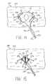
- FIG. 4is a schematic view illustrating a ventral hernia.
- FIG. 5is a schematic view illustrating a graft member used to cover the ventral hernia of FIG. 4 .
- FIG. 6is a schematic view of a method step for treating the ventral hernia of FIG. 4 .
- FIG. 7is a side-sectional view taken along line A--A of FIG. 6 .
- FIG. 8is a side-sectional view showing multiple tacking devices deployed in expanded configurations.
- FIG. 10is a perspective view of an alternative tacking device.
- FIG. 11is a side-sectional view illustrating one method of use of multiple tacking devices of FIG. 10 .
- FIG. 12is a side-sectional view showing multiple tacking devices deployed in the vicinity of an opening.
- FIG. 13is an upper perspective view of the tacking devices and the opening of FIG. 12 .
- FIGS. 14-15illustrate an exemplary method whereby multiple tacking devices are used to close the opening of FIGS. 12-13 .
- proximalrefers to a direction that is generally towards a physician during a medical procedure
- distalrefers to a direction that is generally towards a target site within a patient's anatomy during a medical procedure.
- the tacking device 20comprises at least one tube member 22 having a proximal end 24 and a distal end 26 .
- the tacking device 20further comprises a proximal deployment mechanism 32 and a distal deployment mechanism 42 .
- the proximal deployment mechanism 32comprises three proximal deployable members 35 - 37
- the distal deployment mechanism 42comprises three distal deployable members 45 - 47 .
- proximal deployable members 35 - 37extend proximally from the proximal end 24 of the tube member 22
- distal deployable members 45 - 47extend distally from the distal end 26 of the tube member 22 , as shown in FIG. 1 .
- the devicesince the device is symmetrical, it may be loaded into an insertion tool with either end first, as explained further below.
- the proximal deployable members 35 - 37 and the distal deployable members 45 - 47each may be affixed relative to the tube member 22 .
- each of the proximal and distal deployable members 35 - 37 and 45 - 47may be separate and discrete elements. Accordingly, six separate deployable members may be provided.
- the three proximal deployable members 35 - 37may be coupled to the tube member 22 near the proximal end 24 of the tube member 22 .
- the three proximal deployable members 35 - 37may be coupled to the proximal end 24 of the tube member 22 using an adhesive, frictional fit, mechanical device or other suitable mechanism or processes.
- the three distal deployable members 45 - 47may be coupled to the distal end 26 of the tube member 22 using an adhesive, frictional fit, mechanical device or other suitable mechanism.
- a first wiremay comprise a proximal end that forms the deployable member 35 and a distal end that forms the deployable member 45 , while a central region of the same wire is disposed through the entirety of the tube member 22 .
- second and third wiresmay be disposed through the entirety of the tube member 22 to form the remaining proximal and distal deployable members.
- the three wires that extend through the length of the tube member 22may be affixed to an interior surface of the tube member 22 , for example, using an adhesive or mechanical device.
- the three wiresalso may be sized to create a frictional fit against each other and/or an interior surface of the tube member 22 , thereby inhibiting movement of the proximal and distal deployable members 35 - 37 and 45 - 47 in longitudinal directions with respect to the tube member 22 .
- the tube member 22may comprise any suitable shape and material. Solely by way of example, the tube member 22 may comprise stainless steel or a biocompatible plastic. The tube member 22 may be cylindrically-shaped, as depicted in FIG. 1 , which may facilitate insertion through a lumen of an insertion tool 50 . Further, the tube member 22 may comprise one solid tube, or alternatively may comprise one or more tubes that may comprise slots, holes, cut-out regions and the like, for example, as shown and explained below with respect to the embodiment of FIGS. 10-11 .
- the tube member 22may be omitted entirely in the case where a first wire 125 integrally forms the proximal and distal deployable members 135 and 145 , a second wire 126 integrally forms the proximal and distal deployable members 136 and 146 , and a third wire 127 integrally forms the proximal and distal deployable members 137 and 147 .
- central regions of the first, second and third wires 125 - 127may be affixed together, for example, using a solder or weld, to maintain the structural rigidity of the components.
- the proximal and distal deployable members 35 - 37 and 45 - 47each comprise a contracted delivery configuration, as shown in FIG. 3 below, and further comprise an expanded deployed configuration, as shown in FIG. 1 .
- each of the deployable members 35 - 37 and 45 - 47may comprise a hook-shaped configuration in the expanded state.
- the deployable members 35 - 37 and 45 - 47may comprise a curvature of about 90 to about 360 degrees in the expanded state, and more preferably about 180 degrees, as shown in FIGS. 1-2 .
- the end regions 39 and 49 of the proximal and distal deployable membersare oriented substantially parallel to the tube member 22 .
- the end regions 39 and 49may be radially spaced apart from one another in the expanded state, as shown in FIG. 1 .
- the end regions 39 and 49may be well-suited for engaging, grasping, piercing and/or abutting tissue or graft material.
- a longitudinal distance L 1 between the end regions 39 and 49 of the tacking device 20may be varied to engage tissue in a desirable manner.
- the longitudinal distance L 1may be dimensioned to be substantially equal to or less than the combined thickness t 1 and t 2 of a tissue 74 and a graft member 80 , respectively, as shown in FIG. 8 below, thereby providing a desired compressive force upon the tissue 74 and the graft member 80 .
- the dimension of the tacking device 20may be tailored based on a particular surgical procedure, a particular patient's anatomy and/or other factors.
- the longitudinal length of the tube member 22may range from about 2 mm to about 10 mm
- the straightened (delivery or non-curved) length of the proximal deployable members 35 - 37may range from about 5 mm to about 50 mm
- the straightened (delivery or non-curved) length of the distal deployable members 45 - 47may range from about 5 mm to about 50 mm
- the longitudinal distance L 1 between the end regions 39 and 49may range from about 5 mm to about 30 mm
- the outer diameter of the tube member 22may range from about 0.3 mm to about 1.5 mm
- the outer diameter of the deployable member 35 - 37 and 45 - 47may range from about 0.1 mm to about 0.5 mm.
- Such dimensionsare provided for reference purposes only and are not intended to be limiting.
- the deployable members 35 - 37 and 45 - 47may comprise a shape-memory material, such as a nickel-titanium alloy (nitinol). If a shape-memory material such as nitinol is employed, the deployable members 35 - 37 and 45 - 47 may be manufactured such that they can assume the preconfigured expanded state shown in FIG. 1 upon application of a certain cold or hot medium. More specifically, a shape-memory material may undergo a substantially reversible phase transformation that allows it to “remember” and return to a previous shape or configuration.
- a shape-memory materialsuch as a nickel-titanium alloy (nitinol). If a shape-memory material such as nitinol is employed, the deployable members 35 - 37 and 45 - 47 may be manufactured such that they can assume the preconfigured expanded state shown in FIG. 1 upon application of a certain cold or hot medium. More specifically, a shape-memory material may undergo a substantially re
- a transformation between an austenitic phase and a martensitic phasemay occur by cooling and/or heating (shape memory effect) or by isothermally applying and/or removing stress (superelastic effect).
- Austeniteis characteristically the stronger phase and martensite is the more easily deformable phase.
- a nickel-titanium alloy having an initial configuration in the austenitic phasemay be cooled below a transformation temperature (M f ) to the martensitic phase and then deformed to a second configuration.
- M ftransformation temperature
- a ftransformation temperature
- the materialmay spontaneously return to its initial, predetermined configuration, as shown in FIG. 1 .
- the memory effectis one-way, which means that the spontaneous change from one configuration to another occurs only upon heating.
- the deployable members 35 - 37 and 45 - 47may be made from other metals and alloys that are biased, such that they may be restrained by the insertion tool 50 prior to deployment, but are inclined to return to their relaxed, expanded configuration upon deployment. Solely by way of example, the deployable members 35 - 37 and 45 - 47 may comprise other materials such as stainless steel, cobalt-chrome alloys, amorphous metals, tantalum, platinum, gold and titanium. The deployable members 35 - 37 and 45 - 47 also may be made from non-metallic materials, such as thermoplastics and other polymers.
- the deployable members 35 - 37 and 45 - 47may comprise any shape suitable for engaging, penetrating and/or abutting tissue, for purposes explained further below, and need not necessarily assume the curved shape depicted in FIGS. 1-2 .
- one or more tacking devices 20may be delivered to a target site in a patient's anatomy using an insertion tool 50 .
- the insertion tool 50is capable of carrying multiple different tacking devices, such as six tacking devices 20 a - 20 f, as shown in FIG. 9 and described below.
- FIG. 3one complete tacking device 20 a is shown in the contracted state, while portions of the proximal deployment mechanism 42 b of another tacking device 20 b , and the distal deployment mechanism 32 f of another tacking device 20 f , are also shown.
- the hollow lumen 54 of the insertion tool 50may comprise an inner diameter that is larger than an outer diameter of the tacking device 20 . Therefore, one or more tacking devices, such as six tacking devices 20 a - 20 f , may be loaded into the hollow lumen 54 in a delivery configuration, as shown in FIG. 3 .
- the proximal and distal deployable members 35 - 37 and 45 - 47 of each tacking device 20 a - 20 fmay comprise a substantially longitudinally-oriented profile, i.e., oriented along a longitudinal axis of the insertion tool 50 .
- the multiple tacking devices 20 a - 20 fmay be inserted into the hollow lumen 54 of the insertion tool 50 in a sequential manner, whereby the proximal deployment mechanism 32 a of the first tacking device 20 a may abut the distal deployment mechanism 42 b of the second tacking device 20 b , as depicted in FIG. 3 .
- the distal deployment mechanism 42 a of the first tacking device 20 amay be loaded a distance away from the sharpened distal tip 52 of the insertion tool 50 to prevent inadvertent deployment.
- a stylet 60may be disposed for longitudinal movement within the hollow lumen 52 of the insertion tool 50 , as shown in FIG. 3 .
- the stylet 60may comprise stainless steel or any other suitable material.
- the stylet 60is disposed proximal to the proximal deployment mechanism 32 f of the final sequential tacking device 20 f , as shown in FIG. 3 .
- the insertion tool 50may be proximally retracted, while the stylet 60 may be held longitudinally steady, to facilitate sequential deployment of each of the tacking devices 20 a - 20 f , as explained further below.
- the insertion tool 50may comprise one or more markers 56 , as shown in FIGS. 2-3 , which may be disposed near the distal end of the insertion tool 50 .
- the markers 56may be configured to be visualized under fluoroscopy of other imaging techniques to facilitate location of the distal end of the insertion tool, for example, so that a physician may determine how far the insertion tool 50 has penetrated into tissue 74 , as depicted in FIGS. 7-8 .
- a sheath member 58having an inner diameter larger than an outer diameter of the insertion tool 50 , as shown in FIG. 2 , may be longitudinally advanced over the insertion tool 50 , for various purposes explained further below.
- the insertion tool 50may be used in conjunction with another device, such as an endoscope, and may be delivered through a working lumen of an endoscope or similar device.
- one or more tacking devices 20 described abovemay be used to facilitate treatment of a perforation 75 using a graft member 80 .
- the perforation 75is a ventral hernia located in the abdominal wall 74 .
- the right and left legs 72 and 73 of a patient 70are shown for illustrative purposes. While treatment of a ventral hernia is shown for illustrative purposes, it will be apparent that the tacking devices described herein may be used in a wide range of medical procedures, including but not limited to any exemplary procedures described herein.
- the initial stages of the ventral hernia repairmay be performed using techniques that are known. Specifically, an open technique or laparoscopic technique may be employed. In an open technique, an incision may be made in the abdominal wall and fat and scar tissue may be removed from the area. A graft member 80 then may be applied so that it overlaps the perforation 75 , preferably by several millimeters or centimeters in each direction, as depicted in FIG. 5 . In a laparoscopic technique, two or three smaller incisions may be made to access the hernia site. A laparoscope may be inserted into one incision, and surgical instruments may be inserted into the other incision(s) to remove tissue and place the graft member 80 in the same position as the open procedure.
- an open techniquean incision may be made in the abdominal wall and fat and scar tissue may be removed from the area. A graft member 80 then may be applied so that it overlaps the perforation 75 , preferably by several millimeters or centimeters in each direction,
- the graft member 80may comprise any suitable material for covering the perforation 75 and substantially or entirely inhibiting the protrusion of abdominal matter.
- the graft member 80may comprise small intestinal submucosa (SIS), such as SURGISIS® BIODESIGNTM Soft Tissue Graft, available from Cook Biotech, Inc., West Lafayette, Ind., which provides smart tissue remodeling through its three-dimensional extracellular matrix (ECM) that is colonized by host tissue cells and blood vessels, and provides a scaffold for connective and epithelial tissue growth and differentiation along with the ECM components.
- SISsmall intestinal submucosa
- the graft member 80would be a one to four layer lyophilized soft tissue graft made from any number of tissue engineered products.
- Suitable submucosa materials for these purposesinclude, for instance, intestinal submucosa, including small intestinal submucosa, stomach submucosa, urinary bladder submucosa, and uterine submucosa.
- the graft member 80may also comprise a composite of a biomaterial and a biodegradeable polymer. Additional details may be found in U.S. Pat. No. 6,206,931 to Cook et al., the disclosure of which is incorporated herein by reference in its entirety.
- the insertion tool 50may be advanced in a distal direction to pierce through the graft member 80 , and further may pierce at least partially into the tissue 74 at a first location around the perimeter of the perforation 75 .
- the insertion tool 50is carrying six sequential tacking devices 20 a - 20 f , which may be disposed within the hollow lumen 54 of the insertion tool 50 as shown and explained with respect to FIG. 3 above. With each of the tacking devices 20 a - 20 f in the contracted delivery states, the sharpened tip 52 of the insertion tool 50 may be advanced to a predetermined depth into the tissue 74 .
- the markers 56 of FIGS. 2-3may facilitate in determining how far the insertion tool 50 has penetrated into tissue 74 , as depicted in FIG. 7 .
- the stylet 60 of FIG. 3may be held steady with respect to the insertion tool 50 , while the insertion tool 50 is retracted in a proximal direction.
- Thiscauses the distal deployable members 45 - 47 of the most distal tacking device 20 a to extend distal to the sharpened tip 52 of the insertion tool 50 , as depicted in FIG. 7 .
- the distal deployable members 45 - 47When the distal deployable members 45 - 47 are no longer radially constrained by the insertion tool 50 , they may assume their predetermined expanded configurations in which they may engage, penetrate and/or abut the tissue 74 .
- the proximal deployable members 35 - 37may assume their predetermined expanded configuration when are no longer radially constrained, as shown in FIG. 7 .
- the proximal deployable members 35 - 37may engage, penetrate and/or abut the graft member 80 and optionally penetrate into the tissue 74 .
- the tacking device 20 ahelps secure the graft material 80 against the tissue 74 .
- the substantially 180-degree hook-shaped configuration of the proximal deployable members 35 - 37may urge the graft member 80 in a distal direction towards the tissue 74 .
- the insertion tool 50may be repositioned to deploy another tacking device around the perimeter of the perforation 75 .
- Each subsequent tacking device 20 b - 20 fmay be deployed in the same manner as the tacking device 20 a .
- the tacking devices 20 a - 20 fmay secure the graft member 80 around the perimeter of the perforation 75 , as shown in FIG. 9 .
- greater or fewer tacking devicesmay be used, and the positioning of the tacking devices may be varied to optimize securing the graft member 80 to the tissue 74 in order to substantially seal the perforation 75 .
- the sheath member 58 of FIG. 2may be longitudinally advanced over the insertion tool 50 , for example, if needed to protect the sharpened distal tip 52 of the insertion tool 50 while the insertion tool 50 is being repositioned. Further, the sheath member 58 may be advanced distally over the insertion tool 50 to facilitate deployment of the proximal deployable members 35 - 37 . For example, the sheath member 58 may periodically push against the graft member 80 , thereby temporarily urging the graft member 80 and/or the tissue 74 in a distal direction.
- the sheath member 58may be held steady while the insertion tool 50 is retracted proximally to deploy the proximal deployable members 35 - 37 at a location proximal to the compressed tissue 74 and graft member 80 .
- the compressive force applied by the sheath member 58may be removed so that the graft member 80 and the tissue 74 may engage the deployed proximal deployable members 35 - 37 .
- the tissue 74illustratively comprises a thickness t 1
- the graft member 80comprises a thickness t 2
- the distal deployable members 45 - 47may be deployed entirely within the tissue 74 , as depicted in FIG. 8 , or alternatively may be deployed substantially distal to the tissue 74 while abutting or piercing through a distal edge of the tissue 74 .
- the longitudinal distance L 1 between the end regions 39 and 49 of the tacking device 20may be dimensioned to be substantially equal to, or slightly less than, the combined thickness t 1 +t 2 of the tissue 74 and the graft member 80 .
- the longitudinal distance L 1may be otherwise sized and configured, as desired, to apply desired forces upon the graft member 80 and the tissue 74 .
- FIGS. 4-9have illustrated the use of one or more tacking device 20 for covering a perforation 75 formed in the ventral abdominal wall
- the tacking devices disclosed hereinmay be useful in many other procedures.
- one or more tacking devices 20may be used to treat perforations in a visceral wall, such as the stomach wall.
- a suitable insertion devicesuch as an endoscope
- an endoscopemay be advanced through a bodily lumen such as the alimentary canal to a position proximate the target location.
- One or more componentsmay be advanced through a working lumen of the endoscope.
- the graft member 80may cover the perforation and may be secured in a position overlapping the perforation using the one or more of the tacking devices 20 , which may be deployed using the techniques described hereinabove.
- a tacking device 120may comprise one or more features for facilitating suturing, and preferably purse-string suturing.
- the tacking device 120is similar to the tacking device 20 of FIG. 1 , except as noted below.
- the tacking device 120comprises proximal and distal deployable members 135 - 137 and 145 - 147 , respectively.
- the tacking device 120comprises a proximal tube portion 122 and distal tube portion 123 with an opening, slot or cutout disposed therebetween, as shown in FIG. 10 .
- First, second and third wires 125 - 127may be disposed through the entirety of the proximal and distal tube portions 122 and 123 , as depicted in FIG. 10 .
- the first wire 125may comprise a proximal end that forms deployable member 135 and a distal end that forms deployable member 145 , such that a central region of the first wire 125 is disposed through both tube portions 122 and 123 .
- the second and third wires 126 and 127may be disposed through the entirety of the tube portions 122 and 123 .
- the second wire 126may comprise a proximal end that forms deployable member 136 and a distal end that forms deployable member 146
- the third wire 127may comprise a proximal end that forms deployable member 137 and a distal end that forms deployable member 147 .
- the three wires 125 - 127may be affixed to an interior surface of the tube portions 122 and 123 , for example, using an adhesive, frictional fit or mechanical device.
- the tube portions 122 and 123may be omitted, and central regions of the first, second and third wires 125 - 127 may be affixed to one another, for example, using a solder or weld.
- the second wire 126comprises a loop member 150 , which may be formed by bending a central region of the wire that is disposed between the tube portions 122 and 123 , as shown in FIG. 10 .
- the second wire 126may be bent to form an arch-shaped loop member 150 having an aperture 152 .
- a suture 160may be threaded through the aperture 152 of the loop member 150 , for example, as shown in FIG. 11 below.
- one single tube membermay be employed, in lieu of the proximal and distal tube portions 122 and 123 , and the single tube member may comprise a slot or cutout, such that the loop member 150 may extend radially through the slot or cutout.
- the loop member 150need not be formed integrally from any of the wires 125 - 127 , but rather may be formed as a loop disposed on an exterior surface of the proximal and distal tube portions 122 and 123 , or on an exterior surface of a single tube member if only one tube is used. Still further, while the loop member 150 is shown in a substantially central location, it may be placed closer to the proximal or distal ends of the tacking device 120 .
- a graft member 80may be placed over a perforation 75 , and multiple tacking devices 120 may be deployed using an insertion device to secure the graft member 80 to the tissue 74 , as explained in detail above with respect to FIGS. 4-9 .
- multiple tacking devices 120may be linked together by a single suture 160 , which may be slidably coupled through the loop members 150 of each of the tacking devices 120 , as generally shown in FIG. 11 .
- There are two free ends 161 and 162 of the suture 160which may be independently tensioned to facilitate closure of the perforation 75 .
- multiple tacking devices 120 having loop members 150are sequentially positioned around the perforation 75 in a semi-annular or annular shape, for example, as shown above in FIG. 9 .
- the ends 161 and 162 of the suture 160are then tensioned to reduce the distance between the tacking devices and compress the tissue 74 around the perforation 75 .
- the suture ends 161 and 162may be secured to maintain the compression of the tissue 74 using any suitable technique such as by forming a knot or using clamps, rivets and the like.
- loop members 150may be integrally formed with the tacking device 120 or externally attached thereto. Solely by way of example, such suture retaining mechanisms are explained in pending U.S. patent application Ser. No. 11/946,565, filed Nov. 28, 2007, the entire disclosure of which is hereby incorporated by reference in its entirety.
- sutures 160may be used in conjunction with embodiment of FIGS. 10-11 .
- synthetic suturesmay be made from polypropylene, nylon, polyamide, polyethylene, and polyesters such as polyethylene terephthalate. These materials may be used as monofilament suture strands, or as multifilament strands in a braided, twisted or other multifilament construction.
- the tacking devices 20 and 120may be used in other procedures.
- the tacking devices 20 and 120may be used to secure a graft member to tissue for reconstructing local tissue, and the like.
- the tacking devices 20 and 120may be used in an anastomosis procedure.
- multiple tacking devices 20 or 120may be deployed in a circular manner to couple a proximal vessel, duct or organ to a distal vessel, duct or organ.
- a suitable insertion devicesuch as an endoscope
- a bodily lumensuch as the alimentary canal
- One or more componentssuch as the insertion tool 50
- the distal end of the insertion tool 50may be viewed under fluoroscopy, or via optical elements of the endoscope, or by some other visualization technique.
- multiple tacking devicesthen may be delivered at one time, for example, using the insertion tool 50 .
- a holemay be punched through the middle of the deployed tacking devices to create a flow path between the proximal and distal vessels/ducts/organs. It will be apparent that still further applications of the tacking devices 20 and 120 are possible.
- the insertion tool 50may be used with or without an endoscope or similar device.
- FIGS. 12-15another exemplary use of the tacking device 20 is described.
- a plurality of tacking devices 20are used for facilitating closure of an opening 175 in tissue 174 .
- the tissue 174comprises a mucosal layer 177 and a serosal layer 178 .
- the opening 175may be formed during a translumenal procedure, whereby the tissue 174 may comprise tissue of the stomach, small or large intestines, or another bodily passage.
- a plurality of tacking devices 20are disposed at least partially through the tissue 174 at one or more locations in the vicinity of the opening 175 .
- multiple tacking devices 20at least partially surround the perimeter of the opening 175 .
- a first tacking device 20 ais disposed on one side of the opening 175
- a second tacking device 20 bis disposed on a substantially opposing side of the opening 175 .
- the first and second tacking devices 20 a and 20 bmay be delivered using an insertion tool 50 , preferably in the manner described above.
- the proximal deployable members 35 - 37 of each tacking device 20 a and 20 bmay engage, abut or penetrate the mucosal layer 177 of the tissue 174 .
- the distal deployable members 45 - 47 of each tacking device 20 a and 20 bmay engage, abut or penetrate the serosal layer 178 of the tissue 174 , as best seen in FIG. 12 .
- the longitudinal distance L 1 between the end regions 39 and 49 of the tacking devices 20may be sized to be approximately equal to a thickness t 1 of the tissue 174 .
- a closure member 190 having a loop portion 191may be positioned to surround at least a portion of the first and second tacking devices 20 a and 20 b.
- the closure member 190comprises first and second elongated portions 192 and 193 , which may be formed from one or more elongated wires or sutures.
- the loop portion 191may be formed between the first and second elongated portions 192 and 193 , as shown in FIGS. 14-15 .
- the size and configuration of the loop portion 191may be adjusted, e.g., using a cinching member 195 .
- the cinching member 195may be sized to surround the first and second elongated portions 192 and 193 , as shown in FIGS. 14-15 . In use, distal advancement of the cinching member 195 may decrease the size of the loop portion 191 , as explained further below.
- the loop portion 191 of the closure member 190may be positioned to at least partially surround the first and second tacking devices 20 a and 20 b .
- the loop portion 191may be guided beneath at least one of the proximal deployable members 35 - 37 of each of the first and second tacking devices 20 a and 20 b .
- the proximal deployable members 35 - 37 of the first and second tacking devices 20 a and 20 bpreferably are not substantially or permanently embedded into the mucosal layer 177 , but rather may engage or abut the mucosal layer 177 , to permit the loop portion 191 to slide between the proximal deployable members 35 - 37 and the tissue 174 .
- a physicianmay increase the size of the loop portion 191 by proximally retracting the cinching member 195 .
- the physicianmay maneuver the loop portion 191 as needed until it slides beneath at least one of the proximal deployable members 35 - 37 of each tacking device 20 a and 20 b , as depicted in FIG. 14 .
- the closure member 190is delivered through a channel of an endoscope, the coupling of the loop portion 191 to the tacking devices 20 a and 20 b may be performed under endoscopic guidance, or with other direct or indirect visualization techniques, such as fluoroscopic or ultrasound imaging, e.g., using an endoscopic ultrasound endoscope.
- the loop portion 191may be used to push distally on the tissue 174 to create a gap between one of the deployable members 35 - 37 and the tissue 174 through which the loop portion 191 can be passed. This may further facilitate positioning of the loop portion 191 , as shown in FIG. 14
- a physicianmay distally advance the cinching member 195 to reduce the size of the loop portion 191 .
- a catheter or other pushing instrumentmay be advanced over the urge the cinching member 195 distally. This action may urge the first and second tacking devices 20 a and 20 b in inward directions, i.e., towards the opening 175 , thereby applying a compressive force to facilitate closure of the opening 175 , as shown in FIG. 15 .
- the loop portion 191may engage any of the proximal deployable members 35 - 37 and/or the tube member 22 , if the tube member 22 is employed and extends above the mucosal layer 177 .
- the cinching member 195may comprise a locking feature, such that the positioning of the cinching member 195 may be secured to maintain the compressive force upon the opening 175 .
- a crimpmay be imposed upon the cinching member 195 to secure its positioning relative to the first and second elongated portions 192 and 193 .
- the cinching member 195may click into place using a one-way ratcheting engagement with the first and second elongated portions 192 and 193 .
- one or more external attachment devicessuch as a clamp or ring, may be used to hold the cinching member 195 at a desired location.
- the loop portion 191may be disposed around one or more of the proximal deployable members 35 - 37 of each tacking device 20 a - 20 b .
- the loop portion 191is disposed beneath the hook-shaped portion of at least one of the proximal deployable members 35 - 37 of each tacking device 20 a - 20 b to facilitate the provision of the inward compressive force noted above to close the opening 175 .
- loop portion 191is disposed partially above one or more proximal deployable members 35 - 37 , then distal advancement of the cinching member 195 may cause the loop portion 191 to urge at least one of the proximal deployable members 35 - 37 towards the mucosal layer 177 to penetrate or further abut the tissue 174 .
- tacking devices 20 a and 20 bare shown in FIGS. 12-15 to facilitate closure of the opening 175 , a greater number of tacking devices may be used, and the loop portion 191 may be disposed around portions of the additional tacking devices. Moreover, the tacking devices 20 a and 20 b may be deployed through the tissue 174 before or after creation of the opening 175 . Finally, while a tube member 22 is depicted as part of the first and second tacking devices 20 a and 20 b in FIGS. 12-15 , the tube member 22 may be omitted and the proximal and distal deployable members 35 - 37 and 45 - 47 may be integrally formed or otherwise adhered or coupled together without a tube member, as generally set forth above.
- the apparatus and methods described hereinmay be used for facilitating closure of an opening in a layer of material, and are not restricted to methods for treatment of a human or animal body by surgery or therapy.
- a first tacking deviceis disposed through at least a portion of the material at a first location in a vicinity of an opening in the layer of material.
- at least a portion of a second tacking deviceis disposed through at least a portion of the material at a second location in the vicinity of the opening.
- a closure member having at least one loop portionis advanced towards the first and second tacking devices, and the loop portion is positioned around at least a portion of the first tacking device and at least a portion of the second tacking device.
- the closure memberthen is actuated to urge the first tacking device towards the second tacking device to provide a compressive force upon the opening, as generally described above.
Landscapes
- Health & Medical Sciences (AREA)
- Surgery (AREA)
- Life Sciences & Earth Sciences (AREA)
- Biomedical Technology (AREA)
- Nuclear Medicine, Radiotherapy & Molecular Imaging (AREA)
- Engineering & Computer Science (AREA)
- Heart & Thoracic Surgery (AREA)
- Medical Informatics (AREA)
- Molecular Biology (AREA)
- Animal Behavior & Ethology (AREA)
- General Health & Medical Sciences (AREA)
- Public Health (AREA)
- Veterinary Medicine (AREA)
- Rheumatology (AREA)
- Surgical Instruments (AREA)
Abstract
Description
Claims (22)
Priority Applications (1)
| Application Number | Priority Date | Filing Date | Title |
|---|---|---|---|
| US12/557,204US8192461B2 (en) | 2008-09-11 | 2009-09-10 | Methods for facilitating closure of a bodily opening using one or more tacking devices |
Applications Claiming Priority (2)
| Application Number | Priority Date | Filing Date | Title |
|---|---|---|---|
| US9619708P | 2008-09-11 | 2008-09-11 | |
| US12/557,204US8192461B2 (en) | 2008-09-11 | 2009-09-10 | Methods for facilitating closure of a bodily opening using one or more tacking devices |
Publications (2)
| Publication Number | Publication Date |
|---|---|
| US20100069955A1 US20100069955A1 (en) | 2010-03-18 |
| US8192461B2true US8192461B2 (en) | 2012-06-05 |
Family
ID=42005753
Family Applications (1)
| Application Number | Title | Priority Date | Filing Date |
|---|---|---|---|
| US12/557,204Active2030-06-02US8192461B2 (en) | 2008-09-11 | 2009-09-10 | Methods for facilitating closure of a bodily opening using one or more tacking devices |
Country Status (2)
| Country | Link |
|---|---|
| US (1) | US8192461B2 (en) |
| WO (1) | WO2010030842A2 (en) |
Cited By (3)
| Publication number | Priority date | Publication date | Assignee | Title |
|---|---|---|---|---|
| US20100292719A1 (en)* | 2009-05-14 | 2010-11-18 | Wilson-Cook Medical Inc. | Systems and methods for securing a graft member to tissue using one or more tacking devices |
| US20140371787A1 (en)* | 2009-01-30 | 2014-12-18 | Cook Medical Technologies Llc | Vascular closure device |
| US11304688B2 (en) | 2019-06-05 | 2022-04-19 | Maine Medical Center | Gastrocutaneous closure device |
Families Citing this family (9)
| Publication number | Priority date | Publication date | Assignee | Title |
|---|---|---|---|---|
| WO2016028898A2 (en)* | 2014-08-21 | 2016-02-25 | Boston Scientific Scimed, Inc. | Anchors and cinching for tissue opposition |
| JP7549353B2 (en)* | 2019-03-22 | 2024-09-11 | 国立大学法人東北大学 | Anchor Device |
| CN111150441B (en)* | 2019-12-27 | 2020-11-10 | 广州市花都区人民医院 | Suture device for hernia sac with lineal hernia |
| CN111150442B (en)* | 2019-12-27 | 2020-11-10 | 广州市花都区人民医院 | Suture device for hernia sac with lineal hernia |
| CN111407346B (en)* | 2020-04-02 | 2020-12-22 | 王建龙 | an intestinal closure |
| US12396854B2 (en) | 2021-05-12 | 2025-08-26 | Medtronic, Inc. | Systems, devices and methods for reshaping a bodily lumen |
| EP4358860A1 (en)* | 2021-06-21 | 2024-05-01 | Medtronic, Inc. | Systems, devices and methods including retrievable tissue anchor |
| US20230072138A1 (en)* | 2021-09-08 | 2023-03-09 | Covidien Lp | Surgical fastener having a base and a leg |
| US20230233201A1 (en)* | 2022-01-24 | 2023-07-27 | Covidien Lp | Apparatus and method for endoscopically closing gastrointestinal defects |
Citations (269)
| Publication number | Priority date | Publication date | Assignee | Title |
|---|---|---|---|---|
| US363538A (en)* | 1887-05-24 | Suegical | ||
| US2199025A (en) | 1936-06-08 | 1940-04-30 | Carl E Conn | Means and method of closing surgical incisions |
| US2671444A (en) | 1951-12-08 | 1954-03-09 | Jr Benjamin F Pease | Nonmetallic mesh surgical insert for hernia repair |
| US3209422A (en) | 1963-12-23 | 1965-10-05 | Dritz Arthur | Fastening device |
| US3399432A (en) | 1965-12-08 | 1968-09-03 | Dennison Mfg Co | Button attachment |
| US3470834A (en) | 1968-03-08 | 1969-10-07 | Dennison Mfg Co | Fastener attaching device |
| US3556079A (en) | 1967-05-16 | 1971-01-19 | Haruo Omizo | Method of puncturing a medical instrument under guidance of ultrasound |
| US3814104A (en) | 1971-07-05 | 1974-06-04 | W Irnich | Pacemaker-electrode |
| US3856016A (en) | 1972-11-03 | 1974-12-24 | H Davis | Method for mechanically applying an occlusion clip to an anatomical tubular structure |
| US3954108A (en) | 1972-11-03 | 1976-05-04 | Davis Hugh J | Occlusion clip and instrument for applying same |
| US3958576A (en) | 1973-11-14 | 1976-05-25 | Olympus Optical Co., Ltd. | Surgical instrument for clipping any affected portion of a body cavity |
| US4006747A (en) | 1975-04-23 | 1977-02-08 | Ethicon, Inc. | Surgical method |
| US4204541A (en) | 1977-01-24 | 1980-05-27 | Kapitanov Nikolai N | Surgical instrument for stitching up soft tissues with lengths of spiked suture material |
| US4217902A (en) | 1977-05-02 | 1980-08-19 | March Alfred L | Hemostatic clip |
| US4235238A (en) | 1978-05-11 | 1980-11-25 | Olympus Optical Co., Ltd. | Apparatus for suturing coeliac tissues |
| US4485816A (en) | 1981-06-25 | 1984-12-04 | Alchemia | Shape-memory surgical staple apparatus and method for use in surgical suturing |
| US4621639A (en) | 1982-02-03 | 1986-11-11 | Ethicon, Inc | Surgical instrument with hydraulic actuator |
| US4749114A (en) | 1986-11-10 | 1988-06-07 | United States Surgical Corporation | Purse string applicator and method of affixing a purse string |
| US4773420A (en) | 1987-06-22 | 1988-09-27 | U.S. Surgical Corporation | Purse string applicator |
| US4791707A (en) | 1986-08-26 | 1988-12-20 | Tucker Wilson H | Clip applicator, spreadable clips and method for applying the clips |
| US4796627A (en) | 1986-08-26 | 1989-01-10 | Tucker Wilson H | Clip applicator and spreadable clips for use therein |
| EP0310582A1 (en) | 1987-09-30 | 1989-04-05 | Astra Tech Aktiebolag | Surgical instrument for ligating internal tissues |
| US4821939A (en) | 1987-09-02 | 1989-04-18 | United States Surgical Corporation | Staple cartridge and an anvilless surgical stapler |
| US4832027A (en) | 1985-05-31 | 1989-05-23 | Alice Utz | Surgical clamp |
| US4990156A (en) | 1988-06-21 | 1991-02-05 | Lefebvre Jean Marie | Filter for medical use |
| US5015249A (en) | 1989-12-26 | 1991-05-14 | Nakao Naomi L | Endoscopic stapling device and method |
| US5049153A (en) | 1989-12-26 | 1991-09-17 | Nakao Naomi L | Endoscopic stapling device and method |
| US5059205A (en) | 1989-09-07 | 1991-10-22 | Boston Scientific Corporation | Percutaneous anti-migration vena cava filter |
| US5084057A (en) | 1989-07-18 | 1992-01-28 | United States Surgical Corporation | Apparatus and method for applying surgical clips in laparoscopic or endoscopic procedures |
| US5100420A (en) | 1989-07-18 | 1992-03-31 | United States Surgical Corporation | Apparatus and method for applying surgical clips in laparoscopic or endoscopic procedures |
| US5099827A (en) | 1989-12-13 | 1992-03-31 | Richard Wolf Gmbh | Instrument set for closing opened body organs, wounds or the like |
| US5123914A (en) | 1986-05-19 | 1992-06-23 | Cook Incorporated | Visceral anchor for visceral wall mobilization |
| US5156609A (en) | 1989-12-26 | 1992-10-20 | Nakao Naomi L | Endoscopic stapling device and method |
| US5192303A (en) | 1987-05-18 | 1993-03-09 | Mitek Surgical Products, Inc. | Suture anchor |
| US5203787A (en) | 1990-11-19 | 1993-04-20 | Biomet, Inc. | Suture retaining arrangement |
| US5236438A (en)* | 1992-09-10 | 1993-08-17 | Wilk Peter J | Method and assembly for repairing liver laceration |
| US5242456A (en) | 1991-11-21 | 1993-09-07 | Kensey Nash Corporation | Apparatus and methods for clamping tissue and reflecting the same |
| US5324307A (en) | 1990-07-06 | 1994-06-28 | American Cyanamid Company | Polymeric surgical staple |
| US5333624A (en) | 1992-02-24 | 1994-08-02 | United States Surgical Corporation | Surgical attaching apparatus |
| US5334217A (en) | 1992-01-21 | 1994-08-02 | Regents Of The University Of Minnesota | Septal defect closure device |
| US5350385A (en) | 1993-04-28 | 1994-09-27 | Christy William J | Surgical stab wound closure device and method |
| US5366480A (en) | 1990-12-24 | 1994-11-22 | American Cyanamid Company | Synthetic elastomeric buttressing pledget |
| US5368602A (en) | 1993-02-11 | 1994-11-29 | De La Torre; Roger A. | Surgical mesh with semi-rigid border members |
| US5368600A (en) | 1993-07-23 | 1994-11-29 | Ethicon, Inc. | Steerable bulldog clamp applier |
| US5411522A (en) | 1993-08-25 | 1995-05-02 | Linvatec Corporation | Unitary anchor for soft tissue fixation |
| US5417691A (en) | 1982-05-20 | 1995-05-23 | Hayhurst; John O. | Apparatus and method for manipulating and anchoring tissue |
| US5437266A (en) | 1992-07-02 | 1995-08-01 | Mcpherson; William | Coil screw surgical retractor |
| US5520700A (en) | 1992-11-13 | 1996-05-28 | Technion Research & Development Foundation, Ltd. | Stapler device particularly useful in medical suturing |
| US5527343A (en) | 1993-05-14 | 1996-06-18 | Bonutti; Peter M. | Suture anchor |
| US5554183A (en) | 1994-01-19 | 1996-09-10 | Nazari; Stefano | Vascular prosthesis for the substitution or internal lining of blood vessels of medium or large diameter and device for its application |
| US5573543A (en) | 1992-05-08 | 1996-11-12 | Ethicon, Inc. | Endoscopic surgical instrument and staples for applying purse string sutures |
| US5582616A (en) | 1994-08-05 | 1996-12-10 | Origin Medsystems, Inc. | Surgical helical fastener with applicator |
| US5582615A (en) | 1995-10-30 | 1996-12-10 | Pilling Weck, Incorporated | Handle for surgical clip applicator systems |
| US5593414A (en) | 1993-08-25 | 1997-01-14 | Apollo Camera, L.L.C. | Method of applying a surgical ligation clip |
| EP0774237A2 (en) | 1995-10-20 | 1997-05-21 | United States Surgical Corporation | Apparatus and method for vascular hole closure |
| US5662683A (en) | 1995-08-22 | 1997-09-02 | Ortho Helix Limited | Open helical organic tissue anchor and method of facilitating healing |
| US5667527A (en) | 1993-03-02 | 1997-09-16 | Holobeam, Inc. | Staples |
| US5690656A (en) | 1995-06-27 | 1997-11-25 | Cook Incorporated | Method and apparatus for creating abdominal visceral anastomoses |
| US5695525A (en) | 1992-05-20 | 1997-12-09 | C.R. Bard, Incorporated | Implantable prosthesis and method and apparatus for loading and delivering an implantable prosthesis |
| US5702421A (en)* | 1995-01-11 | 1997-12-30 | Schneidt; Bernhard | Closure device for closing a vascular opening, such as patent ductus arteriosus |
| US5728116A (en) | 1994-01-13 | 1998-03-17 | Ethicon, Inc. | Spiral surgical tack |
| US5741278A (en) | 1994-08-17 | 1998-04-21 | Tahoe Surgical Instruments | Endoscopic suture placement tool |
| US5779720A (en) | 1994-02-11 | 1998-07-14 | Createchnic Ag | One-piece surgical clip |
| US5810848A (en) | 1996-08-21 | 1998-09-22 | Hayhurst; John O. | Suturing system |
| US5865791A (en) | 1995-06-07 | 1999-02-02 | E.P. Technologies Inc. | Atrial appendage stasis reduction procedure and devices |
| US5868763A (en) | 1996-09-16 | 1999-02-09 | Guidant Corporation | Means and methods for performing an anastomosis |
| US5891159A (en) | 1997-05-02 | 1999-04-06 | Cardiothoratic Systems, Inc. | Automatic purse string suture device |
| US5893856A (en)* | 1996-06-12 | 1999-04-13 | Mitek Surgical Products, Inc. | Apparatus and method for binding a first layer of material to a second layer of material |
| US5968078A (en) | 1995-08-25 | 1999-10-19 | Ultraortho, Inc. | Stabilizer for human joints |
| US5972002A (en) | 1998-06-02 | 1999-10-26 | Cabot Technology Corporation | Apparatus and method for surgical ligation |
| US5972022A (en) | 1994-09-26 | 1999-10-26 | Ethicon, Inc. | Tissue attachment device having elastomeric section |
| US5976159A (en) | 1995-02-24 | 1999-11-02 | Heartport, Inc. | Surgical clips and methods for tissue approximation |
| US5984949A (en) | 1997-10-06 | 1999-11-16 | Levin; John M. | Tissue hooks and tools for applying same |
| US6110183A (en) | 1998-12-22 | 2000-08-29 | Cook Incorporated | Suture anchor device |
| US6113612A (en) | 1998-11-06 | 2000-09-05 | St. Jude Medical Cardiovascular Group, Inc. | Medical anastomosis apparatus |
| US6149658A (en) | 1997-01-09 | 2000-11-21 | Coalescent Surgical, Inc. | Sutured staple surgical fasteners, instruments and methods for minimally invasive vascular and endoscopic surgery |
| US6152937A (en) | 1998-11-06 | 2000-11-28 | St. Jude Medical Cardiovascular Group, Inc. | Medical graft connector and methods of making and installing same |
| US6152935A (en) | 1996-12-11 | 2000-11-28 | Ethicon, Inc. | Meniscal repair device having integral spring member |
| US6159223A (en) | 1999-01-26 | 2000-12-12 | Endoscopic Concepts, Inc. | Surgical clip applicator |
| US6171321B1 (en) | 1995-02-24 | 2001-01-09 | Heartport, Inc. | Devices and methods for performing a vascular anastomosis |
| US6183486B1 (en) | 1995-02-24 | 2001-02-06 | Heartport, Inc. | Device and method for minimizing heart displacements during a beating heart surgical procedure |
| US6193732B1 (en) | 1999-01-08 | 2001-02-27 | Cardiothoracic System | Surgical clips and apparatus and method for clip placement |
| US6228055B1 (en) | 1994-09-16 | 2001-05-08 | Ethicon Endo-Surgery, Inc. | Devices for marking and defining particular locations in body tissue |
| US20010002250A1 (en) | 1998-03-03 | 2001-05-31 | Burbank Fred H. | Sentinel node location and biopsy |
| US6241747B1 (en) | 1993-05-03 | 2001-06-05 | Quill Medical, Inc. | Barbed Bodily tissue connector |
| US6290674B1 (en) | 1999-09-20 | 2001-09-18 | Appriva Medical, Inc. | Method and apparatus for closing intracardiac septal defects |
| US6306150B1 (en) | 1999-01-22 | 2001-10-23 | Scion International, Inc. | Surgical clips for surgical instrument for stapling and cutting blood vessels and organic structures |
| US20010037130A1 (en) | 2000-04-28 | 2001-11-01 | Adams Ronald D. | Gastrointestinal compression clips |
| US6352503B1 (en)* | 1998-07-17 | 2002-03-05 | Olympus Optical Co., Ltd. | Endoscopic surgery apparatus |
| US6371963B1 (en) | 1998-11-17 | 2002-04-16 | Scimed Life Systems, Inc. | Device for controlled endoscopic penetration of injection needle |
| US6402765B1 (en) | 2000-06-12 | 2002-06-11 | Niti Alloys Technologies Ltd. | Surgical clip |
| US6425887B1 (en) | 1998-12-09 | 2002-07-30 | Cook Incorporated | Multi-directional needle medical device |
| US6425900B1 (en) | 2000-10-19 | 2002-07-30 | Ethicon Endo-Surgery | Method for attaching hernia mesh |
| US6428548B1 (en) | 1999-11-18 | 2002-08-06 | Russell F. Durgin | Apparatus and method for compressing body tissue |
| US6446854B1 (en) | 1991-10-18 | 2002-09-10 | United States Surgical Corporation | Surgical stapling apparatus |
| US6447530B1 (en) | 1996-11-27 | 2002-09-10 | Scimed Life Systems, Inc. | Atraumatic anchoring and disengagement mechanism for permanent implant device |
| US6468290B1 (en) | 2000-06-05 | 2002-10-22 | Scimed Life Systems, Inc. | Two-planar vena cava filter with self-centering capabilities |
| US6482178B1 (en) | 1999-05-21 | 2002-11-19 | Cook Urological Incorporated | Localization device with anchoring barbs |
| US6491707B2 (en) | 1997-06-28 | 2002-12-10 | Transvascular, Inc. | Transluminal methods and devices for closing, forming attachments to, and/or forming anastomotic junctions in, luminal anatomical structures |
| US6551333B2 (en) | 2000-10-19 | 2003-04-22 | Ethicon Endo-Surgery, Inc. | Method for attaching hernia mesh |
| US6572629B2 (en) | 2000-08-17 | 2003-06-03 | Johns Hopkins University | Gastric reduction endoscopy |
| US20030158578A1 (en) | 2002-02-21 | 2003-08-21 | Integrated Vascular Systems, Inc. | Sheath apparatus and methods for delivering a closure device |
| US20030163160A1 (en)* | 2000-05-10 | 2003-08-28 | O'malley Michael T | System and method for moving and stretching plastic tissue |
| US6623510B2 (en) | 2000-12-07 | 2003-09-23 | Integrated Vascular Systems, Inc. | Closure device and methods for making and using them |
| US20040039414A1 (en) | 2000-12-07 | 2004-02-26 | Integrated Vascular Systems, Inc. | Methods for manufacturing a clip and clip |
| US6699256B1 (en) | 1999-06-04 | 2004-03-02 | St. Jude Medical Atg, Inc. | Medical grafting apparatus and methods |
| US6699263B2 (en) | 2002-04-05 | 2004-03-02 | Cook Incorporated | Sliding suture anchor |
| US20040044364A1 (en) | 2002-08-29 | 2004-03-04 | Devries Robert | Tissue fasteners and related deployment systems and methods |
| US6719777B2 (en) | 2000-12-07 | 2004-04-13 | Integrated Vascular Systems, Inc. | Closure device and methods for making and using them |
| US20040087981A1 (en) | 2000-01-25 | 2004-05-06 | Rod Berube | Tissue fastener |
| US20040087985A1 (en) | 1999-03-19 | 2004-05-06 | Amir Loshakove | Graft and connector delivery |
| US20040097982A1 (en) | 1999-11-18 | 2004-05-20 | Jugenheimer Kristin A. | Apparatus and method for compressing body tissue |
| US6746458B1 (en) | 2000-09-07 | 2004-06-08 | William G. Cloud | Mesh material to repair hernias |
| US6746460B2 (en) | 2002-08-07 | 2004-06-08 | Satiety, Inc. | Intra-gastric fastening devices |
| US20040167539A1 (en) | 1998-07-15 | 2004-08-26 | St. Jude Medical, Inc. | Mitral and tricuspid valve repair |
| US20040186514A1 (en) | 2001-05-18 | 2004-09-23 | Swain Christopher Paul | Flexible device for transfixing and joining tissue |
| US20040220596A1 (en) | 2003-02-04 | 2004-11-04 | Frazier Andrew G.C. | Patent foramen ovale closure system |
| US20050015141A1 (en) | 1994-05-12 | 2005-01-20 | Quiachon Dinah B. | Bifurcated multicapsule intraluminal grafting system and method |
| US20050033313A1 (en) | 1999-09-16 | 2005-02-10 | Scimed Life Systems, Inc. | Laser-resistant medical retrieval device |
| US20050038370A1 (en) | 2003-08-11 | 2005-02-17 | Rainer Kuth | Tissue anchor for endorobots |
| US20050075654A1 (en)* | 2003-10-06 | 2005-04-07 | Brian Kelleher | Methods and devices for soft tissue securement |
| US20050113851A1 (en) | 2002-05-17 | 2005-05-26 | Swain Christopher P. | Device for transfixing and joining tissue |
| US20050143763A1 (en) | 2003-12-29 | 2005-06-30 | Ortiz Mark S. | Device and method for intralumenal anastomosis |
| US6913607B2 (en) | 2001-05-01 | 2005-07-05 | Medtronic, Inc. | Self-closing surgical clip for tissue |
| US20050171562A1 (en) | 2002-06-11 | 2005-08-04 | Criscuolo Christopher J. | Hernia mesh tacks |
| US20050182445A1 (en) | 2002-08-21 | 2005-08-18 | Kci Licensing, Inc. | Circumferential medical closure device and method |
| US20050197594A1 (en) | 1998-09-01 | 2005-09-08 | Senorx, Inc. | Tissue accessing and anchoring device and method |
| US20050234512A1 (en) | 2004-04-19 | 2005-10-20 | Nakao Naomi L | Endoscopic anchoring device and associated method |
| US20050251165A1 (en) | 2004-05-07 | 2005-11-10 | Usgi Medical Inc. | Tissue manipulation and securement system |
| US6966916B2 (en) | 2002-09-26 | 2005-11-22 | Kumar Sarbjeet S | Device and method for surgical repair of abdominal wall hernias |
| US20050267524A1 (en)* | 2004-04-09 | 2005-12-01 | Nmt Medical, Inc. | Split ends closure device |
| US20050277981A1 (en) | 2004-06-09 | 2005-12-15 | Usgi Medical Inc. | Apparatus and methods for optimizing anchoring force |
| US20050277945A1 (en) | 2004-06-14 | 2005-12-15 | Usgi Medical Inc. | Apparatus and methods for performing transluminal gastrointestinal procedures |
| US20050283188A1 (en) | 1998-05-29 | 2005-12-22 | By-Pass, Inc. | Vascular closure device |
| US20060004409A1 (en) | 2004-05-14 | 2006-01-05 | Nobis Rudolph H | Devices for locking and/or cutting a suture |
| US20060004410A1 (en) | 2004-05-14 | 2006-01-05 | Nobis Rudolph H | Suture locking and cutting devices and methods |
| US20060015125A1 (en) | 2004-05-07 | 2006-01-19 | Paul Swain | Devices and methods for gastric surgery |
| US20060015006A1 (en) | 2004-06-01 | 2006-01-19 | Laurence Bernard H | System and method for accessing a body cavity |
| US20060025788A1 (en) | 2002-09-25 | 2006-02-02 | By-Pass, Inc. | Anastomotic leg arrangement |
| US6994713B2 (en) | 1998-01-30 | 2006-02-07 | St. Jude Medical Atg, Inc. | Medical graft connector or plug structures, and methods of making and installing same |
| US7018388B2 (en) | 1998-08-12 | 2006-03-28 | Cardica, Inc. | Method and system for attaching a graft to a blood vessel |
| US20060106279A1 (en) | 2004-05-14 | 2006-05-18 | Ample Medical, Inc. | Devices, systems, and methods for reshaping a heart valve annulus, including the use of a bridge implant having an adjustable bridge stop |
| US20060106405A1 (en) | 2004-11-16 | 2006-05-18 | Fann James I | Systems and methods for delivering fastener to opposed tissue structures |
| US20060116605A1 (en) | 2004-11-29 | 2006-06-01 | Nakao Naomi L | Rotating fine needle for core tissue sampling |
| US7060084B1 (en) | 1998-05-29 | 2006-06-13 | By-Pass, Inc. | Vascular closure device |
| US20060135989A1 (en) | 2000-12-07 | 2006-06-22 | Carley Michael T | Closure device |
| US20060155288A1 (en) | 1999-01-05 | 2006-07-13 | Little James S | Medical tack with a variable effective length |
| US7087073B2 (en) | 2000-05-03 | 2006-08-08 | Marctec, Llc | Method of securing body tissue |
| US20060190016A1 (en) | 2002-07-11 | 2006-08-24 | Olympus Corporation | Endoscopic suture apparatus |
| US20060206063A1 (en) | 2002-11-01 | 2006-09-14 | Jonathan Kagan | Attachment system for transmural attachment at the gastroesophageal junction |
| US20060207606A1 (en) | 2005-02-08 | 2006-09-21 | Roue Chad C | Method for adjusting tissue anchors |
| US7112214B2 (en) | 2002-06-25 | 2006-09-26 | Incisive Surgical, Inc. | Dynamic bioabsorbable fastener for use in wound closure |
| US20060217762A1 (en) | 2004-06-09 | 2006-09-28 | Usgi Medical, Inc. | Compressible tissue anchor assemblies |
| US20060235447A1 (en) | 1998-05-21 | 2006-10-19 | Walshe Christopher J | Tissue anchor system |
| US20060241691A1 (en) | 2005-04-12 | 2006-10-26 | Wilk Patent, Llc | Medical treatment method and device utilizing magnetic elements |
| US20060237023A1 (en) | 2005-04-26 | 2006-10-26 | Usgi Medical Inc. | Transgastric tubal ligation |
| US20060241662A1 (en) | 2000-09-29 | 2006-10-26 | Boston Scientific Scimed, Inc. | Method for performing endoluminal fundoplication and apparatus for use in the method |
| US20060237022A1 (en) | 2005-04-26 | 2006-10-26 | Usgi Medical Inc. | Transgastric abdominal access |
| US20060253144A1 (en) | 2004-01-08 | 2006-11-09 | Olympus Corporation | Anastomosis instrument and method of excising wall portion of hollow organ within a living body |
| US20060271073A1 (en) | 2005-05-26 | 2006-11-30 | Usgi Medical Inc. | Methods and apparatus for securing and deploying tissue anchors |
| US20060271101A1 (en) | 2005-05-26 | 2006-11-30 | Usgi Medical Inc. | Methods and apparatus for securing and deploying tissue anchors |
| WO2007004228A1 (en) | 2005-07-06 | 2007-01-11 | Endogun Medical Systems Ltd. | Surgical fasteners and fastening devices |
| US20070010835A1 (en) | 2003-08-22 | 2007-01-11 | Tom Breton | Eversion apparatus and methods |
| WO2007024615A1 (en) | 2005-08-19 | 2007-03-01 | Boston Scientific Limited | Defect occlusion apparatus, system, and method |
| US20070100376A1 (en) | 2003-06-06 | 2007-05-03 | Olympus Corporation | Suturing instrument |
| US20070112362A1 (en) | 2005-11-14 | 2007-05-17 | Olympus Medical Systems Corp. | Perforation suturing method |
| US20070123840A1 (en) | 2005-10-18 | 2007-05-31 | Usgi Medical, Inc. | Instrument assisted abdominal access |
| US20070129756A1 (en)* | 2005-12-05 | 2007-06-07 | Ryan Abbott | Clip-Based Systems and Methods for Treating Septal Defects |
| US20070173868A1 (en) | 1997-04-23 | 2007-07-26 | St. Jude Medical Atg, Inc. | Medical grafting connectors and fasteners |
| US20070208360A1 (en) | 2004-02-13 | 2007-09-06 | Demarais Denise M | Methods and devices for reducing hollow organ volume |
| US20070219411A1 (en) | 2006-01-13 | 2007-09-20 | Olympus Medical Systems Corp. | Overtube and endoscopic treatment system |
| US20070270943A1 (en) | 2006-05-18 | 2007-11-22 | Jan Otto Solem | Device and method for improving heart valve function |
| US20070270752A1 (en) | 2006-05-18 | 2007-11-22 | Labombard Denis | Multifunctional instrument introducer |
| US20080015633A1 (en) | 2001-09-06 | 2008-01-17 | Ryan Abbott | Systems and Methods for Treating Septal Defects |
| US7326221B2 (en) | 2004-04-07 | 2008-02-05 | Olympus Corporation | Ligature and suture device for medical application, and ligaturing and suturing method for medical application |
| US7326231B2 (en) | 2000-02-09 | 2008-02-05 | Anson Medical Limited | Device for the repair of arteries |
| US7331968B2 (en) | 2004-06-14 | 2008-02-19 | Ethicon Endo-Surgery, Inc. | Endoscopic clip applier with threaded clip |
| US20080091059A1 (en) | 2004-05-14 | 2008-04-17 | Ample Medical, Inc. | Devices, systems, and methods for reshaping a heart valve annulus, including the use of a bridge implant having an adjustable bridge stop |
| US20080097489A1 (en) | 1999-04-09 | 2008-04-24 | Evalve, Inc. | Fixation devices, systems and methods for engaging tissue |
| US20080114378A1 (en) | 2006-11-14 | 2008-05-15 | Olympus Medical Systems Corp. | Perforation suture clip and clip device |
| US20080147116A1 (en) | 2002-09-20 | 2008-06-19 | Ethicon Endo-Surgery, Inc. | Surgical Fastener |
| US20080154290A1 (en) | 2002-10-04 | 2008-06-26 | Steve Golden | Anastomosis apparatus and methods |
| US20080172088A1 (en) | 2007-01-12 | 2008-07-17 | Ethicon Endo-Surgery, Inc. | Adjustable Compression Staple and Method for Stapling with Adjustable Compression |
| US20080177304A1 (en) | 2004-09-29 | 2008-07-24 | Usgi Medical, Inc. | Apparatus for grasping and cinching tissue anchors |
| US7410460B2 (en) | 1998-11-23 | 2008-08-12 | Benderev Theodore V | System for securing sutures, grafts and soft tissue to bone and periosteum |
| US20080200930A1 (en) | 2004-05-07 | 2008-08-21 | Usgi Medical, Inc. | Apparatus for manipulating and securing tissue |
| US7416554B2 (en) | 2002-12-11 | 2008-08-26 | Usgi Medical Inc | Apparatus and methods for forming and securing gastrointestinal tissue folds |
| EP1961388A2 (en) | 2007-02-26 | 2008-08-27 | Olympus Medical Systems Corp. | Applicator and tissue fastening method through natural orifice |
| US20080208220A1 (en) | 2007-02-27 | 2008-08-28 | Olympus Medical Systems Corporation | Suture instrument |
| US20080208219A1 (en) | 2007-02-27 | 2008-08-28 | Olympus Medical Systems Corporation | Endoscopic treatment instrument |
| US20080208218A1 (en) | 2007-02-27 | 2008-08-28 | Olympus Medical Systems Corp. | Suture tool |
| US20080208161A1 (en) | 2007-02-26 | 2008-08-28 | Olympus Medical Systems Corp. | Application of procedure through natural orifice |
| US20080208251A1 (en) | 2005-12-29 | 2008-08-28 | Ethicon, Inc. | Device for attaching, relocating and reinforcing tissue and methods of using same |
| US20080221619A1 (en) | 2007-03-08 | 2008-09-11 | Spivey James T | Surgical suture anchors and deployment device |
| US20080228199A1 (en) | 2007-03-16 | 2008-09-18 | Ethicon Endo-Surgery, Inc. | Endoscopic tissue approximation method |
| US20080228202A1 (en) | 2007-03-16 | 2008-09-18 | Ethicon Endo-Surgery, Inc. | Endoscopic tissue approximation system |
| US20080228203A1 (en) | 2007-03-15 | 2008-09-18 | Minos Medical | System and method for translumenal closure in natural orifice surgery |
| US20080243148A1 (en) | 2005-09-28 | 2008-10-02 | Olympus Medical Systems Corp. | Suture instrument |
| US20080255422A1 (en) | 2006-01-13 | 2008-10-16 | Olympus Medical Systems Corp. | Medical device |
| US20080255423A1 (en) | 2006-01-13 | 2008-10-16 | Olympus Medical Systems Corp. | Medical device |
| US20080262525A1 (en) | 2007-04-17 | 2008-10-23 | Usgi Medical, Inc. | Tissue penetration and grasping apparatus |
| US20080269566A1 (en) | 2007-04-30 | 2008-10-30 | Ethicon Endo-Surgery, Inc. | Endoscopic device |
| US20080275297A1 (en) | 2007-05-01 | 2008-11-06 | Ethicon Endo-Surgery, Inc. | Endoscopic guide device |
| US20080281354A1 (en) | 2007-05-08 | 2008-11-13 | Ethicon Endo-Surgery, Inc. | Surgical Fastener |
| US20080294178A1 (en) | 2000-12-06 | 2008-11-27 | Ethicon Endo-Surgery, Inc. | Surgical clip application assembly |
| US20080300624A1 (en) | 2007-05-30 | 2008-12-04 | Ethicon Endo-Surgery, Inc. | Tissue Stabilizer and Fastener |
| US20080300547A1 (en) | 2007-06-01 | 2008-12-04 | Bakos Gregory J | Integrated securement and closure apparatus |
| US20080300627A1 (en) | 2007-05-30 | 2008-12-04 | Ethicon Endo-Surgery, Inc. | Surgical Instrument |
| US20080300608A1 (en) | 2007-05-30 | 2008-12-04 | Ethicon Endo-Surgery, Inc. | Surgical Instrument |
| US20080296344A1 (en) | 2007-05-30 | 2008-12-04 | Ethicon Endo-Surgery, Inc. | Surgical Instrument |
| US20080319257A1 (en) | 2006-07-05 | 2008-12-25 | Olympus Medical Systems Corp. | Living body wall fixing tool used in endoscope |
| US20090005800A1 (en) | 2007-06-29 | 2009-01-01 | Ethicon Endo-Surgery, Inc. | Insertion device and method of use |
| US7485124B2 (en) | 2000-10-19 | 2009-02-03 | Ethicon Endo-Surgery, Inc. | Surgical instrument having a fastener delivery mechanism |
| US20090069822A1 (en) | 2007-09-10 | 2009-03-12 | Olympus Medical Systems Corp. | Tissue fastening tool, stent, applicator for placing the same, and tissue fastening method through natural orifice |
| US20090088797A1 (en) | 2007-09-28 | 2009-04-02 | Ethicon, Inc. | Surgical anchor device |
| US20090088780A1 (en) | 2007-09-28 | 2009-04-02 | Olympus Medical Systems Corp. | Suturing device |
| US20090125038A1 (en) | 2007-11-05 | 2009-05-14 | Usgi Medical, Inc. | Endoscopic ligation |
| US20090204147A1 (en) | 2007-12-05 | 2009-08-13 | Rahmani Emad Y | Methods and apparatuses for delivering achoring devices into body passage walls |
| US7575586B2 (en)* | 1998-01-30 | 2009-08-18 | St. Jude Medical Atg, Inc. | Medical graft connector or plug structures, and methods of making and installing same |
| US20090222029A1 (en) | 2006-10-03 | 2009-09-03 | Udi Gordin | Clip for assisting surgical procedures |
| US7601159B2 (en) | 2004-05-07 | 2009-10-13 | Usgi Medical, Inc. | Interlocking tissue anchor apparatus and methods |
| US7608091B2 (en) | 1999-04-09 | 2009-10-27 | Evalve, Inc. | Methods and apparatus for cardiac valve repair |
| US7618426B2 (en) | 2002-12-11 | 2009-11-17 | Usgi Medical, Inc. | Apparatus and methods for forming gastrointestinal tissue approximations |
| US20090287080A1 (en) | 2008-05-15 | 2009-11-19 | Olympus Medical Systems Corp. | Treatment instrument for endoscope and lymph node removing method |
| US7622068B2 (en) | 2005-06-23 | 2009-11-24 | Ethicon, Inc. | Tissue repair device and fabrication thereof |
| US20090299385A1 (en) | 2008-05-30 | 2009-12-03 | Ethicon Endo-Surgery, Inc. | Surgical fastening device |
| US20090299406A1 (en) | 2008-05-30 | 2009-12-03 | Ethicon Endo-Surgery, Inc. | Multifunction surgical device |
| US20090306686A1 (en) | 2006-06-30 | 2009-12-10 | Jichi Medical University | Medical holding apparatus and method of using medical holding apparatus |
| US20090318936A1 (en) | 2007-03-13 | 2009-12-24 | Longevity Surgical, Inc. | Methods, devices and systems for approximation and fastening of soft tissue |
| US20090326578A1 (en) | 2004-09-30 | 2009-12-31 | Usgi Medical, Inc. | Interlocking tissue anchor apparatus and methods |
| US7641836B2 (en) | 2005-06-23 | 2010-01-05 | Ethicon, Inc. | Tissue repair device and fabrication thereof |
| US20100010514A1 (en) | 2008-07-11 | 2010-01-14 | Olympus Medical Systems Corp. | Tissue fastening tool |
| US20100010508A1 (en) | 2008-07-11 | 2010-01-14 | Olympus Medical Systems Corp. | Tissue fastening tool and applicator for indwelling the same within body, and tissue fastening method through natural orifice |
| US20100010511A1 (en) | 2008-07-14 | 2010-01-14 | Ethicon Endo-Surgery, Inc. | Tissue apposition clip application devices and methods |
| US20100010520A1 (en) | 2008-07-11 | 2010-01-14 | Olympus Medical Systems Corp. | Tissue fastener |
| US20100042144A1 (en) | 2008-08-12 | 2010-02-18 | Steven Bennett | Medical Device for Wound Closure and Method of Use |
| US7666197B2 (en) | 2002-06-19 | 2010-02-23 | Tyco Healthcare Group Lp | Method and apparatus for anastomosis |
| US20100049244A1 (en) | 2008-08-20 | 2010-02-25 | Tyco Healthcare Group Lp | Double threaded tissue tack |
| US7670362B2 (en) | 2003-06-13 | 2010-03-02 | Tyco Healthcare Group Lp | Multiple member interconnect for surgical instrument and absorbable screw fastener |
| US20100076488A1 (en) | 2008-09-25 | 2010-03-25 | Ethicon Endo-Surgery, Inc. | Methods and devices for delivering and applying multiple suture anchors |
| US20100076462A1 (en) | 2008-09-25 | 2010-03-25 | Ethicon Endo-Surgery, Inc. | Methods and devices for delivering and applying suture anchors |
| US7695493B2 (en) | 2004-06-09 | 2010-04-13 | Usgi Medical, Inc. | System for optimizing anchoring force |
| US20100094341A1 (en) | 2007-01-16 | 2010-04-15 | Board Of Regents, The University Of Texas System | Needle-electrode and tissue anchor system |
| US7704264B2 (en) | 1999-06-25 | 2010-04-27 | Usgi Medical, Inc. | Apparatus and methods for forming and securing gastrointestinal tissue folds |
| US20100106166A1 (en) | 2008-10-29 | 2010-04-29 | Ethicon Endo-Surgery, Inc. | Methods and devices for applying mulitple suture anchors |
| US20100113873A1 (en) | 2008-11-06 | 2010-05-06 | Takayuki Suzuki | Suturing device and suturing system |
| US20100121351A1 (en) | 2004-10-08 | 2010-05-13 | Tyco Healthcare Group Lp | Endoscopic surgical clip applier |
| US7722628B2 (en) | 2007-04-04 | 2010-05-25 | Ethicon Endo-Surgery, Inc. | Device for plicating and fastening gastric tissue |
| US7727247B2 (en) | 2002-08-21 | 2010-06-01 | Olympus Corporation | Living tissue ligation device |
| US7727248B2 (en) | 2001-06-25 | 2010-06-01 | Ethicon Endo-Surgery, Inc. | Surgical clip |
| US7736378B2 (en) | 2004-05-07 | 2010-06-15 | Usgi Medical, Inc. | Apparatus and methods for positioning and securing anchors |
| US7736379B2 (en) | 2004-06-09 | 2010-06-15 | Usgi Medical, Inc. | Compressible tissue anchor assemblies |
| US7758598B2 (en) | 2006-05-19 | 2010-07-20 | Ethicon Endo-Surgery, Inc. | Combination knotting element and suture anchor applicator |
| US7758612B2 (en) | 2004-04-27 | 2010-07-20 | Tyco Healthcare Group Lp | Surgery delivery device and mesh anchor |
| US20100198192A1 (en) | 2009-01-20 | 2010-08-05 | Eugene Serina | Anchor deployment devices and related methods |
| US7799040B2 (en) | 2007-04-04 | 2010-09-21 | Ethicon Endo-Surgery, Inc. | Device for plicating and fastening gastric tissue |
| US7803165B2 (en) | 2007-04-04 | 2010-09-28 | Ethicon Endo-Surgery, Inc. | Device for plicating and fastening gastric tissue |
| US7803166B2 (en) | 2007-04-04 | 2010-09-28 | Ethicon Endo-Surgery, Inc. | Method for plicating and fastening gastric tissue |
| US20100249498A1 (en) | 2009-03-24 | 2010-09-30 | Tyco Healthcare Group Lp | Endoscopic Apparatus for Manipulating Tissue |
| US7815659B2 (en) | 2005-11-15 | 2010-10-19 | Ethicon Endo-Surgery, Inc. | Suture anchor applicator |
| US7815652B2 (en) | 2006-03-21 | 2010-10-19 | Ethicon Endo-Surgery, Inc. | Surgical fastener and instrument |
| US7815653B2 (en) | 2007-04-04 | 2010-10-19 | Ethicon Endo-Surgery, Inc. | Method for plicating and fastening gastric tissue |
| US20100264192A1 (en) | 2009-04-16 | 2010-10-21 | Tyco Healthcare Group Lp | Surgical Apparatus for Applying Tissue Fasteners |
| US20100268270A1 (en) | 2009-04-16 | 2010-10-21 | Tyco Healthcare Group Lp | Magnetically Retained Incision Closure Devices And Methods Of Incision Closure Using Same |
| US20100268253A1 (en) | 2009-04-21 | 2010-10-21 | Sarah Ahlberg | System and method for closure of an internal opening in tissue, such as a trans-apical access opening |
| US7828811B2 (en) | 2000-12-06 | 2010-11-09 | Ethicon Endo-Surgery, Inc. | Surgical clip application assembly |
| US20110022065A1 (en) | 2004-04-27 | 2011-01-27 | Shipp John I | Absorbable anchor for hernia mesh fixation |
| US7914542B2 (en)* | 2005-05-20 | 2011-03-29 | Neotract, Inc. | Devices, systems and methods for treating benign prostatic hyperplasia and other conditions |
| US7972362B2 (en)* | 2004-09-13 | 2011-07-05 | Wound Care Technologies, Llc | Wound closure product |
Family Cites Families (1)
| Publication number | Priority date | Publication date | Assignee | Title |
|---|---|---|---|---|
| PL331765A1 (en) | 1996-08-23 | 1999-08-02 | Cook Biotech Inc | Trnsplant prosthesis, materials and methods |
- 2009
- 2009-09-10USUS12/557,204patent/US8192461B2/enactiveActive
- 2009-09-11WOPCT/US2009/056604patent/WO2010030842A2/enactiveApplication Filing
Patent Citations (324)
| Publication number | Priority date | Publication date | Assignee | Title |
|---|---|---|---|---|
| US363538A (en)* | 1887-05-24 | Suegical | ||
| US2199025A (en) | 1936-06-08 | 1940-04-30 | Carl E Conn | Means and method of closing surgical incisions |
| US2671444A (en) | 1951-12-08 | 1954-03-09 | Jr Benjamin F Pease | Nonmetallic mesh surgical insert for hernia repair |
| US3209422A (en) | 1963-12-23 | 1965-10-05 | Dritz Arthur | Fastening device |
| US3399432A (en) | 1965-12-08 | 1968-09-03 | Dennison Mfg Co | Button attachment |
| US3556079A (en) | 1967-05-16 | 1971-01-19 | Haruo Omizo | Method of puncturing a medical instrument under guidance of ultrasound |
| US3470834A (en) | 1968-03-08 | 1969-10-07 | Dennison Mfg Co | Fastener attaching device |
| US3814104A (en) | 1971-07-05 | 1974-06-04 | W Irnich | Pacemaker-electrode |
| US3856016A (en) | 1972-11-03 | 1974-12-24 | H Davis | Method for mechanically applying an occlusion clip to an anatomical tubular structure |
| US3954108A (en) | 1972-11-03 | 1976-05-04 | Davis Hugh J | Occlusion clip and instrument for applying same |
| US3958576A (en) | 1973-11-14 | 1976-05-25 | Olympus Optical Co., Ltd. | Surgical instrument for clipping any affected portion of a body cavity |
| US4006747A (en) | 1975-04-23 | 1977-02-08 | Ethicon, Inc. | Surgical method |
| US4204541A (en) | 1977-01-24 | 1980-05-27 | Kapitanov Nikolai N | Surgical instrument for stitching up soft tissues with lengths of spiked suture material |
| US4217902A (en) | 1977-05-02 | 1980-08-19 | March Alfred L | Hemostatic clip |
| US4235238A (en) | 1978-05-11 | 1980-11-25 | Olympus Optical Co., Ltd. | Apparatus for suturing coeliac tissues |
| US4485816A (en) | 1981-06-25 | 1984-12-04 | Alchemia | Shape-memory surgical staple apparatus and method for use in surgical suturing |
| US4621639A (en) | 1982-02-03 | 1986-11-11 | Ethicon, Inc | Surgical instrument with hydraulic actuator |
| US5417691A (en) | 1982-05-20 | 1995-05-23 | Hayhurst; John O. | Apparatus and method for manipulating and anchoring tissue |
| US4832027A (en) | 1985-05-31 | 1989-05-23 | Alice Utz | Surgical clamp |
| US5123914A (en) | 1986-05-19 | 1992-06-23 | Cook Incorporated | Visceral anchor for visceral wall mobilization |
| US4791707A (en) | 1986-08-26 | 1988-12-20 | Tucker Wilson H | Clip applicator, spreadable clips and method for applying the clips |
| US4796627A (en) | 1986-08-26 | 1989-01-10 | Tucker Wilson H | Clip applicator and spreadable clips for use therein |
| US4749114A (en) | 1986-11-10 | 1988-06-07 | United States Surgical Corporation | Purse string applicator and method of affixing a purse string |
| US5192303A (en) | 1987-05-18 | 1993-03-09 | Mitek Surgical Products, Inc. | Suture anchor |
| US4773420A (en) | 1987-06-22 | 1988-09-27 | U.S. Surgical Corporation | Purse string applicator |
| US4821939A (en) | 1987-09-02 | 1989-04-18 | United States Surgical Corporation | Staple cartridge and an anvilless surgical stapler |
| EP0310582A1 (en) | 1987-09-30 | 1989-04-05 | Astra Tech Aktiebolag | Surgical instrument for ligating internal tissues |
| US4990156A (en) | 1988-06-21 | 1991-02-05 | Lefebvre Jean Marie | Filter for medical use |
| US5100420A (en) | 1989-07-18 | 1992-03-31 | United States Surgical Corporation | Apparatus and method for applying surgical clips in laparoscopic or endoscopic procedures |
| US5084057A (en) | 1989-07-18 | 1992-01-28 | United States Surgical Corporation | Apparatus and method for applying surgical clips in laparoscopic or endoscopic procedures |
| US5059205A (en) | 1989-09-07 | 1991-10-22 | Boston Scientific Corporation | Percutaneous anti-migration vena cava filter |
| US5099827A (en) | 1989-12-13 | 1992-03-31 | Richard Wolf Gmbh | Instrument set for closing opened body organs, wounds or the like |
| US5015249A (en) | 1989-12-26 | 1991-05-14 | Nakao Naomi L | Endoscopic stapling device and method |
| US5156609A (en) | 1989-12-26 | 1992-10-20 | Nakao Naomi L | Endoscopic stapling device and method |
| US5049153A (en) | 1989-12-26 | 1991-09-17 | Nakao Naomi L | Endoscopic stapling device and method |
| US5324307A (en) | 1990-07-06 | 1994-06-28 | American Cyanamid Company | Polymeric surgical staple |
| US5203787A (en) | 1990-11-19 | 1993-04-20 | Biomet, Inc. | Suture retaining arrangement |
| US5366480A (en) | 1990-12-24 | 1994-11-22 | American Cyanamid Company | Synthetic elastomeric buttressing pledget |
| US6446854B1 (en) | 1991-10-18 | 2002-09-10 | United States Surgical Corporation | Surgical stapling apparatus |
| US5242456A (en) | 1991-11-21 | 1993-09-07 | Kensey Nash Corporation | Apparatus and methods for clamping tissue and reflecting the same |
| US5334217A (en) | 1992-01-21 | 1994-08-02 | Regents Of The University Of Minnesota | Septal defect closure device |
| US5333624A (en) | 1992-02-24 | 1994-08-02 | United States Surgical Corporation | Surgical attaching apparatus |
| US5573543A (en) | 1992-05-08 | 1996-11-12 | Ethicon, Inc. | Endoscopic surgical instrument and staples for applying purse string sutures |
| US5695525A (en) | 1992-05-20 | 1997-12-09 | C.R. Bard, Incorporated | Implantable prosthesis and method and apparatus for loading and delivering an implantable prosthesis |
| US5437266A (en) | 1992-07-02 | 1995-08-01 | Mcpherson; William | Coil screw surgical retractor |
| US5236438A (en)* | 1992-09-10 | 1993-08-17 | Wilk Peter J | Method and assembly for repairing liver laceration |
| US5520700A (en) | 1992-11-13 | 1996-05-28 | Technion Research & Development Foundation, Ltd. | Stapler device particularly useful in medical suturing |
| US5368602A (en) | 1993-02-11 | 1994-11-29 | De La Torre; Roger A. | Surgical mesh with semi-rigid border members |
| US5667527A (en) | 1993-03-02 | 1997-09-16 | Holobeam, Inc. | Staples |
| US5350385A (en) | 1993-04-28 | 1994-09-27 | Christy William J | Surgical stab wound closure device and method |
| US6241747B1 (en) | 1993-05-03 | 2001-06-05 | Quill Medical, Inc. | Barbed Bodily tissue connector |
| USRE36974E (en) | 1993-05-14 | 2000-11-28 | Bonutti; Peter M. | Suture anchor |
| US5527343A (en) | 1993-05-14 | 1996-06-18 | Bonutti; Peter M. | Suture anchor |
| US5368600A (en) | 1993-07-23 | 1994-11-29 | Ethicon, Inc. | Steerable bulldog clamp applier |
| US5593414A (en) | 1993-08-25 | 1997-01-14 | Apollo Camera, L.L.C. | Method of applying a surgical ligation clip |
| US5411522A (en) | 1993-08-25 | 1995-05-02 | Linvatec Corporation | Unitary anchor for soft tissue fixation |
| US5728116A (en) | 1994-01-13 | 1998-03-17 | Ethicon, Inc. | Spiral surgical tack |
| US5554183A (en) | 1994-01-19 | 1996-09-10 | Nazari; Stefano | Vascular prosthesis for the substitution or internal lining of blood vessels of medium or large diameter and device for its application |
| US5779720A (en) | 1994-02-11 | 1998-07-14 | Createchnic Ag | One-piece surgical clip |
| US20050015141A1 (en) | 1994-05-12 | 2005-01-20 | Quiachon Dinah B. | Bifurcated multicapsule intraluminal grafting system and method |
| US5582616A (en) | 1994-08-05 | 1996-12-10 | Origin Medsystems, Inc. | Surgical helical fastener with applicator |
| US6884248B2 (en) | 1994-08-05 | 2005-04-26 | Sherwood Services Ag | Surgical helical fastener with applicator |
| US5741278A (en) | 1994-08-17 | 1998-04-21 | Tahoe Surgical Instruments | Endoscopic suture placement tool |
| US6228055B1 (en) | 1994-09-16 | 2001-05-08 | Ethicon Endo-Surgery, Inc. | Devices for marking and defining particular locations in body tissue |
| US5972022A (en) | 1994-09-26 | 1999-10-26 | Ethicon, Inc. | Tissue attachment device having elastomeric section |
| US5702421A (en)* | 1995-01-11 | 1997-12-30 | Schneidt; Bernhard | Closure device for closing a vascular opening, such as patent ductus arteriosus |
| US6183486B1 (en) | 1995-02-24 | 2001-02-06 | Heartport, Inc. | Device and method for minimizing heart displacements during a beating heart surgical procedure |
| US6171321B1 (en) | 1995-02-24 | 2001-01-09 | Heartport, Inc. | Devices and methods for performing a vascular anastomosis |
| US5976159A (en) | 1995-02-24 | 1999-11-02 | Heartport, Inc. | Surgical clips and methods for tissue approximation |
| US5984917A (en) | 1995-06-07 | 1999-11-16 | Ep Technologies, Inc. | Device and method for remote insertion of a closed loop |
| US5865791A (en) | 1995-06-07 | 1999-02-02 | E.P. Technologies Inc. | Atrial appendage stasis reduction procedure and devices |
| US5690656A (en) | 1995-06-27 | 1997-11-25 | Cook Incorporated | Method and apparatus for creating abdominal visceral anastomoses |
| US5662683A (en) | 1995-08-22 | 1997-09-02 | Ortho Helix Limited | Open helical organic tissue anchor and method of facilitating healing |
| US5968078A (en) | 1995-08-25 | 1999-10-19 | Ultraortho, Inc. | Stabilizer for human joints |
| EP0774237A2 (en) | 1995-10-20 | 1997-05-21 | United States Surgical Corporation | Apparatus and method for vascular hole closure |
| US5674231A (en) | 1995-10-20 | 1997-10-07 | United States Surgical Corporation | Apparatus and method for vascular hole closure |
| US5582615A (en) | 1995-10-30 | 1996-12-10 | Pilling Weck, Incorporated | Handle for surgical clip applicator systems |
| US5893856A (en)* | 1996-06-12 | 1999-04-13 | Mitek Surgical Products, Inc. | Apparatus and method for binding a first layer of material to a second layer of material |
| US5810848A (en) | 1996-08-21 | 1998-09-22 | Hayhurst; John O. | Suturing system |
| US5868763A (en) | 1996-09-16 | 1999-02-09 | Guidant Corporation | Means and methods for performing an anastomosis |
| US6447530B1 (en) | 1996-11-27 | 2002-09-10 | Scimed Life Systems, Inc. | Atraumatic anchoring and disengagement mechanism for permanent implant device |
| US6152935A (en) | 1996-12-11 | 2000-11-28 | Ethicon, Inc. | Meniscal repair device having integral spring member |
| US6156044A (en) | 1996-12-11 | 2000-12-05 | Ethicon, Inc. | Meniscal repair device |
| US6149658A (en) | 1997-01-09 | 2000-11-21 | Coalescent Surgical, Inc. | Sutured staple surgical fasteners, instruments and methods for minimally invasive vascular and endoscopic surgery |
| US20070173868A1 (en) | 1997-04-23 | 2007-07-26 | St. Jude Medical Atg, Inc. | Medical grafting connectors and fasteners |
| US5891159A (en) | 1997-05-02 | 1999-04-06 | Cardiothoratic Systems, Inc. | Automatic purse string suture device |
| US7056325B1 (en) | 1997-06-28 | 2006-06-06 | Medtronic Vascular, Inc. | Transluminal methods and devices for closing, forming attachments to, and/or forming anastomotic junctions in, luminal anatomical structures |
| US6491707B2 (en) | 1997-06-28 | 2002-12-10 | Transvascular, Inc. | Transluminal methods and devices for closing, forming attachments to, and/or forming anastomotic junctions in, luminal anatomical structures |
| US5984949A (en) | 1997-10-06 | 1999-11-16 | Levin; John M. | Tissue hooks and tools for applying same |
| US6994713B2 (en) | 1998-01-30 | 2006-02-07 | St. Jude Medical Atg, Inc. | Medical graft connector or plug structures, and methods of making and installing same |
| US7575586B2 (en)* | 1998-01-30 | 2009-08-18 | St. Jude Medical Atg, Inc. | Medical graft connector or plug structures, and methods of making and installing same |
| US20010002250A1 (en) | 1998-03-03 | 2001-05-31 | Burbank Fred H. | Sentinel node location and biopsy |
| US20060235447A1 (en) | 1998-05-21 | 2006-10-19 | Walshe Christopher J | Tissue anchor system |
| US20050283188A1 (en) | 1998-05-29 | 2005-12-22 | By-Pass, Inc. | Vascular closure device |
| US7060084B1 (en) | 1998-05-29 | 2006-06-13 | By-Pass, Inc. | Vascular closure device |
| US5972002A (en) | 1998-06-02 | 1999-10-26 | Cabot Technology Corporation | Apparatus and method for surgical ligation |
| US20040167539A1 (en) | 1998-07-15 | 2004-08-26 | St. Jude Medical, Inc. | Mitral and tricuspid valve repair |
| US6352503B1 (en)* | 1998-07-17 | 2002-03-05 | Olympus Optical Co., Ltd. | Endoscopic surgery apparatus |
| US7018388B2 (en) | 1998-08-12 | 2006-03-28 | Cardica, Inc. | Method and system for attaching a graft to a blood vessel |
| US20050197594A1 (en) | 1998-09-01 | 2005-09-08 | Senorx, Inc. | Tissue accessing and anchoring device and method |
| US6113612A (en) | 1998-11-06 | 2000-09-05 | St. Jude Medical Cardiovascular Group, Inc. | Medical anastomosis apparatus |
| US6152937A (en) | 1998-11-06 | 2000-11-28 | St. Jude Medical Cardiovascular Group, Inc. | Medical graft connector and methods of making and installing same |
| US6371963B1 (en) | 1998-11-17 | 2002-04-16 | Scimed Life Systems, Inc. | Device for controlled endoscopic penetration of injection needle |
| US7410460B2 (en) | 1998-11-23 | 2008-08-12 | Benderev Theodore V | System for securing sutures, grafts and soft tissue to bone and periosteum |
| US6425887B1 (en) | 1998-12-09 | 2002-07-30 | Cook Incorporated | Multi-directional needle medical device |
| US6592559B1 (en) | 1998-12-09 | 2003-07-15 | Cook Incorporated | Hollow, curved, superlastic medical needle |
| US6110183A (en) | 1998-12-22 | 2000-08-29 | Cook Incorporated | Suture anchor device |
| US20060155288A1 (en) | 1999-01-05 | 2006-07-13 | Little James S | Medical tack with a variable effective length |
| US6776783B1 (en) | 1999-01-08 | 2004-08-17 | Cardiothoracic Systems, Inc. | Surgical clips and apparatus and method for clip placement |
| US6193732B1 (en) | 1999-01-08 | 2001-02-27 | Cardiothoracic System | Surgical clips and apparatus and method for clip placement |
| US6306150B1 (en) | 1999-01-22 | 2001-10-23 | Scion International, Inc. | Surgical clips for surgical instrument for stapling and cutting blood vessels and organic structures |
| US6159223A (en) | 1999-01-26 | 2000-12-12 | Endoscopic Concepts, Inc. | Surgical clip applicator |
| US20040092975A1 (en) | 1999-03-19 | 2004-05-13 | Amir Loshakove | Anastomotic connection system |
| US20040087985A1 (en) | 1999-03-19 | 2004-05-06 | Amir Loshakove | Graft and connector delivery |
| US7655015B2 (en) | 1999-04-09 | 2010-02-02 | Evalve, Inc. | Fixation devices, systems and methods for engaging tissue |
| US7608091B2 (en) | 1999-04-09 | 2009-10-27 | Evalve, Inc. | Methods and apparatus for cardiac valve repair |
| US20080097489A1 (en) | 1999-04-09 | 2008-04-24 | Evalve, Inc. | Fixation devices, systems and methods for engaging tissue |
| US7736388B2 (en) | 1999-04-09 | 2010-06-15 | Evalve, Inc. | Fixation devices, systems and methods for engaging tissue |
| US6482178B1 (en) | 1999-05-21 | 2002-11-19 | Cook Urological Incorporated | Localization device with anchoring barbs |
| US6699256B1 (en) | 1999-06-04 | 2004-03-02 | St. Jude Medical Atg, Inc. | Medical grafting apparatus and methods |
| US7704264B2 (en) | 1999-06-25 | 2010-04-27 | Usgi Medical, Inc. | Apparatus and methods for forming and securing gastrointestinal tissue folds |
| US20100211086A1 (en) | 1999-06-25 | 2010-08-19 | Usgi Medical, Inc. | Apparatus and methods for forming and securing gastrointestinal tissue folds |
| US7744613B2 (en) | 1999-06-25 | 2010-06-29 | Usgi Medical, Inc. | Apparatus and methods for forming and securing gastrointestinal tissue folds |
| US20050033313A1 (en) | 1999-09-16 | 2005-02-10 | Scimed Life Systems, Inc. | Laser-resistant medical retrieval device |
| US7115110B2 (en) | 1999-09-20 | 2006-10-03 | Ev3 Sunnyvale, Inc. | Method and apparatus for closing a body lumen |
| US6290674B1 (en) | 1999-09-20 | 2001-09-18 | Appriva Medical, Inc. | Method and apparatus for closing intracardiac septal defects |
| US20010039435A1 (en) | 1999-09-20 | 2001-11-08 | Roue Chad C. | Method of closing an opening in a wall of the heart |
| US6328727B1 (en) | 1999-09-20 | 2001-12-11 | Appriva Medical, Inc. | Transluminal anastomosis method and apparatus |
| US6746472B2 (en) | 1999-09-20 | 2004-06-08 | Ev3 Sunnyvale, Inc. | Endoluminal anchor |
| US6712804B2 (en) | 1999-09-20 | 2004-03-30 | Ev3 Sunnyvale, Inc. | Method of closing an opening in a wall of the heart |
| US7025756B2 (en) | 1999-09-20 | 2006-04-11 | Ev 3 Sunnyvale, Inc. | Method of securing tissue |
| US6419669B1 (en) | 1999-09-20 | 2002-07-16 | Appriva Medical, Inc. | Method and apparatus for patching a tissue opening |
| US6641557B1 (en) | 1999-09-20 | 2003-11-04 | Ev3 Sunnyvale, Inc. | Method and apparatus for closing a body lumen |
| US20040097982A1 (en) | 1999-11-18 | 2004-05-20 | Jugenheimer Kristin A. | Apparatus and method for compressing body tissue |
| US6911032B2 (en) | 1999-11-18 | 2005-06-28 | Scimed Life Systems, Inc. | Apparatus and method for compressing body tissue |
| US6849078B2 (en) | 1999-11-18 | 2005-02-01 | Ovesco Endoscopy, Gmbh | Apparatus and method for compressing body tissue |
| US20050192596A1 (en) | 1999-11-18 | 2005-09-01 | Scimed Life Systems, Inc. | Apparatus and method for compressing body tissue |
| US6428548B1 (en) | 1999-11-18 | 2002-08-06 | Russell F. Durgin | Apparatus and method for compressing body tissue |
| US20040087981A1 (en) | 2000-01-25 | 2004-05-06 | Rod Berube | Tissue fastener |
| US7326231B2 (en) | 2000-02-09 | 2008-02-05 | Anson Medical Limited | Device for the repair of arteries |
| US20080114398A1 (en) | 2000-02-09 | 2008-05-15 | Peter Phillips | Device for the repair of arteries |
| US20010037130A1 (en) | 2000-04-28 | 2001-11-01 | Adams Ronald D. | Gastrointestinal compression clips |
| US7087073B2 (en) | 2000-05-03 | 2006-08-08 | Marctec, Llc | Method of securing body tissue |
| US20030163160A1 (en)* | 2000-05-10 | 2003-08-28 | O'malley Michael T | System and method for moving and stretching plastic tissue |
| US6468290B1 (en) | 2000-06-05 | 2002-10-22 | Scimed Life Systems, Inc. | Two-planar vena cava filter with self-centering capabilities |
| US6402765B1 (en) | 2000-06-12 | 2002-06-11 | Niti Alloys Technologies Ltd. | Surgical clip |
| US6572629B2 (en) | 2000-08-17 | 2003-06-03 | Johns Hopkins University | Gastric reduction endoscopy |
| US6746458B1 (en) | 2000-09-07 | 2004-06-08 | William G. Cloud | Mesh material to repair hernias |
| US20060241662A1 (en) | 2000-09-29 | 2006-10-26 | Boston Scientific Scimed, Inc. | Method for performing endoluminal fundoplication and apparatus for use in the method |
| US7485124B2 (en) | 2000-10-19 | 2009-02-03 | Ethicon Endo-Surgery, Inc. | Surgical instrument having a fastener delivery mechanism |
| US6551333B2 (en) | 2000-10-19 | 2003-04-22 | Ethicon Endo-Surgery, Inc. | Method for attaching hernia mesh |
| US6425900B1 (en) | 2000-10-19 | 2002-07-30 | Ethicon Endo-Surgery | Method for attaching hernia mesh |
| US20080294178A1 (en) | 2000-12-06 | 2008-11-27 | Ethicon Endo-Surgery, Inc. | Surgical clip application assembly |
| US7828811B2 (en) | 2000-12-06 | 2010-11-09 | Ethicon Endo-Surgery, Inc. | Surgical clip application assembly |
| US7001398B2 (en) | 2000-12-07 | 2006-02-21 | Integrated Vascular Systems, Inc. | Closure device and methods for making and using them |
| US20060167484A1 (en) | 2000-12-07 | 2006-07-27 | Carley Michael T | Closure device and methods for making and using them |
| US6623510B2 (en) | 2000-12-07 | 2003-09-23 | Integrated Vascular Systems, Inc. | Closure device and methods for making and using them |
| US20050119695A1 (en) | 2000-12-07 | 2005-06-02 | Carley Michael T. | Closure device and methods for making and using them |
| US6719777B2 (en) | 2000-12-07 | 2004-04-13 | Integrated Vascular Systems, Inc. | Closure device and methods for making and using them |
| US20040009289A1 (en) | 2000-12-07 | 2004-01-15 | Carley Michael T. | Closure device and methods for making and using them |
| US20060190038A1 (en) | 2000-12-07 | 2006-08-24 | Carley Michael T | Closure device and methods for making and using them |
| US20070276416A1 (en) | 2000-12-07 | 2007-11-29 | Integrated Vascular Systems, Inc. | Closure device and methods for making and using them |
| US20030195561A1 (en) | 2000-12-07 | 2003-10-16 | Carley Michael T. | Closure device and methods for making and using them |
| US20040073236A1 (en) | 2000-12-07 | 2004-04-15 | Integrated Vascular Systems, Inc. | Closure device and methods for making and using them |
| US20060135989A1 (en) | 2000-12-07 | 2006-06-22 | Carley Michael T | Closure device |
| US7211101B2 (en) | 2000-12-07 | 2007-05-01 | Abbott Vascular Devices | Methods for manufacturing a clip and clip |
| US20040039414A1 (en) | 2000-12-07 | 2004-02-26 | Integrated Vascular Systems, Inc. | Methods for manufacturing a clip and clip |
| US6913607B2 (en) | 2001-05-01 | 2005-07-05 | Medtronic, Inc. | Self-closing surgical clip for tissue |
| US20040186514A1 (en) | 2001-05-18 | 2004-09-23 | Swain Christopher Paul | Flexible device for transfixing and joining tissue |
| US7727248B2 (en) | 2001-06-25 | 2010-06-01 | Ethicon Endo-Surgery, Inc. | Surgical clip |
| US20080015633A1 (en) | 2001-09-06 | 2008-01-17 | Ryan Abbott | Systems and Methods for Treating Septal Defects |
| EP1317904A1 (en) | 2001-12-10 | 2003-06-11 | Ethicon Endo-Surgery | Stapler for attaching hernia mesh |
| US20030158578A1 (en) | 2002-02-21 | 2003-08-21 | Integrated Vascular Systems, Inc. | Sheath apparatus and methods for delivering a closure device |
| US6699263B2 (en) | 2002-04-05 | 2004-03-02 | Cook Incorporated | Sliding suture anchor |
| US20050113851A1 (en) | 2002-05-17 | 2005-05-26 | Swain Christopher P. | Device for transfixing and joining tissue |
| US7494496B2 (en) | 2002-05-17 | 2009-02-24 | Ucl Biomedica Plc | Device for transfixing and joining tissue |
| US20050171562A1 (en) | 2002-06-11 | 2005-08-04 | Criscuolo Christopher J. | Hernia mesh tacks |
| US20100256658A1 (en) | 2002-06-11 | 2010-10-07 | Tyco Healthcare Group Lp | Hernia mesh tacks |
| US7666197B2 (en) | 2002-06-19 | 2010-02-23 | Tyco Healthcare Group Lp | Method and apparatus for anastomosis |
| US7112214B2 (en) | 2002-06-25 | 2006-09-26 | Incisive Surgical, Inc. | Dynamic bioabsorbable fastener for use in wound closure |
| US20060190016A1 (en) | 2002-07-11 | 2006-08-24 | Olympus Corporation | Endoscopic suture apparatus |
| US6746460B2 (en) | 2002-08-07 | 2004-06-08 | Satiety, Inc. | Intra-gastric fastening devices |
| US20100217292A1 (en) | 2002-08-21 | 2010-08-26 | Olympus Corporation | Living tissue ligation device |
| US20050182445A1 (en) | 2002-08-21 | 2005-08-18 | Kci Licensing, Inc. | Circumferential medical closure device and method |
| US20100217294A1 (en) | 2002-08-21 | 2010-08-26 | Olympus Corporation | Living tissue ligation device |
| US7727247B2 (en) | 2002-08-21 | 2010-06-01 | Olympus Corporation | Living tissue ligation device |
| US20100217293A1 (en) | 2002-08-21 | 2010-08-26 | Olympus Corporation | Living tissue ligation device |
| US20040044364A1 (en) | 2002-08-29 | 2004-03-04 | Devries Robert | Tissue fasteners and related deployment systems and methods |
| US20080147116A1 (en) | 2002-09-20 | 2008-06-19 | Ethicon Endo-Surgery, Inc. | Surgical Fastener |
| US20060025788A1 (en) | 2002-09-25 | 2006-02-02 | By-Pass, Inc. | Anastomotic leg arrangement |
| US6966916B2 (en) | 2002-09-26 | 2005-11-22 | Kumar Sarbjeet S | Device and method for surgical repair of abdominal wall hernias |
| US20080154290A1 (en) | 2002-10-04 | 2008-06-26 | Steve Golden | Anastomosis apparatus and methods |
| US20060206063A1 (en) | 2002-11-01 | 2006-09-14 | Jonathan Kagan | Attachment system for transmural attachment at the gastroesophageal junction |
| US20100010457A1 (en) | 2002-12-11 | 2010-01-14 | Usgi Medical, Inc. | Apparatus and methods for forming gastrointestinal tissue approximations |
| US20090018552A1 (en) | 2002-12-11 | 2009-01-15 | Usgi Medical, Inc. | Apparatus and methods for forming and securing gastrointestinal tissue folds |
| US7618426B2 (en) | 2002-12-11 | 2009-11-17 | Usgi Medical, Inc. | Apparatus and methods for forming gastrointestinal tissue approximations |
| US7416554B2 (en) | 2002-12-11 | 2008-08-26 | Usgi Medical Inc | Apparatus and methods for forming and securing gastrointestinal tissue folds |
| US20040220596A1 (en) | 2003-02-04 | 2004-11-04 | Frazier Andrew G.C. | Patent foramen ovale closure system |
| US20070100375A1 (en) | 2003-06-06 | 2007-05-03 | Olympus Corporation | Suturing instrument |
| US20070276424A1 (en) | 2003-06-06 | 2007-11-29 | Olympus Corporation | Suturing instrument |
| US20070100376A1 (en) | 2003-06-06 | 2007-05-03 | Olympus Corporation | Suturing instrument |
| US7670362B2 (en) | 2003-06-13 | 2010-03-02 | Tyco Healthcare Group Lp | Multiple member interconnect for surgical instrument and absorbable screw fastener |
| US20050038370A1 (en) | 2003-08-11 | 2005-02-17 | Rainer Kuth | Tissue anchor for endorobots |
| US20070010835A1 (en) | 2003-08-22 | 2007-01-11 | Tom Breton | Eversion apparatus and methods |
| US20050075654A1 (en)* | 2003-10-06 | 2005-04-07 | Brian Kelleher | Methods and devices for soft tissue securement |
| US20050143763A1 (en) | 2003-12-29 | 2005-06-30 | Ortiz Mark S. | Device and method for intralumenal anastomosis |
| US20060253144A1 (en) | 2004-01-08 | 2006-11-09 | Olympus Corporation | Anastomosis instrument and method of excising wall portion of hollow organ within a living body |
| US20070208360A1 (en) | 2004-02-13 | 2007-09-06 | Demarais Denise M | Methods and devices for reducing hollow organ volume |
| US7326221B2 (en) | 2004-04-07 | 2008-02-05 | Olympus Corporation | Ligature and suture device for medical application, and ligaturing and suturing method for medical application |
| US20080086153A1 (en) | 2004-04-07 | 2008-04-10 | Olympus Corporation | Ligature and suture device for medical application, and ligaturing and suturing method for medical application |
| US20050267524A1 (en)* | 2004-04-09 | 2005-12-01 | Nmt Medical, Inc. | Split ends closure device |
| US20050234512A1 (en) | 2004-04-19 | 2005-10-20 | Nakao Naomi L | Endoscopic anchoring device and associated method |
| US20110022065A1 (en) | 2004-04-27 | 2011-01-27 | Shipp John I | Absorbable anchor for hernia mesh fixation |
| US7758612B2 (en) | 2004-04-27 | 2010-07-20 | Tyco Healthcare Group Lp | Surgery delivery device and mesh anchor |
| US20060015125A1 (en) | 2004-05-07 | 2006-01-19 | Paul Swain | Devices and methods for gastric surgery |
| US7736378B2 (en) | 2004-05-07 | 2010-06-15 | Usgi Medical, Inc. | Apparatus and methods for positioning and securing anchors |
| US20050251165A1 (en) | 2004-05-07 | 2005-11-10 | Usgi Medical Inc. | Tissue manipulation and securement system |
| US20100042115A1 (en) | 2004-05-07 | 2010-02-18 | Usgi Medical, Inc. | Needle assembly for tissue manipulation |
| US7601159B2 (en) | 2004-05-07 | 2009-10-13 | Usgi Medical, Inc. | Interlocking tissue anchor apparatus and methods |
| US7621925B2 (en) | 2004-05-07 | 2009-11-24 | Usgi Medical, Inc. | Needle assembly for tissue manipulation |
| US20080200930A1 (en) | 2004-05-07 | 2008-08-21 | Usgi Medical, Inc. | Apparatus for manipulating and securing tissue |
| US20080091059A1 (en) | 2004-05-14 | 2008-04-17 | Ample Medical, Inc. | Devices, systems, and methods for reshaping a heart valve annulus, including the use of a bridge implant having an adjustable bridge stop |
| US20060004409A1 (en) | 2004-05-14 | 2006-01-05 | Nobis Rudolph H | Devices for locking and/or cutting a suture |
| US20060106279A1 (en) | 2004-05-14 | 2006-05-18 | Ample Medical, Inc. | Devices, systems, and methods for reshaping a heart valve annulus, including the use of a bridge implant having an adjustable bridge stop |
| US20060004410A1 (en) | 2004-05-14 | 2006-01-05 | Nobis Rudolph H | Suture locking and cutting devices and methods |
| US20060025819A1 (en) | 2004-05-14 | 2006-02-02 | Nobis Rudolph H | T-type suture anchoring devices and methods of using same |
| US20060015006A1 (en) | 2004-06-01 | 2006-01-19 | Laurence Bernard H | System and method for accessing a body cavity |
| US20050277981A1 (en) | 2004-06-09 | 2005-12-15 | Usgi Medical Inc. | Apparatus and methods for optimizing anchoring force |
| US20100174312A1 (en) | 2004-06-09 | 2010-07-08 | Usgi Medical, Inc. | Compressible tissue anchor assemblies |
| US20060217762A1 (en) | 2004-06-09 | 2006-09-28 | Usgi Medical, Inc. | Compressible tissue anchor assemblies |
| US7695493B2 (en) | 2004-06-09 | 2010-04-13 | Usgi Medical, Inc. | System for optimizing anchoring force |
| US7736379B2 (en) | 2004-06-09 | 2010-06-15 | Usgi Medical, Inc. | Compressible tissue anchor assemblies |
| US7331968B2 (en) | 2004-06-14 | 2008-02-19 | Ethicon Endo-Surgery, Inc. | Endoscopic clip applier with threaded clip |
| US20050277945A1 (en) | 2004-06-14 | 2005-12-15 | Usgi Medical Inc. | Apparatus and methods for performing transluminal gastrointestinal procedures |
| US7972362B2 (en)* | 2004-09-13 | 2011-07-05 | Wound Care Technologies, Llc | Wound closure product |
| US20080177304A1 (en) | 2004-09-29 | 2008-07-24 | Usgi Medical, Inc. | Apparatus for grasping and cinching tissue anchors |
| US20090326578A1 (en) | 2004-09-30 | 2009-12-31 | Usgi Medical, Inc. | Interlocking tissue anchor apparatus and methods |
| US20100121351A1 (en) | 2004-10-08 | 2010-05-13 | Tyco Healthcare Group Lp | Endoscopic surgical clip applier |
| US20060106405A1 (en) | 2004-11-16 | 2006-05-18 | Fann James I | Systems and methods for delivering fastener to opposed tissue structures |
| US20060116605A1 (en) | 2004-11-29 | 2006-06-01 | Nakao Naomi L | Rotating fine needle for core tissue sampling |
| US20060207606A1 (en) | 2005-02-08 | 2006-09-21 | Roue Chad C | Method for adjusting tissue anchors |
| US20060241691A1 (en) | 2005-04-12 | 2006-10-26 | Wilk Patent, Llc | Medical treatment method and device utilizing magnetic elements |
| US20060237023A1 (en) | 2005-04-26 | 2006-10-26 | Usgi Medical Inc. | Transgastric tubal ligation |
| US20060237022A1 (en) | 2005-04-26 | 2006-10-26 | Usgi Medical Inc. | Transgastric abdominal access |
| US7914542B2 (en)* | 2005-05-20 | 2011-03-29 | Neotract, Inc. | Devices, systems and methods for treating benign prostatic hyperplasia and other conditions |
| US20060271101A1 (en) | 2005-05-26 | 2006-11-30 | Usgi Medical Inc. | Methods and apparatus for securing and deploying tissue anchors |
| US20060271073A1 (en) | 2005-05-26 | 2006-11-30 | Usgi Medical Inc. | Methods and apparatus for securing and deploying tissue anchors |
| US7641836B2 (en) | 2005-06-23 | 2010-01-05 | Ethicon, Inc. | Tissue repair device and fabrication thereof |
| US7622068B2 (en) | 2005-06-23 | 2009-11-24 | Ethicon, Inc. | Tissue repair device and fabrication thereof |
| WO2007004228A1 (en) | 2005-07-06 | 2007-01-11 | Endogun Medical Systems Ltd. | Surgical fasteners and fastening devices |
| WO2007024615A1 (en) | 2005-08-19 | 2007-03-01 | Boston Scientific Limited | Defect occlusion apparatus, system, and method |
| US20090125039A1 (en) | 2005-09-28 | 2009-05-14 | Olympus Medical Systems Corp. | Suture instrument |
| US20080243148A1 (en) | 2005-09-28 | 2008-10-02 | Olympus Medical Systems Corp. | Suture instrument |
| US20070123840A1 (en) | 2005-10-18 | 2007-05-31 | Usgi Medical, Inc. | Instrument assisted abdominal access |
| US20070112362A1 (en) | 2005-11-14 | 2007-05-17 | Olympus Medical Systems Corp. | Perforation suturing method |
| US7815659B2 (en) | 2005-11-15 | 2010-10-19 | Ethicon Endo-Surgery, Inc. | Suture anchor applicator |
| US20070129756A1 (en)* | 2005-12-05 | 2007-06-07 | Ryan Abbott | Clip-Based Systems and Methods for Treating Septal Defects |
| US20070129755A1 (en) | 2005-12-05 | 2007-06-07 | Ovalis, Inc. | Clip-based systems and methods for treating septal defects |
| US20080208251A1 (en) | 2005-12-29 | 2008-08-28 | Ethicon, Inc. | Device for attaching, relocating and reinforcing tissue and methods of using same |
| US20070219411A1 (en) | 2006-01-13 | 2007-09-20 | Olympus Medical Systems Corp. | Overtube and endoscopic treatment system |
| US20080255423A1 (en) | 2006-01-13 | 2008-10-16 | Olympus Medical Systems Corp. | Medical device |
| US20080255422A1 (en) | 2006-01-13 | 2008-10-16 | Olympus Medical Systems Corp. | Medical device |
| US7815652B2 (en) | 2006-03-21 | 2010-10-19 | Ethicon Endo-Surgery, Inc. | Surgical fastener and instrument |
| US20070270943A1 (en) | 2006-05-18 | 2007-11-22 | Jan Otto Solem | Device and method for improving heart valve function |
| US20070270752A1 (en) | 2006-05-18 | 2007-11-22 | Labombard Denis | Multifunctional instrument introducer |
| US7758598B2 (en) | 2006-05-19 | 2010-07-20 | Ethicon Endo-Surgery, Inc. | Combination knotting element and suture anchor applicator |
| US20090306686A1 (en) | 2006-06-30 | 2009-12-10 | Jichi Medical University | Medical holding apparatus and method of using medical holding apparatus |
| US7736376B2 (en) | 2006-07-05 | 2010-06-15 | Olympus Medical Systems Corp. | Living body wall fixing tool used in endoscope |
| US20080319257A1 (en) | 2006-07-05 | 2008-12-25 | Olympus Medical Systems Corp. | Living body wall fixing tool used in endoscope |
| US20090222029A1 (en) | 2006-10-03 | 2009-09-03 | Udi Gordin | Clip for assisting surgical procedures |
| US20080114378A1 (en) | 2006-11-14 | 2008-05-15 | Olympus Medical Systems Corp. | Perforation suture clip and clip device |
| US20080172088A1 (en) | 2007-01-12 | 2008-07-17 | Ethicon Endo-Surgery, Inc. | Adjustable Compression Staple and Method for Stapling with Adjustable Compression |
| US20100094341A1 (en) | 2007-01-16 | 2010-04-15 | Board Of Regents, The University Of Texas System | Needle-electrode and tissue anchor system |
| EP1961388A2 (en) | 2007-02-26 | 2008-08-27 | Olympus Medical Systems Corp. | Applicator and tissue fastening method through natural orifice |
| US20080208161A1 (en) | 2007-02-26 | 2008-08-28 | Olympus Medical Systems Corp. | Application of procedure through natural orifice |
| US20080208214A1 (en) | 2007-02-26 | 2008-08-28 | Olympus Medical Systems Corp. | Applicator and tissue fastening method through natural orifice |
| US20080208220A1 (en) | 2007-02-27 | 2008-08-28 | Olympus Medical Systems Corporation | Suture instrument |
| US20080208219A1 (en) | 2007-02-27 | 2008-08-28 | Olympus Medical Systems Corporation | Endoscopic treatment instrument |
| US20080208218A1 (en) | 2007-02-27 | 2008-08-28 | Olympus Medical Systems Corp. | Suture tool |
| US7815662B2 (en) | 2007-03-08 | 2010-10-19 | Ethicon Endo-Surgery, Inc. | Surgical suture anchors and deployment device |
| US20080221619A1 (en) | 2007-03-08 | 2008-09-11 | Spivey James T | Surgical suture anchors and deployment device |
| US20090318936A1 (en) | 2007-03-13 | 2009-12-24 | Longevity Surgical, Inc. | Methods, devices and systems for approximation and fastening of soft tissue |
| US20080228203A1 (en) | 2007-03-15 | 2008-09-18 | Minos Medical | System and method for translumenal closure in natural orifice surgery |
| US20080228202A1 (en) | 2007-03-16 | 2008-09-18 | Ethicon Endo-Surgery, Inc. | Endoscopic tissue approximation system |
| US20080228199A1 (en) | 2007-03-16 | 2008-09-18 | Ethicon Endo-Surgery, Inc. | Endoscopic tissue approximation method |
| US7815653B2 (en) | 2007-04-04 | 2010-10-19 | Ethicon Endo-Surgery, Inc. | Method for plicating and fastening gastric tissue |
| US7803166B2 (en) | 2007-04-04 | 2010-09-28 | Ethicon Endo-Surgery, Inc. | Method for plicating and fastening gastric tissue |
| US7803165B2 (en) | 2007-04-04 | 2010-09-28 | Ethicon Endo-Surgery, Inc. | Device for plicating and fastening gastric tissue |
| US7799040B2 (en) | 2007-04-04 | 2010-09-21 | Ethicon Endo-Surgery, Inc. | Device for plicating and fastening gastric tissue |
| US7722628B2 (en) | 2007-04-04 | 2010-05-25 | Ethicon Endo-Surgery, Inc. | Device for plicating and fastening gastric tissue |
| US20080262525A1 (en) | 2007-04-17 | 2008-10-23 | Usgi Medical, Inc. | Tissue penetration and grasping apparatus |
| US20080269566A1 (en) | 2007-04-30 | 2008-10-30 | Ethicon Endo-Surgery, Inc. | Endoscopic device |
| US20080275297A1 (en) | 2007-05-01 | 2008-11-06 | Ethicon Endo-Surgery, Inc. | Endoscopic guide device |
| US20080281354A1 (en) | 2007-05-08 | 2008-11-13 | Ethicon Endo-Surgery, Inc. | Surgical Fastener |
| US20080300624A1 (en) | 2007-05-30 | 2008-12-04 | Ethicon Endo-Surgery, Inc. | Tissue Stabilizer and Fastener |
| US20080300627A1 (en) | 2007-05-30 | 2008-12-04 | Ethicon Endo-Surgery, Inc. | Surgical Instrument |
| US20080300608A1 (en) | 2007-05-30 | 2008-12-04 | Ethicon Endo-Surgery, Inc. | Surgical Instrument |
| US20080296344A1 (en) | 2007-05-30 | 2008-12-04 | Ethicon Endo-Surgery, Inc. | Surgical Instrument |
| US20080300547A1 (en) | 2007-06-01 | 2008-12-04 | Bakos Gregory J | Integrated securement and closure apparatus |
| US20090005800A1 (en) | 2007-06-29 | 2009-01-01 | Ethicon Endo-Surgery, Inc. | Insertion device and method of use |
| US20090069822A1 (en) | 2007-09-10 | 2009-03-12 | Olympus Medical Systems Corp. | Tissue fastening tool, stent, applicator for placing the same, and tissue fastening method through natural orifice |
| US20090088797A1 (en) | 2007-09-28 | 2009-04-02 | Ethicon, Inc. | Surgical anchor device |
| US20090088780A1 (en) | 2007-09-28 | 2009-04-02 | Olympus Medical Systems Corp. | Suturing device |
| US20090125038A1 (en) | 2007-11-05 | 2009-05-14 | Usgi Medical, Inc. | Endoscopic ligation |
| US20090204147A1 (en) | 2007-12-05 | 2009-08-13 | Rahmani Emad Y | Methods and apparatuses for delivering achoring devices into body passage walls |
| US20090287080A1 (en) | 2008-05-15 | 2009-11-19 | Olympus Medical Systems Corp. | Treatment instrument for endoscope and lymph node removing method |
| US20090299406A1 (en) | 2008-05-30 | 2009-12-03 | Ethicon Endo-Surgery, Inc. | Multifunction surgical device |
| US20090299385A1 (en) | 2008-05-30 | 2009-12-03 | Ethicon Endo-Surgery, Inc. | Surgical fastening device |
| US20100010508A1 (en) | 2008-07-11 | 2010-01-14 | Olympus Medical Systems Corp. | Tissue fastening tool and applicator for indwelling the same within body, and tissue fastening method through natural orifice |
| US20100010514A1 (en) | 2008-07-11 | 2010-01-14 | Olympus Medical Systems Corp. | Tissue fastening tool |
| US20100010509A1 (en) | 2008-07-11 | 2010-01-14 | Olympus Medical Systems Corp. | Tissue fastening apparatus |
| US20100010520A1 (en) | 2008-07-11 | 2010-01-14 | Olympus Medical Systems Corp. | Tissue fastener |
| US20100010511A1 (en) | 2008-07-14 | 2010-01-14 | Ethicon Endo-Surgery, Inc. | Tissue apposition clip application devices and methods |
| US20100042144A1 (en) | 2008-08-12 | 2010-02-18 | Steven Bennett | Medical Device for Wound Closure and Method of Use |
| US20100049244A1 (en) | 2008-08-20 | 2010-02-25 | Tyco Healthcare Group Lp | Double threaded tissue tack |
| US20100076462A1 (en) | 2008-09-25 | 2010-03-25 | Ethicon Endo-Surgery, Inc. | Methods and devices for delivering and applying suture anchors |
| US20100076488A1 (en) | 2008-09-25 | 2010-03-25 | Ethicon Endo-Surgery, Inc. | Methods and devices for delivering and applying multiple suture anchors |
| US20100106166A1 (en) | 2008-10-29 | 2010-04-29 | Ethicon Endo-Surgery, Inc. | Methods and devices for applying mulitple suture anchors |
| US20100113873A1 (en) | 2008-11-06 | 2010-05-06 | Takayuki Suzuki | Suturing device and suturing system |
| US20100198192A1 (en) | 2009-01-20 | 2010-08-05 | Eugene Serina | Anchor deployment devices and related methods |
| US20100249498A1 (en) | 2009-03-24 | 2010-09-30 | Tyco Healthcare Group Lp | Endoscopic Apparatus for Manipulating Tissue |
| US20100268270A1 (en) | 2009-04-16 | 2010-10-21 | Tyco Healthcare Group Lp | Magnetically Retained Incision Closure Devices And Methods Of Incision Closure Using Same |
| US20100264192A1 (en) | 2009-04-16 | 2010-10-21 | Tyco Healthcare Group Lp | Surgical Apparatus for Applying Tissue Fasteners |
| US20100268253A1 (en) | 2009-04-21 | 2010-10-21 | Sarah Ahlberg | System and method for closure of an internal opening in tissue, such as a trans-apical access opening |
Non-Patent Citations (31)
| Title |
|---|
| Article 34 Demand and Amendment for PCT/US2009/056512, dated Jul. 6, 2010, 22 pages. |
| Bergström et al., "Early clinical experience with a new flexible endoscopic suturing method for natural orifice transluminal endoscopic surgery and intraluminal endosurgery", Gastrointestinal Endoscopy, vol. 67, No. 3, 2008, pp. 528-533. |
| Communication Pursuant to Rules 161(1) and 162EPC for European Patent Application Serial No. 09791618.3, dated Mar. 28, 2011, 2 pages. |
| Desilets et al., "Loop-anchor purse-string versus endoscopic clips for gastric closure: a natural orifice transluminal endoscopic surgery comparison study using burst pressures", Gastrointestinal Endoscopy, vol. 70, No. 6, 2009, pp. 1225-1230. |
| Dray et al., "Air and fluid leak tests after NOTES procedures: a pilot study in a live porcine model", Gastrointestinal Endoscopy, vol. 68, No. 3, 2008, pp. 513-519. |
| Fritscher-Ravens, "Transgastric endoscopy- a new fashion, a new excitement!", Endoscopy, vol. 39, 2007, pp. 161-167. |
| International Preliminary Report on Patentability for PCT/US2009/041415, dated Nov. 4, 2010, 6 pages. |
| International Preliminary Report on Patentability for PCT/US2009/054176, dated Mar. 3, 2011, 9 pages. |
| International Preliminary Report on Patentability for PCT/US2009/056512, dated Jan. 10, 2010, 31 pages. |
| International Search Report and Written Opinion for PCT/US2009/056604, dated May 4, 2010, 9 pages. |
| International Search Report and Written Opinion for PCT/US2009/066992, dated Mar. 4, 2010, 15 pages. |
| International Search Report and Written Opinion for PCT/US2009/067992, Jul. 9, 2010, 20 pages. |
| International Search Report and Written Opinion for PCT/US2009/067994, dated Jun. 10, 2010, 18 pages. |
| International Search Report and Written Opinion for PCT/US2010/036188, dated Sep. 14, 2010, 18 pages. |
| International Search Report for PCT/US2009/041415, dated Jul. 24, 2009, 4 pages. |
| International Search Report for PCT/US2009/054176, dated Nov. 20, 2009, 16 pages. |
| International Search Report for PCT/US2009/056512, dated Feb. 10, 2010, 5 pages. |
| International Search Report for PCT/US2009/066983, dated Jan. 19, 2010, 4 pages. |
| Office Action for U.S. Appl. No. 12/428,226, dated Jun. 9, 2011, 8 pages. |
| Office Action for U.S. Appl. No. 12/543,000, dated Mar. 15, 2011, 14 pages. |
| Park et al, "Endoscopic sutured closure of a gastric natural orifice transluminal endoscopic surgery access gastronomy compared with open surgical closure in a porcine model. A randomized, multicenter controlled trial", Endoscopy, vol. 42, 2010 pp. 311-317. |
| Response to Communication Pursuant to Rules 161(1) and 162EPC for European Patent Application Serial No. 09791618.3, dated May 6, 2011, 4 pages. |
| Response to Restriction Requirement for U.S. Appl. No. 12/428,226, dated May 27, 2011, 10 pages. |
| Restriction Requirement for U.S. Appl. No. 12/428,226, dated Apr. 27, 2011, 7 pages. |
| Romanelli et al, "Natural orifice transluminal endoscopic surgery gastrotomy closure in porcine explants with the Padlock-G clip using the Lock-It system", Endoscopy, vol. 42, 2010, pp. 306-310. |
| Sclabas et al., "Endoluminal Methods for Gastrotomy Closure in Natural Orifice TransEnteric Surgery", Surgical Innovation, vol., 13, No. 1, Mar. 2006, pp. 23-30. |
| Shurr et al., "An over-the-scope clip (OTSC) system for closure of iatrogenic colon perforations: results of an experimental survival study in pigs", Endoscopy, vol. 40, 2008, pp. 584-588. |
| Sporn et al., "Endoscopic colotomy closure after full thickness excision: comparison of T fastener with mutliclip applier", Endoscopy, vol. 40, 2008, pp. 589-594. |
| Sporn et al., "Endoscopic colotomy closure for natural orifice transluminal endoscopic surgery using a T-fastener protoype in comparison to conventional laparoscopic suture closure", Gastrointestinal Endoscopy, vol. 68, No. 4, 2008, pp. 724-730. |
| Voermans et al., "In vitro comparison and evaluation of seven gastric closure modalities for natural orifice transluminal endoscopic surgery", Endoscopy, vol.. 40, 2008, pp. 595-601. |
| Yasser M. Bhat, MD, "Transluminal Endosurgery: Novel Use of Endoscopic Tacks for the Closure of Access Sites in Natural Orifice Transluminal Endoscopic Surgery," Gastrointestinal Endoscopy, vol. 69, No. 6, p. 1161. |
Cited By (6)
| Publication number | Priority date | Publication date | Assignee | Title |
|---|---|---|---|---|
| US20140371787A1 (en)* | 2009-01-30 | 2014-12-18 | Cook Medical Technologies Llc | Vascular closure device |
| US9301741B2 (en)* | 2009-01-30 | 2016-04-05 | Cook Medical Technologies Llc | Vascular closure device |
| US20100292719A1 (en)* | 2009-05-14 | 2010-11-18 | Wilson-Cook Medical Inc. | Systems and methods for securing a graft member to tissue using one or more tacking devices |
| US8486093B2 (en)* | 2009-05-14 | 2013-07-16 | Cook Medical Technologies Llc | Systems and methods for securing a graft member to tissue using one or more tacking devices |
| US11304688B2 (en) | 2019-06-05 | 2022-04-19 | Maine Medical Center | Gastrocutaneous closure device |
| US11944285B2 (en) | 2019-06-05 | 2024-04-02 | Maine Medical Center | Gastrocutaneous closure device |
Also Published As
| Publication number | Publication date |
|---|---|
| WO2010030842A9 (en) | 2011-08-04 |
| WO2010030842A2 (en) | 2010-03-18 |
| US20100069955A1 (en) | 2010-03-18 |
Similar Documents
| Publication | Publication Date | Title |
|---|---|---|
| US8500760B2 (en) | Retractable tacking device | |
| US8192461B2 (en) | Methods for facilitating closure of a bodily opening using one or more tacking devices | |
| CA2747258C (en) | Variable thickness tacking devices and methods of delivery and deployment | |
| US20100114119A1 (en) | Tacking Device | |
| US8486093B2 (en) | Systems and methods for securing a graft member to tissue using one or more tacking devices | |
| US20100069924A1 (en) | Methods for achieving serosa-to-serosa closure of a bodily opening using one or more tacking devices | |
| US20120209322A1 (en) | Tacking device | |
| US20100145362A1 (en) | Apparatus and methods for controlled release of tacking devices | |
| US9345476B2 (en) | Tacking device and methods of deployment |
Legal Events
| Date | Code | Title | Description |
|---|---|---|---|
| AS | Assignment | Owner name:WILSON-COOK MEDICAL INC., NORTH CAROLINA Free format text:ASSIGNMENT OF ASSIGNORS INTEREST;ASSIGNORS:SURTI, VIHAR C;KOCHMAN, MICHAEL;SIGNING DATES FROM 20091223 TO 20100115;REEL/FRAME:024665/0035 Owner name:THE TRUSTEES OF THE UNIVERSITY OF PENNSYLVANIA, PE Free format text:ASSIGNMENT OF ASSIGNORS INTEREST;ASSIGNORS:SURTI, VIHAR C;KOCHMAN, MICHAEL;SIGNING DATES FROM 20091223 TO 20100115;REEL/FRAME:024665/0035 | |
| AS | Assignment | Owner name:COOK MEDICAL TECHNOLOGIES LLC, INDIANA Free format text:ASSIGNMENT OF ASSIGNORS INTEREST;ASSIGNOR:WILSON-COOK MEDICAL INC.;REEL/FRAME:028134/0093 Effective date:20120418 | |
| STCF | Information on status: patent grant | Free format text:PATENTED CASE | |
| AS | Assignment | Owner name:COOK MEDICAL TECHNOLOGIES LLC, INDIANA Free format text:ASSIGNMENT OF ASSIGNORS INTEREST;ASSIGNOR:THE TRUSTEES OF THE UNIVERSITY OF PENNSYLVANIA;REEL/FRAME:028836/0632 Effective date:20110602 | |
| CC | Certificate of correction | ||
| FPAY | Fee payment | Year of fee payment:4 | |
| MAFP | Maintenance fee payment | Free format text:PAYMENT OF MAINTENANCE FEE, 8TH YEAR, LARGE ENTITY (ORIGINAL EVENT CODE: M1552); ENTITY STATUS OF PATENT OWNER: LARGE ENTITY Year of fee payment:8 | |
| MAFP | Maintenance fee payment | Free format text:PAYMENT OF MAINTENANCE FEE, 12TH YEAR, LARGE ENTITY (ORIGINAL EVENT CODE: M1553); ENTITY STATUS OF PATENT OWNER: LARGE ENTITY Year of fee payment:12 | |
| AS | Assignment | Owner name:WILMINGTON TRUST, NATIONAL ASSOCIATION, AS COLLATERAL AGENT, DELAWARE Free format text:SECURITY INTEREST;ASSIGNOR:COOK MEDICAL TECHNOLOGIES LLC;REEL/FRAME:066700/0277 Effective date:20240227 |