US8175666B2 - Three diode optical bridge system - Google Patents
Three diode optical bridge systemDownload PDFInfo
- Publication number
- US8175666B2 US8175666B2US11/526,564US52656406AUS8175666B2US 8175666 B2US8175666 B2US 8175666B2US 52656406 AUS52656406 AUS 52656406AUS 8175666 B2US8175666 B2US 8175666B2
- Authority
- US
- United States
- Prior art keywords
- sample
- radiation
- target analyte
- wavelength
- concentration
- Prior art date
- Legal status (The legal status is an assumption and is not a legal conclusion. Google has not performed a legal analysis and makes no representation as to the accuracy of the status listed.)
- Expired - Fee Related, expires
Links
- 230000003287optical effectEffects0.000titledescription32
- 239000012491analyteSubstances0.000claimsabstractdescription38
- 238000000034methodMethods0.000claimsabstractdescription36
- 239000012530fluidSubstances0.000claimsabstractdescription28
- 230000005855radiationEffects0.000claimsabstractdescription28
- 239000008280bloodSubstances0.000claimsabstractdescription24
- 210000004369bloodAnatomy0.000claimsabstractdescription24
- WQZGKKKJIJFFOK-GASJEMHNSA-NGlucoseNatural productsOC[C@H]1OC(O)[C@H](O)[C@@H](O)[C@@H]1OWQZGKKKJIJFFOK-GASJEMHNSA-N0.000claimsabstractdescription13
- 239000008103glucoseSubstances0.000claimsabstractdescription13
- 230000005670electromagnetic radiationEffects0.000claimsabstractdescription12
- 238000010521absorption reactionMethods0.000claimsabstractdescription10
- 238000005259measurementMethods0.000claimsdescription44
- 239000011159matrix materialSubstances0.000claimsdescription17
- 230000003252repetitive effectEffects0.000claimsdescription5
- 239000000203mixtureSubstances0.000claimsdescription4
- 230000005540biological transmissionEffects0.000claimsdescription3
- 239000000523sampleSubstances0.000abstractdescription112
- 230000006835compressionEffects0.000description15
- 238000007906compressionMethods0.000description15
- 230000008569processEffects0.000description12
- 230000007246mechanismEffects0.000description6
- 230000008901benefitEffects0.000description5
- 230000008859changeEffects0.000description5
- 230000004075alterationEffects0.000description4
- 230000006872improvementEffects0.000description4
- 102000001554HemoglobinsHuman genes0.000description3
- 108010054147HemoglobinsProteins0.000description3
- 238000001514detection methodMethods0.000description3
- 230000000747cardiac effectEffects0.000description2
- HVYWMOMLDIMFJA-DPAQBDIFSA-NcholesterolChemical compoundC1C=C2C[C@@H](O)CC[C@]2(C)[C@@H]2[C@@H]1[C@@H]1CC[C@H]([C@H](C)CCCC(C)C)[C@@]1(C)CC2HVYWMOMLDIMFJA-DPAQBDIFSA-N0.000description2
- 230000000694effectsEffects0.000description2
- 230000004941influxEffects0.000description2
- 238000012986modificationMethods0.000description2
- 230000004048modificationEffects0.000description2
- 230000003071parasitic effectEffects0.000description2
- 230000035945sensitivityEffects0.000description2
- 230000001360synchronised effectEffects0.000description2
- 239000013076target substanceSubstances0.000description2
- SNICXCGAKADSCV-JTQLQIEISA-N(-)-NicotineChemical compoundCN1CCC[C@H]1C1=CC=CN=C1SNICXCGAKADSCV-JTQLQIEISA-N0.000description1
- LFQSCWFLJHTTHZ-UHFFFAOYSA-NEthanolChemical compoundCCOLFQSCWFLJHTTHZ-UHFFFAOYSA-N0.000description1
- 241001465754MetazoaSpecies0.000description1
- XSQUKJJJFZCRTK-UHFFFAOYSA-NUreaChemical compoundNC(N)=OXSQUKJJJFZCRTK-UHFFFAOYSA-N0.000description1
- 238000002835absorbanceMethods0.000description1
- 230000008033biological extinctionEffects0.000description1
- 230000036772blood pressureEffects0.000description1
- 238000004364calculation methodMethods0.000description1
- 239000004202carbamideSubstances0.000description1
- 210000003467cheekAnatomy0.000description1
- 235000012000cholesterolNutrition0.000description1
- 239000000470constituentSubstances0.000description1
- 230000003247decreasing effectEffects0.000description1
- 230000001419dependent effectEffects0.000description1
- 229940079593drugDrugs0.000description1
- 239000003814drugSubstances0.000description1
- 210000000624ear auricleAnatomy0.000description1
- 238000011067equilibrationMethods0.000description1
- 210000003128headAnatomy0.000description1
- 229910001385heavy metalInorganic materials0.000description1
- 230000003834intracellular effectEffects0.000description1
- 210000000088lipAnatomy0.000description1
- 230000007774longtermEffects0.000description1
- 210000000492nasalseptumAnatomy0.000description1
- 229960002715nicotineDrugs0.000description1
- SNICXCGAKADSCV-UHFFFAOYSA-NnicotineNatural productsCN1CCCC1C1=CC=CN=C1SNICXCGAKADSCV-UHFFFAOYSA-N0.000description1
- 230000003647oxidationEffects0.000description1
- 238000007254oxidation reactionMethods0.000description1
- 238000007781pre-processingMethods0.000description1
- 241000894007speciesSpecies0.000description1
- 230000003595spectral effectEffects0.000description1
- 238000001228spectrumMethods0.000description1
- 239000000126substanceSubstances0.000description1
- 210000003371toeAnatomy0.000description1
- 210000002105tongueAnatomy0.000description1
- 238000002604ultrasonographyMethods0.000description1
Images

Classifications
- A—HUMAN NECESSITIES
- A61—MEDICAL OR VETERINARY SCIENCE; HYGIENE
- A61B—DIAGNOSIS; SURGERY; IDENTIFICATION
- A61B5/00—Measuring for diagnostic purposes; Identification of persons
- G—PHYSICS
- G01—MEASURING; TESTING
- G01N—INVESTIGATING OR ANALYSING MATERIALS BY DETERMINING THEIR CHEMICAL OR PHYSICAL PROPERTIES
- G01N21/00—Investigating or analysing materials by the use of optical means, i.e. using sub-millimetre waves, infrared, visible or ultraviolet light
- G01N21/17—Systems in which incident light is modified in accordance with the properties of the material investigated
- G01N21/25—Colour; Spectral properties, i.e. comparison of effect of material on the light at two or more different wavelengths or wavelength bands
- G01N21/31—Investigating relative effect of material at wavelengths characteristic of specific elements or molecules, e.g. atomic absorption spectrometry
- G01N21/314—Investigating relative effect of material at wavelengths characteristic of specific elements or molecules, e.g. atomic absorption spectrometry with comparison of measurements at specific and non-specific wavelengths
- G01N21/3151—Investigating relative effect of material at wavelengths characteristic of specific elements or molecules, e.g. atomic absorption spectrometry with comparison of measurements at specific and non-specific wavelengths using two sources of radiation of different wavelengths
- A—HUMAN NECESSITIES
- A61—MEDICAL OR VETERINARY SCIENCE; HYGIENE
- A61B—DIAGNOSIS; SURGERY; IDENTIFICATION
- A61B5/00—Measuring for diagnostic purposes; Identification of persons
- A61B5/145—Measuring characteristics of blood in vivo, e.g. gas concentration or pH-value ; Measuring characteristics of body fluids or tissues, e.g. interstitial fluid or cerebral tissue
- A61B5/14532—Measuring characteristics of blood in vivo, e.g. gas concentration or pH-value ; Measuring characteristics of body fluids or tissues, e.g. interstitial fluid or cerebral tissue for measuring glucose, e.g. by tissue impedance measurement
- A—HUMAN NECESSITIES
- A61—MEDICAL OR VETERINARY SCIENCE; HYGIENE
- A61B—DIAGNOSIS; SURGERY; IDENTIFICATION
- A61B5/00—Measuring for diagnostic purposes; Identification of persons
- A61B5/145—Measuring characteristics of blood in vivo, e.g. gas concentration or pH-value ; Measuring characteristics of body fluids or tissues, e.g. interstitial fluid or cerebral tissue
- A61B5/1455—Measuring characteristics of blood in vivo, e.g. gas concentration or pH-value ; Measuring characteristics of body fluids or tissues, e.g. interstitial fluid or cerebral tissue using optical sensors, e.g. spectral photometrical oximeters
- G—PHYSICS
- G01—MEASURING; TESTING
- G01N—INVESTIGATING OR ANALYSING MATERIALS BY DETERMINING THEIR CHEMICAL OR PHYSICAL PROPERTIES
- G01N21/00—Investigating or analysing materials by the use of optical means, i.e. using sub-millimetre waves, infrared, visible or ultraviolet light
- G01N21/17—Systems in which incident light is modified in accordance with the properties of the material investigated
- G01N21/25—Colour; Spectral properties, i.e. comparison of effect of material on the light at two or more different wavelengths or wavelength bands
- G01N21/31—Investigating relative effect of material at wavelengths characteristic of specific elements or molecules, e.g. atomic absorption spectrometry
- G01N21/35—Investigating relative effect of material at wavelengths characteristic of specific elements or molecules, e.g. atomic absorption spectrometry using infrared light
- G—PHYSICS
- G01—MEASURING; TESTING
- G01N—INVESTIGATING OR ANALYSING MATERIALS BY DETERMINING THEIR CHEMICAL OR PHYSICAL PROPERTIES
- G01N21/00—Investigating or analysing materials by the use of optical means, i.e. using sub-millimetre waves, infrared, visible or ultraviolet light
- G01N21/17—Systems in which incident light is modified in accordance with the properties of the material investigated
- G01N21/47—Scattering, i.e. diffuse reflection
- G01N21/49—Scattering, i.e. diffuse reflection within a body or fluid
Definitions
- This inventionrelates to the non-invasive measurement of the concentration of substances that absorb electromagnetic radiation, such as light or infrared radiation, in absorbing and turbid matrices, such as human or animal body tissue, using a probe beam of electromagnetic radiation.
- the inventionis particularly applicable to glucose measurement in human tissue using near-infrared radiation. It is, however, generally applicable to measurements of the concentration of any species that absorbs electromagnetic radiation, especially in strongly absorbing and turbid matrices.
- the present inventionis an improvement over U.S. Pat. No. 5,099,123, issued to Harjunmaa (hereafter, the “'123 patent”), which is incorporated herein in its entirety by reference.
- the balanced differential (or “optical bridge”) method disclosed in the '123 patentutilizes two wavelengths for target analyte concentration measurements.
- the first, or principal wavelengthis chosen such to be highly absorbed in the target analyte.
- the second, or reference wavelengthis chosen using a balancing process so that both wavelengths have substantially identical extinction coefficients in the background matrix.
- a radiation beamis generated that contains these two wavelengths in alternate succession at a suitable frequency.
- a sample detectorplaced to measure radiation transmitted or reflected by the sample matrix that contains only a residual amount of the target analyte, will detect only a very small alternating component in the radiation, regardless of the thickness of the sample.
- the detectorwill detect a significant alternating signal synchronous with the wavelength alternation.
- This alternating signalis amplified and is then detected using a phase-sensitive detector (or lock-in amplifier).
- the optical bridge balancing processentails nulling out the alternating signal from the sample detector by systematically varying the relative intensities and/or wavelengths of the repetitive radiation periods.
- Harjunmaa et al.disclosed an improved method and apparatus in which the concentration measurement is performed using a two-wavelength alternating radiation probe beam which interacts with the tissue.
- One of the wavelengthsis used as a reference wavelength, and the other is the principal wavelength.
- the reference wavelengthis tunable to account for the expected variability of the background spectrum.
- This inventionrelates to systems and methods for generating a beam of electromagnetic radiation for non-invasively estimating the concentration of a target analyte in a sample.
- the present inventionrelates to an improvement to the known balanced differential, or “optical bridge,” systems for measuring the concentration of a target analyte in a sample.
- optical bridgerefers to an apparatus and/or method for quasi-simultaneous differential optical measurement of very small absorbance differences of a sample, performed at one or more wavelength pairs.
- the improved optical bridge method and system of present inventionincludes: 1) time-series measurements during and after a sample thickness variation; 2) synchronization of the measurements with the unbound fluid (e.g. blood) inrush into the sample; and, 3) the use of parameters extracted from the time-series measurements to compensate for daily and long-term variations in the absorption of the sample background matrix.
- An advantage of the present inventionis the ability to generate the required combined beam without a wavelength-tunable light source. Accordingly, a simpler measurement system is provided which is capable of improved accuracy of target analyte concentration estimation.
- An apparatusincludes a source for producing a beam of electromagnetic radiation.
- This beamconsists of time multiplexed components (principal and reference) of desired narrow line-width wavelengths, and is produced, for instance, using three or more diode lasers.
- the alternating-wavelength probe beampasses through (or is reflected from) a sample mounted in a compression device.
- the compression devicecontrollably varies the thickness of the sample (and consequently its unbound fluid content) during the measurement.
- a sample detectoris positioned to receive the probe beam after it passes through the sample. The sample detector then feeds a signal to an analog signal pre-processing module that includes the hardware implementation of the optical bridge. The output optical bridge signal is then fed to a processor, which is programmed to calculate the target analyte concentration in the unbound fluid, based on parameters extracted from the sample detector signal and other auxiliary variables and time-varying signals.
- Another advantageis that the detailed spectral structure of the combined optical system and sample in the region of the reference wavelength has no effect on the balancing process.
- One of the auxiliary signals used in the calculation of the target analyte concentrationis a time-varying estimate of the unbound fluid (e.g. blood) content within the sample.
- This estimatecan be obtained, for example, by a separate, auxiliary blood signal detector measuring the sample transmission (or reflection) of light from a separate light source that provides radiation distinct from the wavelengths used for the target analyte measurement, preferably at a wavelength where hemoglobin absorbs, and even more particularly at a wavelength where hemoglobin absorption is independent of its oxidation state (i.e., isosbestic point).
- a laser Doppler flow metermay be used to obtain a measurement of sample blood content.
- a method, according to this invention, for non-invasively measuring a concentration of a target substance (e.g., glucose) in a matrix (e.g., tissue)includes the following steps. First, the sample is compressed by the compression device to force out the unbound fluid that contains the majority of the target analyte. The sample is then illuminated with the probe beam of electromagnetic radiation. Preferably, the beam includes a principal period and a reference period, wherein during the principal period the effective wavelength of the radiation is more strongly absorbed by a target analyte, such as glucose, than is the effective wavelength of the radiation during the reference period.
- the wavelength that is strongly absorbed by glucosecan be between approximately 1550 and 1700 nm
- the wavelength lightly absorbed by glucosecan be between approximately 1350 and 1430 nm.
- the principal wavelengthis universally pre-set, or pre-set individually for each patient.
- the intensity relationis adjusted during a balancing process.
- This balancing processis performed prior to measurement.
- the balancing processcomprises, for example, adjusting the intensity of one of the alternating radiation periods, while maintaining the sum of the two component intensities constant, in order to obtain a substantially-zero alternating component of the sample detector signal (i.e. the optical bridge signal) at chosen sample thicknesses/pressures exerted by the sample compression device.
- two fixed laserscan replace one tunable laser, which is typically a more expensive laser.
- the optical bridgeis “balanced” when there is substantially no alternating component in the signal generated by the sample detector.
- a properly balanced optical bridgemeans that the principal and effective reference wavelengths are equally absorbed by the sample matrix, which contains only residual amounts of the target analyte.
- a measurement sequencecomprises a series of individual measurements of intensities of the transmitted/reflected probe beam wavelength components obtained by the sample and auxiliary detector(s). This series of measurements is obtained during an alteration of sample thickness, and also over the subsequent sample content equilibration process that follows the alteration of sample thickness. The measurements are preferably obtained while the unbound fluid content of the sample is changing.
- the sample thickness changeis synchronized with the heartbeat.
- One advantage of thisis that since the influx speed of blood depends on the blood pressure, performing the uncompression at a constant phase of the cardiac cycle produces blood refill time profiles that are substantially constant in shape.
- the cardiac phasecan be chosen so as to also provide the largest possible blood content change.
- Measurements of auxiliary parametersinclude, for example, unbound fluid content, temperature of sample and detector, sample thickness, and/or electronic control system operational parameters) accompany the measurements of the probe beam intensities.
- the recorded datais further combined with corresponding estimates of the time-varying unbound fluid content over the same time.
- An algorithmbased on modeling, is used to extract characteristic parameters from the time-series profiles, and combines these parameters with other measured parameters to achieve improved specificity and sensitivity in the estimation of the target substance concentration.
- the accuracy of the target analyte measurementis improved by isolating and quantifying the component of the optical bridge signal that results from the presence of the analyte rather than other “parasitic” factors. More specifically, where the targeted analyte is located primarily within the unbound fluid rather than the fixed structure of the matrix, the magnitude of the optical bridge signal depends directly on the amount of fluid within the sample.
- the resultis a substantially straight line whose slope is directly related to the concentration of analyte in the sample, assuming that the other factors contributing to the “parasitic” signal, including shifts in the effective wavelength due to changes in sample thickness, remain relatively constant during the measurement process.
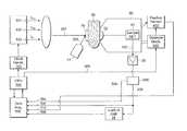
- FIG. 1is a schematic of a system for non-invasive measurement of a target analyte having a three-diode optical bridge apparatus.
- FIG. 1One embodiment of an optical bridge apparatus for measuring glucose concentration in blood based on transmitted light through the is shown in FIG. 1 .
- FIG. 1A similar apparatus may be designed which uses reflected or back-scattered light instead of transmitted light.
- three fixed-wavelength monochromatic light sources 101 , 102 , 103may be used to produce the probe beam 202 .
- Clock generator 110produces a timing signal at the desired chopping frequency f ch needed for time multiplexing of the principal and reference components, as well as the blood-estimation component, of the probe beam.
- the CPU 104generates signals for controlling the principal intensity I P , both wavelengths ⁇ P and ⁇ R , and the chopping frequency f ch of the probe beam 202 .
- the probe beamis directed onto diffuser plate 70 .
- Placing a diffuser plate in the beam path before the sampleprovides the advantage of minimizing the effects of the variation in the scattering properties of the sample.
- the preferred diffuser plateis of the holographic type that has substantially constant diffusing properties across the pertinent wavelength range.
- the sample specimen 80such as an earlobe, lip, cheek, nasal septum, tongue, or the skin between the fingers or toes of the subject, is placed between diffuser plate 70 and sample detector lens 92 , and is compressed by moving the measurement head 90 , mounted on compression mechanism 400 .
- the probe beam 202 transmitted through sample 80is focused by sample detector lens 92 , and directed to sample detector 91 .
- the sample detector 91detects the intensity at each of the wavelength periods of the probe beam 202 transmitted through sample 80 , and sends an electrical signal 302 to preamplifier 26 and on to a demultiplexer 405 that separates the blood estimation wavelength signal from the balanced-pair signal; the latter is fed to a phase sensitive detector (or lock-in amplifier) 24 .
- the output signal 308 from the phase sensitive detector 24is proportional to the difference (or ratio) of the principal and reference intensities detected by sample detector 91 . This signal 308 is referred to as the optical bridge signal.
- a separate auxiliary radiation sourcesuch as an infrared or visible-light LED 44 , is used to provide an estimate of the sample blood content.
- This auxiliary radiation source 44produces a blood detection beam 204 that is directed onto the diffuser plate 70 and into the sample.
- the sample detector 91can be used to detect the transmitted portion of the blood detection beam 204 , producing a blood signal 304 .
- the sample 80is introduced between diffuser plate 70 and sample detector lens 92 .
- the measurement head 90is moved by compression mechanism 400 to gently compress sample 80 until a predetermined pressure is exerted on sample 80 .
- the preferred embodiment of compression mechanism 400includes a miniature linear actuator. Its step size, speed and travel distance are controlled by the CPU 104 . Although this embodiment uses an electrical actuator, a hydraulic or a pneumatic actuator could also be used, with the ensuing advantages of compactness of the compression mechanism.
- Firstis the background matrix
- the secondis the target analyte
- the thirdis the unbound fluid attenuation.
- the background matrix attenuationresults from the absorption of probe beam 202 by sample constituents whose concentrations are substantially constant throughout fixed sample compartments.
- the target analyte attenuationis caused by absorption of probe beam 202 by the target analyte (e.g. glucose), which is mostly concentrated in the unbound fluid (e.g. blood).
- the unbound fluide.g. blood
- the unbound fluidalong with the target analyte (e.g. glucose)
- the concentration of the target analyte in the unbound fluidis different than its concentration in the background matrix (e.g. intracellular concentration), its average concentration in the beam path changes as a result of the compression. This concentration change allows the target analyte to be detected by this method.
- the principal wavelength ⁇ P of probe beam 202is selected in such a way to have high attenuation by the target analyte.
- the principal wavelength intensity I Pis set to achieve an optimal transmitted signal intensity.
- the effective reference wavelength ⁇ R of the probe beamis selected during the optical bridge balancing process. Its intensity I R should be adjusted before each measurement as explained below in the description of the measurement process.
- sample 80is sufficiently compressed to remove the major amount of unbound fluid from the sample tissue.
- the principal wavelength parameters ⁇ P and I Pare set, and the effective reference wavelength ⁇ R is initialized.
- the reference wavelength intensity I Ris set, while the principal wavelength intensity I P is adjusted to balance the bridge.
- the sample compression pressureis released by a predetermined amount (typically less than 0.1 mm) and the probe beam effective reference wavelength ⁇ R is adjusted by controlling the intensity ratio of ⁇ R1 and ⁇ R2 using a signal from CPU 104 so as to again achieve a substantially-zero optical bridge signal 308 .
- the initial compression pressureis chosen such that, even after releasing sample 80 by several times the incremental thickness, there is nearly no unbound fluid reflow into the sample. Changes in the optical bridge signal 308 , due to this thickness increase result merely from increased background matrix thickness and not from any substantial influx of fluid.
- Sample 80is then uncompressed again by one step thickness, and the intensity at the reference (or principal) wavelength is again adjusted by the CPU 104 to achieve minimum optical bridge signal.
- This stepwise increasing thickness proceduremay be continued until a substantially-zero optical bridge signal is obtained. Once the sample thickness has been increased, the procedure may also be reversed, using stepwise decreasing thicknesses. When this balancing procedure is completed, the absorption coefficient of sample 80 in its compressed state is substantially equal at the two wavelengths ⁇ P and effective ⁇ R .
- the balancingis limited to only one cycle in order to speed up the measurement and reduce the compression stress on the sample.
- the sample 80is maintained in the compressed state to displace the unbound fluid content for a time period of approximately 1 to 100 seconds.
- continuous measurements of the time-varying signalsbegin, including time-varying measurements of the optical bridge output 308 , blood signal 304 , and position sensor 402 output 312 .
- the compression mechanism 400then starts opening the measurement head 90 by an amount and rate set by the CPU 104 .
- the amount of head openingmay be fixed (e.g. 0.5 mm for a human ear), or may be thickness dependent (e.g. 20% of the compressed sample thickness). It is directly controlled from the subroutine for compression control, via connection 365 .
- the purpose for the fast opening phaseis to allow the unbound fluid that contains the target analyte to return into the sample.
- the opening of the compression mechanismcauses a change in the sample composition, which makes the sample absorb differently at the principal wavelength and the effective reference wavelength. This relative change in absorption results in a non-zero optical bridge signal 308 .
- the measurementscontinue until stopped by CPU 104 .
- the time-varying signal seriesshould contain several hundred data sets, which are recorded, in a data acquisition block 106 , over a measurement time period of approximately 0.1 to 10 seconds after the sample uncompression begins.
- the methodis here described as applied to an optical bridge employing non-tunable lasers, it can also be applied to different implementations of the optical bridge, such as one equipped with light emitting diodes or superluminescent light emitting diodes or other means to generate a beam containing the required wavelength combinations.
- the methodis here described with a focus toward measuring the concentration of glucose in blood, the method and apparatus of this invention may also be employed to detect the concentration of other analytes such as cholesterol, urea, heavy metals, alcohol, nicotine or drugs in blood or other fluids.
- sinusoidal, rather than square, modulation waveformsthat are set 180° out of phase and result in a substantially constant total intensity, can alternatively be used to form the combined radiation beam.
- measurements of radiation reflected or back-scattered by the tissue, rather than transmitted radiationcan be performed to obtain the desired data.
Landscapes
- Physics & Mathematics (AREA)
- Health & Medical Sciences (AREA)
- Life Sciences & Earth Sciences (AREA)
- Spectroscopy & Molecular Physics (AREA)
- Pathology (AREA)
- General Health & Medical Sciences (AREA)
- Surgery (AREA)
- Public Health (AREA)
- Biomedical Technology (AREA)
- Heart & Thoracic Surgery (AREA)
- Medical Informatics (AREA)
- Molecular Biology (AREA)
- Biophysics (AREA)
- Animal Behavior & Ethology (AREA)
- Veterinary Medicine (AREA)
- Engineering & Computer Science (AREA)
- Optics & Photonics (AREA)
- Chemical & Material Sciences (AREA)
- Analytical Chemistry (AREA)
- Biochemistry (AREA)
- General Physics & Mathematics (AREA)
- Immunology (AREA)
- Emergency Medicine (AREA)
- Toxicology (AREA)
- Measurement Of The Respiration, Hearing Ability, Form, And Blood Characteristics Of Living Organisms (AREA)
- Investigating Or Analysing Materials By Optical Means (AREA)
Abstract
Description
λR=(IR1·λR1+IR2·λR2)/(IR1+IR2)
- where IRi=intensity at wavelength λRi
Claims (6)
Priority Applications (10)
| Application Number | Priority Date | Filing Date | Title |
|---|---|---|---|
| US11/526,564US8175666B2 (en) | 2002-04-26 | 2006-09-25 | Three diode optical bridge system |
| CN2007800354593ACN101605493B (en) | 2006-09-25 | 2007-09-10 | Three diode optical bridge system |
| PCT/US2007/019635WO2008039299A2 (en) | 2006-09-25 | 2007-09-10 | Three diode optical bridge system |
| JP2009529183AJP5723098B2 (en) | 2006-09-25 | 2007-09-10 | Method and system for measuring substance concentration using electromagnetic radiation beam |
| HK10106047.8AHK1139294B (en) | 2006-09-25 | 2007-09-10 | Three diode optical bridge system |
| KR1020097008408AKR101597310B1 (en) | 2006-09-25 | 2007-09-10 | 3 Three diode optical bridge system |
| CA2700133ACA2700133C (en) | 2006-09-25 | 2007-09-10 | Three diode optical bridge system |
| EP07811716AEP2073697A2 (en) | 2006-09-25 | 2007-09-10 | Three diode optical bridge system |
| AU2007300695AAU2007300695B2 (en) | 2006-09-25 | 2007-09-10 | Three diode optical bridge system |
| IL197589AIL197589A (en) | 2006-09-25 | 2009-03-12 | Three diode optical bridge system |
Applications Claiming Priority (3)
| Application Number | Priority Date | Filing Date | Title |
|---|---|---|---|
| US10/134,310US7003337B2 (en) | 2002-04-26 | 2002-04-26 | Non-invasive substance concentration measurement using and optical bridge |
| US33583306A | 2006-01-19 | 2006-01-19 | |
| US11/526,564US8175666B2 (en) | 2002-04-26 | 2006-09-25 | Three diode optical bridge system |
Related Parent Applications (1)
| Application Number | Title | Priority Date | Filing Date |
|---|---|---|---|
| US33583306AContinuation-In-Part | 2002-04-26 | 2006-01-19 |
Publications (2)
| Publication Number | Publication Date |
|---|---|
| US20070208238A1 US20070208238A1 (en) | 2007-09-06 |
| US8175666B2true US8175666B2 (en) | 2012-05-08 |
Family
ID=39146866
Family Applications (1)
| Application Number | Title | Priority Date | Filing Date |
|---|---|---|---|
| US11/526,564Expired - Fee RelatedUS8175666B2 (en) | 2002-04-26 | 2006-09-25 | Three diode optical bridge system |
Country Status (9)
| Country | Link |
|---|---|
| US (1) | US8175666B2 (en) |
| EP (1) | EP2073697A2 (en) |
| JP (1) | JP5723098B2 (en) |
| KR (1) | KR101597310B1 (en) |
| CN (1) | CN101605493B (en) |
| AU (1) | AU2007300695B2 (en) |
| CA (1) | CA2700133C (en) |
| IL (1) | IL197589A (en) |
| WO (1) | WO2008039299A2 (en) |
Cited By (3)
| Publication number | Priority date | Publication date | Assignee | Title |
|---|---|---|---|---|
| US9442065B2 (en) | 2014-09-29 | 2016-09-13 | Zyomed Corp. | Systems and methods for synthesis of zyotons for use in collision computing for noninvasive blood glucose and other measurements |
| US9554738B1 (en) | 2016-03-30 | 2017-01-31 | Zyomed Corp. | Spectroscopic tomography systems and methods for noninvasive detection and measurement of analytes using collision computing |
| US20180217054A1 (en)* | 2015-08-18 | 2018-08-02 | Tokushima University | Concentration measurement device |
Families Citing this family (24)
| Publication number | Priority date | Publication date | Assignee | Title |
|---|---|---|---|---|
| JP5398009B2 (en)* | 2010-03-17 | 2014-01-29 | 学校法人北里研究所 | Optical coherence tomography apparatus and tomographic imaging method |
| WO2012132768A1 (en)* | 2011-03-31 | 2012-10-04 | テルモ株式会社 | Blood component measuring device |
| EP2543839A1 (en)* | 2011-07-04 | 2013-01-09 | Inergy Automotive Systems Research (Société Anonyme) | Device for measuring the concentration of urea |
| US20130267799A1 (en)* | 2012-04-06 | 2013-10-10 | Hannu Harjunmaa | Noninvasive measurement of analyte concentration using a fiberless transflectance probe |
| WO2013160780A1 (en)* | 2012-04-23 | 2013-10-31 | I.R.Med Ltd. | Short-wave infrared imaging and spectroscopy technique for inflammation classification and tumor and inflammation differentiation in human tissues inside the body |
| JP5859414B2 (en)* | 2012-10-16 | 2016-02-10 | 株式会社 堀場アドバンスドテクノ | Color meter |
| EP2969765B1 (en)* | 2013-03-13 | 2018-01-10 | United Technologies Corporation | Hydraulically operated latch for a gas turbine engine nacelle and method of operation |
| EP3011313A1 (en)* | 2013-06-16 | 2016-04-27 | Nielsen, Ulrik Merrild | Detection of indications of psychoactive components in a liquid |
| US9322716B2 (en)* | 2014-01-07 | 2016-04-26 | Panasonic Intellectual Property Corporation Of America | Component measuring apparatus and moving body |
| WO2015177867A1 (en)* | 2014-05-20 | 2015-11-26 | パイオニア株式会社 | Pulse oximeter |
| US20160051147A1 (en)* | 2014-08-21 | 2016-02-25 | Irmed | System and method for noninvasive analysis of subcutaneous tissue |
| US10772541B2 (en) | 2014-08-21 | 2020-09-15 | I. R. Med Ltd. | System and method for noninvasive analysis of subcutaneous tissue |
| US10709365B2 (en) | 2014-08-21 | 2020-07-14 | I. R. Med Ltd. | System and method for noninvasive analysis of subcutaneous tissue |
| CN105249974A (en)* | 2015-10-15 | 2016-01-20 | 华南师范大学 | Pressure-modulation-spectrum-technology-based noninvasive glucose detection system and method |
| CN105784591A (en)* | 2015-12-26 | 2016-07-20 | 深圳市前海安测信息技术有限公司 | Blood glucose test data acquisition system and method |
| US11129556B2 (en) | 2015-12-31 | 2021-09-28 | Wear2B Ltd. | Device, system and method for non-invasive monitoring of physiological measurements |
| US10088468B2 (en) | 2016-02-04 | 2018-10-02 | Nova Biomedical Corporation | Analyte system and method for determining hemoglobin parameters in whole blood |
| CN109863390B (en)* | 2016-10-25 | 2021-10-01 | 日本先锋公司 | Fluid Measuring Equipment |
| KR102005832B1 (en)* | 2018-02-21 | 2019-08-01 | 주식회사 올리브헬스케어 | Signal processing device for bio-signal analysing and bio-signal analysing apparatus using the same |
| CN109406492B (en)* | 2018-12-06 | 2023-12-22 | 深圳网联光仪科技有限公司 | Device capable of measuring Raman spectrum of substance under strong fluorescent background |
| EP4164490A4 (en)* | 2020-06-11 | 2023-12-06 | Siemens Healthcare Diagnostics, Inc. | Method and analyzer to correct for unknown interferences in a patient blood sample |
| EP4392785A4 (en) | 2021-08-27 | 2025-05-07 | Bilibaby, Llc | Systems and methods for determining and communicating levels of bilirubin and other subcutaneous substances |
| TWI817195B (en)* | 2021-09-07 | 2023-10-01 | 劉茂誠 | System and method for optical detection of turbid liquid |
| CN113984663A (en)* | 2021-10-28 | 2022-01-28 | 中国人民解放军海军特色医学中心 | An optical detection device and its working method |
Citations (87)
| Publication number | Priority date | Publication date | Assignee | Title |
|---|---|---|---|---|
| US1758088A (en) | 1926-09-04 | 1930-05-13 | Siemens Ag | Method of and means for determining alpha constituent in alpha mixture of substances |
| US2721942A (en) | 1948-07-15 | 1955-10-25 | Du Pont | Infrared analyzer and method |
| US3463142A (en) | 1966-07-05 | 1969-08-26 | Trw Inc | Blood content monitor |
| US3489906A (en) | 1965-04-26 | 1970-01-13 | Autokemi Ab | Optical system for measuring absorption of light |
| US3614450A (en) | 1969-02-17 | 1971-10-19 | Measurex Corp | Apparatus for measuring the amount of a substance that is associated with a base material |
| US3638640A (en) | 1967-11-01 | 1972-02-01 | Robert F Shaw | Oximeter and method for in vivo determination of oxygen saturation in blood using three or more different wavelengths |
| US3799672A (en) | 1972-09-15 | 1974-03-26 | Us Health Education & Welfare | Oximeter for monitoring oxygen saturation in blood |
| US3926527A (en) | 1974-06-05 | 1975-12-16 | Philco Ford Corp | Rotating gas correlation cell |
| US3957037A (en) | 1975-02-24 | 1976-05-18 | Nasa | Readout electrode assembly for measuring biological impedance |
| US3958560A (en) | 1974-11-25 | 1976-05-25 | Wayne Front March | Non-invasive automatic glucose sensor system |
| US3963019A (en) | 1974-11-25 | 1976-06-15 | Quandt Robert S | Ocular testing method and apparatus |
| US4029085A (en) | 1976-03-26 | 1977-06-14 | Purdue Research Foundation | Method for determining bilirubin concentration from skin reflectance |
| US4033330A (en) | 1975-09-08 | 1977-07-05 | Hewlett-Packard Company | Transcutaneous pH measuring instrument |
| US4169676A (en) | 1976-02-20 | 1979-10-02 | Nils Kaiser | Method for determining the contents of metabolic products in the blood |
| US4266554A (en) | 1978-06-22 | 1981-05-12 | Minolta Camera Kabushiki Kaisha | Digital oximeter |
| US4267844A (en) | 1978-05-15 | 1981-05-19 | Minolta Camera Kabushiki Kaisha | Medical instrument for determining jaundice |
| US4306877A (en) | 1978-07-29 | 1981-12-22 | Max-Planck-Gesellschaft Zur Forderung Der Wissenschaften E.V. | Optical measurement of concentration |
| US4321930A (en) | 1977-06-28 | 1982-03-30 | Duke University, Inc. | Apparatus for monitoring metabolism in body organs |
| US4398541A (en) | 1978-05-25 | 1983-08-16 | Xienta, Inc. | Method and apparatus for measuring moisture content of skin |
| US4427889A (en) | 1979-08-23 | 1984-01-24 | Carl Zeiss Stiftung | Method and apparatus for molecular spectroscopy, particularly for the determination of products of metabolism |
| US4447150A (en) | 1981-02-27 | 1984-05-08 | Bentley Laboratories | Apparatus and method for measuring blood oxygen saturation |
| US4485820A (en) | 1982-05-10 | 1984-12-04 | The Johns Hopkins University | Method and apparatus for the continuous monitoring of hemoglobin saturation in the blood of premature infants |
| US4490845A (en) | 1982-02-02 | 1984-12-25 | Westinghouse Electric Corp. | Automated acousto-optic infrared analyzer system |
| US4513751A (en) | 1979-03-07 | 1985-04-30 | Sumitomo Electric Industries, Ltd. | Method for measuring oxygen metabolism in internal organ or tissue |
| US4523279A (en) | 1980-11-24 | 1985-06-11 | Oximetrix, Inc. | Apparatus for determining oxygen saturation levels in blood |
| EP0152979A1 (en) | 1984-02-07 | 1985-08-28 | B.V. Optische Industrie "De Oude Delft" | Device for detecting differences in color |
| US4570638A (en) | 1983-10-14 | 1986-02-18 | Somanetics Corporation | Method and apparatus for spectral transmissibility examination and analysis |
| US4586513A (en) | 1982-02-19 | 1986-05-06 | Minolta Camera Kabushiki Kaisha | Noninvasive device for photoelectrically measuring the property of arterial blood |
| US4603700A (en) | 1983-12-09 | 1986-08-05 | The Boc Group, Inc. | Probe monitoring system for oximeter |
| US4621643A (en) | 1982-09-02 | 1986-11-11 | Nellcor Incorporated | Calibrated optical oximeter probe |
| US4641658A (en) | 1984-10-01 | 1987-02-10 | American Hospital Supply Corp. | Cardiac flow monitor |
| US4653498A (en) | 1982-09-13 | 1987-03-31 | Nellcor Incorporated | Pulse oximeter monitor |
| US4655225A (en) | 1985-04-18 | 1987-04-07 | Kurabo Industries Ltd. | Spectrophotometric method and apparatus for the non-invasive |
| EP0074428B1 (en) | 1981-09-15 | 1987-04-08 | Arno Dr. Dipl.-Phys. Müller | Method and device for the quantitative determination of dissolved substances in single- or multicomponent systems of laser light scattering |
| EP0226822A2 (en) | 1985-11-21 | 1987-07-01 | Hellige GmbH | Apparatus for continuous determination of changes in concentration in mixtures |
| US4704029A (en) | 1985-12-26 | 1987-11-03 | Research Corporation | Blood glucose monitor |
| US4714080A (en) | 1986-10-06 | 1987-12-22 | Nippon Colin Co., Ltd. | Method and apparatus for noninvasive monitoring of arterial blood oxygen saturation |
| US4725147A (en) | 1984-09-17 | 1988-02-16 | Somanetics Corporation | Calibration method and apparatus for optical-response tissue-examination instrument |
| US4750496A (en) | 1987-01-28 | 1988-06-14 | Xienta, Inc. | Method and apparatus for measuring blood glucose concentration |
| US4759369A (en) | 1986-07-07 | 1988-07-26 | Novametrix Medical Systems, Inc. | Pulse oximeter |
| US4768516A (en) | 1983-10-14 | 1988-09-06 | Somanetics Corporation | Method and apparatus for in vivo evaluation of tissue composition |
| US4785814A (en) | 1987-08-11 | 1988-11-22 | Cordis Corporation | Optical probe for measuring pH and oxygen in blood and employing a composite membrane |
| US4796636A (en) | 1987-09-10 | 1989-01-10 | Nippon Colin Co., Ltd. | Noninvasive reflectance oximeter |
| US4805623A (en) | 1987-09-04 | 1989-02-21 | Vander Corporation | Spectrophotometric method for quantitatively determining the concentration of a dilute component in a light- or other radiation-scattering environment |
| US4817623A (en) | 1983-10-14 | 1989-04-04 | Somanetics Corporation | Method and apparatus for interpreting optical response data |
| US4819752A (en)* | 1987-10-02 | 1989-04-11 | Datascope Corp. | Blood constituent measuring device and method |
| EP0160768B1 (en) | 1984-05-04 | 1989-05-03 | Kurabo Industries Ltd. | Spectrophotometric apparatus for the non-invasive determination of glucose in body tissues |
| US4832484A (en) | 1986-10-29 | 1989-05-23 | Nihon Kohden Corporation | Apparatus for determining the concentration of a light-absorbing material in blood |
| US4854699A (en) | 1987-11-02 | 1989-08-08 | Nippon Colin Co., Ltd. | Backscatter oximeter |
| US4863265A (en) | 1987-10-16 | 1989-09-05 | Mine Safety Appliances Company | Apparatus and method for measuring blood constituents |
| US4867557A (en) | 1987-04-09 | 1989-09-19 | Sumitomo Electric Industries, Ltd. | Reflection type oximeter for applying light pulses to a body tissue to measure oxygen saturation |
| US4882492A (en) | 1988-01-19 | 1989-11-21 | Biotronics Associates, Inc. | Non-invasive near infrared measurement of blood analyte concentrations |
| US4883055A (en) | 1988-03-11 | 1989-11-28 | Puritan-Bennett Corporation | Artificially induced blood pulse for use with a pulse oximeter |
| US4907594A (en) | 1987-07-18 | 1990-03-13 | Nicolay Gmbh | Method for the determination of the saturation of the blood of a living organism with oxygen and electronic circuit for performing this method |
| WO1990004353A2 (en) | 1988-10-28 | 1990-05-03 | Dietrich Gravenstein | Method for noninvasive intermittent and/or continuous hemoglobin, arterial oxygen content, and hematocrit determination |
| US4927264A (en) | 1987-12-02 | 1990-05-22 | Omron Tateisi Electronics Co. | Non-invasive measuring method and apparatus of blood constituents |
| WO1990007905A1 (en) | 1989-01-19 | 1990-07-26 | Futrex, Inc. | Non-invasive measurement of blood glucose |
| US4975581A (en) | 1989-06-21 | 1990-12-04 | University Of New Mexico | Method of and apparatus for determining the similarity of a biological analyte from a model constructed from known biological fluids |
| EP0407992A1 (en) | 1989-07-13 | 1991-01-16 | Kyoto Daiichi Kagaku Co., Ltd. | Method for determination of glucose concentration |
| US5054487A (en) | 1990-02-02 | 1991-10-08 | Boston Advanced Technologies, Inc. | Laser systems for material analysis based on reflectance ratio detection |
| WO1991015991A1 (en) | 1990-04-19 | 1991-10-31 | Worcester Polytechnic Institute | Method and apparatus for monitoring blood analytes noninvasively by pulsatile photoplethysmography |
| US5099123A (en) | 1989-05-23 | 1992-03-24 | Biosensors Technology, Inc. | Method for determining by absorption of radiations the concentration of substances in absorbing and turbid matrices |
| US5112124A (en) | 1990-04-19 | 1992-05-12 | Worcester Polytechnic Institute | Method and apparatus for measuring the concentration of absorbing substances |
| WO1992017765A1 (en) | 1991-03-28 | 1992-10-15 | Johnson & Johnson Professional Products Limited | Method and apparatus for glucose concentration monitoring |
| US5167230A (en) | 1988-11-02 | 1992-12-01 | Nim, Inc. | User-wearable hemoglobinometer for measuring the metabolic condition of a subject |
| US5178142A (en)* | 1989-05-23 | 1993-01-12 | Vivascan Corporation | Electromagnetic method and apparatus to measure constituents of human or animal tissue |
| WO1993000855A1 (en) | 1991-07-03 | 1993-01-21 | Vivascan Corporation | Electromagnetic method and apparatus to measure constituents of human or animal tissue |
| US5183042A (en) | 1989-05-23 | 1993-02-02 | Vivascan Corporation | Electromagnetic method and apparatus to measure constituents of human or animal tissue |
| US5190040A (en) | 1986-12-26 | 1993-03-02 | Nihon Kohden Corporation | Apparatus for measuring the change in the concentration of a pigment in blood |
| US5267152A (en) | 1989-10-28 | 1993-11-30 | Yang Won S | Non-invasive method and apparatus for measuring blood glucose concentration |
| US5277181A (en) | 1991-12-12 | 1994-01-11 | Vivascan Corporation | Noninvasive measurement of hematocrit and hemoglobin content by differential optical analysis |
| US5322067A (en) | 1993-02-03 | 1994-06-21 | Hewlett-Packard Company | Method and apparatus for determining the volume of a body cavity in real time |
| US5348002A (en)* | 1992-04-23 | 1994-09-20 | Sirraya, Inc. | Method and apparatus for material analysis |
| US5372135A (en) | 1991-12-31 | 1994-12-13 | Vivascan Corporation | Blood constituent determination based on differential spectral analysis |
| US5620000A (en) | 1993-07-02 | 1997-04-15 | Heidelberg Engineering, Optische Messsysteme Gmbh | Method and apparatus for measuring flow rate, particularly of blood |
| US5746206A (en) | 1995-03-14 | 1998-05-05 | Nellcor Incorporated | Isolated layer pulse oximetry |
| WO1998043096A2 (en) | 1997-03-25 | 1998-10-01 | Siemens Aktiengesellschaft | Method and device for non-invasive in vivo determination of blood constituents |
| US5827181A (en) | 1995-09-07 | 1998-10-27 | Hewlett-Packard Co. | Noninvasive blood chemistry measurement method and system |
| EP0997103A1 (en) | 1998-10-29 | 2000-05-03 | Colin Corporation | Non-invasive and continuous blood-pressure estimation apparatus |
| US6151516A (en)* | 1995-06-07 | 2000-11-21 | Masimo Laboratories | Active pulse blood constituent monitoring |
| US6223063B1 (en) | 1998-01-27 | 2001-04-24 | Lightouch Medical, Inc. | Method and device for tissue modulation |
| US6292686B1 (en) | 1998-04-24 | 2001-09-18 | Lightouch Medical, Inc. | Apparatus and method for thermal tissue modulation |
| EP1189074A2 (en) | 2000-09-14 | 2002-03-20 | GE Medical Systems Global Technology Company LLC | Method and apparatus for locking sample volume onto moving vessel in pulsed doppler ultrasound imaging |
| US6400972B1 (en) | 1998-06-17 | 2002-06-04 | Orsense Ltd. | Non-invasive method and system of optical measurements for determining the concentration of a substance in blood |
| US6615061B1 (en) | 1998-11-23 | 2003-09-02 | Abbott Laboratories | Optical sensor having a selectable sampling distance for determination of analytes |
| US20030204133A1 (en) | 2002-04-26 | 2003-10-30 | Hannu Harjunmaa | Non-invasive substance concentration measurement using and optical bridge |
| US20040173737A1 (en) | 2003-02-12 | 2004-09-09 | Mitutoyo Corporation | Optical configuration for imaging-type optical encoders |
- 2006
- 2006-09-25USUS11/526,564patent/US8175666B2/ennot_activeExpired - Fee Related
- 2007
- 2007-09-10KRKR1020097008408Apatent/KR101597310B1/ennot_activeExpired - Fee Related
- 2007-09-10WOPCT/US2007/019635patent/WO2008039299A2/enactiveApplication Filing
- 2007-09-10JPJP2009529183Apatent/JP5723098B2/ennot_activeExpired - Fee Related
- 2007-09-10EPEP07811716Apatent/EP2073697A2/ennot_activeWithdrawn
- 2007-09-10CNCN2007800354593Apatent/CN101605493B/ennot_activeExpired - Fee Related
- 2007-09-10CACA2700133Apatent/CA2700133C/ennot_activeExpired - Fee Related
- 2007-09-10AUAU2007300695Apatent/AU2007300695B2/ennot_activeCeased
- 2009
- 2009-03-12ILIL197589Apatent/IL197589A/ennot_activeIP Right Cessation
Patent Citations (94)
| Publication number | Priority date | Publication date | Assignee | Title |
|---|---|---|---|---|
| US1758088A (en) | 1926-09-04 | 1930-05-13 | Siemens Ag | Method of and means for determining alpha constituent in alpha mixture of substances |
| US2721942A (en) | 1948-07-15 | 1955-10-25 | Du Pont | Infrared analyzer and method |
| US3489906A (en) | 1965-04-26 | 1970-01-13 | Autokemi Ab | Optical system for measuring absorption of light |
| US3463142A (en) | 1966-07-05 | 1969-08-26 | Trw Inc | Blood content monitor |
| US3638640A (en) | 1967-11-01 | 1972-02-01 | Robert F Shaw | Oximeter and method for in vivo determination of oxygen saturation in blood using three or more different wavelengths |
| US3614450A (en) | 1969-02-17 | 1971-10-19 | Measurex Corp | Apparatus for measuring the amount of a substance that is associated with a base material |
| US3799672A (en) | 1972-09-15 | 1974-03-26 | Us Health Education & Welfare | Oximeter for monitoring oxygen saturation in blood |
| US3926527A (en) | 1974-06-05 | 1975-12-16 | Philco Ford Corp | Rotating gas correlation cell |
| US3963019A (en) | 1974-11-25 | 1976-06-15 | Quandt Robert S | Ocular testing method and apparatus |
| US3958560A (en) | 1974-11-25 | 1976-05-25 | Wayne Front March | Non-invasive automatic glucose sensor system |
| US3957037A (en) | 1975-02-24 | 1976-05-18 | Nasa | Readout electrode assembly for measuring biological impedance |
| US4033330A (en) | 1975-09-08 | 1977-07-05 | Hewlett-Packard Company | Transcutaneous pH measuring instrument |
| US4169676A (en) | 1976-02-20 | 1979-10-02 | Nils Kaiser | Method for determining the contents of metabolic products in the blood |
| US4029085A (en) | 1976-03-26 | 1977-06-14 | Purdue Research Foundation | Method for determining bilirubin concentration from skin reflectance |
| US4380240A (en) | 1977-06-28 | 1983-04-19 | Duke University, Inc. | Apparatus for monitoring metabolism in body organs |
| US4321930A (en) | 1977-06-28 | 1982-03-30 | Duke University, Inc. | Apparatus for monitoring metabolism in body organs |
| US4267844A (en) | 1978-05-15 | 1981-05-19 | Minolta Camera Kabushiki Kaisha | Medical instrument for determining jaundice |
| US4398541A (en) | 1978-05-25 | 1983-08-16 | Xienta, Inc. | Method and apparatus for measuring moisture content of skin |
| US4266554A (en) | 1978-06-22 | 1981-05-12 | Minolta Camera Kabushiki Kaisha | Digital oximeter |
| US4306877A (en) | 1978-07-29 | 1981-12-22 | Max-Planck-Gesellschaft Zur Forderung Der Wissenschaften E.V. | Optical measurement of concentration |
| US4513751A (en) | 1979-03-07 | 1985-04-30 | Sumitomo Electric Industries, Ltd. | Method for measuring oxygen metabolism in internal organ or tissue |
| US4427889A (en) | 1979-08-23 | 1984-01-24 | Carl Zeiss Stiftung | Method and apparatus for molecular spectroscopy, particularly for the determination of products of metabolism |
| US4523279A (en) | 1980-11-24 | 1985-06-11 | Oximetrix, Inc. | Apparatus for determining oxygen saturation levels in blood |
| US4447150A (en) | 1981-02-27 | 1984-05-08 | Bentley Laboratories | Apparatus and method for measuring blood oxygen saturation |
| EP0074428B1 (en) | 1981-09-15 | 1987-04-08 | Arno Dr. Dipl.-Phys. Müller | Method and device for the quantitative determination of dissolved substances in single- or multicomponent systems of laser light scattering |
| US4490845A (en) | 1982-02-02 | 1984-12-25 | Westinghouse Electric Corp. | Automated acousto-optic infrared analyzer system |
| US4694833A (en) | 1982-02-19 | 1987-09-22 | Minolta Camera Kabushiki Kaisha | Noninvasive device for photoelectrically measuring the property of arterial blood |
| US4586513A (en) | 1982-02-19 | 1986-05-06 | Minolta Camera Kabushiki Kaisha | Noninvasive device for photoelectrically measuring the property of arterial blood |
| US4485820A (en) | 1982-05-10 | 1984-12-04 | The Johns Hopkins University | Method and apparatus for the continuous monitoring of hemoglobin saturation in the blood of premature infants |
| US4621643A (en) | 1982-09-02 | 1986-11-11 | Nellcor Incorporated | Calibrated optical oximeter probe |
| US4653498A (en) | 1982-09-13 | 1987-03-31 | Nellcor Incorporated | Pulse oximeter monitor |
| US4653498B1 (en) | 1982-09-13 | 1989-04-18 | ||
| US4768516A (en) | 1983-10-14 | 1988-09-06 | Somanetics Corporation | Method and apparatus for in vivo evaluation of tissue composition |
| US4817623A (en) | 1983-10-14 | 1989-04-04 | Somanetics Corporation | Method and apparatus for interpreting optical response data |
| US4570638A (en) | 1983-10-14 | 1986-02-18 | Somanetics Corporation | Method and apparatus for spectral transmissibility examination and analysis |
| US4603700A (en) | 1983-12-09 | 1986-08-05 | The Boc Group, Inc. | Probe monitoring system for oximeter |
| EP0152979A1 (en) | 1984-02-07 | 1985-08-28 | B.V. Optische Industrie "De Oude Delft" | Device for detecting differences in color |
| EP0160768B1 (en) | 1984-05-04 | 1989-05-03 | Kurabo Industries Ltd. | Spectrophotometric apparatus for the non-invasive determination of glucose in body tissues |
| US4725147A (en) | 1984-09-17 | 1988-02-16 | Somanetics Corporation | Calibration method and apparatus for optical-response tissue-examination instrument |
| US4641658A (en) | 1984-10-01 | 1987-02-10 | American Hospital Supply Corp. | Cardiac flow monitor |
| US4655225A (en) | 1985-04-18 | 1987-04-07 | Kurabo Industries Ltd. | Spectrophotometric method and apparatus for the non-invasive |
| EP0226822A2 (en) | 1985-11-21 | 1987-07-01 | Hellige GmbH | Apparatus for continuous determination of changes in concentration in mixtures |
| US4704029A (en) | 1985-12-26 | 1987-11-03 | Research Corporation | Blood glucose monitor |
| US4759369A (en) | 1986-07-07 | 1988-07-26 | Novametrix Medical Systems, Inc. | Pulse oximeter |
| US4714080A (en) | 1986-10-06 | 1987-12-22 | Nippon Colin Co., Ltd. | Method and apparatus for noninvasive monitoring of arterial blood oxygen saturation |
| US4832484A (en) | 1986-10-29 | 1989-05-23 | Nihon Kohden Corporation | Apparatus for determining the concentration of a light-absorbing material in blood |
| US5190040A (en) | 1986-12-26 | 1993-03-02 | Nihon Kohden Corporation | Apparatus for measuring the change in the concentration of a pigment in blood |
| US4750496A (en) | 1987-01-28 | 1988-06-14 | Xienta, Inc. | Method and apparatus for measuring blood glucose concentration |
| US4867557A (en) | 1987-04-09 | 1989-09-19 | Sumitomo Electric Industries, Ltd. | Reflection type oximeter for applying light pulses to a body tissue to measure oxygen saturation |
| US4907594A (en) | 1987-07-18 | 1990-03-13 | Nicolay Gmbh | Method for the determination of the saturation of the blood of a living organism with oxygen and electronic circuit for performing this method |
| US4785814A (en) | 1987-08-11 | 1988-11-22 | Cordis Corporation | Optical probe for measuring pH and oxygen in blood and employing a composite membrane |
| US4805623A (en) | 1987-09-04 | 1989-02-21 | Vander Corporation | Spectrophotometric method for quantitatively determining the concentration of a dilute component in a light- or other radiation-scattering environment |
| US4796636A (en) | 1987-09-10 | 1989-01-10 | Nippon Colin Co., Ltd. | Noninvasive reflectance oximeter |
| US4819752A (en)* | 1987-10-02 | 1989-04-11 | Datascope Corp. | Blood constituent measuring device and method |
| US4863265A (en) | 1987-10-16 | 1989-09-05 | Mine Safety Appliances Company | Apparatus and method for measuring blood constituents |
| US4854699A (en) | 1987-11-02 | 1989-08-08 | Nippon Colin Co., Ltd. | Backscatter oximeter |
| US4927264A (en) | 1987-12-02 | 1990-05-22 | Omron Tateisi Electronics Co. | Non-invasive measuring method and apparatus of blood constituents |
| US4882492A (en) | 1988-01-19 | 1989-11-21 | Biotronics Associates, Inc. | Non-invasive near infrared measurement of blood analyte concentrations |
| US4883055A (en) | 1988-03-11 | 1989-11-28 | Puritan-Bennett Corporation | Artificially induced blood pulse for use with a pulse oximeter |
| WO1990004353A2 (en) | 1988-10-28 | 1990-05-03 | Dietrich Gravenstein | Method for noninvasive intermittent and/or continuous hemoglobin, arterial oxygen content, and hematocrit determination |
| US5167230A (en) | 1988-11-02 | 1992-12-01 | Nim, Inc. | User-wearable hemoglobinometer for measuring the metabolic condition of a subject |
| WO1990007905A1 (en) | 1989-01-19 | 1990-07-26 | Futrex, Inc. | Non-invasive measurement of blood glucose |
| US5028787A (en) | 1989-01-19 | 1991-07-02 | Futrex, Inc. | Non-invasive measurement of blood glucose |
| US5183042A (en) | 1989-05-23 | 1993-02-02 | Vivascan Corporation | Electromagnetic method and apparatus to measure constituents of human or animal tissue |
| US5178142A (en)* | 1989-05-23 | 1993-01-12 | Vivascan Corporation | Electromagnetic method and apparatus to measure constituents of human or animal tissue |
| US5099123A (en) | 1989-05-23 | 1992-03-24 | Biosensors Technology, Inc. | Method for determining by absorption of radiations the concentration of substances in absorbing and turbid matrices |
| US4975581A (en) | 1989-06-21 | 1990-12-04 | University Of New Mexico | Method of and apparatus for determining the similarity of a biological analyte from a model constructed from known biological fluids |
| EP0404562B1 (en) | 1989-06-21 | 1998-08-05 | University Of New Mexico | Method of and apparatus for determining the similarity of a biological analyte from a model constructed from known biological fluids |
| EP0407992A1 (en) | 1989-07-13 | 1991-01-16 | Kyoto Daiichi Kagaku Co., Ltd. | Method for determination of glucose concentration |
| US5267152A (en) | 1989-10-28 | 1993-11-30 | Yang Won S | Non-invasive method and apparatus for measuring blood glucose concentration |
| US5054487A (en) | 1990-02-02 | 1991-10-08 | Boston Advanced Technologies, Inc. | Laser systems for material analysis based on reflectance ratio detection |
| US5137023A (en) | 1990-04-19 | 1992-08-11 | Worcester Polytechnic Institute | Method and apparatus for monitoring blood analytes noninvasively by pulsatile photoplethysmography |
| US5112124A (en) | 1990-04-19 | 1992-05-12 | Worcester Polytechnic Institute | Method and apparatus for measuring the concentration of absorbing substances |
| WO1991015991A1 (en) | 1990-04-19 | 1991-10-31 | Worcester Polytechnic Institute | Method and apparatus for monitoring blood analytes noninvasively by pulsatile photoplethysmography |
| WO1992017765A1 (en) | 1991-03-28 | 1992-10-15 | Johnson & Johnson Professional Products Limited | Method and apparatus for glucose concentration monitoring |
| WO1993000855A1 (en) | 1991-07-03 | 1993-01-21 | Vivascan Corporation | Electromagnetic method and apparatus to measure constituents of human or animal tissue |
| US5277181A (en) | 1991-12-12 | 1994-01-11 | Vivascan Corporation | Noninvasive measurement of hematocrit and hemoglobin content by differential optical analysis |
| US5372135A (en) | 1991-12-31 | 1994-12-13 | Vivascan Corporation | Blood constituent determination based on differential spectral analysis |
| US5348002A (en)* | 1992-04-23 | 1994-09-20 | Sirraya, Inc. | Method and apparatus for material analysis |
| US5322067A (en) | 1993-02-03 | 1994-06-21 | Hewlett-Packard Company | Method and apparatus for determining the volume of a body cavity in real time |
| US5620000A (en) | 1993-07-02 | 1997-04-15 | Heidelberg Engineering, Optische Messsysteme Gmbh | Method and apparatus for measuring flow rate, particularly of blood |
| US5746206A (en) | 1995-03-14 | 1998-05-05 | Nellcor Incorporated | Isolated layer pulse oximetry |
| US6151516A (en)* | 1995-06-07 | 2000-11-21 | Masimo Laboratories | Active pulse blood constituent monitoring |
| US5827181A (en) | 1995-09-07 | 1998-10-27 | Hewlett-Packard Co. | Noninvasive blood chemistry measurement method and system |
| WO1998043096A2 (en) | 1997-03-25 | 1998-10-01 | Siemens Aktiengesellschaft | Method and device for non-invasive in vivo determination of blood constituents |
| US6285894B1 (en) | 1997-03-25 | 2001-09-04 | Siemens Aktiengesellschaft | Method and device for non-invasive in vivo determination of blood constituents |
| US6223063B1 (en) | 1998-01-27 | 2001-04-24 | Lightouch Medical, Inc. | Method and device for tissue modulation |
| US6292686B1 (en) | 1998-04-24 | 2001-09-18 | Lightouch Medical, Inc. | Apparatus and method for thermal tissue modulation |
| US6400972B1 (en) | 1998-06-17 | 2002-06-04 | Orsense Ltd. | Non-invasive method and system of optical measurements for determining the concentration of a substance in blood |
| EP0997103A1 (en) | 1998-10-29 | 2000-05-03 | Colin Corporation | Non-invasive and continuous blood-pressure estimation apparatus |
| US6615061B1 (en) | 1998-11-23 | 2003-09-02 | Abbott Laboratories | Optical sensor having a selectable sampling distance for determination of analytes |
| EP1189074A2 (en) | 2000-09-14 | 2002-03-20 | GE Medical Systems Global Technology Company LLC | Method and apparatus for locking sample volume onto moving vessel in pulsed doppler ultrasound imaging |
| US20030204133A1 (en) | 2002-04-26 | 2003-10-30 | Hannu Harjunmaa | Non-invasive substance concentration measurement using and optical bridge |
| US20040173737A1 (en) | 2003-02-12 | 2004-09-09 | Mitutoyo Corporation | Optical configuration for imaging-type optical encoders |
Non-Patent Citations (7)
Cited By (11)
| Publication number | Priority date | Publication date | Assignee | Title |
|---|---|---|---|---|
| US9442065B2 (en) | 2014-09-29 | 2016-09-13 | Zyomed Corp. | Systems and methods for synthesis of zyotons for use in collision computing for noninvasive blood glucose and other measurements |
| US9448165B2 (en) | 2014-09-29 | 2016-09-20 | Zyomed Corp. | Systems and methods for control of illumination or radiation collection for blood glucose and other analyte detection and measurement using collision computing |
| US9448164B2 (en) | 2014-09-29 | 2016-09-20 | Zyomed Corp. | Systems and methods for noninvasive blood glucose and other analyte detection and measurement using collision computing |
| US9453794B2 (en) | 2014-09-29 | 2016-09-27 | Zyomed Corp. | Systems and methods for blood glucose and other analyte detection and measurement using collision computing |
| US9459201B2 (en) | 2014-09-29 | 2016-10-04 | Zyomed Corp. | Systems and methods for noninvasive blood glucose and other analyte detection and measurement using collision computing |
| US9459202B2 (en) | 2014-09-29 | 2016-10-04 | Zyomed Corp. | Systems and methods for collision computing for detection and noninvasive measurement of blood glucose and other substances and events |
| US9459203B2 (en) | 2014-09-29 | 2016-10-04 | Zyomed, Corp. | Systems and methods for generating and using projector curve sets for universal calibration for noninvasive blood glucose and other measurements |
| US9610018B2 (en) | 2014-09-29 | 2017-04-04 | Zyomed Corp. | Systems and methods for measurement of heart rate and other heart-related characteristics from photoplethysmographic (PPG) signals using collision computing |
| US20180217054A1 (en)* | 2015-08-18 | 2018-08-02 | Tokushima University | Concentration measurement device |
| US10976240B2 (en)* | 2015-08-18 | 2021-04-13 | Tokushima University | Concentration measurement device |
| US9554738B1 (en) | 2016-03-30 | 2017-01-31 | Zyomed Corp. | Spectroscopic tomography systems and methods for noninvasive detection and measurement of analytes using collision computing |
Also Published As
| Publication number | Publication date |
|---|---|
| HK1139294A1 (en) | 2010-09-17 |
| IL197589A0 (en) | 2009-12-24 |
| WO2008039299A3 (en) | 2008-05-29 |
| KR101597310B1 (en) | 2016-02-24 |
| US20070208238A1 (en) | 2007-09-06 |
| CN101605493A (en) | 2009-12-16 |
| CA2700133A1 (en) | 2008-04-03 |
| JP2010504148A (en) | 2010-02-12 |
| AU2007300695B2 (en) | 2012-10-04 |
| EP2073697A2 (en) | 2009-07-01 |
| IL197589A (en) | 2014-12-31 |
| KR20090086202A (en) | 2009-08-11 |
| JP5723098B2 (en) | 2015-05-27 |
| CN101605493B (en) | 2012-03-14 |
| AU2007300695A1 (en) | 2008-04-03 |
| CA2700133C (en) | 2016-05-10 |
| WO2008039299A2 (en) | 2008-04-03 |
Similar Documents
| Publication | Publication Date | Title |
|---|---|---|
| US8175666B2 (en) | Three diode optical bridge system | |
| CA2485964C (en) | Non-invasive substance concentration measurement using an optical bridge | |
| US5183042A (en) | Electromagnetic method and apparatus to measure constituents of human or animal tissue | |
| US7343185B2 (en) | Measurement of body compounds | |
| US8326390B2 (en) | Optical non-invasive blood monitoring system and method | |
| CN1327812C (en) | Blood component value measuring device without drawing blood | |
| US20070078312A1 (en) | Method and system for non-invasive measurements in a human body | |
| US20060167349A1 (en) | Apparatus for non-invasive determination of direction and rate of change of an analyte | |
| WO1991015991A1 (en) | Method and apparatus for monitoring blood analytes noninvasively by pulsatile photoplethysmography | |
| GB2329015A (en) | Infra-red multi-wavelength non-invasive measurement of blood component concentrations | |
| WO2001096872A2 (en) | Method and device for measuring concentration of glucose or other substances in blood | |
| JP2000189404A (en) | Blood glucose measurement method and apparatus | |
| CA2597859A1 (en) | Method and apparatus for determining blood analytes | |
| JP2000258343A (en) | Blood glucose measurement method and apparatus | |
| KR20020005697A (en) | Method for improving calibration of a blood monitoring instrument | |
| JP5400483B2 (en) | Component concentration analyzer and component concentration analysis method | |
| JPH07132120A (en) | Nonaggressive measuring method and device of specimen concentration using discontinuous emission | |
| US10219755B2 (en) | Noninvasive measurement of analyte concentration using methods and systems of post-balancing | |
| JP2003149145A (en) | Non-invasive blood glucose meter | |
| HK1139294B (en) | Three diode optical bridge system | |
| Spanner et al. | Noninvasive Determination of Blood Contents | |
| Doemer et al. | Measurement of optical pulsation and transmission spectra as reference for a Monte Carlo simulation of the finger tip | |
| WO1996013204A1 (en) | Determination of analyte concentration using non-continuous radiation |
Legal Events
| Date | Code | Title | Description |
|---|---|---|---|
| AS | Assignment | Owner name:VIVASCAN CORPORATION, MASSACHUSETTS Free format text:ASSIGNMENT OF ASSIGNORS INTEREST;ASSIGNORS:HARJUNMAA, HANNU;KUN, STEVAN;BURRELL, REBECCA;REEL/FRAME:020102/0013 Effective date:20071102 | |
| AS | Assignment | Owner name:GROVE INSTRUMENTS, INC., MASSACHUSETTS Free format text:NUNC PRO TUNC ASSIGNMENT;ASSIGNOR:LEUENBERGER, GEORG;REEL/FRAME:024742/0551 Effective date:20100701 | |
| ZAAA | Notice of allowance and fees due | Free format text:ORIGINAL CODE: NOA | |
| ZAAB | Notice of allowance mailed | Free format text:ORIGINAL CODE: MN/=. | |
| AS | Assignment | Owner name:GROVE INSTRUMENTS, INC., MASSACHUSETTS Free format text:CHANGE OF NAME;ASSIGNOR:VIVASCAN CORPORATION;REEL/FRAME:027986/0401 Effective date:20081010 | |
| STCF | Information on status: patent grant | Free format text:PATENTED CASE | |
| REMI | Maintenance fee reminder mailed | ||
| FPAY | Fee payment | Year of fee payment:4 | |
| SULP | Surcharge for late payment | ||
| MAFP | Maintenance fee payment | Free format text:PAYMENT OF MAINTENANCE FEE, 8TH YR, SMALL ENTITY (ORIGINAL EVENT CODE: M2552); ENTITY STATUS OF PATENT OWNER: SMALL ENTITY Year of fee payment:8 | |
| FEPP | Fee payment procedure | Free format text:MAINTENANCE FEE REMINDER MAILED (ORIGINAL EVENT CODE: REM.); ENTITY STATUS OF PATENT OWNER: SMALL ENTITY | |
| LAPS | Lapse for failure to pay maintenance fees | Free format text:PATENT EXPIRED FOR FAILURE TO PAY MAINTENANCE FEES (ORIGINAL EVENT CODE: EXP.); ENTITY STATUS OF PATENT OWNER: SMALL ENTITY | |
| STCH | Information on status: patent discontinuation | Free format text:PATENT EXPIRED DUE TO NONPAYMENT OF MAINTENANCE FEES UNDER 37 CFR 1.362 | |
| FP | Lapsed due to failure to pay maintenance fee | Effective date:20240508 |