US8162852B2 - Methods for medical device alignment - Google Patents
Methods for medical device alignmentDownload PDFInfo
- Publication number
- US8162852B2 US8162852B2US12/256,485US25648508AUS8162852B2US 8162852 B2US8162852 B2US 8162852B2US 25648508 AUS25648508 AUS 25648508AUS 8162852 B2US8162852 B2US 8162852B2
- Authority
- US
- United States
- Prior art keywords
- light
- guide path
- biopsy device
- patient
- needle
- Prior art date
- Legal status (The legal status is an assumption and is not a legal conclusion. Google has not performed a legal analysis and makes no representation as to the accuracy of the status listed.)
- Active, expires
Links
Images


Classifications
- A—HUMAN NECESSITIES
- A61—MEDICAL OR VETERINARY SCIENCE; HYGIENE
- A61B—DIAGNOSIS; SURGERY; IDENTIFICATION
- A61B10/00—Instruments for taking body samples for diagnostic purposes; Other methods or instruments for diagnosis, e.g. for vaccination diagnosis, sex determination or ovulation-period determination; Throat striking implements
- A61B10/02—Instruments for taking cell samples or for biopsy
- A61B10/0233—Pointed or sharp biopsy instruments
- A61B10/0266—Pointed or sharp biopsy instruments means for severing sample
- A61B10/0275—Pointed or sharp biopsy instruments means for severing sample with sample notch, e.g. on the side of inner stylet
- A—HUMAN NECESSITIES
- A61—MEDICAL OR VETERINARY SCIENCE; HYGIENE
- A61B—DIAGNOSIS; SURGERY; IDENTIFICATION
- A61B17/00—Surgical instruments, devices or methods
- A61B17/34—Trocars; Puncturing needles
- A61B17/3403—Needle locating or guiding means
- A—HUMAN NECESSITIES
- A61—MEDICAL OR VETERINARY SCIENCE; HYGIENE
- A61B—DIAGNOSIS; SURGERY; IDENTIFICATION
- A61B8/00—Diagnosis using ultrasonic, sonic or infrasonic waves
- A61B8/08—Clinical applications
- A61B8/0833—Clinical applications involving detecting or locating foreign bodies or organic structures
- A61B8/0841—Clinical applications involving detecting or locating foreign bodies or organic structures for locating instruments
- A—HUMAN NECESSITIES
- A61—MEDICAL OR VETERINARY SCIENCE; HYGIENE
- A61B—DIAGNOSIS; SURGERY; IDENTIFICATION
- A61B8/00—Diagnosis using ultrasonic, sonic or infrasonic waves
- A61B8/42—Details of probe positioning or probe attachment to the patient
- A—HUMAN NECESSITIES
- A61—MEDICAL OR VETERINARY SCIENCE; HYGIENE
- A61B—DIAGNOSIS; SURGERY; IDENTIFICATION
- A61B8/00—Diagnosis using ultrasonic, sonic or infrasonic waves
- A61B8/44—Constructional features of the ultrasonic, sonic or infrasonic diagnostic device
- A61B8/4444—Constructional features of the ultrasonic, sonic or infrasonic diagnostic device related to the probe
- A61B8/4455—Features of the external shape of the probe, e.g. ergonomic aspects
- A—HUMAN NECESSITIES
- A61—MEDICAL OR VETERINARY SCIENCE; HYGIENE
- A61B—DIAGNOSIS; SURGERY; IDENTIFICATION
- A61B90/00—Instruments, implements or accessories specially adapted for surgery or diagnosis and not covered by any of the groups A61B1/00 - A61B50/00, e.g. for luxation treatment or for protecting wound edges
- A61B90/10—Instruments, implements or accessories specially adapted for surgery or diagnosis and not covered by any of the groups A61B1/00 - A61B50/00, e.g. for luxation treatment or for protecting wound edges for stereotaxic surgery, e.g. frame-based stereotaxis
- A61B90/11—Instruments, implements or accessories specially adapted for surgery or diagnosis and not covered by any of the groups A61B1/00 - A61B50/00, e.g. for luxation treatment or for protecting wound edges for stereotaxic surgery, e.g. frame-based stereotaxis with guides for needles or instruments, e.g. arcuate slides or ball joints
- A61B90/13—Instruments, implements or accessories specially adapted for surgery or diagnosis and not covered by any of the groups A61B1/00 - A61B50/00, e.g. for luxation treatment or for protecting wound edges for stereotaxic surgery, e.g. frame-based stereotaxis with guides for needles or instruments, e.g. arcuate slides or ball joints guided by light, e.g. laser pointers
- A—HUMAN NECESSITIES
- A61—MEDICAL OR VETERINARY SCIENCE; HYGIENE
- A61B—DIAGNOSIS; SURGERY; IDENTIFICATION
- A61B17/00—Surgical instruments, devices or methods
- A61B17/34—Trocars; Puncturing needles
- A61B17/3403—Needle locating or guiding means
- A61B2017/3413—Needle locating or guiding means guided by ultrasound
- A—HUMAN NECESSITIES
- A61—MEDICAL OR VETERINARY SCIENCE; HYGIENE
- A61B—DIAGNOSIS; SURGERY; IDENTIFICATION
- A61B90/00—Instruments, implements or accessories specially adapted for surgery or diagnosis and not covered by any of the groups A61B1/00 - A61B50/00, e.g. for luxation treatment or for protecting wound edges
- A61B90/08—Accessories or related features not otherwise provided for
- A61B2090/0807—Indication means
- A61B2090/0811—Indication means for the position of a particular part of an instrument with respect to the rest of the instrument, e.g. position of the anvil of a stapling instrument
- A—HUMAN NECESSITIES
- A61—MEDICAL OR VETERINARY SCIENCE; HYGIENE
- A61B—DIAGNOSIS; SURGERY; IDENTIFICATION
- A61B90/00—Instruments, implements or accessories specially adapted for surgery or diagnosis and not covered by any of the groups A61B1/00 - A61B50/00, e.g. for luxation treatment or for protecting wound edges
- A61B90/36—Image-producing devices or illumination devices not otherwise provided for
- A61B90/37—Surgical systems with images on a monitor during operation
- A61B2090/378—Surgical systems with images on a monitor during operation using ultrasound
- A—HUMAN NECESSITIES
- A61—MEDICAL OR VETERINARY SCIENCE; HYGIENE
- A61B—DIAGNOSIS; SURGERY; IDENTIFICATION
- A61B8/00—Diagnosis using ultrasonic, sonic or infrasonic waves
- A61B8/42—Details of probe positioning or probe attachment to the patient
- A61B8/4272—Details of probe positioning or probe attachment to the patient involving the acoustic interface between the transducer and the tissue
- A61B8/4281—Details of probe positioning or probe attachment to the patient involving the acoustic interface between the transducer and the tissue characterised by sound-transmitting media or devices for coupling the transducer to the tissue
Definitions
- Biopsy sampleshave been obtained in a variety of ways in various medical procedures using a variety of devices.
- One such biopsy device useful in obtaining breast biopsy tissue samplesis the MAMMOTOME® brand biopsy device from Ethicon Endo-Surgery, Inc. of Cincinnati, Ohio.
- Biopsy devicesmay be used win connection with or under various imaging systems, such as stereotactic (X-Ray) guidance, ultrasound guidance, and/or MRI guidance.
- imaging systemssuch as stereotactic (X-Ray) guidance, ultrasound guidance, and/or MRI guidance.
- Various biopsy devicesare disclosed in U.S. Pat. No. 5,526,822, issued Jun. 18, 1996; U.S. Pat. No. 6,086,544, issued Jul. 11, 2000; U.S. Pat. No. 6,752,768, issued Jun. 22, 2004; and “U.S. Non-Provisional patent application Ser. No.
- Ultrasound diagnostic instrumentsare known for use in diagnostic procedures, including in imaging for obtaining biopsy samples.
- the following U.S. Patent documentsdisclose methods and/or devices useful in ultrasound procedures: U.S. D461,895 issued Aug. 20, 2002; US 2007/0049822 published Mar. 1, 2007; U.S. Pat. No. 6,203,498 issued Mar. 20, 2001; U.S. Pat. No. 6,364,839 issued Apr. 2, 2002; U.S. Pat. No. 6,471,651 issued Oct. 29, 2002; U.S. Pat. No. 7,041,058 issued May 9, 2006; U.S. Pat. No. 7,303,530 issued Dec. 4, 2007; U.S. Pat. No. 7,367,945 issued May 6, 2008.
- the entire disclosure of each of the above-cited U.S. Patent documentsis incorporated by reference herein.
- a biopsy deviceWhen taking a biopsy sample using a biopsy device having outer needle with a tissue receiving port and in internal cutter, it can be desirable to image the needle as well as the tissue to be sampled. For instance, when using ultrasound imaging, it can be desirable to insert the needle and view the position of the needle with respect to a lesion under ultrasound guidance.
- Applicanthas recognized the desirability of providing a device and method for aligning a biopsy device, such as a handheld biopsy device, with respect to a component of an imaging device, such as a handheld probe of an ultrasound imaging system. Applicant has also recognized the desirability of providing such alignment without mechanically or otherwise physically connecting a handheld biopsy device with a handheld probe.
- the inventionprovides a biopsy device having a component that provides a guide path, such as a light source for providing a guide path extending distally from a biopsy needle.
- the inventionprovides a method for aligning at least one medical device.
- the methodcan include projecting a guide path, such as a light guide path, from a first handheld medical device, such as a biopsy device, and aligning a portion of the biopsy device with a second handheld medical device, such as a handheld imaging probe.
- FIG. 1illustrates a biopsy device having a laser line generator for providing a guide path on the outer surface of a patient's breast.
- FIG. 2illustrates a distal portion of the biopsy device shown in FIG. 1 .
- FIG. 3illustrates a guide path for providing proper alignment of a hand held transducer, such as an ultrasound probe, with respect to a biopsy needle of a biopsy device of the type shown in FIG. 1 , and also depicts the guide path aligned to be generally parallel to and aligned with major axis of a non-circular cross-section of a biopsy needle.
- a hand held transducersuch as an ultrasound probe
- FIG. 4illustrates a top view of the distal portion of the biopsy device of the type shown in FIG. 1 , illustrating how a light source such as a laser light source can be disposed on the handpiece of the biopsy device to provide a guide path that is generally parallel to the longitudinal axis of the biopsy needle, and which generally bisects a lateral tissue receiving opening in the biopsy device.
- a light sourcesuch as a laser light source
- FIG. 4Aillustrates a laser light source including a laser diode, a collimator, and a lens disposed within a housing.
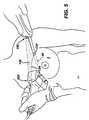
- FIG. 5illustrates a biopsy device having a laser line generator for providing a guide path extending from the biopsy device along an outer surface of a patient's breast, and how the biopsy device can be held in one hand while a hand held transducer, such as an ultrasound probe, can be held in the other hand, with the guide path providing for correct alignment of the long axis of the transducer with respect to the biopsy device needle.
- a hand held transducersuch as an ultrasound probe
- FIG. 6depicts an alternative arrangement where the handheld probe comprises a laser line generator.
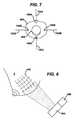
- FIG. 7is a front view of an alternative embodiment of a biopsy device according to the present invention.
- FIG. 8is a schematic illustration of a method of forming a laser light grid pattern on patient's skin.
- a biopsy device 10is shown being used to obtain a breast biopsy sample from a patient's breast 2 .
- the biopsy device 10is shown having a lightweight, ergonomically shaped handpiece 20 that can be grasped and manipulated by a single hand of a user.
- An elongated biopsy needle 30extends proximally from a distal end of the handpiece.
- the handpiece and biopsy needlecan be fully disposable, or alternatively, a portion of the handpiece can be reusable, and the needle and a portion of the handpiece, including the portion from which the needle extends, can be disposable.
- the biopsy needle 30can comprise an outer cannula 32 having side tissue receiving opening 34 and a closed distal end having a tissue piercing tip 36 .
- the tissue receiving opening 34can face upwardly, or vertically (toward the reader as viewed in FIG. 4 ).
- the tip 36can include a generally flat blade 38 , which can be aligned generally vertically.
- the biopsy devicecan include a cannular cutter 46 ( FIG. 4 ) having a cutting edge 48 .
- the cutter 46can be adapted to translate and rotate within the outer cannula 32 . Tissue received in the opening 34 (such as by being drawn into opening 34 by vacuum) can be severed by the cutting edge 48 as the cutter is translated across the length of opening 34 .
- Tissue received in the opening 34(such as by being drawn into opening 34 by vacuum) can be severed by the cutting edge 48 as the cutter is translated across the length of opening 34 .
- biopsy device 46having such an outer cannula and inner cutter is a Mammotome® brand biopsy device marketed by Ethicon Endo-Surgery, Inc.
- the Biopsy devicecan also include a tissue storage assembly 50 disposed at a distal end of the biopsy device, for holding tissue samples obtained by severing tissue received in opening 34 .
- the handpiece 20includes a component for providing a guide path.
- the componentcan comprise a light source 100 adapted to project an illuminated guide path on skin of a patient undergoing a biopsy (or on a drape or sheet covering the patient).
- the light source 100can be mounted or otherwise disposed on a distal portion of the handpiece 20 , such as on a reusable portion of the handpiece, and can direct light in a distal direction in a plane or fan shaped sheet that is generally parallel to and aligned with the long axis of the needle 30 ( the axis indicated by reference number 31 in FIG. 4 ).
- the light source 100can be activated by any suitable switch or control, such as a by a finger activated control switch 102 disposed on an upper surface of the handpiece 20 .
- the light source 100can be powered by any suitable power supply, including by batteries carried in the handpiece 20 , or by a power cord extending from the handpiece.
- the light source 100can comprise a laser line generator adapted to provide a generally planar, fan-shaped sheet of light (indicated by numeral 120 in FIG. 1 ) in a distal direction along at least a portion of the needle 30 , such that the intersection of the light provided by source 100 with a surface (such as an outer surface of the needle 30 or the outer surface of the patient's skin) provides a guide path indicated by numeral 124 .
- the sheet of light 120 provided by light source 100extends along and is aligned with axis 31 , and when light source 100 is activated, the sheet of light 120 can substantially bisect the opening 34 .
- guide pathmeans a visible path having a width and length, with the length being at least 10 times the maximum width, and such that a direction or orientation is readily discernable from viewing the path.
- the length of the guide path providedis at least 50 times the maximum width of the guide path.
- the guide pathcan be in the form of either a continuous line (straight, curved, or combination of straight and curved lines), or a series of closely spaced together discrete segments (such as a series of closely spaced dots, circles, dashes, crosses or other suitable shapes) which when viewed together from the distance mentioned below (2 feet) indicate a path direction to the viewer.
- visibleas used in connection with a path means that a path can be seen by an observer with unmagnified and otherwise unamplified or augmentetd normal color vision (20/20) when the path is projected on a planar flat non-glossy white surface (if the path is not white, otherwise if the path is white, on planar flat black surface) in a darkened room and viewed at a distance of 2 feet from the observer.
- the guide pathcan have a specific color, such as a reddish hue, a greenish hue, a bluish hue, or any other suitable color or combination of colors, such as white.
- the guide path 124can have a maximum width of no more than about 5 mm, and in one embodiment can be less than about 2 mm wide, such as a guide path having a maximum width between about 0.5 mm and about 1.5 millimeter. In another non-limiting alternative embodiment, the guide path can have a width of between about 5 mm and about 10 mm.
- the maximum width of the guide pathcan be less than the maximum diameter of the needle 30 , and in one embodiment is less than half the diameter of the needle. In one embodiment, the guide path is visible as a generally continuous straight or curved line having a generally uniform line width of less than about 2 mm.
- the light source 100can comprise a laser light source, such as laser line generator.
- the light source 100can include a housing 128 , a laser diode 132 disposed in the housing for generating a laser beam, a collimating lens 136 , and a lens 138 , such as a cylindrical lens, for diverging the collimated beam to provide a generally planar sheet or fan of laser light, which provides a guide path when the sheet of light intersects a surface (such as the patient's skin).
- a laser light sourcesuch as laser line generator.
- the light source 100can include a housing 128 , a laser diode 132 disposed in the housing for generating a laser beam, a collimating lens 136 , and a lens 138 , such as a cylindrical lens, for diverging the collimated beam to provide a generally planar sheet or fan of laser light, which provides a guide path when the sheet of light intersects a surface (such as the patient's skin).
- a lens 138
- the light source 100can be permanently joined to the handpiece 20 , such as by being fastened or built into an outside covering of the handpiece 20 , or alternatively, the light source can be removably supported on the handpiece so that the light source can be detached if desired.
- the light source 100can be supported on the handpiece to have one orientation, or alternatively, the light source 100 can be pivotably supported (or supported on a ball and socket type support) so that the direction of the light projected from light source 100 can be varied.
- Laser line generatorsare commercially available having various diode power levels, wavelengths, and fan angles.
- the light source 100can comprise a laser line generator having a fan angle of between about 15 degrees and about 90 degrees, a diode power level less than about 150 milliwatts, and a wavelength of between about 350 to about 850 nanometers.
- Laser line generatorsare available from various commercial sources, such as for instance: StockerYale of Salem New Hamphire (www.stockeryale.com), including Mini Laser products having a diode power of between 1-40 milliWatts and a wavelength of between about 635-830 nanometers, Telecentric Micro-Focus Laser products having a diode power of about 100 mW and a wavelength of about 660 nm, and Green Laser products having a diode power of about 1 to about 20 mW and a wavelength of about 532 nm; and Coherent, Inc. products designated as LG2 635 nm 2 mW 60 and LG3 635 nm 1.5 mW 40 (www.Coherent.com).
- light sourcesmay be used with suitable focusing, directing, collimating, and/or concentrating shaping devices (e.g. lens, collimator, mirror, and other components) to focus, direct, collimate, concentrate and/or otherwise shape light to provide a visible guide path.
- suitable focusing, directing, collimating, and/or concentrating shaping devicese.g. lens, collimator, mirror, and other components
- the cross-section of the biopsy needlecan be non-circular, and can have a major axis aligned generally vertically and a minor axis aligned generally horizontally.
- the light source 100can provide a generally planar sheet of light 120 which extends in a vertical plane generally parallel to and aligned with the major axis of the cross section of needle 30 .
- the guide path 124as visible on the patient's skin, can also indicate the orientation of the tissue receiving opening 34 when the tissue receiving opening 34 is disposed beneath the outer layer of the patient's skin (as in FIG. 1 ).
- the guide path 124 provided by the intersection of the sheet of light 120 and the patient's outer skincan be used to orient the needle 30 with respect to a separate handheld medical device, such as an ultrasound probe.
- a separate handheld medical devicesuch as an ultrasound probe.
- the correct line of alignment of an ultrasound transducer with respect to the needle 30is indicated by the guide path 124
- an incorrect line of alignmentis indicated by dotted line 1024 , with the angle between the two orientations indicated by angle A.
- FIG. 5a perspective view is provided to illustrate how the light plane 120 provided by laser light source 100 intersects the needle 30 , the breast 2 , and a separate handheld ultrasound probe 300 , to provide a light guide path 124 which has a segment visible along a portion of the needle positioned outside the breast, a segment of the path 124 visible along the patient's breast between the insertion point of the needle and the probe 300 , and a segment of the path 124 visible along a portion of a handheld ultrasound probe 300 .
- the probe 300 and the needle 30can be aligned with guide path 120 .
- the needlecan then be inserted into the patient's tissue under ultrasonic imaging.
- the guide path 120can be used to align the probe 300 to image the needle and adjacent tissue.
- FIG. 6illustrates an alternative embodiment, where the ultrasound probe 300 includes a light source 3100 , and the light source 3100 provides a generally planar sheet of light 3120 extending from the probe 300 toward the needle 30 .
- a separate devicecan be used to provide a guide path for aligning the probe 300 and the needle 30 .
- multiple laser light sourcescan be disposed on the biopsy device 10 , the probe 300 , a separate device (e.g. a separate hand held device or a separate fixed or mounted device), or multiple laser light sources can be disposed on both the device 10 and the probe 300 .
- a second laser light sourcecan be disposed on the probe 300 shown in FIG. 6 to provide a light guide path at right angles to the light path provided by light source 3100 .
- FIG. 7illustrates a front view of an alternative handpiece 20 A, the handpiece 20 A including laser light sources 100 A- 100 D.
- the light sourcesare positioned at multiple locations around the circumference of the handpiece to provide a fan of light 120 A associated with the top of the needle 30 (ie.at 12 o'clock), a fan of light 120 B and fan of light 120 D associated with the sides of the needle (i.e at 3 and 9 o'clock), and a fan of light 120 C associated with the bottom of the needle (i.e. at 6 o'clock').
- the four laser light sources 100 A- 100 Dcan each provide a different color of laser light to provide light paths, each having a different hue.
- a beam splitter or other suitable devicecan be employed to split laser light from one source to provide multiple guide paths.
- a biopsy devicecan include a generally stationary housing comprising a mount for supporting the housing with respect to a table or x-ray source, a rotatable needle, and a firing mechanism for firing the needle into tissue, as is shown in U.S. Pat. No. 6,007,497 issued Dec. 28, 1999, and incorporated herein by reference.
- one or more laser light sourcescan be disposed on the housing, or other fixed component, to provide one or more light guide paths on the patient's breast, which guide paths can be fixed with respect to the needle's rotation.
- FIG. 8illustrates use of one or more laser light sources to provide a grid or other pattern on the patient's skin.
- MRImagnetic resonance imaging
- PETmagnetic resonance imaging
- BSGIpositron emission computed tomography
- the MRI device and associated softwarecan be used to determine in Cartesian coordinates (e.g. X, Y) along the grid at which the biopsy needle should be inserted, as is known in the art.
- Cartesian coordinatese.g. X, Y
- the biopsy needleis then inserted through openings in the grid to obtain a biopsy sample.
- Patent Publications 2008/0015429 (Tsonton et al.) and 2007/0232953 (Dietz et al) and U.S. 2007/0167736 (Dietz et al)are incorporated herein by reference in their entirety for their disclosures related to biopsy devices and accessories useful in MRI procedures.
- a laser device 400can be used to project a light pattern, such as a light grid 440 , on the patient's breast 2 .
- the laser device 400can comprise one or more laser light sources for providing intersecting lines of laser light to form grid 440 .
- An associated controller 420can be employed to associate X and Y coordinates with the various grid lines projected on the patient, so that the light grid 440 can be used to determine the position at which the biopsy needle should be inserted.
- the patient's breastcan be compressed, the grid 440 can be projected on the patient's skin, and the biopsy needle 30 can be inserted at the appropriate XY location using the grid 440 as a guide.
Landscapes
- Health & Medical Sciences (AREA)
- Life Sciences & Earth Sciences (AREA)
- Surgery (AREA)
- General Health & Medical Sciences (AREA)
- Veterinary Medicine (AREA)
- Heart & Thoracic Surgery (AREA)
- Medical Informatics (AREA)
- Molecular Biology (AREA)
- Engineering & Computer Science (AREA)
- Animal Behavior & Ethology (AREA)
- Pathology (AREA)
- Public Health (AREA)
- Biomedical Technology (AREA)
- Nuclear Medicine, Radiotherapy & Molecular Imaging (AREA)
- Physics & Mathematics (AREA)
- Biophysics (AREA)
- Radiology & Medical Imaging (AREA)
- Optics & Photonics (AREA)
- Oral & Maxillofacial Surgery (AREA)
- Ultra Sonic Daignosis Equipment (AREA)
- Magnetic Resonance Imaging Apparatus (AREA)
Abstract
Description
Claims (10)
Priority Applications (1)
| Application Number | Priority Date | Filing Date | Title |
|---|---|---|---|
| US12/256,485US8162852B2 (en) | 2008-10-23 | 2008-10-23 | Methods for medical device alignment |
Applications Claiming Priority (1)
| Application Number | Priority Date | Filing Date | Title |
|---|---|---|---|
| US12/256,485US8162852B2 (en) | 2008-10-23 | 2008-10-23 | Methods for medical device alignment |
Publications (2)
| Publication Number | Publication Date |
|---|---|
| US20100106056A1 US20100106056A1 (en) | 2010-04-29 |
| US8162852B2true US8162852B2 (en) | 2012-04-24 |
Family
ID=42118161
Family Applications (1)
| Application Number | Title | Priority Date | Filing Date |
|---|---|---|---|
| US12/256,485Active2029-07-25US8162852B2 (en) | 2008-10-23 | 2008-10-23 | Methods for medical device alignment |
Country Status (1)
| Country | Link |
|---|---|
| US (1) | US8162852B2 (en) |
Cited By (23)
| Publication number | Priority date | Publication date | Assignee | Title |
|---|---|---|---|---|
| USD690811S1 (en)* | 2011-04-25 | 2013-10-01 | Devicor Medical Products, Inc. | Biopsy device |
| USD692134S1 (en)* | 2012-04-23 | 2013-10-22 | Femasys Inc. | Device for sampling tissue |
| USD699347S1 (en)* | 2011-12-07 | 2014-02-11 | Devicor Medical Products, Inc. | Ultrasound biopsy probe |
| US20140357987A1 (en)* | 2013-06-03 | 2014-12-04 | Faculty Physicians And Surgeons Of Loma Linda University School Of Medicine | Methods and apparatuses for fluoro-less or near fluoro-less percutaneous surgery access |
| US8966681B2 (en) | 2013-02-26 | 2015-03-03 | Linda L. Burch | Exercise mat |
| US9216010B2 (en) | 2013-06-26 | 2015-12-22 | General Electric Company | System and method for aligning a biopsy collecting device |
| US20170056062A1 (en) | 2015-08-31 | 2017-03-02 | Neda Buljubasic | Systems and methods for providing ultrasound guidance to target structures within a body |
| US9851060B2 (en) | 2013-04-01 | 2017-12-26 | Vinod V. Pathy | Lighting device for attachment to a tool |
| US9986971B2 (en) | 2013-01-18 | 2018-06-05 | Covidien Lp | Ring laser for use with imaging probe as a safe margin indicator |
| USRE47148E1 (en) | 2010-11-01 | 2018-12-04 | Devicor Medical Products, Inc. | Handheld biopsy device with needle firing |
| US20190054267A1 (en)* | 2013-12-05 | 2019-02-21 | Fisher & Paykel Healthcare Limited | Interface sizing tool and method |
| US10238363B2 (en) | 2014-08-21 | 2019-03-26 | Richard D. Striano | Needle guide for ultrasound transducer |
| US10405943B2 (en) | 2015-09-22 | 2019-09-10 | Faculty Physicians And Surgeons Of Loma Linda University School Of Medicine | Kit and method for reduced radiation procedures |
| US20190366080A1 (en)* | 2018-05-29 | 2019-12-05 | Florida Electrophysiology Llc | Subcutaneous tunneling tool with guiding mechanisms |
| US10667789B2 (en) | 2017-10-11 | 2020-06-02 | Geoffrey Steven Hastings | Laser assisted ultrasound guidance |
| US10682156B2 (en) | 2015-05-28 | 2020-06-16 | Akm A. Rahman | Angle-guidance device and method for CT guided drainage and biopsy procedures |
| US10786224B2 (en) | 2016-04-21 | 2020-09-29 | Covidien Lp | Biopsy devices and methods of use thereof |
| US10792067B2 (en)* | 2013-06-03 | 2020-10-06 | Faculty Physicians And Surgeons Of Loma Linda University Of Medicine | Methods and apparatuses for fluoro-less or near fluoro-less percutaneous surgery access |
| USD938095S1 (en) | 2013-04-01 | 2021-12-07 | Pathy Medical, Llc | Lighting device |
| US11331161B2 (en) | 2018-03-23 | 2022-05-17 | Covidien Lp | Surgical assemblies facilitating tissue marking and methods of use thereof |
| US11357515B2 (en) | 2017-09-09 | 2022-06-14 | June Access Ip, Llc | Intraosseous device having retractable motor/stylet assembly and automatic stylet point cover upon retraction operation |
| US11484339B2 (en) | 2017-09-09 | 2022-11-01 | June Access, IP LLC | Passive safety intraosseous device |
| US11517294B2 (en) | 2019-05-07 | 2022-12-06 | Covidien Lp | Biopsy devices and methods of use thereof |
Families Citing this family (17)
| Publication number | Priority date | Publication date | Assignee | Title |
|---|---|---|---|---|
| US8282573B2 (en) | 2003-02-24 | 2012-10-09 | Senorx, Inc. | Biopsy device with selectable tissue receiving aperture orientation and site illumination |
| US7189206B2 (en) | 2003-02-24 | 2007-03-13 | Senorx, Inc. | Biopsy device with inner cutter |
| US10863970B2 (en) | 2008-12-18 | 2020-12-15 | C. R. Bard, Inc. | Needle guide including enhanced visibility entrance |
| WO2010080637A1 (en) | 2008-12-18 | 2010-07-15 | C. R. Bard, Inc. | Needle guides for a sonographic imaging device |
| US9974516B2 (en) | 2010-12-22 | 2018-05-22 | C. R. Bard, Inc. | Selectable angle needle guide |
| CN103619263B (en) | 2011-06-23 | 2017-02-15 | C·R·巴德股份有限公司 | Needle guide with optional aspect |
| CN104303184B (en)* | 2012-03-21 | 2018-05-15 | 皇家飞利浦有限公司 | Clinical workstation integrating medical imaging and biopsy data and method of using same |
| WO2014162470A1 (en)* | 2013-04-01 | 2014-10-09 | テルモ株式会社 | Medical device |
| ITMO20130206A1 (en)* | 2013-07-17 | 2015-01-18 | Ncs Lab S R L | IMPROVED DEVICE FOR THE TRANSITION OF BONE OF SUTURE. |
| US9993263B2 (en)* | 2013-12-12 | 2018-06-12 | Catalin Esanu | Method and device for ultrasound guided minimal invasive access of a bodily cavity |
| CN104758033A (en)* | 2015-01-23 | 2015-07-08 | 倪家骧 | Puncture needle provided with laser positioning lamp at tail end of needle core |
| KR102530305B1 (en) | 2016-06-21 | 2023-05-10 | 큐렉소 주식회사 | End-Effector with Line Laser |
| US10703411B2 (en)* | 2018-01-16 | 2020-07-07 | Allen J Ho | Configurable parking assistance device and method |
| US10905386B2 (en)* | 2018-03-09 | 2021-02-02 | Suzanne Cano | Orienting X-ray projection for dental imagery |
| US11571205B2 (en)* | 2018-07-16 | 2023-02-07 | Cilag Gmbh International | Surgical visualization feedback system |
| JP2023527614A (en)* | 2020-03-30 | 2023-06-30 | エンプレス メディカル,インク. | Prediction of curved penetration paths for surgical devices |
| CN218419895U (en)* | 2021-06-22 | 2023-02-03 | 巴德阿克塞斯系统股份有限公司 | Ultrasound imaging system configured to guide insertion of medical device |
Citations (51)
| Publication number | Priority date | Publication date | Assignee | Title |
|---|---|---|---|---|
| US4321551A (en) | 1980-06-12 | 1982-03-23 | General Motors Corporation | Laser line of light generator |
| US5320110A (en)* | 1991-10-29 | 1994-06-14 | Wang Ko P | Pleural biopsy syringe-needles |
| US5526822A (en) | 1994-03-24 | 1996-06-18 | Biopsys Medical, Inc. | Method and apparatus for automated biopsy and collection of soft tissue |
| US5598269A (en)* | 1994-05-12 | 1997-01-28 | Children's Hospital Medical Center | Laser guided alignment apparatus for medical procedures |
| US5751869A (en)* | 1996-08-08 | 1998-05-12 | Cogent Light Technologies, Inc. | Optical system for coupling light from a single fiber optic into a fiber bundle |
| US5810541A (en) | 1996-05-21 | 1998-09-22 | International Paper Box Machine Co., Inc. | Apparatus and method for manually exchanging pallets |
| US5810841A (en)* | 1997-01-22 | 1998-09-22 | Minrad Inc. | Energy guided apparatus and method with indication of alignment |
| US5855554A (en) | 1997-03-17 | 1999-01-05 | General Electric Company | Image guided breast lesion localization device |
| US5976092A (en)* | 1998-06-15 | 1999-11-02 | Chinn; Douglas O. | Combination stereotactic surgical guide and ultrasonic probe |
| US6007497A (en) | 1998-06-30 | 1999-12-28 | Ethicon Endo-Surgery, Inc. | Surgical biopsy device |
| US6069748A (en) | 1998-10-20 | 2000-05-30 | Eastman Kodak Company | Laser line generator system |
| US6086544A (en) | 1999-03-31 | 2000-07-11 | Ethicon Endo-Surgery, Inc. | Control apparatus for an automated surgical biopsy device |
| US6203498B1 (en) | 1996-06-28 | 2001-03-20 | Sonosite, Inc. | Ultrasonic imaging device with integral display |
| US6273862B1 (en) | 1998-10-23 | 2001-08-14 | Ethicon Endo-Surgery, Inc | Surgical device for the collection of soft tissue |
| US6364839B1 (en) | 1999-05-04 | 2002-04-02 | Sonosite, Inc. | Ultrasound diagnostic instrument having software in detachable scanhead |
| US6428487B1 (en) | 1999-12-17 | 2002-08-06 | Ethicon Endo-Surgery, Inc. | Surgical biopsy system with remote control for selecting an operational mode |
| USD461895S1 (en) | 2001-04-19 | 2002-08-20 | Sonosite, Inc. | Handheld medical diagnostic ultrasound instrument |
| US6471651B1 (en) | 1999-05-05 | 2002-10-29 | Sonosite, Inc. | Low power portable ultrasonic diagnostic instrument |
| US20030069502A1 (en) | 2001-05-29 | 2003-04-10 | Makin Inder Raj. S. | Ultrasound feedback in medically-treated patients |
| US6605095B2 (en)* | 2000-06-13 | 2003-08-12 | Sdgi Holdings, Inc. | Percutaneous needle alignment system and associated method |
| US20030199785A1 (en) | 2002-04-23 | 2003-10-23 | Ethicon Endo-Surgery | Localization mechanism for an MRI compatible biopsy device |
| US20030199754A1 (en) | 2002-04-23 | 2003-10-23 | Ethicon Endo-Surgery | Method for using an MRI compatible biopsy device with detachable probe |
| US20030199753A1 (en) | 2002-04-23 | 2003-10-23 | Ethicon Endo-Surgery | MRI compatible biopsy device with detachable probe |
| US6688758B2 (en) | 2002-06-14 | 2004-02-10 | Institut National D'optique | Line generator optical apparatus |
| US6689067B2 (en)* | 2001-11-28 | 2004-02-10 | Siemens Corporate Research, Inc. | Method and apparatus for ultrasound guidance of needle biopsies |
| US6692200B2 (en) | 2001-01-16 | 2004-02-17 | Nesson Enterprises | Alignment system for hand-held tools |
| US20040034280A1 (en) | 1998-10-23 | 2004-02-19 | Salvatore Privitera | Surgical device for the collection of soft tissue |
| US6702749B2 (en) | 2001-07-24 | 2004-03-09 | Siemens Corporate Research, Inc. | Optical needle guide for ultrasound guided needle biopsy |
| US20040077972A1 (en) | 2002-10-18 | 2004-04-22 | Mark Tsonton | Localization mechanism for an MRI compatible biopsy device |
| US20040249278A1 (en) | 2003-06-04 | 2004-12-09 | Krause William R. | Biopsy and delivery device |
| US20050065453A1 (en) | 2003-02-24 | 2005-03-24 | Senorx, Inc. | Biopsy device with selectable tissue receiving aperture orientation and site illumination |
| US20050131291A1 (en) | 2003-12-10 | 2005-06-16 | Sonosite, Inc. | Device for assisting the positioning of medical devices |
| US7024791B2 (en) | 2004-01-12 | 2006-04-11 | Black & Decker Inc. | Tape measure with laser beam |
| US7031367B2 (en) | 2002-05-31 | 2006-04-18 | Black & Decker Inc. | Laser level |
| US7041058B2 (en) | 2002-08-29 | 2006-05-09 | Siemens Medical Solutions Usa, Inc. | Medical diagnostic imaging system and method for efficient access of data |
| US20060144548A1 (en) | 2004-12-30 | 2006-07-06 | Beckman Andrew T | Method of manufacturing a needle assembly for use with a biopsy device |
| US20060200041A1 (en) | 2005-03-04 | 2006-09-07 | Ethicon Endo-Surgery, Inc. | Biopsy device incorporating an adjustable probe sleeve |
| US20070049822A1 (en) | 2005-08-31 | 2007-03-01 | Sonosite, Inc. | Medical device guide locator |
| US20070149878A1 (en) | 2003-12-29 | 2007-06-28 | Hankins Carol A | Apparatus and method for guiding a medical device in multiple planes |
| US20070167736A1 (en) | 2004-05-21 | 2007-07-19 | Dietz Timothy G | MRI biopsy apparatus incorporating an imageable penetrating portion |
| US7269907B2 (en) | 2003-07-01 | 2007-09-18 | Irwin Industrial Tool Company | Laser line generating device with swivel base |
| US20070232953A1 (en) | 2006-03-31 | 2007-10-04 | Ethicon Endo-Surgery, Inc. | MRI biopsy device |
| US7303530B2 (en) | 2003-05-22 | 2007-12-04 | Siemens Medical Solutions Usa, Inc. | Transducer arrays with an integrated sensor and methods of use |
| US7310887B2 (en) | 2004-07-21 | 2007-12-25 | Irwin Industrial Tool Company | Intersecting laser line generating device |
| US20080015429A1 (en) | 2004-05-21 | 2008-01-17 | Tsonton Mark T | Mri biopsy device |
| US7367945B2 (en) | 2004-03-29 | 2008-05-06 | General Electric Company | Ultrasound system |
| EP1932481A1 (en) | 2006-12-13 | 2008-06-18 | Ethicon Endo-Surgery, Inc. | Biopsy system with vacuum control module |
| US20080146915A1 (en)* | 2006-10-19 | 2008-06-19 | Mcmorrow Gerald | Systems and methods for visualizing a cannula trajectory |
| US7416533B2 (en) | 2003-08-13 | 2008-08-26 | Scimed Life Systems, Inc. | Marking biopsy sites |
| WO2008120137A1 (en) | 2007-03-30 | 2008-10-09 | Koninklijke Philips Electronics N.V. | Laser guide for percutaneous biopsy interventions on a rotational x-ray device |
| US7858038B2 (en) | 2007-11-20 | 2010-12-28 | Devicor Medical Products, Inc. | Biopsy device with illuminated tissue holder |
Family Cites Families (1)
| Publication number | Priority date | Publication date | Assignee | Title |
|---|---|---|---|---|
| JP4205443B2 (en)* | 2003-01-17 | 2009-01-07 | 株式会社コナミデジタルエンタテインメント | Point switching device |
- 2008
- 2008-10-23USUS12/256,485patent/US8162852B2/enactiveActive
Patent Citations (54)
| Publication number | Priority date | Publication date | Assignee | Title |
|---|---|---|---|---|
| US4321551A (en) | 1980-06-12 | 1982-03-23 | General Motors Corporation | Laser line of light generator |
| US5320110A (en)* | 1991-10-29 | 1994-06-14 | Wang Ko P | Pleural biopsy syringe-needles |
| US5526822A (en) | 1994-03-24 | 1996-06-18 | Biopsys Medical, Inc. | Method and apparatus for automated biopsy and collection of soft tissue |
| US5598269A (en)* | 1994-05-12 | 1997-01-28 | Children's Hospital Medical Center | Laser guided alignment apparatus for medical procedures |
| US5810541A (en) | 1996-05-21 | 1998-09-22 | International Paper Box Machine Co., Inc. | Apparatus and method for manually exchanging pallets |
| US6203498B1 (en) | 1996-06-28 | 2001-03-20 | Sonosite, Inc. | Ultrasonic imaging device with integral display |
| US5751869A (en)* | 1996-08-08 | 1998-05-12 | Cogent Light Technologies, Inc. | Optical system for coupling light from a single fiber optic into a fiber bundle |
| US5810841A (en)* | 1997-01-22 | 1998-09-22 | Minrad Inc. | Energy guided apparatus and method with indication of alignment |
| US5855554A (en) | 1997-03-17 | 1999-01-05 | General Electric Company | Image guided breast lesion localization device |
| US5976092A (en)* | 1998-06-15 | 1999-11-02 | Chinn; Douglas O. | Combination stereotactic surgical guide and ultrasonic probe |
| US6007497A (en) | 1998-06-30 | 1999-12-28 | Ethicon Endo-Surgery, Inc. | Surgical biopsy device |
| US6069748A (en) | 1998-10-20 | 2000-05-30 | Eastman Kodak Company | Laser line generator system |
| US6273862B1 (en) | 1998-10-23 | 2001-08-14 | Ethicon Endo-Surgery, Inc | Surgical device for the collection of soft tissue |
| US20110125055A1 (en) | 1998-10-23 | 2011-05-26 | Devicor Medical Products, Inc. | Surgical device for the collection of soft tissue |
| US20040034280A1 (en) | 1998-10-23 | 2004-02-19 | Salvatore Privitera | Surgical device for the collection of soft tissue |
| US6086544A (en) | 1999-03-31 | 2000-07-11 | Ethicon Endo-Surgery, Inc. | Control apparatus for an automated surgical biopsy device |
| US6364839B1 (en) | 1999-05-04 | 2002-04-02 | Sonosite, Inc. | Ultrasound diagnostic instrument having software in detachable scanhead |
| US6471651B1 (en) | 1999-05-05 | 2002-10-29 | Sonosite, Inc. | Low power portable ultrasonic diagnostic instrument |
| US6428487B1 (en) | 1999-12-17 | 2002-08-06 | Ethicon Endo-Surgery, Inc. | Surgical biopsy system with remote control for selecting an operational mode |
| US6752768B2 (en) | 1999-12-17 | 2004-06-22 | Ethicon Endo-Surgery | Surgical biopsy system with remote control for selecting an operational mode |
| US6605095B2 (en)* | 2000-06-13 | 2003-08-12 | Sdgi Holdings, Inc. | Percutaneous needle alignment system and associated method |
| US20040106934A1 (en) | 2000-06-13 | 2004-06-03 | Sdgi Holdings, Inc. | Percutaneous Needle Alignment System |
| US6692200B2 (en) | 2001-01-16 | 2004-02-17 | Nesson Enterprises | Alignment system for hand-held tools |
| USD461895S1 (en) | 2001-04-19 | 2002-08-20 | Sonosite, Inc. | Handheld medical diagnostic ultrasound instrument |
| US20030069502A1 (en) | 2001-05-29 | 2003-04-10 | Makin Inder Raj. S. | Ultrasound feedback in medically-treated patients |
| US6702749B2 (en) | 2001-07-24 | 2004-03-09 | Siemens Corporate Research, Inc. | Optical needle guide for ultrasound guided needle biopsy |
| US6689067B2 (en)* | 2001-11-28 | 2004-02-10 | Siemens Corporate Research, Inc. | Method and apparatus for ultrasound guidance of needle biopsies |
| US20030199753A1 (en) | 2002-04-23 | 2003-10-23 | Ethicon Endo-Surgery | MRI compatible biopsy device with detachable probe |
| US20030199754A1 (en) | 2002-04-23 | 2003-10-23 | Ethicon Endo-Surgery | Method for using an MRI compatible biopsy device with detachable probe |
| US20030199785A1 (en) | 2002-04-23 | 2003-10-23 | Ethicon Endo-Surgery | Localization mechanism for an MRI compatible biopsy device |
| US7031367B2 (en) | 2002-05-31 | 2006-04-18 | Black & Decker Inc. | Laser level |
| US6688758B2 (en) | 2002-06-14 | 2004-02-10 | Institut National D'optique | Line generator optical apparatus |
| US7041058B2 (en) | 2002-08-29 | 2006-05-09 | Siemens Medical Solutions Usa, Inc. | Medical diagnostic imaging system and method for efficient access of data |
| US20040077972A1 (en) | 2002-10-18 | 2004-04-22 | Mark Tsonton | Localization mechanism for an MRI compatible biopsy device |
| US20050065453A1 (en) | 2003-02-24 | 2005-03-24 | Senorx, Inc. | Biopsy device with selectable tissue receiving aperture orientation and site illumination |
| US7303530B2 (en) | 2003-05-22 | 2007-12-04 | Siemens Medical Solutions Usa, Inc. | Transducer arrays with an integrated sensor and methods of use |
| US20040249278A1 (en) | 2003-06-04 | 2004-12-09 | Krause William R. | Biopsy and delivery device |
| US7269907B2 (en) | 2003-07-01 | 2007-09-18 | Irwin Industrial Tool Company | Laser line generating device with swivel base |
| US7416533B2 (en) | 2003-08-13 | 2008-08-26 | Scimed Life Systems, Inc. | Marking biopsy sites |
| US20050131291A1 (en) | 2003-12-10 | 2005-06-16 | Sonosite, Inc. | Device for assisting the positioning of medical devices |
| US20070149878A1 (en) | 2003-12-29 | 2007-06-28 | Hankins Carol A | Apparatus and method for guiding a medical device in multiple planes |
| US7024791B2 (en) | 2004-01-12 | 2006-04-11 | Black & Decker Inc. | Tape measure with laser beam |
| US7367945B2 (en) | 2004-03-29 | 2008-05-06 | General Electric Company | Ultrasound system |
| US20070167736A1 (en) | 2004-05-21 | 2007-07-19 | Dietz Timothy G | MRI biopsy apparatus incorporating an imageable penetrating portion |
| US20080015429A1 (en) | 2004-05-21 | 2008-01-17 | Tsonton Mark T | Mri biopsy device |
| US7310887B2 (en) | 2004-07-21 | 2007-12-25 | Irwin Industrial Tool Company | Intersecting laser line generating device |
| US20060144548A1 (en) | 2004-12-30 | 2006-07-06 | Beckman Andrew T | Method of manufacturing a needle assembly for use with a biopsy device |
| US20060200041A1 (en) | 2005-03-04 | 2006-09-07 | Ethicon Endo-Surgery, Inc. | Biopsy device incorporating an adjustable probe sleeve |
| US20070049822A1 (en) | 2005-08-31 | 2007-03-01 | Sonosite, Inc. | Medical device guide locator |
| US20070232953A1 (en) | 2006-03-31 | 2007-10-04 | Ethicon Endo-Surgery, Inc. | MRI biopsy device |
| US20080146915A1 (en)* | 2006-10-19 | 2008-06-19 | Mcmorrow Gerald | Systems and methods for visualizing a cannula trajectory |
| EP1932481A1 (en) | 2006-12-13 | 2008-06-18 | Ethicon Endo-Surgery, Inc. | Biopsy system with vacuum control module |
| WO2008120137A1 (en) | 2007-03-30 | 2008-10-09 | Koninklijke Philips Electronics N.V. | Laser guide for percutaneous biopsy interventions on a rotational x-ray device |
| US7858038B2 (en) | 2007-11-20 | 2010-12-28 | Devicor Medical Products, Inc. | Biopsy device with illuminated tissue holder |
Non-Patent Citations (3)
| Title |
|---|
| "Application Engineering Note-Multimode Fiber Compatibility, Application Note," Corning, Supersedes (Apr. 2005) pp. 1-5. |
| "Line Generator Diode Lasers Modules," Coherent, http://www.coherent.com/products/?984/Line-Generator-Diode-Laser-Modules, printed Nov. 11, 2011. |
| "Structured Light Lasers," StockerYale, www.stockeryale.com/lasers, pp. 1-2, printed Nov. 11, 2011. |
Cited By (38)
| Publication number | Priority date | Publication date | Assignee | Title |
|---|---|---|---|---|
| USRE47148E1 (en) | 2010-11-01 | 2018-12-04 | Devicor Medical Products, Inc. | Handheld biopsy device with needle firing |
| USD690811S1 (en)* | 2011-04-25 | 2013-10-01 | Devicor Medical Products, Inc. | Biopsy device |
| USD699347S1 (en)* | 2011-12-07 | 2014-02-11 | Devicor Medical Products, Inc. | Ultrasound biopsy probe |
| USD692134S1 (en)* | 2012-04-23 | 2013-10-22 | Femasys Inc. | Device for sampling tissue |
| US9986971B2 (en) | 2013-01-18 | 2018-06-05 | Covidien Lp | Ring laser for use with imaging probe as a safe margin indicator |
| US8966681B2 (en) | 2013-02-26 | 2015-03-03 | Linda L. Burch | Exercise mat |
| US11519569B2 (en) | 2013-04-01 | 2022-12-06 | Pathy Medical, Llc | Lighting device with cavity for removably attaching to a tool |
| USD938095S1 (en) | 2013-04-01 | 2021-12-07 | Pathy Medical, Llc | Lighting device |
| US10816147B2 (en) | 2013-04-01 | 2020-10-27 | Pathy Medical, Llc | Lighting device with cavity for removably attaching to a tool |
| USD991542S1 (en) | 2013-04-01 | 2023-07-04 | Pathy Medical, Llc | Lighting device |
| US9851060B2 (en) | 2013-04-01 | 2017-12-26 | Vinod V. Pathy | Lighting device for attachment to a tool |
| US10792067B2 (en)* | 2013-06-03 | 2020-10-06 | Faculty Physicians And Surgeons Of Loma Linda University Of Medicine | Methods and apparatuses for fluoro-less or near fluoro-less percutaneous surgery access |
| US9918739B2 (en) | 2013-06-03 | 2018-03-20 | Faculty Physicians And Surgeons Of Loma Linda Univ | Methods and apparatuses for fluoro-less or near fluoro-less percutaneous surgery access |
| US9095361B2 (en) | 2013-06-03 | 2015-08-04 | Faculty Physicians And Surgeons Of Loma Linda University School Of Medicine | Methods and apparatuses for fluoro-less or near fluoro-less percutaneous surgery access |
| US10085767B2 (en) | 2013-06-03 | 2018-10-02 | Faculty Physicians And Surgeons Of Loma Linda University | Methods and apparatuses for fluoro-less or near fluoro-less percutaneous surgery access |
| US8998943B2 (en)* | 2013-06-03 | 2015-04-07 | Faculty Physicians and Surgeons of Loma Linda University School of Medicine; Loma Linda University | Methods and apparatuses for fluoro-less or near fluoro-less percutaneous surgery access |
| US20140357987A1 (en)* | 2013-06-03 | 2014-12-04 | Faculty Physicians And Surgeons Of Loma Linda University School Of Medicine | Methods and apparatuses for fluoro-less or near fluoro-less percutaneous surgery access |
| US10932816B2 (en) | 2013-06-03 | 2021-03-02 | Faculty Physicians And Surgeons Of Loma Linda University School Of Medicine | Methods and apparatuses for fluoro-less or near fluoro-less percutaneous surgery access |
| US9351758B2 (en) | 2013-06-03 | 2016-05-31 | Faculty Physicians And Surgeons Of Loma Linda University School Of Medicine: Loma Linda University | Methods and apparatuses for fluoro-less or near fluoro-less percutaneous surgery access |
| US9216010B2 (en) | 2013-06-26 | 2015-12-22 | General Electric Company | System and method for aligning a biopsy collecting device |
| US20190054267A1 (en)* | 2013-12-05 | 2019-02-21 | Fisher & Paykel Healthcare Limited | Interface sizing tool and method |
| US11318265B2 (en) | 2013-12-05 | 2022-05-03 | Fisher & Paykel Healthcare Limited | Interface sizing tool and method |
| US10569040B2 (en)* | 2013-12-05 | 2020-02-25 | Fisher & Paykel Healthcare Limited | Interface sizing tool and method |
| US10238363B2 (en) | 2014-08-21 | 2019-03-26 | Richard D. Striano | Needle guide for ultrasound transducer |
| US10682156B2 (en) | 2015-05-28 | 2020-06-16 | Akm A. Rahman | Angle-guidance device and method for CT guided drainage and biopsy procedures |
| US10806486B2 (en) | 2015-08-31 | 2020-10-20 | Neda Buljubasic | Systems and methods for providing ultrasound guidance to target structures within a body |
| US20170056062A1 (en) | 2015-08-31 | 2017-03-02 | Neda Buljubasic | Systems and methods for providing ultrasound guidance to target structures within a body |
| WO2017040715A1 (en)* | 2015-08-31 | 2017-03-09 | Buljubasic Neda | Systems and methods for providing ultrasound guidance to target structures within a body |
| EP3344146A4 (en)* | 2015-08-31 | 2019-03-06 | Buljubasic, Neda | SYSTEMS AND METHODS FOR PROVIDING ULTRASONIC GUIDANCE FOR TARGETING STRUCTURES INSIDE A BODY |
| US11324533B2 (en) | 2015-08-31 | 2022-05-10 | Neda Buljubasic | Systems and methods for providing ultrasound guidance to target structures within a body |
| US10405943B2 (en) | 2015-09-22 | 2019-09-10 | Faculty Physicians And Surgeons Of Loma Linda University School Of Medicine | Kit and method for reduced radiation procedures |
| US10786224B2 (en) | 2016-04-21 | 2020-09-29 | Covidien Lp | Biopsy devices and methods of use thereof |
| US11357515B2 (en) | 2017-09-09 | 2022-06-14 | June Access Ip, Llc | Intraosseous device having retractable motor/stylet assembly and automatic stylet point cover upon retraction operation |
| US11484339B2 (en) | 2017-09-09 | 2022-11-01 | June Access, IP LLC | Passive safety intraosseous device |
| US10667789B2 (en) | 2017-10-11 | 2020-06-02 | Geoffrey Steven Hastings | Laser assisted ultrasound guidance |
| US11331161B2 (en) | 2018-03-23 | 2022-05-17 | Covidien Lp | Surgical assemblies facilitating tissue marking and methods of use thereof |
| US20190366080A1 (en)* | 2018-05-29 | 2019-12-05 | Florida Electrophysiology Llc | Subcutaneous tunneling tool with guiding mechanisms |
| US11517294B2 (en) | 2019-05-07 | 2022-12-06 | Covidien Lp | Biopsy devices and methods of use thereof |
Also Published As
| Publication number | Publication date |
|---|---|
| US20100106056A1 (en) | 2010-04-29 |
Similar Documents
| Publication | Publication Date | Title |
|---|---|---|
| US8162852B2 (en) | Methods for medical device alignment | |
| EP2179694B1 (en) | Medical device alignement | |
| US5598269A (en) | Laser guided alignment apparatus for medical procedures | |
| EP2377484B1 (en) | Device and method for contactless determination and measuring of the spatial position and/or spatial orientation of bodies | |
| US6041249A (en) | Device for making a guide path for an instrument on a patient | |
| EP0805657B1 (en) | Hand-held laser scanner | |
| US6875179B2 (en) | Ultrasonic guided catheter deployment system | |
| US20110190637A1 (en) | Medical measuring system, method for surgical intervention as well as use of a medical measuring system | |
| CN212521854U (en) | Medical instrument | |
| JP4303962B2 (en) | Diagnostic imaging intervention device | |
| CN112370647A (en) | Ultrasonic-guided multi-degree-of-freedom radioactive particle implantation robot | |
| CN111839730B (en) | Photoacoustic imaging surgical navigation platform for guiding tumor resection | |
| US5102391A (en) | Guidance device for C. T. guided drainage and biopsy procedures | |
| CN108348277B (en) | Tissue Removal Devices and Tissue Removal Systems | |
| JPH11276422A (en) | Ultrasonic endoscope | |
| EP1983899A1 (en) | Apparatus and method for motorised placement of needle | |
| WO2024157730A1 (en) | Puncture system, puncture assisting tool, body surface-irradiating laser mechanism, and puncture navigation system | |
| GB2443432A (en) | Optical guidance apparatus for x-ray imaging device | |
| CN115040247A (en) | Femtosecond laser minimally invasive surgery robot system | |
| CN114052903A (en) | Near-infrared imaging surgical navigation system and method | |
| CN209899454U (en) | Percutaneous aspiration biopsy needle | |
| JP2000070272A (en) | Biopsy needle insertion guide device | |
| JP4142403B2 (en) | Ultrasound endoscope tip | |
| KR20220148524A (en) | Method for determining location of lesion and endoscopic system therefor | |
| CN112229830A (en) | Endoscopic laser-induced breakdown spectroscopy system |
Legal Events
| Date | Code | Title | Description |
|---|---|---|---|
| AS | Assignment | Owner name:ETHICON ENDO-SURGERY, INC.,OHIO Free format text:ASSIGNMENT OF ASSIGNORS INTEREST;ASSIGNOR:NORRIS, PERRY R.;REEL/FRAME:021723/0471 Effective date:20081022 Owner name:ETHICON ENDO-SURGERY, INC., OHIO Free format text:ASSIGNMENT OF ASSIGNORS INTEREST;ASSIGNOR:NORRIS, PERRY R.;REEL/FRAME:021723/0471 Effective date:20081022 | |
| AS | Assignment | Owner name:DEVICOR MEDICAL PRODUCTS, INC., WISCONSIN Free format text:ASSIGNMENT OF ASSIGNORS INTEREST;ASSIGNOR:ETHICON ENDO-SURGERY, INC.;REEL/FRAME:025348/0035 Effective date:20101103 | |
| STCF | Information on status: patent grant | Free format text:PATENTED CASE | |
| FEPP | Fee payment procedure | Free format text:PAYOR NUMBER ASSIGNED (ORIGINAL EVENT CODE: ASPN); ENTITY STATUS OF PATENT OWNER: LARGE ENTITY | |
| FEPP | Fee payment procedure | Free format text:PAT HOLDER NO LONGER CLAIMS SMALL ENTITY STATUS, ENTITY STATUS SET TO UNDISCOUNTED (ORIGINAL EVENT CODE: STOL); ENTITY STATUS OF PATENT OWNER: LARGE ENTITY | |
| FPAY | Fee payment | Year of fee payment:4 | |
| MAFP | Maintenance fee payment | Free format text:PAYMENT OF MAINTENANCE FEE, 8TH YEAR, LARGE ENTITY (ORIGINAL EVENT CODE: M1552); ENTITY STATUS OF PATENT OWNER: LARGE ENTITY Year of fee payment:8 | |
| MAFP | Maintenance fee payment | Free format text:PAYMENT OF MAINTENANCE FEE, 12TH YEAR, LARGE ENTITY (ORIGINAL EVENT CODE: M1553); ENTITY STATUS OF PATENT OWNER: LARGE ENTITY Year of fee payment:12 |