US8142467B2 - Tamponade trocar device and method - Google Patents
Tamponade trocar device and methodDownload PDFInfo
- Publication number
- US8142467B2 US8142467B2US12/106,920US10692008AUS8142467B2US 8142467 B2US8142467 B2US 8142467B2US 10692008 AUS10692008 AUS 10692008AUS 8142467 B2US8142467 B2US 8142467B2
- Authority
- US
- United States
- Prior art keywords
- balloon
- cannula
- distal end
- trocar
- tamponade
- Prior art date
- Legal status (The legal status is an assumption and is not a legal conclusion. Google has not performed a legal analysis and makes no representation as to the accuracy of the status listed.)
- Expired - Fee Related, expires
Links
- 238000000034methodMethods0.000titledescription7
- 238000003780insertionMethods0.000claimsdescription8
- 230000037431insertionEffects0.000claimsdescription8
- 230000007704transitionEffects0.000claimsdescription7
- 239000000463materialSubstances0.000claimsdescription6
- 239000004677NylonSubstances0.000claimsdescription4
- 239000004698PolyethyleneSubstances0.000claimsdescription4
- 229920001778nylonPolymers0.000claimsdescription4
- -1polyethylenePolymers0.000claimsdescription4
- 229920000573polyethylenePolymers0.000claimsdescription4
- 229920002635polyurethanePolymers0.000claimsdescription4
- 239000004814polyurethaneSubstances0.000claimsdescription4
- 210000000244kidney pelvisAnatomy0.000description16
- 210000003734kidneyAnatomy0.000description12
- 230000035515penetrationEffects0.000description6
- 238000002357laparoscopic surgeryMethods0.000description5
- 238000004891communicationMethods0.000description4
- 238000001356surgical procedureMethods0.000description3
- 208000005646PneumoperitoneumDiseases0.000description2
- 230000000740bleeding effectEffects0.000description2
- 230000008878couplingEffects0.000description2
- 238000010168coupling processMethods0.000description2
- 238000005859coupling reactionMethods0.000description2
- 230000000149penetrating effectEffects0.000description2
- 241001631457CannulaSpecies0.000description1
- 208000000913Kidney CalculiDiseases0.000description1
- 206010029148NephrolithiasisDiseases0.000description1
- 210000003815abdominal wallAnatomy0.000description1
- 230000004075alterationEffects0.000description1
- 238000002399angioplastyMethods0.000description1
- 238000011161developmentMethods0.000description1
- 230000010339dilationEffects0.000description1
- 230000001788irregularEffects0.000description1
- 238000004519manufacturing processMethods0.000description1
- 238000012986modificationMethods0.000description1
- 230000004048modificationEffects0.000description1
- 230000003387muscularEffects0.000description1
- 210000000056organAnatomy0.000description1
- 210000004303peritoneumAnatomy0.000description1
- 230000008569processEffects0.000description1
- 230000004044responseEffects0.000description1
- 238000007789sealingMethods0.000description1
- 238000002560therapeutic procedureMethods0.000description1
- 210000001519tissueAnatomy0.000description1
- 210000000626ureterAnatomy0.000description1
Images


Classifications
- A—HUMAN NECESSITIES
- A61—MEDICAL OR VETERINARY SCIENCE; HYGIENE
- A61M—DEVICES FOR INTRODUCING MEDIA INTO, OR ONTO, THE BODY; DEVICES FOR TRANSDUCING BODY MEDIA OR FOR TAKING MEDIA FROM THE BODY; DEVICES FOR PRODUCING OR ENDING SLEEP OR STUPOR
- A61M25/00—Catheters; Hollow probes
- A61M25/01—Introducing, guiding, advancing, emplacing or holding catheters
- A61M25/06—Body-piercing guide needles or the like
- A61M25/0662—Guide tubes
- A—HUMAN NECESSITIES
- A61—MEDICAL OR VETERINARY SCIENCE; HYGIENE
- A61B—DIAGNOSIS; SURGERY; IDENTIFICATION
- A61B17/00—Surgical instruments, devices or methods
- A61B17/34—Trocars; Puncturing needles
- A61B17/3417—Details of tips or shafts, e.g. grooves, expandable, bendable; Multiple coaxial sliding cannulas, e.g. for dilating
- A61B17/3421—Cannulas
- A—HUMAN NECESSITIES
- A61—MEDICAL OR VETERINARY SCIENCE; HYGIENE
- A61M—DEVICES FOR INTRODUCING MEDIA INTO, OR ONTO, THE BODY; DEVICES FOR TRANSDUCING BODY MEDIA OR FOR TAKING MEDIA FROM THE BODY; DEVICES FOR PRODUCING OR ENDING SLEEP OR STUPOR
- A61M25/00—Catheters; Hollow probes
- A61M25/01—Introducing, guiding, advancing, emplacing or holding catheters
- A61M25/0102—Insertion or introduction using an inner stiffening member, e.g. stylet or push-rod
- A—HUMAN NECESSITIES
- A61—MEDICAL OR VETERINARY SCIENCE; HYGIENE
- A61M—DEVICES FOR INTRODUCING MEDIA INTO, OR ONTO, THE BODY; DEVICES FOR TRANSDUCING BODY MEDIA OR FOR TAKING MEDIA FROM THE BODY; DEVICES FOR PRODUCING OR ENDING SLEEP OR STUPOR
- A61M25/00—Catheters; Hollow probes
- A61M25/10—Balloon catheters
- A61M25/1002—Balloon catheters characterised by balloon shape
- A—HUMAN NECESSITIES
- A61—MEDICAL OR VETERINARY SCIENCE; HYGIENE
- A61M—DEVICES FOR INTRODUCING MEDIA INTO, OR ONTO, THE BODY; DEVICES FOR TRANSDUCING BODY MEDIA OR FOR TAKING MEDIA FROM THE BODY; DEVICES FOR PRODUCING OR ENDING SLEEP OR STUPOR
- A61M25/00—Catheters; Hollow probes
- A61M25/10—Balloon catheters
- A61M25/1018—Balloon inflating or inflation-control devices
- A61M25/10184—Means for controlling or monitoring inflation or deflation
- A61M25/10185—Valves
- A—HUMAN NECESSITIES
- A61—MEDICAL OR VETERINARY SCIENCE; HYGIENE
- A61B—DIAGNOSIS; SURGERY; IDENTIFICATION
- A61B17/00—Surgical instruments, devices or methods
- A61B17/34—Trocars; Puncturing needles
- A61B17/3417—Details of tips or shafts, e.g. grooves, expandable, bendable; Multiple coaxial sliding cannulas, e.g. for dilating
- A61B17/3421—Cannulas
- A61B17/3431—Cannulas being collapsible, e.g. made of thin flexible material
- A—HUMAN NECESSITIES
- A61—MEDICAL OR VETERINARY SCIENCE; HYGIENE
- A61B—DIAGNOSIS; SURGERY; IDENTIFICATION
- A61B17/00—Surgical instruments, devices or methods
- A61B2017/00535—Surgical instruments, devices or methods pneumatically or hydraulically operated
- A61B2017/00557—Surgical instruments, devices or methods pneumatically or hydraulically operated inflatable
- A—HUMAN NECESSITIES
- A61—MEDICAL OR VETERINARY SCIENCE; HYGIENE
- A61B—DIAGNOSIS; SURGERY; IDENTIFICATION
- A61B17/00—Surgical instruments, devices or methods
- A61B17/32—Surgical cutting instruments
- A61B2017/320044—Blunt dissectors
- A61B2017/320048—Balloon dissectors
- A—HUMAN NECESSITIES
- A61—MEDICAL OR VETERINARY SCIENCE; HYGIENE
- A61M—DEVICES FOR INTRODUCING MEDIA INTO, OR ONTO, THE BODY; DEVICES FOR TRANSDUCING BODY MEDIA OR FOR TAKING MEDIA FROM THE BODY; DEVICES FOR PRODUCING OR ENDING SLEEP OR STUPOR
- A61M25/00—Catheters; Hollow probes
- A61M25/01—Introducing, guiding, advancing, emplacing or holding catheters
- A61M25/06—Body-piercing guide needles or the like
- A61M25/0662—Guide tubes
- A61M2025/0681—Systems with catheter and outer tubing, e.g. sheath, sleeve or guide tube
- A—HUMAN NECESSITIES
- A61—MEDICAL OR VETERINARY SCIENCE; HYGIENE
- A61M—DEVICES FOR INTRODUCING MEDIA INTO, OR ONTO, THE BODY; DEVICES FOR TRANSDUCING BODY MEDIA OR FOR TAKING MEDIA FROM THE BODY; DEVICES FOR PRODUCING OR ENDING SLEEP OR STUPOR
- A61M25/00—Catheters; Hollow probes
- A61M25/10—Balloon catheters
- A61M2025/1043—Balloon catheters with special features or adapted for special applications
- A61M2025/1068—Balloon catheters with special features or adapted for special applications having means for varying the length or diameter of the deployed balloon, this variations could be caused by excess pressure
Definitions
- the trocars used to perform laparoscopic surgeryreceive much attention.
- the trocaris generally understood to be an assembly including a cannula, a seal housing and an obturator.
- the cannulais typically an elongate tubular structure that is sized and configured to traverse a biological body wall and communicate between external a biological body to a body cavity within the biological body.
- Cannulas that are presently used for laparoscopic procedurestypically range in size from about 3 mm to about 15 mm in diameter and from about 6 cm to about 18 cm in length.
- the seal housingmay include an enlarged proximal portion of the cannula or may be a separate component that is coupled to the proximal end of the cannula.
- the seal housingcontains at least one substantially gastight seal that allows instruments to be passed through the cannula and utilized in the surgical procedure while maintaining internal pressure, or pneumoperitoneum, of the body cavity into which the cannula communicates.
- the obturatoris typically an elongate sharpened device that is sized and configured to penetrate the body wall. The obturator is often inserted into the lumen of the cannula so that the cannula may be inserted into the body wall concurrently with the obturator. With the distal end of the cannula positioned within the body cavity, the obturator may be removed from the cannula and the trocar is ready for use as a surgical port through which appropriate instruments are used.
- trocarshave been, and continue to be, the subject of much invention, trocars are much the same as they have always been. Seals have improved, materials have evolved and manufacturing methods have resulted in more economical trocars, but there are still unmet needs. What has been needed is a trocar that does not require a relatively large force to penetrate the body wall, such as the abdominal wall, of a patient. Also needed is a trocar that substantially eliminates bleeding at the trocar site during the laparoscopic procedure.
- Dubrul, et al. '676One response to the need for reduced trocar penetration force has been advanced by the device disclosed in U.S. Pat. No. 5,431,676 to Dubrul, et al (Dubrul, et al. '676).
- the device of Dubrul, et al. '676includes a woven tubular sheath that is placed through a puncture or small incision in the body wall. With the sheath placed across the body wall, a trocar is advanced through the sheath and into the body cavity.
- the device of Dubrul, et al. '676reduces the penetration force required to advance the trocar through the body wall, the penetration force is not reduced significantly and the force required to dilate the tissue adjacent the sheath can be overwhelming.
- the present inventionprovides a device and method for significantly reducing the force required to place a trocar through a biological body wall.
- the present inventionfurther provides a trocar device that significantly reduces or prevents bleeding at the trocar penetration site in the body wall.
- a tamponade trocarin one aspect, includes an elongate balloon, a cannula, a rigid tensioning stylet, and a trocar seal housing.
- the balloonhas a proximal end, a distal end, and a lumen that extends between the proximal and distal ends.
- the distal end of the balloonis closed and is gastight.
- the balloonis adapted to expand from a first, low-profile, small diameter condition to a second, fixed, large diameter condition.
- the cannulahas a proximal end, a distal end, and a lumen that extends between the proximal and distal ends.
- the cannulais positioned within the lumen of the balloon at a first, proximal portion of the balloon.
- the stylethas a proximal end and a distal end and is removably positioned within the lumens of the balloon and the cannula.
- the distal end of the styletis positioned at the distal end of the balloon and the proximal end of the stylet is positioned proximal to the proximal end of the balloon.
- the trocar seal housingis adapted to be coupled to the proximal end of the cannula. With the balloon in the first, low profile, small diameter condition, the cannula is prevented from transitioning positions within the lumen of the balloon.
- the internal diameter of the balloonis larger than the outside diameter of the cannula and the cannula is free to transition from the first, proximal portion of the balloon to a second, distal portion of the balloon.
- the application of inflation pressure at the proximal end of the ballooncauses the expansion from the first, low-profile, small diameter condition to the second, fixed, large diameter condition.
- the tamponade trocarincludes a valve that is positioned proximate the proximal end of the balloon. The valve is adapted to communicate between the balloon lumen and the atmosphere external the balloon and the application of inflation pressure through the valve causes the expansion of the balloon from the first condition to the second condition. In this manner, the distal end of the stylet is positioned at the distal end of the balloon and the proximal end of the stylet is positioned proximal to the valve.
- the balloonis constructed of a non-distensible material, or alternatively, from at least one of polyethylene, polyurethane, and nylon.
- the balloonhas a length that is at least twice the length of the cannula, while in another facet the balloon has a length that is less than twice the length of the cannula.
- the distal end of the balloonis rounded, while in another facet the distal end of the balloon is tapered.
- the balloonhas an elongated toroid shape that forms a tubular channel throughout the length of the balloon. The tubular channel of the balloon is coupled to an outer body portion of the balloon at a distal end of the outer body portion of the balloon and at a proximal portion of the outer body portion of the balloon.
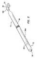
- FIG. 1is a perspective view depicting the device of the present invention including a cannula and a stylet positioned within an unexpanded balloon or sheath with the cannula positioned in a first, proximal portion of the balloon or sheath;
- FIG. 2is a perspective view depicting the cannula within an expanded balloon or sheath with the stylet removed and the cannula advanced toward a second, distal portion of the balloon or sheath;
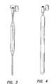
- FIG. 3is a side view depicting the cannula and stylet positioned in the unexpanded balloon or sheath with the cannula positioned in the first, proximal portion of the balloon or sheath;
- FIG. 4is a side view depicting the cannula within the expanded balloon or sheath with the stylet removed and the cannula advanced to the second, distal portion of the balloon or sheath;
- FIG. 5is a side view depicting the device of the invention, including the cannula and stylet positioned within the unexpanded balloon or sheath and the cannula in the first, proximal portion of the balloon, ready for use to penetrate a biological body wall;
- FIG. 6is a side view depicting the distal portion of the balloon or sheath and stylet penetrating the biological body wall;
- FIG. 7is a side view depicting the device with the distal end of the unexpanded balloon positioned in the body cavity, the cannula positioned proximal the body wall and the stylet removed from the balloon or sheath;
- FIG. 8is a side view depicting the device with the balloon or sheath expanded and positioned within the body wall with the distal end of the balloon or sheath positioned in the body cavity and the cannula positioned proximal the body wall;
- FIG. 9is a side view depicting the device with the cannula advanced through the expanded balloon or sheath to a second, distal portion of the balloon or sheath and across the body wall with the distal end of the cannula positioned distal an inner surface of the body wall and the proximal end of the cannula positioned proximal the body wall;
- FIG. 10is a side view depicting the balloon or sheath unexpanded with the balloon or sheath and the cannula positioned across the body wall and the distal ends of the balloon or sheath and the cannula positioned distal the inner surface of the body wall and in the body cavity;
- FIG. 11is a side view depicting a proximal portion of the balloon or sheath separated and removed from the cannula;
- FIG. 12is a side view depicting a piercing device inserted into the lumen of the cannula to pierce the distal end of the balloon or sheath;
- FIG. 13is a side view depicting the distal end of the balloon after being pierced, the piercing device removed, a seal housing being coupled to the proximal end of the cannula, and the cannula in communication between the body cavity and external the body wall;
- FIG. 14is a side view depicting the trocar of the present invention placed across the body wall and into the body cavity and ready for use as a laparoscopic surgery port;
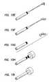
- FIGS. 15A-15Idepict a sequence of use for the trocar device of the present invention.
- FIG. 16is a side view depicting the trocar device of the present invention having a through lumen in the balloon or sheath with the balloon or sheath in the unexpanded condition;
- FIG. 17is a side view depicting the trocar device of FIG. 16 with the balloon or sheath in the expanded condition;
- FIG. 18is a side view depicting a needle inserted from outside a biological body into the renal pelvis of a kidney and a guide wire extending through the needle;
- FIG. 19is a side view depicting the device of FIGS. 16 and 17 advanced along the guide wire of FIG. 18 , with the needle removed, including the cannula and stylet positioned within the unexpanded balloon or sheath and the cannula in the first, proximal portion of the balloon, ready for use to penetrate a biological body wall;
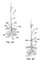
- FIG. 20is a side view depicting the trocar device of FIGS. 16 and 17 advancing along the guide wire of FIG. 18 and the distal portion of the balloon or sheath and stylet penetrating the biological body wall and kidney;
- FIG. 21is a side view depicting the device of FIGS. 16 and 17 with the distal end of the unexpanded balloon positioned in the renal pelvis of the kidney, the cannula positioned proximal the body wall, the guide wire in place and the stylet removed from the balloon or sheath;
- FIG. 22is a side view depicting the device of FIGS. 16 and 17 with the balloon or sheath expanded and positioned within the body wall and kidney with the distal end of the balloon or sheath positioned in the renal pelvis, the cannula positioned proximal the body wall, and the guide wire in place;
- FIG. 23is a side view depicting the device of FIGS. 16 and 17 with the cannula advanced through the expanded balloon or sheath to a second, distal portion of the balloon or sheath and across the body wall with the distal end of the cannula positioned within the renal pelvis, the proximal end of the cannula positioned proximal the body wall, and the guide wire in place;
- FIG. 24is a side view depicting the balloon or sheath unexpanded with the balloon or sheath and the cannula positioned across the body wall, the distal ends of the balloon or sheath and the cannula positioned within the renal pelvis, and the guide wire in place;
- FIG. 25is a side view depicting a proximal portion of the balloon or sheath separated and removed from the cannula;
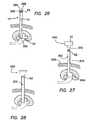
- FIG. 26is a side view depicting the guide wire removed and a piercing device inserted into the lumen of the cannula to pierce the distal end of the balloon or sheath;
- FIG. 27is a side view depicting the distal end of the balloon after being pierced, the piercing device removed, a seal housing being coupled to the proximal end of the cannula, and the cannula in communication between the renal pelvis and external the body wall;
- FIG. 28is a side view depicting the trocar of the present invention placed across the body wall and into the renal pelvis and ready for use as a surgery port.
- FIGS. 1-4depict a tamponade trocar 10 having an elongate balloon or sheath 15 , a cannula 300 and a tensioning stylet 200 .
- the cannula 300includes a first, proximal end 320 , a second, distal end 310 and a lumen 315 extending between the proximal end and the distal end.
- the balloon or sheath 15includes a first, proximal end 40 , a second, distal end 30 and a lumen 56 extending between the proximal and distal ends.
- the distal end 30 of the balloon or sheathmay be closed and substantially gastight.
- the distal end 30 of the balloon or sheath 15may be rounded or tapered 90 to facilitate penetration through a previously existing breach 560 ( FIGS. 6-14 ) through a body wall 500 and into a body cavity 550 .
- the balloon or sheath 15may be constructed of a substantially non-distensible material, such as polyethylene, polyurethane, or nylon and may be cross-linked to provide stability.
- the balloon or sheath 15may be thick-walled and have a length that is at least about twice the length of the cannula 300 . Those familiar with the art will recognize that the balloon 15 may be longer or shorter than about twice the length of the cannula 300 depending on the parameters for which the tamponade trocar 10 will be used and such lengths are contemplated as within the scope of the invention.
- a first, proximal portion 60 of the balloon or sheath 15is sized and configured to contain the cannula 300 .
- the balloon or sheath 15is adapted to expand from a first, low-profile, small diameter condition 70 ( FIGS. 1 and 3 ) to a second, substantially fixed, large diameter condition 72 ( FIGS. 2 and 4 ).
- the internal diameter of the balloon or sheath 15 when the balloon is in the second, large diameter condition 72is slightly larger than the outside diameter of the cannula 300 within it to facilitate transitioning of the cannula from the proximal portion 60 of the balloon or sheath to a second, distal portion 80 of the balloon or sheath.
- a valve 150 or sealmay be positioned in the proximal portion 60 of the balloon or sheath 15 , such as proximate the proximal end 40 of the balloon or sheath.
- the valveis adapted to communicate between the balloon lumen 56 and the atmosphere external the balloon.
- the transition from the first condition 70 to the second condition 72may be accomplished by the application of inflation pressure at the proximal end 40 of the balloon or sheath 15 or the valve 150 .
- the pressure required to fully expand the high-pressure balloon or sheath 15is substantially equivalent to the pressure required to expand a non-distensible balloon of the type used in angioplasty or other dilation driven therapies.
- the balloon or sheath 15may be either undeveloped, having an unformed, thick-walled expandable tube, or it may be a preformed and folded tube.
- An unformed or undeveloped tube of the balloon or sheath 15presents a smooth, small diameter profile but may require more inflation pressure than a preformed balloon or sheath 15 .
- the preformed balloon or sheath 15develops the ultimate expanded diameter at a lower pressure than for the unformed tube but may have a more irregular surface or a less than smooth profile.
- Either modepermits a reduced portion 20 , 50 for insertion through a perforation 560 .
- the cannula 300is substantially prevented from transitioning positions within the lumen 56 of the balloon.
- the cannula 300is substantially free to transition to the second, distal portion 80 of the balloon.
- the stylet 200is substantially rigid and includes a proximal end 220 and a distal end 240 .
- the stylet 200is removably positioned within the lumen 56 of the balloon 15 and within the lumen 315 of the cannula 300 .
- the distal end 240 of the stylet 200is positioned at the distal end 30 of the balloon 15 and the proximal end 220 of the stylet is positioned proximal to the proximal end 40 of the balloon or sheath 15 .
- the proximal end 220 of the stylet 200may be positioned proximal to the valve.
- the stylet 200may be hollow to facilitate insertion of a guide wire therethrough.
- FIGS. 5 through 15a method for placing the tamponade trocar device 10 within a biological body wall 500 is depicted.
- An insufflation needleFIG. 18 ) creates a perforation 560 through the body wall 500 to the body cavity 550 and the body cavity is insufflated.
- the tamponade trocar device 10is positioned external the body wall 500 with the distal end 30 of the balloon 15 proximate the perforation 560 through the body wall ( FIG. 5 ).
- the distal end 30 of the balloon of the tamponade trocar deviceis inserted into the perforation 560 in the body wall 500 .
- the balloon 15is advanced until the distal end 30 of the balloon of the device is distal an inner surface 545 , or peritoneum, of the body wall 500 and within the body cavity 550 while the cannula 300 remains in the first, proximal portion 60 of the balloon and proximal the body wall ( FIG. 6 ).
- the stylet 200may be removed proximally from the proximal end 40 of the balloon 15 or from the valve 150 ( FIG. 7 ).
- a pressurizing or filling source(not shown) is coupled to the proximal end 40 of the balloon 15 or to the valve 150 and the balloon is pressurized to the second, substantially fixed, large diameter condition 72 ( FIG. 8 ).
- the perforation 560is fully dilated and in compressive tamponade.
- the inside diameter 55 of the balloon 15allows for the free movement of the cannula 300 that has been provided in the lumen 56 of the balloon.
- the cannula 300is free to transition positions within the lumen 56 of the balloon.
- the cannula 300may be advanced distally within the lumen 56 of the balloon so that the cannula is positioned across the body wall 500 with the proximal end 320 of the cannula positioned proximal the body wall and the distal end 310 of the cannula positioned distal the inner surface 545 of the body wall ( FIG. 9 ).
- the distal end 30 of the balloon 15 intactthere is no communication between the exterior 510 of the body and the interior of the body cavity 550 through the perforation 560 via the cannula 300 .
- the balloon 15is deflated ( FIG.
- the deflated, unexpanded, balloon 15is severed ( FIG. 11 ) at a position 61 proximate the proximal end 320 of the cannula 300 and the portion of the balloon proximal the point of severance 62 is removed from the remaining portion of the balloon.
- the portion 51 of the balloon 15 proximal the point of severancemay be discarded.
- a piercing member 395may be provided.
- the piercing member 395includes a proximal end 398 and a distal, puncturing end 396 .
- the piercing member 395is adapted to fit through the cannula 300 and is of sufficient length to extend the length of the cannula and penetrate the distal end of the balloon 15 .
- the distal end 396 of the piercing member 395may be inserted into the proximal end 12 of the tamponade trocar device 10 and advanced distally through the tamponade trocar device until the distal, puncturing end of the piercing member punctures and opens the distal end 30 of the balloon 15 .
- the sequence of stepsmay be adjusted so that the piercing member 395 is placed in the cannula after the final placement of a seal housing 400 in a coupling relationship with the proximal end 320 of the cannula 300 so that there is little or no loss of insufflation pressure or pneumoperitoneum during the process for inserting the tamponade trocar device 10 into the body wall 500 .
- a trocar seal housing 400( FIGS. 13 and 14 ) is coupled to the proximal end 320 of the cannula 300 and the trocar is ready for use as a surgical port.
- the balloon 15may include a substantially toroid shape that forms a substantially tubular channel 235 throughout its length to facilitate advancing the distal end 30 of the balloon into the perforation 560 in the body wall 500 without first removing the needle from the body wall.
- the use of the tubular channel 235also facilitates insertion of the tamponade trocar device 10 over a guide wire 600 that can be inserted, for example, into a cavity of a biological organ, such as the renal pelvis 604 of a kidney 602 , to perform a surgical procedure, such as a percutaneous nephrolithotomy.
- the main, outer body portion of the balloon 15is coupled to the tubular channel 235 at the distal end 30 of the outer body portion of the balloon and at a proximal portion 100 of the outer body portion of the balloon so that the expandable portion of the balloon surrounds the tubular channel through which insufflation occurs.
- Percutaneous nephrolithotomyincludes the use of a cannula placed into a kidney, such as in the renal pelvis, to remove kidney stones.
- a method for placing the tamponade trocar device 10 having the tubular channel 235 ( FIGS. 16 and 17 ) within a biological body wall 500 and into the renal pelvis 604 of a kidney 602is depicted
- a needle 606creates a perforation 560 through the body wall 500 and through the wall 608 of the kidney 602 , thereby providing access to the renal pelvis 604 .
- a guide wire 600is inserted into a proximal end of the needle 606 and advanced therethrough until the distal end 610 of the guide wire is positioned distal to the distal end 612 of the needle ( FIG. 18 ).
- the distal end 610 of the guide wire 600may be advanced further than the renal pelvis 604 , such as into the ureter 614 . While maintaining the position of the guide wire 600 within the biological body, the needle 606 may be removed distally over the guide wire.
- the proximal end 616 of the guide wireis inserted into the distal end 618 of the tubular channel 235 of the tamponade trocar device 10 .
- the tamponade trocar device 10is positioned external the body wall 500 , the tamponade trocar device is advanced distally over the guide wire 600 until the distal end 30 of the balloon 15 is positioned proximate the perforation 560 through the body wall and the proximal end 616 of the guide wire is positioned proximal to the proximal end 40 of the balloon 15 or valve 150 ( FIG. 19 ).
- the distal end 30 of the balloon of the tamponade trocar device 10is advanced distally over the guide wire and inserted into the perforation 560 in the body wall 500 and the kidney wall 608 .
- the balloon 15is advanced until the distal end 30 of the balloon of the tamponade trocar device 10 is within the renal pelvis 604 while the cannula 300 remains in the first, proximal portion 60 of the balloon and proximal the body wall 500 ( FIG. 20 ).
- the stylet 200may be used during advancement of the balloon 15 to provide columnar strength to the balloon during insertion into the body.
- the stylet 200may be hollow, thereby providing for insertion of the guide wire 600 therethrough.
- the stylet 200may be removed proximally from the proximal end 40 of the balloon 15 or the valve 150 ( FIG. 21 ).
- a pressurizing or filling source(not shown) is coupled to the proximal end 40 of the balloon 15 or to the valve 150 and the balloon is pressurized to the second, substantially fixed, large diameter condition 72 ( FIG. 22 ).
- the perforation 560 in the body wall 500 and kidney wall 608is fully dilated and in compressive tamponade.
- the inside diameter 55 of the balloon 15allows for the free movement of the cannula 300 that has been provided in the lumen 56 of the balloon.
- the cannula 300may be advanced distally within the lumen 56 of the balloon so that the cannula is positioned across the body wall 500 and kidney wall 608 with the proximal end 320 of the cannula positioned proximal the body wall and the distal end 310 of the cannula positioned within the renal pelvis 604 ( FIG. 23 ).
- the balloon 15is deflated ( FIG. 24 ) or, as is the case with non-distensible balloons, depressurized to ambient pressure.
- the deflated, unexpanded, balloon 15is severed ( FIG. 25 ) at a position 61 proximate the proximal end 320 of the cannula 300 and the portion of the balloon proximal the point of severance 62 is removed from the remaining portion of the balloon.
- the portion 51 of the balloon 15 proximal the point of severancemay be discarded.
- a piercing member 395may be provided ( FIG. 26 ).
- the piercing member 395includes a proximal end 398 and a distal, puncturing end 396 .
- the piercing member 395is adapted to fit through the cannula 300 and is of sufficient length to extend the length of the cannula and penetrate the distal end of the balloon 15 .
- the guide wire 600may be removed proximally from the tamponade trocar device 10 prior to insertion of the piercing member 395 into the cannula 300 .
- the distal end 396 of the piercing member 395may be inserted into the proximal end 12 of the tamponade trocar device 10 and advanced distally through the tamponade trocar device until the distal, puncturing end of the piercing member punctures and opens the distal end 30 of the balloon 15 .
- the sequence of stepsmay be adjusted so that the piercing member 395 is placed in the cannula 300 after the final placement of a seal housing 400 in a coupling relationship with the proximal end 320 of the cannula so that there is little or no loss of sealing between outside 510 the biological body 500 and the renal pelvis 604 .
- a trocar seal housing 400( FIGS. 27 and 28 ) is coupled to the proximal end 320 of the cannula 300 and the trocar is ready for use as a surgical port.
Landscapes
- Health & Medical Sciences (AREA)
- Life Sciences & Earth Sciences (AREA)
- Heart & Thoracic Surgery (AREA)
- Animal Behavior & Ethology (AREA)
- General Health & Medical Sciences (AREA)
- Engineering & Computer Science (AREA)
- Biomedical Technology (AREA)
- Veterinary Medicine (AREA)
- Public Health (AREA)
- Pulmonology (AREA)
- Biophysics (AREA)
- Anesthesiology (AREA)
- Hematology (AREA)
- Surgery (AREA)
- Child & Adolescent Psychology (AREA)
- Pathology (AREA)
- Molecular Biology (AREA)
- Medical Informatics (AREA)
- Nuclear Medicine, Radiotherapy & Molecular Imaging (AREA)
- Surgical Instruments (AREA)
Abstract
Description
Claims (25)
Priority Applications (3)
| Application Number | Priority Date | Filing Date | Title |
|---|---|---|---|
| US12/106,920US8142467B2 (en) | 2008-04-21 | 2008-04-21 | Tamponade trocar device and method |
| US13/411,301US8834505B2 (en) | 2008-04-21 | 2012-03-02 | Tamponade trocar device and method |
| US14/465,020US8951277B2 (en) | 2008-04-21 | 2014-08-21 | Tamponade trocar device and method |
Applications Claiming Priority (1)
| Application Number | Priority Date | Filing Date | Title |
|---|---|---|---|
| US12/106,920US8142467B2 (en) | 2008-04-21 | 2008-04-21 | Tamponade trocar device and method |
Related Child Applications (1)
| Application Number | Title | Priority Date | Filing Date |
|---|---|---|---|
| US13/411,301ContinuationUS8834505B2 (en) | 2008-04-21 | 2012-03-02 | Tamponade trocar device and method |
Publications (2)
| Publication Number | Publication Date |
|---|---|
| US20090264913A1 US20090264913A1 (en) | 2009-10-22 |
| US8142467B2true US8142467B2 (en) | 2012-03-27 |
Family
ID=41201761
Family Applications (3)
| Application Number | Title | Priority Date | Filing Date |
|---|---|---|---|
| US12/106,920Expired - Fee RelatedUS8142467B2 (en) | 2008-04-21 | 2008-04-21 | Tamponade trocar device and method |
| US13/411,301Active2028-10-03US8834505B2 (en) | 2008-04-21 | 2012-03-02 | Tamponade trocar device and method |
| US14/465,020Expired - Fee RelatedUS8951277B2 (en) | 2008-04-21 | 2014-08-21 | Tamponade trocar device and method |
Family Applications After (2)
| Application Number | Title | Priority Date | Filing Date |
|---|---|---|---|
| US13/411,301Active2028-10-03US8834505B2 (en) | 2008-04-21 | 2012-03-02 | Tamponade trocar device and method |
| US14/465,020Expired - Fee RelatedUS8951277B2 (en) | 2008-04-21 | 2014-08-21 | Tamponade trocar device and method |
Country Status (1)
| Country | Link |
|---|---|
| US (3) | US8142467B2 (en) |
Cited By (1)
| Publication number | Priority date | Publication date | Assignee | Title |
|---|---|---|---|---|
| US11051845B2 (en)* | 2017-01-14 | 2021-07-06 | Choon Kee Lee | Non-surgical chest tube introducer |
Families Citing this family (6)
| Publication number | Priority date | Publication date | Assignee | Title |
|---|---|---|---|---|
| DE102009060921A1 (en)* | 2009-12-31 | 2011-08-18 | Bader, Markus, Dr., 81825 | Puncture needle system |
| US20170189059A1 (en)* | 2016-01-06 | 2017-07-06 | Boston Scientific Scimed, Inc. | Percutaneous access device |
| US20200323556A1 (en)* | 2019-04-15 | 2020-10-15 | Covidien Lp | Balloon trocar including a pin |
| WO2021163911A1 (en)* | 2020-02-19 | 2021-08-26 | 上海英诺伟医疗器械有限公司 | Percutaneous renal puncture dilatation kit, usage method thereof and operation assembly |
| CN111450392B (en)* | 2020-05-11 | 2025-01-14 | 上海市东方医院(同济大学附属东方医院) | Multi-level fistula dilatation puncture drainage tube with balloon |
| US11439430B2 (en)* | 2020-05-11 | 2022-09-13 | Covidien Lp | Surgical access device with air release mechanism |
Citations (48)
| Publication number | Priority date | Publication date | Assignee | Title |
|---|---|---|---|---|
| US1621159A (en) | 1925-11-27 | 1927-03-15 | Robert T Evans | Abdominoscope |
| FR748666A (en) | 1932-03-30 | 1933-07-07 | Collin & Cie | Harpoon trocar for septic punctures |
| US3344791A (en) | 1965-02-12 | 1967-10-03 | John W Foderick | Bulbous urinary catheter with axial extension means |
| US3717151A (en) | 1971-03-11 | 1973-02-20 | R Collett | Flesh penetrating apparatus |
| US3789852A (en) | 1972-06-12 | 1974-02-05 | S Kim | Expandable trochar, especially for medical purposes |
| US3970090A (en) | 1975-02-03 | 1976-07-20 | Physio Medics, Inc. | Catheter |
| US4315512A (en) | 1980-01-24 | 1982-02-16 | Fogarty Thomas J | Piston extension balloon dilatation catheter apparatus and method |
| US4411655A (en) | 1981-11-30 | 1983-10-25 | Schreck David M | Apparatus and method for percutaneous catheterization |
| US4699611A (en) | 1985-04-19 | 1987-10-13 | C. R. Bard, Inc. | Biliary stent introducer |
| US4762130A (en) | 1987-01-15 | 1988-08-09 | Thomas J. Fogarty | Catheter with corkscrew-like balloon |
| US4861334A (en) | 1988-06-24 | 1989-08-29 | Nawaz Arain | Self-retaining gastrostomy tube |
| US4921479A (en) | 1987-10-02 | 1990-05-01 | Joseph Grayzel | Catheter sheath with longitudinal seam |
| US5122122A (en) | 1989-11-22 | 1992-06-16 | Dexide, Incorporated | Locking trocar sleeve |
| US5139511A (en) | 1990-02-14 | 1992-08-18 | Gill Steven S | Expansible cannula |
| US5147316A (en) | 1990-11-19 | 1992-09-15 | Castillenti Thomas A | Laparoscopic trocar with self-locking port sleeve |
| US5176697A (en) | 1989-04-06 | 1993-01-05 | Hasson Harrith M | Laparoscopic cannula |
| US5257975A (en) | 1992-08-14 | 1993-11-02 | Edward Weck Incorporated | Cannula retention device |
| US5320611A (en) | 1993-02-04 | 1994-06-14 | Peter M. Bonutti | Expandable cannula having longitudinal wire and method of use |
| US5352199A (en) | 1993-05-28 | 1994-10-04 | Numed, Inc. | Balloon catheter |
| US5443484A (en) | 1992-06-16 | 1995-08-22 | Loma Linda University Medical Center | Trocar and method for endoscopic surgery |
| US5445615A (en) | 1991-11-06 | 1995-08-29 | Yoon; Inbae | Surgical instrument stabilizer |
| US5458583A (en) | 1993-01-07 | 1995-10-17 | Medical Innovations Corporation | Gastrostomy catheter system |
| US5487730A (en) | 1992-12-30 | 1996-01-30 | Medtronic, Inc. | Balloon catheter with balloon surface retention means |
| US5496345A (en) | 1992-06-02 | 1996-03-05 | General Surgical Innovations, Inc. | Expansible tunneling apparatus for creating an anatomic working space |
| US5549625A (en) | 1994-04-26 | 1996-08-27 | Very Inventive Physicians, Inc. | Balloon dissector |
| US5632761A (en)* | 1991-05-29 | 1997-05-27 | Origin Medsystems, Inc. | Inflatable devices for separating layers of tissue, and methods of using |
| US5653690A (en) | 1992-12-30 | 1997-08-05 | Medtronic, Inc. | Catheter having a balloon with retention enhancement |
| US5697913A (en) | 1996-08-09 | 1997-12-16 | Ethicon Endo-Surgery, Inc. | Trocar including cannula with stepped region |
| US5697946A (en) | 1994-10-07 | 1997-12-16 | Origin Medsystems, Inc. | Method and apparatus for anchoring laparoscopic instruments |
| US5762604A (en) | 1994-06-01 | 1998-06-09 | Archimedes Surgical, Inc. | Surgical instrument permitting endoscopic viewing and dissecting |
| US5814058A (en) | 1993-03-05 | 1998-09-29 | Innerdyne, Inc. | Method and apparatus employing conformable sleeve for providing percutaneous access |
| US5827319A (en) | 1996-05-20 | 1998-10-27 | Innerdyne, Inc. | Radially expandable access system having disposable and reusable components |
| US5836913A (en) | 1997-05-02 | 1998-11-17 | Innerdyne, Inc. | Device and method for accessing a body cavity |
| US5944691A (en) | 1996-11-04 | 1999-08-31 | Cordis Corporation | Catheter having an expandable shaft |
| US6015421A (en) | 1997-05-15 | 2000-01-18 | General Surgical Innovations, Inc. | Apparatus and method for developing an anatomic space for laparoscopic procedures |
| US6277066B1 (en) | 1999-04-30 | 2001-08-21 | Civco Medical Instruments Inc. | Endocavity imaging sensor positioning apparatus and method |
| US6325812B1 (en) | 1993-03-05 | 2001-12-04 | Innerdyne, Inc. | Trocar system having expandable port |
| US6432085B1 (en) | 1999-03-17 | 2002-08-13 | Tyco Healthcare Group Lp | Self-retaining surgical access instrument |
| US6613038B2 (en) | 1993-02-04 | 2003-09-02 | Bonutti 2003 Trust-A | Method of using expandable cannula |
| US20030216770A1 (en) | 2002-02-21 | 2003-11-20 | Persidsky Maxim D. | Apparatus and method for making a percutaneous access port of variable size |
| US20040068228A1 (en) | 2002-10-04 | 2004-04-08 | Jon Cunningham | Device and method for stabilizing catheters |
| US6808492B2 (en) | 2002-08-16 | 2004-10-26 | Linvatec Corporation | Endoscopic cannula fixation system |
| US20040230218A1 (en) | 2002-10-04 | 2004-11-18 | Christopher Criscuolo | Balloon dissector with cannula |
| US20040243167A1 (en) | 1999-05-28 | 2004-12-02 | Shigeru Tanaka | Specially shaped balloon device for use in surgery and mehtod of use |
| US20040249243A1 (en) | 2001-09-21 | 2004-12-09 | Kleiner Daniel Eduard | Tamponade apparatus and method of using same |
| US20050165432A1 (en) | 2002-05-09 | 2005-07-28 | Russell Heinrich | Adjustable balloon anchoring trocar |
| US6989018B2 (en) | 1994-06-29 | 2006-01-24 | General Surgical Innovations, Inc. | Extraluminal balloon dissection |
| US20060149136A1 (en) | 2004-12-22 | 2006-07-06 | Kyphon Inc. | Elongating balloon device and method for soft tissue expansion |
Family Cites Families (1)
| Publication number | Priority date | Publication date | Assignee | Title |
|---|---|---|---|---|
| US5545135A (en)* | 1994-10-31 | 1996-08-13 | Boston Scientific Corporation | Perfusion balloon stent |
- 2008
- 2008-04-21USUS12/106,920patent/US8142467B2/ennot_activeExpired - Fee Related
- 2012
- 2012-03-02USUS13/411,301patent/US8834505B2/enactiveActive
- 2014
- 2014-08-21USUS14/465,020patent/US8951277B2/ennot_activeExpired - Fee Related
Patent Citations (51)
| Publication number | Priority date | Publication date | Assignee | Title |
|---|---|---|---|---|
| US1621159A (en) | 1925-11-27 | 1927-03-15 | Robert T Evans | Abdominoscope |
| FR748666A (en) | 1932-03-30 | 1933-07-07 | Collin & Cie | Harpoon trocar for septic punctures |
| US3344791A (en) | 1965-02-12 | 1967-10-03 | John W Foderick | Bulbous urinary catheter with axial extension means |
| US3717151A (en) | 1971-03-11 | 1973-02-20 | R Collett | Flesh penetrating apparatus |
| US3789852A (en) | 1972-06-12 | 1974-02-05 | S Kim | Expandable trochar, especially for medical purposes |
| US3970090A (en) | 1975-02-03 | 1976-07-20 | Physio Medics, Inc. | Catheter |
| US4315512A (en) | 1980-01-24 | 1982-02-16 | Fogarty Thomas J | Piston extension balloon dilatation catheter apparatus and method |
| US4411655A (en) | 1981-11-30 | 1983-10-25 | Schreck David M | Apparatus and method for percutaneous catheterization |
| US4699611A (en) | 1985-04-19 | 1987-10-13 | C. R. Bard, Inc. | Biliary stent introducer |
| US4762130A (en) | 1987-01-15 | 1988-08-09 | Thomas J. Fogarty | Catheter with corkscrew-like balloon |
| US4921479A (en) | 1987-10-02 | 1990-05-01 | Joseph Grayzel | Catheter sheath with longitudinal seam |
| US4861334A (en) | 1988-06-24 | 1989-08-29 | Nawaz Arain | Self-retaining gastrostomy tube |
| US5176697A (en) | 1989-04-06 | 1993-01-05 | Hasson Harrith M | Laparoscopic cannula |
| US5122122A (en) | 1989-11-22 | 1992-06-16 | Dexide, Incorporated | Locking trocar sleeve |
| US5139511A (en) | 1990-02-14 | 1992-08-18 | Gill Steven S | Expansible cannula |
| US5147316A (en) | 1990-11-19 | 1992-09-15 | Castillenti Thomas A | Laparoscopic trocar with self-locking port sleeve |
| US5632761A (en)* | 1991-05-29 | 1997-05-27 | Origin Medsystems, Inc. | Inflatable devices for separating layers of tissue, and methods of using |
| US5445615A (en) | 1991-11-06 | 1995-08-29 | Yoon; Inbae | Surgical instrument stabilizer |
| US5472429A (en) | 1991-11-06 | 1995-12-05 | Yoon; Inbae | Surgical instrument stabilizer |
| US5496345A (en) | 1992-06-02 | 1996-03-05 | General Surgical Innovations, Inc. | Expansible tunneling apparatus for creating an anatomic working space |
| US5443484A (en) | 1992-06-16 | 1995-08-22 | Loma Linda University Medical Center | Trocar and method for endoscopic surgery |
| US5257975A (en) | 1992-08-14 | 1993-11-02 | Edward Weck Incorporated | Cannula retention device |
| US5487730A (en) | 1992-12-30 | 1996-01-30 | Medtronic, Inc. | Balloon catheter with balloon surface retention means |
| US5653690A (en) | 1992-12-30 | 1997-08-05 | Medtronic, Inc. | Catheter having a balloon with retention enhancement |
| US5458583A (en) | 1993-01-07 | 1995-10-17 | Medical Innovations Corporation | Gastrostomy catheter system |
| US5320611A (en) | 1993-02-04 | 1994-06-14 | Peter M. Bonutti | Expandable cannula having longitudinal wire and method of use |
| US6613038B2 (en) | 1993-02-04 | 2003-09-02 | Bonutti 2003 Trust-A | Method of using expandable cannula |
| US5814058A (en) | 1993-03-05 | 1998-09-29 | Innerdyne, Inc. | Method and apparatus employing conformable sleeve for providing percutaneous access |
| US6325812B1 (en) | 1993-03-05 | 2001-12-04 | Innerdyne, Inc. | Trocar system having expandable port |
| US5352199A (en) | 1993-05-28 | 1994-10-04 | Numed, Inc. | Balloon catheter |
| US5549625A (en) | 1994-04-26 | 1996-08-27 | Very Inventive Physicians, Inc. | Balloon dissector |
| US5762604A (en) | 1994-06-01 | 1998-06-09 | Archimedes Surgical, Inc. | Surgical instrument permitting endoscopic viewing and dissecting |
| US6989018B2 (en) | 1994-06-29 | 2006-01-24 | General Surgical Innovations, Inc. | Extraluminal balloon dissection |
| US5697946A (en) | 1994-10-07 | 1997-12-16 | Origin Medsystems, Inc. | Method and apparatus for anchoring laparoscopic instruments |
| US6524283B1 (en) | 1994-10-07 | 2003-02-25 | Sherwood Services Ag | Method and apparatus for anchoring laparoscopic instruments |
| US5827319A (en) | 1996-05-20 | 1998-10-27 | Innerdyne, Inc. | Radially expandable access system having disposable and reusable components |
| US5697913A (en) | 1996-08-09 | 1997-12-16 | Ethicon Endo-Surgery, Inc. | Trocar including cannula with stepped region |
| US5944691A (en) | 1996-11-04 | 1999-08-31 | Cordis Corporation | Catheter having an expandable shaft |
| US5836913A (en) | 1997-05-02 | 1998-11-17 | Innerdyne, Inc. | Device and method for accessing a body cavity |
| US6015421A (en) | 1997-05-15 | 2000-01-18 | General Surgical Innovations, Inc. | Apparatus and method for developing an anatomic space for laparoscopic procedures |
| US6432085B1 (en) | 1999-03-17 | 2002-08-13 | Tyco Healthcare Group Lp | Self-retaining surgical access instrument |
| US6277066B1 (en) | 1999-04-30 | 2001-08-21 | Civco Medical Instruments Inc. | Endocavity imaging sensor positioning apparatus and method |
| US20040243167A1 (en) | 1999-05-28 | 2004-12-02 | Shigeru Tanaka | Specially shaped balloon device for use in surgery and mehtod of use |
| US6860892B1 (en) | 1999-05-28 | 2005-03-01 | General Surgical Innovations, Inc. | Specially shaped balloon device for use in surgery and method of use |
| US20040249243A1 (en) | 2001-09-21 | 2004-12-09 | Kleiner Daniel Eduard | Tamponade apparatus and method of using same |
| US20030216770A1 (en) | 2002-02-21 | 2003-11-20 | Persidsky Maxim D. | Apparatus and method for making a percutaneous access port of variable size |
| US20050165432A1 (en) | 2002-05-09 | 2005-07-28 | Russell Heinrich | Adjustable balloon anchoring trocar |
| US6808492B2 (en) | 2002-08-16 | 2004-10-26 | Linvatec Corporation | Endoscopic cannula fixation system |
| US20040068228A1 (en) | 2002-10-04 | 2004-04-08 | Jon Cunningham | Device and method for stabilizing catheters |
| US20040230218A1 (en) | 2002-10-04 | 2004-11-18 | Christopher Criscuolo | Balloon dissector with cannula |
| US20060149136A1 (en) | 2004-12-22 | 2006-07-06 | Kyphon Inc. | Elongating balloon device and method for soft tissue expansion |
Cited By (1)
| Publication number | Priority date | Publication date | Assignee | Title |
|---|---|---|---|---|
| US11051845B2 (en)* | 2017-01-14 | 2021-07-06 | Choon Kee Lee | Non-surgical chest tube introducer |
Also Published As
| Publication number | Publication date |
|---|---|
| US20120165612A1 (en) | 2012-06-28 |
| US8834505B2 (en) | 2014-09-16 |
| US20140364832A1 (en) | 2014-12-11 |
| US20090264913A1 (en) | 2009-10-22 |
| US8951277B2 (en) | 2015-02-10 |
Similar Documents
| Publication | Publication Date | Title |
|---|---|---|
| US8951277B2 (en) | Tamponade trocar device and method | |
| US10124156B2 (en) | Surgical access port and method of using same | |
| US5964781A (en) | Skin seal with inflatable membrane | |
| US8292919B2 (en) | Skin seal with inflatable membrane | |
| US5836913A (en) | Device and method for accessing a body cavity | |
| EP0956060B1 (en) | Apparatus employing conformable sleeve for providing percutaneous access | |
| US5431676A (en) | Trocar system having expandable port | |
| EP1774918B1 (en) | Trocar anchor | |
| US5871474A (en) | Screw-type skin seal with inflatable membrane | |
| US6238373B1 (en) | Screw-type skin seal with inflatable membrane | |
| US20080171989A1 (en) | Trans Urinary Bladder Access Device and Method | |
| US20110190781A1 (en) | Surgical retrieval apparatus | |
| EP3909531A1 (en) | Surgical access device with air release mechanism | |
| EP2308400A1 (en) | Foam collar for surgical access devices |
Legal Events
| Date | Code | Title | Description |
|---|---|---|---|
| AS | Assignment | Owner name:APPLIED MEDICAL RESOURCES CORPORATION, CALIFORNIA Free format text:ASSIGNMENT OF ASSIGNORS INTEREST;ASSIGNORS:HART, CHARLES C.;BRUSTAD, JOHN R.;REEL/FRAME:020834/0614 Effective date:20080417 | |
| STCF | Information on status: patent grant | Free format text:PATENTED CASE | |
| AS | Assignment | Owner name:CITIBANK, N.A., TEXAS Free format text:SECURITY AGREEMENT;ASSIGNOR:APPLIED MEDICAL RESOURCES CORPORATION;REEL/FRAME:028115/0276 Effective date:20120417 | |
| FPAY | Fee payment | Year of fee payment:4 | |
| AS | Assignment | Owner name:JPMORGAN CHASE BANK, N.A., AS ADMINISTRATIVE AGENT, ILLINOIS Free format text:SECURITY INTEREST;ASSIGNOR:APPLIED MEDICAL RESOURCES CORPORATION;REEL/FRAME:042669/0725 Effective date:20170531 Owner name:JPMORGAN CHASE BANK, N.A., AS ADMINISTRATIVE AGENT Free format text:SECURITY INTEREST;ASSIGNOR:APPLIED MEDICAL RESOURCES CORPORATION;REEL/FRAME:042669/0725 Effective date:20170531 | |
| FEPP | Fee payment procedure | Free format text:MAINTENANCE FEE REMINDER MAILED (ORIGINAL EVENT CODE: REM.); ENTITY STATUS OF PATENT OWNER: LARGE ENTITY | |
| LAPS | Lapse for failure to pay maintenance fees | Free format text:PATENT EXPIRED FOR FAILURE TO PAY MAINTENANCE FEES (ORIGINAL EVENT CODE: EXP.); ENTITY STATUS OF PATENT OWNER: LARGE ENTITY | |
| STCH | Information on status: patent discontinuation | Free format text:PATENT EXPIRED DUE TO NONPAYMENT OF MAINTENANCE FEES UNDER 37 CFR 1.362 | |
| FP | Lapsed due to failure to pay maintenance fee | Effective date:20200327 | |
| AS | Assignment | Owner name:APPLIED MEDICAL RESOURCES CORPORATION, CALIFORNIA Free format text:RELEASE BY SECURED PARTY;ASSIGNOR:JPMORGAN CHASE BANK, N.A.;REEL/FRAME:056751/0169 Effective date:20210625 | |
| AS | Assignment | Owner name:APPLIED MEDICAL RESOURCES CORPORATION, CALIFORNIA Free format text:RELEASE BY SECURED PARTY;ASSIGNOR:CITIBANK N.A., AS ADMINISTRATIVE AGENT;REEL/FRAME:066795/0595 Effective date:20240129 |