US8137256B2 - Brachytherapy apparatus - Google Patents
Brachytherapy apparatusDownload PDFInfo
- Publication number
- US8137256B2 US8137256B2US11/305,437US30543705AUS8137256B2US 8137256 B2US8137256 B2US 8137256B2US 30543705 AUS30543705 AUS 30543705AUS 8137256 B2US8137256 B2US 8137256B2
- Authority
- US
- United States
- Prior art keywords
- tubes
- cavity
- tube
- brachytherapy
- breast
- Prior art date
- Legal status (The legal status is an assumption and is not a legal conclusion. Google has not performed a legal analysis and makes no representation as to the accuracy of the status listed.)
- Expired - Fee Related, expires
Links
Images


Classifications
- A—HUMAN NECESSITIES
- A61—MEDICAL OR VETERINARY SCIENCE; HYGIENE
- A61N—ELECTROTHERAPY; MAGNETOTHERAPY; RADIATION THERAPY; ULTRASOUND THERAPY
- A61N5/00—Radiation therapy
- A61N5/10—X-ray therapy; Gamma-ray therapy; Particle-irradiation therapy
- A61N5/1001—X-ray therapy; Gamma-ray therapy; Particle-irradiation therapy using radiation sources introduced into or applied onto the body; brachytherapy
- A61N5/1014—Intracavitary radiation therapy
- A61N5/1015—Treatment of resected cavities created by surgery, e.g. lumpectomy
- A—HUMAN NECESSITIES
- A61—MEDICAL OR VETERINARY SCIENCE; HYGIENE
- A61N—ELECTROTHERAPY; MAGNETOTHERAPY; RADIATION THERAPY; ULTRASOUND THERAPY
- A61N5/00—Radiation therapy
- A61N5/10—X-ray therapy; Gamma-ray therapy; Particle-irradiation therapy
- A61N5/1001—X-ray therapy; Gamma-ray therapy; Particle-irradiation therapy using radiation sources introduced into or applied onto the body; brachytherapy
- A61N5/1014—Intracavitary radiation therapy
- A61N2005/1018—Intracavitary radiation therapy with multiple channels for guiding radioactive sources
Definitions
- a lumpectomytends to involve removal of only the portion or “lump” of the breast that contains tissue having tumors. The remaining tissue outside the removed lump may be treated subsequently with breast irradiation that is designed to treat abnormal or suspect tissue that surrounds the removed tumor.
- One of the various lumpectomy optionsinvolves full breast irradiation. While this option incorporates the breast-sparing lumpectomy, the treatment time may last for several weeks, with several treatments a day during those weeks. At times, the number of treatments may be as much as thirty treatments. Such a high number of treatments may be not only inconvenient for the patient, it may also be expensive since each time the patient sees a doctor, a charge may follow. Even for insured patients, all of the charges may not be covered by the patient's insurance.
- partial breast irradiation procedureshave been developed. These options may incorporate high dose radiation. With high-dose irradiation, significant patient discomfort may result since multiple needles and catheters are placed into the breast. Moreover, there is a greater chance that surface tissue damage may occur, resulting in scarring and sensitivity.
- Another example of a treatment option that has been developed incorporating partial breast irradiationis one developed by Proxima Therapeutics, Inc., and known as the MAMMOSITETM radiation therapy treatment system.
- This systemappears to be designed to address some of the drawbacks associated with full breast irradiation while also addressing some of the drawbacks associated with high-dose radiation.
- the procedureinvolves inflating a balloon so that it fills the empty cavity and inserting a high-dose radiation source for delivery inside the cavity.
- the treatment timemay be twice a day for five (5) days, for a total of ten (10) treatments.
- the patient selection criteriamay be limited in that patients with small breasts may not be good candidates. For example, the breast may be too small to allow proper inflation of the balloon.
- the high-dose of radiationmay be in dangerous proximity to vital organs, thus possibly resulting in damage to vital organs.
- brachytherapyhas been used for partial breast irradiation to deliver a more localized treatment of tumor cells after a lumpectomy.
- Partial breast irradiationis used to supplement surgical resection by targeting the residual tumor margin after resection, with the goal of treating any residual cancer in the margin.
- Radiation therapycan be administered through one of several methods, or a combination of methods, including external-beam radiation, stereotactic radiosurgery, and permanent or temporary interstitial brachytherapy. Owing to the proximity of the radiation source, brachytherapy offers the advantage of delivering a more localized dose to the target tissue region.
- the present disclosureaddresses the deficiencies noted hereinabove by providing an interstitial brachytherapy apparatus that may be implanted in a single visit, thereby reducing the number of office visits and providing a more convenient treatment regimen.
- a brachytherapy apparatusfor delivery of localized irradiation to tissue that remains after surgical resection.
- the apparatuscomprises one or more thin-walled tubes, each of said thin-walled tubes being configured to contain a plurality of low-dose radioactive seed strands.
- the apparatusfurther comprises a plurality of low-dose radioactive seeds, including at least one low-dose radioactive seed strand disposed within the one or more thin-walled tubes, the plurality of low-dose radioactive seeds being configured to deliver a prescribed dose of radiation.
- the apparatusalso includes an expansion element configured to expand said one or more thin-walled tubes so that at least a portion of the one or more thin-walled tubes is positioned substantially against the remaining tissue.
- a brachytherapy apparatusfor delivery of localized irradiation after surgical tissue removal when the tissue removal results in a cavity surrounded by remaining tissue.
- the brachytherapy apparatuscomprises a seed containment device configured to contain a plurality of low-dose radioactive seeds, and a plurality of low-dose radioactive seeds disposed within the seed containment device.
- the seed containment deviceis further configured to expand so that at least a portion of the seed containment device is positioned against the remaining tissue and the plurality of radioactive seeds are disposed at the perimeter of the seed containment device.
- a brachytherapy apparatusfor delivery of localized irradiation after surgical tissue removal, the tissue removal resulting in a cavity surrounded by remaining tissue.
- the brachytherapy apparatuscomprises a plurality of stacked, substantially elliptical, thin-walled tubes, the plurality of tubes including an uppermost tube, at least one middle tube and a lowermost tube, wherein each tube has a top tube section, and a bottom tube section.
- the apparatusfurther includes connection mechanisms configured to connect each tube to at least one other tube such that the top tube section of one tube is connected to the bottom tube section of another tube with the exception of the uppermost tube.
- the uppermost tube and the lowermost tubeare of a lesser volume than the at least one middle tube.
- the apparatusis configured to expand and collapse, the expanded state resulting in a greater volume for the apparatus than in its collapsed state.
- the apparatusdefines a diameter of an upper tube section, a middle tube section and a lower tube section. In its expanded state, the diameter of the middle tube section is greater than the diameter of the upper tube section and the lower tube section.
- a brachytherapy apparatusfor delivery of localized irradiation after surgical tissue removal when the tissue removal results in a cavity surrounded by remaining tissue.
- the brachytherapy apparatuscomprises a plurality of substantially cylindrical, thin-walled tubes, the wall of each tube being connected to the wall of at least one other tube.
- the apparatusfurther includes a plurality of low-dose radioactive seeds, including at least one low-dose radioactive seed strand disposed within the one or more thin-walled tubes, the plurality of low-dose radioactive seeds being configured to deliver a prescribed dose of radiation.
- the apparatusincludes an expansion element configured to expand said plurality of substantially cylindrical, thin-walled tubes so that at least a portion of the one or more thin-walled tubes is positioned substantially against the remaining tissue.
- a brachytherapy apparatusfor delivery of localized irradiation after surgical tissue removal when the tissue removal results in a cavity surrounded by remaining tissue.
- the brachytherapy apparatuscomprises a first set of tubes being configured in a first direction.
- the apparatusfurther comprises a second set of tubes being configured in a second direction such that the second set of tubes intersects with the first set of tubes to define an upper tube section, a middle tube section and a lower tube section; each of said tubes sections defining a diameter.
- the diameter of the middle tube sectionis greater than the diameter of the upper tube section and the lower tube section.
- the volume of the apparatus in its first stateis lesser than the volume of the apparatus in its second state.
- a method for delivering localized irradiation after surgical tissue removal, the tissue removal resulting in a cavitycomprises creating access to the cavity.
- the methodfurther comprises providing an interstitial brachytherapy apparatus that includes one or more thin-walled tubes, each of said thin-walled tubes being configured to contain a plurality of low-dose radioactive seed strands.
- the brachytherapy apparatus that is providedalso includes a plurality of low-dose radioactive seeds, including at least one low-dose radioactive seed strand disposed within the one or more thin-walled tubes, the plurality of low-dose radioactive seeds being configured to deliver a prescribed dose of radiation.
- the brachytherapy apparatusthat is provided further includes an expansion element configured to expand said one or more tubes so that at least a portion of the one or more tubes is positioned substantially against the remaining tissue.
- the method using this brachytherapy apparatusfurther includes placing the interstitial brachytherapy apparatus into the cavity and expanding the interstitial brachytherapy apparatus so that it substantially fills the cavity, and so that the plurality of thin-walled tubes are placed substantially against the remaining tissue, clamping the interstitial brachytherapy apparatus onto the patient, leaving the interstitial brachytherapy apparatus inside the cavity for a sufficient time to deliver the prescribed radiation dose to remaining tissue that surrounds the cavity and, finally, removing the interstitial brachytherapy apparatus.
- a methodfor delivering localized irradiation after surgical tissue removal, the tissue removal resulting in a cavity.
- the methodcomprises creating access to the cavity.
- An interstitial brachytherapy apparatusis provided for use with the method.
- the brachytherapy apparatusincludes a seed containment device configured to contain a plurality of low-dose radioactive seeds, and a plurality of low-dose radioactive seeds disposed within the seed containment device.
- the seed containment device of the brachytherapy apparatusis further configured to expand so that at least a portion of the seed containment device is positioned against the remaining tissue and the plurality of radioactive seeds are disposed at the perimeter of the seed containment device.
- the methodincludes placing this interstitial brachytherapy apparatus into the cavity, and expanding the interstitial brachytherapy apparatus so that it substantially fills the cavity, and so that the seed containment device is placed substantially against the remaining tissue.
- the methodfurther includes clamping the interstitial brachytherapy apparatus onto the patient, and leaving the interstitial brachytherapy apparatus inside the cavity for a sufficient time to deliver the prescribed radiation dose to remaining tissue that surrounds the cavity.
- the methodincludes removing the interstitial brachytherapy apparatus.
- a treatment apparatusfor delivery of localized treatment to tissue that remains after surgical resection.
- the apparatuscomprises one or more thin-walled tubes, each of said thin-walled tubes being configured to contain a plurality of therapeutic elements, a plurality of therapeutic elements, and an expansion element configured to expand said one or more thin-walled tubes so that at least a portion of the one or more thin-walled tubes is positioned substantially against the remaining tissue.
- a treatment apparatusfor delivery of localized treatment to tissue that remains after surgical resection.
- the treatment apparatusincludes a seed containment device configured to contain a plurality of therapeutic elements, a plurality of therapeutic elements disposed within the seed containment device.
- the seed containment deviceis further configured to expand so that at least a portion of the seed containment device is positioned against the remaining tissue and the therapeutic elements are disposed at the perimeter of the seed containment device.
- FIG. 1Ais a brachytherapy apparatus in a collapsed state in accordance with one embodiment of the present disclosure.
- FIG. 1Bis an embodiment of the apparatus of FIG. 1A in its expanded state.
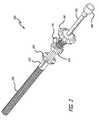
- FIG. 2is a breast brachytherapy apparatus having a clamp and in its collapsed state in accordance with another embodiment of the present disclosure.
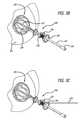
- FIG. 3Ais the brachytherapy apparatus of FIG. 2 as it is used in a brachytherapy procedure.
- FIG. 3Bis the apparatus of FIGS. 2 and 3A in its expanded state in accordance with one embodiment of the present disclosure.
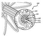
- FIG. 3Cis the apparatus of FIG. 3B showing the process of radioactive strands being placed into the openings.
- FIG. 3Dis the apparatus of FIG. 3C when the tubes are held tightly in place.
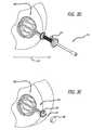
- FIG. 3Eis a post-implantation embodiment of the brachytherapy apparatus of FIG. 3D .
- FIG. 3Fis a post-implantation embodiment of a brachytherapy apparatus with a post-implantation cap on its end.
- FIG. 3Gis an embodiment of a brachytherapy apparatus after an obturator has been inserted.
- FIG. 3His an embodiment where the tubes are collapsed.
- FIG. 3Iis an embodiment showing how the brachytherapy apparatus may be removed from a patient, leaving the suture disk in place temporarily.
- FIG. 3Jis an illustration of the apparatus having been removed fully from the breast.
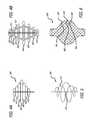
- FIG. 4Ais the brachytherapy apparatus as stacked, substantially elliptical, expandable tubes and in a collapsed state in accordance with one embodiment of the present disclosure
- FIG. 4Bis the apparatus 4 A in its expanded state.
- FIG. 5is a single spiral tube strand brachytherapy apparatus in accordance with another embodiment of the present disclosure.
- FIG. 6is an expandable mesh brachytherapy apparatus in its expanded state in accordance with yet another embodiment of the present disclosure.
- FIGS. 7A and 7Billustrate a stent embodiment of a brachytherapy apparatus in a collapsed and expanded state in accordance with still yet another embodiment of the present disclosure.
- FIG. 8is a multi-tube spiral embodiment of a brachytherapy apparatus in accordance with still yet another embodiment of the present disclosure.
- FIGS. 9A and 9Billustrate a bandoleer-configured brachytherapy apparatus in accordance with still yet another embodiment of the present disclosure.
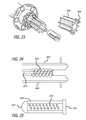
- FIG. 11is a bendable tube configuration that includes a sleeve configured to slide toward the tubes to reduce the length of the tubes to control the expansion volume.
- FIG. 12is a multi-lumen brachytherapy apparatus having tubes that are snugly fit from side-to-side.
- FIG. 13is a multi-lumen tube that could also be configured for expansion at the time of use through the use of molded “knives” at the interior surface of a sleeve of the apparatus.
- FIG. 14Ais a side view of a brachytherapy apparatus that includes a cap joined to the tubes.
- FIG. 14Bis a top view of the cap described in FIG. 14A .
- FIG. 14Cis a dimensional representation of the cross section of the tubes of FIG. 14A .
- FIG. 15is a dual-sleeved proximal end portion of a brachytherapy apparatus in accordance with one embodiment of the present disclosure.
- FIG. 16is a brachytherapy apparatus having a threaded sleeve and clamp in accordance with another embodiment of the present disclosure.
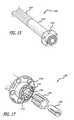
- FIG. 17is the proximal end portion of a brachytherapy apparatus that includes a tapered wedge and collet in accordance with another embodiment of the present disclosure.
- FIG. 18is the proximal end portion of a brachytherapy apparatus that includes clamps for each individual tube as well as a clamp for the center tube.
- FIG. 19is the proximal end portion of a brachytherapy apparatus having a key hole-type toggle mechanism for the center tube.
- FIG. 20is the proximal end of a brachytherapy apparatus having a spiral-type spring mechanism used to hold the lumen in place.
- FIG. 21is the proximal end of a brachytherapy apparatus having a pin mechanism used to hold the lumen in place.
- FIG. 22is another view of a proximal end of a brachytherapy apparatus having a collet system similar to that shown in FIG. 17 .
- FIG. 23is also another view of a proximal end of a brachytherapy apparatus having a collet system similar to that shown in FIG. 17 .
- FIG. 24is another view of a proximal end of a brachytherapy apparatus having a screw mechanism for expanding and collapsing the brachytherapy tubes.
- FIG. 25is still another view of a proximal end of a brachytherapy apparatus having a spring mechanism for expanding and collapsing the brachytherapy tubes.
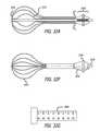
- FIGS. 26A and 26Billustrate an embodiment of a split nut and bump configuration used to hold a center tube in place.
- FIGS. 27A and 27Billustrate a brachytherapy apparatus having a ball of seeds at its end.
- FIG. 28Ais another embodiment of a brachytherapy apparatus that includes supports for the tubes.
- FIG. 28Bshows the apparatus in a collapsed state.
- FIG. 28Cshows yet another position for the apparatus inside a cavity.
- FIG. 29Ais a perspective view of the cutting apparatus.
- FIG. 29Ba front view of the cutting apparatus with the scissor members in an open configuration.
- FIG. 29Dshows the scissor members in a locked state.
- FIG. 30is another clamp with cutter in accordance with one embodiment of the present disclosure.
- FIG. 31is a cone clamp in accordance with one embodiment of the present disclosure.
- FIGS. 32A-32Gillustrate an embodiment of a brachytherapy apparatus having a twist dial expansion apparatus and a gauge to meter the volume of expansion.
- Cancer patientsare often diagnosed via an initial biopsy. The treating surgeon may then refer the patient to a medical oncologist, who may assist in determining a treatment regimen and inform the patient of various treatment options.
- a medical oncologistwho may assist in determining a treatment regimen and inform the patient of various treatment options.
- the cancerous tissueis removed via a lumpectomy
- the present disclosureprovides a brachytherapy apparatus that delivers a low dose, partial breast irradiation treatment for post-lumpectomy patients via introduction of a catheter-like device through a cannula.
- the deviceis designed to be introduced post-surgically with local anesthesia under image guidance into the excision site by a surgeon.
- low-dose radiationmay be construed as a dosage that would allow a patient to receive a prescribed dose if the low-dose radiation source remains in the patient's body over the course of 3, 5 or 7 days.
- the apparatus 10includes a plurality of tubes 100 at its distal end.
- the tubes 100are thin-walled.
- the wall thicknesscan be as small as between 7/1000 of an inch and 12/1000 of an inch.
- the tubes 100are disposed within a sleeve 110 .
- Sleevecould simply be thin-walled heat shrink tubing, and the walls of sleeve could be as thin as 3/1000 of an inch.
- Each of this plurality of tubes 100is configured to contain therein a plurality of low-dose radioactive seed strands.
- the tubeshave a distal end and a proximal end, and are bundled together at the distal end and at the proximal end inside sleeve.
- Disk 120contains a plurality of openings which may be used to suture the apparatus 10 onto a patient so that the apparatus 10 may be left in the patient's breast or other body part upon completion of the surgical procedure to insert the apparatus 10 .
- cap 130which may be used to cover the cut ends of the tubes 100 when the patient wears the apparatus 10 after completion of the surgical procedure for implanting the apparatus 10 .
- the plurality of tubes 100 of apparatus 10may be expanded so that the radioactive seeds (not shown) disposed within the tube may be placed more closely to the tissue within the post-lumpectomy cavity.
- the apparatus 10includes eight (8) tubes 100 ; however, it should be understood that there may be a greater number of tubes or fewer tubes.
- the number of tubesmay be dependent on a number of factors, including but not limited to, the size of the cavity into which the apparatus 10 is inserted, the amount of radiation needed for the patient and the locations in which the radiation is needed.
- the size of the cavity as well as the amounts and locations of radiation neededmay be determined based on radiation therapy planning, which may be performed using software specifically designed to develop such a radiation plan.
- the tubes 100may expanded to conform to the post-lumpectomy cavity.
- the size of the spheremay be largely dependent upon the size of the cavity into which the apparatus 10 is inserted.
- the sleeve 110may be pulled toward the operator so that the tubes 110 expand.
- An actuator or center tube 140which extends along the length of the apparatus 10 from its distal end to its proximal end, further assists with expanding and collapsing the apparatus 10 .
- FIG. 2illustrated is another embodiment of a breast brachytherapy apparatus 200 in a collapsed state in accordance with a embodiment of the present disclosure.
- this embodiment 200includes a plurality of substantially pie-shaped tubes 205 , the proximal ends of which are bundled into a collar 210 .
- Collar 210is connected to disk 220 through which sutures may be disposed in order to suture the apparatus 200 in place once it has been positioned within the patient.
- the apparatusfurther includes a conical piece 240 which assists in expansion and also may be used to insert the radioactive seeds, therapeutic elements or diagnostic elements. The seeds may be separated by one or more spacers to create a radioactive seed strand.
- Lock screw 235may be used to tighten the center tube 250 and hold the center tube in position with respect to tubes 205 .
- the obturatoralso assists in expansion and collapse of the apparatus 200 , when the obturator is held steady and the conical piece 240 is pushed in.
- the physicianuses a cannula or trocar to enter the original scar site that was created at the time of the original tumor removal or resection.
- this scar sitemay have resulted from a lumpectomy.
- a physicianmay place the apparatus 200 through the original incision site 203 of the breast 202 that was originally used to perform the lumpectomy.
- the tubes 205may be placed into the cavity 204 of the breast 202 that remains after the lumpectomy.
- Disk 220is kept outside the patient's breast 202 so that it may be later sutured onto the patient.
- the clamp 230will assist with expansion and tightening of the tubes 205 .
- the apparatus 200may be expanded much like an umbrella when the physician loosens the lock screw 235 , restrains the central tube 250 and pushes member 240 in a distal direction. These motions would cause expansion of the plurality of tubes 205 so that the tubes 205 almost fill the cavity 204 of breast 202 .
- the physicianmay use visual and other surgical aids to better assess the position of the tubes 205 inside breast 202 . Such aids may be beneficial since the tubes 205 may not be readily seen once they are inside cavity 204 .
- An ultrasoundis one such example of visual or surgical aid. The ultrasound may be used to detect the position of the tubes 205 in relation to cavity 204 . When the tubes are touching the walls of cavity 204 , the physician may see this being illustrated on an ultrasound. In some situations, an ultrasound may not be available.
- the physicianmay use his or her senses to determine when resistance is felt resulting from the tubes 205 pushing against the one or more inner surfaces of cavity 204 . Once the tubes are against the inner surfaces of cavity 204 , the physician may tighten the clamp 230 to hold the position of tubes 205 .
- radioactive seed strands or other therapeutic or diagnostic elementsmay be placed into tubes 205 through openings located in conical member 240 .
- the openingsare numbered, each number corresponding to one of the plurality of tubes 205 .
- seed strandsmay be inserted into the tubes via the openings using strand placement tube 270 .
- the openingsserve as funnel holes for the tubes.
- the apparatus of the present disclosuremay be suitable for use with common surgical aids.
- One such surgical aidis a CAT scan which may be used to determine whether the seed strands have been accurately positioned in accordance with the radiation therapy plan.
- the radiation therapy planmay be created with surgical aids such as software designed to form an isodose profile.
- the appropriate isodose profilemay call for the seeds to be inserted in a number of ways so as to vary the applied radiation level. For example, in some situations, the isodose profile may not require that any seed be inserted into one of the plurality of tubes 205 . In some situations, two or more different seeds used on a single patient may have different activity levels so that some seeds are stronger than others.
- Low-dose radioactive seedse.g., iodine 125 isotope, may be used in conjunction with breast irradiation.
- the physicianmay wish to test proper seed insertion prior to actual insertion by inserting dummy or imitation seeds instead of actual radioactive seeds. This process allows the physician to avoid potentially damaging the real seeds, yet this process may be more time-consuming than placing the actual seeds.
- the seed placement tubehas been removed and tubes 205 are now held tightly into place with the radioactive seeds having been placed according to the radiation therapy plan.
- the tubes 205may be cut leaving disk 220 just outside the breast so that the sutures may be disposed through openings in disk 220 .
- the cuttingmay be performed with a surgical instrument such as a scalpel.
- the apparatus 200may be self-cutting using a blade configured to travel across the base of the clamp 230 .
- a cap 280may be attached to the suture disk to protect the ends of the tubes since the patient may wear the post-implantation apparatus 200 for several days before treatment ends and the apparatus 200 is removed.
- the post-implantation embodimentwith a post-implantation cap 280 on its end. It is possible that the cap 280 would be secured so that the patient could not remove the cap 280 and disrupt the protocol. At this point, the patient may be sent home with a radiopaque shield (e.g., a lead bra) for several days, e.g., 3-5 days.
- a radiopaque shielde.g., a lead bra
- the apparatusmay be removed after a minimum prescribed dose of radiation has been delivered. As shown in FIG. 3G , the cap 280 has been removed and the obturator 260 has been inserted. The clamp 230 would need to be loosened so as to allow the tubes to collapse.
- the tubes 205are collapsed.
- the apparatus 200may be removed from the site of insertion 203 or other point deemed appropriate by the physician.
- the apparatus 200may be removed from the patient, leaving the suture disk 220 in place temporarily.
- FIG. 3Jthe apparatus 200 has been removed fully from the breast.
- apparatus 400includes a plurality of stacked expanding tubes 410 , 420 , 430 , 440 , 450 , 460 and 470 .
- Uppermost tube 410is shown above a number of other tubes including middle tubes 430 , 440 and lowermost tube 470 .
- the middle tubes 430 , 440have a greater width (w) than uppermost tube 410 and lowermost tube 470 .
- connection mechanisms 415 , 417connect the top tube section of tube 420 to the bottom tube section of tube 410 .
- connection mechanisms 425 , 427connect the top tube section of tube 430 to the bottom tube section of tube 420 .
- Connection mechanismsconnect the top tube sections of each remaining tube to the bottom tube section of the tube above.
- uppermost tube 410has no tube above it, and therefore, no connection mechanisms connect the uppermost tube 410 to the bottom tube section of the above it since there is no tube above uppermost tube 410 .
- a single spiral tube strand brachytherapy apparatus 500in accordance with another embodiment of the present disclosure.
- a single spiral tube strandis wound around a spiral tube support 520 .
- Spiral tube support 520may be configured to lengthen so that, when lengthened, the spiral tube strand is elongated and expanded to fill a cavity.
- the apparatus 600includes a plurality of tubes that are configured in a first direction.
- tubes 610 , 620 and 630extend diagonally from a northwesterly direction to a southeasterly direction.
- a second set of tubesare configured in a second direction.
- tubes 640 , 650 and 660extend from a northeasterly direction to a southwesterly direction such that tubes 640 , 650 and 660 intersect with tubes 610 , 620 and 630 .
- Radioactive seeds or other therapeutic elementsare disposed within the plurality of tubes.
- the diameter of apparatus 600is greater in the middle section than in the upper and lower sections.
- FIG. 7Aillustrated is a stent embodiment of a brachytherapy apparatus 700 in a collapsed state in accordance with still yet another embodiment of the present disclosure.
- a plurality of tubesare interconnected to define a conical shape when the tube is in its collapsed state.
- FIG. 7Billustrated is the stent embodiment of FIG. 7A in an expanded state. As illustrated the diameter of 700 is greater in the middle section than in the upper and lower sections.
- the apparatus 800includes a plurality of tubes that spiral in the same direction.
- the tubesare joined at the top and converge near the bottom.
- a brachytherapy apparatusmay need to be re-sized to fill the cavity that remains after a surgical resection. It may also be desirable that this re-sizing is performed by the surgeon who is also responsible for implanting the brachytherapy apparatus into a patient. It may be further desirable that the tube bundle be rigid in order to facilitate insertion.
- a multi-lumen tubecould also be configured so that its tubes fit side-to-side when the tube is in its collapsed state. Such an embodiment could enhance stability of the apparatus.
- FIG. 12illustrated is a multi-lumen brachytherapy apparatus 1200 having tubes that are snugly fit from side-to-side.
- the tubesmay be molded or welded to a cap.
- FIG. 14Aillustrated is a side view of a brachytherapy apparatus 1400 that includes a cap 1410 joined to the tubes 1420 .
- Cap 1410may be molded or welded to the tubes 1420 in order to facilitate spherical expansion of the tubes 1420 .
- FIG. 15illustrated is a dual-sleeved proximal end portion of a brachytherapy apparatus 1500 in accordance with one embodiment of the present disclosure.
- This proximal end portionhas two sleeves 1510 , 1520 with sleeve 1510 adapted to be slid proximally toward disk 1530 . Accordingly, sleeve 1510 could be slid into, and thus disposed inside, sleeve 1520 to lengthen any attached tubes, thus accommodating a larger cavity.
- the apparatus 1600includes a threaded sleeve 1610 adapted to receive a plurality of tubes 1605 .
- the sleeve 1605protects the tissue against pressure when the tubes 1605 need to be opened up. If desired, a physician performing brachytherapy could cut the sleeve to the desired size or the sleeves could be manufactured to a certain length.
- sleeve portion 1625can be seen as it protrudes from clamp 1620 .
- Clamp 1620squeezes tubes.
- An obturatoris placed into hole of center tube and physically connected to the luer fitting on the end of tube 1640 . Lock screw 1650 can be used to hold the center tube in place.
- General operation of this apparatusis similar to that shown in FIG. 2 .
- a brachytherapy apparatus 1700that includes a tapered wedge and collet in accordance with another embodiment of the present disclosure.
- the tapered wedge and colletmay be used to secure the eight-lumen tube 1710 and center tube 1730 to the apparatus.
- an eight-lumen tube 1710 having webs, such as web 1715protrudes from sleeve 1720 .
- Center tube 1730can be seen extending from the middle of the eight-lumen tube 1710 .
- Tapered wedgecould be inserted on the inside diameter of sleeve 1720 to secure the eight-lumen tube 1710 .
- the collet 1750could be used to secure the center tube 1730 . Once secured, the center tube 1730 would be clamped into place by collet 1750 .
- a brachytherapy apparatus 1800that includes clamps for each individual tube as well as a clamp for the center tube.
- the center tube 1810may be clamped by squeezing center tube 1820 using forceps or other devices so that center tube clamp structure 1815 holds center tube 1810 into place.
- Each individual clampmay be clamped using individual tube spring member 1830 to hold the individual clamp in individual clamp structure 1840 . When this spring member 1830 is squeezed, it releases the tube in place.
- FIG. 21illustrated is the proximal end of a brachytherapy apparatus 2100 having a pin mechanism used to hold the lumen in place.
- the pin mechanism 2110could be inserted through a web 2130 of the eight-lumen tube 2120 and through the center tube 2140 to hold the center tube in place as well.
- FIG. 22illustrated is another view of a proximal end of a brachytherapy apparatus 2200 having a collet system similar to that shown in FIG. 17 .
- the collet 2210may be ribbed so that pressure is applied only to the web of the lumen tube. This would leave the inside diameter undistorted so that the radioactive seeds or strands may pass freely.
- FIG. 23illustrated is another view of a proximal end of a brachytherapy apparatus 2200 having a collet system similar to that shown in FIG. 17 .
- the collet 2310includes a handle 2320 so that the collet 2310 may be released.
- FIG. 24illustrated is yet another view of a proximal end of a brachytherapy apparatus having a screw mechanism for expanding and collapsing the brachytherapy tubes.
- the center tube 2414may be held in place with screw 2416 .
- the center tube 2416is advanced so that the outer sets of tubes 2410 , 2412 expand.
- the center tube 2416recedes so that the outer sets of tubes 2410 , 2412 collapse.
- FIG. 25illustrated is still another view of a proximal end of a brachytherapy apparatus having a spring mechanism for expanding and collapsing the brachytherapy tubes.
- the spring mechanism 2510may be attached to cap 2512 , while at the other end, spring mechanism 2510 may be attached to suture disk 2514 .
- the spring mechanism 2510may be pulled to expand the tubes 2520 .
- the spring mechanism 2510may be pushed to release the tubes 2520 .
- a center tubehaving bumps 2610 to facilitate holding the center tube in place.
- the bumpsmay fit into a split nut 2620 joined by a handle 2630 to hold the center tube 2640 in place.
- FIG. 27Aillustrated is a brachytherapy apparatus 2700 having a ball of seeds at its end.
- the apparatusis shown in FIG. 27B is shown as it is inserted into a cavity.
- FIG. 28Aillustrated is yet another embodiment of a brachytherapy apparatus that includes supports for the tubes. As illustrated, a cable may be pulled to expand the tubes 2830 disposed inside a cavity.
- FIG. 28Bshows the apparatus 2800 in a collapsed state.
- FIG. 28Cshows yet another position for the apparatus inside a cavity.
- FIGS. 29A-29Dillustrate a scissor-type clamp apparatus for the thin-walled tubes.
- FIG. 29Ais a perspective view of the apparatus 2900 .
- FIG. 29Bis a front view of the apparatus 2900 with the scissor members 2910 , 2920 in an open configuration.
- FIG. 29Cshows the scissor members 2910 , 2920 released.
- FIG. 29Dshows the scissor members 2910 , 2920 in a locked state.
- the clamp member 3000includes blades 3010 , 3020 that may be used to cut the thin-walled tubes.
- a cone clamp 3100designed for use in a brachytherapy apparatus in accordance with one embodiment of the present disclosure.
- the cone clamp 3100is designed to be squeezed and inserted into a conical member 3110 , thus holding a center tube into place. If the center tube is pulled in a distal direction, the clamp 3100 would be more tightly squeezed. In order to release the clamp 3100 , the clamp 3100 would be squeezed again.
- FIG. 32Aillustrated is an embodiment of a brachytherapy apparatus 3200 having a twist dial expansion apparatus 3210 .
- a wire or strip 3220expands the plurality of tubes 3230 of the apparatus 3200 .
- a wire or strip 3220may collapse so that the plurality of tubes 3230 also collapse.
- Dial 3240may be used to expand the apparatus.
- FIG. 32Cillustrated is a cross-sectional view of the twist dial expansion apparatus 3210 .
- a threaded flexible screw 3250may advance when the twist dial expansion apparatus 3240 is turned to expand the apparatus.
- FIG. 32Dillustrated is a single twist dial expansion apparatus having multiple tube openings.
- the brachytherapy apparatuswith a gauge 3270 to meter the volume of expansion.
- the gauge 3270may show the volume of expansion.
- FIG. 32Gillustrates is a gauge 3280 that may be used in conjunction with a brachytherapy apparatus.
- the gaugeshows the volume of expansion in terms of cubic centimeters.
Landscapes
- Health & Medical Sciences (AREA)
- Engineering & Computer Science (AREA)
- Biomedical Technology (AREA)
- Nuclear Medicine, Radiotherapy & Molecular Imaging (AREA)
- Surgery (AREA)
- Pathology (AREA)
- Heart & Thoracic Surgery (AREA)
- Radiology & Medical Imaging (AREA)
- Life Sciences & Earth Sciences (AREA)
- Animal Behavior & Ethology (AREA)
- General Health & Medical Sciences (AREA)
- Public Health (AREA)
- Veterinary Medicine (AREA)
- Radiation-Therapy Devices (AREA)
Abstract
Description
Claims (10)
Priority Applications (7)
| Application Number | Priority Date | Filing Date | Title |
|---|---|---|---|
| US11/305,437US8137256B2 (en) | 2005-12-16 | 2005-12-16 | Brachytherapy apparatus |
| US11/379,739US20070270627A1 (en) | 2005-12-16 | 2006-04-21 | Brachytherapy apparatus for asymmetrical body cavities |
| EP06838196AEP1968701A4 (en) | 2005-12-16 | 2006-11-21 | Brachytherapy apparatus for asymmetrical body cavities |
| PCT/US2006/045081WO2007078453A2 (en) | 2005-12-16 | 2006-11-21 | Brachytherapy apparatus for asymmetrical body cavities |
| EP06838218AEP1968702A4 (en) | 2005-12-16 | 2006-11-21 | Brachytherapy apparatus |
| PCT/US2006/045117WO2007075241A1 (en) | 2005-12-16 | 2006-11-21 | Brachytherapy apparatus |
| US12/554,732US8226539B2 (en) | 2005-12-16 | 2009-09-04 | Brachytherapy apparatus for asymmetrical body cavities |
Applications Claiming Priority (1)
| Application Number | Priority Date | Filing Date | Title |
|---|---|---|---|
| US11/305,437US8137256B2 (en) | 2005-12-16 | 2005-12-16 | Brachytherapy apparatus |
Related Child Applications (2)
| Application Number | Title | Priority Date | Filing Date |
|---|---|---|---|
| US11/379,739Continuation-In-PartUS20070270627A1 (en) | 2005-12-16 | 2006-04-21 | Brachytherapy apparatus for asymmetrical body cavities |
| US11/379,739ContinuationUS20070270627A1 (en) | 2005-12-16 | 2006-04-21 | Brachytherapy apparatus for asymmetrical body cavities |
Publications (2)
| Publication Number | Publication Date |
|---|---|
| US20070142694A1 US20070142694A1 (en) | 2007-06-21 |
| US8137256B2true US8137256B2 (en) | 2012-03-20 |
Family
ID=38174625
Family Applications (1)
| Application Number | Title | Priority Date | Filing Date |
|---|---|---|---|
| US11/305,437Expired - Fee RelatedUS8137256B2 (en) | 2005-12-16 | 2005-12-16 | Brachytherapy apparatus |
Country Status (3)
| Country | Link |
|---|---|
| US (1) | US8137256B2 (en) |
| EP (1) | EP1968702A4 (en) |
| WO (1) | WO2007075241A1 (en) |
Cited By (11)
| Publication number | Priority date | Publication date | Assignee | Title |
|---|---|---|---|---|
| US20100286465A1 (en)* | 2009-05-11 | 2010-11-11 | Maria Benson | Lumen Visualization and Identification System for Multi-Lumen Balloon Catheter |
| US9180312B2 (en) | 2005-11-18 | 2015-11-10 | Hologic, Inc. | Brachytherapy device for asymmetrical irradiation of a body cavity |
| US9248311B2 (en) | 2009-02-11 | 2016-02-02 | Hologic, Inc. | System and method for modifying a flexibility of a brachythereapy catheter |
| US9579524B2 (en) | 2009-02-11 | 2017-02-28 | Hologic, Inc. | Flexible multi-lumen brachytherapy device |
| US9623260B2 (en) | 2004-11-05 | 2017-04-18 | Theragenics Corporation | Expandable brachytherapy device |
| US9764158B2 (en) | 2013-03-12 | 2017-09-19 | The Cleveland Clinic Foundation | Multitube esophageal brachytherapy catheter |
| US10022557B2 (en) | 2010-09-30 | 2018-07-17 | Hologic, Inc. | Using a guided member to facilitate brachytherapy device swap |
| US10335609B2 (en) | 2013-03-12 | 2019-07-02 | The Cleveland Clinic Foundation | Multitube esophageal brachytherapy catheter |
| US10342992B2 (en) | 2011-01-06 | 2019-07-09 | Hologic, Inc. | Orienting a brachytherapy applicator |
| US10646727B2 (en) | 2017-11-13 | 2020-05-12 | Positive Energy, Llc | Anchored brachytherapy device |
| US10874877B2 (en) | 2017-11-13 | 2020-12-29 | Positive Energy, Llc | Anchored brachytherapy device |
Families Citing this family (17)
| Publication number | Priority date | Publication date | Assignee | Title |
|---|---|---|---|---|
| EP1720608B1 (en) | 2004-02-12 | 2010-11-17 | NeoVista, Inc. | Apparatus for intraocular brachytherapy |
| US7563222B2 (en) | 2004-02-12 | 2009-07-21 | Neovista, Inc. | Methods and apparatus for intraocular brachytherapy |
| US7862496B2 (en)* | 2005-11-10 | 2011-01-04 | Cianna Medical, Inc. | Brachytherapy apparatus and methods for using them |
| BRPI0618677A2 (en)* | 2005-11-15 | 2011-09-06 | Neovista Inc | methods and apparatus for intraocular brachytherapy |
| EP2029230A2 (en)* | 2006-06-02 | 2009-03-04 | Cianna Medical, Inc. | Expandable brachytherapy apparatus |
| ATE514457T1 (en)* | 2006-10-08 | 2011-07-15 | Cianna Medical Inc | EXPANDABLE BRACHYTHERAPY DEVICE |
| US20080269539A1 (en)* | 2007-04-27 | 2008-10-30 | North American Scientific, Inc. | Brachytherapy Device Having an Alignment and Seal Adaptor |
| US8473027B2 (en)* | 2008-07-03 | 2013-06-25 | Qsum Biopsy Disposables Llc | Process for draping breast MRI imaging coils |
| US8663210B2 (en) | 2009-05-13 | 2014-03-04 | Novian Health, Inc. | Methods and apparatus for performing interstitial laser therapy and interstitial brachytherapy |
| USD622389S1 (en) | 2009-11-25 | 2010-08-24 | QSUM Biopsy Disposable LLC | Protective drape covering for magnetic resonance imaging device |
| US20110124949A1 (en)* | 2009-11-25 | 2011-05-26 | Qsum Biopsy Disposables Llc | Method and apparatus for stabilizing tubing during a brachytherapy procedure |
| US8814775B2 (en) | 2010-03-18 | 2014-08-26 | Cianna Medical, Inc. | Expandable brachytherapy apparatus and methods for using them |
| US9883919B2 (en) | 2010-07-21 | 2018-02-06 | Cianna Medical, Inc. | Brachytherapy apparatus, systems, and methods for using them |
| US9067063B2 (en) | 2010-11-03 | 2015-06-30 | Cianna Medical, Inc. | Expandable brachytherapy apparatus and methods for using them |
| US9943706B2 (en) | 2012-01-12 | 2018-04-17 | Surgical Radiation Products, Llc | Targeting implant for external beam radiation |
| US9320517B2 (en) | 2012-01-12 | 2016-04-26 | Surgical Radiation Products, Llc | Targeting implant for external beam radiation |
| US12426867B2 (en) | 2019-04-10 | 2025-09-30 | The Chinese University Of Hong Kong | Systems and methods for organ retraction and space opening |
Citations (127)
| Publication number | Priority date | Publication date | Assignee | Title |
|---|---|---|---|---|
| US3224432A (en) | 1963-04-10 | 1965-12-21 | Frank S Billingsley | Device for irradiating a body cavity |
| US3807386A (en) | 1971-04-27 | 1974-04-30 | Commissariat Energie Atomique | Radioactive source applicator for utero-vaginal plesiocurietherapy according to the method of non-radioactive preparation |
| US3872856A (en) | 1971-06-09 | 1975-03-25 | Ralph S Clayton | Apparatus for treating the walls and floor of the pelvic cavity with radiation |
| US3927325A (en) | 1974-07-10 | 1975-12-16 | Us Energy | Tissue irradiator |
| US4434789A (en) | 1979-12-17 | 1984-03-06 | The Board Of Regents Of The University Of Nebraska | Apparatus for treating carcinoma of the uterine cervix |
| US4584991A (en) | 1983-12-15 | 1986-04-29 | Tokita Kenneth M | Medical device for applying therapeutic radiation |
| US4763642A (en) | 1986-04-07 | 1988-08-16 | Horowitz Bruce S | Intracavitational brachytherapy |
| US4815449A (en) | 1984-11-21 | 1989-03-28 | Horowitz Bruce S | Delivery system for interstitial radiation therapy including substantially non-deflecting elongated member |
| US4976680A (en) | 1988-10-07 | 1990-12-11 | Hayman Michael H | Apparatus for in situ radiotherapy |
| US5007437A (en) | 1989-06-16 | 1991-04-16 | Mmtc, Inc. | Catheters for treating prostate disease |
| US5050930A (en) | 1989-08-04 | 1991-09-24 | Wilhelm Schuster | Lordosis-support backrest for a vehicle seat |
| US5125925A (en) | 1988-08-03 | 1992-06-30 | Photoradiation Systems | Intracavity laser catheter with sensing fiber |
| US5147282A (en) | 1989-05-04 | 1992-09-15 | William Kan | Irradiation loading apparatus |
| US5317616A (en) | 1992-03-19 | 1994-05-31 | Wisconsin Alumni Research Foundation | Method and apparatus for radiation therapy |
| US5336178A (en) | 1992-11-02 | 1994-08-09 | Localmed, Inc. | Intravascular catheter with infusion array |
| US5345936A (en) | 1991-02-15 | 1994-09-13 | Cardiac Pathways Corporation | Apparatus with basket assembly for endocardial mapping |
| US5411466A (en) | 1991-09-05 | 1995-05-02 | Robert L. Hess | Apparatus for restenosis treatment |
| US5429582A (en) | 1991-06-14 | 1995-07-04 | Williams; Jeffery A. | Tumor treatment |
| US5431648A (en) | 1991-11-11 | 1995-07-11 | Fondazione Centro S. Raffaele Del Monte Tabor | Radiating device for hyperthermia |
| US5484384A (en) | 1991-01-29 | 1996-01-16 | Med Institute, Inc. | Minimally invasive medical device for providing a radiation treatment |
| WO1996002059A1 (en) | 1994-07-12 | 1996-01-25 | Photoelectron Corporation | X-ray apparatus for applying a predetermined flux to an interior surface of a body cavity |
| US5540659A (en) | 1993-07-15 | 1996-07-30 | Teirstein; Paul S. | Irradiation catheter and method of use |
| US5623940A (en) | 1994-08-02 | 1997-04-29 | S.L.T. Japan Co., Ltd. | Catheter apparatus with a sensor |
| US5643171A (en) | 1993-05-04 | 1997-07-01 | Neocardia, Llc | Method and apparatus for uniform radiation treatment of vascular lumens |
| US5653683A (en) | 1995-02-28 | 1997-08-05 | D'andrea; Mark A. | Intracavitary catheter for use in therapeutic radiation procedures |
| US5662580A (en) | 1994-12-08 | 1997-09-02 | Neocardia, Llc | Combined angioplasty and intravascular radiotherapy method and apparatus |
| RU2089143C1 (en) | 1994-03-02 | 1997-09-10 | Московский научно-исследовательский институт глазных болезней им.Гельмгольца | Method for brachytherapy of intraocular tumors |
| US5678572A (en) | 1995-01-12 | 1997-10-21 | Shaw; Dein | Cavity expanding device for laparoscopic surgery |
| US5683345A (en) | 1994-10-27 | 1997-11-04 | Novoste Corporation | Method and apparatus for treating a desired area in the vascular system of a patient |
| US5688220A (en) | 1994-06-10 | 1997-11-18 | Schneider (Europe) A.G. | Medical appliance for treatment by ionizing radiation |
| US5730698A (en) | 1995-05-09 | 1998-03-24 | Fischell; Robert E. | Balloon expandable temporary radioisotope stent system |
| WO1998015315A1 (en) | 1996-10-07 | 1998-04-16 | Proxima Therapeutics Inc. | Inflatable devices for tumor treatment |
| US5817104A (en) | 1997-04-30 | 1998-10-06 | C.R. Bard, Inc. | Dual purpose mechanism for expanding baskets |
| US5836868A (en) | 1992-11-13 | 1998-11-17 | Scimed Life Systems, Inc. | Expandable intravascular occlusion material removal devices and methods of use |
| US5840008A (en) | 1995-11-13 | 1998-11-24 | Localmed, Inc. | Radiation emitting sleeve catheter and methods |
| US5851171A (en) | 1997-11-04 | 1998-12-22 | Advanced Cardiovascular Systems, Inc. | Catheter assembly for centering a radiation source within a body lumen |
| US5855546A (en) | 1996-02-29 | 1999-01-05 | Sci-Med Life Systems | Perfusion balloon and radioactive wire delivery system |
| WO1999002219A1 (en) | 1997-07-11 | 1999-01-21 | Localmed, Inc. | Method and system for the intramural delivery of radioactive agents |
| US5863285A (en) | 1997-01-30 | 1999-01-26 | Cordis Corporation | Balloon catheter with radioactive means |
| WO1999004856A1 (en) | 1997-07-24 | 1999-02-04 | Proxima Therapeutics, Inc. | Double-wall balloon catheter for treatment of proliferative tissue |
| US5879282A (en) | 1997-01-21 | 1999-03-09 | Cordis A Johnson And Johnson Company | Catheter having an expandable radioactive source |
| RU2128060C1 (en) | 1997-07-01 | 1999-03-27 | Центральный научно-исследовательский рентгено-радиологический институт | Method of treatment of malignant tumors of oral cavity |
| WO1999022812A1 (en) | 1997-11-03 | 1999-05-14 | The State Of Israel Atomic Energy Commission | In situ-generated solid radiation source based on tungsten188/rhenium188 and the use thereof |
| US5904680A (en) | 1992-09-25 | 1999-05-18 | Ep Technologies, Inc. | Multiple electrode support structures having optimal bio-mechanical characteristics |
| WO1999024117A1 (en) | 1997-11-07 | 1999-05-20 | Global Vascular Concepts, Inc. | Device for intravascular delivery of beta emitting isotopes |
| US5910101A (en) | 1996-08-29 | 1999-06-08 | Advanced Cardiovascular Systems, Inc. | Device for loading and centering a vascular radiation therapy source |
| US5916143A (en) | 1996-04-30 | 1999-06-29 | Apple; Marc G. | Brachytherapy catheter system |
| US5938582A (en) | 1997-09-26 | 1999-08-17 | Medtronic, Inc. | Radiation delivery centering catheter |
| US5968040A (en) | 1994-03-04 | 1999-10-19 | Ep Technologies, Inc. | Systems and methods using asymmetric multiple electrode arrays |
| US6011995A (en) | 1997-12-29 | 2000-01-04 | The Regents Of The University Of California | Endovascular device for hyperthermia and angioplasty and method for using the same |
| US6030333A (en) | 1997-10-24 | 2000-02-29 | Radiomed Corporation | Implantable radiotherapy device |
| US6036631A (en) | 1998-03-09 | 2000-03-14 | Urologix, Inc. | Device and method for intracavitary cancer treatment |
| US6050930A (en) | 1998-06-02 | 2000-04-18 | Teirstein; Paul S. | Irradiation catheter with expandable source |
| US6059812A (en) | 1997-03-21 | 2000-05-09 | Schneider (Usa) Inc. | Self-expanding medical device for centering radioactive treatment sources in body vessels |
| US6066083A (en) | 1998-11-27 | 2000-05-23 | Syntheon Llc | Implantable brachytherapy device having at least partial deactivation capability |
| US6074339A (en) | 1998-05-07 | 2000-06-13 | Medtronic Ave, Inc. | Expandable braid device and method for radiation treatment |
| US6149574A (en) | 1997-12-19 | 2000-11-21 | Radiance Medical Systems, Inc. | Dual catheter radiation delivery system |
| US6152869A (en) | 1997-12-24 | 2000-11-28 | Korea Atomic Research Energy Research Institute | Radioactive stent and process for preparation thereof |
| US6196996B1 (en) | 1993-07-15 | 2001-03-06 | Paul S. Teirstein | Irradiation catheter and method of use |
| US6196963B1 (en) | 1999-03-02 | 2001-03-06 | Medtronic Ave, Inc. | Brachytherapy device assembly and method of use |
| US6200257B1 (en) | 1999-03-24 | 2001-03-13 | Proxima Therapeutics, Inc. | Catheter with permeable hydrogel membrane |
| US6216043B1 (en) | 1994-03-04 | 2001-04-10 | Ep Technologies, Inc. | Asymmetric multiple electrode support structures |
| US6213976B1 (en) | 1999-07-22 | 2001-04-10 | Advanced Research And Technology Institute, Inc. | Brachytherapy guide catheter |
| US6217503B1 (en) | 1994-01-21 | 2001-04-17 | The Trustees Of Columbia University In The City Of New York | Apparatus and method to treat a disease process in a luminal structure |
| US6238374B1 (en) | 1999-08-06 | 2001-05-29 | Proxima Therapeutics, Inc. | Hazardous fluid infuser |
| WO2001043826A1 (en) | 1999-12-16 | 2001-06-21 | Proxima Therapeutics, Inc. | Asymmetric radiation dosing apparatus and method |
| US6263236B1 (en) | 1999-11-29 | 2001-07-17 | Illumenex Corporation | Non-occlusive expandable catheter |
| US6338709B1 (en) | 1998-02-19 | 2002-01-15 | Medtronic Percusurge, Inc. | Intravascular radiation therapy device and method of use |
| US6391026B1 (en) | 1998-09-18 | 2002-05-21 | Pro Duct Health, Inc. | Methods and systems for treating breast tissue |
| US6409652B1 (en) | 2000-09-20 | 2002-06-25 | Vascular Architects, Inc. | Device and method for delivery of uniform and controlled radiation dose to blood vessels |
| US6413203B1 (en) | 1998-09-16 | 2002-07-02 | Scimed Life Systems, Inc. | Method and apparatus for positioning radioactive fluids within a body lumen |
| US20020173816A1 (en) | 2001-04-16 | 2002-11-21 | Pro Duct Health, Inc. | Medical instrument with an atraumatic end |
| US20020193653A1 (en) | 2001-06-15 | 2002-12-19 | Winkler Rance A. | Tissue positioning apparatus and method for protecting tissue from radiotherapy |
| US20030022161A1 (en) | 1999-01-26 | 2003-01-30 | Susan Love | Identifying, monitoring, and treating women for breast precancer or cancer |
| US20030032851A1 (en) | 2001-07-13 | 2003-02-13 | Apple Marc G. | Catheter with concentric balloons for radiogas delivery and booster radiosources for use therewith |
| US20030039959A1 (en) | 1998-10-02 | 2003-02-27 | Susan Love | Methods for identification, diagnosis, and treatment of breast cancer |
| US20030049262A1 (en) | 1998-10-02 | 2003-03-13 | Susan Love | Methods for identification, diagnosis, and treatment of breast cancer |
| US6554760B2 (en) | 2000-10-25 | 2003-04-29 | Gary A. Lamoureux | Pre-loaded needle assembly |
| US20030092957A1 (en) | 1998-11-18 | 2003-05-15 | Scott Neal A. | Brachytherapy device and method of use |
| US20030149329A1 (en) | 1999-07-12 | 2003-08-07 | O'foghludha Fearghus | Formable integral source material for medical devices |
| US6607476B1 (en) | 1998-10-01 | 2003-08-19 | University Of Iowa Research Foundation | Brachytherapy positioning system |
| US6607477B1 (en) | 1998-02-16 | 2003-08-19 | Wallace A. Longton | Graduated intraluminal catheter and methods of use thereof |
| US20030191412A1 (en) | 1999-11-10 | 2003-10-09 | Sampson Russell M. | System and method for detecting perforations in a body cavity |
| US6652548B2 (en) | 2000-03-31 | 2003-11-25 | Bacchus Vascular Inc. | Expansible shearing catheters for thrombus removal |
| US6659105B2 (en) | 1998-02-26 | 2003-12-09 | Senorx, Inc. | Tissue specimen isolating and damaging device and method |
| US20040016728A1 (en) | 2002-07-25 | 2004-01-29 | Xinbing Liu | Method and apparatus for aligning a work piece in a laser drilling system |
| US6685718B1 (en) | 1998-03-05 | 2004-02-03 | Scimed Life Systems, Inc. | Expandable ablation burr |
| US20040023912A1 (en) | 2002-03-15 | 2004-02-05 | Cytyc Health Corporation | Method of diagnosis and treatment of breast lesions |
| US6695760B1 (en) | 2002-10-11 | 2004-02-24 | Proxima Therapeutics | Treatment of spinal metastases |
| US20040054368A1 (en) | 1998-07-13 | 2004-03-18 | Novacept | Apparatuses and methods for interstitial tissue removal |
| EP1426063A2 (en) | 2000-02-18 | 2004-06-09 | Civatech Corporation | Formable integral source material for medical and industrial devices |
| US20040109823A1 (en) | 2000-11-16 | 2004-06-10 | Microspherix Llc | Flexible and/or elastic brachytherapy seed or strand |
| US6749555B1 (en) | 2003-02-13 | 2004-06-15 | Proxima Therapeutics, Inc. | System and method for the treatment of spinal metastases |
| US20040116767A1 (en) | 2002-09-10 | 2004-06-17 | Lebovic Gail S. | Brachytherapy apparatus and methods of using same |
| US6770058B1 (en) | 1997-03-11 | 2004-08-03 | Interventional Therapies, Llc | Treatment catheter insert |
| US20040191854A1 (en) | 2003-03-31 | 2004-09-30 | Cytyc Corporation | Papanicolau staining process |
| US20050004471A1 (en) | 2000-08-25 | 2005-01-06 | Neoseed Technology Llc. | Prostate visualization device and methods of use |
| US6840936B2 (en) | 1996-10-22 | 2005-01-11 | Epicor Medical, Inc. | Methods and devices for ablation |
| US6855160B1 (en) | 1999-08-04 | 2005-02-15 | C. R. Bard, Inc. | Implant and agent delivery device |
| US20050101860A1 (en) | 2003-11-07 | 2005-05-12 | Proxima Therapeutics, Inc. | Tissue positioning systems and methods for use with radiation therapy |
| US20050101825A1 (en) | 2003-11-07 | 2005-05-12 | Proxima Therapeutics, Inc. | Brachytherapy apparatus and method for treating a target tissue through an external surface of the tissue |
| US20050101824A1 (en) | 2003-11-07 | 2005-05-12 | Proxima Therapeutics, Inc. | Implantable radiotherapy/brachytherapy radiation detecting apparatus and methods |
| US20050101823A1 (en) | 2002-09-27 | 2005-05-12 | Linares Luis A. | Device for radiation treatment of proliferative tissue surrounding a cavity in an animal body |
| US6893450B2 (en) | 1999-03-26 | 2005-05-17 | Cook Urological Incorporated | Minimally-invasive medical retrieval device |
| US20050107653A1 (en) | 2003-11-14 | 2005-05-19 | Proxima Therapeutics, Inc. | Drug eluting brachytherapy methods and apparatus |
| US20050113629A1 (en) | 2003-11-20 | 2005-05-26 | Proxima Therapeutics | Brachytherapy method and applicator for treatment of metastatic lesions in a load bearing region |
| US20050124843A1 (en) | 2003-12-09 | 2005-06-09 | Washington University | Method and apparatus for delivering targeted therapy to a patient |
| US20050131269A1 (en) | 1995-06-07 | 2005-06-16 | Talmadge Karen D. | System and method for delivering a therapeutic agent for bone disease |
| US20050131268A1 (en) | 1995-06-07 | 2005-06-16 | Talmadge Karen D. | System and method for delivering a therapeutic agent for bone disease |
| US20050131267A1 (en) | 1995-06-07 | 2005-06-16 | Talmadge Karen D. | System and method for delivering a therapeutic agent for bone disease |
| US20050137498A1 (en) | 2003-12-23 | 2005-06-23 | Robert Sakal | Medical instrument for accessing a breast duct for performing a medical procedure |
| US20050137499A1 (en) | 2003-12-23 | 2005-06-23 | Sheets Ellen E. | Ductal lavage catheter |
| US20050149159A1 (en) | 2003-12-23 | 2005-07-07 | Xtent, Inc., A Delaware Corporation | Devices and methods for controlling and indicating the length of an interventional element |
| US6923754B2 (en) | 2002-11-06 | 2005-08-02 | Senorx, Inc. | Vacuum device and method for treating tissue adjacent a body cavity |
| EP1568397A1 (en) | 2004-02-25 | 2005-08-31 | Acrostak Corp. | Balloon for brachytherapy and application of the balloon |
| US20050240073A1 (en) | 2004-04-26 | 2005-10-27 | Apffelstaedt Justus P | Devices and methods to conform and treat body cavities |
| US20060014997A1 (en) | 2004-07-16 | 2006-01-19 | Nucletron B.V. | Device for radiation treatment of proliferative tissue surrounding a cavity in an animal body as well as a method for controlling the performance of radiation treatment of proliferative tissue surrounding a cavity in an animal body |
| US20060020156A1 (en) | 2004-07-05 | 2006-01-26 | Shukla Vishnu S | Therapeutic device |
| US6994688B2 (en) | 2000-05-18 | 2006-02-07 | Theragenics Corporation | Catheter attachment and catheter for brachytherapy |
| US20060063961A1 (en) | 2004-08-24 | 2006-03-23 | Drobnik Christopher D | Brachytherapy apparatus for dispensing medication |
| US20060094923A1 (en) | 2004-10-01 | 2006-05-04 | Calypso Medical Technologies, Inc. | Systems and methods for treating a patient using radiation therapy |
| US20060100475A1 (en) | 2004-11-05 | 2006-05-11 | White Jack C | Expandable brachytherapy device |
| US20060116546A1 (en) | 2004-10-04 | 2006-06-01 | Eng Tony Y | System and method for high dose rate radiation intracavitary brachytherapy |
| US20060129128A1 (en) | 2004-11-15 | 2006-06-15 | Sampson Russel M | Method and system for drug delivery |
| US20060135956A1 (en) | 2004-12-20 | 2006-06-22 | Sampson Russel M | Method and system for transcervical tubal occlusion |
| US20070049786A1 (en) | 2005-08-30 | 2007-03-01 | Cytyc Corporation | Safety-link brachytherapy catheter |
| US20070106108A1 (en) | 2005-11-10 | 2007-05-10 | Hermann George D | Brachytherapy Apparatus and Methods for Using Them |
Family Cites Families (1)
| Publication number | Priority date | Publication date | Assignee | Title |
|---|---|---|---|---|
| DE2154056B2 (en)* | 1971-10-29 | 1974-07-18 | Opti Holding Ag | Device on a sewing machine for sewing seam fastening of coupled rows of zipper links to the associated straps |
- 2005
- 2005-12-16USUS11/305,437patent/US8137256B2/ennot_activeExpired - Fee Related
- 2006
- 2006-11-21WOPCT/US2006/045117patent/WO2007075241A1/enactiveApplication Filing
- 2006-11-21EPEP06838218Apatent/EP1968702A4/ennot_activeWithdrawn
Patent Citations (167)
| Publication number | Priority date | Publication date | Assignee | Title |
|---|---|---|---|---|
| US3224432A (en) | 1963-04-10 | 1965-12-21 | Frank S Billingsley | Device for irradiating a body cavity |
| US3807386A (en) | 1971-04-27 | 1974-04-30 | Commissariat Energie Atomique | Radioactive source applicator for utero-vaginal plesiocurietherapy according to the method of non-radioactive preparation |
| US3872856A (en) | 1971-06-09 | 1975-03-25 | Ralph S Clayton | Apparatus for treating the walls and floor of the pelvic cavity with radiation |
| US3927325A (en) | 1974-07-10 | 1975-12-16 | Us Energy | Tissue irradiator |
| US4434789A (en) | 1979-12-17 | 1984-03-06 | The Board Of Regents Of The University Of Nebraska | Apparatus for treating carcinoma of the uterine cervix |
| US4584991A (en) | 1983-12-15 | 1986-04-29 | Tokita Kenneth M | Medical device for applying therapeutic radiation |
| US4815449A (en) | 1984-11-21 | 1989-03-28 | Horowitz Bruce S | Delivery system for interstitial radiation therapy including substantially non-deflecting elongated member |
| US4763642A (en) | 1986-04-07 | 1988-08-16 | Horowitz Bruce S | Intracavitational brachytherapy |
| US5125925A (en) | 1988-08-03 | 1992-06-30 | Photoradiation Systems | Intracavity laser catheter with sensing fiber |
| US4976680A (en) | 1988-10-07 | 1990-12-11 | Hayman Michael H | Apparatus for in situ radiotherapy |
| US5147282A (en) | 1989-05-04 | 1992-09-15 | William Kan | Irradiation loading apparatus |
| US5007437A (en) | 1989-06-16 | 1991-04-16 | Mmtc, Inc. | Catheters for treating prostate disease |
| US5050930A (en) | 1989-08-04 | 1991-09-24 | Wilhelm Schuster | Lordosis-support backrest for a vehicle seat |
| US5484384A (en) | 1991-01-29 | 1996-01-16 | Med Institute, Inc. | Minimally invasive medical device for providing a radiation treatment |
| US5345936A (en) | 1991-02-15 | 1994-09-13 | Cardiac Pathways Corporation | Apparatus with basket assembly for endocardial mapping |
| US5611767A (en) | 1991-06-14 | 1997-03-18 | Oncocath, Inc. | Radiation treatment of tumors using inflatable devices |
| US6083148A (en) | 1991-06-14 | 2000-07-04 | Proxima Therapeutics, Inc. | Tumor treatment |
| US5429582A (en) | 1991-06-14 | 1995-07-04 | Williams; Jeffery A. | Tumor treatment |
| US5931774A (en) | 1991-06-14 | 1999-08-03 | Proxima Therapeutics, Inc. | Inflatable devices for tumor treatment |
| US6022308A (en) | 1991-06-14 | 2000-02-08 | Proxima Therapeutics, Inc. | Tumor treatment |
| US5411466A (en) | 1991-09-05 | 1995-05-02 | Robert L. Hess | Apparatus for restenosis treatment |
| US5431648A (en) | 1991-11-11 | 1995-07-11 | Fondazione Centro S. Raffaele Del Monte Tabor | Radiating device for hyperthermia |
| US5317616A (en) | 1992-03-19 | 1994-05-31 | Wisconsin Alumni Research Foundation | Method and apparatus for radiation therapy |
| US5904680A (en) | 1992-09-25 | 1999-05-18 | Ep Technologies, Inc. | Multiple electrode support structures having optimal bio-mechanical characteristics |
| US5336178A (en) | 1992-11-02 | 1994-08-09 | Localmed, Inc. | Intravascular catheter with infusion array |
| US5836868A (en) | 1992-11-13 | 1998-11-17 | Scimed Life Systems, Inc. | Expandable intravascular occlusion material removal devices and methods of use |
| US5643171A (en) | 1993-05-04 | 1997-07-01 | Neocardia, Llc | Method and apparatus for uniform radiation treatment of vascular lumens |
| US5540659A (en) | 1993-07-15 | 1996-07-30 | Teirstein; Paul S. | Irradiation catheter and method of use |
| US6196996B1 (en) | 1993-07-15 | 2001-03-06 | Paul S. Teirstein | Irradiation catheter and method of use |
| US6217503B1 (en) | 1994-01-21 | 2001-04-17 | The Trustees Of Columbia University In The City Of New York | Apparatus and method to treat a disease process in a luminal structure |
| RU2089143C1 (en) | 1994-03-02 | 1997-09-10 | Московский научно-исследовательский институт глазных болезней им.Гельмгольца | Method for brachytherapy of intraocular tumors |
| US6216043B1 (en) | 1994-03-04 | 2001-04-10 | Ep Technologies, Inc. | Asymmetric multiple electrode support structures |
| US5968040A (en) | 1994-03-04 | 1999-10-19 | Ep Technologies, Inc. | Systems and methods using asymmetric multiple electrode arrays |
| US5688220A (en) | 1994-06-10 | 1997-11-18 | Schneider (Europe) A.G. | Medical appliance for treatment by ionizing radiation |
| WO1996002059A1 (en) | 1994-07-12 | 1996-01-25 | Photoelectron Corporation | X-ray apparatus for applying a predetermined flux to an interior surface of a body cavity |
| EP0770258B1 (en) | 1994-07-12 | 2001-10-24 | Photoelectron Corporation | X-ray apparatus for applying a predetermined flux to an interior surface of a body cavity |
| US5623940A (en) | 1994-08-02 | 1997-04-29 | S.L.T. Japan Co., Ltd. | Catheter apparatus with a sensor |
| US5683345A (en) | 1994-10-27 | 1997-11-04 | Novoste Corporation | Method and apparatus for treating a desired area in the vascular system of a patient |
| US5662580A (en) | 1994-12-08 | 1997-09-02 | Neocardia, Llc | Combined angioplasty and intravascular radiotherapy method and apparatus |
| US5678572A (en) | 1995-01-12 | 1997-10-21 | Shaw; Dein | Cavity expanding device for laparoscopic surgery |
| US5653683A (en) | 1995-02-28 | 1997-08-05 | D'andrea; Mark A. | Intracavitary catheter for use in therapeutic radiation procedures |
| US5720717A (en) | 1995-02-28 | 1998-02-24 | D'andrea; Mark A. | Intracavitary catheter for use in therapeutic radiation procedures |
| US5730698A (en) | 1995-05-09 | 1998-03-24 | Fischell; Robert E. | Balloon expandable temporary radioisotope stent system |
| US20050131267A1 (en) | 1995-06-07 | 2005-06-16 | Talmadge Karen D. | System and method for delivering a therapeutic agent for bone disease |
| US20050131269A1 (en) | 1995-06-07 | 2005-06-16 | Talmadge Karen D. | System and method for delivering a therapeutic agent for bone disease |
| US20050131268A1 (en) | 1995-06-07 | 2005-06-16 | Talmadge Karen D. | System and method for delivering a therapeutic agent for bone disease |
| US5840008A (en) | 1995-11-13 | 1998-11-24 | Localmed, Inc. | Radiation emitting sleeve catheter and methods |
| US5863284A (en) | 1995-11-13 | 1999-01-26 | Localmed, Inc. | Devices and methods for radiation treatment of an internal body organ |
| US5855546A (en) | 1996-02-29 | 1999-01-05 | Sci-Med Life Systems | Perfusion balloon and radioactive wire delivery system |
| US5916143A (en) | 1996-04-30 | 1999-06-29 | Apple; Marc G. | Brachytherapy catheter system |
| US5910101A (en) | 1996-08-29 | 1999-06-08 | Advanced Cardiovascular Systems, Inc. | Device for loading and centering a vascular radiation therapy source |
| WO1998015315A1 (en) | 1996-10-07 | 1998-04-16 | Proxima Therapeutics Inc. | Inflatable devices for tumor treatment |
| US6840936B2 (en) | 1996-10-22 | 2005-01-11 | Epicor Medical, Inc. | Methods and devices for ablation |
| US5879282A (en) | 1997-01-21 | 1999-03-09 | Cordis A Johnson And Johnson Company | Catheter having an expandable radioactive source |
| US5863285A (en) | 1997-01-30 | 1999-01-26 | Cordis Corporation | Balloon catheter with radioactive means |
| US6770058B1 (en) | 1997-03-11 | 2004-08-03 | Interventional Therapies, Llc | Treatment catheter insert |
| US6267775B1 (en) | 1997-03-21 | 2001-07-31 | Schneider (Usa) Inc. | Self-expanding medical device for centering radioactive treatment sources in body vessels |
| US6059812A (en) | 1997-03-21 | 2000-05-09 | Schneider (Usa) Inc. | Self-expanding medical device for centering radioactive treatment sources in body vessels |
| US5817104A (en) | 1997-04-30 | 1998-10-06 | C.R. Bard, Inc. | Dual purpose mechanism for expanding baskets |
| RU2128060C1 (en) | 1997-07-01 | 1999-03-27 | Центральный научно-исследовательский рентгено-радиологический институт | Method of treatment of malignant tumors of oral cavity |
| WO1999002219A1 (en) | 1997-07-11 | 1999-01-21 | Localmed, Inc. | Method and system for the intramural delivery of radioactive agents |
| US6413204B1 (en) | 1997-07-24 | 2002-07-02 | Proxima Therapeutics, Inc. | Interstitial brachytherapy apparatus and method for treatment of proliferative tissue diseases |
| US5913813A (en) | 1997-07-24 | 1999-06-22 | Proxima Therapeutics, Inc. | Double-wall balloon catheter for treatment of proliferative tissue |
| EP0998330A1 (en) | 1997-07-24 | 2000-05-10 | Proxima Therapeutics, Inc. | Double-wall balloon catheter for treatment of proliferative tissue |
| US6482142B1 (en) | 1997-07-24 | 2002-11-19 | Proxima Therapeutics, Inc. | Asymmetric radiation dosing apparatus and method |
| WO1999004856A1 (en) | 1997-07-24 | 1999-02-04 | Proxima Therapeutics, Inc. | Double-wall balloon catheter for treatment of proliferative tissue |
| US5938582A (en) | 1997-09-26 | 1999-08-17 | Medtronic, Inc. | Radiation delivery centering catheter |
| US6030333A (en) | 1997-10-24 | 2000-02-29 | Radiomed Corporation | Implantable radiotherapy device |
| WO1999022812A1 (en) | 1997-11-03 | 1999-05-14 | The State Of Israel Atomic Energy Commission | In situ-generated solid radiation source based on tungsten188/rhenium188 and the use thereof |
| US5851171A (en) | 1997-11-04 | 1998-12-22 | Advanced Cardiovascular Systems, Inc. | Catheter assembly for centering a radiation source within a body lumen |
| WO1999024117A1 (en) | 1997-11-07 | 1999-05-20 | Global Vascular Concepts, Inc. | Device for intravascular delivery of beta emitting isotopes |
| US6149574A (en) | 1997-12-19 | 2000-11-21 | Radiance Medical Systems, Inc. | Dual catheter radiation delivery system |
| US6152869A (en) | 1997-12-24 | 2000-11-28 | Korea Atomic Research Energy Research Institute | Radioactive stent and process for preparation thereof |
| US6011995A (en) | 1997-12-29 | 2000-01-04 | The Regents Of The University Of California | Endovascular device for hyperthermia and angioplasty and method for using the same |
| US6607477B1 (en) | 1998-02-16 | 2003-08-19 | Wallace A. Longton | Graduated intraluminal catheter and methods of use thereof |
| US6338709B1 (en) | 1998-02-19 | 2002-01-15 | Medtronic Percusurge, Inc. | Intravascular radiation therapy device and method of use |
| US6659105B2 (en) | 1998-02-26 | 2003-12-09 | Senorx, Inc. | Tissue specimen isolating and damaging device and method |
| US6685718B1 (en) | 1998-03-05 | 2004-02-03 | Scimed Life Systems, Inc. | Expandable ablation burr |
| US6036631A (en) | 1998-03-09 | 2000-03-14 | Urologix, Inc. | Device and method for intracavitary cancer treatment |
| US6676658B2 (en) | 1998-04-08 | 2004-01-13 | Senorx, Inc. | Tissue specimen isolating and damaging device and method |
| US6074339A (en) | 1998-05-07 | 2000-06-13 | Medtronic Ave, Inc. | Expandable braid device and method for radiation treatment |
| US6050930A (en) | 1998-06-02 | 2000-04-18 | Teirstein; Paul S. | Irradiation catheter with expandable source |
| US20040054368A1 (en) | 1998-07-13 | 2004-03-18 | Novacept | Apparatuses and methods for interstitial tissue removal |
| US6413203B1 (en) | 1998-09-16 | 2002-07-02 | Scimed Life Systems, Inc. | Method and apparatus for positioning radioactive fluids within a body lumen |
| US6712816B2 (en) | 1998-09-18 | 2004-03-30 | Cytyc Health Corporation | Methods and systems for treating breast tissue |
| US20020133151A1 (en) | 1998-09-18 | 2002-09-19 | Pro Duct Health, Inc. | Methods and systems for treating breast tissue |
| US6391026B1 (en) | 1998-09-18 | 2002-05-21 | Pro Duct Health, Inc. | Methods and systems for treating breast tissue |
| US6607476B1 (en) | 1998-10-01 | 2003-08-19 | University Of Iowa Research Foundation | Brachytherapy positioning system |
| US20040224347A1 (en) | 1998-10-02 | 2004-11-11 | Cytyc Corporation | Methods for identification, diagnosis, and treatment of breast cancer |
| US20030049262A1 (en) | 1998-10-02 | 2003-03-13 | Susan Love | Methods for identification, diagnosis, and treatment of breast cancer |
| US20030039959A1 (en) | 1998-10-02 | 2003-02-27 | Susan Love | Methods for identification, diagnosis, and treatment of breast cancer |
| US20030092957A1 (en) | 1998-11-18 | 2003-05-15 | Scott Neal A. | Brachytherapy device and method of use |
| US6066083A (en) | 1998-11-27 | 2000-05-23 | Syntheon Llc | Implantable brachytherapy device having at least partial deactivation capability |
| US20040091423A1 (en) | 1999-01-26 | 2004-05-13 | Cytyc Health Corporation | Methods for identifying treating or monitoring asymptomatic patients for risk reduction or therapeutic treatment of breast cancer |
| US20030022161A1 (en) | 1999-01-26 | 2003-01-30 | Susan Love | Identifying, monitoring, and treating women for breast precancer or cancer |
| US20040029202A1 (en) | 1999-01-26 | 2004-02-12 | Cytyc Health Corporation | Identifying, Monitoring and treating women with breast precancer or cancer |
| US6638727B1 (en) | 1999-01-26 | 2003-10-28 | Cytyc Health Corporation | Methods for identifying treating or monitoring asymptomatic patients for risk reduction or therapeutic treatment of breast cancer |
| US6642010B2 (en) | 1999-01-26 | 2003-11-04 | Cytyc Health Corporation | Identifying, monitoring, and treating women for breast precancer or cancer |
| US6196963B1 (en) | 1999-03-02 | 2001-03-06 | Medtronic Ave, Inc. | Brachytherapy device assembly and method of use |
| US6607478B2 (en) | 1999-03-02 | 2003-08-19 | Medtronic Ave, Inc. | Brachytherapy device assembly and method of use |
| US6200257B1 (en) | 1999-03-24 | 2001-03-13 | Proxima Therapeutics, Inc. | Catheter with permeable hydrogel membrane |
| US6537194B1 (en) | 1999-03-24 | 2003-03-25 | Proxima Therapeutics, Inc. | Catheter with permeable hydrogel membrane |
| US6893450B2 (en) | 1999-03-26 | 2005-05-17 | Cook Urological Incorporated | Minimally-invasive medical retrieval device |
| US20030149329A1 (en) | 1999-07-12 | 2003-08-07 | O'foghludha Fearghus | Formable integral source material for medical devices |
| US6213976B1 (en) | 1999-07-22 | 2001-04-10 | Advanced Research And Technology Institute, Inc. | Brachytherapy guide catheter |
| US6855160B1 (en) | 1999-08-04 | 2005-02-15 | C. R. Bard, Inc. | Implant and agent delivery device |
| US20010021826A1 (en) | 1999-08-06 | 2001-09-13 | Proxima Therapeutics, Inc. | Radiation shield for a syringe |
| US6238374B1 (en) | 1999-08-06 | 2001-05-29 | Proxima Therapeutics, Inc. | Hazardous fluid infuser |
| US6589158B2 (en) | 1999-08-06 | 2003-07-08 | Proxima Therapeutics, Inc. | Radiation shield for a syringe |
| US20040215099A1 (en) | 1999-11-10 | 2004-10-28 | Sampson Russell M | System and method for detecting perforations in a body cavity |
| US6872183B2 (en) | 1999-11-10 | 2005-03-29 | Cytyc Surgical Product | System and method for detecting perforations in a body cavity |
| US6743184B2 (en) | 1999-11-10 | 2004-06-01 | Novacept | System and method for detecting perforations in a body cavity |
| US7063670B2 (en) | 1999-11-10 | 2006-06-20 | Cytyc Surgical Products | System and method for detecting perforations in a body cavity |
| US20030191412A1 (en) | 1999-11-10 | 2003-10-09 | Sampson Russell M. | System and method for detecting perforations in a body cavity |
| US6263236B1 (en) | 1999-11-29 | 2001-07-17 | Illumenex Corporation | Non-occlusive expandable catheter |
| WO2001043826A1 (en) | 1999-12-16 | 2001-06-21 | Proxima Therapeutics, Inc. | Asymmetric radiation dosing apparatus and method |
| EP1239920B1 (en) | 1999-12-16 | 2006-05-24 | Proxima Therapeutics, Inc. | Asymmetric radiation dosing apparatus and method |
| EP1426063A2 (en) | 2000-02-18 | 2004-06-09 | Civatech Corporation | Formable integral source material for medical and industrial devices |
| US6652548B2 (en) | 2000-03-31 | 2003-11-25 | Bacchus Vascular Inc. | Expansible shearing catheters for thrombus removal |
| US6994688B2 (en) | 2000-05-18 | 2006-02-07 | Theragenics Corporation | Catheter attachment and catheter for brachytherapy |
| US20050004471A1 (en) | 2000-08-25 | 2005-01-06 | Neoseed Technology Llc. | Prostate visualization device and methods of use |
| US6409652B1 (en) | 2000-09-20 | 2002-06-25 | Vascular Architects, Inc. | Device and method for delivery of uniform and controlled radiation dose to blood vessels |
| US6554760B2 (en) | 2000-10-25 | 2003-04-29 | Gary A. Lamoureux | Pre-loaded needle assembly |
| US20040109823A1 (en) | 2000-11-16 | 2004-06-10 | Microspherix Llc | Flexible and/or elastic brachytherapy seed or strand |
| US20020173816A1 (en) | 2001-04-16 | 2002-11-21 | Pro Duct Health, Inc. | Medical instrument with an atraumatic end |
| US20040210101A1 (en) | 2001-06-15 | 2004-10-21 | Winkler Rance A. | Tissue positioning apparatus and method for protecting tissue from radiotherapy |
| US6673006B2 (en) | 2001-06-15 | 2004-01-06 | Proxima Therapeutics, Inc. | Tissue positioning apparatus and method for protecting tissue from radiotherapy |
| US20020193653A1 (en) | 2001-06-15 | 2002-12-19 | Winkler Rance A. | Tissue positioning apparatus and method for protecting tissue from radiotherapy |
| US20050085681A1 (en) | 2001-06-15 | 2005-04-21 | Proxima Therapeutics | Tissue positioning systems and methods for use with radiation therapy |
| US20030032851A1 (en) | 2001-07-13 | 2003-02-13 | Apple Marc G. | Catheter with concentric balloons for radiogas delivery and booster radiosources for use therewith |
| US20040023912A1 (en) | 2002-03-15 | 2004-02-05 | Cytyc Health Corporation | Method of diagnosis and treatment of breast lesions |
| US20040016728A1 (en) | 2002-07-25 | 2004-01-29 | Xinbing Liu | Method and apparatus for aligning a work piece in a laser drilling system |
| US20040116767A1 (en) | 2002-09-10 | 2004-06-17 | Lebovic Gail S. | Brachytherapy apparatus and methods of using same |
| US20050101823A1 (en) | 2002-09-27 | 2005-05-12 | Linares Luis A. | Device for radiation treatment of proliferative tissue surrounding a cavity in an animal body |
| US6976949B2 (en) | 2002-10-11 | 2005-12-20 | Proxima Therapeutics, Inc. | Treatment of spinal metastases |
| US6695760B1 (en) | 2002-10-11 | 2004-02-24 | Proxima Therapeutics | Treatment of spinal metastases |
| US20060047178A1 (en) | 2002-10-11 | 2006-03-02 | Cytyc Corporation | Treatment of spinal metastases |
| US20040167372A1 (en) | 2002-10-11 | 2004-08-26 | Winkler Rance A. | Treatment of spinal metastases |
| US6955641B2 (en) | 2002-11-06 | 2005-10-18 | Senorx, Inc. | Vacuum device and method for treating tissue adjacent a body cavity |
| US20050182286A1 (en) | 2002-11-06 | 2005-08-18 | Senorx, Inc. | Vacuum device and method for treating tissue adjacent a body cavity |
| US6923754B2 (en) | 2002-11-06 | 2005-08-02 | Senorx, Inc. | Vacuum device and method for treating tissue adjacent a body cavity |
| US6749555B1 (en) | 2003-02-13 | 2004-06-15 | Proxima Therapeutics, Inc. | System and method for the treatment of spinal metastases |
| US20050027157A1 (en) | 2003-02-13 | 2005-02-03 | Winkler Rance A. | System and method for the treatment of spinal metastases |
| US20040191854A1 (en) | 2003-03-31 | 2004-09-30 | Cytyc Corporation | Papanicolau staining process |
| US20050101825A1 (en) | 2003-11-07 | 2005-05-12 | Proxima Therapeutics, Inc. | Brachytherapy apparatus and method for treating a target tissue through an external surface of the tissue |
| US20050101860A1 (en) | 2003-11-07 | 2005-05-12 | Proxima Therapeutics, Inc. | Tissue positioning systems and methods for use with radiation therapy |
| US20050101824A1 (en) | 2003-11-07 | 2005-05-12 | Proxima Therapeutics, Inc. | Implantable radiotherapy/brachytherapy radiation detecting apparatus and methods |
| US20050107653A1 (en) | 2003-11-14 | 2005-05-19 | Proxima Therapeutics, Inc. | Drug eluting brachytherapy methods and apparatus |
| US20050113629A1 (en) | 2003-11-20 | 2005-05-26 | Proxima Therapeutics | Brachytherapy method and applicator for treatment of metastatic lesions in a load bearing region |
| US20050124843A1 (en) | 2003-12-09 | 2005-06-09 | Washington University | Method and apparatus for delivering targeted therapy to a patient |
| US20050149159A1 (en) | 2003-12-23 | 2005-07-07 | Xtent, Inc., A Delaware Corporation | Devices and methods for controlling and indicating the length of an interventional element |
| US20050137498A1 (en) | 2003-12-23 | 2005-06-23 | Robert Sakal | Medical instrument for accessing a breast duct for performing a medical procedure |
| US20050137499A1 (en) | 2003-12-23 | 2005-06-23 | Sheets Ellen E. | Ductal lavage catheter |
| EP1568397A1 (en) | 2004-02-25 | 2005-08-31 | Acrostak Corp. | Balloon for brachytherapy and application of the balloon |
| US20050240073A1 (en) | 2004-04-26 | 2005-10-27 | Apffelstaedt Justus P | Devices and methods to conform and treat body cavities |
| US20060020156A1 (en) | 2004-07-05 | 2006-01-26 | Shukla Vishnu S | Therapeutic device |
| US20060014997A1 (en) | 2004-07-16 | 2006-01-19 | Nucletron B.V. | Device for radiation treatment of proliferative tissue surrounding a cavity in an animal body as well as a method for controlling the performance of radiation treatment of proliferative tissue surrounding a cavity in an animal body |
| US20060063961A1 (en) | 2004-08-24 | 2006-03-23 | Drobnik Christopher D | Brachytherapy apparatus for dispensing medication |
| US20060094923A1 (en) | 2004-10-01 | 2006-05-04 | Calypso Medical Technologies, Inc. | Systems and methods for treating a patient using radiation therapy |
| US20060116546A1 (en) | 2004-10-04 | 2006-06-01 | Eng Tony Y | System and method for high dose rate radiation intracavitary brachytherapy |
| US20060100475A1 (en) | 2004-11-05 | 2006-05-11 | White Jack C | Expandable brachytherapy device |
| US20070142695A1 (en) | 2004-11-05 | 2007-06-21 | Theragenics Corporation | Expandable brachytherapy device |
| US20060129128A1 (en) | 2004-11-15 | 2006-06-15 | Sampson Russel M | Method and system for drug delivery |
| US20060135956A1 (en) | 2004-12-20 | 2006-06-22 | Sampson Russel M | Method and system for transcervical tubal occlusion |
| US20070049786A1 (en) | 2005-08-30 | 2007-03-01 | Cytyc Corporation | Safety-link brachytherapy catheter |
| US20070106108A1 (en) | 2005-11-10 | 2007-05-10 | Hermann George D | Brachytherapy Apparatus and Methods for Using Them |
| WO2007056714A1 (en) | 2005-11-10 | 2007-05-18 | Biolucent, Inc. | Brachytherapy apparatus and methods for using them |
Non-Patent Citations (28)
| Title |
|---|
| Anbai, A. et al. Brachytherapy with Iridium-192 Thin Wire for Head and Neck Carcinoma. Jibi Inkoka, Tokeibu Geka (Otolaryngology-Head and Neck Surgery), 1998, vol. 70, No. 7, pp. 417-421. (In Japanese, Abstract only). |
| Armstrong, J.G. et al. The Use of a Prosthetic Tissue Expander to Displace Bowel from a Brachytherapy Implant Site. International Journal of Radiation Oncology, Biology, Physics, Dec. 1990, vol. 19, No. 6, pp. 1521-1523. |
| Balter, S. Endovascular Brachytherapy: Physics and Technology. Catheterization and Cardiovascular Diagnosis, 1998, vol. 45, No. 3, pp. 292-298. |
| Benitez, P.R. et al. Preliminary Results and Evaluation of MammoSite Balloon Brachytherapy for Partial Breast Irradiation for Pure Ductal Carcinoma In Situ: A Phase II Clinical Study. American Journal of Surgery, 2006, vol. 192, No. 4, pp. 427-433. |
| Diederich, C.J. et al. Direct-Coupled Interstitial Ultrasound Applicators for Simultaneous Thermobrachytherapy: A Feasibility Study. International Journal of Hyperthermia, May-Jun. 1996, vol. 12, No. 3, pp. 401-419. |
| Do, L. et al. LDR Brachytherapy Implants as a Boost in Early Stage Breast Cancer in Women with Cilastic Implants. In Proceedings of the American Brachytherapy Society, 27th Annual Meeting, 2006, vol. 5, No. 2, p. 95. (Abstract only). |
| Fukui, A. et al. Problems of Treatment Planning of Intracavitary Brachytherapy for Advanced Esophageal Cancer. Rinsho Hoshasen (Japanese Journal of Clinical Radiology), 1999, vol. 44, No. 8, pp. 981-984. (in Japanese, Abstract only). |
| Gildenberg, P.L. et al. Fractionated Brachytherapy: Catheter Insertion and Dosimetry. Stereotact. Funct. Neurosurg. 1994, vol. 63, Nos. 1-4, pp. 246-249. |
| Hirschberg, H. et al. An Indwelling Brachytherapy Balloon Catheter: Potential Use as an Intracranial Light Applicator for Photodynamic Therapy. Journal of Neuro-Oncology, Aug. 1999, vol. 44, No. 1, pp. 15-21. |
| Inakoshi H. et al. A New Radiotherapy Planning Program Using a New Double-Lumen Balloon Applicator for High-Dose-Rate Intracavitary Irradiation of Esophageal Cancer. Rinsho Hoshasen (Japanese Journal of Clinical Radiology), 1993, vol. 38, No. 5, pp. 565-569. (In Japanese, Abstract only). |
| International Search Report for PCT/US06/45081, International filing date Nov. 21, 2006, date of mailing Apr. 4, 2008. |
| Jani, S.K. et al. Dose Anisotropy Around an Au-198 Seed Source. Medical Physics, Jul.-Aug. 1989, vol. 16, No. 4, pp. 632-635. |
| Kubo, H.D. et al. Impact of Collimator Leaf Width on Stereotactic Radioosurgery and 3D Conformal Radiotherapy Treatment Plans. International Journal of Radiation Oncology Biology Physics, Jul. 1, 1999, vol. 44, No. 4, pp. 937-945. |
| Kuske, R.R. et al. Wide Volume Brachytherapy Alone for Select Breast Cancers: The Ten Year Experience of the Ochsner Clinic. International Journal of Radiation Oncology Biology Physics, 2000: vol. 48, No. 3; Suppl. 1, p. 2063. (Abstract only). |
| Malgieri, M. Relative Effects of Different Modalities of Brachytherapy on Late Responding Tissues and Tumours. Radiotherapy and Oncology, 1996, vol. 39, No. Sup. 1, p. 88. (Abstract only). |
| Marinello, G. et al. Comparative Dosimetry Between Iridium Wires and Seed Ribbons. International Journal of Radiation Oncology, Biology, Physics, Sep. 1985, vol. 11, No. 9, pp. 1733-1739. |
| Mazeron, J.J. et al. Treatment of Bladder Tumors by Iridium 192 Implantation. The Creteil Technique. Oncology (Netherlands), 1985, vol. 4, No. 2, pp. 111-119. |
| Muller-Runkel R. et al. Brachytherapy Implants with Differently Spaced Ir-192 Seeds: A Dosimetric Study. Radiology, Oct. 1987, vol. 165, No. 1, pp. 271-274. |
| Ogawa, K. et al. Intraluminal Brachytherapy Using a Balloon Applicator for Superficial Esophageal Carcinoma: Importance of Applicator Confirmation by Computed Tomography. Radiation Medicine, 1999, vol. 17, No. 5, pp. 399-401. |
| Pierquin, B. et al. Intracavitary Irradiation of Carcinomas of the Uterus and Cervix: The Cretail Method. International Journal of Radiation Oncology, Biology, Physics, Dec. 1988, vol. 15, No. 6, pp. 1465-1473. |
| Popowski, Y. et al. Intra-arterial 90Y Brachytherapy: Preliminary Dosimetric Study Using a Specially Modified Angioplasty Balloon. International Journal of Radiation Oncology Biology Physics, Oct. 1995, vol. 33, No. 3, pp. 713-717. |
| Rownd, J. Applicator Design and Dose Distributions. In Medical Physics Monograph, No. 31, pp. 797-804 (Madison, WI: Medical Physics Publishing, 2005). |
| Sealy, R. et al. The Treatment of Cancer of the Uvula and Soft Palate with Interstitial Radioactive Wire Implants. International Journal of Radiation Oncology, Biology, Physics, 1984, vol. 10, No. 10, pp. 1951-1955. |
| U.S. Appl. No. 60/625,355, filed Nov. 5, 2004, White et al. |
| U.S. Appl. No. 60/735,649, filed Nov. 10, 2005, Hermann et al. |
| Xu, Z. et al. The Investigation of 32P Wire for Catheter-Based Endovascular Irradiation. Medical Physics, Nov. 1997, vol. 24, No. 11, pp. 1788-1792. |
| Yorozu, A. et al. Curative Radiotherapy with High-Dose-Rate Brachytherapy Boost for Localized Esophageal Carcinoma: Dose-Effect Relationship of Brachytherapy with the Balloon Type Applicator System. Radiotherapy and Oncology, May 1999, vol. 51, No. 2, pp. 133-139. |
| Zavgorodni, S.F. et al. Comparison of Three Simply Radiosurgery Techniques for Treating Elongated Lesions. Physica Medica, Apr.-Jun. 1999, vol. 15, No. 2, pp. 57-62. |
Cited By (15)
| Publication number | Priority date | Publication date | Assignee | Title |
|---|---|---|---|---|
| US9623260B2 (en) | 2004-11-05 | 2017-04-18 | Theragenics Corporation | Expandable brachytherapy device |
| US9180312B2 (en) | 2005-11-18 | 2015-11-10 | Hologic, Inc. | Brachytherapy device for asymmetrical irradiation of a body cavity |
| US9415239B2 (en) | 2005-11-18 | 2016-08-16 | Hologic, Inc. | Brachytherapy device for facilitating asymmetrical irradiation of a body cavity |
| US10413750B2 (en) | 2005-11-18 | 2019-09-17 | Hologic, Inc. | Brachytherapy device for facilitating asymmetrical irradiation of a body cavity |
| US9248311B2 (en) | 2009-02-11 | 2016-02-02 | Hologic, Inc. | System and method for modifying a flexibility of a brachythereapy catheter |
| US9579524B2 (en) | 2009-02-11 | 2017-02-28 | Hologic, Inc. | Flexible multi-lumen brachytherapy device |
| US20100286465A1 (en)* | 2009-05-11 | 2010-11-11 | Maria Benson | Lumen Visualization and Identification System for Multi-Lumen Balloon Catheter |
| US10207126B2 (en)* | 2009-05-11 | 2019-02-19 | Cytyc Corporation | Lumen visualization and identification system for multi-lumen balloon catheter |
| US10022557B2 (en) | 2010-09-30 | 2018-07-17 | Hologic, Inc. | Using a guided member to facilitate brachytherapy device swap |
| US10342992B2 (en) | 2011-01-06 | 2019-07-09 | Hologic, Inc. | Orienting a brachytherapy applicator |
| US9764158B2 (en) | 2013-03-12 | 2017-09-19 | The Cleveland Clinic Foundation | Multitube esophageal brachytherapy catheter |
| US10335609B2 (en) | 2013-03-12 | 2019-07-02 | The Cleveland Clinic Foundation | Multitube esophageal brachytherapy catheter |
| US10646727B2 (en) | 2017-11-13 | 2020-05-12 | Positive Energy, Llc | Anchored brachytherapy device |
| US10874877B2 (en) | 2017-11-13 | 2020-12-29 | Positive Energy, Llc | Anchored brachytherapy device |
| US11389666B2 (en) | 2017-11-13 | 2022-07-19 | Positive Energy, Llc | Anchored brachytherapy device |
Also Published As
| Publication number | Publication date |
|---|---|
| WO2007075241A1 (en) | 2007-07-05 |
| US20070142694A1 (en) | 2007-06-21 |
| EP1968702A4 (en) | 2010-05-12 |
| EP1968702A1 (en) | 2008-09-17 |
Similar Documents
| Publication | Publication Date | Title |
|---|---|---|
| US8137256B2 (en) | Brachytherapy apparatus | |
| US8226539B2 (en) | Brachytherapy apparatus for asymmetrical body cavities | |
| US20220347489A1 (en) | Dosimetrically customizable brachytherapy carriers and methods thereof in the treatment of tumors | |
| AU2007256766B2 (en) | Expandable brachytherapy apparatus | |
| JP5129751B2 (en) | Brachytherapy devices and methods of use thereof | |
| US7534202B2 (en) | System and method for high dose rate radiation intracavitary brachytherapy | |
| JP5563049B2 (en) | Expandable proximity irradiation device | |
| US8814775B2 (en) | Expandable brachytherapy apparatus and methods for using them | |
| US8360950B2 (en) | Multilumen brachytherapy balloon catheter | |
| US9883919B2 (en) | Brachytherapy apparatus, systems, and methods for using them | |
| US20090156882A1 (en) | Expandable brachytherapy apparatus and methods for using them | |
| US20050240073A1 (en) | Devices and methods to conform and treat body cavities | |
| CA2816859C (en) | Expandable brachytherapy apparatus and methods for using them | |
| US10322298B2 (en) | Devices and methods for treatment of cancers | |
| US20040245483A1 (en) | Radiation application method and device | |
| CN201921324U (en) | Brach therapy device and system | |
| CN201921325U (en) | Brachytherapy adapter and brachytherapy system | |
| CN201988055U (en) | Close treatment device and treatment device |
Legal Events
| Date | Code | Title | Description |
|---|---|---|---|
| AS | Assignment | Owner name:NORTH AMERICAN SCIENTIFIC, INC., CALIFORNIA Free format text:ASSIGNMENT OF ASSIGNORS INTEREST;ASSIGNORS:CUTRER, L. MICHAEL;TERWILLIGER, RICHARD A;WINCH, FREDRICK;AND OTHERS;SIGNING DATES FROM 20060420 TO 20060525;REEL/FRAME:017678/0320 Owner name:NORTH AMERICAN SCIENTIFIC, INC., CALIFORNIA Free format text:ASSIGNMENT OF ASSIGNORS INTEREST;ASSIGNORS:CUTRER, L. MICHAEL;TERWILLIGER, RICHARD A;WINCH, FREDRICK;AND OTHERS;REEL/FRAME:017678/0320;SIGNING DATES FROM 20060420 TO 20060525 | |
| AS | Assignment | Owner name:AGILITY CAPITAL, LLC, CALIFORNIA Free format text:SECURITY AGREEMENT;ASSIGNORS:NORTH AMERICAN SCIENTIFIC, INC. (DELAWARE CORPORATION);NORTH AMERICAN SCIENTIFIC, INC. (CALIFORNIA CORPORATION);REEL/FRAME:019864/0357 Effective date:20070921 | |
| AS | Assignment | Owner name:PORTOLA MEDICAL, INC., CALIFORNIA Free format text:ASSIGNMENT OF ASSIGNORS INTEREST;ASSIGNOR:NORTH AMERICAN SCIENTIFIC, INC.;REEL/FRAME:023234/0726 Effective date:20090513 | |
| AS | Assignment | Owner name:PORTOLA MEDICAL, INC., CALIFORNIA Free format text:CORRECTIV;ASSIGNOR:NORTH AMERICAN SCIENTIFIC, INC.;REEL/FRAME:023330/0313 Effective date:20090513 Owner name:PORTOLA MEDICAL, INC., CALIFORNIA Free format text:CORRECTIVE ASSIGNMENT TO CORRECT THE ASSIGNEE, PRE;ASSIGNOR:NORTH AMERICAN SCIENTIFIC, INC.;REEL/FRAME:023330/0313 Effective date:20090513 | |
| AS | Assignment | Owner name:NORTH AMERICAN SCIENTIFIC, INC.,CALIFORNIA Free format text:RELEASE BY SECURED PARTY;ASSIGNOR:AGILITY CAPITAL, LLC;REEL/FRAME:024170/0405 Effective date:20090512 Owner name:NORTH AMERICAN SCIENTIFIC, INC., CALIFORNIA Free format text:RELEASE BY SECURED PARTY;ASSIGNOR:AGILITY CAPITAL, LLC;REEL/FRAME:024170/0405 Effective date:20090512 | |
| STCF | Information on status: patent grant | Free format text:PATENTED CASE | |
| FEPP | Fee payment procedure | Free format text:PAYER NUMBER DE-ASSIGNED (ORIGINAL EVENT CODE: RMPN); ENTITY STATUS OF PATENT OWNER: SMALL ENTITY Free format text:PAYOR NUMBER ASSIGNED (ORIGINAL EVENT CODE: ASPN); ENTITY STATUS OF PATENT OWNER: SMALL ENTITY | |
| FPAY | Fee payment | Year of fee payment:4 | |
| FEPP | Fee payment procedure | Free format text:MAINTENANCE FEE REMINDER MAILED (ORIGINAL EVENT CODE: REM.); ENTITY STATUS OF PATENT OWNER: SMALL ENTITY | |
| LAPS | Lapse for failure to pay maintenance fees | Free format text:PATENT EXPIRED FOR FAILURE TO PAY MAINTENANCE FEES (ORIGINAL EVENT CODE: EXP.); ENTITY STATUS OF PATENT OWNER: SMALL ENTITY | |
| STCH | Information on status: patent discontinuation | Free format text:PATENT EXPIRED DUE TO NONPAYMENT OF MAINTENANCE FEES UNDER 37 CFR 1.362 | |
| FP | Lapsed due to failure to pay maintenance fee | Effective date:20200320 |