US8126531B2 - Miniature spectrometer - Google Patents
Miniature spectrometerDownload PDFInfo
- Publication number
- US8126531B2 US8126531B2US10/022,965US2296501AUS8126531B2US 8126531 B2US8126531 B2US 8126531B2US 2296501 AUS2296501 AUS 2296501AUS 8126531 B2US8126531 B2US 8126531B2
- Authority
- US
- United States
- Prior art keywords
- light
- filter
- tissue
- light emitting
- emitting portion
- Prior art date
- Legal status (The legal status is an assumption and is not a legal conclusion. Google has not performed a legal analysis and makes no representation as to the accuracy of the status listed.)
- Expired - Fee Related, expires
Links
Images


Classifications
- A—HUMAN NECESSITIES
- A61—MEDICAL OR VETERINARY SCIENCE; HYGIENE
- A61B—DIAGNOSIS; SURGERY; IDENTIFICATION
- A61B5/00—Measuring for diagnostic purposes; Identification of persons
- A61B5/68—Arrangements of detecting, measuring or recording means, e.g. sensors, in relation to patient
- A61B5/6846—Arrangements of detecting, measuring or recording means, e.g. sensors, in relation to patient specially adapted to be brought in contact with an internal body part, i.e. invasive
- A61B5/6847—Arrangements of detecting, measuring or recording means, e.g. sensors, in relation to patient specially adapted to be brought in contact with an internal body part, i.e. invasive mounted on an invasive device
- A61B5/6848—Needles
- A—HUMAN NECESSITIES
- A61—MEDICAL OR VETERINARY SCIENCE; HYGIENE
- A61B—DIAGNOSIS; SURGERY; IDENTIFICATION
- A61B5/00—Measuring for diagnostic purposes; Identification of persons
- A61B5/0059—Measuring for diagnostic purposes; Identification of persons using light, e.g. diagnosis by transillumination, diascopy, fluorescence
- A61B5/0075—Measuring for diagnostic purposes; Identification of persons using light, e.g. diagnosis by transillumination, diascopy, fluorescence by spectroscopy, i.e. measuring spectra, e.g. Raman spectroscopy, infrared absorption spectroscopy
- A—HUMAN NECESSITIES
- A61—MEDICAL OR VETERINARY SCIENCE; HYGIENE
- A61B—DIAGNOSIS; SURGERY; IDENTIFICATION
- A61B5/00—Measuring for diagnostic purposes; Identification of persons
- A61B5/0059—Measuring for diagnostic purposes; Identification of persons using light, e.g. diagnosis by transillumination, diascopy, fluorescence
- A61B5/0082—Measuring for diagnostic purposes; Identification of persons using light, e.g. diagnosis by transillumination, diascopy, fluorescence adapted for particular medical purposes
- A61B5/0084—Measuring for diagnostic purposes; Identification of persons using light, e.g. diagnosis by transillumination, diascopy, fluorescence adapted for particular medical purposes for introduction into the body, e.g. by catheters
- G—PHYSICS
- G01—MEASURING; TESTING
- G01J—MEASUREMENT OF INTENSITY, VELOCITY, SPECTRAL CONTENT, POLARISATION, PHASE OR PULSE CHARACTERISTICS OF INFRARED, VISIBLE OR ULTRAVIOLET LIGHT; COLORIMETRY; RADIATION PYROMETRY
- G01J3/00—Spectrometry; Spectrophotometry; Monochromators; Measuring colours
- G01J3/02—Details
- G01J3/0205—Optical elements not provided otherwise, e.g. optical manifolds, diffusers, windows
- G01J3/0208—Optical elements not provided otherwise, e.g. optical manifolds, diffusers, windows using focussing or collimating elements, e.g. lenses or mirrors; performing aberration correction
- G—PHYSICS
- G01—MEASURING; TESTING
- G01J—MEASUREMENT OF INTENSITY, VELOCITY, SPECTRAL CONTENT, POLARISATION, PHASE OR PULSE CHARACTERISTICS OF INFRARED, VISIBLE OR ULTRAVIOLET LIGHT; COLORIMETRY; RADIATION PYROMETRY
- G01J3/00—Spectrometry; Spectrophotometry; Monochromators; Measuring colours
- G01J3/02—Details
- G01J3/0256—Compact construction
- G01J3/0259—Monolithic
- G—PHYSICS
- G01—MEASURING; TESTING
- G01J—MEASUREMENT OF INTENSITY, VELOCITY, SPECTRAL CONTENT, POLARISATION, PHASE OR PULSE CHARACTERISTICS OF INFRARED, VISIBLE OR ULTRAVIOLET LIGHT; COLORIMETRY; RADIATION PYROMETRY
- G01J3/00—Spectrometry; Spectrophotometry; Monochromators; Measuring colours
- G01J3/28—Investigating the spectrum
- G01J3/30—Measuring the intensity of spectral lines directly on the spectrum itself
- G01J3/36—Investigating two or more bands of a spectrum by separate detectors
- G—PHYSICS
- G01—MEASURING; TESTING
- G01N—INVESTIGATING OR ANALYSING MATERIALS BY DETERMINING THEIR CHEMICAL OR PHYSICAL PROPERTIES
- G01N21/00—Investigating or analysing materials by the use of optical means, i.e. using sub-millimetre waves, infrared, visible or ultraviolet light
- G01N21/17—Systems in which incident light is modified in accordance with the properties of the material investigated
- G01N21/25—Colour; Spectral properties, i.e. comparison of effect of material on the light at two or more different wavelengths or wavelength bands
- G01N21/31—Investigating relative effect of material at wavelengths characteristic of specific elements or molecules, e.g. atomic absorption spectrometry
- G01N21/33—Investigating relative effect of material at wavelengths characteristic of specific elements or molecules, e.g. atomic absorption spectrometry using ultraviolet light
- G—PHYSICS
- G01—MEASURING; TESTING
- G01J—MEASUREMENT OF INTENSITY, VELOCITY, SPECTRAL CONTENT, POLARISATION, PHASE OR PULSE CHARACTERISTICS OF INFRARED, VISIBLE OR ULTRAVIOLET LIGHT; COLORIMETRY; RADIATION PYROMETRY
- G01J3/00—Spectrometry; Spectrophotometry; Monochromators; Measuring colours
- G01J3/12—Generating the spectrum; Monochromators
- G01J2003/1213—Filters in general, e.g. dichroic, band
- G—PHYSICS
- G01—MEASURING; TESTING
- G01N—INVESTIGATING OR ANALYSING MATERIALS BY DETERMINING THEIR CHEMICAL OR PHYSICAL PROPERTIES
- G01N21/00—Investigating or analysing materials by the use of optical means, i.e. using sub-millimetre waves, infrared, visible or ultraviolet light
- G01N21/17—Systems in which incident light is modified in accordance with the properties of the material investigated
- G01N21/25—Colour; Spectral properties, i.e. comparison of effect of material on the light at two or more different wavelengths or wavelength bands
- G01N21/31—Investigating relative effect of material at wavelengths characteristic of specific elements or molecules, e.g. atomic absorption spectrometry
- G01N21/314—Investigating relative effect of material at wavelengths characteristic of specific elements or molecules, e.g. atomic absorption spectrometry with comparison of measurements at specific and non-specific wavelengths
- G01N2021/3166—Investigating relative effect of material at wavelengths characteristic of specific elements or molecules, e.g. atomic absorption spectrometry with comparison of measurements at specific and non-specific wavelengths using separate detectors and filters
Definitions
- the inventionfeatures a method for characterizing tissue.
- a spectrometerwhich includes a light source and a plurality of light detectors is provided.
- the spectrometeris placed inside a body near a tissue region to be characterized.
- the light source and the detectorsare connected to a power source through electrical wires.
- the energized light sourcegenerates light and illuminates the tissue region.
- the detectorsmeasure light signals modified as a result of interacting with the tissue region.
- the light detectorsconvert received optical signals to electrical signals.
- an optically transparent tipencapsulates the spectrometer and is located near a distal end of an interventional probe.
- the methodcan further include the step of rotating the spectrometer with respect to the tip. The rotation adjusts optical properties of the light transmitted to illuminate the tissue.
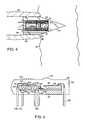
- An example of an optically nonlinear substance suitable for use with a spectrometer module of the inventionis a potassium dihydrogen phosphate (KH 2 PO 4 ) or KDP crystal.
- KH 2 PO 4potassium dihydrogen phosphate
- at least one surface of the KDP crystalmay be coated with a fluoride layer that acts as a one-quarter wave matching layer 53 .
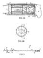
- a window 55is placed at the distal end of the spectrometer module 41 to protect the module.
- the window 55may be held in place by bonding the window 55 to the distal tip of the interventional device with an adhesive 57 .
- the window 55includes a bandstop filter characteristic that attenuates output frequencies of the light generated by the light source 3 while permitting passage of light output having a predetermined frequency.
Landscapes
- Physics & Mathematics (AREA)
- Health & Medical Sciences (AREA)
- Life Sciences & Earth Sciences (AREA)
- Spectroscopy & Molecular Physics (AREA)
- General Health & Medical Sciences (AREA)
- General Physics & Mathematics (AREA)
- Pathology (AREA)
- Biomedical Technology (AREA)
- Biophysics (AREA)
- Molecular Biology (AREA)
- Surgery (AREA)
- Animal Behavior & Ethology (AREA)
- Heart & Thoracic Surgery (AREA)
- Public Health (AREA)
- Veterinary Medicine (AREA)
- Engineering & Computer Science (AREA)
- Medical Informatics (AREA)
- Chemical & Material Sciences (AREA)
- Biochemistry (AREA)
- Analytical Chemistry (AREA)
- Immunology (AREA)
- Investigating Or Analysing Materials By Optical Means (AREA)
- Investigating, Analyzing Materials By Fluorescence Or Luminescence (AREA)
- Measurement Of The Respiration, Hearing Ability, Form, And Blood Characteristics Of Living Organisms (AREA)
Abstract
Description
Claims (20)
Priority Applications (2)
| Application Number | Priority Date | Filing Date | Title |
|---|---|---|---|
| US10/022,965US8126531B2 (en) | 1996-11-21 | 2001-12-13 | Miniature spectrometer |
| US13/357,112US8660637B2 (en) | 1996-11-21 | 2012-01-24 | Miniature spectrometer |
Applications Claiming Priority (4)
| Application Number | Priority Date | Filing Date | Title |
|---|---|---|---|
| US3333496P | 1996-11-21 | 1996-11-21 | |
| US08/898,604US6119031A (en) | 1996-11-21 | 1997-07-22 | Miniature spectrometer |
| US09/478,774US6343227B1 (en) | 1996-11-21 | 2000-01-06 | Miniature spectrometer |
| US10/022,965US8126531B2 (en) | 1996-11-21 | 2001-12-13 | Miniature spectrometer |
Related Parent Applications (1)
| Application Number | Title | Priority Date | Filing Date |
|---|---|---|---|
| US09/478,774ContinuationUS6343227B1 (en) | 1996-11-21 | 2000-01-06 | Miniature spectrometer |
Related Child Applications (1)
| Application Number | Title | Priority Date | Filing Date |
|---|---|---|---|
| US13/357,112ContinuationUS8660637B2 (en) | 1996-11-21 | 2012-01-24 | Miniature spectrometer |
Publications (2)
| Publication Number | Publication Date |
|---|---|
| US20020115918A1 US20020115918A1 (en) | 2002-08-22 |
| US8126531B2true US8126531B2 (en) | 2012-02-28 |
Family
ID=26709571
Family Applications (4)
| Application Number | Title | Priority Date | Filing Date |
|---|---|---|---|
| US08/898,604Expired - LifetimeUS6119031A (en) | 1996-11-21 | 1997-07-22 | Miniature spectrometer |
| US09/478,774Expired - Fee RelatedUS6343227B1 (en) | 1996-11-21 | 2000-01-06 | Miniature spectrometer |
| US10/022,965Expired - Fee RelatedUS8126531B2 (en) | 1996-11-21 | 2001-12-13 | Miniature spectrometer |
| US13/357,112Expired - Fee RelatedUS8660637B2 (en) | 1996-11-21 | 2012-01-24 | Miniature spectrometer |
Family Applications Before (2)
| Application Number | Title | Priority Date | Filing Date |
|---|---|---|---|
| US08/898,604Expired - LifetimeUS6119031A (en) | 1996-11-21 | 1997-07-22 | Miniature spectrometer |
| US09/478,774Expired - Fee RelatedUS6343227B1 (en) | 1996-11-21 | 2000-01-06 | Miniature spectrometer |
Family Applications After (1)
| Application Number | Title | Priority Date | Filing Date |
|---|---|---|---|
| US13/357,112Expired - Fee RelatedUS8660637B2 (en) | 1996-11-21 | 2012-01-24 | Miniature spectrometer |
Country Status (6)
| Country | Link |
|---|---|
| US (4) | US6119031A (en) |
| EP (1) | EP0939894B1 (en) |
| JP (1) | JP4380798B2 (en) |
| AU (1) | AU5172998A (en) |
| DE (1) | DE69737478T2 (en) |
| WO (1) | WO1998022805A1 (en) |
Cited By (4)
| Publication number | Priority date | Publication date | Assignee | Title |
|---|---|---|---|---|
| US20100004518A1 (en)* | 2008-07-03 | 2010-01-07 | Masimo Laboratories, Inc. | Heat sink for noninvasive medical sensor |
| US20100030040A1 (en)* | 2008-08-04 | 2010-02-04 | Masimo Laboratories, Inc. | Multi-stream data collection system for noninvasive measurement of blood constituents |
| US20100198025A1 (en)* | 2007-10-01 | 2010-08-05 | Olof Lindahl | Method and arrangement for unaffected material analyse |
| US9186102B2 (en) | 2009-09-03 | 2015-11-17 | Cercacor Laboratories, Inc. | Emitter driver for noninvasive patient monitor |
Families Citing this family (182)
| Publication number | Priority date | Publication date | Assignee | Title |
|---|---|---|---|---|
| US6119031A (en) | 1996-11-21 | 2000-09-12 | Boston Scientific Corporation | Miniature spectrometer |
| US6826422B1 (en) | 1997-01-13 | 2004-11-30 | Medispectra, Inc. | Spectral volume microprobe arrays |
| US6847490B1 (en) | 1997-01-13 | 2005-01-25 | Medispectra, Inc. | Optical probe accessory device for use in vivo diagnostic procedures |
| US6441943B1 (en) | 1997-04-02 | 2002-08-27 | Gentex Corporation | Indicators and illuminators using a semiconductor radiation emitter package |
| US6324418B1 (en)* | 1997-09-29 | 2001-11-27 | Boston Scientific Corporation | Portable tissue spectroscopy apparatus and method |
| US6185443B1 (en) | 1997-09-29 | 2001-02-06 | Boston Scientific Corporation | Visible display for an interventional device |
| US5984861A (en)* | 1997-09-29 | 1999-11-16 | Boston Scientific Corporation | Endofluorescence imaging module for an endoscope |
| US6238348B1 (en)* | 1997-07-22 | 2001-05-29 | Scimed Life Systems, Inc. | Miniature spectrometer system and method |
| US7637948B2 (en) | 1997-10-10 | 2009-12-29 | Senorx, Inc. | Tissue marking implant |
| US8668737B2 (en) | 1997-10-10 | 2014-03-11 | Senorx, Inc. | Tissue marking implant |
| JP2001519536A (en)* | 1997-10-10 | 2001-10-23 | ボストン サイエンティフィック コーポレイション | Micro-spectrometer system |
| US6289229B1 (en)* | 1998-01-20 | 2001-09-11 | Scimed Life Systems, Inc. | Readable probe array for in vivo use |
| US6922576B2 (en)* | 1998-06-19 | 2005-07-26 | Becton, Dickinson And Company | Micro optical sensor device |
| US6444970B1 (en) | 1998-06-26 | 2002-09-03 | Scimed Life Systems, Inc. | Miniature low-noise photodiode system |
| US6535753B1 (en) | 1998-08-20 | 2003-03-18 | Microsense International, Llc | Micro-invasive method for painless detection of analytes in extra-cellular space |
| US6197257B1 (en)* | 1998-08-20 | 2001-03-06 | Microsense Of St. Louis, Llc | Micro sensor device |
| CA2356623C (en) | 1998-12-23 | 2005-10-18 | Medispectra, Inc. | Systems and methods for optical examination of samples |
| CA2356195A1 (en) | 1998-12-23 | 2000-06-29 | Medispectra, Inc. | Optical methods and systems for cervical screening |
| US7524289B2 (en)* | 1999-01-25 | 2009-04-28 | Lenker Jay A | Resolution optical and ultrasound devices for imaging and treatment of body lumens |
| US6592526B1 (en)* | 1999-01-25 | 2003-07-15 | Jay Alan Lenker | Resolution ultrasound devices for imaging and treatment of body lumens |
| US8361082B2 (en) | 1999-02-02 | 2013-01-29 | Senorx, Inc. | Marker delivery device with releasable plug |
| US6862470B2 (en) | 1999-02-02 | 2005-03-01 | Senorx, Inc. | Cavity-filling biopsy site markers |
| US9820824B2 (en) | 1999-02-02 | 2017-11-21 | Senorx, Inc. | Deployment of polysaccharide markers for treating a site within a patent |
| US8498693B2 (en) | 1999-02-02 | 2013-07-30 | Senorx, Inc. | Intracorporeal marker and marker delivery device |
| US7983734B2 (en) | 2003-05-23 | 2011-07-19 | Senorx, Inc. | Fibrous marker and intracorporeal delivery thereof |
| US7651505B2 (en) | 2002-06-17 | 2010-01-26 | Senorx, Inc. | Plugged tip delivery for marker placement |
| US6725083B1 (en) | 1999-02-02 | 2004-04-20 | Senorx, Inc. | Tissue site markers for in VIVO imaging |
| US20090030309A1 (en) | 2007-07-26 | 2009-01-29 | Senorx, Inc. | Deployment of polysaccharide markers |
| US6575991B1 (en) | 1999-06-17 | 2003-06-10 | Inrad, Inc. | Apparatus for the percutaneous marking of a lesion |
| US6902935B2 (en) | 1999-12-15 | 2005-06-07 | Medispectra, Inc. | Methods of monitoring effects of chemical agents on a sample |
| US7187810B2 (en) | 1999-12-15 | 2007-03-06 | Medispectra, Inc. | Methods and systems for correcting image misalignment |
| US7260248B2 (en) | 1999-12-15 | 2007-08-21 | Medispectra, Inc. | Image processing using measures of similarity |
| US7373197B2 (en)* | 2000-03-03 | 2008-05-13 | Intramedical Imaging, Llc | Methods and devices to expand applications of intraoperative radiation probes |
| US8527046B2 (en) | 2000-04-20 | 2013-09-03 | Medtronic, Inc. | MRI-compatible implantable device |
| US6795730B2 (en) | 2000-04-20 | 2004-09-21 | Biophan Technologies, Inc. | MRI-resistant implantable device |
| US6925328B2 (en) | 2000-04-20 | 2005-08-02 | Biophan Technologies, Inc. | MRI-compatible implantable device |
| WO2002033385A2 (en)* | 2000-10-19 | 2002-04-25 | Motorola, Inc. | Biochip excitation and analysis structure |
| AU3659002A (en)* | 2000-11-09 | 2002-05-21 | Sicel Technologies Inc | Methods, circuits and compositions of matter for in vivo detection of biomolecule concentrations using fluorescent tags |
| EA200300574A1 (en)* | 2000-11-16 | 2004-06-24 | Кэмилийен Медикал Инновейшн Лтд. | SYSTEM FOR DIAGNOSTIC EARS |
| CA2659518A1 (en) | 2000-11-20 | 2002-05-30 | Senorx, Inc. | Tissue site markers for in vivo imaging |
| US6839661B2 (en) | 2000-12-15 | 2005-01-04 | Medispectra, Inc. | System for normalizing spectra |
| US6657197B2 (en)* | 2000-12-22 | 2003-12-02 | Honeywell International Inc. | Small profile spectrometer |
| DE10203720B4 (en)* | 2001-02-02 | 2012-11-22 | Nippon Telegraph And Telephone Corp. | Blood flow meter and sensor part of the blood flow meter |
| US20020116028A1 (en) | 2001-02-20 | 2002-08-22 | Wilson Greatbatch | MRI-compatible pacemaker with pulse carrying photonic catheter providing VOO functionality |
| US6829509B1 (en) | 2001-02-20 | 2004-12-07 | Biophan Technologies, Inc. | Electromagnetic interference immune tissue invasive system |
| US6488704B1 (en)* | 2001-05-07 | 2002-12-03 | Biomed Solutions, Llc | Implantable particle measuring apparatus |
| US6793642B2 (en)* | 2001-05-07 | 2004-09-21 | Biomed Solutions, Llc | Flow cytometer |
| US6898451B2 (en)* | 2001-03-21 | 2005-05-24 | Minformed, L.L.C. | Non-invasive blood analyte measuring system and method utilizing optical absorption |
| US20040225326A1 (en)* | 2001-05-07 | 2004-11-11 | Weiner Mike L. | Apparatus for the detection of restenosis |
| US6654630B2 (en)* | 2001-05-31 | 2003-11-25 | Infraredx, Inc. | Apparatus and method for the optical imaging of tissue samples |
| US7054686B2 (en) | 2001-08-30 | 2006-05-30 | Biophan Technologies, Inc. | Pulsewidth electrical stimulation |
| US6731979B2 (en) | 2001-08-30 | 2004-05-04 | Biophan Technologies Inc. | Pulse width cardiac pacing apparatus |
| WO2003020103A2 (en)* | 2001-09-04 | 2003-03-13 | Amit Technology Science & Medicine Ltd. | Method of and device for therapeutic illumination of internal organs and tissues |
| JP2003102672A (en)* | 2001-10-01 | 2003-04-08 | Japan Science & Technology Corp | Method and apparatus for automatically detecting and treating or collecting a target site such as a lesion |
| US6988001B2 (en) | 2001-10-31 | 2006-01-17 | Biophan Technologies, Inc. | Hermetic component housing for photonic catheter |
| AT412695B (en)* | 2001-11-20 | 2005-05-25 | Efkon Ag | INFRARED (IR) TRANSMISSION DEVICE |
| TWI293363B (en) | 2001-12-11 | 2008-02-11 | Sensors For Med & Science Inc | High performance fluorescent optical sensor |
| US8423110B2 (en)* | 2002-01-09 | 2013-04-16 | Boston Scientific Scimed, Inc. | Imaging device and related methods |
| US6968236B2 (en)* | 2002-01-28 | 2005-11-22 | Biophan Technologies, Inc. | Ceramic cardiac electrodes |
| US8504128B2 (en)* | 2002-03-08 | 2013-08-06 | Glt Acquisition Corp. | Method and apparatus for coupling a channeled sample probe to tissue |
| US8718738B2 (en) | 2002-03-08 | 2014-05-06 | Glt Acquisition Corp. | Method and apparatus for coupling a sample probe with a sample site |
| US20090318786A1 (en)* | 2002-03-08 | 2009-12-24 | Blank Thomas B | Channeled tissue sample probe method and apparatus |
| US20040064018A1 (en)* | 2002-03-22 | 2004-04-01 | Robert Dunki-Jacobs | Integrated visualization system |
| US8131332B2 (en)* | 2002-04-04 | 2012-03-06 | Veralight, Inc. | Determination of a measure of a glycation end-product or disease state using tissue fluorescence of various sites |
| US20080009689A1 (en)* | 2002-04-09 | 2008-01-10 | Benaron David A | Difference-weighted somatic spectroscopy |
| US20070015981A1 (en)* | 2003-08-29 | 2007-01-18 | Benaron David A | Device and methods for the detection of locally-weighted tissue ischemia |
| US6711426B2 (en)* | 2002-04-09 | 2004-03-23 | Spectros Corporation | Spectroscopy illuminator with improved delivery efficiency for high optical density and reduced thermal load |
| US6711440B2 (en) | 2002-04-11 | 2004-03-23 | Biophan Technologies, Inc. | MRI-compatible medical device with passive generation of optical sensing signals |
| US6725092B2 (en) | 2002-04-25 | 2004-04-20 | Biophan Technologies, Inc. | Electromagnetic radiation immune medical assist device adapter |
| EP1534386A4 (en)* | 2002-06-04 | 2006-03-15 | Biophan Technologies Inc | Nuclear magnetic resonance spectrometer assembly |
| US7672713B2 (en)* | 2002-06-19 | 2010-03-02 | Infraredx, Inc. | Multi-channel catheter tip |
| US7282723B2 (en) | 2002-07-09 | 2007-10-16 | Medispectra, Inc. | Methods and apparatus for processing spectral data for use in tissue characterization |
| US7469160B2 (en) | 2003-04-18 | 2008-12-23 | Banks Perry S | Methods and apparatus for evaluating image focus |
| US7309867B2 (en) | 2003-04-18 | 2007-12-18 | Medispectra, Inc. | Methods and apparatus for characterization of tissue samples |
| US7136518B2 (en) | 2003-04-18 | 2006-11-14 | Medispectra, Inc. | Methods and apparatus for displaying diagnostic data |
| US7459696B2 (en) | 2003-04-18 | 2008-12-02 | Schomacker Kevin T | Methods and apparatus for calibrating spectral data |
| US6933154B2 (en) | 2002-07-09 | 2005-08-23 | Medispectra, Inc. | Optimal windows for obtaining optical data for characterization of tissue samples |
| US6818903B2 (en) | 2002-07-09 | 2004-11-16 | Medispectra, Inc. | Method and apparatus for identifying spectral artifacts |
| US6768918B2 (en) | 2002-07-10 | 2004-07-27 | Medispectra, Inc. | Fluorescent fiberoptic probe for tissue health discrimination and method of use thereof |
| US7103401B2 (en) | 2002-07-10 | 2006-09-05 | Medispectra, Inc. | Colonic polyp discrimination by tissue fluorescence and fiberoptic probe |
| US6925322B2 (en) | 2002-07-25 | 2005-08-02 | Biophan Technologies, Inc. | Optical MRI catheter system |
| US7535935B2 (en)* | 2002-09-27 | 2009-05-19 | Infraredx, Inc. | Spectroscopic catheter system with widely tunable source and method of operation |
| DE10249674B4 (en)* | 2002-10-24 | 2014-12-24 | Carl Zeiss Meditec Ag | Surgical instrument for cutting, ablating or aspirating material in an operating area |
| US20060036158A1 (en) | 2003-11-17 | 2006-02-16 | Inrad, Inc. | Self-contained, self-piercing, side-expelling marking apparatus |
| US20050154277A1 (en)* | 2002-12-31 | 2005-07-14 | Jing Tang | Apparatus and methods of using built-in micro-spectroscopy micro-biosensors and specimen collection system for a wireless capsule in a biological body in vivo |
| US20040193023A1 (en)* | 2003-03-28 | 2004-09-30 | Aris Mardirossian | System, method and apparatus for monitoring recording and reporting physiological data |
| US7877133B2 (en) | 2003-05-23 | 2011-01-25 | Senorx, Inc. | Marker or filler forming fluid |
| DE10349839B4 (en)* | 2003-10-25 | 2006-05-18 | Hbw-Gubesch Kunststoff-Engineering Gmbh | Electrical component with a transparent plastic housing |
| US20050273002A1 (en) | 2004-06-04 | 2005-12-08 | Goosen Ryan L | Multi-mode imaging marker |
| JP4759519B2 (en)* | 2003-12-17 | 2011-08-31 | チェック キャップ リミテッド | Detection of intraluminal polyps |
| US9392961B2 (en) | 2003-12-17 | 2016-07-19 | Check-Cap Ltd. | Intra-lumen polyp detection |
| US20050203419A1 (en)* | 2004-02-24 | 2005-09-15 | Nirmala Ramanujam | Side-firing probe for performing optical spectroscopy during core needle biopsy |
| JP2005278762A (en)* | 2004-03-29 | 2005-10-13 | Fujinon Corp | Centesis type probe for endoscope |
| US7268871B2 (en)* | 2004-08-12 | 2007-09-11 | Datacolor Holding Ag | Measuring head for planar measurement of a sample |
| US8419656B2 (en) | 2004-11-22 | 2013-04-16 | Bard Peripheral Vascular, Inc. | Post decompression marker introducer system |
| US8070767B2 (en) | 2005-01-28 | 2011-12-06 | Tyco Healthcare Group Lp | Optical penetrating adapter for surgical portal |
| US7470230B2 (en) | 2005-03-31 | 2008-12-30 | Tyco Healthcare Group Lp | Optical obturator |
| US10357328B2 (en) | 2005-04-20 | 2019-07-23 | Bard Peripheral Vascular, Inc. and Bard Shannon Limited | Marking device with retractable cannula |
| US7813778B2 (en)* | 2005-07-29 | 2010-10-12 | Spectros Corporation | Implantable tissue ischemia sensor |
| US8052658B2 (en) | 2005-10-07 | 2011-11-08 | Bard Peripheral Vascular, Inc. | Drug-eluting tissue marker |
| EP1948296B2 (en) | 2005-10-14 | 2017-10-11 | Pacesetter, Inc. | Leadless cardiac pacemaker and system |
| US9168383B2 (en) | 2005-10-14 | 2015-10-27 | Pacesetter, Inc. | Leadless cardiac pacemaker with conducted communication |
| NL1031588C2 (en) | 2006-04-13 | 2007-10-19 | D O R C Dutch Ophthalmic Res C | Eye surgical instrument. |
| EP2033036A4 (en)* | 2006-06-13 | 2009-07-15 | Invent Technologies Llc | DEVICE AND METHOD FOR OPTICAL UV DEEP MICROSCOPY |
| WO2007147058A2 (en)* | 2006-06-14 | 2007-12-21 | Cornova, Inc. | Method and apparatus for identifying and treating myocardial infarction |
| WO2008051749A2 (en) | 2006-10-23 | 2008-05-02 | C. R. Bard, Inc. | Breast marker |
| US9579077B2 (en) | 2006-12-12 | 2017-02-28 | C.R. Bard, Inc. | Multiple imaging mode tissue marker |
| WO2008076973A2 (en) | 2006-12-18 | 2008-06-26 | C.R.Bard Inc. | Biopsy marker with in situ-generated imaging properties |
| JP2010530055A (en) | 2007-02-06 | 2010-09-02 | ヨアブ キムチ | Lumen polyp detection |
| WO2008113024A1 (en)* | 2007-03-14 | 2008-09-18 | Spectros Corporation | Metabolism-or biochemical-based anti-spoofing biometrics devices, systems, and methods |
| US7711412B2 (en)* | 2007-05-23 | 2010-05-04 | Ethicon Endo-Surgery, Inc. | Mucosal tissue illuminator and method for use |
| AU2008202266B2 (en)* | 2007-06-01 | 2013-09-12 | Covidien Lp | Obturator tips |
| JP2010540037A (en)* | 2007-09-20 | 2010-12-24 | ナノスティム インコーポレイテッド | Leadless cardiac pacemaker with secondary fixation capability |
| DE102008049078A1 (en)* | 2007-09-26 | 2009-04-09 | Mbr Optical Systems Gmbh & Co. Kg | Method and measuring device for collecting spectrometric measuring signals from vital tissue |
| US7817274B2 (en) | 2007-10-05 | 2010-10-19 | Jingyun Zhang | Compact spectrometer |
| WO2009050667A1 (en)* | 2007-10-18 | 2009-04-23 | Koninklijke Philips Electronics N.V. | Tumor demarcation using targeted fluorescent probe and photonic needle |
| US8345226B2 (en) | 2007-11-30 | 2013-01-01 | Jingyun Zhang | Spectrometers miniaturized for working with cellular phones and other portable electronic devices |
| WO2009099767A2 (en) | 2008-01-31 | 2009-08-13 | C.R. Bard, Inc. | Biopsy tissue marker |
| CA3194784A1 (en) | 2008-05-20 | 2009-11-26 | University Health Network | Device and method for fluorescence-based imaging and monitoring |
| US20100069760A1 (en)* | 2008-09-17 | 2010-03-18 | Cornova, Inc. | Methods and apparatus for analyzing and locally treating a body lumen |
| US9327061B2 (en) | 2008-09-23 | 2016-05-03 | Senorx, Inc. | Porous bioabsorbable implant |
| CN101721194B (en)* | 2008-10-14 | 2011-11-09 | 鸿富锦精密工业(深圳)有限公司 | Capsule endoscope and method for manufacturing lenses thereof |
| EP4215147A3 (en) | 2008-12-30 | 2023-10-18 | C. R. Bard, Inc. | Marker delivery device for tissue marker placement |
| US8527068B2 (en) | 2009-02-02 | 2013-09-03 | Nanostim, Inc. | Leadless cardiac pacemaker with secondary fixation capability |
| US20110077708A1 (en)* | 2009-09-28 | 2011-03-31 | Alan Ostroff | MRI Compatible Leadless Cardiac Pacemaker |
| CN102596044A (en)* | 2009-09-29 | 2012-07-18 | 卧龙岗大学 | Imaging method and system |
| US8979883B2 (en)* | 2009-12-17 | 2015-03-17 | Covidien Lp | Obturator tip |
| DE102010014702A1 (en)* | 2010-04-12 | 2011-10-13 | Mbr Optikal Systems Gmbh & Co. Kg | Measuring arrangement for recording a spectrum, in particular on vital tissue |
| US8543205B2 (en) | 2010-10-12 | 2013-09-24 | Nanostim, Inc. | Temperature sensor for a leadless cardiac pacemaker |
| US9060692B2 (en) | 2010-10-12 | 2015-06-23 | Pacesetter, Inc. | Temperature sensor for a leadless cardiac pacemaker |
| EP2627406A1 (en) | 2010-10-13 | 2013-08-21 | Nanostim, Inc. | Leadless cardiac pacemaker with anti-unscrewing feature |
| US8615310B2 (en) | 2010-12-13 | 2013-12-24 | Pacesetter, Inc. | Delivery catheter systems and methods |
| EP2651502B1 (en) | 2010-12-13 | 2016-11-09 | Pacesetter, Inc. | Pacemaker retrieval systems |
| WO2012088118A1 (en) | 2010-12-20 | 2012-06-28 | Nanostim, Inc. | Leadless pacemaker with radial fixation mechanism |
| USD663422S1 (en) | 2011-03-24 | 2012-07-10 | First Pulse Medical, Inc. | Fetal pulse oximeter clip |
| US9140604B2 (en)* | 2011-06-17 | 2015-09-22 | Kla-Tencor Corporation | Wafer level spectrometer |
| US9511236B2 (en) | 2011-11-04 | 2016-12-06 | Pacesetter, Inc. | Leadless cardiac pacemaker with integral battery and redundant welds |
| WO2013108209A1 (en)* | 2012-01-19 | 2013-07-25 | Technion Research & Development Foundation Ltd. | Vessel imaging system and method |
| NL2008455C2 (en) | 2012-03-09 | 2013-09-10 | D O R C Dutch Ophthalmic Res Ct International B V | EYE-SURGICAL LIGHTING UNIT. |
| CN203719767U (en)* | 2012-06-11 | 2014-07-16 | 保生国际生医股份有限公司 | Biochemical detection system and light source module thereof |
| WO2014022661A1 (en) | 2012-08-01 | 2014-02-06 | Nanostim, Inc. | Biostimulator circuit with flying cell |
| US10206583B2 (en) | 2012-10-31 | 2019-02-19 | Covidien Lp | Surgical devices and methods utilizing optical coherence tomography (OCT) to monitor and control tissue sealing |
| WO2014137357A1 (en)* | 2013-03-08 | 2014-09-12 | Alethus, Inc. | Optically discriminative detection of matters in tissues and turbid media and applications for non-invasive assay |
| USD716451S1 (en) | 2013-09-24 | 2014-10-28 | C. R. Bard, Inc. | Tissue marker for intracorporeal site identification |
| USD716450S1 (en) | 2013-09-24 | 2014-10-28 | C. R. Bard, Inc. | Tissue marker for intracorporeal site identification |
| USD715442S1 (en) | 2013-09-24 | 2014-10-14 | C. R. Bard, Inc. | Tissue marker for intracorporeal site identification |
| USD715942S1 (en) | 2013-09-24 | 2014-10-21 | C. R. Bard, Inc. | Tissue marker for intracorporeal site identification |
| JP6769949B2 (en) | 2014-07-24 | 2020-10-14 | ユニバーシティー ヘルス ネットワーク | Data collection and analysis for diagnostic purposes |
| FI127170B (en)* | 2014-10-03 | 2017-12-29 | Pulseon Oy | Portable biometric device and process for its manufacture |
| WO2016063284A2 (en) | 2014-10-23 | 2016-04-28 | Verifood, Ltd. | Accessories for handheld spectrometer |
| WO2018015951A1 (en)* | 2016-07-20 | 2018-01-25 | Verifood, Ltd. | Accessories for handheld spectrometer |
| WO2018075643A1 (en) | 2016-10-18 | 2018-04-26 | Atoptix, Inc. | Self-referencing mobile-compatible spectrophotometer platform |
| EP3606592B1 (en) | 2017-04-07 | 2025-01-08 | Bard Access Systems, Inc. | Optical fiber-based medical device tracking and monitoring system |
| KR102727114B1 (en)* | 2019-02-01 | 2024-11-06 | 현대자동차주식회사 | Non-invasive optical internal substance detector |
| WO2020191269A1 (en)* | 2019-03-21 | 2020-09-24 | The Brigham And Women's Hospital, Inc. | Robotic artificial intelligence nasal/oral/ rectal enteric tube |
| EP4013338A4 (en) | 2019-08-12 | 2023-08-30 | Bard Access Systems, Inc. | SHAPE DETECTION SYSTEMS AND METHODS FOR MEDICAL DEVICES |
| DE102019212202B4 (en)* | 2019-08-14 | 2021-12-23 | Richard Wolf Gmbh | Light applicator |
| EP4061272A4 (en) | 2019-11-25 | 2023-11-22 | Bard Access Systems, Inc. | Shape-sensing systems with filters and methods thereof |
| CN112826497B (en) | 2019-11-25 | 2025-09-09 | 巴德阿克塞斯系统股份有限公司 | Optical tip tracking system and method thereof |
| CN215461207U (en) | 2020-02-28 | 2022-01-11 | 巴德阿克塞斯系统股份有限公司 | Catheter and medical instrument monitoring system |
| US11474310B2 (en) | 2020-02-28 | 2022-10-18 | Bard Access Systems, Inc. | Optical connection systems and methods thereof |
| CN113332561A (en) | 2020-03-03 | 2021-09-03 | 巴德阿克塞斯系统股份有限公司 | System and method for optical shape sensing and electrical signal conduction |
| WO2021202589A1 (en) | 2020-03-30 | 2021-10-07 | Bard Access Systems, Inc. | Optical and electrical diagnostic systems and methods thereof |
| CN113842536A (en) | 2020-06-26 | 2021-12-28 | 巴德阿克塞斯系统股份有限公司 | Dislocation detection system |
| WO2022005870A1 (en) | 2020-06-29 | 2022-01-06 | Bard Access Systems, Inc. | Automatic dimensional frame reference for fiber optic |
| WO2022011287A1 (en) | 2020-07-10 | 2022-01-13 | Bard Access Systems, Inc. | Continuous fiber optic functionality monitoring and self-diagnostic reporting system |
| WO2022031613A1 (en) | 2020-08-03 | 2022-02-10 | Bard Access Systems, Inc. | Bragg grated fiber optic fluctuation sensing and monitoring system |
| CN114246583A (en) | 2020-09-25 | 2022-03-29 | 巴德阿克塞斯系统股份有限公司 | Fiber Optic Oximetry Systems for Detection and Confirmation |
| CN114344514A (en) | 2020-10-13 | 2022-04-15 | 巴德阿克塞斯系统股份有限公司 | Disinfection enclosure for fiber optic connectors and method thereof |
| CN114518075A (en) | 2020-11-18 | 2022-05-20 | 巴德阿克塞斯系统股份有限公司 | fiber optic stylet holder |
| WO2022115624A1 (en) | 2020-11-24 | 2022-06-02 | Bard Access Systems, Inc. | Steerable fiber optic shape sensing enabled elongated medical instrument |
| WO2022150411A1 (en) | 2021-01-06 | 2022-07-14 | Bard Access Systems, Inc. | Needle guidance using fiber optic shape sensing |
| US12426954B2 (en) | 2021-01-26 | 2025-09-30 | Bard Access Systems, Inc. | Fiber optic shape sensing system associated with port placement |
| US12061116B2 (en)* | 2021-07-29 | 2024-08-13 | Si-Ware Systems | Compact spectral analyzer |
| US12419694B2 (en) | 2021-10-25 | 2025-09-23 | Bard Access Systems, Inc. | Reference plane for medical device placement |
| US12318149B2 (en) | 2022-03-08 | 2025-06-03 | Bard Access Systems, Inc. | Medical shape sensing devices and systems |
| US12426956B2 (en) | 2022-03-16 | 2025-09-30 | Bard Access Systems, Inc. | Medical system and method for monitoring medical device insertion and illumination patterns |
| US12089815B2 (en) | 2022-03-17 | 2024-09-17 | Bard Access Systems, Inc. | Fiber optic medical systems and devices with atraumatic tip |
| US12343117B2 (en) | 2022-06-28 | 2025-07-01 | Bard Access Systems, Inc. | Fiber optic medical systems and methods for identifying blood vessels |
| US12349984B2 (en) | 2022-06-29 | 2025-07-08 | Bard Access Systems, Inc. | System, method, and apparatus for improved confirm of an anatomical position of a medical instrument |
| EP4559373A1 (en) | 2023-11-24 | 2025-05-28 | Erbe Elektromedizin GmbH | Endoscope with sensor for image acquisition and tissue examination |
| WO2025151357A1 (en)* | 2024-01-10 | 2025-07-17 | Gyrus Acmi, Inc. D/B/A Olympus Surgical Technologies America | Light source independent optical measurements |
Citations (121)
| Publication number | Priority date | Publication date | Assignee | Title |
|---|---|---|---|---|
| US2002559A (en) | 1932-04-13 | 1935-05-28 | Wappler Frederick Charles | Means for surgical resection |
| US2583937A (en) | 1952-01-29 | Surgical exploring and operating | ||
| DE888727C (en) | 1941-04-22 | 1953-09-03 | Quarzlampen Gmbh | Body cavity irradiation lamp |
| US3176114A (en) | 1962-07-16 | 1965-03-30 | Richard F Kneisley | Device for removing nasal hair |
| US4075475A (en) | 1976-05-03 | 1978-02-21 | Chemetron Corporation | Programmed thermal degradation-mass spectrometry analysis method facilitating identification of a biological specimen |
| US4233493A (en) | 1974-05-21 | 1980-11-11 | Nath Guenther | Apparatus for applying intense light radiation to a limited area |
| US4274706A (en) | 1979-08-30 | 1981-06-23 | Hughes Aircraft Company | Wavelength multiplexer/demultiplexer for optical circuits |
| US4289966A (en) | 1980-04-10 | 1981-09-15 | The United States Of America As Represented By The Secretary Of The Army | Radiation energy detector and analyzer |
| DE3023130A1 (en) | 1980-06-20 | 1982-01-21 | Hasso von 4000 Düsseldorf Blücher | Mouth and nose steriliser - uses high intensity light flash of millisecond duration |
| US4340307A (en) | 1980-07-07 | 1982-07-20 | Beckman Instruments, Inc. | Bichromatic spectrophotometer with wavelength reversal |
| US4472728A (en) | 1982-02-19 | 1984-09-18 | The United States Of America As Represented By The Administrator Of The National Aeronautics And Space Administration | Imaging X-ray spectrometer |
| US4541272A (en) | 1983-05-13 | 1985-09-17 | Roland Bause | Electronically controlled fuel injection system |
| US4548505A (en) | 1981-04-22 | 1985-10-22 | Sumitomo Electric Industries, Ltd. | Sensor for spectral analyzer for living tissues |
| US4556057A (en) | 1982-08-31 | 1985-12-03 | Hamamatsu Tv Co., Ltd. | Cancer diagnosis device utilizing laser beam pulses |
| US4570638A (en) | 1983-10-14 | 1986-02-18 | Somanetics Corporation | Method and apparatus for spectral transmissibility examination and analysis |
| US4578061A (en) | 1980-10-28 | 1986-03-25 | Lemelson Jerome H | Injection catheter and method |
| US4672972A (en) | 1984-08-13 | 1987-06-16 | Berke Howard R | Solid state NMR probe |
| US4718417A (en) | 1985-03-22 | 1988-01-12 | Massachusetts Institute Of Technology | Visible fluorescence spectral diagnostic for laser angiosurgery |
| US4803992A (en) | 1980-10-28 | 1989-02-14 | Lemelson Jerome H | Electro-optical instruments and methods for producing same |
| EP0304321A1 (en) | 1987-08-21 | 1989-02-22 | Paul Okimoto | Vaginal testing applicator and method |
| US4872458A (en) | 1986-09-16 | 1989-10-10 | Olympus Optical Co., Ltd. | Thermotherapy apparatus |
| US4882623A (en) | 1988-08-11 | 1989-11-21 | Olympus Optical Co., Ltd. | Signal processing apparatus for endoscope capable of changing outline enhancement frequency |
| US4894547A (en) | 1987-09-28 | 1990-01-16 | Yale University | Optical method and apparatus for detecting and measuring aging, photoaging, dermal disease and pigmentation in skin |
| US4902896A (en) | 1987-05-08 | 1990-02-20 | Mine Safety Appliances Company | Infrared fluid analyzer |
| US4928172A (en) | 1988-01-07 | 1990-05-22 | Olympus Optical Co., Ltd. | Endoscope output signal control device and endoscope apparatus making use of the same |
| US4930516A (en) | 1985-11-13 | 1990-06-05 | Alfano Robert R | Method for detecting cancerous tissue using visible native luminescence |
| US4938602A (en) | 1987-10-15 | 1990-07-03 | Electronic Instrumentation And Technology, Inc. | Automated process monitoring |
| US4981138A (en) | 1988-06-30 | 1991-01-01 | Yale University | Endoscopic fiberoptic fluorescence spectrometer |
| US5001556A (en) | 1987-09-30 | 1991-03-19 | Olympus Optical Co., Ltd. | Endoscope apparatus for processing a picture image of an object based on a selected wavelength range |
| US5009655A (en) | 1989-05-24 | 1991-04-23 | C. R. Bard, Inc. | Hot tip device with optical diagnostic capability |
| US5021888A (en) | 1987-12-18 | 1991-06-04 | Kabushiki Kaisha Toshiba | Miniaturized solid state imaging device |
| US5034010A (en) | 1985-03-22 | 1991-07-23 | Massachusetts Institute Of Technology | Optical shield for a laser catheter |
| US5036853A (en) | 1988-08-26 | 1991-08-06 | Polartechnics Ltd. | Physiological probe |
| US5042494A (en) | 1985-11-13 | 1991-08-27 | Alfano Robert R | Method and apparatus for detecting cancerous tissue using luminescence excitation spectra |
| DE4005743A1 (en) | 1990-02-23 | 1991-08-29 | Wolf Gmbh Richard | Lithotriptor to destroy gallstones intra-or trans-luminally - has shock-wave generator, lead contg. incompressible material, and opt balloon to press generator against gall bladder |
| US5045056A (en) | 1989-09-15 | 1991-09-03 | Behl Robert S | Method and device for thermal ablation of hollow body organs |
| US5056503A (en) | 1983-10-03 | 1991-10-15 | Olympus Optical Co., Ltd. | Endoscope with high frequency accessory and reduced video interference |
| US5062428A (en) | 1990-06-04 | 1991-11-05 | Nim Incorporated | Method and device for in vivo diagnosis detecting IR emission by body organ |
| US5106387A (en) | 1985-03-22 | 1992-04-21 | Massachusetts Institute Of Technology | Method for spectroscopic diagnosis of tissue |
| US5115137A (en) | 1989-02-22 | 1992-05-19 | Spectraphos Ab | Diagnosis by means of fluorescent light emission from tissue |
| US5116759A (en) | 1990-06-27 | 1992-05-26 | Fiberchem Inc. | Reservoir chemical sensors |
| US5125404A (en) | 1985-03-22 | 1992-06-30 | Massachusetts Institute Of Technology | Apparatus and method for obtaining spectrally resolved spatial images of tissue |
| US5127407A (en) | 1989-08-17 | 1992-07-07 | Critikon, Inc. | Epidural oxygen sensor |
| US5131398A (en) | 1990-01-22 | 1992-07-21 | Mediscience Technology Corp. | Method and apparatus for distinguishing cancerous tissue from benign tumor tissue, benign tissue or normal tissue using native fluorescence |
| US5152295A (en) | 1989-07-26 | 1992-10-06 | Kowa Company Ltd. | Fundus examination device |
| US5166755A (en) | 1990-05-23 | 1992-11-24 | Nahum Gat | Spectrometer apparatus |
| US5172693A (en) | 1990-01-16 | 1992-12-22 | Doody Michael C | Prenatal non-invasive detection of meconium stained amniotic fluid |
| US5174297A (en) | 1989-11-22 | 1992-12-29 | S.L.T. Japan Co., Ltd. | Diagnostic apparatus for living tissues and medical treatment apparatus with diagnostic apparatus |
| US5187572A (en) | 1990-10-31 | 1993-02-16 | Olympus Optical Co., Ltd. | Endoscope system with a plurality of synchronized light source apparatuses |
| US5187672A (en) | 1989-02-06 | 1993-02-16 | Nim Incorporated | Phase modulation spectroscopic system |
| US5193542A (en) | 1991-01-28 | 1993-03-16 | Missanelli John S | Peripartum oximetric monitoring apparatus |
| US5197470A (en) | 1990-07-16 | 1993-03-30 | Eastman Kodak Company | Near infrared diagnostic method and instrument |
| US5199431A (en) | 1985-03-22 | 1993-04-06 | Massachusetts Institute Of Technology | Optical needle for spectroscopic diagnosis |
| US5201318A (en) | 1989-04-24 | 1993-04-13 | Rava Richard P | Contour mapping of spectral diagnostics |
| US5206174A (en) | 1992-09-24 | 1993-04-27 | Eg&G Idaho, Inc. | Method of photon spectral analysis |
| US5213569A (en) | 1992-03-31 | 1993-05-25 | Davis Peter L | Tip for a tissue phacoemulsification device |
| US5233621A (en) | 1991-06-27 | 1993-08-03 | Intellectual Property Development Associates Of Connecticut, Inc. | Second harmonic generation and self frequency doubling laser materials comprised of bulk germanosilicate and aluminosilicate glasses |
| US5242437A (en) | 1988-06-10 | 1993-09-07 | Trimedyne Laser Systems, Inc. | Medical device applying localized high intensity light and heat, particularly for destruction of the endometrium |
| US5261410A (en) | 1991-02-07 | 1993-11-16 | Alfano Robert R | Method for determining if a tissue is a malignant tumor tissue, a benign tumor tissue, or a normal or benign tissue using Raman spectroscopy |
| US5262645A (en) | 1991-09-03 | 1993-11-16 | General Motors Corporation | Sensor for measuring alcohol content of alcohol gasoline fuel mixtures |
| US5280788A (en) | 1991-02-26 | 1994-01-25 | Massachusetts Institute Of Technology | Devices and methods for optical diagnosis of tissue |
| US5304173A (en) | 1985-03-22 | 1994-04-19 | Massachusetts Institute Of Technology | Spectral diagonostic and treatment system |
| US5305748A (en) | 1992-06-05 | 1994-04-26 | Wilk Peter J | Medical diagnostic system and related method |
| US5309907A (en) | 1991-09-04 | 1994-05-10 | Siemens Aktiengesellschaft | Measuring arrangement for examining a subject with visible, NIR or IR light |
| US5348018A (en) | 1991-11-25 | 1994-09-20 | Alfano Robert R | Method for determining if tissue is malignant as opposed to non-malignant using time-resolved fluorescence spectroscopy |
| US5350375A (en) | 1993-03-15 | 1994-09-27 | Yale University | Methods for laser induced fluorescence intensity feedback control during laser angioplasty |
| US5351532A (en) | 1992-10-08 | 1994-10-04 | Paradigm Technologies | Methods and apparatus for making chemical concentration measurements in a sub-surface exploration probe |
| US5377676A (en) | 1991-04-03 | 1995-01-03 | Cedars-Sinai Medical Center | Method for determining the biodistribution of substances using fluorescence spectroscopy |
| US5383467A (en) | 1992-11-18 | 1995-01-24 | Spectrascience, Inc. | Guidewire catheter and apparatus for diagnostic imaging |
| US5386827A (en) | 1993-03-30 | 1995-02-07 | Nim Incorporated | Quantitative and qualitative in vivo tissue examination using time resolved spectroscopy |
| US5398844A (en) | 1994-01-31 | 1995-03-21 | Boston Scientific Corporation | Multiple ligating band dispenser |
| US5402792A (en) | 1993-03-30 | 1995-04-04 | Shimadzu Corporation | Ultrasonic medical apparatus |
| US5402778A (en) | 1993-01-19 | 1995-04-04 | Nim Incorporated | Spectrophotometric examination of tissue of small dimension |
| US5405369A (en) | 1994-01-25 | 1995-04-11 | Medical College Of Ohio | Photochemical ablation of gastro-intestinal tissue for augmentation of an organ |
| US5413108A (en) | 1993-04-21 | 1995-05-09 | The Research Foundation Of City College Of New York | Method and apparatus for mapping a tissue sample for and distinguishing different regions thereof based on luminescence measurements of cancer-indicative native fluorophor |
| US5417207A (en) | 1993-12-06 | 1995-05-23 | Sensor Devices, Inc. | Apparatus for the invasive use of oximeter probes |
| US5419323A (en) | 1988-12-21 | 1995-05-30 | Massachusetts Institute Of Technology | Method for laser induced fluorescence of tissue |
| US5421339A (en) | 1993-05-12 | 1995-06-06 | Board Of Regents, The University Of Texas System | Diagnosis of dysplasia using laser induced fluoroescence |
| US5421337A (en) | 1989-04-14 | 1995-06-06 | Massachusetts Institute Of Technology | Spectral diagnosis of diseased tissue |
| US5445608A (en) | 1993-08-16 | 1995-08-29 | James C. Chen | Method and apparatus for providing light-activated therapy |
| US5452723A (en) | 1992-07-24 | 1995-09-26 | Massachusetts Institute Of Technology | Calibrated spectrographic imaging |
| US5456252A (en) | 1993-09-30 | 1995-10-10 | Cedars-Sinai Medical Center | Induced fluorescence spectroscopy blood perfusion and pH monitor and method |
| US5461229A (en) | 1994-06-06 | 1995-10-24 | Unisys Corporation | On-the-go optical spectroscopy soil analyzer |
| US5467767A (en) | 1991-11-25 | 1995-11-21 | Alfano; Robert R. | Method for determining if tissue is malignant as opposed to non-malignant using time-resolved fluorescence spectroscopy |
| EP0314937B1 (en) | 1987-10-08 | 1995-12-13 | Pacesetter AB | Implantable blood oxygen sensor and method of use |
| WO1996010363A1 (en) | 1994-09-30 | 1996-04-11 | Lockheed Martin Energy Systems, Inc. | Laser-induced differential normalized fluorescence method for cancer diagnosis |
| US5512757A (en) | 1992-04-06 | 1996-04-30 | Rosemount Analytical, Inc. | Spectrophotometer and optical system therefor |
| US5517313A (en) | 1995-02-21 | 1996-05-14 | Colvin, Jr.; Arthur E. | Fluorescent optical sensor |
| US5528368A (en)* | 1992-03-06 | 1996-06-18 | The United States Of America As Represented By The Department Of Health And Human Services | Spectroscopic imaging device employing imaging quality spectral filters |
| US5542928A (en) | 1991-05-17 | 1996-08-06 | Innerdyne, Inc. | Method and device for thermal ablation having improved heat transfer |
| US5545897A (en) | 1994-10-04 | 1996-08-13 | Santa Barbara Research Center | Optically-based chemical detection system |
| US5553614A (en) | 1988-12-21 | 1996-09-10 | Non-Invasive Technology, Inc. | Examination of biological tissue using frequency domain spectroscopy |
| US5555885A (en) | 1988-12-21 | 1996-09-17 | Non-Invasive Technology, Inc. | Examination of breast tissue using time-resolved spectroscopy |
| US5556421A (en) | 1995-02-22 | 1996-09-17 | Intermedics, Inc. | Implantable medical device with enclosed physiological parameter sensors or telemetry link |
| US5571152A (en) | 1995-05-26 | 1996-11-05 | Light Sciences Limited Partnership | Microminiature illuminator for administering photodynamic therapy |
| WO1997006724A1 (en) | 1995-08-16 | 1997-02-27 | Centre National De La Recherche Scientifique (Cnrs) | Endoscopic imaging for the detection of cancer lesions |
| US5632740A (en) | 1991-01-30 | 1997-05-27 | Ceram Optec Industries, Inc. | Illuminated leading probe device |
| WO1997022848A1 (en) | 1995-12-20 | 1997-06-26 | Spectral Diagnostic Ltd. | Method for simultaneous detection of multiple fluorophores for in situ hybridization and chromosome painting |
| US5647368A (en) | 1996-02-28 | 1997-07-15 | Xillix Technologies Corp. | Imaging system for detecting diseased tissue using native fluorsecence in the gastrointestinal and respiratory tract |
| US5679965A (en)* | 1995-03-29 | 1997-10-21 | North Carolina State University | Integrated heterostructures of Group III-V nitride semiconductor materials including epitaxial ohmic contact, non-nitride buffer layer and methods of fabricating same |
| US5686734A (en)* | 1993-01-22 | 1997-11-11 | Canon Kabushiki Kaisha | Thin film semiconductor device and photoelectric conversion device using the thin film semiconductor device |
| US5743260A (en) | 1990-08-22 | 1998-04-28 | Nellcor Puritan Bennett Incorporated | Fetal pulse oximetry apparatus and method of use |
| US5769791A (en) | 1992-09-14 | 1998-06-23 | Sextant Medical Corporation | Tissue interrogating device and methods |
| EP0629380B1 (en) | 1990-10-10 | 1998-07-22 | Angiomedics Ii, Inc. | Inhibition of restenosis by ultraviolet radiation |
| US5785704A (en) | 1996-07-29 | 1998-07-28 | Mrc Systems Gmbh | Method for performing stereotactic laser surgery |
| EP0650694B1 (en) | 1993-11-01 | 1998-07-29 | Polartechnics Ltd | Apparatus for diseased tissue type recognition |
| US5799656A (en) | 1996-10-21 | 1998-09-01 | The Research Foundation Of City College Of New York | Optical imaging of breast tissues to enable the detection therein of calcification regions suggestive of cancer |
| EP0777119A3 (en) | 1995-11-29 | 1998-09-09 | Boehringer Mannheim Gmbh | Apparatus for light reflection measurements |
| EP0872211A1 (en) | 1997-03-17 | 1998-10-21 | Polartechnics Ltd | Hybrid probe for tissue type recognition |
| WO1999018844A1 (en) | 1997-10-10 | 1999-04-22 | Boston Scientific Corporation | Miniature spectrometer system |
| US5984861A (en) | 1997-09-29 | 1999-11-16 | Boston Scientific Corporation | Endofluorescence imaging module for an endoscope |
| US6026331A (en) | 1993-07-27 | 2000-02-15 | Microsulis Limited | Treatment apparatus |
| US6096065A (en) | 1997-09-29 | 2000-08-01 | Boston Scientific Corporation | Sheath for tissue spectroscopy |
| US6119031A (en) | 1996-11-21 | 2000-09-12 | Boston Scientific Corporation | Miniature spectrometer |
| US6129667A (en) | 1998-02-02 | 2000-10-10 | General Electric Company | Luminal diagnostics employing spectral analysis |
| US6185443B1 (en) | 1997-09-29 | 2001-02-06 | Boston Scientific Corporation | Visible display for an interventional device |
| US6238348B1 (en) | 1997-07-22 | 2001-05-29 | Scimed Life Systems, Inc. | Miniature spectrometer system and method |
| EP0728440B1 (en) | 1995-02-24 | 2001-10-04 | Lutz Ott | Method and device for deep-selective, non-invasive detection of muscle activity |
| US6324418B1 (en) | 1997-09-29 | 2001-11-27 | Boston Scientific Corporation | Portable tissue spectroscopy apparatus and method |
| EP0920831B1 (en) | 1994-03-28 | 2005-08-17 | Xillix Technologies Corporation | Apparatus for imaging diseased tissue using integrated autofluorescence |
| DE19512518B4 (en) | 1994-04-04 | 2008-07-03 | Fujishima, Akira, Kawasaki | Device for the treatment of tumors |
Family Cites Families (5)
| Publication number | Priority date | Publication date | Assignee | Title |
|---|---|---|---|---|
| US5035010A (en)* | 1988-08-26 | 1991-07-30 | Matsushita Electric Works, Ltd. | Reciprocating shower device for human usage when showering |
| US5099842A (en)* | 1988-10-28 | 1992-03-31 | Nellcor Incorporated | Perinatal pulse oximetry probe |
| US5387827A (en)* | 1990-01-20 | 1995-02-07 | Hitachi, Ltd. | Semiconductor integrated circuit having logic gates |
| US5503559A (en)* | 1993-09-30 | 1996-04-02 | Cedars-Sinai Medical Center | Fiber-optic endodontic apparatus and method |
| CA2231114A1 (en)* | 1995-09-06 | 1997-03-13 | The Research Foundation Of State University Of New York | Two-photon upconverting dyes and applications |
- 1997
- 1997-07-22USUS08/898,604patent/US6119031A/ennot_activeExpired - Lifetime
- 1997-11-07WOPCT/US1997/020324patent/WO1998022805A1/enactiveIP Right Grant
- 1997-11-07JPJP52369698Apatent/JP4380798B2/ennot_activeExpired - Fee Related
- 1997-11-07DEDE69737478Tpatent/DE69737478T2/ennot_activeExpired - Lifetime
- 1997-11-07EPEP97946586Apatent/EP0939894B1/ennot_activeExpired - Lifetime
- 1997-11-07AUAU51729/98Apatent/AU5172998A/ennot_activeAbandoned
- 2000
- 2000-01-06USUS09/478,774patent/US6343227B1/ennot_activeExpired - Fee Related
- 2001
- 2001-12-13USUS10/022,965patent/US8126531B2/ennot_activeExpired - Fee Related
- 2012
- 2012-01-24USUS13/357,112patent/US8660637B2/ennot_activeExpired - Fee Related
Patent Citations (131)
| Publication number | Priority date | Publication date | Assignee | Title |
|---|---|---|---|---|
| US2583937A (en) | 1952-01-29 | Surgical exploring and operating | ||
| US2002559A (en) | 1932-04-13 | 1935-05-28 | Wappler Frederick Charles | Means for surgical resection |
| DE888727C (en) | 1941-04-22 | 1953-09-03 | Quarzlampen Gmbh | Body cavity irradiation lamp |
| US3176114A (en) | 1962-07-16 | 1965-03-30 | Richard F Kneisley | Device for removing nasal hair |
| US4233493A (en) | 1974-05-21 | 1980-11-11 | Nath Guenther | Apparatus for applying intense light radiation to a limited area |
| US4075475A (en) | 1976-05-03 | 1978-02-21 | Chemetron Corporation | Programmed thermal degradation-mass spectrometry analysis method facilitating identification of a biological specimen |
| US4274706A (en) | 1979-08-30 | 1981-06-23 | Hughes Aircraft Company | Wavelength multiplexer/demultiplexer for optical circuits |
| US4289966A (en) | 1980-04-10 | 1981-09-15 | The United States Of America As Represented By The Secretary Of The Army | Radiation energy detector and analyzer |
| DE3023130A1 (en) | 1980-06-20 | 1982-01-21 | Hasso von 4000 Düsseldorf Blücher | Mouth and nose steriliser - uses high intensity light flash of millisecond duration |
| US4340307A (en) | 1980-07-07 | 1982-07-20 | Beckman Instruments, Inc. | Bichromatic spectrophotometer with wavelength reversal |
| US4803992A (en) | 1980-10-28 | 1989-02-14 | Lemelson Jerome H | Electro-optical instruments and methods for producing same |
| US4578061A (en) | 1980-10-28 | 1986-03-25 | Lemelson Jerome H | Injection catheter and method |
| US4548505A (en) | 1981-04-22 | 1985-10-22 | Sumitomo Electric Industries, Ltd. | Sensor for spectral analyzer for living tissues |
| US4472728A (en) | 1982-02-19 | 1984-09-18 | The United States Of America As Represented By The Administrator Of The National Aeronautics And Space Administration | Imaging X-ray spectrometer |
| US4556057A (en) | 1982-08-31 | 1985-12-03 | Hamamatsu Tv Co., Ltd. | Cancer diagnosis device utilizing laser beam pulses |
| US4541272A (en) | 1983-05-13 | 1985-09-17 | Roland Bause | Electronically controlled fuel injection system |
| US5056503A (en) | 1983-10-03 | 1991-10-15 | Olympus Optical Co., Ltd. | Endoscope with high frequency accessory and reduced video interference |
| US4570638A (en) | 1983-10-14 | 1986-02-18 | Somanetics Corporation | Method and apparatus for spectral transmissibility examination and analysis |
| US4672972A (en) | 1984-08-13 | 1987-06-16 | Berke Howard R | Solid state NMR probe |
| US5034010A (en) | 1985-03-22 | 1991-07-23 | Massachusetts Institute Of Technology | Optical shield for a laser catheter |
| US5199431A (en) | 1985-03-22 | 1993-04-06 | Massachusetts Institute Of Technology | Optical needle for spectroscopic diagnosis |
| US5304173A (en) | 1985-03-22 | 1994-04-19 | Massachusetts Institute Of Technology | Spectral diagonostic and treatment system |
| US5318024A (en) | 1985-03-22 | 1994-06-07 | Massachusetts Institute Of Technology | Laser endoscope for spectroscopic imaging |
| US5125404A (en) | 1985-03-22 | 1992-06-30 | Massachusetts Institute Of Technology | Apparatus and method for obtaining spectrally resolved spatial images of tissue |
| US5106387A (en) | 1985-03-22 | 1992-04-21 | Massachusetts Institute Of Technology | Method for spectroscopic diagnosis of tissue |
| US4718417A (en) | 1985-03-22 | 1988-01-12 | Massachusetts Institute Of Technology | Visible fluorescence spectral diagnostic for laser angiosurgery |
| US4930516A (en) | 1985-11-13 | 1990-06-05 | Alfano Robert R | Method for detecting cancerous tissue using visible native luminescence |
| US5042494A (en) | 1985-11-13 | 1991-08-27 | Alfano Robert R | Method and apparatus for detecting cancerous tissue using luminescence excitation spectra |
| US4930516B1 (en) | 1985-11-13 | 1998-08-04 | Laser Diagnostic Instr Inc | Method for detecting cancerous tissue using visible native luminescence |
| US4872458A (en) | 1986-09-16 | 1989-10-10 | Olympus Optical Co., Ltd. | Thermotherapy apparatus |
| US4902896A (en) | 1987-05-08 | 1990-02-20 | Mine Safety Appliances Company | Infrared fluid analyzer |
| EP0304321A1 (en) | 1987-08-21 | 1989-02-22 | Paul Okimoto | Vaginal testing applicator and method |
| US4894547A (en) | 1987-09-28 | 1990-01-16 | Yale University | Optical method and apparatus for detecting and measuring aging, photoaging, dermal disease and pigmentation in skin |
| US5001556A (en) | 1987-09-30 | 1991-03-19 | Olympus Optical Co., Ltd. | Endoscope apparatus for processing a picture image of an object based on a selected wavelength range |
| EP0314937B1 (en) | 1987-10-08 | 1995-12-13 | Pacesetter AB | Implantable blood oxygen sensor and method of use |
| US4938602A (en) | 1987-10-15 | 1990-07-03 | Electronic Instrumentation And Technology, Inc. | Automated process monitoring |
| US5021888A (en) | 1987-12-18 | 1991-06-04 | Kabushiki Kaisha Toshiba | Miniaturized solid state imaging device |
| US4928172A (en) | 1988-01-07 | 1990-05-22 | Olympus Optical Co., Ltd. | Endoscope output signal control device and endoscope apparatus making use of the same |
| US5242437A (en) | 1988-06-10 | 1993-09-07 | Trimedyne Laser Systems, Inc. | Medical device applying localized high intensity light and heat, particularly for destruction of the endometrium |
| US4981138A (en) | 1988-06-30 | 1991-01-01 | Yale University | Endoscopic fiberoptic fluorescence spectrometer |
| US4882623A (en) | 1988-08-11 | 1989-11-21 | Olympus Optical Co., Ltd. | Signal processing apparatus for endoscope capable of changing outline enhancement frequency |
| US5036853A (en) | 1988-08-26 | 1991-08-06 | Polartechnics Ltd. | Physiological probe |
| US5562100A (en) | 1988-12-21 | 1996-10-08 | Massachusetts Institute Of Technology | Method for laser induced fluorescence of tissue |
| US5555885A (en) | 1988-12-21 | 1996-09-17 | Non-Invasive Technology, Inc. | Examination of breast tissue using time-resolved spectroscopy |
| US5553614A (en) | 1988-12-21 | 1996-09-10 | Non-Invasive Technology, Inc. | Examination of biological tissue using frequency domain spectroscopy |
| US5419323A (en) | 1988-12-21 | 1995-05-30 | Massachusetts Institute Of Technology | Method for laser induced fluorescence of tissue |
| US5187672A (en) | 1989-02-06 | 1993-02-16 | Nim Incorporated | Phase modulation spectroscopic system |
| US5115137A (en) | 1989-02-22 | 1992-05-19 | Spectraphos Ab | Diagnosis by means of fluorescent light emission from tissue |
| US5421337A (en) | 1989-04-14 | 1995-06-06 | Massachusetts Institute Of Technology | Spectral diagnosis of diseased tissue |
| US5201318A (en) | 1989-04-24 | 1993-04-13 | Rava Richard P | Contour mapping of spectral diagnostics |
| US5009655A (en) | 1989-05-24 | 1991-04-23 | C. R. Bard, Inc. | Hot tip device with optical diagnostic capability |
| US5152295A (en) | 1989-07-26 | 1992-10-06 | Kowa Company Ltd. | Fundus examination device |
| US5127407A (en) | 1989-08-17 | 1992-07-07 | Critikon, Inc. | Epidural oxygen sensor |
| US5045056A (en) | 1989-09-15 | 1991-09-03 | Behl Robert S | Method and device for thermal ablation of hollow body organs |
| US5174297A (en) | 1989-11-22 | 1992-12-29 | S.L.T. Japan Co., Ltd. | Diagnostic apparatus for living tissues and medical treatment apparatus with diagnostic apparatus |
| US5172693A (en) | 1990-01-16 | 1992-12-22 | Doody Michael C | Prenatal non-invasive detection of meconium stained amniotic fluid |
| US5131398A (en) | 1990-01-22 | 1992-07-21 | Mediscience Technology Corp. | Method and apparatus for distinguishing cancerous tissue from benign tumor tissue, benign tissue or normal tissue using native fluorescence |
| DE4005743A1 (en) | 1990-02-23 | 1991-08-29 | Wolf Gmbh Richard | Lithotriptor to destroy gallstones intra-or trans-luminally - has shock-wave generator, lead contg. incompressible material, and opt balloon to press generator against gall bladder |
| US5166755A (en) | 1990-05-23 | 1992-11-24 | Nahum Gat | Spectrometer apparatus |
| US5062428A (en) | 1990-06-04 | 1991-11-05 | Nim Incorporated | Method and device for in vivo diagnosis detecting IR emission by body organ |
| US5116759A (en) | 1990-06-27 | 1992-05-26 | Fiberchem Inc. | Reservoir chemical sensors |
| US5197470A (en) | 1990-07-16 | 1993-03-30 | Eastman Kodak Company | Near infrared diagnostic method and instrument |
| US5743260A (en) | 1990-08-22 | 1998-04-28 | Nellcor Puritan Bennett Incorporated | Fetal pulse oximetry apparatus and method of use |
| EP0629380B1 (en) | 1990-10-10 | 1998-07-22 | Angiomedics Ii, Inc. | Inhibition of restenosis by ultraviolet radiation |
| US5187572A (en) | 1990-10-31 | 1993-02-16 | Olympus Optical Co., Ltd. | Endoscope system with a plurality of synchronized light source apparatuses |
| US5193542A (en) | 1991-01-28 | 1993-03-16 | Missanelli John S | Peripartum oximetric monitoring apparatus |
| US5632740A (en) | 1991-01-30 | 1997-05-27 | Ceram Optec Industries, Inc. | Illuminated leading probe device |
| US5261410A (en) | 1991-02-07 | 1993-11-16 | Alfano Robert R | Method for determining if a tissue is a malignant tumor tissue, a benign tumor tissue, or a normal or benign tissue using Raman spectroscopy |
| US5280788A (en) | 1991-02-26 | 1994-01-25 | Massachusetts Institute Of Technology | Devices and methods for optical diagnosis of tissue |
| US5377676A (en) | 1991-04-03 | 1995-01-03 | Cedars-Sinai Medical Center | Method for determining the biodistribution of substances using fluorescence spectroscopy |
| US5542928A (en) | 1991-05-17 | 1996-08-06 | Innerdyne, Inc. | Method and device for thermal ablation having improved heat transfer |
| US5233621A (en) | 1991-06-27 | 1993-08-03 | Intellectual Property Development Associates Of Connecticut, Inc. | Second harmonic generation and self frequency doubling laser materials comprised of bulk germanosilicate and aluminosilicate glasses |
| US5262645A (en) | 1991-09-03 | 1993-11-16 | General Motors Corporation | Sensor for measuring alcohol content of alcohol gasoline fuel mixtures |
| US5309907A (en) | 1991-09-04 | 1994-05-10 | Siemens Aktiengesellschaft | Measuring arrangement for examining a subject with visible, NIR or IR light |
| US5467767A (en) | 1991-11-25 | 1995-11-21 | Alfano; Robert R. | Method for determining if tissue is malignant as opposed to non-malignant using time-resolved fluorescence spectroscopy |
| US5348018A (en) | 1991-11-25 | 1994-09-20 | Alfano Robert R | Method for determining if tissue is malignant as opposed to non-malignant using time-resolved fluorescence spectroscopy |
| US5528368A (en)* | 1992-03-06 | 1996-06-18 | The United States Of America As Represented By The Department Of Health And Human Services | Spectroscopic imaging device employing imaging quality spectral filters |
| US5213569A (en) | 1992-03-31 | 1993-05-25 | Davis Peter L | Tip for a tissue phacoemulsification device |
| US5512757A (en) | 1992-04-06 | 1996-04-30 | Rosemount Analytical, Inc. | Spectrophotometer and optical system therefor |
| US5305748A (en) | 1992-06-05 | 1994-04-26 | Wilk Peter J | Medical diagnostic system and related method |
| US5452723A (en) | 1992-07-24 | 1995-09-26 | Massachusetts Institute Of Technology | Calibrated spectrographic imaging |
| US5769791A (en) | 1992-09-14 | 1998-06-23 | Sextant Medical Corporation | Tissue interrogating device and methods |
| US5785658A (en) | 1992-09-14 | 1998-07-28 | Sexant Medical Corporation | In vivo tissue analysis methods and apparatus |
| US5807261A (en) | 1992-09-14 | 1998-09-15 | Sextant Medical Corporation | Noninvasive system for characterizing tissue in vivo |
| US5206174A (en) | 1992-09-24 | 1993-04-27 | Eg&G Idaho, Inc. | Method of photon spectral analysis |
| US5351532A (en) | 1992-10-08 | 1994-10-04 | Paradigm Technologies | Methods and apparatus for making chemical concentration measurements in a sub-surface exploration probe |
| US5383467A (en) | 1992-11-18 | 1995-01-24 | Spectrascience, Inc. | Guidewire catheter and apparatus for diagnostic imaging |
| US5402778A (en) | 1993-01-19 | 1995-04-04 | Nim Incorporated | Spectrophotometric examination of tissue of small dimension |
| US5686734A (en)* | 1993-01-22 | 1997-11-11 | Canon Kabushiki Kaisha | Thin film semiconductor device and photoelectric conversion device using the thin film semiconductor device |
| US5350375A (en) | 1993-03-15 | 1994-09-27 | Yale University | Methods for laser induced fluorescence intensity feedback control during laser angioplasty |
| US5402792A (en) | 1993-03-30 | 1995-04-04 | Shimadzu Corporation | Ultrasonic medical apparatus |
| US5386827A (en) | 1993-03-30 | 1995-02-07 | Nim Incorporated | Quantitative and qualitative in vivo tissue examination using time resolved spectroscopy |
| US5413108A (en) | 1993-04-21 | 1995-05-09 | The Research Foundation Of City College Of New York | Method and apparatus for mapping a tissue sample for and distinguishing different regions thereof based on luminescence measurements of cancer-indicative native fluorophor |
| US5421339A (en) | 1993-05-12 | 1995-06-06 | Board Of Regents, The University Of Texas System | Diagnosis of dysplasia using laser induced fluoroescence |
| US6026331A (en) | 1993-07-27 | 2000-02-15 | Microsulis Limited | Treatment apparatus |
| US5445608A (en) | 1993-08-16 | 1995-08-29 | James C. Chen | Method and apparatus for providing light-activated therapy |
| US5456252A (en) | 1993-09-30 | 1995-10-10 | Cedars-Sinai Medical Center | Induced fluorescence spectroscopy blood perfusion and pH monitor and method |
| US5800350A (en) | 1993-11-01 | 1998-09-01 | Polartechnics, Limited | Apparatus for tissue type recognition |
| EP0650694B1 (en) | 1993-11-01 | 1998-07-29 | Polartechnics Ltd | Apparatus for diseased tissue type recognition |
| US5417207A (en) | 1993-12-06 | 1995-05-23 | Sensor Devices, Inc. | Apparatus for the invasive use of oximeter probes |
| US5405369A (en) | 1994-01-25 | 1995-04-11 | Medical College Of Ohio | Photochemical ablation of gastro-intestinal tissue for augmentation of an organ |
| US5398844A (en) | 1994-01-31 | 1995-03-21 | Boston Scientific Corporation | Multiple ligating band dispenser |
| EP0920831B1 (en) | 1994-03-28 | 2005-08-17 | Xillix Technologies Corporation | Apparatus for imaging diseased tissue using integrated autofluorescence |
| DE19512518B4 (en) | 1994-04-04 | 2008-07-03 | Fujishima, Akira, Kawasaki | Device for the treatment of tumors |
| US5461229A (en) | 1994-06-06 | 1995-10-24 | Unisys Corporation | On-the-go optical spectroscopy soil analyzer |
| US5579773A (en) | 1994-09-30 | 1996-12-03 | Martin Marietta Energy Systems, Inc. | Laser-induced differential normalized fluorescence method for cancer diagnosis |
| WO1996010363A1 (en) | 1994-09-30 | 1996-04-11 | Lockheed Martin Energy Systems, Inc. | Laser-induced differential normalized fluorescence method for cancer diagnosis |
| US5545897A (en) | 1994-10-04 | 1996-08-13 | Santa Barbara Research Center | Optically-based chemical detection system |
| US5517313A (en) | 1995-02-21 | 1996-05-14 | Colvin, Jr.; Arthur E. | Fluorescent optical sensor |
| US5556421A (en) | 1995-02-22 | 1996-09-17 | Intermedics, Inc. | Implantable medical device with enclosed physiological parameter sensors or telemetry link |
| EP0728440B1 (en) | 1995-02-24 | 2001-10-04 | Lutz Ott | Method and device for deep-selective, non-invasive detection of muscle activity |
| US5679965A (en)* | 1995-03-29 | 1997-10-21 | North Carolina State University | Integrated heterostructures of Group III-V nitride semiconductor materials including epitaxial ohmic contact, non-nitride buffer layer and methods of fabricating same |
| US5571152A (en) | 1995-05-26 | 1996-11-05 | Light Sciences Limited Partnership | Microminiature illuminator for administering photodynamic therapy |
| WO1997006724A1 (en) | 1995-08-16 | 1997-02-27 | Centre National De La Recherche Scientifique (Cnrs) | Endoscopic imaging for the detection of cancer lesions |
| EP0777119A3 (en) | 1995-11-29 | 1998-09-09 | Boehringer Mannheim Gmbh | Apparatus for light reflection measurements |
| WO1997022848A1 (en) | 1995-12-20 | 1997-06-26 | Spectral Diagnostic Ltd. | Method for simultaneous detection of multiple fluorophores for in situ hybridization and chromosome painting |
| US5647368A (en) | 1996-02-28 | 1997-07-15 | Xillix Technologies Corp. | Imaging system for detecting diseased tissue using native fluorsecence in the gastrointestinal and respiratory tract |
| EP0792618A1 (en) | 1996-02-28 | 1997-09-03 | Xillix Technologies Corporation | Imaging system for detecting diseased tissue using native fluorescence in the gastrointestinal and respiratory tract |
| US5785704A (en) | 1996-07-29 | 1998-07-28 | Mrc Systems Gmbh | Method for performing stereotactic laser surgery |
| US5799656A (en) | 1996-10-21 | 1998-09-01 | The Research Foundation Of City College Of New York | Optical imaging of breast tissues to enable the detection therein of calcification regions suggestive of cancer |
| US6119031A (en) | 1996-11-21 | 2000-09-12 | Boston Scientific Corporation | Miniature spectrometer |
| US6343227B1 (en) | 1996-11-21 | 2002-01-29 | Boston Scientific Corporation | Miniature spectrometer |
| EP0872211A1 (en) | 1997-03-17 | 1998-10-21 | Polartechnics Ltd | Hybrid probe for tissue type recognition |
| US6405073B1 (en) | 1997-07-22 | 2002-06-11 | Scimed Life Systems, Inc. | Miniature spectrometer system and method |
| US6238348B1 (en) | 1997-07-22 | 2001-05-29 | Scimed Life Systems, Inc. | Miniature spectrometer system and method |
| US6185443B1 (en) | 1997-09-29 | 2001-02-06 | Boston Scientific Corporation | Visible display for an interventional device |
| US6324418B1 (en) | 1997-09-29 | 2001-11-27 | Boston Scientific Corporation | Portable tissue spectroscopy apparatus and method |
| US6096065A (en) | 1997-09-29 | 2000-08-01 | Boston Scientific Corporation | Sheath for tissue spectroscopy |
| US5984861A (en) | 1997-09-29 | 1999-11-16 | Boston Scientific Corporation | Endofluorescence imaging module for an endoscope |
| WO1999018844A1 (en) | 1997-10-10 | 1999-04-22 | Boston Scientific Corporation | Miniature spectrometer system |
| US6129667A (en) | 1998-02-02 | 2000-10-10 | General Electric Company | Luminal diagnostics employing spectral analysis |
Non-Patent Citations (20)
| Title |
|---|
| Anidjar et al, ULIAD Btw Malignant and Normal Urothelial Cells and Tissues, Jul. 1996, Journal of Biomedical Optics p. 335-340.* |
| Anidjar et al., "Ultraviolet Laser-Induced Autofluorescence Distinction Between Malignant and Normal Urothelial Cells and Tissues", Journal of Biomedical Optics, vol. 1 No. 3, pp. 335-341, 1996. |
| Coleman et al., "Acoustic Emission and Sonoluminescence Due to Cavitation at the Beam Focus of an Electrohydraulic Shock Wave Lithotripter", Ultrasound in Med. Biol, vol. 18, No. 3, pp. 267-281 (1992). |
| Cothren et al., "Gastrointestinal Tissue Diagnosis by Laser-Induced Fluorescence Spectroscopy at Endoscopy" Gastro Endoscopy, vol. 36 No. 2, pp. 105-111, 1990. |
| Crowley et al., "Ultrasound Guided Therapeutic Catherters: Recent Developments and Clinical Results", The International Journal of Cardiac Imaging, vol. 6, pp. 145-156, 1991. |
| http://iqe.ethz.ch/'fpst/Final-Report/M4/M4PO4-1.html, Jul. 25, 1996-LESIT Final Report 1995, Module 4 Microsensor Technology (Baltes), Project 4.4: Integrated UV-Sensor by Bollinger, et al. |
| Huang et al., "Fluorescence Diagnosis of Gynecological Cancerous and Normal Tissues", SPIE, vol. 2135, pp. 42-44, 1994. |
| International Search Report for PCT/US97/20324 dated Mar. 11, 1998. |
| International Search Report for PCT/US97/20367 dated Mar. 23, 1998. |
| International Search Report for PCT/US97/20435 dated May 19, 1998. |
| International Search Report for PCT/US98/20018 dated Jan. 21, 1999. |
| International Search Report for PCT/US98/20019 dated Jan. 20, 1999. |
| International Search Report for PCT/US98/21100 dated Feb. 8, 1999. |
| Kapadia et al, "Laser-induced fluorescence spectroscopy of human colonic mucosa", Gastroentrerology, vol. 29, pp. 150-157, 1990. |
| Ko, Biomedical Sensors and Actuators, Electronics Engineers' Handbook, McGraw-Hill 1989, pp. 26-53-26-68. |
| Kopp et al., "Stay Tuned: Photonic Filters Color Your World", Photonics Spectra, Mar. 1997, pp. 125-129. |
| Lilge et al., "Light Induced Fluorescennce Spectroscopy at Endoscopy", Presented at the 10th Asisan Pacific Congress of Gastroenterology, 1996. |
| Meindi, J. Implantable Telemetry in Biomedical Research, Electronics Engineers' Handbook, McGraw-Hill 1989, pp. 26-41-25-53. |
| Petrofsky, "In Vivo Measurement of Brain blood Flow in the Cat," IEEE Transaction on Biomedical Engineering BME-26(8):441-445 (Aug. 1979). |
| Vona et al., "A Test of the Hypothesis that Cavitation at the Focal Area of an Extracorporeal Shock Wave Lithotripter Produces Far Ultraviolet and Soft X-Ray Emissions", J. Acoust. Soc. Am., vol. 98 (2), pp. 706-711, (Aug. 1995). |
Cited By (43)
| Publication number | Priority date | Publication date | Assignee | Title |
|---|---|---|---|---|
| US20100198025A1 (en)* | 2007-10-01 | 2010-08-05 | Olof Lindahl | Method and arrangement for unaffected material analyse |
| US10617338B2 (en) | 2008-07-03 | 2020-04-14 | Masimo Corporation | Multi-stream data collection system for noninvasive measurement of blood constituents |
| US10624564B1 (en) | 2008-07-03 | 2020-04-21 | Masimo Corporation | Multi-stream data collection system for noninvasive measurement of blood constituents |
| US10631765B1 (en) | 2008-07-03 | 2020-04-28 | Masimo Corporation | Multi-stream data collection system for noninvasive measurement of blood constituents |
| US12036009B1 (en) | 2008-07-03 | 2024-07-16 | Masimo Corporation | User-worn device for noninvasively measuring a physiological parameter of a user |
| US9277880B2 (en) | 2008-07-03 | 2016-03-08 | Masimo Corporation | Multi-stream data collection system for noninvasive measurement of blood constituents |
| US9591975B2 (en) | 2008-07-03 | 2017-03-14 | Masimo Corporation | Contoured protrusion for improving spectroscopic measurement of blood constituents |
| US12023139B1 (en) | 2008-07-03 | 2024-07-02 | Masimo Corporation | User-worn device for noninvasively measuring a physiological parameter of a user |
| US9717425B2 (en) | 2008-07-03 | 2017-08-01 | Masimo Corporation | Noise shielding for a noninvaise device |
| US10258266B1 (en) | 2008-07-03 | 2019-04-16 | Masimo Corporation | Multi-stream data collection system for noninvasive measurement of blood constituents |
| US10258265B1 (en) | 2008-07-03 | 2019-04-16 | Masimo Corporation | Multi-stream data collection system for noninvasive measurement of blood constituents |
| US10292628B1 (en) | 2008-07-03 | 2019-05-21 | Masimo Corporation | Multi-stream data collection system for noninvasive measurement of blood constituents |
| US10299708B1 (en) | 2008-07-03 | 2019-05-28 | Masimo Corporation | Multi-stream data collection system for noninvasive measurement of blood constituents |
| US10335068B2 (en) | 2008-07-03 | 2019-07-02 | Masimo Corporation | Multi-stream data collection system for noninvasive measurement of blood constituents |
| US10376191B1 (en) | 2008-07-03 | 2019-08-13 | Masimo Corporation | Multi-stream data collection system for noninvasive measurement of blood constituents |
| US10376190B1 (en) | 2008-07-03 | 2019-08-13 | Masimo Corporation | Multi-stream data collection system for noninvasive measurement of blood constituents |
| US10582886B2 (en) | 2008-07-03 | 2020-03-10 | Masimo Corporation | Multi-stream data collection system for noninvasive measurement of blood constituents |
| US10588554B2 (en) | 2008-07-03 | 2020-03-17 | Masimo Corporation | Multi-stream data collection system for noninvasive measurement of blood constituents |
| US10588553B2 (en) | 2008-07-03 | 2020-03-17 | Masimo Corporation | Multi-stream data collection system for noninvasive measurement of blood constituents |
| US10610138B2 (en) | 2008-07-03 | 2020-04-07 | Masimo Corporation | Multi-stream data collection system for noninvasive measurement of blood constituents |
| US20100004518A1 (en)* | 2008-07-03 | 2010-01-07 | Masimo Laboratories, Inc. | Heat sink for noninvasive medical sensor |
| US11751773B2 (en) | 2008-07-03 | 2023-09-12 | Masimo Corporation | Emitter arrangement for physiological measurements |
| US10624563B2 (en) | 2008-07-03 | 2020-04-21 | Masimo Corporation | Multi-stream data collection system for noninvasive measurement of blood constituents |
| US11647914B2 (en) | 2008-07-03 | 2023-05-16 | Masimo Corporation | User-worn device for noninvasively measuring a physiological parameter of a user |
| US10702194B1 (en) | 2008-07-03 | 2020-07-07 | Masimo Corporation | Multi-stream data collection system for noninvasive measurement of blood constituents |
| US10702195B1 (en) | 2008-07-03 | 2020-07-07 | Masimo Corporation | Multi-stream data collection system for noninvasive measurement of blood constituents |
| US10709366B1 (en) | 2008-07-03 | 2020-07-14 | Masimo Corporation | Multi-stream data collection system for noninvasive measurement of blood constituents |
| US10743803B2 (en) | 2008-07-03 | 2020-08-18 | Masimo Corporation | Multi-stream data collection system for noninvasive measurement of blood constituents |
| US10758166B2 (en) | 2008-07-03 | 2020-09-01 | Masimo Corporation | Multi-stream data collection system for noninvasive measurement of blood constituents |
| US10912500B2 (en) | 2008-07-03 | 2021-02-09 | Masimo Corporation | Multi-stream data collection system for noninvasive measurement of blood constituents |
| US10912501B2 (en) | 2008-07-03 | 2021-02-09 | Masimo Corporation | User-worn device for noninvasively measuring a physiological parameter of a user |
| US10912502B2 (en) | 2008-07-03 | 2021-02-09 | Masimo Corporation | User-worn device for noninvasively measuring a physiological parameter of a user |
| US10945648B2 (en) | 2008-07-03 | 2021-03-16 | Masimo Corporation | User-worn device for noninvasively measuring a physiological parameter of a user |
| US11426103B2 (en) | 2008-07-03 | 2022-08-30 | Masimo Corporation | Multi-stream data collection system for noninvasive measurement of blood constituents |
| US11484229B2 (en) | 2008-07-03 | 2022-11-01 | Masimo Corporation | User-worn device for noninvasively measuring a physiological parameter of a user |
| US11484230B2 (en) | 2008-07-03 | 2022-11-01 | Masimo Corporation | User-worn device for noninvasively measuring a physiological parameter of a user |
| US11638532B2 (en) | 2008-07-03 | 2023-05-02 | Masimo Corporation | User-worn device for noninvasively measuring a physiological parameter of a user |
| US11642036B2 (en) | 2008-07-03 | 2023-05-09 | Masimo Corporation | User-worn device for noninvasively measuring a physiological parameter of a user |
| US11642037B2 (en) | 2008-07-03 | 2023-05-09 | Masimo Corporation | User-worn device for noninvasively measuring a physiological parameter of a user |
| US8909310B2 (en) | 2008-08-04 | 2014-12-09 | Cercacor Laboratories, Inc. | Multi-stream sensor front ends for noninvasive measurement of blood constituents |
| US20100030040A1 (en)* | 2008-08-04 | 2010-02-04 | Masimo Laboratories, Inc. | Multi-stream data collection system for noninvasive measurement of blood constituents |
| US9668680B2 (en) | 2009-09-03 | 2017-06-06 | Masimo Corporation | Emitter driver for noninvasive patient monitor |
| US9186102B2 (en) | 2009-09-03 | 2015-11-17 | Cercacor Laboratories, Inc. | Emitter driver for noninvasive patient monitor |
Also Published As
| Publication number | Publication date |
|---|---|
| US6343227B1 (en) | 2002-01-29 |
| US8660637B2 (en) | 2014-02-25 |
| EP0939894A1 (en) | 1999-09-08 |
| US6119031A (en) | 2000-09-12 |
| AU5172998A (en) | 1998-06-10 |
| WO1998022805A1 (en) | 1998-05-28 |
| JP4380798B2 (en) | 2009-12-09 |
| US20130023774A1 (en) | 2013-01-24 |
| DE69737478T2 (en) | 2008-01-03 |
| EP0939894B1 (en) | 2007-03-14 |
| JP2001504371A (en) | 2001-04-03 |
| US20020115918A1 (en) | 2002-08-22 |
| DE69737478D1 (en) | 2007-04-26 |
Similar Documents
| Publication | Publication Date | Title |
|---|---|---|
| US8126531B2 (en) | Miniature spectrometer | |
| US6026312A (en) | Method and apparatus for diode laser pulse oximetry using fiber optical cables | |
| US6024562A (en) | Device for the recognition of caries, plaque or bacterial infection on teeth | |
| US6095974A (en) | Disposable fiber optic probe | |
| US6053731A (en) | Device for the recognition of caries, plaque or bacterial infection of teeth | |
| JP4781528B2 (en) | Internal fluorescence imaging module for endoscope | |
| US20070184402A1 (en) | Caries detection using real-time imaging and multiple excitation frequencies | |
| US5687730A (en) | Apparatus for detecting the presence of abnormal tissue within a target tissue beneath the skin of a patient | |
| US20080070185A1 (en) | Caries detection using timing differentials between excitation and return pulses | |
| TW201348753A (en) | Angle of incidence selective band pass filter for implantable chemical sensor | |
| US6970736B2 (en) | Analysis system of matter adhered to inside wall of vessel | |
| US8369915B2 (en) | Integrated miniaturized fiber optic probe | |
| US5569911A (en) | Fiber optic system for remote fluorescent sensing using excitation and return fibers | |
| KR101224330B1 (en) | Evanescent catheter system | |
| US20100219354A1 (en) | Device for measuring fluorescent radiation on biological substances with a semi-conductor sensor arrangement | |
| CN1182374C (en) | Elastic radiation scatter-detecting safety device, analyzer apparatus provided with safety device, and method for controlling laser excitation source | |
| US8456636B2 (en) | Device for detecting signs of bacterial infection of the teeth | |
| JP6463218B2 (en) | Laser therapy apparatus and photodynamic therapy apparatus for esophageal cancer | |
| WO2016192958A1 (en) | Catheter for measuring the blood flow of a body tissue | |
| EP1021127B1 (en) | Miniature spectrometer system | |
| EP3600000A1 (en) | Probe for optical spectroscopy | |
| CN117137432A (en) | Parathyroid gland identification and blood supply detection device and detection method | |
| JPH0280992A (en) | Radiation detecting endoscopic apparatus |
Legal Events
| Date | Code | Title | Description |
|---|---|---|---|
| AS | Assignment | Owner name:BOSTON SCIENTIFIC CORPORATION, MASSACHUSETTS Free format text:ASSIGNMENT OF ASSIGNORS INTEREST;ASSIGNOR:CROWLEY, ROBERT J.;REEL/FRAME:013686/0340 Effective date:19970715 | |
| AS | Assignment | Owner name:BOSTON SCIENTIFIC SCIMED, INC., MINNESOTA Free format text:CHANGE OF NAME;ASSIGNOR:BOSTON SCIENTIFIC CORPORATION;REEL/FRAME:016487/0265 Effective date:20041222 | |
| ZAAA | Notice of allowance and fees due | Free format text:ORIGINAL CODE: NOA | |
| ZAAB | Notice of allowance mailed | Free format text:ORIGINAL CODE: MN/=. | |
| FEPP | Fee payment procedure | Free format text:PAYOR NUMBER ASSIGNED (ORIGINAL EVENT CODE: ASPN); ENTITY STATUS OF PATENT OWNER: LARGE ENTITY | |
| STCF | Information on status: patent grant | Free format text:PATENTED CASE | |
| FPAY | Fee payment | Year of fee payment:4 | |
| MAFP | Maintenance fee payment | Free format text:PAYMENT OF MAINTENANCE FEE, 8TH YEAR, LARGE ENTITY (ORIGINAL EVENT CODE: M1552); ENTITY STATUS OF PATENT OWNER: LARGE ENTITY Year of fee payment:8 | |
| FEPP | Fee payment procedure | Free format text:MAINTENANCE FEE REMINDER MAILED (ORIGINAL EVENT CODE: REM.); ENTITY STATUS OF PATENT OWNER: LARGE ENTITY | |
| LAPS | Lapse for failure to pay maintenance fees | Free format text:PATENT EXPIRED FOR FAILURE TO PAY MAINTENANCE FEES (ORIGINAL EVENT CODE: EXP.); ENTITY STATUS OF PATENT OWNER: LARGE ENTITY | |
| STCH | Information on status: patent discontinuation | Free format text:PATENT EXPIRED DUE TO NONPAYMENT OF MAINTENANCE FEES UNDER 37 CFR 1.362 | |
| FP | Lapsed due to failure to pay maintenance fee | Effective date:20240228 |