US8114070B2 - Methods and systems for treating BPH using electroporation - Google Patents
Methods and systems for treating BPH using electroporationDownload PDFInfo
- Publication number
- US8114070B2 US8114070B2US11/166,974US16697405AUS8114070B2US 8114070 B2US8114070 B2US 8114070B2US 16697405 AUS16697405 AUS 16697405AUS 8114070 B2US8114070 B2US 8114070B2
- Authority
- US
- United States
- Prior art keywords
- tissue site
- bph tissue
- bph
- electroporation
- electrode
- Prior art date
- Legal status (The legal status is an assumption and is not a legal conclusion. Google has not performed a legal analysis and makes no representation as to the accuracy of the status listed.)
- Active, expires
Links
- 238000004520electroporationMethods0.000titleclaimsabstractdescription160
- 238000000034methodMethods0.000titleclaimsdescription114
- 210000001519tissueAnatomy0.000claimsabstractdescription250
- 206010004446Benign prostatic hyperplasiaDiseases0.000claimsabstractdescription209
- 210000002307prostateAnatomy0.000claimsabstractdescription13
- 210000004027cellAnatomy0.000claimsdescription90
- 238000012544monitoring processMethods0.000claimsdescription74
- 238000003384imaging methodMethods0.000claimsdescription47
- 230000002427irreversible effectEffects0.000claimsdescription46
- 210000000170cell membraneAnatomy0.000claimsdescription37
- 230000005684electric fieldEffects0.000claimsdescription35
- 230000000694effectsEffects0.000claimsdescription16
- 238000012360testing methodMethods0.000claimsdescription14
- 238000001574biopsyMethods0.000claimsdescription11
- 230000004044responseEffects0.000claimsdescription10
- 210000004877mucosaAnatomy0.000claimsdescription9
- 210000004303peritoneumAnatomy0.000claimsdescription7
- 230000003685thermal hair damageEffects0.000claimsdescription7
- 238000012546transferMethods0.000claimsdescription7
- 230000015572biosynthetic processEffects0.000claimsdescription4
- 230000004048modificationEffects0.000claimsdescription4
- 238000012986modificationMethods0.000claimsdescription4
- 239000011148porous materialSubstances0.000claimsdescription4
- 230000017074necrotic cell deathEffects0.000abstractdescription14
- 230000000254damaging effectEffects0.000abstractdescription6
- 230000002441reversible effectEffects0.000description15
- 239000003814drugSubstances0.000description14
- 238000005516engineering processMethods0.000description14
- 229940079593drugDrugs0.000description11
- 238000002604ultrasonographyMethods0.000description11
- 238000002679ablationMethods0.000description9
- 210000004379membraneAnatomy0.000description9
- 239000012528membraneSubstances0.000description9
- 238000002593electrical impedance tomographyMethods0.000description8
- 206010028980NeoplasmDiseases0.000description7
- 238000001727in vivoMethods0.000description7
- 230000008823permeabilizationEffects0.000description7
- 210000003708urethraAnatomy0.000description7
- 238000002591computed tomographyMethods0.000description6
- 238000002595magnetic resonance imagingMethods0.000description6
- 201000011510cancerDiseases0.000description5
- 239000000523sampleSubstances0.000description5
- 238000001356surgical procedureMethods0.000description5
- 230000015556catabolic processEffects0.000description4
- 238000002059diagnostic imagingMethods0.000description4
- 229920002521macromoleculePolymers0.000description4
- 210000005036nerveAnatomy0.000description4
- 230000008569processEffects0.000description4
- 108090000623proteins and genesProteins0.000description4
- 238000012552reviewMethods0.000description4
- 230000001225therapeutic effectEffects0.000description4
- 238000002560therapeutic procedureMethods0.000description4
- 238000003325tomographyMethods0.000description4
- 108010006654BleomycinProteins0.000description3
- 238000010521absorption reactionMethods0.000description3
- 238000012382advanced drug deliveryMethods0.000description3
- 230000000259anti-tumor effectEffects0.000description3
- 229960001561bleomycinDrugs0.000description3
- OYVAGSVQBOHSSS-UAPAGMARSA-Obleomycin A2Chemical compoundN([C@H](C(=O)N[C@H](C)[C@@H](O)[C@H](C)C(=O)N[C@@H]([C@H](O)C)C(=O)NCCC=1SC=C(N=1)C=1SC=C(N=1)C(=O)NCCC[S+](C)C)[C@@H](O[C@H]1[C@H]([C@@H](O)[C@H](O)[C@H](CO)O1)O[C@@H]1[C@H]([C@@H](OC(N)=O)[C@H](O)[C@@H](CO)O1)O)C=1N=CNC=1)C(=O)C1=NC([C@H](CC(N)=O)NC[C@H](N)C(N)=O)=NC(N)=C1COYVAGSVQBOHSSS-UAPAGMARSA-O0.000description3
- 238000002681cryosurgeryMethods0.000description3
- 230000006378damageEffects0.000description3
- 230000006870functionEffects0.000description3
- 238000002324minimally invasive surgeryMethods0.000description3
- 230000035699permeabilityEffects0.000description3
- 239000000126substanceSubstances0.000description3
- 239000002246antineoplastic agentSubstances0.000description2
- 229940041181antineoplastic drugDrugs0.000description2
- 231100000433cytotoxicToxicity0.000description2
- 230000001472cytotoxic effectEffects0.000description2
- 238000013461designMethods0.000description2
- 238000013213extrapolationMethods0.000description2
- 230000006698inductionEffects0.000description2
- 230000003834intracellular effectEffects0.000description2
- 230000001788irregularEffects0.000description2
- 239000000463materialSubstances0.000description2
- 238000011089mechanical engineeringMethods0.000description2
- 230000007246mechanismEffects0.000description2
- 210000001087myotubuleAnatomy0.000description2
- 230000001338necrotic effectEffects0.000description2
- 230000000704physical effectEffects0.000description2
- 238000011160researchMethods0.000description2
- 206010004146Basal cell carcinomaDiseases0.000description1
- 241000283690Bos taurusSpecies0.000description1
- OYPRJOBELJOOCE-UHFFFAOYSA-NCalciumChemical compound[Ca]OYPRJOBELJOOCE-UHFFFAOYSA-N0.000description1
- 102100034624Cilia- and flagella-associated protein 97Human genes0.000description1
- 229920002307DextranPolymers0.000description1
- 102000004190EnzymesHuman genes0.000description1
- 108090000790EnzymesProteins0.000description1
- LFQSCWFLJHTTHZ-UHFFFAOYSA-NEthanolChemical compoundCCOLFQSCWFLJHTTHZ-UHFFFAOYSA-N0.000description1
- 241000195480FucusSpecies0.000description1
- 206010018910HaemolysisDiseases0.000description1
- 101000710072Homo sapiens Cilia- and flagella-associated protein 97Proteins0.000description1
- UFHFLCQGNIYNRP-UHFFFAOYSA-NHydrogenChemical compound[H][H]UFHFLCQGNIYNRP-UHFFFAOYSA-N0.000description1
- 206010020843HyperthermiaDiseases0.000description1
- 239000000232Lipid BilayerSubstances0.000description1
- 208000000102Squamous Cell Carcinoma of Head and NeckDiseases0.000description1
- 108700005077Viral GenesProteins0.000description1
- 230000003213activating effectEffects0.000description1
- 230000004913activationEffects0.000description1
- 208000009956adenocarcinomaDiseases0.000description1
- 230000001919adrenal effectEffects0.000description1
- 230000002411adverseEffects0.000description1
- 238000004458analytical methodMethods0.000description1
- 238000012443analytical studyMethods0.000description1
- 238000010171animal modelMethods0.000description1
- 230000008901benefitEffects0.000description1
- 210000000481breastAnatomy0.000description1
- 229910052791calciumInorganic materials0.000description1
- 239000011575calciumSubstances0.000description1
- 238000004364calculation methodMethods0.000description1
- 230000030833cell deathEffects0.000description1
- 230000001413cellular effectEffects0.000description1
- 230000008859changeEffects0.000description1
- 239000013626chemical specieSubstances0.000description1
- 239000013078crystalSubstances0.000description1
- 208000035250cutaneous malignant susceptibility to 1 melanomaDiseases0.000description1
- 230000001419dependent effectEffects0.000description1
- 238000010586diagramMethods0.000description1
- 230000005518electrochemistryEffects0.000description1
- 210000003743erythrocyteAnatomy0.000description1
- 230000028023exocytosisEffects0.000description1
- 239000012530fluidSubstances0.000description1
- 239000007850fluorescent dyeSubstances0.000description1
- 238000001476gene deliveryMethods0.000description1
- 238000001415gene therapyMethods0.000description1
- 201000000459head and neck squamous cell carcinomaDiseases0.000description1
- 230000008588hemolysisEffects0.000description1
- 230000002440hepatic effectEffects0.000description1
- 229910052739hydrogenInorganic materials0.000description1
- 239000001257hydrogenSubstances0.000description1
- 230000036031hyperthermiaEffects0.000description1
- 238000011065in-situ storageMethods0.000description1
- 238000002347injectionMethods0.000description1
- 239000007924injectionSubstances0.000description1
- 238000003780insertionMethods0.000description1
- 230000037431insertionEffects0.000description1
- 238000000608laser ablationMethods0.000description1
- 150000002632lipidsChemical class0.000description1
- 238000007443liposuctionMethods0.000description1
- 239000004973liquid crystal related substanceSubstances0.000description1
- 210000004185liverAnatomy0.000description1
- 210000002540macrophageAnatomy0.000description1
- 238000004519manufacturing processMethods0.000description1
- 201000001441melanomaDiseases0.000description1
- 102000039446nucleic acidsHuman genes0.000description1
- 108020004707nucleic acidsProteins0.000description1
- 150000007523nucleic acidsChemical class0.000description1
- 210000000056organAnatomy0.000description1
- 230000037361pathwayEffects0.000description1
- 230000000149penetrating effectEffects0.000description1
- 229910052697platinumInorganic materials0.000description1
- BASFCYQUMIYNBI-UHFFFAOYSA-NplatinumSubstances[Pt]BASFCYQUMIYNBI-UHFFFAOYSA-N0.000description1
- 238000012545processingMethods0.000description1
- 102000004169proteins and genesHuman genes0.000description1
- 239000000700radioactive tracerSubstances0.000description1
- 238000007674radiofrequency ablationMethods0.000description1
- 210000000664rectumAnatomy0.000description1
- 230000009467reductionEffects0.000description1
- 150000003384small moleculesChemical class0.000description1
- 239000000243solutionSubstances0.000description1
- 210000005070sphincterAnatomy0.000description1
- 238000007920subcutaneous administrationMethods0.000description1
- 238000013271transdermal drug deliveryMethods0.000description1
- 230000001052transient effectEffects0.000description1
- 238000012285ultrasound imagingMethods0.000description1
Images


Classifications
- A—HUMAN NECESSITIES
- A61—MEDICAL OR VETERINARY SCIENCE; HYGIENE
- A61N—ELECTROTHERAPY; MAGNETOTHERAPY; RADIATION THERAPY; ULTRASOUND THERAPY
- A61N1/00—Electrotherapy; Circuits therefor
- A61N1/18—Applying electric currents by contact electrodes
- A61N1/32—Applying electric currents by contact electrodes alternating or intermittent currents
- A61N1/327—Applying electric currents by contact electrodes alternating or intermittent currents for enhancing the absorption properties of tissue, e.g. by electroporation
- A—HUMAN NECESSITIES
- A61—MEDICAL OR VETERINARY SCIENCE; HYGIENE
- A61N—ELECTROTHERAPY; MAGNETOTHERAPY; RADIATION THERAPY; ULTRASOUND THERAPY
- A61N1/00—Electrotherapy; Circuits therefor
- A61N1/02—Details
- A61N1/04—Electrodes
- A61N1/0404—Electrodes for external use
- A61N1/0408—Use-related aspects
- A61N1/0412—Specially adapted for transcutaneous electroporation, e.g. including drug reservoirs
- A—HUMAN NECESSITIES
- A61—MEDICAL OR VETERINARY SCIENCE; HYGIENE
- A61B—DIAGNOSIS; SURGERY; IDENTIFICATION
- A61B18/00—Surgical instruments, devices or methods for transferring non-mechanical forms of energy to or from the body
- A61B2018/00571—Surgical instruments, devices or methods for transferring non-mechanical forms of energy to or from the body for achieving a particular surgical effect
- A61B2018/00613—Irreversible electroporation
- A—HUMAN NECESSITIES
- A61—MEDICAL OR VETERINARY SCIENCE; HYGIENE
- A61N—ELECTROTHERAPY; MAGNETOTHERAPY; RADIATION THERAPY; ULTRASOUND THERAPY
- A61N1/00—Electrotherapy; Circuits therefor
- A61N1/02—Details
- A61N1/04—Electrodes
- A61N1/0404—Electrodes for external use
- A61N1/0472—Structure-related aspects
- A61N1/0476—Array electrodes (including any electrode arrangement with more than one electrode for at least one of the polarities)
Definitions
- This inventionrelates generally to electroporation, and more particularly to systems and methods for treating BPH tissue sites of a patient using electroporation.
- Electroporationis defined as the phenomenon that makes cell membranes permeable by exposing them to certain electric pulses (Weaver, J. C. and Y. A. Chizmadzhev, Theory of electroporation: a review . Bioelectrochem. Bioenerg., 1996. 41: p. 135-60).
- the permeabilization of the membranecan be reversible or irreversible as a function of the electrical parameters used. In reversible electroporation the cell membrane reseals a certain time after the pulses cease and the cell survives. In irreversible electroporation the cell membrane does not reseal and the cell lyses. (Dev, S. B., Rabussay, D. P., Widera, G., Hofmann, G. A., Medical applications of electroporation , IEEE Transactions of Plasma Science, Vol 28 No 1, February 2000, pp 206-223).
- electroporationThe mechanism of electroporation is not yet fully understood. It is thought that the electrical field changes the electrochemical potential around a cell membrane and induces instabilities in the polarized cell membrane lipid bilayer. The unstable membrane then alters its shape forming aqueous pathways that possibly are nano-scale pores through the membrane, hence the term “electroporation” (Chang, D. C., et al., Guide to Electroporation and Electrofusion. 1992, San Diego, Calif.: Academic Press, Inc.). Mass transfer can now occur through these channels under electrochemical control. Whatever the mechanism through which the cell membrane becomes permeabilized, electroporation has become an important method for enhanced mass transfer across the cell membrane.
- the first important application of the cell membrane permeabilizing properties of electroporationis due to Neumann (Neumann, E., et al., Gene transfer into mouse lyoma cells by electroporation in high electric fields . J. EMBO, 1982. 1: p. 841-5). He has shown that by applying reversible electroporation to cells it is possible to sufficiently permeabilize the cell membrane so that genes, which are macromolecules that normally are too large to enter cells, can after electroporation enter the cell. Using reversible electroporation electrical parameters is crucial to the success of the procedure, since the goal of the procedure is to have a viable cell that incorporates the gene.
- electroporationbecame commonly used to reversible permeabilize the cell membrane for various applications in medicine and biotechnology to introduce into cells or to extract from cells chemical species that normally do not pass, or have difficulty passing across the cell membrane, from small molecules such as fluorescent dyes, drugs and radioactive tracers to high molecular weight molecules such as antibodies, enzymes, nucleic acids, HMW dextrans and DNA.
- Tissue electroporationis now becoming an increasingly popular minimally invasive surgical technique for introducing small drugs and macromolecules into cells in specific areas of the body. This technique is accomplished by injecting drugs or macromolecules into the affected area and placing electrodes into or around the targeted tissue to generate reversible permeabilizing electric field in the tissue, thereby introducing the drugs or macromolecules into the cells of the affected area (Mir, L. M., Therapeutic perspectives of in vivo cell electropermeabilization . Bioelectrochemistry, 2001. 53: p. 1-10).
- Electrochemotherapyis a promising minimally invasive surgical technique to locally ablate tissue and treat tumors regardless of their histological type with minimal adverse side effects and a high response rate (Dev, S. B., et al., Medical Applications of Electroporation . IEEE Transactions on Plasma Science, 2000. 28(1): p. 206-223; Heller, R., R. Gilbert, and M. J. Jaroszeski, Clinical applications of electrochemotherapy . Advanced drug delivery reviews, 1999. 35: p. 119-129).
- Electrochemotherapywhich is performed through the insertion of electrodes into the undesirable tissue, the injection of cytotoxic dugs in the tissue and the application of reversible electroporation parameters, benefits from the ease of application of both high temperature treatment therapies and non-selective chemical therapies and results in outcomes comparable of both high temperature therapies and non-selective chemical therapies.
- Irreversible electroporationthe application of electrical pulses which induce irreversible electroporation in cells is also considered for tissue ablation (Davalos, R. V., Real Time Imaging for Molecular Medicine through electrical Impedance Tomography of Electroporation , in Mechanical Engineering. 2002, PhD Thesis, University of California at Berkeley: Berkeley, Davalos, R., L. Mir, Rubinsky B., “ Tissue ablation with irreversible electroporation ” in print February 2005 Annals of Biomedical Eng ,). Irreversible electroporation has the potential for becoming and important minimally invasive surgical technique.
- Medical imaginginvolves the production of a map of various physical properties of tissue, which the imaging technique uses to generate a distribution.
- a map of the x-ray absorption characteristics of various tissuesis produced, in ultrasound a map of the pressure wave reflection characteristics of the tissue is produced, in magnetic resonance imaging a map of proton density is produced, in light imaging a map of either photon scattering or absorption characteristics of tissue is produced, in electrical impedance tomography or induction impedance tomography or microwave tomography a map of electrical impedance is produced.
- Minimally invasive surgeryinvolves causing desirable changes in tissue, by minimally invasive means.
- minimally invasive surgeryis used for the ablation of certain undesirable tissues by various means. For instance in cryosurgery the undesirable tissue is frozen, in radio-frequency ablation, focused ultrasound, electrical and micro-waves hyperthermia tissue is heated, in alcohol ablation proteins are denaturized, in laser ablation photons are delivered to elevate the energy of electrons.
- theseshould produce changes in the physical properties that the imaging technique monitors.
- nanopores in the cell membranehas the effect of changing the electrical impedance properties of the cell (Huang, Y, Rubinsky, B., “ Micro - electroporation: improving the efficiency and understanding of electrical permeabilization of cells” Biomedical Microdevices , Vo 3, 145-150, 2000. (Discussed in “ Nature Biotechnology ” Vol 18. pp 368, April 2000), B. Rubinsky, Y Huang. “Controlled electroporation and mass transfer across cell membranes U.S. Pat. No. 6,300,108, Oct. 9, 2001).
- an object of the present inventionis to provide improved systems and methods for treating BPH tissue sites using electroporation.
- Another object of the present inventionis to provide systems and method for treating BPH tissue sites using electroporation using sufficient electrical pulses to induce electroporation of cells in the BPH tissue site, without creating a thermal damage effect to a majority of the BPH tissue site.
- Yet another object of the present inventionis to provide systems and methods for treating BPH tissue sites using electroporation with real time monitoring.
- a further object of the present inventionis to provide systems and methods for treating BPH tissue sites using electroporation where the electroporation is performed in a controlled manner with monitoring of electrical impedance;
- Still a further object of the present inventionis to provide systems and methods for treating BPH tissue sites using electroporation that is performed in a controlled manner, with controlled intensity and duration of voltage.
- Another object of the present inventionis to provide systems and methods for treating BPH tissue sites using electroporation that is performed in a controlled manner, with a proper selection of voltage magnitude.
- Yet another object of the present inventionis to provide systems and methods for treating BPH tissue sites using electroporation that is performed in a controlled manner, with a proper selection of voltage application time.
- a further object of the present inventionis to provide systems and methods for treating BPH tissue sites using electroporation, and a monitoring electrode configured to measure a test voltage delivered to cells in the BPH tissue site and remote sites such as the rectum and the urethra.
- Still a further object of the present inventionis to provide systems and methods for treating BPH tissue sites using electroporation that is performed in a controlled manner to provide for controlled pore formation in cell membranes.
- Still another object of the present inventionis to provide systems and methods for treating BPH tissue sites using electroporation that is performed in a controlled manner to create a tissue effect in the cells at the BPH tissue site while preserving surrounding tissue.
- Another object of the present inventionis to provide systems and methods for treating BPH tissue sites using electroporation, and detecting an onset of electroporation of cells at the BPH tissue site.
- Yet another object of the present inventionis to provide systems and methods for treating BPH tissue sites using electroporation where the electroporation is performed in a manner for modification and control of mass transfer across cell membranes.
- a further object of the present inventionis to provide systems and methods for treating BPH tissue sites using electroporation, and an array of electrodes that creates a boundary around the BPH tissue site to produce a volumetric cell necrosis region.
- a system for treating benign prostate hyperplasia (BPH) of a prostateAt least first and second mono-polar electrodes are configured to be introduced at or near a BPH tissue site of the prostate gland of the patient.
- a voltage pulse generatoris coupled to the first and second mono-polar electrodes. The voltage pulse generator is configured to apply sufficient electrical pulses between the first and second mono-polar electrodes to induce electroporation of cells in the BPH tissue site, to create necrosis of cells of the BPH tissue site, but insufficient to create a thermal damaging effect to a majority of the BPH tissue site.
- a system for treating BPH of a prostateis provided.
- a bipolar electrodeis configured to be introduced at or near a BPH tissue site of the prostate gland of the patient.
- a voltage pulse generatoris coupled to the bipolar electrode. The voltage pulse generator is configured to apply sufficient electrical pulses to the bipolar electrode to induce electroporation of cells in the BPH tissue site, to create necrosis of cells of the BPH tissue site, but insufficient to create a thermal damaging effect to a majority of the BPH tissue site.
- a methodfor treating BPH of a prostate. At least first and second mono-polar electrodes are introduced to a BPH tissue site of a patient. The at least first and second mono-polar electrodes are positioned at or near the BPH tissue site. An electric field is applied in a controlled manner to the BPH tissue site. The electric field is sufficient to produce electroporation of cells at the BPH tissue site, and below an amount that causes thermal damage to a majority of the BPH tissue site.
- a methodfor treating BPH of a prostate.
- a bipolar electrodeis introduced to a BPH tissue site of a patient.
- the bipolar electrodeis positioned at or near the BPH tissue site.
- An electric fieldis applied in a controlled manner to the BPH tissue site. The electric field is sufficient to produce electroporation of cells at the BPH tissue site, and below an amount that causes thermal damage to a majority of the BPH tissue site.
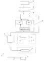
- FIG. 1illustrates a schematic diagram for one embodiment of a electroporation system of the present invention.
- FIG. 2( a )illustrates an embodiment of the present invention with two mono-polar electrodes that can be utilized for electroporation with the FIG. 1 system.
- FIG. 2( b )illustrates an embodiment of the present invention with three mono-polar electrodes that can be utilized for electroporation with the FIG. 1 system.
- FIG. 2( c )illustrates an embodiment of the present invention with a single bi-polar electrode that can be utilized for electroporation with the FIG. 1 system.
- FIG. 2( d )illustrates an embodiment of the present invention with an array of electrodes coupled to a template that can be utilized for electroporation with the FIG. 1 system.
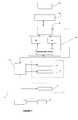
- FIG. 3illustrates one embodiment of the present invention with an array of electrodes positioned around a BPH tissue site, creating a boundary around the BPH tissue site to produce a volumetric cell necrosis region.
- reversible electroporationencompasses permeabilization of a cell membrane through the application of electrical pulses across the cell.
- reversible electroporationthe permeabilization of the cell membrane ceases after the application of the pulse and the cell membrane permeability reverts to normal or at least to a level such that the cell is viable. Thus, the cell survives “reversible electroporation.” It may be used as a means for introducing chemicals, DNA, or other materials into cells.
- the term “irreversible electroporation”also encompasses the permeabilization of a cell membrane through the application of electrical pulses across the cell. However, in “irreversible electroporation” the permeabilization of the cell membrane does not cease after the application of the pulse and the cell membrane permeability does not revert to normal and as such cell is not viable. Thus, the cell does not survive “irreversible electroporation” and the cell death is caused by the disruption of the cell membrane and not merely by internal perturbation of cellular components. Openings in the cell membrane are created and/or expanded in size resulting in a fatal disruption in the normal controlled flow of material across the cell membrane. The cell membrane is highly specialized in its ability to regulate what leaves and enters the cell. Irreversible electroporation destroys that ability to regulate in a manner such that the cell can not compensate and as such the cell dies.
- Ultrasoundis a method used to image tissue in which pressure waves are sent into the tissue using a piezoelectric crystal. The resulting returning waves caused by tissue reflection are transformed into an image.
- MRIis an imaging modality that uses the perturbation of hydrogen molecules caused by a radio pulse to create an image.
- CTis an imaging modality that uses the attenuation of an x-ray beam to create an image.
- Light imagingis an imaging method in which electromagnetic waves with frequencies in the range of visible to far infrared are send into tissue and the tissue's reflection and/or absorption characteristics are reconstructed.
- Electrode impedance tomographyis an imaging technique in which a tissue's electrical impedance characteristics are reconstructed by applying a current across the tissue and measuring electrical currents and potentials
- specific imaging technologies used in the field of medicineare used to create images of tissue affected by electroporation pulses.
- the imagesare created during the process of carrying out irreversible electroporation and are used to focus the electroporation on tissue to be ablated and to avoid ablating tissue such as nerves.
- the process of the inventionmay be carried out by placing electrodes, such as a needle electrode in the imaging path of an imaging device. When the electrodes are activated the image device creates an image of tissue being subjected to electroporation. The effectiveness and extent of the electroporation over a given area of tissue can be determined in real time using the imaging technology.
- Reversible electroporationrequires electrical parameters in a precise range of values that induce only reversible electroporation.
- the limitis more focused on the lower value of the pulse which should be high enough to induce irreversible electroporation.
- methodsare provided to apply an electrical pulse or pulses to BPH tissue sites.
- the pulsesare applied between electrodes and are applied in numbers with currents so as to result in irreversible electroporation of the cells without damaging surrounding cells.
- Energy wavesare emitted from an imaging device such that the energy waves of the imaging device pass through the area positioned between the electrodes and the irreversible electroporation of the cells effects the energy waves of the imaging device in a manner so as to create an image.
- Typical values for pulse length for irreversible electroporationare in a range of from about 5 microseconds to about 62,000 milliseconds or about 75 microseconds to about 20,000 milliseconds or about 100 microseconds ⁇ 10 microseconds. This is significantly longer than the pulse length generally used in intracellular (nano-seconds) electro-manipulation which is 1 microsecond or less—see published U.S. application 2002/0010491 published Jan. 24, 2002. Pulse lengths can be adjusted based on the real time imaging.
- the pulseis at voltage of about 100 V/cm to 7,000 V/cm or 200 V/cm to 2000 V/cm or 300V/cm to 1000 V/cm about 600 V/cm ⁇ 10% for irreversible electroporation. This is substantially lower than that used for intracellular electro-manipulation which is about 10,000 V/cm, see U.S. application 2002/0010491 published Jan. 24, 2002.
- the voltagecan be adjusted alone or with the pulse length based on real time imaging information.
- the voltage expressed aboveis the voltage gradient (voltage per centimeter).
- the electrodesmay be different shapes and sizes and be positioned at different distances from each other.
- the shapemay be circular, oval, square, rectangular or irregular etc.
- the distance of one electrode to anothermay be 0.5 to 10 cm., 1 to 5 cm., or 2-3 cm.
- the electrodemay have a surface area of 0.1-5 sq. cm. or 1-2 sq. cm.
- the size, shape and distances of the electrodescan vary and such can change the voltage and pulse duration used and can be adjusted based on imaging information. Those skilled in the art will adjust the parameters in accordance with this disclosure and imaging to obtain the desired degree of electroporation and avoid thermal damage to surrounding cells.
- Thermal effectsrequire electrical pulses that are substantially longer from those used in irreversible electroporation (Davalos, R. V., B. Rubinsky, and L. M. Mir, Theoretical analysis of the thermal effects during in vivo tissue electroporation . Bioelectrochemistry, 2003. Vol 61(1-2): p. 99-107).
- irreversible electroporation pulseswill be as large as to cause thermal damaging effects to the surrounding tissue and the extent of the BPH tissue site ablated by irreversible electroporation will not be significant relative to that ablated by thermal effects.
- irreversible electroporationcould not be considered as an effective BPH tissue site ablation modality as it will act in superposition with thermal ablation. To a degree, this problem is addressed via the present invention using imaging technology.
- the imaging deviceis any medical imaging device including ultrasound, X-ray technologies, magnetic resonance imaging (MRI), light imaging, electrical impedance tomography, electrical induction impedance tomography and microwave tomography. It is possible to use combinations of different imaging technologies at different points in the process.
- medical imaging deviceincluding ultrasound, X-ray technologies, magnetic resonance imaging (MRI), light imaging, electrical impedance tomography, electrical induction impedance tomography and microwave tomography. It is possible to use combinations of different imaging technologies at different points in the process.
- one type of imaging technologycan be used to precisely locate a BPH tissue site
- a second type of imaging technologycan be used to confirm the placement of electrodes relative to the BPH tissue site.
- yet another type of imaging technologycould be used to create images of the currents of irreversible electroporation in real time.
- MRI technologycould be used to precisely locate the BPH tissue site.
- Electrodescould be placed and identified as being well positioned using X-ray imaging technologies. Current could be applied to carry out irreversible electroporation while using ultrasound technology to determine the extent of BPH tissue site effected by the electroporation pulses. It has been found that within the resolution of calculations and imaging the extent of the image created on ultrasound corresponds to an area calculated to be irreversibly electroporated. Within the resolution of histology the image created by the ultrasound image corresponds to the extent of BPH tissue site ablated as examined histologically.
- the effectiveness of the irreversible electroporationcan be immediately verified with the imaging it is possible to limit the amount of unwanted damage to surrounding tissues and limit the amount of electroporation that is carried out. Further, by using the imaging technology it is possible to reposition the electrodes during the process. The electrode repositioning may be carried out once, twice or a plurality of times as needed in order to obtain the desired degree of irreversible electroporation on the desired BPH tissue site.
- a methodmay be carried out which comprises several steps.
- a first stepan area of BPH tissue site to be treated by irreversible electroporation is imaged. Electrodes are then placed in the BPH tissue site with the BPH tissue site to be ablated being positioned between the electrodes. Imaging can also be carried out at this point to confirm that the electrodes are properly placed.
- pulses of currentare run between the two electrodes and the pulsing current is designed so as to minimize damage to surrounding tissue and achieve the desired irreversible electroporation of the BPH tissue site. While the irreversible electroporation is being carried out imaging technology is used and that imaging technology images the irreversible electroporation occurring in real time.
- the amount of current and number of pulsesmay be adjusted so as to achieve the desired degree of electroporation. Further, one or more of the electrodes may be repositioned so as to make it possible to target the irreversible electroporation and ablate the desired BPH tissue site.
- one embodiment of the present inventionprovides a system, generally denoted as 10 , for treating a BPH tissue site of a patient.
- Two or more monopolar electrodes 12 , one or more bipolar electrodes 14 or an array 16 of electrodescan be utilized, as illustrated in FIGS. 2( a )- 2 ( d ).
- at least first and second monopolar electrodes 12are configured to be introduced at or near the BPH tissue site of the patient. It will be appreciated that three or more monopolar electrodes 12 can be utilized.
- the array 16 of electrodesis configured to be in a substantially surrounding relationship to the BPH tissue site.
- the array 16 of electrodescan employ a template 17 to position and/or retain each of the electrodes. Template 17 can maintain a geometry of the array 16 of electrodes. Electrode placement and depth can be determined by the physician.
- the monopolar and bi-polar electrodes 12 and 14 , and the array 16 of electrodescan be introduced through, the rectal wall, the peritoneum, urethra and the like.
- the array 16 of electrodescreates a boundary around the BPH tissue site to produce a volumetric cell necrosis region. Essentially, the array 16 of electrodes makes a treatment area the extends from the array 16 of electrodes, and extends in an inward direction.
- the array 16 of electrodescan have a pre-determined geometry, and each of the associated electrodes can be deployed individually or simultaneously at the BPH tissue site either percutaneously, or planted in-situ in the patient.
- the monopolar electrodes 12are separated by a distance of about 5 mm to 10 cm and they have a circular cross-sectional geometry.
- One or more additional probes 18can be provided, including monitoring probes, an aspiration probe such as one used for liposuction, fluid introduction probes, and the like.
- Each bipolar electrode 14can have multiple electrode bands 20 .
- the spacing and the thickness of the electrode bands 20is selected to optimize the shape of the electric field. In one embodiment, the spacing is about 1 mm to 5 cm typically, and the thickness of the electrode bands 20 can be from 0.5 mm to 5 cm.
- a voltage pulse generator 22is coupled to the electrodes 12 , 14 and the array 16 .
- the voltage pulse generator 22is configured to apply sufficient electrical pulses between the first and second monopolar electrodes 12 , bi-polar electrode 14 and array 16 to induce electroporation of cells in the BPH tissue site, and create necrosis of cells of the BPH tissue site.
- the applied electrical pulsesare insufficient to create a thermal damaging effect to a majority of the BPH tissue site.
- the electrodes 12 , 14 and array 16are each connected through cables to the voltage pulse generator 22 .
- a switching device 24can be included.
- the switching device 24with software, provides for simultaneous or individual activation of multiple electrodes 12 , 14 and array 16 .
- the switching device 24is coupled to the voltage pulse generator 22 .
- meansare provided for individually activating the electrodes 12 , 14 and array 16 in order to produce electric fields that are produced between pre-selected electrodes 12 , 14 and array 16 in a selected pattern relative to the BPH tissue site.
- the switching of electrical signals between the individual electrodes 12 , 14 and array 16can be accomplished by a variety of different means including but not limited to, manually, mechanically, electrically, with a circuit controlled by a programmed digital computer, and the like.
- each individual electrode 12 , 14 and array 16is individually controlled.
- the pulsesare applied for a duration and magnitude in order to permanently disrupt the cell membranes of cells at the BPH tissue site.
- a ratio of electric current through cells at the BPH tissue site to voltage across the cellscan be detected, and a magnitude of applied voltage to the BPH tissue site is then adjusted in accordance with changes in the ratio of current to voltage.
- an onset of electroporation of cells at the BPH tissue siteis detected by measuring the current.
- monitoring the effects of electroporation on cell membranes of cells at the BPH tissue siteare monitored. The monitoring can be preformed by image monitoring using ultrasound, CT scan, MRI, CT scan, and the like.
- the monitoringis achieved using a monitoring electrode 18 .
- the monitoring electrode 18is a high impedance needle that can be utilized to prevent preferential current flow to a monitoring needle.
- the high impedance needleis positioned adjacent to or in the BPH tissue site, at a critical location. This is similar in concept and positioning as that of placing a thermocouple as in a thermal monitoring.
- a “test pulse”Prior to the full electroporation pulse being delivered a “test pulse” is delivered that is some fraction of the proposed full electroporation pulse, which can be, by way of illustration and without limitation, 10%, and the like. This test pulse is preferably in a range that does not cause irreversible electroporation.
- the monitoring electrode 18measures the test voltage at the location. The voltage measured is then extrapolated back to what would be seen by the monitoring electrode 18 during the full pulse, e.g., multiplied by 10 in one embodiment, because the relationship is linear). If monitoring for a potential complication at the BPH tissue site, a voltage extrapolation that falls under the known level of irreversible electroporation indicates that the BPH tissue site where monitoring is taking place is safe. If monitoring at that BPH tissue site for adequacy of electroporation, the extrapolation falls above the known level of voltage adequate for irreversible tissue electroporation.
- the monitoring electrode 18is integral to the bipolar electrode 14 placed either distal or proximal to the active bipolar electrodes 14 .
- the monitoring electrode 18is a fixed distance form the bipolar electrode 14 .
- the monitoring electrode 18is mounted on a sheath through which the bipolar electrode 14 is placed. The distance from the bipolar electrode 14 can then be varied and positioned based on imaging and the structure to be monitored, such as the rectal mucosa.
- the monitoring electrode 18is mounted on a biopsy guide through which the bipolar electrode 14 is placed. The monitoring electrode 18 is placed at the tip of the guide and rests against the rectal mucosa as the bipolar electrode 14 is placed.
- the effects of electroporation on cell membranes of cells at the BPH tissue sitecan be detected by measuring the current flow.
- the electroporationis performed in a controlled manner, with real time monitoring, to provide for controlled pore formation in cell membranes of cells at the BPH tissue site, to create a tissue effect in the cells at the BPH tissue site while preserving surrounding tissue, with monitoring of electrical impedance, and the like.
- the electroporationcan be performed in a controlled manner by controlling the intensity and duration of the applied voltage and with or without real time control. Additionally, the electroporation is performed in a manner to provide for modification and control of mass transfer across cell membranes. Performance of the electroporation in the controlled manner can be achieved by selection of a proper selection of voltage magnitude, proper selection of voltage application time, and the like.
- the system 10can include a control board 26 that functions to control temperature of the BPH tissue site.
- the control board 26receives its program from a controller.
- Programmingcan be in computer languages such as C or BASIC (registered trade mark) if a personnel computer is used for a controller 28 or assembly language if a microprocessor is used for the controller 28 .
- a user specified control of temperaturecan be programmed in the controller 28 .
- the controller 28can include a computer, a digital or analog processing apparatus, programmable logic array, a hardwired logic circuit, an application specific integrated circuit (“ASIC”), or other suitable device.
- the controller 28includes a microprocessor accompanied by appropriate RAM and ROM modules, as desired.
- the controller 28can be coupled to a user interface 30 for exchanging data with a user. The user can operate the user interface 30 to input a desired pulsing pattern and corresponding temperature profile to be applied to the electrodes 12 , 14 and array 16 .
- the user interface 30can include an alphanumeric keypad, touch screen, computer mouse, push-buttons and/or toggle switches, or another suitable component to receive input from a human user.
- the user interface 30can also include a CRT screen, LED screen, LCD screen, liquid crystal display, printer, display panel, audio speaker, or another suitable component to convey data to a human user.
- the control board 26can function to receive controller input and can be driven by the voltage pulse generator 22 .
- the voltage pulse generator 22is configured to provide that each pulse is applied for a duration of about, 5 microseconds to about 62 seconds, 90 to 110 microseconds, 100 microseconds, and the like.
- a variety of different number of pulsescan be applied, including but not limited to, from about 1 to 15 pulses, about eight pulses of about 100 microseconds each in duration, and the like.
- the pulsesare applied to produce a voltage gradient at the BPH tissue site in a range of from about 50 volt/cm to about 8000 volt/cm.
- the BPH tissue siteis monitored and the pulses are adjusted to maintain a temperature of, 100 degrees C. or less at the BPH tissue site, 75 degrees C. or less at the BPH tissue site, 60 degrees C. or less at the BPH tissue site, 50 degrees C. or less at the BPH tissue site, and the like.
- the temperatureis controlled in order to minimize the occurrence of a thermal effect to the BPH tissue site. These temperatures can be controlled by adjusting the current-to-voltage ratio based on temperature.
- the system 10is utilized to treat BPH with electroporation of cells at a BPH tissue site, creating cell necrosis in the BPH tissue site around the urethra.
- the system 10delivers electroporation pulses along the muscular fibers and nerves at the BPH tissue site and produces a volume of necrotic cells at the BPH tissue site around the urethra. Destruction of these nerves, that create an elevation in tension of the muscle fibers, is also achieved. The resulting necrotic tissue is removed by macrophages.
- electroporationresults in the removal of cells at the BPH tissue site, associated nerves, and the total volume of the BPH tissue site is reduced, causing a reduction in pressure on the urethra and a relaxation of the prostate.
- the electroporationis controllably applied to spare urethral sphincters and other tissues in the prostate, as well as in adjacent tissues and organs.
- First and second mono-polar electrodes 12 , or more, the bi-polar electrode 14 or the array 16 of electrodesare introduced through the rectal wall, the peritoneum or the urethra of the patient.
- the electroporationis positioned and monitored by image monitoring with ultrasound, CT scan, MRI, CT scan, and the like, or with a monitoring electrode 18 .
- Each of the electrodes 12 , 14 or array 16can have insulated portions and is connected to the voltage pulse generator 22 .
- An area of the BPH tissue siteis imaged.
- Two bi-polar electrodes 12with sharpened distal ends, are introduced into in the BPH tissue site through the rectal wall of the patient.
- the area of the BPH tissue site to be ablatedis positioned between the two electrodes. Imaging is used to confirm that the mono-polar electrodes are properly placed.
- the two mono-polar electrodesare separated by a distance of 5 mm to 10 cm at various locations of the BPH tissue site.
- Pulsesare applied with a duration of 5 microseconds to about 62 seconds each.
- Monitoringis preformed using ultrasound.
- the BPH tissue siteis monitored. In response to the monitoring, pulses are adjusted to maintain a temperature of no more than 100 degrees C.
- a voltage gradient at the BPH tissue sitein a range of from about 50 volt/cm to about 1000 volt/cm is created.
- a volume of the BPH tissue siteof about 1 cm by 0.5 cm undergoes cell necrosis.
- An area of the BPH tissue siteis imaged.
- Two mono-polar electrodes 12are introduced into in the BPH tissue site through the urethra of the patient.
- the area of the BPH tissue site to be ablatedis positioned between the two mono-polar electrodes 12 .
- Imagingis used to confirm that the electrodes are properly placed.
- the two mono-polar electrodes 12are separated by a distance of 5 mm to 10 cm at various locations of the BPH tissue site.
- Pulsesare applied with a duration of about 90 to 110 microseconds each.
- Monitoringis performed using a CT scan.
- the BPH tissue siteis monitored.
- pulsesare adjusted to maintain a temperature of no more than 75 degrees C.
- a voltage gradient at the BPH tissue sitein a range of from about 50 volt/cm to about 5000 volt/cm is created.
- the BPH tissue siteundergoes cell necrosis.
- An area of the BPH tissue siteis imaged.
- the array 16 of electrodesare introduced into in the BPH tissue site through the peritoneum of the patient.
- the array 16 of electrodesis positioned in a surrounding relationship to the BPH. Imaging is used to confirm that the electrodes are properly placed.
- Pulsesare applied with a duration of about 100 microseconds each.
- a monitoring electrode 18is utilized. Prior to the full electroporation pulse being delivered a test pulse is delivered that is about 10% of the proposed full electroporation pulse. The test pulse does not cause irreversible electroporation.
- the BPH tissue siteis monitored. In response to the monitoring, pulses are adjusted to maintain a temperature of no more than 60 degrees C.
- a voltage gradient at the BPH tissue sitein a range of from about 50 volt/cm to about 8000 volt/cm is created. The BPH tissue site undergoes cell necrosis.
- An area of the BPH tissue siteis imaged.
- a single bi-polar electrode 14with a sharpened distal end, is introduced into the BPH tissue site through the rectal wall of the patient.
- a monitoring electrode 18is placed at a tip of a biopsy guide and rests against the rectal mucosa when the bipolar electrode 14 is placed. Imaging is used to confirm that the bi-polar electrode 14 is properly placed.
- Pulsesare applied with a duration of 5 microseconds to about 62 seconds each. Monitoring is preformed using ultrasound.
- the BPH tissue siteis monitored. In response to the monitoring, pulses are adjusted to maintain a temperature of no more than 100 degrees C.
- a voltage gradient at the BPH tissue sitein a range of from about 50 volt/cm to about 1000 volt/cm is created.
- the BPH tissue siteundergoes cell necrosis.
- An area of the BPH tissue siteis imaged.
- a array 16 of electrodesis introduced into the BPH tissue site through the rectal wall of the patient, and are positioned around the BPH tissue site. Imaging is used to confirm that the array 16 of electrodes is properly placed.
- Pulsesare applied with a duration of about 90 to 110 microseconds each. Monitoring is performed using a CT scan. The BPH tissue site is monitored. In response to the monitoring, pulses are adjusted to maintain a temperature of no more than 75 degrees C.
- a voltage gradient at the BPH tissue site in a range of from about 50 volt/cm to about 5000 volt/cmis created. The BPH tissue site undergoes cell necrosis.
- An area of the BPH tissue siteis imaged.
- the array 16 of electrodesis introduced into the BPH tissue site through the peritoneum of the patient, and positioned in a surrounding relationship to the BPH tissue site. Imaging is used to confirm that the array 16 of electrodes is properly placed.
- Pulsesare applied with a duration of about 100 microseconds each.
- a monitoring electrode 18is utilized. Prior to the full electroporation pulse being delivered a test pulse is delivered that is about 10% of the proposed full electroporation pulse. The test pulse does not cause irreversible electroporation.
- the BPH tissue siteis monitored. In response to the monitoring, pulses are adjusted to maintain a temperature of no more than 60 degrees C.
- a voltage gradient at the BPH tissue sitein a range of from about 50 volt/cm to about 8000 volt/cm is created. The BPH tissue site undergoes cell necrosis.
Landscapes
- Life Sciences & Earth Sciences (AREA)
- Health & Medical Sciences (AREA)
- Engineering & Computer Science (AREA)
- Radiology & Medical Imaging (AREA)
- Biomedical Technology (AREA)
- Nuclear Medicine, Radiotherapy & Molecular Imaging (AREA)
- Biophysics (AREA)
- Animal Behavior & Ethology (AREA)
- General Health & Medical Sciences (AREA)
- Public Health (AREA)
- Veterinary Medicine (AREA)
- Bioinformatics & Cheminformatics (AREA)
- Electrotherapy Devices (AREA)
- Surgical Instruments (AREA)
Abstract
Description
Claims (84)
Priority Applications (6)
| Application Number | Priority Date | Filing Date | Title |
|---|---|---|---|
| US11/166,974US8114070B2 (en) | 2005-06-24 | 2005-06-24 | Methods and systems for treating BPH using electroporation |
| CA002612530ACA2612530A1 (en) | 2005-06-24 | 2006-06-05 | Methods and systems for treating bph using electroporation |
| EP06772212AEP1898993A1 (en) | 2005-06-24 | 2006-06-05 | Methods and systems for treating bph using electroporation |
| PCT/US2006/021812WO2007001751A1 (en) | 2005-06-24 | 2006-06-05 | Methods and systems for treating bph using electroporation |
| JP2008518194AJP2008546474A (en) | 2005-06-24 | 2006-06-05 | Method and system for treating BPH using electroporation |
| US12/510,011US20090292342A1 (en) | 2005-06-24 | 2009-07-27 | Methods and Systems for Treating BPH Using Electroporation |
Applications Claiming Priority (1)
| Application Number | Priority Date | Filing Date | Title |
|---|---|---|---|
| US11/166,974US8114070B2 (en) | 2005-06-24 | 2005-06-24 | Methods and systems for treating BPH using electroporation |
Related Child Applications (1)
| Application Number | Title | Priority Date | Filing Date |
|---|---|---|---|
| US12/510,011DivisionUS20090292342A1 (en) | 2005-06-24 | 2009-07-27 | Methods and Systems for Treating BPH Using Electroporation |
Publications (2)
| Publication Number | Publication Date |
|---|---|
| US20060293713A1 US20060293713A1 (en) | 2006-12-28 |
| US8114070B2true US8114070B2 (en) | 2012-02-14 |
Family
ID=37568577
Family Applications (2)
| Application Number | Title | Priority Date | Filing Date |
|---|---|---|---|
| US11/166,974Active2029-12-29US8114070B2 (en) | 2005-06-24 | 2005-06-24 | Methods and systems for treating BPH using electroporation |
| US12/510,011AbandonedUS20090292342A1 (en) | 2005-06-24 | 2009-07-27 | Methods and Systems for Treating BPH Using Electroporation |
Family Applications After (1)
| Application Number | Title | Priority Date | Filing Date |
|---|---|---|---|
| US12/510,011AbandonedUS20090292342A1 (en) | 2005-06-24 | 2009-07-27 | Methods and Systems for Treating BPH Using Electroporation |
Country Status (5)
| Country | Link |
|---|---|
| US (2) | US8114070B2 (en) |
| EP (1) | EP1898993A1 (en) |
| JP (1) | JP2008546474A (en) |
| CA (1) | CA2612530A1 (en) |
| WO (1) | WO2007001751A1 (en) |
Cited By (63)
| Publication number | Priority date | Publication date | Assignee | Title |
|---|---|---|---|---|
| US20090269317A1 (en)* | 2008-04-29 | 2009-10-29 | Davalos Rafael V | Irreversible electroporation to create tissue scaffolds |
| US8926606B2 (en) | 2009-04-09 | 2015-01-06 | Virginia Tech Intellectual Properties, Inc. | Integration of very short electric pulses for minimally to noninvasive electroporation |
| US8992517B2 (en) | 2008-04-29 | 2015-03-31 | Virginia Tech Intellectual Properties Inc. | Irreversible electroporation to treat aberrant cell masses |
| US9005189B2 (en)* | 2003-12-24 | 2015-04-14 | The Regents Of The University Of California | Tissue ablation with irreversible electroporation |
| US9198733B2 (en) | 2008-04-29 | 2015-12-01 | Virginia Tech Intellectual Properties, Inc. | Treatment planning for electroporation-based therapies |
| US9283051B2 (en) | 2008-04-29 | 2016-03-15 | Virginia Tech Intellectual Properties, Inc. | System and method for estimating a treatment volume for administering electrical-energy based therapies |
| US9757196B2 (en) | 2011-09-28 | 2017-09-12 | Angiodynamics, Inc. | Multiple treatment zone ablation probe |
| US9867652B2 (en) | 2008-04-29 | 2018-01-16 | Virginia Tech Intellectual Properties, Inc. | Irreversible electroporation using tissue vasculature to treat aberrant cell masses or create tissue scaffolds |
| US9895189B2 (en) | 2009-06-19 | 2018-02-20 | Angiodynamics, Inc. | Methods of sterilization and treating infection using irreversible electroporation |
| US10052148B2 (en) | 2005-03-07 | 2018-08-21 | Boston Scientific Scimed, Inc. | Method for ablating tissue with multiple ablation probes |
| US10105132B2 (en) | 2005-05-20 | 2018-10-23 | Neotract, Inc. | Devices, systems and methods for treating benign prostatic hyperplasia and other conditions |
| US10117707B2 (en) | 2008-04-29 | 2018-11-06 | Virginia Tech Intellectual Properties, Inc. | System and method for estimating tissue heating of a target ablation zone for electrical-energy based therapies |
| US10130353B2 (en) | 2012-06-29 | 2018-11-20 | Neotract, Inc. | Flexible system for delivering an anchor |
| US10143461B2 (en) | 2005-05-20 | 2018-12-04 | Neotract, Inc. | Devices, systems and methods for retracting, lifting, compressing, supporting or repositioning tissues or anatomical structures |
| US10154869B2 (en) | 2013-08-02 | 2018-12-18 | Gary M. Onik | System and method for creating radio-frequency energy electrical membrane breakdown for tissue ablation |
| US10154874B2 (en) | 2008-04-29 | 2018-12-18 | Virginia Tech Intellectual Properties, Inc. | Immunotherapeutic methods using irreversible electroporation |
| US10195014B2 (en) | 2005-05-20 | 2019-02-05 | Neotract, Inc. | Devices, systems and methods for treating benign prostatic hyperplasia and other conditions |
| US10238447B2 (en) | 2008-04-29 | 2019-03-26 | Virginia Tech Intellectual Properties, Inc. | System and method for ablating a tissue site by electroporation with real-time monitoring of treatment progress |
| US10265061B2 (en) | 2005-05-20 | 2019-04-23 | Neotract, Inc. | Latching anchor device |
| US10271893B2 (en) | 2014-12-15 | 2019-04-30 | Medtronic Ablation Frontiers Llc | Timed energy delivery |
| US10272178B2 (en) | 2008-04-29 | 2019-04-30 | Virginia Tech Intellectual Properties Inc. | Methods for blood-brain barrier disruption using electrical energy |
| US10292801B2 (en) | 2012-03-29 | 2019-05-21 | Neotract, Inc. | System for delivering anchors for treating incontinence |
| US10292755B2 (en) | 2009-04-09 | 2019-05-21 | Virginia Tech Intellectual Properties, Inc. | High frequency electroporation for cancer therapy |
| US10299780B2 (en) | 2005-05-20 | 2019-05-28 | Neotract, Inc. | Apparatus and method for manipulating or retracting tissue and anatomical structure |
| US10426509B2 (en) | 2005-05-20 | 2019-10-01 | Neotract, Inc. | Median lobe destruction apparatus and method |
| US10471254B2 (en) | 2014-05-12 | 2019-11-12 | Virginia Tech Intellectual Properties, Inc. | Selective modulation of intracellular effects of cells using pulsed electric fields |
| US10492792B2 (en) | 2005-05-20 | 2019-12-03 | Neotract, Inc. | Devices, systems and methods for treating benign prostatic hyperplasia and other conditions |
| US10694972B2 (en) | 2014-12-15 | 2020-06-30 | Virginia Tech Intellectual Properties, Inc. | Devices, systems, and methods for real-time monitoring of electrophysical effects during tissue treatment |
| US10702337B2 (en) | 2016-06-27 | 2020-07-07 | Galary, Inc. | Methods, apparatuses, and systems for the treatment of pulmonary disorders |
| US10702326B2 (en) | 2011-07-15 | 2020-07-07 | Virginia Tech Intellectual Properties, Inc. | Device and method for electroporation based treatment of stenosis of a tubular body part |
| US10849678B2 (en) | 2013-12-05 | 2020-12-01 | Immunsys, Inc. | Cancer immunotherapy by radiofrequency electrical membrane breakdown (RF-EMB) |
| US10925587B2 (en) | 2005-05-20 | 2021-02-23 | Neotract, Inc. | Anchor delivery system |
| US11045648B2 (en) | 2017-11-14 | 2021-06-29 | Boston Scientific Scimed, Inc. | Irreversible electroporation through a combination of substance injection and electrical field application |
| US11141216B2 (en) | 2015-01-30 | 2021-10-12 | Immunsys, Inc. | Radio-frequency electrical membrane breakdown for the treatment of high risk and recurrent prostate cancer, unresectable pancreatic cancer, tumors of the breast, melanoma or other skin malignancies, sarcoma, soft tissue tumors, ductal carcinoma, neoplasia, and intra and extra luminal abnormal tissue |
| US11254926B2 (en) | 2008-04-29 | 2022-02-22 | Virginia Tech Intellectual Properties, Inc. | Devices and methods for high frequency electroporation |
| US11272979B2 (en) | 2008-04-29 | 2022-03-15 | Virginia Tech Intellectual Properties, Inc. | System and method for estimating tissue heating of a target ablation zone for electrical-energy based therapies |
| US11311329B2 (en) | 2018-03-13 | 2022-04-26 | Virginia Tech Intellectual Properties, Inc. | Treatment planning for immunotherapy based treatments using non-thermal ablation techniques |
| US11364070B2 (en) | 2018-01-23 | 2022-06-21 | Boston Scientific Scimed, Inc. | Enhanced needle array and therapies for tumor ablation |
| US11382681B2 (en) | 2009-04-09 | 2022-07-12 | Virginia Tech Intellectual Properties, Inc. | Device and methods for delivery of high frequency electrical pulses for non-thermal ablation |
| US11497544B2 (en) | 2016-01-15 | 2022-11-15 | Immunsys, Inc. | Immunologic treatment of cancer |
| US11607537B2 (en) | 2017-12-05 | 2023-03-21 | Virginia Tech Intellectual Properties, Inc. | Method for treating neurological disorders, including tumors, with electroporation |
| US11638603B2 (en) | 2009-04-09 | 2023-05-02 | Virginia Tech Intellectual Properties, Inc. | Selective modulation of intracellular effects of cells using pulsed electric fields |
| US11672520B2 (en) | 2017-12-23 | 2023-06-13 | Teleflex Life Sciences Limited | Expandable tissue engagement apparatus and method |
| US11707629B2 (en) | 2009-05-28 | 2023-07-25 | Angiodynamics, Inc. | System and method for synchronizing energy delivery to the cardiac rhythm |
| US11717337B2 (en)* | 2016-11-29 | 2023-08-08 | St. Jude Medical, Cardiology Division, Inc. | Electroporation systems and catheters for electroporation systems |
| US11723710B2 (en) | 2016-11-17 | 2023-08-15 | Angiodynamics, Inc. | Techniques for irreversible electroporation using a single-pole tine-style internal device communicating with an external surface electrode |
| US11786300B2 (en) | 2021-04-07 | 2023-10-17 | Btl Medical Technologies S.R.O. | Pulsed field ablation device and method |
| US11896298B2 (en) | 2021-07-06 | 2024-02-13 | Btl Medical Development A.S. | Pulsed field ablation device and method |
| US11925405B2 (en) | 2018-03-13 | 2024-03-12 | Virginia Tech Intellectual Properties, Inc. | Treatment planning system for immunotherapy enhancement via non-thermal ablation |
| US11931096B2 (en) | 2010-10-13 | 2024-03-19 | Angiodynamics, Inc. | System and method for electrically ablating tissue of a patient |
| US11931094B2 (en) | 2019-10-15 | 2024-03-19 | Boston Scientific Scimed, Inc. | Control system and user interface for an ablation system |
| US11938317B2 (en) | 2017-12-26 | 2024-03-26 | Galvanize Therapeutics, Inc. | Optimization of energy delivery for various applications |
| US11950835B2 (en) | 2019-06-28 | 2024-04-09 | Virginia Tech Intellectual Properties, Inc. | Cycled pulsing to mitigate thermal damage for multi-electrode irreversible electroporation therapy |
| US11992260B2 (en) | 2020-03-31 | 2024-05-28 | Boston Scientific Scimed, Inc. | Smart probe identification for ablation modalities |
| US12076067B2 (en) | 2022-10-05 | 2024-09-03 | Btl Medical Development A.S. | Pulsed field ablation device and method |
| US12076070B2 (en) | 2019-03-15 | 2024-09-03 | Boston Scientific Scimed, Inc. | Time multiplexed waveform for selective cell ablation |
| US12102376B2 (en) | 2012-02-08 | 2024-10-01 | Angiodynamics, Inc. | System and method for increasing a target zone for electrical ablation |
| US12114911B2 (en) | 2014-08-28 | 2024-10-15 | Angiodynamics, Inc. | System and method for ablating a tissue site by electroporation with real-time pulse monitoring |
| US12201349B2 (en) | 2009-04-03 | 2025-01-21 | Angiodynamics, Inc. | Congestive obstruction pulmonary disease (COPD) |
| US12214189B2 (en) | 2019-07-24 | 2025-02-04 | Virginia Tech Intellectual Properties, Inc. | Fourier analysis spectroscopy for monitoring tissue impedance changes and treatment outcome during electroporation-based-therapies |
| US12390262B2 (en) | 2018-03-13 | 2025-08-19 | Virginia Tech Intellectual Properties, Inc. | Treatment planning system for immunotherapy enhancement via non-thermal ablation |
| US12403305B2 (en) | 2016-06-27 | 2025-09-02 | Galvanize Therapeutics, Inc. | Immunostimulation in the treatment of viral infection |
| US12440301B2 (en) | 2019-10-30 | 2025-10-14 | Teleflex Life Sciences Llc | System for delivery of a fiducial marker |
Families Citing this family (15)
| Publication number | Priority date | Publication date | Assignee | Title |
|---|---|---|---|---|
| US6994706B2 (en)* | 2001-08-13 | 2006-02-07 | Minnesota Medical Physics, Llc | Apparatus and method for treatment of benign prostatic hyperplasia |
| JP5032500B2 (en)* | 2006-01-03 | 2012-09-26 | アルコン,インコーポレイティド | System for dissociation and removal of proteinaceous tissue |
| US20080132885A1 (en)* | 2006-12-01 | 2008-06-05 | Boris Rubinsky | Methods for treating tissue sites using electroporation |
| US20090076502A1 (en)* | 2007-09-14 | 2009-03-19 | Lazure Technologies, Llc. | Prostate cancer ablation |
| WO2010117806A1 (en)* | 2009-03-31 | 2010-10-14 | Angiodynamics, Inc. | System and method for estimating a treatment region for a medical treatment device and for interactively planning a treatment of a patient |
| US20110118729A1 (en)* | 2009-11-13 | 2011-05-19 | Alcon Research, Ltd | High-intensity pulsed electric field vitrectomy apparatus with load detection |
| US20110135626A1 (en)* | 2009-12-08 | 2011-06-09 | Alcon Research, Ltd. | Localized Chemical Lysis of Ocular Tissue |
| US20110144562A1 (en)* | 2009-12-14 | 2011-06-16 | Alcon Research, Ltd. | Localized Pharmacological Treatment of Ocular Tissue Using High-Intensity Pulsed Electrical Fields |
| WO2011081897A1 (en)* | 2009-12-15 | 2011-07-07 | Alcon Research, Ltd. | High-intensity pulsed electric field vitrectomy apparatus |
| US20110202052A1 (en)* | 2010-02-12 | 2011-08-18 | Daniel Gelbart | System for treating benign prostatic hyperplasia |
| US8546979B2 (en) | 2010-08-11 | 2013-10-01 | Alcon Research, Ltd. | Self-matching pulse generator with adjustable pulse width and pulse frequency |
| US20120089009A1 (en)* | 2010-10-11 | 2012-04-12 | Omary Reed A | Methods and apparatus to deliver nanoparticles to tissue usingelectronanotherapy |
| EP3495018B1 (en)* | 2014-05-07 | 2023-09-06 | Farapulse, Inc. | Apparatus for selective tissue ablation |
| CA2975926A1 (en)* | 2015-02-04 | 2016-08-11 | Rfemb Holdings, Llc | Radio-frequency electrical membrane breakdown for the treatment of benign prostatic hyperplasia |
| US12285206B2 (en)* | 2020-06-01 | 2025-04-29 | Biosense Webster (Israel) Ltd. | Application of irreversible electroporation (IRE) ablation using catheter with electrode array |
Citations (128)
| Publication number | Priority date | Publication date | Assignee | Title |
|---|---|---|---|---|
| US1653819A (en) | 1926-08-07 | 1927-12-27 | Northcott Ephraim | Electrotherapeutical apparatus |
| DE863111C (en) | 1951-07-03 | 1953-01-15 | Walter Hallegger | Instrument for transcutaneous and subcutaneous heating and iontophoresis and method of its use |
| US4016886A (en) | 1974-11-26 | 1977-04-12 | The United States Of America As Represented By The United States Energy Research And Development Administration | Method for localizing heating in tumor tissue |
| US4226246A (en) | 1977-05-27 | 1980-10-07 | Carba Societe Anonyme | Apparatus for maintaining the negative potential of human, animal, and plant cells |
| US4262672A (en) | 1978-01-02 | 1981-04-21 | Horst Kief | Acupuncture instrument |
| US4407943A (en) | 1976-12-16 | 1983-10-04 | Millipore Corporation | Immobilized antibody or antigen for immunoassay |
| US4810963A (en) | 1984-04-03 | 1989-03-07 | Public Health Laboratory Service Board | Method for investigating the condition of a bacterial suspension through frequency profile of electrical admittance |
| US4907601A (en) | 1988-06-15 | 1990-03-13 | Etama Ag | Electrotherapy arrangement |
| US4946793A (en) | 1986-05-09 | 1990-08-07 | Electropore, Inc. | Impedance matching for instrumentation which electrically alters vesicle membranes |
| US5019034A (en) | 1988-01-21 | 1991-05-28 | Massachusetts Institute Of Technology | Control of transport of molecules across tissue using electroporation |
| DE4000893A1 (en) | 1990-01-15 | 1991-07-18 | Bosch Gmbh Robert | Multichannel appts. for electro-simulation - provides several current circuits for patient with electrodes applying pulse signals |
| US5052391A (en) | 1990-10-22 | 1991-10-01 | R.F.P., Inc. | High frequency high intensity transcutaneous electrical nerve stimulator and method of treatment |
| US5058605A (en) | 1989-02-22 | 1991-10-22 | Ceske Vysoke Uceni Technicke | Method and device for the controlled local, non-invasive application of dc pulses to human and animal tissues |
| US5098843A (en) | 1987-06-04 | 1992-03-24 | Calvin Noel M | Apparatus for the high efficiency transformation of living cells |
| US5134070A (en) | 1990-06-04 | 1992-07-28 | Casnig Dael R | Method and device for cell cultivation on electrodes |
| US5173158A (en) | 1991-07-22 | 1992-12-22 | Schmukler Robert E | Apparatus and methods for electroporation and electrofusion |
| US5193537A (en) | 1990-06-12 | 1993-03-16 | Zmd Corporation | Method and apparatus for transcutaneous electrical cardiac pacing |
| US5273525A (en) | 1992-08-13 | 1993-12-28 | Btx Inc. | Injection and electroporation apparatus for drug and gene delivery |
| US5318563A (en) | 1992-06-04 | 1994-06-07 | Valley Forge Scientific Corporation | Bipolar RF generator |
| US5328451A (en) | 1991-08-15 | 1994-07-12 | Board Of Regents, The University Of Texas System | Iontophoretic device and method for killing bacteria and other microbes |
| US5389069A (en) | 1988-01-21 | 1995-02-14 | Massachusetts Institute Of Technology | Method and apparatus for in vivo electroporation of remote cells and tissue |
| US5403311A (en) | 1993-03-29 | 1995-04-04 | Boston Scientific Corporation | Electro-coagulation and ablation and other electrotherapeutic treatments of body tissue |
| US5425752A (en) | 1991-11-25 | 1995-06-20 | Vu'nguyen; Dung D. | Method of direct electrical myostimulation using acupuncture needles |
| US5439440A (en) | 1993-04-01 | 1995-08-08 | Genetronics, Inc. | Electroporation system with voltage control feedback for clinical applications |
| US5458625A (en) | 1994-05-04 | 1995-10-17 | Kendall; Donald E. | Transcutaneous nerve stimulation device and method for using same |
| US5533999A (en) | 1993-08-23 | 1996-07-09 | Refractec, Inc. | Method and apparatus for modifications of visual acuity by thermal means |
| US5536240A (en) | 1992-08-12 | 1996-07-16 | Vidamed, Inc. | Medical probe device and method |
| US5575811A (en) | 1993-07-08 | 1996-11-19 | Urologix, Inc. | Benign prostatic hyperplasia treatment catheter with urethral cooling |
| EP0378132B1 (en) | 1989-01-09 | 1996-11-27 | Cit Ionofor, S.L. | A device for the administration of medication by iontopheresis for local - regional treatment. |
| US5626146A (en) | 1992-12-18 | 1997-05-06 | British Technology Group Limited | Electrical impedance tomography |
| US5634899A (en) | 1993-08-20 | 1997-06-03 | Cortrak Medical, Inc. | Simultaneous cardiac pacing and local drug delivery method |
| US5674267A (en) | 1993-03-30 | 1997-10-07 | Centre National De La Recherche Scientifique | Electric pulse applicator using pairs of needle electrodes for the treatment of biological tissue |
| US5702359A (en) | 1995-06-06 | 1997-12-30 | Genetronics, Inc. | Needle electrodes for mediated delivery of drugs and genes |
| US5720921A (en) | 1995-03-10 | 1998-02-24 | Entremed, Inc. | Flow electroporation chamber and method |
| US5778894A (en) | 1996-04-18 | 1998-07-14 | Elizabeth Arden Co. | Method for reducing human body cellulite by treatment with pulsed electromagnetic energy |
| US5782882A (en) | 1995-11-30 | 1998-07-21 | Hewlett-Packard Company | System and method for administering transcutaneous cardiac pacing with transcutaneous electrical nerve stimulation |
| US5810762A (en) | 1995-04-10 | 1998-09-22 | Genetronics, Inc. | Electroporation system with voltage control feedback for clinical applications |
| US5836905A (en) | 1994-06-20 | 1998-11-17 | Lemelson; Jerome H. | Apparatus and methods for gene therapy |
| US5843026A (en) | 1992-08-12 | 1998-12-01 | Vidamed, Inc. | BPH ablation method and apparatus |
| US5873849A (en) | 1997-04-24 | 1999-02-23 | Ichor Medical Systems, Inc. | Electrodes and electrode arrays for generating electroporation inducing electrical fields |
| US5919142A (en) | 1995-06-22 | 1999-07-06 | Btg International Limited | Electrical impedance tomography method and apparatus |
| US5947889A (en) | 1995-01-17 | 1999-09-07 | Hehrlein; Christoph | Balloon catheter used to prevent re-stenosis after angioplasty and process for producing a balloon catheter |
| US5983131A (en) | 1995-08-11 | 1999-11-09 | Massachusetts Institute Of Technology | Apparatus and method for electroporation of tissue |
| US5991697A (en) | 1996-12-31 | 1999-11-23 | The Regents Of The University Of California | Method and apparatus for optical Doppler tomographic imaging of fluid flow velocity in highly scattering media |
| US5999847A (en) | 1997-10-21 | 1999-12-07 | Elstrom; John A. | Apparatus and method for delivery of surgical and therapeutic agents |
| US6009347A (en) | 1998-01-27 | 1999-12-28 | Genetronics, Inc. | Electroporation apparatus with connective electrode template |
| US6010613A (en) | 1995-12-08 | 2000-01-04 | Cyto Pulse Sciences, Inc. | Method of treating materials with pulsed electrical fields |
| US6016452A (en) | 1996-03-19 | 2000-01-18 | Kasevich; Raymond S. | Dynamic heating method and radio frequency thermal treatment |
| US6041252A (en) | 1995-06-07 | 2000-03-21 | Ichor Medical Systems Inc. | Drug delivery system and method |
| US6055453A (en) | 1997-08-01 | 2000-04-25 | Genetronics, Inc. | Apparatus for addressing needle array electrodes for electroporation therapy |
| US6085115A (en) | 1997-05-22 | 2000-07-04 | Massachusetts Institite Of Technology | Biopotential measurement including electroporation of tissue surface |
| US6090106A (en) | 1996-01-09 | 2000-07-18 | Gyrus Medical Limited | Electrosurgical instrument |
| US6102885A (en) | 1996-08-08 | 2000-08-15 | Bass; Lawrence S. | Device for suction-assisted lipectomy and method of using same |
| US6106521A (en) | 1996-08-16 | 2000-08-22 | United States Surgical Corporation | Apparatus for thermal treatment of tissue |
| US6109270A (en) | 1997-02-04 | 2000-08-29 | The United States Of America As Represented By The Administrator Of The National Aeronautics And Space Administration | Multimodality instrument for tissue characterization |
| US6122599A (en) | 1998-02-13 | 2000-09-19 | Mehta; Shailesh | Apparatus and method for analyzing particles |
| US6132419A (en) | 1992-05-22 | 2000-10-17 | Genetronics, Inc. | Electroporetic gene and drug therapy |
| US6159163A (en) | 1998-05-07 | 2000-12-12 | Cedars-Sinai Medical Center | System for attenuating pain during bone marrow aspiration and method |
| US6208893B1 (en) | 1998-01-27 | 2001-03-27 | Genetronics, Inc. | Electroporation apparatus with connective electrode template |
| US6212433B1 (en) | 1998-07-28 | 2001-04-03 | Radiotherapeutics Corporation | Method for treating tumors near the surface of an organ |
| US6210402B1 (en) | 1995-11-22 | 2001-04-03 | Arthrocare Corporation | Methods for electrosurgical dermatological treatment |
| US6216034B1 (en) | 1997-08-01 | 2001-04-10 | Genetronics, Inc. | Method of programming an array of needle electrodes for electroporation therapy of tissue |
| US6219577B1 (en) | 1998-04-14 | 2001-04-17 | Global Vascular Concepts, Inc. | Iontophoresis, electroporation and combination catheters for local drug delivery to arteries and other body tissues |
| US6241702B1 (en) | 1992-08-12 | 2001-06-05 | Vidamed, Inc. | Radio frequency ablation device for treatment of the prostate |
| US6261831B1 (en) | 1999-03-26 | 2001-07-17 | The United States Of America As Represented By The Secretary Of The Air Force | Ultra-wide band RF-enhanced chemotherapy for cancer treatmeat |
| US6300108B1 (en)* | 1999-07-21 | 2001-10-09 | The Regents Of The University Of California | Controlled electroporation and mass transfer across cell membranes |
| US20010044596A1 (en) | 2000-05-10 | 2001-11-22 | Ali Jaafar | Apparatus and method for treatment of vascular restenosis by electroporation |
| US6326177B1 (en)* | 1999-08-04 | 2001-12-04 | Eastern Virginia Medical School Of The Medical College Of Hampton Roads | Method and apparatus for intracellular electro-manipulation |
| US20020010491A1 (en) | 1999-08-04 | 2002-01-24 | Schoenbach Karl H. | Method and apparatus for intracellular electro-manipulation |
| US6347247B1 (en) | 1998-05-08 | 2002-02-12 | Genetronics Inc. | Electrically induced vessel vasodilation |
| US6349233B1 (en) | 1993-02-22 | 2002-02-19 | Angeion Corporation | Neuro-stimulation to control pain during cardioversion defibrillation |
| US6351674B2 (en) | 1998-11-23 | 2002-02-26 | Synaptic Corporation | Method for inducing electroanesthesia using high frequency, high intensity transcutaneous electrical nerve stimulation |
| US20020055731A1 (en) | 1997-10-24 | 2002-05-09 | Anthony Atala | Methods for promoting cell transfection in vivo |
| US6387671B1 (en) | 1999-07-21 | 2002-05-14 | The Regents Of The University Of California | Electrical impedance tomography to control electroporation |
| US6403348B1 (en) | 1999-07-21 | 2002-06-11 | The Regents Of The University Of California | Controlled electroporation and mass transfer across cell membranes |
| US20020077676A1 (en) | 1999-04-09 | 2002-06-20 | Schroeppel Edward A. | Implantable device and method for the electrical treatment of cancer |
| US20020099323A1 (en) | 1998-07-13 | 2002-07-25 | Nagendu B. Dev | Skin and muscle-targeted gene therapy by pulsed electrical field |
| US20020138117A1 (en) | 2000-06-21 | 2002-09-26 | Son Young Tae | Apparatus and method for selectively removing a body fat mass in human body |
| US6470211B1 (en) | 1997-06-03 | 2002-10-22 | Uab Research Foundation | Method and apparatus for treating cardiac arrhythmia |
| US6493592B1 (en) | 1999-12-01 | 2002-12-10 | Vertis Neuroscience, Inc. | Percutaneous electrical therapy system with electrode position maintenance |
| US20020193831A1 (en) | 2001-04-26 | 2002-12-19 | Smith Edward Dewey | Method and apparatus for the treatment of cosmetic skin conditions |
| US6500173B2 (en) | 1992-01-07 | 2002-12-31 | Ronald A. Underwood | Methods for electrosurgical spine surgery |
| US20030009110A1 (en) | 2001-07-06 | 2003-01-09 | Hosheng Tu | Device for tumor diagnosis and methods thereof |
| US6526320B2 (en) | 1998-11-16 | 2003-02-25 | United States Surgical Corporation | Apparatus for thermal treatment of tissue |
| US20030060856A1 (en)* | 2001-08-13 | 2003-03-27 | Victor Chornenky | Apparatus and method for treatment of benign prostatic hyperplasia |
| US20030088189A1 (en) | 2001-11-05 | 2003-05-08 | Hosheng Tu | Apparatus and methods for monitoring tissue impedance |
| US20030130711A1 (en) | 2001-09-28 | 2003-07-10 | Pearson Robert M. | Impedance controlled tissue ablation apparatus and method |
| US6607529B1 (en) | 1995-06-19 | 2003-08-19 | Medtronic Vidamed, Inc. | Electrosurgical device |
| US6611706B2 (en) | 1998-11-09 | 2003-08-26 | Transpharma Ltd. | Monopolar and bipolar current application for transdermal drug delivery and analyte extraction |
| US6613211B1 (en) | 1999-08-27 | 2003-09-02 | Aclara Biosciences, Inc. | Capillary electrokinesis based cellular assays |
| US20030170898A1 (en) | 2001-12-04 | 2003-09-11 | Gundersen Martin A. | Method for intracellular modifications within living cells using pulsed electric fields |
| US6627421B1 (en) | 1999-04-13 | 2003-09-30 | Imarx Therapeutics, Inc. | Methods and systems for applying multi-mode energy to biological samples |
| US20030208200A1 (en) | 2002-05-03 | 2003-11-06 | Palanker Daniel V. | Method and apparatus for plasma-mediated thermo-electrical ablation |
| US6653091B1 (en) | 1998-09-30 | 2003-11-25 | Cyngnus, Inc. | Method and device for predicting physiological values |
| US20030225360A1 (en) | 2002-03-11 | 2003-12-04 | Jonathan Eppstein | Transdermal drug delivery patch system, method of making same and method of using same |
| US6669691B1 (en) | 2000-07-18 | 2003-12-30 | Scimed Life Systems, Inc. | Epicardial myocardial revascularization and denervation methods and apparatus |
| US20040019371A1 (en) | 2001-02-08 | 2004-01-29 | Ali Jaafar | Apparatus and method for reducing subcutaneous fat deposits, virtual face lift and body sculpturing by electroporation |
| US6692493B2 (en) | 1998-02-11 | 2004-02-17 | Cosman Company, Inc. | Method for performing intraurethral radio-frequency urethral enlargement |
| US6697670B2 (en) | 2001-08-17 | 2004-02-24 | Minnesota Medical Physics, Llc | Apparatus and method for reducing subcutaneous fat deposits by electroporation with improved comfort of patients |
| US6702808B1 (en) | 2000-09-28 | 2004-03-09 | Syneron Medical Ltd. | Device and method for treating skin |
| US20040059389A1 (en)* | 2002-08-13 | 2004-03-25 | Chornenky Victor I. | Apparatus and method for the treatment of benign prostatic hyperplasia |
| US20040146877A1 (en) | 2001-04-12 | 2004-07-29 | Diss James K.J. | Diagnosis and treatment of cancer:I |
| US20040153057A1 (en) | 1998-11-20 | 2004-08-05 | Arthrocare Corporation | Electrosurgical apparatus and methods for ablating tissue |
| US6795728B2 (en) | 2001-08-17 | 2004-09-21 | Minnesota Medical Physics, Llc | Apparatus and method for reducing subcutaneous fat deposits by electroporation |
| US6801804B2 (en) | 2002-05-03 | 2004-10-05 | Aciont, Inc. | Device and method for monitoring and controlling electrical resistance at a tissue site undergoing iontophoresis |
| US20040243107A1 (en) | 2001-10-01 | 2004-12-02 | Macoviak John A | Methods and devices for treating atrial fibrilation |
| US20040267189A1 (en) | 2001-10-24 | 2004-12-30 | Daniela Mavor | Device and method for controlled delivery of active substance into the skin |
| WO2004037341A3 (en) | 2002-05-07 | 2005-01-27 | Edward A Schroeppel | Method and device for treating concer with electrical therapy in conjunction with chemotherapeutic agents and radiation therapy |
| US20050043726A1 (en) | 2001-03-07 | 2005-02-24 | Mchale Anthony Patrick | Device II |
| US20050049541A1 (en) | 2001-10-12 | 2005-03-03 | Francine Behar | Device for medicine delivery by intraocular iontophoresis or electroporation |
| EP0935482B1 (en) | 1996-07-18 | 2005-05-04 | Radinvent AB | An apparatus for treating tumoral diseases (cancer) |
| US6912417B1 (en) | 2002-04-05 | 2005-06-28 | Ichor Medical Systmes, Inc. | Method and apparatus for delivery of therapeutic agents |
| US20050165393A1 (en) | 1996-12-31 | 2005-07-28 | Eppstein Jonathan A. | Microporation of tissue for delivery of bioactive agents |
| US20050171523A1 (en) | 2003-12-24 | 2005-08-04 | The Regents Of The University Of California | Irreversible electroporation to control bleeding |
| US6927049B2 (en) | 1999-07-21 | 2005-08-09 | The Regents Of The University Of California | Cell viability detection using electrical measurements |
| US6962587B2 (en)* | 2000-07-25 | 2005-11-08 | Rita Medical Systems, Inc. | Method for detecting and treating tumors using localized impedance measurement |
| US20050261672A1 (en) | 2004-05-18 | 2005-11-24 | Mark Deem | Systems and methods for selective denervation of heart dysrhythmias |
| US6972014B2 (en) | 2003-01-04 | 2005-12-06 | Endocare, Inc. | Open system heat exchange catheters and methods of use |
| US20050288730A1 (en) | 2002-04-08 | 2005-12-29 | Mark Deem | Methods and apparatus for renal neuromodulation |
| US20060015147A1 (en) | 1998-03-31 | 2006-01-19 | Aditus Medical Ab. | Apparatus for controlling the generation of electric fields |
| US20060025760A1 (en) | 2002-05-06 | 2006-02-02 | Podhajsky Ronald J | Blood detector for controlling anesu and method therefor |
| US20060079883A1 (en) | 2004-10-13 | 2006-04-13 | Ahmed Elmouelhi | Transurethral needle ablation system |
| US7053063B2 (en) | 1999-07-21 | 2006-05-30 | The Regents Of The University Of California | Controlled electroporation and mass transfer across cell membranes in tissue |
| US7063698B2 (en) | 2002-06-14 | 2006-06-20 | Ncontact Surgical, Inc. | Vacuum coagulation probes |
| US20060212078A1 (en) | 2002-04-08 | 2006-09-21 | Ardian, Inc. | Methods and apparatus for treating congestive heart failure |
| US20060264752A1 (en) | 2005-04-27 | 2006-11-23 | The Regents Of The University Of California | Electroporation controlled with real time imaging |
| US7211083B2 (en) | 2003-03-17 | 2007-05-01 | Minnesota Medical Physics, Llc | Apparatus and method for hair removal by electroporation |
| US20080052786A1 (en)* | 2006-08-24 | 2008-02-28 | Pei-Cheng Lin | Animal Model of Prostate Cancer and Use Thereof |
Family Cites Families (11)
| Publication number | Priority date | Publication date | Assignee | Title |
|---|---|---|---|---|
| US5389096A (en)* | 1990-12-18 | 1995-02-14 | Advanced Cardiovascular Systems | System and method for percutaneous myocardial revascularization |
| US5947142A (en)* | 1998-01-30 | 1999-09-07 | Pgi International, Ltd. | Breakaway coupling |
| KR100304197B1 (en)* | 1998-03-30 | 2001-11-30 | 윤종용 | Method for manufacturing silicon on insulator |
| US6090016A (en)* | 1998-11-18 | 2000-07-18 | Kuo; Hai Pin | Collapsible treader with enhanced stability |
| JP4676042B2 (en)* | 1999-10-01 | 2011-04-27 | 帝國製薬株式会社 | Topical analgesic / anti-inflammatory patch containing felbinac |
| US6428504B1 (en)* | 2000-04-06 | 2002-08-06 | Varian Medical Systems, Inc. | Multipurpose template and needles for the delivery and monitoring of multiple minimally invasive therapies |
| EP1292359A4 (en)* | 2000-05-22 | 2005-03-30 | Merck & Co Inc | System and method for assessing the performance of a pharmaceutical agent delivery system |
| JP2002033538A (en)* | 2000-07-13 | 2002-01-31 | Mitsubishi Electric Corp | Semiconductor laser pumped solid-state laser device |
| US7160296B2 (en)* | 2001-05-10 | 2007-01-09 | Rita Medical Systems, Inc. | Tissue ablation apparatus and method |
| US20030204161A1 (en)* | 2002-04-25 | 2003-10-30 | Bozidar Ferek-Petric | Implantable electroporation therapy device and method for using same |
| JP4841346B2 (en)* | 2006-02-16 | 2011-12-21 | 日本碍子株式会社 | Electron emitter |
- 2005
- 2005-06-24USUS11/166,974patent/US8114070B2/enactiveActive
- 2006
- 2006-06-05CACA002612530Apatent/CA2612530A1/ennot_activeAbandoned
- 2006-06-05JPJP2008518194Apatent/JP2008546474A/enactivePending
- 2006-06-05EPEP06772212Apatent/EP1898993A1/ennot_activeWithdrawn
- 2006-06-05WOPCT/US2006/021812patent/WO2007001751A1/enactiveApplication Filing
- 2009
- 2009-07-27USUS12/510,011patent/US20090292342A1/ennot_activeAbandoned
Patent Citations (147)
| Publication number | Priority date | Publication date | Assignee | Title |
|---|---|---|---|---|
| US1653819A (en) | 1926-08-07 | 1927-12-27 | Northcott Ephraim | Electrotherapeutical apparatus |
| DE863111C (en) | 1951-07-03 | 1953-01-15 | Walter Hallegger | Instrument for transcutaneous and subcutaneous heating and iontophoresis and method of its use |
| US4016886A (en) | 1974-11-26 | 1977-04-12 | The United States Of America As Represented By The United States Energy Research And Development Administration | Method for localizing heating in tumor tissue |
| US4407943A (en) | 1976-12-16 | 1983-10-04 | Millipore Corporation | Immobilized antibody or antigen for immunoassay |
| US4226246A (en) | 1977-05-27 | 1980-10-07 | Carba Societe Anonyme | Apparatus for maintaining the negative potential of human, animal, and plant cells |
| US4262672A (en) | 1978-01-02 | 1981-04-21 | Horst Kief | Acupuncture instrument |
| US4810963A (en) | 1984-04-03 | 1989-03-07 | Public Health Laboratory Service Board | Method for investigating the condition of a bacterial suspension through frequency profile of electrical admittance |
| US4946793A (en) | 1986-05-09 | 1990-08-07 | Electropore, Inc. | Impedance matching for instrumentation which electrically alters vesicle membranes |
| US5098843A (en) | 1987-06-04 | 1992-03-24 | Calvin Noel M | Apparatus for the high efficiency transformation of living cells |
| US5019034A (en) | 1988-01-21 | 1991-05-28 | Massachusetts Institute Of Technology | Control of transport of molecules across tissue using electroporation |
| US5389069A (en) | 1988-01-21 | 1995-02-14 | Massachusetts Institute Of Technology | Method and apparatus for in vivo electroporation of remote cells and tissue |
| US5019034B1 (en) | 1988-01-21 | 1995-08-15 | Massachusetts Inst Technology | Control of transport of molecules across tissue using electroporation |
| US4907601A (en) | 1988-06-15 | 1990-03-13 | Etama Ag | Electrotherapy arrangement |
| EP0378132B1 (en) | 1989-01-09 | 1996-11-27 | Cit Ionofor, S.L. | A device for the administration of medication by iontopheresis for local - regional treatment. |
| US5058605A (en) | 1989-02-22 | 1991-10-22 | Ceske Vysoke Uceni Technicke | Method and device for the controlled local, non-invasive application of dc pulses to human and animal tissues |
| DE4000893A1 (en) | 1990-01-15 | 1991-07-18 | Bosch Gmbh Robert | Multichannel appts. for electro-simulation - provides several current circuits for patient with electrodes applying pulse signals |
| US5134070A (en) | 1990-06-04 | 1992-07-28 | Casnig Dael R | Method and device for cell cultivation on electrodes |
| US5193537A (en) | 1990-06-12 | 1993-03-16 | Zmd Corporation | Method and apparatus for transcutaneous electrical cardiac pacing |
| US5052391A (en) | 1990-10-22 | 1991-10-01 | R.F.P., Inc. | High frequency high intensity transcutaneous electrical nerve stimulator and method of treatment |
| US5283194A (en) | 1991-07-22 | 1994-02-01 | Schmukler Robert E | Apparatus and methods for electroporation and electrofusion |
| US5173158A (en) | 1991-07-22 | 1992-12-22 | Schmukler Robert E | Apparatus and methods for electroporation and electrofusion |
| US5328451A (en) | 1991-08-15 | 1994-07-12 | Board Of Regents, The University Of Texas System | Iontophoretic device and method for killing bacteria and other microbes |
| US5425752A (en) | 1991-11-25 | 1995-06-20 | Vu'nguyen; Dung D. | Method of direct electrical myostimulation using acupuncture needles |
| US6500173B2 (en) | 1992-01-07 | 2002-12-31 | Ronald A. Underwood | Methods for electrosurgical spine surgery |
| US6132419A (en) | 1992-05-22 | 2000-10-17 | Genetronics, Inc. | Electroporetic gene and drug therapy |
| US5318563A (en) | 1992-06-04 | 1994-06-07 | Valley Forge Scientific Corporation | Bipolar RF generator |
| US6241702B1 (en) | 1992-08-12 | 2001-06-05 | Vidamed, Inc. | Radio frequency ablation device for treatment of the prostate |
| US5536240A (en) | 1992-08-12 | 1996-07-16 | Vidamed, Inc. | Medical probe device and method |
| US5843026A (en) | 1992-08-12 | 1998-12-01 | Vidamed, Inc. | BPH ablation method and apparatus |
| US5800378A (en) | 1992-08-12 | 1998-09-01 | Vidamed, Inc. | Medical probe device and method |
| US5273525A (en) | 1992-08-13 | 1993-12-28 | Btx Inc. | Injection and electroporation apparatus for drug and gene delivery |
| US5626146A (en) | 1992-12-18 | 1997-05-06 | British Technology Group Limited | Electrical impedance tomography |
| US6349233B1 (en) | 1993-02-22 | 2002-02-19 | Angeion Corporation | Neuro-stimulation to control pain during cardioversion defibrillation |
| US5403311A (en) | 1993-03-29 | 1995-04-04 | Boston Scientific Corporation | Electro-coagulation and ablation and other electrotherapeutic treatments of body tissue |
| US5674267A (en) | 1993-03-30 | 1997-10-07 | Centre National De La Recherche Scientifique | Electric pulse applicator using pairs of needle electrodes for the treatment of biological tissue |
| US5439440A (en) | 1993-04-01 | 1995-08-08 | Genetronics, Inc. | Electroporation system with voltage control feedback for clinical applications |
| US5575811A (en) | 1993-07-08 | 1996-11-19 | Urologix, Inc. | Benign prostatic hyperplasia treatment catheter with urethral cooling |
| US5634899A (en) | 1993-08-20 | 1997-06-03 | Cortrak Medical, Inc. | Simultaneous cardiac pacing and local drug delivery method |
| US5533999A (en) | 1993-08-23 | 1996-07-09 | Refractec, Inc. | Method and apparatus for modifications of visual acuity by thermal means |
| US5458625A (en) | 1994-05-04 | 1995-10-17 | Kendall; Donald E. | Transcutaneous nerve stimulation device and method for using same |
| US5836905A (en) | 1994-06-20 | 1998-11-17 | Lemelson; Jerome H. | Apparatus and methods for gene therapy |
| US5947889A (en) | 1995-01-17 | 1999-09-07 | Hehrlein; Christoph | Balloon catheter used to prevent re-stenosis after angioplasty and process for producing a balloon catheter |
| US5720921A (en) | 1995-03-10 | 1998-02-24 | Entremed, Inc. | Flow electroporation chamber and method |
| US5810762A (en) | 1995-04-10 | 1998-09-22 | Genetronics, Inc. | Electroporation system with voltage control feedback for clinical applications |
| US5702359A (en) | 1995-06-06 | 1997-12-30 | Genetronics, Inc. | Needle electrodes for mediated delivery of drugs and genes |
| US6041252A (en) | 1995-06-07 | 2000-03-21 | Ichor Medical Systems Inc. | Drug delivery system and method |
| US6607529B1 (en) | 1995-06-19 | 2003-08-19 | Medtronic Vidamed, Inc. | Electrosurgical device |
| US5919142A (en) | 1995-06-22 | 1999-07-06 | Btg International Limited | Electrical impedance tomography method and apparatus |
| US5983131A (en) | 1995-08-11 | 1999-11-09 | Massachusetts Institute Of Technology | Apparatus and method for electroporation of tissue |
| US6210402B1 (en) | 1995-11-22 | 2001-04-03 | Arthrocare Corporation | Methods for electrosurgical dermatological treatment |
| US5782882A (en) | 1995-11-30 | 1998-07-21 | Hewlett-Packard Company | System and method for administering transcutaneous cardiac pacing with transcutaneous electrical nerve stimulation |
| US6010613A (en) | 1995-12-08 | 2000-01-04 | Cyto Pulse Sciences, Inc. | Method of treating materials with pulsed electrical fields |
| US6090106A (en) | 1996-01-09 | 2000-07-18 | Gyrus Medical Limited | Electrosurgical instrument |
| US6016452A (en) | 1996-03-19 | 2000-01-18 | Kasevich; Raymond S. | Dynamic heating method and radio frequency thermal treatment |
| US5778894A (en) | 1996-04-18 | 1998-07-14 | Elizabeth Arden Co. | Method for reducing human body cellulite by treatment with pulsed electromagnetic energy |
| EP0935482B1 (en) | 1996-07-18 | 2005-05-04 | Radinvent AB | An apparatus for treating tumoral diseases (cancer) |
| US6102885A (en) | 1996-08-08 | 2000-08-15 | Bass; Lawrence S. | Device for suction-assisted lipectomy and method of using same |
| US6106521A (en) | 1996-08-16 | 2000-08-22 | United States Surgical Corporation | Apparatus for thermal treatment of tissue |
| US20050165393A1 (en) | 1996-12-31 | 2005-07-28 | Eppstein Jonathan A. | Microporation of tissue for delivery of bioactive agents |
| US5991697A (en) | 1996-12-31 | 1999-11-23 | The Regents Of The University Of California | Method and apparatus for optical Doppler tomographic imaging of fluid flow velocity in highly scattering media |
| US6109270A (en) | 1997-02-04 | 2000-08-29 | The United States Of America As Represented By The Administrator Of The National Aeronautics And Space Administration | Multimodality instrument for tissue characterization |
| US6278895B1 (en) | 1997-04-24 | 2001-08-21 | Ichor Medical Systems, Inc. | Electrodes and electrode arrays for generating electroporation inducing electrical fields |
| US5873849A (en) | 1997-04-24 | 1999-02-23 | Ichor Medical Systems, Inc. | Electrodes and electrode arrays for generating electroporation inducing electrical fields |
| US6085115A (en) | 1997-05-22 | 2000-07-04 | Massachusetts Institite Of Technology | Biopotential measurement including electroporation of tissue surface |
| US6470211B1 (en) | 1997-06-03 | 2002-10-22 | Uab Research Foundation | Method and apparatus for treating cardiac arrhythmia |
| US6055453A (en) | 1997-08-01 | 2000-04-25 | Genetronics, Inc. | Apparatus for addressing needle array electrodes for electroporation therapy |
| US6068650A (en) | 1997-08-01 | 2000-05-30 | Gentronics Inc. | Method of Selectively applying needle array configurations |
| US6216034B1 (en) | 1997-08-01 | 2001-04-10 | Genetronics, Inc. | Method of programming an array of needle electrodes for electroporation therapy of tissue |
| US5999847A (en) | 1997-10-21 | 1999-12-07 | Elstrom; John A. | Apparatus and method for delivery of surgical and therapeutic agents |
| US20020055731A1 (en) | 1997-10-24 | 2002-05-09 | Anthony Atala | Methods for promoting cell transfection in vivo |
| US6009347A (en) | 1998-01-27 | 1999-12-28 | Genetronics, Inc. | Electroporation apparatus with connective electrode template |
| US6208893B1 (en) | 1998-01-27 | 2001-03-27 | Genetronics, Inc. | Electroporation apparatus with connective electrode template |
| US6692493B2 (en) | 1998-02-11 | 2004-02-17 | Cosman Company, Inc. | Method for performing intraurethral radio-frequency urethral enlargement |
| US6122599A (en) | 1998-02-13 | 2000-09-19 | Mehta; Shailesh | Apparatus and method for analyzing particles |
| US20060015147A1 (en) | 1998-03-31 | 2006-01-19 | Aditus Medical Ab. | Apparatus for controlling the generation of electric fields |
| US20070118069A1 (en) | 1998-03-31 | 2007-05-24 | Aditus Medical Ab | Apparatus for controlling the generation of electric fields |
| US6219577B1 (en) | 1998-04-14 | 2001-04-17 | Global Vascular Concepts, Inc. | Iontophoresis, electroporation and combination catheters for local drug delivery to arteries and other body tissues |
| US6159163A (en) | 1998-05-07 | 2000-12-12 | Cedars-Sinai Medical Center | System for attenuating pain during bone marrow aspiration and method |
| US6865416B2 (en) | 1998-05-08 | 2005-03-08 | Genetronics, Inc. | Electrically induced vessel vasodilation |
| US6347247B1 (en) | 1998-05-08 | 2002-02-12 | Genetronics Inc. | Electrically induced vessel vasodilation |
| US6697669B2 (en) | 1998-07-13 | 2004-02-24 | Genetronics, Inc. | Skin and muscle-targeted gene therapy by pulsed electrical field |
| US20020099323A1 (en) | 1998-07-13 | 2002-07-25 | Nagendu B. Dev | Skin and muscle-targeted gene therapy by pulsed electrical field |
| US6212433B1 (en) | 1998-07-28 | 2001-04-03 | Radiotherapeutics Corporation | Method for treating tumors near the surface of an organ |
| US6653091B1 (en) | 1998-09-30 | 2003-11-25 | Cyngnus, Inc. | Method and device for predicting physiological values |
| US6611706B2 (en) | 1998-11-09 | 2003-08-26 | Transpharma Ltd. | Monopolar and bipolar current application for transdermal drug delivery and analyte extraction |
| US6526320B2 (en) | 1998-11-16 | 2003-02-25 | United States Surgical Corporation | Apparatus for thermal treatment of tissue |
| US20040153057A1 (en) | 1998-11-20 | 2004-08-05 | Arthrocare Corporation | Electrosurgical apparatus and methods for ablating tissue |
| US6351674B2 (en) | 1998-11-23 | 2002-02-26 | Synaptic Corporation | Method for inducing electroanesthesia using high frequency, high intensity transcutaneous electrical nerve stimulation |
| US6261831B1 (en) | 1999-03-26 | 2001-07-17 | The United States Of America As Represented By The Secretary Of The Air Force | Ultra-wide band RF-enhanced chemotherapy for cancer treatmeat |
| US20020077676A1 (en) | 1999-04-09 | 2002-06-20 | Schroeppel Edward A. | Implantable device and method for the electrical treatment of cancer |
| US6627421B1 (en) | 1999-04-13 | 2003-09-30 | Imarx Therapeutics, Inc. | Methods and systems for applying multi-mode energy to biological samples |
| US6387671B1 (en) | 1999-07-21 | 2002-05-14 | The Regents Of The University Of California | Electrical impedance tomography to control electroporation |
| US6562604B2 (en) | 1999-07-21 | 2003-05-13 | The Regents Of The University Of California | Controlled electroporation and mass transfer across cell membranes |
| US6482619B1 (en) | 1999-07-21 | 2002-11-19 | The Regents Of The University Of California | Cell/tissue analysis via controlled electroporation |
| US7053063B2 (en) | 1999-07-21 | 2006-05-30 | The Regents Of The University Of California | Controlled electroporation and mass transfer across cell membranes in tissue |
| US6403348B1 (en) | 1999-07-21 | 2002-06-11 | The Regents Of The University Of California | Controlled electroporation and mass transfer across cell membranes |
| US6927049B2 (en) | 1999-07-21 | 2005-08-09 | The Regents Of The University Of California | Cell viability detection using electrical measurements |
| US6300108B1 (en)* | 1999-07-21 | 2001-10-09 | The Regents Of The University Of California | Controlled electroporation and mass transfer across cell membranes |
| US20060121610A1 (en) | 1999-07-21 | 2006-06-08 | The Regents Of The University Of California | Controlled electroporation and mass transfer across cell membranes |
| US20020010491A1 (en) | 1999-08-04 | 2002-01-24 | Schoenbach Karl H. | Method and apparatus for intracellular electro-manipulation |
| US6326177B1 (en)* | 1999-08-04 | 2001-12-04 | Eastern Virginia Medical School Of The Medical College Of Hampton Roads | Method and apparatus for intracellular electro-manipulation |
| US6613211B1 (en) | 1999-08-27 | 2003-09-02 | Aclara Biosciences, Inc. | Capillary electrokinesis based cellular assays |
| US6493592B1 (en) | 1999-12-01 | 2002-12-10 | Vertis Neuroscience, Inc. | Percutaneous electrical therapy system with electrode position maintenance |
| US20010044596A1 (en) | 2000-05-10 | 2001-11-22 | Ali Jaafar | Apparatus and method for treatment of vascular restenosis by electroporation |
| US20020138117A1 (en) | 2000-06-21 | 2002-09-26 | Son Young Tae | Apparatus and method for selectively removing a body fat mass in human body |
| US6669691B1 (en) | 2000-07-18 | 2003-12-30 | Scimed Life Systems, Inc. | Epicardial myocardial revascularization and denervation methods and apparatus |
| US6962587B2 (en)* | 2000-07-25 | 2005-11-08 | Rita Medical Systems, Inc. | Method for detecting and treating tumors using localized impedance measurement |
| US20050182462A1 (en) | 2000-08-17 | 2005-08-18 | Chornenky Victor I. | Apparatus and method for reducing subcutaneous fat deposits, virtual face lift and body sculpturing by electroporation |
| US6702808B1 (en) | 2000-09-28 | 2004-03-09 | Syneron Medical Ltd. | Device and method for treating skin |
| US20040019371A1 (en) | 2001-02-08 | 2004-01-29 | Ali Jaafar | Apparatus and method for reducing subcutaneous fat deposits, virtual face lift and body sculpturing by electroporation |
| US6892099B2 (en) | 2001-02-08 | 2005-05-10 | Minnesota Medical Physics, Llc | Apparatus and method for reducing subcutaneous fat deposits, virtual face lift and body sculpturing by electroporation |
| US20050043726A1 (en) | 2001-03-07 | 2005-02-24 | Mchale Anthony Patrick | Device II |
| US20040146877A1 (en) | 2001-04-12 | 2004-07-29 | Diss James K.J. | Diagnosis and treatment of cancer:I |
| US20020193831A1 (en) | 2001-04-26 | 2002-12-19 | Smith Edward Dewey | Method and apparatus for the treatment of cosmetic skin conditions |
| US20030009110A1 (en) | 2001-07-06 | 2003-01-09 | Hosheng Tu | Device for tumor diagnosis and methods thereof |
| US6994706B2 (en)* | 2001-08-13 | 2006-02-07 | Minnesota Medical Physics, Llc | Apparatus and method for treatment of benign prostatic hyperplasia |
| US20060217703A1 (en)* | 2001-08-13 | 2006-09-28 | Chornenky Victor I | Apparatus and method for treatment of benign prostatic hyperplasia |
| US20030060856A1 (en)* | 2001-08-13 | 2003-03-27 | Victor Chornenky | Apparatus and method for treatment of benign prostatic hyperplasia |
| US6697670B2 (en) | 2001-08-17 | 2004-02-24 | Minnesota Medical Physics, Llc | Apparatus and method for reducing subcutaneous fat deposits by electroporation with improved comfort of patients |
| US6795728B2 (en) | 2001-08-17 | 2004-09-21 | Minnesota Medical Physics, Llc | Apparatus and method for reducing subcutaneous fat deposits by electroporation |
| US20030130711A1 (en) | 2001-09-28 | 2003-07-10 | Pearson Robert M. | Impedance controlled tissue ablation apparatus and method |
| US20040243107A1 (en) | 2001-10-01 | 2004-12-02 | Macoviak John A | Methods and devices for treating atrial fibrilation |
| US20050049541A1 (en) | 2001-10-12 | 2005-03-03 | Francine Behar | Device for medicine delivery by intraocular iontophoresis or electroporation |
| US20040267189A1 (en) | 2001-10-24 | 2004-12-30 | Daniela Mavor | Device and method for controlled delivery of active substance into the skin |
| US20030088189A1 (en) | 2001-11-05 | 2003-05-08 | Hosheng Tu | Apparatus and methods for monitoring tissue impedance |
| US20030170898A1 (en) | 2001-12-04 | 2003-09-11 | Gundersen Martin A. | Method for intracellular modifications within living cells using pulsed electric fields |
| US20030225360A1 (en) | 2002-03-11 | 2003-12-04 | Jonathan Eppstein | Transdermal drug delivery patch system, method of making same and method of using same |
| US6912417B1 (en) | 2002-04-05 | 2005-06-28 | Ichor Medical Systmes, Inc. | Method and apparatus for delivery of therapeutic agents |
| US20060212078A1 (en) | 2002-04-08 | 2006-09-21 | Ardian, Inc. | Methods and apparatus for treating congestive heart failure |
| US20050288730A1 (en) | 2002-04-08 | 2005-12-29 | Mark Deem | Methods and apparatus for renal neuromodulation |
| US6801804B2 (en) | 2002-05-03 | 2004-10-05 | Aciont, Inc. | Device and method for monitoring and controlling electrical resistance at a tissue site undergoing iontophoresis |
| US20030208200A1 (en) | 2002-05-03 | 2003-11-06 | Palanker Daniel V. | Method and apparatus for plasma-mediated thermo-electrical ablation |
| US20060025760A1 (en) | 2002-05-06 | 2006-02-02 | Podhajsky Ronald J | Blood detector for controlling anesu and method therefor |
| WO2004037341A3 (en) | 2002-05-07 | 2005-01-27 | Edward A Schroeppel | Method and device for treating concer with electrical therapy in conjunction with chemotherapeutic agents and radiation therapy |
| US7063698B2 (en) | 2002-06-14 | 2006-06-20 | Ncontact Surgical, Inc. | Vacuum coagulation probes |
| US20040059389A1 (en)* | 2002-08-13 | 2004-03-25 | Chornenky Victor I. | Apparatus and method for the treatment of benign prostatic hyperplasia |
| US7130697B2 (en)* | 2002-08-13 | 2006-10-31 | Minnesota Medical Physics Llc | Apparatus and method for the treatment of benign prostatic hyperplasia |
| US6972014B2 (en) | 2003-01-04 | 2005-12-06 | Endocare, Inc. | Open system heat exchange catheters and methods of use |
| US7211083B2 (en) | 2003-03-17 | 2007-05-01 | Minnesota Medical Physics, Llc | Apparatus and method for hair removal by electroporation |
| US7267676B2 (en) | 2003-03-17 | 2007-09-11 | Minnesota Medical Physics Llc | Method for hair removal by electroporation |
| US20050171574A1 (en) | 2003-12-24 | 2005-08-04 | The Regents Of The University Of California | Electroporation to interrupt blood flow |
| US20070043345A1 (en) | 2003-12-24 | 2007-02-22 | Rafael Davalos | Tissue ablation with irreversible electroporation |
| US20050171523A1 (en) | 2003-12-24 | 2005-08-04 | The Regents Of The University Of California | Irreversible electroporation to control bleeding |
| US20050261672A1 (en) | 2004-05-18 | 2005-11-24 | Mark Deem | Systems and methods for selective denervation of heart dysrhythmias |
| US20060079883A1 (en) | 2004-10-13 | 2006-04-13 | Ahmed Elmouelhi | Transurethral needle ablation system |
| US20060264752A1 (en) | 2005-04-27 | 2006-11-23 | The Regents Of The University Of California | Electroporation controlled with real time imaging |
| US20080052786A1 (en)* | 2006-08-24 | 2008-02-28 | Pei-Cheng Lin | Animal Model of Prostate Cancer and Use Thereof |
Non-Patent Citations (109)
| Title |
|---|
| Amasha, et al., Quantitative Assessment of Impedance Tomography for Temperature Measurements in Microwave Hyperthermia, Clin. Phys. Physiol. Meas., 1998, Suppl. A, 49-53. |
| Andreason, Electroporation as a Technique for the Transfer of Macromolecules into Mammalian Cell Lines, J. Tiss. Cult. Meth., 15:56-62, 1993. |
| B. Gauger, et al, "A Study of Dielectric Membrane Breakdown in the Fucus Egg", J. Membrane Biol., vol. 48, No. 3, pp. 249-264, 1979. |
| Baker, et al., Calcium-Dependent Exocytosis in Bovine Adrenal Medullary Cells with Leaky Plasma Membranes, Nature, vol. 276, pp. 620-622, 1978. |
| Barber, Electrical Impedance Tomography Applied Potential Tomography, Advances in Biomedical Engineering, Beneken and Thevenin, eds., IOS Press, 1993. |
| Beebe, S.J., et al., Nanosecond pulsed electric field (nsPEF) effects on cells and tissues: apoptosis induction and tumor growth inhibition. PPPS-2001 Pulsed Power Plasma Science 2001, 28th IEEE International Conference on Plasma Science and 13th IEEE International Pulsed Power Conference, Digest of Technical Papers (Cat. No. 01CH37251). IEEE, Part vol. 1, 2001, pp. 211-215, vol. I, Piscataway, NJ, USA. |
| Blad, et al., Impedance Spectra of Tumour Tissue in Comparison with Normal Tissue; a Possible Clinical Application for Electrical Impedance Tomography, Physiol. Meas. 17 (1996) A105-A115. |
| Bown, S.G., Phototherapy of tumors. World J. Surgery, 1983. 7: p. 700-9. |
| BPH Management Strategies: Improving Patient Satisfaction, Urology Times, May 2001, vol. 29, Supplement 1. |
| Brown, et al., Blood Flow Imaging Using Electrical Impedance Tomography, Clin. Phys. Physiol. Meas., 1992, vol. 13, Suppl. A, 175-179. |
| Chandrasekar, et al., Transurethral Needle Ablation of the Prostate (TUNA)-a Propsective Study, Six Year Follow Up, (Abstract), Presented at 2001 National Meeting, Anaheim, CA, Jun. 5, 2001. |
| Coates, C.W.,et al., "The Electrical Discharge of the Electric Eel, Electrophorous electricus," Zoologica, 1937, 22(1), pp. 1-32. |
| Cook, et al., ACT3: A High-Speed, High-Precision Electrical Impedance Tomograph, IEEE Transactions on Biomedical Engineering, vol. 41, No. 8, Aug. 1994. |
| Cowley, Good News for Boomers, Newsweek, Dec. 30, 1996/Jan. 6, 1997. |
| Cox, et al., Surgical Treatment of Atrial Fibrillation: A Review, Europace (2004) 5, S20-S-29. |
| Crowley, Electrical Breakdown of Biomolecular Lipid Membranes as an Electromechanical Instability, Biophysical Journal, vol. 13, pp. 711-724, 1973. |
| Davalos, et al ., Theoretical Analysis of the Thermal Effects During In Vivo Tissue Electroporation, Bioelectrochemistry, vol. 61, pp. 99-107, 2003. |
| Davalos, et al., A Feasibility Study for Electrical Impedance Tomography as a Means to Monitor T issue Electroporation for Molecular Medicine, IEEE Transactions on Biomedical Engineering, vol. 49, No. 4, Apr. 2002. |
| Davalos, et al., Tissue Ablation with Irreversible Electroporation, Annals of Biomedical Engineering, vol. 33, No. 2, Feb. 2005. |
| Davalos, Real-Time Imaging for Molecular Medicine through Electrical Impedance Tomography of Electroporation, Dissertation for Ph.D. in Engineering-Mechanical Engineering, Graduate Division of University of California, Berkeley, 2002. |
| Dean, Nonviral Gene Transfer to Skeletal, Smooth, and Cardiac Muscle in Living Animals, Am J. Physiol Cell Physiol 289: 233-245, 2005. |
| Dev, et al., Medical Applications of Electroporation, IEEE Transactions of Plasma Science, vol. 28, No. 1, pp. 206-223, Feb. 2000. |
| Dev, et al., Sustained Local Delivery of Heparin to the Rabbit Arterial Wall with an Electroporation Catheter, Catheterization and Cardiovascular Diagnosis, Nov. 1998, vol. 45, No. 3, pp. 337-343. |
| Duraiswami, et al., Boundary Element Techniques for Efficient 2-D and 3-D Electrical Impedance Tomography, Chemical Engineering Science, vol. 52, No. 13, pp. 2185-2196, 1997. |
| Duraiswami, et al., Efficient 2D and 3D Electrical Impedance Tomography Using Dual Reciprocity Boundary Element Techniques, Engineering Analysis with Boundary Elements 22, (1998) 13-31. |
| Duraiswami, et al., Solution of Electrical Impedance Tomography Equations Using Boundary Element Methods, Boundary Element Technology XII, 1997, pp. 226-237. |
| E. Neumann, et al., "Gene Transfer into Mouse Lyoma Cells by Electroporation in High Electric Fields", J. EMBO, vol. 1, No. 7, pp. 841-845, 1982. |
| E. Neumann, et al., "Permeability Changes Induced by Electric Impulses in Vesicular Membranes", J. Membrane Biol., vol. 10, pp. 279-290, 1972. |
| Edd, J., et al., In-Vivo Results of a New Focal Tissue Ablation Technique: Irreversible Electroporaton, IEEE Trans. Biomed. Eng. 53 (2006) p. 1409-1415. |
| Erez, et al., Controlled Destruction and Temperature Distributions in Biological Tissues Subjected to Monoactive Electrocoagulation, Transactions of the ASME: Journal of Mechanical Design, vol. 102, Feb. 1980. |
| Foster, R.S., et al., High-intensity focused ultrasound in the treatment of prostatic disease. Eur. Urol., 1993. 23: 44-7). |
| Fox, et al., Sampling Conductivity Images via MCMC, Mathematics Department, Auckland University, New Zealand, May 1997. |
| G. Onik, et al., "Sonographic Monitoring of Hepatic Cryosurgery in an Experimental Animal Model", AJR American J. of Roentgenology, vol. 144, pp. 1043-1047, May 1985. |
| G. Onik, et al., "Ultrasonic Characteristics of Frozen Liver", Cryobiology, vol. 21, pp. 321-328, 1984. |
| Gauger, et al., A Study of Dielectric Membrane Breakdown in the Fucus Egg, J. Membrane Biol., vol. 48, No. 3, pp. 249-264, 1979. |
| Gehl, et al., In Vivo Electroporation of Skeletal Muscle: Threshold, Efficacy and Relation to Electric Field Distribution, Biochimica et Biphysica Acta 1428, 1999, pp. 233-240. |
| Gençer, et al., Electrical Impedance Tomography: Induced-Current Imaging Achieved with a Multiple Coil System, IEEE Transactions on Biomedical Engineering, vol. 43, No. 2, Feb. 1996. |
| Gilbert, et al., Novel Electrode Designs for Electrochemotherapy, Biochimica et Biophysica Acta 1334, 1997, pp. 9-14. |
| Gilbert, et al., The Use of Ultrasound Imaging for Monitoring Cryosurgery, Proceedings 6th Annual Conference, IEEE Engineering in Medicine and Biology, 107-111, 1984. |
| Glidewell, et al., The Use of Magnetic Resonance Imaging Data and the Inclusion of Anisotropic Regions in Electrical Impedance Tomography, Biomed, Sci. Instrum. 1993; 29: 251-7. |
| Gothelf, et al., Electrochemotherapy: Results of Cancer Treatment Using Enhanced Delivery of Bleomycin by Electroporation, Cancer Treatment Reviews 2003: 29: 371-387. |
| Griffiths, The Importance of Phase Measurement in Electrical Impedance Tomography, Phys. Med. Biol., 1987, vol. 32, No. 11, pp. 1435-1444. |
| Griffiths, Tissue Spectroscopy with Electrical Impedance Tomography: Computer Simulations, IEEE Transactions on Biomedical Engineering, vol. 42, No. 9, Sep. 1995. |
| Griffths, et al., A Dual-Frequency Electrical Impedance Tomography System, Phys. Med. Biol., 1989, vol. 34, No. 10, pp. 1465-1476. |
| Gumerov, et al., The Dipole Approximation Method and Its Coupling with the Regular Boundary Element Method for Efficient Electrical Impedance Tomography, Boundary Element Technology XIII, 1999. |
| Hapala, Breaking the Barrier: Methods for Reversible Permeabilization of Cellular Membranes, Critical Reviews in Biotechnology, 17(2): 105-122, 1997. |
| Heller, et al., Clinical Applications of Electrochemotherapy, Advanced Drug Delivery Reviews, vol. 35, pp. 119-129, 1999. |
| Ho, et al., Electroporation of Cell Membranes: A Review, Critical Reviews in Biotechnology, 16(4): 349-362, 1996. |
| Holder, et al., Assessment and Calibration of a Low-Frequency System for Electrical Impedance Tomography (EIT), Optimized for Use in Imaging Brain Function in Ambulant Human Subjects, Annals of the New York Academy of Science, vol. 873, Issue 1, Electrical BI, pp. 512-519, 1999. |
| Huang, et al., Micro-Electroporation: Improving the Efficiency and Understanding of Electrical Permeabilization of Cells, Biomedical Microdevices, vol. 2, pp. 145-150, 1999. |
| Hughes, et al., An Analysis of Studies Comparing Electrical Impedance Tomography with X-Ray Videofluoroscopy in the Assessment of Swallowing, Physiol. Meas. 15, 1994, pp. A199-A209. |
| Issa, et al., The TUNA Procedure for BPH: Review of the Technology: The TUNA Procedure for BPH: Basic Procedure and Clinical Results, Reprinted from Infections in Urology, Jul./Aug. 1998 and Sep./Oct. 1998. |
| Ivanu{hacek over (s)}a, et al., MRI Macromolecular Contrast Agents as Indicators of Changed Tumor Blood Flow, Radiol. Oncol. 2001; 35(2): 139-47. |
| J.C. Gilbert, et al., "The Use of Ultrasound Imaging for Monitoring Cryosurgery", IEEE Engineering in Medicine and Biology, pp. 107-112, 1984. |
| J.C. Weaver, et al., "Theory of Electroporation; A Review" Bioelectrochemistry and Bioenergetics, vol. 41, pp. 135-160, 1996. |
| J.M. Crowley, "Electrical Breakdown of Biomolecular Lipid Membranes as an Electromechanical Instability", Biophysical Journal, vol. 13, pp. 711-724, 1973. |
| Jaroszeski, et al., In Vivo Gene Delivery by Electroporation, Advanced Drug Delivery Review, vol. 35, pp. 131-137, 1999. |
| K. Kinosita Jr., et al., "Hemolysis of Human Erythrocytes by a Transient Electric Field", Proc. Natl. Acad. Sci. USA, vol. 74, No. 5, pp. 1923-1927, 1977. |
| Kinosita, et al., Hemolysis of Human Erythrocytes by a Transient Electric Field, Proc. Natl. Acad. Sci. USA, vol. 74, No. 5, pp. 1923-1927, 1977. |
| L.M. Mir, et al, "Electrochemotherapy Potentiation of Antitumour Effect of Bleomycin by Local Electric Pulses", European Journal of Cancer, vol. 27, No. 1, pp. 68-72, 1991. |
| L.M. Mir, et al., "Effective Treatment of Cutaneous and Subcutaneous Malignant Tumours by Electrochemotherapy", British Journal of Cancer, vol. 77, No. 12, pp. 2336-2342, 1998. |
| L.M. Mir, et al., "Electrochemotherapy, a Novel Antitumor Treatment: First Clinical Trial", C.R. Acad. Sci. Paris, Ser. III, vol. 313, pp. 613-618, 1991. |
| L.M. Mir, et al., "Therapeutic Perspectives of in vivo Cell Electropermeabilization", Bioelectrochmeistry, vol. 53, pp. 1-10, 2000. |
| Liu, et al., Measurement of Pharyngeal Transit Time by Electrical Impedance Tomography, Clin. Phys. Physiol. Meas., 1992, vol. 13, Suppl. A, pp. 197-200. |
| Lundqvist, et al., Altering the Biochemical State of Individual Cultured Cells and Organelles with Ultramicroelectrodes, Proc. Natl. Acad. Sci. USA, vol. 95, pp. 10356-10360, Sep. 1998. |
| Lurquin, Gene Transfer by Electroporation, Molecular Biotechnology, vol. 7, 1997. |
| Lynn, et al., A New Method for the Generation and Use of Focused Ultrasound in Experimental Biology, The Journal of General Physiology, vol. 26, 179-193, 1942. |
| M. Okino, et al., "Effects of a High-Voltage Electrical Impulse and an Anticancer Drug on in Vivo Growing Tumors", Japenese Journal of Cancer Research, vol. 78, pp. 1319-1321, 1987. |
| M.J. Jaroszeski, et al., "In Vivo Gene Delivery by Electroporation", Advanced Drug Delivery Review, vol. 35, pp. 131-137, 1999. |
| Miklav{hacek over (c)}i{hacek over (c)}, et al., A Validated Model of an in Vivo Electric Field Distribution in Tissues for Electrochemotherapy and for DNA Electrotransfer for Gene Therapy, Biochimica et Biophysica Acta 1523 (2000), pp. 73-83. |
| Miklav{hacek over (c)}i{hacek over (c)}, et al., The Importance of Electric Field Distribution for Effective in Vivo Electroporation of Tissues, Biophysical Journal, vol. 74, May 1998, pp. 2152-2158. |
| Miller, L., et al., Cancer cells ablation with irreversible electroporation, Technology in Cancer Research and Treatment 4 (2005) 699-706. |
| Mir, et al., Effective Treatment of Cutaneous and Subcutaneous Malignant Tumours by Electrochemotherapy, British Journal of Cancer, vol. 77, No. 12, pp. 2336-2342, 1998. |
| Mir, et al., Electrochemotherapy Potentiation of Antitumour Effect of Bleomycin by Local Electric Pulses, European Journal of Cancer, vol. 27, No. 1, pp. 68-72, 1991. |
| Mir, et al., Electrochemotherapy, a Novel Antitumor Treatment: First Clinical Trial, C.R. Acad. Sci. Paris, Ser. III, vol. 313, pp. 613-618, 1991. |
| Mir, L.M. and Orlowski, S., The basis of electrochemotherapy, in Electrochemotherapy, electrogenetherapy, and transdermal drug delivery: electrically mediated delivery of molecules to cells, M.J. Jaroszeski, R. Heller, R. Gilbert, Editors, 2000, Humana Press, p. 99-118. |
| Mir, L.M., et al., Electric Pulse-Mediated Gene Delivery to Various Animal Tissues, in Advances in Genetics, Academic Press, 2005, p. 83-114. |
| Mir, Therapeutic Perspectives of In Vivo Cell Electropermeabilization, Bioelectrochemistry, vol. 53, pp. 1-10, 2000. |
| Narayan, et al., Establishment and Characterization of a Human Primary Prostatic Adenocarcinoma Cell Line (ND-1), The Journal of Urology, vol. 148, 1600-1604, Nov. 1992. |
| Naslund, Cost-Effectiveness of Minimally Invasive Treatments and Transurethral Resection (TURP) in Benign Prostatic Hyperplasia (BPH), (Abstract), Presented at 2001 AUA National Meeting,, Anaheim, CA, Jun. 5, 2001. |
| Naslund, Michael J., Transurethral Needle Ablation of the Prostate, Urology, vol. 50, No. 2, Aug. 1997. |
| Neumann, et al., Gene Transfer into Mouse Lyoma Cells by Electroporation in High Electric Fields, J. Embo., vol. 1, No. 7, pp. 841-845, 1982. |
| Neumann, et al., Permeability Changes Induced by Electric Impulses in Vesicular Membranes, J. Membrane Biol., vol. 10, pp. 279-290, 1972. |
| Okino, et al., Effects of High-Voltage Electrical Impulse and an Anticancer Drug on In Vivo Growing Tumors, Japanese Journal of Cancer Research, vol. 78, pp. 1319-1321, 1987. |
| Onik, et al., Sonographic Monitoring of Hepatic Cryosurgery in an Experimental Animal Model, AJR American J. of Roentgenology, vol. 144, pp. 1043-1047, May 1985. |
| Onik, et al., Ultrasonic Characteristics of Frozen Liver, Cryobiology, vol. 21, pp. 321-328, 1984. |
| Organ, L.W., Electrophysiological principles of radiofrequency lesion making, Apply. Neurophysiol., 1976. 39: p. 69-76. |
| P.F. Baker, et al., "Calcium-Dependent Exocytosis in Bovine Adrenal Medullary Cells with Leaky Plasma Membranes", Nature, vol. 276, pp. 620-622, 1978. |
| Piñero, et al., Apoptotic and Necrotic Cell Death Are Both Induced by Electroporation in HL60 Human Promyeloid Leukaemia Cells, Apoptosis, vol. 2, No. 3, 330-336, Aug. 1997. |
| R. Davalos, et al., "Tissue Ablation with Irreversible Electroporation", Annals of Biomedical Engineering., vol. 33, No. 2, Feb. 2005. |
| R. Heller, et al., "Clinical Applications of Electrochemotherapy", Advanced Drug Delivery Reviews, vol. 35, pp. 119-129, 1999. |
| R.V. Davalos, "Real Time Imaging for Molecular Medicine through Electrical Impedance Tomography of Electroporation", Mechanical Engineering, University of California at Berkeley, pp. 237, 2002. |
| R.V. Davalos, et al., "A Feasibility Study for Electrical impedance Tomography as a Means to Monitor Tissue Electorporation in Molecular Medicine", IEEE Transactions on Biomedical Engineering, vol. 49, No. 4, pp. 400-404, 2002. |
| R.V. Davalos, et al., "Theoretical Analysis of the Thermal Effects During in vivo Tissue Electroporation", Bioelectrochemistry, vol. 61, pp. 99-107, 2003. |
| Rols, M.P., et al., Highly Efficient Transfection of Mammalian Cells by Electric Field Pulses: Application to Large Volumes of Cell Culture by Using a Flow System, Eur. J. Biochem. 1992, 206, pp. 115-121. |
| Rubinsky, B., ed, Cryosurgery. Annu Rev. Biomed. Eng. vol. 2 2000. 157-187. |
| S.B. Dev, et al., "Medical Applications of Electroporation", IEEE Transactions of Plasma Science, vol. 28, No. 1, pp. 206-223, Feb. 2000. |
| Sersa, et al., Reduced Blood Flow and Oxygenation in SA-1 Tumours after Electrochemotherapy with Cisplatin, British Journal of Cancer, 87, 1047-1054, 2002. |
| Sersa, et al., Tumour Blood Flow Modifying Effects of Electrochemotherapy: a Potential Vascular Targeted Mechanism, Radiol. Oncol., 37(1): 43-8, 2003. |
| Sharma, et al., Poloxamer 188 Decreases Susceptibility of Artificial Lipid Membranes to Electroporation, Biophysical Journal, vol. 71, No. 6, pp. 3229-3241, Dec. 1996. |
| Shiina, S., et al, Percutaneous ethanol injection therapy for hepatocellular carcinoma: results in 146 patients. AJR, 1993, 160: p. 1023-8. |
| Thompson, et al., To determine whether the temperature of 2% lignocaine gel affects the initial discomfort which may be associated with its instillation into the male urethra, BJU International (1999), 84, 1035-1037. |
| U. Zimmermann, et al., "Dielectric Breakdown of Cell Membranes", Biophysical Journal, vol. 14, No. 11, pp. 881-899, 1974. |
| Weaver, Electroporation: A General Phenomenon for Manipulating Cells and Tissues, Journal of Cellular Biochemistry, 51: 426-435, 1993. |
| Weaver, et al., Theory of Electroporation: A Review, Bioelectrochemistry and Bioenergetics, vol. 41, pp. 136-160, 1996. |
| Y. Huang, et al., "Micro-Electroporation: Improving the efficiency and Understanding of Electrical Permeabilization of Cells", Biomedical Microdevices, vol. 2, No. 2, pp. 145-150, 1999. |
| Zimmermann, et al., Dielectric Breakdown of Cell Membranes, Biophysical Journal, vol. 14, No. 11, pp. 881-899, 1974. |
| Zlotta, et al., Long-Term Evaluation of Transurethral Needle Ablation of the Prostate (TUNA) for Treatment of Benign Prostatic Hyperplasia (BPH): Clinical Outcome After 5 Years. (Abstract) Presented at 2001 AUA National Meeting, Anaheim, CA—Jun. 5, 2001. |
| Zlotta, et al., Possible Mechanisms of Action of Transurethral Needle Ablation of the Prostate on Benign Prostatic Hyperplasia Symptoms: a Neurohistochemical Study, Reprinted from Journal of Urology, vol. 157, No. 3, Mar. 1997, pp. 894-899. |
Cited By (114)
| Publication number | Priority date | Publication date | Assignee | Title |
|---|---|---|---|---|
| US11033321B2 (en) | 2003-12-24 | 2021-06-15 | The Regents Of The University Of California | Tissue ablation with irreversible electroporation |
| US9005189B2 (en)* | 2003-12-24 | 2015-04-14 | The Regents Of The University Of California | Tissue ablation with irreversible electroporation |
| US10052148B2 (en) | 2005-03-07 | 2018-08-21 | Boston Scientific Scimed, Inc. | Method for ablating tissue with multiple ablation probes |
| US11013551B2 (en) | 2005-03-07 | 2021-05-25 | Boston Scientific Scimed, Inc. | Method for ablating tissue with multiple ablation probes |
| US10945719B2 (en) | 2005-05-20 | 2021-03-16 | Neotract, Inc. | Devices, systems and methods for retracting, lifting, compressing, supporting or repositioning tissues or anatomical structures |
| US10105132B2 (en) | 2005-05-20 | 2018-10-23 | Neotract, Inc. | Devices, systems and methods for treating benign prostatic hyperplasia and other conditions |
| US11471148B2 (en) | 2005-05-20 | 2022-10-18 | Teleflex Life Sciences Limited | Devices, systems and methods for treating benign prostatic hyperplasia and other conditions |
| US11504149B2 (en) | 2005-05-20 | 2022-11-22 | Teleflex Life Sciences Limited | Median lobe destruction apparatus and method |
| US10265061B2 (en) | 2005-05-20 | 2019-04-23 | Neotract, Inc. | Latching anchor device |
| US10925587B2 (en) | 2005-05-20 | 2021-02-23 | Neotract, Inc. | Anchor delivery system |
| US12201283B2 (en) | 2005-05-20 | 2025-01-21 | Teleflex Life Sciences Llc | Devices, systems and methods for treating benign prostatic hyperplasia and other conditions |
| US11090036B2 (en) | 2005-05-20 | 2021-08-17 | Neotract, Inc. | Devices, systems and methods for treating benign prostatic hyperplasia and other conditions |
| US10575844B2 (en) | 2005-05-20 | 2020-03-03 | Neotract, Inc. | Devices, systems and methods for treating benign prostatic hyperplasia and other conditions |
| US10492792B2 (en) | 2005-05-20 | 2019-12-03 | Neotract, Inc. | Devices, systems and methods for treating benign prostatic hyperplasia and other conditions |
| US10143461B2 (en) | 2005-05-20 | 2018-12-04 | Neotract, Inc. | Devices, systems and methods for retracting, lifting, compressing, supporting or repositioning tissues or anatomical structures |
| US10426509B2 (en) | 2005-05-20 | 2019-10-01 | Neotract, Inc. | Median lobe destruction apparatus and method |
| US10299780B2 (en) | 2005-05-20 | 2019-05-28 | Neotract, Inc. | Apparatus and method for manipulating or retracting tissue and anatomical structure |
| US10195014B2 (en) | 2005-05-20 | 2019-02-05 | Neotract, Inc. | Devices, systems and methods for treating benign prostatic hyperplasia and other conditions |
| US12390268B2 (en) | 2008-04-29 | 2025-08-19 | Virginia Tech Intellectual Properties, Inc. | System and method for estimating tissue heating of a target ablation zone for electrical-energy based therapies |
| US11952568B2 (en) | 2008-04-29 | 2024-04-09 | Virginia Tech Intellectual Properties, Inc. | Device and methods for delivery of biphasic electrical pulses for non-thermal ablation |
| US10245105B2 (en) | 2008-04-29 | 2019-04-02 | Virginia Tech Intellectual Properties, Inc. | Electroporation with cooling to treat tissue |
| US10238447B2 (en) | 2008-04-29 | 2019-03-26 | Virginia Tech Intellectual Properties, Inc. | System and method for ablating a tissue site by electroporation with real-time monitoring of treatment progress |
| US11974800B2 (en) | 2008-04-29 | 2024-05-07 | Virginia Tech Intellectual Properties, Inc. | Irreversible electroporation using tissue vasculature to treat aberrant cell masses or create tissue scaffolds |
| US10272178B2 (en) | 2008-04-29 | 2019-04-30 | Virginia Tech Intellectual Properties Inc. | Methods for blood-brain barrier disruption using electrical energy |
| US10286108B2 (en) | 2008-04-29 | 2019-05-14 | Virginia Tech Intellectual Properties, Inc. | Irreversible electroporation to create tissue scaffolds |
| US12059197B2 (en) | 2008-04-29 | 2024-08-13 | Virginia Tech Intellectual Properties, Inc. | Blood-brain barrier disruption using reversible or irreversible electroporation |
| US11272979B2 (en) | 2008-04-29 | 2022-03-15 | Virginia Tech Intellectual Properties, Inc. | System and method for estimating tissue heating of a target ablation zone for electrical-energy based therapies |
| US10154874B2 (en) | 2008-04-29 | 2018-12-18 | Virginia Tech Intellectual Properties, Inc. | Immunotherapeutic methods using irreversible electroporation |
| US11890046B2 (en) | 2008-04-29 | 2024-02-06 | Virginia Tech Intellectual Properties, Inc. | System and method for ablating a tissue site by electroporation with real-time monitoring of treatment progress |
| US11254926B2 (en) | 2008-04-29 | 2022-02-22 | Virginia Tech Intellectual Properties, Inc. | Devices and methods for high frequency electroporation |
| US10470822B2 (en) | 2008-04-29 | 2019-11-12 | Virginia Tech Intellectual Properties, Inc. | System and method for estimating a treatment volume for administering electrical-energy based therapies |
| US11737810B2 (en) | 2008-04-29 | 2023-08-29 | Virginia Tech Intellectual Properties, Inc. | Immunotherapeutic methods using electroporation |
| US12173280B2 (en) | 2008-04-29 | 2024-12-24 | Virginia Tech Intellectual Properties, Inc. | Methods of reducing adverse effects of non-thermal ablation |
| US10537379B2 (en) | 2008-04-29 | 2020-01-21 | Virginia Tech Intellectual Properties, Inc. | Irreversible electroporation using tissue vasculature to treat aberrant cell masses or create tissue scaffolds |
| US10117707B2 (en) | 2008-04-29 | 2018-11-06 | Virginia Tech Intellectual Properties, Inc. | System and method for estimating tissue heating of a target ablation zone for electrical-energy based therapies |
| US9283051B2 (en) | 2008-04-29 | 2016-03-15 | Virginia Tech Intellectual Properties, Inc. | System and method for estimating a treatment volume for administering electrical-energy based therapies |
| US10245098B2 (en) | 2008-04-29 | 2019-04-02 | Virginia Tech Intellectual Properties, Inc. | Acute blood-brain barrier disruption using electrical energy based therapy |
| US11453873B2 (en) | 2008-04-29 | 2022-09-27 | Virginia Tech Intellectual Properties, Inc. | Methods for delivery of biphasic electrical pulses for non-thermal ablation |
| US10828086B2 (en) | 2008-04-29 | 2020-11-10 | Virginia Tech Intellectual Properties, Inc. | Immunotherapeutic methods using irreversible electroporation |
| US10828085B2 (en) | 2008-04-29 | 2020-11-10 | Virginia Tech Intellectual Properties, Inc. | Immunotherapeutic methods using irreversible electroporation |
| US11655466B2 (en) | 2008-04-29 | 2023-05-23 | Virginia Tech Intellectual Properties, Inc. | Methods of reducing adverse effects of non-thermal ablation |
| US9598691B2 (en) | 2008-04-29 | 2017-03-21 | Virginia Tech Intellectual Properties, Inc. | Irreversible electroporation to create tissue scaffolds |
| US11607271B2 (en) | 2008-04-29 | 2023-03-21 | Virginia Tech Intellectual Properties, Inc. | System and method for estimating a treatment volume for administering electrical-energy based therapies |
| US20090269317A1 (en)* | 2008-04-29 | 2009-10-29 | Davalos Rafael V | Irreversible electroporation to create tissue scaffolds |
| US9867652B2 (en) | 2008-04-29 | 2018-01-16 | Virginia Tech Intellectual Properties, Inc. | Irreversible electroporation using tissue vasculature to treat aberrant cell masses or create tissue scaffolds |
| US10959772B2 (en) | 2008-04-29 | 2021-03-30 | Virginia Tech Intellectual Properties, Inc. | Blood-brain barrier disruption using electrical energy |
| US8992517B2 (en) | 2008-04-29 | 2015-03-31 | Virginia Tech Intellectual Properties Inc. | Irreversible electroporation to treat aberrant cell masses |
| US9198733B2 (en) | 2008-04-29 | 2015-12-01 | Virginia Tech Intellectual Properties, Inc. | Treatment planning for electroporation-based therapies |
| US12201349B2 (en) | 2009-04-03 | 2025-01-21 | Angiodynamics, Inc. | Congestive obstruction pulmonary disease (COPD) |
| US11638603B2 (en) | 2009-04-09 | 2023-05-02 | Virginia Tech Intellectual Properties, Inc. | Selective modulation of intracellular effects of cells using pulsed electric fields |
| US8926606B2 (en) | 2009-04-09 | 2015-01-06 | Virginia Tech Intellectual Properties, Inc. | Integration of very short electric pulses for minimally to noninvasive electroporation |
| US11382681B2 (en) | 2009-04-09 | 2022-07-12 | Virginia Tech Intellectual Properties, Inc. | Device and methods for delivery of high frequency electrical pulses for non-thermal ablation |
| US10448989B2 (en) | 2009-04-09 | 2019-10-22 | Virginia Tech Intellectual Properties, Inc. | High-frequency electroporation for cancer therapy |
| US10292755B2 (en) | 2009-04-09 | 2019-05-21 | Virginia Tech Intellectual Properties, Inc. | High frequency electroporation for cancer therapy |
| US11707629B2 (en) | 2009-05-28 | 2023-07-25 | Angiodynamics, Inc. | System and method for synchronizing energy delivery to the cardiac rhythm |
| US9895189B2 (en) | 2009-06-19 | 2018-02-20 | Angiodynamics, Inc. | Methods of sterilization and treating infection using irreversible electroporation |
| US11931096B2 (en) | 2010-10-13 | 2024-03-19 | Angiodynamics, Inc. | System and method for electrically ablating tissue of a patient |
| US10702326B2 (en) | 2011-07-15 | 2020-07-07 | Virginia Tech Intellectual Properties, Inc. | Device and method for electroporation based treatment of stenosis of a tubular body part |
| US12232792B2 (en) | 2011-07-15 | 2025-02-25 | Virginia Tech Intellectual Properties, Inc. | Device and method for electroporation based treatment |
| US9757196B2 (en) | 2011-09-28 | 2017-09-12 | Angiodynamics, Inc. | Multiple treatment zone ablation probe |
| US11779395B2 (en) | 2011-09-28 | 2023-10-10 | Angiodynamics, Inc. | Multiple treatment zone ablation probe |
| US12102376B2 (en) | 2012-02-08 | 2024-10-01 | Angiodynamics, Inc. | System and method for increasing a target zone for electrical ablation |
| US10292801B2 (en) | 2012-03-29 | 2019-05-21 | Neotract, Inc. | System for delivering anchors for treating incontinence |
| US12376842B2 (en) | 2012-06-29 | 2025-08-05 | Teleflex Life Sciences Llc | Flexible system for delivering an anchor |
| US11331093B2 (en) | 2012-06-29 | 2022-05-17 | Teleflex Life Sciences Limited | Flexible system for delivering an anchor |
| US10130353B2 (en) | 2012-06-29 | 2018-11-20 | Neotract, Inc. | Flexible system for delivering an anchor |
| US12324576B2 (en) | 2012-06-29 | 2025-06-10 | Teleflex Life Sciences Llc | Flexible system for delivering an anchor |
| US12042372B2 (en) | 2013-03-14 | 2024-07-23 | Teleflex Life Sciences Llc | Devices, systems and methods for treating benign prostatic hyperplasia and other conditions |
| US11850140B2 (en) | 2013-03-14 | 2023-12-26 | Teleflex Life Sciences Limited | Devices, systems and methods for treating benign prostatic hyperplasia and other conditions |
| US10912637B2 (en) | 2013-03-14 | 2021-02-09 | Neotract, Inc. | Devices, systems and methods for treating benign prostatic hyperplasia and other conditions |
| US11957405B2 (en) | 2013-06-13 | 2024-04-16 | Angiodynamics, Inc. | Methods of sterilization and treating infection using irreversible electroporation |
| US10154869B2 (en) | 2013-08-02 | 2018-12-18 | Gary M. Onik | System and method for creating radio-frequency energy electrical membrane breakdown for tissue ablation |
| US12295640B2 (en) | 2013-12-05 | 2025-05-13 | Immunsys, Inc. | Cancer immunotherapy by radiofrequency electrical membrane breakdown (RF-EMB) |
| US11696797B2 (en) | 2013-12-05 | 2023-07-11 | Immunsys, Inc. | Cancer immunotherapy by radiofrequency electrical membrane breakdown (RF-EMB) |
| US10849678B2 (en) | 2013-12-05 | 2020-12-01 | Immunsys, Inc. | Cancer immunotherapy by radiofrequency electrical membrane breakdown (RF-EMB) |
| US11406820B2 (en) | 2014-05-12 | 2022-08-09 | Virginia Tech Intellectual Properties, Inc. | Selective modulation of intracellular effects of cells using pulsed electric fields |
| US10471254B2 (en) | 2014-05-12 | 2019-11-12 | Virginia Tech Intellectual Properties, Inc. | Selective modulation of intracellular effects of cells using pulsed electric fields |
| US12114911B2 (en) | 2014-08-28 | 2024-10-15 | Angiodynamics, Inc. | System and method for ablating a tissue site by electroporation with real-time pulse monitoring |
| US11903690B2 (en) | 2014-12-15 | 2024-02-20 | Virginia Tech Intellectual Properties, Inc. | Devices, systems, and methods for real-time monitoring of electrophysical effects during tissue treatment |
| US11033329B2 (en) | 2014-12-15 | 2021-06-15 | Medtronic Ablation Frontiers Llc | Timed energy delivery |
| US10694972B2 (en) | 2014-12-15 | 2020-06-30 | Virginia Tech Intellectual Properties, Inc. | Devices, systems, and methods for real-time monitoring of electrophysical effects during tissue treatment |
| US12048480B2 (en) | 2014-12-15 | 2024-07-30 | Medtronic Ablation Frontiers Llc | Timed energy delivery |
| US10271893B2 (en) | 2014-12-15 | 2019-04-30 | Medtronic Ablation Frontiers Llc | Timed energy delivery |
| US12402942B2 (en) | 2014-12-15 | 2025-09-02 | Medtronic Ablation Frontiers Llc | Timed energy delivery |
| US11141216B2 (en) | 2015-01-30 | 2021-10-12 | Immunsys, Inc. | Radio-frequency electrical membrane breakdown for the treatment of high risk and recurrent prostate cancer, unresectable pancreatic cancer, tumors of the breast, melanoma or other skin malignancies, sarcoma, soft tissue tumors, ductal carcinoma, neoplasia, and intra and extra luminal abnormal tissue |
| US11497544B2 (en) | 2016-01-15 | 2022-11-15 | Immunsys, Inc. | Immunologic treatment of cancer |
| US11612426B2 (en) | 2016-01-15 | 2023-03-28 | Immunsys, Inc. | Immunologic treatment of cancer |
| US11369433B2 (en) | 2016-06-27 | 2022-06-28 | Galvanize Therapeutics, Inc. | Methods, apparatuses, and systems for the treatment of pulmonary disorders |
| US10702337B2 (en) | 2016-06-27 | 2020-07-07 | Galary, Inc. | Methods, apparatuses, and systems for the treatment of pulmonary disorders |
| US12403305B2 (en) | 2016-06-27 | 2025-09-02 | Galvanize Therapeutics, Inc. | Immunostimulation in the treatment of viral infection |
| US10939958B2 (en) | 2016-06-27 | 2021-03-09 | Galary, Inc. | Methods, apparatuses, and systems for the treatment of pulmonary disorders |
| US12349967B2 (en) | 2016-06-27 | 2025-07-08 | Galvanize Therapeutics, Inc. | Methods, apparatuses, and systems for the treatment of pulmonary disorders |
| US11723710B2 (en) | 2016-11-17 | 2023-08-15 | Angiodynamics, Inc. | Techniques for irreversible electroporation using a single-pole tine-style internal device communicating with an external surface electrode |
| US11717337B2 (en)* | 2016-11-29 | 2023-08-08 | St. Jude Medical, Cardiology Division, Inc. | Electroporation systems and catheters for electroporation systems |
| US11045648B2 (en) | 2017-11-14 | 2021-06-29 | Boston Scientific Scimed, Inc. | Irreversible electroporation through a combination of substance injection and electrical field application |
| US11607537B2 (en) | 2017-12-05 | 2023-03-21 | Virginia Tech Intellectual Properties, Inc. | Method for treating neurological disorders, including tumors, with electroporation |
| US11672520B2 (en) | 2017-12-23 | 2023-06-13 | Teleflex Life Sciences Limited | Expandable tissue engagement apparatus and method |
| US12121228B2 (en) | 2017-12-23 | 2024-10-22 | Teleflex Life Sciences Llc | Expandable tissue engagement apparatus and method |
| US11938317B2 (en) | 2017-12-26 | 2024-03-26 | Galvanize Therapeutics, Inc. | Optimization of energy delivery for various applications |
| US11364070B2 (en) | 2018-01-23 | 2022-06-21 | Boston Scientific Scimed, Inc. | Enhanced needle array and therapies for tumor ablation |
| US11311329B2 (en) | 2018-03-13 | 2022-04-26 | Virginia Tech Intellectual Properties, Inc. | Treatment planning for immunotherapy based treatments using non-thermal ablation techniques |
| US12390262B2 (en) | 2018-03-13 | 2025-08-19 | Virginia Tech Intellectual Properties, Inc. | Treatment planning system for immunotherapy enhancement via non-thermal ablation |
| US11925405B2 (en) | 2018-03-13 | 2024-03-12 | Virginia Tech Intellectual Properties, Inc. | Treatment planning system for immunotherapy enhancement via non-thermal ablation |
| US12076070B2 (en) | 2019-03-15 | 2024-09-03 | Boston Scientific Scimed, Inc. | Time multiplexed waveform for selective cell ablation |
| US11950835B2 (en) | 2019-06-28 | 2024-04-09 | Virginia Tech Intellectual Properties, Inc. | Cycled pulsing to mitigate thermal damage for multi-electrode irreversible electroporation therapy |
| US12214189B2 (en) | 2019-07-24 | 2025-02-04 | Virginia Tech Intellectual Properties, Inc. | Fourier analysis spectroscopy for monitoring tissue impedance changes and treatment outcome during electroporation-based-therapies |
| US11931094B2 (en) | 2019-10-15 | 2024-03-19 | Boston Scientific Scimed, Inc. | Control system and user interface for an ablation system |
| US12440301B2 (en) | 2019-10-30 | 2025-10-14 | Teleflex Life Sciences Llc | System for delivery of a fiducial marker |
| US11992260B2 (en) | 2020-03-31 | 2024-05-28 | Boston Scientific Scimed, Inc. | Smart probe identification for ablation modalities |
| US11832785B2 (en) | 2021-04-07 | 2023-12-05 | Btl Medical Development A.S. | Pulsed field ablation device and method |
| US11786300B2 (en) | 2021-04-07 | 2023-10-17 | Btl Medical Technologies S.R.O. | Pulsed field ablation device and method |
| US11896298B2 (en) | 2021-07-06 | 2024-02-13 | Btl Medical Development A.S. | Pulsed field ablation device and method |
| US12279801B2 (en) | 2022-10-05 | 2025-04-22 | Btl Medical Development A.S. | Pulsed field ablation device and method |
| US12076067B2 (en) | 2022-10-05 | 2024-09-03 | Btl Medical Development A.S. | Pulsed field ablation device and method |
Also Published As
| Publication number | Publication date |
|---|---|
| WO2007001751A1 (en) | 2007-01-04 |
| JP2008546474A (en) | 2008-12-25 |
| US20090292342A1 (en) | 2009-11-26 |
| CA2612530A1 (en) | 2007-01-04 |
| US20060293713A1 (en) | 2006-12-28 |
| EP1898993A1 (en) | 2008-03-19 |
Similar Documents
| Publication | Publication Date | Title |
|---|---|---|
| US8114070B2 (en) | Methods and systems for treating BPH using electroporation | |
| US8603087B2 (en) | Methods and systems for treating restenosis using electroporation | |
| US20080015571A1 (en) | Methods and systems for treating tumors using electroporation | |
| US20080132884A1 (en) | Systems for treating tissue sites using electroporation | |
| US20080132885A1 (en) | Methods for treating tissue sites using electroporation | |
| US20060293725A1 (en) | Methods and systems for treating fatty tissue sites using electroporation | |
| US10117701B2 (en) | Tissue ablation with irreversible electroporation | |
| AU2006239295B2 (en) | Electroporation controlled with real time imaging | |
| AU2017279765A1 (en) | Tissue ablation with irreversible electroporation |
Legal Events
| Date | Code | Title | Description |
|---|---|---|---|
| AS | Assignment | Owner name:ONCOBONIC, INC., CALIFORNIA Free format text:ASSIGNMENT OF ASSIGNORS INTEREST;ASSIGNOR:MIKUS, PAUL;REEL/FRAME:016738/0596 Effective date:20050620 | |
| AS | Assignment | Owner name:ONCOBIONIC, INC., CALIFORNIA Free format text:ASSIGNMENT OF ASSIGNORS INTEREST;ASSIGNOR:ONIK, GARY;REEL/FRAME:016308/0602 Effective date:20050629 | |
| AS | Assignment | Owner name:ONCOBIONIC, INC., CALIFORNIA Free format text:ASSIGNMENT OF ASSIGNORS INTEREST;ASSIGNOR:RUBINSKY, BORIS;REEL/FRAME:020974/0898 Effective date:20050628 | |
| AS | Assignment | Owner name:ANGIODYNAMICS INCORPORATED, NEW YORK Free format text:ASSIGNMENT OF ASSIGNORS INTEREST;ASSIGNOR:ONCOBIONIC, INC.;REEL/FRAME:021144/0963 Effective date:20080530 | |
| STCF | Information on status: patent grant | Free format text:PATENTED CASE | |
| AS | Assignment | Owner name:JPMORGAN CHASE BANK, N.A., AS ADMINISTRATIVE AGENT Free format text:SECURITY AGREEMENT;ASSIGNOR:ANGIODYNAMICS, INC.;REEL/FRAME:028260/0329 Effective date:20120522 | |
| AS | Assignment | Owner name:JPMORGAN CHASE BANK, N.A., AS ADMINISTRATIVE AGENT, ILLINOIS Free format text:SECURITY AGREEMENT;ASSIGNOR:ANGIODYNAMICS, INC.;REEL/FRAME:031315/0720 Effective date:20130919 Owner name:JPMORGAN CHASE BANK, N.A., AS ADMINISTRATIVE AGENT Free format text:SECURITY AGREEMENT;ASSIGNOR:ANGIODYNAMICS, INC.;REEL/FRAME:031315/0720 Effective date:20130919 Owner name:ANGIODYNAMICS, INC., NEW YORK Free format text:RELEASE BY SECURED PARTY;ASSIGNOR:JPMORGAN CHASE BANK N.A., AS ADMINISTRATIVE AGENT;REEL/FRAME:031315/0361 Effective date:20130919 | |
| FPAY | Fee payment | Year of fee payment:4 | |
| AS | Assignment | Owner name:JPMORGAN CHASE BANK, N.A., AS ADMINISTRATIVE AGENT, ILLINOIS Free format text:SECURITY INTEREST;ASSIGNOR:ANGIODYNAMICS, INC.;REEL/FRAME:040613/0049 Effective date:20161107 Owner name:JPMORGAN CHASE BANK, N.A., AS ADMINISTRATIVE AGENT Free format text:SECURITY INTEREST;ASSIGNOR:ANGIODYNAMICS, INC.;REEL/FRAME:040613/0049 Effective date:20161107 | |
| AS | Assignment | Owner name:ANGIODYNAMICS, INC., NEW YORK Free format text:RELEASE BY SECURED PARTY;ASSIGNOR:JPMORGAN CHASE BANK, N.A., AS ADMINISTRATIVE AGENT;REEL/FRAME:040688/0540 Effective date:20161107 | |
| AS | Assignment | Owner name:JPMORGAN CHASE BANK, N.A., AS ADMINISTRATIVE AGENT Free format text:CONFIRMATORY GRANT OF SECURITY INTEREST IN UNITED STATES PATENTS;ASSIGNOR:ANGIODYNAMICS, INC.;REEL/FRAME:049371/0657 Effective date:20190603 Owner name:JPMORGAN CHASE BANK, N.A., AS ADMINISTRATIVE AGENT, ILLINOIS Free format text:CONFIRMATORY GRANT OF SECURITY INTEREST IN UNITED STATES PATENTS;ASSIGNOR:ANGIODYNAMICS, INC.;REEL/FRAME:049371/0657 Effective date:20190603 | |
| MAFP | Maintenance fee payment | Free format text:PAYMENT OF MAINTENANCE FEE, 8TH YEAR, LARGE ENTITY (ORIGINAL EVENT CODE: M1552); ENTITY STATUS OF PATENT OWNER: LARGE ENTITY Year of fee payment:8 | |
| AS | Assignment | Owner name:JPMORGAN CHASE BANK, N.A., AS ADMINISTRATIVE AGENT, ILLINOIS Free format text:SECURITY INTEREST;ASSIGNOR:ANGIODYNAMICS, INC.;REEL/FRAME:061360/0668 Effective date:20220830 Owner name:ANGIODYNAMICS, INC., NEW YORK Free format text:RELEASE BY SECURED PARTY;ASSIGNOR:JPMORGAN CHASE BANK, N.A., AS ADMINISTRATIVE AGENT;REEL/FRAME:061363/0446 Effective date:20220830 | |
| AS | Assignment | Owner name:ANGIODYNAMICS, INC., NEW YORK Free format text:RELEASE BY SECURED PARTY;ASSIGNOR:JPMORGAN CHASE BANK, N.A., AS ADMINISTRATIVE AGENT;REEL/FRAME:063940/0362 Effective date:20230608 | |
| MAFP | Maintenance fee payment | Free format text:PAYMENT OF MAINTENANCE FEE, 12TH YEAR, LARGE ENTITY (ORIGINAL EVENT CODE: M1553); ENTITY STATUS OF PATENT OWNER: LARGE ENTITY Year of fee payment:12 |