US8111892B2 - Registration of CT image onto ultrasound images - Google Patents
Registration of CT image onto ultrasound imagesDownload PDFInfo
- Publication number
- US8111892B2 US8111892B2US12/476,153US47615309AUS8111892B2US 8111892 B2US8111892 B2US 8111892B2US 47615309 AUS47615309 AUS 47615309AUS 8111892 B2US8111892 B2US 8111892B2
- Authority
- US
- United States
- Prior art keywords
- ultrasound
- images
- image
- ultrasound images
- registered
- Prior art date
- Legal status (The legal status is an assumption and is not a legal conclusion. Google has not performed a legal analysis and makes no representation as to the accuracy of the status listed.)
- Active, expires
Links
Images


Classifications
- A—HUMAN NECESSITIES
- A61—MEDICAL OR VETERINARY SCIENCE; HYGIENE
- A61B—DIAGNOSIS; SURGERY; IDENTIFICATION
- A61B8/00—Diagnosis using ultrasonic, sonic or infrasonic waves
- A61B8/13—Tomography
- A61B8/14—Echo-tomography
- A—HUMAN NECESSITIES
- A61—MEDICAL OR VETERINARY SCIENCE; HYGIENE
- A61B—DIAGNOSIS; SURGERY; IDENTIFICATION
- A61B6/00—Apparatus or devices for radiation diagnosis; Apparatus or devices for radiation diagnosis combined with radiation therapy equipment
- A61B6/02—Arrangements for diagnosis sequentially in different planes; Stereoscopic radiation diagnosis
- A61B6/03—Computed tomography [CT]
- A61B6/032—Transmission computed tomography [CT]
- A—HUMAN NECESSITIES
- A61—MEDICAL OR VETERINARY SCIENCE; HYGIENE
- A61B—DIAGNOSIS; SURGERY; IDENTIFICATION
- A61B6/00—Apparatus or devices for radiation diagnosis; Apparatus or devices for radiation diagnosis combined with radiation therapy equipment
- A61B6/02—Arrangements for diagnosis sequentially in different planes; Stereoscopic radiation diagnosis
- A61B6/03—Computed tomography [CT]
- A—HUMAN NECESSITIES
- A61—MEDICAL OR VETERINARY SCIENCE; HYGIENE
- A61B—DIAGNOSIS; SURGERY; IDENTIFICATION
- A61B6/00—Apparatus or devices for radiation diagnosis; Apparatus or devices for radiation diagnosis combined with radiation therapy equipment
- A61B6/52—Devices using data or image processing specially adapted for radiation diagnosis
- A61B6/5211—Devices using data or image processing specially adapted for radiation diagnosis involving processing of medical diagnostic data
- A61B6/5229—Devices using data or image processing specially adapted for radiation diagnosis involving processing of medical diagnostic data combining image data of a patient, e.g. combining a functional image with an anatomical image
- A61B6/5247—Devices using data or image processing specially adapted for radiation diagnosis involving processing of medical diagnostic data combining image data of a patient, e.g. combining a functional image with an anatomical image combining images from an ionising-radiation diagnostic technique and a non-ionising radiation diagnostic technique, e.g. X-ray and ultrasound
- A—HUMAN NECESSITIES
- A61—MEDICAL OR VETERINARY SCIENCE; HYGIENE
- A61B—DIAGNOSIS; SURGERY; IDENTIFICATION
- A61B8/00—Diagnosis using ultrasonic, sonic or infrasonic waves
- A—HUMAN NECESSITIES
- A61—MEDICAL OR VETERINARY SCIENCE; HYGIENE
- A61B—DIAGNOSIS; SURGERY; IDENTIFICATION
- A61B8/00—Diagnosis using ultrasonic, sonic or infrasonic waves
- A61B8/52—Devices using data or image processing specially adapted for diagnosis using ultrasonic, sonic or infrasonic waves
- A61B8/5215—Devices using data or image processing specially adapted for diagnosis using ultrasonic, sonic or infrasonic waves involving processing of medical diagnostic data
- A61B8/5238—Devices using data or image processing specially adapted for diagnosis using ultrasonic, sonic or infrasonic waves involving processing of medical diagnostic data for combining image data of patient, e.g. merging several images from different acquisition modes into one image
- G—PHYSICS
- G06—COMPUTING OR CALCULATING; COUNTING
- G06T—IMAGE DATA PROCESSING OR GENERATION, IN GENERAL
- G06T7/00—Image analysis
- G06T7/30—Determination of transform parameters for the alignment of images, i.e. image registration
- G06T7/33—Determination of transform parameters for the alignment of images, i.e. image registration using feature-based methods
- G—PHYSICS
- G06—COMPUTING OR CALCULATING; COUNTING
- G06T—IMAGE DATA PROCESSING OR GENERATION, IN GENERAL
- G06T7/00—Image analysis
- G06T7/30—Determination of transform parameters for the alignment of images, i.e. image registration
- G06T7/38—Registration of image sequences
- A—HUMAN NECESSITIES
- A61—MEDICAL OR VETERINARY SCIENCE; HYGIENE
- A61B—DIAGNOSIS; SURGERY; IDENTIFICATION
- A61B6/00—Apparatus or devices for radiation diagnosis; Apparatus or devices for radiation diagnosis combined with radiation therapy equipment
- A61B6/54—Control of apparatus or devices for radiation diagnosis
- A61B6/541—Control of apparatus or devices for radiation diagnosis involving acquisition triggered by a physiological signal
- G—PHYSICS
- G06—COMPUTING OR CALCULATING; COUNTING
- G06T—IMAGE DATA PROCESSING OR GENERATION, IN GENERAL
- G06T2207/00—Indexing scheme for image analysis or image enhancement
- G06T2207/10—Image acquisition modality
- G06T2207/10072—Tomographic images
- G06T2207/10081—Computed x-ray tomography [CT]
- G—PHYSICS
- G06—COMPUTING OR CALCULATING; COUNTING
- G06T—IMAGE DATA PROCESSING OR GENERATION, IN GENERAL
- G06T2207/00—Indexing scheme for image analysis or image enhancement
- G06T2207/10—Image acquisition modality
- G06T2207/10132—Ultrasound image
- G—PHYSICS
- G06—COMPUTING OR CALCULATING; COUNTING
- G06T—IMAGE DATA PROCESSING OR GENERATION, IN GENERAL
- G06T2207/00—Indexing scheme for image analysis or image enhancement
- G06T2207/30—Subject of image; Context of image processing
- G06T2207/30004—Biomedical image processing
Definitions
- the present disclosurerelates to image registrations, and more particularly to the registration of a computerized-tomography (CT) image onto ultrasound images.
- CTcomputerized-tomography
- Surgical treatment using a medical needle such as ablator or biopsyhas recently become popular due to relatively small incisions made in such a procedure.
- the surgical treatmentis performed by inserting the medical needle into an internal region of a human body while referring to an internal image of the human body.
- Such surgical treatmentwhich is performed while observing internal organs of the human body with the help of a diagnostic imaging system, is referred to as an interventional treatment.
- the interventional treatmentis performed by directing the medical needle to the lesion to be treated or examined through a skin with reference to images during the treatment.
- the imagesare acquired by employing a computerized tomography (CT) scanner generally used in a radiology department or a magnetic resonance imaging (MRI) system.
- CTcomputerized tomography
- MRImagnetic resonance imaging
- the interventional treatmentCompared to a normal surgical treatment requiring relatively wide incisions to open the lesion, the interventional treatment has the advantages of low costs and obtaining effective operation results. This is because general anesthesia is not necessary for the interventional treatment and patients are subjected to less pain while benefiting from rapid recovery.
- the CThas problems since it is difficult to obtain an image in real time. Further, an operator and a patient may be exposed to radiation for a long time during the interventional treatment. Compared to the interventional treatment using the CT, an ultrasound diagnostic system may obtain an image in real time and is relatively harmless. However, it is difficult to examine all the lesions of a patient through the ultrasound image. Also, since a viewing angle of the ultrasound diagnostic system is relatively narrow, only a portion of a lesion may be obtained. That is, neighboring regions, which are used for tracing a path of the lesion, cannot be obtained with the target object. Further, a signal to noise ratio of the ultrasound image is low. Thus, it is not possible to practice the interventional treatment with mere reference to ultrasound images.
- a system for registering a computerized tomography (CT) image to ultrasound imagescomprises: an ultrasound image forming unit configured to sequentially form a plurality of first ultrasound images during a predetermined cycle at a preoperative stage; a CT image forming unit configured to provide a CT image obtained at a predetermined time of the predetermined cycle; a registration unit configured to register the CT image to the ultrasound images to thereby form ultrasound-CT registered images; a storage unit configured to store the ultrasound-CT registered images, wherein the ultrasound forming unit is further configured to form a plurality of second ultrasound images in real time at an intraoperative stage, and wherein the registration unit is further configured to measure a similarity between the first ultrasound images and the second ultrasound images and retrieve an ultrasound-CT registered image corresponding to a first ultrasound image having a highest similarity to the second ultrasound image; and a display unit configured to display the retrieved ultrasound image and the second ultrasound image.
- CTcomputerized tomography
- a method of registering a computerized tomography (CT) image to ultrasound imagescomprises: a) sequentially forming a plurality of first ultrasound images during a predetermined cycle at a preoperative stage; b) providing a CT image obtained at a predetermined time of the predetermined cycle; c) registering the CT image to the ultrasound images to thereby form ultrasound-CT registered images; d) storing the ultrasound-CT registered images; e) forming a plurality of second ultrasound images in real time at an intraoperative stage; f) measuring a similarity between the first ultrasound images and the second ultrasound images and retrieving an ultrasound-CT registered image corresponding to a first ultrasound image having a highest similarity to the second ultrasound image; and g) displaying the retrieved ultrasound image and the second ultrasound image.
- CTcomputerized tomography
- FIG. 1is a block diagram showing an illustrative embodiment of a system for registering a CT image to ultrasound images.
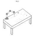
- FIG. 2is a schematic diagram showing an example of a probe placed over a patient.
- FIG. 3is a flowchart showing a procedure of establishing registration functions.
- FIG. 4is a schematic diagram showing an example of registration of a CT image onto ultrasound images.
- FIG. 5is a flowchart showing an example of establishing a local registration function.
- FIG. 6is a schematic diagram showing an example of probe movement between a preoperative stage and an intraoperative stage.
- FIG. 7is a schematic diagram showing an example of displaying a real-time ultrasound image and an ultrasound-CT registered image.
- a system 100may include an ultrasound image forming unit 110 and a CT image forming unit 120 .
- the ultrasound image forming unit 110may include a probe (not shown) for transmitting and receiving ultrasound signals.
- the ultrasound image forming unit 110may be configured to sequentially form ultrasound images at a predetermined interval.
- the ultrasound image forming unit 110may be configured to repeat forming the ultrasound image during a first period while the probe is placed at a first position on a target object.
- the CT image forming unit 120may form a CT image of the target object.
- the CT imagemay be obtained at the maximum inspiration.
- the system 100may further include a registration unit 130 that may be configured to register the CT image to each of the ultrasound images.
- the registered CT-ultrasound imagesmay be stored in a storage unit 140 .
- the probemay be moved to a second position adjacent to the first position.
- the system 100may further include a calibration unit 150 for establishing a rigid motion calibration function to calibrate the probe movement from the first position to the second position.
- the calibration functionmay be applied to the registered ultrasound-CT images.
- the ultrasound image forming unit 10may be configured to consecutively form 3-dimensional ultrasound images I US (t i ) representative of the target object at a predetermined interval, wherein i is a positive integer and 1 ⁇ i ⁇ N, during a half respiratory cycle representing from the inspiration up to the expiration at the preoperative stage.
- the CT image forming unit 120may be also configured to provide the CT image I CT at the preoperative stage.
- the ultrasound image forming unit 110may form the 3-dimensional ultrasound image I US (t i ) at the brightness-mode (B-mode).
- the ultrasound image forming unit 110may form the 3-dimensional ultrasound images I US (t i ) while the probe PB is fixed at the first position on a patient P, as illustrated in FIG. 2 .
- the target objectmay be a liver, which may be moved according to the respiration.
- the livermay move about 30 mm for a half respiratory cycle (about 2 seconds) on average.
- a required volume rate of the 3-dimensional ultrasound imagesmay be over 1/[2 mm+(2 s/30 mm)] ⁇ 8 vol/s. That is, the required number of the volumes obtained for the half respiratory cycle, i.e., the required number of the 3-dimensional ultrasound images I US (t i ), may be over 16 .
- a transform T for the registration of the CT image I CT to the 3-dimensional ultrasound images I US (t i )is initialized at block 340 .
- the initial transform T initial (t i )may be set to a transform T(t i ⁇ 1 ) previously used to register a CT image I CT onto an ultrasound image I US (t i ⁇ 1 ).
- the previous transform T(t i ⁇ 1 )may be used as a current transform T(t i ) for the ultrasound-CT registration.
- the transform T(t i ) at subsequent respiration intervalsmay be similar to each other.
- the previous transform T(t i ⁇ 1 ) resulting from the previous registrationis used as the current transform T(t i ), then it may be advantageous for an appropriate and robust final transform.
- affine registrationmay be carried out for each interval of the respiration at block 350 .
- a set of common feature pointsmay be first extracted from the ultrasound images I US (t i ) and the CT image I CT .
- an affine transform function T affinemay be established with the iterative closet point (ICP) algorithm using the set of the feature points.
- ICPiterative closet point
- diaphragm extraction and vessel segmentationmay be carried out upon the CT image at blocks 401 and 402 . Further, a masking process for selecting boundary regions is carried out based on the information obtained through the diaphragm extraction and the vessel segmentation at block 403 .
- Gradient vectorsmay be extracted from the masked CT image and the B-mode ultrasound images at block 404 and 405 . The similarity may be measured based on the gradient vectors and brightness values obtained from each image to perform the local registration at block 406 and to obtain a local transform function T local at block 407 .
- the local transform function T localmay be defined by using the B-spline free-form deformation (FFD).
- the smoothness constraint based on a bending energy of a thin-plate of metalmay be imposed so as to plate the local transformation T local .
- the affine transform function T affine and the local transform function T localmay be summed to obtain a transform function T(t i ) at block 370 .
- the CT image I CTmay be transformed by using the transform function T(t i ) to obtain a CT image registered onto the ultrasound image I′ CT (t N ) (“ultrasound-CT registered image”) at each interval of the respiration.
- the ultrasound-CT registered images I′ CT (t N ) at respective intervals of the respirationmay be stored in the storage unit 140 .
- the above process 320 - 370may be repeatedly carried out upon all the ultrasound images obtained at the preoperative stage.
- a rigid motion calibration function T rigidmay be established for calibrating a position difference between the first position A and the second position B.
- an ultrasound image obtained at the maximum inspiration at the preoperative stagemay be set to a reference ultrasound image.
- the calibration unit 150may be configured to measure similarities of the reference ultrasound image to the ultrasound images obtained at the preoperative stage.
- a first candidate image and a second candidate imagemay be selected based on the order of the measured similarities among the ultrasound images obtained at the respective intervals of the respiration.
- the calibration unit 150may be configured to use the reference ultrasound image, the first candidate image and the second candidate image to determine a rigid transformation parameter, which produces maximal similarity, to thereby establish a calibration function T rigid .
- the calibration function T rigidmay be applied to the ultrasound images obtained at the preoperative stage and the ultrasound-CT registered images to perform the calibration of the probe movement.
- real-time ultrasound imagesmay be acquired. Further, ultrasound images, which are most similar to the real-time ultrasound images I US-REAL (t), may be selected among the ultrasound images with the probe movement calibrated. An ultrasound-CT registered image corresponding to the selected ultrasound image may be retrieved.
- the real-time ultrasound image I US-REAL (t) and the retrieved ultrasound-CT registered image I′ CT (x, t N )may be displayed at the same time on a display unit 160 , as shown in FIG. 7 .
- the display unit 160may display the real-time ultrasound image and the ultrasound-CT registered image with superimposed or fused.
- the rigid motion calibration functionis applied to the ultrasound images obtained at the preoperative stage and the registered images by considering the probe movement at the preoperative stage and the intraoperative stage. However, if the position of the probe is not changed at the preoperative stage and the intraoperative stage, then the application of the rigid motion calibration function may be omitted.
- the calibration unit 150may be configured to select ultrasound images in the order of the measured similarity among the ultrasound images obtained at the respective intervals of the respiration.
- the calibration unit 150may be configured to retrieve the ultrasound-CT registered image corresponding to the respective selected ultrasound images.
- the calibration unit 150may be configured to perform interpolation upon the retrieved ultrasound-CT registered images.
- the display unit 160may display the real-time ultrasound images obtained at the intraoperative stage together with the interpolated ultrasound-CT registered images.
Landscapes
- Health & Medical Sciences (AREA)
- Life Sciences & Earth Sciences (AREA)
- Engineering & Computer Science (AREA)
- Medical Informatics (AREA)
- Physics & Mathematics (AREA)
- Radiology & Medical Imaging (AREA)
- General Health & Medical Sciences (AREA)
- Veterinary Medicine (AREA)
- Public Health (AREA)
- Nuclear Medicine, Radiotherapy & Molecular Imaging (AREA)
- Biophysics (AREA)
- Pathology (AREA)
- Animal Behavior & Ethology (AREA)
- Biomedical Technology (AREA)
- Heart & Thoracic Surgery (AREA)
- Molecular Biology (AREA)
- Surgery (AREA)
- Computer Vision & Pattern Recognition (AREA)
- Theoretical Computer Science (AREA)
- Optics & Photonics (AREA)
- High Energy & Nuclear Physics (AREA)
- General Physics & Mathematics (AREA)
- Pulmonology (AREA)
- Apparatus For Radiation Diagnosis (AREA)
- Ultra Sonic Daignosis Equipment (AREA)
Abstract
Description
Claims (20)
Applications Claiming Priority (2)
| Application Number | Priority Date | Filing Date | Title |
|---|---|---|---|
| KR10-2008-0052652 | 2008-06-04 | ||
| KR20080052652 | 2008-06-04 |
Publications (2)
| Publication Number | Publication Date |
|---|---|
| US20090303252A1 US20090303252A1 (en) | 2009-12-10 |
| US8111892B2true US8111892B2 (en) | 2012-02-07 |
Family
ID=41055317
Family Applications (1)
| Application Number | Title | Priority Date | Filing Date |
|---|---|---|---|
| US12/476,153Active2030-07-08US8111892B2 (en) | 2008-06-04 | 2009-06-01 | Registration of CT image onto ultrasound images |
Country Status (4)
| Country | Link |
|---|---|
| US (1) | US8111892B2 (en) |
| EP (1) | EP2131326B1 (en) |
| JP (1) | JP5067398B2 (en) |
| KR (1) | KR101017610B1 (en) |
Cited By (10)
| Publication number | Priority date | Publication date | Assignee | Title |
|---|---|---|---|---|
| US9351709B2 (en) | 2013-08-23 | 2016-05-31 | General Electric Company | Image processing method and apparatus and program |
| US9495725B2 (en) | 2013-01-29 | 2016-11-15 | Samsung Electronics Co., Ltd. | Method and apparatus for medical image registration |
| US20170169609A1 (en)* | 2014-02-19 | 2017-06-15 | Koninklijke Philips N.V. | Motion adaptive visualization in medical 4d imaging |
| US10026191B2 (en) | 2013-11-27 | 2018-07-17 | Analogic Corporation | Multi-imaging modality navigation system |
| US10368809B2 (en) | 2012-08-08 | 2019-08-06 | Samsung Electronics Co., Ltd. | Method and apparatus for tracking a position of a tumor |
| CN110934613A (en)* | 2018-09-21 | 2020-03-31 | 佳能医疗系统株式会社 | Ultrasonic diagnostic apparatus and ultrasonic diagnostic method |
| US10719935B2 (en) | 2015-12-11 | 2020-07-21 | Samsung Electronics Co., Ltd. | Image processing apparatus and image processing method thereof |
| US11135447B2 (en)* | 2015-07-17 | 2021-10-05 | Koninklijke Philips N.V. | Guidance for lung cancer radiation |
| US11227399B2 (en) | 2018-09-21 | 2022-01-18 | Canon Medical Systems Corporation | Analysis apparatus, ultrasound diagnostic apparatus, and analysis method |
| US11369348B2 (en) | 2018-07-31 | 2022-06-28 | Canon Medical Systems Corporation | Ultrasound diagnostic apparatus, image processing apparatus, and image processing method |
Families Citing this family (30)
| Publication number | Priority date | Publication date | Assignee | Title |
|---|---|---|---|---|
| US8064664B2 (en)* | 2006-10-18 | 2011-11-22 | Eigen, Inc. | Alignment method for registering medical images |
| EP2131212A3 (en)* | 2008-06-05 | 2011-10-05 | Medison Co., Ltd. | Non-Rigid Registration Between CT Images and Ultrasound Images |
| KR101121396B1 (en)* | 2009-07-31 | 2012-03-05 | 한국과학기술원 | System and method for providing 2-dimensional ct image corresponding to 2-dimensional ultrasound image |
| JP5538861B2 (en)* | 2009-12-18 | 2014-07-02 | キヤノン株式会社 | Information processing apparatus, information processing method, information processing system, and program |
| KR101089567B1 (en)* | 2010-01-19 | 2011-12-06 | 주식회사 나노포커스레이 | Method of generating a breathing gate signal of a 선 ―line tomography scanner |
| KR101232925B1 (en) | 2011-04-27 | 2013-02-13 | 인텔렉추얼디스커버리 주식회사 | Apparatus and method for producing a real-time tomography, and a medical device using the real-time tomography |
| KR101118549B1 (en)* | 2010-06-15 | 2012-02-22 | 연세대학교 산학협력단 | Apparatus and Method for obtaining medical fusion image |
| KR101805619B1 (en)* | 2011-01-25 | 2017-12-07 | 삼성전자주식회사 | Apparatus and method for creating optimal 2-dimensional medical image automatically from 3-dimensional medical image |
| US20120253170A1 (en)* | 2011-03-29 | 2012-10-04 | Samsung Electronics Co., Ltd. | Method and apparatus for generating medical image of body organ by using 3-d model |
| JP5685133B2 (en)* | 2011-04-13 | 2015-03-18 | キヤノン株式会社 | Image processing apparatus, image processing apparatus control method, and program |
| JP5409719B2 (en)* | 2011-07-27 | 2014-02-05 | 日立アロカメディカル株式会社 | Ultrasonic image processing device |
| KR101982149B1 (en)* | 2011-09-05 | 2019-05-27 | 삼성전자주식회사 | Method and apparatus for creating medical image using partial medical image |
| KR101286222B1 (en)* | 2011-09-19 | 2013-07-15 | 삼성메디슨 주식회사 | Method and apparatus for processing image, ultrasound diagnosis apparatus and medical imaging system |
| KR101468418B1 (en) | 2012-01-13 | 2014-12-03 | 삼성메디슨 주식회사 | Method and apparatus for processing ultrasound images |
| KR101768526B1 (en) | 2012-07-27 | 2017-08-17 | 삼성전자주식회사 | Method and apparatus for creating model of patient specified target organ based on blood vessel structure |
| KR101932721B1 (en)* | 2012-09-07 | 2018-12-26 | 삼성전자주식회사 | Method and Appartus of maching medical images |
| KR102001219B1 (en) | 2012-11-26 | 2019-07-17 | 삼성전자주식회사 | Method and Apparatus of matching medical images |
| KR102106535B1 (en)* | 2013-02-06 | 2020-05-06 | 삼성전자주식회사 | Method, apparatus and system for generating model representing deformation of shape and location of organ in respiration cycle |
| KR102273020B1 (en) | 2014-06-18 | 2021-07-05 | 삼성전자주식회사 | Method and appartus for registering medical images |
| US10991069B2 (en)* | 2014-10-08 | 2021-04-27 | Samsung Electronics Co., Ltd. | Method and apparatus for registration of medical images |
| EP3230951A1 (en) | 2014-12-09 | 2017-10-18 | Koninklijke Philips N.V. | Feedback for multi-modality auto-registration |
| KR102372214B1 (en)* | 2015-01-19 | 2022-03-14 | 삼성전자주식회사 | Image processing apparatus, medical image apparatus and image processing method |
| JP6510301B2 (en)* | 2015-04-17 | 2019-05-08 | 株式会社日本未来医療研究所 | MEDICAL SUPPORT SYSTEM, MEDICAL SUPPORT METHOD, IMAGE PROCESSING APPARATUS, AND CONTROL METHOD AND CONTROL PROGRAM THEREOF |
| WO2017109685A1 (en) | 2015-12-22 | 2017-06-29 | Koninklijke Philips N.V. | Medical imaging apparatus and medical imaging method for inspecting a volume of a subject |
| US11484288B2 (en)* | 2017-05-11 | 2022-11-01 | Koninklijke Philips N.V. | Workflow, system and method for motion compensation in ultrasound procedures |
| KR102704243B1 (en)* | 2018-11-22 | 2024-09-09 | 삼성메디슨 주식회사 | Ultrasound imaging apparatus and control method for the same |
| WO2020246773A1 (en)* | 2019-06-06 | 2020-12-10 | 삼성메디슨 주식회사 | Device and method for alignment of ultrasonic image and three-dimensional medical image |
| KR102375910B1 (en)* | 2020-03-02 | 2022-03-16 | 연세대학교 산학협력단 | Ultrasound Image-guided Method and Apparatus |
| CN112184781A (en)* | 2020-09-14 | 2021-01-05 | 中国科学院深圳先进技术研究院 | Method, device and equipment for registering ultrasonic image and CT image |
| CN113936008A (en)* | 2021-09-13 | 2022-01-14 | 哈尔滨医科大学 | Multi-scale image registration method for multi-core magnetic resonance |
Citations (11)
| Publication number | Priority date | Publication date | Assignee | Title |
|---|---|---|---|---|
| WO2002009588A1 (en) | 2000-08-01 | 2002-02-07 | Tony Falco | Method and apparatus for lesion localization, definition and verification |
| JP2002112998A (en) | 2000-10-05 | 2002-04-16 | Toshiba Medical System Co Ltd | Ultrasonic puncture supporting apparatus |
| US20040122325A1 (en) | 2002-12-18 | 2004-06-24 | Barbara Ann Karmanos Cancer Institute | Diagnostic analysis of ultrasound data |
| WO2004098414A1 (en) | 2003-05-08 | 2004-11-18 | Hitachi Medical Corporation | Reference image display method for ultrasonography and ultrasonograph |
| US20060004275A1 (en)* | 2004-06-30 | 2006-01-05 | Vija A H | Systems and methods for localized image registration and fusion |
| JP2006068102A (en) | 2004-08-31 | 2006-03-16 | Toshiba Corp | Ultrasonic therapy device |
| WO2006064676A1 (en) | 2004-12-13 | 2006-06-22 | Hitachi Medical Corporation | Ultrasonic diagnosis apparatus |
| JP2007275440A (en) | 2006-04-11 | 2007-10-25 | Fujifilm Corp | Similar image retrieval apparatus and method, and program |
| US20080009699A1 (en)* | 2003-04-11 | 2008-01-10 | Georgios Sakas | Combining first and second image data of an object |
| US20090054772A1 (en)* | 2005-01-31 | 2009-02-26 | Chongqing Haifu(Hifu) Technology Co., Ltd. | Focused Ultrasound Therapy System |
| US20090067752A1 (en) | 2007-09-11 | 2009-03-12 | Samsung Electronics Co., Ltd. | Image-registration method, medium, and apparatus |
- 2009
- 2009-06-01USUS12/476,153patent/US8111892B2/enactiveActive
- 2009-06-02EPEP09007286Apatent/EP2131326B1/enactiveActive
- 2009-06-04KRKR1020090049325Apatent/KR101017610B1/enactiveActive
- 2009-06-04JPJP2009134963Apatent/JP5067398B2/ennot_activeExpired - Fee Related
Patent Citations (15)
| Publication number | Priority date | Publication date | Assignee | Title |
|---|---|---|---|---|
| WO2002009588A1 (en) | 2000-08-01 | 2002-02-07 | Tony Falco | Method and apparatus for lesion localization, definition and verification |
| JP2007054641A (en) | 2000-08-01 | 2007-03-08 | Tony Falco | Method and apparatus for resolution and confirmation of lesion |
| JP2002112998A (en) | 2000-10-05 | 2002-04-16 | Toshiba Medical System Co Ltd | Ultrasonic puncture supporting apparatus |
| US20040122325A1 (en) | 2002-12-18 | 2004-06-24 | Barbara Ann Karmanos Cancer Institute | Diagnostic analysis of ultrasound data |
| US20080009699A1 (en)* | 2003-04-11 | 2008-01-10 | Georgios Sakas | Combining first and second image data of an object |
| WO2004098414A1 (en) | 2003-05-08 | 2004-11-18 | Hitachi Medical Corporation | Reference image display method for ultrasonography and ultrasonograph |
| US20070010743A1 (en) | 2003-05-08 | 2007-01-11 | Osamu Arai | Reference image display method for ultrasonography and ultrasonograph |
| US20060004275A1 (en)* | 2004-06-30 | 2006-01-05 | Vija A H | Systems and methods for localized image registration and fusion |
| JP2006068102A (en) | 2004-08-31 | 2006-03-16 | Toshiba Corp | Ultrasonic therapy device |
| WO2006064676A1 (en) | 2004-12-13 | 2006-06-22 | Hitachi Medical Corporation | Ultrasonic diagnosis apparatus |
| US20100036247A1 (en) | 2004-12-13 | 2010-02-11 | Masa Yamamoto | Ultrasonic diagnosis apparatus |
| US20090054772A1 (en)* | 2005-01-31 | 2009-02-26 | Chongqing Haifu(Hifu) Technology Co., Ltd. | Focused Ultrasound Therapy System |
| JP2007275440A (en) | 2006-04-11 | 2007-10-25 | Fujifilm Corp | Similar image retrieval apparatus and method, and program |
| US20090067752A1 (en) | 2007-09-11 | 2009-03-12 | Samsung Electronics Co., Ltd. | Image-registration method, medium, and apparatus |
| JP2009071821A (en) | 2007-09-11 | 2009-04-02 | Samsung Electronics Co Ltd | Video alignment method and apparatus |
Non-Patent Citations (6)
| Title |
|---|
| Blackall, Jane M., et al., "Alignment of Sparse Freehand 3-D Ultrasound With Preoperative Images of the Liver Using Models of Respiratory Motion and Deformation", Nov. 2005, pp. 1405-1416, IEEE Transactions of Medical Imaging, vol. 24, No. 11. |
| Extended European Search Report, issued in European Patent Application No. 09 007 286.9, dated Nov. 4, 2011. |
| Huang, Xishi, et al., "Dynamic 3D Ultrasound and MR Image Registration of the Beating Heart", 2005, pp. 171-178, LNCS 3750, MICCAI 2005, Duncan, J., et al. (Eds.), Springer-Verlag Berlin Heidelberg. |
| Lindseth, Frank, et al., "Multimodal image fusion in ultrasound-based neuronavigation: improving overview and interpretation by integrating preoperative MRI with intraoperative 3D ultrasound", Jan. 1, 2003, vol. 8 No. 2, pp. 1-30, Biomedical paper. |
| Pitiot et al., "Piecewise Affine Registration of Biological Images for Volume Reconstruction," Medical Image Analysis, Elsevier, 19 pages (Received Aug. 9, 2004). |
| Xu, Sheng, et al., "Closed-Loop Control in Fused MR-TRUS Image-Guided Prostate Biopsy", 2007, pp. 128-135, Part I. LNCS 4791, MICCAI 2007, Ayache, N., et al (Eds.), Springer-Verlag Berlin Heidelberg. |
Cited By (11)
| Publication number | Priority date | Publication date | Assignee | Title |
|---|---|---|---|---|
| US10368809B2 (en) | 2012-08-08 | 2019-08-06 | Samsung Electronics Co., Ltd. | Method and apparatus for tracking a position of a tumor |
| US9495725B2 (en) | 2013-01-29 | 2016-11-15 | Samsung Electronics Co., Ltd. | Method and apparatus for medical image registration |
| US9351709B2 (en) | 2013-08-23 | 2016-05-31 | General Electric Company | Image processing method and apparatus and program |
| US10026191B2 (en) | 2013-11-27 | 2018-07-17 | Analogic Corporation | Multi-imaging modality navigation system |
| US20170169609A1 (en)* | 2014-02-19 | 2017-06-15 | Koninklijke Philips N.V. | Motion adaptive visualization in medical 4d imaging |
| US11135447B2 (en)* | 2015-07-17 | 2021-10-05 | Koninklijke Philips N.V. | Guidance for lung cancer radiation |
| US10719935B2 (en) | 2015-12-11 | 2020-07-21 | Samsung Electronics Co., Ltd. | Image processing apparatus and image processing method thereof |
| US11369348B2 (en) | 2018-07-31 | 2022-06-28 | Canon Medical Systems Corporation | Ultrasound diagnostic apparatus, image processing apparatus, and image processing method |
| CN110934613A (en)* | 2018-09-21 | 2020-03-31 | 佳能医疗系统株式会社 | Ultrasonic diagnostic apparatus and ultrasonic diagnostic method |
| US11227399B2 (en) | 2018-09-21 | 2022-01-18 | Canon Medical Systems Corporation | Analysis apparatus, ultrasound diagnostic apparatus, and analysis method |
| CN110934613B (en)* | 2018-09-21 | 2023-01-13 | 佳能医疗系统株式会社 | Ultrasonic diagnostic apparatus and ultrasonic diagnostic method |
Also Published As
| Publication number | Publication date |
|---|---|
| EP2131326B1 (en) | 2013-02-13 |
| EP2131326A2 (en) | 2009-12-09 |
| EP2131326A3 (en) | 2011-12-07 |
| JP2009291614A (en) | 2009-12-17 |
| KR20090127091A (en) | 2009-12-09 |
| KR101017610B1 (en) | 2011-02-28 |
| US20090303252A1 (en) | 2009-12-10 |
| JP5067398B2 (en) | 2012-11-07 |
Similar Documents
| Publication | Publication Date | Title |
|---|---|---|
| US8111892B2 (en) | Registration of CT image onto ultrasound images | |
| US8126239B2 (en) | Registering 2D and 3D data using 3D ultrasound data | |
| JP4490442B2 (en) | Method and system for affine superposition of an intraoperative 2D image and a preoperative 3D image | |
| US20090275831A1 (en) | Image registration and methods for compensating intraoperative motion in image-guided interventional procedures | |
| US7480398B2 (en) | Method of registering a sequence of 2D image data with 3D image data | |
| EP1837828B1 (en) | Image registration using locally-weighted fitting | |
| JP2011502687A (en) | Interventional navigation using 3D contrast ultrasound | |
| KR20140096919A (en) | Method and Apparatus for medical image registration | |
| CN109152566B (en) | Correcting for probe-induced deformations in ultrasound fusion imaging systems | |
| EP2252204A1 (en) | Ct surrogate by auto-segmentation of magnetic resonance images | |
| KR20150118484A (en) | Method and Apparatus for medical image registration | |
| US8068665B2 (en) | 3D-image processing apparatus, 3D-image processing method, storage medium, and program | |
| EP1652122B1 (en) | Automatic registration of intra-modality medical volume images using affine transformation | |
| US8224047B2 (en) | System and method for measuring left ventricular torsion | |
| Weon et al. | Position tracking of moving liver lesion based on real‐time registration between 2D ultrasound and 3D preoperative images | |
| Timinger et al. | Motion compensated coronary interventional navigation by means of diaphragm tracking and elastic motion models | |
| Reinertsen et al. | Vessel driven correction of brain shift | |
| US9186086B2 (en) | Small tissue property change detection in magnetic resonance guided intervention | |
| US10346989B2 (en) | Method and system for calculating a displacement of an object of interest | |
| Leroy et al. | Intensity-based registration of freehand 3D ultrasound and CT-scan images of the kidney | |
| US20240320842A1 (en) | Providing a result dataset |
Legal Events
| Date | Code | Title | Description |
|---|---|---|---|
| AS | Assignment | Owner name:MEDISON CO., LTD., KOREA, REPUBLIC OF Free format text:ASSIGNMENT OF ASSIGNORS INTEREST;ASSIGNORS:HYUN, DONG GYU;RA, JONG BEOM;LEE, DUHGOON;AND OTHERS;REEL/FRAME:022769/0972 Effective date:20090513 Owner name:KOREA ADVANCED INSTITUTE OF SCIENCE AND TECHNOLOGY Free format text:ASSIGNMENT OF ASSIGNORS INTEREST;ASSIGNORS:HYUN, DONG GYU;RA, JONG BEOM;LEE, DUHGOON;AND OTHERS;REEL/FRAME:022769/0972 Effective date:20090513 | |
| FEPP | Fee payment procedure | Free format text:PAYOR NUMBER ASSIGNED (ORIGINAL EVENT CODE: ASPN); ENTITY STATUS OF PATENT OWNER: LARGE ENTITY | |
| STCF | Information on status: patent grant | Free format text:PATENTED CASE | |
| AS | Assignment | Owner name:SAMSUNG MEDISON CO., LTD., KOREA, REPUBLIC OF Free format text:CHANGE OF NAME;ASSIGNOR:MEDISON CO., LTD.;REEL/FRAME:032874/0741 Effective date:20110329 | |
| FPAY | Fee payment | Year of fee payment:4 | |
| MAFP | Maintenance fee payment | Free format text:PAYMENT OF MAINTENANCE FEE, 8TH YEAR, LARGE ENTITY (ORIGINAL EVENT CODE: M1552); ENTITY STATUS OF PATENT OWNER: LARGE ENTITY Year of fee payment:8 | |
| MAFP | Maintenance fee payment | Free format text:PAYMENT OF MAINTENANCE FEE, 12TH YEAR, LARGE ENTITY (ORIGINAL EVENT CODE: M1553); ENTITY STATUS OF PATENT OWNER: LARGE ENTITY Year of fee payment:12 |