US8101169B2 - Ocular gene therapy using avalanche-mediated transfection - Google Patents
Ocular gene therapy using avalanche-mediated transfectionDownload PDFInfo
- Publication number
- US8101169B2 US8101169B2US11/505,249US50524906AUS8101169B2US 8101169 B2US8101169 B2US 8101169B2US 50524906 AUS50524906 AUS 50524906AUS 8101169 B2US8101169 B2US 8101169B2
- Authority
- US
- United States
- Prior art keywords
- cells
- tissue
- nucleic acid
- subject
- electrode
- Prior art date
- Legal status (The legal status is an assumption and is not a legal conclusion. Google has not performed a legal analysis and makes no representation as to the accuracy of the status listed.)
- Expired - Fee Related, expires
Links
- 238000001890transfectionMethods0.000titleclaimsabstractdescription28
- 230000001404mediated effectEffects0.000titledescription11
- 238000001415gene therapyMethods0.000titledescription10
- 150000007523nucleic acidsChemical class0.000claimsabstractdescription59
- 238000000034methodMethods0.000claimsabstractdescription50
- 102000039446nucleic acidsHuman genes0.000claimsabstractdescription42
- 108020004707nucleic acidsProteins0.000claimsabstractdescription42
- 230000005684electric fieldEffects0.000claimsabstractdescription33
- 210000004027cellAnatomy0.000claimsdescription107
- 210000001519tissueAnatomy0.000claimsdescription103
- 108090000623proteins and genesProteins0.000claimsdescription80
- 102000004169proteins and genesHuman genes0.000claimsdescription36
- 230000001225therapeutic effectEffects0.000claimsdescription28
- 210000002950fibroblastAnatomy0.000claimsdescription8
- 108091027967Small hairpin RNAProteins0.000claimsdescription6
- 206010064930age-related macular degenerationDiseases0.000claimsdescription6
- 208000002780macular degenerationDiseases0.000claimsdescription6
- 239000004055small Interfering RNASubstances0.000claimsdescription6
- 230000000735allogeneic effectEffects0.000claimsdescription5
- 210000002919epithelial cellAnatomy0.000claimsdescription5
- 208000005590Choroidal NeovascularizationDiseases0.000claimsdescription4
- 206010060823Choroidal neovascularisationDiseases0.000claimsdescription4
- 210000001423scleral cellAnatomy0.000claimsdescription4
- 206010012689Diabetic retinopathyDiseases0.000claimsdescription3
- 208000010412GlaucomaDiseases0.000claimsdescription3
- 201000007737Retinal degenerationDiseases0.000claimsdescription3
- 208000032430Retinal dystrophyDiseases0.000claimsdescription3
- 210000003205muscleAnatomy0.000claimsdescription3
- 230000004258retinal degenerationEffects0.000claimsdescription3
- 210000004240ciliary bodyAnatomy0.000claims2
- 230000001886ciliary effectEffects0.000claims2
- 238000012258culturingMethods0.000claims1
- 208000022873Ocular diseaseDiseases0.000abstractdescription6
- 230000008823permeabilizationEffects0.000abstractdescription6
- 230000015572biosynthetic processEffects0.000abstractdescription4
- 239000013612plasmidSubstances0.000description26
- 230000014509gene expressionEffects0.000description23
- 239000003550markerSubstances0.000description23
- 239000013598vectorSubstances0.000description21
- 108020004414DNAProteins0.000description20
- 238000005215recombinationMethods0.000description18
- 108090000102pigment epithelium-derived factorProteins0.000description17
- 230000006798recombinationEffects0.000description17
- 102100035846Pigment epithelium-derived factorHuman genes0.000description16
- 108020004684Internal Ribosome Entry SitesProteins0.000description15
- 239000000523sampleSubstances0.000description15
- 241000701022CytomegalovirusSpecies0.000description12
- 108010048367enhanced green fluorescent proteinProteins0.000description12
- 238000002054transplantationMethods0.000description12
- 102100034343IntegraseHuman genes0.000description10
- 108010061833IntegrasesProteins0.000description10
- 108091028043Nucleic acid sequenceProteins0.000description10
- 239000002609mediumSubstances0.000description10
- 108091033319polynucleotideProteins0.000description10
- 102000040430polynucleotideHuman genes0.000description10
- 239000002157polynucleotideSubstances0.000description10
- 108060001084LuciferaseProteins0.000description9
- 239000002773nucleotideSubstances0.000description9
- 125000003729nucleotide groupChemical group0.000description9
- 239000000758substrateSubstances0.000description9
- 229920001184polypeptidePolymers0.000description8
- 108090000765processed proteins & peptidesProteins0.000description8
- 102000004196processed proteins & peptidesHuman genes0.000description8
- 238000013519translationMethods0.000description7
- 108091006047fluorescent proteinsProteins0.000description6
- 102000034287fluorescent proteinsHuman genes0.000description6
- 108700039691Genetic Promoter RegionsProteins0.000description5
- 239000005089LuciferaseSubstances0.000description5
- 206010025421MaculeDiseases0.000description5
- 210000000795conjunctivaAnatomy0.000description5
- 238000004520electroporationMethods0.000description5
- 230000010354integrationEffects0.000description5
- 230000028327secretionEffects0.000description5
- 239000000243solutionSubstances0.000description5
- 108091026890Coding regionProteins0.000description4
- 230000001464adherent effectEffects0.000description4
- 238000013459approachMethods0.000description4
- 238000001727in vivoMethods0.000description4
- 210000003583retinal pigment epitheliumAnatomy0.000description4
- 210000003786scleraAnatomy0.000description4
- 238000009834vaporizationMethods0.000description4
- 230000008016vaporizationEffects0.000description4
- 210000004127vitreous bodyAnatomy0.000description4
- 102000008579TransposasesHuman genes0.000description3
- 108010020764TransposasesProteins0.000description3
- 230000002051biphasic effectEffects0.000description3
- 230000001413cellular effectEffects0.000description3
- 238000013461designMethods0.000description3
- 201000010099diseaseDiseases0.000description3
- 208000037265diseases, disorders, signs and symptomsDiseases0.000description3
- 210000003527eukaryotic cellAnatomy0.000description3
- 239000012634fragmentSubstances0.000description3
- 210000005260human cellAnatomy0.000description3
- 238000000338in vitroMethods0.000description3
- 239000007788liquidSubstances0.000description3
- 230000007774longtermEffects0.000description3
- 239000011159matrix materialSubstances0.000description3
- 230000008439repair processEffects0.000description3
- 230000004083survival effectEffects0.000description3
- 238000013518transcriptionMethods0.000description3
- 230000035897transcriptionEffects0.000description3
- 230000003612virological effectEffects0.000description3
- 108091032973(ribonucleotides)n+mProteins0.000description2
- 108091023037AptamerProteins0.000description2
- 201000004569BlindnessDiseases0.000description2
- 102000008137Bone Morphogenetic Protein 4Human genes0.000description2
- 108010049955Bone Morphogenetic Protein 4Proteins0.000description2
- 102000004219Brain-derived neurotrophic factorHuman genes0.000description2
- 108090000715Brain-derived neurotrophic factorProteins0.000description2
- 108010005939Ciliary Neurotrophic FactorProteins0.000description2
- 102100031614Ciliary neurotrophic factorHuman genes0.000description2
- IGXWBGJHJZYPQS-SSDOTTSWSA-ND-LuciferinChemical compoundOC(=O)[C@H]1CSC(C=2SC3=CC=C(O)C=C3N=2)=N1IGXWBGJHJZYPQS-SSDOTTSWSA-N0.000description2
- CYCGRDQQIOGCKX-UHFFFAOYSA-NDehydro-luciferinNatural productsOC(=O)C1=CSC(C=2SC3=CC(O)=CC=C3N=2)=N1CYCGRDQQIOGCKX-UHFFFAOYSA-N0.000description2
- 239000006144Dulbecco’s modified Eagle's mediumSubstances0.000description2
- 102000018233Fibroblast Growth FactorHuman genes0.000description2
- 108050007372Fibroblast Growth FactorProteins0.000description2
- BJGNCJDXODQBOB-UHFFFAOYSA-NFivefly LuciferinNatural productsOC(=O)C1CSC(C=2SC3=CC(O)=CC=C3N=2)=N1BJGNCJDXODQBOB-UHFFFAOYSA-N0.000description2
- DDWFXDSYGUXRAY-UHFFFAOYSA-NLuciferinNatural productsCCc1c(C)c(CC2NC(=O)C(=C2C=C)C)[nH]c1Cc3[nH]c4C(=C5/NC(CC(=O)O)C(C)C5CC(=O)O)CC(=O)c4c3CDDWFXDSYGUXRAY-UHFFFAOYSA-N0.000description2
- 108091034117OligonucleotideProteins0.000description2
- 240000007019Oxalis corniculataSpecies0.000description2
- 102000004160Phosphoric Monoester HydrolasesHuman genes0.000description2
- 108090000608Phosphoric Monoester HydrolasesProteins0.000description2
- 241000709664PicornaviridaeSpecies0.000description2
- 108091030071RNAIProteins0.000description2
- 102000018120RecombinasesHuman genes0.000description2
- 108010091086RecombinasesProteins0.000description2
- 108010073929Vascular Endothelial Growth Factor AProteins0.000description2
- 102000005789Vascular Endothelial Growth FactorsHuman genes0.000description2
- 108010019530Vascular Endothelial Growth FactorsProteins0.000description2
- 230000001772anti-angiogenic effectEffects0.000description2
- 230000003115biocidal effectEffects0.000description2
- 238000005415bioluminescenceMethods0.000description2
- 230000029918bioluminescenceEffects0.000description2
- 229940077737brain-derived neurotrophic factorDrugs0.000description2
- 210000003161choroidAnatomy0.000description2
- 239000002299complementary DNASubstances0.000description2
- 210000004087corneaAnatomy0.000description2
- 230000007423decreaseEffects0.000description2
- 239000003814drugSubstances0.000description2
- 238000005538encapsulationMethods0.000description2
- 238000002474experimental methodMethods0.000description2
- 229940126864fibroblast growth factorDrugs0.000description2
- 238000000799fluorescence microscopyMethods0.000description2
- 238000001943fluorescence-activated cell sortingMethods0.000description2
- 230000030279gene silencingEffects0.000description2
- 230000009368gene silencing by RNAEffects0.000description2
- 238000012226gene silencing methodMethods0.000description2
- 230000006801homologous recombinationEffects0.000description2
- 238000002744homologous recombinationMethods0.000description2
- 230000028993immune responseEffects0.000description2
- 238000002513implantationMethods0.000description2
- 210000004962mammalian cellAnatomy0.000description2
- 238000004519manufacturing processMethods0.000description2
- 108020004999messenger RNAProteins0.000description2
- 238000012986modificationMethods0.000description2
- 230000004048modificationEffects0.000description2
- 238000012544monitoring processMethods0.000description2
- 210000000663muscle cellAnatomy0.000description2
- 210000005036nerveAnatomy0.000description2
- 230000000508neurotrophic effectEffects0.000description2
- 125000002467phosphate groupChemical group[H]OP(=O)(O[H])O[*]0.000description2
- 239000011148porous materialSubstances0.000description2
- 230000008569processEffects0.000description2
- 108091008146restriction endonucleasesProteins0.000description2
- 210000001525retinaAnatomy0.000description2
- 108091008601sVEGFRProteins0.000description2
- 230000035939shockEffects0.000description2
- 230000001360synchronised effectEffects0.000description2
- 238000012360testing methodMethods0.000description2
- 241001430294unidentified retrovirusSpecies0.000description2
- 230000004393visual impairmentEffects0.000description2
- BJHCYTJNPVGSBZ-YXSASFKJSA-N1-[4-[6-amino-5-[(Z)-methoxyiminomethyl]pyrimidin-4-yl]oxy-2-chlorophenyl]-3-ethylureaChemical compoundCCNC(=O)Nc1ccc(Oc2ncnc(N)c2\C=N/OC)cc1ClBJHCYTJNPVGSBZ-YXSASFKJSA-N0.000description1
- CPKVUHPKYQGHMW-UHFFFAOYSA-N1-ethenylpyrrolidin-2-one;molecular iodineChemical compoundII.C=CN1CCCC1=OCPKVUHPKYQGHMW-UHFFFAOYSA-N0.000description1
- 206010002091AnaesthesiaDiseases0.000description1
- 102400000068AngiostatinHuman genes0.000description1
- 108010079709AngiostatinsProteins0.000description1
- 208000002177CataractDiseases0.000description1
- 102000016550Complement Factor HHuman genes0.000description1
- 108010053085Complement Factor HProteins0.000description1
- 102000004594DNA Polymerase IHuman genes0.000description1
- 108010017826DNA Polymerase IProteins0.000description1
- 206010061818Disease progressionDiseases0.000description1
- 206010059866Drug resistanceDiseases0.000description1
- 208000003556Dry Eye SyndromesDiseases0.000description1
- 206010013774Dry eyeDiseases0.000description1
- 241000710188Encephalomyocarditis virusSpecies0.000description1
- 102400001047EndostatinHuman genes0.000description1
- 108010079505EndostatinsProteins0.000description1
- 241000991587Enterovirus CSpecies0.000description1
- 108090000790EnzymesProteins0.000description1
- 108700028146Genetic Enhancer ElementsProteins0.000description1
- 108010043121Green Fluorescent ProteinsProteins0.000description1
- 102000004144Green Fluorescent ProteinsHuman genes0.000description1
- 101001023784Heteractis crispa GFP-like non-fluorescent chromoproteinProteins0.000description1
- 241000282412HomoSpecies0.000description1
- 206010062016ImmunosuppressionDiseases0.000description1
- 108091026898Leader sequence (mRNA)Proteins0.000description1
- 102000006830Luminescent ProteinsHuman genes0.000description1
- 108010047357Luminescent ProteinsProteins0.000description1
- 102000005741MetalloproteasesHuman genes0.000description1
- 108010006035MetalloproteasesProteins0.000description1
- 108700026244Open Reading FramesProteins0.000description1
- 238000012408PCR amplificationMethods0.000description1
- 102000045595Phosphoprotein PhosphatasesHuman genes0.000description1
- 108700019535Phosphoprotein PhosphatasesProteins0.000description1
- 241001144416PicornaviralesSpecies0.000description1
- 108010076504Protein Sorting SignalsProteins0.000description1
- 208000017442Retinal diseaseDiseases0.000description1
- MTCFGRXMJLQNBG-UHFFFAOYSA-NSerineNatural productsOCC(N)C(O)=OMTCFGRXMJLQNBG-UHFFFAOYSA-N0.000description1
- 241000701955Streptomyces virus phiC31Species0.000description1
- 108700019146TransgenesProteins0.000description1
- 108091023045Untranslated RegionProteins0.000description1
- 208000027076Uveal diseaseDiseases0.000description1
- 108010053096Vascular Endothelial Growth Factor Receptor-1Proteins0.000description1
- 108010053099Vascular Endothelial Growth Factor Receptor-2Proteins0.000description1
- 102100033178Vascular endothelial growth factor receptor 1Human genes0.000description1
- 102100033177Vascular endothelial growth factor receptor 2Human genes0.000description1
- 241000021375XenogenesSpecies0.000description1
- 230000005856abnormalityEffects0.000description1
- 238000009825accumulationMethods0.000description1
- 230000004075alterationEffects0.000description1
- 230000037005anaesthesiaEffects0.000description1
- 238000004458analytical methodMethods0.000description1
- 238000004873anchoringMethods0.000description1
- 230000000416anti-micotic effectEffects0.000description1
- 210000004507artificial chromosomeAnatomy0.000description1
- FZCSTZYAHCUGEM-UHFFFAOYSA-Naspergillomarasmine BNatural productsOC(=O)CNC(C(O)=O)CNC(C(O)=O)CC(O)=OFZCSTZYAHCUGEM-UHFFFAOYSA-N0.000description1
- 230000001580bacterial effectEffects0.000description1
- 230000008901benefitEffects0.000description1
- 229940064804betadineDrugs0.000description1
- 238000001574biopsyMethods0.000description1
- 210000001775bruch membraneAnatomy0.000description1
- 244000309466calfSpecies0.000description1
- 239000002775capsuleSubstances0.000description1
- 230000030833cell deathEffects0.000description1
- 230000012292cell migrationEffects0.000description1
- 239000003795chemical substances by applicationSubstances0.000description1
- 210000000991chicken eggAnatomy0.000description1
- 238000010367cloningMethods0.000description1
- 238000010276constructionMethods0.000description1
- 239000013068control sampleSubstances0.000description1
- 210000004748cultured cellAnatomy0.000description1
- 108010082025cyan fluorescent proteinProteins0.000description1
- 230000003247decreasing effectEffects0.000description1
- 230000001419dependent effectEffects0.000description1
- 238000011161developmentMethods0.000description1
- 238000010586diagramMethods0.000description1
- 230000003292diminished effectEffects0.000description1
- LOKCTEFSRHRXRJ-UHFFFAOYSA-Idipotassium trisodium dihydrogen phosphate hydrogen phosphate dichlorideChemical compoundP(=O)(O)(O)[O-].[K+].P(=O)(O)([O-])[O-].[Na+].[Na+].[Cl-].[K+].[Cl-].[Na+]LOKCTEFSRHRXRJ-UHFFFAOYSA-I0.000description1
- 230000005750disease progressionEffects0.000description1
- 238000009826distributionMethods0.000description1
- 230000000694effectsEffects0.000description1
- 239000003623enhancerSubstances0.000description1
- 210000005081epithelial layerAnatomy0.000description1
- 210000000981epitheliumAnatomy0.000description1
- 230000003628erosive effectEffects0.000description1
- 238000000684flow cytometryMethods0.000description1
- 108010021843fluorescent protein 583Proteins0.000description1
- 230000006870functionEffects0.000description1
- 238000010353genetic engineeringMethods0.000description1
- 239000005090green fluorescent proteinSubstances0.000description1
- 230000036541healthEffects0.000description1
- 230000005847immunogenicityEffects0.000description1
- 230000001506immunosuppresive effectEffects0.000description1
- 239000007943implantSubstances0.000description1
- 230000001939inductive effectEffects0.000description1
- 239000003112inhibitorSubstances0.000description1
- 239000003999initiatorSubstances0.000description1
- 238000009413insulationMethods0.000description1
- 239000012212insulatorSubstances0.000description1
- 230000000968intestinal effectEffects0.000description1
- 230000002147killing effectEffects0.000description1
- 230000002045lasting effectEffects0.000description1
- 239000002502liposomeSubstances0.000description1
- 239000000463materialSubstances0.000description1
- 230000002503metabolic effectEffects0.000description1
- 238000002324minimally invasive surgeryMethods0.000description1
- 238000011587new zealand white rabbitMethods0.000description1
- 210000001328optic nerveAnatomy0.000description1
- 238000005457optimizationMethods0.000description1
- 244000052769pathogenSpecies0.000description1
- 230000007170pathologyEffects0.000description1
- 230000037361pathwayEffects0.000description1
- 239000002953phosphate buffered salineSubstances0.000description1
- 239000000049pigmentSubstances0.000description1
- 239000004417polycarbonateSubstances0.000description1
- 229920000515polycarbonatePolymers0.000description1
- 229920000642polymerPolymers0.000description1
- 208000037821progressive diseaseDiseases0.000description1
- 230000001902propagating effectEffects0.000description1
- 210000001747pupilAnatomy0.000description1
- 238000011002quantificationMethods0.000description1
- 230000009467reductionEffects0.000description1
- 230000022532regulation of transcription, DNA-dependentEffects0.000description1
- 230000001105regulatory effectEffects0.000description1
- 238000011160researchMethods0.000description1
- 230000004044responseEffects0.000description1
- 210000003705ribosomeAnatomy0.000description1
- 238000012216screeningMethods0.000description1
- 210000002966serumAnatomy0.000description1
- 241000894007speciesSpecies0.000description1
- 230000006641stabilisationEffects0.000description1
- 238000011105stabilizationMethods0.000description1
- 239000000126substanceSubstances0.000description1
- 238000006467substitution reactionMethods0.000description1
- 230000008685targetingEffects0.000description1
- 238000002560therapeutic procedureMethods0.000description1
- 239000003860topical agentSubstances0.000description1
- 238000002691topical anesthesiaMethods0.000description1
- 238000012546transferMethods0.000description1
- 230000001052transient effectEffects0.000description1
- 241000701161unidentified adenovirusSpecies0.000description1
- 238000011144upstream manufacturingMethods0.000description1
- 239000013603viral vectorSubstances0.000description1
- 230000000007visual effectEffects0.000description1
- 108091005957yellow fluorescent proteinsProteins0.000description1
Images


Classifications
- C—CHEMISTRY; METALLURGY
- C12—BIOCHEMISTRY; BEER; SPIRITS; WINE; VINEGAR; MICROBIOLOGY; ENZYMOLOGY; MUTATION OR GENETIC ENGINEERING
- C12N—MICROORGANISMS OR ENZYMES; COMPOSITIONS THEREOF; PROPAGATING, PRESERVING, OR MAINTAINING MICROORGANISMS; MUTATION OR GENETIC ENGINEERING; CULTURE MEDIA
- C12N13/00—Treatment of microorganisms or enzymes with electrical or wave energy, e.g. magnetism, sonic waves
- C—CHEMISTRY; METALLURGY
- C12—BIOCHEMISTRY; BEER; SPIRITS; WINE; VINEGAR; MICROBIOLOGY; ENZYMOLOGY; MUTATION OR GENETIC ENGINEERING
- C12M—APPARATUS FOR ENZYMOLOGY OR MICROBIOLOGY; APPARATUS FOR CULTURING MICROORGANISMS FOR PRODUCING BIOMASS, FOR GROWING CELLS OR FOR OBTAINING FERMENTATION OR METABOLIC PRODUCTS, i.e. BIOREACTORS OR FERMENTERS
- C12M35/00—Means for application of stress for stimulating the growth of microorganisms or the generation of fermentation or metabolic products; Means for electroporation or cell fusion
- C—CHEMISTRY; METALLURGY
- C12—BIOCHEMISTRY; BEER; SPIRITS; WINE; VINEGAR; MICROBIOLOGY; ENZYMOLOGY; MUTATION OR GENETIC ENGINEERING
- C12N—MICROORGANISMS OR ENZYMES; COMPOSITIONS THEREOF; PROPAGATING, PRESERVING, OR MAINTAINING MICROORGANISMS; MUTATION OR GENETIC ENGINEERING; CULTURE MEDIA
- C12N15/00—Mutation or genetic engineering; DNA or RNA concerning genetic engineering, vectors, e.g. plasmids, or their isolation, preparation or purification; Use of hosts therefor
- C12N15/09—Recombinant DNA-technology
- C12N15/87—Introduction of foreign genetic material using processes not otherwise provided for, e.g. co-transformation
- A—HUMAN NECESSITIES
- A61—MEDICAL OR VETERINARY SCIENCE; HYGIENE
- A61K—PREPARATIONS FOR MEDICAL, DENTAL OR TOILETRY PURPOSES
- A61K48/00—Medicinal preparations containing genetic material which is inserted into cells of the living body to treat genetic diseases; Gene therapy
Definitions
- the present inventionrelates generally to medicine. More particularly, the present invention relates to a method of treating ocular diseases with gene therapy using avalanche-mediated transfection to genetically modify cells or tissue.
- Age-related macular degenerationis a “wet” form of age-related macular degeneration, choroidal neovascularization leads to progressive disease and vision loss.
- Viral vectorssuch as retroviruses and adenoviruses
- Non-viral methodssuch as liposomes
- liposomeshave low host immunogenicity but tend to suffer from inefficient DNA delivery to cells. Accordingly, there is a need in the art for new methods of introducing DNA into cells and tissues for the purpose of gene therapy.
- the present inventionprovides a method of treating an ocular disease in a subject.
- a nucleic acidis introduced into cells or a tissue.
- the nucleic acidis introduced by electron avalanche-mediated transfection.
- a high electric fieldinduces a vapor bubble and plasma discharge between an electrode and the surrounding medium.
- the formation of a vapor bubblegenerates mechanical stress.
- Plasma discharge through the ionized vapor in the bubbleenables connectivity between the electrode and the surrounding medium, so that the mechanical stress and electric field are applied simultaneously, which results in permeabilization of the cells or tissue.
- This permeabilizationin turn allows the nucleic acid to enter the cell or tissue.
- Cells or tissue containing the nucleic acidare then transplanted into an ocular region of the subject.
- Cells and tissue according to the present inventionare preferably autologous (i.e. from the subject), or allogeneic (i.e. from an individual of the same species).
- the cellsmay be primary cells or cell lines.
- Preferred primary cellsare conjunctival fibroblasts, scleral cells, or epithelial cells.
- Preferred cell linesare fibroblast cell lines or muscle cell lines.
- Preferred tissuesare conjunctival tissue and scleral tissue.
- the cells or tissuemay be cultured prior to transplantation. Alternatively, or in addition, the cells or tissue may be placed in a cage, such as a polymeric cage, or a scaffold or matrix to support the growth of the cells.
- the nucleic acidis DNA.
- the DNAmay encode, for example, a therapeutic protein or an RNAi cassette, such as a short-hairpin RNA (shRNA).
- the DNAmay be used for modifying an endogenous gene.
- the DNAmay be an oligonucleotide used for gene repair, or may be used for homologous recombination with an endogenous gene, for the purpose of modifying the gene. Modifications include, for example, modifying expression levels of the gene and/or replacing a mutant gene with a wild-type copy of the gene.
- the nucleic acidis part of a plasmid.
- the plasmidmay, in addition to a therapeutic gene, contain a marker gene.
- the plasmidmay contain integration elements, such as a phiC31 attB site or inverted repeats recognized by transposases such as Sleeping Beauty.
- integration elementssuch as a phiC31 attB site or inverted repeats recognized by transposases such as Sleeping Beauty.
- a source of phiC31 integrase or a transposasewould also be provided.
- Genetically-modified cells or tissuemay be transplanted into any ocular region of the subject.
- Preferred regionsare the choroid, vitreous humor, retinal pigment epithelium, near the macula, and behind the sclera.
- the ocular regionmay be epiretinal to the macula, subretinal to the macula, or intra-retinal to the macula.
- the ocular regionis preferably a region of the vitreous humor near the pars plana.
- Any ocular diseasemay be treated according to the present invention.
- examplesinclude, but are not limited to, age-related macular degeneration, choroidal neovascularization, retinal degeneration, glaucoma, diabetic retinopathy, and retinal dystrophies.
- any subjectmay be treated according to the present invention.
- Preferred subjectsare humans and non-human mammals.
- FIG. 1shows the avalanche method according to the present invention.
- FIG. 2shows the use of the avalanche method according to the present invention with wire electrodes.
- FIGS. 3-6show examples of electrode geometries suitable for practicing the avalanche method according to the present invention.
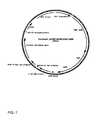
- FIG. 7shows an example of a plasmid construct suitable for gene therapy of an ocular disease according to the present invention.
- the plasmidcontains a nucleotide sequence encoding for pigment epithelium-derived factor (PEDF) and a nucleotide sequence encoding for enhanced green fluorescent protein (eGFP) under control of a cytomegalovirus (CMV) promoter, the two sequences linked by an internal ribosome entry site (IRES) coding sequence.
- PEDFpigment epithelium-derived factor
- eGFPenhanced green fluorescent protein
- CMVcytomegalovirus
- FIG. 8shows ocular regions suitable for transplantation according to the present invention.
- FIG. 9-10show examples of electron avalanche-mediated transfection according to the present invention.
- the present inventionprovides an ex vivo gene therapy method based on a novel method of introducing DNA into cells called the avalanche method.
- a mechanical stress wave synchronized with a pulse of electric currentcan be produced and applied to cells or tissue, as shown in FIG. 1 .
- FIG. 1A-Cshows three stages that occur when a high voltage is applied to an electrode 110 covered by insulation 120 .
- Electrode 110is situated in tissue culture well 130 , with conductive liquid medium 132 , cells 134 , and nucleic acid 136 . (although cells are pictured in this figure, tissue could also be used).
- an electric field 140is generated around the un-insulated portion of electrode 110 .
- vapor bubble 150( FIG. 1B ).
- vapor bubble 150disconnects the surface of electrode 110 from conductive medium 132 , so that the electric current stops flowing, and the electric field on the target cells is terminated.
- the vapor in the bubblecan be ionized to form ionized vapor 160 , which restores the electrical conductivity, as shown in FIG. 1C .
- Ionized vapor 160also known as plasma, forms a kind of virtual electrode with electric field 170 .
- the formation of the vapor bubble, and its subsequent collapsecauses a propagating shock wave through the medium, exposing the cells or tissue to mechanical stress 180 .
- the combination of the shock wave and the electric fieldleads to permeabilization of cells 132 , such that nucleic acid 136 may enter cells 132 ( FIG. 1D ).
- this techniqueelectron avalanche-mediated transfection, or, for simplicity, the avalanche method.
- FIG. 1shows when the electrode produces a relatively uniform electric field.
- electrodes with a very uneven electric fieldmay be used, so that the vapor cavity formed at the apex does not cover the whole surface of the electrode with a lower electric field. This way the electric current to the medium will not be completely disconnected.
- One example of an electrode geometry with a non-uniform electric fieldis a cylindrical probe, such as a wire, with a sharp end.
- FIG. 2Ashows an image of a wire electrode 210 producing a plasma discharge 220 . As can be seen from FIG. 2A , the plasma discharge is clearly visible. It is also clearly audible.
- FIG. 2Bshows current 230 and voltage 240 versus time when a voltage is applied to a wire probe.
- the wire probewas 50 ⁇ m in diameter and electrical pulses of up to 600 V were used to produce an electric field at the tip of the wire of about 30 kV/cm.
- FIG. 2Bshows that when a voltage is applied to such a probe, the initial 20 ⁇ s of the waveform exhibits reduction of the current due to beginning of vaporization. This is followed by stabilization of conductivity following ionization of the vapor cavity.
- the ionized vapor cavityserves as a transient electrode, which can greatly exceed the size of the probe, as shown in FIG. 2A .
- the distribution of the electric fieldbecomes much more uniform than the one generated initially on the small wire electrode, thus leading to more uniform electroporation of the target cells or tissue.
- FIG. 2Cshows, for different diameters of electrodes, the field strength (kV/mm) along the length of electrode 230 covered by insulator 240 .
- the electrode diameter indicated by the solid line 250is 10 ⁇ m
- the dotted line 260is 25 ⁇ m
- the dashed line 270is 50 ⁇ m.
- 600 Vwas applied to the electrode.
- FIG. 2Cshows that for a cylindrical electrode with a sharp tip, there is a rapid decrease in electric field as one moves farther away from the tip of the electrode.
- the strength of the electric field at the apex of the electrodecan be varied by changing the electrode diameter.
- FIG. 3shows a version of a probe in which active electrodes 310 are plated on a substrate 320 .
- FIG. 3Ashows a top view and FIG. 3B shows a side view of the probe.
- substrate 320is surrounded by return electrode 330 .
- the pattern of active electrodes 310 on substrate 320forms the necessary proportion between electric field 340 and mechanical stress wave 350 due to plasma discharge 352 .
- the probe in FIG. 3has a singularity of the electric field 340 at the edges 312 of active electrodes 310 . Singularities serve as ignition points for plasma discharge 352 and generation of mechanical stress wave 350 .
- plasmaoccupies the whole volume of the vapor cavity.
- FIG. 4Another probe implementation, which allows separate control of mechanical stress wave 450 and electric field 440 , is shown in FIG. 4 .
- FIG. 4Ais a top view
- FIG. 4Bis a side view
- two types of active electrodes, 410 and 412are patterned on substrate 420 , with return electrode 430 surrounding substrate 420 .
- Electrodes 412may be driven to generate an electric field 440
- electrodes 410may be driven to generate plasma 452 and mechanical stress wave 450 .
- Pulsma 454also generates an accompanying electric field, not shown).
- Separate control of the amplitude of stress wave and electric fieldmight be desirable for optimization of permeabilization. Generating them on the same electrode will make these values mutually dependent, while generation on two separate electrodes may provide independent control of these phenomena.
- FIG. 5shows an example of a transfection device suitable for molecular delivery of nucleic acid to adherent cells or tissue according to the present invention.
- cells 510are growing on an adherent surface 520 placed in a nonporous substrate 530 , such as a tissue culture plate.
- Adherent surface 520may be, for example, a tissue culture insert made of porous material such as polycarbonate. Cells could also be grown directly on nonporous substrate 530 .
- a gelatinous matrix and/or feeder layermay also be present (not shown).
- a probe 540 with pillar electrodes 542 , return electrode 544 , and connection 546 to a voltage source(not shown) is brought into a solution 550 containing nucleic acid 560 .
- Pillar electrodes 542are positioned a finite distance from cells 510 , e.g. about 1 mm. This finite distance is preferably in the range of about 0.5 mm to about 2 cm. In the embodiment shown, the return electrode 544 extends beyond pillar electrodes 542 a distance equal to this finite distance such that the finite distance is defined when the return electrode 544 is touching adherent surface 520 . However, this distance could be defined by any substance. In addition, pillar electrodes 542 are preferably about 0.5 mm to about 2 cm apart.
- FIG. 6shows an example of a transfection device suitable for molecular delivery of nucleic acid to cells or tissue in solution according to the present invention.
- cells or tissue 610are suspended in solution 620 with nucleic acids 630 in cuvette 640 .
- Cuvette 640contains return electrode 642 , pillar electrodes 644 , and connection 646 to a voltage source (not shown).
- pillar electrodes 644are preferably between about 0.5 mm and about 2 cm apart to provide adequate coverage of the solution volume. In this arrangement, the pillar electrodes could be simultaneously or alternately active.
- the electric field on the electrode surfaceshould be sufficient for rapid vaporization of the liquid medium.
- the electric fieldshould be high enough to induce ionization of the vapor. In this way, both a mechanical stress wave and an electric field can be synchronized, with maximal intensity at the surface of the electrode.
- the plasma dischargemust be controlled in order to maximize transfection efficiency and minimize cell death.
- applied voltagesare preferably in the range of about 1 V to about 10 kV, more preferably between about 100 V and about 1 kV.
- Applied voltagepreferably results in an electric field between about 1 to about 100 kV/cm, more preferably about 10 to about 50 kV/cm, and most preferably about 30 kV/cm.
- Pulse durationis preferably in the range of about 1 ns to about 100 ms, more preferably between about 100 ns and about 1 ms.
- Either monophasic or biphasic pulsesare suitable for the purposes of the present invention. However, biphasic pulses are preferred as they lead to less gas formation, nerve and muscle response, and electrode erosion. Any number of pulses may be used according to the present invention. The number of pulses is preferably between about 1 and 100, more preferably between about 1 and 50. When multiple pulses are used, the frequency of pulses should be in the range of about 0.1 Hz to about 1 kHz. Preferably, the frequency is less than about 1 kHz to prevent heat accumulation.
- Any cell or tissuemay be suitable for practicing the invention.
- Examplesinclude primary cells, primary tissues, and cell lines.
- Preferred cellsinclude conjunctival fibroblasts, epithelial cells and scleral cells.
- Preferred tissuesinclude conjunctival tissue and scleral tissue.
- Preferred cell linesinclude fibroblast cell lines and muscle cell lines. The cells and tissue are preferably autologous or allogeneic.
- the method of the present inventioninvolves obtaining tissue from a subject having or at risk of developing an undesirable eye condition.
- the conditioncan range from a minor or nuisance condition, such as dry eye, to a more serious disease with possible vision loss, such as age-related macular degeneration.
- tissue from the patentis harvested in an invasive, minimally invasive, or non-invasive procedure, the degree of invasiveness dictated, in part, by the tissue to be harvested.
- Candidate tissuesare preferably those capable of transfection and production of a protein, and that are capable of survival in the transplanted environment.
- tissueis harvested from the eye and it is contemplated that any tissue in the eye may be harvested in any feasible manner.
- conjunctival fibroblastscan be excised from the eye by, for example, anesthetizing the conjunctiva with a topical agent such as propraracaine, cleansing and preparing the area with betadine or another cidal agent, and then taking a snip biopsy with a pair of toothed forceps and Wescott scissors. Subconjunctival anesthesia may be preferred by some surgeons or patients. The excised conjunctiva or other tissue is removed and then transfected either in the operating room or in an adjacent area then reimplanted in the appropriate location in the same session.
- a topical agentsuch as propraracaine
- tissuecan be maintained under sterile conditions, taken to a sterile facility where transfection and subsequent subculture and testing can be performed, and reimplantation of the tissue performed one to three weeks later.
- a similar procedurecan be performed on the sclera, except it may be preferred to use subconjunctival rather than topical anesthesia.
- tissue substratessuch as iris pigment epithelium may be substituted for conjunctiva or sclera.
- a tissue sample of any size or dimensioncan be removed, typically a tissue sample of approximately one cubic millimeter of tissue or less is obtained. After removal of the tissue, the site can sutured or treated as needed.
- the tissueis harvested from a donor, rather than the patient.
- donor tissuewould be isolated and transfected as described above for autologous transplantation. It may be transplanted after transfection in the same session, or, alternatively the tissue can be maintained under sterile conditions, taken to a sterile facility where transfection and subsequent subculture and testing can be performed, and reimplantation of the tissue performed one to three weeks later.
- donor tissuemay be tested to determine suitability of transplantation, for example for viral or other pathogens or immunocompatibility with recipient.
- the nucleic acidmay encode, for example, a therapeutic protein or an RNAi cassette, such as a shRNA.
- the nucleic acidmay be used to repair or replace an endogenous gene, for example DNA used for homologous recombination, or an oligonucleotide used for gene repair. Modifications include, for example, modifying expression levels of the gene and/or replacing a mutant gene with a wild-type copy of the gene.
- the nucleic acidmay be DNA or RNA, but is preferably DNA.
- the nucleic acidis a DNA construct, in particular a cDNA or synthetic DNA, and can be further modified to improve transcription and/or translation in the host cell, or to reduce or minimize gene silencing.
- the nucleic acid constructmay comprise, operably linked, a promoter region, a nucleotide, and optionally, a termination signal.
- this constructis part of a plasmid.
- the cells or tissueare stably transfected so that the transplanted cells or tissue may act, for example, as a bio-factory to produce a therapeutic protein for a long period of time.
- nucleic acid sequencescan be introduced into the cells or tissue, including multiple copies of the same nucleic acid sequence and/or multiple copies of differing nucleic acid sequences encoding for different therapeutic or marker proteins.
- each nucleic acid sequenceis present on a separate polynucleotide construct, plasmid, or vector.
- both nucleic acid sequencesare present on one polynucleotide construct, plasmid, or vector, with each sequence under the control of a separate promoter.
- both nucleic acid sequencesare present on one polynucleotide construct, plasmid, or vector, with the polynucleotide structured so that it is bicistronic and where both nucleic acid sequences are under the control of a single promoter.
- each sequencecan be under the control of a separate promoter or can be under the control of a single promoter.
- a second nucleic acid sequence encoding, for example, a second therapeutic protein or a markeris included in the construct. Expression of this gene may be constitutive; in the case of a selectable marker this may be useful for selecting successfully transfected cells or for selecting cells or transfected populations of cells that are producing particularly high levels or optimal therapeutic levels of the protein.
- a selectable markermay be used to provide a means for enriching for transfected cells or positively selecting for those cells which have been transfected, before reintroducing the cells into the patient, as will be described below.
- Markersmay include selectable drug resistance genes, metabolic enzyme genes, fluorescent proteins, bioluminescent proteins, or any other markers known in the art.
- Exemplary fluorescent proteinsinclude, but are not limited to: green fluorescent protein, cyan fluorescent protein, yellow fluorescent protein, DsRed fluorescent protein, AsRed fluorescent protein, HcRed fluorescent protein, and maxFP-green protein.
- a marker geneis included in the vector construct, it will be appreciated that the marker can be used to quantify the amount of fluorescence after transfection and/or before transplantation and/or after transplantation.
- Quantitative determination of fluorescencecan be undertaken after transfection but before transplanting the tissue using, for example, fluorescence microscopy, flow cytometry, or fluorescence-activated cell sorting (FACS) analysis, in order to quantify the expression of fluorescence markers ex vivo.
- FACSfluorescence-activated cell sorting
- monitoring of the extent of fluorescence, as a measure of production of the therapeutic proteincan be done by examining the patient with a fluorescent ophthalmoscope or a surgical microscope equipped for fluorescence imaging, and can be documented with a CCD camera.
- the marker genecan be used to indicate levels of transgene expression and can be monitored by a non-invasive or a minimally invasive procedure. If marker gene expression decreases, another tissue implant can be inserted into the patient to increase the level of therapeutic protein. By using a marker gene, diminished expression of the therapeutic protein can be recognized early, rather than waiting until decreased levels of the therapeutic gene lead to disease progression.
- IRESinternal ribosome entry site
- picornaviral RNAe.g. from picornaviral RNA
- Retroviruses incorporating IRES sequencesare known in the art, for example in U.S. Pat. No. 5,665,567. Briefly, in bicistronic or multicistronic vectors, the individual reading frames of the gene segments encoding the proteins lie on the transcription unit (expression unit). Expression of each cistron is effected using a single promoter, in conjunction with a specific nucleic acid sequence, typically untranslated regions of individual picorna viruses, e.g.
- IRESinternal ribosomal entry site
- the cells or tissuecan be transfected with a plasmid having one promoter that drives the expression of a first therapeutic protein, such as pigment epithelium-derived factor (PEDF), and of a selectable marker, such as a fluorescent protein like enhanced green fluorescent protein (eGFP) under control of a cytomegalovirus (CMV) promoter.
- a first therapeutic proteinsuch as pigment epithelium-derived factor (PEDF)
- eGFPenhanced green fluorescent protein
- CMVcytomegalovirus
- the CMV promoteris positioned at the 5′ end of the construct. Downstream of the 3′ end of the CMV promoter is the PEDF nucleotide sequence that encodes for PEDF protein.
- IRES sitewhich is designed to allow translation of multiple genes on an mRNA transcript.
- the eGFP coding sequenceFollowing the IRES site in the 3′ direction is the eGFP coding sequence. The IRES will allow translation of eGFP as well as translation of PEDF.
- the promoter region of the constructcan be chosen from among all promoter regions that are functional in mammalian cells, in particular human cells.
- the promotercan be a strong or weak promoter, a constitutive or a regulated/inducible promoter, a ubiquitous or selective promoter.
- the promotercan be of different origin such as cellular, viral, artificial, and the like.
- Particular types of promotersare house-keeping promoters, i.e., promoters from cellular genes expressed in mammalian tissues or cells, or viral promoters (CMV, LTR, SV40, etc.).
- the promoter regioncan be modified artificially to include enhancer element(s), inducibility element(s) and the like.
- the promoter, secretion and termination region sequencescan be selected and adapted by the skilled artisan based on the polypeptide, the pathology, the vector used, etc.
- the nucleic acid constructcan be inserted into various kinds of vectors such as plasmids, episomes, artificial chromosomes and the like.
- the nucleic acid constructcan optionally include a secretion signal, positioned between the promoter and coding regions, which allows, or facilitates, the secretion of the polypeptide outside of the cells.
- the secretion signalmay be homologous with respect to the polypeptide (i.e., from the same gene) or heterologous thereto (i.e., from any other gene encoding a secreted polypeptide, in particular a mammalian gene, or artificial).
- Examples of secretion signalsinclude the signal peptide of vascular endothelial growth factor (VEGF), pre pro nerve growth sequence (NGS), and the like.
- VEGFvascular endothelial growth factor
- NGSpre pro nerve growth sequence
- One approachinvolves a circular vector carrying a recombination site and the polynucleotide sequence encoding for the therapeutic protein, shRNA, etc., and the transfection is accompanied by introduction of a recombinase that facilitates recombination between the vector's recombination site and a second recombination site in the genome of the cell being transfected.
- Constructs carrying a recombination sitesuch as a phiC31 attB site, are described, for example, in U.S. Pat. No. 6,632,672, which is incorporated by reference herein.
- nucleic acid constructsare comprised of the phiC31 integrase system (described in U.S. Pat. Nos. 6,632,672 and 6,808,925, which are incorporated by reference herein) to achieve site-specific integration into a target genome of interest.
- Bacteriophage phi-C31 integtraserecognizes pseudo-recombination sites present in eukaryotic cells.
- phiC31 integrase and a vector carrying a phiC31 wild-type recombination siteare placed into the cell.
- the wild-type recombination sequencealigns itself with a sequence in the eukaryotic cell genome and the phiC31 integrase facilitates a recombination that results in integration of a heterologous gene into the eukaryotic genome.
- any attB site, any attP site, or any pseudo att siteis present on any nucleotide sequence used to introduce genetic material into the genome of the harvested or cultured cells.
- the method of integrating a polynucleotide sequence into a genome of a cellcomprises introducing into the cell (i) a circular targeting construct, comprising a first recombination site and a polynucleotide sequence of interest, and (ii) a phiC31 integrase, native or modified, wherein the genome of the cell comprises a second recombination site (ie. a pseudo att site) native to the human genome. Recombination between the first and second recombination sites is facilitated by the site-specific integrase.
- the therapeutic gene and the attB sequenceare preferably introduced into the target cell as circular plasmid DNA.
- the integrasemay be introduced into the target cell (i) as DNA encoding the integrase on a second plasmid, (ii) mRNA encoding the integrase, or (iii) in polypeptide form.
- phiC31Once phiC31 is introduced into the cell, the cell is maintained under conditions that allow recombination between the first and second recombination sites and the recombination is mediated by the phiC31 integrase. The result of the recombination is site-specific integration of the polynucleotide sequence of interest in the genome of the cell.
- a plasmidis constructed having a cytomegalovirus (CMV) promoter that drives the expression of a therapeutic protein, pigment epithelium-derived factor (PEDF), and as a marker, enhanced green fluorescent protein (eGFP).
- CMVcytomegalovirus
- PEDFpigment epithelium-derived factor
- eGFPenhanced green fluorescent protein
- IRESIn the 3′ direction of the PEDF nucleotide sequence is an IRES site, followed in the 3′ direction by the eGFP coding sequence.
- the IRESallows translation of eGFP as well as translation of PEDF.
- the plasmidwhich also includes an attB nucleic acid sequence, is detailed in Example 1 and the plasmid sequence is identified herein as SEQ ID NO: 1.
- Transfection of a wide variety of genes encoding for therapeutic proteinsis contemplated, and preferred candidate genes include genes that encode for diffusible proteins that act extracellularly to have a therapeutic effect.
- preferred candidate genesinclude genes that encode for diffusible proteins that act extracellularly to have a therapeutic effect.
- a nucleic acid sequence coding for a protein with anti-angiogenic activity or with neurotrophic activityis transfected into human cells.
- Exemplary proteinsinclude, but are not limited to, pigment epithelium-derived factor (PEDF), truncated soluble VEGF receptor sFlt-1, truncated soluble VEGF receptor sFlk-1, VEGFR-1, VEGFR-2, angiostatin, endostatin, tissue inhibitor of metalloprotease 3 (TIMP-3), ExTek, ciliary neurotrophic factor (CNTF), brain-derived neurotrophic factor (BDNF), bone morphogenetic protein 4 (BMP4), alpha fibroblast growth factor (aFGF), beta fibroblast growth factor (bFGF), and any protein having activity on or within the compliment factor H pathway.
- Preferred biologically active polypeptidesexhibit neurotrophic and/or anti-angiogenic activity.
- the most preferred biologically active polypeptidesare autogenic and thus do not invoke an immune response in the subject or are known in the art not to invoke an immune response.
- human cellsare genetically modified to contain a recombinant nucleic acid construct that directs the cells to produce the therapeutic protein encoded by the recombinant nucleic acid.
- the cellscan be immediately transplanted into the subject or can be cultured in vitro for a period of time.
- mammalian cells modified with a vector containing at least one nucleic acid sequence coding for a therapeutic protein and another nucleic acid sequence coding for a marker geneare prepared for transplantation.
- a selection stepcan be performed in order to isolate the cells that effectively contain the recombinant nucleic acid construct and express the polypeptide.
- the selection stepwill depend in part on the marker gene and can involve measuring fluorescence, screening for antibiotic resistance, or the like.
- Cells expressing the marker geneare selected for transplantation.
- the treatment methodis performed on a subject over more than one visit to the medical provider.
- the tissueis harvested.
- the tissue cellsare transfected and cultured in vitro, during which time the level of expression can be monitored and stably-transfected cells from the tissue selected, by, for example, quantifying expression of a marker or of the desired protein by methods noted above for measuring marker expression, for transplantation.
- the subjectreturns to the medical provider for a second visit during which the transfected tissue is transplanted.
- tissuecan be obtained, transfected, and transplanted during a single patient visit to a medical provider.
- the level of expression of a marker or the desired therapeutic proteincan be monitored in vivo, by methods mentioned above, such as ophthalmoscope or a surgical microscope.
- one or more nucleotide sequences coding for a therapeutic protein and one nucleotide sequence coding for a marker geneare present in the same polynucleotide vector construct.
- the marker geneis coupled to the therapeutic gene by an IRES sequence. Quantification of the degree of fluorescence emitted from a cell or group of clonal cells would correlate with the amount of expression of the therapeutic protein, enabling selection of stably transfected cells or monitoring of protein expression after transplantation.
- FIG. 8is a diagram showing an eye 800 in cross-sectional view, and indicating some of the preferred sites for placing genetically modified cells or tissue into the patient. Identified anatomical features are retina 830 , sclera 840 , optic nerve 850 , cornea 860 , pupil 870 and iris 880 .
- Sites in eye 800 preferred for implanting the transfected cells or tissueinclude the vitreous humor 810 , near the pars plana 820 , near the posterior retina 832 , or sub-sclerally 842 .
- Other sites for implanting tissuewhich are not specifically indicated in FIG. 8 , include the choroid, retinal pigment epithelium (RPE), and near the macula epi-retinally, sub-retinally, or intra-retinally.
- RPEretinal pigment epithelium
- the transfected cells or tissueare implanted into the subject in the absence of an encapsulating member, such as a polymer capsule or a so-called “cage”.
- an encapsulating membersuch as a polymer capsule or a so-called “cage”.
- encapsulation of the tissue or cells within a cageis not necessary for immunosuppression.
- encapsulationcould be used to enhance graft survival and/or to reduce possible splintering of cells away from the graft to other sites in the eye.
- a number of cage designshave been proposed for ophthalmologic use for various purposes, as described in U.S. Pat. Nos. 6,500,449 and 6,663,894.
- the cagewould be able to house the tissue or cell transplant and would have pores large enough for proteins to diffuse out, but small enough so that cells could not enter or leave.
- the cagemay contain a matrix or other materials to support cell survival and cell anchoring to prevent cell migration to other sites.
- SEQ ID NO: 1includes a cytomegalovirus (CMV) promoter (1-589 bp), a nucleotide sequence encoding for pigment epithelium-derived factor (PEDF; 590-2131 bp), an internal ribosome entry site (IRES) coding sequence (b2151-2735 bp), and a nucleotide sequence encoding for enhanced green fluorescent protein (eGFP; bp 2739-3455), an SV40 polyA sequence (3612-3662 bp), a phi C31 attB site (3952-4245 bp), a bacterial kan promoter (4541-4576 bp), SV40 origin and promoter enhancer (4653-4955 bp), neo for G418 selection (5004-5798 bp), and an pUC origin (6383-7026 bp).
- CMVcytomegalovirus
- PEDFpigment epithelium-derived factor
- IVSinternal ribo
- vector pIRES-EGFPcommercially available from Clontech. Cut the vector with the restriction enzyme BsaI (New England Biolabs) to linearize the vector, make blunt ends (e.g., using DNA Polymerase I, Large (Klenow) Fragment, New England Biolabs), and treat with a phosphatase to remove the phosphate groups (e.g., using calf intestinal phosphatase, New England Biolabs). Ligate this vector to the fragment containing attB when pTA-attB+ is cleaved with EcoRJ and then its ends blunted, to form the plasmid pIRES-EGFP-attB.
- BsaINew England Biolabs
- the second cloning stepuse PCR amplification with primers designed to amplify the PEDF gene from human cDNA.
- luciferase marker genewas transfected into conjunctiva tissue.
- Conjunctival tissuewas explanted from adult New Zealand White rabbits and placed in tissue culture dishes. All samples were placed in 1 mL phosphate buffered saline solution with 100 micrograms of plasmid DNA encoding the luciferase gene under a CMV promoter. All samples were cultured in Dulbecco's Modified Eagle Medium (DMEM) plus 10% serum and antibiotic/antimicotic for 24 hours after transfection. Samples were then treated with luciferin substrate (150 micrograms luciferin per ml medium) and imaged using the IVIS-200 system (Xenogen Corp.).
- DMEMDulbecco's Modified Eagle Medium
- the conjunctival tissuewhich contained conjunctival fibroblasts, was transfected using electron-avalanche mediated transfection with a luciferase marker gene.
- a control sample of tissuewas contacted with the luciferase gene in the absence of electron-avalanche mediated transfection. Twenty-four hours after transfection, bioluminescence was measured.
- the tissue transfected with electron-avalanche mediated transfectionemitted 2.2 ⁇ 10 5 photons/sec, two orders of magnitude higher than the cells transfected in the absence of the electron-avalanche mediated transfection (4.6 ⁇ 10 3 photons/sec). Background emission was measured at 3.7 ⁇ 10 3 photons/sec.
- CAMis a live, readily available, and inexpensive tissue. Its epithelial layer is uniform and has high resistance, making it a good model for epithelial cell layers, such as retinal pigment epithelium.
- 100 ⁇ g of pNBL2 plasmid DNA encoding the luciferase genewas pipetted onto the CAM, and pulses were applied. Specifically, a 250- ⁇ s, 150-V phase, followed by a 5-ms, 5-V phase in the same polarity was applied. Optimal results were achieved with 50 cycles applied at 1 Hz. The tissue was then cultured and assayed for luciferase bioluminescence. Luciferase expression using this method was about 10 4 photons/s.
- a 50- ⁇ m wire microelectrode 1 mm in lengthwas used to apply a series of symmetric biphasic pulses, with each phase 250 ⁇ s in duration and 600 V in amplitude.
- the microelectrodewas scanned over a 4-mm 2 area, and approximately 50 pulses were applied.
- the resultant luciferase expressionwas about 10 9 photons/s, 10,000-fold higher than levels seen with conventional electroporation.
Landscapes
- Health & Medical Sciences (AREA)
- Genetics & Genomics (AREA)
- Life Sciences & Earth Sciences (AREA)
- Engineering & Computer Science (AREA)
- Organic Chemistry (AREA)
- Chemical & Material Sciences (AREA)
- Zoology (AREA)
- Wood Science & Technology (AREA)
- Bioinformatics & Cheminformatics (AREA)
- Biotechnology (AREA)
- Biomedical Technology (AREA)
- General Engineering & Computer Science (AREA)
- Microbiology (AREA)
- Biochemistry (AREA)
- General Health & Medical Sciences (AREA)
- Plant Pathology (AREA)
- Molecular Biology (AREA)
- Biophysics (AREA)
- Physics & Mathematics (AREA)
- Cell Biology (AREA)
- Sustainable Development (AREA)
- Apparatus Associated With Microorganisms And Enzymes (AREA)
- Medicines That Contain Protein Lipid Enzymes And Other Medicines (AREA)
- Micro-Organisms Or Cultivation Processes Thereof (AREA)
- Electrotherapy Devices (AREA)
Abstract
Description
Claims (20)
Priority Applications (8)
| Application Number | Priority Date | Filing Date | Title |
|---|---|---|---|
| US11/505,249US8101169B2 (en) | 2005-02-23 | 2006-08-15 | Ocular gene therapy using avalanche-mediated transfection |
| US11/526,153US7923251B2 (en) | 2005-02-23 | 2006-09-22 | Method and apparatus for avalanche-mediated transfer of agents into cells |
| JP2008556302AJP2009527321A (en) | 2006-02-22 | 2006-09-22 | Methods and apparatus for avalanche-mediated introduction of drugs into cells |
| EP06804041AEP1989317A4 (en) | 2006-02-22 | 2006-09-22 | METHOD AND APPARATUS FOR TRANSFERRING AVALANCHE-MEDIATED AGENTS INTO CELLS |
| PCT/US2006/037010WO2007106136A2 (en) | 2006-02-22 | 2006-09-22 | Method and apparatus for avalanche-mediated transfer of agents into cells |
| AU2006340055AAU2006340055A1 (en) | 2006-02-22 | 2006-09-22 | Method and apparatus for avalanche-mediated transfer of agents into cells |
| CA002643078ACA2643078A1 (en) | 2006-02-22 | 2006-09-22 | Method and apparatus for avalanche-mediated transfer of agents into cells |
| US13/042,934US8283171B2 (en) | 2005-02-23 | 2011-03-08 | Method and apparatus for avalanche-mediated transfer of agents into cells |
Applications Claiming Priority (4)
| Application Number | Priority Date | Filing Date | Title |
|---|---|---|---|
| US65555905P | 2005-02-23 | 2005-02-23 | |
| US70848605P | 2005-08-15 | 2005-08-15 | |
| US36098406A | 2006-02-22 | 2006-02-22 | |
| US11/505,249US8101169B2 (en) | 2005-02-23 | 2006-08-15 | Ocular gene therapy using avalanche-mediated transfection |
Related Parent Applications (1)
| Application Number | Title | Priority Date | Filing Date |
|---|---|---|---|
| US36098406AContinuation | 2005-02-23 | 2006-02-22 |
Related Child Applications (2)
| Application Number | Title | Priority Date | Filing Date |
|---|---|---|---|
| US36098406AContinuation-In-Part | 2005-02-23 | 2006-02-22 | |
| US13/042,934Continuation-In-PartUS8283171B2 (en) | 2005-02-23 | 2011-03-08 | Method and apparatus for avalanche-mediated transfer of agents into cells |
Publications (2)
| Publication Number | Publication Date |
|---|---|
| US20070059835A1 US20070059835A1 (en) | 2007-03-15 |
| US8101169B2true US8101169B2 (en) | 2012-01-24 |
Family
ID=38509932
Family Applications (1)
| Application Number | Title | Priority Date | Filing Date |
|---|---|---|---|
| US11/505,249Expired - Fee RelatedUS8101169B2 (en) | 2005-02-23 | 2006-08-15 | Ocular gene therapy using avalanche-mediated transfection |
Country Status (6)
| Country | Link |
|---|---|
| US (1) | US8101169B2 (en) |
| EP (1) | EP1989317A4 (en) |
| JP (1) | JP2009527321A (en) |
| AU (1) | AU2006340055A1 (en) |
| CA (1) | CA2643078A1 (en) |
| WO (1) | WO2007106136A2 (en) |
Cited By (10)
| Publication number | Priority date | Publication date | Assignee | Title |
|---|---|---|---|---|
| WO2019008335A1 (en) | 2017-07-07 | 2019-01-10 | Avacta Life Sciences Limited | Scaffold proteins |
| WO2021030251A1 (en) | 2019-08-12 | 2021-02-18 | Purinomia Biotech, Inc. | Methods and compositions for promoting and potentiating t-cell mediated immune responses through adcc targeting of cd39 expressing cells |
| EP3799879A1 (en) | 2015-01-09 | 2021-04-07 | OncoSec Medical Incorporated | Gene therapy using co-stimulatory molecule and cytokine for the treatment of malignancies |
| WO2021074695A1 (en) | 2019-10-16 | 2021-04-22 | Avacta Life Sciences Limited | PD-L1 INHIBITOR - TGFβ INHIBITOR BISPECIFIC DRUG MOIETIES. |
| EP3881860A1 (en) | 2015-03-26 | 2021-09-22 | OncoSec Medical Incorporated | Method for the treatment of malignancies |
| WO2021249786A1 (en) | 2020-06-09 | 2021-12-16 | Avacta Life Sciences Limited | Sars-cov2 diagnostic polypeptides and methods |
| WO2022234003A1 (en) | 2021-05-07 | 2022-11-10 | Avacta Life Sciences Limited | Cd33 binding polypeptides with stefin a protein |
| WO2023057567A1 (en) | 2021-10-07 | 2023-04-13 | Avacta Life Sciences Limited | Pd-l1 binding affimers |
| WO2023057946A1 (en) | 2021-10-07 | 2023-04-13 | Avacta Life Sciences Limited | Serum half-life extended pd-l1 binding polypeptides |
| WO2023212483A1 (en) | 2022-04-29 | 2023-11-02 | Purinomia Biotech, Inc. | Methods and compositions for treating eosinophil driven diseases and disorders |
Families Citing this family (21)
| Publication number | Priority date | Publication date | Assignee | Title |
|---|---|---|---|---|
| US9034650B2 (en) | 2005-02-02 | 2015-05-19 | Intrexon Corporation | Site-specific serine recombinases and methods of their use |
| ATE526421T1 (en)* | 2005-02-14 | 2011-10-15 | Univ Iowa Res Found | METHODS AND REAGENTS FOR THE TREATMENT AND DIAGNOSIS OF AGE-RELATED MACULAR DEGENERATION |
| JP5032500B2 (en)* | 2006-01-03 | 2012-09-26 | アルコン,インコーポレイティド | System for dissociation and removal of proteinaceous tissue |
| EP2130915A4 (en)* | 2007-02-28 | 2010-04-21 | Senju Pharma Co | Cell capable of expressing lacritin at high level |
| DE102008008614A1 (en)* | 2008-02-12 | 2009-08-13 | Leibniz-Institut für Plasmaforschung und Technologie e.V. | Plama device for the selective treatment of electroporated cells |
| DE102008047399A1 (en)* | 2008-09-16 | 2010-04-15 | MAX-PLANCK-Gesellschaft zur Förderung der Wissenschaften e.V. | Electrode device, generator device and method for generating electricity by membrane potential dissipation |
| US20110118729A1 (en)* | 2009-11-13 | 2011-05-19 | Alcon Research, Ltd | High-intensity pulsed electric field vitrectomy apparatus with load detection |
| US20110118734A1 (en)* | 2009-11-16 | 2011-05-19 | Alcon Research, Ltd. | Capsularhexis device using pulsed electric fields |
| US20110135626A1 (en)* | 2009-12-08 | 2011-06-09 | Alcon Research, Ltd. | Localized Chemical Lysis of Ocular Tissue |
| US20110144562A1 (en)* | 2009-12-14 | 2011-06-16 | Alcon Research, Ltd. | Localized Pharmacological Treatment of Ocular Tissue Using High-Intensity Pulsed Electrical Fields |
| WO2011081897A1 (en)* | 2009-12-15 | 2011-07-07 | Alcon Research, Ltd. | High-intensity pulsed electric field vitrectomy apparatus |
| US8546979B2 (en) | 2010-08-11 | 2013-10-01 | Alcon Research, Ltd. | Self-matching pulse generator with adjustable pulse width and pulse frequency |
| WO2012086702A1 (en)* | 2010-12-24 | 2012-06-28 | タカラバイオ株式会社 | Method for gene introduction |
| JP6317927B2 (en)* | 2012-01-09 | 2018-04-25 | ムー・メディカル・デバイスズ・エルエルシーMoe Medical Devices Llc | Plasma assisted skin treatment |
| KR20140128378A (en)* | 2012-03-02 | 2014-11-05 | 도꾸리쯔교세이호징 가가꾸 기쥬쯔 신꼬 기꼬 | Bubble-spraying member and method for producing same, gas-liquid-spraying member and method for producing same, local ablation device and local ablation method, injection device and injection method, plasma-bubble-spraying member, and healing device and healing method |
| JP2016187335A (en)* | 2015-03-30 | 2016-11-04 | 国立大学法人東北大学 | Plasma generator for use in vivo, transgenic apparatus, drug introducing apparatus, and endoscope |
| EP3335760B1 (en)* | 2015-08-10 | 2021-09-29 | Ajou University Industry-Academic Cooperation Foundation | Nitrogen-based, low-temperature atmospheric pressure plasma for treating muscle damage |
| JP7397489B2 (en)* | 2018-07-23 | 2023-12-13 | 株式会社ベックス | Bubble ejection method, power supply device, and bubble ejection device |
| WO2020085281A1 (en)* | 2018-10-26 | 2020-04-30 | 国立大学法人九州大学 | Bubble jetting method, bubble jetting device and bubble jetting apparatus |
| JP7121419B2 (en)* | 2018-10-29 | 2022-08-18 | 株式会社ベックス | Bubble ejection method, bubble ejection power supply device, and bubble ejection device |
| WO2025070172A1 (en)* | 2023-09-29 | 2025-04-03 | 積水化学工業株式会社 | Container for plasma-type cell processing device, plasma-type cell processing device, and molecule introduction method |
Citations (31)
| Publication number | Priority date | Publication date | Assignee | Title |
|---|---|---|---|---|
| US5019034A (en) | 1988-01-21 | 1991-05-28 | Massachusetts Institute Of Technology | Control of transport of molecules across tissue using electroporation |
| US5028597A (en) | 1986-04-07 | 1991-07-02 | Agency Of Industrial Science And Technology | Antithrombogenic materials |
| US5128257A (en) | 1987-08-31 | 1992-07-07 | Baer Bradford W | Electroporation apparatus and process |
| US5304486A (en) | 1987-10-09 | 1994-04-19 | Baylor College Of Medicine | Method of and apparatus for cell portion and cell fusion using radiofrequency electrical pulse |
| US5318514A (en) | 1992-08-17 | 1994-06-07 | Btx, Inc. | Applicator for the electroporation of drugs and genes into surface cells |
| US5389069A (en) | 1988-01-21 | 1995-02-14 | Massachusetts Institute Of Technology | Method and apparatus for in vivo electroporation of remote cells and tissue |
| US5468223A (en) | 1992-11-30 | 1995-11-21 | C.N.R.S. Paris | Electrochemotherapy |
| US5665567A (en) | 1992-08-27 | 1997-09-09 | Beiersdorf Ag | Preparation of heterodimeric PDGF-AB using a bicistronic vector system in mammalian cells |
| US5688233A (en) | 1992-08-17 | 1997-11-18 | Genetronics, Inc. | Electronincorporation enhanced transdermal delivery of molecules |
| US5749847A (en) | 1988-01-21 | 1998-05-12 | Massachusetts Institute Of Technology | Delivery of nucleotides into organisms by electroporation |
| US5964726A (en) | 1994-02-25 | 1999-10-12 | Ramot University Authority For Applied Research And Industrial Development | Apparatus and method for efficient incorporation of molecules into cells |
| US6267954B1 (en) | 1999-11-24 | 2001-07-31 | Universite De Paris V Rene-Descartes | Intraocular transplantation of encapsulated cells |
| US6352535B1 (en) | 1997-09-25 | 2002-03-05 | Nanoptics, Inc. | Method and a device for electro microsurgery in a physiological liquid environment |
| US6384301B1 (en) | 1999-01-14 | 2002-05-07 | Monsanto Technology Llc | Soybean agrobacterium transformation method |
| US6521430B1 (en) | 1997-11-06 | 2003-02-18 | Cellectricon Ab | Method for electro-permeabilization of individual cellular and organellar structures and use thereof |
| US6528315B2 (en) | 1997-06-30 | 2003-03-04 | Aventis Pharma S.A. | Method for transferring nucleic acid into multicelled eukaryotic organism cells and combination therefor |
| US20030087859A1 (en) | 2001-02-21 | 2003-05-08 | Stefan Kochanek | Pigment epithelial cell of the eye, its production and use in therapy of an eye or CNS disease |
| US6593130B1 (en) | 1999-04-16 | 2003-07-15 | The Regents Of The University Of California | Method and apparatus for ex vivo and in vivo cellular electroporation of gene protein or drug therapy |
| US6627421B1 (en) | 1999-04-13 | 2003-09-30 | Imarx Therapeutics, Inc. | Methods and systems for applying multi-mode energy to biological samples |
| US6632672B2 (en) | 1998-08-19 | 2003-10-14 | The Board Of Trustees Of The Leland Stanford Junior University | Methods and compositions for genomic modification |
| US6678558B1 (en) | 1999-03-25 | 2004-01-13 | Genetronics, Inc. | Method and apparatus for reducing electroporation-mediated muscle reaction and pain response |
| US6780178B2 (en) | 2002-05-03 | 2004-08-24 | The Board Of Trustees Of The Leland Stanford Junior University | Method and apparatus for plasma-mediated thermo-electrical ablation |
| US20040193097A1 (en) | 1999-05-10 | 2004-09-30 | Hofmann Gunter A. | Devices for needle-free injection and electroporation |
| US6800484B2 (en) | 1998-06-24 | 2004-10-05 | Genetronics, Inc. | High efficiency transfection based on low electric field strength, long pulse length |
| US6808925B2 (en) | 2000-02-18 | 2004-10-26 | The Board Of Trustees Of The Leland Stanford Junior University | Altered recombinases for genome modification |
| US20050064578A1 (en) | 2002-02-20 | 2005-03-24 | Amaxa Gmbh | Container with at least one electrode |
| US20050171039A1 (en) | 2002-02-20 | 2005-08-04 | Sirna Therapeutics, Inc. | RNA interference mediated inhibition of vascular endothelial growth factor and vascular endothelial growth factor receptor gene expression using short interfering nucleic acid (siNA) |
| US6939862B2 (en) | 1997-06-30 | 2005-09-06 | Aventis Pharma S.A. | Method for transferring nucleic acid into striated muscles |
| US20060115888A1 (en) | 2002-12-03 | 2006-06-01 | Genetronics, Inc. | Large-scale electroporation plates, systems and methods of use |
| US20060123248A1 (en) | 2001-05-07 | 2006-06-08 | Porter Allen J | Method and apparatus for maintaining secure and nonsecure data in a shared memory system |
| US20060269531A1 (en) | 2003-07-18 | 2006-11-30 | Eastern Virginia Medical School | Apparatus for generating electrical pulses and methods of using the same |
Family Cites Families (1)
| Publication number | Priority date | Publication date | Assignee | Title |
|---|---|---|---|---|
| EP1924698A4 (en)* | 2005-08-15 | 2010-12-15 | Univ Leland Stanford Junior | EYE GENE THERAPY USING AVALANCHE MEDIATED TRANSFECTION |
- 2006
- 2006-08-15USUS11/505,249patent/US8101169B2/ennot_activeExpired - Fee Related
- 2006-09-22CACA002643078Apatent/CA2643078A1/ennot_activeAbandoned
- 2006-09-22AUAU2006340055Apatent/AU2006340055A1/ennot_activeAbandoned
- 2006-09-22JPJP2008556302Apatent/JP2009527321A/enactivePending
- 2006-09-22EPEP06804041Apatent/EP1989317A4/ennot_activeWithdrawn
- 2006-09-22WOPCT/US2006/037010patent/WO2007106136A2/enactiveApplication Filing
Patent Citations (35)
| Publication number | Priority date | Publication date | Assignee | Title |
|---|---|---|---|---|
| US5028597A (en) | 1986-04-07 | 1991-07-02 | Agency Of Industrial Science And Technology | Antithrombogenic materials |
| US5128257A (en) | 1987-08-31 | 1992-07-07 | Baer Bradford W | Electroporation apparatus and process |
| US5304486A (en) | 1987-10-09 | 1994-04-19 | Baylor College Of Medicine | Method of and apparatus for cell portion and cell fusion using radiofrequency electrical pulse |
| US5019034B1 (en) | 1988-01-21 | 1995-08-15 | Massachusetts Inst Technology | Control of transport of molecules across tissue using electroporation |
| US5389069A (en) | 1988-01-21 | 1995-02-14 | Massachusetts Institute Of Technology | Method and apparatus for in vivo electroporation of remote cells and tissue |
| US5019034A (en) | 1988-01-21 | 1991-05-28 | Massachusetts Institute Of Technology | Control of transport of molecules across tissue using electroporation |
| US5749847A (en) | 1988-01-21 | 1998-05-12 | Massachusetts Institute Of Technology | Delivery of nucleotides into organisms by electroporation |
| US5318514A (en) | 1992-08-17 | 1994-06-07 | Btx, Inc. | Applicator for the electroporation of drugs and genes into surface cells |
| US5688233A (en) | 1992-08-17 | 1997-11-18 | Genetronics, Inc. | Electronincorporation enhanced transdermal delivery of molecules |
| US5665567A (en) | 1992-08-27 | 1997-09-09 | Beiersdorf Ag | Preparation of heterodimeric PDGF-AB using a bicistronic vector system in mammalian cells |
| US5468223A (en) | 1992-11-30 | 1995-11-21 | C.N.R.S. Paris | Electrochemotherapy |
| US5964726A (en) | 1994-02-25 | 1999-10-12 | Ramot University Authority For Applied Research And Industrial Development | Apparatus and method for efficient incorporation of molecules into cells |
| US6939862B2 (en) | 1997-06-30 | 2005-09-06 | Aventis Pharma S.A. | Method for transferring nucleic acid into striated muscles |
| US6528315B2 (en) | 1997-06-30 | 2003-03-04 | Aventis Pharma S.A. | Method for transferring nucleic acid into multicelled eukaryotic organism cells and combination therefor |
| US6352535B1 (en) | 1997-09-25 | 2002-03-05 | Nanoptics, Inc. | Method and a device for electro microsurgery in a physiological liquid environment |
| US6521430B1 (en) | 1997-11-06 | 2003-02-18 | Cellectricon Ab | Method for electro-permeabilization of individual cellular and organellar structures and use thereof |
| US7109034B2 (en) | 1997-11-06 | 2006-09-19 | Cellectricon Ab | Method for electro-permeabilization of individual cellular and organellar structures and use thereof |
| US6800484B2 (en) | 1998-06-24 | 2004-10-05 | Genetronics, Inc. | High efficiency transfection based on low electric field strength, long pulse length |
| US6632672B2 (en) | 1998-08-19 | 2003-10-14 | The Board Of Trustees Of The Leland Stanford Junior University | Methods and compositions for genomic modification |
| US6384301B1 (en) | 1999-01-14 | 2002-05-07 | Monsanto Technology Llc | Soybean agrobacterium transformation method |
| US6678558B1 (en) | 1999-03-25 | 2004-01-13 | Genetronics, Inc. | Method and apparatus for reducing electroporation-mediated muscle reaction and pain response |
| US6627421B1 (en) | 1999-04-13 | 2003-09-30 | Imarx Therapeutics, Inc. | Methods and systems for applying multi-mode energy to biological samples |
| US6593130B1 (en) | 1999-04-16 | 2003-07-15 | The Regents Of The University Of California | Method and apparatus for ex vivo and in vivo cellular electroporation of gene protein or drug therapy |
| US20040193097A1 (en) | 1999-05-10 | 2004-09-30 | Hofmann Gunter A. | Devices for needle-free injection and electroporation |
| US6663894B2 (en) | 1999-11-24 | 2003-12-16 | Universite De Paris V Rene-Descartes | Intraocular transplantation of encapsulated cells |
| US6267954B1 (en) | 1999-11-24 | 2001-07-31 | Universite De Paris V Rene-Descartes | Intraocular transplantation of encapsulated cells |
| US6500449B2 (en) | 1999-11-24 | 2002-12-31 | Universite De Paris V Rene-Descartes | Intraocular transplantation of encapsulated cells |
| US6808925B2 (en) | 2000-02-18 | 2004-10-26 | The Board Of Trustees Of The Leland Stanford Junior University | Altered recombinases for genome modification |
| US20030087859A1 (en) | 2001-02-21 | 2003-05-08 | Stefan Kochanek | Pigment epithelial cell of the eye, its production and use in therapy of an eye or CNS disease |
| US20060123248A1 (en) | 2001-05-07 | 2006-06-08 | Porter Allen J | Method and apparatus for maintaining secure and nonsecure data in a shared memory system |
| US20050064578A1 (en) | 2002-02-20 | 2005-03-24 | Amaxa Gmbh | Container with at least one electrode |
| US20050171039A1 (en) | 2002-02-20 | 2005-08-04 | Sirna Therapeutics, Inc. | RNA interference mediated inhibition of vascular endothelial growth factor and vascular endothelial growth factor receptor gene expression using short interfering nucleic acid (siNA) |
| US6780178B2 (en) | 2002-05-03 | 2004-08-24 | The Board Of Trustees Of The Leland Stanford Junior University | Method and apparatus for plasma-mediated thermo-electrical ablation |
| US20060115888A1 (en) | 2002-12-03 | 2006-06-01 | Genetronics, Inc. | Large-scale electroporation plates, systems and methods of use |
| US20060269531A1 (en) | 2003-07-18 | 2006-11-30 | Eastern Virginia Medical School | Apparatus for generating electrical pulses and methods of using the same |
Non-Patent Citations (14)
| Title |
|---|
| Borras T. "Recent developments in ocular gene therapy," Experimental Eye Research, 76(6): 643-652 (2003). |
| Chalberg TW et al. "phiC31 integrase confers genomic integration and long-term transgene expression in rat retina," Investigative Ophthalmology & Visual Science, 46(6): 2140-2146 (2005). |
| Chalberg, T.W., et al., "Novel Physical Methods for Nonviral DNA Transfer to Rabbit Retina," Molecular Therapy, vol. 11(1): Abstract 770 (May 2005). |
| Hageman, G.S. et al., "A common haplotype in the complement regulatory gene factor H *HF1/CFH) predisposes individuals to age-related macular degeneration," PNAS, vol. 102(20): 7227-7232 (May 17, 2005). |
| International Search Report issued for PCT/US06/32249, dated Mar. 2, 2010 (1 page). |
| International Search Report issued for PCT/US06/37010 dated Aug. 5, 2008 (1 page). |
| Kumar-Singh et al (Vision Research 48: 1671-1680, 2008).* |
| Lai, Chooi-May, et al., "Long-Term Evaluation of AAV-Mediated sFlt-1 Gene Therapy for Ocular Neovascularization in Mice and Monkeys," Mol. Therapy, vol. 12(4): 659-668 (Oct. 2005; available online Jul. 14, 2005). |
| Lund et al (J Leukoc Biol, 74: 151-160, 2003).* |
| Miller JM et al., "Precision and safety of the pulsed electron avalanche knife in vitreoretinal surgery," Archives of Ophthalmology, 121(6): 871-877 (2003). |
| Mohan et al, (Progress in Retinal and Eye Research, 24: 537-559, 2005).* |
| Palanker et al, (Investigative Ophthalmology & Visual Science, 42(11): 2673-2678, 2001).* |
| Semkova I et al., "Autologous transplantation of genetically modified iris pigment epithelial cells: a promising concept for the treatment of age-related macular degeneration and other disorders of the eye," PNAS (USA), 99(20): 13090-13095 (2002). |
| Tamai M, "Progress in pathogenesis and therapeutic research in retinitis pigmentosa and age-related macular degeneration," Nippon Ganka Gakkai Zasshi, 108(12): 750-751 (2004). (English translation of abstract submitted). |
Cited By (11)
| Publication number | Priority date | Publication date | Assignee | Title |
|---|---|---|---|---|
| EP3799879A1 (en) | 2015-01-09 | 2021-04-07 | OncoSec Medical Incorporated | Gene therapy using co-stimulatory molecule and cytokine for the treatment of malignancies |
| EP3881860A1 (en) | 2015-03-26 | 2021-09-22 | OncoSec Medical Incorporated | Method for the treatment of malignancies |
| WO2019008335A1 (en) | 2017-07-07 | 2019-01-10 | Avacta Life Sciences Limited | Scaffold proteins |
| EP4467650A2 (en) | 2017-07-07 | 2024-11-27 | Avacta Life Sciences Limited | Scaffold proteins |
| WO2021030251A1 (en) | 2019-08-12 | 2021-02-18 | Purinomia Biotech, Inc. | Methods and compositions for promoting and potentiating t-cell mediated immune responses through adcc targeting of cd39 expressing cells |
| WO2021074695A1 (en) | 2019-10-16 | 2021-04-22 | Avacta Life Sciences Limited | PD-L1 INHIBITOR - TGFβ INHIBITOR BISPECIFIC DRUG MOIETIES. |
| WO2021249786A1 (en) | 2020-06-09 | 2021-12-16 | Avacta Life Sciences Limited | Sars-cov2 diagnostic polypeptides and methods |
| WO2022234003A1 (en) | 2021-05-07 | 2022-11-10 | Avacta Life Sciences Limited | Cd33 binding polypeptides with stefin a protein |
| WO2023057567A1 (en) | 2021-10-07 | 2023-04-13 | Avacta Life Sciences Limited | Pd-l1 binding affimers |
| WO2023057946A1 (en) | 2021-10-07 | 2023-04-13 | Avacta Life Sciences Limited | Serum half-life extended pd-l1 binding polypeptides |
| WO2023212483A1 (en) | 2022-04-29 | 2023-11-02 | Purinomia Biotech, Inc. | Methods and compositions for treating eosinophil driven diseases and disorders |
Also Published As
| Publication number | Publication date |
|---|---|
| WO2007106136A3 (en) | 2009-04-23 |
| WO2007106136A2 (en) | 2007-09-20 |
| AU2006340055A1 (en) | 2007-09-20 |
| EP1989317A4 (en) | 2010-04-28 |
| JP2009527321A (en) | 2009-07-30 |
| CA2643078A1 (en) | 2007-09-20 |
| EP1989317A2 (en) | 2008-11-12 |
| US20070059835A1 (en) | 2007-03-15 |
Similar Documents
| Publication | Publication Date | Title |
|---|---|---|
| US8101169B2 (en) | Ocular gene therapy using avalanche-mediated transfection | |
| JP5000638B2 (en) | Improved method and apparatus for delivering a therapeutic product to a subject's eye | |
| JP7745594B2 (en) | Materials and methods for delivering nucleic acids to cochlear and vestibular cells | |
| JP6220785B2 (en) | Methods and pharmaceutical compositions for the treatment of eye diseases in a subject | |
| Johnson et al. | Technical brief: subretinal injection and electroporation into adult mouse eyes | |
| Bloquel et al. | Plasmid electrotransfer of eye ciliary muscle: principles and therapeutic efficacy using hTNF‐α soluble receptor in uveitis | |
| EP1924698A2 (en) | Ocular gene therapy using avalanche-mediated transfection | |
| JP2014524302A (en) | Device for the treatment of eye diseases | |
| JP6058667B2 (en) | Device for the treatment of eye diseases | |
| Chen et al. | Targeted gene expression in the chicken eye by in ovo electroporation | |
| Chalberg et al. | Gene transfer to rabbit retina with electron avalanche transfection | |
| Touchard et al. | The ciliary smooth muscle electrotransfer: basic principles and potential for sustained intraocular production of therapeutic proteins | |
| US20070123486A1 (en) | Composition for coordinated VEGF and PDGF expression, and methods of use | |
| JP4464098B2 (en) | Method for electroporation | |
| US9315829B2 (en) | Methods for intracellular delivery of nucleic acids | |
| Palanker et al. | Plasma-mediated transfection of RPE | |
| Chalberg Jr | PhiC31 integrase for non-viral gene therapy in the mammalian retina | |
| McFarland et al. | Genetic Approach to Retinal Vascular Disease |
Legal Events
| Date | Code | Title | Description |
|---|---|---|---|
| AS | Assignment | Owner name:BOARD OF TRUSTEES OF THE LELAND STANFORD JUNIOR UN Free format text:ASSIGNMENT OF ASSIGNORS INTEREST;ASSIGNORS:CHALBERG, THOMAS W., JR;BLUMENKRANZ, MARK;PALANKER, DANIEL V.;AND OTHERS;SIGNING DATES FROM 20060928 TO 20061016;REEL/FRAME:018549/0022 Owner name:BOARD OF TRUSTEES OF THE LELAND STANFORD JUNIOR UN Free format text:ASSIGNMENT OF ASSIGNORS INTEREST;ASSIGNORS:CHALBERG, THOMAS W., JR;BLUMENKRANZ, MARK;PALANKER, DANIEL V.;AND OTHERS;REEL/FRAME:018549/0022;SIGNING DATES FROM 20060928 TO 20061016 | |
| AS | Assignment | Owner name:NATIONAL INSTITUTES OF HEALTH (NIH), U.S. DEPT. OF Free format text:EXECUTIVE ORDER 9424, CONFIRMATORY LICENSE;ASSIGNOR:STANFORD UNIVERSITY;REEL/FRAME:021879/0122 Effective date:20061121 | |
| ZAAA | Notice of allowance and fees due | Free format text:ORIGINAL CODE: NOA | |
| ZAAB | Notice of allowance mailed | Free format text:ORIGINAL CODE: MN/=. | |
| AS | Assignment | Owner name:NATIONAL INSTITUTES OF HEALTH (NIH), U.S. DEPT. OF Free format text:CONFIRMATORY LICENSE;ASSIGNOR:STANFORD UNIVERSITY;REEL/FRAME:024754/0459 Effective date:20061121 | |
| ZAAA | Notice of allowance and fees due | Free format text:ORIGINAL CODE: NOA | |
| ZAAB | Notice of allowance mailed | Free format text:ORIGINAL CODE: MN/=. | |
| STCF | Information on status: patent grant | Free format text:PATENTED CASE | |
| FPAY | Fee payment | Year of fee payment:4 | |
| MAFP | Maintenance fee payment | Free format text:PAYMENT OF MAINTENANCE FEE, 8TH YR, SMALL ENTITY (ORIGINAL EVENT CODE: M2552); ENTITY STATUS OF PATENT OWNER: SMALL ENTITY Year of fee payment:8 | |
| FEPP | Fee payment procedure | Free format text:MAINTENANCE FEE REMINDER MAILED (ORIGINAL EVENT CODE: REM.); ENTITY STATUS OF PATENT OWNER: SMALL ENTITY | |
| LAPS | Lapse for failure to pay maintenance fees | Free format text:PATENT EXPIRED FOR FAILURE TO PAY MAINTENANCE FEES (ORIGINAL EVENT CODE: EXP.); ENTITY STATUS OF PATENT OWNER: SMALL ENTITY | |
| STCH | Information on status: patent discontinuation | Free format text:PATENT EXPIRED DUE TO NONPAYMENT OF MAINTENANCE FEES UNDER 37 CFR 1.362 | |
| FP | Lapsed due to failure to pay maintenance fee | Effective date:20240124 |