US8066689B2 - Methods and systems for submucosal implantation of a device for diagnosis and treatment with a therapeutic agent - Google Patents
Methods and systems for submucosal implantation of a device for diagnosis and treatment with a therapeutic agentDownload PDFInfo
- Publication number
- US8066689B2 US8066689B2US11/776,097US77609707AUS8066689B2US 8066689 B2US8066689 B2US 8066689B2US 77609707 AUS77609707 AUS 77609707AUS 8066689 B2US8066689 B2US 8066689B2
- Authority
- US
- United States
- Prior art keywords
- submucosal
- instrument
- layer
- tunneling
- distal end
- Prior art date
- Legal status (The legal status is an assumption and is not a legal conclusion. Google has not performed a legal analysis and makes no representation as to the accuracy of the status listed.)
- Active, expires
Links
Images


Classifications
- A—HUMAN NECESSITIES
- A61—MEDICAL OR VETERINARY SCIENCE; HYGIENE
- A61B—DIAGNOSIS; SURGERY; IDENTIFICATION
- A61B17/00—Surgical instruments, devices or methods
- A61B17/34—Trocars; Puncturing needles
- A61B17/3468—Trocars; Puncturing needles for implanting or removing devices, e.g. prostheses, implants, seeds, wires
- A—HUMAN NECESSITIES
- A61—MEDICAL OR VETERINARY SCIENCE; HYGIENE
- A61B—DIAGNOSIS; SURGERY; IDENTIFICATION
- A61B1/00—Instruments for performing medical examinations of the interior of cavities or tubes of the body by visual or photographical inspection, e.g. endoscopes; Illuminating arrangements therefor
- A61B1/00163—Optical arrangements
- A61B1/00165—Optical arrangements with light-conductive means, e.g. fibre optics
- A—HUMAN NECESSITIES
- A61—MEDICAL OR VETERINARY SCIENCE; HYGIENE
- A61B—DIAGNOSIS; SURGERY; IDENTIFICATION
- A61B1/00—Instruments for performing medical examinations of the interior of cavities or tubes of the body by visual or photographical inspection, e.g. endoscopes; Illuminating arrangements therefor
- A61B1/273—Instruments for performing medical examinations of the interior of cavities or tubes of the body by visual or photographical inspection, e.g. endoscopes; Illuminating arrangements therefor for the upper alimentary canal, e.g. oesophagoscopes, gastroscopes
- A61B1/2736—Gastroscopes
- A—HUMAN NECESSITIES
- A61—MEDICAL OR VETERINARY SCIENCE; HYGIENE
- A61B—DIAGNOSIS; SURGERY; IDENTIFICATION
- A61B17/00—Surgical instruments, devices or methods
- A61B17/28—Surgical forceps
- A61B17/29—Forceps for use in minimally invasive surgery
- A—HUMAN NECESSITIES
- A61—MEDICAL OR VETERINARY SCIENCE; HYGIENE
- A61B—DIAGNOSIS; SURGERY; IDENTIFICATION
- A61B17/00—Surgical instruments, devices or methods
- A61B17/32—Surgical cutting instruments
- A61B17/320016—Endoscopic cutting instruments, e.g. arthroscopes, resectoscopes
- A—HUMAN NECESSITIES
- A61—MEDICAL OR VETERINARY SCIENCE; HYGIENE
- A61B—DIAGNOSIS; SURGERY; IDENTIFICATION
- A61B17/00—Surgical instruments, devices or methods
- A61B17/34—Trocars; Puncturing needles
- A61B17/3478—Endoscopic needles, e.g. for infusion
- A—HUMAN NECESSITIES
- A61—MEDICAL OR VETERINARY SCIENCE; HYGIENE
- A61B—DIAGNOSIS; SURGERY; IDENTIFICATION
- A61B17/00—Surgical instruments, devices or methods
- A61B17/22—Implements for squeezing-off ulcers or the like on inner organs of the body; Implements for scraping-out cavities of body organs, e.g. bones; for invasive removal or destruction of calculus using mechanical vibrations; for removing obstructions in blood vessels, not otherwise provided for
- A61B17/22031—Gripping instruments, e.g. forceps, for removing or smashing calculi
- A—HUMAN NECESSITIES
- A61—MEDICAL OR VETERINARY SCIENCE; HYGIENE
- A61B—DIAGNOSIS; SURGERY; IDENTIFICATION
- A61B17/00—Surgical instruments, devices or methods
- A61B17/00234—Surgical instruments, devices or methods for minimally invasive surgery
- A61B2017/00238—Type of minimally invasive operation
- A61B2017/00269—Type of minimally invasive operation endoscopic mucosal resection EMR
- A—HUMAN NECESSITIES
- A61—MEDICAL OR VETERINARY SCIENCE; HYGIENE
- A61B—DIAGNOSIS; SURGERY; IDENTIFICATION
- A61B17/00—Surgical instruments, devices or methods
- A61B2017/00831—Material properties
- A61B2017/00893—Material properties pharmaceutically effective
- A—HUMAN NECESSITIES
- A61—MEDICAL OR VETERINARY SCIENCE; HYGIENE
- A61B—DIAGNOSIS; SURGERY; IDENTIFICATION
- A61B17/00—Surgical instruments, devices or methods
- A61B17/28—Surgical forceps
- A61B17/29—Forceps for use in minimally invasive surgery
- A61B2017/2926—Details of heads or jaws
- A61B2017/2932—Transmission of forces to jaw members
- A61B2017/2933—Transmission of forces to jaw members camming or guiding means
- A61B2017/2937—Transmission of forces to jaw members camming or guiding means with flexible part
- A—HUMAN NECESSITIES
- A61—MEDICAL OR VETERINARY SCIENCE; HYGIENE
- A61B—DIAGNOSIS; SURGERY; IDENTIFICATION
- A61B17/00—Surgical instruments, devices or methods
- A61B17/30—Surgical pincettes, i.e. surgical tweezers without pivotal connections
- A61B2017/306—Surgical pincettes, i.e. surgical tweezers without pivotal connections holding by means of suction
- A—HUMAN NECESSITIES
- A61—MEDICAL OR VETERINARY SCIENCE; HYGIENE
- A61B—DIAGNOSIS; SURGERY; IDENTIFICATION
- A61B17/00—Surgical instruments, devices or methods
- A61B17/32—Surgical cutting instruments
- A61B2017/320044—Blunt dissectors
- A—HUMAN NECESSITIES
- A61—MEDICAL OR VETERINARY SCIENCE; HYGIENE
- A61B—DIAGNOSIS; SURGERY; IDENTIFICATION
- A61B17/00—Surgical instruments, devices or methods
- A61B17/32—Surgical cutting instruments
- A61B2017/320044—Blunt dissectors
- A61B2017/320048—Balloon dissectors
- A—HUMAN NECESSITIES
- A61—MEDICAL OR VETERINARY SCIENCE; HYGIENE
- A61B—DIAGNOSIS; SURGERY; IDENTIFICATION
- A61B17/00—Surgical instruments, devices or methods
- A61B17/32—Surgical cutting instruments
- A61B2017/320056—Tunnelers
- A—HUMAN NECESSITIES
- A61—MEDICAL OR VETERINARY SCIENCE; HYGIENE
- A61B—DIAGNOSIS; SURGERY; IDENTIFICATION
- A61B5/00—Measuring for diagnostic purposes; Identification of persons
- A61B5/48—Other medical applications
- A61B5/4836—Diagnosis combined with treatment in closed-loop systems or methods
- A61B5/4839—Diagnosis combined with treatment in closed-loop systems or methods combined with drug delivery
- A—HUMAN NECESSITIES
- A61—MEDICAL OR VETERINARY SCIENCE; HYGIENE
- A61B—DIAGNOSIS; SURGERY; IDENTIFICATION
- A61B90/00—Instruments, implements or accessories specially adapted for surgery or diagnosis and not covered by any of the groups A61B1/00 - A61B50/00, e.g. for luxation treatment or for protecting wound edges
- A61B90/02—Devices for expanding tissue, e.g. skin tissue
Definitions
- the present inventionrelates to a safe access needle injection instrument, a submucosal tunneling instrument, a submucosal dissection instrument, a submucosal implant device a system and a method for performing submucosal medical procedures in a desired area of the digestive tract using an endoscope.
- One aspectin particular relates to the implantation of a device in the submucosal layer of the digestive tract for diagnosing or treating disorders of the body.
- gastrointestinal endoscopyhas for many years focused on diagnostic and therapeutic techniques to observe, modify and remove tissues located in the digestive tract.
- General endoscopic procedural techniquessuch as visualizing, dilating, cutting and manipulating tissue have been accomplished using flexible devices such as endoscopes, balloons, snares and electrosurgical tools well known in the art.
- Endoscopic Mucosal Resectioninvolves the injection of saline or other biocompatible solution beneath the lesion in an attempt to raise the lesion thereby changing the geometry to make it suitable for resection using conventional snare devices.
- U.S. Pat. No. 5,961,526discloses a coaxial needle and severing snare assembly in which a needle is used to pierce tissue adjacent a target lesion to elevate the lesion with saline. Once the lesion is elevated, the needle is retracted from the tissue and the snare is extended from the needle lumen to surround the lesion. The lesion is then aspirated into an aspiration cylinder adjacent the distal end of the endoscope and the snare is cinched to sever the tissue surrounding the lesion.
- EMR techniqueshave been shown to be effective in treating some flat neoplastic lesions there are limitations and complications associated with these techniques.
- a major limitation associated with this techniqueis the size of the lesion that can be resected. Generally, these EMR techniques are suitable only for resecting mucosal lesions which are less than 2 cm in diameter. While larger or irregular shaped lesions may be resected in a piecemeal fashion, this is undesirable since small portions of the lesion may remain.
- Another limitation of these techniquesincludes uncertainty of the area being resected. Once tissue has been suctioned into a cap ligator or aspiration cylinder, the tissue is directly adjacent the visualization means of the endoscope obscuring the field of view.
- EMR techniquesOne complication associated with these EMR techniques is in relation to the use of the needle injection system. Manipulating the injection catheter to position the needle through the mucosal layer into the submucosal layer can ultimately result in puncturing the muscular wall of the digestive tract which may lead to infection or peritonitis. Another complication associated with EMR techniques is damage to the underlying muscular layer. Saline and other non-viscous fluids used to elevate the lesion dissipate relatively quickly after injection into the submucosal layer, such that portions of the underlying muscular layer may be included in the suctioned tissue and inadvertently damaged when using the electrosurgical tool for resection.
- ESDEndoscopic Submucosal Dissection
- the physicianuses the needle knife to manually cut the submucosal connective tissue binding the mucosal layer to the muscular wall. Once the physician has completed the submucosal dissection, the mucosal layer is free to be removed in one piece. While this procedure allows the physician to resect large, irregular shaped lesions en bloc, it requires a high degree of skill on the part of the physician and is still subject to the complications associated with needle perforations and muscular layer injury.
- U.S. Pat. No. 6,098,629a method of implanting a submucosal esophageal bulking device is disclosed.
- the patentfurther discloses the use of a blunt dissecting member to create a submucosal pocket.
- the patentdiscloses the use of a balloon inserted into the submucosal layer to dissect the submucosal tissue when dilated to form a submucosal pocket.
- a submucosal dissection instrument, system and methodare disclosed.
- the applicationfurther discloses an electrosurgical high frequency knife in combination with a submucosal dissection balloon. Included in the method are the steps of sequentially activating the high frequency knife to create a hole and advancing the balloon assembly into the hole with expansion of the balloon dissecting the connective tissue of the submucosal layer. These steps of the method are repeated until all of the connective tissue beneath the lesion is completely dissected.
- the initial hole through the mucosal layermay be visualized endocopically.
- the balloon assemblyis advanced into the submucosal incision hole and expanded to create a cavity, further advancement of the high frequency knife to form a second hole must be conducted without visualization.
- the second hole formation and subsequent holeswithout visual confirmation of the orientation of the high frequency knife there is a risk of perforating the muscular wall or mucosal layer.
- a safe access needle injection instrumentfor use in a mammal.
- the safe access needle injection instrumentincludes an elongated flexible tubular member with proximal and distal ends and a lumen extending therethrough.
- a tissue holding memberis positioned adjacent the distal end of the tubular member.
- a needle member having proximal and distal ends with a lumen extending therethroughis slidably positioned within the lumen of the tubular member.
- the tissue holding memberis integrally formed with the tubular member and is in the form of a window member adapted to engage the mucosal tissue within the digestive tract.
- a seal plugis included within the lumen of the tubular member distal to the window member.
- the needle memberis coaxially disposed within the lumen of the tubular member.
- the distal end of the needle memberis operable from a first position proximal to the window member to a second position within the window member by axially advancing the needle member relative to the tubular member.
- the distal end of the needleis operable from a second position within the window member to a first position proximal to the window member by axially retracting the needle member relative to the tubular member.
- a safe access needle injection instrumentfor use in a mammal.
- the safe access needle injection instrumentincludes an elongated flexible tubular sheath member with proximal and distal ends and a lumen extending therethrough.
- a tissue holding memberis positioned adjacent the distal end of the tubular sheath member.
- a needle member having proximal and distal ends with a lumen extending therethroughis slidably positioned within the lumen of the tubular sheath member.
- the tissue holding membertakes the form of a pair of operable jaws connected to the distal end of an elongate shaft member.
- the jawsare adapted to engage the mucosal tissue within the digestive tract.
- the elongate shaft memberis slidably disposed within the lumen of the tubular sheath member.
- the jawsare operable from an open configuration in which the jaws are biased outwardly when unconstrained, to a closed configuration in which the jaws approach each other when partially or fully constrained.
- tissue holding memberWhen the tissue holding member is positioned adjacent the distal end of the tubular sheath member and the jaws are unconstrained, proximal movement of the elongate shaft member relative to the distal end of the tubular sheath, causes the jaws to be partially constrained and move from the open configuration to the closed configuration.
- the needle memberis coaxially disposed within the lumen of the tubular member.
- the distal end of the needle memberis operable from a first position proximal to the tissue holding member jaws to a second position between the tissue holding member jaws, by axially advancing the needle member relative to the elongate shaft member.
- the distal end of the needleis operable from a second position between the tissue holding member jaws to a first position proximal to the tissue holding jaws by axially retracting the needle member relative to the elongate shaft member.
- the needle memberfurther includes a stop member positioned adjacent the distal end of the needle member.
- the stop memberengages the mucosal tissue to thereby limit the depth to which the needle penetrates through the mucosal layer. Once the stop member engages the mucosal tissue it may also seal around the needle such that fluid injected through the lumen of the needle into the submucosal layer does not exit the puncture site of the needle.
- a methodfor operating a safe access needle instrument to create a safety bleb beneath the mucosal layer in the digestive tract of a mammal.
- the methodincludes the step of providing a safe access needle injection instrument.
- the safe access needle injection instrumenthaving a tubular member, tissue holding member and a needle member slidably disposed within the lumen of the tubular member.
- the methodalso includes the step of inserting the safe access needle injection instrument through a natural orifice into the digestive tract of a mammal.
- the methodadditionally includes the step operating the safe access needle injection instrument to engage mucosal tissue with the tissue holding member.
- the methodalso includes the step of piercing the mucosal layer with the needle member.
- the methodfurther includes the step of injecting fluid through the needle member into the submucosal layer.
- a safe access dissection systemfor use in a mammal.
- the safe access dissection systemincludes safe access needle injection instrument and an injectable dissection material.
- the injectable dissection materialmay take the form of a solution capable of dissolving the submucosal connective tissue.
- An example of this type of dissolving solutionis sodium 2-mercaptoethanesulfanate (MESNA).
- MESNAsodium 2-mercaptoethanesulfanate
- Additional substances which may dissolve the submucosal connective tissueinclude acids and enzymes such as a peptase enzyme solution, protease/collagenase, papain, chymotrypsin and acetylcycteine.
- the injectable dissection materialmay take the form of a non-pharmacological agent and provide a pure mechanical dissection of the submucosal tissue.
- the mechanical injectable dissection materialincludes injectable solutions which solidify upon entering the submucosal space, injectable semisolid gelatins, and injectable gelled microspheres. Solutions which solidify after injection into the submucosal space may be thermosensitive polymer solutions such as Pluronic 127. Additional injectable solidifying solutions include monomer and polymer solutions like hydrogels and cyanoacrylates which polymerize or crosslink upon contact with tissue or added chemical agents.
- the semisolid gelatins and gelled microspheresmay be formed of natural materials such as collagen and alginates or synthetic materials like polyvinylalcohol (PVA), polyvinylpyrolidone (PVP) and acrylate polymers.
- a methodfor operating a safe access dissection system to create a dissected safety bleb beneath the mucosal layer in the digestive tract of a mammal.
- the methodincludes the step of providing a safe access needle injection instrument and a dissecting material.
- the safe access needle injection instrumenthaving a tubular member, tissue holding member and a needle member slidably disposed within the lumen of the tubular member.
- the methodalso includes the step of inserting the safe access needle injection instrument through a natural orifice into the digestive tract of a mammal.
- the methodadditionally includes the step operating the safe access needle injection instrument to engage mucosal tissue with the tissue holding member.
- the methodalso includes the step of piercing the mucosal layer with the needle member.
- the methodfurther includes the step of injecting a dissecting material through the needle member into the submucosal layer where the submucosal connective tissue is dissected, separating the mucosal layer from the muscular layer.
- the methodmay additionally include the step of removing the dissecting material from the mammal.
- a submucosal tunneling instrumentin accordance with an aspect of the present invention, there is provided a submucosal tunneling instrument.
- the submucosal tunneling instrumentincludes an elongate tubular member having proximal and distal ends and a lumen extending therethrough and an elongate expandable member located at the distal end of the tubular member.
- the expandable memberhas proximal and distal ends wherein the proximal end of the expandable member is connected to the distal end of the tubular member.
- the expandable memberis everted, such that the distal end of the expandable member is positioned within the lumen of the tubular member.
- a submucosal tunneling instrumentin accordance with an aspect of the present invention, there is provided a submucosal tunneling instrument.
- the submucosal tunneling instrumentincludes an elongate tubular member having proximal and distal ends and a lumen extending therethrough and an elongate expandable member located at the distal end of the tubular member.
- the expandable memberhas proximal and distal ends wherein the proximal end of the expandable member is connected to the distal end of the tubular member.
- the expandable memberhas a first spiral configuration, in which the distal end of the expandable member is positioned within center of the rolled spiral shape, and a second extended configuration in which the proximal and distal ends of the expandable member generally take the form of a straight line shape.
- the expandable memberis operable from a first spiral configuration to a second extended configuration.
- the expandable membermay also include a retaining member which maintains the shape of the expandable member in its first spiral configuration during delivery and positioning of the submucosal tunneling instrument.
- the retaining membermay take the form of a spiral shaped coil member affixed to the balloon.
- the spiral shaped coil membermay be formed from metals or polymers which may be resilient or non-resilient.
- the submucosal tunneling instrument expandable membertakes the form of a balloon.
- the balloonmay be of the compliant or non-compliant type generally known in the art.
- the balloonmay be formed from biocompatible polymer types such as olefins, elastomers, thermoplastic elastomers, vinyls, polyamides, polyimides, polyesters, fluoropolymers, copolymers and blends of any of the aforementioned.
- the expandable membertakes the form of a tubular framework.
- the tubular frameworkmay be constructed in different fashions such as a laser cut tube, braided and non braided mesh tubes.
- the tubular frameworkmay be formed from polymers such as olefins, thermoplastic elastomers, vinyls, polyamides, polyimides, polyesters, fluoropolymers, copolymers and blends of any of the aforementioned or metals such as stainless steel, nitinol and other biocompatible metallic alloys.
- the distal end of the expandable memberis connected to the distal end of a tether member.
- the tether memberis slidably disposed with the lumen of the tubular member and has a proximal end which is connected to a handle member.
- the tether membertakes the form of a flexible filament which may include a through lumen.
- the handle membermay be used to adjust the length of the tether member to thereby control the length of the expandable member that is allowed to exit the lumen of the tubular member.
- a methodfor operating a submucosal tunneling instrument to create a submucosal tunnel beneath the mucosal layer in the digestive tract of a mammal.
- the methodincludes the step of creating a safety bleb beneath the mucosal layer.
- the methodalso includes the step of providing a submucosal tunneling instrument.
- the submucosal tunneling instrumenthas an elongate tubular member, and an everted expandable member located within the distal lumen of the tubular member.
- the methodalso includes the step of inserting the submucosal tunneling instrument through a natural orifice into the digestive tract of a mammal.
- the methodadditionally includes the step of forming an opening in the mucosal layer of the safety bleb.
- the methodalso includes the step of positioning the distal end of the submucosal tunneling instrument through the formed opening in the mucosal layer.
- the methodfurther includes the step of operating the submucosal tunneling instrument to thereby extend and expand the expandable member from the tubular member, thereby forming a submucosal tunnel.
- the methodthen includes the step of removing the submucosal tunneling instrument from the mammal.
- a submucosal tunneling systemthat includes a safe access needle injection instrument, a submucosal tunneling instrument.
- the submucosal tunneling systemmay be provided in the form of a kit.
- a submucosal dissecting instrumentin accordance with still another aspect of the present invention there is provided a submucosal dissecting instrument.
- the submucosal dissecting instrumentincludes an elongate tubular shaft member having proximal and distal ends and a lumen extending therethrough and an expandable member located at the distal end of the tubular shaft member.
- the submucosal dissecting instrumentmay further include a marker or markers spaced apart known distances on the shaft of the tubular member to visually determine the length to which the distal end of the tubular member has been inserted into a submucosal tunnel.
- the markersmay additionally be made of radio-opaque material to thereby be visible under fluoroscopy.
- the expandable member of the submucosal dissecting instrumenttakes the form of a balloon.
- the balloonmay be of the compliant or non-compliant type generally known in the art.
- the balloonmay be formed from biocompatible polymer types such as olefins, elastomers, thermoplastic elastomers, vinyls, polyamides, polyimides, polyesters, fluoropolymers, copolymers and blends of any of the aforementioned.
- a methodfor operating a submucosal dissecting instrument to create a large mucosal layer dissected area in the digestive tract of a mammal.
- the methodincludes the step of forming an elongate submucosal tunnel beneath the mucosal layer.
- the methodalso includes the step of providing a submucosal dissecting instrument.
- the submucosal dissecting instrumenthas an elongate tubular member, and an expandable member located at the distal end of the tubular member.
- the methodalso includes the step of inserting the submucosal dissecting instrument through a natural orifice into the digestive tract of a mammal.
- the methodadditionally includes the step of positioning the distal end of the submucosal dissecting instrument through an opening formed in the mucosal layer into an elongate submucosal tunnel.
- the methodfurther includes the step of operating the submucosal dissecting instrument to thereby dilate the expandable member at the distal end of the tubular member, thereby forming a large mucosal layer dissected area.
- the methodthen includes the step of removing the submucosal dissecting instrument from the mammal.
- a submucosal tunneling and dissecting instrumentin accordance with a further aspect of the present invention there is provided a submucosal tunneling and dissecting instrument.
- the submucosal tunneling and dissecting instrumentincludes an elongate first tubular member having proximal and distal ends and a lumen extending therethrough and an elongate first expandable member located at the distal end of the first tubular member.
- the first expandable memberhas proximal and distal ends wherein the proximal end of the first expandable member is connected to the distal end of the first tubular member.
- the first expandable memberis everted, such that the distal end of the first expandable member is positioned within the lumen of the first tubular member.
- the submucosal tunneling and dissecting instrumentalso includes a second elongate tubular member having proximal and distal ends and a lumen extending therethrough and a second expandable member located at the distal end of the second tubular member.
- the elongate first tubular memberis slidably disposed within the lumen of the elongate second tubular member, such that the distal end of the first tubular member may extend from the distal lumen of the second tubular member.
- a submucosal dissection systemthat includes a safe access needle injection instrument, a submucosal tunneling instrument and a submucosal dissecting instrument.
- the submucosal dissection systemmay be provided in the form of a kit.
- the submucosal dissection systemmay include a submucosal tunneling instrument and a submucosal dissecting instrument which are integrally formed.
- a submucosal implant devicefor diagnosing and treating disorders of the body.
- the submucosal implant devicemay be a passive or active device.
- a passive submucosal implant devicemay take the form of a drug delivery depot in which a therapeutic agent within the depot elutes from the depot according to a predetermined elution profile.
- An active submucosal implant devicemay take the form of a drug delivery device that incorporates a self contained diagnostic system to determine the appropriate delivery time and dosage of a therapeutic agent to be administered.
- the passive or active submucosal implant that takes the form of a drug delivery devicemay include a port positioned through the mucosal layer to allow endoscopic refilling of the drug delivery device with therapeutic agents.
- the submucosal implantmay include an anchor member in which to secure the implant to the muscular wall beneath the mucosal layer.
- a method for performing a submucosal medical procedure to deploy a submucosal implant device in the digestive tract of a mammalincludes the step of forming a submucosal tunnel beneath the mucosal layer in the digestive tract.
- the methodalso includes the step of providing a submucosal implant device.
- the methodincludes the step of inserting an endoscope through a mucosal opening into the area beneath the mucosal layer.
- the methodalso includes the step of positioning a submucosal implant device in the area beneath the mucosal layer.
- the methodadditionally includes the step of releasing the submucosal implant device beneath the mucosal layer.
- the methodalso additionally includes the step of closing the mucosal opening.
- the methodthen includes the step of removing the endoscope from the mammal.
- FIG. 1is a side view of an endoscope of a submucosal medical procedure system according to an embodiment of the present invention.
- FIG. 2is a side view of a safe access needle injection instrument according to an embodiment of a submucosal medical procedure system of the present invention.
- FIG. 3is a partial cross-sectional view of a safe access needle injection instrument according to an embodiment of a submucosal medical procedure system of the present invention.
- FIG. 4is a cross-sectional view taken along 4 - 4 of FIG. 3 .
- FIG. 5Ais a partial cross-sectional view showing a first position of the needle of a safe access needle injection instrument according to an embodiment of a submucosal medical procedure system of the present invention.
- FIG. 5Bis a partial cross-sectional view showing a second position of the needle of a safe access needle injection instrument according to an embodiment of a submucosal medical procedure system of the present invention.
- FIG. 6A through 6Eare cross-sectional views showing a method creating a submucosal bleb using the safe access needle injection instrument in accordance with an embodiment of the present invention.
- FIGS. 7A through 7Care partial cross-sectional views of a safe access needle injection instrument according to another embodiment of a submucosal medical procedure system of the present invention.
- FIGS. 8A through 8Eare cross sectional views showing a method creating a submucosal bleb using the safe access needle injection instrument according to another embodiment of present invention.
- FIG. 9is partial cross-sectional view of a safe access needle injection instrument according to another embodiment of the present invention.
- FIG. 10is a perspective view of a safe access needle injection instrument according to another embodiment of the present invention.
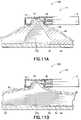
- FIGS. 11A and 11Bare cross-sectional views showing a method for dissecting a submucosal layer according to an embodiment of the present invention.
- FIGS. 12A and 12Bare cross-sectional views showing a method for dissecting a submucosal layer according to another embodiment of the present invention.
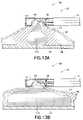
- FIGS. 13A and 13Bare cross-sectional views showing a method for dissecting a submucosal layer according to yet another embodiment of the present invention.
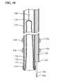
- FIG. 14is a side view showing a submucosal tunneling instrument of a submucosal medical procedure system in accordance with an embodiment of the present invention.
- FIGS. 15A and Bare a cross-sectional views showing a submucosal tunneling instrument of a submucosal medical procedure system in accordance with an embodiment of the present invention.
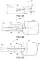
- FIGS. 16A through 16Care partial cross-sectional views showing a sequence of expanding the distal end of a submucosal tunneling instrument of a submucosal medical procedure system in accordance with an embodiment of the present invention.
- FIG. 17is a side view showing a variation of a submucosal tunneling instrument of a submucosal medical procedure system in accordance with an embodiment of the present invention.
- FIG. 18is a cross-sectional view showing a sequence of expanding the distal end of another variation a submucosal tunneling instrument of a submucosal medical procedure system in accordance with an embodiment of the present invention.
- FIGS. 19A through 19Care a side views showing another variation of a submucosal tunneling instrument of a submucosal medical procedure system in accordance with an embodiment of the present invention.
- FIGS. 20A and 20Bare perspective views showing a variation of the distal end of a submucosal tunneling instrument of a submucosal medical procedure system in accordance with an embodiment of the present invention.
- FIGS. 21A and 21Bare perspective and cross sectional views of an area of tissue in the digestive tract having a submucosal saline bleb in accordance with an embodiment of the present invention.
- FIGS. 22A and 22Bare perspective and cross sectional views of an area of tissue in the digestive tract having a submucosal saline bleb in which an opening through the mucosal layer is made in accordance with an embodiment of the present invention.
- FIGS. 23A through 23Dare perspective and cross sectional views showing a method of forming a submucosal tunnel using a submucosal tunneling instrument according to an embodiment of a submucosal medical procedure system of the present invention.
- FIGS. 24A through 24Dare perspective and cross sectional views showing a method of using a submucosal tunneling instrument according to an embodiment of a submucosal medical procedure system of the present invention.
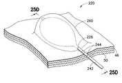
- FIGS. 25A through 25Dare perspective and cross sectional views showing a method of using a submucosal dissection instrument in a submucosal tunnel according to an embodiment of a submucosal medical procedure system of the present invention.
- FIG. 26is a side view showing a combined submucosal tunneling and dissection instrument according to an embodiment of a submucosal medical procedure system of the present invention.
- FIG. 27is a broken cross-sectional view of a combined submucosal tunneling and dissection instrument according to an embodiment of a submucosal medical procedure system of the present invention.
- FIG. 28is a cross-sectional view of the stomach showing a submucosal implant according to an embodiment of a submucosal medical procedure system of the present invention.
- FIGS. 29A through 29Care partial cross-sectional perspective views showing a method of positioning a submucosal implant according to an embodiment of a submucosal medical procedure system of the present invention.
- FIGS. 30A through 30Care partial cross-sectional perspective views showing a method of positioning a submucosal implant according to another embodiment of a submucosal medical procedure system of the present invention.
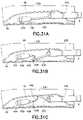
- FIGS. 31A through 31Fare partial cross-sectional perspective views showing a method of delivering a submucosal implant to a desired location within the digestive tract according to yet another embodiment of a submucosal medical procedure system of the present invention.
- FIG. 1illustrates an endoscope 2 of the type used in endoscopic procedures and suitable for use with embodiments of the present invention.
- the endoscope 2has a working channel 4 extending from a proximal portion of the endoscope to the distal end of the endoscope.
- the endoscope 2also has an insertion section 6 which enters the body of a patient passing through a natural orifice such as the mouth or rectum.
- the insertion section 6is generally navigated to a position with the digestive tract when performing a submucosal medical procedure.
- Devices for use in performing submucosal medical proceduresare preferably delivered through working channel 4 of the endoscope 2 ; however devices may be delivered along side the insertion section 6 of the endoscope.
- FIG. 2illustrates a safe access needle injection instrument 10 which is used to aid the physician in obtaining access to the submucosal layer to perform a submucosal medical procedure.
- the safe access needle injection instrument 10includes a tubular shaft 12 having a distal end 13 .
- the diameter of tubular shaft 12is generally in the range 1 mm to 10 mm with a preferred range of 2.0 to 6.0 mm.
- Adjacent distal end 13a portion of the wall of tubular shaft 12 is removed to form window member 14 .
- window member 14can be formed with a cap element coupled to the distal end of the tubular shaft 12 .
- needle member 16Slidably disposed within the lumen of tubular shaft 12 is needle member 16 .
- the proximal portion of tubular shaftincludes vacuum port 18 which is capable of being coupled to a vacuum source such as a syringe or vacuum pump (not shown).
- Valve assembly 20provides a releasable seal to tubular shaft 12 .
- a handle assembly 21is connected to tubular shaft 12 through connector tubing 22 .
- the handle assembly 21includes needle fluid port 24 and valve assembly 26 for injecting fluid through and sealing around the proximal portion of needle member 16 .
- the proximal portion of needle member 16is also connected to a needle slide member 28 positioned on handle body 30 . The proximal movement of needle slide member 28 on handle body 30 causes needle member 16 to move proximally within tubular shaft 12 .
- Distance markers 32are located on handle body 30 to gauge the movement of needle member 16 within tubular shaft 12 .
- needle member 16is positioned within lumen 34 of tubular shaft 12 .
- stop member 36Located on the exterior of needle member 16 is stop member 36 .
- the distal end 13 of tubular shaft 12is closed with seal plug 38 .
- needle member tip 40 and needle lumen 42are also shown. Needle lumen 42 communicates needle tip 40 with needle fluid port 24 so that fluid injected through needle port 24 exits the lumen at needle tip 40 .
- FIGS. 5A and 5Billustrate the actuation of needle member 16 .
- Needle member 16is shown in detail with needle body 44 connected to needle tip 40 .
- Needle body 44may be constructed of a separate material as shown or integrally formed with needle tip 40 .
- Needle body 44may be constructed from flexible tubing having good axial pushability.
- needle member 16is in a first position in which needle tip 40 is located within lumen 34 proximal to window member 14 . This is the preferred position for needle member 16 when tubular shaft 12 is deployed within the body.
- needle member 16Upon actuation, needle member 16 is moved to a second position in which needle tip 40 is positioned within window member 14 .
- FIGS. 6A through 6Eillustrate the operation of safe access needle injection instrument 10 .
- Insertion section 6 of endoscope 2is passed through a natural orifice in a patient and positioned at a location in the digestive tract in which to perform a submucosal procedure.
- Safe access needle injection instrument 10is deployed through the working channel 4 of endoscope 2 .
- the distal potion of safe access needle injection instrument 10is positioned within the digestive tract adjacent mucosal layer 46 . Beneath the mucosal layer are the submucosal layer 48 and the muscular layer 50 .
- Window member 14is oriented towards mucosal layer 46 .
- Needle member 16is located in a first position proximal to window member 14 .
- a vacuum sourceis connected to vacuum port 18 , which communicates with lumen 34 , and the applied vacuum causes the tissue of the digestive tract to be suctioned into window member 14 as shown in FIG. 6B .
- the actuation of needle member 16is shown in FIG. 6C as it is moved to its second position. Distal movement of needle member 16 causes needle tip 40 to move distally relative to tubular shaft 12 to thereby pierce the mucosal layer 46 and enter the submucosal layer 48 of the suctioned tissue.
- stop member 36engages the mucosal layer 46 . Stop member 36 prevents further distal movement of needle member 16 and provides a seal around needle tip 40 .
- a pressurized fluid sourceis connected to needle fluid port 24 to deliver fluid through the lumen of needle member 16 to the submucosal layer 48 .
- the mucosal layer 46is elevated forming a submucosal bleb.
- the fluid used to create the blebmay be of any type suitable for the environment such as solutions containing saline, hypertonic solutions of saline-epinephrine, sodium hyaluronate, poly-N-acetylglucosamine, sodium chondroitin sulfate, chitosans or other biocompatible mucopolysaccharides.
- the safe access needle injection instrument 10may be removed from the safety bleb as illustrated in FIG. 6E .
- FIGS. 7A through 7Cillustrate safe access needle injection instrument 60 which is another preferred embodiment of the present invention to aid the physician in obtaining access to the submucosal space when performing a submucosal medical procedure.
- Safe access needle injection instrument 60includes a sheath member 62 having a proximal and distal end 64 and a lumen 66 extending therethrough. Slidably disposed within lumen 66 of sheath member 62 , there is an elongate shaft member 68 having a shaft lumen 69 and distal end 70 . Located adjacent to distal end 70 of elongated shaft member 68 is a pair of jaw members 72 . Jaw members 72 may be formed from the wall of elongated shaft member 68 .
- the jaw members 72are formed of a resilient material and biased outwardly in an open configuration when unconstrained.
- the jaw members 72may be formed from biocompatible resilient materials such as nitinol, stainless steel and plastics.
- Needle shaft 74is slidably disposed within lumen 66 and preferably slidably disposed shaft lumen 69 .
- Needle shaft 74includes a needle tip 76 and a needle lumen 78 .
- Needle lumen 78extends from the proximal end of needle shaft 74 to needle tip 76 .
- FIG. 7Aillustrates jaw members 72 in a first state in which jaw members 72 are closed and constrained by the walls of sheath member 62 and disposed within lumen 66 .
- needle tip 78 of needle shaft 74is in a first configuration in which needle tip 78 is positioned proximal to jaw members 72 .
- jaw members 72are caused to exit the lumen 66 at distal end 64 of sheath 62 .
- FIG. 7Bshows jaw members 72 in a second state in which in which the jaw members 72 are open and unconstrained after exiting lumen 66 .
- FIG. 7Cillustrates needle tip 78 of needle shaft 74 positioned in a second configuration in which needle tip 78 is positioned between jaw members 72 .
- FIGS. 8A through 8Eillustrates the operation of safe access needle injection instrument 60 .
- Insertion section 6 of endoscope 2is passed through a natural orifice in a patient and positioned at a location in the digestive tract in which to perform a submucosal procedure.
- Safe access needle injection instrument 60is deployed through the working channel 4 of endoscope 2 .
- the distal potion of safe access needle injection instrument 60is positioned within the digestive tract adjacent mucosal layer 46 .
- Beneath the mucosal layerare the submucosal layer 48 and the muscular layer 50 .
- Jaw members 72are positioned on mucosal layer 46 .
- Needle tip 76is located in a first position proximal to jaw members 72 .
- FIG. 8Cis a cross-section view along section line 8 C- 8 C in FIG. 8B showing the tissue engaged by jaw members 72 .
- the mucosal layer 46 and submucosal layer 48are firmly held between jaw members 72 .
- Needle tip 76is moved from the first position proximal to jaw members 72 to the second position between jaw members 72 thereby piercing the mucosal layer 46 and entering the submucosal layer 48 as shown in FIG. 8D .
- FIG. 8Dis a cross-section view along section line 8 C- 8 C in FIG. 8B showing the tissue engaged by jaw members 72 .
- Needle tip 76is moved from the first position proximal to jaw members 72 to the second position between jaw members 72 thereby piercing the mucosal layer 46 and entering the submucosal layer 48 as shown in FIG. 8D .
- the fluid used to create the blebmay be of any type suitable for the environment such as solutions containing saline, hypertonic solutions of saline-epinephrine, sodium hyaluronate, poly-N-acetylglucosamine, sodium chondroitin sulfate, chitosans or other biocompatible mucopolysaccharides.
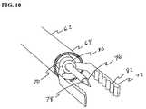
- FIGS. 9 and 10show a variation of safe access needle injection instrument 60 in which jaw members 72 are coupled to distal end 70 of elongate shaft member 68 by a collar member 80 .
- Collar member 80is joined to distal end 70 by suitable means known in the art such as gluing, soldering or welding.
- Jaw members 72are connected to collar member 80 in a way that allows the jaw members 72 to rotate relative to distal end 70 of elongate shaft member 68 .
- the rotational movement of jaw members 72allows the physician to quickly orient the jaw members 72 relative the surface of the tissue surface within the digestive tract.
- jaw members 72are connected to collar member 80 , proximal movement of elongate shaft member 68 relative to sheath member 62 will cause jaw members 72 to move to a closed position within the lumen 66 of sheath member 62 .
- Collar member 80includes an aperture through which needle shaft 74 may extend in a slidable fashion.
- jaw members 72include a tissue grasping surface 82 that facilitates the engagement of tissue within the digestive tract. While tissue grasping surface 82 is shown in the form of a serrated surface, it may also take the form of a knurled surface or a surface containing multiple protrusions or dimples to improve the tissue grasping ability of jaw members 72 .
- Tissue grasping surface 82may be integrally formed with jaw members 72 or bonded to jaw members 72 .
- Tissue grasping surface 82may be formed of metals, polymers or composite materials.
- FIGS. 11A and 11Bshow a safe access dissection system 100 of the present invention that includes a safe access needle injection instrument 10 and an injectable dissection material 102 .
- the injectable dissection material 102takes the form of a solution capable of dissolving the submucosal connective tissue.
- An example of this type of dissolving solutionis sodium 2-mercaptoethanesulfanate (MESNA).
- MESNAsodium 2-mercaptoethanesulfanate
- Additional substances which may dissolve the submucosal connective tissueinclude acids and enzymes such as a peptase enzyme solution, protease/collagenase, papain, chymotrypsin and acetylcysteine.
- a safe access needle injection instrument 10is used to create a safety bleb beneath the mucosal layer 46 in the digestive tract of a mammal.
- the injectable dissection material 102may be delivered through needle tip 40 into the submucosal layer 48 as shown in FIG. 11A .
- Injectable dissection material 102begins to breakdown the stretched submucosal connective tissue 52 .
- the submucosal connective tissue 52breaks, thereby causing the mucosal layer 46 to be come detached from muscular layer 50 in bleb region as shown in FIG. 11B .
- FIGS. 12A and 12Bshow a safe access dissection system 104 of the present invention that includes a safe access needle injection instrument 10 and an injectable dissection material 106 .
- the injectable dissection material 106takes the form of a semisolid gelatin capable of mechanically breaking the submucosal connective tissue 52 .
- the semisolid gelatinmay be formed using biocompatible commercially available gelatins. Generally, these gelatins are in a powdered form and mixed with warm water. Upon cooling, the gelatin forms a semisolid consistency with physical cross links.
- the gelatin materialis preferably formed within the barrel of a pressurizable syringe since aspiration of this material is difficult.
- a safe access needle injection instrument 10is used to create a safety bleb beneath the mucosal layer 46 in the digestive tract of a mammal.
- the injectable dissection material 106may be delivered through needle tip 40 into the submucosal layer 48 as shown in FIG. 12A .
- the mass and semisolid nature of the injectable dissection material 106begins to apply force to the stretched submucosal connective tissue 52 , unlike a saline solution which only permeates the submucosal layer 48 .
- the injectable dissection material 106may also take the form of injectable solutions which solidify upon entering the submucosal space. Solutions which solidify after injection into the submucosal space may be thermo sensitive polymer solutions such as Pluronic 127. Additional injectable solidifying solutions include monomer and polymer solutions like hydrogels and cyanoacrylates which polymerize or crosslink upon contact with tissue or added chemical agents.
- FIGS. 13A and 13Bshow a safe access dissection system 108 of the present invention that includes a safe access needle injection instrument 10 and an injectable dissection material 110 .
- the injectable dissection material 110takes the form of gelled microspheres dispersed in a solution capable of mechanically breaking the submucosal connective tissue 52 .
- the microspheresmay be formed using biocompatible natural materials such as collagen and alginates or synthetic materials like polyvinylalcohol (PVA), polyvinylpyrolidone (PVP) and acrylate polymers.
- a safe access needle injection instrument 10is used to create a safety bleb beneath the mucosal layer 46 in the digestive tract of a mammal.
- the injectable dissection material 110may be delivered through needle tip 40 into the submucosal layer 48 as shown in FIG. 13A .
- the mass and solid nature of the injectable dissection material 110begins to apply force to the stretched submucosal connective tissue 52 , unlike a saline solution which only permeates the submucosal layer 48 .
- the submucosal connective tissue 52breaks, thereby causing the mucosal layer 46 to become detached from muscular layer 50 in bleb region as shown in FIG. 13B .
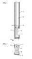
- FIG. 14illustrates a submucosal tunneling instrument 120 for performing a submucosal medical procedure of the present invention.
- the submucosal tunneling instrument 120includes a catheter 122 having proximal and distal ends and a balloon member 124 located adjacent the distal end.
- the proximal end of catheter 122is attached to connector tubing 126 to access inflation port 128 .
- Valve assembly 130provides a seal for fluid introduced into inflation port 128 .
- Tether slide 132is slidably positioned on handle body 134 .
- Handle body 134includes distance markers 136 to gauge the movement of tether slide 132 .
- FIG. 15A cross sectioned view of the distal end of the submucosal tunneling instrument 120 is shown in more detail in FIG. 15 .
- Catheter 122has a distal end 138 and a lumen 123 .
- balloon member 124Located within lumen 123 is balloon member 124 .
- the balloon member 124is preferably noncompliant of the type generally known in the art, however, balloon member 124 may be of the compliant or semi-compliant type.
- the balloon member 124may be formed from biocompatible polymer types such as olefins, elastomers, thermoplastic elastomers, vinyls, polyamides, polyimides, polyesters, fluoropolymers, copolymers and blends of any of the aforementioned.
- the proximal end 140 of balloon member 124is attached to the distal end 138 of catheter 122 .
- the distal end 142 of balloon member 124is positioned within the lumen 123 in an everted configuration.
- a tether member 144is connected to the distal end 142 of balloon member 124 .
- Tether member 144is flexible and preferably takes the form of a filament, as shown, however tether member 144 may take the form of a tube.
- the proximal end of tether member 144is connected to tether slide 132 through valve assembly 130 . Tether member 144 aids in initially positioning balloon member 124 within the lumen 123 of catheter 122 .
- the aforementioned embodiment of the submucosal tunneling instrumentinclude an expandable member which preferably takes the form of an inflatable balloon
- other devicesmay be suitable for essentially performing the same function.
- the expandable member 124may take the form of an expandable elongate braid, mesh or tubular framework in which the proximal end of the expandable member is connected to the distal end of the catheter and the distal end of the expandable member is everted and positioned within the lumen of the catheter.
- a stiffened tether member 144 located within the lumen of the catheter 122may be used as a pusher to push the expandable elongate member from the lumen of the catheter in essentially the same way that the balloon expands from the lumen of the catheter.
- the expandable elongate braidmay be formed from resilient materials such as nitinol, spring steels, vitreloy, as well as polymers and composites.
- the expandable membermay take comprise individual wires to form a braid or mesh configuration. Alternatively the expandable member may be laser cut from a tube forming a tubular framework.
- the expanded diameteris larger than the outer diameter of the catheter.
- FIGS. 16A through 16Cillustrate various stages of deployment of balloon member 124 from the lumen 123 of catheter 122 .
- a fluid filled syringe or other sourceis connected to inflation port 128 .
- Balloon member 124has an expanded diameter range of about 1 mm to about 30 mm and is preferably in the range of 2 mm to 20 mm.
- the length of balloon member 124is a long as necessary to perform a desired submucosal medical procedure. This length can be in the range of 5 mm to 50 cm and preferably in the range of 7 mm to about 10 cm.
- balloon member 124As balloon member 124 expands it extends in a linear fashion moving the distal end 142 of balloon member 124 towards the distal end 138 of catheter 122 . As long as pressurized fluid is applied to catheter lumen 123 , balloon 124 will extend to its full length. Alternatively, since the distal end 142 of balloon 124 is connected to tether member 144 , the amount of linear extension balloon 124 takes may be controlled by the tether slide 132 to define an extension length shorter than the full length of the balloon.
- FIG. 17illustrates a submucosal tunneling instrument 150 for performing a submucosal medical procedure of the present invention.
- the submucosal tunneling instrument 150includes a catheter 152 having proximal and distal ends and a balloon member 154 located adjacent the distal end. Positioned on the exterior of catheter 152 adjacent the distal end is a series of markers 156 . These markers may be visible under direct visualization of the endoscope and may be additionally visible under fluoroscopy. Adjacent the proximal end of catheter 152 is an auxiliary device port 158 . The proximal end of catheter 152 is attached to connector tubing 160 to access inflation port 162 . Valve assembly 164 provides a seal for fluid introduced into inflation port 162 .
- Tether slide 166is slidably positioned on handle body 168 . Handle body 168 includes distance markers 170 to gauge the movement of tether slide 166 .
- FIG. 18A cross sectioned view of the distal end of the submucosal tunneling instrument 150 is shown in more detail in FIG. 18 .
- Catheter 152has a distal end 172 and a first lumen 174 .
- balloon member 154Located within first lumen 174 is balloon member 154 .
- the balloon member 154is preferably non-compliant of the type generally known in the art, however, balloon member 154 may be of the compliant or semi-compliant type.
- the balloon member 154may be formed from biocompatible polymer types such as olefins, elastomers, thermoplastic elastomers, vinyls, polyamides, polyimides, polyesters, fluoropolymers, copolymers and blends of any of the aforementioned.
- the proximal end 176 of balloon member 154is attached to the distal end 172 of catheter 152 .
- the distal end 178 of balloon member 154is positioned within the first lumen 174 in an everted configuration.
- a tether member 180is connected to the distal end 178 of balloon member 154 .
- Tether member 180is flexible and preferably takes the form of a filament, as shown, however tether member 180 may take the form of a tube.
- the proximal end of tether member 180is connected to tether slide 166 through valve assembly 164 . Tether member 180 aids in initially positioning balloon member 154 within the first lumen 174 of catheter 152 .
- Catheter 152has a second lumen 182 that extends from auxiliary device port 158 to distal end 184 .
- Distal end 184is located proximal to distal end 172 of catheter 152 .
- Slidably disposed within second lumen 182is a needle knife 184 that has a knife tip 188 .
- Needle knife 184is preferably of the endoscopic electrosurgical type however any form of incision device that may be operated to form an incision in tissue such as mechanical cutters, water jets or lasers may be suitable.
- FIGS. 19A through 19Cillustrate a submucosal tunneling instrument 200 , according to another embodiment of the present invention.
- Catheter 202has a distal end 204 which is connected to balloon member 206 .
- the proximal end 208 of balloon 206is connected to distal end 204 of catheter 202 .
- Balloon member 206is rolled into a spiral configuration in which the distal end 210 is located in the center of the spiral.
- balloon member 206inflates.
- the inflation of balloon member 206causes the balloon to unroll from a spiral configuration extending linearly.
- the balloon member 206may be thermally treated to retain the spiral configuration for delivery through the working channel of an endoscope.
- the balloon member 206may incorporate a spiral shaped member 212 attached the wall of balloon member 206 as shown in FIGS. 20A and 20B .
- the spiral shaped membermay be formed from a resilient filament as shown in an outstretched configuration in FIG. 20A .
- the spiral shaped memberbeing formed of a resilient filament and incorporated into the wall of the balloon preferably takes its spiral shape and in doing so forms the balloon member into a spiral shape as shown in FIG. 20B .
- FIGS. 21A and 21Billustrate a desired region 220 of the digestive tract of a mammal.
- a safe access needle injection instrumentaccording to any of the embodiments previously described may be used to form a safety bleb 222 beneath the mucosal layer 46 .
- the safety blebcontains the submucosal connective tissue 52 generally in a stretched condition attached to both the mucosal layer 46 and the muscular layer 50 .
- an endoscopic incision tool 224is positioned adjacent to the safety bleb 222 .
- FIGS. 23A through 24Dillustrate the introduction and operation of a submucosal tunneling instrument into submucosal layer 48 .
- the distal end 138 of the submucosal tunneling instrument 120is positioned through the mucosal opening 228 . Once the proximal end 140 of balloon 124 is through the mucosal opening 228 the submucosal tunneling instrument 120 may be operated.
- the proximal end 140 of balloon 124By delivering pressurized fluid through the lumen of catheter 122 , the proximal end 140 of balloon 124 inflates to its expanded diameter as depicted in FIG. 23C .
- the expanded diameter of the proximal end 140 of balloon 124is larger than the diameter of the mucosal opening 228 .
- the larger diameterprevents balloon 124 from pushing the distal end 138 of catheter 122 backwards out of the submucosal layer 48 through mucosal opening 228 .
- further inflationextends balloon 124 in a linear fashion within the submucosal layer 48 causing the submucosal connective tissue 52 to break in regions adjacent to the balloon.
- the balloon 124can only expand by increasing the volume of the surrounding area between the mucosal layer 46 and the muscular layer 50 .
- the application of force during expansion of balloon 124is concentrated on the submucosal connective tissue 52 , thereby causing the submucosal connective tissue 52 to break, whereas the force applied to the mucosal layer 46 or the muscular layer 50 by balloon 124 is diluted over a larger portion of balloon 124 .
- the force required to break the submucosal connective tissue 52 as applied by balloon 124is less than the force required to perforate the mucosal layer 46 or muscular layer 50 by balloon 124 thereby minimizing trauma to surrounding tissue.
- 24Aillustrates a perspective view of region 220 in the digestive tract having a submucosal tunnel 230 formed by submucosal tunneling instrument 120 .
- balloon 124is fully expanded and generally occupies the majority of the space of submucosal tunnel 230 .
- Balloon 124is then deflated by applying a negative pressure to catheter 122 and retracting tether member 144 .
- the distal end 138 of catheter 122is then removed from mucosal opening 228 , leaving submucosal tunnel 230 generally deflated.
- the submucosal connective tissue 52 within the tunnelis broken.
- the submucosal tunnel 230may provide suitable access to the muscular wall or the placement of an implant device. However to perform other submucosal medical procedures an area larger than the submucosal tunnel 230 may be desired.
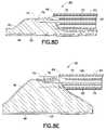
- FIGS. 25A through 25Dillustrate the formation of an area larger than a submucosal tunnel for performing a submucosal medical procedure according to an embodiment of the present invention.
- a region 220 in the digestive tractis prepared by forming a submucosal tunnel 230 .
- a submucosal dissection instrument 240 having a catheter 242is positioned through mucosal opening 228 into submucosal tunnel 230 .
- markers 244Located on catheter 242 are markers 244 that indicate the insertion depth of catheter distal end 246 within submucosal tunnel 230 .
- the submucosal dissection instrument 240is in a proper position for operation when balloon member 248 including distal end 250 and proximal end 252 are sufficiently located within submucosal tunnel 230 , as shown in FIG. 25B .
- balloon member 248inflates.
- the submucosal connective tissue 52is broken in the area of the expanded balloon member 248 and the mucosal layer 46 is elevated.
- the elevated mucosal layer 46forms a large mucosal dissected area 260 .
- the balloon member 248may be deflated and submucosal dissecting instrument 240 removed from the large mucosal dissected area 260 .
- the dissected region beneath mucosal layer 46has transformed in geometry from a high aspect ratio tunnel to a low aspect ratio chamber suitable for performing some submucosal medical procedures.
- the submucosal tunneling dissecting instrument 270includes a dissection catheter 272 having a distal end 274 and a dissection balloon 276 having an expanded dissection balloon 276 a configuration.
- the dissection balloon 276can be compliant of the type generally known in the art or dissection balloon 276 may be of the compliant or semi-compliant type.
- the dissection balloon 276may be formed from biocompatible polymer types such as olefins, elastomers, thermoplastic elastomers, vinyls, polyamides, polyimides, polyesters, fluoropolymers, copolymers and blends of any of the aforementioned.
- the dissection catheter 272has insertion markers 278 positioned along its shaft.
- the proximal end of dissection catheter 272includes both an inflation port 280 that is in fluid communication with dissection balloon 276 , and a valve assembly 282 .
- Tunneling catheter 284is slidably disposed through valve assembly 282 extending within a lumen of dissection catheter 272 .
- the tunneling catheter distal end 286may extend beyond the dissection catheter distal end 274 .
- Tunneling catheter 284includes an inflation port 288 and valve assembly 290 .
- a tether slide member 292is slidably disposed on handle body 294 with distance markers 296 .
- FIG. 27illustrates a detailed cross section of the distal portion of the submucosal tunneling dissecting instrument 270 .
- the distal end 298 and proximal end 300 of dissection balloon 276are connected to the exterior of dissection catheter 272 .
- Inflation lumen 302connects inflation port 280 with the interior of dissection balloon 276 through inflation aperture 304 .
- Tunneling catheter 284is slidably disposed within the lumen 306 of dissection catheter 272 .
- tunneling balloon 310Positioned within the lumen 308 of tunneling catheter 284 there is an everted expandable tunneling balloon 310 .
- the tunneling balloon 310is preferably non-compliant of the type generally known in the art, however, tunneling balloon 310 may be of the compliant or semi-compliant type.
- the tunneling balloon 310may be formed from biocompatible polymer types such as olefins, elastomers, thermoplastic elastomers, vinyls, polyamides, polyimides, polyesters, fluoropolymers, copolymers and blends of any of the aforementioned.
- the distal end of tunneling balloon 310is connected to a tether member 312 which has a proximal end that is connected to tether slide 292 .
- the operation of the submucosal tunneling dissecting instrument 270 to form a submucosal tunnel and large mucosal dissected areais similar to the operation of the separate instruments.
- the distal end 286 of tunneling catheter 284is positioned through a mucosal opening formed in a safety bleb.
- the tunneling catheter 284is pressurized with fluid to linearly expand tunneling balloon 310 .
- tunneling balloon 310may be deflated and dissection catheter 272 may be advanced through the mucosal opening into the submucosal tunnel.
- the markers 278may be used to determine the depth in which the dissection catheter 272 has been advanced into the submucosal tunnel.
- dissection catheter 272Once the dissection catheter 272 has been properly positioned within the submucosal tunnel it may be operated. By applying pressurized fluid to inflation port 280 , dissection balloon 276 is dilated to an expanded dissection balloon 276 a configuration. During the expansion a large mucosal dissected area is created which is accessible for performing a subsequent submucosal medical procedure.
- the submucosal implant devicemay be a passive or active device. Active devices may include self contained electronics for controlling the device, as well as an internal power supply. Alternatively, the active devices may be controlled externally by telemetry.
- a passive submucosal implant devicemay take the form of a drug delivery depot in which a therapeutic agent within the depot elutes from the depot according to a predetermined elution profile.
- An active submucosal implant devicemay take the form of a drug delivery device that incorporates a self contained diagnostic system to determine the appropriate delivery time and dosage of a therapeutic agent to be administered.
- the passive or active submucosal implant devicewhich takes the form of a drug delivery device may include a port positioned through the mucosal layer to allow endoscopic refilling of the drug delivery device with therapeutic agents.
- Submucosal implant devicesthat are drug delivery devices have the ability to deliver therapeutic agents directly to the portal circulation. Many different types of therapeutic agents may be delivered such as pharmaceuticals, hormones, growth factors and other biological compounds.
- Some typical therapeutic agents for use in a submucosal implant device of the present inventioninclude dromostanolone, dromostanolone propionate, chlormadinone, chlormadinone acetate, dimethisterone, ethisterone, hydroxyprogesterone, norgestomet and other norsteroids such as norethindrone, norgesterone, northylodrel, norgestrel, noregestrienone, and norgestoniel; melengestrol acetate, estradiol, 17 ⁇ -acetoxy-11 ⁇ -methyl-19-norpregn-4-ene-3,20-dione, 3(3-oxo-11 ⁇ ,13 ⁇ -dialkl-17 ⁇ -hydroxygon-4-en-17 ⁇ -yl)propionic acid-lactone, 3-(3-oxo-11 ⁇ methyl-17 ⁇ -hydroxyestr-4-en-17 ⁇ -yl)-propionic acid lactone, 11,13 ⁇ -dialkyl-17 lower alkyl-17 lower al
- cholecytokinincholecytokinin
- An active submucosal implant devicemay take the form of a drug delivery device in which electrical signals are received from the contraction of the muscular wall of a mammal to deliver a specified amount of therapeutic agent.
- the submucosal implantmay include an anchor member in which to secure the implant to the muscular wall beneath the mucosal layer. This anchor may be integrally formed with an electrode for receiving electrical signals from the wall.
- Multiple submucosal implant devicesmay be deposited at various locations along the stomach wall.
- FIG. 28shows a portion 409 of the digestive system in which a submucosal implant device 410 is placed within the wall of the stomach.
- FIGS. 29A through 29Cillustrate a sequence for deploying a submucosal implant device according to an embodiment of the present invention.
- a submucosal tunnel 230 or a large mucosal dissected areais formed in a portion of the digestive tract.
- the insertion section 6 of an endoscopeis positioned through an enlarged mucosal opening 228 into the submucosal space.
- the submucosal implant device 410 and an implant deployment device 412are inserted through the mucosal opening 228 and preferably through a working channel of the endoscope into the submucosal space.
- Implant deployment device 412is used to position submucosal implant device 410 within the submucosal space.
- Submucosal implant device 410preferably takes the form of a drug delivery depot.
- the submucosal implant devicemay be in the form of a hydrogel, gel matrix, soluble wax or configurations commonly used in transdermal drug delivery devices.
- Submucosal implant device 410is shown in a first configuration for delivery into the submucosal space and a second configuration for deployment adjacent muscular layer 50 .
- Implant deployment device 412is operated to move submucosal implant device 410 from its first configuration to the second configuration.
- Implant deployment device 412then releases submucosal implant device 410 into the submucosal space.
- the implant deployment device 412 and endoscope insertion section 6are then removed from the submucosal space and the mucosal opening 228 is then closed using clips, suture or other closure techniques.
- FIGS. 30A through 30Cillustrate a submucosal implant device and sequence for deploying the device according to another embodiment of the present invention.
- a submucosal tunnel 230 or a large mucosal dissected areais formed in a portion of the digestive tract.
- the insertion section 6 of an endoscopeis positioned through an enlarged mucosal opening 228 into the submucosal space.
- the submucosal implant device 414 and an implant deployment device 416are inserted through the mucosal opening 228 and preferably through a working channel of the endoscope into the submucosal space.
- the relative sizes shownare for clarity; in practice, implant device be relatively smaller to permit passage through the working channel.
- Implant deployment device 416is used to position submucosal implant device 414 within the submucosal space.
- Submucosal implant device 414also includes an anchor member 418 and a tether member 420 .
- Anchor member 418is adapted to be affixed to the muscular layer 50 and retain the submucosal implant device 414 in position by tether member 420 .
- Submucosal implant device 414preferably takes the form of a drug delivery device.
- anchor member 418 and tether member 420also function as an electrode and electrical conductor respectively. Electrical signals may be received by the submucosal implant device 414 from muscular layer 50 via anchor member 418 and tether member 420 .
- Implant deployment device 416then releases submucosal implant device 414 into the submucosal space.
- the implant deployment device 416 and endoscope insertion section 6are then removed from the submucosal space and the mucosal opening 228 is then closed using closure clips 422 with a clip applier 424 .
- Other closure techniques and devicessuch as sutures, staples and adhesives may also be suitable for closing mucosal opening 228 .
- FIGS. 31A through 31Fillustrate another submucosal implant device and a sequence for deploying the device according to another embodiment of the present invention.
- a submucosal tunnel 230 or a large mucosal dissected areais formed in a portion of the digestive tract.
- the insertion section 6 of an endoscopeis positioned through an enlarged mucosal opening 228 into the submucosal space.
- the submucosal implant device 430 and an implant deployment device 432are inserted through the mucosal opening 228 and preferably through a working channel of the endoscope into the submucosal space.
- the relative sizes shownare for clarity; in practice, implant device be relatively smaller to permit passage through the working channel.
- Implant deployment device 432is used to position submucosal implant device 430 within the submucosal space.
- Submucosal implant device 430includes a therapeutic agent delivery port 434 , an anchor member 436 and a tether member 438 .
- Anchor member 436is adapted to be affixed to the muscular layer 50 and retain the submucosal implant device 430 in position by tether member 438 .
- Submucosal implant device 430preferably takes the form of a therapeutic drug delivery device incorporating an internal drug reservoir.
- anchor member 436 and tether member 438also function as an electrode and electrical conductor respectively.
- Submucosal implant device 430may be sent and received by the submucosal implant device 430 to and from muscular layer 50 via anchor member 436 and tether member 438 .
- the electrical signalsmay be used by the submucosal implant device 430 to initiate or terminate delivery of a therapeutic agent through delivery ports 434 .
- Submucosal implant device 430also includes a refill tube 440 connected to a mucosal port 442 .
- Mucosal port 442contains a valve assembly that can restrict fluid access to refill tube 440 .
- Implant deployment device 432positions the refill tube 440 within the submucosal space while the mucosal port 442 is positioned through mucosal layer 46 .
- Implant deployment device 432then releases submucosal implant device 430 into the submucosal space.
- the implant deployment device 432 and endoscope insertion section 6are then removed from the submucosal space and the mucosal opening 228 is then closed using closure clips 422 with a clip applier 424 .
- Other closure techniques and devicessuch as sutures, staples and adhesives may also be suitable for closing mucosal opening 228 .
- the endoscope and related materialsmay then be removed from the patient.
- the physicianmay return endocopically to the site of the implant and use a refilling instrument 444 having a tip 446 that engages the mucosal port 442 to deliver more drugs to refill the reservoir.
Landscapes
- Health & Medical Sciences (AREA)
- Life Sciences & Earth Sciences (AREA)
- Surgery (AREA)
- Animal Behavior & Ethology (AREA)
- Public Health (AREA)
- Engineering & Computer Science (AREA)
- Biomedical Technology (AREA)
- Heart & Thoracic Surgery (AREA)
- Medical Informatics (AREA)
- Molecular Biology (AREA)
- Veterinary Medicine (AREA)
- General Health & Medical Sciences (AREA)
- Nuclear Medicine, Radiotherapy & Molecular Imaging (AREA)
- Pathology (AREA)
- Physics & Mathematics (AREA)
- Optics & Photonics (AREA)
- Biophysics (AREA)
- Radiology & Medical Imaging (AREA)
- Orthopedic Medicine & Surgery (AREA)
- Gastroenterology & Hepatology (AREA)
- Ophthalmology & Optometry (AREA)
- Infusion, Injection, And Reservoir Apparatuses (AREA)
- Surgical Instruments (AREA)
Abstract
Description
Claims (20)
Priority Applications (3)
| Application Number | Priority Date | Filing Date | Title |
|---|---|---|---|
| US11/776,097US8066689B2 (en) | 2007-07-11 | 2007-07-11 | Methods and systems for submucosal implantation of a device for diagnosis and treatment with a therapeutic agent |
| PCT/US2008/067642WO2009009276A1 (en) | 2007-07-11 | 2008-06-20 | Methods and systems for submucosal implantation of a device for diagnosis and treatment with a therapeutic agent |
| US13/304,055US8491472B2 (en) | 2007-07-11 | 2011-11-23 | Methods and systems for submucosal implantation of a device for diagnosis and treatment with a therapeutic agent |
Applications Claiming Priority (1)
| Application Number | Priority Date | Filing Date | Title |
|---|---|---|---|
| US11/776,097US8066689B2 (en) | 2007-07-11 | 2007-07-11 | Methods and systems for submucosal implantation of a device for diagnosis and treatment with a therapeutic agent |
Related Child Applications (1)
| Application Number | Title | Priority Date | Filing Date |
|---|---|---|---|
| US13/304,055DivisionUS8491472B2 (en) | 2007-07-11 | 2011-11-23 | Methods and systems for submucosal implantation of a device for diagnosis and treatment with a therapeutic agent |
Publications (2)
| Publication Number | Publication Date |
|---|---|
| US20090018604A1 US20090018604A1 (en) | 2009-01-15 |
| US8066689B2true US8066689B2 (en) | 2011-11-29 |
Family
ID=40228967
Family Applications (2)
| Application Number | Title | Priority Date | Filing Date |
|---|---|---|---|
| US11/776,097Active2030-08-27US8066689B2 (en) | 2007-07-11 | 2007-07-11 | Methods and systems for submucosal implantation of a device for diagnosis and treatment with a therapeutic agent |
| US13/304,055ActiveUS8491472B2 (en) | 2007-07-11 | 2011-11-23 | Methods and systems for submucosal implantation of a device for diagnosis and treatment with a therapeutic agent |
Family Applications After (1)
| Application Number | Title | Priority Date | Filing Date |
|---|---|---|---|
| US13/304,055ActiveUS8491472B2 (en) | 2007-07-11 | 2011-11-23 | Methods and systems for submucosal implantation of a device for diagnosis and treatment with a therapeutic agent |
Country Status (2)
| Country | Link |
|---|---|
| US (2) | US8066689B2 (en) |
| WO (1) | WO2009009276A1 (en) |
Cited By (34)
| Publication number | Priority date | Publication date | Assignee | Title |
|---|---|---|---|---|
| US20090018603A1 (en)* | 2007-07-11 | 2009-01-15 | Vladimir Mitelberg | Methods and Systems for Submucosal Implantation of a Device for Diagnosis and Treatment of a Body |
| US8226730B2 (en)* | 2003-08-11 | 2012-07-24 | Cook Medical Technologies Llc | Surgical implant |
| US20140214085A1 (en)* | 2013-01-25 | 2014-07-31 | Kyphon Sarl | Expandable device and methods of use |
| US9011365B2 (en) | 2013-03-12 | 2015-04-21 | Medibotics Llc | Adjustable gastrointestinal bifurcation (AGB) for reduced absorption of unhealthy food |
| US9067070B2 (en) | 2013-03-12 | 2015-06-30 | Medibotics Llc | Dysgeusia-inducing neurostimulation for modifying consumption of a selected nutrient type |
| WO2015148541A1 (en)* | 2014-03-24 | 2015-10-01 | Fractyl Laboratories, Inc. | Injectate delivery devices, systems and methods |
| US9204888B2 (en) | 2007-06-08 | 2015-12-08 | United States Endoscopy Group, Inc. | Retrieval device |
| US20160256663A1 (en)* | 2013-11-22 | 2016-09-08 | Fractyl Laboratories Inc. | Systems, devices and methods for the creation of a therapeutic restriction in the gastrointestinal tract |
| US9456916B2 (en) | 2013-03-12 | 2016-10-04 | Medibotics Llc | Device for selectively reducing absorption of unhealthy food |
| US9486188B2 (en) | 2001-05-18 | 2016-11-08 | United States Endoscopy Group, Inc. | Retrieval device |
| US9526648B2 (en) | 2010-06-13 | 2016-12-27 | Synerz Medical, Inc. | Intragastric device for treating obesity |
| US9572591B2 (en) | 2013-09-03 | 2017-02-21 | United States Endoscopy Group, Inc. | Endoscopic snare device |
| US9757535B2 (en) | 2014-07-16 | 2017-09-12 | Fractyl Laboratories, Inc. | Systems, devices and methods for performing medical procedures in the intestine |
| US9872700B2 (en) | 2013-09-03 | 2018-01-23 | United States Endoscopy Group, Inc. | Endoscopic snare device |
| US10299857B2 (en) | 2013-06-04 | 2019-05-28 | Fractyl Laboratories, Inc. | Methods, systems and devices for reducing the luminal surface area of the gastrointestinal tract |
| US10349998B2 (en) | 2012-02-27 | 2019-07-16 | Fractyl Laboratories, Inc. | Heat ablation systems, devices and methods for the treatment of tissue |
| US10413436B2 (en) | 2010-06-13 | 2019-09-17 | W. L. Gore & Associates, Inc. | Intragastric device for treating obesity |
| US10420665B2 (en) | 2010-06-13 | 2019-09-24 | W. L. Gore & Associates, Inc. | Intragastric device for treating obesity |
| US20190290282A1 (en)* | 2018-03-26 | 2019-09-26 | Olympus Corporation | Procedure for endoscopic full-thickness resection |
| US10667838B2 (en) | 2017-01-09 | 2020-06-02 | United States Endoscopy Group, Inc. | Endoscopic snare device |
| US10765474B2 (en) | 2012-02-27 | 2020-09-08 | Fractyl Laboratories, Inc. | Injectate delivery devices, systems and methods |
| US10779980B2 (en) | 2016-04-27 | 2020-09-22 | Synerz Medical, Inc. | Intragastric device for treating obesity |
| US10869718B2 (en) | 2014-07-16 | 2020-12-22 | Fractyl Laboratories, Inc. | Methods and systems for treating diabetes and related diseases and disorders |
| US10933171B2 (en)* | 2016-11-17 | 2021-03-02 | Tokyo Metropolitan Industrial Technology Research Institute | Collagen sol for submucosal local injection |
| US10959774B2 (en) | 2014-03-24 | 2021-03-30 | Fractyl Laboratories, Inc. | Injectate delivery devices, systems and methods |
| US10973561B2 (en) | 2012-08-09 | 2021-04-13 | Fractyl Laboratories, Inc. | Ablation systems, devices and methods for the treatment of tissue |
| US10980590B2 (en) | 2011-01-19 | 2021-04-20 | Fractyl Laboratories, Inc. | Devices and methods for the treatment of tissue |
| US11039850B2 (en) | 2018-02-28 | 2021-06-22 | Gi Supply | Endoscopic tool with suction for facilitating injection of a fluid into a submucosal layer of tissue |
| US11135078B2 (en) | 2010-06-13 | 2021-10-05 | Synerz Medical, Inc. | Intragastric device for treating obesity |
| US11185367B2 (en) | 2014-07-16 | 2021-11-30 | Fractyl Health, Inc. | Methods and systems for treating diabetes and related diseases and disorders |
| US11246639B2 (en) | 2012-10-05 | 2022-02-15 | Fractyl Health, Inc. | Methods, systems and devices for performing multiple treatments on a patient |
| US11439457B2 (en) | 2012-07-30 | 2022-09-13 | Fractyl Health, Inc. | Electrical energy ablation systems, devices and methods for the treatment of tissue |
| US11986235B2 (en) | 2013-09-12 | 2024-05-21 | Fractyl Health, Inc. | Systems, methods and devices for treatment of target tissue |
| US12364554B2 (en) | 2020-01-15 | 2025-07-22 | Fractyl Health, Inc. | Automated tissue treatment devices, systems, and methods |
Families Citing this family (34)
| Publication number | Priority date | Publication date | Assignee | Title |
|---|---|---|---|---|
| US8761903B2 (en) | 2000-10-11 | 2014-06-24 | The Board Of Regents Of The University Of Texas | Gastrointestinal electrical stimulation |
| JP2003530169A (en) | 2000-04-11 | 2003-10-14 | ザ・ボード・オブ・リージェンツ・オブ・ザ・ユニバーシティ・オブ・テキサス・システム | Electrical stimulation of the gastrointestinal tract |
| US8467874B2 (en) | 2000-04-11 | 2013-06-18 | The Board Of Regents Of The University Of Texas System | Gastrointestinal electrical stimulation |
| US12343530B2 (en) | 2006-05-18 | 2025-07-01 | Paras Holdings, Llc | Device and implantation system for electrical stimulation of biological systems |
| US9020597B2 (en) | 2008-11-12 | 2015-04-28 | Endostim, Inc. | Device and implantation system for electrical stimulation of biological systems |
| US8712529B2 (en) | 2010-03-05 | 2014-04-29 | Endostim, Inc. | Device and implantation system for electrical stimulation of biological systems |
| US9345879B2 (en) | 2006-10-09 | 2016-05-24 | Endostim, Inc. | Device and implantation system for electrical stimulation of biological systems |
| US11577077B2 (en) | 2006-10-09 | 2023-02-14 | Endostim, Inc. | Systems and methods for electrical stimulation of biological systems |
| US20150224310A1 (en) | 2006-10-09 | 2015-08-13 | Endostim, Inc. | Device and Implantation System for Electrical Stimulation of Biological Systems |
| US9724510B2 (en) | 2006-10-09 | 2017-08-08 | Endostim, Inc. | System and methods for electrical stimulation of biological systems |
| DK2240222T3 (en) | 2008-01-11 | 2018-06-25 | Ucb Biopharma Sprl | SYSTEMS FOR THE ADMINISTRATION OF PHARMACEUTICALS FOR PATIENTS WITH RHEUMATOID ARTHRITIS |
| WO2009097542A2 (en) | 2008-01-30 | 2009-08-06 | Board Of Regents Of The University Of Texas System | Ileal electrical stimulation |
| EP3581223B1 (en) | 2008-07-18 | 2024-07-10 | UCB Biopharma SRL | Systems for automatically administering medication |
| USD641078S1 (en) | 2008-12-29 | 2011-07-05 | Ucb Pharma, S.A. | Medical syringe with needle tip cap |
| EP2328485B1 (en)* | 2008-08-18 | 2012-06-06 | Cook Medical Technologies LLC | Device for closure of vessel access site |
| US8343912B2 (en) | 2008-12-23 | 2013-01-01 | Biosource Pharm, Inc. | Antibiotic compositions for the treatment of gram negative infections |
| US11717681B2 (en) | 2010-03-05 | 2023-08-08 | Endostim, Inc. | Systems and methods for treating gastroesophageal reflux disease |
| US8415307B1 (en) | 2010-06-23 | 2013-04-09 | Biosource Pharm, Inc. | Antibiotic compositions for the treatment of gram negative infections |
| EP2696792B8 (en) | 2011-04-14 | 2018-05-23 | Endostim, Inc. | Systems for treating gastroesophageal reflux disease |
| WO2013033673A1 (en) | 2011-09-02 | 2013-03-07 | Endostim, Inc. | Endoscopic lead implantation method |
| US9925367B2 (en) | 2011-09-02 | 2018-03-27 | Endostim, Inc. | Laparoscopic lead implantation method |
| WO2014015259A1 (en)* | 2012-07-19 | 2014-01-23 | Creighton University | Tissue resection device and method |
| US9623238B2 (en) | 2012-08-23 | 2017-04-18 | Endostim, Inc. | Device and implantation system for electrical stimulation of biological systems |
| US9498619B2 (en) | 2013-02-26 | 2016-11-22 | Endostim, Inc. | Implantable electrical stimulation leads |
| WO2015034867A2 (en) | 2013-09-03 | 2015-03-12 | Endostim, Inc. | Methods and systems of electrode polarity switching in electrical stimulation therapy |
| US9682234B2 (en) | 2014-11-17 | 2017-06-20 | Endostim, Inc. | Implantable electro-medical device programmable for improved operational life |
| US10299795B2 (en) | 2016-04-28 | 2019-05-28 | Mayo Foundation For Medical Education And Research | Devices and methods for esophageal lengthening and anastomosis formation |
| WO2018094207A1 (en) | 2016-11-17 | 2018-05-24 | Endostim, Inc. | Modular stimulation system for the treatment of gastrointestinal disorders |
| EP3606603A1 (en) | 2017-04-06 | 2020-02-12 | Endostim, Inc. | Implantable surface electrodes and method of implantation |
| US11737745B2 (en) | 2018-10-24 | 2023-08-29 | Mayo Foundation For Medical Education And Research | Medical devices and methods for body conduit lengthening and anastomosis formation |
| US11819244B2 (en)* | 2019-11-27 | 2023-11-21 | Gyrus Acmi, Inc. | Surgical device for treatment of endometriosis |
| WO2021247130A1 (en)* | 2020-06-01 | 2021-12-09 | Biorasis Inc. | Needle-assisted automated insertion and extraction of implants |
| EP4404914A4 (en)* | 2021-09-22 | 2025-07-16 | Celanese Eva Performance Polymers Llc | Refillable implantable contraceptive delivery device |
| WO2025080758A1 (en)* | 2023-10-11 | 2025-04-17 | Eclipse Medical Inc. | Medical device for voluntarily creating a remedial atrial septal defect |
Citations (154)
| Publication number | Priority date | Publication date | Assignee | Title |
|---|---|---|---|---|
| US3039468A (en) | 1959-01-07 | 1962-06-19 | Joseph L Price | Trocar and method of treating bloat |
| US3910279A (en) | 1973-06-20 | 1975-10-07 | Olympus Optical Co | Electrosurgical instrument |
| US3980861A (en) | 1973-03-26 | 1976-09-14 | Akio Fukunaga | Electrically heated miniature thermal implement |
| US4222380A (en) | 1977-12-02 | 1980-09-16 | Olympus Optical Co., Ltd. | Celiac injector |
| US4271839A (en)* | 1979-07-25 | 1981-06-09 | Thomas J. Fogarty | Dilation catheter method and apparatus |
| US4418692A (en) | 1978-11-17 | 1983-12-06 | Guay Jean Louis | Device for treating living tissue with an electric current |
| US4445892A (en) | 1982-05-06 | 1984-05-01 | Laserscope, Inc. | Dual balloon catheter device |
| US4630609A (en) | 1981-05-14 | 1986-12-23 | Thomas J. Fogarty | Dilatation catheter method and apparatus |
| US4655219A (en) | 1983-07-22 | 1987-04-07 | American Hospital Supply Corporation | Multicomponent flexible grasping device |
| US4779611A (en) | 1987-02-24 | 1988-10-25 | Grooters Ronald K | Disposable surgical scope guide |
| US4819633A (en) | 1986-09-02 | 1989-04-11 | Richard Wolf Gmbh | Coagulation forceps |
| US4887598A (en) | 1982-09-24 | 1989-12-19 | Berke Joseph J | Manual rotary scalpel structure |
| US4907591A (en) | 1988-03-29 | 1990-03-13 | Pfizer Hospital Products Group, Inc. | Surgical instrument for establishing compression anastomosis |
| US5009659A (en) | 1989-10-30 | 1991-04-23 | Schneider (Usa) Inc. | Fiber tip atherectomy catheter |
| US5014708A (en) | 1988-09-14 | 1991-05-14 | Olympus Optical Co. | Radioactive ray detecting therapeutic apparatus |
| US5078716A (en) | 1990-05-11 | 1992-01-07 | Doll Larry F | Electrosurgical apparatus for resecting abnormal protruding growth |
| US5080660A (en) | 1990-05-11 | 1992-01-14 | Applied Urology, Inc. | Electrosurgical electrode |
| US5135484A (en) | 1990-05-09 | 1992-08-04 | Pioneering Technologies, Inc. | Method of removing plaque from vessels |
| US5150717A (en) | 1988-11-10 | 1992-09-29 | Arye Rosen | Microwave aided balloon angioplasty with guide filament |
| US5158543A (en) | 1990-10-30 | 1992-10-27 | Lazarus Harrison M | Laparoscopic surgical system and method |
| US5159925A (en) | 1988-09-09 | 1992-11-03 | Gynelab, Inc. | Cauterizing apparatus and method for laparoscopic cholecystostomy, gallbladder ablation and treatment of benign prostate hypertrophy |
| US5196024A (en) | 1990-07-03 | 1993-03-23 | Cedars-Sinai Medical Center | Balloon catheter with cutting edge |
| US5195507A (en) | 1990-11-06 | 1993-03-23 | Ethicon, Inc. | Endoscopic surgical instrument for displacing tissue or organs |
| US5226908A (en) | 1989-12-05 | 1993-07-13 | Inbae Yoon | Multi-functional instruments and stretchable ligating and occluding devices |
| US5290284A (en) | 1992-05-01 | 1994-03-01 | Adair Edwin Lloyd | Laparoscopic surgical ligation and electrosurgical coagulation and cutting device |
| US5300023A (en) | 1991-10-18 | 1994-04-05 | Imagyn Medical, Inc. | Apparatus and method for independent movement of an instrument within a linear catheter |
| US5308327A (en) | 1991-11-25 | 1994-05-03 | Advanced Surgical Inc. | Self-deployed inflatable retractor |
| US5318564A (en) | 1992-05-01 | 1994-06-07 | Hemostatic Surgery Corporation | Bipolar surgical snare and methods of use |
| US5318543A (en) | 1992-10-08 | 1994-06-07 | Abbott Laboratories | Laparoscopic jejunostomy instrumentation kit |
| US5339799A (en) | 1991-04-23 | 1994-08-23 | Olympus Optical Co., Ltd. | Medical system for reproducing a state of contact of the treatment section in the operation unit |
| US5370134A (en) | 1991-05-29 | 1994-12-06 | Orgin Medsystems, Inc. | Method and apparatus for body structure manipulation and dissection |
| US5372601A (en) | 1993-03-30 | 1994-12-13 | Lary; Banning G. | Longitudinal reciprocating incisor |
| US5383889A (en) | 1991-05-29 | 1995-01-24 | Origin Medsystems, Inc. | Tethered everting balloon retractor for hollow bodies and method of using |
| US5395312A (en) | 1991-10-18 | 1995-03-07 | Desai; Ashvin | Surgical tool |
| US5417697A (en) | 1993-07-07 | 1995-05-23 | Wilk; Peter J. | Polyp retrieval assembly with cauterization loop and suction web |
| US5431173A (en) | 1991-05-29 | 1995-07-11 | Origin Medsystems, Inc. | Method and apparatus for body structure manipulation and dissection |
| US5458583A (en) | 1993-01-07 | 1995-10-17 | Medical Innovations Corporation | Gastrostomy catheter system |
| US5507795A (en) | 1994-04-29 | 1996-04-16 | Devices For Vascular Intervention, Inc. | Catheter with perfusion system |
| US5507744A (en) | 1992-04-23 | 1996-04-16 | Scimed Life Systems, Inc. | Apparatus and method for sealing vascular punctures |
| US5507765A (en) | 1994-04-28 | 1996-04-16 | Mott; James B. | Punch-type surgical instrument for skin incision, set of parts for making such an instrument of selectably variable size, and blade unit for such instrument |
| US5527273A (en) | 1994-10-06 | 1996-06-18 | Misonix, Inc. | Ultrasonic lipectomy probe and method for manufacture |
| US5531699A (en) | 1994-09-19 | 1996-07-02 | Abbott Laboratories | Spring-loaded reciprocable stylet holder |
| US5556405A (en) | 1995-10-13 | 1996-09-17 | Interventional Technologies Inc. | Universal dilator with reciprocal incisor |
| US5570700A (en) | 1994-10-03 | 1996-11-05 | Vogeler; Douglas M. | Elliptical biopsy punch |
| US5571130A (en) | 1994-10-04 | 1996-11-05 | Advanced Cardiovascular Systems, Inc. | Atherectomy and prostectomy system |
| US5582609A (en) | 1993-10-14 | 1996-12-10 | Ep Technologies, Inc. | Systems and methods for forming large lesions in body tissue using curvilinear electrode elements |
| US5588951A (en) | 1993-01-19 | 1996-12-31 | Loma Linda University Medical Center | Inflatable endoscopic retractor with multiple rib-reinforced projections |
| US5599300A (en) | 1992-05-11 | 1997-02-04 | Arrow Precision Products, Inc. | Method for electrosurgically obtaining access to the biliary tree with an adjustably positionable needle-knife |
| US5599294A (en) | 1992-08-12 | 1997-02-04 | Vidamed, Inc. | Microwave probe device and method |
| US5628753A (en) | 1995-06-01 | 1997-05-13 | Sandoz Nutrition Ltd. | Gastrostomy tube removal tool |
| US5632746A (en) | 1989-08-16 | 1997-05-27 | Medtronic, Inc. | Device or apparatus for manipulating matter |
| US5643305A (en) | 1994-11-18 | 1997-07-01 | Al-Tameem; Moshin | Device for excision of a fistula |
| US5651788A (en) | 1995-05-17 | 1997-07-29 | C.R. Bard, Inc. | Mucosectomy process and device |
| US5697944A (en) | 1995-11-15 | 1997-12-16 | Interventional Technologies Inc. | Universal dilator with expandable incisor |
| US5702438A (en) | 1995-06-08 | 1997-12-30 | Avitall; Boaz | Expandable recording and ablation catheter system |
| US5709224A (en) | 1995-06-07 | 1998-01-20 | Radiotherapeutics Corporation | Method and device for permanent vessel occlusion |
| US5713364A (en) | 1995-08-01 | 1998-02-03 | Medispectra, Inc. | Spectral volume microprobe analysis of materials |
| US5718703A (en) | 1993-09-17 | 1998-02-17 | Origin Medsystems, Inc. | Method and apparatus for small needle electrocautery |
| US5738683A (en) | 1994-07-16 | 1998-04-14 | Osypka; Peter | Mapping and ablation catheter |
| US5782800A (en) | 1988-07-22 | 1998-07-21 | Yoon; Inbae | Expandable multifunctional manipulating instruments for various medical procedures and methods therefor |
| US5782747A (en) | 1996-04-22 | 1998-07-21 | Zimmon Science Corporation | Spring based multi-purpose medical instrument |
| US5800449A (en) | 1997-03-11 | 1998-09-01 | Ethicon Endo-Surgery, Inc. | Knife shield for surgical instruments |
| US5823947A (en) | 1988-07-22 | 1998-10-20 | Yoon; Inbae | Method of creating an operating space endoscopically at an obstructed site |
| US5827268A (en) | 1996-10-30 | 1998-10-27 | Hearten Medical, Inc. | Device for the treatment of patent ductus arteriosus and method of using the device |
| US5836947A (en) | 1994-10-07 | 1998-11-17 | Ep Technologies, Inc. | Flexible structures having movable splines for supporting electrode elements |
| US5868767A (en) | 1994-12-23 | 1999-02-09 | Devices For Vascular Intervention | Universal catheter with interchangeable work element |
| US5871475A (en) | 1995-06-05 | 1999-02-16 | Frassica; James J. | Catheter system |
| US5885278A (en) | 1994-10-07 | 1999-03-23 | E.P. Technologies, Inc. | Structures for deploying movable electrode elements |
| US5887594A (en) | 1997-09-22 | 1999-03-30 | Beth Israel Deaconess Medical Center Inc. | Methods and devices for gastroesophageal reflux reduction |
| US5891141A (en) | 1997-09-02 | 1999-04-06 | Everest Medical Corporation | Bipolar electrosurgical instrument for cutting and sealing tubular tissue structures |
| US5913870A (en) | 1996-08-13 | 1999-06-22 | United States Surgical Corporation | Surgical dissector |
| US5961526A (en) | 1998-02-18 | 1999-10-05 | Boston Scientific Corporation | Coaxial needle and severing snare |
| US5984939A (en) | 1989-12-05 | 1999-11-16 | Yoon; Inbae | Multifunctional grasping instrument with cutting member and operating channel for use in endoscopic and non-endoscopic procedures |
| US5997536A (en) | 1996-11-13 | 1999-12-07 | Sulzer Osypka Gmbh | Heart catheter with an electrode on a spreadable device |
| US6016452A (en) | 1996-03-19 | 2000-01-18 | Kasevich; Raymond S. | Dynamic heating method and radio frequency thermal treatment |
| US6032077A (en) | 1996-03-06 | 2000-02-29 | Cardiac Pathways Corporation | Ablation catheter with electrical coupling via foam drenched with a conductive fluid |
| US6045570A (en) | 1997-02-11 | 2000-04-04 | Biointerventional Corporation | Biological sealant mixture and system for use in percutaneous occlusion of puncture sites and tracts in the human body and method |
| US6056763A (en) | 1998-11-18 | 2000-05-02 | Parsons; Lorna | Tongue scraper |
| US6071283A (en) | 1997-06-06 | 2000-06-06 | Medical Scientific, Inc. | Selectively coated electrosurgical instrument |
| US6099518A (en) | 1998-10-20 | 2000-08-08 | Boston Scientific Corporation | Needle herniorrhaphy devices |
| US6098629A (en) | 1999-04-07 | 2000-08-08 | Endonetics, Inc. | Submucosal esophageal bulking device |
| US6117148A (en) | 1997-10-17 | 2000-09-12 | Ravo; Biagio | Intraluminal anastomotic device |
| US6117101A (en) | 1997-07-08 | 2000-09-12 | The Regents Of The University Of California | Circumferential ablation device assembly |
| US6132428A (en) | 1996-12-20 | 2000-10-17 | Vandusseldorp; Gregg A. | Cutting loop for an electrocautery probe |
| US6152920A (en) | 1997-10-10 | 2000-11-28 | Ep Technologies, Inc. | Surgical method and apparatus for positioning a diagnostic or therapeutic element within the body |
| US6171319B1 (en) | 1997-05-19 | 2001-01-09 | Cardio Medical Solutions, Inc. | Anastomosis device with hole punch |
| US6190384B1 (en) | 1998-04-03 | 2001-02-20 | Asahi Kogaku Kogyo Kabushiki Kaisha | Endoscopic high-frequency treatment tool |
| US6190382B1 (en) | 1998-12-14 | 2001-02-20 | Medwaves, Inc. | Radio-frequency based catheter system for ablation of body tissues |
| US6193129B1 (en) | 2000-01-24 | 2001-02-27 | Ethicon Endo-Surgery, Inc. | Cutting blade for a surgical anastomosis stapling instrument |
| US6258086B1 (en) | 1996-10-23 | 2001-07-10 | Oratec Interventions, Inc. | Catheter for delivery of energy to a surgical site |
| US20010009985A1 (en) | 1998-10-26 | 2001-07-26 | Durgin Russell F. | Multi-function surgical instrument |
| US6270501B1 (en) | 1999-11-08 | 2001-08-07 | The Regents Of The University Of Michigan | Surgical method and apparatus and cannulated scalpel for use therein |
| US6279809B1 (en) | 1998-03-10 | 2001-08-28 | Enrico Nicolo | Circular stapler for side to end, side to side and end to side anastomosis |
| US6319230B1 (en) | 1999-05-07 | 2001-11-20 | Scimed Life Systems, Inc. | Lateral needle injection apparatus and method |
| US6324418B1 (en) | 1997-09-29 | 2001-11-27 | Boston Scientific Corporation | Portable tissue spectroscopy apparatus and method |
| US20010047177A1 (en) | 1999-12-07 | 2001-11-29 | Klaus Kasten | Hand-held device for preparation of a skin graft by tangential excision of layers of tissue |
| US20010053909A1 (en) | 2000-05-26 | 2001-12-20 | Olympus Optical Co., Ltd. | Endoscope hood for mucous membrane resection |
| US6346099B1 (en) | 1998-08-11 | 2002-02-12 | Biocardia, Inc. | Catheter drug delivery system and method for use |
| US6354297B1 (en) | 1998-04-16 | 2002-03-12 | The Uniformed Services University Of The Health Sciences | Method and device for destroying fat cells by induction of programmed cell death |
| US6358262B1 (en) | 1999-11-05 | 2002-03-19 | Alcon Universal Ltd. | Lamellar dissecting instrument |
| US20020055757A1 (en) | 2000-11-03 | 2002-05-09 | Torre Roger De La | Method and device for use in minimally invasive placement of intragastric devices |
| US20020077642A1 (en) | 2000-12-20 | 2002-06-20 | Fox Hollow Technologies, Inc. | Debulking catheter |
| US6454727B1 (en) | 1998-03-03 | 2002-09-24 | Senorx, Inc. | Tissue acquisition system and method of use |
| US20020139379A1 (en) | 1998-02-19 | 2002-10-03 | Curon Medical, Inc. | Method for treating a sphincter |
| US6475226B1 (en) | 1999-02-03 | 2002-11-05 | Scimed Life Systems, Inc. | Percutaneous bypass apparatus and method |
| US20020165589A1 (en) | 2001-05-01 | 2002-11-07 | Imran Mir A. | Gastric treatment and diagnosis device and method |
| US6478794B1 (en) | 1998-01-26 | 2002-11-12 | Forschungszenlrum Karlsruhe Gmbh | Bipolar coagulation and cutting device for endoscopic surgery |
| WO2002089655A2 (en)* | 2001-05-01 | 2002-11-14 | Intrapace, Inc. | Submucosal gastric implant device and method |
| US6494881B1 (en) | 1997-09-30 | 2002-12-17 | Scimed Life Systems, Inc. | Apparatus and method for electrode-surgical tissue removal having a selectively insulated electrode |
| US20030045811A1 (en) | 2001-08-28 | 2003-03-06 | Rex Medical | Tissue biopsy apparatus |
| US6572615B2 (en) | 2001-09-28 | 2003-06-03 | Ethicon, Inc. | Surgical device for applying radio frequency energy to a portion of a captured vessel |
| US6592608B2 (en) | 2001-12-07 | 2003-07-15 | Biopsy Sciences, Llc | Bioabsorbable sealant |
| US6623494B1 (en) | 1999-04-16 | 2003-09-23 | Integrated Vascular Interventional Technologies, L.C. (Ivit, Lc) | Methods and systems for intraluminally directed vascular anastomosis |
| US20030181901A1 (en) | 2000-06-13 | 2003-09-25 | Maguire Mark A. | Surgical ablation probe for forming a circumferential lesion |
| US6626921B2 (en) | 1999-04-16 | 2003-09-30 | Integrated Vascular Interventional Technologies, L.C. | Externally positioned anvil apparatus for cutting anastomosis |
| US6632231B2 (en) | 2001-08-23 | 2003-10-14 | Scimed Life Systems, Inc. | Segmented balloon catheter blade |
| US6645200B1 (en) | 1997-10-10 | 2003-11-11 | Scimed Life Systems, Inc. | Method and apparatus for positioning a diagnostic or therapeutic element within the body and tip electrode for use with same |
| US6660003B1 (en) | 1997-11-04 | 2003-12-09 | Scimed Life Systems, Inc. | Percutaneous myocardial revascularization basket delivery system and radiofrequency therapeutic device |
| US6689130B2 (en) | 2001-06-04 | 2004-02-10 | Olympus Corporation | Treatment apparatus for endoscope |
| US6695810B2 (en) | 1997-11-21 | 2004-02-24 | Advanced Interventional Technologies, Inc. | Endolumenal aortic isolation assembly and method |
| US6736828B1 (en) | 2000-09-29 | 2004-05-18 | Scimed Life Systems, Inc. | Method for performing endoluminal fundoplication and apparatus for use in the method |
| US6736822B2 (en) | 2002-02-20 | 2004-05-18 | Mcclellan Scott B. | Device and method for internal ligation of tubular structures |
| US6743244B2 (en) | 1999-04-16 | 2004-06-01 | Integrated Vascular Interventional Technologies, L.C. | Soft anvil apparatus for cutting anastomosis fenestra |
| US6764497B2 (en) | 1994-06-29 | 2004-07-20 | General Surgical Innovations, Inc. | Extraluminal balloon dissection |
| US20040148034A1 (en) | 2002-11-01 | 2004-07-29 | Jonathan Kagan | Apparatus and methods for treatment of morbid obesity |
| US6770026B2 (en) | 1999-03-18 | 2004-08-03 | Hitachi, Ltd. | Operating instrument insertion apparatus and operation supporting apparatus |
| US6796963B2 (en) | 2001-07-10 | 2004-09-28 | Myocardial Therapeutics, Inc. | Flexible tissue injection catheters with controlled depth penetration |
| US6852111B1 (en) | 2002-04-11 | 2005-02-08 | David Lieber | Laparoscopic electrotome |
| US20050038462A1 (en) | 1998-04-08 | 2005-02-17 | Senorx, Inc. | Dilation devices and methods for removing tissue specimens |
| US6860892B1 (en) | 1999-05-28 | 2005-03-01 | General Surgical Innovations, Inc. | Specially shaped balloon device for use in surgery and method of use |
| US6866676B2 (en) | 1992-06-02 | 2005-03-15 | General Surgical Innovations, Inc. | Apparatus and method for dissecting tissue layers |
| US6899721B2 (en) | 2001-04-26 | 2005-05-31 | Ruben Juan Sferco | Instrument for performing surgical cholangiography |
| US20050149099A1 (en)* | 2003-12-19 | 2005-07-07 | Olympus Corporation | Submucosal layer dissection instrument, submucosal layer dissection system, and submucosal layer dissection method |
| US6921361B2 (en) | 2000-07-24 | 2005-07-26 | Olympus Corporation | Endoscopic instrument for forming an artificial valve |
| US6932833B1 (en) | 2002-04-01 | 2005-08-23 | Bobby W. Presley | Method and barrier for limiting fluid movement through a tissue rent |
| US6936014B2 (en) | 2002-10-16 | 2005-08-30 | Rubicor Medical, Inc. | Devices and methods for performing procedures on a breast |
| US6936024B1 (en) | 1995-01-23 | 2005-08-30 | Russell A. Houser | Percutaneous transmyocardial revascularization (PTMR) system |
| US20050209653A1 (en) | 2004-03-16 | 2005-09-22 | Medtronic, Inc. | Intra-luminal device for gastrointestinal electrical stimulation |
| US6949099B2 (en) | 2002-04-12 | 2005-09-27 | Olympus Corporation | Incising device for use with an endoscope |
| US6951566B2 (en) | 2002-01-25 | 2005-10-04 | Scimed Life Systems, Inc. | Reciprocating cutting and dilating balloon |
| US20050222567A1 (en) | 2004-03-31 | 2005-10-06 | Pentax Corporation | High-frequency treating instrument for endoscope |
| US20050234294A1 (en) | 2004-04-14 | 2005-10-20 | Usgi Medical Inc. | Methods and apparatus for obtaining endoluminal access |
| US20050247320A1 (en) | 2003-10-10 | 2005-11-10 | Stack Richard S | Devices and methods for retaining a gastro-esophageal implant |
| US6964660B2 (en) | 1997-07-08 | 2005-11-15 | Atrionix, Inc. | Tissue ablation device assembly and method of electrically isolating a pulmonary vein ostium from an atrial wall |
| US6971989B2 (en) | 2000-03-30 | 2005-12-06 | Ofer Yossepowitch | Resectoscope |
| US6994705B2 (en) | 2003-09-29 | 2006-02-07 | Ethicon-Endo Surgery, Inc. | Endoscopic mucosal resection device with conductive tissue stop |
| US7029471B2 (en) | 1996-12-19 | 2006-04-18 | Ep Technologies, Inc. | Loop structures for supporting multiple electrode elements |
| US20060095079A1 (en) | 2004-10-29 | 2006-05-04 | Gerber Martin T | Sub-mucosal medical device implantation |
| US20060100614A1 (en) | 2004-11-10 | 2006-05-11 | Long Gary L | Tissue resection device |
| US7063682B1 (en) | 1996-12-19 | 2006-06-20 | Ep Technologies, Inc. | Catheter distal assembly with pull wires |
| US7063712B2 (en) | 2001-04-27 | 2006-06-20 | Cardica, Inc. | Anastomosis method |
| US7089045B2 (en) | 2002-08-30 | 2006-08-08 | Biosense Webster, Inc. | Catheter and method for mapping Purkinje fibers |
| US20060189889A1 (en) | 2004-03-23 | 2006-08-24 | Michael Gertner | Management Systems For The Surgically Treated Obese Patient |
| US7252665B2 (en) | 2003-10-31 | 2007-08-07 | Medtronic, Inc | Ablation of stomach lining to reduce stomach acid secretion |
Family Cites Families (4)
| Publication number | Priority date | Publication date | Assignee | Title |
|---|---|---|---|---|
| US5591183A (en) | 1995-04-12 | 1997-01-07 | Origin Medsystems, Inc. | Dissection apparatus |
| JP4157183B2 (en) | 1998-02-17 | 2008-09-24 | オリンパス株式会社 | Endoscopic treatment tool |
| US8066732B2 (en)* | 2004-12-30 | 2011-11-29 | Cook Incorporated | Inverting occlusion devices, methods, and systems |
| US8641729B2 (en) | 2005-07-13 | 2014-02-04 | Creighton University | Systems and techniques for minimally invasive gastrointestinal procedures |
- 2007
- 2007-07-11USUS11/776,097patent/US8066689B2/enactiveActive
- 2008
- 2008-06-20WOPCT/US2008/067642patent/WO2009009276A1/enactiveApplication Filing
- 2011
- 2011-11-23USUS13/304,055patent/US8491472B2/enactiveActive
Patent Citations (159)
| Publication number | Priority date | Publication date | Assignee | Title |
|---|---|---|---|---|
| US3039468A (en) | 1959-01-07 | 1962-06-19 | Joseph L Price | Trocar and method of treating bloat |
| US3980861A (en) | 1973-03-26 | 1976-09-14 | Akio Fukunaga | Electrically heated miniature thermal implement |
| US3910279A (en) | 1973-06-20 | 1975-10-07 | Olympus Optical Co | Electrosurgical instrument |
| US4222380A (en) | 1977-12-02 | 1980-09-16 | Olympus Optical Co., Ltd. | Celiac injector |
| US4418692A (en) | 1978-11-17 | 1983-12-06 | Guay Jean Louis | Device for treating living tissue with an electric current |
| US4271839A (en)* | 1979-07-25 | 1981-06-09 | Thomas J. Fogarty | Dilation catheter method and apparatus |
| US4630609A (en) | 1981-05-14 | 1986-12-23 | Thomas J. Fogarty | Dilatation catheter method and apparatus |
| US4445892A (en) | 1982-05-06 | 1984-05-01 | Laserscope, Inc. | Dual balloon catheter device |
| US4887598A (en) | 1982-09-24 | 1989-12-19 | Berke Joseph J | Manual rotary scalpel structure |
| US4655219A (en) | 1983-07-22 | 1987-04-07 | American Hospital Supply Corporation | Multicomponent flexible grasping device |
| US4819633A (en) | 1986-09-02 | 1989-04-11 | Richard Wolf Gmbh | Coagulation forceps |
| US4779611A (en) | 1987-02-24 | 1988-10-25 | Grooters Ronald K | Disposable surgical scope guide |
| US4907591A (en) | 1988-03-29 | 1990-03-13 | Pfizer Hospital Products Group, Inc. | Surgical instrument for establishing compression anastomosis |
| US5823947A (en) | 1988-07-22 | 1998-10-20 | Yoon; Inbae | Method of creating an operating space endoscopically at an obstructed site |
| US6146401A (en) | 1988-07-22 | 2000-11-14 | Yoon; Inbae | Expandable multifunctional instruments for creating spaces at obstructed sites endoscopically |
| US5782800A (en) | 1988-07-22 | 1998-07-21 | Yoon; Inbae | Expandable multifunctional manipulating instruments for various medical procedures and methods therefor |
| US5159925A (en) | 1988-09-09 | 1992-11-03 | Gynelab, Inc. | Cauterizing apparatus and method for laparoscopic cholecystostomy, gallbladder ablation and treatment of benign prostate hypertrophy |
| US5014708A (en) | 1988-09-14 | 1991-05-14 | Olympus Optical Co. | Radioactive ray detecting therapeutic apparatus |
| US5150717A (en) | 1988-11-10 | 1992-09-29 | Arye Rosen | Microwave aided balloon angioplasty with guide filament |
| US5632746A (en) | 1989-08-16 | 1997-05-27 | Medtronic, Inc. | Device or apparatus for manipulating matter |
| US5009659A (en) | 1989-10-30 | 1991-04-23 | Schneider (Usa) Inc. | Fiber tip atherectomy catheter |
| US5984939A (en) | 1989-12-05 | 1999-11-16 | Yoon; Inbae | Multifunctional grasping instrument with cutting member and operating channel for use in endoscopic and non-endoscopic procedures |
| US5226908A (en) | 1989-12-05 | 1993-07-13 | Inbae Yoon | Multi-functional instruments and stretchable ligating and occluding devices |
| US5135484A (en) | 1990-05-09 | 1992-08-04 | Pioneering Technologies, Inc. | Method of removing plaque from vessels |
| US5078716A (en) | 1990-05-11 | 1992-01-07 | Doll Larry F | Electrosurgical apparatus for resecting abnormal protruding growth |
| US5080660A (en) | 1990-05-11 | 1992-01-14 | Applied Urology, Inc. | Electrosurgical electrode |
| US5196024A (en) | 1990-07-03 | 1993-03-23 | Cedars-Sinai Medical Center | Balloon catheter with cutting edge |
| US5158543A (en) | 1990-10-30 | 1992-10-27 | Lazarus Harrison M | Laparoscopic surgical system and method |
| US5195507A (en) | 1990-11-06 | 1993-03-23 | Ethicon, Inc. | Endoscopic surgical instrument for displacing tissue or organs |
| US5339799A (en) | 1991-04-23 | 1994-08-23 | Olympus Optical Co., Ltd. | Medical system for reproducing a state of contact of the treatment section in the operation unit |
| US5431173A (en) | 1991-05-29 | 1995-07-11 | Origin Medsystems, Inc. | Method and apparatus for body structure manipulation and dissection |
| US5370134A (en) | 1991-05-29 | 1994-12-06 | Orgin Medsystems, Inc. | Method and apparatus for body structure manipulation and dissection |
| US5383889A (en) | 1991-05-29 | 1995-01-24 | Origin Medsystems, Inc. | Tethered everting balloon retractor for hollow bodies and method of using |
| US5395312A (en) | 1991-10-18 | 1995-03-07 | Desai; Ashvin | Surgical tool |
| US5300023A (en) | 1991-10-18 | 1994-04-05 | Imagyn Medical, Inc. | Apparatus and method for independent movement of an instrument within a linear catheter |
| US5308327A (en) | 1991-11-25 | 1994-05-03 | Advanced Surgical Inc. | Self-deployed inflatable retractor |
| US5507744A (en) | 1992-04-23 | 1996-04-16 | Scimed Life Systems, Inc. | Apparatus and method for sealing vascular punctures |
| US5290284A (en) | 1992-05-01 | 1994-03-01 | Adair Edwin Lloyd | Laparoscopic surgical ligation and electrosurgical coagulation and cutting device |
| US5318564A (en) | 1992-05-01 | 1994-06-07 | Hemostatic Surgery Corporation | Bipolar surgical snare and methods of use |
| US5599300A (en) | 1992-05-11 | 1997-02-04 | Arrow Precision Products, Inc. | Method for electrosurgically obtaining access to the biliary tree with an adjustably positionable needle-knife |
| US6866676B2 (en) | 1992-06-02 | 2005-03-15 | General Surgical Innovations, Inc. | Apparatus and method for dissecting tissue layers |
| US6852091B2 (en) | 1992-08-12 | 2005-02-08 | Medtronic Vidamed, Inc. | Medical probe device and method |
| US5599294A (en) | 1992-08-12 | 1997-02-04 | Vidamed, Inc. | Microwave probe device and method |
| US5318543A (en) | 1992-10-08 | 1994-06-07 | Abbott Laboratories | Laparoscopic jejunostomy instrumentation kit |
| US5458583A (en) | 1993-01-07 | 1995-10-17 | Medical Innovations Corporation | Gastrostomy catheter system |
| US5588951A (en) | 1993-01-19 | 1996-12-31 | Loma Linda University Medical Center | Inflatable endoscopic retractor with multiple rib-reinforced projections |
| US5372601A (en) | 1993-03-30 | 1994-12-13 | Lary; Banning G. | Longitudinal reciprocating incisor |
| US5417697A (en) | 1993-07-07 | 1995-05-23 | Wilk; Peter J. | Polyp retrieval assembly with cauterization loop and suction web |
| US5718703A (en) | 1993-09-17 | 1998-02-17 | Origin Medsystems, Inc. | Method and apparatus for small needle electrocautery |
| US5582609A (en) | 1993-10-14 | 1996-12-10 | Ep Technologies, Inc. | Systems and methods for forming large lesions in body tissue using curvilinear electrode elements |
| US5507765A (en) | 1994-04-28 | 1996-04-16 | Mott; James B. | Punch-type surgical instrument for skin incision, set of parts for making such an instrument of selectably variable size, and blade unit for such instrument |
| US5507795A (en) | 1994-04-29 | 1996-04-16 | Devices For Vascular Intervention, Inc. | Catheter with perfusion system |
| US6764497B2 (en) | 1994-06-29 | 2004-07-20 | General Surgical Innovations, Inc. | Extraluminal balloon dissection |
| US5738683A (en) | 1994-07-16 | 1998-04-14 | Osypka; Peter | Mapping and ablation catheter |
| US5531699A (en) | 1994-09-19 | 1996-07-02 | Abbott Laboratories | Spring-loaded reciprocable stylet holder |
| US5570700A (en) | 1994-10-03 | 1996-11-05 | Vogeler; Douglas M. | Elliptical biopsy punch |
| US5571130A (en) | 1994-10-04 | 1996-11-05 | Advanced Cardiovascular Systems, Inc. | Atherectomy and prostectomy system |
| US5527273A (en) | 1994-10-06 | 1996-06-18 | Misonix, Inc. | Ultrasonic lipectomy probe and method for manufacture |
| US6893439B2 (en) | 1994-10-07 | 2005-05-17 | Ep Technologies, Inc. | Structures and methods for deploying electrode elements |
| US5885278A (en) | 1994-10-07 | 1999-03-23 | E.P. Technologies, Inc. | Structures for deploying movable electrode elements |
| US5836947A (en) | 1994-10-07 | 1998-11-17 | Ep Technologies, Inc. | Flexible structures having movable splines for supporting electrode elements |
| US5643305A (en) | 1994-11-18 | 1997-07-01 | Al-Tameem; Moshin | Device for excision of a fistula |
| US5868767A (en) | 1994-12-23 | 1999-02-09 | Devices For Vascular Intervention | Universal catheter with interchangeable work element |
| US6936024B1 (en) | 1995-01-23 | 2005-08-30 | Russell A. Houser | Percutaneous transmyocardial revascularization (PTMR) system |
| US5651788A (en) | 1995-05-17 | 1997-07-29 | C.R. Bard, Inc. | Mucosectomy process and device |
| US5628753A (en) | 1995-06-01 | 1997-05-13 | Sandoz Nutrition Ltd. | Gastrostomy tube removal tool |
| US5871475A (en) | 1995-06-05 | 1999-02-16 | Frassica; James J. | Catheter system |
| US5709224A (en) | 1995-06-07 | 1998-01-20 | Radiotherapeutics Corporation | Method and device for permanent vessel occlusion |
| US5702438A (en) | 1995-06-08 | 1997-12-30 | Avitall; Boaz | Expandable recording and ablation catheter system |
| US5713364A (en) | 1995-08-01 | 1998-02-03 | Medispectra, Inc. | Spectral volume microprobe analysis of materials |
| US5556405A (en) | 1995-10-13 | 1996-09-17 | Interventional Technologies Inc. | Universal dilator with reciprocal incisor |
| US5697944A (en) | 1995-11-15 | 1997-12-16 | Interventional Technologies Inc. | Universal dilator with expandable incisor |
| US6032077A (en) | 1996-03-06 | 2000-02-29 | Cardiac Pathways Corporation | Ablation catheter with electrical coupling via foam drenched with a conductive fluid |
| US6016452A (en) | 1996-03-19 | 2000-01-18 | Kasevich; Raymond S. | Dynamic heating method and radio frequency thermal treatment |
| US5782747A (en) | 1996-04-22 | 1998-07-21 | Zimmon Science Corporation | Spring based multi-purpose medical instrument |
| US5913870A (en) | 1996-08-13 | 1999-06-22 | United States Surgical Corporation | Surgical dissector |
| US6258086B1 (en) | 1996-10-23 | 2001-07-10 | Oratec Interventions, Inc. | Catheter for delivery of energy to a surgical site |
| US5827268A (en) | 1996-10-30 | 1998-10-27 | Hearten Medical, Inc. | Device for the treatment of patent ductus arteriosus and method of using the device |
| US5997536A (en) | 1996-11-13 | 1999-12-07 | Sulzer Osypka Gmbh | Heart catheter with an electrode on a spreadable device |
| US7063682B1 (en) | 1996-12-19 | 2006-06-20 | Ep Technologies, Inc. | Catheter distal assembly with pull wires |
| US7029471B2 (en) | 1996-12-19 | 2006-04-18 | Ep Technologies, Inc. | Loop structures for supporting multiple electrode elements |
| US6132428A (en) | 1996-12-20 | 2000-10-17 | Vandusseldorp; Gregg A. | Cutting loop for an electrocautery probe |
| US6045570A (en) | 1997-02-11 | 2000-04-04 | Biointerventional Corporation | Biological sealant mixture and system for use in percutaneous occlusion of puncture sites and tracts in the human body and method |
| US5800449A (en) | 1997-03-11 | 1998-09-01 | Ethicon Endo-Surgery, Inc. | Knife shield for surgical instruments |
| US6171319B1 (en) | 1997-05-19 | 2001-01-09 | Cardio Medical Solutions, Inc. | Anastomosis device with hole punch |
| US6071283A (en) | 1997-06-06 | 2000-06-06 | Medical Scientific, Inc. | Selectively coated electrosurgical instrument |
| US6117101A (en) | 1997-07-08 | 2000-09-12 | The Regents Of The University Of California | Circumferential ablation device assembly |
| US6964660B2 (en) | 1997-07-08 | 2005-11-15 | Atrionix, Inc. | Tissue ablation device assembly and method of electrically isolating a pulmonary vein ostium from an atrial wall |
| US5891141A (en) | 1997-09-02 | 1999-04-06 | Everest Medical Corporation | Bipolar electrosurgical instrument for cutting and sealing tubular tissue structures |
| US5887594A (en) | 1997-09-22 | 1999-03-30 | Beth Israel Deaconess Medical Center Inc. | Methods and devices for gastroesophageal reflux reduction |
| US6324418B1 (en) | 1997-09-29 | 2001-11-27 | Boston Scientific Corporation | Portable tissue spectroscopy apparatus and method |
| US6494881B1 (en) | 1997-09-30 | 2002-12-17 | Scimed Life Systems, Inc. | Apparatus and method for electrode-surgical tissue removal having a selectively insulated electrode |
| US6645200B1 (en) | 1997-10-10 | 2003-11-11 | Scimed Life Systems, Inc. | Method and apparatus for positioning a diagnostic or therapeutic element within the body and tip electrode for use with same |
| US6152920A (en) | 1997-10-10 | 2000-11-28 | Ep Technologies, Inc. | Surgical method and apparatus for positioning a diagnostic or therapeutic element within the body |
| US6117148A (en) | 1997-10-17 | 2000-09-12 | Ravo; Biagio | Intraluminal anastomotic device |
| US6660003B1 (en) | 1997-11-04 | 2003-12-09 | Scimed Life Systems, Inc. | Percutaneous myocardial revascularization basket delivery system and radiofrequency therapeutic device |
| US6695810B2 (en) | 1997-11-21 | 2004-02-24 | Advanced Interventional Technologies, Inc. | Endolumenal aortic isolation assembly and method |
| US6478794B1 (en) | 1998-01-26 | 2002-11-12 | Forschungszenlrum Karlsruhe Gmbh | Bipolar coagulation and cutting device for endoscopic surgery |
| US5961526A (en) | 1998-02-18 | 1999-10-05 | Boston Scientific Corporation | Coaxial needle and severing snare |
| US20020139379A1 (en) | 1998-02-19 | 2002-10-03 | Curon Medical, Inc. | Method for treating a sphincter |
| US6454727B1 (en) | 1998-03-03 | 2002-09-24 | Senorx, Inc. | Tissue acquisition system and method of use |
| US6712775B2 (en) | 1998-03-03 | 2004-03-30 | Senorx, Inc. | Tissue acquisition system and method of use |
| US6279809B1 (en) | 1998-03-10 | 2001-08-28 | Enrico Nicolo | Circular stapler for side to end, side to side and end to side anastomosis |
| US6190384B1 (en) | 1998-04-03 | 2001-02-20 | Asahi Kogaku Kogyo Kabushiki Kaisha | Endoscopic high-frequency treatment tool |
| US20050038462A1 (en) | 1998-04-08 | 2005-02-17 | Senorx, Inc. | Dilation devices and methods for removing tissue specimens |
| US6354297B1 (en) | 1998-04-16 | 2002-03-12 | The Uniformed Services University Of The Health Sciences | Method and device for destroying fat cells by induction of programmed cell death |
| US6346099B1 (en) | 1998-08-11 | 2002-02-12 | Biocardia, Inc. | Catheter drug delivery system and method for use |
| US6099518A (en) | 1998-10-20 | 2000-08-08 | Boston Scientific Corporation | Needle herniorrhaphy devices |
| US20010009985A1 (en) | 1998-10-26 | 2001-07-26 | Durgin Russell F. | Multi-function surgical instrument |
| US6056763A (en) | 1998-11-18 | 2000-05-02 | Parsons; Lorna | Tongue scraper |
| US6190382B1 (en) | 1998-12-14 | 2001-02-20 | Medwaves, Inc. | Radio-frequency based catheter system for ablation of body tissues |
| US6475226B1 (en) | 1999-02-03 | 2002-11-05 | Scimed Life Systems, Inc. | Percutaneous bypass apparatus and method |
| US6770026B2 (en) | 1999-03-18 | 2004-08-03 | Hitachi, Ltd. | Operating instrument insertion apparatus and operation supporting apparatus |
| US6098629A (en) | 1999-04-07 | 2000-08-08 | Endonetics, Inc. | Submucosal esophageal bulking device |
| US6623494B1 (en) | 1999-04-16 | 2003-09-23 | Integrated Vascular Interventional Technologies, L.C. (Ivit, Lc) | Methods and systems for intraluminally directed vascular anastomosis |
| US6626921B2 (en) | 1999-04-16 | 2003-09-30 | Integrated Vascular Interventional Technologies, L.C. | Externally positioned anvil apparatus for cutting anastomosis |
| US6743244B2 (en) | 1999-04-16 | 2004-06-01 | Integrated Vascular Interventional Technologies, L.C. | Soft anvil apparatus for cutting anastomosis fenestra |
| US6319230B1 (en) | 1999-05-07 | 2001-11-20 | Scimed Life Systems, Inc. | Lateral needle injection apparatus and method |
| US6860892B1 (en) | 1999-05-28 | 2005-03-01 | General Surgical Innovations, Inc. | Specially shaped balloon device for use in surgery and method of use |
| US6358262B1 (en) | 1999-11-05 | 2002-03-19 | Alcon Universal Ltd. | Lamellar dissecting instrument |
| US6270501B1 (en) | 1999-11-08 | 2001-08-07 | The Regents Of The University Of Michigan | Surgical method and apparatus and cannulated scalpel for use therein |
| US20010047177A1 (en) | 1999-12-07 | 2001-11-29 | Klaus Kasten | Hand-held device for preparation of a skin graft by tangential excision of layers of tissue |
| US6193129B1 (en) | 2000-01-24 | 2001-02-27 | Ethicon Endo-Surgery, Inc. | Cutting blade for a surgical anastomosis stapling instrument |
| US6971989B2 (en) | 2000-03-30 | 2005-12-06 | Ofer Yossepowitch | Resectoscope |
| US20010053909A1 (en) | 2000-05-26 | 2001-12-20 | Olympus Optical Co., Ltd. | Endoscope hood for mucous membrane resection |
| US20030181901A1 (en) | 2000-06-13 | 2003-09-25 | Maguire Mark A. | Surgical ablation probe for forming a circumferential lesion |
| US6921361B2 (en) | 2000-07-24 | 2005-07-26 | Olympus Corporation | Endoscopic instrument for forming an artificial valve |
| US6736828B1 (en) | 2000-09-29 | 2004-05-18 | Scimed Life Systems, Inc. | Method for performing endoluminal fundoplication and apparatus for use in the method |
| US20020055757A1 (en) | 2000-11-03 | 2002-05-09 | Torre Roger De La | Method and device for use in minimally invasive placement of intragastric devices |
| US20020077642A1 (en) | 2000-12-20 | 2002-06-20 | Fox Hollow Technologies, Inc. | Debulking catheter |
| US6899721B2 (en) | 2001-04-26 | 2005-05-31 | Ruben Juan Sferco | Instrument for performing surgical cholangiography |
| US7063712B2 (en) | 2001-04-27 | 2006-06-20 | Cardica, Inc. | Anastomosis method |
| US20020165589A1 (en) | 2001-05-01 | 2002-11-07 | Imran Mir A. | Gastric treatment and diagnosis device and method |
| WO2002089655A2 (en)* | 2001-05-01 | 2002-11-14 | Intrapace, Inc. | Submucosal gastric implant device and method |
| US6535764B2 (en) | 2001-05-01 | 2003-03-18 | Intrapace, Inc. | Gastric treatment and diagnosis device and method |
| US6689130B2 (en) | 2001-06-04 | 2004-02-10 | Olympus Corporation | Treatment apparatus for endoscope |
| US6796963B2 (en) | 2001-07-10 | 2004-09-28 | Myocardial Therapeutics, Inc. | Flexible tissue injection catheters with controlled depth penetration |
| US6632231B2 (en) | 2001-08-23 | 2003-10-14 | Scimed Life Systems, Inc. | Segmented balloon catheter blade |
| US20030045811A1 (en) | 2001-08-28 | 2003-03-06 | Rex Medical | Tissue biopsy apparatus |
| US6572615B2 (en) | 2001-09-28 | 2003-06-03 | Ethicon, Inc. | Surgical device for applying radio frequency energy to a portion of a captured vessel |
| US6592608B2 (en) | 2001-12-07 | 2003-07-15 | Biopsy Sciences, Llc | Bioabsorbable sealant |
| US6951566B2 (en) | 2002-01-25 | 2005-10-04 | Scimed Life Systems, Inc. | Reciprocating cutting and dilating balloon |
| US6736822B2 (en) | 2002-02-20 | 2004-05-18 | Mcclellan Scott B. | Device and method for internal ligation of tubular structures |
| US6932833B1 (en) | 2002-04-01 | 2005-08-23 | Bobby W. Presley | Method and barrier for limiting fluid movement through a tissue rent |
| US6852111B1 (en) | 2002-04-11 | 2005-02-08 | David Lieber | Laparoscopic electrotome |
| US6949099B2 (en) | 2002-04-12 | 2005-09-27 | Olympus Corporation | Incising device for use with an endoscope |
| US7089045B2 (en) | 2002-08-30 | 2006-08-08 | Biosense Webster, Inc. | Catheter and method for mapping Purkinje fibers |
| US6936014B2 (en) | 2002-10-16 | 2005-08-30 | Rubicor Medical, Inc. | Devices and methods for performing procedures on a breast |
| US20040148034A1 (en) | 2002-11-01 | 2004-07-29 | Jonathan Kagan | Apparatus and methods for treatment of morbid obesity |
| US6994705B2 (en) | 2003-09-29 | 2006-02-07 | Ethicon-Endo Surgery, Inc. | Endoscopic mucosal resection device with conductive tissue stop |
| US20050247320A1 (en) | 2003-10-10 | 2005-11-10 | Stack Richard S | Devices and methods for retaining a gastro-esophageal implant |
| US7252665B2 (en) | 2003-10-31 | 2007-08-07 | Medtronic, Inc | Ablation of stomach lining to reduce stomach acid secretion |
| US20050149099A1 (en)* | 2003-12-19 | 2005-07-07 | Olympus Corporation | Submucosal layer dissection instrument, submucosal layer dissection system, and submucosal layer dissection method |
| US20050209653A1 (en) | 2004-03-16 | 2005-09-22 | Medtronic, Inc. | Intra-luminal device for gastrointestinal electrical stimulation |
| US20060189889A1 (en) | 2004-03-23 | 2006-08-24 | Michael Gertner | Management Systems For The Surgically Treated Obese Patient |
| US20050222567A1 (en) | 2004-03-31 | 2005-10-06 | Pentax Corporation | High-frequency treating instrument for endoscope |
| US20050234294A1 (en) | 2004-04-14 | 2005-10-20 | Usgi Medical Inc. | Methods and apparatus for obtaining endoluminal access |
| US20060095079A1 (en) | 2004-10-29 | 2006-05-04 | Gerber Martin T | Sub-mucosal medical device implantation |
| US20060100614A1 (en) | 2004-11-10 | 2006-05-11 | Long Gary L | Tissue resection device |
Cited By (73)
| Publication number | Priority date | Publication date | Assignee | Title |
|---|---|---|---|---|
| US9486188B2 (en) | 2001-05-18 | 2016-11-08 | United States Endoscopy Group, Inc. | Retrieval device |
| US9730716B2 (en) | 2001-05-18 | 2017-08-15 | United States Endoscopy Group, Inc. | Retrieval device |
| US10772648B2 (en) | 2001-05-18 | 2020-09-15 | United States Endoscopy Group, Inc. | Retrieval device |
| US8226730B2 (en)* | 2003-08-11 | 2012-07-24 | Cook Medical Technologies Llc | Surgical implant |
| US9826997B2 (en) | 2007-06-08 | 2017-11-28 | U.S. Endoscopy Group, Inc. | Retrieval device |
| US11166735B2 (en) | 2007-06-08 | 2021-11-09 | United States Endoscopy Group, Inc. | Retrieval device |
| US9204888B2 (en) | 2007-06-08 | 2015-12-08 | United States Endoscopy Group, Inc. | Retrieval device |
| US20090018603A1 (en)* | 2007-07-11 | 2009-01-15 | Vladimir Mitelberg | Methods and Systems for Submucosal Implantation of a Device for Diagnosis and Treatment of a Body |
| US8929988B2 (en)* | 2007-07-11 | 2015-01-06 | Apollo Endosurgery, Inc. | Methods and systems for submucosal implantation of a device for diagnosis and treatment of a body |
| US10512557B2 (en) | 2010-06-13 | 2019-12-24 | W. L. Gore & Associates, Inc. | Intragastric device for treating obesity |
| US10420665B2 (en) | 2010-06-13 | 2019-09-24 | W. L. Gore & Associates, Inc. | Intragastric device for treating obesity |
| US10413436B2 (en) | 2010-06-13 | 2019-09-17 | W. L. Gore & Associates, Inc. | Intragastric device for treating obesity |
| US11596538B2 (en) | 2010-06-13 | 2023-03-07 | Synerz Medical, Inc. | Intragastric device for treating obesity |
| US9526648B2 (en) | 2010-06-13 | 2016-12-27 | Synerz Medical, Inc. | Intragastric device for treating obesity |
| US11607329B2 (en) | 2010-06-13 | 2023-03-21 | Synerz Medical, Inc. | Intragastric device for treating obesity |
| US11135078B2 (en) | 2010-06-13 | 2021-10-05 | Synerz Medical, Inc. | Intragastric device for treating obesity |
| US11351050B2 (en) | 2010-06-13 | 2022-06-07 | Synerz Medical, Inc. | Intragastric device for treating obesity |
| US10980590B2 (en) | 2011-01-19 | 2021-04-20 | Fractyl Laboratories, Inc. | Devices and methods for the treatment of tissue |
| US10987149B2 (en) | 2011-01-19 | 2021-04-27 | Fractyl Laboratories, Inc. | Devices and methods for the treatment of tissue |
| US11419659B2 (en) | 2012-02-27 | 2022-08-23 | Fractyl Health, Inc. | Heat ablation systems, devices and methods for the treatment of tissue |
| US12201342B2 (en) | 2012-02-27 | 2025-01-21 | Fractyl Health, Inc. | Heat ablation systems, devices and methods for the treatment of tissue |
| US10349998B2 (en) | 2012-02-27 | 2019-07-16 | Fractyl Laboratories, Inc. | Heat ablation systems, devices and methods for the treatment of tissue |
| US10765474B2 (en) | 2012-02-27 | 2020-09-08 | Fractyl Laboratories, Inc. | Injectate delivery devices, systems and methods |
| US12178502B2 (en) | 2012-04-19 | 2024-12-31 | Fractyl Health, Inc. | Tissue expansion devices, systems and methods |
| US11439457B2 (en) | 2012-07-30 | 2022-09-13 | Fractyl Health, Inc. | Electrical energy ablation systems, devices and methods for the treatment of tissue |
| US12303185B2 (en) | 2012-07-30 | 2025-05-20 | Fractyl Health, Inc. | Electrical energy ablation systems, devices and methods for the treatment of tissue |
| US12089887B2 (en) | 2012-08-09 | 2024-09-17 | Fractyl Health, Inc. | Ablation systems, devices and methods for the treatment of tissue |
| US10973561B2 (en) | 2012-08-09 | 2021-04-13 | Fractyl Laboratories, Inc. | Ablation systems, devices and methods for the treatment of tissue |
| US12396777B2 (en) | 2012-08-09 | 2025-08-26 | Fractyl Health, Inc. | Ablation systems, devices and methods for the treatment of tissue |
| US11246639B2 (en) | 2012-10-05 | 2022-02-15 | Fractyl Health, Inc. | Methods, systems and devices for performing multiple treatments on a patient |
| US9351779B2 (en)* | 2013-01-25 | 2016-05-31 | Kyphon SÀRL | Expandable device and methods of use |
| US20140214085A1 (en)* | 2013-01-25 | 2014-07-31 | Kyphon Sarl | Expandable device and methods of use |
| US9456916B2 (en) | 2013-03-12 | 2016-10-04 | Medibotics Llc | Device for selectively reducing absorption of unhealthy food |
| US9067070B2 (en) | 2013-03-12 | 2015-06-30 | Medibotics Llc | Dysgeusia-inducing neurostimulation for modifying consumption of a selected nutrient type |
| US9011365B2 (en) | 2013-03-12 | 2015-04-21 | Medibotics Llc | Adjustable gastrointestinal bifurcation (AGB) for reduced absorption of unhealthy food |
| US10299857B2 (en) | 2013-06-04 | 2019-05-28 | Fractyl Laboratories, Inc. | Methods, systems and devices for reducing the luminal surface area of the gastrointestinal tract |
| US11311333B2 (en) | 2013-06-04 | 2022-04-26 | Fractyl Health, Inc. | Methods, systems and devices for reducing the luminal surface area of the gastrointestinal tract |
| US12102380B2 (en) | 2013-06-04 | 2024-10-01 | Fractyl Health, Inc. | Methods, systems and devices for reducing the luminal surface area of the gastrointestinal tract |
| US9872700B2 (en) | 2013-09-03 | 2018-01-23 | United States Endoscopy Group, Inc. | Endoscopic snare device |
| US9572591B2 (en) | 2013-09-03 | 2017-02-21 | United States Endoscopy Group, Inc. | Endoscopic snare device |
| US11648027B2 (en) | 2013-09-03 | 2023-05-16 | United States Endoscopy Group, Inc. | Endoscopic snare device |
| US10675053B2 (en) | 2013-09-03 | 2020-06-09 | United States Endoscopy Group, Inc. | Endoscopic snare device |
| US12207837B2 (en) | 2013-09-03 | 2025-01-28 | United States Endoscopy Group, Inc. | Endoscopic snare device |
| US11986235B2 (en) | 2013-09-12 | 2024-05-21 | Fractyl Health, Inc. | Systems, methods and devices for treatment of target tissue |
| US10864352B2 (en) | 2013-11-22 | 2020-12-15 | Fractyl Laboratories, Inc. | Systems, devices and methods for the creation of a therapeutic restriction in the gastrointestinal tract |
| US10232143B2 (en)* | 2013-11-22 | 2019-03-19 | Fractyl Laboratories, Inc. | Systems, devices and methods for the creation of a therapeutic restriction in the gastrointestinal tract |
| US11826521B2 (en) | 2013-11-22 | 2023-11-28 | Fractyl Health, Inc. | Systems, devices and methods for the creation of a therapeutic restriction in the gastrointestinal tract |
| US20160256663A1 (en)* | 2013-11-22 | 2016-09-08 | Fractyl Laboratories Inc. | Systems, devices and methods for the creation of a therapeutic restriction in the gastrointestinal tract |
| KR20160135360A (en)* | 2014-03-24 | 2016-11-25 | 프랙틸 래브러토리스 인코포레이티드 | Injectate delivery devices, systems and methods |
| US11166761B2 (en) | 2014-03-24 | 2021-11-09 | Fractyl Health, Inc. | Injectate delivery devices, systems and methods |
| US10959774B2 (en) | 2014-03-24 | 2021-03-30 | Fractyl Laboratories, Inc. | Injectate delivery devices, systems and methods |
| KR20220033523A (en)* | 2014-03-24 | 2022-03-16 | 프랙틸 헬쓰, 인코포레이티드 | Injectate delivery devices, systems and methods |
| WO2015148541A1 (en)* | 2014-03-24 | 2015-10-01 | Fractyl Laboratories, Inc. | Injectate delivery devices, systems and methods |
| US11185367B2 (en) | 2014-07-16 | 2021-11-30 | Fractyl Health, Inc. | Methods and systems for treating diabetes and related diseases and disorders |
| US12127785B2 (en) | 2014-07-16 | 2024-10-29 | Fractyl Health, Inc. | Methods and systems for treating diabetes and related diseases and disorders |
| US11103674B2 (en) | 2014-07-16 | 2021-08-31 | Fractyl Health, Inc. | Systems, devices and methods for performing medical procedures in the intestine |
| US10869718B2 (en) | 2014-07-16 | 2020-12-22 | Fractyl Laboratories, Inc. | Methods and systems for treating diabetes and related diseases and disorders |
| US10610663B2 (en) | 2014-07-16 | 2020-04-07 | Fractyl Laboratories, Inc. | Systems, devices and methods for performing medical procedures in the intestine |
| US11565078B2 (en) | 2014-07-16 | 2023-01-31 | Fractyl Health Inc. | Systems, devices and methods for performing medical procedures in the intestine |
| US9757535B2 (en) | 2014-07-16 | 2017-09-12 | Fractyl Laboratories, Inc. | Systems, devices and methods for performing medical procedures in the intestine |
| US9844641B2 (en) | 2014-07-16 | 2017-12-19 | Fractyl Laboratories, Inc. | Systems, devices and methods for performing medical procedures in the intestine |
| US11878128B2 (en) | 2014-07-16 | 2024-01-23 | Fractyl Health, Inc. | Systems, devices and methods for performing medical procedures in the intestine |
| US10779980B2 (en) | 2016-04-27 | 2020-09-22 | Synerz Medical, Inc. | Intragastric device for treating obesity |
| US10933171B2 (en)* | 2016-11-17 | 2021-03-02 | Tokyo Metropolitan Industrial Technology Research Institute | Collagen sol for submucosal local injection |
| US11871957B2 (en) | 2017-01-09 | 2024-01-16 | United States Endoscopy Group, Inc. | Retrieval device |
| US10786277B2 (en) | 2017-01-09 | 2020-09-29 | United State Endoscopy Group, Inc. | Retrieval device |
| US10667838B2 (en) | 2017-01-09 | 2020-06-02 | United States Endoscopy Group, Inc. | Endoscopic snare device |
| US11596434B2 (en) | 2017-01-09 | 2023-03-07 | United States Endoscopy Group, Inc. | Endoscopic snare device |
| US11771452B2 (en) | 2018-02-28 | 2023-10-03 | Gi Supply | Endoscopic tool for facilitating injection of a fluid into a submucosal layer of tissue |
| US11039850B2 (en) | 2018-02-28 | 2021-06-22 | Gi Supply | Endoscopic tool with suction for facilitating injection of a fluid into a submucosal layer of tissue |
| US20190290282A1 (en)* | 2018-03-26 | 2019-09-26 | Olympus Corporation | Procedure for endoscopic full-thickness resection |
| US10869670B2 (en)* | 2018-03-26 | 2020-12-22 | Olympus Corporation | Procedure for endoscopic full-thickness resection |
| US12364554B2 (en) | 2020-01-15 | 2025-07-22 | Fractyl Health, Inc. | Automated tissue treatment devices, systems, and methods |
Also Published As
| Publication number | Publication date |
|---|---|
| WO2009009276A1 (en) | 2009-01-15 |
| US20090018604A1 (en) | 2009-01-15 |
| US20120071812A1 (en) | 2012-03-22 |
| US8491472B2 (en) | 2013-07-23 |
Similar Documents
| Publication | Publication Date | Title |
|---|---|---|
| US8066689B2 (en) | Methods and systems for submucosal implantation of a device for diagnosis and treatment with a therapeutic agent | |
| US12426917B2 (en) | Methods and systems for performing submucosal medical procedures | |
| US8929988B2 (en) | Methods and systems for submucosal implantation of a device for diagnosis and treatment of a body | |
| US8317771B2 (en) | Methods and systems for performing submucosal medical procedures | |
| US20100217151A1 (en) | Methods and Systems for Performing Submucosal Medical Procedures | |
| US7588557B2 (en) | Medical instrument for fluid injection and related method | |
| AU2007238753B2 (en) | Apparatus and methods for endoscopic resection of tissue | |
| CN100382770C (en) | Therapeutic device for endoscopic mucosal resection and endoscopic mucosal resection device | |
| US20090054905A1 (en) | Embolization coil delivery systems and methods | |
| JP5602756B2 (en) | Hood method and apparatus for material resection | |
| Jones et al. | Methods and systems for submucosal implantation of a device for diagnosis and treatment with a therapeutic agent | |
| US20160228287A1 (en) | Systems and methods for permanent female contraception | |
| US20230320716A1 (en) | Method of endoscopic lesion traction | |
| Jones et al. | Methods and systems for performing submucosal medical procedures | |
| JP2000271217A (en) | Endoscopic drug solution injection needle | |
| JP2009095514A (en) | Gas injection device for forming submucosal bulge and endoscope using the same |
Legal Events
| Date | Code | Title | Description |
|---|---|---|---|
| AS | Assignment | Owner name:APOLLO ENDOSURGERY, INC., TEXAS Free format text:ASSIGNMENT OF ASSIGNORS INTEREST;ASSIGNORS:MITELBERG, VLADIMIR;JONES, DONALD K.;YUAN, DAVID Y.;AND OTHERS;REEL/FRAME:020312/0258;SIGNING DATES FROM 20070919 TO 20071218 Owner name:BOARD OF REGENTS OF THE UNIVERSITY OF TEXAS SYSTEM Free format text:ASSIGNMENT OF ASSIGNORS INTEREST;ASSIGNOR:PASRICHA, PANKAJ J.;REEL/FRAME:020313/0068 Effective date:20070801 Owner name:APOLLO ENDOSURGERY, INC., TEXAS Free format text:ASSIGNMENT OF ASSIGNORS INTEREST;ASSIGNORS:MITELBERG, VLADIMIR;JONES, DONALD K.;YUAN, DAVID Y.;AND OTHERS;SIGNING DATES FROM 20070919 TO 20071218;REEL/FRAME:020312/0258 | |
| AS | Assignment | Owner name:COMERICA BANK, A TEXAS BANKING ASSOCIATION, MICHIG Free format text:SECURITY AGREEMENT;ASSIGNOR:APOLLO ENDOSURGERY, INC.;REEL/FRAME:026530/0286 Effective date:20110622 | |
| STCF | Information on status: patent grant | Free format text:PATENTED CASE | |
| AS | Assignment | Owner name:APOLLO ENDOSURGERY, INC., TEXAS Free format text:RELEASE BY SECURED PARTY;ASSIGNOR:COMERICA BANK;REEL/FRAME:031699/0959 Effective date:20131202 | |
| AS | Assignment | Owner name:OXFORD FINANCE LLC, AS AGENT, VIRGINIA Free format text:SECURITY AGREEMENT;ASSIGNOR:APOLLO ENDOSURGERY, INC.;REEL/FRAME:031756/0729 Effective date:20131202 | |
| AS | Assignment | Owner name:ATHYRIUM OPPORTUNITIES II ACQUISITION LP, AS ADMINISTRATIVE AGENT, NEW YORK Free format text:NOTICE OF GRANT OF SECURITY INTEREST IN PATENTS;ASSIGNOR:APOLLO ENDOSURGERY, INC.;REEL/FRAME:035120/0843 Effective date:20150227 Owner name:APOLLO ENDOSURGERY, INC., TEXAS Free format text:TERMINATION OF PATENT SECURITY INTEREST (RECORDED ON 12/3/13 AT REEL/FRAME 031756/0729 AND ON 1/2/14 AT REEL/FRAME 031910/0460);ASSIGNOR:OXFORD FINANCE LLC, AS AGENT;REEL/FRAME:035120/0740 Effective date:20150227 Owner name:ATHYRIUM OPPORTUNITIES II ACQUISITION LP, AS ADMIN Free format text:NOTICE OF GRANT OF SECURITY INTEREST IN PATENTS;ASSIGNOR:APOLLO ENDOSURGERY, INC.;REEL/FRAME:035120/0843 Effective date:20150227 | |
| FPAY | Fee payment | Year of fee payment:4 | |
| AS | Assignment | Owner name:ATHYRIUM OPPORTUNITIES II ACQUISITION LP, AS ADMINISTRATIVE AGENT, NEW YORK Free format text:NOTICE OF GRANT OF SECURITY INTEREST IN PATENTS;ASSIGNOR:APOLLO ENDOSURGERY US, INC.;REEL/FRAME:041224/0821 Effective date:20150227 Owner name:ATHYRIUM OPPORTUNITIES II ACQUISITION LP, AS ADMIN Free format text:NOTICE OF GRANT OF SECURITY INTEREST IN PATENTS;ASSIGNOR:APOLLO ENDOSURGERY US, INC.;REEL/FRAME:041224/0821 Effective date:20150227 | |
| AS | Assignment | Owner name:APOLLO ENDOSURGERY US, INC., TEXAS Free format text:CHANGE OF NAME;ASSIGNOR:APOLLO ENDOSURGERY, INC.;REEL/FRAME:042106/0912 Effective date:20161229 | |
| AS | Assignment | Owner name:APOLLO ENDOSURGERY US, INC. (F/K/A APOLLO ENDOSURG Free format text:TERMINATION AND RELEASE OF SECURITY INTEREST IN PATENTS;ASSIGNOR:ATHYRIUM OPPORTUNITIES II ACQUISITION LP, AS ADMINISTRATIVE AGENT;REEL/FRAME:048622/0328 Effective date:20190315 Owner name:APOLLO ENDOSURGERY US, INC. (F/K/A APOLLO ENDOSURG Free format text:TERMINATION AND RELEASE OF SECURITY INTEREST IN PATENTS;ASSIGNOR:ATHYRIUM OPPORTUNITIES II ACQUISITION LP, AS ADMINISTRATIVE AGENT;REEL/FRAME:048622/0343 Effective date:20190315 Owner name:APOLLO ENDOSURGERY US, INC. (F/K/A APOLLO ENDOSURGERY, INC.), TEXAS Free format text:TERMINATION AND RELEASE OF SECURITY INTEREST IN PATENTS;ASSIGNOR:ATHYRIUM OPPORTUNITIES II ACQUISITION LP, AS ADMINISTRATIVE AGENT;REEL/FRAME:048622/0343 Effective date:20190315 Owner name:APOLLO ENDOSURGERY US, INC. (F/K/A APOLLO ENDOSURGERY, INC.), TEXAS Free format text:TERMINATION AND RELEASE OF SECURITY INTEREST IN PATENTS;ASSIGNOR:ATHYRIUM OPPORTUNITIES II ACQUISITION LP, AS ADMINISTRATIVE AGENT;REEL/FRAME:048622/0328 Effective date:20190315 | |
| AS | Assignment | Owner name:SOLAR CAPITAL LTD., AS COLLATERAL AGENT, NEW YORK Free format text:INTELLECTUAL PROPERTY SECURITY AGREEMENT;ASSIGNOR:APOLLO ENDOSURGERY US, INC.;REEL/FRAME:048885/0687 Effective date:20190315 | |
| MAFP | Maintenance fee payment | Free format text:PAYMENT OF MAINTENANCE FEE, 8TH YR, SMALL ENTITY (ORIGINAL EVENT CODE: M2552); ENTITY STATUS OF PATENT OWNER: SMALL ENTITY Year of fee payment:8 | |
| AS | Assignment | Owner name:INNOVATUS LIFE SCIENCES LENDING FUND I, LP, NEW YORK Free format text:SECURITY INTEREST;ASSIGNORS:APOLLO ENDOSURGERY, INC.;APOLLO ENDOSURGERY US, INC. (F/K/A LPATH, INC.);REEL/FRAME:058980/0199 Effective date:20211221 | |
| AS | Assignment | Owner name:LPATH THERAPEUTICS INC., TEXAS Free format text:RELEASE BY SECURED PARTY;ASSIGNOR:SLR INVESTMENT CORP. (F/K/A SOLAR CAPITAL LTD.);REEL/FRAME:058599/0297 Effective date:20211221 Owner name:APOLLO ENDOSURGERY, INC., TEXAS Free format text:RELEASE BY SECURED PARTY;ASSIGNOR:SLR INVESTMENT CORP. (F/K/A SOLAR CAPITAL LTD.);REEL/FRAME:058599/0297 Effective date:20211221 Owner name:APOLLO ENDOSURGERY INTERNATIONAL, LLC, TEXAS Free format text:RELEASE BY SECURED PARTY;ASSIGNOR:SLR INVESTMENT CORP. (F/K/A SOLAR CAPITAL LTD.);REEL/FRAME:058599/0297 Effective date:20211221 Owner name:APOLLO ENDOSURGERY US, INC., TEXAS Free format text:RELEASE BY SECURED PARTY;ASSIGNOR:SLR INVESTMENT CORP. (F/K/A SOLAR CAPITAL LTD.);REEL/FRAME:058599/0297 Effective date:20211221 | |
| AS | Assignment | Owner name:INNOVATUS LIFE SCIENCES LENDING FUND I, LP, NEW YORK Free format text:TERMINATION AND RELEASE OF INTELLECTUAL PROPERTY SECURITY AGREEMENT;ASSIGNORS:APOLLO ENDOSURGERY, INC.;APOLLO ENDOSURGERY US, INC.;APOLLO ENDOSURGERY INTERNATIONAL, LLC;AND OTHERS;REEL/FRAME:063264/0046 Effective date:20230404 | |
| MAFP | Maintenance fee payment | Free format text:PAYMENT OF MAINTENANCE FEE, 12TH YR, SMALL ENTITY (ORIGINAL EVENT CODE: M2553); ENTITY STATUS OF PATENT OWNER: SMALL ENTITY Year of fee payment:12 | |
| FEPP | Fee payment procedure | Free format text:ENTITY STATUS SET TO UNDISCOUNTED (ORIGINAL EVENT CODE: BIG.); ENTITY STATUS OF PATENT OWNER: LARGE ENTITY | |
| AS | Assignment | Owner name:APOLLO ENDOSURGERY, INC., TEXAS Free format text:ASSIGNMENT OF ASSIGNORS INTEREST;ASSIGNOR:APOLLO ENDOSURGERY US, INC.;REEL/FRAME:066142/0705 Effective date:20230404 Owner name:BOSTON SCIENTIFIC SCIMED, INC., MINNESOTA Free format text:ASSIGNMENT OF ASSIGNORS INTEREST;ASSIGNOR:APOLLO ENDOSURGERY, INC;REEL/FRAME:066142/0596 Effective date:20230404 |