US8031927B2 - Medical image processing - Google Patents
Medical image processingDownload PDFInfo
- Publication number
- US8031927B2 US8031927B2US11/320,153US32015305AUS8031927B2US 8031927 B2US8031927 B2US 8031927B2US 32015305 AUS32015305 AUS 32015305AUS 8031927 B2US8031927 B2US 8031927B2
- Authority
- US
- United States
- Prior art keywords
- denotes
- image
- recited
- sub
- component
- Prior art date
- Legal status (The legal status is an assumption and is not a legal conclusion. Google has not performed a legal analysis and makes no representation as to the accuracy of the status listed.)
- Active, expires
Links
Images


Classifications
- G—PHYSICS
- G06—COMPUTING OR CALCULATING; COUNTING
- G06T—IMAGE DATA PROCESSING OR GENERATION, IN GENERAL
- G06T11/00—2D [Two Dimensional] image generation
- G06T11/003—Reconstruction from projections, e.g. tomography
- G06T11/008—Specific post-processing after tomographic reconstruction, e.g. voxelisation, metal artifact correction
Definitions
- This inventionrelates generally to image processing and more specifically to medical images.
- a computed tomography (CT) systemgenerates three-dimensional (3-D) images by generating a series of two-dimensional (2-D) X-ray images about an axis of rotation.
- a positron emission tomography (PET) systemmeasures positron emissions from human tissue as a result of absorption of a radioactive molecule injected in to a human body.
- Other medical imaging systemsinclude a magnetic resonance imaging system (MRI) which generates images by applying magnetic fields and radio frequency (RF) energy to the human body and recording the resultant effects on the nuclei in the human tissue.
- MRImagnetic resonance imaging system
- RFradio frequency
- the inventionis a method of medical image processing.
- the methodincludes receiving data representing a medical image and generating the medical image based on a model.
- the modelcharacterizes the medical image as a composition of at least two components having processing constraints.
- the inventionis an apparatus.
- the apparatusincludes a memory that stores executable instructions for medical image processing.
- the apparatusalso includes a processor that executes the instructions to receive data representing a medical image and to generate the medical image based on a model, the model characterizing the medical image as a composition of at least two components having processing constraints.
- the inventionis an article.
- the articleincludes a machine-readable medium that stores executable instructions for medical image processing.
- the instructionscause a machine to receive data representing a medical image and to generate the medical image based on a model, the model characterizing the medical image as a composition of at least two components having processing constraints.
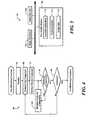
- FIG. 1is a diagrammatic view of a computed tomography (CT) system.
- CTcomputed tomography
- FIG. 2Ais a diagrammatic view of an artery having calcification.
- FIG. 2Bis a CT image of the artery.
- FIG. 3is a flowchart for a process of processing an image.
- FIG. 4is a flowchart for a process of minimizing total energy functional across all image components.
- FIG. 5is a diagrammatic view of a computer system on which the processes of FIG. 3 and FIG. 4 may be implemented.
- a key problem in medical imagingis the removal of artifacts which degrade the quality of the image for diagnostic purposes.
- a model-based approach to image processingwherein the structures or areas of interest in the image are explicitly partitioned according to their behavior and different constraints are imposed separately on each structure or area.
- This approachcan be employed as either a post-processing (restoration) method or as an image formation (reconstruction) method.
- This approachdiffers considerably from conventional deblurring and filtered back projection reconstruction techniques.
- To partition the image structuressignificant features of the different structures are used. These are flexible, but could include characteristics such as smoothness, concentration of energy or brightness, intensity, etc.
- a single functional or energyis then defined which combines all these model elements. The minimum of this combined energy is then sought.
- CTcomputed tomography
- the system and techniques described hereinuses an embodiment that includes computed tomography (CT), the system and techniques are not limited to CT.
- the system and techniquesmay be used in any image processing or acquisition system such as a magnetic resonance imaging (MRI) system, a positron emission tomography (PET) system, ultrasound and so forth.
- MRImagnetic resonance imaging
- PETpositron emission tomography
- the system and techniques described hereinare used for image processing to detect calcium in an artery, the system may be used in any image processing system such as to minimize blurring or blooming effects between any different materials.
- the method described hereinuses two components to distinguish between materials, but the method may be used with any number of components.
- a computed tomography system 10includes an x-ray source 12 , a patient 14 , a detector 16 , a computer 18 and a display 20 .
- the X-ray source 12emits an x-ray beam 13 which impinges upon or illuminates a portion (sometimes referred to as a slice) of patient 14 . Portions of the x-ray beam are absorbed by structures within the patient (both physiological and non-physiological structures) and a portion of the beam 15 reached a detector 16 .
- Detector 16measures the absorption along numerous paths with both radial and tangential components.
- the computer 18uses the measured absorption information collected by the detector 16 to form a cross-sectional image, which may be viewed on display 20 .
- an artery 22includes a wall 24 having an inner wall portion 24 a and an outer wall portion 24 b .
- Inner wall portion 24 adefines, a lumen 26 .
- a portion of wall 24may include a calcified region 28 .
- FIG. 2Ba CT image 30 of an artery similar to artery 22 ( FIG. 2A ) is shown.
- CT image 30is provided using known image processing techniques. Despite the well-localized nature of the calcium region 28 , the resulting image 30 shows nearly total obscuration of the luminal region 26 , rendering the image 30 non-evaluable.
- a calcium artifact in coronary CTarises when the intensity of small dense calcium regions in the vessel wall spills into adjacent pixels due to the blurring induced by the resolution-limited image formation process.
- the CT system 10 together with the reconstruction processspreads the intensity of such calcified regions over the image, resulting in an over-representation of the size of the calcification (“calcium blooming”) and often an under-representation of the size of the lumen 26 .
- the “blooming” effectis intrinsic to any imaging modality in which the point-spread function is of a scale comparable to the size of the objects that are of interest, and the field of view may contain bright objects which, due to the point-spread function, obscures nearby lower-intensity objects of interest.
- an image processing technique(e.g., a process 50 ) which may be performed, for example, by computer 18 .
- the image processing described in FIG. 3may be performed at post-processing, for example, after filtered back projection processing or with no image pre-processing.
- Process 50determines functions for each of the components identified in processing block 56 .
- f sis assumed to have a behavior having a lower amplitude and greater spatial extent than f c .
- the observed data gmay be an image formed using standard techniques (e.g., back filter projection), which exhibit calcium blooming or the data could be raw data, which corresponds to a set of tomographic projections obtained prior to image formation.
- Hdenotes an image blurring operator that maps the ideal image f to a set of observed data g.
- Hrepresents convolutional blurring with the point spread function corresponding to the imaging process.
- a filtered back projection generated imagemay be approximated as being shift invariant and convolutional.
- Hrepresents the tomographic projection process.
- the operator Hmay be linear, though this is not essential.
- An enhanced estimate of the ideal image by minimization of an energy functionmay be represented as:
- f ⁇arg ⁇ ⁇ min f ⁇ E d ⁇ ( g , f ) + ⁇ s ⁇ E s ⁇ ( f s ) + ⁇ c ⁇ E c ⁇ ( f c )
- Edenotes the “component energy”
- ddenotes the data term
- ⁇ s , and ⁇ care positive weights balancing the contributions of the different terms.
- the three termscapture the imaging process and the behavior of the underlying image.
- a Poisson observation modelmay be used.
- E c (f c )represents the behavior of calcified regions in cardiac CT.
- T cis about 1.2 times the density of water.
- a formulation for enhanced cardiac CT imagingis:
- the formulationexplicitly represents a single unified approach to calcium deblurring.
- the formulationmay be readily extended to capture other effects, including Poisson observation models, additional field constraints, components for additional biological structures, etc.
- process 50minimizes the functions for the components using, for example, a process 60 .
- the energy functionare minimized with respect to the values of the components f s and f c across an image to obtain a reconstructed image with enhanced behavior (i.e., better image) than a standard filtered back projection image.
- Minimization for the optimal enhanced imagemay be conducted through a series of iterative steps, for example, using a block coordinate descent approach and defining an outer iteration, which alternatively minimizes with respect to the tissue components f s and f c while the other component is held fixed.
- Process 60minimizes the first component ( 68 ). For example, minimizing the calcium component:
- f ⁇ c ( k + 1 )arg ⁇ ⁇ min f c ⁇ T c ⁇ ⁇ g - H ⁇ ( f s ( k ) + f c ) ⁇ 2 2 + ⁇ c ⁇ ⁇ Df c ⁇ 1 1
- Process 60minimizes the second component ( 72 ). For example, minimizing the non-calcified component:
- f ⁇ s ( k + 1 )arg ⁇ ⁇ min f s ⁇ T s ⁇ ⁇ g - H ⁇ ( f c ( k + 1 ) + f s ) ⁇ 2 2 + ⁇ s ⁇ ⁇ Df s ⁇ 2 2
- ARTalgebraic reconstruction technique
- process 60may be initialized with, for example, a filtered back projection based reconstruction to provide an initial estimate of ⁇ circumflex over (f) ⁇ s (0) .
- FIG. 5shows a computer 18 using process 50 including process 60 .
- Computer 100includes an image processor 102 , a volatile memory 104 , and a non-volatile memory 106 (e.g., hard disk).
- Non-volatile memory 106stores operating system 110 ; image data 112 ; and computer instructions 114 , which are executed by the image processor 102 out of volatile memory 104 to perform all or part of process 50 and process 60 .
- Processes 50 and 60are not limited to use with the hardware and software of FIG. 5 ; it may find applicability in any computing or processing environment and with any type of machine that is capable of running a computer program.
- Processes 50 and 60may be implemented in hardware, software, or a combination of the two.
- Processes 50 and 60may be implemented in computer programs executed on programmable computers/machines that each includes a processor, a storage medium or other article of manufacture that is readable by the processor (including volatile and non-volatile memory and/or storage elements), at least one input device, and one or more output devices.
- Program codemay be applied to data entered using an input device to perform processes 50 and 60 and to generate output information.
- the systemcan be implemented, at least in part, via a computer program product (i.e., a computer program tangibly embodied in an information carrier (e.g., in a machine-readable storage device or in a propagated signal) for execution by, or to control the operation of, data processing apparatus (e.g., a programmable processor, a computer, or multiple computers)).
- a computer program producti.e., a computer program tangibly embodied in an information carrier (e.g., in a machine-readable storage device or in a propagated signal) for execution by, or to control the operation of, data processing apparatus (e.g., a programmable processor, a computer, or multiple computers)).
- Each such programmay be implemented in a high level procedural or object-oriented programming language to communicate with a computer system.
- the programscan be implemented in assembly or machine language.
- the languagemay be a compiled or an interpreted language and it can be deployed in any form, including as a stand-alone
- a computer programcan be deployed to be executed on one computer or on multiple computers at one site or distributed across multiple sites and interconnected by a communication network.
- a computer programmay be stored on a storage medium or device (e.g., CD-ROM, hard disk, or magnetic diskette) that is readable by a general or special purpose programmable computer for configuring and operating the computer when the storage medium or device is read by the computer to perform process 80 .
- Process 80may also be implemented as a machine-readable storage medium, configured with a computer program, where upon execution, instructions in the computer program cause the computer to operate in accordance with process 50 .
- the system described hereinis not limited to use with the hardware and software described above.
- the systemcan be implemented in digital electronic circuitry, or in computer hardware, firmware, software, or in combinations thereof.
- Method steps associated with implementing the systemcan be performed by one or more programmable processors executing one or more computer programs to perform the functions of the system. All or part of the system can be implemented as, special purpose logic circuitry (e.g., an FPGA (field programmable gate array) and/or an ASIC (application-specific integrated circuit)).
- special purpose logic circuitrye.g., an FPGA (field programmable gate array) and/or an ASIC (application-specific integrated circuit)
- processors suitable for the execution of a computer programinclude, by way of example, both general and special purpose microprocessors, and any one or more processors of any kind of digital computer.
- a processorwill receive instructions and data from a read-only memory or a random access memory or both.
- Elements of a computerinclude a processor for executing instructions and one or more memory devices for storing instructions and data.
Landscapes
- Physics & Mathematics (AREA)
- General Physics & Mathematics (AREA)
- Engineering & Computer Science (AREA)
- Theoretical Computer Science (AREA)
- Apparatus For Radiation Diagnosis (AREA)
Abstract
Description
f=fc+fs.
Ed(f,g)=∥g−Hf∥22
Es(fs)=∥Dfs∥22
where D is a discrete approximation to a derivative operator. Since the non-calcium component is assumed to have a lower amplitude, then fs≦Ts, for a positive threshold Ts. In one embodiment, Tsis about 0.05 times the density of water.
Ec(fc)=∥Dfc∥pp
where D is a discrete approximation to a derivative operator and 0<p≦2. Penalty, p, has localization and super-resolution ability when p≈1. In addition, under the assumption that the calcium component will generally be denser than non-calcific tissue, then Tc≦fcfor a positive threshold Tc. In one embodiment, Tcis about 1.2 times the density of water.
Each subminimization is solved using an algebraic reconstruction technique (ART)-like iteration. Such ART-like techniques are well-known to persons of ordinary skill in the art.
Claims (24)
Priority Applications (3)
| Application Number | Priority Date | Filing Date | Title |
|---|---|---|---|
| US11/320,153US8031927B2 (en) | 2005-12-28 | 2005-12-28 | Medical image processing |
| US11/424,923US7689017B2 (en) | 2005-12-28 | 2006-06-19 | Medical image processing |
| PCT/US2006/048398WO2007075664A1 (en) | 2005-12-28 | 2006-12-18 | Medical image processing |
Applications Claiming Priority (1)
| Application Number | Priority Date | Filing Date | Title |
|---|---|---|---|
| US11/320,153US8031927B2 (en) | 2005-12-28 | 2005-12-28 | Medical image processing |
Related Child Applications (1)
| Application Number | Title | Priority Date | Filing Date |
|---|---|---|---|
| US11/424,923Continuation-In-PartUS7689017B2 (en) | 2005-12-28 | 2006-06-19 | Medical image processing |
Publications (2)
| Publication Number | Publication Date |
|---|---|
| US20070147672A1 US20070147672A1 (en) | 2007-06-28 |
| US8031927B2true US8031927B2 (en) | 2011-10-04 |
Family
ID=38193794
Family Applications (1)
| Application Number | Title | Priority Date | Filing Date |
|---|---|---|---|
| US11/320,153Active2028-07-15US8031927B2 (en) | 2005-12-28 | 2005-12-28 | Medical image processing |
Country Status (1)
| Country | Link |
|---|---|
| US (1) | US8031927B2 (en) |
Cited By (79)
| Publication number | Priority date | Publication date | Assignee | Title |
|---|---|---|---|---|
| US20140044332A1 (en)* | 2012-08-10 | 2014-02-13 | National Taiwan University | Transformation method for diffusion spectrum imaging using large deformation diffeomorphic metric mapping |
| US8880185B2 (en) | 2010-06-11 | 2014-11-04 | Boston Scientific Scimed, Inc. | Renal denervation and stimulation employing wireless vascular energy transfer arrangement |
| US8939970B2 (en) | 2004-09-10 | 2015-01-27 | Vessix Vascular, Inc. | Tuned RF energy and electrical tissue characterization for selective treatment of target tissues |
| US8951251B2 (en) | 2011-11-08 | 2015-02-10 | Boston Scientific Scimed, Inc. | Ostial renal nerve ablation |
| US8974451B2 (en) | 2010-10-25 | 2015-03-10 | Boston Scientific Scimed, Inc. | Renal nerve ablation using conductive fluid jet and RF energy |
| US9023034B2 (en) | 2010-11-22 | 2015-05-05 | Boston Scientific Scimed, Inc. | Renal ablation electrode with force-activatable conduction apparatus |
| US9028485B2 (en) | 2010-11-15 | 2015-05-12 | Boston Scientific Scimed, Inc. | Self-expanding cooling electrode for renal nerve ablation |
| US9028472B2 (en) | 2011-12-23 | 2015-05-12 | Vessix Vascular, Inc. | Methods and apparatuses for remodeling tissue of or adjacent to a body passage |
| US9050106B2 (en) | 2011-12-29 | 2015-06-09 | Boston Scientific Scimed, Inc. | Off-wall electrode device and methods for nerve modulation |
| US9060761B2 (en) | 2010-11-18 | 2015-06-23 | Boston Scientific Scime, Inc. | Catheter-focused magnetic field induced renal nerve ablation |
| US9079000B2 (en) | 2011-10-18 | 2015-07-14 | Boston Scientific Scimed, Inc. | Integrated crossing balloon catheter |
| US9084609B2 (en) | 2010-07-30 | 2015-07-21 | Boston Scientific Scime, Inc. | Spiral balloon catheter for renal nerve ablation |
| US9089350B2 (en) | 2010-11-16 | 2015-07-28 | Boston Scientific Scimed, Inc. | Renal denervation catheter with RF electrode and integral contrast dye injection arrangement |
| US9119632B2 (en) | 2011-11-21 | 2015-09-01 | Boston Scientific Scimed, Inc. | Deflectable renal nerve ablation catheter |
| US9119600B2 (en) | 2011-11-15 | 2015-09-01 | Boston Scientific Scimed, Inc. | Device and methods for renal nerve modulation monitoring |
| US9125667B2 (en) | 2004-09-10 | 2015-09-08 | Vessix Vascular, Inc. | System for inducing desirable temperature effects on body tissue |
| US9125666B2 (en) | 2003-09-12 | 2015-09-08 | Vessix Vascular, Inc. | Selectable eccentric remodeling and/or ablation of atherosclerotic material |
| US9155589B2 (en) | 2010-07-30 | 2015-10-13 | Boston Scientific Scimed, Inc. | Sequential activation RF electrode set for renal nerve ablation |
| US9162046B2 (en) | 2011-10-18 | 2015-10-20 | Boston Scientific Scimed, Inc. | Deflectable medical devices |
| US9173696B2 (en) | 2012-09-17 | 2015-11-03 | Boston Scientific Scimed, Inc. | Self-positioning electrode system and method for renal nerve modulation |
| WO2015065781A3 (en)* | 2013-10-30 | 2015-11-05 | Mayo Foundation For Medical Education And Research | System and method for model-based reconstruction of quantitative images |
| US9186210B2 (en) | 2011-10-10 | 2015-11-17 | Boston Scientific Scimed, Inc. | Medical devices including ablation electrodes |
| US9186209B2 (en) | 2011-07-22 | 2015-11-17 | Boston Scientific Scimed, Inc. | Nerve modulation system having helical guide |
| US9192790B2 (en) | 2010-04-14 | 2015-11-24 | Boston Scientific Scimed, Inc. | Focused ultrasonic renal denervation |
| US9192435B2 (en) | 2010-11-22 | 2015-11-24 | Boston Scientific Scimed, Inc. | Renal denervation catheter with cooled RF electrode |
| US9220561B2 (en) | 2011-01-19 | 2015-12-29 | Boston Scientific Scimed, Inc. | Guide-compatible large-electrode catheter for renal nerve ablation with reduced arterial injury |
| US9220558B2 (en) | 2010-10-27 | 2015-12-29 | Boston Scientific Scimed, Inc. | RF renal denervation catheter with multiple independent electrodes |
| US9265969B2 (en) | 2011-12-21 | 2016-02-23 | Cardiac Pacemakers, Inc. | Methods for modulating cell function |
| US9277955B2 (en) | 2010-04-09 | 2016-03-08 | Vessix Vascular, Inc. | Power generating and control apparatus for the treatment of tissue |
| US9297845B2 (en) | 2013-03-15 | 2016-03-29 | Boston Scientific Scimed, Inc. | Medical devices and methods for treatment of hypertension that utilize impedance compensation |
| US9327100B2 (en) | 2008-11-14 | 2016-05-03 | Vessix Vascular, Inc. | Selective drug delivery in a lumen |
| US9326751B2 (en) | 2010-11-17 | 2016-05-03 | Boston Scientific Scimed, Inc. | Catheter guidance of external energy for renal denervation |
| US9358365B2 (en) | 2010-07-30 | 2016-06-07 | Boston Scientific Scimed, Inc. | Precision electrode movement control for renal nerve ablation |
| US9364284B2 (en) | 2011-10-12 | 2016-06-14 | Boston Scientific Scimed, Inc. | Method of making an off-wall spacer cage |
| US9408661B2 (en) | 2010-07-30 | 2016-08-09 | Patrick A. Haverkost | RF electrodes on multiple flexible wires for renal nerve ablation |
| US9420955B2 (en) | 2011-10-11 | 2016-08-23 | Boston Scientific Scimed, Inc. | Intravascular temperature monitoring system and method |
| US9433760B2 (en) | 2011-12-28 | 2016-09-06 | Boston Scientific Scimed, Inc. | Device and methods for nerve modulation using a novel ablation catheter with polymeric ablative elements |
| US9463062B2 (en) | 2010-07-30 | 2016-10-11 | Boston Scientific Scimed, Inc. | Cooled conductive balloon RF catheter for renal nerve ablation |
| US9486355B2 (en) | 2005-05-03 | 2016-11-08 | Vessix Vascular, Inc. | Selective accumulation of energy with or without knowledge of tissue topography |
| US9579030B2 (en) | 2011-07-20 | 2017-02-28 | Boston Scientific Scimed, Inc. | Percutaneous devices and methods to visualize, target and ablate nerves |
| US9649156B2 (en) | 2010-12-15 | 2017-05-16 | Boston Scientific Scimed, Inc. | Bipolar off-wall electrode device for renal nerve ablation |
| US9668811B2 (en) | 2010-11-16 | 2017-06-06 | Boston Scientific Scimed, Inc. | Minimally invasive access for renal nerve ablation |
| US9687166B2 (en) | 2013-10-14 | 2017-06-27 | Boston Scientific Scimed, Inc. | High resolution cardiac mapping electrode array catheter |
| US9693821B2 (en) | 2013-03-11 | 2017-07-04 | Boston Scientific Scimed, Inc. | Medical devices for modulating nerves |
| US9707036B2 (en) | 2013-06-25 | 2017-07-18 | Boston Scientific Scimed, Inc. | Devices and methods for nerve modulation using localized indifferent electrodes |
| US9713730B2 (en) | 2004-09-10 | 2017-07-25 | Boston Scientific Scimed, Inc. | Apparatus and method for treatment of in-stent restenosis |
| US9770606B2 (en) | 2013-10-15 | 2017-09-26 | Boston Scientific Scimed, Inc. | Ultrasound ablation catheter with cooling infusion and centering basket |
| US9808311B2 (en) | 2013-03-13 | 2017-11-07 | Boston Scientific Scimed, Inc. | Deflectable medical devices |
| US9808300B2 (en) | 2006-05-02 | 2017-11-07 | Boston Scientific Scimed, Inc. | Control of arterial smooth muscle tone |
| US9827039B2 (en) | 2013-03-15 | 2017-11-28 | Boston Scientific Scimed, Inc. | Methods and apparatuses for remodeling tissue of or adjacent to a body passage |
| US9833283B2 (en) | 2013-07-01 | 2017-12-05 | Boston Scientific Scimed, Inc. | Medical devices for renal nerve ablation |
| US9895194B2 (en) | 2013-09-04 | 2018-02-20 | Boston Scientific Scimed, Inc. | Radio frequency (RF) balloon catheter having flushing and cooling capability |
| US9907609B2 (en) | 2014-02-04 | 2018-03-06 | Boston Scientific Scimed, Inc. | Alternative placement of thermal sensors on bipolar electrode |
| US9925001B2 (en) | 2013-07-19 | 2018-03-27 | Boston Scientific Scimed, Inc. | Spiral bipolar electrode renal denervation balloon |
| US9943365B2 (en) | 2013-06-21 | 2018-04-17 | Boston Scientific Scimed, Inc. | Renal denervation balloon catheter with ride along electrode support |
| US9956033B2 (en) | 2013-03-11 | 2018-05-01 | Boston Scientific Scimed, Inc. | Medical devices for modulating nerves |
| US9962223B2 (en) | 2013-10-15 | 2018-05-08 | Boston Scientific Scimed, Inc. | Medical device balloon |
| US9974607B2 (en) | 2006-10-18 | 2018-05-22 | Vessix Vascular, Inc. | Inducing desirable temperature effects on body tissue |
| US10022182B2 (en) | 2013-06-21 | 2018-07-17 | Boston Scientific Scimed, Inc. | Medical devices for renal nerve ablation having rotatable shafts |
| US10085799B2 (en) | 2011-10-11 | 2018-10-02 | Boston Scientific Scimed, Inc. | Off-wall electrode device and methods for nerve modulation |
| US10265122B2 (en) | 2013-03-15 | 2019-04-23 | Boston Scientific Scimed, Inc. | Nerve ablation devices and related methods of use |
| US10271898B2 (en) | 2013-10-25 | 2019-04-30 | Boston Scientific Scimed, Inc. | Embedded thermocouple in denervation flex circuit |
| US10321946B2 (en) | 2012-08-24 | 2019-06-18 | Boston Scientific Scimed, Inc. | Renal nerve modulation devices with weeping RF ablation balloons |
| US10342609B2 (en) | 2013-07-22 | 2019-07-09 | Boston Scientific Scimed, Inc. | Medical devices for renal nerve ablation |
| US10398464B2 (en) | 2012-09-21 | 2019-09-03 | Boston Scientific Scimed, Inc. | System for nerve modulation and innocuous thermal gradient nerve block |
| US10413357B2 (en) | 2013-07-11 | 2019-09-17 | Boston Scientific Scimed, Inc. | Medical device with stretchable electrode assemblies |
| US10543037B2 (en) | 2013-03-15 | 2020-01-28 | Medtronic Ardian Luxembourg S.A.R.L. | Controlled neuromodulation systems and methods of use |
| US10549127B2 (en) | 2012-09-21 | 2020-02-04 | Boston Scientific Scimed, Inc. | Self-cooling ultrasound ablation catheter |
| US10660698B2 (en) | 2013-07-11 | 2020-05-26 | Boston Scientific Scimed, Inc. | Devices and methods for nerve modulation |
| US10660703B2 (en) | 2012-05-08 | 2020-05-26 | Boston Scientific Scimed, Inc. | Renal nerve modulation devices |
| US10695124B2 (en) | 2013-07-22 | 2020-06-30 | Boston Scientific Scimed, Inc. | Renal nerve ablation catheter having twist balloon |
| US10722300B2 (en) | 2013-08-22 | 2020-07-28 | Boston Scientific Scimed, Inc. | Flexible circuit having improved adhesion to a renal nerve modulation balloon |
| US10835305B2 (en) | 2012-10-10 | 2020-11-17 | Boston Scientific Scimed, Inc. | Renal nerve modulation devices and methods |
| US10945786B2 (en) | 2013-10-18 | 2021-03-16 | Boston Scientific Scimed, Inc. | Balloon catheters with flexible conducting wires and related methods of use and manufacture |
| US10952790B2 (en) | 2013-09-13 | 2021-03-23 | Boston Scientific Scimed, Inc. | Ablation balloon with vapor deposited cover layer |
| US11000679B2 (en) | 2014-02-04 | 2021-05-11 | Boston Scientific Scimed, Inc. | Balloon protection and rewrapping devices and related methods of use |
| US11202671B2 (en) | 2014-01-06 | 2021-12-21 | Boston Scientific Scimed, Inc. | Tear resistant flex circuit assembly |
| US11246654B2 (en) | 2013-10-14 | 2022-02-15 | Boston Scientific Scimed, Inc. | Flexible renal nerve ablation devices and related methods of use and manufacture |
| US20220151580A1 (en)* | 2019-07-22 | 2022-05-19 | Siemens Healthcare Gmbh | Assessment of coronary artery calcification in angiographic images |
Families Citing this family (4)
| Publication number | Priority date | Publication date | Assignee | Title |
|---|---|---|---|---|
| US8632448B1 (en) | 2009-02-05 | 2014-01-21 | Loma Linda University Medical Center | Proton scattering analysis system |
| EP2483710A4 (en) | 2009-10-01 | 2016-04-27 | Univ Loma Linda Med | Ion Induced IMPACT Ionization SENSOR AND USES THEREOF |
| US9207193B2 (en) | 2010-02-12 | 2015-12-08 | Loma Linda University Medical Center | Systems and methodologies for proton computed tomography |
| EP2684034A4 (en) | 2011-03-07 | 2014-09-03 | Univ Loma Linda Med | SYSTEMS, DEVICES AND METHODS RELATING TO CALIBRATION OF COMPUTER-COMPUTED PROTONE TRANSMISSION TOMOGRAPHY SCANNER |
Citations (11)
| Publication number | Priority date | Publication date | Assignee | Title |
|---|---|---|---|---|
| EP0434872A1 (en) | 1989-12-28 | 1991-07-03 | Matsushita Electric Industrial Co., Ltd. | Energy difference image processing method |
| US20020070970A1 (en) | 2000-11-22 | 2002-06-13 | Wood Susan A. | Graphical user interface for display of anatomical information |
| US20030091142A1 (en)* | 2001-11-15 | 2003-05-15 | Jianying Li | System and method of medical imaging having task and/or patient size dependent processing |
| US20030215124A1 (en)* | 2002-05-20 | 2003-11-20 | Jianying Li | Method and apparatus of scoring an arterial obstruction |
| US20060098010A1 (en)* | 2004-03-09 | 2006-05-11 | Jeff Dwyer | Anatomical visualization and measurement system |
| US7283604B2 (en)* | 2004-11-24 | 2007-10-16 | General Electric Company | Method and system of CT data correction |
| US7372937B2 (en)* | 2004-07-16 | 2008-05-13 | University Of Iowa Research Foundation | Systems and methods of non-standard spiral cone-beam computed tomograpy (CT) |
| US7394923B2 (en)* | 2004-02-10 | 2008-07-01 | The University Of Chicago | Imaging system for generating a substantially exact reconstruction of a region of interest |
| US7444011B2 (en)* | 2004-02-10 | 2008-10-28 | University Of Chicago | Imaging system performing substantially exact reconstruction and using non-traditional trajectories |
| US7646898B1 (en)* | 2000-11-24 | 2010-01-12 | Kent Ridge Digital Labs | Methods and apparatus for processing medical images |
| US7689017B2 (en)* | 2005-12-28 | 2010-03-30 | The General Hospital Corporation | Medical image processing |
- 2005
- 2005-12-28USUS11/320,153patent/US8031927B2/enactiveActive
Patent Citations (11)
| Publication number | Priority date | Publication date | Assignee | Title |
|---|---|---|---|---|
| EP0434872A1 (en) | 1989-12-28 | 1991-07-03 | Matsushita Electric Industrial Co., Ltd. | Energy difference image processing method |
| US20020070970A1 (en) | 2000-11-22 | 2002-06-13 | Wood Susan A. | Graphical user interface for display of anatomical information |
| US7646898B1 (en)* | 2000-11-24 | 2010-01-12 | Kent Ridge Digital Labs | Methods and apparatus for processing medical images |
| US20030091142A1 (en)* | 2001-11-15 | 2003-05-15 | Jianying Li | System and method of medical imaging having task and/or patient size dependent processing |
| US20030215124A1 (en)* | 2002-05-20 | 2003-11-20 | Jianying Li | Method and apparatus of scoring an arterial obstruction |
| US7394923B2 (en)* | 2004-02-10 | 2008-07-01 | The University Of Chicago | Imaging system for generating a substantially exact reconstruction of a region of interest |
| US7444011B2 (en)* | 2004-02-10 | 2008-10-28 | University Of Chicago | Imaging system performing substantially exact reconstruction and using non-traditional trajectories |
| US20060098010A1 (en)* | 2004-03-09 | 2006-05-11 | Jeff Dwyer | Anatomical visualization and measurement system |
| US7372937B2 (en)* | 2004-07-16 | 2008-05-13 | University Of Iowa Research Foundation | Systems and methods of non-standard spiral cone-beam computed tomograpy (CT) |
| US7283604B2 (en)* | 2004-11-24 | 2007-10-16 | General Electric Company | Method and system of CT data correction |
| US7689017B2 (en)* | 2005-12-28 | 2010-03-30 | The General Hospital Corporation | Medical image processing |
Non-Patent Citations (61)
| Title |
|---|
| American Heart Association. Heart Disease and Stroke Statistics-2003 Update, Dallas, TX: American Heart Association, 2003, 46 pages. |
| Barrett et al., "Artifiacts in CT: Recognition and Avoidance", Radiographics 2004;24:1679-91. |
| Bertalmio et al., "Simultaneous Structure and Texture Image Inpainting", IEEE Trans. Image Proc. 2003;12:882-889. |
| Bhatia, et al., "A Wavelet-Based Method for Multiscale Tomographic Reconstruction", IEEE Transactions on Medical Imaging 1996;15:92-101. |
| Bhatia, et al., "Tomographic Reconstruction and Estimation Based on Multiscale Natural-Pixel Bases", IEEE Transactions on Image Processing special issue on Automatic Target Recognition 1997;6:463-478. |
| Cetin, et al., "Feature Enhancement and ATR Performance Using Nonquadratic Optimization-Based SAR Imaging", IEEE Trans Aerospace and Electronic Systems 2003;39:1375-1395. |
| Cetin, et al., "Feature-Enhanced Synthetic Aperture Radar Image Formation Based on Nonquadratic Regularization", IEEE Transactions on Image Processing special issue on Automatic Target Recognition 2001;10:623-631. |
| Cetin, et al., "Superresolution and Edge-Preserving Reconstruction of Complex-Valued Synthetic Aperture Radar Images", IEEE Int'l Conf. on Image Processing, Vancouver, British Columbia, Canada, 2000:701-704. |
| Chan, et al., "A New Model-Based Technique for Enhanced Small-Vessel Measurements in X-Ray Cine-Angiograms", IEEE Transactions on Medical Imaging 2000;19:243-255. |
| Chan, et al., "OCT-Based Arterial Elastography: Robust Estimation Exploiting Tissue Biomechanics", Optics Express 2004;12:4558-4572. |
| Chandrasekar et al., "Complications of Cardiac Catheterization in the Current Era: A single-Center Experience" Catheter Cardiovas. Interv. 2001;52:289-95. |
| De Man et al., "An Iterative Maximum-Likelihood Polychromatic Algorithm for CT", IEEE Trans. Med. Imaging 2001;20:999-1008. |
| DeMan et al., "Metal Streak Artifacts in X-Ray Computed Tomography: A Simulation Study", IEEE Trans. Nuclear Sci. 1999;46:691-696. |
| Desai et al., "Functional MRI Activity Characterization Using Response Time Shift Estimates from Curve Evolution", IEEE Transactions on Image Processing, 2002;21:1402-1412. |
| Donoho et al., "Maximum Entropy and the Nearly Black Object", J.R. Statist. Soc. 1992; B54:41-81. |
| Donoho, David L., "Superresolution via Sparsity Constraints", SIAM J. Math. Anal. 1993;23:1309-1331. |
| Feng, et al., "A Curve Evolution Approach to Object-Based Tomographic Reconstruction", IEEE Transactions on Image Processing special issue on Automatic Target Recognition 2003;12:44-57.49. |
| Fosgate, et al., "Multiscale Segmentation and Anomaly Enhancement of SAR Imagery", IEEE Transactions on Image Processing Special issue on Automatic Target Recognition 1997;6:7-20. |
| Galdi, et al., "Moderately Rough Surface Underground Imaging via Short-Pulse Quasi-Ray Gaussian Beams", IEEE Transactions on Antennas and Propagation 2003;51:2304-2318. |
| Gauch, et al., "Hybrid Boundary-Based and Region-Based Deformable Models for Biomedical Image Segmentation", SPIE Mathematical Methods in Medical Imaging III 1994;2299:72-83. |
| Gauch, et al., "Hybrid Deformable Models for Three-Dimensional Biomedical Image Segmentation", IEEE Proc. Nuclear Science and Medical Imaging 1994;4:1935-1939. |
| Glover, et al., "An Algorithm for the Reduction of Clip Artifacts in CT Reconstructions", Med. Phys. 1981;8:799-807. |
| International Search Report. (Form PCT/ISA/210), 4 pages. |
| Jiang et al. "Blind Deblurring of Spiral CT Images-Comparative Studies on Edge-To-Noise Ratios", Med. Phys. 2002;29:821-9. |
| Jiang et al., "Blind Deblurring of Spiral CT Images", IEEE Trans. Med. Imaging 2003;22:837-45. |
| Kacheiriess, et al., "Generalized Multi-Dimensional Adaptive Filtering for Conventional and Spiral Single-Slice, Multi-Slice, and Cone-Bean CT", Med. Phys. 2001:28:475-90. |
| Kalender et al., "Reduction of CT Artifacts Caused by Metallic Implants", Radiology 1987;164:576-7. |
| Karl, "Fusing Multi-Modality Inverse Data Through Shared Boundary Structure", presented at National Science Foundation Visit 2006 at Northeastern University, Boston, Ma, Apr. 6, 2006, 14 pages. |
| Karl, et al., "Capabilities and Limitations of Pupil-Plane Filters for Superresolution and Image Enhancement", Opt. Express 2004;12:4150-4156. |
| Kaufhold, et al., "A Statistical Method for Efficient Segmentation of MR Imagery", Int'l Journal of Pattern Recognition and Artificial Intelligence Special issue on Processing of MRI Imagery 1997;11:1213-1231. |
| Kaufhold, et al., "A Texture-Based Variational Segmentation Method for Ultrasound Blood Vessel Imagery", Abstract, Annals of Biomedical Engineering 1998;26. |
| Liang et al., "Image Enhancement in Detection of Coronary Stenosis Using MDCT", Poster Posted at National Science Foundation Visit 2005 at Northeastern University, Boston, MA, Apr. 7-8, 2005, 1 page. |
| Liang et al., "Image Enhancement in Detection of Coronary Stenosis Using MDCT", Poster Posted at National Science Foundation Visit 2006 at Northeastern University, Boston, MA, Apr. 4-6, 2006, 1 page. |
| Nakanishi et al., "Pitfalls in 16-Detector row CT of the Coronary Arteries", Radiographics 2005;25:425-38. |
| Notification of transmittal of the International Search Report and the Written Opinion of the International Searching Authority, or the Declaration. (Form PCT/ISA/220), 2 pages, Apr. 24, 2007. |
| Nuyts et al., "Iterative Reconstruction for Helical CT: A Simulation Study", Phys. Med. Biol. 1998;43:729-37. |
| Osher et al., "Image Decomposition and Restoration using Total Variation Minimization and the H-1 Norm", Multiscale Model. Simul. 2003;1:349-370. |
| Perkins et al., "DE Report UCRL-ID117796, ENDL Type Formats for the LLNL;Evaluated Atomic Data Library (EADL), Evaluated Electron Data Library EEDL, and Evaluated Photon Data Library (EPDL)", Univ. of California, 32 Pages. |
| Pien, et al. "A Variational Approach to Multi-Sensor Fusion of Images", Int'l J. Applied Intelligence 1995;5:217-235. |
| Pien, et al., "Segmentation of MR Images Using Curve Evolution and Prior Information", Int'l Journal of Pattern Recognition and Artificial Intelligence 1997;11:1233-1245. |
| Pien, et al., "Variational Segmentation of Multi-Channel MRI Images", IEEE Int'l Conf. Image Processing 1994;3-508-512. |
| Robertson et al., "Total Hip Prosthesis Metal-Artifact Suppression Using Iterative Deblurring Reconstruction", J. Comput. Assist. Tomogr. 1997;21:293-8. |
| Rollano-Hijarrubia et al., "Improving the Imaging of Calcifications in CT by Histogram-Based Selective Deblurring", Proceedings of the SPIE Conference on Medical Imaging 2005,12 pages. |
| Sakai et al., "The Use of Deblurring Technique for Improving the Longitudinal Resolution in Helical CT of the Head and Neck Region", Comput. Med. Imaging Graph 1997;21:153-64. |
| Saleh et al., "Center for Subsurface Sensing & Imaging Systems", Research Overview Presentation at National Science Visit 2006 at Northeastern University, Boston, MA, Apr. 4-6, 2006, 52 pages. |
| Schneider, et al., "Multiscale Methods for the Segmentation and Reconstruction of Signals and Images", IEEE Transactions on Image Processing special issue on Automatic Target Recognition 2000;9:456-468. |
| Shah, et al., "Recovery of Surfaces with Discontinuities by Fusing Shading and Range Data Within a Variational Framework", IEEE Trans. Image Processing 1996;5:1243-1251. |
| Tari, et al., "A Computationally Efficient Shape Analysis Via Level Sets", IEEE Mathematical Methods in Biomedical Image Analysis 1996:234-243. |
| Tari, et al., "Extraction of Shape Skeletons from Grayscale Images", Computer Vision and Image Understanding 1997;66:133-146. |
| Thompson et al., "Imaging of Coronary Calcification by Computed Tomography" J. Magn. Reson. Imaging. 2004; 19:720-33. |
| Vese et al., "Modeling Textures with Total Variation Minimization and Oscillating Patterns in Image Processing", Journal of Scientific Computing 2003:19. |
| Vese, "Image Denoising and Decomposition with Total Variation Minimization and Oscillatory Functions", Journal of Mathematical Imaging and Vision 2004;20:7-18. |
| Wang et al., "Fast Iterative Algorithm for Metal Artifact Reduction in X-ray CT", Acad. Radiol. 2000;7:607-14. |
| Wang et al., "Iterative Deblurring for CT Metal Artifact Reduction", IEEE Transactions on Medical Imaging 1996;15:657-664. |
| Wang et al., "Iterative X-Ray Cone-Beam Tomography for Metal Artifact Reduction and Local Region Reconstruction", Microscopy and Microanalysis 1999;5:58-65. |
| Wang et al., "Local Computed Tomography via Iterative Deblurring", Scanning 1996;18:582-8. |
| Wang et al., "Spiral CT Image Deblurring for Cochlear Implantation", IEEE Trans. Med. Imaging 1998;17:251-62. |
| Watzke O. Kalender WA. A Pragmatic Approach to Metal Artifact Reduction in CT: Merging of Metal Artifact Reduced Images, Eur. Radiol. 2004:14:849-56. |
| Weisenseel, et al., "A Variational Approach to Multi-Modality Subsurface Data Inversion and Fusion Based on Shared Image Structure", Subsurface Sensing Technologies and Applications 2003;4:375-394. |
| Williamson et al., "Prospects for Quantitative Computed Tomography Imaging In the Presence of Foreign Metal Bodies Using Statistical Image Reconstruction", Med. Phys. 2002;29:2404-18. |
| Written Opinion of the International Searching Authority. (Form PCT/ISA/237), 5 pages. |
Cited By (95)
| Publication number | Priority date | Publication date | Assignee | Title |
|---|---|---|---|---|
| US10188457B2 (en) | 2003-09-12 | 2019-01-29 | Vessix Vascular, Inc. | Selectable eccentric remodeling and/or ablation |
| US9510901B2 (en) | 2003-09-12 | 2016-12-06 | Vessix Vascular, Inc. | Selectable eccentric remodeling and/or ablation |
| US9125666B2 (en) | 2003-09-12 | 2015-09-08 | Vessix Vascular, Inc. | Selectable eccentric remodeling and/or ablation of atherosclerotic material |
| US8939970B2 (en) | 2004-09-10 | 2015-01-27 | Vessix Vascular, Inc. | Tuned RF energy and electrical tissue characterization for selective treatment of target tissues |
| US9125667B2 (en) | 2004-09-10 | 2015-09-08 | Vessix Vascular, Inc. | System for inducing desirable temperature effects on body tissue |
| US9713730B2 (en) | 2004-09-10 | 2017-07-25 | Boston Scientific Scimed, Inc. | Apparatus and method for treatment of in-stent restenosis |
| US9486355B2 (en) | 2005-05-03 | 2016-11-08 | Vessix Vascular, Inc. | Selective accumulation of energy with or without knowledge of tissue topography |
| US9808300B2 (en) | 2006-05-02 | 2017-11-07 | Boston Scientific Scimed, Inc. | Control of arterial smooth muscle tone |
| US10213252B2 (en) | 2006-10-18 | 2019-02-26 | Vessix, Inc. | Inducing desirable temperature effects on body tissue |
| US9974607B2 (en) | 2006-10-18 | 2018-05-22 | Vessix Vascular, Inc. | Inducing desirable temperature effects on body tissue |
| US12161392B2 (en) | 2006-10-18 | 2024-12-10 | Boston Scientific Scimed, Inc. | System for inducing desirable temperature effects on body tissue |
| US10413356B2 (en) | 2006-10-18 | 2019-09-17 | Boston Scientific Scimed, Inc. | System for inducing desirable temperature effects on body tissue |
| US9327100B2 (en) | 2008-11-14 | 2016-05-03 | Vessix Vascular, Inc. | Selective drug delivery in a lumen |
| US9277955B2 (en) | 2010-04-09 | 2016-03-08 | Vessix Vascular, Inc. | Power generating and control apparatus for the treatment of tissue |
| US9192790B2 (en) | 2010-04-14 | 2015-11-24 | Boston Scientific Scimed, Inc. | Focused ultrasonic renal denervation |
| US8880185B2 (en) | 2010-06-11 | 2014-11-04 | Boston Scientific Scimed, Inc. | Renal denervation and stimulation employing wireless vascular energy transfer arrangement |
| US9155589B2 (en) | 2010-07-30 | 2015-10-13 | Boston Scientific Scimed, Inc. | Sequential activation RF electrode set for renal nerve ablation |
| US9084609B2 (en) | 2010-07-30 | 2015-07-21 | Boston Scientific Scime, Inc. | Spiral balloon catheter for renal nerve ablation |
| US9463062B2 (en) | 2010-07-30 | 2016-10-11 | Boston Scientific Scimed, Inc. | Cooled conductive balloon RF catheter for renal nerve ablation |
| US9408661B2 (en) | 2010-07-30 | 2016-08-09 | Patrick A. Haverkost | RF electrodes on multiple flexible wires for renal nerve ablation |
| US9358365B2 (en) | 2010-07-30 | 2016-06-07 | Boston Scientific Scimed, Inc. | Precision electrode movement control for renal nerve ablation |
| US8974451B2 (en) | 2010-10-25 | 2015-03-10 | Boston Scientific Scimed, Inc. | Renal nerve ablation using conductive fluid jet and RF energy |
| US9220558B2 (en) | 2010-10-27 | 2015-12-29 | Boston Scientific Scimed, Inc. | RF renal denervation catheter with multiple independent electrodes |
| US9028485B2 (en) | 2010-11-15 | 2015-05-12 | Boston Scientific Scimed, Inc. | Self-expanding cooling electrode for renal nerve ablation |
| US9848946B2 (en) | 2010-11-15 | 2017-12-26 | Boston Scientific Scimed, Inc. | Self-expanding cooling electrode for renal nerve ablation |
| US9089350B2 (en) | 2010-11-16 | 2015-07-28 | Boston Scientific Scimed, Inc. | Renal denervation catheter with RF electrode and integral contrast dye injection arrangement |
| US9668811B2 (en) | 2010-11-16 | 2017-06-06 | Boston Scientific Scimed, Inc. | Minimally invasive access for renal nerve ablation |
| US9326751B2 (en) | 2010-11-17 | 2016-05-03 | Boston Scientific Scimed, Inc. | Catheter guidance of external energy for renal denervation |
| US9060761B2 (en) | 2010-11-18 | 2015-06-23 | Boston Scientific Scime, Inc. | Catheter-focused magnetic field induced renal nerve ablation |
| US9192435B2 (en) | 2010-11-22 | 2015-11-24 | Boston Scientific Scimed, Inc. | Renal denervation catheter with cooled RF electrode |
| US9023034B2 (en) | 2010-11-22 | 2015-05-05 | Boston Scientific Scimed, Inc. | Renal ablation electrode with force-activatable conduction apparatus |
| US9649156B2 (en) | 2010-12-15 | 2017-05-16 | Boston Scientific Scimed, Inc. | Bipolar off-wall electrode device for renal nerve ablation |
| US9220561B2 (en) | 2011-01-19 | 2015-12-29 | Boston Scientific Scimed, Inc. | Guide-compatible large-electrode catheter for renal nerve ablation with reduced arterial injury |
| US9579030B2 (en) | 2011-07-20 | 2017-02-28 | Boston Scientific Scimed, Inc. | Percutaneous devices and methods to visualize, target and ablate nerves |
| US9186209B2 (en) | 2011-07-22 | 2015-11-17 | Boston Scientific Scimed, Inc. | Nerve modulation system having helical guide |
| US9186210B2 (en) | 2011-10-10 | 2015-11-17 | Boston Scientific Scimed, Inc. | Medical devices including ablation electrodes |
| US10085799B2 (en) | 2011-10-11 | 2018-10-02 | Boston Scientific Scimed, Inc. | Off-wall electrode device and methods for nerve modulation |
| US9420955B2 (en) | 2011-10-11 | 2016-08-23 | Boston Scientific Scimed, Inc. | Intravascular temperature monitoring system and method |
| US9364284B2 (en) | 2011-10-12 | 2016-06-14 | Boston Scientific Scimed, Inc. | Method of making an off-wall spacer cage |
| US9162046B2 (en) | 2011-10-18 | 2015-10-20 | Boston Scientific Scimed, Inc. | Deflectable medical devices |
| US9079000B2 (en) | 2011-10-18 | 2015-07-14 | Boston Scientific Scimed, Inc. | Integrated crossing balloon catheter |
| US8951251B2 (en) | 2011-11-08 | 2015-02-10 | Boston Scientific Scimed, Inc. | Ostial renal nerve ablation |
| US9119600B2 (en) | 2011-11-15 | 2015-09-01 | Boston Scientific Scimed, Inc. | Device and methods for renal nerve modulation monitoring |
| US9119632B2 (en) | 2011-11-21 | 2015-09-01 | Boston Scientific Scimed, Inc. | Deflectable renal nerve ablation catheter |
| US9265969B2 (en) | 2011-12-21 | 2016-02-23 | Cardiac Pacemakers, Inc. | Methods for modulating cell function |
| US9174050B2 (en) | 2011-12-23 | 2015-11-03 | Vessix Vascular, Inc. | Methods and apparatuses for remodeling tissue of or adjacent to a body passage |
| US9402684B2 (en) | 2011-12-23 | 2016-08-02 | Boston Scientific Scimed, Inc. | Methods and apparatuses for remodeling tissue of or adjacent to a body passage |
| US9592386B2 (en) | 2011-12-23 | 2017-03-14 | Vessix Vascular, Inc. | Methods and apparatuses for remodeling tissue of or adjacent to a body passage |
| US9186211B2 (en) | 2011-12-23 | 2015-11-17 | Boston Scientific Scimed, Inc. | Methods and apparatuses for remodeling tissue of or adjacent to a body passage |
| US9037259B2 (en) | 2011-12-23 | 2015-05-19 | Vessix Vascular, Inc. | Methods and apparatuses for remodeling tissue of or adjacent to a body passage |
| US9072902B2 (en) | 2011-12-23 | 2015-07-07 | Vessix Vascular, Inc. | Methods and apparatuses for remodeling tissue of or adjacent to a body passage |
| US9028472B2 (en) | 2011-12-23 | 2015-05-12 | Vessix Vascular, Inc. | Methods and apparatuses for remodeling tissue of or adjacent to a body passage |
| US9433760B2 (en) | 2011-12-28 | 2016-09-06 | Boston Scientific Scimed, Inc. | Device and methods for nerve modulation using a novel ablation catheter with polymeric ablative elements |
| US9050106B2 (en) | 2011-12-29 | 2015-06-09 | Boston Scientific Scimed, Inc. | Off-wall electrode device and methods for nerve modulation |
| US10660703B2 (en) | 2012-05-08 | 2020-05-26 | Boston Scientific Scimed, Inc. | Renal nerve modulation devices |
| US20140044332A1 (en)* | 2012-08-10 | 2014-02-13 | National Taiwan University | Transformation method for diffusion spectrum imaging using large deformation diffeomorphic metric mapping |
| US9047695B2 (en)* | 2012-08-10 | 2015-06-02 | National Taiwan University | Transformation method for diffusion spectrum imaging using large deformation diffeomorphic metric mapping |
| US10321946B2 (en) | 2012-08-24 | 2019-06-18 | Boston Scientific Scimed, Inc. | Renal nerve modulation devices with weeping RF ablation balloons |
| US9173696B2 (en) | 2012-09-17 | 2015-11-03 | Boston Scientific Scimed, Inc. | Self-positioning electrode system and method for renal nerve modulation |
| US10549127B2 (en) | 2012-09-21 | 2020-02-04 | Boston Scientific Scimed, Inc. | Self-cooling ultrasound ablation catheter |
| US10398464B2 (en) | 2012-09-21 | 2019-09-03 | Boston Scientific Scimed, Inc. | System for nerve modulation and innocuous thermal gradient nerve block |
| US10835305B2 (en) | 2012-10-10 | 2020-11-17 | Boston Scientific Scimed, Inc. | Renal nerve modulation devices and methods |
| US9693821B2 (en) | 2013-03-11 | 2017-07-04 | Boston Scientific Scimed, Inc. | Medical devices for modulating nerves |
| US9956033B2 (en) | 2013-03-11 | 2018-05-01 | Boston Scientific Scimed, Inc. | Medical devices for modulating nerves |
| US9808311B2 (en) | 2013-03-13 | 2017-11-07 | Boston Scientific Scimed, Inc. | Deflectable medical devices |
| US10543037B2 (en) | 2013-03-15 | 2020-01-28 | Medtronic Ardian Luxembourg S.A.R.L. | Controlled neuromodulation systems and methods of use |
| US10265122B2 (en) | 2013-03-15 | 2019-04-23 | Boston Scientific Scimed, Inc. | Nerve ablation devices and related methods of use |
| US9297845B2 (en) | 2013-03-15 | 2016-03-29 | Boston Scientific Scimed, Inc. | Medical devices and methods for treatment of hypertension that utilize impedance compensation |
| US9827039B2 (en) | 2013-03-15 | 2017-11-28 | Boston Scientific Scimed, Inc. | Methods and apparatuses for remodeling tissue of or adjacent to a body passage |
| US10022182B2 (en) | 2013-06-21 | 2018-07-17 | Boston Scientific Scimed, Inc. | Medical devices for renal nerve ablation having rotatable shafts |
| US9943365B2 (en) | 2013-06-21 | 2018-04-17 | Boston Scientific Scimed, Inc. | Renal denervation balloon catheter with ride along electrode support |
| US9707036B2 (en) | 2013-06-25 | 2017-07-18 | Boston Scientific Scimed, Inc. | Devices and methods for nerve modulation using localized indifferent electrodes |
| US9833283B2 (en) | 2013-07-01 | 2017-12-05 | Boston Scientific Scimed, Inc. | Medical devices for renal nerve ablation |
| US10660698B2 (en) | 2013-07-11 | 2020-05-26 | Boston Scientific Scimed, Inc. | Devices and methods for nerve modulation |
| US10413357B2 (en) | 2013-07-11 | 2019-09-17 | Boston Scientific Scimed, Inc. | Medical device with stretchable electrode assemblies |
| US9925001B2 (en) | 2013-07-19 | 2018-03-27 | Boston Scientific Scimed, Inc. | Spiral bipolar electrode renal denervation balloon |
| US10342609B2 (en) | 2013-07-22 | 2019-07-09 | Boston Scientific Scimed, Inc. | Medical devices for renal nerve ablation |
| US10695124B2 (en) | 2013-07-22 | 2020-06-30 | Boston Scientific Scimed, Inc. | Renal nerve ablation catheter having twist balloon |
| US10722300B2 (en) | 2013-08-22 | 2020-07-28 | Boston Scientific Scimed, Inc. | Flexible circuit having improved adhesion to a renal nerve modulation balloon |
| US12167889B2 (en) | 2013-08-22 | 2024-12-17 | Boston Scientific Scimed, Inc. | Flexible circuit having improved adhesion to a renal nerve modulation balloon |
| US9895194B2 (en) | 2013-09-04 | 2018-02-20 | Boston Scientific Scimed, Inc. | Radio frequency (RF) balloon catheter having flushing and cooling capability |
| US10952790B2 (en) | 2013-09-13 | 2021-03-23 | Boston Scientific Scimed, Inc. | Ablation balloon with vapor deposited cover layer |
| US11246654B2 (en) | 2013-10-14 | 2022-02-15 | Boston Scientific Scimed, Inc. | Flexible renal nerve ablation devices and related methods of use and manufacture |
| US9687166B2 (en) | 2013-10-14 | 2017-06-27 | Boston Scientific Scimed, Inc. | High resolution cardiac mapping electrode array catheter |
| US9962223B2 (en) | 2013-10-15 | 2018-05-08 | Boston Scientific Scimed, Inc. | Medical device balloon |
| US9770606B2 (en) | 2013-10-15 | 2017-09-26 | Boston Scientific Scimed, Inc. | Ultrasound ablation catheter with cooling infusion and centering basket |
| US10945786B2 (en) | 2013-10-18 | 2021-03-16 | Boston Scientific Scimed, Inc. | Balloon catheters with flexible conducting wires and related methods of use and manufacture |
| US10271898B2 (en) | 2013-10-25 | 2019-04-30 | Boston Scientific Scimed, Inc. | Embedded thermocouple in denervation flex circuit |
| US10769820B2 (en) | 2013-10-30 | 2020-09-08 | Mayo Foundation For Medical Education And Research | System and method for model-based reconstruction of quantitative images |
| WO2015065781A3 (en)* | 2013-10-30 | 2015-11-05 | Mayo Foundation For Medical Education And Research | System and method for model-based reconstruction of quantitative images |
| US11202671B2 (en) | 2014-01-06 | 2021-12-21 | Boston Scientific Scimed, Inc. | Tear resistant flex circuit assembly |
| US11000679B2 (en) | 2014-02-04 | 2021-05-11 | Boston Scientific Scimed, Inc. | Balloon protection and rewrapping devices and related methods of use |
| US9907609B2 (en) | 2014-02-04 | 2018-03-06 | Boston Scientific Scimed, Inc. | Alternative placement of thermal sensors on bipolar electrode |
| US20220151580A1 (en)* | 2019-07-22 | 2022-05-19 | Siemens Healthcare Gmbh | Assessment of coronary artery calcification in angiographic images |
| US11931195B2 (en)* | 2019-07-22 | 2024-03-19 | Siemens Healthineers Ag | Assessment of coronary artery calcification in angiographic images |
Also Published As
| Publication number | Publication date |
|---|---|
| US20070147672A1 (en) | 2007-06-28 |
Similar Documents
| Publication | Publication Date | Title |
|---|---|---|
| US8031927B2 (en) | Medical image processing | |
| US9514550B2 (en) | Methods for motion compensated image reconstruction and system related thereto | |
| US7689017B2 (en) | Medical image processing | |
| US8605977B2 (en) | Iterative CT image filter for noise reduction | |
| Toennies | Guide to medical image analysis | |
| US7558439B2 (en) | Motion artifact correction of tomographical images | |
| JP5737725B2 (en) | Localization and highly limited image reconstruction methods | |
| US8761478B2 (en) | System and method for tomographic data acquisition and image reconstruction | |
| US8055050B2 (en) | Motion compensation in energy-sensitive computed tomography | |
| US8457380B2 (en) | PET local tomography | |
| US9053569B2 (en) | Generating attenuation correction maps for combined modality imaging studies and improving generated attenuation correction maps using MLAA and DCC algorithms | |
| JP2014517277A (en) | List mode dynamic image reconstruction | |
| CN102293660B (en) | Temporal resolution in cardio CT | |
| WO2009136347A1 (en) | Image artifact reduction | |
| US10740932B2 (en) | Method for the reconstruction of quantitative iodine maps using energy resolved tomography | |
| CN110536640B (en) | Noise robust real-time extraction of respiratory motion signals from PET list data | |
| JP7312820B2 (en) | Active image reconstruction using anatomical data | |
| Xu et al. | Statistical iterative reconstruction to improve image quality for digital breast tomosynthesis | |
| US9767536B2 (en) | Medical imaging | |
| US7242004B2 (en) | Image correction method, image correction apparatus, and image correction program | |
| Tekchandani et al. | An overview-artifacts and their reduction techniques in cardiac computed tomography | |
| Do et al. | A projection-driven pre-correction technique for iterative reconstruction of helical cone-beam cardiac CT images | |
| Niki et al. | Three-dimensional image analysis of blood vessels using cone-beam CT | |
| WO2025034238A1 (en) | Region-based motion correction using extra modal information for single photon emission computed tomography | |
| El Hakimi | Accurate 3D-reconstruction and-navigation for high-precision minimal-invasive interventions |
Legal Events
| Date | Code | Title | Description |
|---|---|---|---|
| AS | Assignment | Owner name:GENERAL HOSPITAL CORPORATION, THE, MASSACHUSETTS Free format text:ASSIGNMENT OF ASSIGNORS INTEREST;ASSIGNORS:KARL, WILLIAM C.;LIANG, ZHUANGLI;PIEN, HOMER;AND OTHERS;REEL/FRAME:017449/0304;SIGNING DATES FROM 20060227 TO 20060403 Owner name:BOSTON UNIVERSITY, MASSACHUSETTS Free format text:ASSIGNMENT OF ASSIGNORS INTEREST;ASSIGNORS:KARL, WILLIAM C.;LIANG, ZHUANGLI;PIEN, HOMER;AND OTHERS;REEL/FRAME:017449/0304;SIGNING DATES FROM 20060227 TO 20060403 Owner name:GENERAL HOSPITAL CORPORATION, THE, MASSACHUSETTS Free format text:ASSIGNMENT OF ASSIGNORS INTEREST;ASSIGNORS:KARL, WILLIAM C.;LIANG, ZHUANGLI;PIEN, HOMER;AND OTHERS;SIGNING DATES FROM 20060227 TO 20060403;REEL/FRAME:017449/0304 Owner name:BOSTON UNIVERSITY, MASSACHUSETTS Free format text:ASSIGNMENT OF ASSIGNORS INTEREST;ASSIGNORS:KARL, WILLIAM C.;LIANG, ZHUANGLI;PIEN, HOMER;AND OTHERS;SIGNING DATES FROM 20060227 TO 20060403;REEL/FRAME:017449/0304 | |
| STCF | Information on status: patent grant | Free format text:PATENTED CASE | |
| FPAY | Fee payment | Year of fee payment:4 | |
| FEPP | Fee payment procedure | Free format text:7.5 YR SURCHARGE - LATE PMT W/IN 6 MO, SMALL ENTITY (ORIGINAL EVENT CODE: M2555); ENTITY STATUS OF PATENT OWNER: SMALL ENTITY | |
| MAFP | Maintenance fee payment | Free format text:PAYMENT OF MAINTENANCE FEE, 8TH YR, SMALL ENTITY (ORIGINAL EVENT CODE: M2552); ENTITY STATUS OF PATENT OWNER: SMALL ENTITY Year of fee payment:8 | |
| MAFP | Maintenance fee payment | Free format text:PAYMENT OF MAINTENANCE FEE, 12TH YR, SMALL ENTITY (ORIGINAL EVENT CODE: M2553); ENTITY STATUS OF PATENT OWNER: SMALL ENTITY Year of fee payment:12 |