US8018598B2 - Process, system and software arrangement for a chromatic dispersion compensation using reflective layers in optical coherence tomography (OCT) imaging - Google Patents
Process, system and software arrangement for a chromatic dispersion compensation using reflective layers in optical coherence tomography (OCT) imagingDownload PDFInfo
- Publication number
- US8018598B2 US8018598B2US11/569,790US56979008AUS8018598B2US 8018598 B2US8018598 B2US 8018598B2US 56979008 AUS56979008 AUS 56979008AUS 8018598 B2US8018598 B2US 8018598B2
- Authority
- US
- United States
- Prior art keywords
- dispersion
- image
- spectral
- data
- processing arrangement
- Prior art date
- Legal status (The legal status is an assumption and is not a legal conclusion. Google has not performed a legal analysis and makes no representation as to the accuracy of the status listed.)
- Active, expires
Links
Images


Classifications
- G—PHYSICS
- G01—MEASURING; TESTING
- G01B—MEASURING LENGTH, THICKNESS OR SIMILAR LINEAR DIMENSIONS; MEASURING ANGLES; MEASURING AREAS; MEASURING IRREGULARITIES OF SURFACES OR CONTOURS
- G01B9/00—Measuring instruments characterised by the use of optical techniques
- G01B9/02—Interferometers
- A—HUMAN NECESSITIES
- A61—MEDICAL OR VETERINARY SCIENCE; HYGIENE
- A61B—DIAGNOSIS; SURGERY; IDENTIFICATION
- A61B3/00—Apparatus for testing the eyes; Instruments for examining the eyes
- A61B3/10—Objective types, i.e. instruments for examining the eyes independent of the patients' perceptions or reactions
- A61B3/102—Objective types, i.e. instruments for examining the eyes independent of the patients' perceptions or reactions for optical coherence tomography [OCT]
- A—HUMAN NECESSITIES
- A61—MEDICAL OR VETERINARY SCIENCE; HYGIENE
- A61B—DIAGNOSIS; SURGERY; IDENTIFICATION
- A61B5/00—Measuring for diagnostic purposes; Identification of persons
- G—PHYSICS
- G01—MEASURING; TESTING
- G01B—MEASURING LENGTH, THICKNESS OR SIMILAR LINEAR DIMENSIONS; MEASURING ANGLES; MEASURING AREAS; MEASURING IRREGULARITIES OF SURFACES OR CONTOURS
- G01B9/00—Measuring instruments characterised by the use of optical techniques
- G01B9/02—Interferometers
- G01B9/02001—Interferometers characterised by controlling or generating intrinsic radiation properties
- G01B9/02002—Interferometers characterised by controlling or generating intrinsic radiation properties using two or more frequencies
- G01B9/02004—Interferometers characterised by controlling or generating intrinsic radiation properties using two or more frequencies using frequency scans
- G—PHYSICS
- G01—MEASURING; TESTING
- G01B—MEASURING LENGTH, THICKNESS OR SIMILAR LINEAR DIMENSIONS; MEASURING ANGLES; MEASURING AREAS; MEASURING IRREGULARITIES OF SURFACES OR CONTOURS
- G01B9/00—Measuring instruments characterised by the use of optical techniques
- G01B9/02—Interferometers
- G01B9/02041—Interferometers characterised by particular imaging or detection techniques
- G01B9/02044—Imaging in the frequency domain, e.g. by using a spectrometer
- G—PHYSICS
- G01—MEASURING; TESTING
- G01B—MEASURING LENGTH, THICKNESS OR SIMILAR LINEAR DIMENSIONS; MEASURING ANGLES; MEASURING AREAS; MEASURING IRREGULARITIES OF SURFACES OR CONTOURS
- G01B9/00—Measuring instruments characterised by the use of optical techniques
- G01B9/02—Interferometers
- G01B9/02055—Reduction or prevention of errors; Testing; Calibration
- G01B9/02062—Active error reduction, i.e. varying with time
- G01B9/02067—Active error reduction, i.e. varying with time by electronic control systems, i.e. using feedback acting on optics or light
- G01B9/02069—Synchronization of light source or manipulator and detector
- G—PHYSICS
- G01—MEASURING; TESTING
- G01B—MEASURING LENGTH, THICKNESS OR SIMILAR LINEAR DIMENSIONS; MEASURING ANGLES; MEASURING AREAS; MEASURING IRREGULARITIES OF SURFACES OR CONTOURS
- G01B9/00—Measuring instruments characterised by the use of optical techniques
- G01B9/02—Interferometers
- G01B9/02083—Interferometers characterised by particular signal processing and presentation
- G—PHYSICS
- G01—MEASURING; TESTING
- G01B—MEASURING LENGTH, THICKNESS OR SIMILAR LINEAR DIMENSIONS; MEASURING ANGLES; MEASURING AREAS; MEASURING IRREGULARITIES OF SURFACES OR CONTOURS
- G01B9/00—Measuring instruments characterised by the use of optical techniques
- G01B9/02—Interferometers
- G01B9/0209—Low-coherence interferometers
- G01B9/02091—Tomographic interferometers, e.g. based on optical coherence
- G—PHYSICS
- G01—MEASURING; TESTING
- G01N—INVESTIGATING OR ANALYSING MATERIALS BY DETERMINING THEIR CHEMICAL OR PHYSICAL PROPERTIES
- G01N21/00—Investigating or analysing materials by the use of optical means, i.e. using sub-millimetre waves, infrared, visible or ultraviolet light
- G01N21/17—Systems in which incident light is modified in accordance with the properties of the material investigated
- G01N21/47—Scattering, i.e. diffuse reflection
- G01N21/4795—Scattering, i.e. diffuse reflection spatially resolved investigating of object in scattering medium
Definitions
- the present inventionrelates generally to chromatic dispersion compensation in optical coherence tomography (“OCT”) imaging, and more particularly to processes, systems and software arrangements which can compensate for dispersions in OCT images.
- OCToptical coherence tomography
- the spectral-domain variant of optical coherence tomography (“OCT”)is a technique is a technology that is suitable for ultrahigh-resolution ophthalmic imaging.
- This techniquehas been described in Cense, B. et al., “Ultrahigh-resolution high-speed retinal imaging using spectral-domain optical coherence tomography”, Optics Express, 2004 and in International Patent Publication No. WO 03/062802.
- U.S. patent application Ser. No. 10/272,171 filed on Oct. 16, 2002also relates to this subject matter.
- the axial resolution of an OCT systemmay be defined in terms of the coherence length (L coh ), which can be determined by the center wavelength and bandwidth of the source and the index of refraction of the medium, as described in greater detail in Swanson, E. A. et al., “High-Speed Optical Coherence Domain Reflectometry”, Optics Letters, 1992, 17(2), pp. 151-153.
- the axial resolution of the OCT systemcan be improved by using an ultra broadband source, as provided in further detail in Drexler, W. et al., “Enhanced Visualization of Macular Pathology with the Use of Ultrahigh-Resolution Optical Coherence Tomography”, Archives of Ophthalmology, 2003, 121(5), pp. 695-706.
- chromatic dispersioncan lead to smearing of the coherence function and/or point spread function in the axial direction, which can significantly affect the image quality.
- Considerable amounts of dispersioncan be tolerated if the dispersion in the two arms of the interferometer is balanced, thus creating a coherence function that would likely to be free from dispersion artifacts.
- sample and reference armscontain different lengths of optical fiber or other dispersive media, a dispersion mismatch can occur.
- the analysis of an eye as a sample with unknown axial lengthmay introduce an unknown amount of chromatic dispersion.
- the coherence functionmay be broadened by an unbalanced dispersion, and the peak intensity of the coherence function can decrease as well.
- a second order or a group-velocity dispersioncan be compensated for using hardware by, e.g., changing the lens to grating distance in a rapid scanning optical delay line.
- this techniquegenerally does not compensate for higher orders of dispersion.
- it is possible to balance a dispersion in the OCT systemby inserting variable-thickness optical materials with different dispersion properties (such as BK7 and fused silica prisms) in the path of the reference arm or the sample arm.
- the number of materials with different optical properties that are inserted in the path of the reference arm or the sample armmay determine the number of orders of dispersion one can compensate.
- the axial length of an eyecan vary from one person to another, thus changing the amount of dispersion between patients. Therefore a flexible technique for a dispersion compensation is desirable.
- the exemplary embodiment of a system, process and software arrangement according to the present inventionis capable of using a dispersion broadened reflection of a layer or structure in the biological sample (e.g., retina, skin, coronary artery) to derive parameters to compensate for the chromatic dispersion.
- a layer or structure in the biological samplee.g., retina, skin, coronary artery
- One of the advantages of the exemplary system, process and software arrangement according to the present inventionis the ease of its implementation, the flexibility thereof, and its adaptation to individual patients or sample locations without the need to make hardware changes so as to compensate for the chromatic dispersion.
- a process, system and software arrangementwhich can compensate for a dispersion using a numerical technique (e.g., without the need to modify hardware), and can be configured to remove artifacts from OCT images.
- a spectral-domain OCT or optical frequency domain interferometry (“OFDI”) setupas described in Wojtkowski et al., “In Vivo Human Retinal Imaging by Fourier Domain Optical Coherence Tomography”, Journal of Biomedical Optics, 2002, 7(3), pp. 457-463, Nassif, N.
- the spectrometer datacan be acquired as a function of a wavelength. Such data may be transformed to k-space.
- the relation between the phase ⁇ (k) and the multiple orders of dispersioncan be described by a Taylor series expansion:
- ⁇ ⁇ ( k )⁇ ⁇ ⁇ ( k 0 ) + ⁇ ⁇ ⁇ ( k ) ⁇ k ⁇ k 0 ⁇ ( k 0 - k ) + 1 2 ⁇ ⁇ 2 ⁇ ⁇ ⁇ ( k ) ⁇ k 2 ⁇ k 0 ⁇ ( k 0 - k ) 2 + ... + 1 n ! ⁇ ⁇ n ⁇ ⁇ ⁇ ( k ) ⁇ k n ⁇ k 0 ⁇ ( k 0 - k ) n ( 1 ) with ⁇ 0 being the center wavelength, and k 0 being equal to 2 ⁇ / ⁇ 0 .
- the first two termsgenerally describe a constant offset and group velocity, respectively, and are likely not related to dispersive broadening.
- the third termrepresents a second order or a group-velocity dispersion.
- a dispersion mismatch in the sample arm and the reference armcan to a large extend be attributed to this term.
- higher order dispersion termsmay contribute to the dispersion mismatch as well, for example when an ultra-broadband source is used.
- the dispersioncan be removed by multiplying the dispersed cross-spectral density function I(k) with a phase term e ⁇ t ⁇ (k) .
- datamay be obtained with the interferometer using an object in the sample arm with a reflection. This object can be a mirror or a biological sample with a distinct reflection.
- the spectrum, I(k), acquired with the spectral domain OCT systemis Fourier transformed to z-space, resulting in a depth profile of the reflectivity of the sample. A single reflective peak is determined in the depth profile, and the remaining points in the depth profile are set to zero. An inverse transform can be performed to obtain cross spectral density for this single reflective peak.
- the phase term ⁇ (k)can be approximately equal to the arctangent of the imaginary component divided by the real component.
- this functioncan be fit to a polynomial expression yielding a set of N coefficients ⁇ 1 - ⁇ N .
- Individual spectramay be multiplied with a phase e ⁇ (k) as determined from the polynomial coefficients, where the first two coefficients of the polynomial fit that correspond to a phase offset and a group velocity are omitted.
- the chromatic dispersion corrected spectramay then be Fourier transformed to z-space into A-lines, thus resulting in A-lines or depth profiles, where the dispersion has been removed substantially.
- a system, method and software arrangementcan be provided to compensate for a dispersion in at least one portion of an image.
- information associated with the portion of the imageis obtained.
- the portion of the imagecan be associated with an interference signal that includes a first electromagnetic radiation received from a sample and a second electromagnetic radiation received from a reference.
- the dispersion in the at least one portion of the imagecan be compensated by controlling a phase of at least one spectral component of the interference signal.
- the dispersionmay be an indication of a difference between the first and second electromagnetic radiations.
- the dispersionmay be compensated by reducing and/or removing the dispersion in the portion of the image.
- data associated with reflective layers in a tissue of the samplemay be determined from the interference signal, and information associated with the dispersion that is provided in the data can be obtained. Such information may be used to reduce and/or remove the dispersion from the data.
- the phase of the spectral component of the portion of the imagecan be controlled using software.
- the dispersionprior to controlling the phase of the at least one spectral component of the interference signal, the dispersion may be quantified, and the dispersion may be corrected for in the image based on the quantification.
- the dispersioncan be a chromatic dispersion. Data associated with the dispersion of the image may also be determined, the dispersion quantified using the data, and the dispersion in the image corrected for based on the quantification.
- the samplemay be a retina of an eye, and the information may include data associated with spectral reflections obtained from the retina. Further, an operator may be enable to select at least one dispersed spectral reflection of the spectral reflections.
- the dispersed spectral reflectionmay be selected using a graphical user interface, e.g., during an acquisition of the image and/or after the acquisition of the image.
- the dispersioncan be quantified using the dispersed spectral reflection, and corrected for in the image based on the quantification.
- a brightest one of the spectral reflectionsmay be interactively searched for, the dispersion quantified using the brightest one of the spectral reflections, and corrected for in the image based on the quantification.
- the dispersioncan be a depth dependent chromatic dispersion.
- the informationmay include dispersed image data, and the dispersion may be quantified using the dispersed image data, and corrected for in the image based on the quantification.
- the samplemay be a retina of an eye, and the dispersed image data may includes spectral reflections. The dispersion may be quantified using the spectral reflections.
- the dispersioncan be compensated for by correcting the dispersion in the image using predetermined constant chromatic dispersion parameters, e.g., based on an estimate of an axial eye length and/or an estimate of an axial eye length.
- FIG. 1is a block diagram of an exemplary embodiment of a spectral domain optical coherence tomography (“SD-OCT”) arrangement according to the present invention which is capable of implementing the exemplary embodiments of the system, process and software arrangement according to the present invention;
- SD-OCTspectral domain optical coherence tomography
- FIG. 2is a block diagram of an exemplary embodiment of an optical frequency domain intereferometry (“OFDI”) arrangement according to the present invention which is capable of implementing the exemplary embodiments of the system, process and software arrangement according to the present invention;
- OFDIoptical frequency domain intereferometry
- FIG. 3is an exemplary graph illustrating an absolute value/depth which can be used for the exemplary embodiments of the system, process and software arrangement according to the present invention
- FIG. 4is an exemplary graph illustrating curves without dispersion compensation, and with the dispersion compensation applied according to the exemplary embodiment of the present invention
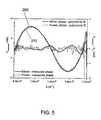
- FIG. 5is an exemplary graph of a phase ⁇ (k) obtained according to an exemplary embodiment of the present invention from a model eye and from a spectral reflection in a fovea;
- FIG. 6is a retinal image of a human subject which include spectral reflections that may be utilized according to an exemplary embodiment of the present invention
- FIG. 7is an exemplary image that may be obtained from a human subject, which illustrates the fovea after the dispersion compensation according to an exemplary embodiment of the present invention has been applied;
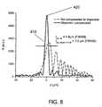
- FIG. 8is an exemplary graph of a coherence function obtained from a reflective spot in the fovea obtained using an exemplary embodiment of the present invention.
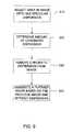
- FIG. 9is a high level flow diagram of a process according to an exemplary embodiment of the present invention.
- FIG. 10is another exemplary image that may be obtained from a human subject, in which a portion of dispersion can be selected via software by an operator;
- FIG. 11is a detailed flow diagram of a process according to yet another exemplary embodiment of the present invention.
- FIG. 1shows an exemplary embodiment of a sample configuration of a spectral domain optical coherence tomography (“SD-OCT”) arrangement which can be used for implementing the exemplary embodiments of the system, process and software arrangement according to the present invention.
- SD-OCTspectral domain optical coherence tomography
- the signal splitterforwards one portion of the split signal to a reference arm (which includes a second PC 20 ′′, a reference, certain optics and a neutral density filter (“NFD”) 50 ) and another portion of the split signal to a sample arm (which includes a third PC 20 ′′′, certain optics and a sample 60 such as the eye). Thereafter, an electromagnetic signal is reflected from the sample 60 and is combined with the light from the reference arm to form an interference signal.
- a reference armwhich includes a second PC 20 ′′, a reference, certain optics and a neutral density filter (“NFD”) 50
- a sample armwhich includes a third PC 20 ′′′, certain optics and a sample 60 such as the eye.
- This interference signalis forwarded to a fourth PC 20 ′′′′, and forwarded to a collimator (“Col”) 70 , a transmission grating (“TG”) 80 , an air-spaced focusing lens (“ASL”) 90 , and a linescan camera (“LSC”) 100 to be detected by a detecting arrangement (e.g., provided in the linescan camera), and then analyzed by a processing arrangement, e.g., a computer (not shown).
- a processing arrangemente.g., a computer (not shown).
- processing arrangementis capable of implementing the various exemplary embodiments of the system, process and software arrangement according to the present invention.
- FIG. 2shows an exemplary embodiment of an optical imaging frequency domain intereferometry (“OFDI”) arrangement according to the present invention which is capable of implementing the exemplary embodiments of the system, process and software arrangement according to the present invention.
- OFDIoptical imaging frequency domain intereferometry
- the light sourcemay be a wavelength-swept source 110 .
- a portion of the laser output(for example ⁇ 20%) is obtained, and detected using a fast InGaAs photo-detector through a narrowband fixed-wavelength filter.
- the detectorgenerates a pulse when the output spectrum of the laser sweeps through the narrow passband of the filter.
- the detector pulseis fed to a digital circuit 120 , e.g., a synchronous TTL pulse generator, for converting the resultant signal to a TTL pulse train.
- the TTL pulsesare used to generate gating pulses for signal sampling. 90% of the remaining light is directed to the sample arm and 10% to the reference mirror 130 .
- This exemplary arrangementcan utilize an optical probe based on a galvanometer mirror (e.g., scanner) 140 and an imaging lens.
- the galvanometer-mounted mirror 140is controlled by a glava-driver 145 so as to scan the probe light transversely on the sample 60 .
- the total optical power illuminated on the sample 60may be approximately 3.5 mW.
- the light reflected from the reference mirror 130 and the sample 60is received through magneto-optic circulators 150 ′, 150 ′′, and combined by a 50/50 coupler 160 .
- a fiber-optic polarization controllermay be used in the reference arm to align polarization states of the reference and sample arms.
- a relative intensity noise (“RIN”) of the received light signalmay be proportional to a reciprocal of the linewidth, and the relatively high RIN can be reduced by dual balanced detection (e.g., using a dual balanced receiver 170 ).
- the differential current of two InGaAs detectors D 1 and D 2 in the receiver 170may be amplified using trans-impedance amplifiers (“TIA”) having a total gain of 56 dB, and passed through a low pass filter (“LPF”) with a 3-dB cutoff frequency at approximately half the sampling rate.
- TIAtrans-impedance amplifiers
- LPFlow pass filter
- the common-noise rejection efficiency of the receiver 170may be typically greater than 20 dB.
- the balanced detectionmay provide other significant benefits—a suppression of a self-interference noise originating from multiple reflections within the sample and optical components; an improvement in the dynamic range; and a reduction of a fixed-pattern noise by greatly reducing the strong background signal from the reference light.
- a detecting arrangement 180receives such signals, and forward them to a processing arrangement 190 (e.g., a computer) which implements the exemplary embodiments of the system, process and software arrangement according to the present invention to reduce dispersion, and assist in displaying a resultant image that is based on the original image and the reduction of the dispersion.
- a processing arrangement 190e.g., a computer
- a dispersion broadened peakmay be observed at a depth of approximately 0.6 mm.
- the function I(z)may be shifted such that the coherence function is centered on the origin.
- a windowcan be selected around the coherence function so as to possibly eliminate coherence functions from other reflective structures in the depth profile, and all values outside the window may be set equal to zero.
- a complex spectrum in k-spacemay be obtained after an inverse Fourier transformation.
- the phase term ⁇ (k)can be equal to the arctangent of the imaginary component divided by the real component. Such term can indicate by how much are the subsequent wave numbers k out of phase with each other.
- this functioncan be provided to a polynomial expression of 9 th order, yielding a set of coefficients ⁇ 1-9 .
- individual spectramay be multiplied with a phase e ⁇ i ⁇ (k) as determined from the previous seven polynomial coefficients, where the first two polynomial coefficients may be set to zero, and then inversely Fourier transformed into A-lines, thus removing dispersion.
- the original and resulting exemplary coherence functionsare illustrated in FIG. 4 .
- the curve of FIG. 4shows the results without the dispersion compensation is shown as a dashed line, and referred to by numeral 210
- the curve illustrating the results after the dispersion compensation has been successfully appliedwhich is shown as a solid line, and referred to by numeral 220 .
- FIG. 5shows a an illustration which aids in the determination of the phase function ⁇ (k) based on certain measurements according to an exemplary embodiment of the present invention, as well as the phase function that subtracts the polynomial fit of 9 th order to the phase function.
- the phase ⁇ (k)may be obtained from a mirror in a model eye and from a spectral reflection in the fovea (e.g., the left axis).
- FIG. 6shows an exemplary retinal image of a human subject, in which three spectral reflections 300 , 310 , 320 are marked with arrows. These exemplary reflections 300 , 310 , 320 originate from an internal limiting membrane on top of the retinal nerve fiber layer and the foveolar umbo and from the external limiting membrane. Unmarked, still visible is an exemplary spectral reflection on the surface between the inner and outer segments of the photoreceptor layer, just below the external limiting membrane.
- FIG. 6shows typical examples of strong reflections in an image that can be used to determine the phase function for the optimal dispersion compensation.
- a coherence function obtained from a well-reflecting reference point in the eyeIn this example, the reflection of the foveal umbo can be used. Other regions in the eye may also create spectral reflections. Spectral reflections may be present from the interface between the inner and outer segments of the photoreceptor layer (“IPRL”) and retinal pigmented epitheleum (“RPE”). In addition, spectral reflections may also be found on the inner limiting membrane, e.g., on top of the retinal nerve fiber layer.
- IPRLphotoreceptor layer
- RPEretinal pigmented epitheleum
- phase function ⁇ (k)may then be determined as described herein above.
- the phase function ⁇ (k) as shown in FIG. 5can be determined from this measurement, as well as based on the phase function minus the polynomial fit of 9 th order to the phase function.
- Individual spectra of the imagecan be first multiplied with a phase e ⁇ i ⁇ (k) as determined from the last seven polynomial coefficients, and then inversely Fourier transformed into A-lines, thus removing dispersion.
- the fit to the dispersion data as determined from the well reflecting reference point in the eyecan be a polynomial of any order. Use of a 9 th order polynomial was demonstrated. Instead of a polynomial, the data can be fitted to a Fourier series or any other known function set so as to determine a set of coefficients.
- a polynomial of limited order to filter the dispersion curveis a better immunity to noise of the determined phase correction function.
- FIG. 7shows an exemplary image that may be obtained from a human subject, which illustrates the fovea after the dispersion compensation.
- the spectral reflection marked with an Rcan be first used to determine the amount of a chromatic dispersion (as described above), and to remove the chromatic dispersion.
- the dimensions of the image illustrated in FIG. 7are 3.1 mm ⁇ 0.61 mm.
- RNFLretinal nerve fiber layer
- GCLganglion cell layer
- IPLinner plexiform layer
- INLinner nuclear layer
- OPLouter plexiform layer
- ONLouter nuclear layer
- ELMexternal limiting membrane
- IPRLinternal limiting membrane
- RPEretinal pigmented epithelium
- Cchoriocapillaris and choroid.
- a highly reflective spot in the center of the foveais marked with an R.
- a blood vesselis marked with a large circle (BV) and structures in the outer plexiform layer are marked with smaller circles.
- FIG. 8shows a graph of a coherence function obtained from a reflective spot in the fovea. For example, the coherence length is equal to 4.8 ⁇ m in air.
- phase term ⁇ (k) obtained from a mirror in a water-filled model eye (averaged over 100 A-lines) and from a spectral reflective spot in the human fovea (averaged over 5 A-lines, see FIG. 7 )are illustrated.
- the differences between the measured phase terms and polynomial fits (9 th order) to the dataare also shown, with the corresponding axis provided on a right side thereof. Both phases show approximately the same pattern, which indicates that the model eye and the real eye generally experience similar amounts of dispersion.
- the phase term obtained from the spectral reflection of the foveacan be used (e.g., curve 270 of FIG. 5 ) to remove chromatic dispersion artifacts in data obtained from a human subject in vivo, as shown in the graph of FIG. 7 and quantified in the graph of FIG. 8 .
- the coherence lengthcan be determined in vivo from the spectral reflection in the center of the fovea labeled as R in FIG. 7 , averaged over 5 A-lines.
- the curve of FIG. 8(similarly to the graph in FIG.
- FIG. 9illustrates a flow diagram according to one exemplary embodiment of the present invention.
- an area in the image containing a spectral reflectionis selected (step 510 ).
- the existing algorithmdetermines the amount of chromatic dispersion (step 520 ) and removes such dispersion from the image (step 530 ).
- the dispersioncan be removed by multiplying spectra in k-space with a phase e ⁇ i ⁇ (k) .
- the earlier described polynomial fitcan be used. Since the polynomial fit and the original phase are approximately similar (as shown in FIG.
- the selection procedure for selecting the location of the spectral reflectioncan be either a manual procedure or an automated procedure. Thereafter, a new image may be generated based on the originally-selected image, but with the dispersions that was removed according to the exemplary technique of the present invention (step 540 ).
- the previously-described exemplary resultsmay be obtained using a simple manual procedure, where the operator generally selects the specific portion of the image by hand, e.g., by determining the coordinates of the reflecting spots.
- Such procedurecan be simplified with, e.g., MatLab software, in which the operator may be requested to draw a rectangular shape around a reflecting spot, (see numeral 600 in FIG. 10 ), thus selecting the location of the spectral spot.
- the dispersioncan be compensated using the compensation described above.
- spectral reflectionscan also be located automatically by using a particular technique.
- This exemplary techniquecan be based on an algorithm that finds a maximum signal For example, the signal returning from the spectral reflection, e.g., in the center of the fovea generally has a higher value than any of the other reflections.
- a feedback signalcan be forwarded to the scanning apparatus, so that this apparatus monitors for the brightest spectral reflection in the sample 60 (e.g., the eye).
- a series of smaller and smaller three-dimensional raster scanscan be acquired, until the center of the fovea is located. If the subject moves during this operation, the raster scanning can expanded and confined the target image again.
- an exemplary technique used to track the surface of the retina and compensate for motion artifactshas been described. See Cense, B. et al., “In Vivo Birefringence and Thickness Measurements of the Human Retinal Nerve Fiber Layer Using Polarization-Sensitive Optical Coherence Tomography”, Journal of Biomedical Optics, 2004, 9(1), pp. 121-125.
- Another exemplary embodiment of the present inventionuses compensated dispersion in dependence of depth.
- the technique according to the exemplary embodiment of the present invention described aboveis capable of compensating for a constant dispersion mismatch between the sample and the reference arm.
- dispersion broadening between superficial and deeper layers within an imagemay becomes important. The dispersion broadening may be due to the accumulated dispersion between the superficial and deeper layer.
- Described herein belowis a technique according to another exemplary embodiment of the present invention which is capable of depth dependent dispersion compensation, i.e., a correction for the dispersion that varies with depth.
- the signal in SD-OCT and OFDIis defined by,
- I ⁇ ( k )I r ⁇ ( k ) + 2 ⁇ I s ⁇ ( k ) ⁇ I r ⁇ ( k ) ⁇ ⁇ n ⁇ ⁇ n ⁇ cos ⁇ ( kz n ) + I s ⁇ ( k ) ( 2 )
- I r (k) and I s (k)are the wavelength-dependent intensities reflected from the reference and sample arms, respectively, and k is the wave number.
- the second term on the right hand side of Eq. (2)represents the interference between the light signal returning from the reference and sample arms.
- ⁇ nis the square root of the sample reflectivity at depth z n . As described in Hausler, G.
- depth informationcan be obtained by performing an inverse Fourier transform of Eq. (2), yielding the following convolution
- ⁇ FT - 1 ⁇ [ I ⁇ ( k ) ] ⁇ 2⁇ 2 ⁇ ( z ) ⁇ ⁇ ⁇ ⁇ ( 0 ) + ⁇ n ⁇ ⁇ n 2 ⁇ ⁇ ⁇ ( z - z n ) + ⁇ n ⁇ ⁇ n 2 ⁇ ⁇ ⁇ ( z + z n ) + O ⁇ [ I s 2 / I r 2 ] ⁇ , ( 3 ) with ⁇ (z) representing the envelope of the coherence function.
- the first term in the brackets on the right hand siderefers to an autocorrelation signal from the reference arm, and has magnitude unity.
- the second and third termsare reflect the interference between light returning from the reference and sample arms and from two images, where each has magnitude on the order of I s /I r . These two terms provide mirror images.
- the final term, with magnitude on the order of I s 2 /I r 2describes autocorrelation noise due to interference within the sample arm.
- I s and I rrepresent the total intensity reflected from sample and reference arms, respectively.
- a depth dependent dispersion termis described by introducing a depth dependent phase term, f(k)z n in the cosine term,
- the depth dependent dispersion termcan be compensated for by a remapping operation of the data in k-space.
- the cosine termcan be rewritten as
- the datacan be linearized in k-space before the Fourier transform resulting in Eq. (3).
- the function f(k)can be determined by measuring the dispersion F(k) n and F(k) m at two different locations, z n and z m using the method described for a constant dispersion term, where the function f(k) is given by
- f ⁇ ( k )F ⁇ ( k ) m - F ⁇ ( k ) n z m - z n .
- the locations for determining F(k)n and F(k)mare preferably locations in the material (tissue, vitrious, retina, coronary artery, etc) with strong reflections. Filtering the function f(k) to reject noise can be performed by retaining only a limited or predetermined number of coefficients from a polynomial or Fourier series fit to the data. This exemplary technique can be used to predetermine the dispersion in various materials or biological tissues, and utilize the determined values to implement depth dependent dispersion compensation during imaging or post processing of SD-OCT and OFDI data.
- Spectral reflectionscan also be located on the inner limiting membrane, on top of the retinal nerve fiber layer.
- the samplee.g., the eye
- a further technique according to yet another exemplary embodiment of the present inventioncan be used to determine a constant and depth dependent dispersion.
- the interference signal associated with the n-th reflection point in the samplecan be defined by
- I ⁇ ( k , z s , n , z r )2 ⁇ I s ⁇ ( k ) ⁇ I r ⁇ ( k ) ⁇ ⁇ n ⁇ cos ⁇ [ k ⁇ ( z s , n - z r ) + f ⁇ ( k ) ⁇ z s , n + ⁇ ⁇ ( k ) ] , ( 4 )
- z s,nrefers to the distance of the reflection point from the surface of the sample
- a constant phase errorcan be differentiated from the 1 st and 2 nd terms which are both dependent on k.
- FIG. 11shows another exemplary embodiment of the process according to the present invention which can be used to control the dispersion of the data associated with the image obtained from the reference and sample arms.
- a detectore.g., the detectors of the arrangements shown in FIG. 1 and/or 2
- This spectrum signal I(k)is forwarded to the processing arrangement, e.g., as data, which performs a Fast Fourrier Transform (“FFT”) on the spectrum signal I(k) (step 615 ).
- FFTFast Fourrier Transform
- an initial signal I(z) associated with the spectrum signal I(k)is set to 0 for z>0 and z ⁇ 0 (step 620 ), and in step 625 , an absolute values for the initial signal I(z) is set.
- a signal I(k)is generated based on the detected signal, a window of interest of the image may be determined in step 635 .
- Such are of interestcan be a region of the peak of the absolute value signal (ABS(I(z))), a center peak at around z ⁇ 0 by shifting the signal, etc.
- the windowcan be obtained automatically by the processing arrangement and/or manually by an operator.
- step 640an inverse FFT is performed on the signal I(z), and a phase term ⁇ (k) of the transformed I(z) signal is determined (step 645 ).
- step 650the exemplary process according to the present invention the phase function that can apply a polynomial of the order of N to ⁇ (k), e.g., by subtracting the polynomial fit of 9 th order, yielding a set of coefficients ⁇ 1-9 .
- the phase ⁇ (k)may be obtained from a mirror in a model eye and from a spectral reflection in the fovea.
- the filtered phase termcan then be determined from the polynomial fit parameters/coefficients, e.g., by setting the first two polynomial coefficients to zero.
- the filtered phase of the signal e ⁇ i ⁇ (k)can be stored for use in multiple images.
- a correction curve of the filtered phase term ⁇ (k)can be applied, e.g., by multiplying all spectra of the image may be multiplied by e ⁇ i ⁇ (k) .
Landscapes
- Physics & Mathematics (AREA)
- Health & Medical Sciences (AREA)
- General Physics & Mathematics (AREA)
- Life Sciences & Earth Sciences (AREA)
- General Health & Medical Sciences (AREA)
- Engineering & Computer Science (AREA)
- Pathology (AREA)
- Nuclear Medicine, Radiotherapy & Molecular Imaging (AREA)
- Radiology & Medical Imaging (AREA)
- Optics & Photonics (AREA)
- Molecular Biology (AREA)
- Animal Behavior & Ethology (AREA)
- Biochemistry (AREA)
- Analytical Chemistry (AREA)
- Veterinary Medicine (AREA)
- Public Health (AREA)
- Biophysics (AREA)
- Immunology (AREA)
- Biomedical Technology (AREA)
- Heart & Thoracic Surgery (AREA)
- Medical Informatics (AREA)
- Chemical & Material Sciences (AREA)
- Surgery (AREA)
- Ophthalmology & Optometry (AREA)
- Signal Processing (AREA)
- Automation & Control Theory (AREA)
- Investigating Or Analysing Materials By Optical Means (AREA)
- Eye Examination Apparatus (AREA)
Abstract
Description
with λ0being the center wavelength, and k0being equal to 2π/λ0. The first two terms generally describe a constant offset and group velocity, respectively, and are likely not related to dispersive broadening. The third term represents a second order or a group-velocity dispersion. A dispersion mismatch in the sample arm and the reference arm can to a large extend be attributed to this term. However, higher order dispersion terms may contribute to the dispersion mismatch as well, for example when an ultra-broadband source is used.
where Ir(k) and Is(k) are the wavelength-dependent intensities reflected from the reference and sample arms, respectively, and k is the wave number. The second term on the right hand side of Eq. (2) represents the interference between the light signal returning from the reference and sample arms. αnis the square root of the sample reflectivity at depth zn. As described in Hausler, G. et al., “Coherence Radar and Spectral Radar—New Tools for Dermatological Diagnosis”, J. Biomed. Opt., 1998, 3(1), pp. 21-31, depth information can be obtained by performing an inverse Fourier transform of Eq. (2), yielding the following convolution
with Γ(z) representing the envelope of the coherence function. The first term in the brackets on the right hand side refers to an autocorrelation signal from the reference arm, and has magnitude unity. The second and third terms are reflect the interference between light returning from the reference and sample arms and from two images, where each has magnitude on the order of Is/Ir. These two terms provide mirror images. The final term, with magnitude on the order of Is2/Ir2, describes autocorrelation noise due to interference within the sample arm. Isand Irrepresent the total intensity reflected from sample and reference arms, respectively.
a constant dispersion mismatch can be described by introducing a phase term θ(k) in the cosine term,
The constant dispersion mismatch can be compensated for with the method described before. A depth dependent dispersion term is described by introducing a depth dependent phase term, f(k)znin the cosine term,
The depth dependent dispersion term can be compensated for by a remapping operation of the data in k-space. The cosine term can be rewritten as
with k′=k+f(k). After the remapping operation, the data can be linearized in k-space before the Fourier transform resulting in Eq. (3).
The locations for determining F(k)n and F(k)m are preferably locations in the material (tissue, vitrious, retina, coronary artery, etc) with strong reflections. Filtering the function f(k) to reject noise can be performed by retaining only a limited or predetermined number of coefficients from a polynomial or Fourier series fit to the data. This exemplary technique can be used to predetermine the dispersion in various materials or biological tissues, and utilize the determined values to implement depth dependent dispersion compensation during imaging or post processing of SD-OCT and OFDI data. For use in retinal data, several locations can provide strong reflections that can be used to determine the dispersion, such as the center of the fovea (fovealar umbo), external limiting membrane, interface between the inner and outer segments of the photoreceptor layer (“IPRL”) and retinal pigmented epitheleum (“RPE”). Spectral reflections can also be located on the inner limiting membrane, on top of the retinal nerve fiber layer. In order to see these reflections, the sample (e.g., the eye) should be tilted such that the surface thereof is exactly perpendicular to the beam.
where zs,nrefers to the distance of the reflection point from the surface of the sample, and zrrefers to the position of the reference mirror with respect to the sample surface. Shifting the position of the reference mirror to zr′=2zs,n−zrprovides the following
where δ refers to any possible phase error introduced in the measurement. It is possible to determine the phase functions, φ(k,zs,n,zr) and φ(k,zs,n,zr′) of the interference signals in Eq. (4) and (5), respectively. It follows that
φ(k,zs,n,zr)−φ(k,zs,n,zr′)=2f(k)zs,n+2θ(k)+δ (6)
Claims (47)
Priority Applications (1)
| Application Number | Priority Date | Filing Date | Title |
|---|---|---|---|
| US11/569,790US8018598B2 (en) | 2004-05-29 | 2004-07-23 | Process, system and software arrangement for a chromatic dispersion compensation using reflective layers in optical coherence tomography (OCT) imaging |
Applications Claiming Priority (3)
| Application Number | Priority Date | Filing Date | Title |
|---|---|---|---|
| US57577304P | 2004-05-29 | 2004-05-29 | |
| PCT/US2004/023585WO2005117534A2 (en) | 2004-05-29 | 2004-07-23 | Process, system and software arrangement for a chromatic dispersion compensation using reflective layers in optical coherence tomography (oct) imaging |
| US11/569,790US8018598B2 (en) | 2004-05-29 | 2004-07-23 | Process, system and software arrangement for a chromatic dispersion compensation using reflective layers in optical coherence tomography (OCT) imaging |
Publications (2)
| Publication Number | Publication Date |
|---|---|
| US20090196477A1 US20090196477A1 (en) | 2009-08-06 |
| US8018598B2true US8018598B2 (en) | 2011-09-13 |
Family
ID=35463245
Family Applications (1)
| Application Number | Title | Priority Date | Filing Date |
|---|---|---|---|
| US11/569,790Active2026-08-25US8018598B2 (en) | 2004-05-29 | 2004-07-23 | Process, system and software arrangement for a chromatic dispersion compensation using reflective layers in optical coherence tomography (OCT) imaging |
Country Status (6)
| Country | Link |
|---|---|
| US (1) | US8018598B2 (en) |
| EP (1) | EP1754016B1 (en) |
| JP (1) | JP4750786B2 (en) |
| KR (1) | KR101239250B1 (en) |
| AU (1) | AU2004320269B2 (en) |
| WO (1) | WO2005117534A2 (en) |
Cited By (23)
| Publication number | Priority date | Publication date | Assignee | Title |
|---|---|---|---|---|
| US20100245838A1 (en)* | 2005-01-21 | 2010-09-30 | Carl Zeiss Meditec, Inc. | Method of motion correction in optical coherence tomography imaging |
| US20130286454A1 (en)* | 2011-01-05 | 2013-10-31 | Nippon Telegraph And Telephone Corporation | Wavelength swept light source |
| US8649611B2 (en) | 2005-04-06 | 2014-02-11 | Carl Zeiss Meditec, Inc. | Method and apparatus for measuring motion of a subject using a series of partial images from an imaging system |
| US20140115022A1 (en)* | 2011-07-12 | 2014-04-24 | University Of Tsukuba | Program for correcting data measured by ps-oct and ps-oct system equipped with the program |
| WO2014105911A1 (en)* | 2012-12-24 | 2014-07-03 | Luna Innovations Incorporated | Dispersion correction in optical frequency-domain reflectometry |
| US8857988B2 (en) | 2011-07-07 | 2014-10-14 | Carl Zeiss Meditec, Inc. | Data acquisition methods for reduced motion artifacts and applications in OCT angiography |
| US9033510B2 (en) | 2011-03-30 | 2015-05-19 | Carl Zeiss Meditec, Inc. | Systems and methods for efficiently obtaining measurements of the human eye using tracking |
| US9044166B2 (en) | 2011-04-18 | 2015-06-02 | Nidek Co., Ltd. | Optical tomographic image photographing apparatus |
| US9101294B2 (en) | 2012-01-19 | 2015-08-11 | Carl Zeiss Meditec, Inc. | Systems and methods for enhanced accuracy in OCT imaging of the cornea |
| US9299134B2 (en) | 2010-03-25 | 2016-03-29 | Canon Kabushiki Kaisha | Optical tomographic imaging apparatus |
| US9677869B2 (en) | 2012-12-05 | 2017-06-13 | Perimeter Medical Imaging, Inc. | System and method for generating a wide-field OCT image of a portion of a sample |
| US20190101489A1 (en)* | 2017-09-29 | 2019-04-04 | Michael John Darwin | Method and Apparatus for Simultaneously Measuring 3Dimensional Structures and Spectral Content of Said Structures |
| US10529061B2 (en) | 2015-08-13 | 2020-01-07 | University Of Washington | Systems and methods of forming enhanced medical images |
| US10577573B2 (en) | 2017-07-18 | 2020-03-03 | Perimeter Medical Imaging, Inc. | Sample container for stabilizing and aligning excised biological tissue samples for ex vivo analysis |
| US10631718B2 (en) | 2015-08-31 | 2020-04-28 | Gentuity, Llc | Imaging system includes imaging probe and delivery devices |
| US10646105B2 (en) | 2013-09-17 | 2020-05-12 | The Johns Hopkins University | Device and methods for color corrected OCT imaging endoscope/catheter to achieve high-resolution |
| US11278206B2 (en) | 2015-04-16 | 2022-03-22 | Gentuity, Llc | Micro-optic probes for neurology |
| WO2022106510A1 (en) | 2020-11-19 | 2022-05-27 | Leica Microsystems Inc. | Processing system for oct imaging, oct imaging system and method for oct imaging |
| US11684242B2 (en) | 2017-11-28 | 2023-06-27 | Gentuity, Llc | Imaging system |
| US12004718B2 (en)* | 2017-01-27 | 2024-06-11 | The John Hopkins University | Device and methods for color corrected OCT imaging endoscope/catheter/capsule to achieve high-resolution |
| US12239412B2 (en) | 2019-05-21 | 2025-03-04 | Spryte Medical, Inc. | Systems and methods for OCT-guided treatment of a patient |
| US12262872B2 (en) | 2018-09-17 | 2025-04-01 | Gentuity, Llc | Imaging system with optical pathway |
| US12364385B2 (en) | 2019-04-30 | 2025-07-22 | Gentuity, Llc | Imaging probe with fluid pressurization element |
Families Citing this family (118)
| Publication number | Priority date | Publication date | Assignee | Title |
|---|---|---|---|---|
| US7231243B2 (en) | 2000-10-30 | 2007-06-12 | The General Hospital Corporation | Optical methods for tissue analysis |
| US9295391B1 (en) | 2000-11-10 | 2016-03-29 | The General Hospital Corporation | Spectrally encoded miniature endoscopic imaging probe |
| AT503309B1 (en) | 2001-05-01 | 2011-08-15 | Gen Hospital Corp | DEVICE FOR DETERMINING ATHEROSCLEROTIC BEARING BY MEASURING OPTICAL TISSUE PROPERTIES |
| US7355716B2 (en) | 2002-01-24 | 2008-04-08 | The General Hospital Corporation | Apparatus and method for ranging and noise reduction of low coherence interferometry LCI and optical coherence tomography OCT signals by parallel detection of spectral bands |
| US8054468B2 (en) | 2003-01-24 | 2011-11-08 | The General Hospital Corporation | Apparatus and method for ranging and noise reduction of low coherence interferometry LCI and optical coherence tomography OCT signals by parallel detection of spectral bands |
| EP2436307B1 (en) | 2003-03-31 | 2015-10-21 | The General Hospital Corporation | Speckle reduction in optical coherence tomography by path length encoded angular compounding |
| KR101386971B1 (en) | 2003-06-06 | 2014-04-18 | 더 제너럴 하스피탈 코포레이션 | Process and apparatus for a wavelength tunning source |
| EP2280256B1 (en) | 2003-10-27 | 2016-11-16 | The General Hospital Corporation | Method and apparatus for performing optical imaging using frequency-domain interferometry |
| KR101239250B1 (en)* | 2004-05-29 | 2013-03-05 | 더 제너럴 하스피탈 코포레이션 | Process, system and software arrangement for a chromatic dispersion compensation using reflective layers in optical coherence tomography (oct) imaging |
| AU2005270037B2 (en) | 2004-07-02 | 2012-02-09 | The General Hospital Corporation | Endoscopic imaging probe comprising dual clad fibre |
| EP1782020B1 (en) | 2004-08-06 | 2012-10-03 | The General Hospital Corporation | Process, system and software arrangement for determining at least one location in a sample using an optical coherence tomography |
| EP2272421A1 (en) | 2004-08-24 | 2011-01-12 | The General Hospital Corporation | Method and apparatus for imaging of vessel segments |
| WO2006024014A2 (en) | 2004-08-24 | 2006-03-02 | The General Hospital Corporation | Process, system and software arrangement for measuring a mechanical strain and elastic properties of a sample |
| US7365859B2 (en) | 2004-09-10 | 2008-04-29 | The General Hospital Corporation | System and method for optical coherence imaging |
| KR101257100B1 (en) | 2004-09-29 | 2013-04-22 | 더 제너럴 하스피탈 코포레이션 | System and Method for Optical Coherence Imaging |
| WO2006058346A1 (en) | 2004-11-29 | 2006-06-01 | The General Hospital Corporation | Arrangements, devices, endoscopes, catheters and methods for performing optical imaging by simultaneously illuminating and detecting multiple points on a sample |
| ES2337497T3 (en) | 2005-04-28 | 2010-04-26 | The General Hospital Corporation | EVALUATION OF CHARACTERISTICS OF THE IMAGE OF AN ANATOMICAL STRUCTURE IN IMAGES OF TOMOGRAPHY OF OPTICAL COHERENCE. |
| US9060689B2 (en) | 2005-06-01 | 2015-06-23 | The General Hospital Corporation | Apparatus, method and system for performing phase-resolved optical frequency domain imaging |
| EP2267404B1 (en) | 2005-08-09 | 2016-10-05 | The General Hospital Corporation | Apparatus and method for performing polarization-based quadrature demodulation in optical coherence tomography |
| US7843572B2 (en) | 2005-09-29 | 2010-11-30 | The General Hospital Corporation | Method and apparatus for optical imaging via spectral encoding |
| US8145018B2 (en) | 2006-01-19 | 2012-03-27 | The General Hospital Corporation | Apparatus for obtaining information for a structure using spectrally-encoded endoscopy techniques and methods for producing one or more optical arrangements |
| DK1973466T3 (en) | 2006-01-19 | 2021-02-01 | Massachusetts Gen Hospital | BALLOON IMAGING CATHETER |
| JP5680829B2 (en) | 2006-02-01 | 2015-03-04 | ザ ジェネラル ホスピタル コーポレイション | A device that irradiates a sample with multiple electromagnetic radiations |
| WO2007149602A2 (en) | 2006-02-01 | 2007-12-27 | The General Hospital Corporation | Methods and systems for providing electromagnetic radiation to at least one portion of a sample using conformal laser therapy procedures |
| US9777053B2 (en) | 2006-02-08 | 2017-10-03 | The General Hospital Corporation | Methods, arrangements and systems for obtaining information associated with an anatomical sample using optical microscopy |
| EP2982929A1 (en) | 2006-02-24 | 2016-02-10 | The General Hospital Corporation | Methods and systems for performing angle-resolved fourier-domain optical coherence tomography |
| WO2007127395A2 (en)* | 2006-04-28 | 2007-11-08 | Bioptigen, Inc. | Methods, systems and computer program products for optical coherence tomography (oct) using automatic dispersion compensation |
| WO2007133961A2 (en) | 2006-05-10 | 2007-11-22 | The General Hospital Corporation | Processes, arrangements and systems for providing frequency domain imaging of a sample |
| DE102006028238B3 (en)* | 2006-06-20 | 2007-07-19 | Benecke-Kaliko Ag | Three dimensionally structured original surface e.g. grained surface, reflection characteristics analysis and specification method, involves storing reflection value in data record that is provided to processing or verification system |
| JP4969925B2 (en) | 2006-06-28 | 2012-07-04 | 株式会社トプコン | Fundus observation device |
| US8838213B2 (en)* | 2006-10-19 | 2014-09-16 | The General Hospital Corporation | Apparatus and method for obtaining and providing imaging information associated with at least one portion of a sample, and effecting such portion(s) |
| WO2008088868A2 (en) | 2007-01-19 | 2008-07-24 | Bioptigen, Inc. | Methods, systems and computer program products for processing images generated using fourier domain optical coherence tomography (fdoct) |
| US9176319B2 (en) | 2007-03-23 | 2015-11-03 | The General Hospital Corporation | Methods, arrangements and apparatus for utilizing a wavelength-swept laser using angular scanning and dispersion procedures |
| US10534129B2 (en) | 2007-03-30 | 2020-01-14 | The General Hospital Corporation | System and method providing intracoronary laser speckle imaging for the detection of vulnerable plaque |
| US8045177B2 (en) | 2007-04-17 | 2011-10-25 | The General Hospital Corporation | Apparatus and methods for measuring vibrations using spectrally-encoded endoscopy |
| WO2008137077A1 (en) | 2007-05-04 | 2008-11-13 | Bioptigen, Inc. | Methods, systems and computer program products for mixed-density optical coherence tomography (oct) imaging |
| JP5032203B2 (en) | 2007-05-24 | 2012-09-26 | 株式会社トプコン | Fundus observation apparatus and program for controlling the same |
| US9125562B2 (en)* | 2009-07-01 | 2015-09-08 | Avinger, Inc. | Catheter-based off-axis optical coherence tomography imaging system |
| US9788790B2 (en) | 2009-05-28 | 2017-10-17 | Avinger, Inc. | Optical coherence tomography for biological imaging |
| US8696695B2 (en) | 2009-04-28 | 2014-04-15 | Avinger, Inc. | Guidewire positioning catheter |
| US8079711B2 (en)* | 2008-04-24 | 2011-12-20 | Carl Zeiss Meditec, Inc. | Method for finding the lateral position of the fovea in an SDOCT image volume |
| US7898656B2 (en) | 2008-04-30 | 2011-03-01 | The General Hospital Corporation | Apparatus and method for cross axis parallel spectroscopy |
| EP2274572A4 (en) | 2008-05-07 | 2013-08-28 | Gen Hospital Corp | SYSTEM, METHOD AND COMPUTER MEDIUM FOR MONITORING THE MOVEMENT OF VESSELS DURING A THREE-DIMENSIONAL MICROSCOPY EXAMINATION OF CORONARY ARTERIES |
| US8861910B2 (en) | 2008-06-20 | 2014-10-14 | The General Hospital Corporation | Fused fiber optic coupler arrangement and method for use thereof |
| JP5255524B2 (en)* | 2008-07-04 | 2013-08-07 | 株式会社ニデック | Optical tomographic imaging device, optical tomographic image processing device. |
| JP5331395B2 (en) | 2008-07-04 | 2013-10-30 | 株式会社ニデック | Optical tomography system |
| WO2010009136A2 (en) | 2008-07-14 | 2010-01-21 | The General Hospital Corporation | Apparatus and methods for color endoscopy |
| WO2010031163A1 (en)* | 2008-09-17 | 2010-03-25 | Institut National De La Recherche Scientifique | Cross-chirped interferometry system and method for light detection and ranging |
| KR101000974B1 (en)* | 2008-12-01 | 2010-12-13 | 인하대학교 산학협력단 | Measurement method of color dispersion characteristics of optical waveguide sample using interference fringe measurement system |
| JP5731394B2 (en) | 2008-12-10 | 2015-06-10 | ザ ジェネラル ホスピタル コーポレイション | System, apparatus and method for extending imaging depth range of optical coherence tomography through optical subsampling |
| JP2012515576A (en) | 2009-01-20 | 2012-07-12 | ザ ジェネラル ホスピタル コーポレイション | Endoscopic biopsy device, system, and method |
| US8097864B2 (en) | 2009-01-26 | 2012-01-17 | The General Hospital Corporation | System, method and computer-accessible medium for providing wide-field superresolution microscopy |
| CA2749670A1 (en) | 2009-02-04 | 2010-08-12 | The General Hospital Corporation | Apparatus and method for utilization of a high-speed optical wavelength tuning source |
| WO2011003006A2 (en) | 2009-07-01 | 2011-01-06 | Avinger, Inc. | Atherectomy catheter with laterally-displaceable tip |
| JP5819823B2 (en) | 2009-07-14 | 2015-11-24 | ザ ジェネラル ホスピタル コーポレイション | Device for measuring the flow and pressure inside a blood vessel and method of operating the device |
| KR20130004326A (en) | 2010-03-05 | 2013-01-09 | 더 제너럴 하스피탈 코포레이션 | Systems, methods and computer-accessible medium which provide microscopic images of at least one anatomical structure at a particular resolution |
| US9069130B2 (en) | 2010-05-03 | 2015-06-30 | The General Hospital Corporation | Apparatus, method and system for generating optical radiation from biological gain media |
| EP2575598A2 (en) | 2010-05-25 | 2013-04-10 | The General Hospital Corporation | Apparatus, systems, methods and computer-accessible medium for spectral analysis of optical coherence tomography images |
| EP2575597B1 (en) | 2010-05-25 | 2022-05-04 | The General Hospital Corporation | Apparatus for providing optical imaging of structures and compositions |
| JP6066901B2 (en) | 2010-06-03 | 2017-01-25 | ザ ジェネラル ホスピタル コーポレイション | Method for apparatus and device for imaging structures in or in one or more luminal organs |
| US8787645B2 (en) | 2010-07-21 | 2014-07-22 | Diopsys, Inc. | Method and system for analyzing optical coherence tomography (OCT) results using color reflectivity discretization analysis |
| WO2012058381A2 (en) | 2010-10-27 | 2012-05-03 | The General Hospital Corporation | Apparatus, systems and methods for measuring blood pressure within at least one vessel |
| CN103261878B (en)* | 2010-12-13 | 2016-03-30 | 皇家飞利浦电子股份有限公司 | Method and apparatus for analyzing a region of interest in an object using X-rays |
| US9279659B2 (en)* | 2011-01-21 | 2016-03-08 | Duke University | Systems and methods for complex conjugate artifact resolved optical coherence tomography |
| ES2396784B2 (en) | 2011-03-15 | 2014-07-23 | Medlumics, S.L. | INTEGRABLE ACTIVE EQUALIZATION SYSTEM OF CHROMATIC DISPERSION. |
| EP2691038B1 (en) | 2011-03-28 | 2016-07-20 | Avinger, Inc. | Occlusion-crossing devices, imaging, and atherectomy devices |
| US9949754B2 (en) | 2011-03-28 | 2018-04-24 | Avinger, Inc. | Occlusion-crossing devices |
| US9330092B2 (en) | 2011-07-19 | 2016-05-03 | The General Hospital Corporation | Systems, methods, apparatus and computer-accessible-medium for providing polarization-mode dispersion compensation in optical coherence tomography |
| EP3835718B1 (en) | 2011-08-25 | 2023-07-26 | The General Hospital Corporation | Apparatus for providing micro-optical coherence tomography inside a respiratory system |
| JP2015502562A (en) | 2011-10-18 | 2015-01-22 | ザ ジェネラル ホスピタル コーポレイション | Apparatus and method for generating and / or providing recirculating optical delay |
| US9345406B2 (en) | 2011-11-11 | 2016-05-24 | Avinger, Inc. | Occlusion-crossing devices, atherectomy devices, and imaging |
| DE202012002375U1 (en) | 2012-03-08 | 2012-04-23 | Wavelight Gmbh | Device for optical coherence tomography |
| WO2013148306A1 (en) | 2012-03-30 | 2013-10-03 | The General Hospital Corporation | Imaging system, method and distal attachment for multidirectional field of view endoscopy |
| US9557156B2 (en) | 2012-05-14 | 2017-01-31 | Avinger, Inc. | Optical coherence tomography with graded index fiber for biological imaging |
| EP2849660B1 (en) | 2012-05-14 | 2021-08-25 | Avinger, Inc. | Atherectomy catheter drive assemblies |
| JP2015517387A (en) | 2012-05-21 | 2015-06-22 | ザ ジェネラル ホスピタル コーポレイション | Apparatus, device and method for capsule microscopy |
| JP6227652B2 (en) | 2012-08-22 | 2017-11-08 | ザ ジェネラル ホスピタル コーポレイション | System, method, and computer-accessible medium for fabricating a miniature endoscope using soft lithography |
| JP6195334B2 (en)* | 2012-08-30 | 2017-09-13 | キヤノン株式会社 | Imaging apparatus, imaging method, and program |
| US9115974B2 (en)* | 2012-09-14 | 2015-08-25 | The Johns Hopkins University | Motion-compensated optical coherence tomography system |
| US9968261B2 (en) | 2013-01-28 | 2018-05-15 | The General Hospital Corporation | Apparatus and method for providing diffuse spectroscopy co-registered with optical frequency domain imaging |
| WO2014120791A1 (en) | 2013-01-29 | 2014-08-07 | The General Hospital Corporation | Apparatus, systems and methods for providing information regarding the aortic valve |
| US11179028B2 (en) | 2013-02-01 | 2021-11-23 | The General Hospital Corporation | Objective lens arrangement for confocal endomicroscopy |
| US11096717B2 (en) | 2013-03-15 | 2021-08-24 | Avinger, Inc. | Tissue collection device for catheter |
| WO2014143064A1 (en) | 2013-03-15 | 2014-09-18 | Avinger, Inc. | Chronic total occlusion crossing devices with imaging |
| US10478072B2 (en) | 2013-03-15 | 2019-11-19 | The General Hospital Corporation | Methods and system for characterizing an object |
| CN105228514B (en) | 2013-03-15 | 2019-01-22 | 阿维格公司 | Optical Pressure Sensor Assembly |
| EP2997354A4 (en) | 2013-05-13 | 2017-01-18 | The General Hospital Corporation | Detecting self-interefering fluorescence phase and amplitude |
| EP3019096B1 (en) | 2013-07-08 | 2023-07-05 | Avinger, Inc. | System for identification of elastic lamina to guide interventional therapy |
| WO2015009932A1 (en) | 2013-07-19 | 2015-01-22 | The General Hospital Corporation | Imaging apparatus and method which utilizes multidirectional field of view endoscopy |
| EP3021735A4 (en) | 2013-07-19 | 2017-04-19 | The General Hospital Corporation | Determining eye motion by imaging retina. with feedback |
| WO2015013651A2 (en) | 2013-07-26 | 2015-01-29 | The General Hospital Corporation | System, apparatus and method utilizing optical dispersion for fourier-domain optical coherence tomography |
| US9347872B1 (en)* | 2013-09-23 | 2016-05-24 | Kla-Tencor Corporation | Meta-model based measurement refinement |
| WO2015105870A1 (en) | 2014-01-08 | 2015-07-16 | The General Hospital Corporation | Method and apparatus for microscopic imaging |
| US10188808B2 (en) | 2014-01-24 | 2019-01-29 | The Johns Hopkins University | Fiber optic distal sensor controlled drug injector |
| US10736494B2 (en) | 2014-01-31 | 2020-08-11 | The General Hospital Corporation | System and method for facilitating manual and/or automatic volumetric imaging with real-time tension or force feedback using a tethered imaging device |
| WO2015153982A1 (en) | 2014-04-04 | 2015-10-08 | The General Hospital Corporation | Apparatus and method for controlling propagation and/or transmission of electromagnetic radiation in flexible waveguide(s) |
| US9907696B2 (en) | 2014-04-18 | 2018-03-06 | The Johns Hopkins University | Fiber optic distal sensor controlled micro-manipulation systems and methods |
| US10357277B2 (en) | 2014-07-08 | 2019-07-23 | Avinger, Inc. | High speed chronic total occlusion crossing devices |
| US10912462B2 (en) | 2014-07-25 | 2021-02-09 | The General Hospital Corporation | Apparatus, devices and methods for in vivo imaging and diagnosis |
| US10568520B2 (en) | 2015-07-13 | 2020-02-25 | Avinger, Inc. | Micro-molded anamorphic reflector lens for image guided therapeutic/diagnostic catheters |
| EP3435892B1 (en) | 2016-04-01 | 2024-04-03 | Avinger, Inc. | Atherectomy catheter with serrated cutter |
| US11344327B2 (en) | 2016-06-03 | 2022-05-31 | Avinger, Inc. | Catheter device with detachable distal end |
| WO2018006041A1 (en) | 2016-06-30 | 2018-01-04 | Avinger, Inc. | Atherectomy catheter with shapeable distal tip |
| JP6776076B2 (en)* | 2016-09-23 | 2020-10-28 | 株式会社トプコン | OCT device |
| JP6556199B2 (en)* | 2017-08-10 | 2019-08-07 | キヤノン株式会社 | Imaging apparatus and imaging method |
| TWI794416B (en)* | 2018-02-28 | 2023-03-01 | 美商賽格股份有限公司 | Metrology of multi-layer stacks and interferometer system |
| JP7243023B2 (en) | 2018-03-06 | 2023-03-22 | 株式会社ニデック | OCT device |
| US12167867B2 (en) | 2018-04-19 | 2024-12-17 | Avinger, Inc. | Occlusion-crossing devices |
| US11357399B2 (en) | 2018-08-02 | 2022-06-14 | Nidek Co., Ltd. | OCT apparatus |
| WO2020087452A1 (en) | 2018-11-01 | 2020-05-07 | Lenovo (Beijing) Limited | Buffer status report indicator |
| US11883100B2 (en) | 2019-09-30 | 2024-01-30 | Nidek Co., Ltd. | Ophthalmologic image processing method and OCT apparatus |
| CN114746033B (en) | 2019-10-18 | 2025-01-10 | 阿维格公司 | Blocking crossing device |
| JP7412229B2 (en)* | 2020-03-10 | 2024-01-12 | 株式会社トプコン | Ophthalmology information processing device, ophthalmology device, ophthalmology information processing method, and program |
| CA3234556A1 (en) | 2021-11-19 | 2023-05-25 | Steven T. Charles | Ophthalmic procedure contact lens with enhanced vitreous visualization |
| JP2024540025A (en) | 2021-11-19 | 2024-10-31 | アルコン インコーポレイティド | Producing and evaluating two-dimensional and three-dimensional images of the inside of the eye |
| JP2024539331A (en) | 2021-11-19 | 2024-10-28 | アルコン インコーポレイティド | Evaluation and Treatment of Eye Floats |
| US20240377184A1 (en)* | 2023-05-12 | 2024-11-14 | The Regents Of The University Of California | Systems and Methods for 1-Micron Frequency Comb Optical Coherence Tomography |
| CN119879720B (en)* | 2025-03-28 | 2025-07-04 | 吉林大学 | K domain homogenizing and dispersion compensating method based on SD-OCT technology |
Citations (464)
| Publication number | Priority date | Publication date | Assignee | Title |
|---|---|---|---|---|
| US2339754A (en) | 1941-03-04 | 1944-01-25 | Westinghouse Electric & Mfg Co | Supervisory apparatus |
| US3090753A (en) | 1960-08-02 | 1963-05-21 | Exxon Research Engineering Co | Ester oil compositions containing acid anhydride |
| US3601480A (en) | 1968-07-10 | 1971-08-24 | Physics Int Co | Optical tunnel high-speed camera system |
| GB1257778A (en) | 1967-12-07 | 1971-12-22 | ||
| US3856000A (en) | 1972-06-19 | 1974-12-24 | Machido Seisakusho Kk | Endoscope |
| US3872407A (en) | 1972-09-01 | 1975-03-18 | Us Navy | Rapidly tunable laser |
| US3941121A (en) | 1974-12-20 | 1976-03-02 | The University Of Cincinnati | Focusing fiber-optic needle endoscope |
| US3973219A (en) | 1975-04-24 | 1976-08-03 | Cornell Research Foundation, Inc. | Very rapidly tuned cw dye laser |
| US3983507A (en) | 1975-01-06 | 1976-09-28 | Research Corporation | Tunable laser systems and method |
| US4030831A (en) | 1976-03-22 | 1977-06-21 | The United States Of America As Represented By The Secretary Of The Navy | Phase detector for optical figure sensing |
| US4030827A (en) | 1973-12-03 | 1977-06-21 | Institut National De La Sante Et De La Recherche Medicale (Inserm) | Apparatus for the non-destructive examination of heterogeneous samples |
| US4140364A (en) | 1973-06-23 | 1979-02-20 | Olympus Optical Co., Ltd. | Variable field optical system for endoscopes |
| US4141362A (en) | 1977-05-23 | 1979-02-27 | Richard Wolf Gmbh | Laser endoscope |
| GB2030313A (en) | 1978-06-29 | 1980-04-02 | Wolf Gmbh Richard | Endoscopes |
| US4224929A (en) | 1977-11-08 | 1980-09-30 | Olympus Optical Co., Ltd. | Endoscope with expansible cuff member and operation section |
| US4295738A (en) | 1979-08-30 | 1981-10-20 | United Technologies Corporation | Fiber optic strain sensor |
| US4300816A (en) | 1979-08-30 | 1981-11-17 | United Technologies Corporation | Wide band multicore optical fiber |
| US4303300A (en) | 1979-02-07 | 1981-12-01 | Thomson-Csf | Rotary-joint device providing for an optical waveguide transmission |
| US4428643A (en) | 1981-04-08 | 1984-01-31 | Xerox Corporation | Optical scanning system with wavelength shift correction |
| US4479499A (en) | 1982-01-29 | 1984-10-30 | Alfano Robert R | Method and apparatus for detecting the presence of caries in teeth using visible light |
| EP0110201A3 (en) | 1982-11-25 | 1985-01-23 | Központi Elelmiszeripari Kutato Intezet | Apparatus for providing radiation of controlled spectral composition |
| US4533247A (en) | 1981-09-03 | 1985-08-06 | International Standard Electric Corporation | Optical transmission system |
| US4585349A (en) | 1983-09-12 | 1986-04-29 | Battelle Memorial Institute | Method of and apparatus for determining the position of a device relative to a reference |
| US4601036A (en) | 1982-09-30 | 1986-07-15 | Honeywell Inc. | Rapidly tunable laser |
| US4607622A (en) | 1985-04-11 | 1986-08-26 | Charles D. Fritch | Fiber optic ocular endoscope |
| US4631498A (en) | 1985-04-26 | 1986-12-23 | Hewlett-Packard Company | CW Laser wavemeter/frequency locking technique |
| US4639999A (en) | 1984-11-02 | 1987-02-03 | Xerox Corporation | High resolution, high efficiency I.R. LED printing array fabrication method |
| US4650327A (en) | 1985-10-28 | 1987-03-17 | Oximetrix, Inc. | Optical catheter calibrating assembly |
| US4734578A (en) | 1985-03-27 | 1988-03-29 | Olympus Optical Co., Ltd. | Two-dimensional scanning photo-electric microscope |
| US4744656A (en) | 1986-12-08 | 1988-05-17 | Spectramed, Inc. | Disposable calibration boot for optical-type cardiovascular catheter |
| US4751706A (en) | 1986-12-31 | 1988-06-14 | The United States Of America As Represented By The Secretary Of The Army | Laser for providing rapid sequence of different wavelengths |
| US4763977A (en) | 1985-01-09 | 1988-08-16 | Canadian Patents And Development Limited-Societe | Optical fiber coupler with tunable coupling ratio and method of making |
| US4770492A (en) | 1986-10-28 | 1988-09-13 | Spectran Corporation | Pressure or strain sensitive optical fiber |
| US4827907A (en) | 1986-11-28 | 1989-05-09 | Teac Optical Co., Ltd. | Intra-observation apparatus |
| US4834111A (en) | 1987-01-12 | 1989-05-30 | The Trustees Of Columbia University In The City Of New York | Heterodyne interferometer |
| US4868834A (en) | 1988-09-14 | 1989-09-19 | The United States Of America As Represented By The Secretary Of The Army | System for rapidly tuning a low pressure pulsed laser |
| US4890901A (en) | 1987-12-22 | 1990-01-02 | Hughes Aircraft Company | Color corrector for embedded prisms |
| US4892406A (en) | 1988-01-11 | 1990-01-09 | United Technologies Corporation | Method of and arrangement for measuring vibrations |
| US4905169A (en) | 1988-06-02 | 1990-02-27 | The United States Of America As Represented By The United States Department Of Energy | Method and apparatus for simultaneously measuring a plurality of spectral wavelengths present in electromagnetic radiation |
| US4909631A (en) | 1987-12-18 | 1990-03-20 | Tan Raul Y | Method for film thickness and refractive index determination |
| US4925302A (en) | 1988-04-13 | 1990-05-15 | Hewlett-Packard Company | Frequency locking device |
| US4928005A (en) | 1988-01-25 | 1990-05-22 | Thomson-Csf | Multiple-point temperature sensor using optic fibers |
| US4940328A (en) | 1988-11-04 | 1990-07-10 | Georgia Tech Research Corporation | Optical sensing apparatus and method |
| US4965441A (en) | 1988-01-27 | 1990-10-23 | Commissariat A L'energie Atomique | Method for the scanning confocal light-optical microscopic and indepth examination of an extended field and devices for implementing said method |
| US4965599A (en) | 1989-11-13 | 1990-10-23 | Eastman Kodak Company | Scanning apparatus for halftone image screen writing |
| US4966589A (en) | 1988-11-14 | 1990-10-30 | Hemedix International, Inc. | Intravenous catheter placement device |
| US4984888A (en) | 1989-12-13 | 1991-01-15 | Imo Industries, Inc. | Two-dimensional spectrometer |
| US4993834A (en) | 1988-10-03 | 1991-02-19 | Fried. Krupp Gmbh | Spectrometer for the simultaneous measurement of intensity in various spectral regions |
| US4998972A (en) | 1988-04-28 | 1991-03-12 | Thomas J. Fogarty | Real time angioscopy imaging system |
| US5039193A (en) | 1990-04-03 | 1991-08-13 | Focal Technologies Incorporated | Fibre optic single mode rotary joint |
| US5040889A (en) | 1986-05-30 | 1991-08-20 | Pacific Scientific Company | Spectrometer with combined visible and ultraviolet sample illumination |
| US5045936A (en) | 1988-07-25 | 1991-09-03 | Keymed (Medical And Industrial Equipment) Limited | Laser scanning imaging apparatus and method of ranging |
| US5046501A (en) | 1989-01-18 | 1991-09-10 | Wayne State University | Atherosclerotic identification |
| DE4105221A1 (en) | 1990-03-05 | 1991-09-12 | Jenoptik Jena Gmbh | LIGHT GUIDE FOR A MEDICAL OBSERVATION DEVICE |
| GB2209221B (en) | 1987-09-01 | 1991-10-23 | Litton Systems Inc | Hydrophone demodulator circuit and method |
| US5065331A (en) | 1981-05-18 | 1991-11-12 | Vachon Reginald I | Apparatus and method for determining the stress and strain in pipes, pressure vessels, structural members and other deformable bodies |
| US5085496A (en) | 1989-03-31 | 1992-02-04 | Sharp Kabushiki Kaisha | Optical element and optical pickup device comprising it |
| US5120953A (en) | 1988-07-13 | 1992-06-09 | Harris Martin R | Scanning confocal microscope including a single fibre for transmitting light to and receiving light from an object |
| US5121983A (en) | 1989-12-14 | 1992-06-16 | Goldstar Co., Ltd. | Stereoscopic projector |
| US5127730A (en) | 1990-08-10 | 1992-07-07 | Regents Of The University Of Minnesota | Multi-color laser scanning confocal imaging system |
| US5197470A (en) | 1990-07-16 | 1993-03-30 | Eastman Kodak Company | Near infrared diagnostic method and instrument |
| US5202745A (en) | 1990-11-07 | 1993-04-13 | Hewlett-Packard Company | Polarization independent optical coherence-domain reflectometry |
| US5202931A (en) | 1987-10-06 | 1993-04-13 | Cell Analysis Systems, Inc. | Methods and apparatus for the quantitation of nuclear protein |
| US5208651A (en) | 1991-07-16 | 1993-05-04 | The Regents Of The University Of California | Apparatus and method for measuring fluorescence intensities at a plurality of wavelengths and lifetimes |
| US5212667A (en) | 1992-02-03 | 1993-05-18 | General Electric Company | Light imaging in a scattering medium, using ultrasonic probing and speckle image differencing |
| US5214538A (en) | 1988-07-25 | 1993-05-25 | Keymed (Medical And Industrial Equipment) Limited | Optical apparatus |
| US5217456A (en) | 1992-02-24 | 1993-06-08 | Pdt Cardiovascular, Inc. | Device and method for intra-vascular optical radial imaging |
| US5228001A (en) | 1991-01-23 | 1993-07-13 | Syracuse University | Optical random access memory |
| US5241364A (en) | 1990-10-19 | 1993-08-31 | Fuji Photo Film Co., Ltd. | Confocal scanning type of phase contrast microscope and scanning microscope |
| US5248876A (en) | 1992-04-21 | 1993-09-28 | International Business Machines Corporation | Tandem linear scanning confocal imaging system with focal volumes at different heights |
| US5251009A (en) | 1990-01-22 | 1993-10-05 | Ciba-Geigy Corporation | Interferometric measuring arrangement for refractive index measurements in capillary tubes |
| US5250186A (en) | 1990-10-23 | 1993-10-05 | Cetus Corporation | HPLC light scattering detector for biopolymers |
| US5262644A (en) | 1990-06-29 | 1993-11-16 | Southwest Research Institute | Remote spectroscopy for raman and brillouin scattering |
| EP0251062B1 (en) | 1986-06-20 | 1993-12-22 | Fujitsu Limited | Dual balanced optical signal receiver |
| US5275594A (en) | 1990-11-09 | 1994-01-04 | C. R. Bard, Inc. | Angioplasty system having means for identification of atherosclerotic plaque |
| US5281811A (en) | 1991-06-17 | 1994-01-25 | Litton Systems, Inc. | Digital wavelength division multiplex optical transducer having an improved decoder |
| US5283795A (en) | 1992-04-21 | 1994-02-01 | Hughes Aircraft Company | Diffraction grating driven linear frequency chirped laser |
| US5291885A (en) | 1990-11-27 | 1994-03-08 | Kowa Company Ltd. | Apparatus for measuring blood flow |
| US5293872A (en) | 1991-04-03 | 1994-03-15 | Alfano Robert R | Method for distinguishing between calcified atherosclerotic tissue and fibrous atherosclerotic tissue or normal cardiovascular tissue using Raman spectroscopy |
| US5293873A (en) | 1991-08-29 | 1994-03-15 | Siemens Aktiengesellschaft | Measuring arrangement for tissue-optical examination of a subject with visible, NIR or IR light |
| US5302025A (en) | 1982-08-06 | 1994-04-12 | Kleinerman Marcos Y | Optical systems for sensing temperature and other physical parameters |
| US5304810A (en) | 1990-07-18 | 1994-04-19 | Medical Research Council | Confocal scanning optical microscope |
| US5304173A (en) | 1985-03-22 | 1994-04-19 | Massachusetts Institute Of Technology | Spectral diagonostic and treatment system |
| US5305759A (en) | 1990-09-26 | 1994-04-26 | Olympus Optical Co., Ltd. | Examined body interior information observing apparatus by using photo-pulses controlling gains for depths |
| US5317389A (en) | 1989-06-12 | 1994-05-31 | California Institute Of Technology | Method and apparatus for white-light dispersed-fringe interferometric measurement of corneal topography |
| US5321501A (en) | 1991-04-29 | 1994-06-14 | Massachusetts Institute Of Technology | Method and apparatus for optical imaging with means for controlling the longitudinal range of the sample |
| US5348003A (en) | 1992-09-03 | 1994-09-20 | Sirraya, Inc. | Method and apparatus for chemical analysis |
| US5353790A (en) | 1992-01-17 | 1994-10-11 | Board Of Regents, The University Of Texas System | Method and apparatus for optical measurement of bilirubin in tissue |
| US5383467A (en) | 1992-11-18 | 1995-01-24 | Spectrascience, Inc. | Guidewire catheter and apparatus for diagnostic imaging |
| US5394235A (en) | 1993-03-17 | 1995-02-28 | Ando Electric Co., Ltd. | Apparatus for measuring distortion position of optical fiber |
| US5404415A (en) | 1993-01-27 | 1995-04-04 | Shin-Etsu Chemical Co., Ltd. | Optical fiber coupler and method for preparing same |
| US5411016A (en) | 1994-02-22 | 1995-05-02 | Scimed Life Systems, Inc. | Intravascular balloon catheter for use in combination with an angioscope |
| US5419323A (en) | 1988-12-21 | 1995-05-30 | Massachusetts Institute Of Technology | Method for laser induced fluorescence of tissue |
| US5424827A (en) | 1993-04-30 | 1995-06-13 | Litton Systems, Inc. | Optical system and method for eliminating overlap of diffraction spectra |
| US5439000A (en) | 1992-11-18 | 1995-08-08 | Spectrascience, Inc. | Method of diagnosing tissue with guidewire |
| US5441053A (en) | 1991-05-03 | 1995-08-15 | University Of Kentucky Research Foundation | Apparatus and method for multiple wavelength of tissue |
| US5450203A (en) | 1993-12-22 | 1995-09-12 | Electroglas, Inc. | Method and apparatus for determining an objects position, topography and for imaging |
| US5454807A (en) | 1993-05-14 | 1995-10-03 | Boston Scientific Corporation | Medical treatment of deeply seated tissue using optical radiation |
| US5459325A (en) | 1994-07-19 | 1995-10-17 | Molecular Dynamics, Inc. | High-speed fluorescence scanner |
| US5465147A (en) | 1991-04-29 | 1995-11-07 | Massachusetts Institute Of Technology | Method and apparatus for acquiring images using a ccd detector array and no transverse scanner |
| US5486701A (en) | 1992-06-16 | 1996-01-23 | Prometrix Corporation | Method and apparatus for measuring reflectance in two wavelength bands to enable determination of thin film thickness |
| US5491552A (en) | 1993-03-29 | 1996-02-13 | Bruker Medizintechnik | Optical interferometer employing mutually coherent light source and an array detector for imaging in strongly scattered media |
| US5491524A (en) | 1994-10-05 | 1996-02-13 | Carl Zeiss, Inc. | Optical coherence tomography corneal mapping apparatus |
| US5522004A (en)* | 1993-04-30 | 1996-05-28 | Telefonaktiebolaget Lm Ericsson | Device and method for dispersion compensation in a fiber optic transmission system |
| US5526338A (en) | 1995-03-10 | 1996-06-11 | Yeda Research & Development Co. Ltd. | Method and apparatus for storage and retrieval with multilayer optical disks |
| US5555087A (en) | 1993-06-15 | 1996-09-10 | Fuji Photo Film Co., Ltd. | Method and apparatus for employing a light source and heterodyne interferometer for obtaining information representing the microstructure of a medium at various depths therein |
| US5565986A (en) | 1994-03-30 | 1996-10-15 | Kn+E,Uml U+Ee Ttel; Alexander | Stationary optical spectroscopic imaging in turbid objects by special light focusing and signal detection of light with various optical wavelengths |
| US5566267A (en) | 1994-12-15 | 1996-10-15 | Ceram Optec Industries Inc. | Flat surfaced optical fibers and diode laser medical delivery devices |
| US5565983A (en) | 1995-05-26 | 1996-10-15 | The Perkin-Elmer Corporation | Optical spectrometer for detecting spectra in separate ranges |
| US5583342A (en) | 1993-06-03 | 1996-12-10 | Hamamatsu Photonics K.K. | Laser scanning optical system and laser scanning optical apparatus |
| US5590660A (en) | 1994-03-28 | 1997-01-07 | Xillix Technologies Corp. | Apparatus and method for imaging diseased tissue using integrated autofluorescence |
| US5600486A (en) | 1995-01-30 | 1997-02-04 | Lockheed Missiles And Space Company, Inc. | Color separation microlens |
| US5621830A (en) | 1995-06-07 | 1997-04-15 | Smith & Nephew Dyonics Inc. | Rotatable fiber optic joint |
| GB2298054B (en) | 1995-02-01 | 1997-04-16 | Nikon Precision Inc | Apparatus and method for delivering orthogonally polarised light beam(s) for interferometery |
| US5623336A (en) | 1993-04-30 | 1997-04-22 | Raab; Michael | Method and apparatus for analyzing optical fibers by inducing Brillouin spectroscopy |
| US5635830A (en) | 1993-03-29 | 1997-06-03 | Matsushita Electric Industrial Co., Ltd. | Optical magnetic field sensor employing differently sized transmission lines |
| US5649924A (en) | 1988-06-10 | 1997-07-22 | Trimedyne, Inc. | Medical device for irradiation of tissue |
| FR2738343B1 (en) | 1995-08-30 | 1997-10-24 | Cohen Sabban Joseph | OPTICAL MICROSTRATIGRAPHY DEVICE |
| US5697373A (en) | 1995-03-14 | 1997-12-16 | Board Of Regents, The University Of Texas System | Optical method and apparatus for the diagnosis of cervical precancers using raman and fluorescence spectroscopies |
| US5698397A (en) | 1995-06-07 | 1997-12-16 | Sri International | Up-converting reporters for biological and other assays using laser excitation techniques |
| US5710630A (en) | 1994-05-05 | 1998-01-20 | Boehringer Mannheim Gmbh | Method and apparatus for determining glucose concentration in a biological sample |
| US5716324A (en) | 1992-08-25 | 1998-02-10 | Fuji Photo Film Co., Ltd. | Endoscope with surface and deep portion imaging systems |
| US5719399A (en) | 1995-12-18 | 1998-02-17 | The Research Foundation Of City College Of New York | Imaging and characterization of tissue based upon the preservation of polarized light transmitted therethrough |
| US5730731A (en) | 1988-04-28 | 1998-03-24 | Thomas J. Fogarty | Pressure-based irrigation accumulator |
| US5735276A (en) | 1995-03-21 | 1998-04-07 | Lemelson; Jerome | Method and apparatus for scanning and evaluating matter |
| US5740808A (en) | 1996-10-28 | 1998-04-21 | Ep Technologies, Inc | Systems and methods for guilding diagnostic or therapeutic devices in interior tissue regions |
| US5748318A (en) | 1996-01-23 | 1998-05-05 | Brown University Research Foundation | Optical stress generator and detector |
| US5748598A (en) | 1995-12-22 | 1998-05-05 | Massachusetts Institute Of Technology | Apparatus and methods for reading multilayer storage media using short coherence length sources |
| US5752518A (en) | 1996-10-28 | 1998-05-19 | Ep Technologies, Inc. | Systems and methods for visualizing interior regions of the body |
| EP0590268B1 (en) | 1985-03-22 | 1998-07-01 | Massachusetts Institute Of Technology | Fiber Optic Probe System for Spectrally Diagnosing Tissue |
| US5784352A (en) | 1995-07-21 | 1998-07-21 | Massachusetts Institute Of Technology | Apparatus and method for accessing data on multilayered optical media |
| US5785651A (en) | 1995-06-07 | 1998-07-28 | Keravision, Inc. | Distance measuring confocal microscope |
| US5795295A (en) | 1996-06-25 | 1998-08-18 | Carl Zeiss, Inc. | OCT-assisted surgical microscope with multi-coordinate manipulator |
| US5801826A (en) | 1997-02-18 | 1998-09-01 | Williams Family Trust B | Spectrometric device and method for recognizing atomic and molecular signatures |
| US5801831A (en) | 1996-09-20 | 1998-09-01 | Institute For Space And Terrestrial Science | Fabry-Perot spectrometer for detecting a spatially varying spectral signature of an extended source |
| US5803082A (en) | 1993-11-09 | 1998-09-08 | Staplevision Inc. | Omnispectramammography |
| US5807261A (en) | 1992-09-14 | 1998-09-15 | Sextant Medical Corporation | Noninvasive system for characterizing tissue in vivo |
| US5817144A (en) | 1994-10-25 | 1998-10-06 | Latis, Inc. | Method for contemporaneous application OF laser energy and localized pharmacologic therapy |
| US5836877A (en) | 1997-02-24 | 1998-11-17 | Lucid Inc | System for facilitating pathological examination of a lesion in tissue |
| US5840023A (en) | 1996-01-31 | 1998-11-24 | Oraevsky; Alexander A. | Optoacoustic imaging for medical diagnosis |
| US5840075A (en) | 1996-08-23 | 1998-11-24 | Eclipse Surgical Technologies, Inc. | Dual laser device for transmyocardial revascularization procedures |
| US5843000A (en) | 1996-05-07 | 1998-12-01 | The General Hospital Corporation | Optical biopsy forceps and method of diagnosing tissue |
| US5842995A (en) | 1996-06-28 | 1998-12-01 | Board Of Regents, The Univerisity Of Texas System | Spectroscopic probe for in vivo measurement of raman signals |
| US5843052A (en) | 1996-10-04 | 1998-12-01 | Benja-Athon; Anuthep | Irrigation kit for application of fluids and chemicals for cleansing and sterilizing wounds |
| US5847827A (en) | 1995-06-23 | 1998-12-08 | Carl Zeiss Jena Gmbh | Coherence biometry and coherence tomography with dynamic coherent |
| US5862273A (en) | 1996-02-23 | 1999-01-19 | Kaiser Optical Systems, Inc. | Fiber optic probe with integral optical filtering |
| US5867268A (en) | 1995-03-01 | 1999-02-02 | Optical Coherence Technologies, Inc. | Optical fiber interferometer with PZT scanning of interferometer arm optical length |
| US5865754A (en) | 1995-08-24 | 1999-02-02 | Purdue Research Foundation Office Of Technology Transfer | Fluorescence imaging system and method |
| US5871449A (en) | 1996-12-27 | 1999-02-16 | Brown; David Lloyd | Device and method for locating inflamed plaque in an artery |
| US5872879A (en) | 1996-11-25 | 1999-02-16 | Boston Scientific Corporation | Rotatable connecting optical fibers |
| DE19542955C2 (en) | 1995-11-17 | 1999-02-18 | Schwind Gmbh & Co Kg Herbert | endoscope |
| US5877856A (en) | 1996-05-14 | 1999-03-02 | Carl Zeiss Jena Gmbh | Methods and arrangement for increasing contrast in optical coherence tomography by means of scanning an object with a dual beam |
| US5887009A (en) | 1997-05-22 | 1999-03-23 | Optical Biopsy Technologies, Inc. | Confocal optical scanning system employing a fiber laser |
| US5892583A (en) | 1997-08-21 | 1999-04-06 | Li; Ming-Chiang | High speed inspection of a sample using superbroad radiation coherent interferometer |
| US5910839A (en) | 1996-02-05 | 1999-06-08 | The Regents Of The University Of California | White light velocity interferometer |
| US5912764A (en) | 1996-09-18 | 1999-06-15 | Olympus Optical Co., Ltd. | Endoscope optical system and image pickup apparatus |
| US5920373A (en) | 1997-09-24 | 1999-07-06 | Heidelberg Engineering Optische Messysteme Gmbh | Method and apparatus for determining optical characteristics of a cornea |
| US5920390A (en) | 1997-06-26 | 1999-07-06 | University Of North Carolina | Fiberoptic interferometer and associated method for analyzing tissue |
| US5921926A (en) | 1997-07-28 | 1999-07-13 | University Of Central Florida | Three dimensional optical imaging colposcopy |
| US5926592A (en) | 1995-03-24 | 1999-07-20 | Optiscan Pty Ltd | Optical fibre confocal imager with variable near-confocal control |
| US5951482A (en) | 1997-10-03 | 1999-09-14 | Intraluminal Therapeutics, Inc. | Assemblies and methods for advancing a guide wire through body tissue |
| US5956355A (en) | 1991-04-29 | 1999-09-21 | Massachusetts Institute Of Technology | Method and apparatus for performing optical measurements using a rapidly frequency-tuned laser |
| US5955737A (en) | 1997-10-27 | 1999-09-21 | Systems & Processes Engineering Corporation | Chemometric analysis for extraction of individual fluorescence spectrum and lifetimes from a target mixture |
| US5968064A (en) | 1997-02-28 | 1999-10-19 | Lumend, Inc. | Catheter system for treating a vascular occlusion |
| US5975697A (en) | 1998-11-25 | 1999-11-02 | Oti Ophthalmic Technologies, Inc. | Optical mapping apparatus with adjustable depth resolution |
| US5983125A (en) | 1993-12-13 | 1999-11-09 | The Research Foundation Of City College Of New York | Method and apparatus for in vivo examination of subcutaneous tissues inside an organ of a body using optical spectroscopy |
| US5987346A (en) | 1993-02-26 | 1999-11-16 | Benaron; David A. | Device and method for classification of tissue |
| US5991697A (en) | 1996-12-31 | 1999-11-23 | The Regents Of The University Of California | Method and apparatus for optical Doppler tomographic imaging of fluid flow velocity in highly scattering media |
| US5994690A (en) | 1997-03-17 | 1999-11-30 | Kulkarni; Manish D. | Image enhancement in optical coherence tomography using deconvolution |
| US5995223A (en) | 1998-06-01 | 1999-11-30 | Power; Joan Fleurette | Apparatus for rapid phase imaging interferometry and method therefor |
| US6002480A (en) | 1997-06-02 | 1999-12-14 | Izatt; Joseph A. | Depth-resolved spectroscopic optical coherence tomography |
| US6004314A (en) | 1994-08-18 | 1999-12-21 | Carl Zeiss, Inc. | Optical coherence tomography assisted surgical apparatus |
| US6006128A (en) | 1997-06-02 | 1999-12-21 | Izatt; Joseph A. | Doppler flow imaging using optical coherence tomography |
| EP0617286B1 (en) | 1993-03-18 | 1999-12-22 | Wallac Oy | Biospecific solid phase carrier |
| US6007996A (en) | 1995-12-12 | 1999-12-28 | Applied Spectral Imaging Ltd. | In situ method of analyzing cells |
| US6010449A (en) | 1997-02-28 | 2000-01-04 | Lumend, Inc. | Intravascular catheter system for treating a vascular occlusion |
| US6014214A (en) | 1997-08-21 | 2000-01-11 | Li; Ming-Chiang | High speed inspection of a sample using coherence processing of scattered superbroad radiation |
| US6016197A (en) | 1995-08-25 | 2000-01-18 | Ceramoptec Industries Inc. | Compact, all-optical spectrum analyzer for chemical and biological fiber optic sensors |
| US6020963A (en) | 1996-06-04 | 2000-02-01 | Northeastern University | Optical quadrature Interferometer |
| US6025956A (en) | 1995-12-26 | 2000-02-15 | Olympus Optical Co., Ltd. | Incident-light fluorescence microscope |
| US6033721A (en) | 1994-10-26 | 2000-03-07 | Revise, Inc. | Image-based three-axis positioner for laser direct write microchemical reaction |
| US6037579A (en) | 1997-11-13 | 2000-03-14 | Biophotonics Information Laboratories, Ltd. | Optical interferometer employing multiple detectors to detect spatially distorted wavefront in imaging of scattering media |
| US6044288A (en) | 1996-11-08 | 2000-03-28 | Imaging Diagnostics Systems, Inc. | Apparatus and method for determining the perimeter of the surface of an object being scanned |
| US6045511A (en) | 1995-02-24 | 2000-04-04 | Dipl-Ing. Lutz Ott | Device and evaluation procedure for the depth-selective, noninvasive detection of the blood flow and/or intra and/or extra-corporeally flowing liquids in biological tissue |
| US6048742A (en) | 1998-02-26 | 2000-04-11 | The United States Of America As Represented By The Secretary Of The Air Force | Process for measuring the thickness and composition of thin semiconductor films deposited on semiconductor wafers |
| US6053613A (en) | 1998-05-15 | 2000-04-25 | Carl Zeiss, Inc. | Optical coherence tomography with new interferometer |
| US6069698A (en) | 1997-08-28 | 2000-05-30 | Olympus Optical Co., Ltd. | Optical imaging apparatus which radiates a low coherence light beam onto a test object, receives optical information from light scattered by the object, and constructs therefrom a cross-sectional image of the object |
| US6078047A (en) | 1997-03-14 | 2000-06-20 | Lucent Technologies Inc. | Method and apparatus for terahertz tomographic imaging |
| US6091496A (en) | 1997-01-28 | 2000-07-18 | Zetetic Institute | Multiple layer, multiple track optical disk access by confocal interference microscopy using wavenumber domain reflectometry and background amplitude reduction and compensation |
| US6091984A (en) | 1997-10-10 | 2000-07-18 | Massachusetts Institute Of Technology | Measuring tissue morphology |
| US6094274A (en) | 1998-06-05 | 2000-07-25 | Olympus Optical Co., Ltd. | Fluorescence detecting device |
| US6107048A (en) | 1997-11-20 | 2000-08-22 | Medical College Of Georgia Research Institute, Inc. | Method of detecting and grading dysplasia in epithelial tissue |
| US6111645A (en) | 1991-04-29 | 2000-08-29 | Massachusetts Institute Of Technology | Grating based phase control optical delay line |
| US6117128A (en) | 1997-04-30 | 2000-09-12 | Kenton W. Gregory | Energy delivery catheter and method for the use thereof |
| US6120516A (en) | 1997-02-28 | 2000-09-19 | Lumend, Inc. | Method for treating vascular occlusion |
| US6134003A (en) | 1991-04-29 | 2000-10-17 | Massachusetts Institute Of Technology | Method and apparatus for performing optical measurements using a fiber optic imaging guidewire, catheter or endoscope |
| US6134033A (en) | 1998-02-26 | 2000-10-17 | Tyco Submarine Systems Ltd. | Method and apparatus for improving spectral efficiency in wavelength division multiplexed transmission systems |
| US6134010A (en) | 1997-11-07 | 2000-10-17 | Lucid, Inc. | Imaging system using polarization effects to enhance image quality |
| US6151522A (en) | 1998-03-16 | 2000-11-21 | The Research Foundation Of Cuny | Method and system for examining biological materials using low power CW excitation raman spectroscopy |
| US6159445A (en) | 1994-07-20 | 2000-12-12 | Nycomed Imaging As | Light imaging contrast agents |
| US6161031A (en) | 1990-08-10 | 2000-12-12 | Board Of Regents Of The University Of Washington | Optical imaging methods |
| US6166373A (en) | 1998-07-21 | 2000-12-26 | The Institute For Technology Development | Focal plane scanner with reciprocating spatial window |
| US6174291B1 (en) | 1998-03-09 | 2001-01-16 | Spectrascience, Inc. | Optical biopsy system and methods for tissue diagnosis |
| US6175669B1 (en) | 1998-03-30 | 2001-01-16 | The Regents Of The Universtiy Of California | Optical coherence domain reflectometry guidewire |
| US6185271B1 (en) | 1999-02-16 | 2001-02-06 | Richard Estyn Kinsinger | Helical computed tomography with feedback scan control |
| US6191862B1 (en) | 1999-01-20 | 2001-02-20 | Lightlab Imaging, Llc | Methods and apparatus for high speed longitudinal scanning in imaging systems |
| US6193676B1 (en) | 1997-10-03 | 2001-02-27 | Intraluminal Therapeutics, Inc. | Guide wire assembly |
| US6198956B1 (en) | 1999-09-30 | 2001-03-06 | Oti Ophthalmic Technologies Inc. | High speed sector scanning apparatus having digital electronic control |
| US6201989B1 (en) | 1997-03-13 | 2001-03-13 | Biomax Technologies Inc. | Methods and apparatus for detecting the rejection of transplanted tissue |
| US6208887B1 (en) | 1999-06-24 | 2001-03-27 | Richard H. Clarke | Catheter-delivered low resolution Raman scattering analyzing system for detecting lesions |
| US6208415B1 (en) | 1997-06-12 | 2001-03-27 | The Regents Of The University Of California | Birefringence imaging in biological tissue using polarization sensitive optical coherent tomography |
| US6245026B1 (en) | 1996-07-29 | 2001-06-12 | Farallon Medsystems, Inc. | Thermography catheter |
| US6249630B1 (en) | 1996-12-13 | 2001-06-19 | Imra America, Inc. | Apparatus and method for delivery of dispersion-compensated ultrashort optical pulses with high peak power |
| US6249349B1 (en) | 1996-09-27 | 2001-06-19 | Vincent Lauer | Microscope generating a three-dimensional representation of an object |
| US6249381B1 (en) | 1998-05-13 | 2001-06-19 | Sony Corporation | Illuminating method and illuminating device |
| US6263234B1 (en) | 1996-10-01 | 2001-07-17 | Leica Microsystems Heidelberg Gmbh | Confocal surface-measuring device |
| US6264610B1 (en) | 1999-05-05 | 2001-07-24 | The University Of Connecticut | Combined ultrasound and near infrared diffused light imaging system |
| US6272376B1 (en) | 1999-01-22 | 2001-08-07 | Cedars-Sinai Medical Center | Time-resolved, laser-induced fluorescence for the characterization of organic material |
| US6274871B1 (en) | 1998-10-22 | 2001-08-14 | Vysis, Inc. | Method and system for performing infrared study on a biological sample |
| US6297018B1 (en) | 1998-04-17 | 2001-10-02 | Ljl Biosystems, Inc. | Methods and apparatus for detecting nucleic acid polymorphisms |
| US6301048B1 (en) | 2000-05-19 | 2001-10-09 | Avanex Corporation | Tunable chromatic dispersion and dispersion slope compensator utilizing a virtually imaged phased array |
| US6308092B1 (en) | 1999-10-13 | 2001-10-23 | C. R. Bard Inc. | Optical fiber tissue localization device |
| US6324419B1 (en) | 1998-10-27 | 2001-11-27 | Nejat Guzelsu | Apparatus and method for non-invasive measurement of stretch |
| US20010047137A1 (en) | 1998-10-08 | 2001-11-29 | University Of Kentucky Research Foundation, Kentucky Corporation | Methods and apparatus for in vivo identification and characterization of vulnerable atherosclerotic plaques |
| US6341036B1 (en) | 1998-02-26 | 2002-01-22 | The General Hospital Corporation | Confocal microscopy with multi-spectral encoding |
| US20020016533A1 (en) | 2000-05-03 | 2002-02-07 | Marchitto Kevin S. | Optical imaging of subsurface anatomical structures and biomolecules |
| US20020024015A1 (en) | 2000-08-30 | 2002-02-28 | Juergen Hoffmann | Device and method for the excitation of fluorescent labels and scanning microscope |
| US6353693B1 (en) | 1999-05-31 | 2002-03-05 | Sanyo Electric Co., Ltd. | Optical communication device and slip ring unit for an electronic component-mounting apparatus |
| US6359692B1 (en) | 1999-07-09 | 2002-03-19 | Zygo Corporation | Method and system for profiling objects having multiple reflective surfaces using wavelength-tuning phase-shifting interferometry |
| US6374128B1 (en) | 1998-11-20 | 2002-04-16 | Fuji Photo Film Co., Ltd. | Blood vessel imaging system |
| US6377349B1 (en) | 1998-03-30 | 2002-04-23 | Carl Zeiss Jena Gmbh | Arrangement for spectral interferometric optical tomography and surface profile measurement |
| US20020048026A1 (en) | 2000-03-28 | 2002-04-25 | Fumio Isshiki | Laser interferometer displacement measuring system, exposure apparatus, and elecron beam lithography apparatus |
| US20020048025A1 (en) | 2000-08-12 | 2002-04-25 | Hideyuki Takaoka | Optical system and optical apparatus |
| US20020052547A1 (en) | 2000-10-31 | 2002-05-02 | Fuji Photo Film Co., Ltd. | Endoscope apparatus |
| US6384915B1 (en) | 1998-03-30 | 2002-05-07 | The Regents Of The University Of California | Catheter guided by optical coherence domain reflectometry |
| US20020057431A1 (en) | 1999-04-09 | 2002-05-16 | Fateley William G. | System and method for encoded spatio-spectral information processing |
| US6393312B1 (en) | 1999-10-13 | 2002-05-21 | C. R. Bard, Inc. | Connector for coupling an optical fiber tissue localization device to a light source |
| US6396941B1 (en) | 1996-08-23 | 2002-05-28 | Bacus Research Laboratories, Inc. | Method and apparatus for internet, intranet, and local viewing of virtual microscope slides |
| US6394964B1 (en) | 1998-03-09 | 2002-05-28 | Spectrascience, Inc. | Optical forceps system and method of diagnosing and treating tissue |
| US20020064341A1 (en) | 2000-11-27 | 2002-05-30 | Fauver Mark E. | Micro-fabricated optical waveguide for use in scanning fiber displays and scanned fiber image acquisition |
| US20020076152A1 (en) | 2000-12-14 | 2002-06-20 | Hughes Richard P. | Optical fiber termination |
| US20020086347A1 (en) | 1999-06-23 | 2002-07-04 | Johnson Peter C. | Method for quantitative analysis of blood vessel structure |
| US20020085209A1 (en) | 2000-11-14 | 2002-07-04 | Mittleman Daniel M. | Interferometric imaging system and method |
| US20020091322A1 (en) | 1998-12-03 | 2002-07-11 | Joseph Chaiken | Method and apparatus for noninvasive assessment of skin condition and diagnosis of skin abnormalities |
| US20020093662A1 (en) | 2000-09-05 | 2002-07-18 | Chen Gang Paul | System and method for fabricating components of precise optical path length |
| US20020109851A1 (en) | 2000-08-08 | 2002-08-15 | Deck Leslie L. | Frequency transform phase shifting interferometry |
| US6437867B2 (en) | 1996-12-04 | 2002-08-20 | The Research Foundation Of The City University Of New York | Performing selected optical measurements with optical coherence domain reflectometry |
| US6441959B1 (en) | 2000-05-19 | 2002-08-27 | Avanex Corporation | Method and system for testing a tunable chromatic dispersion, dispersion slope, and polarization mode dispersion compensator utilizing a virtually imaged phased array |
| US6441892B2 (en) | 1999-11-19 | 2002-08-27 | Jobin Yvon, Inc. | Compact spectrofluorometer |
| US6445485B1 (en) | 2000-01-21 | 2002-09-03 | At&T Corp. | Micro-machine polarization-state controller |
| US6445939B1 (en) | 1999-08-09 | 2002-09-03 | Lightlab Imaging, Llc | Ultra-small optical probes, imaging optics, and methods for using same |
| US6445944B1 (en) | 1999-02-01 | 2002-09-03 | Scimed Life Systems | Medical scanning system and related method of scanning |
| US20020122182A1 (en)* | 2001-03-01 | 2002-09-05 | Carl Zeiss, Inc. | Optical delay line |
| US20020122246A1 (en) | 1998-02-26 | 2002-09-05 | Tearney Guillermo J. | Confocal microscopy with multi-spectral encoding and system and apparatus for spectroscopically encoded confocal microscopy |
| US20020140942A1 (en) | 2001-02-17 | 2002-10-03 | Fee Michale Sean | Acousto-optic monitoring and imaging in a depth sensitive manner |
| US6463313B1 (en) | 1997-07-09 | 2002-10-08 | Thomas R. Winston | Systems for guiding a medical instrument through a body |
| US6469846B2 (en) | 2000-06-29 | 2002-10-22 | Riken | Grism |
| WO2002084263A1 (en) | 2001-04-17 | 2002-10-24 | Medizinisches Laserzentrum Lübeck GmbH | Interferometric arrangement for determining the transit time of light in a sample |
| US20020158211A1 (en) | 2001-04-16 | 2002-10-31 | Dakota Technologies, Inc. | Multi-dimensional fluorescence apparatus and method for rapid and highly sensitive quantitative analysis of mixtures |
| US20020161357A1 (en) | 2000-12-28 | 2002-10-31 | Anderson R. Rox | Method and apparatus for EMR treatment |
| US6475210B1 (en) | 2000-02-11 | 2002-11-05 | Medventure Technology Corp | Light treatment of vulnerable atherosclerosis plaque |
| US6477403B1 (en) | 1999-08-09 | 2002-11-05 | Asahi Kogaku Kogyo Kabushiki Kaisha | Endoscope system |
| US6475159B1 (en) | 1995-09-20 | 2002-11-05 | S. Ward Casscells | Method of detecting vulnerable atherosclerotic plaque |
| US20020163622A1 (en) | 2001-04-06 | 2002-11-07 | Paul Magnin | Apparatus and method for selective data collection and signal to noise ratio enhancement using optical coherence tomography |
| US20020168158A1 (en) | 1999-09-02 | 2002-11-14 | Asahi Kogaku Kogyo Kabushiki Kaisha | Fiber bundle and endoscope apparatus |
| US20020172485A1 (en) | 2001-04-02 | 2002-11-21 | Keaton Gregory L. | Optical wavelength filtering apparatus with depressed-index claddings |
| US6485413B1 (en)* | 1991-04-29 | 2002-11-26 | The General Hospital Corporation | Methods and apparatus for forward-directed optical scanning instruments |
| US6485482B1 (en) | 1999-07-30 | 2002-11-26 | Scimed Life Systems, Inc. | Rotational and translational drive coupling for catheter assembly |
| US20020183623A1 (en) | 2001-05-31 | 2002-12-05 | Jing Tang | Multi-path optical catheter |
| US20020188204A1 (en) | 2001-06-07 | 2002-12-12 | Mcnamara Edward I. | Fiber optic endoscopic gastrointestinal probe |
| US20020196446A1 (en) | 2001-01-22 | 2002-12-26 | Roth Jonathan E. | Method and apparatus for polarization-sensitive optical coherence tomography |
| US20020198457A1 (en) | 2001-04-30 | 2002-12-26 | Tearney Guillermo J. | Method and apparatus for improving image clarity and sensitivity in optical coherence tomography using dynamic feedback to control focal properties and coherence gating |
| US6501551B1 (en)* | 1991-04-29 | 2002-12-31 | Massachusetts Institute Of Technology | Fiber optic imaging endoscope interferometer with at least one faraday rotator |
| US20030001071A1 (en) | 2000-07-28 | 2003-01-02 | Mandella Michael J. | Fiber-coupled, high-speed, angled-dual-axis optical coherence scanning microscopes |
| US20030013973A1 (en) | 2001-01-19 | 2003-01-16 | Massachusetts Institute Of Technology | System and methods of fluorescence, reflectance and light scattering spectroscopy for measuring tissue characteristics |
| US6516014B1 (en) | 1998-11-13 | 2003-02-04 | The Research And Development Institute, Inc. | Programmable frequency reference for laser frequency stabilization, and arbitrary optical clock generator, using persistent spectral hole burning |
| US20030026735A1 (en) | 2001-06-22 | 2003-02-06 | Nolte David D. | Bio-optical compact disk system |
| US20030028114A1 (en) | 1995-09-20 | 2003-02-06 | Texas Heart Institute | Method and apparatus for detecting vulnerable atherosclerotic plaque |
| US6517532B1 (en) | 1997-05-15 | 2003-02-11 | Palomar Medical Technologies, Inc. | Light energy delivery head |
| US20030030816A1 (en) | 2001-08-11 | 2003-02-13 | Eom Tae Bong | Nonlinearity error correcting method and phase angle measuring method for displacement measurement in two-freqency laser interferometer and displacement measurement system using the same |
| US20030043381A1 (en) | 2001-06-15 | 2003-03-06 | Carl Zeiss Jena Gmbh | Numerical a posteriori dispersion compensation in PCI measurement signals and OCT A-scan signals with spatially variant correlation core |
| US20030053673A1 (en) | 2001-09-18 | 2003-03-20 | Piet Dewaele | Radiographic scoring method |
| US6538817B1 (en) | 1999-10-25 | 2003-03-25 | Aculight Corporation | Method and apparatus for optical coherence tomography with a multispectral laser source |
| US6540391B2 (en) | 2000-04-27 | 2003-04-01 | Iridex Corporation | Method and apparatus for real-time detection, control and recording of sub-clinical therapeutic laser lesions during ocular laser photocoagulation |
| US20030067607A1 (en)* | 2001-10-09 | 2003-04-10 | Ralf Wolleschensky | Method and arrangement for the depth-resolved detection of specimens |
| US6549801B1 (en) | 1998-06-11 | 2003-04-15 | The Regents Of The University Of California | Phase-resolved optical coherence tomography and optical doppler tomography for imaging fluid flow in tissue with fast scanning speed and high velocity sensitivity |
| US6556853B1 (en) | 1995-12-12 | 2003-04-29 | Applied Spectral Imaging Ltd. | Spectral bio-imaging of the eye |
| US6556305B1 (en) | 2000-02-17 | 2003-04-29 | Veeco Instruments, Inc. | Pulsed source scanning interferometer |
| US20030082105A1 (en) | 2001-06-04 | 2003-05-01 | Alan Fischman | Methods and devices for detection and therapy of atheromatous plaque |
| US6558324B1 (en) | 2000-11-22 | 2003-05-06 | Siemens Medical Solutions, Inc., Usa | System and method for strain image display |
| US6564089B2 (en) | 1999-02-04 | 2003-05-13 | University Hospital Of Cleveland | Optical imaging device |
| US6564087B1 (en) | 1991-04-29 | 2003-05-13 | Massachusetts Institute Of Technology | Fiber optic needle probes for optical coherence tomography imaging |
| US6567585B2 (en) | 2000-04-04 | 2003-05-20 | Optiscan Pty Ltd | Z sharpening for fibre confocal microscopes |
| US20030097048A1 (en) | 2001-05-31 | 2003-05-22 | Ryan S. Eric | Referencing optical catheters |
| US20030108911A1 (en) | 2001-08-01 | 2003-06-12 | Chromeon Gmbh | Arrangement and method for multiple-fluorescence measurement |
| US20030120137A1 (en) | 2001-12-21 | 2003-06-26 | Romuald Pawluczyk | Raman spectroscopic system with integrating cavity |
| EP1324051A1 (en) | 2001-12-26 | 2003-07-02 | Kevin R. Forrester | Motion measuring device |
| US6593101B2 (en) | 2000-03-28 | 2003-07-15 | Board Of Regents, The University Of Texas System | Enhancing contrast in biological imaging |
| US20030135101A1 (en) | 2001-12-21 | 2003-07-17 | Advanced Cardiovascular Systems, Inc. | System and methods for imaging within a body lumen |
| US20030137669A1 (en) | 2001-08-03 | 2003-07-24 | Rollins Andrew M. | Aspects of basic OCT engine technologies for high speed optical coherence tomography and light source and other improvements in optical coherence tomography |
| US6611833B1 (en) | 1999-06-23 | 2003-08-26 | Tissueinformatics, Inc. | Methods for profiling and classifying tissue using a database that includes indices representative of a tissue population |
| US6615071B1 (en) | 1995-09-20 | 2003-09-02 | Board Of Regents, The University Of Texas System | Method and apparatus for detecting vulnerable atherosclerotic plaque |
| US20030165263A1 (en) | 2002-02-19 | 2003-09-04 | Hamer Michael J. | Histological assessment |
| US20030164952A1 (en) | 2000-08-25 | 2003-09-04 | Nikolaj Deichmann | Method and apparatus for three-dimensional optical scanning of interior surfaces |
| US20030171691A1 (en) | 1999-06-25 | 2003-09-11 | Casscells S. Ward | Method and apparatus for detecting vulnerable atherosclerotic plaque |
| US20030174339A1 (en) | 2002-02-14 | 2003-09-18 | Feldchtein Felix I. | Method for studying a sample and optical interferometer for doing the same |
| US6622732B2 (en) | 1998-07-15 | 2003-09-23 | Corazon Technologies, Inc. | Methods and devices for reducing the mineral content of vascular calcified lesions |
| US20030199769A1 (en) | 2002-04-08 | 2003-10-23 | Adrian Podoleanu | Apparatus for high resolution imaging of moving organs |
| US20030216719A1 (en) | 2001-12-12 | 2003-11-20 | Len Debenedictis | Method and apparatus for treating skin using patterns of optical energy |
| US20030220749A1 (en) | 2002-04-09 | 2003-11-27 | Zhongping Chen | Phase-resolved functional optical coherence tomography: simultaneous imaging of the stokes vectors, structure, blood flow velocity, standard deviation and birefringence in biological samples |
| US6658278B2 (en) | 2001-10-17 | 2003-12-02 | Terumo Cardiovascular Systems Corporation | Steerable infrared imaging catheter having steering fins |
| US6657730B2 (en) | 2001-01-04 | 2003-12-02 | Tilman Pfau | Interferometer with angled beam entry |
| US20030236443A1 (en) | 2002-04-19 | 2003-12-25 | Cespedes Eduardo Ignacio | Methods and apparatus for the identification and stabilization of vulnerable plaque |
| US20040002650A1 (en) | 2001-06-20 | 2004-01-01 | Evgenia Mandrusov | Agents that stimulate therapeutic angiogenesis and techniques and devices that enable their delivery |
| US6680780B1 (en) | 1999-12-23 | 2004-01-20 | Agere Systems, Inc. | Interferometric probe stabilization relative to subject movement |
| US6687036B2 (en) | 2000-11-03 | 2004-02-03 | Nuonics, Inc. | Multiplexed optical scanner technology |
| US6687010B1 (en) | 1999-09-09 | 2004-02-03 | Olympus Corporation | Rapid depth scanning optical imaging device |
| US6687007B1 (en) | 2000-12-14 | 2004-02-03 | Kestrel Corporation | Common path interferometer for spectral image generation |
| US6692430B2 (en) | 2000-04-10 | 2004-02-17 | C2Cure Inc. | Intra vascular imaging apparatus |
| US20040039298A1 (en) | 1996-09-04 | 2004-02-26 | Abreu Marcio Marc | Noninvasive measurement of chemical substances |
| US20040054268A1 (en) | 2000-03-01 | 2004-03-18 | Rinat Esenaliev | Continuous optoacoustic monitoring of hemoglobin concentration and hematocrit |
| US6721094B1 (en) | 2001-03-05 | 2004-04-13 | Sandia Corporation | Long working distance interference microscope |
| US20040072200A1 (en) | 2000-11-13 | 2004-04-15 | Rudolf Rigler | Detection of nucleic acid polymorphisms |
| US20040076940A1 (en) | 1998-01-28 | 2004-04-22 | Immersion Medical, Inc. | Interface device and method for interfacing instruments to medical procedure simulation systems |
| US20040075841A1 (en) | 2002-10-16 | 2004-04-22 | Fiso Technologies, Inc. | System and method for measuring an optical path difference in a sensing interferometer |
| US20040077949A1 (en) | 2001-01-11 | 2004-04-22 | Blofgett David W. | Assessment of tooth structure using laser based ultrasonics |
| US20040086245A1 (en) | 2002-03-19 | 2004-05-06 | Farroni Julia A. | Optical fiber |
| US20040085540A1 (en) | 2000-12-28 | 2004-05-06 | Lapotko Dmitri Olegovich | Method and device for photothermal examination of microinhomogeneities |
| US6738144B1 (en) | 1999-12-17 | 2004-05-18 | University Of Central Florida | Non-invasive method and low-coherence apparatus system analysis and process control |
| US6741355B2 (en) | 2000-11-20 | 2004-05-25 | Robert Bosch Gmbh | Short coherence fiber probe interferometric measuring device |
| US20040100681A1 (en) | 2000-08-11 | 2004-05-27 | Anders Bjarklev | Optical wavelength converter |
| US20040100631A1 (en) | 2002-11-27 | 2004-05-27 | Mark Bashkansky | Method and apparatus for reducing speckle in optical coherence tomography images |
| EP1426799A2 (en) | 2002-11-29 | 2004-06-09 | Matsushita Electric Industrial Co., Ltd. | Optical demultiplexer, optical multi-/demultiplexer, and optical device |
| US20040110206A1 (en) | 2002-09-26 | 2004-06-10 | Bio Techplex Corporation | Waveform modulated light emitting diode (LED) light source for use in a method of and apparatus for screening to identify drug candidates |
| US6757467B1 (en) | 2000-07-25 | 2004-06-29 | Optical Air Data Systems, Lp | Optical fiber system |
| US20040126048A1 (en)* | 2001-01-12 | 2004-07-01 | Dave Digant P. | Fiber-based optical low coherence tomography |
| US20040126120A1 (en) | 2001-03-08 | 2004-07-01 | Xtellus, Inc. | Dynamic gain equalizer |
| US20040133191A1 (en) | 2002-12-27 | 2004-07-08 | Masayuki Momiuchi | Laser device for medical treatment system |
| WO2004034869A3 (en) | 2002-10-18 | 2004-07-08 | Arieh Sher | Atherectomy system with imaging guidewire |
| US20040150830A1 (en) | 2003-01-29 | 2004-08-05 | Chan Winston Kong | Interferometer having a scanning mirror |
| US20040152989A1 (en) | 2003-01-03 | 2004-08-05 | Jayanth Puttappa | Speckle pattern analysis method and system |
| US20040166593A1 (en) | 2001-06-22 | 2004-08-26 | Nolte David D. | Adaptive interferometric multi-analyte high-speed biosensor |
| US20040165184A1 (en) | 2003-02-24 | 2004-08-26 | Pentax Corporation | Confocal probe |
| US6790175B1 (en) | 1999-10-28 | 2004-09-14 | Pentax Corporation | Endoscope system |
| US20040189999A1 (en) | 2003-03-06 | 2004-09-30 | De Groot Peter J. | Profiling complex surface structures using scanning interferometry |
| US6806963B1 (en) | 1999-11-24 | 2004-10-19 | Haag-Streit Ag | Method and device for measuring the optical properties of at least two regions located at a distance from one another in a transparent and/or diffuse object |
| US20040212808A1 (en) | 2002-09-25 | 2004-10-28 | Olympus Optical Co., Ltd. | Optical probe system |
| CN1550203A (en) | 2003-05-12 | 2004-12-01 | ��ʿд������ʽ���� | Air bag type endoscope |
| US20040239938A1 (en) | 2003-05-28 | 2004-12-02 | Duke University | System for fourier domain optical coherence tomography |
| US20040246490A1 (en) | 2003-06-04 | 2004-12-09 | Feiling Wang | Measurements of substances using two different propagation modes of light through a common optical path |
| US20040246583A1 (en) | 2001-12-14 | 2004-12-09 | Emmerich Mueller | Retro-reflecting device in particular for tunable lasers |
| WO2004105598A1 (en) | 2003-05-22 | 2004-12-09 | Boston Scientific Limited | Systems and methods for dynamic optical imaging |
| US20040254474A1 (en) | 2001-05-07 | 2004-12-16 | Eric Seibel | Optical fiber scanner for performing multimodal optical imaging |
| WO2004066824A3 (en) | 2003-01-24 | 2004-12-29 | Gen Hospital Corp | System and method for identifying tissue using low-coherence interferometry |
| US20040263843A1 (en) | 2003-04-18 | 2004-12-30 | Knopp Kevin J. | Raman spectroscopy system and method and specimen holder therefor |
| US6839496B1 (en) | 1999-06-28 | 2005-01-04 | University College Of London | Optical fibre probe for photoacoustic material analysis |
| WO2005000115A1 (en) | 2003-06-23 | 2005-01-06 | Infraredx, Inc. | Intraluminal spectroscope with wall-contacting probe |
| WO2004057266A8 (en) | 2002-12-20 | 2005-01-20 | Zeiss Carl | Interferometer system and measuring device |
| US20050018201A1 (en)* | 2002-01-24 | 2005-01-27 | De Boer Johannes F | Apparatus and method for ranging and noise reduction of low coherence interferometry lci and optical coherence tomography oct signals by parallel detection of spectral bands |
| US20050018133A1 (en) | 2003-05-01 | 2005-01-27 | The Cleveland Clinic Foundation | Method and apparatus for measuring a retinal sublayer characteristic |
| US20050036150A1 (en) | 2003-01-24 | 2005-02-17 | Duke University | Method for optical coherence tomography imaging with molecular contrast |
| US20050035295A1 (en) | 2003-06-06 | 2005-02-17 | Brett Bouma | Process and apparatus for a wavelength tuning source |
| US20050046837A1 (en) | 2003-09-03 | 2005-03-03 | Fujitsu Limited | Spectroscopic apparatus |
| US20050059894A1 (en) | 2003-09-16 | 2005-03-17 | Haishan Zeng | Automated endoscopy device, diagnostic method, and uses |
| US20050057756A1 (en) | 2001-12-18 | 2005-03-17 | Massachusetts Institute Of Technology | Systems and methods for phase measurements |
| US20050057680A1 (en) | 2003-09-16 | 2005-03-17 | Agan Martin J. | Method and apparatus for controlling integration time in imagers |
| US20050065421A1 (en) | 2003-09-19 | 2005-03-24 | Siemens Medical Solutions Usa, Inc. | System and method of measuring disease severity of a patient before, during and after treatment |
| US20050075547A1 (en) | 2003-06-04 | 2005-04-07 | Feiling Wang | Coherence-gated optical glucose monitor |
| WO2004088361A3 (en) | 2003-03-31 | 2005-04-07 | Gen Hospital Corp | Speckle reduction in optical coherence tomography by path length encoded angular compounding |
| US20050083534A1 (en) | 2003-08-28 | 2005-04-21 | Riza Nabeel A. | Agile high sensitivity optical sensor |
| WO2005047813A1 (en) | 2003-10-27 | 2005-05-26 | The General Hospital Corporation | Method and apparatus for performing optical imaging using frequency-domain interferometry |
| US6900899B2 (en) | 2001-08-20 | 2005-05-31 | Agilent Technologies, Inc. | Interferometers with coated polarizing beam splitters that are rotated to optimize extinction ratios |
| US20050119567A1 (en) | 2002-04-25 | 2005-06-02 | Cardiac Pacemakers, Inc. | Methods using a dual balloon telescoping guiding catheter |
| WO2005054780A1 (en) | 2003-11-28 | 2005-06-16 | The General Hospital Corporation | Method and apparatus for three-dimensional spectrally encoded imaging |
| US6909105B1 (en) | 1999-03-02 | 2005-06-21 | Max-Planck-Gesellschaft Zur Forderung Der Wissenschaften E.V. | Method and device for representing an object |
| US20050165303A1 (en) | 2003-12-15 | 2005-07-28 | Martin Kleen | Catheter device |
| US20050171438A1 (en) | 2003-12-09 | 2005-08-04 | Zhongping Chen | High speed spectral domain functional optical coherence tomography and optical doppler tomography for in vivo blood flow dynamics and tissue structure |
| US20050190372A1 (en) | 2003-08-14 | 2005-09-01 | Aristide Dogariu | Interferometric sensor for characterizing materials |
| WO2005082225A1 (en) | 2004-02-27 | 2005-09-09 | Optiscan Pty Ltd | Optical element |
| US6949072B2 (en) | 2003-09-22 | 2005-09-27 | Infraredx, Inc. | Devices for vulnerable plaque detection |
| DE10351319B4 (en) | 2003-10-31 | 2005-10-20 | Med Laserzentrum Luebeck Gmbh | Interferometer for optical coherence tomography |
| US6961123B1 (en)* | 2001-09-28 | 2005-11-01 | The Texas A&M University System | Method and apparatus for obtaining information from polarization-sensitive optical coherence tomography |
| US20050254061A1 (en) | 2004-05-14 | 2005-11-17 | Alphonse Gerard A | Low coherence interferometry for detecting and characterizing plaques |
| US6980299B1 (en)* | 2001-10-16 | 2005-12-27 | General Hospital Corporation | Systems and methods for imaging a sample |
| US6996549B2 (en) | 1998-05-01 | 2006-02-07 | Health Discovery Corporation | Computer-aided image analysis |
| WO2006014392A1 (en) | 2004-07-02 | 2006-02-09 | The General Hospital Corporation | Endoscopic imaging probe comprising dual clad fibre |
| US20060033923A1 (en)* | 2002-05-17 | 2006-02-16 | Japan Science And Technology Agency | Autonomous ultra-short optical pulse compression, phase compensating and waveform shaping device |
| US7006232B2 (en) | 2002-04-05 | 2006-02-28 | Case Western Reserve University | Phase-referenced doppler optical coherence tomography |
| US7006231B2 (en) | 2001-10-18 | 2006-02-28 | Scimed Life Systems, Inc. | Diffraction grating based interferometric systems and methods |
| US7019838B2 (en) | 2003-05-30 | 2006-03-28 | Duke University | System and method for low coherence broadband quadrature interferometry |
| US7027633B2 (en) | 2000-11-30 | 2006-04-11 | Foran David J | Collaborative diagnostic systems |
| WO2006038876A1 (en) | 2004-10-08 | 2006-04-13 | Trajan Badju | A method and a system for generating three- or two-dimensional images |
| US20060093276A1 (en) | 2004-11-02 | 2006-05-04 | The General Hospital Corporation | Fiber-optic rotational device, optical system and method for imaging a sample |
| US20060103850A1 (en) | 2004-11-12 | 2006-05-18 | Alphonse Gerard A | Single trace multi-channel low coherence interferometric sensor |
| DE4309056B4 (en) | 1993-03-20 | 2006-05-24 | Häusler, Gerd, Prof. Dr. | Method and device for determining the distance and scattering intensity of scattering points |
| WO2006059109A1 (en) | 2004-12-03 | 2006-06-08 | Ic Innovations Limited | Method for the analysis of cells |
| US7072047B2 (en) | 2002-07-12 | 2006-07-04 | Case Western Reserve University | Method and system for quantitative image correction for optical coherence tomography |
| US20060146339A1 (en) | 2004-12-06 | 2006-07-06 | Fujinon Corporation | Optical tomographic apparatus |
| US20060155193A1 (en) | 2000-01-27 | 2006-07-13 | National Research Center Of Canada | Visible-near infrared spectroscopy in burn injury assessment |
| WO2006039091A3 (en) | 2004-09-10 | 2006-07-27 | Gen Hospital Corp | System and method for optical coherence imaging |
| US20060164639A1 (en) | 2005-01-21 | 2006-07-27 | Horn Jochen M M | Cross-dispersed spectrometer in a spectral domain optical coherence tomography system |
| US20060171503A1 (en)* | 2005-01-21 | 2006-08-03 | O'hara Keith E | Method to suppress artifacts in frequency-domain optical coherence tomography |
| US20060184048A1 (en) | 2005-02-02 | 2006-08-17 | Vahid Saadat | Tissue visualization and manipulation system |
| US7099358B1 (en) | 2005-08-05 | 2006-08-29 | Santec Corporation | Tunable laser light source |
| US20060193352A1 (en) | 2005-02-25 | 2006-08-31 | Changho Chong | Tunable fiber laser light source |
| US7113625B2 (en) | 2004-10-01 | 2006-09-26 | U.S. Pathology Labs, Inc. | System and method for image analysis of slides |
| US7130320B2 (en) | 2003-11-13 | 2006-10-31 | Mitutoyo Corporation | External cavity laser with rotary tuning element |
| US7139598B2 (en) | 2002-04-04 | 2006-11-21 | Veralight, Inc. | Determination of a measure of a glycation end-product or disease state using tissue fluorescence |
| WO2006124860A1 (en) | 2005-05-13 | 2006-11-23 | The General Hospital Corporation | Arrangements, systems and methods capable of providing spectral-domain optical coherence reflectometry for a sensitive detection of chemical and biological sample |
| US7142835B2 (en) | 2003-09-29 | 2006-11-28 | Silicon Laboratories, Inc. | Apparatus and method for digital image correction in a receiver |
| WO2006130797A3 (en) | 2005-05-31 | 2007-01-18 | Gen Hospital Corp | Spectral encoding heterodyne interferometry techniques for imaging |
| US20070019208A1 (en) | 2004-12-10 | 2007-01-25 | Fuji Photo Film Co., Ltd. | Optical tomography apparatus |
| US20070038040A1 (en) | 2005-04-22 | 2007-02-15 | The General Hospital Corporation | Arrangements, systems and methods capable of providing spectral-domain polarization-sensitive optical coherence tomography |
| WO2007028531A1 (en) | 2005-09-09 | 2007-03-15 | Carl Zeiss Meditec Ag | Method of bioimage data processing for revealing more meaningful anatomic features of diseased tissues |
| US20070070496A1 (en) | 2005-09-23 | 2007-03-29 | Gweon Dae G | Confocal self-interference microscopy from which side lobe has been removed |
| US20070076217A1 (en) | 2005-10-05 | 2007-04-05 | Chris Baker | Optical coherence tomography for eye-length measurement |
| WO2007038787A1 (en) | 2005-09-29 | 2007-04-05 | General Hospital Corporation | Method and apparatus for optical imaging via spectral encoding |
| US20070086013A1 (en) | 2005-10-11 | 2007-04-19 | Zygo Corporation | Interferometry method and system including spectral decomposition |
| US20070086017A1 (en)* | 2005-10-07 | 2007-04-19 | Bioptigen, Inc. | Imaging Systems Using Unpolarized Light And Related Methods And Controllers |
| US20070091317A1 (en) | 2005-10-26 | 2007-04-26 | Klaus Freischlad | Method and apparatus for optically analyzing a surface |
| US7231243B2 (en) | 2000-10-30 | 2007-06-12 | The General Hospital Corporation | Optical methods for tissue analysis |
| US20070133002A1 (en) | 2005-10-11 | 2007-06-14 | Duke University | Systems and methods for endoscopic angle-resolved low coherence interferometry |
| US7236637B2 (en) | 1999-11-24 | 2007-06-26 | Ge Medical Systems Information Technologies, Inc. | Method and apparatus for transmission and display of a compressed digitized image |
| US7242480B2 (en) | 2004-05-14 | 2007-07-10 | Medeikon Corporation | Low coherence interferometry for detecting and characterizing plaques |
| WO2007083138A1 (en) | 2006-01-20 | 2007-07-26 | Perkinelmer Singapore Pte Ltd. | Improvements in and relating to imaging of biological samples |
| US20070188855A1 (en) | 2006-01-19 | 2007-08-16 | The General Hospital Corporation | Apparatus for obtaining information for a structure using spectrally-encoded endoscopy teachniques and methods for producing one or more optical arrangements |
| US7267494B2 (en) | 2005-02-01 | 2007-09-11 | Finisar Corporation | Fiber stub for cladding mode coupling reduction |
| US7272252B2 (en) | 2002-06-12 | 2007-09-18 | Clarient, Inc. | Automated system for combining bright field and fluorescent microscopy |
| US20070223006A1 (en) | 2006-01-19 | 2007-09-27 | The General Hospital Corporation | Systems and methods for performing rapid fluorescence lifetime, excitation and emission spectral measurements |
| US20070236700A1 (en) | 2006-04-05 | 2007-10-11 | The General Hospital Corporation | Methods, arrangements and systems for polarization-sensitive optical frequency domain imaging of a sample |
| JP2007271761A (en) | 2006-03-30 | 2007-10-18 | Fujitsu Ltd | Spectroscopic device and wavelength dispersion control device |
| US20070258094A1 (en)* | 2006-04-28 | 2007-11-08 | Bioptigen, Inc. | Methods, systems and computer program products for optical coherence tomography (OCT) using automatic dispersion compensation |
| US20070291277A1 (en) | 2006-06-20 | 2007-12-20 | Everett Matthew J | Spectral domain optical coherence tomography system |
| EP0933096B1 (en) | 1998-01-29 | 2008-01-02 | International Business Machines Corporation | Laser for dermal ablation |
| US20080002197A1 (en) | 2006-06-19 | 2008-01-03 | Ke-Xun Sun | Grating angle magnification enhanced angular sensor and scanner |
| US20080007734A1 (en) | 2004-10-29 | 2008-01-10 | The General Hospital Corporation | System and method for providing Jones matrix-based analysis to determine non-depolarizing polarization parameters using polarization-sensitive optical coherence tomography |
| US7336366B2 (en) | 2005-01-20 | 2008-02-26 | Duke University | Methods and systems for reducing complex conjugate ambiguity in interferometric data |
| US20080049220A1 (en) | 2006-08-28 | 2008-02-28 | Federico Izzia | Spectroscopic microscopy with image-driven analysis |
| US7355721B2 (en) | 2003-05-05 | 2008-04-08 | D4D Technologies, Llc | Optical coherence tomography imaging |
| US20080094613A1 (en)* | 2003-01-24 | 2008-04-24 | The General Hospital Corporation | Apparatus and method for ranging and noise reduction of low coherence interferometry lci and optical coherence tomography oct signals by parallel detection of spectral bands |
| US20080097225A1 (en) | 2006-10-19 | 2008-04-24 | The General Hospital Corporation | Apparatus and method for obtaining and providing imaging information associated with at least one portion of a sample, and effecting such portion(s) |
| US7366376B2 (en) | 2004-09-29 | 2008-04-29 | The General Hospital Corporation | System and method for optical coherence imaging |
| US7391520B2 (en) | 2005-07-01 | 2008-06-24 | Carl Zeiss Meditec, Inc. | Fourier domain optical coherence tomography employing a swept multi-wavelength laser and a multi-channel receiver |
| US20080154090A1 (en) | 2005-01-04 | 2008-06-26 | Dune Medical Devices Ltd. | Endoscopic System for In-Vivo Procedures |
| WO2007084995A3 (en) | 2006-01-19 | 2008-07-03 | Gen Hospital Corp | Methods and systems for optical imaging of epithelial luminal organs by beam scanning thereof |
| JP4135550B2 (en) | 2003-04-18 | 2008-08-20 | 日立電線株式会社 | Semiconductor light emitting device |
| JP4135551B2 (en) | 2002-05-07 | 2008-08-20 | 松下電工株式会社 | Position sensor |
| US20080204762A1 (en)* | 2007-01-17 | 2008-08-28 | Duke University | Methods, systems, and computer program products for removing undesired artifacts in fourier domain optical coherence tomography (FDOCT) systems using integrating buckets |
| US20080265130A1 (en) | 2005-02-23 | 2008-10-30 | Tristan Colomb | Wave Front Sensing Method and Apparatus |
| US7458683B2 (en) | 2003-06-16 | 2008-12-02 | Amo Manufacturing Usa, Llc | Methods and devices for registering optical measurement datasets of an optical system |
| US20080308730A1 (en) | 2005-01-27 | 2008-12-18 | Vizi Szilveszter E | Real-Time, 3D, Non-Linear Microscope Measuring System and Method for Application of the Same |
| US20090011948A1 (en) | 2005-04-25 | 2009-01-08 | Unlu M Selim | Structured Substrates for Optical Surface Profiling |
| US7530948B2 (en) | 2005-02-28 | 2009-05-12 | University Of Washington | Tethered capsule endoscope for Barrett's Esophagus screening |
| US7539530B2 (en) | 2003-08-22 | 2009-05-26 | Infraredx, Inc. | Method and system for spectral examination of vascular walls through blood during cardiac motion |
| US20090196477A1 (en)* | 2004-05-29 | 2009-08-06 | The General Hospital Corporation | Process, System And Software Arrangement For A Chromatic Dispersion Compensation Using Reflective Layers In Optical Coherence Tomography (OCT) Imaging |
| US7609391B2 (en) | 2004-11-23 | 2009-10-27 | Robert Eric Betzig | Optical lattice microscopy |
| US20090273777A1 (en) | 2008-04-30 | 2009-11-05 | The General Hospital Corporation | Apparatus and method for cross axis parallel spectroscopy |
| US20090290156A1 (en) | 2008-05-21 | 2009-11-26 | The Board Of Trustee Of The University Of Illinois | Spatial light interference microscopy and fourier transform light scattering for cell and tissue characterization |
| US7646905B2 (en) | 2002-12-23 | 2010-01-12 | Qinetiq Limited | Scoring estrogen and progesterone receptors expression based on image analysis |
| US7664300B2 (en) | 2005-02-03 | 2010-02-16 | Sti Medical Systems, Llc | Uterine cervical cancer computer-aided-diagnosis (CAD) |
| US20100086251A1 (en)* | 2006-10-26 | 2010-04-08 | Chris Xu | Production of optical pulses at a desired wavelength using solition self-frequency shift in higher-order-mode fiber |
| US20100150467A1 (en)* | 2008-07-21 | 2010-06-17 | Mingtao Zhao | Methods, systems, and computer readable media for synthetic wavelength-based phase unwrapping in optical coherence tomography and spectral domain phase microscopy |
| US7782464B2 (en)* | 2006-05-12 | 2010-08-24 | The General Hospital Corporation | Processes, arrangements and systems for providing a fiber layer thickness map based on optical coherence tomography images |
| US7805034B2 (en) | 2008-01-29 | 2010-09-28 | Fujifilm Corporation | OCT probe for eliminating ghost images |
Family Cites Families (9)
| Publication number | Priority date | Publication date | Assignee | Title |
|---|---|---|---|---|
| JP3236055B2 (en)* | 1992-03-24 | 2001-12-04 | オリンパス光学工業株式会社 | Optical CT device |
| JP3325061B2 (en)* | 1992-11-30 | 2002-09-17 | オリンパス光学工業株式会社 | Optical tomographic imaging system |
| EP0745211B1 (en)* | 1994-01-24 | 2003-05-14 | Svg Lithography Systems, Inc. | Grating-grating interferometric alignment system |
| US6567582B1 (en)* | 1995-08-31 | 2003-05-20 | Biolase Tech Inc | Fiber tip fluid output device |
| JP3492543B2 (en)* | 1999-03-15 | 2004-02-03 | 金子工業有限会社 | Injection needle and its manufacturing method |
| US6795199B2 (en) | 2001-07-18 | 2004-09-21 | Avraham Suhami | Method and apparatus for dispersion compensated reflected time-of-flight tomography |
| JP2005516187A (en)* | 2002-01-24 | 2005-06-02 | ザ ジェネラル ホスピタル コーポレーション | Apparatus and method for ranging with parallel detection of spectral bands and noise reduction of low coherence interferometry (LCI) and optical coherence tomography (OCT) signals |
| WO2003105678A2 (en) | 2002-06-12 | 2003-12-24 | Advanced Research And Technology Institute, Inc. | Method and apparatus for improving both lateral and axial resolution in ophthalmoscopy |
| JP5028007B2 (en)* | 2005-12-01 | 2012-09-19 | ラピスセミコンダクタ株式会社 | Nonvolatile storage device and writing method thereof |
- 2004
- 2004-07-23KRKR1020067027699Apatent/KR101239250B1/ennot_activeExpired - Lifetime
- 2004-07-23USUS11/569,790patent/US8018598B2/enactiveActive
- 2004-07-23JPJP2007515029Apatent/JP4750786B2/ennot_activeExpired - Lifetime
- 2004-07-23AUAU2004320269Apatent/AU2004320269B2/ennot_activeCeased
- 2004-07-23WOPCT/US2004/023585patent/WO2005117534A2/enactiveApplication Filing
- 2004-07-23EPEP04822169.1Apatent/EP1754016B1/ennot_activeExpired - Lifetime
Patent Citations (514)
| Publication number | Priority date | Publication date | Assignee | Title |
|---|---|---|---|---|
| US2339754A (en) | 1941-03-04 | 1944-01-25 | Westinghouse Electric & Mfg Co | Supervisory apparatus |
| US3090753A (en) | 1960-08-02 | 1963-05-21 | Exxon Research Engineering Co | Ester oil compositions containing acid anhydride |
| GB1257778A (en) | 1967-12-07 | 1971-12-22 | ||
| US3601480A (en) | 1968-07-10 | 1971-08-24 | Physics Int Co | Optical tunnel high-speed camera system |
| US3856000A (en) | 1972-06-19 | 1974-12-24 | Machido Seisakusho Kk | Endoscope |
| US3872407A (en) | 1972-09-01 | 1975-03-18 | Us Navy | Rapidly tunable laser |
| US4140364A (en) | 1973-06-23 | 1979-02-20 | Olympus Optical Co., Ltd. | Variable field optical system for endoscopes |
| US4030827A (en) | 1973-12-03 | 1977-06-21 | Institut National De La Sante Et De La Recherche Medicale (Inserm) | Apparatus for the non-destructive examination of heterogeneous samples |
| US3941121A (en) | 1974-12-20 | 1976-03-02 | The University Of Cincinnati | Focusing fiber-optic needle endoscope |
| US3983507A (en) | 1975-01-06 | 1976-09-28 | Research Corporation | Tunable laser systems and method |
| US3973219A (en) | 1975-04-24 | 1976-08-03 | Cornell Research Foundation, Inc. | Very rapidly tuned cw dye laser |
| US4030831A (en) | 1976-03-22 | 1977-06-21 | The United States Of America As Represented By The Secretary Of The Navy | Phase detector for optical figure sensing |
| US4141362A (en) | 1977-05-23 | 1979-02-27 | Richard Wolf Gmbh | Laser endoscope |
| US4224929A (en) | 1977-11-08 | 1980-09-30 | Olympus Optical Co., Ltd. | Endoscope with expansible cuff member and operation section |
| GB2030313A (en) | 1978-06-29 | 1980-04-02 | Wolf Gmbh Richard | Endoscopes |
| US4303300A (en) | 1979-02-07 | 1981-12-01 | Thomson-Csf | Rotary-joint device providing for an optical waveguide transmission |
| US4300816A (en) | 1979-08-30 | 1981-11-17 | United Technologies Corporation | Wide band multicore optical fiber |
| US4295738A (en) | 1979-08-30 | 1981-10-20 | United Technologies Corporation | Fiber optic strain sensor |
| US4428643A (en) | 1981-04-08 | 1984-01-31 | Xerox Corporation | Optical scanning system with wavelength shift correction |
| US5065331A (en) | 1981-05-18 | 1991-11-12 | Vachon Reginald I | Apparatus and method for determining the stress and strain in pipes, pressure vessels, structural members and other deformable bodies |
| US4533247A (en) | 1981-09-03 | 1985-08-06 | International Standard Electric Corporation | Optical transmission system |
| US4479499A (en) | 1982-01-29 | 1984-10-30 | Alfano Robert R | Method and apparatus for detecting the presence of caries in teeth using visible light |
| US5302025A (en) | 1982-08-06 | 1994-04-12 | Kleinerman Marcos Y | Optical systems for sensing temperature and other physical parameters |
| US4601036A (en) | 1982-09-30 | 1986-07-15 | Honeywell Inc. | Rapidly tunable laser |
| EP0110201A3 (en) | 1982-11-25 | 1985-01-23 | Központi Elelmiszeripari Kutato Intezet | Apparatus for providing radiation of controlled spectral composition |
| US4585349A (en) | 1983-09-12 | 1986-04-29 | Battelle Memorial Institute | Method of and apparatus for determining the position of a device relative to a reference |
| US4639999A (en) | 1984-11-02 | 1987-02-03 | Xerox Corporation | High resolution, high efficiency I.R. LED printing array fabrication method |
| US4763977A (en) | 1985-01-09 | 1988-08-16 | Canadian Patents And Development Limited-Societe | Optical fiber coupler with tunable coupling ratio and method of making |
| US5304173A (en) | 1985-03-22 | 1994-04-19 | Massachusetts Institute Of Technology | Spectral diagonostic and treatment system |
| US5318024A (en) | 1985-03-22 | 1994-06-07 | Massachusetts Institute Of Technology | Laser endoscope for spectroscopic imaging |
| EP0590268B1 (en) | 1985-03-22 | 1998-07-01 | Massachusetts Institute Of Technology | Fiber Optic Probe System for Spectrally Diagnosing Tissue |
| US4734578A (en) | 1985-03-27 | 1988-03-29 | Olympus Optical Co., Ltd. | Two-dimensional scanning photo-electric microscope |
| US4607622A (en) | 1985-04-11 | 1986-08-26 | Charles D. Fritch | Fiber optic ocular endoscope |
| US4631498A (en) | 1985-04-26 | 1986-12-23 | Hewlett-Packard Company | CW Laser wavemeter/frequency locking technique |
| US4650327A (en) | 1985-10-28 | 1987-03-17 | Oximetrix, Inc. | Optical catheter calibrating assembly |
| US5040889A (en) | 1986-05-30 | 1991-08-20 | Pacific Scientific Company | Spectrometer with combined visible and ultraviolet sample illumination |
| EP0251062B1 (en) | 1986-06-20 | 1993-12-22 | Fujitsu Limited | Dual balanced optical signal receiver |
| US4770492A (en) | 1986-10-28 | 1988-09-13 | Spectran Corporation | Pressure or strain sensitive optical fiber |
| US4827907A (en) | 1986-11-28 | 1989-05-09 | Teac Optical Co., Ltd. | Intra-observation apparatus |
| US4744656A (en) | 1986-12-08 | 1988-05-17 | Spectramed, Inc. | Disposable calibration boot for optical-type cardiovascular catheter |
| US4751706A (en) | 1986-12-31 | 1988-06-14 | The United States Of America As Represented By The Secretary Of The Army | Laser for providing rapid sequence of different wavelengths |
| US4834111A (en) | 1987-01-12 | 1989-05-30 | The Trustees Of Columbia University In The City Of New York | Heterodyne interferometer |
| GB2209221B (en) | 1987-09-01 | 1991-10-23 | Litton Systems Inc | Hydrophone demodulator circuit and method |
| US5202931A (en) | 1987-10-06 | 1993-04-13 | Cell Analysis Systems, Inc. | Methods and apparatus for the quantitation of nuclear protein |
| US4909631A (en) | 1987-12-18 | 1990-03-20 | Tan Raul Y | Method for film thickness and refractive index determination |
| US4890901A (en) | 1987-12-22 | 1990-01-02 | Hughes Aircraft Company | Color corrector for embedded prisms |
| US4892406A (en) | 1988-01-11 | 1990-01-09 | United Technologies Corporation | Method of and arrangement for measuring vibrations |
| US4928005A (en) | 1988-01-25 | 1990-05-22 | Thomson-Csf | Multiple-point temperature sensor using optic fibers |
| US4965441A (en) | 1988-01-27 | 1990-10-23 | Commissariat A L'energie Atomique | Method for the scanning confocal light-optical microscopic and indepth examination of an extended field and devices for implementing said method |
| US4925302A (en) | 1988-04-13 | 1990-05-15 | Hewlett-Packard Company | Frequency locking device |
| US4998972A (en) | 1988-04-28 | 1991-03-12 | Thomas J. Fogarty | Real time angioscopy imaging system |
| US5730731A (en) | 1988-04-28 | 1998-03-24 | Thomas J. Fogarty | Pressure-based irrigation accumulator |
| US4905169A (en) | 1988-06-02 | 1990-02-27 | The United States Of America As Represented By The United States Department Of Energy | Method and apparatus for simultaneously measuring a plurality of spectral wavelengths present in electromagnetic radiation |
| US5649924A (en) | 1988-06-10 | 1997-07-22 | Trimedyne, Inc. | Medical device for irradiation of tissue |
| US5120953A (en) | 1988-07-13 | 1992-06-09 | Harris Martin R | Scanning confocal microscope including a single fibre for transmitting light to and receiving light from an object |
| US5045936A (en) | 1988-07-25 | 1991-09-03 | Keymed (Medical And Industrial Equipment) Limited | Laser scanning imaging apparatus and method of ranging |
| US5214538A (en) | 1988-07-25 | 1993-05-25 | Keymed (Medical And Industrial Equipment) Limited | Optical apparatus |
| US4868834A (en) | 1988-09-14 | 1989-09-19 | The United States Of America As Represented By The Secretary Of The Army | System for rapidly tuning a low pressure pulsed laser |
| US4993834A (en) | 1988-10-03 | 1991-02-19 | Fried. Krupp Gmbh | Spectrometer for the simultaneous measurement of intensity in various spectral regions |
| US4940328A (en) | 1988-11-04 | 1990-07-10 | Georgia Tech Research Corporation | Optical sensing apparatus and method |
| US4966589A (en) | 1988-11-14 | 1990-10-30 | Hemedix International, Inc. | Intravenous catheter placement device |
| US5562100A (en) | 1988-12-21 | 1996-10-08 | Massachusetts Institute Of Technology | Method for laser induced fluorescence of tissue |
| US5419323A (en) | 1988-12-21 | 1995-05-30 | Massachusetts Institute Of Technology | Method for laser induced fluorescence of tissue |
| US5046501A (en) | 1989-01-18 | 1991-09-10 | Wayne State University | Atherosclerotic identification |
| US5085496A (en) | 1989-03-31 | 1992-02-04 | Sharp Kabushiki Kaisha | Optical element and optical pickup device comprising it |
| US5317389A (en) | 1989-06-12 | 1994-05-31 | California Institute Of Technology | Method and apparatus for white-light dispersed-fringe interferometric measurement of corneal topography |
| US4965599A (en) | 1989-11-13 | 1990-10-23 | Eastman Kodak Company | Scanning apparatus for halftone image screen writing |
| US4984888A (en) | 1989-12-13 | 1991-01-15 | Imo Industries, Inc. | Two-dimensional spectrometer |
| US5121983A (en) | 1989-12-14 | 1992-06-16 | Goldstar Co., Ltd. | Stereoscopic projector |
| US5251009A (en) | 1990-01-22 | 1993-10-05 | Ciba-Geigy Corporation | Interferometric measuring arrangement for refractive index measurements in capillary tubes |
| DE4105221A1 (en) | 1990-03-05 | 1991-09-12 | Jenoptik Jena Gmbh | LIGHT GUIDE FOR A MEDICAL OBSERVATION DEVICE |
| US5039193A (en) | 1990-04-03 | 1991-08-13 | Focal Technologies Incorporated | Fibre optic single mode rotary joint |
| US5262644A (en) | 1990-06-29 | 1993-11-16 | Southwest Research Institute | Remote spectroscopy for raman and brillouin scattering |
| US5197470A (en) | 1990-07-16 | 1993-03-30 | Eastman Kodak Company | Near infrared diagnostic method and instrument |
| US5304810A (en) | 1990-07-18 | 1994-04-19 | Medical Research Council | Confocal scanning optical microscope |
| US5127730A (en) | 1990-08-10 | 1992-07-07 | Regents Of The University Of Minnesota | Multi-color laser scanning confocal imaging system |
| US6161031A (en) | 1990-08-10 | 2000-12-12 | Board Of Regents Of The University Of Washington | Optical imaging methods |
| US5305759A (en) | 1990-09-26 | 1994-04-26 | Olympus Optical Co., Ltd. | Examined body interior information observing apparatus by using photo-pulses controlling gains for depths |
| US5241364A (en) | 1990-10-19 | 1993-08-31 | Fuji Photo Film Co., Ltd. | Confocal scanning type of phase contrast microscope and scanning microscope |
| US5250186A (en) | 1990-10-23 | 1993-10-05 | Cetus Corporation | HPLC light scattering detector for biopolymers |
| US5202745A (en) | 1990-11-07 | 1993-04-13 | Hewlett-Packard Company | Polarization independent optical coherence-domain reflectometry |
| US5275594A (en) | 1990-11-09 | 1994-01-04 | C. R. Bard, Inc. | Angioplasty system having means for identification of atherosclerotic plaque |
| US5291885A (en) | 1990-11-27 | 1994-03-08 | Kowa Company Ltd. | Apparatus for measuring blood flow |
| US5228001A (en) | 1991-01-23 | 1993-07-13 | Syracuse University | Optical random access memory |
| US5293872A (en) | 1991-04-03 | 1994-03-15 | Alfano Robert R | Method for distinguishing between calcified atherosclerotic tissue and fibrous atherosclerotic tissue or normal cardiovascular tissue using Raman spectroscopy |
| US6160826A (en) | 1991-04-29 | 2000-12-12 | Massachusetts Institute Of Technology | Method and apparatus for performing optical frequency domain reflectometry |
| US6501551B1 (en)* | 1991-04-29 | 2002-12-31 | Massachusetts Institute Of Technology | Fiber optic imaging endoscope interferometer with at least one faraday rotator |
| US5459570A (en) | 1991-04-29 | 1995-10-17 | Massachusetts Institute Of Technology | Method and apparatus for performing optical measurements |
| US6421164B2 (en) | 1991-04-29 | 2002-07-16 | Massachusetts Institute Of Technology | Interferometeric imaging with a grating based phase control optical delay line |
| US20010036002A1 (en)* | 1991-04-29 | 2001-11-01 | Guillermo Tearney | Grating based phase control optical delay line |
| US6485413B1 (en)* | 1991-04-29 | 2002-11-26 | The General Hospital Corporation | Methods and apparatus for forward-directed optical scanning instruments |
| US6282011B1 (en) | 1991-04-29 | 2001-08-28 | Massachusetts Institute Of Technology | Grating based phase control optical delay line |
| US5321501A (en) | 1991-04-29 | 1994-06-14 | Massachusetts Institute Of Technology | Method and apparatus for optical imaging with means for controlling the longitudinal range of the sample |
| US6134003A (en) | 1991-04-29 | 2000-10-17 | Massachusetts Institute Of Technology | Method and apparatus for performing optical measurements using a fiber optic imaging guidewire, catheter or endoscope |
| US6564087B1 (en) | 1991-04-29 | 2003-05-13 | Massachusetts Institute Of Technology | Fiber optic needle probes for optical coherence tomography imaging |
| US6111645A (en) | 1991-04-29 | 2000-08-29 | Massachusetts Institute Of Technology | Grating based phase control optical delay line |
| US5465147A (en) | 1991-04-29 | 1995-11-07 | Massachusetts Institute Of Technology | Method and apparatus for acquiring images using a ccd detector array and no transverse scanner |
| US5956355A (en) | 1991-04-29 | 1999-09-21 | Massachusetts Institute Of Technology | Method and apparatus for performing optical measurements using a rapidly frequency-tuned laser |
| US5441053A (en) | 1991-05-03 | 1995-08-15 | University Of Kentucky Research Foundation | Apparatus and method for multiple wavelength of tissue |
| US5281811A (en) | 1991-06-17 | 1994-01-25 | Litton Systems, Inc. | Digital wavelength division multiplex optical transducer having an improved decoder |
| US5208651A (en) | 1991-07-16 | 1993-05-04 | The Regents Of The University Of California | Apparatus and method for measuring fluorescence intensities at a plurality of wavelengths and lifetimes |
| US5293873A (en) | 1991-08-29 | 1994-03-15 | Siemens Aktiengesellschaft | Measuring arrangement for tissue-optical examination of a subject with visible, NIR or IR light |
| US5353790A (en) | 1992-01-17 | 1994-10-11 | Board Of Regents, The University Of Texas System | Method and apparatus for optical measurement of bilirubin in tissue |
| US5212667A (en) | 1992-02-03 | 1993-05-18 | General Electric Company | Light imaging in a scattering medium, using ultrasonic probing and speckle image differencing |
| US5217456A (en) | 1992-02-24 | 1993-06-08 | Pdt Cardiovascular, Inc. | Device and method for intra-vascular optical radial imaging |
| US5283795A (en) | 1992-04-21 | 1994-02-01 | Hughes Aircraft Company | Diffraction grating driven linear frequency chirped laser |
| US5248876A (en) | 1992-04-21 | 1993-09-28 | International Business Machines Corporation | Tandem linear scanning confocal imaging system with focal volumes at different heights |
| US5486701A (en) | 1992-06-16 | 1996-01-23 | Prometrix Corporation | Method and apparatus for measuring reflectance in two wavelength bands to enable determination of thin film thickness |
| US5716324A (en) | 1992-08-25 | 1998-02-10 | Fuji Photo Film Co., Ltd. | Endoscope with surface and deep portion imaging systems |
| US5810719A (en) | 1992-08-25 | 1998-09-22 | Fuji Photo Film Co., Ltd. | Endoscope |
| US5348003A (en) | 1992-09-03 | 1994-09-20 | Sirraya, Inc. | Method and apparatus for chemical analysis |
| US5807261A (en) | 1992-09-14 | 1998-09-15 | Sextant Medical Corporation | Noninvasive system for characterizing tissue in vivo |
| US5383467A (en) | 1992-11-18 | 1995-01-24 | Spectrascience, Inc. | Guidewire catheter and apparatus for diagnostic imaging |
| US5601087A (en) | 1992-11-18 | 1997-02-11 | Spectrascience, Inc. | System for diagnosing tissue with guidewire |
| US5439000A (en) | 1992-11-18 | 1995-08-08 | Spectrascience, Inc. | Method of diagnosing tissue with guidewire |
| US5404415A (en) | 1993-01-27 | 1995-04-04 | Shin-Etsu Chemical Co., Ltd. | Optical fiber coupler and method for preparing same |
| US5987346A (en) | 1993-02-26 | 1999-11-16 | Benaron; David A. | Device and method for classification of tissue |
| US5394235A (en) | 1993-03-17 | 1995-02-28 | Ando Electric Co., Ltd. | Apparatus for measuring distortion position of optical fiber |
| EP0617286B1 (en) | 1993-03-18 | 1999-12-22 | Wallac Oy | Biospecific solid phase carrier |
| DE4309056B4 (en) | 1993-03-20 | 2006-05-24 | Häusler, Gerd, Prof. Dr. | Method and device for determining the distance and scattering intensity of scattering points |
| US5491552A (en) | 1993-03-29 | 1996-02-13 | Bruker Medizintechnik | Optical interferometer employing mutually coherent light source and an array detector for imaging in strongly scattered media |
| US5635830A (en) | 1993-03-29 | 1997-06-03 | Matsushita Electric Industrial Co., Ltd. | Optical magnetic field sensor employing differently sized transmission lines |
| US5424827A (en) | 1993-04-30 | 1995-06-13 | Litton Systems, Inc. | Optical system and method for eliminating overlap of diffraction spectra |
| US5623336A (en) | 1993-04-30 | 1997-04-22 | Raab; Michael | Method and apparatus for analyzing optical fibers by inducing Brillouin spectroscopy |
| US5522004A (en)* | 1993-04-30 | 1996-05-28 | Telefonaktiebolaget Lm Ericsson | Device and method for dispersion compensation in a fiber optic transmission system |
| US5454807A (en) | 1993-05-14 | 1995-10-03 | Boston Scientific Corporation | Medical treatment of deeply seated tissue using optical radiation |
| US5583342A (en) | 1993-06-03 | 1996-12-10 | Hamamatsu Photonics K.K. | Laser scanning optical system and laser scanning optical apparatus |
| US5555087A (en) | 1993-06-15 | 1996-09-10 | Fuji Photo Film Co., Ltd. | Method and apparatus for employing a light source and heterodyne interferometer for obtaining information representing the microstructure of a medium at various depths therein |
| US5803082A (en) | 1993-11-09 | 1998-09-08 | Staplevision Inc. | Omnispectramammography |
| US5983125A (en) | 1993-12-13 | 1999-11-09 | The Research Foundation Of City College Of New York | Method and apparatus for in vivo examination of subcutaneous tissues inside an organ of a body using optical spectroscopy |
| US5450203A (en) | 1993-12-22 | 1995-09-12 | Electroglas, Inc. | Method and apparatus for determining an objects position, topography and for imaging |
| US5411016A (en) | 1994-02-22 | 1995-05-02 | Scimed Life Systems, Inc. | Intravascular balloon catheter for use in combination with an angioscope |
| US5590660A (en) | 1994-03-28 | 1997-01-07 | Xillix Technologies Corp. | Apparatus and method for imaging diseased tissue using integrated autofluorescence |
| US5565986A (en) | 1994-03-30 | 1996-10-15 | Kn+E,Uml U+Ee Ttel; Alexander | Stationary optical spectroscopic imaging in turbid objects by special light focusing and signal detection of light with various optical wavelengths |
| US5710630A (en) | 1994-05-05 | 1998-01-20 | Boehringer Mannheim Gmbh | Method and apparatus for determining glucose concentration in a biological sample |
| US5459325A (en) | 1994-07-19 | 1995-10-17 | Molecular Dynamics, Inc. | High-speed fluorescence scanner |
| US6159445A (en) | 1994-07-20 | 2000-12-12 | Nycomed Imaging As | Light imaging contrast agents |
| US6004314A (en) | 1994-08-18 | 1999-12-21 | Carl Zeiss, Inc. | Optical coherence tomography assisted surgical apparatus |
| US5491524A (en) | 1994-10-05 | 1996-02-13 | Carl Zeiss, Inc. | Optical coherence tomography corneal mapping apparatus |
| US5817144A (en) | 1994-10-25 | 1998-10-06 | Latis, Inc. | Method for contemporaneous application OF laser energy and localized pharmacologic therapy |
| US6033721A (en) | 1994-10-26 | 2000-03-07 | Revise, Inc. | Image-based three-axis positioner for laser direct write microchemical reaction |
| US5566267A (en) | 1994-12-15 | 1996-10-15 | Ceram Optec Industries Inc. | Flat surfaced optical fibers and diode laser medical delivery devices |
| US5600486A (en) | 1995-01-30 | 1997-02-04 | Lockheed Missiles And Space Company, Inc. | Color separation microlens |
| GB2298054B (en) | 1995-02-01 | 1997-04-16 | Nikon Precision Inc | Apparatus and method for delivering orthogonally polarised light beam(s) for interferometery |
| US6045511A (en) | 1995-02-24 | 2000-04-04 | Dipl-Ing. Lutz Ott | Device and evaluation procedure for the depth-selective, noninvasive detection of the blood flow and/or intra and/or extra-corporeally flowing liquids in biological tissue |
| EP0728440B1 (en) | 1995-02-24 | 2001-10-04 | Lutz Ott | Method and device for deep-selective, non-invasive detection of muscle activity |
| US5867268A (en) | 1995-03-01 | 1999-02-02 | Optical Coherence Technologies, Inc. | Optical fiber interferometer with PZT scanning of interferometer arm optical length |
| US5526338A (en) | 1995-03-10 | 1996-06-11 | Yeda Research & Development Co. Ltd. | Method and apparatus for storage and retrieval with multilayer optical disks |
| US5697373A (en) | 1995-03-14 | 1997-12-16 | Board Of Regents, The University Of Texas System | Optical method and apparatus for the diagnosis of cervical precancers using raman and fluorescence spectroscopies |
| US5735276A (en) | 1995-03-21 | 1998-04-07 | Lemelson; Jerome | Method and apparatus for scanning and evaluating matter |
| US5926592A (en) | 1995-03-24 | 1999-07-20 | Optiscan Pty Ltd | Optical fibre confocal imager with variable near-confocal control |
| US5565983A (en) | 1995-05-26 | 1996-10-15 | The Perkin-Elmer Corporation | Optical spectrometer for detecting spectra in separate ranges |
| US5621830A (en) | 1995-06-07 | 1997-04-15 | Smith & Nephew Dyonics Inc. | Rotatable fiber optic joint |
| US5785651A (en) | 1995-06-07 | 1998-07-28 | Keravision, Inc. | Distance measuring confocal microscope |
| US5698397A (en) | 1995-06-07 | 1997-12-16 | Sri International | Up-converting reporters for biological and other assays using laser excitation techniques |
| US5847827A (en) | 1995-06-23 | 1998-12-08 | Carl Zeiss Jena Gmbh | Coherence biometry and coherence tomography with dynamic coherent |
| US5784352A (en) | 1995-07-21 | 1998-07-21 | Massachusetts Institute Of Technology | Apparatus and method for accessing data on multilayered optical media |
| US5865754A (en) | 1995-08-24 | 1999-02-02 | Purdue Research Foundation Office Of Technology Transfer | Fluorescence imaging system and method |
| US6016197A (en) | 1995-08-25 | 2000-01-18 | Ceramoptec Industries Inc. | Compact, all-optical spectrum analyzer for chemical and biological fiber optic sensors |
| FR2738343B1 (en) | 1995-08-30 | 1997-10-24 | Cohen Sabban Joseph | OPTICAL MICROSTRATIGRAPHY DEVICE |
| US20030028114A1 (en) | 1995-09-20 | 2003-02-06 | Texas Heart Institute | Method and apparatus for detecting vulnerable atherosclerotic plaque |
| US6475159B1 (en) | 1995-09-20 | 2002-11-05 | S. Ward Casscells | Method of detecting vulnerable atherosclerotic plaque |
| US6615071B1 (en) | 1995-09-20 | 2003-09-02 | Board Of Regents, The University Of Texas System | Method and apparatus for detecting vulnerable atherosclerotic plaque |
| DE19542955C2 (en) | 1995-11-17 | 1999-02-18 | Schwind Gmbh & Co Kg Herbert | endoscope |
| US6556853B1 (en) | 1995-12-12 | 2003-04-29 | Applied Spectral Imaging Ltd. | Spectral bio-imaging of the eye |
| US6007996A (en) | 1995-12-12 | 1999-12-28 | Applied Spectral Imaging Ltd. | In situ method of analyzing cells |
| US5719399A (en) | 1995-12-18 | 1998-02-17 | The Research Foundation Of City College Of New York | Imaging and characterization of tissue based upon the preservation of polarized light transmitted therethrough |
| US5748598A (en) | 1995-12-22 | 1998-05-05 | Massachusetts Institute Of Technology | Apparatus and methods for reading multilayer storage media using short coherence length sources |
| US6025956A (en) | 1995-12-26 | 2000-02-15 | Olympus Optical Co., Ltd. | Incident-light fluorescence microscope |
| US5748318A (en) | 1996-01-23 | 1998-05-05 | Brown University Research Foundation | Optical stress generator and detector |
| US5840023A (en) | 1996-01-31 | 1998-11-24 | Oraevsky; Alexander A. | Optoacoustic imaging for medical diagnosis |
| US5910839A (en) | 1996-02-05 | 1999-06-08 | The Regents Of The University Of California | White light velocity interferometer |
| US5862273A (en) | 1996-02-23 | 1999-01-19 | Kaiser Optical Systems, Inc. | Fiber optic probe with integral optical filtering |
| US5843000A (en) | 1996-05-07 | 1998-12-01 | The General Hospital Corporation | Optical biopsy forceps and method of diagnosing tissue |
| US5877856A (en) | 1996-05-14 | 1999-03-02 | Carl Zeiss Jena Gmbh | Methods and arrangement for increasing contrast in optical coherence tomography by means of scanning an object with a dual beam |
| US6020963A (en) | 1996-06-04 | 2000-02-01 | Northeastern University | Optical quadrature Interferometer |
| US5795295A (en) | 1996-06-25 | 1998-08-18 | Carl Zeiss, Inc. | OCT-assisted surgical microscope with multi-coordinate manipulator |
| US5842995A (en) | 1996-06-28 | 1998-12-01 | Board Of Regents, The Univerisity Of Texas System | Spectroscopic probe for in vivo measurement of raman signals |
| US6245026B1 (en) | 1996-07-29 | 2001-06-12 | Farallon Medsystems, Inc. | Thermography catheter |
| US5840075A (en) | 1996-08-23 | 1998-11-24 | Eclipse Surgical Technologies, Inc. | Dual laser device for transmyocardial revascularization procedures |
| US6396941B1 (en) | 1996-08-23 | 2002-05-28 | Bacus Research Laboratories, Inc. | Method and apparatus for internet, intranet, and local viewing of virtual microscope slides |
| US20040039298A1 (en) | 1996-09-04 | 2004-02-26 | Abreu Marcio Marc | Noninvasive measurement of chemical substances |
| US5912764A (en) | 1996-09-18 | 1999-06-15 | Olympus Optical Co., Ltd. | Endoscope optical system and image pickup apparatus |
| US5801831A (en) | 1996-09-20 | 1998-09-01 | Institute For Space And Terrestrial Science | Fabry-Perot spectrometer for detecting a spatially varying spectral signature of an extended source |
| US6249349B1 (en) | 1996-09-27 | 2001-06-19 | Vincent Lauer | Microscope generating a three-dimensional representation of an object |
| US6263234B1 (en) | 1996-10-01 | 2001-07-17 | Leica Microsystems Heidelberg Gmbh | Confocal surface-measuring device |
| US5843052A (en) | 1996-10-04 | 1998-12-01 | Benja-Athon; Anuthep | Irrigation kit for application of fluids and chemicals for cleansing and sterilizing wounds |
| US5752518A (en) | 1996-10-28 | 1998-05-19 | Ep Technologies, Inc. | Systems and methods for visualizing interior regions of the body |
| US5740808A (en) | 1996-10-28 | 1998-04-21 | Ep Technologies, Inc | Systems and methods for guilding diagnostic or therapeutic devices in interior tissue regions |
| US6044288A (en) | 1996-11-08 | 2000-03-28 | Imaging Diagnostics Systems, Inc. | Apparatus and method for determining the perimeter of the surface of an object being scanned |
| US5949929A (en) | 1996-11-25 | 1999-09-07 | Boston Scientific Corporation | Rotatably connecting optical fibers |
| US5872879A (en) | 1996-11-25 | 1999-02-16 | Boston Scientific Corporation | Rotatable connecting optical fibers |
| US6437867B2 (en) | 1996-12-04 | 2002-08-20 | The Research Foundation Of The City University Of New York | Performing selected optical measurements with optical coherence domain reflectometry |
| US6249630B1 (en) | 1996-12-13 | 2001-06-19 | Imra America, Inc. | Apparatus and method for delivery of dispersion-compensated ultrashort optical pulses with high peak power |
| US5871449A (en) | 1996-12-27 | 1999-02-16 | Brown; David Lloyd | Device and method for locating inflamed plaque in an artery |
| US5991697A (en) | 1996-12-31 | 1999-11-23 | The Regents Of The University Of California | Method and apparatus for optical Doppler tomographic imaging of fluid flow velocity in highly scattering media |
| US6091496A (en) | 1997-01-28 | 2000-07-18 | Zetetic Institute | Multiple layer, multiple track optical disk access by confocal interference microscopy using wavenumber domain reflectometry and background amplitude reduction and compensation |
| US5801826A (en) | 1997-02-18 | 1998-09-01 | Williams Family Trust B | Spectrometric device and method for recognizing atomic and molecular signatures |
| US5836877A (en) | 1997-02-24 | 1998-11-17 | Lucid Inc | System for facilitating pathological examination of a lesion in tissue |
| US6010449A (en) | 1997-02-28 | 2000-01-04 | Lumend, Inc. | Intravascular catheter system for treating a vascular occlusion |
| US5968064A (en) | 1997-02-28 | 1999-10-19 | Lumend, Inc. | Catheter system for treating a vascular occlusion |
| US6120516A (en) | 1997-02-28 | 2000-09-19 | Lumend, Inc. | Method for treating vascular occlusion |
| US6201989B1 (en) | 1997-03-13 | 2001-03-13 | Biomax Technologies Inc. | Methods and apparatus for detecting the rejection of transplanted tissue |
| US6078047A (en) | 1997-03-14 | 2000-06-20 | Lucent Technologies Inc. | Method and apparatus for terahertz tomographic imaging |
| US5994690A (en) | 1997-03-17 | 1999-11-30 | Kulkarni; Manish D. | Image enhancement in optical coherence tomography using deconvolution |
| US6117128A (en) | 1997-04-30 | 2000-09-12 | Kenton W. Gregory | Energy delivery catheter and method for the use thereof |
| US6517532B1 (en) | 1997-05-15 | 2003-02-11 | Palomar Medical Technologies, Inc. | Light energy delivery head |
| US5887009A (en) | 1997-05-22 | 1999-03-23 | Optical Biopsy Technologies, Inc. | Confocal optical scanning system employing a fiber laser |
| US6002480A (en) | 1997-06-02 | 1999-12-14 | Izatt; Joseph A. | Depth-resolved spectroscopic optical coherence tomography |
| US20030023153A1 (en) | 1997-06-02 | 2003-01-30 | Joseph A. Izatt | Doppler flow imaging using optical coherence tomography |
| US6006128A (en) | 1997-06-02 | 1999-12-21 | Izatt; Joseph A. | Doppler flow imaging using optical coherence tomography |
| US6208415B1 (en) | 1997-06-12 | 2001-03-27 | The Regents Of The University Of California | Birefringence imaging in biological tissue using polarization sensitive optical coherent tomography |
| US5920390A (en) | 1997-06-26 | 1999-07-06 | University Of North Carolina | Fiberoptic interferometer and associated method for analyzing tissue |
| US6463313B1 (en) | 1997-07-09 | 2002-10-08 | Thomas R. Winston | Systems for guiding a medical instrument through a body |
| US6141577A (en) | 1997-07-28 | 2000-10-31 | University Of Central Florida | Three dimensional optical imaging colposcopy |
| US5921926A (en) | 1997-07-28 | 1999-07-13 | University Of Central Florida | Three dimensional optical imaging colposcopy |
| US6014214A (en) | 1997-08-21 | 2000-01-11 | Li; Ming-Chiang | High speed inspection of a sample using coherence processing of scattered superbroad radiation |
| US5892583A (en) | 1997-08-21 | 1999-04-06 | Li; Ming-Chiang | High speed inspection of a sample using superbroad radiation coherent interferometer |
| US6069698A (en) | 1997-08-28 | 2000-05-30 | Olympus Optical Co., Ltd. | Optical imaging apparatus which radiates a low coherence light beam onto a test object, receives optical information from light scattered by the object, and constructs therefrom a cross-sectional image of the object |
| US5920373A (en) | 1997-09-24 | 1999-07-06 | Heidelberg Engineering Optische Messysteme Gmbh | Method and apparatus for determining optical characteristics of a cornea |
| US6193676B1 (en) | 1997-10-03 | 2001-02-27 | Intraluminal Therapeutics, Inc. | Guide wire assembly |
| US5951482A (en) | 1997-10-03 | 1999-09-14 | Intraluminal Therapeutics, Inc. | Assemblies and methods for advancing a guide wire through body tissue |
| US6091984A (en) | 1997-10-10 | 2000-07-18 | Massachusetts Institute Of Technology | Measuring tissue morphology |
| US5955737A (en) | 1997-10-27 | 1999-09-21 | Systems & Processes Engineering Corporation | Chemometric analysis for extraction of individual fluorescence spectrum and lifetimes from a target mixture |
| US6134010A (en) | 1997-11-07 | 2000-10-17 | Lucid, Inc. | Imaging system using polarization effects to enhance image quality |
| US6037579A (en) | 1997-11-13 | 2000-03-14 | Biophotonics Information Laboratories, Ltd. | Optical interferometer employing multiple detectors to detect spatially distorted wavefront in imaging of scattering media |
| US6107048A (en) | 1997-11-20 | 2000-08-22 | Medical College Of Georgia Research Institute, Inc. | Method of detecting and grading dysplasia in epithelial tissue |
| US20040076940A1 (en) | 1998-01-28 | 2004-04-22 | Immersion Medical, Inc. | Interface device and method for interfacing instruments to medical procedure simulation systems |
| EP0933096B1 (en) | 1998-01-29 | 2008-01-02 | International Business Machines Corporation | Laser for dermal ablation |
| US6134033A (en) | 1998-02-26 | 2000-10-17 | Tyco Submarine Systems Ltd. | Method and apparatus for improving spectral efficiency in wavelength division multiplexed transmission systems |
| US6048742A (en) | 1998-02-26 | 2000-04-11 | The United States Of America As Represented By The Secretary Of The Air Force | Process for measuring the thickness and composition of thin semiconductor films deposited on semiconductor wafers |
| US20020122246A1 (en) | 1998-02-26 | 2002-09-05 | Tearney Guillermo J. | Confocal microscopy with multi-spectral encoding and system and apparatus for spectroscopically encoded confocal microscopy |
| US6341036B1 (en) | 1998-02-26 | 2002-01-22 | The General Hospital Corporation | Confocal microscopy with multi-spectral encoding |
| US6831781B2 (en) | 1998-02-26 | 2004-12-14 | The General Hospital Corporation | Confocal microscopy with multi-spectral encoding and system and apparatus for spectroscopically encoded confocal microscopy |
| US6174291B1 (en) | 1998-03-09 | 2001-01-16 | Spectrascience, Inc. | Optical biopsy system and methods for tissue diagnosis |
| US6394964B1 (en) | 1998-03-09 | 2002-05-28 | Spectrascience, Inc. | Optical forceps system and method of diagnosing and treating tissue |
| US6151522A (en) | 1998-03-16 | 2000-11-21 | The Research Foundation Of Cuny | Method and system for examining biological materials using low power CW excitation raman spectroscopy |
| US6175669B1 (en) | 1998-03-30 | 2001-01-16 | The Regents Of The Universtiy Of California | Optical coherence domain reflectometry guidewire |
| US6384915B1 (en) | 1998-03-30 | 2002-05-07 | The Regents Of The University Of California | Catheter guided by optical coherence domain reflectometry |
| US6377349B1 (en) | 1998-03-30 | 2002-04-23 | Carl Zeiss Jena Gmbh | Arrangement for spectral interferometric optical tomography and surface profile measurement |
| US6297018B1 (en) | 1998-04-17 | 2001-10-02 | Ljl Biosystems, Inc. | Methods and apparatus for detecting nucleic acid polymorphisms |
| US6996549B2 (en) | 1998-05-01 | 2006-02-07 | Health Discovery Corporation | Computer-aided image analysis |
| US6249381B1 (en) | 1998-05-13 | 2001-06-19 | Sony Corporation | Illuminating method and illuminating device |
| US6053613A (en) | 1998-05-15 | 2000-04-25 | Carl Zeiss, Inc. | Optical coherence tomography with new interferometer |
| US5995223A (en) | 1998-06-01 | 1999-11-30 | Power; Joan Fleurette | Apparatus for rapid phase imaging interferometry and method therefor |
| US6094274A (en) | 1998-06-05 | 2000-07-25 | Olympus Optical Co., Ltd. | Fluorescence detecting device |
| US6549801B1 (en) | 1998-06-11 | 2003-04-15 | The Regents Of The University Of California | Phase-resolved optical coherence tomography and optical doppler tomography for imaging fluid flow in tissue with fast scanning speed and high velocity sensitivity |
| US6622732B2 (en) | 1998-07-15 | 2003-09-23 | Corazon Technologies, Inc. | Methods and devices for reducing the mineral content of vascular calcified lesions |
| US6166373A (en) | 1998-07-21 | 2000-12-26 | The Institute For Technology Development | Focal plane scanner with reciprocating spatial window |
| US20010047137A1 (en) | 1998-10-08 | 2001-11-29 | University Of Kentucky Research Foundation, Kentucky Corporation | Methods and apparatus for in vivo identification and characterization of vulnerable atherosclerotic plaques |
| US6816743B2 (en) | 1998-10-08 | 2004-11-09 | University Of Kentucky Research Foundation | Methods and apparatus for in vivo identification and characterization of vulnerable atherosclerotic plaques |
| US6274871B1 (en) | 1998-10-22 | 2001-08-14 | Vysis, Inc. | Method and system for performing infrared study on a biological sample |
| US6324419B1 (en) | 1998-10-27 | 2001-11-27 | Nejat Guzelsu | Apparatus and method for non-invasive measurement of stretch |
| US6516014B1 (en) | 1998-11-13 | 2003-02-04 | The Research And Development Institute, Inc. | Programmable frequency reference for laser frequency stabilization, and arbitrary optical clock generator, using persistent spectral hole burning |
| US6374128B1 (en) | 1998-11-20 | 2002-04-16 | Fuji Photo Film Co., Ltd. | Blood vessel imaging system |
| US5975697A (en) | 1998-11-25 | 1999-11-02 | Oti Ophthalmic Technologies, Inc. | Optical mapping apparatus with adjustable depth resolution |
| US20020091322A1 (en) | 1998-12-03 | 2002-07-11 | Joseph Chaiken | Method and apparatus for noninvasive assessment of skin condition and diagnosis of skin abnormalities |
| US6191862B1 (en) | 1999-01-20 | 2001-02-20 | Lightlab Imaging, Llc | Methods and apparatus for high speed longitudinal scanning in imaging systems |
| US6272376B1 (en) | 1999-01-22 | 2001-08-07 | Cedars-Sinai Medical Center | Time-resolved, laser-induced fluorescence for the characterization of organic material |
| US6445944B1 (en) | 1999-02-01 | 2002-09-03 | Scimed Life Systems | Medical scanning system and related method of scanning |
| US6564089B2 (en) | 1999-02-04 | 2003-05-13 | University Hospital Of Cleveland | Optical imaging device |
| US6185271B1 (en) | 1999-02-16 | 2001-02-06 | Richard Estyn Kinsinger | Helical computed tomography with feedback scan control |
| US6909105B1 (en) | 1999-03-02 | 2005-06-21 | Max-Planck-Gesellschaft Zur Forderung Der Wissenschaften E.V. | Method and device for representing an object |
| US20020057431A1 (en) | 1999-04-09 | 2002-05-16 | Fateley William G. | System and method for encoded spatio-spectral information processing |
| US6264610B1 (en) | 1999-05-05 | 2001-07-24 | The University Of Connecticut | Combined ultrasound and near infrared diffused light imaging system |
| US6353693B1 (en) | 1999-05-31 | 2002-03-05 | Sanyo Electric Co., Ltd. | Optical communication device and slip ring unit for an electronic component-mounting apparatus |
| US6611833B1 (en) | 1999-06-23 | 2003-08-26 | Tissueinformatics, Inc. | Methods for profiling and classifying tissue using a database that includes indices representative of a tissue population |
| US20020086347A1 (en) | 1999-06-23 | 2002-07-04 | Johnson Peter C. | Method for quantitative analysis of blood vessel structure |
| US6208887B1 (en) | 1999-06-24 | 2001-03-27 | Richard H. Clarke | Catheter-delivered low resolution Raman scattering analyzing system for detecting lesions |
| US20030171691A1 (en) | 1999-06-25 | 2003-09-11 | Casscells S. Ward | Method and apparatus for detecting vulnerable atherosclerotic plaque |
| US6839496B1 (en) | 1999-06-28 | 2005-01-04 | University College Of London | Optical fibre probe for photoacoustic material analysis |
| US6359692B1 (en) | 1999-07-09 | 2002-03-19 | Zygo Corporation | Method and system for profiling objects having multiple reflective surfaces using wavelength-tuning phase-shifting interferometry |
| US6485482B1 (en) | 1999-07-30 | 2002-11-26 | Scimed Life Systems, Inc. | Rotational and translational drive coupling for catheter assembly |
| US6445939B1 (en) | 1999-08-09 | 2002-09-03 | Lightlab Imaging, Llc | Ultra-small optical probes, imaging optics, and methods for using same |
| US6477403B1 (en) | 1999-08-09 | 2002-11-05 | Asahi Kogaku Kogyo Kabushiki Kaisha | Endoscope system |
| US20020168158A1 (en) | 1999-09-02 | 2002-11-14 | Asahi Kogaku Kogyo Kabushiki Kaisha | Fiber bundle and endoscope apparatus |
| US6687010B1 (en) | 1999-09-09 | 2004-02-03 | Olympus Corporation | Rapid depth scanning optical imaging device |
| US6198956B1 (en) | 1999-09-30 | 2001-03-06 | Oti Ophthalmic Technologies Inc. | High speed sector scanning apparatus having digital electronic control |
| US6393312B1 (en) | 1999-10-13 | 2002-05-21 | C. R. Bard, Inc. | Connector for coupling an optical fiber tissue localization device to a light source |
| US6308092B1 (en) | 1999-10-13 | 2001-10-23 | C. R. Bard Inc. | Optical fiber tissue localization device |
| US6538817B1 (en) | 1999-10-25 | 2003-03-25 | Aculight Corporation | Method and apparatus for optical coherence tomography with a multispectral laser source |
| US6790175B1 (en) | 1999-10-28 | 2004-09-14 | Pentax Corporation | Endoscope system |
| US6441892B2 (en) | 1999-11-19 | 2002-08-27 | Jobin Yvon, Inc. | Compact spectrofluorometer |
| US6806963B1 (en) | 1999-11-24 | 2004-10-19 | Haag-Streit Ag | Method and device for measuring the optical properties of at least two regions located at a distance from one another in a transparent and/or diffuse object |
| US7236637B2 (en) | 1999-11-24 | 2007-06-26 | Ge Medical Systems Information Technologies, Inc. | Method and apparatus for transmission and display of a compressed digitized image |
| US6738144B1 (en) | 1999-12-17 | 2004-05-18 | University Of Central Florida | Non-invasive method and low-coherence apparatus system analysis and process control |
| US6680780B1 (en) | 1999-12-23 | 2004-01-20 | Agere Systems, Inc. | Interferometric probe stabilization relative to subject movement |
| US6445485B1 (en) | 2000-01-21 | 2002-09-03 | At&T Corp. | Micro-machine polarization-state controller |
| US20060155193A1 (en) | 2000-01-27 | 2006-07-13 | National Research Center Of Canada | Visible-near infrared spectroscopy in burn injury assessment |
| US6475210B1 (en) | 2000-02-11 | 2002-11-05 | Medventure Technology Corp | Light treatment of vulnerable atherosclerosis plaque |
| US6556305B1 (en) | 2000-02-17 | 2003-04-29 | Veeco Instruments, Inc. | Pulsed source scanning interferometer |
| US20040054268A1 (en) | 2000-03-01 | 2004-03-18 | Rinat Esenaliev | Continuous optoacoustic monitoring of hemoglobin concentration and hematocrit |
| US6593101B2 (en) | 2000-03-28 | 2003-07-15 | Board Of Regents, The University Of Texas System | Enhancing contrast in biological imaging |
| US20020048026A1 (en) | 2000-03-28 | 2002-04-25 | Fumio Isshiki | Laser interferometer displacement measuring system, exposure apparatus, and elecron beam lithography apparatus |
| US6567585B2 (en) | 2000-04-04 | 2003-05-20 | Optiscan Pty Ltd | Z sharpening for fibre confocal microscopes |
| US6692430B2 (en) | 2000-04-10 | 2004-02-17 | C2Cure Inc. | Intra vascular imaging apparatus |
| US6540391B2 (en) | 2000-04-27 | 2003-04-01 | Iridex Corporation | Method and apparatus for real-time detection, control and recording of sub-clinical therapeutic laser lesions during ocular laser photocoagulation |
| US20020016533A1 (en) | 2000-05-03 | 2002-02-07 | Marchitto Kevin S. | Optical imaging of subsurface anatomical structures and biomolecules |
| US6441959B1 (en) | 2000-05-19 | 2002-08-27 | Avanex Corporation | Method and system for testing a tunable chromatic dispersion, dispersion slope, and polarization mode dispersion compensator utilizing a virtually imaged phased array |
| US6301048B1 (en) | 2000-05-19 | 2001-10-09 | Avanex Corporation | Tunable chromatic dispersion and dispersion slope compensator utilizing a virtually imaged phased array |
| US6469846B2 (en) | 2000-06-29 | 2002-10-22 | Riken | Grism |
| US6757467B1 (en) | 2000-07-25 | 2004-06-29 | Optical Air Data Systems, Lp | Optical fiber system |
| US20030001071A1 (en) | 2000-07-28 | 2003-01-02 | Mandella Michael J. | Fiber-coupled, high-speed, angled-dual-axis optical coherence scanning microscopes |
| US20020109851A1 (en) | 2000-08-08 | 2002-08-15 | Deck Leslie L. | Frequency transform phase shifting interferometry |
| US6882432B2 (en) | 2000-08-08 | 2005-04-19 | Zygo Corporation | Frequency transform phase shifting interferometry |
| US20040100681A1 (en) | 2000-08-11 | 2004-05-27 | Anders Bjarklev | Optical wavelength converter |
| US20020048025A1 (en) | 2000-08-12 | 2002-04-25 | Hideyuki Takaoka | Optical system and optical apparatus |
| US20030164952A1 (en) | 2000-08-25 | 2003-09-04 | Nikolaj Deichmann | Method and apparatus for three-dimensional optical scanning of interior surfaces |
| US20020024015A1 (en) | 2000-08-30 | 2002-02-28 | Juergen Hoffmann | Device and method for the excitation of fluorescent labels and scanning microscope |
| US6459487B1 (en) | 2000-09-05 | 2002-10-01 | Gang Paul Chen | System and method for fabricating components of precise optical path length |
| US20020093662A1 (en) | 2000-09-05 | 2002-07-18 | Chen Gang Paul | System and method for fabricating components of precise optical path length |
| US7231243B2 (en) | 2000-10-30 | 2007-06-12 | The General Hospital Corporation | Optical methods for tissue analysis |
| US20020052547A1 (en) | 2000-10-31 | 2002-05-02 | Fuji Photo Film Co., Ltd. | Endoscope apparatus |
| US6687036B2 (en) | 2000-11-03 | 2004-02-03 | Nuonics, Inc. | Multiplexed optical scanner technology |
| US20040072200A1 (en) | 2000-11-13 | 2004-04-15 | Rudolf Rigler | Detection of nucleic acid polymorphisms |
| US20020085209A1 (en) | 2000-11-14 | 2002-07-04 | Mittleman Daniel M. | Interferometric imaging system and method |
| US6741355B2 (en) | 2000-11-20 | 2004-05-25 | Robert Bosch Gmbh | Short coherence fiber probe interferometric measuring device |
| US6558324B1 (en) | 2000-11-22 | 2003-05-06 | Siemens Medical Solutions, Inc., Usa | System and method for strain image display |
| US20020064341A1 (en) | 2000-11-27 | 2002-05-30 | Fauver Mark E. | Micro-fabricated optical waveguide for use in scanning fiber displays and scanned fiber image acquisition |
| US7027633B2 (en) | 2000-11-30 | 2006-04-11 | Foran David J | Collaborative diagnostic systems |
| US20020076152A1 (en) | 2000-12-14 | 2002-06-20 | Hughes Richard P. | Optical fiber termination |
| US6501878B2 (en) | 2000-12-14 | 2002-12-31 | Nortel Networks Limited | Optical fiber termination |
| US6687007B1 (en) | 2000-12-14 | 2004-02-03 | Kestrel Corporation | Common path interferometer for spectral image generation |
| US7230708B2 (en) | 2000-12-28 | 2007-06-12 | Dmitri Olegovich Lapotko | Method and device for photothermal examination of microinhomogeneities |
| US20020161357A1 (en) | 2000-12-28 | 2002-10-31 | Anderson R. Rox | Method and apparatus for EMR treatment |
| US20040085540A1 (en) | 2000-12-28 | 2004-05-06 | Lapotko Dmitri Olegovich | Method and device for photothermal examination of microinhomogeneities |
| US6657730B2 (en) | 2001-01-04 | 2003-12-02 | Tilman Pfau | Interferometer with angled beam entry |
| US20040077949A1 (en) | 2001-01-11 | 2004-04-22 | Blofgett David W. | Assessment of tooth structure using laser based ultrasonics |
| US20040126048A1 (en)* | 2001-01-12 | 2004-07-01 | Dave Digant P. | Fiber-based optical low coherence tomography |
| US20030013973A1 (en) | 2001-01-19 | 2003-01-16 | Massachusetts Institute Of Technology | System and methods of fluorescence, reflectance and light scattering spectroscopy for measuring tissue characteristics |
| US20020196446A1 (en) | 2001-01-22 | 2002-12-26 | Roth Jonathan E. | Method and apparatus for polarization-sensitive optical coherence tomography |
| US20020140942A1 (en) | 2001-02-17 | 2002-10-03 | Fee Michale Sean | Acousto-optic monitoring and imaging in a depth sensitive manner |
| US20020122182A1 (en)* | 2001-03-01 | 2002-09-05 | Carl Zeiss, Inc. | Optical delay line |
| US6654127B2 (en)* | 2001-03-01 | 2003-11-25 | Carl Zeiss Ophthalmic Systems, Inc. | Optical delay line |
| US6721094B1 (en) | 2001-03-05 | 2004-04-13 | Sandia Corporation | Long working distance interference microscope |
| US20040126120A1 (en) | 2001-03-08 | 2004-07-01 | Xtellus, Inc. | Dynamic gain equalizer |
| US20020172485A1 (en) | 2001-04-02 | 2002-11-21 | Keaton Gregory L. | Optical wavelength filtering apparatus with depressed-index claddings |
| US6552796B2 (en) | 2001-04-06 | 2003-04-22 | Lightlab Imaging, Llc | Apparatus and method for selective data collection and signal to noise ratio enhancement using optical coherence tomography |
| US20020163622A1 (en) | 2001-04-06 | 2002-11-07 | Paul Magnin | Apparatus and method for selective data collection and signal to noise ratio enhancement using optical coherence tomography |
| US20020158211A1 (en) | 2001-04-16 | 2002-10-31 | Dakota Technologies, Inc. | Multi-dimensional fluorescence apparatus and method for rapid and highly sensitive quantitative analysis of mixtures |
| US20040150829A1 (en) | 2001-04-17 | 2004-08-05 | Peter Koch | Interferometric arrangement for determining the transit time of light in a sample |
| WO2002084263A1 (en) | 2001-04-17 | 2002-10-24 | Medizinisches Laserzentrum Lübeck GmbH | Interferometric arrangement for determining the transit time of light in a sample |
| US20020198457A1 (en) | 2001-04-30 | 2002-12-26 | Tearney Guillermo J. | Method and apparatus for improving image clarity and sensitivity in optical coherence tomography using dynamic feedback to control focal properties and coherence gating |
| US20040254474A1 (en) | 2001-05-07 | 2004-12-16 | Eric Seibel | Optical fiber scanner for performing multimodal optical imaging |
| US6701181B2 (en) | 2001-05-31 | 2004-03-02 | Infraredx, Inc. | Multi-path optical catheter |
| US20020183623A1 (en) | 2001-05-31 | 2002-12-05 | Jing Tang | Multi-path optical catheter |
| US20030097048A1 (en) | 2001-05-31 | 2003-05-22 | Ryan S. Eric | Referencing optical catheters |
| US20030082105A1 (en) | 2001-06-04 | 2003-05-01 | Alan Fischman | Methods and devices for detection and therapy of atheromatous plaque |
| US20020188204A1 (en) | 2001-06-07 | 2002-12-12 | Mcnamara Edward I. | Fiber optic endoscopic gastrointestinal probe |
| US7113288B2 (en)* | 2001-06-15 | 2006-09-26 | Carl Zeiss Jena Gmbh | Numerical a posteriori dispersion compensation in PCI measurement signals and OCT A-scan signals with spatially variant correlation core |
| US20030043381A1 (en) | 2001-06-15 | 2003-03-06 | Carl Zeiss Jena Gmbh | Numerical a posteriori dispersion compensation in PCI measurement signals and OCT A-scan signals with spatially variant correlation core |
| US20040002650A1 (en) | 2001-06-20 | 2004-01-01 | Evgenia Mandrusov | Agents that stimulate therapeutic angiogenesis and techniques and devices that enable their delivery |
| US20030026735A1 (en) | 2001-06-22 | 2003-02-06 | Nolte David D. | Bio-optical compact disk system |
| US20040166593A1 (en) | 2001-06-22 | 2004-08-26 | Nolte David D. | Adaptive interferometric multi-analyte high-speed biosensor |
| US6685885B2 (en) | 2001-06-22 | 2004-02-03 | Purdue Research Foundation | Bio-optical compact dist system |
| US20030108911A1 (en) | 2001-08-01 | 2003-06-12 | Chromeon Gmbh | Arrangement and method for multiple-fluorescence measurement |
| US7061622B2 (en) | 2001-08-03 | 2006-06-13 | Case Western Reserve University | Aspects of basic OCT engine technologies for high speed optical coherence tomography and light source and other improvements in optical coherence tomography |
| US20030137669A1 (en) | 2001-08-03 | 2003-07-24 | Rollins Andrew M. | Aspects of basic OCT engine technologies for high speed optical coherence tomography and light source and other improvements in optical coherence tomography |
| US20030030816A1 (en) | 2001-08-11 | 2003-02-13 | Eom Tae Bong | Nonlinearity error correcting method and phase angle measuring method for displacement measurement in two-freqency laser interferometer and displacement measurement system using the same |
| US6900899B2 (en) | 2001-08-20 | 2005-05-31 | Agilent Technologies, Inc. | Interferometers with coated polarizing beam splitters that are rotated to optimize extinction ratios |
| US20030053673A1 (en) | 2001-09-18 | 2003-03-20 | Piet Dewaele | Radiographic scoring method |
| US6961123B1 (en)* | 2001-09-28 | 2005-11-01 | The Texas A&M University System | Method and apparatus for obtaining information from polarization-sensitive optical coherence tomography |
| US20030067607A1 (en)* | 2001-10-09 | 2003-04-10 | Ralf Wolleschensky | Method and arrangement for the depth-resolved detection of specimens |
| US7148970B2 (en)* | 2001-10-16 | 2006-12-12 | The General Hospital Corporation | Systems and methods for imaging a sample |
| US6980299B1 (en)* | 2001-10-16 | 2005-12-27 | General Hospital Corporation | Systems and methods for imaging a sample |
| US6658278B2 (en) | 2001-10-17 | 2003-12-02 | Terumo Cardiovascular Systems Corporation | Steerable infrared imaging catheter having steering fins |
| US7006231B2 (en) | 2001-10-18 | 2006-02-28 | Scimed Life Systems, Inc. | Diffraction grating based interferometric systems and methods |
| US20030216719A1 (en) | 2001-12-12 | 2003-11-20 | Len Debenedictis | Method and apparatus for treating skin using patterns of optical energy |
| US20040246583A1 (en) | 2001-12-14 | 2004-12-09 | Emmerich Mueller | Retro-reflecting device in particular for tunable lasers |
| US20050057756A1 (en) | 2001-12-18 | 2005-03-17 | Massachusetts Institute Of Technology | Systems and methods for phase measurements |
| US20030135101A1 (en) | 2001-12-21 | 2003-07-17 | Advanced Cardiovascular Systems, Inc. | System and methods for imaging within a body lumen |
| US20030120137A1 (en) | 2001-12-21 | 2003-06-26 | Romuald Pawluczyk | Raman spectroscopic system with integrating cavity |
| EP1324051A1 (en) | 2001-12-26 | 2003-07-02 | Kevin R. Forrester | Motion measuring device |
| US7630083B2 (en)* | 2002-01-24 | 2009-12-08 | The General Hospital Corporation | Apparatus and method for ranging and noise reduction of low coherence interferometry LCI and optical coherence tomography OCT signals by parallel detection of spectral bands |
| US20080152353A1 (en)* | 2002-01-24 | 2008-06-26 | The General Hospital Corporation | Apparatus and method for ranging and noise reduction of low coherence interferometry lci and optical coherence tomography oct signals by parallel detection of spectral bands |
| US20080100837A1 (en)* | 2002-01-24 | 2008-05-01 | The General Hospital Corporation | Apparatus and method for ranging and noise reduction of low coherence interferometry lci and optical coherence tomography oct signals by parallel detection of spectral bands |
| US7643152B2 (en)* | 2002-01-24 | 2010-01-05 | The General Hospital Corporation | Apparatus and method for ranging and noise reduction of low coherence interferometry LCI and optical coherence tomography OCT signals by parallel detection of spectral bands |
| US20100094576A1 (en)* | 2002-01-24 | 2010-04-15 | The General Hospital Corporation | Apparatus and method for ranging and noise reduction of low coherence interferometry lci and optical coherence tomography oct signals by parallel detection of spectral bands |
| US20050018201A1 (en)* | 2002-01-24 | 2005-01-27 | De Boer Johannes F | Apparatus and method for ranging and noise reduction of low coherence interferometry lci and optical coherence tomography oct signals by parallel detection of spectral bands |
| US7355716B2 (en)* | 2002-01-24 | 2008-04-08 | The General Hospital Corporation | Apparatus and method for ranging and noise reduction of low coherence interferometry LCI and optical coherence tomography OCT signals by parallel detection of spectral bands |
| US20080097709A1 (en)* | 2002-01-24 | 2008-04-24 | The General Hospital Corporation | Apparatus and method for rangings and noise reduction of low coherence interferometry lci and optical coherence tomography oct signals by parallel detection of spectral bands |
| US20030174339A1 (en) | 2002-02-14 | 2003-09-18 | Feldchtein Felix I. | Method for studying a sample and optical interferometer for doing the same |
| US20030165263A1 (en) | 2002-02-19 | 2003-09-04 | Hamer Michael J. | Histological assessment |
| US20040086245A1 (en) | 2002-03-19 | 2004-05-06 | Farroni Julia A. | Optical fiber |
| US7139598B2 (en) | 2002-04-04 | 2006-11-21 | Veralight, Inc. | Determination of a measure of a glycation end-product or disease state using tissue fluorescence |
| US7006232B2 (en) | 2002-04-05 | 2006-02-28 | Case Western Reserve University | Phase-referenced doppler optical coherence tomography |
| US20030199769A1 (en) | 2002-04-08 | 2003-10-23 | Adrian Podoleanu | Apparatus for high resolution imaging of moving organs |
| US20030220749A1 (en) | 2002-04-09 | 2003-11-27 | Zhongping Chen | Phase-resolved functional optical coherence tomography: simultaneous imaging of the stokes vectors, structure, blood flow velocity, standard deviation and birefringence in biological samples |
| US20030236443A1 (en) | 2002-04-19 | 2003-12-25 | Cespedes Eduardo Ignacio | Methods and apparatus for the identification and stabilization of vulnerable plaque |
| US20050119567A1 (en) | 2002-04-25 | 2005-06-02 | Cardiac Pacemakers, Inc. | Methods using a dual balloon telescoping guiding catheter |
| JP4135551B2 (en) | 2002-05-07 | 2008-08-20 | 松下電工株式会社 | Position sensor |
| US20060033923A1 (en)* | 2002-05-17 | 2006-02-16 | Japan Science And Technology Agency | Autonomous ultra-short optical pulse compression, phase compensating and waveform shaping device |
| US7177027B2 (en)* | 2002-05-17 | 2007-02-13 | Japan Science And Technology Agency | Autonomous ultra-short optical pulse compression, phase compensating and waveform shaping device |
| US7272252B2 (en) | 2002-06-12 | 2007-09-18 | Clarient, Inc. | Automated system for combining bright field and fluorescent microscopy |
| US7072047B2 (en) | 2002-07-12 | 2006-07-04 | Case Western Reserve University | Method and system for quantitative image correction for optical coherence tomography |
| US20040212808A1 (en) | 2002-09-25 | 2004-10-28 | Olympus Optical Co., Ltd. | Optical probe system |
| US20040110206A1 (en) | 2002-09-26 | 2004-06-10 | Bio Techplex Corporation | Waveform modulated light emitting diode (LED) light source for use in a method of and apparatus for screening to identify drug candidates |
| US20040075841A1 (en) | 2002-10-16 | 2004-04-22 | Fiso Technologies, Inc. | System and method for measuring an optical path difference in a sensing interferometer |
| WO2004034869A3 (en) | 2002-10-18 | 2004-07-08 | Arieh Sher | Atherectomy system with imaging guidewire |
| US20040100631A1 (en) | 2002-11-27 | 2004-05-27 | Mark Bashkansky | Method and apparatus for reducing speckle in optical coherence tomography images |
| EP1426799A2 (en) | 2002-11-29 | 2004-06-09 | Matsushita Electric Industrial Co., Ltd. | Optical demultiplexer, optical multi-/demultiplexer, and optical device |
| WO2004057266A8 (en) | 2002-12-20 | 2005-01-20 | Zeiss Carl | Interferometer system and measuring device |
| US7646905B2 (en) | 2002-12-23 | 2010-01-12 | Qinetiq Limited | Scoring estrogen and progesterone receptors expression based on image analysis |
| US20040133191A1 (en) | 2002-12-27 | 2004-07-08 | Masayuki Momiuchi | Laser device for medical treatment system |
| US20040152989A1 (en) | 2003-01-03 | 2004-08-05 | Jayanth Puttappa | Speckle pattern analysis method and system |
| US7075658B2 (en) | 2003-01-24 | 2006-07-11 | Duke University | Method for optical coherence tomography imaging with molecular contrast |
| US20050036150A1 (en) | 2003-01-24 | 2005-02-17 | Duke University | Method for optical coherence tomography imaging with molecular contrast |
| US20080094613A1 (en)* | 2003-01-24 | 2008-04-24 | The General Hospital Corporation | Apparatus and method for ranging and noise reduction of low coherence interferometry lci and optical coherence tomography oct signals by parallel detection of spectral bands |
| US7643153B2 (en)* | 2003-01-24 | 2010-01-05 | The General Hospital Corporation | Apparatus and method for ranging and noise reduction of low coherence interferometry LCI and optical coherence tomography OCT signals by parallel detection of spectral bands |
| US20080094637A1 (en)* | 2003-01-24 | 2008-04-24 | The General Hospital Corporation | Apparatus and method for ranging and noise reduction of low coherence interferometry lci and optical coherence tomography oct signals by parallel detection of spectral bands |
| WO2004066824A3 (en) | 2003-01-24 | 2004-12-29 | Gen Hospital Corp | System and method for identifying tissue using low-coherence interferometry |
| US20040150830A1 (en) | 2003-01-29 | 2004-08-05 | Chan Winston Kong | Interferometer having a scanning mirror |
| US20040165184A1 (en) | 2003-02-24 | 2004-08-26 | Pentax Corporation | Confocal probe |
| US20040189999A1 (en) | 2003-03-06 | 2004-09-30 | De Groot Peter J. | Profiling complex surface structures using scanning interferometry |
| WO2004088361A3 (en) | 2003-03-31 | 2005-04-07 | Gen Hospital Corp | Speckle reduction in optical coherence tomography by path length encoded angular compounding |
| JP4135550B2 (en) | 2003-04-18 | 2008-08-20 | 日立電線株式会社 | Semiconductor light emitting device |
| US20040263843A1 (en) | 2003-04-18 | 2004-12-30 | Knopp Kevin J. | Raman spectroscopy system and method and specimen holder therefor |
| US20050018133A1 (en) | 2003-05-01 | 2005-01-27 | The Cleveland Clinic Foundation | Method and apparatus for measuring a retinal sublayer characteristic |
| US7355721B2 (en) | 2003-05-05 | 2008-04-08 | D4D Technologies, Llc | Optical coherence tomography imaging |
| CN1550203A (en) | 2003-05-12 | 2004-12-01 | ��ʿд������ʽ���� | Air bag type endoscope |
| WO2004105598A1 (en) | 2003-05-22 | 2004-12-09 | Boston Scientific Limited | Systems and methods for dynamic optical imaging |
| US20040239938A1 (en) | 2003-05-28 | 2004-12-02 | Duke University | System for fourier domain optical coherence tomography |
| US7019838B2 (en) | 2003-05-30 | 2006-03-28 | Duke University | System and method for low coherence broadband quadrature interferometry |
| US20050075547A1 (en) | 2003-06-04 | 2005-04-07 | Feiling Wang | Coherence-gated optical glucose monitor |
| US6903820B2 (en) | 2003-06-04 | 2005-06-07 | Tomophase Corporation | Measurements of substances using two different propagation modes of light through a common optical path |
| US20040246490A1 (en) | 2003-06-04 | 2004-12-09 | Feiling Wang | Measurements of substances using two different propagation modes of light through a common optical path |
| US20050035295A1 (en) | 2003-06-06 | 2005-02-17 | Brett Bouma | Process and apparatus for a wavelength tuning source |
| US7458683B2 (en) | 2003-06-16 | 2008-12-02 | Amo Manufacturing Usa, Llc | Methods and devices for registering optical measurement datasets of an optical system |
| WO2005000115A1 (en) | 2003-06-23 | 2005-01-06 | Infraredx, Inc. | Intraluminal spectroscope with wall-contacting probe |
| US20050190372A1 (en) | 2003-08-14 | 2005-09-01 | Aristide Dogariu | Interferometric sensor for characterizing materials |
| US7539530B2 (en) | 2003-08-22 | 2009-05-26 | Infraredx, Inc. | Method and system for spectral examination of vascular walls through blood during cardiac motion |
| US20050083534A1 (en) | 2003-08-28 | 2005-04-21 | Riza Nabeel A. | Agile high sensitivity optical sensor |
| US7304798B2 (en) | 2003-09-03 | 2007-12-04 | Fujitsu Limited | Spectroscopic apparatus |
| US20050046837A1 (en) | 2003-09-03 | 2005-03-03 | Fujitsu Limited | Spectroscopic apparatus |
| US20050059894A1 (en) | 2003-09-16 | 2005-03-17 | Haishan Zeng | Automated endoscopy device, diagnostic method, and uses |
| US20050057680A1 (en) | 2003-09-16 | 2005-03-17 | Agan Martin J. | Method and apparatus for controlling integration time in imagers |
| US20050065421A1 (en) | 2003-09-19 | 2005-03-24 | Siemens Medical Solutions Usa, Inc. | System and method of measuring disease severity of a patient before, during and after treatment |
| US6949072B2 (en) | 2003-09-22 | 2005-09-27 | Infraredx, Inc. | Devices for vulnerable plaque detection |
| US7142835B2 (en) | 2003-09-29 | 2006-11-28 | Silicon Laboratories, Inc. | Apparatus and method for digital image correction in a receiver |
| WO2005047813A1 (en) | 2003-10-27 | 2005-05-26 | The General Hospital Corporation | Method and apparatus for performing optical imaging using frequency-domain interferometry |
| US7733497B2 (en) | 2003-10-27 | 2010-06-08 | The General Hospital Corporation | Method and apparatus for performing optical imaging using frequency-domain interferometry |
| US20060244973A1 (en) | 2003-10-27 | 2006-11-02 | Seok-Hyun Yun | Method and apparatus for performing optical imaging using frequency-domain interferometry |
| DE10351319B4 (en) | 2003-10-31 | 2005-10-20 | Med Laserzentrum Luebeck Gmbh | Interferometer for optical coherence tomography |
| US7130320B2 (en) | 2003-11-13 | 2006-10-31 | Mitutoyo Corporation | External cavity laser with rotary tuning element |
| WO2005054780A1 (en) | 2003-11-28 | 2005-06-16 | The General Hospital Corporation | Method and apparatus for three-dimensional spectrally encoded imaging |
| US20050128488A1 (en) | 2003-11-28 | 2005-06-16 | Dvir Yelin | Method and apparatus for three-dimensional spectrally encoded imaging |
| US7359062B2 (en) | 2003-12-09 | 2008-04-15 | The Regents Of The University Of California | High speed spectral domain functional optical coherence tomography and optical doppler tomography for in vivo blood flow dynamics and tissue structure |
| US20050171438A1 (en) | 2003-12-09 | 2005-08-04 | Zhongping Chen | High speed spectral domain functional optical coherence tomography and optical doppler tomography for in vivo blood flow dynamics and tissue structure |
| US20050165303A1 (en) | 2003-12-15 | 2005-07-28 | Martin Kleen | Catheter device |
| WO2005082225A1 (en) | 2004-02-27 | 2005-09-09 | Optiscan Pty Ltd | Optical element |
| US7190464B2 (en) | 2004-05-14 | 2007-03-13 | Medeikon Corporation | Low coherence interferometry for detecting and characterizing plaques |
| US7242480B2 (en) | 2004-05-14 | 2007-07-10 | Medeikon Corporation | Low coherence interferometry for detecting and characterizing plaques |
| US20050254061A1 (en) | 2004-05-14 | 2005-11-17 | Alphonse Gerard A | Low coherence interferometry for detecting and characterizing plaques |
| US20090196477A1 (en)* | 2004-05-29 | 2009-08-06 | The General Hospital Corporation | Process, System And Software Arrangement For A Chromatic Dispersion Compensation Using Reflective Layers In Optical Coherence Tomography (OCT) Imaging |
| WO2006004743A3 (en) | 2004-06-28 | 2007-04-19 | Univ Washington | Optical fiber scanner for performing multimodal optical imaging |
| WO2006014392A1 (en) | 2004-07-02 | 2006-02-09 | The General Hospital Corporation | Endoscopic imaging probe comprising dual clad fibre |
| WO2006039091A3 (en) | 2004-09-10 | 2006-07-27 | Gen Hospital Corp | System and method for optical coherence imaging |
| US7366376B2 (en) | 2004-09-29 | 2008-04-29 | The General Hospital Corporation | System and method for optical coherence imaging |
| US7113625B2 (en) | 2004-10-01 | 2006-09-26 | U.S. Pathology Labs, Inc. | System and method for image analysis of slides |
| WO2006038876A1 (en) | 2004-10-08 | 2006-04-13 | Trajan Badju | A method and a system for generating three- or two-dimensional images |
| US20080007734A1 (en) | 2004-10-29 | 2008-01-10 | The General Hospital Corporation | System and method for providing Jones matrix-based analysis to determine non-depolarizing polarization parameters using polarization-sensitive optical coherence tomography |
| US20060093276A1 (en) | 2004-11-02 | 2006-05-04 | The General Hospital Corporation | Fiber-optic rotational device, optical system and method for imaging a sample |
| US20060103850A1 (en) | 2004-11-12 | 2006-05-18 | Alphonse Gerard A | Single trace multi-channel low coherence interferometric sensor |
| US7609391B2 (en) | 2004-11-23 | 2009-10-27 | Robert Eric Betzig | Optical lattice microscopy |
| WO2006059109A1 (en) | 2004-12-03 | 2006-06-08 | Ic Innovations Limited | Method for the analysis of cells |
| US20060146339A1 (en) | 2004-12-06 | 2006-07-06 | Fujinon Corporation | Optical tomographic apparatus |
| US20070019208A1 (en) | 2004-12-10 | 2007-01-25 | Fuji Photo Film Co., Ltd. | Optical tomography apparatus |
| US20080154090A1 (en) | 2005-01-04 | 2008-06-26 | Dune Medical Devices Ltd. | Endoscopic System for In-Vivo Procedures |
| US7336366B2 (en) | 2005-01-20 | 2008-02-26 | Duke University | Methods and systems for reducing complex conjugate ambiguity in interferometric data |
| US20060164639A1 (en) | 2005-01-21 | 2006-07-27 | Horn Jochen M M | Cross-dispersed spectrometer in a spectral domain optical coherence tomography system |
| US20060171503A1 (en)* | 2005-01-21 | 2006-08-03 | O'hara Keith E | Method to suppress artifacts in frequency-domain optical coherence tomography |
| US7330270B2 (en)* | 2005-01-21 | 2008-02-12 | Carl Zeiss Meditec, Inc. | Method to suppress artifacts in frequency-domain optical coherence tomography |
| US7342659B2 (en) | 2005-01-21 | 2008-03-11 | Carl Zeiss Meditec, Inc. | Cross-dispersed spectrometer in a spectral domain optical coherence tomography system |
| US20080308730A1 (en) | 2005-01-27 | 2008-12-18 | Vizi Szilveszter E | Real-Time, 3D, Non-Linear Microscope Measuring System and Method for Application of the Same |
| US7267494B2 (en) | 2005-02-01 | 2007-09-11 | Finisar Corporation | Fiber stub for cladding mode coupling reduction |
| US20060184048A1 (en) | 2005-02-02 | 2006-08-17 | Vahid Saadat | Tissue visualization and manipulation system |
| US7664300B2 (en) | 2005-02-03 | 2010-02-16 | Sti Medical Systems, Llc | Uterine cervical cancer computer-aided-diagnosis (CAD) |
| US20080265130A1 (en) | 2005-02-23 | 2008-10-30 | Tristan Colomb | Wave Front Sensing Method and Apparatus |
| US7649160B2 (en) | 2005-02-23 | 2010-01-19 | Lyncee Tec S.A. | Wave front sensing method and apparatus |
| US20060193352A1 (en) | 2005-02-25 | 2006-08-31 | Changho Chong | Tunable fiber laser light source |
| US7382809B2 (en) | 2005-02-25 | 2008-06-03 | Santec Corporation | Tunable fiber laser light source |
| US7530948B2 (en) | 2005-02-28 | 2009-05-12 | University Of Washington | Tethered capsule endoscope for Barrett's Esophagus screening |
| US20070038040A1 (en) | 2005-04-22 | 2007-02-15 | The General Hospital Corporation | Arrangements, systems and methods capable of providing spectral-domain polarization-sensitive optical coherence tomography |
| US20090011948A1 (en) | 2005-04-25 | 2009-01-08 | Unlu M Selim | Structured Substrates for Optical Surface Profiling |
| WO2006124860A1 (en) | 2005-05-13 | 2006-11-23 | The General Hospital Corporation | Arrangements, systems and methods capable of providing spectral-domain optical coherence reflectometry for a sensitive detection of chemical and biological sample |
| WO2006130797A3 (en) | 2005-05-31 | 2007-01-18 | Gen Hospital Corp | Spectral encoding heterodyne interferometry techniques for imaging |
| US7391520B2 (en) | 2005-07-01 | 2008-06-24 | Carl Zeiss Meditec, Inc. | Fourier domain optical coherence tomography employing a swept multi-wavelength laser and a multi-channel receiver |
| US7099358B1 (en) | 2005-08-05 | 2006-08-29 | Santec Corporation | Tunable laser light source |
| WO2007028531A1 (en) | 2005-09-09 | 2007-03-15 | Carl Zeiss Meditec Ag | Method of bioimage data processing for revealing more meaningful anatomic features of diseased tissues |
| US20070070496A1 (en) | 2005-09-23 | 2007-03-29 | Gweon Dae G | Confocal self-interference microscopy from which side lobe has been removed |
| WO2007038787A1 (en) | 2005-09-29 | 2007-04-05 | General Hospital Corporation | Method and apparatus for optical imaging via spectral encoding |
| US20070076217A1 (en) | 2005-10-05 | 2007-04-05 | Chris Baker | Optical coherence tomography for eye-length measurement |
| US20070086017A1 (en)* | 2005-10-07 | 2007-04-19 | Bioptigen, Inc. | Imaging Systems Using Unpolarized Light And Related Methods And Controllers |
| US20070086013A1 (en) | 2005-10-11 | 2007-04-19 | Zygo Corporation | Interferometry method and system including spectral decomposition |
| US20070133002A1 (en) | 2005-10-11 | 2007-06-14 | Duke University | Systems and methods for endoscopic angle-resolved low coherence interferometry |
| US20070091317A1 (en) | 2005-10-26 | 2007-04-26 | Klaus Freischlad | Method and apparatus for optically analyzing a surface |
| WO2007084995A3 (en) | 2006-01-19 | 2008-07-03 | Gen Hospital Corp | Methods and systems for optical imaging of epithelial luminal organs by beam scanning thereof |
| US20070188855A1 (en) | 2006-01-19 | 2007-08-16 | The General Hospital Corporation | Apparatus for obtaining information for a structure using spectrally-encoded endoscopy teachniques and methods for producing one or more optical arrangements |
| US20070223006A1 (en) | 2006-01-19 | 2007-09-27 | The General Hospital Corporation | Systems and methods for performing rapid fluorescence lifetime, excitation and emission spectral measurements |
| WO2007083138A1 (en) | 2006-01-20 | 2007-07-26 | Perkinelmer Singapore Pte Ltd. | Improvements in and relating to imaging of biological samples |
| JP2007271761A (en) | 2006-03-30 | 2007-10-18 | Fujitsu Ltd | Spectroscopic device and wavelength dispersion control device |
| US20070236700A1 (en) | 2006-04-05 | 2007-10-11 | The General Hospital Corporation | Methods, arrangements and systems for polarization-sensitive optical frequency domain imaging of a sample |
| US20070258094A1 (en)* | 2006-04-28 | 2007-11-08 | Bioptigen, Inc. | Methods, systems and computer program products for optical coherence tomography (OCT) using automatic dispersion compensation |
| US7782464B2 (en)* | 2006-05-12 | 2010-08-24 | The General Hospital Corporation | Processes, arrangements and systems for providing a fiber layer thickness map based on optical coherence tomography images |
| US20080002197A1 (en) | 2006-06-19 | 2008-01-03 | Ke-Xun Sun | Grating angle magnification enhanced angular sensor and scanner |
| US20070291277A1 (en) | 2006-06-20 | 2007-12-20 | Everett Matthew J | Spectral domain optical coherence tomography system |
| US20080049220A1 (en) | 2006-08-28 | 2008-02-28 | Federico Izzia | Spectroscopic microscopy with image-driven analysis |
| US20080097225A1 (en) | 2006-10-19 | 2008-04-24 | The General Hospital Corporation | Apparatus and method for obtaining and providing imaging information associated with at least one portion of a sample, and effecting such portion(s) |
| US20100086251A1 (en)* | 2006-10-26 | 2010-04-08 | Chris Xu | Production of optical pulses at a desired wavelength using solition self-frequency shift in higher-order-mode fiber |
| US20080204762A1 (en)* | 2007-01-17 | 2008-08-28 | Duke University | Methods, systems, and computer program products for removing undesired artifacts in fourier domain optical coherence tomography (FDOCT) systems using integrating buckets |
| US7805034B2 (en) | 2008-01-29 | 2010-09-28 | Fujifilm Corporation | OCT probe for eliminating ghost images |
| US20090273777A1 (en) | 2008-04-30 | 2009-11-05 | The General Hospital Corporation | Apparatus and method for cross axis parallel spectroscopy |
| US20090290156A1 (en) | 2008-05-21 | 2009-11-26 | The Board Of Trustee Of The University Of Illinois | Spatial light interference microscopy and fourier transform light scattering for cell and tissue characterization |
| US20100150467A1 (en)* | 2008-07-21 | 2010-06-17 | Mingtao Zhao | Methods, systems, and computer readable media for synthetic wavelength-based phase unwrapping in optical coherence tomography and spectral domain phase microscopy |
Non-Patent Citations (914)
| Title |
|---|
| A. Ymeti et al., "Integration of microfluidics with a four-channel integrated optical Young interferometer immunosensor", Biosensors and Bioelectronics, Elsevier Science Publishers, 2005, pp. 1417-1421. |
| Abbas, G.L., V.W.S. Chan et al., "Local-Oscillator Excess-Noise Suppression for Homodyne and Heterodyne-Detection," Optics Letters, vol. 8, pp. 419-421, Aug. 1983 issue. |
| Acioli, L. H., M. Ulman, et al. (1991). "Femtosecond Temporal Encoding in Barium-Titanate." Optics Letters 16(24): 1984-1986. |
| Adrain, Alyn L. et al. (1997) "High-Resolution Endoluminal Sonography is a Sensitive Modality for the Identification of Barrett's Meaplasia" Gastrointestinal Endoscopy vol. 46, No. 2, pp. 147-151. |
| Agrawal, G.P., "Population Pulsations and Nondegenerate 4-Wave Mixing in Semiconductor-Lasers and Amplifiers," Journal of the Optical Society of America B-Optical Physics, vol. 5, pp. 147-159, Jan. 1998. |
| Aigouy, L., A. Lahrech, et al. (1999). "Polarization effects in apertureless scanning near-field optical microscopy: an experimental study." Optics Letters 24(4): 187-189. |
| Aizu, Y et al. (1991) "Bio-Speckle Phenomena and Their Application to the Evaluation of Blood Flow" Optics and Laser Technology, vol. 23, No. 4, Aug. 1, 1991. |
| Akiba, M., K. P. Chan, et al. (2003). "Full-field optical coherence tomography by two-dimensional heterodyne detection with a pair of CCD cameras." Optics Letters 28(10): 816-818. |
| Akiba, Masahiro et al. "En-face optical coherence imaging for three-dimensional microscopy", SPIE, 2002, pp. 8-15. |
| Akkin, T., D. P. Dave, et al. (2003). "Imaging tissue response to electrical and photothermal stimulation with nanometer sensitivity." Lasers in Surgery and Medicine 33(4): 219-225. |
| Akkin, T., D. P. Dave, et al. (2003). "Surface analysis using phase sensitive optical low coherence reflectometry." Lasers in Surgery and Medicine: 4-4. |
| Akkin, T., D. P. Dave, et al. (2004). "Detection of neural activity using phase-sensitive optical low-coherence reflectometry." Optics Express 12(11): 2377-2386. |
| Akkin, T., T. E. Milner, et al. (2002). "Phase-sensitive measurement of birefringence change as an indication of neural functionality and diseases." Lasers in Surgery and Medicine: 6-6. |
| Anderson, R. Rox et al. (1983) "Selective Photothermolysis" Precise Microsurgery by Selective Absorption of Pulsed Radiation Science vol. 220, No. 4596, pp. 524-527. |
| Andreas Zumbusch et al. "Three-dimensional vibrational imaging by coherent anti-Stokes Raman scattering", Physical Review Letters 1999, 82 (20): 4142-4145. |
| Andretzky, P. et al., "Optical Coherence Tomography by Spectral Radar: Improvement of Signal-to-Noise Ratio," The International Society for Optical Engineering, USA, vol. 3915, 2000. |
| Andretzky, P., Lindner, M.W., Herrmann, J.M., Schultz, A., Konzog, M., Kiesewetter, F., Haeusler, G. (1999). "Optical coherence tomography by ‘spectral radar’: Dynamic range estimation and in vivo measurements of skins." Proceedings of SPIE—The International Society for Optical Engineering 3567: pp. 78-87. |
| Antcliff, R. J., M. R. Stanford, et al. (2000). "Comparison between optical coherence tomography and fundus fluorescein angiography for the detection of cystoid macular edema in patients with uveitis." Ophthalmology 107(3): 593-9. |
| Antcliff, R. J., T. J. ffytche, et al. (2000). "Optical coherence tomography of melanocytoma." American Journal of Ophthalmology 130(6): 845-7. |
| Anvari, B., B. S. Tanenbaum, et al. (1995). "A Theoretical-Study of the Thermal Response of Skin to Cryogen Spray Cooling and Pulsed-Laser Irradiation—Implications for Treatment of Port-Wine Stain Birthmarks." Physics in Medicine and Biology 40(9): 1451-1465. |
| Anvari, B., T. E. Milner, et al. (1995). "Selective Cooling of Biological Tissues—Application for Thermally Mediated Therapeutic Procedures." Physics in Medicine and Biology 40(2): 241-252. |
| Arend, O., M. Ruffer, et al. (2000). "Macular circulation in patients with diabetes mellitus with and without arterial hypertension." British Journal of Ophthalmology 84(12): 1392-1396. |
| Arimoto, H. and Y. Ohtsuka (1997). "Measurements of the complex degree of spectral coherence by use of a wave-front-folded interferometer." Optics Letters 22(13): 958-960. |
| Australian Examiner's Report mailed May 27, 2008 for Australian patent application No. 2003210669. |
| Azzolini, C., F. Patelli, et al. (2001). "Correlation between optical coherence tomography data and biomicroscopic interpretation of idiopathic macular hole." American Journal of Ophthalmology 132(3): 348-55. |
| Baba, T., K. Ohno-Matsui, et al. (2002). "Optical coherence tomography of choroidal neovascularization in high myopia." Acta Ophthalmoloqica Scandinavica 80(1): 82-7. |
| Bail, M. A. H., Gerd; Herrmann, Juergen M.; Lindner, Michael W.; Ringler, R. (1996). "Optical coherence tomography with the "spectral radar": fast optical analysis in volume scatterers by short-coherence interferometry." Proc. SPIE, 2925: p. 298-303. |
| Ballif, J. et al., "Rapid and Scalable Scans at 21 m/s in optical Low-Coherence Reflectometry," Optics Letters, vol. 22, pp. 757-759, Jun. 1997. |
| Baney, D. M. and W. V. Sorin (1993). "Extended-Range Optical Low-Coherence Reflectometry Using a Recirculating Delay Technique." Ieee Photonics Technology Letters 5(9): 1109-1112. |
| Baney, D. M., B. Szafraniec, et al. (2002). "Coherent optical spectrum analyzer." Ieee Photonics Technology Letters 14(3): 355-357. |
| Barakat, R. (1981). "Bilinear Constraints between Elements of the 4by4 Mueller-Jones Transfer-Matrix of Polarization Theory." Optics Communications 38(3): 159-161. |
| Barakat, R. (1993). "Analytic Proofs of the Arago-Fresnel Laws for the Interference of Polarized-Light." Journal of the Optical Society of America a-Optics Image Science and Vision 10(1): 180-185. |
| Barakat, Richard, "Statistics of the Stokes Parameters," J. Opt. Soc. Am. B., vol. 4, No. 7, Jul. 1987, pp. 1256-1263. |
| Barbastathis, G. and D. J. Brady (1999). "Multidimensional tomographic imaging using volume holography." Proceedings of the Ieee 87(12): 2098-2120. |
| Bardal, S., A. Kamal, et al. (1992). "Photoinduced Birefringence in Optical Fibers—a Comparative-Study of Low-Birefringence and High-Birefringence Fibers." Optics Letters 17(6): 411-413. |
| Barfuss et al (1989) "Modified Optical Frequency Domain Reflectometry with High spatial Resolution for Components of integrated optic Systems", Journal of Lightwave Technology, IEEE vol. 7., No. 1. |
| Barfuss H. et al., "Modified Optical Frequency-Domain Reflectometry with High Spatial-Resolution for Components of Integrated Optic Systems," Journal of Lightwave Technology, vol. 7, pp. 3-10, Jan. 1989. |
| Barr, H et al. (2005) "Endoscopic Therapy for Barrett's Oesophaugs" Gut vol. 54:875-884. |
| Barry Cense et al., "Spectral-domain polarization-sensitive optical coherence tomography at 850nm", Coherence Domain Optical Methods and Optical Coherence Tomography in Biomedicine IX, 2005, pp. 159-162. |
| Barsky, S. H., S. Rosen, et al. (1980). "Nature and Evolution of Port Wine Stains—Cumputer-Assisted Study." Journal of Invesigative Dermatology 74(3): 154-157. |
| Barton, J. K., A. J. Welch, et al. (1998). "Investigating pulsed dye laser-blood vessel interaction with color Doppler optical coherence tomography." Optics Express 3. |
| Barton, J. K., A. Rollins, et al. (2001)."Photothermal coagulation of blood vessels: a comparison of high-speed optical coherence tomography and numerical modelling." Physics in Medicine and Biology 46. |
| Barton, J. K., J. A. Izatt, et al. (1999). "Three-dimensional reconstruction of blood vessels from in vivo color Doppler optical coherence tomography images." Dermatology 198(4): 355-361. |
| Bashkansky, et al., "Signal Processing for Improving Field Cross-Correlation Function in Optical Coherence Tomography," Optics & Photonics News, vol. 9, pp. 8137-8138, May 1998. |
| Bashkansky, M. and J. Reintjes (2000). "Statistics and reduction of speckle in optical coherence tomography." Optics Letters 25(8): 545-547. |
| Bashkansky, M., M. D. Duncan, et al. (1997). "Subsurface defect detection in ceramics by high-speed high-resolution optical coherent tomography." Optics Letters 22 (1): 61-63. |
| Baumgartner, A., C. K. Hitzenberger, et al. (1998). "Signal and resolution enhancements in dual beam optical coherence tomography of the human eye." Journal of Biomedical Optics 3(1): 45-54. |
| Baumgartner, A., C. K. Hitzenberger, et al. (2000). "Resolution-improved dual-beam and standard optical coherence tomography: a comparison." Graefes Archive for Clinical and Experimental Ophthalmology 238(5): 385-392. |
| Baumgartner, A., S. Dichtl, et al. (2000). "Polarization-sensitive optical coherence tomography of dental structures." Caries Research 34(1): 59-69. |
| Beaud, P. et al., "Optical Reflectometry with Micrometer Resolution for the Investigation of Integrated Optical-Devices," Leee Journal of Quantum Electronics, vol. 25, pp. 755-759, Apr. 1989. |
| Beaurepaire, E., L. Moreaux, et al. (1999). "Combined scanning optical coherence and two-photon-excited fluorescene microscopy." Optics Letters 24(14): 969-971. |
| Beaurepaire, E., P. Gleyzes, et at. (1998). Optical coherence microscopy for the in-depth study of biological structures: System based on a parallel detection scheme, Proceedings of SPIE—The International Society for Optical Engineering. |
| Bechara, F. G., T. Gambichler, et al. (2004). "Histomorphologic correlation with routine histology and optical coherence tomography." Skin Research and Technology 10 (3): 169-173. |
| Bechmann, M., M. J. Thiel, et al. (2000). "Central corneal thickness determined with optical coherence tomography in various types of glaucoma. [see comments]." British Journal of Ophthalmology 84(11): 1233-7. |
| Beddow et al, (May 2002) "Improved Performance Interferomater Designs for Optical Coherence Tomography", IEEE Optical Fiber Sensors Conference, pp. 527-530. |
| Bek, T. and M. Kandi (2000). "Quantitative anomaloscopy and optical coherence tomography scanning in central serous chorioretinopathy." Acta Ophthalmologica Scandinavica 78(6): 632-7. |
| Benoit, A. M., K. Naoun, et al. (2001). "Linear dichroism of the retinal nerve fiber layer expressed with Mueller matrices." Applied Optics 40(4): 565-569. |
| Bernet, S et al: "Quantitative Imaging of Complex Samples by Spiral Phase Contrast Microscopy", Optics Express, May 9, 2006. |
| Berovic, N. "Observation of Brillion scattering from single muscle fibers", European Biophysics Journal, 1989, vol. 17, pp. 69-74. |
| Bickel, S. William et al., "Stokes Vectors, Mueller Matrices, and Polarized Scattered Light," Am. J. Phys., vol. 53, No. 5, May 1985 pp. 468-478. |
| Bicout, D., C. Brosseau, et al. (1994). "Depolarization of Multiply Scattered Waves by Spherical Diffusers—Influence of the Size Parameter." Physical Review E 49(2): 1767-1770. |
| Bilenca A et al: "The Role of Amplitude and phase in Fluorescence Coherence Imaging: From Wide Filed to Nanometer Depth Profiling", Optics IEEE, May 5, 2007. |
| Bingid U. et al., "Fibre-Optic Laser-Assisted Infrared Tumour Diagnostics (FLAIR); Infrared Tomour Diagnostics" Journal of Physics D. Applied Physics, vol. 38, No. 15, Aug. 7, 2005. |
| Blanchot, L., M. Lebec, et al. (1997). Low-coherence in depth microscopy for biological tissues imaging: Design of a real time control system. Proceedings of SPIE—The International Society of Optical Engineering. |
| Blumenthal, E. Z. and R. N. Weinreb (2001). "Assessment of the retinal nerve fiber layer in clinical trials of glaucoma neuroprotection. [Review] [36 refs]." Survey of Ophthalmology 45(Suppl 3): S305-12; discussion S332-4. |
| Blumenthal, E. Z., J. M. Williams, et al. (2000). "Reproducibility of nerve fiber layer thickness measurements by use of optical coherence tomography." Ophthalmology 107(12): 2278-82. |
| Boas et al., "Diffusing temporal light correlation for burn diagnosis", SPIE, 1999, 2979:468-477. |
| Boogert, Jolanda Van Den et al. (1999) "Endoscopic Ablation Therapy for Barrett's Esophagua with High-Grade Dysplasia: A Review" The American Journal of Gastroenterology vol. 94, No. 5, pp. 1153-1160. |
| Boppart, S. A., B. E. Bouma, et al. (1996) "Imaging developing neural morphology using optical coherence tomography." Journal of Neuroscience Methods 70. |
| Boppart, S. A., B. E. Bouma, et al. (1997). "Forward-imaging instruments for optical coherence tomography." Optics Letters 22. |
| Boppart, S. A., B. E. Bouma, et al. (1998). "Intraoperative assessment of microsurgery with three-dimensional optical coherence tomography." Radiology 208: 81-86. |
| Boppart, S. A., J. Herrmann, et al. (1999). "High-resolution optical coherence tomography-guided laser ablation of surgical tissue." Journal of Surgical Research 82(2): 275-84. |
| Bouma, B et al. (1994) "Hybrid Mode Locking of a Flash-Lamp-Pumped Ti: AI2O3Laser" Optics Letters vol. 19, No. 22, pp. 1858-1860. |
| Bouma, B et al. (1995) "High Resolution Optical Coherence Tomography Imaging Using a Mode-Locked Ti: AI2O3 Laser Source" Optics Letters vol. 20, No. 13, pp. 1486-1488. |
| Bouma, B. E. and G. J. Teamey (2002). "Clinical imaging with optical coherence tomography." Academic Radiology 9(8): 942-953. |
| Bouma, B. E. and J. G. Fujimoto (1996). "Compact Kerr-lens mode-locked resonators." Optics Letters 21. 134-136. |
| Bouma, B. E., G. J. Tearney, et al. (1996). "Self-phase-modulated Kerr-lens mode-locked Cr:forsterite laser source for optical coherence tomography." Optics Letters 21(22): 1839. |
| Bouma, B. E., G. J. Tearney, et al. (2000). "High-resolution imaging of the human esophagus and stomach in vivo using optical coherence tomography." Gastrointestinal Endoscopy 51(4): 467-474. |
| Bouma, B. E., G. J. Tearney, et al. (2003). "Evaluation of intracoronary stenting by intravascular optical coherence tomography." Heart 89(3): 317-320. |
| Bouma, B. E., L. E. Nelson, et al. (1998). "Optical coherence tomographic imaging of human tissue at 1.55 mu m and 1.81 mu m using Er and Tm-doped fiber sources." Journal of Biomedical Optics 3. 76-79. |
| Bouma, B. E., M. Ramaswamy-Paye, et al. (1997). "Compact resonator designs for mode-locked solid-state lasers." Applied Physics B (Lasers and Optics) B65. 213-220. |
| Bouma, Brett et al., "Power-Efficient Nonreciprocal Interferometer and Linear-Scanning Fiber-Optic Catheter for Optical Coherence Tomography," Optics Letters, vol. 24, pp. 531-533, Apr. 1999. |
| Bourquin, S., P. Seitz, et al. (2001). "Optical coherence topography based on a two-dimensional smart detector array." Optics Letters 26(8): 512-514. |
| Bourquin, S., V. Monterosso, et al. (2000). "Video-rate optical low-coherence reflectometry based on a linear smart detector array." Optics Letters 25(2): 102-104. |
| Bouzid, A., M. A. G. Abushagur, et al. (1995). "Fiber-optic four-detector polarimeter." Optics Communications 118(3-4): 329-334. |
| Bowd, C., L. M. Zangwill, et al. (2001). "Detecting early glaucoma by assessment of retinal nerve fiber layer thickness and visual function." Investigative Ophthalmology & Visual Science 42(9): 1993-2003. |
| Bowd, C., L. M. Zangwill, et al. (2002). "Imaging of the optic disc and retinal nerve fiber layer: the effects of age, optic disc area, refractive error, and gender." Journal of the Optical Society of America, A, Optics, Image Science, & Vision 19(1): 197-207. |
| Bowd, C., R. N. Weinreb, et al. (2000). "The retinal nerve fiber layer thickness in ocular hypertensive, normal, and glaucomatous eyes with optical coherence tomography." Archives of Ophthalmology 118(1): 22-6. |
| Brand, S., J. M. Poneros, et al. (2000). "Optical coherence tomography in the gastrointestinal tract." Endoscopy 32(10): 796-803. |
| Bréhonnet, F. Le Roy et al., "Optical Media and Target Characterization by Mueller Matrix Decomposition," J. Phys. D: Appl. Phys. 29, 1996, pp. 34-38. |
| Brezinski, M. E. and J. G. Fujimoto (1999). "Optical coherence tomography: high-resolution imaging in nontransparent tissue." IEEE Journal of Selected Topics in Quantum Electronics 5(4): 1185-1192. |
| Brezinski, M. E., G. J. Tearney, et al. (1996). "Imaging of coronary artery microstructure (in vitro) with optical coherence tomography." American Journal of Cardiology 77 (1): 92-93. |
| Brezinski, M. E., G. J. Tearney, et al. (1996). "Optical coherence tomography for optical biopsy—Properties and demonstration of vascular pathology." Circulation 93(6): 1206-1213. |
| Brezinski, M. E., G. J. Tearney, et al. (1997). "Assessing atherosclerotic plaque morphology: Comparison of optical coherence tomography and high frequency intravascular ultrasound." Heart 77(5): 397-403. |
| Brink, H. B. K. and G. J. Vanblokland (1988). "Birefringence of the Human Foveal Area Assessed Invivo with Mueller-Matrix Ellipsometry." Journal of the Optical Society of America a-Optics Image Science and Vision 5(1): 49-57. |
| Brinkman, Ralf et al. (1996) "Analysis of Cavitation Dynamics During Pulsed Laser Tissue Ablation by Optical On-Line Monitoring" IEEE Journal of Selected Topics in Quantum Electronics vol. 2, No. 4, pp. 826-835. |
| Brinkmeyer, E. et al., "Efficient Algorithm for Non-Equidistant Interpolation of Sampled Data," Electronics Letters, vol. 28, p. 693, Mar. 1992. |
| Brinkmeyer, E. et al., "High-Resolution OCDR in Dispersive Wave-Guides," Electronics Letters, vol. 26, pp. 413-414, Mar. 1990. |
| Brosseau, C. and D. Bicout (1994). "Entropy Production in Multiple-Scattering of Light by a Spatially Random Medium." Physical Review E 50(6): 4997-5005. |
| Brown, Stanley B. et al. (2004) "The Present and Future Role of Photodynamic Therapy in Cancer Treatment" The Lancet Oncology vol. 5, pp. 497-508. |
| Burgoyne, C. F., D. E. Mercante, et al. (2002). "Change detection in regional and volumetric disc parameters using longitudinal confocal scanning laser tomography." Ophthalmology 109(3): 455-66. |
| C.J. Stewart et al., "A comparison of two laser-based methods for determination of burn scar perfusion: Laser Doppler versus laser speckle imaging", Elsevier Ltd., 2005, vol. 31, pp. 744-752. |
| Cameron, Brent D. et al., "Measurement and Calculation of the Two-Dimensional Backscattering Mueller Matrix of a Turbid Medium," Optics Letters, vol. 23, No. 7, Apr. 1, 1998, pp. 485-487. |
| Candido, R. and T. J. Allen (2002). "Haemodynamics in microvascular complications in type 1 diabetes." Diabetes-Metabolism Research and Reviews 18(4): 286-304. |
| Canto, Marcia Irene et al (1999) "Vital Staining and Barrett's Esophagus" Gastrointestinal Endoscopy vol. 49, No. 3, part 2, pp. 12-16. |
| Cense, B., N. Nassif, et al. (2004). "Ultrahigh-Resolution High-Speed Retinal Imaging Using Spectral-Domain Optical Coherence Tomography." Optics Express 12(11): 2435-2447. |
| Cense, B., T. C. Chen, et al. (2004). "Thickness and birefringence of healthy retinal nerve fiber layer tissue measured with polarization-sensitive optical coherence tomography." Investigative Ophthalmology & Visual Science 45(8): 2606-2612. |
| Cense, Barry et al., "In Vivo Birefringence and Thickness Measurements of the Human Retinal Nerve Fiber Layer Using Polarization-Sensitive Optical Coherence Tomography," Journal of Biomedical Optics, vol. 9, No. 1, Jan./Feb. 2004, pp. 121-125. |
| Cense, Barry et al., "In Vivo Depth-Resolved Birefringence Measurements of the Human Retinal Nerve Fiber Layer by Polarization-Sensitive Optical Coherence Tomography," Optics Letters, vol. 27, No. 18, Sep. 15, 2002, pp. 1610-1612. |
| Chance, B., J. S. Leigh, et al. (1988). "Comparison of Time-Resolved and Time-Unresolved Measurements of Deoxyhemoglobin in Brain." Proceedings of the National Academy of Sciences of the United States of America 85(14): 4971-4975. |
| Chang, E. P., D. A. Keedy, et al. (1974). "Ultrastructures of Rabbit Corneal Stroma—Mapping of Optical and Morphological Anisotropies." Biochimica Et Biophysica Acta 343(3): 615-626. |
| Chartier, T., A. Hideur, et al. (2001). "Measurement of the elliptical birefringence of single-mode optical fibers." Applied Optics 40(30): 5343-5353. |
| Chauhan, B. C., J. W. Blanchard, et al. (2000). "Technique for Detecting Serial Topographic Changes in the Optic Disc and Peripapillary Retina Using Scanning Laser Tomograph." Invest Ophthalmol Vis Sci 41: 775-782. |
| Chen, Z. P., T. E. Milner, et al. (1997). "Noninvasive imaging of in vivo blood flow velocity using optical Doppler tomography." Optics Letters 22(14): 1119-1121. |
| Chen, Z. P., T. E. Milner, et al. (1997). "Optical Doppler tomographic imaging of fluid flow velocity in highly scattering media." Optics Letters 22(1): 64-66. |
| Chen, Z. P., Y. H. Zhao, et al. (1999). "Optical Doppler tomography." Ieee Journal of Selected Topics in Quantum Electronics 5(4): 1134-1142. |
| Cheong, W. F., S. A. Prahl, et al. (1990). "A Review of the Optical-Properties of Biological Tissues." Ieee Journal of Quantum Electronics 26(12): 2166-2185. |
| Chernikov, S. V., Y. Zhu, et al. (1997). "Supercontinuum self-Q-switched ytterbium fiber laser." Optics Letters 22(5): 298-300. |
| Chinese office action dated Aug. 4, 2010 for CN 200780005949.9. |
| Chinese office action dated Aug. 4, 2010 for CN 200780016266.3. |
| Chinn, S.R. et al., "Optical Coherence Tomography Using a Frequency-Tunable Optical Source," Optics Letters, vol. 22, pp. 340-342, Mar. 1997. |
| Cho, S. H., B. E. Bouma, et al. (1999). "Low-repetition-rate high-peak-power Kerr-lens mode-locked Ti:AI/sub 2/0/sub 3/ laser with a multiple-pass cavity." Optics Letters 24(6): 417-419. |
| Choma, M. A., C. H. Yang, et al. (2003). "Instantaneous quadrature low-coherence interferometry with 3×3 fiber-optic couplers." Optics Letters 28(22): 2162-2164. |
| Choma, M. A., M. V. Sarunic, et al. (2003). "Sensitivity advantage of swept source and Fourier domain optical coherence tomography." Optics Express 11(18): 2183-2189. |
| Choplin, N. T. and D. C. Lundy (2001). "The sensitivity and specificity of scanning laser polarimetry in the detection of glaucoma in a clinical setting." Ophthalmology 108 (5): 899-904. |
| Christens Barry, W. A., W. J. Green, et al. (1996). "Spatial mapping of polarized light transmission in the central rabbit cornea." Experimental Eye Research 62(6): 651-662. |
| Chvapil, M., D. P. Speer, et al. (1984). "Identification of the depth of burn injury by collagen stainability." Plastic & Reconstructive Surgery 73(3): 438-41. |
| Cioffi, G. A. (2001). "Three common assumptions about ocular blood flow and glaucoma." Survey of Ophthalmology 45: S325-S331. |
| Clark et al., "Tracking Speckle Patterns with Optical Correlation", SPIE, 1992, 1772:77-87. |
| Coleman, A. L. (1999). "Glaucoma." Lancet 354(9192): 1803-10. |
| Collaborative Normal-Tension Glaucoma Study Group (1998). "Comparison of Glaucomatous Progression Between Untreated Patients With Normal Tension Glaucoma and Patients with Therapeutically Reduced Intraocular Pressures." Am J Ophthalmol 126: 487-97. |
| Collaborative Normal-Tension Glaucoma Study Group (1998). "The effectiveness of intraocular pressure reduction in the treatment of normal-tension glaucoma." Am J Ophthalmol 126: 498-505. |
| Collaborative Normal-Tension Glaucoma Study Group (2001). "Natural History of Normal-Tension Glaucoma." Ophthalmology 108: 247-253. |
| Colston, B. W., M. J. Everett, et al. (1998). "Imaging of hard- and soft-tissue structure in the oral cavity by optical coherence tomography." Applied Optics 37(16): 3582-3585. |
| Colston, B. W., U. S. Sathyam, et al. (1998). "Dental OCT." Optics Express 3(6): 230-238. |
| Congdon, N. G., D. S. Friedman, et al. (2003). "Important causes of visual impairment in the world today." Jama-Journal of the American Medical Association 290(15): 2057-2060. |
| Cregan, R. F., B. J. Mangan, et al. (1999). "Single-mode photonic band gap guidance of light in air." Science 285(5433): 1537-1539. |
| D. Fu et al., "Non-invasive quantitative reconstruction of tissue elasticity using an iterative forward approach", Phys. Med. Biol. 2000 (45): 1495-1509. |
| D. Huang et al., "Optical Coherence Tomography," Science, vol. 254, pp. 1178-1181, Nov. 1991. |
| D. Yelin et al., "Three-dimensional imaging using spectral encoding heterodyne interferometry", Optics Letters, Jul. 15, 2005, vol. 30, No. 14, pp. 1794-1796. |
| D.J. Bowery et al., (1999) "Patterns of Gastritis in Patients with Gastro-Oesophageal Reflux Disease,", Gut, vol. 45, pp. 798-803. |
| DalMolin, M., A. Galtarossa, et al. (1997). "Experimental investigation of linear polarization in high-birefringence single-mode fibers." Applied Optics 36(12): 2526-2528. |
| Danielson, B. L. and C. D. Whittenberg (1987). "Guided-Wave Reflectometry with Micrometer Resolution." Applied Optics 26(14): 2836-2842. |
| Danielson, B.L. et al., "Absolute Optical Ranging Using Low Coherence Interferometry," Applied Optics, vol. 30, p. 2975, Jul. 1991. |
| Database Biosis Biosciences Information Service, Philadelphia, PA, US; Oct. 2006, Yelin D. et al: "Three-Dimensional Miniature Endoscopy". |
| Database Compendex Engineering Information, Inc., New York, NY, US; Mar. 5, 2007, Yelin, Dvir et al: "Spectral-Domain Spectrally-Encoded Endoscopy". |
| Dave, D. P. and T. E. Milner (2000). "Doppler-angle measurement in highly scattering media." Optics Letters 25(20): 1523-1525. |
| Davé, Digant P. et al., "Polarization-Maintaining Fiber-Based Optical Low-Coherence Reflectometer for Characterization and Ranging of Birefringence," Optics Letters, vol. 28, No. 19, Oct. 1, 2003, pp. 1775-1777. |
| David J. Briers, "Speckle fluctuations and biomedical optics: implications and applications", Optical Engineering, 1993, 32(2):277-283.Clark et al., "Tracking Speckle Patterns with Optical Correlation", SPIE, 1992, 1772:77-87. |
| de Boer, J. F., C. E. Saxer, et al. (2001). "Stable carrier generation and phase-resolved digital data processing in optical coherence tomography." Applied Optics 40(31): 5787-5790. |
| de Boer, J. F., T. E. Milner, et al. (1998). Two dimensional birefringence imaging in biological tissue using phase and polarization sensitive optical coherence tomography. Trends in Optics and Photonics (TOPS): Advances in Optical Imaging and Photon Migration, Orlando, USA, Optical Society of America, Washington, DC 1998. |
| De Boer, Johannes F. et al., "Determination of the Depth-Resolved Stokes Parameters of Light Backscattered from Turbid Media by use of Polarization-Sensitive Optical Coherence Tomography," Optics Letters, vol. 24, No. 5, Mar. 1, 1999, pp. 300-302. |
| De Boer, Johannes F. et al., "Imaging Thermally Damaged Tissue by Polarization Sensitive Optical Coherence Tomography," Optics Express, vol. 3, No. 6, Sep. 14, 1998, pp. 212-218. |
| De Boer, Johannes F. et al., "Improved Signal-to-Noise Ratio in Spectral-Domain Compared with Time-Domain Optical Coherence Tomography," Optics Letters, vol. 28, No. 21, Nov. 1, 2003, pp. 2067-2069. |
| De Boer, Johannes F. et al., "Polarization Effects in Optical Coherence Tomography of Various Viological Tissues," IEEE Journal of Selected Topics in Quantum Electronics, vol. 5, No. 4, Jul./Aug. 1999, pp. 1200-1204. |
| De Boer, Johannes F. et al., "Review of Polarization Sensitive Optical Coherence Tomography and Stokes Vector Determination," Journal of Biomedical Optics, Jul. 2002, vol. 7, No. 3, pp. 359-371. |
| De Boer, Johannes F. et al., "Review of Polarization Sensitive Optical Coherence Tomography and Stokes Vector Determination," Journal of Biomedical Optics, vol. 7, No. 3, Jul. 2002, pp. 359-371. |
| De Boer, Johannes F. et al., "Two-Dimensional Birefringence Imaging in Biological Tissue by Polarization-Sensitive Optical Coherence Tomography," Optics Letters, vol. 22, No. 12, Jun. 15, 1997, pp. 934-936. |
| Deckelbaum, Lawrence I. (1994) "Coronary Lasers Angioplasty" Lasers in Surgery and Medicine vol. 14, pp. 101-110. |
| Degroot, P. and L. Deck (1993). "3-Dimensional Imaging by Sub-Nyquist Sampling of White-Light interferograms." Optics Letters 18(17): 1462-1464. |
| Denk, W., J. H. Strickler, et al. (1990). "2-Photon Laser Scanning Fluorescence Microscopy." Science 248(4951): 73-76. |
| Descour, M. R., A. H. O. Karkkainen, et al. (2002). "Toward the development of miniaturized Imaging systems for detection of pre-cancer." Ieee Journal of Quantum Electronics 38(2): 122-130. |
| Desjardins A.E., et al., "Speckle reduction in OCT using massively-parallel detection and frequency-domain ranging", Optics Express, May 15, 2006, vol. 14, No. 11, pp. 4736-4745. |
| Dettwiller, L. (1997). "Polarization state interference: A general investigation." Pure and Applied Optics 6(1): 41-53. |
| Devesa, Susan S. et al. (1998) "Changing Patterns in the Incidence of Esophegeal and Gastric Carcinoma in the United States." American Cancer Society vol. 83, No. 10 pp. 2049-2053. |
| DiCarlo, C. D., W. P. Roach, et al. (1999). "Comparison of optical coherence tomography imaging of cataracts with histopathology." Journal of Biomedical Optics 4. |
| Ding, Z., Y. Zhao, et al. (2002). "Real-time phase-resolved optical coherence tomography and optical Doppler tomography." Optics Express 10(5): 236-245. |
| Dobrin, P. B. (1996). "Effect of histologic preparation on the cross-sectional area of arterial rings." Journal of Surgical Research 61(2): 413-5. |
| Donohue, D. J., B. J. Stoyanov, et al. (1995). "Numerical Modeling of the Corneas Lamellar Structure and Birefringence Properties." Journal of the Optical Society of Ameria a-Optics Image Science and Vision 12(7): 1425-1438. |
| Doornbos, R. M. P., R. Lang, et al. (1999). "The determination of in vivo human tissue optical properties and absolute chromophore concentrations using spatially resolved steady-state diffuse reflectance spectroscopy." Physics in Medicine and Biology 44(4): 967-981. |
| Dorrer, C. et al., "Spectral Resolution and Sampling Issues in Fourier-Transform Spectral Interferometry," Journal of the Optical Society of America B-Optical Physics, vol. 17, pp. 1795-1802, Oct. 2000. |
| Drexler, W., A. Baumgartner, et al. (1997). "Biometric investigation of changes in the anterior eye segment during accommodation." Vision Research 37(19): 2789-2800. |
| Drexler, W., A. Baumgartner, et al. (1997). "Submicrometer precision biometry of the anterior segment of the human eye." Investigative Ophthalmology & Visual Science 38(7): 1304-1313. |
| Drexler, W., A. Baumgartner, et al. (1998). "Dual beam optical coherence tomography: signal identification for ophthalmologic diagnosis." Journal of Biomedical Optics 3(1): 55-65. |
| Drexler, W., C. K. Hitzenberger, et aI. (1995). "Measurement of the Thickness of Fundus Layers by Partial Coherence Tomography." Optical Engineering 34(3): 701-710. |
| Drexler, W., C. K. Hitzenberger, et al. (1996). "(Sub)micrometer precision biometry of the human eye by optical coherence tomography and topography." Investigative Ophthalmology & Visual Science 37(3): 4374-4374. |
| Drexler, W., C. K. Hitzenberger, et al. (1998). "Investigation of dispersion effects in ocular media by multiple wavelength partial coherence interferometry." Experimental Eye Research 66(1): 25-33. |
| Drexler, W., D. Stamper, et al. (2001). "Correlation of collagen organization with polarization sensitive imaging of in vitro cartilage: implications for osteoarthritis." Journal of Rheumatology 28(6): 1311-8. |
| Drexler, W., H. Sattmann, et al. (2003). "Enhanced visualization of macular pathology with the use of ultrahigh-resolution optical coherence tomography." Archives of Ophthalmology 121(5): 695-706. |
| Drexler, W., O. Findl, et al. (1997). "Clinical feasibility of dual beam optical coherence topography and tomography for ophthalmologic diagnosis." Investigative Ophthalmology & Visual Science 38(4): 1038-1038. |
| Drexler, W., O. Findl, et al. (1998). "Partial coherence interferometry: A novel approach to biometry in cataract surgery." American Journal of Ophthalmology 126(4): 524-534. |
| Drexler, W., U. Morgner, et al. (2001). "Ultrahigh-resolution ophthalmic optical coherence tomography. [erratum appears in Nat Med May 2001;7(5):636.]." Nature Medicine 7(4): 502-7. |
| Drexler, W., U. Morgner, et al. (2001). "Ultrahigh-resolution ophthalmic optical coherence tomography." Nature Medicine 7(4): 502-507. |
| Droog, E. J., W. Steenbergen, et al. (2001). "Measurement of depth of burns by laser Doppler perfusion imaging." Burns 27(6): 561-8. |
| Dubois Arnaud et al., "Ultrahigh-resolution OCT using white-light interference microscopy", Proceedings of SPIE, 2003, vol. 4956, pp. 14-21. |
| Dubois, A., K. Grieve, et al. (2004). "Ultrahigh-resolution full-field optical coherence tomography." Applied Optics 43(14): 2874-2883. |
| Dubois, A., L. Vabre, et al. (2002). "High-resolution full-field optical coherence tomography with a Linnik microscope." Applied Optics 41(4): 805-812. |
| Ducros, M. G., J. D. Marsack, et al. (2001). "Primate retina imaging with polarization-sensitive optical coherence tomography." Journal of the Optical Society of America a-Optics Image Science and Vision 18(12): 2945-2956. |
| Ducros, M., M. Laubscher, et al. (2002). "Parallel optical coherence tomography in scattering samples using a two-dimensional smart-pixel detector array." Optics Communications 202(1-3): 29-35. |
| Ducros, Mathieu G. et al., "Polarization Sensitive Optical Coherence Tomography of the Rabbit Eye," IEEE Journal of Selected Topics in Quantum Electronics, vol. 5, No. 4, Jul./Aug. 1999, pp. 1159-1167. |
| Dudley, J.M. et al., "Cross-Correlation Frequency Resolved Optical Gating Analysis of Broadband Continuum Generation in Photonic Crystal Fiber: Simulations and Experiments," Optics Express, vol. 10, p. 1215, Oct. 2002. |
| Duncan, A., J. H. Meek, et al. (1995). "Optical Pathlength Measurements on Adult Head, Calf and Forearm and the Head of the Newborn-Infant Using Phase-Resolved Optical Spectroscopy." Physics in Medicine and Biology 40(2): 295-304. |
| E. Montgomery et al., "Reproducibility of the Diagnosis of Dysplasia in Barrett Esophagus: A Reaffirmation," Apr. 2001, Human Pathology, vol. 32, No. 4, pp. 368-378. |
| Eickhoff, W. et al., "Optical Frequency-Domain Reflectometry in Single-Mode Fiber," Applied Physics Letters, vol. 39, pp. 693-695, 1981. |
| Eigensee, A., G. Haeusler, et al. (1996). "New method of short-coherence interferometry in human skin (in vivo) and in solid volume scatterers." Proceedings of SPIE—The International Society for Optical Engineering 2925: 169-178. |
| Eisenbeiss, W., J. Marotz, et al. (1999). "Reflection-optical multispectral imaging method for objective determination of burn depth." Burns 25(8): 697-704. |
| Elbaum, M., M. King, et al. (1972). "Wavelength-Diversity Technique for Reduction of Speckle Size." Journal of the Optical Society of America 62(5): 732-&. |
| Elliott, K. H. "The use of commercial CCD cameras as linear detectors in the physics undergraduate teaching laboratory", European Journal of Physics 19, 1998, pp. 107-117. |
| Erdelyi et al. "Generation of diffraction-free beams for applications in optical microlithography", J. Vac. Sci. Technol. B 15 (12), Mar./Apr. 1997, pp. 287-292. |
| Ervin, J. C., H. G. Lemij, et al. (2002). "Clinician change detection viewing longitudinal stereophotographs compared to confocal scanning laser tomography in the LSU Experimental Glaucoma (LEG) Study." Ophthalmology 109(3): 467-81. |
| Essenpreis, M., C. E. Elwell, et al. (1993). "Spectral Dependence of Temporal Point Spread Functions in Human Tissues." Applied Optics 32(4): 418-425. |
| Eun, H. C. (1995). "Evaluation of skin blood flow by laser Doppler flowmetry. [Review] [151 refs]." Clinics in Dermatology 13(4): 337-47. |
| European communication dated May 15, 2008 for European patent application No. 05819917.5. |
| European Official Action dated Dec. 2, 2008 for EP 07718117.0. |
| European Patent Office Search report for Application No. 01991092.6-23050 dated Jan. 12, 2006. |
| European Patent Office Search Report for Application No. 05791226.3. |
| European Search Report issued May 5, 2009 for European Application No. 01991471.2. |
| Evans, J. A., J. M. Poneros, et al. (2004). "Application of a histopathologic scoring system to optical coherence tomography (OCT) images to identify high-grade dysplasia in Barrett's esophagus." Gastroenterology 126(4): A51-A51. |
| Evans, John A. et al. (2006) "Optical Coherence Tomography to Identify Intramucosal Carcinoma and High-Grade Dysplasia in Barrett'Esophagus" Clinical Gastroenterology and Hepatology vol. 4, pp. 38-3. |
| Everett, M.J. et al., "Birefringence Characterization of Biological Tissue by Use of Optical Coherence Tomography," Optics Letters, vol. 23, No. 3, Feb. 1, 1998, pp. 228-230. |
| Facchini et al., "An endoscopic system for DSPI", Optik, 1993, 95(1):27-30. |
| Falk, Gary W. et al. (1997) "Surveillance of Patients with Barrett's Esophagus for Dysplasia and Cancer with Ballon Cytology" Gastrorenterology vol. 112, pp. 1787-1797. |
| Feldchtein, F. I., G. V. Gelikonov, et al. (1998). "Endoscopic applications of optical coherence tomography." Optics Express 3(6): 257-270. |
| Feldchtein, F. I., G. V. Gelikonov, et al. (1998). "In vivo OCT imaging of hard and soft tissue of the oral cavity."Optics Express 3(6): 239-250. |
| Fercher, A. F., C. Hitzenberger, et al. (1991). "Measurement of Intraocular Optical Distances Using Partially Coherent Laser-Light." Journal of Modern Optics 38(7): 1327-1333. |
| Fercher, A. F., C. K. Hitzenberger, et al. (1993) "In-Vivo Optical Coherence Tomography." American Journal of Ophthalmology 116(1): 113-115. |
| Fercher, A. F., C. K. Hitzenberger, et al. (1994). In-vivo dual-beam optical coherence tomography. Proceedings of SPIE—The International Society for Optical Engineering. |
| Fercher, A. F., C. K. Hitzenberger, et al. (1995). "Measurement of Intraocular Distances by Backscattering Spectral Interferometry." Optics Communications 117(1-2): 43-48. |
| Fercher, A. F., C. K. Hitzenberger, et al. (1996). Ocular partial coherence interferometry. Proceedings of SPIE—The International Society for Optical Engineering. |
| Fercher, A. F., C. K. Hitzenberger, et al. (2000). "A thermal light source technique for optical coherence tomography." Optics Communications 185(1-3): 57-64. |
| Fercher, A. F., C. K. Hitzenberger, et al. (2001). "Numerical dispersion compensation for Partial Coherence Interferometry and Optical Coherence Tomography." Optics Express 9(12): 610-615. |
| Fercher, A. F., C. K. Hitzenberger, et al. (2002). "Dispersion compensation for optical coherence tomography depth-scan signals by a numerical technique." Optics Communications 204(1-6): 67-74. |
| Fercher, A. F., H. C. Li, et al. (1993). "Slit Lamp Laser-Doppler Interferometer." Lasers in Surgery and Medicine 13(4): 447-452. |
| Fercher, A. F., K. Mengedoht, et at. (1988). "Eye-Length Measurement by Interferometry with Partially Coherent-Light." Optics Letters 13(3): 186-188. |
| Fercher, A. F., W. Drexler, et al. (1994). Measurement of optical distances by optical spectrum modulation. Proceedings of SPIE—The International Society for Optical Engineering. |
| Fercher, A. F., W. Drexler, et al. (1997). "Optical ocular tomography." Neuro-Ophthalmology 18(2): 39-49. |
| Fercher, A. F., W. Drexler, et al. (2003). "Optical coherence tomography—principles and applications." Reports on Progress in Physics 66(2): 239-303. |
| Fercher, Adolf "Optical Coherence Tomography," Journal of Biomedical Optics, vol. 1, pp. 157-173, Apr. 1996. |
| Fernández, Cabrera Delia et al. "Automated detection of retinal layer structures on optical coherence tomography images", Optics Express vol. 13, No. 25, Oct. 4, 2005, pp. 10200-10216. |
| Fernandez-Suarez, M. et al., "Fluorescent Probes for Super-Resolution Imaging in Living Cells" Nature Reviews Molecular Cell Biology vol. 9, Dec. 2008. |
| Ferreira, L.A. et al., "Polarization-Insensitive Fiberoptic White-Light Interferometry," Optics Communications, vol. 114, pp. 386-392, Feb. 1995 |
| Ferro, P., M. Haelterman, et al. (1991). "All-Optical Polarization Switch with Long Low-Birefringence Fiber." Electronics Letters 27(16): 1407-1408. |
| Fetterman, M. R., D. Goswami, et al. (1998). "Ultrafast pulse shaping: amplification and characterization." Optics Express 3(10): 366-375. |
| Findl, O., W. Drexler, et al. (2001). "Improved prediction of intraocular lens power using partial coherence interferometry." Journal of Cataract and Refractive Surgery 27 (6): 861-867. |
| Fork, R. L., C. H. B. Cruz, et al. (1987). "Compression of Optical Pulses to 6 Femtoseconds by Using Cubic Phase Compensation." Optics Letters 12(7): 483-485. |
| Foschini, G. J. and C. D. Poole (1991). "Statistical-Theory of Polarization Dispersion in Single-Mode Fibers." Journal of Lightwave Technology 9(11): 1439-1456. |
| Fox, J.A. et al; "A New Galvanometric Scanner for Rapid tuning of CO2 Lasers" New York, IEEE, US vol. Apr. 7, 1991. |
| Francia, C., F. Bruyere, et al. (1998). "PMD second-order effects on pulse propagation in single-mode optical fibers." Ieee Photonics Technology Letters 10(12): 1739-1741. |
| French, P.M.W. et al. (1993) "Continuous-wave Mode-Locked Cr4+: YAG Laser" Optics Letters vol. 18, No. 1, pp. 39-41. |
| Fried, D., R. E. Glena, et al. (1995). "Nature of Light-Scattering in Dental Enamel and Dentin at Visible and near-Infrared Wavelengths." Applied Optics 34(7): 1278-1285. |
| Fried, Daniel et al., "Imaging Caries Lesions and Lesion Progression with Polarization Sensitive Optical Coherence Tomography," Journal of Biomedical Optics, vol. 7, No. 4, Oct. 2002, pp. 618-627. |
| Froehly, J. et al. (2003) "Multiplexed 3D Imaging Using Wavelength Encoded Spectral Interferometry: A Proof of Principle" Optics Communications vol. 222, pp. 127-136. |
| Fujii, Yohji, "High-Isolation Polarization-Independent Optical Circulator", Journal of Lightwave Technology, vol. 9, pp. 1239-1243, Oct. 1991. |
| Fujimoto et al., "High Resolution in Vivo Intra-Arterial Imaging with Optical Coherence Tomography," Official Journal of the British Cardiac Society, vol. 82, pp. 128-133 Heart, 1999. |
| Fujimoto, J. G., M. E. Brezinski, et al. (1995). "Optical Biopsy and Imaging Using Optical Coherence Tomography." Nature Medicine 1(9): 970-972. |
| Fukasawa, A. and H. Iijima (2002). "Optical coherence tomography of choroidal osteoma." American Journal of Ophthalmology 133(3): 419-21. |
| Fymat, A. L. (1981). "High-Resolution Interferometric Spectrophotopolarimetry." Optical Engineering 20(1): 25-30. |
| G. J. Tearney et al., "Atherosclerotic plaque characterization by spatial and temporal speckle pattern analysis", CLEO 2001, vol. 56, pp. 307-307. |
| Galtarossa, A., L. Palmieri, et al. (2000). "Measurements of beat length and perturbation length in long single-mode fibers." Optics Letters 25(6): 384-386. |
| Galtarossa, A., L. Palmieri, et al. (2000). "Statistical characterization of fiber random birefringence." Optics Letters 25(18): 1322-1324. |
| Gandjbakhche, A. H., P. Mills, et al. (1994). "Light-Scattering Technique for the Study of Orientation and Deformation of Red-Blood-Cells in a Concentrated Suspension." Applied Optics 33(6): 1070-1078. |
| Ganz, Robert A. et al. (2004) "Complete Ablation of Esophageal Epithelium with a Balloon-based Bipolar Electrode: A Phased Evaluation in the Porcine and in the Human Esophagus" Gastrointestinal Endoscopy vol. 60, No. 6, pp. 1002-1010. |
| Garcia, N. and M. Nieto-Vesperinas (2002). "Left-handed materials do not make a perfect lens." Physical Review Letters 88(20). |
| Gelikono, V. M. et al. Oct. 1, 2004 "Two-Wavelength Optical Coherence Tomography" Radio physics and Quantum Electronics, Kluwer Academic Publishers-Consultants. vol. 47, No. 10-1. |
| Gelikonov, V. M., G. V. Gelikonov, et al. (1995). "Coherent Optical Tomography of Microscopic Inhomogeneities in Biological Tissues." Jetp Letters 61(2): 158-162. |
| Georgakoudi, Irene et al. (2001) "Fluorescence, Reflectance, and Light-Scattering Spectroscopy for Evaluating Dysplasia in Patients with Barrett's Esophagus" Gastroenterology vol. 120, pp. 1620-1629. |
| George, N. and A. Jain (1973). "Speckle Reduction Using Multiple Tones of Illumination." Applied Optics 12(6): 1202-1212. |
| Gibson, G. N., R. Klank, et al. (1996). "Electro-optically cavity-dumped ultrashort-pulse Ti:sapphire oscillator." Optics Letters 21(14): 1055. |
| Gil, J. J. (2000). "Characteristic properties of Mueller matrices." Journal of the Optical Society of America a-Optics Image Science and Vision 17(2): 328-334. |
| Gil, J. J. and E. Bernabeu (1987). "Obtainment of the Polarizing and Retardation Parameters of a Nondepolarizing Optical-System from the Polar Decomposition of Its Mueller Matrix." Optik 76(2): 67-71. |
| Gladkova, N. D., G. A. Petrova, et al. (2000). "In vivo optical coherence tomography imaging of human skin: norm and pathology." Skin Research and Technology 6 (1): 6-16. |
| Glaessl, A., A. G. Schreyer, et al. (2001). "Laser surgical planning with magnetic resonance imaging-based 3-dimensional reconstructions for intralesional Nd : YAG laser therapy of a venous malformation of the neck." Archives of Dermatology 137(10): 1331-1335. |
| Glance, B., "Polarization Independent Coherent Optical Receiver," Journal of Lightwave Technology, vol. LT-5, p. 274, Feb. 1987. |
| Gloesmann, M., B. Hermann, et al. (2003). "Histologic correlation of pig retina radial stratification with ultrahigh-resolution optical coherence tomography." Investigative Ophthalmology & Visual Science 44(4): 1696-1703. |
| Glombitza, U., "Coherent Frequency-Domain Reflectometry for Characterization of Single-Mode Integrated-Optical Wave-Guides," Journal of Lightwave Technology, vol. 11, pp. 1377-1384, Aug. 1993. |
| Goldberg, L. and D. Mehuys (1994). "High-Power Superluminescent Diode Source." Electronics Letters 30(20): 1682-1684. |
| Goldsmith, J. A., Y. Li, et al. (2005). "Anterior chamber width measurement by high speed optical coherence tomography." Ophthalmology 112(2): 238-244. |
| Goldstein, L. E., J. A. Muffat, et al. (2003). "Cytosolic beta-amyloid deposition and supranuclear cataracts in lenses from people with Alzheimer's disease." Lancet 361(9365): 1258-1265. |
| Golubovic, B. et al., "Optical Frequency-Domain Reflectometry Using Rapid Wavelength Tuning of a Cr4+:Forsterite Laser," Optics Letters, vol. 11, pp. 1704-1706, Nov. 1997. |
| Golubovic, B., B. E. Bouma, et al. (1996). "Thin crystal, room-temperature Cr/sup 4 +/:forstefite laser using near-infrared pumping." Optics Letters 21(24): 1993-1995. |
| Gonick, Maria M., et al (2002) "Visualization of Blood Microcirculation Parameters in Human Tissues by Time Integrated Dynamic Speckles Analysis" vol. 972, No. 1, Oct. 1, 2002. |
| Gonzalez, S. and Z. Tannous (2002). "Real-time, in vivo confocal reflectance microscopy of basal cell carcinoma." Journal of the American Academy of Dermatology 47(6): 869-874. |
| Gordon, M. O. and M. A. Kass (1999). "The Ocular Hypertension Treatment Study: design and baseline description of the participants." Archives of Ophthalmology 117(5): 573-83. |
| Götzinger, Erich et al., "Measurement and Imaging of Birefringent Properties of the Human Cornea with Phase-Resolved, Polarization-Sensitive Optical Coherence Tomography," Journal of Biomedical Optics, vol. 9, No. 1, Jan./Feb. 2004, pp. 94-102. |
| Grayson, T. P., J. R. Torgerson, et al. (1994). "Observation of a Nonlocal Pancharatnam Phase-Shift in the Process of Induced Coherence without Induced Emission." Physical Review A 49(1): 626-628. |
| Greaney, M. J., D. C. Hoffman, et al. (2002). "Comparison of optic nerve imaging methods to distinguish normal eyes from those with glaucoma." Investigative Ophthalmology & Visual Science 43(1): 140-5. |
| Greenfield, D. S., H. Bagga, et al. (2003). "Macular thickness changes in glaucomatous optic neuropathy detected using optical coherence tomography." Archives of Ophthalmology 121(1): 41-46. |
| Greenfield, D. S., R. W. Knighton, et al. (2000). "Effect of corneal polarization axis on assessment of retinal nerve fiber layer thickness by scanning laser polarimetry." American Journal of Ophthalmology 129(6): 715-722. |
| Griffin, R. A., D. D. Sampson, et al. (1995). "Coherence Coding for Photonic Code-Division Multiple-Access Networks." Journal of Lightwave Technology 13(9): 1826-1837. |
| Groner, Warren et al., "Orthogonal Polarization Spectral Imaging: A New Method for Study of the Microcirculation," Nature Medicine Inc., vol. 5 No. 10, Oct. 1999, pp. 1209-1213. |
| Guedes, V., J. S. Schuman, et al. (2003). "Optical coherence tomography measurement of macular and nerve fiber layer thickness in normal and glaucomatous human eyes." Ophthalmology 110(1): 177-189. |
| Gueugniaud, P. Y., H. Carsin, et al. (2000). "Current advances in the initial management of major thermal burns. [Review] [76 refs]." Intensive Care Medicine 26(7): 848-56. |
| Guido, S. and R. T. Tranquillo (1993). "A Methodology for the Systematic and Quantitative Study of Cell Contact Guidance in Oriented Collagen Gels—Correlation of Fibroblast Orientation and Gel Birefringence." Journal of Cell Science 105: 317-331. |
| Guo, Bujin et al., "Laser-based mid-infrared reflectance imaging of biological tissues", Optics Express, Jan. 12, 2004, vol. 12, No. 1, pp. 208-219. |
| Guo, Shuguang et al., "Depth-Resolved Birefringence and Differential Optical Axis Orientation Measurements with Finer-based Polarization-Sensitive Optical Coherence Tomography," Optics Letters, vol. 29, No. 17, Sep. 1, 2004, pp. 2025-2027. |
| Gurses-Ozden, R., H. Ishikawa, et al. (1999). "Increasing sampling density improves reproducibility of optical coherence tomography measurements." Journal of Glaucoma 8(4): 238-41. |
| Guzzi, R. (1998). "Scattering Theory from Homogeneous and Coated Spheres." 1-11. |
| H. Geddert et al., "Expression of Cyclin B1 in the Metaplasia-Dysphasia-Carcinoma Sequence of Barrett Esophagus," Jan. 2002, Cancer, vol. 94, No. 1, pp. 212-218. |
| Haberland, U. B., Vladimir; Schmitt, Hans J. (1996). "Optical coherent tomography of scattering media using electrically tunable near-infrared semiconductor laser." Applied Optics Draft Copy. |
| Haberland, U. H. P. et al., "Chirp Optical Coherence Tomography of Layered Scattering Media," Journal of Biomedical Optics, vol. 3, pp. 259-266, Jul. 1998. |
| Haberland, U. R., Walter; Blazek, Vladimir; Schmitt, Hans J. (1995). "Investigation of highly scattering media using near-infrared continuous wave tunable semiconductor laser." Proc. SPIE, 2389: 503-512. |
| Hajime Tanaka et al., "New Method of Superheterodyne Light Beating Spectroscopy for Brillouin-Scattering Using Frequency-Tunable Lasers", Physical Review Letters 1995, 74 (9): 1609-1612. |
| Hale, G. M. and M. R. Querry (1973). "Optical-Constants of Water in 200-Nm to 200-Mum Wavelength Region." Applied Optics 12(3): 555-563. |
| Hammer, D. X., R. D. Ferguson, et al. (2002). "Image stabilization for scanning laser ophthalmoscopy." Optics Express 10(26): 1542. |
| Hammer, Daniel X. et al., "Spectrally Resolved White-Light Interferometry for Measurement of Ocular Dispersion," Journal of the Optical Society of America A-Optics Image Science and Vision, vol. 16, pp. 2092-2102, Sep. 1999. |
| Hara, T., Y. Ooi, et al. (1989). "Transfer Characteristics of the Microchannel Spatial Light-Modulator." Applied Optics 28(22): 4781-4786. |
| Hariri, Lida P. et al. "Endoscopic Optical Coherence Tomography and Laser-Induced Fluorescence Spectroscopy in a Murine Colon Cancer Model", Laser in Surgery and Medicine, vol. 38, 2006, pp. 305-313. |
| Harland, C. C., S. G. Kale, et al. (2000). "Differentiation of common benign pigmented skin lesions from melanoma by high-resolution ultrasound." British Journal of Dermatology 143(2): 281-289. |
| Hartl, I., X. D. Li, et al. (2001). "Ultrahigh-resolution optical coherence tomography using continuum generation in an air-silica microstructure optical fiber." Optics Letters 26(9): 608-610. |
| Harvey, K. C. et al., "External-Cavity Diode-Laser Using a Grazing-Incidence Diffraction Grating," Optics Letters, vol. 16, pp. 910-912, Jun. 1991. |
| Hassenstein, A., A. A. Bialasiewicz, et al. (2000). "Optical coherence tomography in uveitis patients." American Journal of Ophthalmology 130(5): 669-70. |
| Hattenhauer, M. G., D. H. Johnson, et al. (1998). "The probability of blindness from open-angle glaucoma. [see comments]." Ophthalmology 105(11): 2099-104. |
| Hausler, G., J. M. Herrmann, et al. (1996). "Observation of light propagation in volume scatterers with 10(11)-fold slow motion." Optics Letters 21(14): 1087-1089. |
| Hausler, Gerd et al., " 'Coherence Radar' and 'Spectral Radar' New Tools for Dermatological Diagnosis," Journal of Biomedical Optics, vol. 3, pp. 21-31, Jan. 1998. |
| Hazebroek, H. F. and A. A. Holscher (1973). "Interferometric Ellipsometry." Journal of Physics E-Scientific Instruments 6(9): 822-826. |
| Hazebroek, H. F. and W. M. Visser (1983). "Automated Laser Interferometric Ellipsometry and Precision Reflectometry." Journal of Physics E-Scientific Instruments 16(7): 654-661. |
| He, Z. Y., N. Mukohzaka, et al. (1997). "Selective image extraction by synthesis of the coherence function using two-dimensional optical lock-in amplifier with microchannel spatial light modulator." Ieee Photonics Technology Letters 9(4): 514-516. |
| Hee, M. R., C. A. Puliafito, et al. (1995). "Quantitative assessment of macular edema with optical coherence tomography." Archives of Ophthalmology 113(8): 1019-29. |
| Hee, M. R., C. A. Puliafito, et al. (1998). "Topography of diabetic macular edema with optical coherence tomography." Ophthalmology 105(2): 360-70. |
| Hee, M. R., J. A. Izatt, et al. (1993). "Femtosecond Transillumination Optical Coherence Tomography." Optics Letters 18(12): 950-952. |
| Hee, M. R., J. A. Izatt, et al. (1995). "Optical coherence tomography of the human retina." Archives of Ophthalmology 113(3): 325-32. |
| Hee, Michael R. et al., "Polarization-Sensitive Low-Coherence Reflectometer for Birefringence Characterization and Ranging," J. Opt. Soc. Am. B., vol. 9, No. 6, Jun. 1992, pp. 903-908. |
| Hee, Michael R. et al., "Polarization-Sensitive Low-Coherence Reflectometer for Birefringence Characterization and Ranging," Journal of the Optical Society of America B (Optical Physics), vol. 9, p. 903-908, Jun. 1992. |
| Hellmuth, T. and M. Welle (1998). "Simultaneous measurement of dispersion, spectrum, and distance with a fourier transform spectrometer." Journal of Biomedical Optics 3(1): 7-11. |
| Hemenger, R. P. (1989). "Birefringence of a medium of tenuous parallel cylinders." Applied Optics 28(18): 4030-4034. |
| Hendrik Verschueren, "Interference Reflection Microscopy in Cell Biology", J. Cell Sci. 75, 1985, pp. 289-301. |
| Henry, M. (1981). "Fresnel-Arago Laws for Interference in Polarized-Light-Demonstration Experiment." American Journal of Physics 49(7): 690-691. |
| Herz, P. R., Y. Chen, et al. (2004). "Micromotor endoscope catheter for in vivo, ultrahigh-resolution optical coherence tomography." Optics Letters 29(19): 2261-2263. |
| Hess, S.T. et al. "Ultra-high Resolution Imaging by Fluorescence Photoactivation Localization Microscopy" Biophysical Journal vol. 91, Dec. 2006, 4258-4272. |
| Hirakawa, H., H. Iijima, et al. (1999). "Optical coherence tomography of cystoid macular edema associated with retinitis pigmentosa." American Journal of Ophthalmology 128(2): 185-91. |
| Hitzenberger, C. K. and A. F. Fercher (1999). "Differential phase contrast in optical coherence tomography." Optics Letters 24(9): 622-624. |
| Hitzenberger, C. K., A. Baumgartner, et al. (1994). "Interferometric Measurement of Corneal Thickness with Micrometer Precision." American Journal of Ophthalmology 118(4): 468-476. |
| Hitzenberger, C. K., A. Baumgartner, et al. (1998). "Dispersion induced multiple signal peak splitting in partial coherence interferometry." Optics Communications 154 (4): 179-185. |
| Hitzenberger, C. K., A. Baumgartner, et al. (1999). "Dispersion effects in partial coherence interferometry: Implications for intraocular ranging." Journal of Biomedical Optics 4(1): 144-151. |
| Hitzenberger, C. K., M. Danner, et al. (1999). "Measurement of the spatial coherence of superluminescent diodes." Journal of Modern Optics 46(12): 1763-1774. |
| Hitzenberger, C. K., M. Sticker, et al. (2001). "Differential phase measurements in low-coherence interferometry without 2 pi ambiguity." Optics Letters 26(23): 1864-1866. |
| Hitzenberger, Christopher K. et al., "Measurement and Imaging of Birefringence and Optic Axis Orientation by Phase Resolved Polarization Sensitive Optical Coherence Tomography," Optics Express, vol. 9, No. 13, Dec. 17, 2001, pp. 780-790. |
| Ho, W. Y. et al. (2005) "115 KHz Tuning Repetition Rate Ultrahigh-Speed Wavelength-Swept Semiconductor Laser" Optics Letters col. 30, No. 23, pp. 3159-3161. |
| Hoeling, B. M., A. D. Fernandez, et al. (2000). "An optical coherence microscope for 3-dimensional imaging in developmental biology." Optics Express 6(7): 136-146. |
| Hoerauf, H., C. Scholz, et al. (2002). "Transscleral optical coherence tomography: a new imaging method for the anterior segment of the eye." Archives of Ophthalmology 120(6): 816-9. |
| Hoffmann, K., M. Happe, et al. (1998). "Optical coherence tomography (OCT) in dermatology." Journal of Investigative Dermatology 110(4): 583-583. |
| Hoh, S. T., D. S. Greenfield, et al. (2000). "Optical coherence tomography and scanning laser polarimetry in normal, ocular hypertensive, and glaucomatous eyes." American Journal of Ophthalmology 129(2): 129-35. |
| Hohenleutner, U., M. Hilbert, et al. (1995). "Epidermal Damage and Limited Coagulation Depth with the Flashlamp-Pumped Pulsed Dye-Laser-a Histochemical-Study." Journal of Investigative Dermatology 104(5): 798-802. |
| Holland, A. J. A., H. C. O. Martin, et al. (2002). "Laser Doppler imaging prediction of burn wound outcome in children." Burns 28(1): 11-17. |
| Hotate Kazuo et al., "Optical Coherence Domain Reflectometry by Synthesis of Coherence Function," Journal of Lightwave Technology, vol. 11, pp. 1701-1710, Oct. 1993. |
| Hotate, K. and T. Okugawa (1994). "Optical Information-Processing by Synthesis of the Coherence Function." Journal of Lightwave Technology 12(7): 1247-1255. |
| Hourdakis, C. J. and A. Perris (1995). "A Monte-Carlo Estimation of Tissue Optical-Properties for Use in Laser Dosimetry." Physics in Medicine and Biology 40(3): 351-364. |
| Hrabovsky, M., "Theory of speckle dispacement and decorrelation: application in mechanics", SPIE, 1998, 3479:345-354. |
| Hu, Z., F. Li, et al. (2000). "Wavelength-tunable narrow-linewidth semiconductor fiber-ring laser." IEEE Photonics Technology Letters 12(8): 977-979. |
| Huang, F., W. Yang, et al. (2001). "Quadrature spectral interferometric detection and pulse shaping." Optics Letters 26(6): 382-384. |
| Huang, X. R. and R. W. Knighton (2002). "Linear birefringence of the retinal nerve fiber layer measured in vitro with a multispectral imaging micropolarimeter." Journal of Biomedical Optics 7(2): 199-204. |
| Huang, Xiang-Run et al.,"Variation of Peripapillary Retinal Nerve Fiber Layer Birefringence in Normal Human Subjects," Investigative Ophthalmology & Visual Science, vol. 45, No. 9, Sep. 2004, pp. 3073-3080. |
| Huber, R., M. Wojtkowski, et al. (2005). "Amplified, frequency swept lasers for frequency domain reflectometry and OCT imaging: design and scaling principles." Optics Express 13(9): 3513-3528. |
| Hunter, D. G., J. C. Sandruck, et al. (1999). "Mathematical modeling of retinal birefringence scanning." Journal of the Optical Society of America a-Optics Image Science and Vision 16(9): 2103-2111. |
| Hurwitz, H. H. and R. C. Jones (1941). "A new calculus for the treatment of optical systems II. Proof of three general equivalence theorems." Journal of the Optical Society of America 31(7): 493-499. |
| Huttner, B., B. Gisin, et al. (1999). "Distributed PMD measurement with a polarization-OTDR in optical fibers." Journal of Lightwave Technology 17(10): 1843-1848. |
| Huttner, B., C. De Barros, et al. (1999). "Polarization-induced pulse spreading in birefringent optical fibers with zero differential group delay." Optics Letters 24(6): 370-372. |
| Huttner, B., J. Reecht, et al. (1998). "Local birefringence measurements in single-mode fibers with coherent optical frequency-domain reflectometry." Ieee Photonics Technology Letters 10(10): 1458-1460. |
| Hyde, S. C. W., N. P. Barry, et al. (1995). "Depth-Resolved Holographic Imaging through Scattering Media by Photorefraction." Optics Letters 20(11): 1331-1333. |
| Hyde, S. C. W., N. P. Barry, et al. (1995). "Sub-100-Mu-M Depth-Resolved Holographic Imaging through Scattering Media in the near-Infrared." Optics Letters 20(22): 2330-2332. |
| Iftimia, N. V., B. E. Bouma, et al. (2004). "Adaptive ranging for optical coherence tomography." Optics Express 12(17): 4025-4034. |
| Iida, T., N. Hagimura, et al. (2000). "Evaluation of central serous chorioretinopathy with optical coherence tomography." American Journal of Ophthalmology 129(1): 16-20. |
| Imai, M., H. Iijima, et al. (2001). "Optical coherence tomography of tractional macular elevations in eyes with proliferative diabetic retinopathy. [republished in Am J Ophthalmol. 2001 Sep.;132(3):458-61 ; 11530091.]." American Journal of Ophthalmology 132(1): 81-4. |
| Indebetouw, G. and P. Klysubun (2000). "Imaging through scattering media with depth resolution by use of low-coherence gating in spatiotemporal digital holography." Optics Letters 25(4): 212-214. |
| Inoue, Kyo et al., "Nearly Degenerate 4-Wave-Mixing in a Traveling-Wave Semiconductor-Laser Amplifier," Applied Physics Letters, vol. 51, pp. 1051-1053, 1987. |
| Inoue, Yusuke et al: "Varible Phase-Contrast Fluorescence Spectrometry for Fluorescently Strained Cells", Applied Physics Letters, Sep. 18, 2006. |
| International Preliminary Report on Patentability dated Jun. 7, 2007 for PCT/US2005/042408. |
| International Search Report and Written Opinion dated Aug. 11, 2008 for International Application No. PCT/US2008/058703. |
| International Search Report and Written Opinion dated Dec. 20, 2004 for PCT/US04/10152. |
| International Search Report and Written Opinion dated Feb. 2, 2009 for International Application No. PCT/US2008/071786. |
| International Search Report and Written Opinion dated Feb. 24, 2009 for PCT/US2008/076447. |
| International Search Report and Written Opinion dated Feb. 28, 2007 for International Application No. PCT/US2006/038277. |
| International Search Report and Written Opinion dated Jan. 15, 2009 for International Application No. PCT/US2008/074863. |
| International Search Report and Written Opinion dated Jan. 30, 2009 for International Application No. PCT/US2008/081834. |
| International Search Report and Written Opinion dated Jul. 17, 2008 for International Application No. PCT/US2008/057450. |
| International Search Report and Written Opinion dated Jun. 10, 2009 for PCT/US08/075456. |
| International Search Report and Written Opinion dated Mar. 23, 2006 for PCT/US2005/042408. |
| International Search Report and Written Opinion dated May 26, 2008 for International Application No. PCT/US2008/051404. |
| International Search Report and Written Opinion for PCT/US2007/081982 dated Oct. 19, 2007. |
| International Search Report and Written Opinion mailed Jul. 18, 2008 for PCT/US2008/057533. |
| International Search Report and Written Opinion mailed Jul. 4, 2008 for PCT/US2008/051432. |
| International Search Report and Written Opinion mailed Jun. 10, 2008 for PCT/US2008/051335. |
| International Search Report and Written Opinion mailed Mar. 14, 2005 for PCT/US2004/018045. |
| International Search Report and Written Opinion mailed Mar. 7, 2006 for PCT/US2005/035711. |
| International Search Report dated Feb. 23, 2010 for U.S. Appl. No. 11/445,131. |
| International Search Report dated Jan. 27, 2010 for PCT/US2009/047988. |
| International Search Report dated Jan. 27, 2010 for PCT/US2009/050553. |
| International Search Report dated May 27, 2010 for PCT/US2009/063420. |
| International Search Report for International Patent application No. PCT/US2001/049704. |
| International Search Report for International Patent application No. PCT/US2004/039454. |
| International Search Report for International Patent application No. PCT/US2005/023664. |
| International Search Report for International Patent application No. PCT/US2005/030294. |
| International Search Report for International Patent application No. PCT/US2005/039740. |
| International Search Report for International Patent application No. PCT/US2005/043951. |
| International Written Opinion for International Patent application No. PCT/US2004/039454. |
| International Written Opinion for International Patent application No. PCT/US2005/023664. |
| International Written Opinion for International Patent application No. PCT/US2005/039740. |
| International Written Opinion for International Patent application No. PCT/US2005/043951. |
| International Written Opinion for International Patent application No. PCT/US2006/016677 filed Apr. 28, 2006. |
| International Written Opinion for International Patent application No. PCT/US2006/018865 filed May 5, 2006. |
| Invitation of Pay Additional Fees mailed Aug. 7, 2008 for International Application No. PCT/US2008/062354. |
| Invitation of Pay Additional Fees mailed Jul. 20, 2008 for International Application No. PCT/US2007/081982. |
| Ip, M. S., B. J. Baker, et al. (2002). "Anatomical outcomes of surgery for idiopathic macular hole as determined by optical coherence tomography." Archives of Ophthalmology 120(1): 29-35. |
| Ishikawa, Hiroshi et al. "Macular Segmentation with optical coherence tomography", Investigative Ophthalmology & Visual Science, vol. 46, No. 6, Jun. 2005, pp. 2012-2017. |
| Ismail, R., V. Tanner, et al. (2002). "Optical coherence tomography imaging of severe commotio retinae and associated macular hole." British Journal of Ophthalmology 86(4): 473-4. |
| Ivanov, A. P. et al., "Interferometric Study of the Spatial Structure of a Light-Scattering Medium," Journal of Applied Spectroscopy, vol. 28, pp. 518-525, 1978. |
| Ivanov, A. P. et al., "New Method for High-Range Resolution Measurements of Light Scattering in Optically Dense Inhomogeneous Media," Optics Letters, vol. 1, pp. 226-228, Dec. 1977. |
| Izatt, J. A., M. D. Kulkami, et al. (1997). "In vivo bidirectional color Doppler flow imaging of picoliter blood volumes using optical coherence tomography." Optics Letters 22(18): 1439-1441. |
| Izatt, J. A., M. D. Kulkarni, et al. (1996). "Optical coherence tomography and microscopy in gastrointestinal tissues." IEEE Journal of Selected Topics in Quantum Electronics 2(4): 1017. |
| Izatt, J. A., M. R. Hee, et al. (1994). "Micrometer-scale resolution imaging of the anterior eye in vivo with optical coherence tomography." Archives of Ophthalmology 112 (12): 1584-9. |
| Izatt, J. A., M. R. Hee, et al. (1994). "Optical Coherence Microscopy in Scattering Media." Optics Letters 19(8): 590-592. |
| J.R. Turner et al., MN Antigen Expression in Normal Preneoplastic, and Neoplastic Esophagus: A Clinicopathological Study of a New Cancer-Associated Biomarker,: Jun. 1997, Human Pathology, vol. 28, No. 6, pp. 740-744. |
| Jacques, S. L., J. R. Roman, et al. (2000). "Imaging superficial tissues with polarized light." Lasers in Surgery and Medicine 26(2): 119-129. |
| Jacques, S. L., J. S. Nelson, et al. (1993). "Pulsed Photothermal Radiometry of Port-Wine-Stain Lesions." Applied Optics 32(13): 2439-2446. |
| Jacques, Steven L. (1993) "Role of Tissue Optics and Pulse Duration on Tissue Effects During High-Power Laser Irradiation" Applied Optics vol. 32, No. 13, pp. 2447-2454. |
| Jang, I. K., B. D. MacNeill, et al. (2002). "In-vivo characterization of coronary plaques in patients with ST elevation acute myocardial infarction using optical coherence tomography (OCT)." Circulation 106(19): 698-698 3440 Suppl. S,. |
| Jang, I. K., B. E. Bouma, et al. (2002). "Visualization of coronary atherosclerotic plaques in patients using optical coherence tomography: Comparison with intravascular ultrasound." Journal of the American College of Cardiology 39(4): 604-609. |
| Jang, I. K., G. J. Tearney, et al. (2000). "Comparison of optical coherence tomography and intravascular ultrasound for detection of coronary plaques with large lipid-core in living patients." Circulation 102(18): 509-509. |
| Japanese Notice of Reasons for Rejection dated Jul. 14, 2009 for Japanese Patent application No. 2006-503161. |
| Japanese Office Action dated Apr. 13, 2010 for Japanese Patent application No. 2007-515029. |
| Jeng, J. C., A. Bridgeman, et al. (2003). "Laser Doppler imaging determines need for excision and grafting in advance of clinical judgment: a prospective blinded trial." Burns 29(7): 665-670. |
| Jerath, Maya R. et al (1992) "Dynamic Optical Property Changes: Implications for Reflectance Feedback Control of Photocoagulation" Journal of Photochemical,.Photobiology. B: Biol vol. 16, pp. 113-176. |
| Jerath, Maya R. et al. (1993) "Calibrated Real-Time Control of Lesion Size Based on Reflectance Images" Applied Optics vol. 32, No. 7, pp. 1200-1209. |
| Jesser, C. A., S. A. Boppart, et al. (1999). "High resolution imaging of transitional cell carcinoma with optical coherence tomography: feasibility for the evaluation of bladder pathology." British Journal of Radiology 72: 1170-1176. |
| Jiao, Shuliang et al., "Contrast Mechanisms in Polarization-Sensitive Mueller-Matrix Optical Coherence Tomography and Application in Burn Imaging," Applied Optics, vol. 42, No. 25, Sep. 1, 2003, pp. 5191-5197. |
| Jiao, Shuliang et al., "Depth-Resolved Two-Dimensional Stokes Vectors of Backscattered Light and Mueller Matrices of Biological Tissue Measured with Optical Coherence Tomography," Applied Optics, vol. 39, No. 34, Dec. 1, 2000, pp. 6318-6324. |
| Jiao, Shuliang et al., "Jones-Matrix Imaging of Biological Tissues with Quadruple-Channel Optical Coherence Tomography," Journal of Biomedical Optics, vol. 7, No. 3, Jul. 2002, pp. 350-358. |
| Jiao, Shuliang et al., "Optical-Fiber-Based Mueller Optical Coherence Tomography," Optics Letters, vol. 28, No. 14, Jul. 15, 2003, pp. 1206-1208. |
| Jiao, Shuliang et al., "Two-Dimensional Depth-Resolved Mueller Matrix of Biological Tissue Measured with Double-Beam Polarization-Sensitive Optical Coherence Tomography," Optics Letters, vol. 27, No. 2, Jan. 15, 2002, pp. 101-103. |
| John M. Poneros, "Diagnosis of Barrett's esophagus using optical coherence tomography", Gastrointestinal Endoscopy clinics of North America, 14 (2004) pp. 573-588. |
| Johnson, C. A., J. L. Keltner, et al. (2002). "Baseline visual field characteristics in the ocular hypertension treatment study." Ophthalmology 109(3): 432-7. |
| Johnston, Mark H.(2005) "Technology Insight: Ablative Techniques for Barrett's Esophagus—Current and Emerging Trends" www.Nature.com/clinicalpractice/gasthep. |
| Jonathan, Enock (2005) "Dual Reference Arm Low-Coherence Interferometer-Based Reflectometer for Optical Coherence Tomography (OCT) Application" Optics Communications vol. 252. |
| Jones, R. C. (1941). "A new calculus for the treatment of optical systems I. Description and discussion of the calculus." Journal of the Optical Society of America 31(7): 488-493. |
| Jones, R. C. (1941). "A new calculus for the treatment of optical systems III. The Sohncke theory of optical activity." Journal of the Optical Society of America 31 (7): 500-503. |
| Jones, R. C. (1942). "A new calculus for the treatment of optical systems. IV." Journal of the Optical Society of America 32(8): 486-493. |
| Jones, R. C. (1947). "A New Calculus for the Treatment of Optical Systems .5. A More General Formulation, and Description of Another Calculus." Journal of the Optical Society of America 37(2): 107-110. |
| Jones, R. C. (1947). "A New Calculus for the Treatment of Optical Systems .6. Experimental Determination of the Matrix." Journal of the Optical Society of America 37(2): 110-112. |
| Jones, R. C. (1948). "A New Calculus for the Treatment of Optical Systems .7. Properties of the N-Matrices." Journal of the Optical Society of America 38(8): 671-685. |
| Jones, R. C. (1956). "New Calculus for the Treatment of Optical Systems .8. Electromagnetic Theory." Journal of the Optical Society of America 46(2): 126-131. |
| Joo, Chulmin et al., Spectral-domain optical coherence phase microscopy for quantitative phase-contrast imaging, Optics Letters, Aug. 15, 2005, vol. 30, No. 16, pp. 2131-2133. |
| Jopson, R. M., L. E. Nelson, et al. (1999). "Measurement of second-order polarization-mode dispersion vectors in optical fibers." Ieee Photonics Technology Letters 11 (9): 1153-1155. |
| Jost, B. M., A. V. Sergienko, et al. (1998). "Spatial correlations of spontaneously down-converted photon pairs detected with a single-photon-sensitive CCD camera." Optics Express 3(2): 81-88. |
| Jun Zhang et al. "Full Range Polarization-Sensitive Fourier Domain Optical Coherence Tomography" Optics Express, vol. 12, No. 24. Nov. 29, 2004. |
| K.J. Koski et al., "Brillouin imaging" Applied Physics Letters 87, 2005. |
| Kaplan, B., E. Compain, et al. (2000). "Phase-modulated Mueller ellipsometry characterization of scattering by latex sphere suspensions." Applied Optics 39 (4): 629-636. |
| Kass, M. A., D. K. Heuer, et al. (2002). "The Ocular Hypertension Treatment Study: a randomized trial determines that topical ocular hypotensive medication delays or prevents the onset of primary open-angle glaucoma." Archives of Ophthalmology 120(6): 701-13; discussion 829-30. |
| Kasuga, Y., J. Arai, et al. (2000). "Optical coherence tomograghy to confirm early closure of macular holes." American Journal of Ophthalmology 130(5): 675-6. |
| Katrin Kneipp et al., "Single molecule detection using surface-enhanced Raman scattering (SERS)", Physical Review Letters 1997, 78 (9): 1667-1670. |
| Kaufman, T., S. N. Lusthaus, et al. (1990). "Deep Partial Skin Thickness Burns—a Reproducible Animal-Model to Study Burn Wound-Healing." Burns 16(1): 13-16. |
| Kazovsky, L. G. et al., "Heterodyne Detection Through Rain, Snow, and Turbid Media: Effective Receiver Size at Optical Through Millimeter Wavelenghths," Applied Optics, vol. 22, pp. 706-710, Mar. 1983. |
| Kemp, N. J., J. Park, et al. (2005). "High-sensitivity determination of birefringence in turbid media with enhanced polarization-sensitive optical coherence tomography." Journal of the Optical Society of America a-Optics Image Science and Vision 22(3): 552-560. |
| Kerrigan-Baumrind, L. A., H. A. Quigley, et al. (2000). "Number of ganglion cells in glaucoma eyes compared with threshold visual field tests in the same persons." Investigative Ophthalmology & Visual Science 41(3): 741-8. |
| Kersey, A. D. et al., "Adaptive Polarization Diversity Receiver Configuration for Coherent Optical Fiber Communications," Electronics Letters, vol. 25, pp. 275-277, Feb. 1989. |
| Kesen, M. R., G. L. Spaeth, et al. (2002). "The Heidelberg Retina Tomograph vs clinical impression in the diagnosis of glaucoma." American Journal of Ophthalmology 133(5): 613-6. |
| Khan, Misban Huzaira et al. (2005) "Intradermally Focused Infrared Laser Pulses: Thermal Effects at Defined Tissue Depths" Lasers in Surgery and Medicine vol. 36, pp. 270-280. |
| Kienle, A. and R. Hibst (1995). "A New Optimal Wavelength for Treatment of Port-Wine Stains." Physics in Medicine and Biology 40(10):1559-1576. |
| Kienle, A., L. Lilge, et al. (1996). "Spatially resolved absolute diffuse reflectance measurements for noninvasive determination of the optical scattering and absorption coefficients of biological tissue." Applied Optics 35(13): 2304-2314. |
| Kim, B. Y. and S. S. Choi (1981). "Analysis and Measurement of Birefringence in Single-Mode Fibers Using the Backscattering Method." Optics Letters 6(11): 578-580. |
| Kim, B.M. et al. (1998) "Optical Feedback Signal for Ultrashort Laser Pulse Ablation of Tissue" Applied Surface Science vol. 127-129, pp. 857-862. |
| Kimel, S., L. O. Svaasand, et al. (1994). "Differential Vascular-Response to Laser Photothermolysis." Journal of Investigative Dermatology 103(5): 693-700. |
| Kirkpatrick J. Sean et al. "Optical Assessment of Tissue Mechanical Properties", Proceedings of the SPIE—The International Society for Optical Engineering SPIE—vol. 4001, 2000, pp. 92-101. |
| Kloppenberg, F. W. H., G. Beerthuizen, et al. (2001). "Perfusion of burn wounds assessed by Laser Doppler Imaging is related to burn depth and healing time." Burns 27(4): 359-363. |
| Knighton, R. W. and X. R. Huang (2002). "Analytical methods for scanning laser polarimetry." Optics Express 10(21): 1179-1189. |
| Knighton, R. W., X. R. Huang, et al. (2002). "Analytical model of scanning laser polarimetry for retinal nerve fiber layer assessment." Investigative Ophthalmology & Visual Science 43(2): 383-392. |
| Knuettel, A. R. S., Joseph M.: Shay, M.; Knutson, Jay R. (1994). "Stationary low-coherence light imaging and spectroscopy using a CCD camera." Proc. SPIE, vol. 2135: p. 239-250. |
| Knuttel, A. and J. M. Schmitt (1993). "Stationary Depth-Profiling Reflectometer Based on Low-Coherence Interferometry." Optics Communications 102(3-4): 193-198. |
| Knuttel, A. and M. Boehlau-Godau (2000). "Spatially confined and temporally resolved refractive index and scattering evaluation in human skin performed with optical coherence tomography." Journal of Biomedical Optics 5(1): 83-92. |
| Knuttel, A., J. M. Schmitt, et al. (1994). "Low-Coherence Reflectometry for Stationary Lateral and Depth Profiling with Acoustooptic Deflectors and a Ccd Camera." Optics Letters 19(4): 302-304. |
| Ko T et al., "Ultrahigh resolution in vivo versus ex vivo OCT imaging and tissue preservation", Conference on Lasers and electro-optics, 2001, pp. 252-253. |
| Kobayashi, M., H. Hanafusa, et al. (1991). "Polarization-Independent Interferometric Optical-Time-Domain Reflectometer." Journal of Lightwave Technology 9(5): 623-628. |
| Kohlhaas, Andreas et al., "High-Resolution OCDR for Testing Integrated-Optical Waveguides: Dispersion-Corrupted Experimental Data Corrected by a Numerical Algorithm," Journal of Lightwave Technology, vol. 9, pp. 1493-1502, Nov. 1991. |
| Kolios, M. C., M. D. Sherar, et al. (1995). "Large Blood-Vessel Cooling in Heated Tissues-a Numerical Study." Physics in Medicine and Biology 40(4): 477-494. |
| Koozekanani, D., K. Boyer, et al. (2001). "Retinal thickness measurements from optical coherence tomography using a Markov boundary model." Ieee Transactions on Medical Imaging 20(9): 900-916. |
| Kop, R. H. J. and R. Sprik (1995). "Phase-sensitive interferometry with ultrashort optical pulses." Review of Scientific Instruments 66(12):5459-5463. |
| Kramer, R. Z., J. Bella, et al. (1999). "Sequence dependent conformational variations of collagen triple-helical structure." Nature Structural Biology 6(5): 454-7. |
| Kubba A.K. et al. (1999) "Role of p53 Assessment in Management of Barrett's Esophagus" Digestive Disease and Sciences vol. 44, No. 4. pp. 659-667. |
| Kuipers E.J et al. (2005) "Diagnostic and Therapeutic Endoscopy" Journal of Surgical Oncology vol. 92, pp. 203-209. |
| Kulkarni, et al., "Image Enhancement in Optical Coherence Tomography Using Deconvolution," Electronics Letters, vol. 33, pp. 1365-1367, Jul. 1997. |
| Kulkarni, M. D., T. G. van Leeuwen, et al. (1998). "Velocity-estimation accuracy and frame-rate limitations in color Doppler optical coherence tomography." Optics Letters 23(13): 1057-1059. |
| Kuranov, R.V. et al., "Complementary Use of Cross-Polarization and Standard OCT for Differential Diagnosis of Pathological Tissues," Optics Express, vol. 10, No. 15, Jul. 29, 2002, pp. 707-713. |
| Kwon, Y. H., C. S. Kim, et al. (2001). "Rate of visual field loss and long-term visual outcome in primary open-angle glaucoma." American Journal of Ophthalmology 132(1): 47-56. |
| Kwong, K. F., D. Yankelevich, et al. (1993). "400-Hz Mechanical Scanning Optical Delay-Line." Optics Letters 18(7): 558-560. |
| Landers, J., I. Goldberg, et al. (2002). "Analysis of risk factors that may be associated with progression from ocular hypertension to primary open angle glaucoma." Clin Experiment Ophthalmogy 30(4): 242-7. |
| Larkin, Kieran G., "Efficient Nonlinear Algorithm for Envelope Detection in White Light Interferometry," Journal of the Optical Society of America A-Optics Image Science and Vision, vol. 13, pp. 832-843, Apr. 1996. |
| Laszlo, A. and A. Venetianer (1998). "Heat resistance in mammalian cells: lessons and challenges. [Review] [52 refs]." Annals of the New York Academy of Sciences 851: 169-78. |
| Laszlo, A. and A. Venetianer (1998). Heat resistance in mammalian cells: Lessons and challenges. Stress of Life. 851: 169-178. |
| Lauer, V. "New approach to optical diffraction tomography yielding a vector equation of diffraction tomography and a novel tomographic microscope", Journal of Microscopy vol. 205, Issue 2. 2002. pp. 165-176. |
| Laufer, J., R. Simpson, et al. (1998). "Effect of temperature on the optical properties of ex vivo human dermis and subdermis." Physics in Medicine and Biology 43(9): 2479-2489. |
| Lederer, D. E., J. S. Schuman, et al. (2003). "Analysis of macular volume in normal and glaucomatous eyes using optical coherence tomography." American Journal of Ophthalmology 135(6): 838-843. |
| Lee, P. P., Z. W. Feldman, et al. (2003). "Longitudinal prevalence of major eye diseases." Archives of Ophthalmology 121(9): 1303-1310. |
| Lees, S. et al., "Studies of Compact Hard Tissues and Collagen by Means of Brillouin Light Scattering", Connective Tissue Research, 1990, vol. 24, pp. 187-205. |
| Lehrer, M. S., T. T. Sun, et al. (1998). "Strategies of epithelial repair: modulation of stem cell and transit amplifying cell proliferation." Journal of Cell Science 111(Pt 19): 2867-75. |
| Leibowitz, H. M., D. E. Krueger, et al. (1980). "The Framingham Eye Study monograph: An ophthalmological and epidemiological study of cataract, glaucoma, diabetic retinopathy, macular degeneration, and visual acuity in a general population of 2631 adults, 1973-1975." Survey of Ophthalmology 24(Suppl): 335-610. |
| Leitgeb, R. A., C. K. Hitzenberger, et al. (2003). "Phase-shifting algorithm to achieve high-speed long-depth-range probing by frequency-domain optical coherence tomography." Optics Letters 28(22): 2201-2203. |
| Leitgeb, R. A., L. Schmetterer, et al. (2003). "Real-time assessment of retinal blood flow with ultrafast acquisition by color Doppler Fourier domain optical coherence tomography." Optics Express 11(23): 3116-3121. |
| Leitgeb, R. A., L. Schmetterer, et al. (2004). "Real-time measurement of in vitro flow by Fourier-domain color Doppler optical coherence tomography." Optics Letters 29 (2): 171-173. |
| Leitgeb, R. A., W. Drexler, et al. (2004). "Ultrahigh resolution Fourier domain optical coherence tomography." Optics Express 12(10): 2156-2165. |
| Leitgeb, R. et al., "Spectral measurement of Absorption by Spectroscopic Frequency-Domain Optical Coherence Tomography," Optics Letters, vol. 25, pp. 820-822, Jun. 2000. |
| Leitgeb, R., C. K. Hitzenberger, et al. (2003). "Performance of fourier domain vs. time domain optical coherence tomography." Optics Express 11(8): 889-894. |
| Leitgeb, R., L. F. Schmetterer, et al. (2002). "Flow velocity measurements by frequency domain short coherence interferometry." Proc. SPIE 4619: 16-21. |
| LeRoyBrehonnet, F. and B. LeJeune (1997). "Utilization of Mueller matrix formalism to obtain optical targets depolarization and polarization properties." Progress in Quantum Electronics 21(2): 109-151. |
| Leske, M. C., A. Heijl, et al. (1999). "Early Manifest Glaucoma Trial. Design and Baseline Data." Ophthalmology 106(11): 2144-2153. |
| Leske, M. C., A. M. Connell, et al. (1995). "Risk factors for open-angle glaucoma. The Barbados Eye Study. [see comments]." Archives of Ophthalmology 113(7): 918-24. |
| Leske, M. C., A. M. Connell, et al. (2001). "Incidence of open-angle glaucoma: the Barbados Eye Studies. The Barbados Eye Studies Group. [see comments]." Archives of Ophthalmology 119(1): 89-95. |
| Lewis, S. E., J. R. DeBoer, et al. (2005). "Sensitive, selective, and analytical improvements to a porous silicon gas sensor." Sensors and Actuators B: Chemical 110(1): 54-65. |
| Lexer, F. et al., "Wavelength-Tuning Interferometry of Intraocular Distances," Applied Optics,vol. 36, pp. 6548-6553, Sep. 1997. |
| Lexer, F., C. K. Hitzenberger, et al. (1999). "Dynamic coherent focus OCT with depth-independent transversal resolution." Journal of Modern Optics 46(3): 541-553. |
| Li, X., C. Chudoba, et al. (2000). "Imaging needle for optical coherence tomography." Optics Letters 25: 1520-1522. |
| Li, X., T. H. Ko, et al. (2001). "Intraluminal fiber-optic Doppler imaging catheter for structural and functional optical coherence tomography." Optics Letters 26: 1906-1908. |
| Liddington, M. I. and P. G. Shakespeare (1996). "Timing of the thermographic assessment of burns." Burns 22(1): 26-8. |
| Lin, Stollen et al., (1977) "A CW Tunable Near-infrared (1.085-1.175-um) Raman Oscillator," Optics Letters, vol. 1, 96. |
| Lindmo, T., D. J. Smithies, et al. (1998). "Accuracy and noise in optical Doppler tomography studied by Monte Carlo simulation." Physics in Medicine and Biology 43(10): 3045-3064. |
| Liptak David C. et al., (2007) "On the Development of a Confocal Rayleigh-Brillouin Microscope" American Institute of Physics vol. 78, 016106. |
| Lisauskas B. Jennifer et al., "Investigation of Plaque Biomechanics from Intravascular Ultrasound Images using Finite Element Modeling", Proceedings of the 19th International Conference—IEEE Oct. 30-Nov. 2, 1997, pp. 887-888. |
| Liu, J., X. Chen, et al. (1999). "New thermal wave aspects on burn evaluation of skin subjected to instantaneous heating." IEEE Transactions on Biomedical Engineering 46(4): 420-8. |
| Loree et al., "Mechanical Properties of Model Atherosclerotic Lesion Lipid Pools", Arteriosclerosis and Thrombosis, 1994, 14(2):230-234. |
| Lu, Shih-Yau et al., "Homogeneous and Inhomogeneous Jones Matrices," J. Opt. Soc. Am. A., vol. 11, No. 2, Feb. 1994, pp. 766-773. |
| Luke, D. G., R. McBride, et al. (1995). "Polarization mode dispersion minimization in fiber-wound piezoelectric cylinders." Optics Letters 20(24): 2550-2552. |
| M. Gualini Muddassir et al., "Recent Advancements of Optical Interferometry Applied to Medicine", IEEE Transactions on Medical Imaging, vol. 23, No. 2, Feb. 2004, pp. 205-212. |
| M. Wussling et al., "Laser diffraction and speckling studies in skeletal and heart muscle", Biomed, Biochim, Acta, 1986, 45(1/2):S 23-S 27. |
| M.I. Canto et al., (1999) "Vital Staining and Barrett's Esophagus," Gastrointestinal Endoscopy, vol. 49, No. 3, Part 2, pp. S12-S16. |
| MacNeill, B. D., I. K. Jang, et al. (2004). "Focal and multi-focal plaque distributions in patient with macrophage acute and stable presentations of coronary artery disease." Journal of the American College of Cardiology 44(5): 972-979. |
| Mahgerefteh, D. and C. R. Menyuk (1999). "Effect of first-order PMD compensation on the statistics of pulse broadening in a fiber with randomly varying birefringence." Ieee Photonics Technology Letters 11(3): 340-342. |
| Maitland, D. J. and J. T. Walsh, Jr. (1997). "Quantitative measurements of linear birefringence during heating of native collagen." Lasers in Surgery & Medicine 20 (3): 310-8. |
| Majaron, B., S. M. Srinivas, et al. (2000). "Deep coagulation of dermal collagen with repetitive Er : YAG laser irradiation." Lasers in Surgery and Medicine 26(2): 215-222. |
| Mansuripur, M. (1991). "Effects of High-Numerical-Aperture Focusing on the State of Polarization in Optical and Magnetooptic Data-Storage Systems." Applied Optics 30(22): 3154-3162. |
| Marc Nikles et al., "Brillouin gain spectrum characterization in single-mode optical fibers", Journal of Lightwave Technology 1997, 15 (10): 1842-1851. |
| Marshall, G. W., S. J. Marshall, et al. (1997). "The dentin substrate: structure and properties related to bonding." Journal of Dentistry 25(6): 441-458. |
| Martin, P. (1997). "Wound healing—Aiming for perfect skin regeneration." Science 276 (5309): 75-81. |
| Martinez, O. E. (1987). "3000 Times Grating Compressor with Positive Group—Velocity Dispersion—Application to Fiber Compensation in 1.3-1.6 Mu-M Region." Ieee Journal of Quantum Electronics 23(1): 59-64. |
| Martinez, O. E., J. P. Gordon, et al. (1984). "Negative Group-Velocity Dispersion Using Refraction." Journal of the Optical Society of America a-Optics Image Science and Vision 1(10): 1003-1006. |
| Matcher, Stephen J. et al., "The Collagen Structure of Bovine Intervertebral Disc Studied Using Polarization-Sensitive Optical Coherence Tomography," Physics in Medicine and Biology, 2004, pp. 1295-1306. |
| Maurice L. Roch et al. "Noninvasive Vascular Elastography: Theoretical Framework", IEEE Transactions on Medical Imaging, vol. 23, No. 2, Feb. 2004, pp. 164-180. |
| McKenzie, A. L. (1990) "Physics of Thermal Processes in Laser-Tissue Interaction" Phys. Med. Biol vol. 35, No. 9, pp. 1175-1209. |
| McKinney, J. D., M. A. Webster, et al. (2000). "Characterization and imaging in optically scattering media by use of laser speckle and a variable-coherence source." Optics Letters 25(1): 4-6. |
| Miglior, S., M. Casula, et al. (2001). "Clinical ability of Heidelberg retinal tomograph examination to detect glaucomatous visual field changes." Ophthalmology 108 (9): 1621-7. |
| Milner, T. E., D. J. Smithies, et al. (1996). "Depth determination of chromophores in human skin by pulsed photothermal radiometry." Applied Optics 35(19): 3379-3385. |
| Milner, T. E., D. M. Goodman, et al. (1995). "Depth Profiling of Laser-Heated Chromophores in Biological Tissues by Pulsed Photothermal Radiometry." Journal of the Optical Society of America a-Optics Image Science and Vision 12 (7): 1479-1488. |
| Milner, T. E., D. M. Goodman, et al. (1996). "Imaging laser heated subsurface chromophores in biological materials: Determination of lateral physical dimensions." Physics in Medicine and Biology 41(1): 31-44. |
| Mishchenko, M. I. and J. W. Hovenier (1995). "Depolarization of Light Backscattered by Randomly Oriented Nonspherical Particles." Optics Letters 20(12): 1356-&. |
| Mistlberger, A., J. M. Liebmann, et al. (1999). "Heidelberg retina tomography and optical coherence tomography in normal, ocular-hypertensive, and glaucomatous eyes." Ophthalmology 106(10): 2027-32. |
| Mitsui, T. (1999). "High-speed detection of ballistic photons propagating through suspensions using spectral interferometry." Japanese Journal of Applied Physics Part 1-Regular Papers Short Notes & Review Papers 38(5A): 2978-2982. |
| Mitsui, Takahisa, "Dynamic Range of Optical Reflectometry with Spectral Interferometry," Japanese Journal of Applied Physics Part 1-Regular Papers Short Notes & Review Papers, vol. 38, pp. 6133-6137, 1999. |
| Moiseev et al., "Spectral Self-Interfence Fluorescence Microscopy", J. Appl. Phys. 96 (9) 2004, pp. 5311-5315. |
| Molteno, A. C., N. J. Bosma, et al. (1999). "Otago glaucoma surgery outcome study: long-term results of trabeculectomy—1976 to 1995." Ophthalmology 106(9): 1742-50. |
| Montag Ethan D., "Parts of the Eye" online textbook for JIMG 774: Vision & Psycophysics, download on Jun. 23, 2010 from http://www.cis.rit.edu/people/faculty/montag/vandplite/pages/chap—8/ch8p3.html. |
| Moreau, Julien et al., "Full-Field Birefringence Imaging by Thermal-Light Polarization-Sensitive Optical Coherence Tomography. I. Theory," Applied Optics, vol. 42, No. 19, Jul. 1, 2003, pp. 3800-3810. |
| Moreau, Julien et al., "Full-Field Birefringence Imaging by Thermal-Light Polarization-Sensitive Optical Coherence Tomography. II. Instrument and Results," Applied Optics, vol. 42, No. 19, Jul. 1, 2003, pp. 3811-3818. |
| Morelli, J.G., et al (1986) "Tunable Dye Laser (577 nm) Treatment of Port Wine Stains" Lasers in Surgery and Medicine vol. 6, pp. 94-99 |
| Morgan, Stephen P. et al., "Surface-Reflection Elimination in Polarization Imaging of Superficial Tissue," Optics Letters, vol. 28, No. 2, Jan. 15, 2003, pp. 114-116. |
| Morgner, U., F. X. Kartner, et al. (1999). "Sub-two-cycle pulses from a Kerr-lens mode-locked Ti : sapphire laser (vol. 24, p. 411, 1999)." Optics Letters 24(13): 920-920. |
| Morgner, U., W. Drexler, et al. (2000). "Spectroscopic optical coherence tomography." Optics Letters 25(2): 111-113. |
| Motaghian Nezam, S.M. et al: "High-speed Wavelength-Swept Semiconductor laser using a Diffrection Grating and a Polygon Scanner in Littro Configuration" Optical Fiber Communication and the National Fiber Optic Engineers Conference Mar. 29, 2007. |
| Motaghian Nezam, S.M.R. (2007) "increased Ranging Depth in optical Frequency Domain Imaging by Frequency Encoding" Optics Letters, vol. 32, No. 19, Oct. 1, 2007. |
| Motz, J.T. et al: "Spectral-and Frequency-Encoded Fluorescence Imaging" Optics Letters, OSA, Optical Society of America, Washington, DC, US, vol. 30, No. 20, Oct. 15, 2005, pp. 2760-2762. |
| Mourant, J. R., A. H. Hielscher, et al. (1998). "Evidence of intrinsic differences in the light scattering properties of tumorigenic and nontumorigenic cells." Cancer Cytopathology 84(6): 366-374. |
| Muller, M., J. Squier, et al. (1998). "Dispersion pre-compensation of 15 femtosecond optical pulses for high-numerical-aperture objectives." Journal of Microscopy-Oxford 191: 141-150. |
| Muscat, S., N. McKay, et al. (2002). "Repeatability and reproducibility of corneal thickness measurements by optical coherence tomography." Investigative Ophthalmology & Visual Science 43(6): 1791-5. |
| Musch, D. C., P. R. Lichter, et al. (1999). "The Collaborative Initial Glaucoma Treatment Study. Study Design, Methods, and Baseline Characteristics of Enrolled Patients." Ophthalmology 106: 653-662. |
| N.V. Salunke et al., "Biomechanics of Atherosclerotic Plaque" Critical Reviews™ in Biomedical Engineering 1997, 25(3):243-285. |
| Nadkarni, Seemantini K. et al (2005) "Charaterization of Atherosclerotic Plaques by Laser Speckle Imaging" Circulation vol. 112, pp. 885-892. |
| Nadkarni, Seemantini K., et al., "Measurement of fibrous cap thickness in atherosclerotic plaques by spatiotemporal analysis of laser speckle images", Journal of Biomedical Optics, vol. 11 Marsh/Apr. 2006, pp. 021006-1-8. |
| Naganuma, Kazunori et al., "Group-Delay Measurement Using the Fourier-Transform of an Interferometric Cross-Correlation Generated by White Light," Optics Letters, vol. 15, pp. 393-395, Apr. 1990. |
| Nahen, Kester et al. (1999) "Investigations on Acosustic On-Line Monitoring of IR Laser Ablation of burned Skin" Lasers in Surgery and Medicine vol. 25, pp. 69-78. |
| Nassif, N.A. et al., "In Vivo High-Resolution Video-Rate Spectral-Domain Optical Coherence Tomography for the Human Retina and Optic Nerve," Optics Express, vol. 12, No. 3, Feb. 9, 2004, pp. 367-376. |
| Nassif, Nader et al., "In Vivo Human Retinal Imaging by Ultrahigh-Speed Spectral Domain Optical Coherence Tomography," Optics Letters, vol. 29, No. 5, Mar. 1, 2004, pp. 480-482. |
| Neerken, S., Lucassen, G.W., Bisschop, M.A., Lenderink, E., Nuijs, T.A.M. (2004). "Characterization of age-related effects in human skin: A comparative study that applies confocal laser scanning microscopy and optical coherence tomography." Journal of Biomedical Optics 9(2): 274-281. |
| Nelson, J. S., K. M. Kelly, et al. (2001). "Imaging blood flow in human port-wine stain in situ and in real time using optical Doppler tomography." Archives of Dermatology 137(6): 741-744. |
| Neumann, R.A. et al. (1991) "Enzyme Histochemical Analysis of Cell Viability After Argon Laser-Induced Coagulation Necrosis of the Skin" Journal of the American Academy of Dermatology vol. 25, No. 6, pp. 991-998. |
| Newson, T. P., F. Farahi, et al. (1988). "Combined Interferometric and Polarimetric Fiber Optic Temperature Sensor with a Short Coherence Length Source." Optics Communications 68(3): 161-165. |
| Nicusor V. Iftimia et al., "A Portable, Low Coherence Interferometry Based Instrument for Fine Needle Aspiration Biopsy Guidance," Accepted to Review of Scientific Instruments, 2005. |
| Notice of Allowance dated Oct. 3, 2007 for U.S. Appl. No. 11/225,840. |
| Notice of Allowance mailed Jun. 4, 2008 for U.S. Patent application No. 11/174,425. |
| Notice of Reasons for Rejection and English translation for Japanese Patent Application No. 2002-538830. |
| Notice of Reasons for Rejection mailed Dec. 2, 2008 for Japanese patent application No. 2000-533782. |
| Notification Concerning Transmittal of International Preliminary Report on Patentability dated Oct. 13, 2005 for PCT/US04/10152. |
| Notification of the international Preliminary Report on Patentability mailed Oct. 21, 2005. |
| November, L. J. (1993). "Recovery of the Matrix Operators in the Similarity and Congruency Transformations—Applications in Polarimetry." Journal of the Optical Society of America a-Optics Image Science and Vision 10(4): 719-739. |
| Office Action dated Apr. 15, 2009 for U.S. Appl. No. 12/205,775. |
| Office Action dated Apr. 17, 2009 for U.S. Appl. No. 11/537,343. |
| Office Action dated Apr. 8, 2010 of U.S. Appl. No. 11/414,564. |
| Office Action dated Aug. 10, 2007 for U.S. Appl. No. 10/997,789. |
| Office Action dated Aug. 13, 2009 for U.S. Appl. No. 10/136,813. |
| Office Action dated Aug. 18, 2009 for U.S. Appl. No. 12/277,178. |
| Office Action dated Aug. 21, 2008 for U.S. Appl. No. 11/505,700. |
| Office Action dated Aug. 21, 2008 for U.S. Appl. No. 11/956,079. |
| Office Action dated Aug. 24, 2006 for U.S. Appl. No. 10/137,749. |
| Office Action dated Aug. 25, 2008 for U.S. Appl. No. 11/264,655. |
| Office Action dated Aug. 27, 2010 for U.S. Appl. No. 11/569,790. |
| Office Action dated Aug. 31, 2010 for U.S. Appl. No. 11/677,278. |
| Office Action dated Aug. 5, 2010 for U.S. Appl. No. 11/623,852. |
| Office Action dated Aug. 6, 2009 for U.S. Appl. No. 11/624,455. |
| Office Action dated Dec. 15, 2009 for U.S. Appl. No. 11/549,397. |
| Office Action dated Dec. 18, 2006 for U.S. Appl. No. 10/501,276. |
| Office Action dated Dec. 18, 2007 for U.S. Appl. No. 11/288,994. |
| Office Action dated Dec. 21, 2007 for U.S. Appl. No. 11/264,655. |
| Office Action dated Dec. 23, 2008 for U.S. Appl. No. 11/780,261. |
| Office Action dated Dec. 6, 2006 for U.S. Appl. No. 10/997,789. |
| Office Action dated Dec. 9, 2008 for U.S. Appl. No. 09/709,162. |
| Office Action dated Feb. 17, 2009 for U.S. Appl. No. 11/211,483. |
| Office Action dated Feb. 18, 2009 for U.S. Appl. No. 11/285,301. |
| Office Action dated Feb. 18, 2009 for U.S. Appl. No. 11/697,012. |
| Office Action dated Feb. 2, 2007 for U.S. Appl. No. 11/174,425. |
| Office Action dated Feb. 23, 2009 for U.S. Appl. No. 11/956,129. |
| Office Action dated Feb. 4, 2008 for U.S. Appl. No. 10/861,179. |
| Office Action dated Jan. 10, 2008 for U.S. Appl. No. 11/410,937. |
| Office Action dated Jan. 10, 2008 for U.S. Appl. No. 11/435,228. |
| Office Action dated Jan. 11, 2008 for U.S. Appl. No. 11/445,990. |
| Office Action dated Jan. 25, 2010 for U.S. Appl. No. 11/537,048. |
| Office Action dated Jan. 3, 2008 for U.S. Appl. No. 10/997,789. |
| Office Action dated Jan. 9, 2010 for U.S. Appl. No. 11/624,455. |
| Office Action dated Jul. 16, 2010 for U.S. Appl. No. 11/445,990. |
| Office Action dated Jul. 20, 2010 for U.S. Appl. No. 11/625, 135. |
| Office Action dated Jul. 7, 2008 for U.S. Appl. No. 10/551,735. |
| Office Action dated Jul. 7, 2010 for U.S. Appl. No. 11/624,277. |
| Office Action dated Jun. 2, 2010 for U.S. Appl. No. 12/112,205. |
| Office Action dated Jun. 30, 2008 for U.S. Appl. No. 11/670,058. |
| Office Action dated Mar. 16, 2009 for U.S. Appl. No. 11/621,694. |
| Office Action dated Mar. 18, 2010 of U.S. Appl. No. 11/844,454. |
| Office Action dated Mar. 28, 2007 for U.S. Appl. No. 11/241,907. |
| Office Action dated May 15, 2009 for U.S. Appl. No. 11/537,123. |
| Office Action dated May 23, 2007 for U.S. Appl. No. 10/406,751. |
| Office Action dated May 23, 2007 for U.S. Appl. No. 10/551,735. |
| Office Action dated May 28, 2010 for U.S. Appl. No. 12/015,642. |
| Office Action dated Nov. 13, 2006 for U.S. Appl. No. 10/501,268. |
| Office Action dated Nov. 20, 2006 for U.S. Appl. No. 09/709,162. |
| Office Action dated Oct. 1, 2009 for U.S. Appl. No. 11/677,278. |
| Office Action dated Oct. 11, 2007 for U.S. Appl. No. 11/534,095. |
| Office Action dated Oct. 30, 2007 for U.S. Appl. No. 11/670,069. |
| Office Action dated Oct. 6, 2009 for U.S. Appl. No. 12/015,642. |
| Office Action dated Oct. 9, 2007 for U.S. Appl. No. 09/709,162. |
| Office Action dated Sep. 11, 2008 for U.S. Appl. No. 11/624,334. |
| Office Action dated Sep. 3, 2010 for U.S. Appl. No. 12/139,314. |
| Office Action mailed Oct. 1, 2008 for U.S. Appl. No. 11/955,986. |
| Oh, Jung-Taek et al., "Polarization-Sensitive Optical Coherence Tomography for Photoelasticity Testing of Glass/Epoxy Composites," Optics Express, vol. 11, No. 14, Jul. 14, 2003, pp. 1669-1676. |
| Oh, W. Y., S. H. Yun, et al. (2005). "Wide tuning range wavelength-swept laser with two semiconductor optical amplifiers." Ieee Photonics Technology Letters 17(3): 678-680. |
| Oh. W.Y. et al (2006) "Ultrahigh-Speed Optical Frequency Domain Imaging and Application to laser Ablation Monitoring" Applied Physics Letters, vol. 88. |
| Oka, K. and T. Kato (1999). "Spectroscopic polarimetry with a channeled spectrum." Optics Letters 24(21): 1475-1477. |
| Okoshi,Takanori, "Polarization-State Control Schemes for Heterodyne or Homodyne Optical Fiber Communications," Journal of Lightwave Technology, vol. LT-3, pp. 1232-1237, Dec. 1995. |
| Okugawa, T. and K. Rotate (1996). "Real-time optical image processing by synthesis of the coherence function using real-time holography." Ieee Photonics Technology Letters 8(2): 257-259. |
| O'Reich et al., (2000) "Expression of Oestrogen and Progesterone Receptors in Low-Grade Endometrial Stromal Sarcomas,", British Journal of Cancer, vol. 82, No. 5, pp. 1030-1034. |
| Oscar Eduardo Martinez, "3000 Times Grating Compress or with Positive Group Velocity Dispersion," IEEE, vol. QE-23, pp. 59-64, Jan. 1987. |
| Oshima, M., R. Torii, et al. (2001). "Finite element simulation of blood flow in the cerebral artery." Computer Methods in Applied Mechanics and Engineering 191 (6-7): 661-671. |
| Overholt, Bergein F. et al. (1999) "Photodynamic Therapy for Barrett's Esophagus: Follow-Up in 100 Patients" Gastrointestinal Endoscopy vol. 49, No. 1, pp. 1-7. |
| P. Pfau et al., (2003) "Criteria for the Diagnosis of Dysphasia by Endoscopic Optical Coherence Tomography," Gastrointestinal Endoscopy, vol. 58, No. 2, pp. 196-2002. |
| P.F. Escobar et al., "Diagnostic efficacy of optical coherence tomography in the management of preinvasive and invasive cancer of uterine cervix and vulva", Int. Journal of Gynecological Cancer 2004, 14, pp. 470-474. |
| Pan, Y. T., H. K. Xie, et al. (2001). "Endoscopic optical coherence tomography based on a microelectromechanical mirror." Optics Letters 26(24): 1966-1968. |
| Parisi, V., G. Manni, et al. (2001). "Correlation between optical coherence tomography, pattern electroretinogram, and visual evoked potentials in open-angle glaucoma patients." Ophthalmology 108(5): 905-12. |
| Park et al., "Diffraction Phase and Fluorescence Microscopy", Opt. Expr. 14 (18) 2006, pp. 8263-8268. |
| Park, B. H., M. C. Pierce, et al. (2005). "Real-time fiber-based multi-functional spectral-domain optical coherence tomography at 1.3 mu m." Optics Express 13(11): 3931-3944. |
| Park, B. Hyle et al., "In Vivo Burn Depth Determination by High-Speed Fiber-Based Polarization Sensitive Optical Coherence Tomography," Journal of Biomedical Optics, vol. 6 No. 4, Oct. 2001, pp. 474-479. |
| Park, B. Hyle et al., "Jones Matrix Analysis for a Polarization-Sensitive Optical Coherence Tomography System Using Fiber-Optic Components," Optics Letters, vol. 29, No. 21, Nov. 1, 2004, pp. 2512-2514. |
| Park, B. Hyle et al., "Real-Time Multi-Functional Optical Coherence Tomography," Optics Express, vol. 11, No. 7, Apr. 7, 2003, pp. 782-793. |
| Park, B. Hyle et al., Comment on "Optical-Fiber-Based Mueller Optical Coherence Tomography," Optics Letters, vol. 29, No. 24, Dec. 15, 2004, pp. 2873-2874. |
| Park, D. H., J. W. Hwang, et al. (1998). "Use of laser Doppler flowmetry for estimation of the depth of burns." Plastic and Reconstructive Surgery 101(6): 1516-1523. |
| Parker K. J. et al., "Techniques for Elastic Imaging: A Review", IEEE Engineering in Medicine and Biology, Nov./Dec. 1996, pp. 52-59. |
| Passy, R. et al., "Experimental and Theoretical Investigations of Coherent OFDR with Semiconductor-Laser Sources," Journal of Lightwave Technology, vol. 12, pp. 1622-1630, Sep. 1994. |
| Paul M. Ripley et al., "A comparison of Artificial Intelligence techniques for spectral classification in the diagnosis of human pathologies based upon optical biopsy", Journal of Optical Society of America, 2000, pp. 217-219. |
| PCT International Preliminary Report on Patentability for Application No. PCT/US2005/043951 dated Jun. 7, 2007. |
| PCT International Preliminary Report on Patentability for International Application No. PCT/US2004/038404 dated Jun. 2, 2006. |
| PCT International Search Report 0for Application No. PCT/US2006/016677 filed Apr. 28, 2006. |
| PCT International Search Report and Written Opinion for Application No. PCT/US2004/023585 filed Jul. 23, 2004. |
| PCT International Search Report and Written Opinion for Application No. PCT/US2006/031905 dated May 3, 2007. |
| PCT International Search Report and Written Opinion for Application No. PCT/US2007/060319 dated Jun. 6, 2007. |
| PCT International Search Report and Written Opinion for Application No. PCT/US2007/060481 dated May 23, 2007. |
| PCT International Search Report and Written Opinion for Application No. PCT/US2007/060657 dated Aug. 13, 2007. |
| PCT International Search Report and Written Opinion for Application No. PCT/US2007/060670 dated Sep. 21, 2007. |
| PCT International Search Report and Written Opinion for Application No. PCT/US2007/060717 dated May 24, 2007. |
| PCT International Search Report and Written Opinion for Application No. PCT/US2007/061463 dated Jan. 23, 2008. |
| PCT International Search Report and Written Opinion for Application No. PCT/US2007/061481 dated Mar. 17, 2008. |
| PCT International Search Report and Written Opinion for Application No. PCT/US2007/061815 dated Aug. 2, 2007. |
| PCT International Search Report and Written Opinion for Application No. PCT/US2007/062465 dated Aug. 8, 2007. |
| PCT International Search Report and Written Opinion for Application No. PCT/US2007/066017 dated Aug. 30, 2007. |
| PCT International Search Report and Written Opinion for Application No. PCT/US2007/078254 dated Mar. 28, 2008. |
| PCT International Search Report for Application No. PCT/US2006/018865 filed May 5, 2006. |
| PCT International Search Report for Application No. PCT/US2007/060787 dated Mar. 18, 2008. |
| PCT International Search Report for Application No. PCT/US2007/068233 dated Feb. 21, 2008. |
| Pendry, J. B., A. J. Holden, et al. (1999). "Magnetism from conductors and enhanced nonlinear phenomena." Ieee Transactions on Microwave Theory and Techniques 47(11): 2075-2084. |
| Penninckx, D. and V. Morenas (1999). "Jones matrix of polarization mode dispersion." Optics Letters 24(13): 875-877. |
| Pfefer, Jorje at al. (2006) "Performance of the Aer-O-Scope, a Pneumatic, Self Propelling, Self Navigating Colonoscope in Animal Experiments" Gastrointestinal Endoscopy vol. 63, No. 5, pp. AB223. |
| Pierce, M. C., M. Shishkov, et al. (2005). "Effects of sample arm motion in endoscopic polarization-sensitive optical coherence tomography." Optics Express 13(15): 5739-5749. |
| Pierce, Mark C. et al., "Advances in Optical Coherence Tomography Imaging for Dermatology," The Society for Investigative Dermatology, Inc. 2004, pp. 458-463. |
| Pierce, Mark C. et al., "Birefringence Measurements in Human Skin Using Polarization-Sensitive Optical Coherence Tomography," Journal of Biomedical Optics, vol. 9, No. 2, Mar./Apr. 2004, pp. 287-291. |
| Pierce, Mark C. et al., "Collagen Denaturation can be Quantified in Burned Human Skin Using Polarization-Sensitive Optical Coherence Tomography," Elsevier, Burns, 2004, pp. 511-517. |
| Pierce, Mark C. et al., "Simultaneous Intensity, Birefringence, and Flow Measurements with High-Speed Fiber-Based Optical Coherence Tomography," Optics Letters, vol. 27, No. 17, Sep. 1, 2002, pp. 1534-1536. |
| Pircher, M., E. Gotzinger, et al. (2003). "Measurement and imaging of water concentration in human cornea with differential absorption optical coherence tomography." Optics Express 11(18): 2190-2197. |
| Pircher, M., E. Gotzinger, et al. (2003). "Speckle reduction in optical coherence tomography by frequency compounding." Journal of Biomedical Optics 8(3): 565-569. |
| Pircher, Michael et al., "Imaging of Polarization Properties of Human Retina in Vivo with Phase Resolved Transversal PS-OCT," Optics Express, vol. 12, No. 24, Nov. 29, 2004 pp. 5940-5951. |
| Pircher, Michael et al., "Three Dimensional Polarization Sensitive OCT of Human Skin In Vivo," 2004, Optical Society of America. |
| Pircher, Michael et al., "Transversal Phase Resolved Polarization Sensitive Optical Coherence Tomography," Physics in Medicine & Biology, 2004, pp. 1257-1263. |
| Podbielska, H. "Interferometric Methods and Biomedical Research", SPIE, 1999, 2732:134-141. |
| Podoleanu, A. G. and D. A. Jackson (1999). "Noise analysis of a combined optical coherence tomograph and a confocal scanning ophthalmoscope." Applied Optics 38(10): 2116-2127. |
| Podoleanu, A. G., G. M. Dobre, et al. (1998). "En-face coherence imaging using galvanometer scanner modulation." Optics Letters 23(3): 147-149. |
| Podoleanu, A. G., J. A. Rogers, et al. (2000). "Three dimensional OCT images from retina and skin." Optics Express 7(9): 292-298. |
| Podoleanu, A. G., M. Seeger, et al. (1998). "Transversal and longitudinal images from the retina of the living eye using low coherence reflectometry." Journal of Biomedical Optics 3(1): 12-20. |
| Podoleanu, Adrian G., "Unbalanced Versus Balanced Operation in an Optical Coherence Tomography System," Applied Optics, vol. 39, pp. 173-182, Jan. 2000. |
| Poneros, John M. et al. (2001) "Diagnosis of Specialized Intestinal Metaplasia by Optical Coherence Tomography" Gastroenterology vol. 120, pp. 7-12. |
| Poole, C. D. (1988). "Statistical Treatment of Polarization Dispersion in Single-Mode Fiber." Optics Letters 13(8): 687-689. |
| Povazay, B., K. Bizheva, et al. (2002). "Submicrometer axial resolution optical coherence tomography." Optics Letters 27(20): 1800-1802. |
| Price, J. H. V. et al., "Tunable, Femtosecond Pulse Source Operating in the Range 1.06-1.33 mu m Based on an Yb3+-doped Holey Fiber Amplifier," Journal of the Optical Society of America B-Optical Physics, vol. 19, pp. 1286-1294, Jun. 2002. |
| Pyhtila John W. et al., "Determining nuclear morphology using an improved angle-resolved low coherence interferometry system", Optics Express, Dec. 15, 2003, vol. 11, No. 25, pp. 3473-3484. |
| Pyhtila John W. et al., "Rapid, depth-resolved light scattering measurements using Fourier domain, angle-resolved low coherence interferometry", Optics Society of America, 2004. |
| Qi, B., A. P. Himmer, et al. (2004). "Dynamic focus control in high-speed optical coherence tomography based on a microelectromechanical mirror." Optics Communications 232(1-6): 123-128. |
| R. Haggitt et al., "Barrett's Esophagus Correlation Between Mucin Histochemistry, Flow Cytometry, and Histological Diagnosis for Predicting Increased Cancer Risk," Apr. 1988, American Journal of Pathology, vol. 131, No. 1, pp. 53-61. |
| R. Kiesslich et al., (2004) "Confocal Laser Endoscopy for Diagnosing Intraepithelial Neoplasias and Colorectal Cancer in Vivo," Gastroenterology, vol. 127, No. 3, pp. 706-713. |
| R.H. Hardwick et al., (1995) "c-erbB-2 Overexpression in the Dysplasia/Carcinoma Sequence of Barrett's Oesophagus," Journal of Clinical Pathology, vol. 48, No. 2, pp. 129-132. |
| Radhakrishnan, S., A. M. Rollins, et al. (2001). "Real-time optical coherence tomography of the anterior segment at 1310 nm." Archives of Ophthalmology 119(8): 1179-1185. |
| Ramasamy Manoharan et al., "Biochemical analysis and mapping of atherosclerotic human artery using FT-IR microspectroscopy", Atherosclerosis, May 1993, 181-1930. |
| Reid, Brian J. (2001) "p53 and Neoplastic Progression in Barrett's Esophagus" The American Journal of Gastroenterology vol. 96, No. 5, pp. 1321-1323. |
| Ren, Hongwu et al., "Phase-Resolved Functional Optical Coherence Tomography: Simultaneous Imaging of in Situ Tissue Structure, Blood Flow Velocity, Standard Deviation, Birefringence, and Stokes Vectors in Human Skin," Optics Letters, vol. 27, No. 19, Oct. 1, 2002, pp. 1702-1704. |
| Richards G.J. et al. (1997) "Laser Speckle Contrast Analysis (LASCA): A Technique for Measuring Capillary Blood Flow Using the First Order Statistics of Laser Speckle Patterns" Apr. 2, 1997. |
| Richards-Kortum et al., "Spectral diagnosis of atherosclerosis using an optical fiber laser catheter", American Heart Journal, 1989, 118(2):381-391. |
| Rogers, A. J. (1981). "Polarization-Optical Time Domain Reflectometry—a Technique for the Measurement of Field Distributions." Applied Optics 20(6): 1060-1074. |
| Rollins, A. M. and J. A. Izatt (1999). "Optimal interferometer designs for optical coherence tomography." Optics Letters 24(21): 1484-1486. |
| Rollins, A. M., R. Ung-arunyawee, et al. (1999). "Real-time in vivo imaging of human gastrointestinal ultrastructure by use of endoscopic optical coherence tomography with a novel efficient interferometer design." Optics Letters 24(19): 1358-1360. |
| Rollins, A. M., S. Yazdanfar, et al. (2000). "Imaging of human retinal hemodynamics using color Doppler optical coherence tomography." Investigative Ophthalmology & Visual Science 41(4): S548-S548. |
| Rollins, A. M., S. Yazdanfar, et al. (2002). "Real-time in vivo colors Doppler optical coherence tomography." Journal of Biomedical Optics 7(1): 123-129. |
| Rollins, et al., "In Vivo Video Rate Optical Coherence Tomography," Optics Express, vol. 3, pp. 219-229, Sep. 1998. |
| Roth, Jonathan E. et al., "Simplified Method for Polarization-Sensitive Optical Coherence Tomography," Optics Letters, vol. 26, No. 14, Jul. 15, 2001, pp. 1069-1071. |
| Ruth, B. "blood flow determination by the laser speckle method", Int J Microcirc: Clin Exp, 1990, 9:21-45. |
| S. Jackle et al., (2000) "In Vivo Endoscopic Optical Coherence Tomography of the Human Gastrointestinal Tract-Toward Optical Biopsy," Encoscopy, vol. 32, No. 10, pp. 743-749. |
| S.B. Adams Jr. et al., "The use of polarization sensitive optical coherence tomography and elastography to assess connective tissue", Optical Soc. of American Washington 2002, p. 3. |
| Sadhwani, Ajay et al., "Determination of Teflon thickness with laser speckle I. Potential for burn depth diagnosis", Optical Society of America, 1996, vol. 35, No. 28, pp. 5727-5735. |
| Sampliner, Richard E. (2004) "Endoscopic Ablative Therapy for Barrett's Esophagus: Current Status" Gastrointestinal Endoscopy vol. 59, No. 1, pp. 66-69. |
| Sampliner, Richard E. et al. (1996) "Reversal of Barrett's Esophagus with Acid Suppression and Multipolar Electrocoagulation: Preliminary Results" Gastrointestinal Endoscopy vol. 44, No. 5, pp. 532-535. |
| Sanchez et al., "Near-Field Fluorscence Microscopy Based on Two-Photon Excvitation with Metal Tips", Phys. Rev. Lett. 82 (20) 1999, pp. 4014-4017. |
| Sandoz, P. (1997). "Wavelet transform as a processing tool in white-light interferometry." Optics Letters 22(14): 1065-1067. |
| Sankaran, V., J. T. Walsh, et al. (2000). "Polarized light propagation through tissue phanto, ehms containing densely packed scatterers." Optics Letters 25(4): 239-241. |
| Sankaran, V., M. J. Everett, et al. (1999). "Comparison of polarized-light propagation in biological tissue and phantoms." Optics Letters 24(15): 1044-1046. |
| Sarunic, M. V., M. A. Choma, et al. (2005). "Instantaneous complex conjugate resolved spectral domain and swept-source OCT using 3×3 fiber couplers." Optics Express 13(3): 957-967. |
| Sathyam, U. S., B. W. Colston, et al. (1999). "Evaluation of optical coherence quantitation of analytes in turbid media by use of two wavelengths." Applied Optics 38(10): 2097-2104. |
| Saxer, et al., High Speed Fiber-Based Polarization-Sensitive Optical Coherence Tomography of in Vivo Human Skin, Optical Society of America, vol. 25, pp. 1355-1357, Sep. 2000. |
| Schmitt M. Joseph et al. "OCT elastography: imaging microscopic deformation and strain of tissue", Optics Express, vol. 3, No. 6, Sep. 14, 1998, pp. 199-211. |
| Schmitt, J. M. (1997). "Array detection for speckle reduction in optical coherence microscopy." Physics in Medicine and Biology 42(7): 1427-1439. |
| Schmitt, J. M. (1999). "Optical coherence tomography (OCT): A review." Ieee Journal of Selected Topics in Quantum Electronics 5(4): 1205-1215. |
| Schmitt, J. M. and A. Knuttel (1997). "Model of optical coherence tomography of heterogeneous tissue." Journal of the Optical Society of America a-Optics Image Science and Vision 14(6): 1231-1242. |
| Schmitt, J. M. et al, "Measurement of Optical-Properties of Biological Tissues By Low-Coherence Reflectometry," Applied Optics, vol. 32, pp. 6032-6042, Oct. 1993. |
| Schmitt, J. M., M. J. Yadlowsky, et al. (1995). "Subsurface Imaging of Living Skin with Optical Coherence Microscopy." Dermatology 191(2): 93-98. |
| Schmitt, J. M., S. H. Xiang, et aI. (1999). "Speckle in optical coherence tomography." Journal of Biomedical Optics 4(1): 95-105. |
| Schmitt, J. M., S. H. Xiang, et al. (1998). "Differential absorption imaging with optical coherence tomography." Journal of the Optical Society of America a-Optics Image Science and Vision 15(9): 2288-2296. |
| Schmitt, J. M., S. L. Lee, et al. (1997). "An optical coherence microscope with enhanced resolving power in thick tissue." Optics Communications 142(4-6): 203-207. |
| Schmitt, J.M. et al., "Cross-Polarized Backscatter in Optical Coherence Tomography of Biological Tissue," Optics Letters, vol. 23, No. 13, Jul. 1, 1998, pp. 1060-1062. |
| Schoenenberger, Klaus et al., "Mapping of Birefringence and Thermal Damage in Tissue by use of Polarization-Sensitive Optical Coherence Tomography," Applied Optics, vol. 37, No. 25, Sep. 1, 1998, pp. 6026-6036. |
| Sean J. Kirkpatrick et al., "Laser speckle microstrain measurements in vascular tissue", SPIE, 1999, 3598:121-129. |
| Sean J. Kirkpatrick et al., "Micromechanical behavior of cortical bone as inferred from laser speckle data", Journal of Biomedical Materials Research, 1998, 39(3):373-379. |
| Seltzer et al., (1991) "160 nm Continuous Tuning of a MQW Laser in an External Cavity Across the Entire 1.3 μm Communications Window," Electronics Letters, vol. 27, pp. 95-96. |
| Sennaroglu, Alphan at al. (1995) "Efficient Continuous-Wave Chromium-Doped YAG Laser" Journal of Optical Society of America vol. 12, No. 5, pp. 930-937. |
| Sepchler, Stuart Jon. (1997) "Barrett's Esophagus: Should We Brush off this Balloning Problem?" Gastroenterology vol. 112, pp. 2138-2152. |
| Shapo et al., "Intravascular strain imaging: Experiments on an Inhomogeneous Phantom", IEEE Ultrasonics Symposium 1996, 2:1177-1180. |
| Shapo et al., "Ultrasonic displacement and strain imaging of coronary arteries with a catheter array", IEEE Ultrasonics Symposium 1995, 2:1511-1514. |
| Sharma, P. et al.(2003) "Magnification Chromoendoscopy for the Detection of Intestinal Metaplasia and Dysplasia in Barrett's Oesophagus" Gut vol. 52, pp. 24-27. |
| Shi, H., I. Nitta, et al. (1999). "Demonstration of phase correlation in multiwavelength mode-locked semiconductor diode lasers." Optics Letters 24(4): 238-240. |
| Shi, H., J. Finlay, et al. (1997). "Multiwavelength 10-GHz picosecond pulse generation from a single-stripe semiconductor diode laser." Ieee Photonics Technology Letters 9(11): 1439-1441. |
| Shim M.G. et al., "Study of Fiber-Optic Probes for in vivo Medical Raman Spectroscopy" Applied Spectroscopy. vol. 53, No. 6, Jun. 1999. |
| Shribak, Michael et al., "Techniques for Fast and Sensitive Measurements of Two-Dimensional Birefringence Distributions," Applied Optics, vol. 42, No. 16, Jun. 1, 2003, pp. 3009-3017. |
| Siavash et al., "Self-Referenced Doppler Optical Coherence Tomography" Optics Letters, vol. 27, No. 23, Dec. 1, 2002. |
| Siavash Yazdanfar et al., "In Vivo imaging in blood flow in human retinal vessels using color Doppler optical coherence tomography", SPIE, 1999 vol. 3598, pp. 177-184. |
| Silberberg, Y. et al., "Passive-Mode Locking of a Semiconductor Diode-Laser," Optics Letters, vol. 9, pp. 507-509, Nov. 1984. |
| Simon, R. (1982). "The Connection between Mueller and Jones Matrices of Polarization Optics." Optics Communications 42(5): 293-297. |
| Sir Randall, John et al., "Brillouin scattering in systems of biological significance", Phil. Trans. R. Soc. Lond. A 293, 1979, pp. 341-348. |
| Smith, L. Montgomery et al., "Absolute Displacement Measurements Using Modulation of the Spectrum of White-Light in a Interferometer," Applied Optics, vol. 28, pp. 3339-3342, Aug. 1989. |
| Smithies, D. J., T. Lindmo, et al. (1998). "Signal attenuation and localization in optical coherence tomography studied by Monte Carlo simulation." Physics in Medicine and Biology 43(10): 3025-3044. |
| Soetikno, Roy M. et al. (2003) "Endoscopic Mucosal resection" Gastrointestinal Endoscopy vol. 57, No. 4, pp. 567-579. |
| Somervell, A.R.D. et al., "Direct Measurement Heterodyne of Fringe Amplitude and Phase Using a Heterodyne Interferometer Operating in Broadband Light," Elsevier, Optics Communications, Oct. 2003. |
| Sonnenschein, C. M. et al., "Signal-To-Noise Relationships for Coaxial Systems that Heterodyne Backscatter from Atmosphere," Applied Optics, vol. 10, pp. 1600-1604, Jul. 1971. |
| Sorin, W. V. and D. F. Gray (1992). "Simultaneous Thickness and Group Index Measurement Using Optical Low-Coherence Reflectometry." Ieee Photonics Technology Letters 4(1): 105-107. |
| Sorin, W. V. et al., "A Simple Intensity Noise-Reduction Technique for Optical Low-Coherence Reflectometry," IEEE Photonics Technology Letters, vol. 4, pp. 1404-1406, Dec. 1992. |
| Sorin, W. V. et al., "Measurement of Rayleigh Backscattering at 1.55 mu m with 32 mu m Spatial Resolution," IEEE Photonics Technology Letters, vol. 4, pp. 374-376, Apr. 1992. |
| Srinivas, Shyam M. et al., "Determination of Burn Depth by Polarization-Sensitive Optical Coherence Tomography," Journal of Biomedical Optics, vol. 9, No. 1, Jan./Feb. 2004, pp. 207-212. |
| Statement under Article 19 and Reply to PCT Written Opinion for PCT International Application No. PCT/US2005/043951 dated Jun. 6, 2006. |
| Sticker, M., C. K. Hitzenberger, et al. (2001). "Quantitative differential phase measurement and imaging in transparent and turbid media by optical coherence tomography." Optics Letters 26(8): 518-520. |
| Sticker, M., M. Pircher, et al. (2002). "En face imaging of single cell layers by differential phase-contrast optical coherence microscopy." Optics Letters 27(13): 1126-1128. |
| Sticker, Markus (2002) En Face Imaging of Single Cell layers by Differential Phase-Contrast Optical Coherence Microscopy) Optics Letters, col. 27, No. 13, Jul. 1, 2002. |
| Stifter, D. et al., "Polarisation-Sensitive Optical Coherence Tomography for Material Characterisation and Strain-Field Mapping," Applied Physics A 76, Materials Science & Processing, Jan. 2003, pp. 947-951. |
| Stoller, P., B. M. Kim, et al. (2002). "Polarization-dependent optical second-harmonic imaging of a rat-tail tendon." Journal of Biomedical Optics 7(2): 205-214. |
| Strasswimmer, John et al., "Polarization-Sensitive Optical Coherence Tomography of Invasive Basal Cell Carcinoma," Journal of Biomedical Optics, vol. 9, No. 2, Mar./Apr. 2004, pp. 292-298. |
| Summons to attend Oral Proceedings dated Oct. 9, 2009 for European patent application No. 06813365.1. |
| Sun, C. S. (2003). "Multiplexing of fiber-optic acoustic sensors in a Michelson interferometer configuration." Optics Letters 28(12): 1001-1003. |
| Swan et al, "High Resolution Spectral Self-Interference Fluorescence Microscopy", Proc. SPIE 4621, 2002, pp. 77-85. |
| Swan et al., "Toward Nanometer-Scale Resolution in Fluorescence Microscopy using Spectral Self-Inteference" IEEE Journal. Selected Topics in Quantum Electronics 9 (2) 2003, pp. 294-300. |
| Swanson, E. A. et al., "High-Speed Optical Coherence Domain Reflectometry," Optics Letters, vol. 17, pp. 151-153, Jan. 1992. |
| Swanson, E. A., J. A. Izatt, et al. (1993). "In-Vivo Retinal Imaging by Optical Coherence Tomography." Optics Letters 18(21): 1864-1866. |
| T. Yoshimura et al., "Statistical properties of dynamic speckles", J. Opt. Soc. Am A. 1986, 3(7):1032-1054. |
| Takada, K. et al., "High-Resolution OFDR with Incorporated Fiberoptic Frequency Encoder," IEEE Photonics Technology Letters, vol. 4, pp. 1069-1072, Sep. 1992. |
| Takada, K., A. Himeno, et al. (1991). "Phase-Noise and Shot-Noise Limited Operations of Low Coherence Optical-Time Domain Reflectometry." Applied Physics Letters 59(20): 2483-2485. |
| Takada, Kazumasa et al., "Narrow-Band light Source with Acoustooptic Tunable Filter for Optical Low-Coherence Reflectometry," IEEE Photonics Technology Letters, vol. 8, pp. 658-660, May 1996. |
| Takada, Kazumasa et al., "New Measurement System for Fault Location in Optical Wave-Guide Devices Based on an Interometric-Technique," Applied Optics, vol. 26, pp. 1603-1606, May 1987. |
| Takagi, Yasunari, "Application of a microscope to Brillouin scattering spectroscopy", Review of Scientific Instruments, No. 12, Dec. 1992, pp. 5552-5555. |
| Takenaka, H. (1973). "Unified Formalism for Polarization Optics by Using Group-Theory I (Theory)." Japanese Journal of Applied Physics 12(2): 226-231. |
| Tang C. L. et al., "Transient effects in wavelength-modulated dye lasers", Applied Physics Letters, vol. 26, No. 9, May 1, 1975, pp. 534-537. |
| Tang C. L. et al., "Wide-band electro-optical tuning of semiconductor lasers", Applied Physics Letters, vol. 30, No. 2, Jan. 15, 1977, pp. 113-116. |
| Tan-no, N., T. Ichimura, et al. (1994). "Optical Multimode Frequency-Domain Reflectometer." Optics Letters 19(8): 587-589. |
| Tanno, N., T. Ichimura, et al. (1994). "Optical Multimode Frequency-Domain Reflectometer." Optics Letters 19(8): 587-589. |
| Targowski, P., M. Wojtkowski, et al. (2004). "Complex spectral OCT in human eye imaging in vivo." Optics Communications 229(1-6): 79-84. |
| Tateda, Mitsuhiro et al., "Interferometric Method for Chromatic Dispersion Measurement in a Single-Mode Optical Fiber," IEEE Journal of Quantum Electronics, vol. 17, pp. 404-407, Mar. 1981. |
| Tearney et al., "High-Speed Phase -and Group Delay Scanning with a Grating Based Phase Control Delay Line," Optics Letters, vol. 22, pp. 1811-1813, Dec. 1997. |
| Tearney et al., "Spectrally encoded miniature endoscopy" Optical Society of America; Optical Letters vol. 27, No. 6, Mar. 15, 2002; pp. 412-414. |
| Tearney, et al., "In Vivo Endoscopic Optical Biopsy with Optical Coherence Tomography," Science, vol. 276, Jun. 1997. |
| Tearney, G. J., B. E. Bouma, et al. (1996). "Rapid acquisition of in vivo biological images by use of optical coherence tomography." Optics Letters 21(17): 1408-1410. |
| Tearney, G. J., B. E. Bouma, et al. (1997). "In vivo endoscopic optical biopsy with optical coherence tomography." Science 276(5321): 2037-2039. |
| Tearney, G. J., H. Yabushita, et al. (2003). "Quantification of macrophage content in atherosclerotic plaques by optical coherence tomography." Circulation 107(1): 113-119. |
| Tearney, G. J., I. K. Jang, et al. (2000). "Porcine coronary imaging in vivo by optical coherence tomography." Acta Cardiologica 55(4): 233-237. |
| Tearney, G. J., M. E. Brezinski, et al. (1995). "Determination of the refractive index of highly scattering human tissue by optical coherence tomography." Optics Letters 20(21): 2258-2260. |
| Tearney, G. J., M. E. Brezinski, et al. (1996). "Catheter-based optical imaging of a human coronary artery." Circulation 94(11): 3013-3013. |
| Tearney, G. J., M. E. Brezinski, et al. (1997). "In vivo endoscopic optical biopsy with optical coherence tomography." Science 276(5321): 2037-9. |
| Tearney, G. J., M. E. Brezinski, et al. (1997). "Optical biopsy in human gastrointestinal tissue using optical coherence tomography." American Journal of Gastroenterology 92(10): 1800-1804. |
| Tearney, G. J., R. H. Webb, et al. (1998). "Spectrally encoded confocal microscopy." Optics Letters 23(15): 1152-1154. |
| Tearney, G. J., S. A. Boppart, et al. (1996). "Scanning single-mode fiber optic catheter-endoscope for optical coherence tomography (vol. 21, p. 543, 1996)." Optics Letters 21(12): 912-912. |
| Telle M. John, et al., "New method for electro-optical tuning of tunable lasers", Applied Physics Letters, vol. 24, No. 2, Jan. 15, 1974, pp. 85-87. |
| Telle M. John, et al., "Very rapid tuning of cw dye laser", Applied Physics Letters, vol. 26, No. 10, May 15, 1975, pp. 572-574. |
| Thompson et al., "Diffusive media characterization with laser speckle", Applied Optics, 1997, 36(16):3726-3734. |
| Thompson et al., "Imaging in scattering media by use of laser speckle", Opt. Soc. Am. A., 1997, 14(9):2269-2277. |
| Thomsen, Sharon et al. (1990) "Microscopic Correlates of Macroscopic Optical Property Changes During Thermal Coagulation of Myocardium" SPIE vol. 1202, pp. 2-11. |
| Todorovi{hacek over (c)}, Milo{hacek over (s)} et al., "Determination of Local Polarization Properties of Biological Samples in the Presence of Diattenuation by use of Mueller Optical Coherence Tomography," Optics Letters, vol. 29, No. 20, Oct. 15, 2004, pp. 2402-2404. |
| Toide, M. et al., "Two-Dimensional Coherent Detection Imaging in Multiple Scattering Media Based the Directional Resolution Capability of the Optical Heterodyne Method," Applied Physics B (Photophysics and Laser Chemistry), vol. B52, pp. 391-394, 1991. |
| Tower, T. T. and R. T. Tranquillo (2001). "Alignment maps of tissues: I. Microscopic elliptical polarimetry." Biophysical Journal 81(5): 2954-2963. |
| Tower, T. T. and R. T. Tranquillo (2001). "Alignment maps of tissues: II. Fast harmonic analysis for imaging." Biophysical Journal 81(5): 2964-2971. |
| Tripathi, Renu et al., "Spectral Shaping for Non-Gaussian Source Spectra in Optical Coherence Tomography," Optics Letters, vol. 27, No. 6, Mar. 15, 2002, pp. 406-408. |
| Troy, T. L. and S. N. Thennadil (2001). "Optical properties of human skin in the near infrared wavelength range of 1000 to 2200 nm." Journal of Biomedical Optics 6 (2): 167-176. |
| Trutna, W. R. et al., "Continuously Tuned External-Cavity Semiconductor-Laser," Journal of Lightwave Technology, vol. 11, pp. 1279-1286, Aug. 1993. |
| Tsuyoshi Sonehara et al., "Forced Brillouin Spectroscopy Using Frequency-Tunable Continuous-Wave Lasers", Physical Review Letters 1995, 75 (23): 4234-4237. |
| Tuchin, Valery V., "Coherent Optical Techniques for the Analysis of Tissue Structure and Dynamics," Journal of Biomedical Optics, 1999, 4(1):106-124. |
| US National Library of Medicine (NLM), Bethesda, MD, US; Oct. 2007, "Abstracts of the 19th Annual Symposium of Transcatheter Cardiovascular Therapeutics, Oct. 20-25, 2007, Washington, DC, USA.". |
| Uttam, Deepak et al., "Precision Time Domain Reflectometry in Optical Fiber Systems Using a Frequency Modulated Continuous Wave Ranging Technique," Journal of Lightwave Technology, vol. 3, pp. 971-977, Oct. 1985. |
| Vabre, L., A. Dubois, et al. (2002). "Thermal-light full-field optical coherence tomography." Optics Letters 27(7): 530-532. |
| Vakhtin, A. B., D. J. Kane, et al. (2003). "Common-path interferometer for frequency-domain optical coherence tomography." Applied Optics 42(34): 6953-6958. |
| Vakhtin, A. B., K. A. Peterson, et al. (2003). "Differential spectral interferometry: an imaging technique for biomedical applications." Optics Letters 28(15): 1332-1334. |
| Vakoc, B. J., S. H. Yun, et al. (2005). "Phase-resolved optical frequency domain imaging." Optics Express 13(14): 5483-5493. |
| van Leeuwen, T. G., M. D. Kulkarni, et al. (1999). "High-flow-velocity and shear-rate imaging by use of color Doppler optical coherence tomography." Optics Letters 24(22): 1584-1586. |
| Vansteenkiste, N., P. Vignolo, et al. (1993). "Optical Reversibility Theorems for Polarization—Application to Remote-Control of Polarization." Journal of the Optical Society of America a-Optics Image Science and Vision 10(10): 2240-2245. |
| Vargas, O., E. K. Chan, et al. (1999). "Use of an agent to reduce scattering in skin." Lasers in Surgery and Medicine 24(2): 133-141. |
| Vaughan, J.M. et al., "Brillouin Scattering, Density and Elastic Properties of the Lens and Cornea of the Eye", Nature, vol. 284, Apr. 3, 1980, pp. 489-491. |
| Victor S. Y. Lin et al., "A Porous Silicon-Based Optical Interferometric Biosensor," Science Magazine, vol. 278, pp. 840-843, Oct. 31, 1997. |
| Vogel, Alfred et al. (2003) "Mechanisms of Pulsed Laser Ablation of Biological Tissues" American Chemical Society vol. 103, pp. 577-644. |
| Von Der Weid, J. P. et al., "On the Characterization of Optical Fiber Network Components with Optical Frequency Domain Reflectometry," Journal of Lightwave Technology, vol. 15, pp. 1131-1141, Jul. 1997. |
| W. Drexler et al., "In Vivo Ultrahigh-Resolution Optical Coherence Tomography," Optics Letters vol. 24, pp. 1221-1223, Sep. 1999. |
| W. Polkowski et al, (1998) Clinical Decision making in Barrett's Oesophagus can be supported by Computerized Immunoquantitation and Morphometry of Features Associated with Proliferation and Differentiation, Journal of pathology, vol. 184, pp. 161-168. |
| Wang, R. K. (1999). "Resolution improved optical coherence-gated tomography for imaging through biological tissues." Journal of Modern Optics 46(13): 1905-1912. |
| Wang, X. J., T. E. Milner, et al. (1995). "Characterization of Fluid-Flow Velocity by Optical Doppler Tomography." Optics Letters20(11): 1337-1339. |
| Wang, X. J., T. E. Milner, et al. (1997). "Measurement of fluid-flow-velocity profile in turbid media by the use of optical Doppler tomography." Applied Optics 36(1): 144-149. |
| Wang, Xiao-Jun et al., "Characterization of Dentin and Enamel by Use of Optical Coherence Tomography," Applied Optics, vol. 38, No. 10, Apr. 1, 1999, pp. 2092-2096. |
| Wang, Y. M., J. S. Nelson, et al. (2003). "Optimal wavelength for ultrahigh-resolution optical coherence tomography." Optics Express 11(12): 1411-1417. |
| Wang, Y. M., Y. H. Zhao, et al. (2003). "Ultrahigh-resolution optical coherence tomography by broadband continuum generation from a photonic crystal fiber." Optics Letters 28(3): 182-184. |
| Watkins, L. R., S. M. Tan, et al. (1999). "Determination of interferometer phase distributions by use of wavelets." Optics Letters 24(13): 905-907. |
| Webb RH et al. "Confocal Scanning Laser Ophthalmoscope", Applied Optics 1987, 26 (8): 1492-1499. |
| Wentworth, R. H. (1989). "Theoretical Noise Performance of Coherence-Multiplexed Interferometric Sensors." Journal of Lightwave Technology 7(6): 941-956. |
| Westphal, V., A. M. Rollins, et al. (2002). "Correction of geometric and refractive image distortions in optical coherence tomography applying Fermat's principle." Optics Express 10(9): 397-404. |
| Westphal, V., S. Yazdanfar, et al. (2002). "Real-time, high velocity-resolution color Doppler optical coherence tomography." Optics Letters 27(1): 34-36. |
| Wetzel, J. (2001). "Optical coherence tomography in dermatology: a review." Skin Research and Technology 7(1): 1-9. |
| Whelan, W.M. et al. (2005) "A novel Strategy for Monitoring Laser Thermal Therapy Based on Changes in Optothermal Properties of Heated Tissues" International Journal of Thermophysics vol. 26., No. 1, pp. 233-241. |
| White, Brian R. et al., "In Vivo Dynamic Human Retinal Blood Flow Imaging Using Ultra-High-Speed Spectral Domain Optical Doppler Tomography," Optics Express, vol. 11, No. 25, Dec. 15, 2003, pp. 3490-3497. |
| Williams, P. A. (1999). "Rotating-wave-plate Stokes polarimeter for differential group delay measurements of polarization-mode dispersion." Applied Optics 38(31): 6508-6515. |
| Wojtkowski, M., A. Kowalczyk, et al. (2002). "Full range complex spectral optical coherence tomography technique in eye imaging." Optics Letters 27(16): 1415-1417. |
| Wojtkowski, M., R. Leitgeb, et al. (2002). "Fourier domain OCT imaging of the human eye in vivo." Proc. SPIE 4619: 230-236. |
| Wojtkowski, M., R. Leitgeb, et al. (2002). "In vivo human retinal imaging by Fourier domain optical coherence tomography." Journal of Biomedical Optics 7(3): 457-463. |
| Wojtkowski, M., T. Bajraszewski, et al. (2003). "Real-time in vivo imaging by high-speed spectral optical coherence tomography." Optics Letters 28(19): 1745-1747. |
| Wojtkowski, M., V. J. Srinivasan, et al. (2004). "Ultrahigh-resolution, high-speed, Fourier domain optical coherence tomography and methods for dispersion compensation." Optics Express 12(11): 2404-2422. |
| Wojtkowski, Maciej, Ph.D. "Three-Dimensional Retinal Imaging with High-Speed Ultrahigh-Resolution Optical Coherence Tomography" Ophthalmology, Oct. 2005, 112(10): 1734-1746. |
| Wolfgang Drexler et al., "Ultrahigh-resolution optical coherence tomography", Journal of Biomedical Optics Spie USA, 2004, pp. 47-74. |
| Wong, B. J. F., Y. H. Zhao, et al. (2004). "Imaging the internal structure of the rat cochlea using optical coherence tomography at 0.827 mu m and 1.3 mu m." Otolaryngology-Head and Neck Surgery 130(3): 334-338. |
| Wong, Brian J.F. et al., "Optical Coherence Tomography of the Rat Cochlea," Journal of Biomedical Optics, vol. 5, No. 4, Oct. 2000, pp. 367-370. |
| Wysocki, P.F. et al., "Broad-Spectrum, Wavelength-Swept, Erbium-Doped Fiber Laser at 1.55-Mu-M," Optics Letters, vol. 15, pp. 879-881, Aug. 1990. |
| X. Qi et al., (2004) "Computer Aided Diagnosis of Dysphasia in Barrett's Esophagus Using Endoscopic Optical Coherence Tomography," SPIE, Coherence Domain Optical Methods and Optical Coherence Tomography in Biomedicine VIII. Proc. of Conference on., vol. 5316, pp. 33-40. |
| Yamanari M. et al., "Polarization sensitive Fourier domain optical coherence tomography with continuous polarization modulation", Proc. of Spie, vol. 6079, 2006. |
| Yang, C. H., A. Wax, et al. (2000). "Interferometric phase-dispersion microscopy." Optics Letters 25(20): 1526-1528. Optics Letters. |
| Yang, C. H., A. Wax, et al. (2001). "Phase-dispersion optical tomography." Optics Letters 26(10): 686-688. |
| Yang, C., A. Wax, et al. (2001). "Phase-dispersion optical tomography." Optics Letters26(10): 686-688. |
| Yang, C., A. Wax, et al. (2001). "Phase-referenced interferometer with subwavelength and subhertz sensitivity applied to the study of cell membrane dynamics" Optics Letters 26(16): 1271-1273. |
| Yang, V. X. D., B. Qi, et al. (2003). "In vivo feasibility of endoscopic catheter-based Doppler optical coherence tomography." Gastroenterology 124(4): A49-A50. |
| Yang, V. X. D., M. L. Gordon, et al. (2002). "Improved phase-resolved optical Doppler tomography using the Kasai velocity estimator and histogram segmentation." Optics Communications 208(4-6): 209-214. |
| Yang, V. X. D., M. L. Gordon, et al. (2003). "High speed, wide velocity dynamic range Doppler optical coherence tomography (Part I): System design, signal processing, and performance." Optics Express 11(7): 794-809. |
| Yang, V. X. D., M. L. Gordon, et al. (2003). "High speed, wide velocity dynamic range Doppler optical coherence tomography (Part II): Imaging in vivo cardiac dynamics of Xenopus laevis." Optics Express 11(14): 1650-1658. |
| Yang, V. X. D., M. L. Gordon, et al. (2003). "High speed, wide velocity dynamic range Doppler optical coherence tomography (Part III): in vivo endoscopic imaging of blood flow in the rat and human gastrointestinal tracts." Optics Express 11(19): 2416-2424. |
| Yang, Ying et al., "Observations of Birefringence in Tissues from Optic-Fibre-Based Optical Coherence Tomography," Measurement Science and Technology, Nov. 2002, pp. 41-46. |
| Yao, G. and L. H. V. Wang (2000). "Theoretical and experimental studies of ultrasound-modulated optical tomography in biological tissue." Applied Optics 39(4): 659-664. |
| Yao, Gang et al., "Propagation of Polarized Light in Turbid Media: Simulated Animation Sequences," Optics Express, vol. 7, No. 5, Aug. 28, 2000, pp. 198-203. |
| Yao, Gang et al., "Two-Dimensional Depth-Resolved Mueller Matrix Characterization of Biological Tissue by Optical Coherence Tomography," Optics Letters, Apr. 15, 1999, vol. 24, No. 8, pp. 537-539. |
| Yaqoob et al., (Jun. 2002) "High-Speed Wavelength-Multiplexed Fiber-Optic Sensors for Biomedicine," Sensors Proceedings of the IEEE, pp. 325-330. |
| Yasuno, Y. et al., "Birefringence Imaging of Human Skin by Polarization-Sensitive Spectral Interferometric Optical Coherence Tomography," Optics Letters, vol. 27, No. 20, Oct. 15, 2002 pp. 1803-1805. |
| Yasuno, Yoshiaki et al., "Polarization-Sensitive Complex Fourier Domain Optical Coherence Tomography for Jones Matrix Imaging of Biological Samples," Applied Physics Letters, vol. 85, No. 15, Oct. 11, 2004, pp. 3023-3025. |
| Yazdanfar, S. and J. A. Izatt (2002). "Self-referenced Doppler optical coherence tomography."Optics Letters 27(23): 2085-2087. |
| Yazdanfar, S., A. M. Rollins, et al. (2000). "Imaging and velocimetry of the human retinal circulation with color Doppler optical coherence tomography." Optics Letters 25(19): 1448-1450. |
| Yazdanfar, S., A. M. Rollins, et al. (2000). "Noninvasive imaging and velocimetry of human retinal blood flow using color Doppler optical coherence tomography." Investigative Ophthalmology & Visual Science 41(4): S548-S548. |
| Yazdanfar, S., A. M. Rollins, et al. (2003). "In vivo imaging of human retinal flow dynamics by color Doppler optical coherence tomography." Archives of Ophthalmology 121(2): 235-239. |
| Yazdanfar, S., C. H. Yang, et al. (2005). "Frequency estimation precision in Doppler optical coherence tomography using the Cramer-Rao lower bound." Optics Express 13(2): 410-416. |
| Yazdanfar, S., M. D. Kulkarni, et al. (1997). "High resolution imaging of in vivo cardiac dynamics using color Doppler optical coherence tomography." Optics Express 1 (13) : 424-431. |
| Yelin et al., "Double-clad Fiber for Endoscopy" Optical Society of America; Optical Letters vol. 29, No. 20, Oct. 16, 2005; pp. 2408-2410. |
| Yong Zhao et al: "Virtual Data Grid Middleware Services for Data-Intensive Science", Concurrency and Computation: Practice and Experience, Wiley, London, GB, Jan. 1, 2000, pp. 1-7, pp. 1532-0626. |
| Yonghua et al., "Real-Time Phase-Resolved Functional Optical Hilbert Transformation" Optics Letters, vol. 27, No. 2, Jan. 15, 2002. |
| Youngquist, Robert C. et al., "Optical Coherence-Domain Reflectometry-A New Optical Evaluation Technique," Optics Letters, vol. 12, pp. 158-160, Mar. 1987. |
| Yu, P. et al. "Imaging of tumor necroses using full-frame optical coherence imaging", Proceedings of SPIE vol. 4956, 2003, pp. 34-41. |
| Yun et al., (2004) "Removing the Depth-Degeneracy in Optical Frequency Domain Imaging with Frequency Shifting", Optics Express, vol. 12, No. 20. |
| Yun, S. H. et al., "Interrogation of Fiber Grating Sensor Arrays with a Wavelength-Swept Fiber Laser," Optics Letters, vol. 23, pp. 843-845, Jun. 1998. |
| Yun, S. H. et al., "Wavelength-Swept Fiber Laser with Frequency Shifted Feedback and Resonantly Swept Intra-Cavity Acoustooptic Tunable Filter," IEEE Journal of Selected Topics in Quantum Electronics, vol. 3, pp. 1087-1096, Aug. 1997. |
| Yun, S. H., C. Boudoux, et al. (2003). "High-speed wavelength-swept semiconductor laser with a polygon-scanner-based wavelength filter." Optics Letters 28(20): 1981-1983. |
| Yun, S. H., C. Boudoux, et al. (2004). "Extended-cavity semiconductor wavelength-swept laser for biomedical imaging." Ieee Photonics Technology Letters 16(1): 293-295. |
| Yun, S. H., G. J. Tearney, et al. (2004). "Motion artifacts in optical coherence tomography with frequency-domain ranging." Optics Express 12(13): 2977-2998. |
| Yun, S. H., G. J. Tearney, et al. (2004). "Pulsed-source and swept-source spectral-domain optical coherence tomography with reduced motion artifacts." Optics Express 12(23): 5614-5624. |
| Yun, S. H., G. J. Tearney, et al. (2004). "Removing the depth-degeneracy in optical frequency domain imaging with frequency shifting." Optics Express 12(20): 4822-4828. |
| Yun, S.H. et al., "High-Speed Optical Frequency-Domain Imaging," Optics Express, vol. 11, No. 22, Nov. 3, 2003, pp. 2953-2963. |
| Yun, S.H. et al., "High-Speed Spectral-Domain Optical Coherence Tomography at 1.3 μm Wavelength," Optics Express, vol. 11, No. 26, Dec. 29, 2003, pp. 3598-3604. |
| Yung et al., "Phase-Domain Processing of Optical Coherence Tomography Images," Journal of Biomedical Optics, vol. 4, pp. 125-136, Jan. 1999. |
| Yung, K. M., "Phase-Domain Processing of Optical Coherence Tomography Images," Journal of Biomedical Optics, vol. 4, pp. 125-136, Jan. 1999. |
| Zhang et al, (Sep. 2004), "Fourier Domain Functional Optical Coherence Tomography", Saratov Fall Meeting 2004, pp. 8-14. |
| Zhang et al., "Full Range Polarization-Sensitive Fourier Domain Optical Coherence Tomography" Optics Express, Nov. 29, 2004, vol. 12, No. 24. |
| Zhang Jun et al., "Full range polarization-sensitive Fourier domain optical coherence tomography", Optics Express, Nov. 29, 2004, vol. 12, No. 24, pp. 6033-6039. |
| Zhang, J., J. S. Nelson, et al. (2005). "Removal of a mirror image and enhancement of the signal-to-noise ratio in Fourier-domain optical coherence tomography using an electro-optic phase modulator." Optics Letters 30(2): 147-149. |
| Zhang, Jun et al., "Determination of Birefringence and Absolute Optic Axis Orientation Using Polarization-Sensitive Optical Coherence Tomography with PM Fibers," Optics Express, vol. 11, No. 24, Dec. 1, 2003, pp. 3262-3270. |
| Zhang, Y., M. Sato, et al. (2001). "Numerical investigations of optimal synthesis of several low coherence sources for resolution improvement." Optics Communications 192(3-6): 183-192. |
| Zhang, Y., M. Sato, et al. (2001). "Resolution improvement in optical coherence tomography by optimal synthesis of light-emitting diodes." Optics Letters 26(4): 205-207. |
| Zhao, Y. et al. "Three-dimensional reconstruction of in vivo blood vessels in human skin using phase-resolved optical Doppler tomography", IEEE Journal of Selected Topics in Quantum Electronics 7.6 (2001): 931-935. |
| Zhao, Y. H., Z. P. Chen, et al. (2000). "Doppler standard deviation imaging for clinical monitoring of in vivo human skin blood flow." Optics Letters 25(18): 1358-1360. |
| Zhao, Y. H., Z. P. Chen, et al. (2000). "Phase-resolved optical coherence tomography and optical Doppler tomography for imaging blood flow in human skin with fast scanning speed and high velocity sensitivity." Optics Letters 25(2): 114-116. |
| Zhao, Y., Z. Chen, et al. (2002). "Real-time phase-resolved functional optical coherence tomography by use of optical Hilbert transformation." Optics Letters 27(2): 98-100. |
| Zhou, D., P. R. Prucnal, et al. (1998). "A widely tunable narrow linewidth semiconductor fiber ring laser." IEEE Photonics Technology Letters 10(6): 781-783. |
| Zhou, Xiao-Qun et al., "Extended-Range FMCW Reflectometry Using an optical Loop with a Frequency Shifter," IEEE Photonics Technology Letters, vol. 8, pp. 248-250, Feb. 1996. |
| Zimnyakov et al., "A study of statistical properties of partially developed speckle fields as applied to the diagnosis of structural changes in human skin", Optics and Spectroscopy, 1994, 76(5): 747-753. |
| Zimnyakov et al., "Spatial speckle correlometry in applications to tissue structure monitoring", Applied Optics 1997, 36(22): 5594-5607. |
| Zimnyakov et al., "Speckle patterns polarization analysis as an approach to turbid tissue structure monitoring", SPIE 1999, 2981:172-180. |
| Zimnyakov, Dmitry A. et al (2002) "Speckle-Contrast Monitoring of Tissue Thermal Modification" Applied Optics vol. 41, No. 28, pp. 5989-5996. |
| Zorabedian, Paul et al., "Tuning Fidelity of Acoustooptically Controlled External Cavity Semiconductor-Lasers," Journal of Lightwave Technology, vol. 13, pp. 62-66, Jan. 1995. |
| Zuluaga, A. F. and R. Richards-Kortum (1999). "Spatially resolved spectral interferometry for determination of subsurface structure." Optics Letters 24(8): 519-521. |
| Zvyagin, A. V., J. B. FitzGerald, et al. (2000). "Real-time detection technique for Doppler optical coherence tomography." Optics Letters 25(22): 1645-1647. |
Cited By (40)
| Publication number | Priority date | Publication date | Assignee | Title |
|---|---|---|---|---|
| US9167964B2 (en) | 2005-01-21 | 2015-10-27 | Carl Zeiss Meditec, Inc. | Method of motion correction in optical coherence tomography imaging |
| US8115935B2 (en)* | 2005-01-21 | 2012-02-14 | Carl Zeiss Meditec, Inc. | Method of motion correction in optical coherence tomography imaging |
| US8711366B2 (en) | 2005-01-21 | 2014-04-29 | Carl Zeiss Meditec, Inc. | Method of motion correction in optical coherence tomography imaging |
| US9706915B2 (en) | 2005-01-21 | 2017-07-18 | Carl Zeiss Meditec, Inc. | Method of motion correction in optical coherence tomography imaging |
| US20100245838A1 (en)* | 2005-01-21 | 2010-09-30 | Carl Zeiss Meditec, Inc. | Method of motion correction in optical coherence tomography imaging |
| US8649611B2 (en) | 2005-04-06 | 2014-02-11 | Carl Zeiss Meditec, Inc. | Method and apparatus for measuring motion of a subject using a series of partial images from an imaging system |
| US9033504B2 (en) | 2005-04-06 | 2015-05-19 | Carl Zeiss Meditec, Inc. | Method and apparatus for measuring motion of a subject using a series of partial images from an imaging system |
| US9299134B2 (en) | 2010-03-25 | 2016-03-29 | Canon Kabushiki Kaisha | Optical tomographic imaging apparatus |
| US20130286454A1 (en)* | 2011-01-05 | 2013-10-31 | Nippon Telegraph And Telephone Corporation | Wavelength swept light source |
| US9372339B2 (en)* | 2011-01-05 | 2016-06-21 | Nippon Telegraph and Telephone Communications | Wavelength swept light source |
| US10092178B2 (en) | 2011-03-30 | 2018-10-09 | Carl Zeiss Meditec, Inc. | Systems and methods for efficiently obtaining measurements of the human eye using tracking |
| US9033510B2 (en) | 2011-03-30 | 2015-05-19 | Carl Zeiss Meditec, Inc. | Systems and methods for efficiently obtaining measurements of the human eye using tracking |
| US9044166B2 (en) | 2011-04-18 | 2015-06-02 | Nidek Co., Ltd. | Optical tomographic image photographing apparatus |
| US8857988B2 (en) | 2011-07-07 | 2014-10-14 | Carl Zeiss Meditec, Inc. | Data acquisition methods for reduced motion artifacts and applications in OCT angiography |
| US20140115022A1 (en)* | 2011-07-12 | 2014-04-24 | University Of Tsukuba | Program for correcting data measured by ps-oct and ps-oct system equipped with the program |
| US9471277B2 (en)* | 2011-07-12 | 2016-10-18 | University Of Tsukuba | Program for correcting data measured by PS-OCT and PS-OCT system equipped with the program |
| US9101294B2 (en) | 2012-01-19 | 2015-08-11 | Carl Zeiss Meditec, Inc. | Systems and methods for enhanced accuracy in OCT imaging of the cornea |
| US9706914B2 (en) | 2012-01-19 | 2017-07-18 | Carl Zeiss Meditec, Inc. | Systems and methods for enhanced accuracy in OCT imaging of the cornea |
| US9677869B2 (en) | 2012-12-05 | 2017-06-13 | Perimeter Medical Imaging, Inc. | System and method for generating a wide-field OCT image of a portion of a sample |
| US10359271B2 (en) | 2012-12-05 | 2019-07-23 | Perimeter Medical Imaging, Inc. | System and method for tissue differentiation in imaging |
| WO2014105911A1 (en)* | 2012-12-24 | 2014-07-03 | Luna Innovations Incorporated | Dispersion correction in optical frequency-domain reflectometry |
| US9885633B2 (en) | 2012-12-24 | 2018-02-06 | Intuitive Surgical Operations, Inc. | Dispersion correction in optical frequency-domain reflectometry |
| US10545070B2 (en) | 2012-12-24 | 2020-01-28 | Intuitive Surgical Operations, Inc. | Dispersion correction in optical frequency-domain reflectometry |
| US10646105B2 (en) | 2013-09-17 | 2020-05-12 | The Johns Hopkins University | Device and methods for color corrected OCT imaging endoscope/catheter to achieve high-resolution |
| US11278206B2 (en) | 2015-04-16 | 2022-03-22 | Gentuity, Llc | Micro-optic probes for neurology |
| US10529061B2 (en) | 2015-08-13 | 2020-01-07 | University Of Washington | Systems and methods of forming enhanced medical images |
| US10631718B2 (en) | 2015-08-31 | 2020-04-28 | Gentuity, Llc | Imaging system includes imaging probe and delivery devices |
| US11583172B2 (en) | 2015-08-31 | 2023-02-21 | Gentuity, Llc | Imaging system includes imaging probe and delivery devices |
| US11064873B2 (en) | 2015-08-31 | 2021-07-20 | Gentuity, Llc | Imaging system includes imaging probe and delivery devices |
| US12232705B2 (en) | 2015-08-31 | 2025-02-25 | Spryte Medical, Inc. | Imaging system includes imaging probe and delivery devices |
| US11937786B2 (en) | 2015-08-31 | 2024-03-26 | Gentuity, Llc | Imaging system includes imaging probe and delivery devices |
| US12004718B2 (en)* | 2017-01-27 | 2024-06-11 | The John Hopkins University | Device and methods for color corrected OCT imaging endoscope/catheter/capsule to achieve high-resolution |
| US10577573B2 (en) | 2017-07-18 | 2020-03-03 | Perimeter Medical Imaging, Inc. | Sample container for stabilizing and aligning excised biological tissue samples for ex vivo analysis |
| US10894939B2 (en) | 2017-07-18 | 2021-01-19 | Perimeter Medical Imaging, Inc. | Sample container for stabilizing and aligning excised biological tissue samples for ex vivo analysis |
| US20190101489A1 (en)* | 2017-09-29 | 2019-04-04 | Michael John Darwin | Method and Apparatus for Simultaneously Measuring 3Dimensional Structures and Spectral Content of Said Structures |
| US11684242B2 (en) | 2017-11-28 | 2023-06-27 | Gentuity, Llc | Imaging system |
| US12262872B2 (en) | 2018-09-17 | 2025-04-01 | Gentuity, Llc | Imaging system with optical pathway |
| US12364385B2 (en) | 2019-04-30 | 2025-07-22 | Gentuity, Llc | Imaging probe with fluid pressurization element |
| US12239412B2 (en) | 2019-05-21 | 2025-03-04 | Spryte Medical, Inc. | Systems and methods for OCT-guided treatment of a patient |
| WO2022106510A1 (en) | 2020-11-19 | 2022-05-27 | Leica Microsystems Inc. | Processing system for oct imaging, oct imaging system and method for oct imaging |
Also Published As
| Publication number | Publication date |
|---|---|
| AU2004320269A1 (en) | 2005-12-15 |
| US20090196477A1 (en) | 2009-08-06 |
| KR101239250B1 (en) | 2013-03-05 |
| EP1754016A2 (en) | 2007-02-21 |
| WO2005117534A3 (en) | 2006-11-30 |
| WO2005117534A2 (en) | 2005-12-15 |
| EP1754016B1 (en) | 2016-05-18 |
| KR20070062456A (en) | 2007-06-15 |
| JP4750786B2 (en) | 2011-08-17 |
| EP1754016A4 (en) | 2012-11-07 |
| AU2004320269B2 (en) | 2011-07-21 |
| JP2008501118A (en) | 2008-01-17 |
Similar Documents
| Publication | Publication Date | Title |
|---|---|---|
| US8018598B2 (en) | Process, system and software arrangement for a chromatic dispersion compensation using reflective layers in optical coherence tomography (OCT) imaging | |
| EP1782020B1 (en) | Process, system and software arrangement for determining at least one location in a sample using an optical coherence tomography | |
| Ju et al. | Advanced multi-contrast Jones matrix optical coherence tomography for Doppler and polarization sensitive imaging | |
| US10413175B2 (en) | Process, arrangements and systems for providing frequency domain imaging of a sample | |
| Cense et al. | Ultrahigh-resolution high-speed retinal imaging using spectral-domain optical coherence tomography | |
| Braaf et al. | Fiber-based polarization-sensitive OCT of the human retina with correction of system polarization distortions | |
| CN100493444C (en) | High Resolution Optical Coherence Tomography Method | |
| JP2019141692A (en) | Device for swept source optical coherence domain reflectometry | |
| CN106361279B (en) | Full Detection Depth Dispersion Compensation Method for Optical Coherence Tomography System | |
| JP6491540B2 (en) | Optical coherence tomography and control method thereof | |
| Poddar et al. | In vitro 3D anterior segment imaging in lamb eye with extended depth range swept source optical coherence tomography | |
| Tomlins et al. | Digital phase stabilization to improve detection sensitivity for optical coherence tomography | |
| Kim et al. | Complex conjugate artifact-free adaptive optics optical coherence tomography of in vivo human optic nerve head | |
| Wang et al. | A simple system of swept source optical coherence tomography for a large imaging depth range | |
| Grajciar et al. | High sensitive measurement of the human axial eye length in vivo with Fourier domain low coherence interferometry | |
| Ding et al. | A new spectral calibration method for Fourier domain optical coherence tomography | |
| Wang et al. | Highly reproducible swept-source, dispersion-encoded full-range biometry and imaging of the mouse eye | |
| US20250102289A1 (en) | Methods and systems for confocal function determination and correction in an oct imaging system | |
| Duan et al. | A novel spectral calibration method in spectral domain optical coherence tomography | |
| Hauger et al. | Theoretical and experimental characterization of a stationary low coherence interferometer for optical coherence tomography | |
| Meadway et al. | Adaptive optics assisted Fourier domain OCT with balanced detection | |
| Rechenmacher | Polarization-sensitive adaptive optics enhanced optical coherence tomography with dynamic focus |
Legal Events
| Date | Code | Title | Description |
|---|---|---|---|
| AS | Assignment | Owner name:GENERAL HOSPITAL CORPORATION, THE, MASSACHUSETTS Free format text:ASSIGNMENT OF ASSIGNORS INTEREST;ASSIGNORS:CENSE, ABRAHAM J.;DE BOER, JOHANNES F.;YUN, SEOK-HYUN;REEL/FRAME:018582/0794 Effective date:20041116 | |
| AS | Assignment | Owner name:NATIONAL INSTITUTES OF HEALTH (NIH), U.S. DEPT. OF Free format text:CONFIRMATORY LICENSE;ASSIGNOR:GENERAL HOSPITAL CORPORATION DBA MASS;REEL/FRAME:023071/0179 Effective date:20090805 | |
| STCF | Information on status: patent grant | Free format text:PATENTED CASE | |
| FPAY | Fee payment | Year of fee payment:4 | |
| MAFP | Maintenance fee payment | Free format text:PAYMENT OF MAINTENANCE FEE, 8TH YEAR, LARGE ENTITY (ORIGINAL EVENT CODE: M1552); ENTITY STATUS OF PATENT OWNER: LARGE ENTITY Year of fee payment:8 | |
| MAFP | Maintenance fee payment | Free format text:PAYMENT OF MAINTENANCE FEE, 12TH YEAR, LARGE ENTITY (ORIGINAL EVENT CODE: M1553); ENTITY STATUS OF PATENT OWNER: LARGE ENTITY Year of fee payment:12 |