US8016785B2 - Gastrojejunal feeding tube - Google Patents
Gastrojejunal feeding tubeDownload PDFInfo
- Publication number
- US8016785B2 US8016785B2US12/583,355US58335509AUS8016785B2US 8016785 B2US8016785 B2US 8016785B2US 58335509 AUS58335509 AUS 58335509AUS 8016785 B2US8016785 B2US 8016785B2
- Authority
- US
- United States
- Prior art keywords
- tube
- feeding
- balloon
- patient
- enteral
- Prior art date
- Legal status (The legal status is an assumption and is not a legal conclusion. Google has not performed a legal analysis and makes no representation as to the accuracy of the status listed.)
- Expired - Fee Related
Links
- 230000002572peristaltic effectEffects0.000claimsdescription10
- 239000012530fluidSubstances0.000claimsdescription8
- 229940079593drugDrugs0.000claimsdescription6
- 239000003814drugSubstances0.000claimsdescription6
- 230000002496gastric effectEffects0.000claimsdescription5
- 238000003780insertionMethods0.000claimsdescription5
- 230000037431insertionEffects0.000claimsdescription5
- 230000035764nutritionEffects0.000claimsdescription5
- 235000016709nutritionNutrition0.000claimsdescription5
- 210000001035gastrointestinal tractAnatomy0.000claimsdescription3
- 230000000717retained effectEffects0.000claimsdescription3
- 230000014759maintenance of locationEffects0.000claimsdescription2
- 230000001079digestive effectEffects0.000claims1
- 230000035611feedingEffects0.000description28
- 210000001630jejunumAnatomy0.000description14
- 210000001198duodenumAnatomy0.000description10
- 210000002784stomachAnatomy0.000description9
- 238000000034methodMethods0.000description3
- 210000003815abdominal wallAnatomy0.000description2
- 238000004873anchoringMethods0.000description2
- 230000000694effectsEffects0.000description2
- 239000007788liquidSubstances0.000description2
- 238000012986modificationMethods0.000description2
- 230000004048modificationEffects0.000description2
- 208000035943AphagiaDiseases0.000description1
- 206010041101Small intestinal obstructionDiseases0.000description1
- 210000001015abdomenAnatomy0.000description1
- 230000002183duodenal effectEffects0.000description1
- 210000003238esophagusAnatomy0.000description1
- 230000000968intestinal effectEffects0.000description1
- 210000000936intestineAnatomy0.000description1
- 230000000474nursing effectEffects0.000description1
- 230000008855peristalsisEffects0.000description1
- 210000001187pylorusAnatomy0.000description1
- 210000000813small intestineAnatomy0.000description1
Images


Classifications
- A—HUMAN NECESSITIES
- A61—MEDICAL OR VETERINARY SCIENCE; HYGIENE
- A61J—CONTAINERS SPECIALLY ADAPTED FOR MEDICAL OR PHARMACEUTICAL PURPOSES; DEVICES OR METHODS SPECIALLY ADAPTED FOR BRINGING PHARMACEUTICAL PRODUCTS INTO PARTICULAR PHYSICAL OR ADMINISTERING FORMS; DEVICES FOR ADMINISTERING FOOD OR MEDICINES ORALLY; BABY COMFORTERS; DEVICES FOR RECEIVING SPITTLE
- A61J15/00—Feeding-tubes for therapeutic purposes
- A61J15/0003—Nasal or oral feeding-tubes, e.g. tube entering body through nose or mouth
- A—HUMAN NECESSITIES
- A61—MEDICAL OR VETERINARY SCIENCE; HYGIENE
- A61J—CONTAINERS SPECIALLY ADAPTED FOR MEDICAL OR PHARMACEUTICAL PURPOSES; DEVICES OR METHODS SPECIALLY ADAPTED FOR BRINGING PHARMACEUTICAL PRODUCTS INTO PARTICULAR PHYSICAL OR ADMINISTERING FORMS; DEVICES FOR ADMINISTERING FOOD OR MEDICINES ORALLY; BABY COMFORTERS; DEVICES FOR RECEIVING SPITTLE
- A61J15/00—Feeding-tubes for therapeutic purposes
- A61J15/0015—Gastrostomy feeding-tubes
- A—HUMAN NECESSITIES
- A61—MEDICAL OR VETERINARY SCIENCE; HYGIENE
- A61J—CONTAINERS SPECIALLY ADAPTED FOR MEDICAL OR PHARMACEUTICAL PURPOSES; DEVICES OR METHODS SPECIALLY ADAPTED FOR BRINGING PHARMACEUTICAL PRODUCTS INTO PARTICULAR PHYSICAL OR ADMINISTERING FORMS; DEVICES FOR ADMINISTERING FOOD OR MEDICINES ORALLY; BABY COMFORTERS; DEVICES FOR RECEIVING SPITTLE
- A61J15/00—Feeding-tubes for therapeutic purposes
- A61J15/0026—Parts, details or accessories for feeding-tubes
- A61J15/003—Means for fixing the tube inside the body, e.g. balloons, retaining means
- A61J15/0046—Expandable retainers inside body lumens of the enteral tract, e.g. fixing by radially contacting a lumen wall
- A61J15/0049—Inflatable Balloons
- A—HUMAN NECESSITIES
- A61—MEDICAL OR VETERINARY SCIENCE; HYGIENE
- A61J—CONTAINERS SPECIALLY ADAPTED FOR MEDICAL OR PHARMACEUTICAL PURPOSES; DEVICES OR METHODS SPECIALLY ADAPTED FOR BRINGING PHARMACEUTICAL PRODUCTS INTO PARTICULAR PHYSICAL OR ADMINISTERING FORMS; DEVICES FOR ADMINISTERING FOOD OR MEDICINES ORALLY; BABY COMFORTERS; DEVICES FOR RECEIVING SPITTLE
- A61J15/00—Feeding-tubes for therapeutic purposes
- A61J15/0026—Parts, details or accessories for feeding-tubes
- A61J15/0069—Tubes feeding directly to the intestines, e.g. to the jejunum
- A—HUMAN NECESSITIES
- A61—MEDICAL OR VETERINARY SCIENCE; HYGIENE
- A61J—CONTAINERS SPECIALLY ADAPTED FOR MEDICAL OR PHARMACEUTICAL PURPOSES; DEVICES OR METHODS SPECIALLY ADAPTED FOR BRINGING PHARMACEUTICAL PRODUCTS INTO PARTICULAR PHYSICAL OR ADMINISTERING FORMS; DEVICES FOR ADMINISTERING FOOD OR MEDICINES ORALLY; BABY COMFORTERS; DEVICES FOR RECEIVING SPITTLE
- A61J15/00—Feeding-tubes for therapeutic purposes
- A61J15/0026—Parts, details or accessories for feeding-tubes
- A61J15/0073—Multi-lumen tubes
- A—HUMAN NECESSITIES
- A61—MEDICAL OR VETERINARY SCIENCE; HYGIENE
- A61M—DEVICES FOR INTRODUCING MEDIA INTO, OR ONTO, THE BODY; DEVICES FOR TRANSDUCING BODY MEDIA OR FOR TAKING MEDIA FROM THE BODY; DEVICES FOR PRODUCING OR ENDING SLEEP OR STUPOR
- A61M25/00—Catheters; Hollow probes
- A61M25/01—Introducing, guiding, advancing, emplacing or holding catheters
- A61M25/0105—Steering means as part of the catheter or advancing means; Markers for positioning
- A61M25/0116—Steering means as part of the catheter or advancing means; Markers for positioning self-propelled, e.g. autonomous robots
- A—HUMAN NECESSITIES
- A61—MEDICAL OR VETERINARY SCIENCE; HYGIENE
- A61M—DEVICES FOR INTRODUCING MEDIA INTO, OR ONTO, THE BODY; DEVICES FOR TRANSDUCING BODY MEDIA OR FOR TAKING MEDIA FROM THE BODY; DEVICES FOR PRODUCING OR ENDING SLEEP OR STUPOR
- A61M25/00—Catheters; Hollow probes
- A61M25/10—Balloon catheters
Definitions
- the inventionrelates to enteral feeding, and more particularly to gastrojejunal feeding.
- the present inventionrelates to a feeding tube capable of being placed into the small bowel and anchored with the aid of the natural peristaltic action of the stomach and intestines.
- Feeding tubesalso known as enteral feeding tubes, are widely used in hospitals and nursing homes to provide nourishment to patients that are unable to eat normally.
- Various gastrojejunal transnasal or transoral feeding systems of the kind relating to this inventionare set forth at length in U.S. Pat. No. 6,458,106, incorporated herein by reference.
- PEG-Jpercutaneous endoscopic gastrojejunostomy
- DPEJdirect percutaneous endoscopic jejunostomy
- kitsuse an over-the-wire J-tube method through an existing PEG. These kits allow a 9F to 12F J-tube to be passed through an existing 18F to 28F PEG. After standard PEG placement, the endoscope is reinserted and a guidewire passed through the PEG is grasped in the stomach. The guidewire is advanced with the endoscope into the small intestine. The J-tube is passed over the guidewire into position in the small bowel and plugged into the proximal end of the PEG.
- Modifications of this techniqueinclude maintaining the grasp on the guidewire in the small bowel as the endoscope is withdrawn to help to prevent dislodgment of the J-tube or by using an ultrathin endoscope passed through a 28F PEG tube.
- the guidewireis fed through the endoscope into position in the small bowel, the endoscope removed, and the J-tube is passed over the wire into the jejunum, where it is the most effective.
- the feeding tubepulls back into the stomach as the scope is withdrawn and the procedure must be repeated. Notwithstanding such, it is most desirable for the tube to be positioned in the jejunum. Generally, the feeding tube is left in the duodenum with the hope that it will travel on its own into the jejunum. Often, however, the tube migrates back into the stomach instead. Weights have been inserted into the end of the tubes to keep the tube from migrating into the stomach and help with a natural advancement into the jejunum.
- the present inventionuses a balloon capable of being varied in size by inflation and deflation, at the distal end of the feeding tube.
- the balloonis fully inflated after the tube is placed in the duodenum.
- the fully inflated balloonserves to anchor the feeding tube in the duodenum as the endoscope used to place the tube is withdrawn.
- the fully inflated balloonprevents the tube from being pulled along with the scope into the stomach.
- the balloon on the end of the feeding tubeis then partially deflated to allow duodenal peristalsis to carry the balloon and tube into the jejunum. Such placement is difficult to do endoscopically alone.
- the ballooncan be expanded to a much larger size than a fixed size bolus, it is therefore more effective than the prior art.
- the balloonby being deflated below its fully expanded state that is necessary for anchoring purposes, avoids the unwanted effect of causing small bowel obstruction by a fixed size balloon or bolus.
- the ballooncan be adjusted to a size that the body's natural peristaltic action can have effect but kept small enough that it does not cause obstruction.
- FIG. 1shows the balloon fully inflated anchoring the feeding tube in the second portion of the duodenum.
- the tubehas been placed through the abdominal wall.
- FIG. 2shows the feeding tube of FIG. 1 with the balloon in the jejunum. It is partially deflated, thus preventing obstruction, but large enough to allow the natural peristaltic action of the small bowel to carry the tube in to the jejunum.
- FIG. 3shows the feeding tube of FIGS. 1 and 2 with the balloon in the jejunum but fully deflated. This is desired after the tube has been carried into the jejunum, thus eliminating any chance of obstruction.
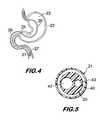
- FIG. 4shows the tube of FIG. 1 through 3 entering the stomach through the esophagus via the nose.
- FIG. 5is a cross section of the feeding tube of FIG. 1 through 3 taken along the tube length above the balloon.
- FIG. 6shows another preferred embodiment.
- FIG. 7shows another preferred embodiment.
- a feeding tube 20for instance of the type shown in U.S. Pat. No. 4,490,143, incorporated herein by reference, is placed into the position as shown in FIG. 1 , using an endoscope in the well-known prior art manner.
- the feeding tube 20has at the end thereof an inflatable and deflatable balloon 21 capable of being so inflated and deflated with either air or liquid passed to the balloon through the feeding tube itself during the placement of the tube.
- the tube 20has a body 40 containing lumen 42 and lumen 43 . Air or other fluids is pumped, or released, through lumen 43 to selectively inflate and deflate balloon 21 . Food and liquid is passed along tube 20 through lumen 42 .
- the balloon 21is partially inflated when the tube 20 is passed into the stomach 22 through the abdominal wall 23 in a well-known prior art manner as shown, for instance, in the '106 patent, with the aid of an endoscope, to a position as shown in FIG. 1 .
- the end of the tubehas passed through the pylorus 25 into the duodenum 26 .
- the end of tube 20is held at this location, while the balloon 21 is substantially inflated until it enlarges to contact the wall of the duodenum 26 .
- the endoscopeis withdrawn with the tube 20 being held in place by the inflated balloon 21 which extends against the duodenum walls in a fit that retains the tube 21 in position.
- the balloon 21is partially deflated as the tube 20 moves through the duodenum 26 into the jejunum 27 .
- the duodenumconsiders the balloon as a food bolus and begins its peristaltic action to move the balloon 21 along the duodenum into the jejunum.
- the balloon 21is deflated, either partially or fully, so that no possible blockage can occur.
- the balloon 21is left in a deflated condition during a subsequent withdrawal.
- FIG. 6shows another embodiment for enteral feeding through percutaneous endoscopic jejunostomy.
- a percutaneous endoscopic gastrostomy tube (“PEG tube”) 61has been placed in a patient.
- the PEG tubehas a proximal end 61 A and a distal end 61 B.
- the proximal end 61 Ahas an open end for the insertion of fluids, medicines, nutrition and/or apparatus as desired
- the distal end 61 Bhas an open end for termination in a patient's gastric region 62 .
- the proximal open endprojects outwardly from the patient's abdomen and the distal end terminates in the patient's gastric region.
- a single open endis seen.
- PEG tubes with various open ends at the proximal endcan be used, as desired. Usually those open ends would share a common channel with termination at the distal end. However, in some embodiments it may be desired to have separate channels associated with a specific open end located at the proximal end terminating in a particular open end of the distal end.
- J-tubepercutaneous endoscopic jejunostomy tube
- proximal endhas an open end for the insertion of fluids, medicines, nutrition and/or apparatus as desired, either with or without a fitting as desired
- distal end 63 Bis for terminating in a patient's postpyloric region.
- Lumensextend substantially through the length of J-tube 63 , and access is provided to each through fitting 65 . As can be seen in the cross sectional view of FIG.
- lumen 64which is for fluid access to the balloon, is smaller in diameter than lumen 66 which is for access to the post pyloric region.
- the diameter of lumen 66may be as much as half or more of the diameter of J-tube 63 for providing desired volume for feeding, medicines, etc.
- the J-tubeis adapted for retention and is retained within the PEG tube.
- the proximal end of the PEG tubehas projecting therefrom the proximal end of the J-tube, and the distal end of the PEG tube has projecting therefrom the distal end of the J tube.
- Access to the PEG tubemay be provided through fitting 65 , so that suction using the J-tube may be performed while the J-tube is retained therein.
- a balloon 69is shown at the distal end of the J-tube.
- the balloon of this embodimentis for providing self propelling of the J-tube into a desired position within the patient's postpyloric region 67 through intestinal peristaltic action upon the balloon.
- the balloonBefore placement of the J-tube within the PEG tube, the balloon is deflated. Post placement, the balloon is inflated for movement through the patient's system via peristaltic activity of the digestive tract. Such movement has been referred to as self propelled movement herein.
- Access for deflation and inflationis provided through fitting 65 . Peristaltic activity causes the J-tube distal end to reach a desired position, often as long as a week or two.
- Fitting 65provides for fixation as well, through a nut (not shown) which provides a lock or anchor between PEG tube 61 and J-tube 63 when fastened, usually at the time of placement.
- the J-tube anchored within the PEG tubemay provide a fixed point for the J-tube's feeding end.
- Initial placement of a J-tube embodiment through a PEG tubeis usually in combination with an endoscope, so as to provide for initial placement of the distal end of the J-tube.
- enteral feeding and more particularly gastrojejunal feedingincludes nasal, oral and percutaneous feeding. Endoscopic jejunostomy tube embodiments therefore may be used in each of these types of feeding.
Landscapes
- Health & Medical Sciences (AREA)
- Life Sciences & Earth Sciences (AREA)
- Animal Behavior & Ethology (AREA)
- General Health & Medical Sciences (AREA)
- Public Health (AREA)
- Veterinary Medicine (AREA)
- Pulmonology (AREA)
- Engineering & Computer Science (AREA)
- Robotics (AREA)
- Biophysics (AREA)
- Gastroenterology & Hepatology (AREA)
- Anesthesiology (AREA)
- Biomedical Technology (AREA)
- Heart & Thoracic Surgery (AREA)
- Hematology (AREA)
- Otolaryngology (AREA)
- Medical Preparation Storing Or Oral Administration Devices (AREA)
- Infusion, Injection, And Reservoir Apparatuses (AREA)
- Media Introduction/Drainage Providing Device (AREA)
Abstract
Description
Claims (11)
Priority Applications (1)
| Application Number | Priority Date | Filing Date | Title |
|---|---|---|---|
| US12/583,355US8016785B2 (en) | 2004-12-02 | 2009-08-18 | Gastrojejunal feeding tube |
Applications Claiming Priority (3)
| Application Number | Priority Date | Filing Date | Title |
|---|---|---|---|
| US11/001,846US7220253B2 (en) | 2004-12-02 | 2004-12-02 | Gastrojejunal feeding tube |
| US79985607A | 2007-05-03 | 2007-05-03 | |
| US12/583,355US8016785B2 (en) | 2004-12-02 | 2009-08-18 | Gastrojejunal feeding tube |
Related Parent Applications (1)
| Application Number | Title | Priority Date | Filing Date |
|---|---|---|---|
| US79985607AContinuation-In-Part | 2004-12-02 | 2007-05-03 |
Publications (2)
| Publication Number | Publication Date |
|---|---|
| US20100030138A1 US20100030138A1 (en) | 2010-02-04 |
| US8016785B2true US8016785B2 (en) | 2011-09-13 |
Family
ID=41609091
Family Applications (1)
| Application Number | Title | Priority Date | Filing Date |
|---|---|---|---|
| US12/583,355Expired - Fee RelatedUS8016785B2 (en) | 2004-12-02 | 2009-08-18 | Gastrojejunal feeding tube |
Country Status (1)
| Country | Link |
|---|---|
| US (1) | US8016785B2 (en) |
Cited By (2)
| Publication number | Priority date | Publication date | Assignee | Title |
|---|---|---|---|---|
| US10722267B2 (en) | 2015-11-30 | 2020-07-28 | Piranha Medical, LLC | Blockage removal |
| US11141177B2 (en) | 2015-11-30 | 2021-10-12 | Piranha Medical Llc | Blockage clearing devices, systems, and methods |
Families Citing this family (6)
| Publication number | Priority date | Publication date | Assignee | Title |
|---|---|---|---|---|
| MX350734B (en) | 2010-09-08 | 2017-09-15 | Covidien Lp | Catheter with imaging assembly. |
| US9095502B2 (en) | 2011-02-09 | 2015-08-04 | Applied Medical Technology, Inc. | Low profile G-J feeding tube |
| US9517184B2 (en)* | 2012-09-07 | 2016-12-13 | Covidien Lp | Feeding tube with insufflation device and related methods therefor |
| US20140142552A1 (en)* | 2012-11-16 | 2014-05-22 | Shaun M. Honig | Percutaneous Feeding Tube Including a Rescue Port |
| US9713578B2 (en)* | 2012-12-20 | 2017-07-25 | Sabry Gabriel | Feeding tube with inflatable balloon component |
| WO2018009662A1 (en)* | 2016-07-06 | 2018-01-11 | Hamad Medical Corporation | Enteral feeding tube with inflatable cuff |
Citations (11)
| Publication number | Priority date | Publication date | Assignee | Title |
|---|---|---|---|---|
| US4368739A (en)* | 1979-07-18 | 1983-01-18 | Nelson Jr Richard L | Long intestinal catheter |
| US4490143A (en) | 1982-09-24 | 1984-12-25 | Viridian, Inc. | Feeding tube assembly |
| US4496347A (en) | 1982-09-24 | 1985-01-29 | Viridian, Inc. | Feeding tube stylet |
| US4594074A (en) | 1985-05-06 | 1986-06-10 | Viridian, Inc. | Non-occluding high flow enteral feeding tube |
| US4676778A (en) | 1986-10-27 | 1987-06-30 | Nelson Jr Richard L | Long intestinal catheter with sump |
| US4798592A (en) | 1984-11-05 | 1989-01-17 | Medical Innovations Corporation | Gastrostomy feeding device |
| US5057091A (en) | 1989-07-31 | 1991-10-15 | Corpak, Inc. | Enteral feeding tube with a flexible bolus and feeding bolus |
| US5152756A (en) | 1990-01-24 | 1992-10-06 | Corpak, Inc. | Distal gripping tip for enteral feeding tube |
| US5318530A (en) | 1991-12-06 | 1994-06-07 | Bissel Medical Products, Inc. | Gastrointestinal tube with inflatable bolus |
| US6264631B1 (en)* | 1998-02-12 | 2001-07-24 | Ballard Medical Products | Catheter with distally distending balloon |
| US6458106B1 (en) | 2000-02-17 | 2002-10-01 | Sherwood Services, Ag | Low profile jejunal adapter for a gastrojejunal feeding system |
- 2009
- 2009-08-18USUS12/583,355patent/US8016785B2/ennot_activeExpired - Fee Related
Patent Citations (11)
| Publication number | Priority date | Publication date | Assignee | Title |
|---|---|---|---|---|
| US4368739A (en)* | 1979-07-18 | 1983-01-18 | Nelson Jr Richard L | Long intestinal catheter |
| US4490143A (en) | 1982-09-24 | 1984-12-25 | Viridian, Inc. | Feeding tube assembly |
| US4496347A (en) | 1982-09-24 | 1985-01-29 | Viridian, Inc. | Feeding tube stylet |
| US4798592A (en) | 1984-11-05 | 1989-01-17 | Medical Innovations Corporation | Gastrostomy feeding device |
| US4594074A (en) | 1985-05-06 | 1986-06-10 | Viridian, Inc. | Non-occluding high flow enteral feeding tube |
| US4676778A (en) | 1986-10-27 | 1987-06-30 | Nelson Jr Richard L | Long intestinal catheter with sump |
| US5057091A (en) | 1989-07-31 | 1991-10-15 | Corpak, Inc. | Enteral feeding tube with a flexible bolus and feeding bolus |
| US5152756A (en) | 1990-01-24 | 1992-10-06 | Corpak, Inc. | Distal gripping tip for enteral feeding tube |
| US5318530A (en) | 1991-12-06 | 1994-06-07 | Bissel Medical Products, Inc. | Gastrointestinal tube with inflatable bolus |
| US6264631B1 (en)* | 1998-02-12 | 2001-07-24 | Ballard Medical Products | Catheter with distally distending balloon |
| US6458106B1 (en) | 2000-02-17 | 2002-10-01 | Sherwood Services, Ag | Low profile jejunal adapter for a gastrojejunal feeding system |
Non-Patent Citations (2)
| Title |
|---|
| Literature issued by Boston Scientific Corporation, Natick, MA, USA, for EndoVive(TM) Standard PEG Kits, (® 2005 Boston Scientific Corporation or its affiliates); copy consists of 7 pages. |
| Literature issued by Boston Scientific Corporation, Natick, MA, USA, for EndoVive™ Standard PEG Kits, (® 2005 Boston Scientific Corporation or its affiliates); copy consists of 7 pages. |
Cited By (2)
| Publication number | Priority date | Publication date | Assignee | Title |
|---|---|---|---|---|
| US10722267B2 (en) | 2015-11-30 | 2020-07-28 | Piranha Medical, LLC | Blockage removal |
| US11141177B2 (en) | 2015-11-30 | 2021-10-12 | Piranha Medical Llc | Blockage clearing devices, systems, and methods |
Also Published As
| Publication number | Publication date |
|---|---|
| US20100030138A1 (en) | 2010-02-04 |
Similar Documents
| Publication | Publication Date | Title |
|---|---|---|
| US8016785B2 (en) | Gastrojejunal feeding tube | |
| KR0160284B1 (en) | Replacement gastrotomy tube | |
| US5098378A (en) | Method of jejunal feeding | |
| AU2001232180B2 (en) | Low profile jejunal adapter for a gastrojejunal feeding system | |
| US5902285A (en) | Jejunal feeding tube | |
| JP3347148B2 (en) | Fistula formation device | |
| US5871467A (en) | Post-pyloric feeding tubes | |
| GB2554064B (en) | An enteral feeding tube | |
| Niv et al. | Post-pyloric feeding | |
| MXPA04011303A (en) | Low profile transplyoric jejunostomy system. | |
| Shin et al. | Updates on percutaneous radiologic gastrostomy/gastrojejunostomy and jejunostomy | |
| AU2001232180A1 (en) | Low profile jejunal adapter for a gastrojejunal feeding system | |
| HK1219687A1 (en) | Apparatus and method for treating obesity by extracting food | |
| US7220253B2 (en) | Gastrojejunal feeding tube | |
| Keymling | Technical aspects of enteral nutrition. | |
| Holmes | Enteral feeding and percutaneous endoscopic gastrostomy. | |
| EP0853937A1 (en) | Gastrostomy tube device for enteral nutrition | |
| McClave | Techniques in enteral access | |
| CN203598248U (en) | Self-feeding type jejunum nutrition tube | |
| Hauenschild et al. | Prospective evaluation of novel system for jejunal feeding | |
| Rosenberger et al. | Jejunal tube extensions via percutaneous endoscopic gastrostomy and delayed small-bowel perforations: a case series | |
| Yeung et al. | Percutaneous radiologic gastrostomy | |
| DeLegge | Administration Routes for Enteral Nutrition | |
| Darcy | Radiological percutaneous transgastric jejunostomy | |
| Ockenga et al. | Approach to Oral and Enteral Nutrition in Adults Topic 8 |
Legal Events
| Date | Code | Title | Description |
|---|---|---|---|
| AS | Assignment | Owner name:CHEK-MED SYSTEMS, INC.,PENNSYLVANIA Free format text:ASSIGNMENT OF ASSIGNORS INTEREST;ASSIGNORS:KANTSEVOY, SERGEY V.;KALLOO, ANTHONY N.;REEL/FRAME:023153/0088 Effective date:20090814 Owner name:CHEK-MED SYSTEMS, INC., PENNSYLVANIA Free format text:ASSIGNMENT OF ASSIGNORS INTEREST;ASSIGNORS:KANTSEVOY, SERGEY V.;KALLOO, ANTHONY N.;REEL/FRAME:023153/0088 Effective date:20090814 | |
| ZAAA | Notice of allowance and fees due | Free format text:ORIGINAL CODE: NOA | |
| ZAAB | Notice of allowance mailed | Free format text:ORIGINAL CODE: MN/=. | |
| ZAAA | Notice of allowance and fees due | Free format text:ORIGINAL CODE: NOA | |
| STCF | Information on status: patent grant | Free format text:PATENTED CASE | |
| AS | Assignment | Owner name:GI SUPPLY, INC., PENNSYLVANIA Free format text:CHANGE OF NAME;ASSIGNOR:CHEK-MED SYSTEMS, INC.;REEL/FRAME:033696/0936 Effective date:20140401 | |
| FPAY | Fee payment | Year of fee payment:4 | |
| MAFP | Maintenance fee payment | Free format text:PAYMENT OF MAINTENANCE FEE, 8TH YR, SMALL ENTITY (ORIGINAL EVENT CODE: M2552); ENTITY STATUS OF PATENT OWNER: SMALL ENTITY Year of fee payment:8 | |
| FEPP | Fee payment procedure | Free format text:ENTITY STATUS SET TO UNDISCOUNTED (ORIGINAL EVENT CODE: BIG.); ENTITY STATUS OF PATENT OWNER: LARGE ENTITY | |
| AS | Assignment | Owner name:JPMORGAN CHASE BANK, N.A., ILLINOIS Free format text:PATENT SECURITY AGREEMENT;ASSIGNOR:GI SUPPLY, INC.;REEL/FRAME:060560/0880 Effective date:20220608 | |
| FEPP | Fee payment procedure | Free format text:MAINTENANCE FEE REMINDER MAILED (ORIGINAL EVENT CODE: REM.); ENTITY STATUS OF PATENT OWNER: LARGE ENTITY | |
| LAPS | Lapse for failure to pay maintenance fees | Free format text:PATENT EXPIRED FOR FAILURE TO PAY MAINTENANCE FEES (ORIGINAL EVENT CODE: EXP.); ENTITY STATUS OF PATENT OWNER: LARGE ENTITY | |
| STCH | Information on status: patent discontinuation | Free format text:PATENT EXPIRED DUE TO NONPAYMENT OF MAINTENANCE FEES UNDER 37 CFR 1.362 | |
| FP | Lapsed due to failure to pay maintenance fee | Effective date:20230913 | |
| AS | Assignment | Owner name:GI SUPPLY, INC., PENNSYLVANIA Free format text:RELEASE BY SECURED PARTY;ASSIGNOR:JPMORGAN CHASE BANK, N.A., AS ADMINISTRATIVE AGENT;REEL/FRAME:066984/0479 Effective date:20240402 |