US8007437B2 - Method and apparatus for interactive 4-dimensional (4D) virtual endoscopy - Google Patents
Method and apparatus for interactive 4-dimensional (4D) virtual endoscopyDownload PDFInfo
- Publication number
- US8007437B2 US8007437B2US11/874,975US87497507AUS8007437B2US 8007437 B2US8007437 B2US 8007437B2US 87497507 AUS87497507 AUS 87497507AUS 8007437 B2US8007437 B2US 8007437B2
- Authority
- US
- United States
- Prior art keywords
- images
- patient
- views
- heart
- cardiac
- Prior art date
- Legal status (The legal status is an assumption and is not a legal conclusion. Google has not performed a legal analysis and makes no representation as to the accuracy of the status listed.)
- Expired - Fee Related, expires
Links
Images


Classifications
- G—PHYSICS
- G06—COMPUTING OR CALCULATING; COUNTING
- G06T—IMAGE DATA PROCESSING OR GENERATION, IN GENERAL
- G06T7/00—Image analysis
- G06T7/0002—Inspection of images, e.g. flaw detection
- G06T7/0012—Biomedical image inspection
- A—HUMAN NECESSITIES
- A61—MEDICAL OR VETERINARY SCIENCE; HYGIENE
- A61B—DIAGNOSIS; SURGERY; IDENTIFICATION
- A61B5/00—Measuring for diagnostic purposes; Identification of persons
- A61B5/72—Signal processing specially adapted for physiological signals or for diagnostic purposes
- A61B5/7271—Specific aspects of physiological measurement analysis
- A61B5/7285—Specific aspects of physiological measurement analysis for synchronizing or triggering a physiological measurement or image acquisition with a physiological event or waveform, e.g. an ECG signal
- A61B5/7289—Retrospective gating, i.e. associating measured signals or images with a physiological event after the actual measurement or image acquisition, e.g. by simultaneously recording an additional physiological signal during the measurement or image acquisition
- A—HUMAN NECESSITIES
- A61—MEDICAL OR VETERINARY SCIENCE; HYGIENE
- A61B—DIAGNOSIS; SURGERY; IDENTIFICATION
- A61B8/00—Diagnosis using ultrasonic, sonic or infrasonic waves
- A61B8/13—Tomography
- A61B8/14—Echo-tomography
- G—PHYSICS
- G06—COMPUTING OR CALCULATING; COUNTING
- G06T—IMAGE DATA PROCESSING OR GENERATION, IN GENERAL
- G06T15/00—3D [Three Dimensional] image rendering
- G06T15/08—Volume rendering
- A—HUMAN NECESSITIES
- A61—MEDICAL OR VETERINARY SCIENCE; HYGIENE
- A61B—DIAGNOSIS; SURGERY; IDENTIFICATION
- A61B6/00—Apparatus or devices for radiation diagnosis; Apparatus or devices for radiation diagnosis combined with radiation therapy equipment
- A61B6/50—Apparatus or devices for radiation diagnosis; Apparatus or devices for radiation diagnosis combined with radiation therapy equipment specially adapted for specific body parts; specially adapted for specific clinical applications
- A61B6/503—Apparatus or devices for radiation diagnosis; Apparatus or devices for radiation diagnosis combined with radiation therapy equipment specially adapted for specific body parts; specially adapted for specific clinical applications for diagnosis of the heart
- A—HUMAN NECESSITIES
- A61—MEDICAL OR VETERINARY SCIENCE; HYGIENE
- A61B—DIAGNOSIS; SURGERY; IDENTIFICATION
- A61B8/00—Diagnosis using ultrasonic, sonic or infrasonic waves
- A61B8/08—Clinical applications
- A61B8/0883—Clinical applications for diagnosis of the heart
- G—PHYSICS
- G06—COMPUTING OR CALCULATING; COUNTING
- G06T—IMAGE DATA PROCESSING OR GENERATION, IN GENERAL
- G06T2207/00—Indexing scheme for image analysis or image enhancement
- G06T2207/10—Image acquisition modality
- G06T2207/10072—Tomographic images
- G06T2207/10076—4D tomography; Time-sequential 3D tomography
- G—PHYSICS
- G06—COMPUTING OR CALCULATING; COUNTING
- G06T—IMAGE DATA PROCESSING OR GENERATION, IN GENERAL
- G06T2207/00—Indexing scheme for image analysis or image enhancement
- G06T2207/10—Image acquisition modality
- G06T2207/10132—Ultrasound image
- G06T2207/10136—3D ultrasound image
- G—PHYSICS
- G06—COMPUTING OR CALCULATING; COUNTING
- G06T—IMAGE DATA PROCESSING OR GENERATION, IN GENERAL
- G06T2207/00—Indexing scheme for image analysis or image enhancement
- G06T2207/30—Subject of image; Context of image processing
- G06T2207/30004—Biomedical image processing
- G06T2207/30048—Heart; Cardiac
Definitions
- the inventionrelates generally to Interactive 4-dimensional (4D) imaging and, more particularly, to 4D Virtual Endoscopy for Cardiac Imaging.
- Virtual endoscopyallows visualization of a scene setup from pre-acquired data. Images are created with a virtual camera interactively manipulated by a user. Imaging techniques currently in use and how the acquired data is visualized are first briefly reviewed below.
- CTcomputed tomography
- MRmagnetic resonance
- 3D volumeswith the composition of 2D ultrasound frames.
- Acquired datacan be visualized in a volume renderer, in a virtual endoscopy, or as single slices. It can be any arbitrary slice, called multi-planar reconstruction (MPR). By manipulating these slices, it is possible to get a 3D sense of the volume.
- MPRmulti-planar reconstruction
- the datais directly displayed in 3D. It is possible to rotate around the volume, and to zoom or pan the volume. By rendering certain intensities of the data, different tissues can be revealed. As an additional help to seeing inside, the volume can be cut or intercepted by an arbitrary plane.
- a virtual cameraIn a virtual endoscopy, a virtual camera is located inside the volume. The camera can be moved or rotated and it is possible to change the camera parameters such as its field of view. To define what constitutes a cavity in the volume, an iso-surface value is chosen. Intensities below this value will be considered a cavity, and intensities above it will be rendered as opaque. In the endoscopic view, the boundary seen will therefore be dependent on the iso-surface value.
- the first methodcasts rays from the virtual camera and detects when the rays hit the volume. At the boundary, the surface normal is used to compute the correct lighting of the current pixel. Each ray will generate a pixel on the screen.
- the second methodextracts an iso-surface from the volume, for instance. with the marching cube algorithm.
- the resulting meshis placed in a 3D world and the virtual camera can navigate the mesh. This can be efficiently done with OpenGL, or any other suitable graphic language. Background information can be found in the literature: see, for example, the literature cited below.
- a 4D volume renderer and dynamic MPRare currently available: see, for example, Kostas Anagnostou, Tim J. Atherton, Andrew E. Waterfall, “4D volume Rendering With The Shear Warp Factorisation”, Proceedings of the 2000 IEEE Symposium on Volume Visualization. pp. 129-137, 2000; however, this material does not show a 4D virtual endoscopy system.
- Textbooks useful in providing background material helpful to gaining a better understanding of the present inventioninclude, for example, VIRTUAL ENDOSCOPY and RELATED 3D TECHNIQUES by P. Rogalla et al., Springer, Berlin & New York, 2002, FUNDAMENTALS OF IMAGE PROCESSING by Arthur R. Weeks, SPIE Optical Engineering Press & IEEE Press; 1996; IMAGE PROCESSING, ANALYSIS, AND MACHINE VISION, Second Edition, by Milan Sonka et al., PWS Publishing; 1999; and DIGITAL IMAGE PROCESSING, Second Edition, by Rafael C. Gonzalez et al., Prentice Hall; 2002.
- endoscopic visualizationutilizes cardiac imaging data, such as from Computed Tomography (CT), Magnetic Resonance (MR), or Ultrasound (US) imaging.
- CTComputed Tomography
- MRMagnetic Resonance
- USUltrasound
- a system for 4D (3D+time) endoscopic visualizationuses cardiac imaging data, such as Computed Tomography, Magnetic Resonance, or Ultrasound Imaging.
- cardiac imaging datasuch as Computed Tomography, Magnetic Resonance, or Ultrasound Imaging.
- a system in accordance with the present inventionallows the visualization of a dynamic scene as observed from inside the heart. It can be used in diagnostics or for surgery planning. As compared to 3D static endoscopic visualization, the system provides more visual information to the user.
- a method of 4-dimensional virtual endoscopyincludes acquiring a sequence of cardiac images of a patient's heart from an imaging device; acquiring an electrocardiographic signal exhibiting cardiac cycles of the heart; and utilizing the electrocardiographic signal to gate the images for deriving respective 3-dimensional views in succession at corresponding cardiac cycles, from respective cardiac images at a selected phase point common to each of the corresponding cardiac cycles.
- the step for deriving respective 3D viewsincludes placing the cardiac images in a 3D space and reconstructing a volume.
- the step of acquiring an electrocardiographic signalincludes a step of detecting the cardiac cycles and a step of interactively selecting the phase point.
- the step for deriving respective 3D viewsutilizes one of (a) a ray casting algorithm and (b) a mesh extraction algorithm.
- the step for deriving a series of respective 3D viewsincludes amending a 3D view so as to produce the next 3D view in the succession.
- the step for deriving a series of respective 3D viewsincludes a step of sensing breathing of the patient and excluding images not in the same phase of the respiratory cycle.
- the step for deriving a series of respective 3D viewsincludes a step of sensing sudden catheter movements in the patient and excluding images where such a sudden motion occurs.
- the step for acquiring a sequence of cardiac imagesincludes: a step of utilizing a virtual camera; and a step of virtually attaching the virtual camera to a wall image of the patient's heart so as to produce a substantially steady image of the heart.
- a method of 4-dimensional (4D) virtual endoscopycomprises: acquiring a motion signal of a patient's organ exhibiting motion cycles of the organ; acquiring a sequence of 3-dimensional (3D) images of the organ from an imaging device; utilizing the motion signal to gate the images for deriving respective 3D views in succession at corresponding motion cycles, from respective organ images at a selected phase point common to each of the corresponding motion cycles.
- the step for acquiring a sequence of 3D viewsincludes one of: (a) utilizing a real-time 3D ultrasound imaging device with multiple transducer arrays, and (b) utilizing a tracked ultrasound imaging device collecting data over space.
- the step of utilizing a tracked ultrasound imaging device collecting data over spacecomprises utilizing an AcunavTM system.
- a system for performing 4-dimensional (4D) virtual endoscopycomprises: a memory device for storing a program and other data; and a processor in communication with the memory device, the processor operative with the program to perform: acquiring a sequence of cardiac images of a patient's heart from an imaging device; acquiring an electrocardiographic signal exhibiting cardiac cycles of the heart; and utilizing the electrocardiographic signal to gate the images for deriving respective 3-dimensional (3D) views in succession at corresponding cardiac cycles, from respective cardiac images at a selected phase point common to each of the corresponding cardiac cycles.
- a system for performing 4-dimensional (4D) virtual endoscopycomprises: apparatus for acquiring a motion signal of a patient's organ exhibiting motion cycles of the organ; apparatus for acquiring a sequence of 3-dimensional (3D) images of the organ from an imaging device; and apparatus for utilizing the motion signal to gate the images for deriving respective 3D views in succession at corresponding motion cycles, from respective organ images at a selected phase point common to each of the corresponding motion cycles.
- a computer program productcomprises a computer useable medium having computer program logic recorded thereon for program code for performing 4-dimensional (4D) virtual endoscopy, comprising: acquiring a sequence of cardiac images of a patient's heart from an imaging device; acquiring an electrocardiographic signal exhibiting cardiac cycles of the heart; and utilizing the electrocardiographic signal to gate the images for deriving respective 3-dimensional (3D) views in succession at corresponding cardiac cycles, from respective cardiac images at a selected phase point common to each of the corresponding cardiac cycles.
- FIG. 1shows the reconstruction of a plurality of 3D volumes at corresponding different cardiac cycles from frames of the same given cardiac phase in the respective cardiac cycles in accordance with principles of the present invention
- FIG. 2shows successively generated views for virtual endoscopy in accordance with the present invention
- FIGS. 3-7show schematic flow diagrams of steps in accordance with principles of the present invention.
- FIG. 8shows in block schematic form the application of a digital computer in the present invention.
- a general framework for a method and system for an interactive 4D virtual endoscopy systemincludes:
- Ultrasound sequencesare acquired with either a real-time 3D ultrasound with multiple transducer arrays, or with a tracked AcunavTM collecting data over space. Images thus acquired are placed in a 3D space to reconstruct the volume.
- ECGelectrocardiogram signal
- US framesare gated and multiple 3D volumes are created from frames corresponding to the same cardiac phases. The result is a reconstructed 4D volume.
- Rendering of a dynamic scenerequires the use of a different 3D data set at each successive frame, as will also be illustrated below in reference to FIGS. 1 and 2 .
- the system in accordance with the present inventionuses a single 3D volume to generate the endoscopic view, and changes the 3D volume at each new time step.
- Each single image generationcan use any virtual endoscopy imaging. It can be, for instance, a ray casting or a mesh extraction algorithm as described in the previous section.
- the algorithmcan also be hardware accelerated for smooth visualization of the dynamic scene.
- the endoscopecan be manipulated interactively by the user.
- An embodiment in accordance with the inventionincludes the commands of camera rotation; camera translation; camera tilting; and camera field of view selection.
- the system in accordance with the inventioncan handle different modalities such as, for instance CT, MR or 3D compounded ultrasound. Each modality requires parameter tuning items which are provided with the available presets.
- temporal commandswhich may include commands such as:
- an embodiment of the system in accordance with the present inventioncomprises features such as real time virtual endoscopy; with ultrasound, acquisition can be done in real-time—the latest available 3 d volume can be used in the rendering algorithm.
- an embodiment of the present inventionmay include a feature of attaching the camera to the wall or surface in the image which, for example in the case of the heart, can be the endocardium.
- Temporal markersmay also be provided wherein a point on the surface can be marked and later tracked over time, thus allowing the visualization and analysis of a landmark motion.
- Collision detectionmay also be provided such that when the user moves the camera, the system detects the camera colliding with the iso-surface of a certain volume so that the camera cannot be moved across the iso-surface.
- FIG. 1shows the reconstruction of a plurality of 3D volumes at corresponding different cardiac cycles from frames of the same given cardiac phase in the respective cardiac cycles.
- Reference numeral 10indicates cardiac cycle waveforms used for gating, with sagittal lines pointing to the respective frames 12 - 18 below.
- a representative reconstructed 3D volume corresponding to a given frameis indicated by numeral 20 .
- FIG. 2shows successively generated views (A), (B) and (C) for virtual endoscopy.
- FIGS. 3-7provide flow charts of aspects of the method of the present invention.
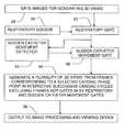
- FIG. 3shows an ECG signal source 22 providing a waveform signal to a cardiac cycle detector and phase point selector 30 which is coupled to an interactive control 32 for phase point selection.
- An ultrasound image sequenceis acquired in 24 and the images are placed in a 3D space in 26 , where volume images are reconstructed and are then gated at 28 .
- a plurality of 3D viewsis generated at 34 from frames corresponding to a selected cardiac phase point in respective successive cardiac cycles, and thence outputted at 36 .
- FIG. 4shows an ECG signal source 22 providing a waveform signal to a cardiac cycle detector and phase point selector 30 which is coupled to an interactive control 32 for phase point selection.
- the occurrenceis sensed of a selected cardiac phase point in respective successive cardiac cycles.
- a gating signal from selector 30enables gating images at 48 for generating 3D views.
- a plurality of 3D viewsis generated at 50 from frames corresponding to a selected cardiac phase point in respective successive cardiac cycles and 3D views are stored at 52 .
- a comparisonis made at 54 between the stored view and the next or succeeding view.
- the stored viewis then amended at 56 to conform it to the next view.
- FIG. 6shows the gating of images at 28 for generating 3D views.
- a signal from a respiratory sensor 60is applied to a respiratory gate 62 for corresponding gating of images.
- a detector 64 for sudden catheter movementsprovides a signal representative of such sudden movements for a gating function at 66 , so that at 68 a plurality of 3D views is generated from frames corresponding to a selected cardiac phase point in respective successive cardiac cycles excluding frames not gated in by respiratory and sudden catheter movement gates.
- the viewsare outputted at 36 to image processing and viewing devices.
- FIG. 7shows the generation of a plurality of 3D views at 70 from frames corresponding to a selected cardiac phase point in respective successive cardiac cycles by using a ray casting algorithm or a mesh extraction algorithm.
- FIG. 8shows in basic schematic form a digital processor coupled for two way data communication with an input device, an output device, and a memory device, in applications for embodiments of the present invention.
- Data buss 80couples input devices at 82 , such as an ECG, an ultrasound device or a CT scan device or MRI scan device to a processor 86 and graphics card, a memory 84 , an output device 88 , and interactive user controls 90 .
- the memory deviceis generally used for storing a program and other data.
- the input deviceis so designated in broad terms as a device for providing an appropriate image or images for processing in accordance with the present invention.
- the inputmay be from an imaging device, such as a device incorporated in a CATSCAN, X-ray machine, an MRI or other device, or a stored image, or by communication with another computer or device by way of direct connection, a modulated infrared beam, radio, land line, facsimile, or satellite as, for example, by way of the World Wide Web or Internet, or any other appropriate source of such data.
- the output devicemay include a computer type display device using any suitable apparatus such as a cathode-ray kinescope tube, a plasma display, liquid crystal display, and so forth, or it may or may not include a device for rendering an image and may include a memory device or part of the memory device of FIG. 8 for storing an image for further processing, or for viewing, or evaluation, as may be convenient, or it may utilize a connection or coupling including such as are noted above in relation to the input device.
- the processoris operative with a program set up in accordance with the present invention for implementing steps of the invention.
- Such a programmed computermay interface readily through communications media such as land line, radio, the Internet, and so forth for image data acquisition and transmission.
- the inventionmay be readily implemented, at least in part, in a software memory device and packaged in that form as a software product.
- Thiscan be in the form of a computer program product comprising a computer useable medium having computer program logic recorded thereon for program code for performing the method of the present invention.
- the present inventionis not limited to cardiac applications. For convenience, embodiments were described in a cardiac setting for illustrative purposes, but the invention is applicable to other body parts with periodic movements such as, for example, respiratory motion.
Landscapes
- Engineering & Computer Science (AREA)
- Health & Medical Sciences (AREA)
- Life Sciences & Earth Sciences (AREA)
- Physics & Mathematics (AREA)
- General Health & Medical Sciences (AREA)
- Medical Informatics (AREA)
- Molecular Biology (AREA)
- Surgery (AREA)
- Nuclear Medicine, Radiotherapy & Molecular Imaging (AREA)
- Radiology & Medical Imaging (AREA)
- Veterinary Medicine (AREA)
- Computer Vision & Pattern Recognition (AREA)
- Public Health (AREA)
- Animal Behavior & Ethology (AREA)
- Theoretical Computer Science (AREA)
- Heart & Thoracic Surgery (AREA)
- General Physics & Mathematics (AREA)
- Biophysics (AREA)
- Pathology (AREA)
- Biomedical Technology (AREA)
- Signal Processing (AREA)
- Computer Graphics (AREA)
- Psychiatry (AREA)
- Physiology (AREA)
- Artificial Intelligence (AREA)
- Quality & Reliability (AREA)
- Ultra Sonic Daignosis Equipment (AREA)
- Apparatus For Radiation Diagnosis (AREA)
- Measuring And Recording Apparatus For Diagnosis (AREA)
Abstract
Description
- reconstructing the 4D volume from a 3D image;
- rendering a dynamic endoscopic scene;
- interactively manipulating the virtual endoscope; and
- interactively tuning parameters of the system.
- changing the iso-surface value;
- changing the iso-surface color;
- changing the lighting and the field of view;
- changing the image resolution; and
- smoothing the rendered data (e.g. by the use of an average or median filter).
- a temporal navigation bar including a play and pause button, a stop button, a fast forward button, and a fast rewind button;
- a command to record the endoscopic view in a movie file; and
- a command to change the frame rate of the dynamic scene.
Claims (22)
Priority Applications (2)
| Application Number | Priority Date | Filing Date | Title |
|---|---|---|---|
| US11/874,975US8007437B2 (en) | 2006-11-08 | 2007-10-19 | Method and apparatus for interactive 4-dimensional (4D) virtual endoscopy |
| CN2007103071780ACN101190124B (en) | 2006-11-08 | 2007-11-08 | Interactive type four-dimensional dummy endoscopy method and apparatus |
Applications Claiming Priority (2)
| Application Number | Priority Date | Filing Date | Title |
|---|---|---|---|
| US85763706P | 2006-11-08 | 2006-11-08 | |
| US11/874,975US8007437B2 (en) | 2006-11-08 | 2007-10-19 | Method and apparatus for interactive 4-dimensional (4D) virtual endoscopy |
Publications (2)
| Publication Number | Publication Date |
|---|---|
| US20080270095A1 US20080270095A1 (en) | 2008-10-30 |
| US8007437B2true US8007437B2 (en) | 2011-08-30 |
Family
ID=39485431
Family Applications (1)
| Application Number | Title | Priority Date | Filing Date |
|---|---|---|---|
| US11/874,975Expired - Fee RelatedUS8007437B2 (en) | 2006-11-08 | 2007-10-19 | Method and apparatus for interactive 4-dimensional (4D) virtual endoscopy |
Country Status (2)
| Country | Link |
|---|---|
| US (1) | US8007437B2 (en) |
| CN (1) | CN101190124B (en) |
Cited By (5)
| Publication number | Priority date | Publication date | Assignee | Title |
|---|---|---|---|---|
| US9498231B2 (en) | 2011-06-27 | 2016-11-22 | Board Of Regents Of The University Of Nebraska | On-board tool tracking system and methods of computer assisted surgery |
| US10105149B2 (en) | 2013-03-15 | 2018-10-23 | Board Of Regents Of The University Of Nebraska | On-board tool tracking system and methods of computer assisted surgery |
| US10219811B2 (en) | 2011-06-27 | 2019-03-05 | Board Of Regents Of The University Of Nebraska | On-board tool tracking system and methods of computer assisted surgery |
| US11116574B2 (en) | 2006-06-16 | 2021-09-14 | Board Of Regents Of The University Of Nebraska | Method and apparatus for computer aided surgery |
| US11911117B2 (en) | 2011-06-27 | 2024-02-27 | Board Of Regents Of The University Of Nebraska | On-board tool tracking system and methods of computer assisted surgery |
Families Citing this family (22)
| Publication number | Priority date | Publication date | Assignee | Title |
|---|---|---|---|---|
| US8103070B2 (en)* | 2007-11-22 | 2012-01-24 | Toshiba Medical Visualization Systems Europe, Limited | Volume rendering apparatus and method |
| US8200466B2 (en) | 2008-07-21 | 2012-06-12 | The Board Of Trustees Of The Leland Stanford Junior University | Method for tuning patient-specific cardiovascular simulations |
| JP5371703B2 (en)* | 2008-11-27 | 2013-12-18 | 株式会社東芝 | Medical image generation device, medical image storage device, medical image display device, and medical image display system |
| US9405886B2 (en) | 2009-03-17 | 2016-08-02 | The Board Of Trustees Of The Leland Stanford Junior University | Method for determining cardiovascular information |
| JP5395538B2 (en)* | 2009-06-30 | 2014-01-22 | 株式会社東芝 | Ultrasonic diagnostic apparatus and image data display control program |
| JP6035148B2 (en)* | 2009-12-08 | 2016-11-30 | コーニンクレッカ フィリップス エヌ ヴェKoninklijke Philips N.V. | Ablation treatment plan and device |
| CN102802534B (en)* | 2010-03-17 | 2015-05-06 | 富士胶片株式会社 | Medical image conversion device, method, and program |
| CN102283673B (en) | 2010-06-21 | 2015-05-20 | 深圳迈瑞生物医疗电子股份有限公司 | 3D/4D (Three Dimensional/Four Dimensional) imaging equipment as well as method and device for adjusting a region of interest in imaging |
| US8157742B2 (en) | 2010-08-12 | 2012-04-17 | Heartflow, Inc. | Method and system for patient-specific modeling of blood flow |
| US8315812B2 (en) | 2010-08-12 | 2012-11-20 | Heartflow, Inc. | Method and system for patient-specific modeling of blood flow |
| US8548778B1 (en) | 2012-05-14 | 2013-10-01 | Heartflow, Inc. | Method and system for providing information from a patient-specific model of blood flow |
| ITGE20130032A1 (en)* | 2013-03-19 | 2014-09-20 | Esaote Spa | METHOD AND IMAGING DEVICE OF THE CARDIOVASCULAR SYSTEM |
| JP2016034451A (en)* | 2014-08-04 | 2016-03-17 | 株式会社東芝 | X-ray diagnostic equipment |
| CN104887314A (en)* | 2015-04-21 | 2015-09-09 | 长春理工大学 | Virtual three-dimensional endoscope displaying method and equipment for three-dimensional endoscopic surgery navigation |
| WO2017197114A1 (en) | 2016-05-11 | 2017-11-16 | Affera, Inc. | Anatomical model generation |
| WO2017197294A1 (en)* | 2016-05-12 | 2017-11-16 | Affera, Inc. | Three-dimensional cardiac representation |
| JP6193449B1 (en)* | 2016-06-20 | 2017-09-06 | 株式会社日立製作所 | Ultrasonic diagnostic equipment |
| CN106175750A (en)* | 2016-08-08 | 2016-12-07 | 江苏金马扬名信息技术股份有限公司 | A kind of electrocardiosignal long distance control system and method |
| CN106725598B (en)* | 2016-12-28 | 2023-09-12 | 苏州科技城医院 | Heart ultrasonic system based on multiple percutaneous ultrasonic transducers and imaging method |
| CN107330236B (en)* | 2017-05-11 | 2021-06-11 | 青岛大学附属医院 | Virtual endoscope system with improved roaming effect |
| US12268456B2 (en) | 2019-01-23 | 2025-04-08 | Affera, Inc. | Systems and methods for therapy annotation |
| WO2025030238A1 (en)* | 2023-08-04 | 2025-02-13 | University Health Network | System and method for aortic assessment based on kinetic energy loss |
Citations (3)
| Publication number | Priority date | Publication date | Assignee | Title |
|---|---|---|---|---|
| US6443894B1 (en)* | 1999-09-29 | 2002-09-03 | Acuson Corporation | Medical diagnostic ultrasound system and method for mapping surface data for three dimensional imaging |
| US6892090B2 (en)* | 2002-08-19 | 2005-05-10 | Surgical Navigation Technologies, Inc. | Method and apparatus for virtual endoscopy |
| US20050283075A1 (en)* | 2004-06-16 | 2005-12-22 | Siemens Medical Solutions Usa, Inc. | Three-dimensional fly-through systems and methods using ultrasound data |
- 2007
- 2007-10-19USUS11/874,975patent/US8007437B2/ennot_activeExpired - Fee Related
- 2007-11-08CNCN2007103071780Apatent/CN101190124B/enactiveActive
Patent Citations (3)
| Publication number | Priority date | Publication date | Assignee | Title |
|---|---|---|---|---|
| US6443894B1 (en)* | 1999-09-29 | 2002-09-03 | Acuson Corporation | Medical diagnostic ultrasound system and method for mapping surface data for three dimensional imaging |
| US6892090B2 (en)* | 2002-08-19 | 2005-05-10 | Surgical Navigation Technologies, Inc. | Method and apparatus for virtual endoscopy |
| US20050283075A1 (en)* | 2004-06-16 | 2005-12-22 | Siemens Medical Solutions Usa, Inc. | Three-dimensional fly-through systems and methods using ultrasound data |
Non-Patent Citations (2)
| Title |
|---|
| Anna Vilanova i Bartroli, "Visualization Techniques for Virtual Endoscopy", PhD thesis, Technische Universität Wien, Sep. 2001; anna@cg.tuwien.ac.at; http://www.cgv.tugraz.at/; Abstract only enclosed (German and English). |
| Kostas Anagnostou, Tim J. Atherton, Andrew E. Waterfall, "4D volume Rendering With The Shear Warp Factorisation", Proceedings of the 2000 IEEE Symposium on Volume Visualization. pp. 129-137, 2000. |
Cited By (8)
| Publication number | Priority date | Publication date | Assignee | Title |
|---|---|---|---|---|
| US11116574B2 (en) | 2006-06-16 | 2021-09-14 | Board Of Regents Of The University Of Nebraska | Method and apparatus for computer aided surgery |
| US11857265B2 (en) | 2006-06-16 | 2024-01-02 | Board Of Regents Of The University Of Nebraska | Method and apparatus for computer aided surgery |
| US9498231B2 (en) | 2011-06-27 | 2016-11-22 | Board Of Regents Of The University Of Nebraska | On-board tool tracking system and methods of computer assisted surgery |
| US10080617B2 (en) | 2011-06-27 | 2018-09-25 | Board Of Regents Of The University Of Nebraska | On-board tool tracking system and methods of computer assisted surgery |
| US10219811B2 (en) | 2011-06-27 | 2019-03-05 | Board Of Regents Of The University Of Nebraska | On-board tool tracking system and methods of computer assisted surgery |
| US11911117B2 (en) | 2011-06-27 | 2024-02-27 | Board Of Regents Of The University Of Nebraska | On-board tool tracking system and methods of computer assisted surgery |
| US12232828B2 (en) | 2011-06-27 | 2025-02-25 | Board Of Regents Of The University Of Nebraska | On-board tool tracking system and methods of computer assisted surgery |
| US10105149B2 (en) | 2013-03-15 | 2018-10-23 | Board Of Regents Of The University Of Nebraska | On-board tool tracking system and methods of computer assisted surgery |
Also Published As
| Publication number | Publication date |
|---|---|
| US20080270095A1 (en) | 2008-10-30 |
| CN101190124B (en) | 2012-06-20 |
| CN101190124A (en) | 2008-06-04 |
Similar Documents
| Publication | Publication Date | Title |
|---|---|---|
| US8007437B2 (en) | Method and apparatus for interactive 4-dimensional (4D) virtual endoscopy | |
| JP6242569B2 (en) | Medical image display apparatus and X-ray diagnostic apparatus | |
| RU2654608C2 (en) | Ultrasound imaging system and method for image guidance procedure | |
| JP5639739B2 (en) | Method and system for volume rendering of multiple views | |
| JP4421016B2 (en) | Medical image processing device | |
| JP4709177B2 (en) | Three-dimensional image processing apparatus and method, and program | |
| US8079957B2 (en) | Synchronized three or four-dimensional medical ultrasound imaging and measurements | |
| US20100201786A1 (en) | Method and apparatus for reconstructing an image | |
| US20070247454A1 (en) | 3D visualization with synchronous X-ray image display | |
| US20150289848A1 (en) | Method and apparatus for registration of medical images | |
| US8094772B2 (en) | Reconstruction unit for reconstructing a fine reproduction of at least a part of an object | |
| JP2008125616A (en) | Image display method and apparatus, and image display program | |
| JP2005103263A (en) | Method of operating image forming inspection apparatus having tomographic capability and X-ray computed tomography apparatus | |
| JP4996128B2 (en) | Medical image processing apparatus and medical image processing method | |
| US20140015836A1 (en) | System and method for generating and displaying a 2d projection from a 3d or 4d dataset | |
| US20110176715A1 (en) | Four-dimensional volume imaging system | |
| US10881379B2 (en) | Method of visualizing a sequence of ultrasound images, computer program product and ultrasound system | |
| JP2001276066A (en) | 3D image processing device | |
| US20080130826A1 (en) | Method for generating tomographical recordings of a partially cyclically moving examination object | |
| JP5498185B2 (en) | Ultrasonic diagnostic apparatus and ultrasonic image display program | |
| EP3708085B1 (en) | System and method for simulating bilateral injection of contrast agent into a patient | |
| US20230181165A1 (en) | System and methods for image fusion | |
| Porter et al. | Three dimensional frameless fusion of ultrasound liver volumes | |
| JPH06348863A (en) | Picture processor | |
| JP2024022447A (en) | Medical imaging device and volume rendering method |
Legal Events
| Date | Code | Title | Description |
|---|---|---|---|
| AS | Assignment | Owner name:SIEMENS CORPORATE RESEARCH, INC., NEW JERSEY Free format text:ASSIGNMENT OF ASSIGNORS INTEREST;ASSIGNORS:SAUER, FRANK;SUN, YIYONG;XU, CHENYANG;REEL/FRAME:022472/0576;SIGNING DATES FROM 20071219 TO 20080108 Owner name:SIEMENS CORPORATE RESEARCH, INC., NEW JERSEY Free format text:ASSIGNMENT OF ASSIGNORS INTEREST;ASSIGNORS:SAUER, FRANK;SUN, YIYONG;XU, CHENYANG;SIGNING DATES FROM 20071219 TO 20080108;REEL/FRAME:022472/0576 | |
| AS | Assignment | Owner name:SIEMENS AKTIENGESELLSCHAFT, GERMANY Free format text:ASSIGNMENT OF ASSIGNORS INTEREST;ASSIGNOR:SIEMENS CORPORATE RESEARCH, INC.;REEL/FRAME:022506/0596 Effective date:20090403 Owner name:SIEMENS AKTIENGESELLSCHAFT,GERMANY Free format text:ASSIGNMENT OF ASSIGNORS INTEREST;ASSIGNOR:SIEMENS CORPORATE RESEARCH, INC.;REEL/FRAME:022506/0596 Effective date:20090403 | |
| ZAAA | Notice of allowance and fees due | Free format text:ORIGINAL CODE: NOA | |
| ZAAB | Notice of allowance mailed | Free format text:ORIGINAL CODE: MN/=. | |
| STCF | Information on status: patent grant | Free format text:PATENTED CASE | |
| FPAY | Fee payment | Year of fee payment:4 | |
| AS | Assignment | Owner name:SIEMENS HEALTHCARE GMBH, GERMANY Free format text:ASSIGNMENT OF ASSIGNORS INTEREST;ASSIGNOR:SIEMENS AKTIENGESELLSCHAFT;REEL/FRAME:038958/0348 Effective date:20160513 | |
| MAFP | Maintenance fee payment | Free format text:PAYMENT OF MAINTENANCE FEE, 8TH YEAR, LARGE ENTITY (ORIGINAL EVENT CODE: M1552); ENTITY STATUS OF PATENT OWNER: LARGE ENTITY Year of fee payment:8 | |
| FEPP | Fee payment procedure | Free format text:MAINTENANCE FEE REMINDER MAILED (ORIGINAL EVENT CODE: REM.); ENTITY STATUS OF PATENT OWNER: LARGE ENTITY | |
| LAPS | Lapse for failure to pay maintenance fees | Free format text:PATENT EXPIRED FOR FAILURE TO PAY MAINTENANCE FEES (ORIGINAL EVENT CODE: EXP.); ENTITY STATUS OF PATENT OWNER: LARGE ENTITY | |
| STCH | Information on status: patent discontinuation | Free format text:PATENT EXPIRED DUE TO NONPAYMENT OF MAINTENANCE FEES UNDER 37 CFR 1.362 | |
| FP | Lapsed due to failure to pay maintenance fee | Effective date:20230830 |