US8006700B2 - Soft tissue repair system - Google Patents
Soft tissue repair systemDownload PDFInfo
- Publication number
- US8006700B2 US8006700B2US10/114,709US11470902AUS8006700B2US 8006700 B2US8006700 B2US 8006700B2US 11470902 AUS11470902 AUS 11470902AUS 8006700 B2US8006700 B2US 8006700B2
- Authority
- US
- United States
- Prior art keywords
- shank
- tear
- adhesive
- stent
- soft tissue
- Prior art date
- Legal status (The legal status is an assumption and is not a legal conclusion. Google has not performed a legal analysis and makes no representation as to the accuracy of the status listed.)
- Expired - Fee Related, expires
Links
Images


Classifications
- A—HUMAN NECESSITIES
- A61—MEDICAL OR VETERINARY SCIENCE; HYGIENE
- A61B—DIAGNOSIS; SURGERY; IDENTIFICATION
- A61B17/00—Surgical instruments, devices or methods
- A61B17/064—Surgical staples, i.e. penetrating the tissue
- A—HUMAN NECESSITIES
- A61—MEDICAL OR VETERINARY SCIENCE; HYGIENE
- A61B—DIAGNOSIS; SURGERY; IDENTIFICATION
- A61B17/00—Surgical instruments, devices or methods
- A61B17/00491—Surgical glue applicators
- A—HUMAN NECESSITIES
- A61—MEDICAL OR VETERINARY SCIENCE; HYGIENE
- A61B—DIAGNOSIS; SURGERY; IDENTIFICATION
- A61B17/00—Surgical instruments, devices or methods
- A61B2017/00004—(bio)absorbable, (bio)resorbable or resorptive
- A—HUMAN NECESSITIES
- A61—MEDICAL OR VETERINARY SCIENCE; HYGIENE
- A61B—DIAGNOSIS; SURGERY; IDENTIFICATION
- A61B17/00—Surgical instruments, devices or methods
- A61B17/064—Surgical staples, i.e. penetrating the tissue
- A61B2017/0646—Surgical staples, i.e. penetrating the tissue for insertion into cartillege, e.g. meniscus
- A—HUMAN NECESSITIES
- A61—MEDICAL OR VETERINARY SCIENCE; HYGIENE
- A61B—DIAGNOSIS; SURGERY; IDENTIFICATION
- A61B17/00—Surgical instruments, devices or methods
- A61B17/064—Surgical staples, i.e. penetrating the tissue
- A61B2017/0647—Surgical staples, i.e. penetrating the tissue having one single leg, e.g. tacks
Definitions
- the present inventionrelates generally to systems for repairing body tissue and more specifically to systems for repairing meniscal tissue.
- Meniscal tissue in the kneemay develop a longitudinal, vertical lesion sometimes referred to as a “bucket handle” lesion. It is recognized that such lesions will heal over time if the lesion is closed and stabilized.
- One known method for repairing a meniscus tearincludes making an arthrotomy incision in the knee in order to place a suture into the inner portion of the torn meniscus, through to the outer portion.
- Another known procedureinvolves the use of a pair of long needles which contain a suture between them, and placing the two needles through the torn meniscus from the front of the knee joint exiting percutaneously the posterior area of the joint.
- Another meniscal repair systemuses a fastener which is proposed to be inserted arthroscopically.
- the fastenerhas a shank, an enlarged head at one end of the shank, and one or more barbs at the other end and/or spaced along the length of the shank.
- the barbed end of the fasteneris tapered to a point.
- the fasteneris inserted, pointed end first, into the interior region of the meniscus adjacent to the tear. Insertion is continued until the enlarged head engages meniscal tissue.
- the length of the shankis selected to extend beyond the opposite side of the meniscus.
- the barbsare intended to prevent retraction of the fastener so that the meniscal tear is closed and the opposing sides of the tear will heal together.
- the present inventionprovides an improved insertable surgical fastener and method for repairing torn body tissue.
- the improved fastener or stentpreferably includes an elongated shank having an enlarged proximate head end and a pointed distal tip end.
- a long blind boreextends through the head end and shank to a location close to the tip.
- Transverse holescommunicate between the blind bore and the exterior of the shank.
- FIG. 1(prior art) is a diagrammatic top perspective of a torn meniscus and adjacent anatomy, showing a known device for repairing a meniscal tear.
- FIG. 2(prior art) is an enlarged fragmentary perspective of part of the known system represented in FIG. 1 , including a barbed fastener.
- FIG. 3(prior art) is a top perspective corresponding to FIG. 1 showing a meniscal tear repaired in accordance with the known system.
- FIGS. 4A through 4Eare corresponding diagrammatic sectional views illustrating repair of a meniscal tear in accordance with the known system.
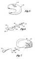
- FIG. 5is an enlarged perspective of a torn medial meniscus of the type that can be repaired in accordance with the system of the present invention.
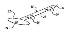
- FIG. 6is an enlarged top perspective of an improved fastener or stent in accordance with the present invention.
- FIG. 7is an enlarged diagrammatic perspective of a tear of the type shown in FIG. 5 and fastener of the type shown in FIG. 6 illustrating how the fastener is used for repairing the tear.
- FIG. 8is a top perspective of an alternative fastener usable in a system in accordance with the present invention.
- FIG. 9is a top perspective of another fastener usable in a system in accordance with the present invention.
- FIG. 10is a top perspective of another fastener usable in a system in accordance with the present invention.
- FIG. 11is a top perspective of another fastener usable in a system in accordance with the present invention.
- FIG. 12is a top perspective of another fastener usable in a system in accordance with the present invention.
- FIG. 13is a top perspective of another fastener usable in a system in accordance with the present invention.
- the present inventionis used to repair soft tissue, particularly a meniscal tear.
- FIGS. 1-4illustrate a known prior art system promoted by Bionx Implants, Inc., which is the subject of Schreiber U.S. Pat. No. 4,873,976.
- the Schreiber-Bionx systemuses a special applicator tube 10 positioned for insertion of a fastener 12 ( FIG. 2 ) with its barbed shank extending across a tear 14 in the medial meniscus 16 .
- FIG. 3shows a “repaired” tear in which three such fasteners 12 have been inserted.
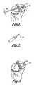
- FIGS. 4A-4Eillustrate one procedure recommended by Bionx Implants, Inc., for using Schreiber-type fasteners to repair a meniscal tear.
- the tear or rupture 14 of the meniscus 16is freshened and reduced with an arthroscopic rasp as represented in FIG. 4A .
- an “arrow” 15is placed at the most posterior area first, whereas for medial lesions the arrow is placed at the middle of the tear first.
- a cannula 10is inserted with a blunt obturator inside. The obturator is removed while the meniscus is maintained in its reduced state with the cannula. The exact position of the cannula is to be maintained during the entire procedure by pressing it against the meniscus.
- a channelis made through the meniscus into the joint capsule with a special needle 17 .
- irrigation fluidwill flood the area during the initial stages of the procedure, and it is recommended that the fluid be turned off prior to retracting the needle.
- the fasteneris then pushed to the surface of the meniscus with the obturator.
- a piston mounted on a reciprocating instrumentrun by air pressure or electricity uses the obturator as a manual driver to hammer the implant fastener 12 into the meniscus.
- the T-shaped headis pushed into the groove formed during driving on the surface of the meniscus.
- the cannulais shifted to a new position and the procedure repeated again until the desired number of implant fasteners has closed the meniscal tear.
- Fastenersare recommended to be separated by five to ten millimeters, and may be provided in different lengths, depending on the location of the tear.
- the Schreiber-Bionx systemrelies on the barbs of the fastener to prevent it from becoming dislodged and possibly allowing the tear to reopen or at least not be held together sufficiently to heal together as completely or reliably as otherwise might occur.
- FIG. 5illustrates diagrammatically a representative tear, namely, a tear 14 in the medial meniscus 16 .
- a fastener or “stent” 18 of the type shown in FIG. 6is used.
- Stent 18includes a shank 20 which preferably is of cylindrical cross section but which could be of square, rectangular, octagonal, or oval cross section.
- a distal tip end 21 of the shankis tapered to a point.
- a proximate head end 22 of the shankis enlarged.
- a long blind bore 24extends through the head end 22 and shank 18 to a location close to the tip end 21 .
- Transverse holes 26communicate between the blind bore and the exterior of the shank.
- the tear site 14can be prepared conventionally, i.e., by freshening the torn area with an arthroscopic rasp.
- Stent 18is inserted adjacent to the tear site 14 in a generally horizontal direction, transversely of the vertical tear. This can be done arthroscopically through a cannula.
- a pusher rodmay be used to advance the stent, and can have a releasable grasper to assist in positioning the stent. While barbs may be provided along the shank, it is preferred that the shank be smooth and of uniform cross section for minimal trauma to undamaged tissue through which the shank is inserted. Insertion of the shank is limited by the enlarged head 22 .
- the interior surface (hidden from the viewer in FIGS. 6 and 7 ) of the enlarged head 22is flat for firmly engaging in the meniscal tissue.
- Such headalso can be oval with its major axis extending horizontally for maximum surface area of engagement with the meniscal tissue, but without projecting unduly in a vertical direction which could be abrasive to surrounding tissue or bone.
- the exterior surface (the surface closest to the viewer in FIGS. 6 and 7 )can be slightly inclined and/or curved to at least approximate the surface of the meniscal tissue in the area of the embedded head.
- a medical adhesiveis injected through the open end of the bore 24 , such as by a syringe 25 , to pass through such bore and outward through the openings 26 .
- the adhesivesets quickly and adheres to the meniscal tissue for maintaining the stent in a position closing the tear 14 .
- the stentwill be sufficiently dimensionally stable that it will not bend or deflect substantially as it is inserted, nor elongate or stretch which could alter the abutting relationship of opposite sides of the tear and interfere with healing.
- the stentcan be somewhat flexible in a transverse direction so as not to interfere with normal functioning of the repaired tissue. This may require that the stent, particularly the shank, be narrow. If too stiff or too large, abrasion could occur in the area of the shank of the stent. For the same reason, it is believed that the stent should not have exposed sharp edges.
- the viscosity of the adhesive and size and location of the transverse holesare somewhat related. It is envisioned that such holes should be provided at least in opposite sides of the shank 20 of the stent 18 for adhering to surrounding tissue. Nevertheless, the adhesive should not be so fluid nor the transverse holes 26 so large that an excess quantity of the adhesive is injected, which could interfere with the healing process (e.g., the revascularization and/or neovascularization of the repair site). It may be preferable to position the holes toward the opposite ends of the stent shank, or at least away from the area expected to be adjacent to the tear, to prevent the adhesive from flowing between the opposite faces of the tear. For example, in the embodiment of FIG.
- the holes 26are located at opposite end portions of the shank 20 but not in the central portion which is positioned to bridge across the tear.
- the exit holescould be located in sockets or depressions 27 formed in the exterior of the stent shank ( FIG. 8 ) if required to reliably and uniformly disperse the adhesive over the surface of the stent.
- Another possibilityis to use barbs 28 on the stent shank 20 with openings at their tips or free ends ( FIG. 9 ) and supply adhesive under sufficient pressure to penetrate surrounding tissue.
- the adhesivecan be of the general type described in U.S. Pat. No. 5,350,798 of Linden et al. or a variant.
- Such an adhesiveis, in general, a polymer gel and, more specifically, a cyanoacrylate polymer. Modified gels are described in U.S. Pat. Nos. 5,714,159 and 5,612,052 of Shalaby.
- the adhesiveflows freely without high adhesive properties relative to the tissue being repaired, but will thereafter set quickly and secure the sides of the tear in the desired abutting relationship.
- the adhesivewill set within about 10 seconds to a condition of high shear strength and substantial rigidity, but not so rigid as to crack in the area of the shank if it flexes slightly during normal use of the joint.
- the adhesivemay inherently have disinfectant characteristics and/or may be coated or impregnated with a compound having disinfectant characteristics.
- the adhesive and/or stentmay serve as a delivery system for drugs and/or agents and/or factors to promote healing and/or growth.
- Both the stent and the adhesivepreferably are bioabsorbable, but over a sufficiently long length of time that full healing of the tear occurs.
- the adhesive-stent combinationshould maintain full strength for approximately eight to twelve weeks and then degrade as the meniscus heals further.
- the adhesivecould be injected by syringe from the exterior of the meniscus into the area of an inserted stent.
- a stentcould have circumferential grooves 29 ( FIG. 10 ) and/or longitudinal grooves 30 ( FIG. 11 ) or a pattern of depressions 31 ( FIG.
- the shank of the stentcould be flat ( FIG. 13 ) with a pattern of grooves 32 in its opposite sides, but preferably the stent still would have a sharpened or pointed leading end for ease of insertion and an enlarged end to limit the degree of insertion of the stent into the tissue.
- the stentcould be held in position for a period sufficient to allow the adhesive to be injected and to set for securing the torn tissue in an abutting relationship for promoting healing.
- the amount of adhesive usedwill be metered for consistency in the adhesive properties and to prevent an insufficient or excessive quantity of adhesive from being used adjacent to the stent.
- the exterior surface of the meniscus at the tear sitecould be further stabilized and protected by a film or patch using the same or a similar adhesive.
Landscapes
- Health & Medical Sciences (AREA)
- Life Sciences & Earth Sciences (AREA)
- Surgery (AREA)
- Molecular Biology (AREA)
- Engineering & Computer Science (AREA)
- Biomedical Technology (AREA)
- Heart & Thoracic Surgery (AREA)
- Medical Informatics (AREA)
- Nuclear Medicine, Radiotherapy & Molecular Imaging (AREA)
- Animal Behavior & Ethology (AREA)
- General Health & Medical Sciences (AREA)
- Public Health (AREA)
- Veterinary Medicine (AREA)
- Prostheses (AREA)
- Apparatus For Radiation Diagnosis (AREA)
- Media Introduction/Drainage Providing Device (AREA)
Abstract
Description
Claims (8)
Priority Applications (1)
| Application Number | Priority Date | Filing Date | Title |
|---|---|---|---|
| US10/114,709US8006700B2 (en) | 2000-02-07 | 2002-04-02 | Soft tissue repair system |
Applications Claiming Priority (3)
| Application Number | Priority Date | Filing Date | Title |
|---|---|---|---|
| US18070200P | 2000-02-07 | 2000-02-07 | |
| PCT/US2001/040061WO2001056533A2 (en) | 2000-02-07 | 2001-02-07 | Soft tissue repair system |
| US10/114,709US8006700B2 (en) | 2000-02-07 | 2002-04-02 | Soft tissue repair system |
Related Parent Applications (1)
| Application Number | Title | Priority Date | Filing Date |
|---|---|---|---|
| PCT/US2001/040061ContinuationWO2001056533A2 (en) | 2000-02-07 | 2001-02-07 | Soft tissue repair system |
Publications (2)
| Publication Number | Publication Date |
|---|---|
| US20020169477A1 US20020169477A1 (en) | 2002-11-14 |
| US8006700B2true US8006700B2 (en) | 2011-08-30 |
Family
ID=22661427
Family Applications (1)
| Application Number | Title | Priority Date | Filing Date |
|---|---|---|---|
| US10/114,709Expired - Fee RelatedUS8006700B2 (en) | 2000-02-07 | 2002-04-02 | Soft tissue repair system |
Country Status (6)
| Country | Link |
|---|---|
| US (1) | US8006700B2 (en) |
| EP (1) | EP1257205B1 (en) |
| AT (1) | ATE490734T1 (en) |
| DE (1) | DE60143603D1 (en) |
| ES (1) | ES2357334T3 (en) |
| WO (1) | WO2001056533A2 (en) |
Cited By (17)
| Publication number | Priority date | Publication date | Assignee | Title |
|---|---|---|---|---|
| US20080288004A1 (en)* | 2007-05-16 | 2008-11-20 | Genesis Biosystems Corporation | Tissue suspension device |
| US9295488B2 (en) | 2012-08-09 | 2016-03-29 | Wilson T. Asfora | Joint fusion |
| US9381019B2 (en) | 2011-02-04 | 2016-07-05 | University Of Utah Research Foundation | System for tissue fixation to bone |
| US9427309B2 (en) | 2012-07-30 | 2016-08-30 | Conextions, Inc. | Soft tissue repair devices, systems, and methods |
| US9451961B2 (en) | 2010-05-19 | 2016-09-27 | University Of Utah Research Foundation | Tissue stabilization system |
| US9629632B2 (en) | 2012-07-30 | 2017-04-25 | Conextions, Inc. | Soft tissue repair devices, systems, and methods |
| US10219804B2 (en) | 2012-07-30 | 2019-03-05 | Conextions, Inc. | Devices, systems, and methods for repairing soft tissue and attaching soft tissue to bone |
| US10299829B2 (en) | 2016-11-07 | 2019-05-28 | Ronald Yamada | Tissue repair system |
| US10390935B2 (en) | 2012-07-30 | 2019-08-27 | Conextions, Inc. | Soft tissue to bone repair devices, systems, and methods |
| US10835241B2 (en) | 2012-07-30 | 2020-11-17 | Conextions, Inc. | Devices, systems, and methods for repairing soft tissue and attaching soft tissue to bone |
| US10973509B2 (en) | 2017-12-20 | 2021-04-13 | Conextions, Inc. | Devices, systems, and methods for repairing soft tissue and attaching soft tissue to bone |
| US11253252B2 (en) | 2012-07-30 | 2022-02-22 | Conextions, Inc. | Devices, systems, and methods for repairing soft tissue and attaching soft tissue to bone |
| US11583384B2 (en) | 2014-03-12 | 2023-02-21 | Conextions, Inc. | Devices, systems, and methods for repairing soft tissue and attaching soft tissue to bone |
| US11696822B2 (en) | 2016-09-28 | 2023-07-11 | Conextions, Inc. | Devices, systems, and methods for repairing soft tissue and attaching soft tissue to bone |
| US11944531B2 (en) | 2012-07-30 | 2024-04-02 | Conextions, Inc. | Devices, systems, and methods for repairing soft tissue and attaching soft tissue to bone |
| US11957334B2 (en) | 2012-07-30 | 2024-04-16 | Conextions, Inc. | Devices, systems, and methods for repairing soft tissue and attaching soft tissue to bone |
| US12102317B2 (en) | 2017-12-20 | 2024-10-01 | Conextions, Inc. | Devices, systems, and methods for repairing soft tissue and attaching soft tissue to bone |
Families Citing this family (90)
| Publication number | Priority date | Publication date | Assignee | Title |
|---|---|---|---|---|
| AU2002348033B2 (en) | 2001-10-23 | 2008-05-29 | Covidien Lp | Surgical fasteners |
| AU2003243219B2 (en) | 2003-05-09 | 2009-10-29 | Covidien Lp | Anastomotic staple with fluid dispensing capillary |
| US7608092B1 (en) | 2004-02-20 | 2009-10-27 | Biomet Sports Medicince, LLC | Method and apparatus for performing meniscus repair |
| CA2562061C (en)* | 2004-04-26 | 2011-06-21 | Bioduct Llc | Stent for avascular meniscal repair and regeneration |
| ES2748926T3 (en) | 2004-10-18 | 2020-03-18 | Covidien Lp | Surgical fixings coated with wound treatment materials |
| US7905904B2 (en) | 2006-02-03 | 2011-03-15 | Biomet Sports Medicine, Llc | Soft tissue repair device and associated methods |
| US8128658B2 (en) | 2004-11-05 | 2012-03-06 | Biomet Sports Medicine, Llc | Method and apparatus for coupling soft tissue to bone |
| US7658751B2 (en) | 2006-09-29 | 2010-02-09 | Biomet Sports Medicine, Llc | Method for implanting soft tissue |
| US7909851B2 (en) | 2006-02-03 | 2011-03-22 | Biomet Sports Medicine, Llc | Soft tissue repair device and associated methods |
| US20060189993A1 (en)* | 2004-11-09 | 2006-08-24 | Arthrotek, Inc. | Soft tissue conduit device |
| US8118836B2 (en) | 2004-11-05 | 2012-02-21 | Biomet Sports Medicine, Llc | Method and apparatus for coupling soft tissue to a bone |
| US8303604B2 (en) | 2004-11-05 | 2012-11-06 | Biomet Sports Medicine, Llc | Soft tissue repair device and method |
| US7857830B2 (en) | 2006-02-03 | 2010-12-28 | Biomet Sports Medicine, Llc | Soft tissue repair and conduit device |
| US8088130B2 (en) | 2006-02-03 | 2012-01-03 | Biomet Sports Medicine, Llc | Method and apparatus for coupling soft tissue to a bone |
| US8137382B2 (en) | 2004-11-05 | 2012-03-20 | Biomet Sports Medicine, Llc | Method and apparatus for coupling anatomical features |
| US7905903B2 (en) | 2006-02-03 | 2011-03-15 | Biomet Sports Medicine, Llc | Method for tissue fixation |
| US8840645B2 (en) | 2004-11-05 | 2014-09-23 | Biomet Sports Medicine, Llc | Method and apparatus for coupling soft tissue to a bone |
| US7749250B2 (en) | 2006-02-03 | 2010-07-06 | Biomet Sports Medicine, Llc | Soft tissue repair assembly and associated method |
| US9801708B2 (en) | 2004-11-05 | 2017-10-31 | Biomet Sports Medicine, Llc | Method and apparatus for coupling soft tissue to a bone |
| US8361113B2 (en) | 2006-02-03 | 2013-01-29 | Biomet Sports Medicine, Llc | Method and apparatus for coupling soft tissue to a bone |
| US8298262B2 (en) | 2006-02-03 | 2012-10-30 | Biomet Sports Medicine, Llc | Method for tissue fixation |
| US9017381B2 (en) | 2007-04-10 | 2015-04-28 | Biomet Sports Medicine, Llc | Adjustable knotless loops |
| US8998949B2 (en)* | 2004-11-09 | 2015-04-07 | Biomet Sports Medicine, Llc | Soft tissue conduit device |
| US7594922B1 (en) | 2005-04-07 | 2009-09-29 | Medicine Lodge, Inc | System and method for meniscal repair through a meniscal capsular tunnel |
| US20060280768A1 (en)* | 2005-06-13 | 2006-12-14 | Julia Hwang | Meniscal repair device and method |
| US20070156174A1 (en)* | 2006-01-03 | 2007-07-05 | Arthrotek, Inc. | Method and apparatus for repairing a meniscus |
| US11311287B2 (en) | 2006-02-03 | 2022-04-26 | Biomet Sports Medicine, Llc | Method for tissue fixation |
| US10517587B2 (en) | 2006-02-03 | 2019-12-31 | Biomet Sports Medicine, Llc | Method and apparatus for forming a self-locking adjustable loop |
| US8652172B2 (en) | 2006-02-03 | 2014-02-18 | Biomet Sports Medicine, Llc | Flexible anchors for tissue fixation |
| US8968364B2 (en) | 2006-02-03 | 2015-03-03 | Biomet Sports Medicine, Llc | Method and apparatus for fixation of an ACL graft |
| US8771352B2 (en) | 2011-05-17 | 2014-07-08 | Biomet Sports Medicine, Llc | Method and apparatus for tibial fixation of an ACL graft |
| US8652171B2 (en) | 2006-02-03 | 2014-02-18 | Biomet Sports Medicine, Llc | Method and apparatus for soft tissue fixation |
| US8574235B2 (en) | 2006-02-03 | 2013-11-05 | Biomet Sports Medicine, Llc | Method for trochanteric reattachment |
| US9271713B2 (en) | 2006-02-03 | 2016-03-01 | Biomet Sports Medicine, Llc | Method and apparatus for tensioning a suture |
| US8562645B2 (en) | 2006-09-29 | 2013-10-22 | Biomet Sports Medicine, Llc | Method and apparatus for forming a self-locking adjustable loop |
| US9538998B2 (en) | 2006-02-03 | 2017-01-10 | Biomet Sports Medicine, Llc | Method and apparatus for fracture fixation |
| US8506597B2 (en) | 2011-10-25 | 2013-08-13 | Biomet Sports Medicine, Llc | Method and apparatus for interosseous membrane reconstruction |
| US8251998B2 (en) | 2006-08-16 | 2012-08-28 | Biomet Sports Medicine, Llc | Chondral defect repair |
| US8562647B2 (en) | 2006-09-29 | 2013-10-22 | Biomet Sports Medicine, Llc | Method and apparatus for securing soft tissue to bone |
| US8597327B2 (en) | 2006-02-03 | 2013-12-03 | Biomet Manufacturing, Llc | Method and apparatus for sternal closure |
| US11259792B2 (en) | 2006-02-03 | 2022-03-01 | Biomet Sports Medicine, Llc | Method and apparatus for coupling anatomical features |
| US7959650B2 (en) | 2006-09-29 | 2011-06-14 | Biomet Sports Medicine, Llc | Adjustable knotless loops |
| US9468433B2 (en) | 2006-02-03 | 2016-10-18 | Biomet Sports Medicine, Llc | Method and apparatus for forming a self-locking adjustable loop |
| US8801783B2 (en) | 2006-09-29 | 2014-08-12 | Biomet Sports Medicine, Llc | Prosthetic ligament system for knee joint |
| US9149267B2 (en) | 2006-02-03 | 2015-10-06 | Biomet Sports Medicine, Llc | Method and apparatus for coupling soft tissue to a bone |
| US9078644B2 (en) | 2006-09-29 | 2015-07-14 | Biomet Sports Medicine, Llc | Fracture fixation device |
| US20080033487A1 (en)* | 2006-08-07 | 2008-02-07 | Bioduct, Llc | Medical device for repair of tissue and method for implantation and fixation |
| US8403943B2 (en)* | 2006-08-07 | 2013-03-26 | Howmedica Osteonics Corp. | Insertion system for implanting a medical device and surgical methods |
| US8672969B2 (en) | 2006-09-29 | 2014-03-18 | Biomet Sports Medicine, Llc | Fracture fixation device |
| US9918826B2 (en) | 2006-09-29 | 2018-03-20 | Biomet Sports Medicine, Llc | Scaffold for spring ligament repair |
| US11259794B2 (en) | 2006-09-29 | 2022-03-01 | Biomet Sports Medicine, Llc | Method for implanting soft tissue |
| US8500818B2 (en) | 2006-09-29 | 2013-08-06 | Biomet Manufacturing, Llc | Knee prosthesis assembly with ligament link |
| US10441273B2 (en) | 2007-07-03 | 2019-10-15 | Ceterix Orthopaedics, Inc. | Pre-tied surgical knots for use with suture passers |
| US9861354B2 (en) | 2011-05-06 | 2018-01-09 | Ceterix Orthopaedics, Inc. | Meniscus repair |
| US8465505B2 (en) | 2011-05-06 | 2013-06-18 | Ceterix Orthopaedics, Inc. | Suture passer devices and methods |
| US8172871B2 (en)* | 2007-08-31 | 2012-05-08 | Ken Christopher G M | Closure medical device |
| US8052719B2 (en) | 2008-04-01 | 2011-11-08 | Lonnie Paulos | Suture anchoring assemblies and methods of use |
| US9445804B2 (en) | 2008-04-01 | 2016-09-20 | The Lonnie And Shannon Paulos Trust (As Amended And Restated) | Suture anchoring assemblies and methods of use |
| US12245759B2 (en) | 2008-08-22 | 2025-03-11 | Biomet Sports Medicine, Llc | Method and apparatus for coupling soft tissue to bone |
| US12419632B2 (en) | 2008-08-22 | 2025-09-23 | Biomet Sports Medicine, Llc | Method and apparatus for coupling anatomical features |
| US8343227B2 (en) | 2009-05-28 | 2013-01-01 | Biomet Manufacturing Corp. | Knee prosthesis assembly with ligament link |
| US12096928B2 (en) | 2009-05-29 | 2024-09-24 | Biomet Sports Medicine, Llc | Method and apparatus for coupling soft tissue to a bone |
| US9848868B2 (en) | 2011-01-10 | 2017-12-26 | Ceterix Orthopaedics, Inc. | Suture methods for forming locking loops stitches |
| US11744575B2 (en) | 2009-11-09 | 2023-09-05 | Ceterix Orthopaedics, Inc. | Suture passer devices and methods |
| US9011454B2 (en)* | 2009-11-09 | 2015-04-21 | Ceterix Orthopaedics, Inc. | Suture passer with radiused upper jaw |
| US9913638B2 (en) | 2011-01-10 | 2018-03-13 | Ceterix Orthopaedics, Inc. | Transosteal anchoring methods for tissue repair |
| US12329373B2 (en) | 2011-05-02 | 2025-06-17 | Biomet Sports Medicine, Llc | Method and apparatus for soft tissue fixation |
| US9492162B2 (en) | 2013-12-16 | 2016-11-15 | Ceterix Orthopaedics, Inc. | Automatically reloading suture passer devices and methods |
| US10524778B2 (en) | 2011-09-28 | 2020-01-07 | Ceterix Orthopaedics | Suture passers adapted for use in constrained regions |
| US9357991B2 (en) | 2011-11-03 | 2016-06-07 | Biomet Sports Medicine, Llc | Method and apparatus for stitching tendons |
| US9381013B2 (en) | 2011-11-10 | 2016-07-05 | Biomet Sports Medicine, Llc | Method for coupling soft tissue to a bone |
| US9370350B2 (en) | 2011-11-10 | 2016-06-21 | Biomet Sports Medicine, Llc | Apparatus for coupling soft tissue to a bone |
| US9314241B2 (en) | 2011-11-10 | 2016-04-19 | Biomet Sports Medicine, Llc | Apparatus for coupling soft tissue to a bone |
| US9259217B2 (en) | 2012-01-03 | 2016-02-16 | Biomet Manufacturing, Llc | Suture Button |
| US9757119B2 (en) | 2013-03-08 | 2017-09-12 | Biomet Sports Medicine, Llc | Visual aid for identifying suture limbs arthroscopically |
| US9918827B2 (en) | 2013-03-14 | 2018-03-20 | Biomet Sports Medicine, Llc | Scaffold for spring ligament repair |
| US9247935B2 (en) | 2013-09-23 | 2016-02-02 | Ceterix Orthopaedics, Inc. | Arthroscopic knot pusher and suture cutter |
| US10136886B2 (en) | 2013-12-20 | 2018-11-27 | Biomet Sports Medicine, Llc | Knotless soft tissue devices and techniques |
| CN204951031U (en) | 2014-04-08 | 2016-01-13 | 赛特里克斯整形公司 | Ware device is worn to draw by suture |
| US9615822B2 (en) | 2014-05-30 | 2017-04-11 | Biomet Sports Medicine, Llc | Insertion tools and method for soft anchor |
| US9700291B2 (en) | 2014-06-03 | 2017-07-11 | Biomet Sports Medicine, Llc | Capsule retractor |
| AU2015302209B2 (en)* | 2014-08-11 | 2020-05-07 | Smith & Nephew, Inc. | A medical device for repairing soft tissue and method of using same |
| US10039543B2 (en) | 2014-08-22 | 2018-08-07 | Biomet Sports Medicine, Llc | Non-sliding soft anchor |
| US9955980B2 (en) | 2015-02-24 | 2018-05-01 | Biomet Sports Medicine, Llc | Anatomic soft tissue repair |
| US9974534B2 (en) | 2015-03-31 | 2018-05-22 | Biomet Sports Medicine, Llc | Suture anchor with soft anchor of electrospun fibers |
| US10226245B2 (en) | 2015-07-21 | 2019-03-12 | Ceterix Orthopaedics, Inc. | Automatically reloading suture passer devices that prevent entanglement |
| US10405853B2 (en) | 2015-10-02 | 2019-09-10 | Ceterix Orthpaedics, Inc. | Knot tying accessory |
| US10575841B1 (en) | 2016-11-29 | 2020-03-03 | The Lonnie and Shannon Paulos Trust | Soft locking suture anchor assembly and methods of use |
| US20200113558A1 (en)* | 2017-03-16 | 2020-04-16 | Cannuflow, Inc. | System And Method For Fixing Sheet-Like Materials To A Target Tissue |
| US12226094B2 (en)* | 2019-07-22 | 2025-02-18 | Suturegard Medical Inc. | Hemi-bridge and methods of manufacturing and using same |
Citations (56)
| Publication number | Priority date | Publication date | Assignee | Title |
|---|---|---|---|---|
| US3176316A (en) | 1963-01-07 | 1965-04-06 | Bruce R Bodell | Plastic prosthetic tendon |
| US3716058A (en) | 1970-07-17 | 1973-02-13 | Atlanta Res Inst | Barbed suture |
| US3833002A (en) | 1973-09-10 | 1974-09-03 | J Palma | Apparatus for aiding severed nerves to join |
| US3842441A (en) | 1972-10-12 | 1974-10-22 | A Kaiser | A temporary implant and method for tendon surgery |
| US3960152A (en) | 1974-01-21 | 1976-06-01 | American Cyanamid Company | Surgical sutures of unsymmetrically substituted 1,4-dioxane-2,5-diones |
| US3987497A (en) | 1974-03-29 | 1976-10-26 | Ceskoslovenska Akademie Ved | Tendon prosthesis |
| US3991766A (en) | 1973-05-31 | 1976-11-16 | American Cyanamid Company | Controlled release of medicaments using polymers from glycolic acid |
| SU566575A1 (en) | 1976-01-07 | 1977-07-30 | Bogachenko Nikolaj | Tendon suture clipper |
| US4501029A (en) | 1982-04-22 | 1985-02-26 | Mcminn Derek J W | Tendon repair |
| US4512038A (en) | 1979-04-27 | 1985-04-23 | University Of Medicine And Dentistry Of New Jersey | Bio-absorbable composite tissue scaffold |
| US4534349A (en) | 1983-02-02 | 1985-08-13 | Minnesota Mining And Manufacturing Company | Absorbable sutureless nerve repair device |
| US4653489A (en) | 1984-04-02 | 1987-03-31 | Tronzo Raymond G | Fenestrated hip screw and method of augmented fixation |
| US4653487A (en)* | 1986-01-29 | 1987-03-31 | Maale Gerhard E | Intramedullary rod assembly for cement injection system |
| US4662884A (en) | 1984-04-25 | 1987-05-05 | University Of Utah Research Foundation | Prostheses and methods for promoting nerve regeneration |
| US4873976A (en) | 1984-02-28 | 1989-10-17 | Schreiber Saul N | Surgical fasteners and method |
| US4976715A (en)* | 1986-05-20 | 1990-12-11 | Concept, Inc. | Repair tack for bodily tissue |
| US4979956A (en) | 1987-10-30 | 1990-12-25 | Pfizer Hospital Products Group, Inc. | Device and method for tendon and ligament repair |
| US5047030A (en)* | 1987-02-20 | 1991-09-10 | Klaus Draenert | Suction drainage-bone screw |
| US5061281A (en) | 1985-12-17 | 1991-10-29 | Allied-Signal Inc. | Bioresorbable polymers and implantation devices thereof |
| US5061283A (en) | 1987-10-30 | 1991-10-29 | Pfizer Hospital Products Group, Inc. | Method for tendon and ligament repair |
| US5102413A (en)* | 1990-11-14 | 1992-04-07 | Poddar Satish B | Inflatable bone fixation device |
| US5147362A (en) | 1991-04-08 | 1992-09-15 | Marlowe Goble E | Endosteal ligament fixation device |
| EP0520177A1 (en) | 1991-05-24 | 1992-12-30 | Synthes AG, Chur | Resorbable tendon and bone augmentation device |
| US5249899A (en)* | 1992-10-28 | 1993-10-05 | Wilson Robert L | Head bolt and driver therefore |
| US5254132A (en) | 1992-09-01 | 1993-10-19 | Medlogic, Inc. | Methods for treating suturable wounds by use of sutures and cyanoacrylate adhesives |
| US5350798A (en) | 1993-08-17 | 1994-09-27 | The United States Of America As Represented By The Secretary Of The Army | Absorbable tissue adhesives |
| US5354305A (en) | 1991-09-26 | 1994-10-11 | United States Surgical Corporation | Nerve repair device |
| US5425766A (en) | 1987-03-09 | 1995-06-20 | Astra Tech Aktiebolag | Resorbable prosthesis |
| US5458636A (en) | 1994-07-20 | 1995-10-17 | U.S. Biomaterials Corporation | Prosthetic device for repair and replacement of fibrous connective tissue |
| US5584859A (en)* | 1993-10-12 | 1996-12-17 | Brotz; Gregory R. | Suture assembly |
| US5612052A (en) | 1995-04-13 | 1997-03-18 | Poly-Med, Inc. | Hydrogel-forming, self-solvating absorbable polyester copolymers, and methods for use thereof |
| US5653769A (en) | 1994-02-24 | 1997-08-05 | Medlogic Global Corporation | Methods for reducing skin irritation from artificial devices by use of cyanoacrylate adhesives |
| US5666779A (en)* | 1995-06-24 | 1997-09-16 | Hilti Aktiengesellschaft | Method of forming a pressure free expansion anchorage |
| US5723008A (en) | 1995-07-20 | 1998-03-03 | Gordon; Leonard | Splint for repair of tendons or ligaments and method |
| US5743912A (en)* | 1995-08-23 | 1998-04-28 | Biomat | Upper femoral epiphysis osteosynthesis implant |
| US5800407A (en)* | 1995-12-21 | 1998-09-01 | Eldor; Joseph | Multiple hole epidural catheter |
| US5800544A (en) | 1994-12-02 | 1998-09-01 | Omeros Medical Systems, Inc. | Tendon and ligament repair system |
| US5811091A (en) | 1997-01-10 | 1998-09-22 | Medlogic Global Corporation | Cyanoacrylate compostions comprising an antimicrobial agent |
| US5843084A (en)* | 1995-11-17 | 1998-12-01 | Innovasive Devices, Inc. | Surgical fastening system and method for using the same |
| US5900245A (en) | 1996-03-22 | 1999-05-04 | Focal, Inc. | Compliant tissue sealants |
| US5976127A (en)* | 1998-01-14 | 1999-11-02 | Lax; Ronald | Soft tissue fixation devices |
| US5998472A (en) | 1997-10-09 | 1999-12-07 | Medlogic Global Corporation | Mixed cyanoacrylate ester compositions |
| US6001345A (en) | 1997-11-03 | 1999-12-14 | Medlogic Global Corporation | Application of cyanoacrylate/anti-microbial compositions to the peri-wound or peri-mucosal area |
| US6048343A (en) | 1999-06-02 | 2000-04-11 | Mathis; John M. | Bone screw system |
| US6083244A (en) | 1996-09-13 | 2000-07-04 | Tendon Technology, Ltd. | Apparatus and method for tendon or ligament repair |
| US6102947A (en) | 1995-07-20 | 2000-08-15 | Gordon; Leonard | Splint with flexible body for repair of tendons or ligaments and method |
| US6106556A (en) | 1994-12-02 | 2000-08-22 | Omeros Medical Systems, Inc. | Tendon and ligament repair system |
| US6214012B1 (en) | 1998-11-13 | 2001-04-10 | Harrington Arthritis Research Center | Method and apparatus for delivering material to a desired location |
| WO2001028457A1 (en) | 1999-10-18 | 2001-04-26 | Tendon Technology, Ltd. | Apparatus and methods for tendon or ligament repair |
| US6264675B1 (en)* | 2000-02-04 | 2001-07-24 | Gregory R. Brotz | Single suture structure |
| US6270517B1 (en)* | 2000-02-04 | 2001-08-07 | Gregory R. Brotz | Suture assembly and method |
| US6296641B2 (en)* | 1998-04-03 | 2001-10-02 | Bionx Implants Oy | Anatomical fixation implant |
| US6565572B2 (en)* | 2000-04-10 | 2003-05-20 | Sdgi Holdings, Inc. | Fenestrated surgical screw and method |
| US6610079B1 (en)* | 1999-12-14 | 2003-08-26 | Linvatec Corporation | Fixation system and method |
| US6620185B1 (en)* | 2000-06-27 | 2003-09-16 | Smith & Nephew, Inc. | Surgical procedures and instruments |
| US6740100B2 (en)* | 1999-12-23 | 2004-05-25 | Omeros Corporation | Tendon repair using adhesive |
- 2001
- 2001-02-07DEDE60143603Tpatent/DE60143603D1/ennot_activeExpired - Lifetime
- 2001-02-07EPEP01948931Apatent/EP1257205B1/ennot_activeExpired - Lifetime
- 2001-02-07ATAT01948931Tpatent/ATE490734T1/ennot_activeIP Right Cessation
- 2001-02-07WOPCT/US2001/040061patent/WO2001056533A2/enactiveApplication Filing
- 2001-02-07ESES01948931Tpatent/ES2357334T3/ennot_activeExpired - Lifetime
- 2002
- 2002-04-02USUS10/114,709patent/US8006700B2/ennot_activeExpired - Fee Related
Patent Citations (57)
| Publication number | Priority date | Publication date | Assignee | Title |
|---|---|---|---|---|
| US3176316A (en) | 1963-01-07 | 1965-04-06 | Bruce R Bodell | Plastic prosthetic tendon |
| US3716058A (en) | 1970-07-17 | 1973-02-13 | Atlanta Res Inst | Barbed suture |
| US3842441A (en) | 1972-10-12 | 1974-10-22 | A Kaiser | A temporary implant and method for tendon surgery |
| US3991766A (en) | 1973-05-31 | 1976-11-16 | American Cyanamid Company | Controlled release of medicaments using polymers from glycolic acid |
| US3833002A (en) | 1973-09-10 | 1974-09-03 | J Palma | Apparatus for aiding severed nerves to join |
| US3960152A (en) | 1974-01-21 | 1976-06-01 | American Cyanamid Company | Surgical sutures of unsymmetrically substituted 1,4-dioxane-2,5-diones |
| US3987497A (en) | 1974-03-29 | 1976-10-26 | Ceskoslovenska Akademie Ved | Tendon prosthesis |
| SU566575A1 (en) | 1976-01-07 | 1977-07-30 | Bogachenko Nikolaj | Tendon suture clipper |
| US4512038A (en) | 1979-04-27 | 1985-04-23 | University Of Medicine And Dentistry Of New Jersey | Bio-absorbable composite tissue scaffold |
| US4501029A (en) | 1982-04-22 | 1985-02-26 | Mcminn Derek J W | Tendon repair |
| US4534349A (en) | 1983-02-02 | 1985-08-13 | Minnesota Mining And Manufacturing Company | Absorbable sutureless nerve repair device |
| US4873976A (en) | 1984-02-28 | 1989-10-17 | Schreiber Saul N | Surgical fasteners and method |
| US4653489A (en) | 1984-04-02 | 1987-03-31 | Tronzo Raymond G | Fenestrated hip screw and method of augmented fixation |
| US4662884A (en) | 1984-04-25 | 1987-05-05 | University Of Utah Research Foundation | Prostheses and methods for promoting nerve regeneration |
| US5061281A (en) | 1985-12-17 | 1991-10-29 | Allied-Signal Inc. | Bioresorbable polymers and implantation devices thereof |
| US4653487A (en)* | 1986-01-29 | 1987-03-31 | Maale Gerhard E | Intramedullary rod assembly for cement injection system |
| US4976715A (en)* | 1986-05-20 | 1990-12-11 | Concept, Inc. | Repair tack for bodily tissue |
| US5047030A (en)* | 1987-02-20 | 1991-09-10 | Klaus Draenert | Suction drainage-bone screw |
| US5425766A (en) | 1987-03-09 | 1995-06-20 | Astra Tech Aktiebolag | Resorbable prosthesis |
| US4979956A (en) | 1987-10-30 | 1990-12-25 | Pfizer Hospital Products Group, Inc. | Device and method for tendon and ligament repair |
| US5061283A (en) | 1987-10-30 | 1991-10-29 | Pfizer Hospital Products Group, Inc. | Method for tendon and ligament repair |
| US5102413A (en)* | 1990-11-14 | 1992-04-07 | Poddar Satish B | Inflatable bone fixation device |
| US5147362A (en) | 1991-04-08 | 1992-09-15 | Marlowe Goble E | Endosteal ligament fixation device |
| EP0520177A1 (en) | 1991-05-24 | 1992-12-30 | Synthes AG, Chur | Resorbable tendon and bone augmentation device |
| US5354305A (en) | 1991-09-26 | 1994-10-11 | United States Surgical Corporation | Nerve repair device |
| US5254132A (en) | 1992-09-01 | 1993-10-19 | Medlogic, Inc. | Methods for treating suturable wounds by use of sutures and cyanoacrylate adhesives |
| US5249899A (en)* | 1992-10-28 | 1993-10-05 | Wilson Robert L | Head bolt and driver therefore |
| US5350798A (en) | 1993-08-17 | 1994-09-27 | The United States Of America As Represented By The Secretary Of The Army | Absorbable tissue adhesives |
| US5584859A (en)* | 1993-10-12 | 1996-12-17 | Brotz; Gregory R. | Suture assembly |
| US5653769A (en) | 1994-02-24 | 1997-08-05 | Medlogic Global Corporation | Methods for reducing skin irritation from artificial devices by use of cyanoacrylate adhesives |
| US5458636A (en) | 1994-07-20 | 1995-10-17 | U.S. Biomaterials Corporation | Prosthetic device for repair and replacement of fibrous connective tissue |
| US6106556A (en) | 1994-12-02 | 2000-08-22 | Omeros Medical Systems, Inc. | Tendon and ligament repair system |
| US5800544A (en) | 1994-12-02 | 1998-09-01 | Omeros Medical Systems, Inc. | Tendon and ligament repair system |
| US6080192A (en) | 1994-12-02 | 2000-06-27 | Omeros Medical Systems, Inc. | Tendon and ligament repair system |
| US5612052A (en) | 1995-04-13 | 1997-03-18 | Poly-Med, Inc. | Hydrogel-forming, self-solvating absorbable polyester copolymers, and methods for use thereof |
| US5666779A (en)* | 1995-06-24 | 1997-09-16 | Hilti Aktiengesellschaft | Method of forming a pressure free expansion anchorage |
| US5723008A (en) | 1995-07-20 | 1998-03-03 | Gordon; Leonard | Splint for repair of tendons or ligaments and method |
| US6102947A (en) | 1995-07-20 | 2000-08-15 | Gordon; Leonard | Splint with flexible body for repair of tendons or ligaments and method |
| US5743912A (en)* | 1995-08-23 | 1998-04-28 | Biomat | Upper femoral epiphysis osteosynthesis implant |
| US5843084A (en)* | 1995-11-17 | 1998-12-01 | Innovasive Devices, Inc. | Surgical fastening system and method for using the same |
| US5800407A (en)* | 1995-12-21 | 1998-09-01 | Eldor; Joseph | Multiple hole epidural catheter |
| US5900245A (en) | 1996-03-22 | 1999-05-04 | Focal, Inc. | Compliant tissue sealants |
| US6083244A (en) | 1996-09-13 | 2000-07-04 | Tendon Technology, Ltd. | Apparatus and method for tendon or ligament repair |
| US5811091A (en) | 1997-01-10 | 1998-09-22 | Medlogic Global Corporation | Cyanoacrylate compostions comprising an antimicrobial agent |
| US5998472A (en) | 1997-10-09 | 1999-12-07 | Medlogic Global Corporation | Mixed cyanoacrylate ester compositions |
| US6001345A (en) | 1997-11-03 | 1999-12-14 | Medlogic Global Corporation | Application of cyanoacrylate/anti-microbial compositions to the peri-wound or peri-mucosal area |
| US5976127A (en)* | 1998-01-14 | 1999-11-02 | Lax; Ronald | Soft tissue fixation devices |
| US6296641B2 (en)* | 1998-04-03 | 2001-10-02 | Bionx Implants Oy | Anatomical fixation implant |
| US6214012B1 (en) | 1998-11-13 | 2001-04-10 | Harrington Arthritis Research Center | Method and apparatus for delivering material to a desired location |
| US6048343A (en) | 1999-06-02 | 2000-04-11 | Mathis; John M. | Bone screw system |
| WO2001028457A1 (en) | 1999-10-18 | 2001-04-26 | Tendon Technology, Ltd. | Apparatus and methods for tendon or ligament repair |
| US6610079B1 (en)* | 1999-12-14 | 2003-08-26 | Linvatec Corporation | Fixation system and method |
| US6740100B2 (en)* | 1999-12-23 | 2004-05-25 | Omeros Corporation | Tendon repair using adhesive |
| US6264675B1 (en)* | 2000-02-04 | 2001-07-24 | Gregory R. Brotz | Single suture structure |
| US6270517B1 (en)* | 2000-02-04 | 2001-08-07 | Gregory R. Brotz | Suture assembly and method |
| US6565572B2 (en)* | 2000-04-10 | 2003-05-20 | Sdgi Holdings, Inc. | Fenestrated surgical screw and method |
| US6620185B1 (en)* | 2000-06-27 | 2003-09-16 | Smith & Nephew, Inc. | Surgical procedures and instruments |
Non-Patent Citations (26)
Cited By (26)
| Publication number | Priority date | Publication date | Assignee | Title |
|---|---|---|---|---|
| US20080288004A1 (en)* | 2007-05-16 | 2008-11-20 | Genesis Biosystems Corporation | Tissue suspension device |
| US9451961B2 (en) | 2010-05-19 | 2016-09-27 | University Of Utah Research Foundation | Tissue stabilization system |
| US9381019B2 (en) | 2011-02-04 | 2016-07-05 | University Of Utah Research Foundation | System for tissue fixation to bone |
| US11253252B2 (en) | 2012-07-30 | 2022-02-22 | Conextions, Inc. | Devices, systems, and methods for repairing soft tissue and attaching soft tissue to bone |
| US10660643B2 (en) | 2012-07-30 | 2020-05-26 | Conextions, Inc. | Soft tissue repair devices, systems, and methods |
| US11980360B2 (en) | 2012-07-30 | 2024-05-14 | Conextions, Inc. | Devices, systems, and methods for repairing soft tissue and attaching soft tissue to bone |
| US11957334B2 (en) | 2012-07-30 | 2024-04-16 | Conextions, Inc. | Devices, systems, and methods for repairing soft tissue and attaching soft tissue to bone |
| US9629632B2 (en) | 2012-07-30 | 2017-04-25 | Conextions, Inc. | Soft tissue repair devices, systems, and methods |
| US9655625B2 (en) | 2012-07-30 | 2017-05-23 | Conextions, Inc. | Soft tissue repair devices, systems, and methods |
| US10219804B2 (en) | 2012-07-30 | 2019-03-05 | Conextions, Inc. | Devices, systems, and methods for repairing soft tissue and attaching soft tissue to bone |
| US11944531B2 (en) | 2012-07-30 | 2024-04-02 | Conextions, Inc. | Devices, systems, and methods for repairing soft tissue and attaching soft tissue to bone |
| US11446024B2 (en) | 2012-07-30 | 2022-09-20 | Conextions, Inc. | Devices, systems, and methods for repairing soft tissue and attaching soft tissue to bone |
| US10390935B2 (en) | 2012-07-30 | 2019-08-27 | Conextions, Inc. | Soft tissue to bone repair devices, systems, and methods |
| US10660642B2 (en) | 2012-07-30 | 2020-05-26 | Conextions, Inc. | Soft tissue repair devices, systems, and methods |
| US9427309B2 (en) | 2012-07-30 | 2016-08-30 | Conextions, Inc. | Soft tissue repair devices, systems, and methods |
| US10835241B2 (en) | 2012-07-30 | 2020-11-17 | Conextions, Inc. | Devices, systems, and methods for repairing soft tissue and attaching soft tissue to bone |
| US10987144B2 (en) | 2012-08-09 | 2021-04-27 | Asfora Ip, Llc | Screw for joint fusion |
| US9295488B2 (en) | 2012-08-09 | 2016-03-29 | Wilson T. Asfora | Joint fusion |
| US10251688B2 (en)* | 2012-08-09 | 2019-04-09 | Asfora Ip, Llc | Screw for joint fusion |
| US9566100B2 (en) | 2012-08-09 | 2017-02-14 | Asfora Ip, Llc | Screw for joint fusion |
| US9526548B2 (en) | 2012-08-09 | 2016-12-27 | Asfora Ip, Llc | System for joint fusion |
| US11583384B2 (en) | 2014-03-12 | 2023-02-21 | Conextions, Inc. | Devices, systems, and methods for repairing soft tissue and attaching soft tissue to bone |
| US11696822B2 (en) | 2016-09-28 | 2023-07-11 | Conextions, Inc. | Devices, systems, and methods for repairing soft tissue and attaching soft tissue to bone |
| US10299829B2 (en) | 2016-11-07 | 2019-05-28 | Ronald Yamada | Tissue repair system |
| US10973509B2 (en) | 2017-12-20 | 2021-04-13 | Conextions, Inc. | Devices, systems, and methods for repairing soft tissue and attaching soft tissue to bone |
| US12102317B2 (en) | 2017-12-20 | 2024-10-01 | Conextions, Inc. | Devices, systems, and methods for repairing soft tissue and attaching soft tissue to bone |
Also Published As
| Publication number | Publication date |
|---|---|
| EP1257205B1 (en) | 2010-12-08 |
| HK1050468A1 (en) | 2003-06-27 |
| ATE490734T1 (en) | 2010-12-15 |
| WO2001056533A2 (en) | 2001-08-09 |
| WO2001056533A3 (en) | 2002-03-21 |
| EP1257205A4 (en) | 2004-12-22 |
| DE60143603D1 (en) | 2011-01-20 |
| US20020169477A1 (en) | 2002-11-14 |
| EP1257205A2 (en) | 2002-11-20 |
| ES2357334T3 (en) | 2011-04-25 |
Similar Documents
| Publication | Publication Date | Title |
|---|---|---|
| US8006700B2 (en) | Soft tissue repair system | |
| US6423073B2 (en) | Instrument for inserting graft fixation device | |
| US6447517B1 (en) | Instrument for inserting graft fixation device | |
| US8449561B2 (en) | Graft fixation device combination | |
| US6364884B1 (en) | Method of securing a graft using a graft fixation device | |
| US6436110B2 (en) | Method of securing a graft using a graft fixation device | |
| US6402766B2 (en) | Graft fixation device combination | |
| US7214232B2 (en) | Graft fixation device | |
| JP4463501B2 (en) | Suture anchor | |
| US20060282085A1 (en) | Soft tissue conduit device | |
| JP2009515596A (en) | Orthopedic delivery device and instrument to minimize invasion | |
| EP1138263B1 (en) | Graft fixation system | |
| HK1050468B (en) | Soft tissue repair system | |
| AU781967B2 (en) | Method of securing a graft using a graft fixation device | |
| CA2342037C (en) | Graft fixation device combination | |
| CA2342041C (en) | Instrument for inserting graft fixation device | |
| AU1544102A (en) | Method of securing a graft using a graft fixation device |
Legal Events
| Date | Code | Title | Description |
|---|---|---|---|
| AS | Assignment | Owner name:OMEROS CORPORATION, WASHINGTON Free format text:CHANGE OF NAME;ASSIGNOR:OMEROS MEDICAL SYSTEMS, INC.;REEL/FRAME:012916/0530 Effective date:20020401 | |
| ZAAA | Notice of allowance and fees due | Free format text:ORIGINAL CODE: NOA | |
| ZAAB | Notice of allowance mailed | Free format text:ORIGINAL CODE: MN/=. | |
| STCF | Information on status: patent grant | Free format text:PATENTED CASE | |
| CC | Certificate of correction | ||
| FPAY | Fee payment | Year of fee payment:4 | |
| AS | Assignment | Owner name:CRG SERVICING LLC, AS ADMINISTRATIVE AGENT, TEXAS Free format text:SECURITY INTEREST;ASSIGNOR:OMEROS CORPORATION;REEL/FRAME:040575/0110 Effective date:20161103 | |
| AS | Assignment | Owner name:OMEROS CORPORATION, WASHINGTON Free format text:RELEASE BY SECURED PARTY;ASSIGNOR:CRG SERVICING LLC;REEL/FRAME:047573/0577 Effective date:20181115 | |
| MAFP | Maintenance fee payment | Free format text:PAYMENT OF MAINTENANCE FEE, 8TH YR, SMALL ENTITY (ORIGINAL EVENT CODE: M2552); ENTITY STATUS OF PATENT OWNER: SMALL ENTITY Year of fee payment:8 | |
| FEPP | Fee payment procedure | Free format text:MAINTENANCE FEE REMINDER MAILED (ORIGINAL EVENT CODE: REM.); ENTITY STATUS OF PATENT OWNER: SMALL ENTITY | |
| LAPS | Lapse for failure to pay maintenance fees | Free format text:PATENT EXPIRED FOR FAILURE TO PAY MAINTENANCE FEES (ORIGINAL EVENT CODE: EXP.); ENTITY STATUS OF PATENT OWNER: SMALL ENTITY | |
| STCH | Information on status: patent discontinuation | Free format text:PATENT EXPIRED DUE TO NONPAYMENT OF MAINTENANCE FEES UNDER 37 CFR 1.362 | |
| FP | Lapsed due to failure to pay maintenance fee | Effective date:20230830 |