US7976578B2 - Buffer for a human joint and method of arthroscopically inserting - Google Patents
Buffer for a human joint and method of arthroscopically insertingDownload PDFInfo
- Publication number
- US7976578B2 US7976578B2US12/133,211US13321108AUS7976578B2US 7976578 B2US7976578 B2US 7976578B2US 13321108 AUS13321108 AUS 13321108AUS 7976578 B2US7976578 B2US 7976578B2
- Authority
- US
- United States
- Prior art keywords
- buffer
- ring
- sack
- femur
- tibia
- Prior art date
- Legal status (The legal status is an assumption and is not a legal conclusion. Google has not performed a legal analysis and makes no representation as to the accuracy of the status listed.)
- Active, expires
Links
Images


Classifications
- A—HUMAN NECESSITIES
- A61—MEDICAL OR VETERINARY SCIENCE; HYGIENE
- A61F—FILTERS IMPLANTABLE INTO BLOOD VESSELS; PROSTHESES; DEVICES PROVIDING PATENCY TO, OR PREVENTING COLLAPSING OF, TUBULAR STRUCTURES OF THE BODY, e.g. STENTS; ORTHOPAEDIC, NURSING OR CONTRACEPTIVE DEVICES; FOMENTATION; TREATMENT OR PROTECTION OF EYES OR EARS; BANDAGES, DRESSINGS OR ABSORBENT PADS; FIRST-AID KITS
- A61F2/00—Filters implantable into blood vessels; Prostheses, i.e. artificial substitutes or replacements for parts of the body; Appliances for connecting them with the body; Devices providing patency to, or preventing collapsing of, tubular structures of the body, e.g. stents
- A61F2/02—Prostheses implantable into the body
- A61F2/30—Joints
- A61F2/38—Joints for elbows or knees
- A61F2/3872—Meniscus for implantation between the natural bone surfaces
- A—HUMAN NECESSITIES
- A61—MEDICAL OR VETERINARY SCIENCE; HYGIENE
- A61F—FILTERS IMPLANTABLE INTO BLOOD VESSELS; PROSTHESES; DEVICES PROVIDING PATENCY TO, OR PREVENTING COLLAPSING OF, TUBULAR STRUCTURES OF THE BODY, e.g. STENTS; ORTHOPAEDIC, NURSING OR CONTRACEPTIVE DEVICES; FOMENTATION; TREATMENT OR PROTECTION OF EYES OR EARS; BANDAGES, DRESSINGS OR ABSORBENT PADS; FIRST-AID KITS
- A61F2/00—Filters implantable into blood vessels; Prostheses, i.e. artificial substitutes or replacements for parts of the body; Appliances for connecting them with the body; Devices providing patency to, or preventing collapsing of, tubular structures of the body, e.g. stents
- A61F2/02—Prostheses implantable into the body
- A61F2/30—Joints
- A61F2/30756—Cartilage endoprostheses
- A—HUMAN NECESSITIES
- A61—MEDICAL OR VETERINARY SCIENCE; HYGIENE
- A61F—FILTERS IMPLANTABLE INTO BLOOD VESSELS; PROSTHESES; DEVICES PROVIDING PATENCY TO, OR PREVENTING COLLAPSING OF, TUBULAR STRUCTURES OF THE BODY, e.g. STENTS; ORTHOPAEDIC, NURSING OR CONTRACEPTIVE DEVICES; FOMENTATION; TREATMENT OR PROTECTION OF EYES OR EARS; BANDAGES, DRESSINGS OR ABSORBENT PADS; FIRST-AID KITS
- A61F2/00—Filters implantable into blood vessels; Prostheses, i.e. artificial substitutes or replacements for parts of the body; Appliances for connecting them with the body; Devices providing patency to, or preventing collapsing of, tubular structures of the body, e.g. stents
- A61F2/02—Prostheses implantable into the body
- A61F2/30—Joints
- A61F2/46—Special tools for implanting artificial joints
- A61F2/4603—Special tools for implanting artificial joints for insertion or extraction of endoprosthetic joints or of accessories thereof
- A61F2/461—Special tools for implanting artificial joints for insertion or extraction of endoprosthetic joints or of accessories thereof of knees
- A—HUMAN NECESSITIES
- A61—MEDICAL OR VETERINARY SCIENCE; HYGIENE
- A61F—FILTERS IMPLANTABLE INTO BLOOD VESSELS; PROSTHESES; DEVICES PROVIDING PATENCY TO, OR PREVENTING COLLAPSING OF, TUBULAR STRUCTURES OF THE BODY, e.g. STENTS; ORTHOPAEDIC, NURSING OR CONTRACEPTIVE DEVICES; FOMENTATION; TREATMENT OR PROTECTION OF EYES OR EARS; BANDAGES, DRESSINGS OR ABSORBENT PADS; FIRST-AID KITS
- A61F2/00—Filters implantable into blood vessels; Prostheses, i.e. artificial substitutes or replacements for parts of the body; Appliances for connecting them with the body; Devices providing patency to, or preventing collapsing of, tubular structures of the body, e.g. stents
- A61F2/02—Prostheses implantable into the body
- A61F2/30—Joints
- A61F2/46—Special tools for implanting artificial joints
- A61F2/4603—Special tools for implanting artificial joints for insertion or extraction of endoprosthetic joints or of accessories thereof
- A61F2/4618—Special tools for implanting artificial joints for insertion or extraction of endoprosthetic joints or of accessories thereof of cartilage
- A—HUMAN NECESSITIES
- A61—MEDICAL OR VETERINARY SCIENCE; HYGIENE
- A61F—FILTERS IMPLANTABLE INTO BLOOD VESSELS; PROSTHESES; DEVICES PROVIDING PATENCY TO, OR PREVENTING COLLAPSING OF, TUBULAR STRUCTURES OF THE BODY, e.g. STENTS; ORTHOPAEDIC, NURSING OR CONTRACEPTIVE DEVICES; FOMENTATION; TREATMENT OR PROTECTION OF EYES OR EARS; BANDAGES, DRESSINGS OR ABSORBENT PADS; FIRST-AID KITS
- A61F2/00—Filters implantable into blood vessels; Prostheses, i.e. artificial substitutes or replacements for parts of the body; Appliances for connecting them with the body; Devices providing patency to, or preventing collapsing of, tubular structures of the body, e.g. stents
- A61F2/02—Prostheses implantable into the body
- A61F2/30—Joints
- A61F2002/30001—Additional features of subject-matter classified in A61F2/28, A61F2/30 and subgroups thereof
- A61F2002/30108—Shapes
- A61F2002/30199—Three-dimensional shapes
- A61F2002/302—Three-dimensional shapes toroidal, e.g. rings
- A—HUMAN NECESSITIES
- A61—MEDICAL OR VETERINARY SCIENCE; HYGIENE
- A61F—FILTERS IMPLANTABLE INTO BLOOD VESSELS; PROSTHESES; DEVICES PROVIDING PATENCY TO, OR PREVENTING COLLAPSING OF, TUBULAR STRUCTURES OF THE BODY, e.g. STENTS; ORTHOPAEDIC, NURSING OR CONTRACEPTIVE DEVICES; FOMENTATION; TREATMENT OR PROTECTION OF EYES OR EARS; BANDAGES, DRESSINGS OR ABSORBENT PADS; FIRST-AID KITS
- A61F2/00—Filters implantable into blood vessels; Prostheses, i.e. artificial substitutes or replacements for parts of the body; Appliances for connecting them with the body; Devices providing patency to, or preventing collapsing of, tubular structures of the body, e.g. stents
- A61F2/02—Prostheses implantable into the body
- A61F2/30—Joints
- A61F2002/30001—Additional features of subject-matter classified in A61F2/28, A61F2/30 and subgroups thereof
- A61F2002/30108—Shapes
- A61F2002/30199—Three-dimensional shapes
- A61F2002/30224—Three-dimensional shapes cylindrical
- A61F2002/30228—Cylinders of elliptical or oval basis
- A—HUMAN NECESSITIES
- A61—MEDICAL OR VETERINARY SCIENCE; HYGIENE
- A61F—FILTERS IMPLANTABLE INTO BLOOD VESSELS; PROSTHESES; DEVICES PROVIDING PATENCY TO, OR PREVENTING COLLAPSING OF, TUBULAR STRUCTURES OF THE BODY, e.g. STENTS; ORTHOPAEDIC, NURSING OR CONTRACEPTIVE DEVICES; FOMENTATION; TREATMENT OR PROTECTION OF EYES OR EARS; BANDAGES, DRESSINGS OR ABSORBENT PADS; FIRST-AID KITS
- A61F2/00—Filters implantable into blood vessels; Prostheses, i.e. artificial substitutes or replacements for parts of the body; Appliances for connecting them with the body; Devices providing patency to, or preventing collapsing of, tubular structures of the body, e.g. stents
- A61F2/02—Prostheses implantable into the body
- A61F2/30—Joints
- A61F2002/30001—Additional features of subject-matter classified in A61F2/28, A61F2/30 and subgroups thereof
- A61F2002/30316—The prosthesis having different structural features at different locations within the same prosthesis; Connections between prosthetic parts; Special structural features of bone or joint prostheses not otherwise provided for
- A61F2002/30535—Special structural features of bone or joint prostheses not otherwise provided for
- A61F2002/30581—Special structural features of bone or joint prostheses not otherwise provided for having a pocket filled with fluid, e.g. liquid
- A—HUMAN NECESSITIES
- A61—MEDICAL OR VETERINARY SCIENCE; HYGIENE
- A61F—FILTERS IMPLANTABLE INTO BLOOD VESSELS; PROSTHESES; DEVICES PROVIDING PATENCY TO, OR PREVENTING COLLAPSING OF, TUBULAR STRUCTURES OF THE BODY, e.g. STENTS; ORTHOPAEDIC, NURSING OR CONTRACEPTIVE DEVICES; FOMENTATION; TREATMENT OR PROTECTION OF EYES OR EARS; BANDAGES, DRESSINGS OR ABSORBENT PADS; FIRST-AID KITS
- A61F2/00—Filters implantable into blood vessels; Prostheses, i.e. artificial substitutes or replacements for parts of the body; Appliances for connecting them with the body; Devices providing patency to, or preventing collapsing of, tubular structures of the body, e.g. stents
- A61F2/02—Prostheses implantable into the body
- A61F2/30—Joints
- A61F2/30721—Accessories
- A61F2002/30754—Implants for interposition between two natural articular surfaces
- A—HUMAN NECESSITIES
- A61—MEDICAL OR VETERINARY SCIENCE; HYGIENE
- A61F—FILTERS IMPLANTABLE INTO BLOOD VESSELS; PROSTHESES; DEVICES PROVIDING PATENCY TO, OR PREVENTING COLLAPSING OF, TUBULAR STRUCTURES OF THE BODY, e.g. STENTS; ORTHOPAEDIC, NURSING OR CONTRACEPTIVE DEVICES; FOMENTATION; TREATMENT OR PROTECTION OF EYES OR EARS; BANDAGES, DRESSINGS OR ABSORBENT PADS; FIRST-AID KITS
- A61F2/00—Filters implantable into blood vessels; Prostheses, i.e. artificial substitutes or replacements for parts of the body; Appliances for connecting them with the body; Devices providing patency to, or preventing collapsing of, tubular structures of the body, e.g. stents
- A61F2/02—Prostheses implantable into the body
- A61F2/30—Joints
- A61F2/38—Joints for elbows or knees
- A61F2002/3895—Joints for elbows or knees unicompartimental
- A—HUMAN NECESSITIES
- A61—MEDICAL OR VETERINARY SCIENCE; HYGIENE
- A61F—FILTERS IMPLANTABLE INTO BLOOD VESSELS; PROSTHESES; DEVICES PROVIDING PATENCY TO, OR PREVENTING COLLAPSING OF, TUBULAR STRUCTURES OF THE BODY, e.g. STENTS; ORTHOPAEDIC, NURSING OR CONTRACEPTIVE DEVICES; FOMENTATION; TREATMENT OR PROTECTION OF EYES OR EARS; BANDAGES, DRESSINGS OR ABSORBENT PADS; FIRST-AID KITS
- A61F2/00—Filters implantable into blood vessels; Prostheses, i.e. artificial substitutes or replacements for parts of the body; Appliances for connecting them with the body; Devices providing patency to, or preventing collapsing of, tubular structures of the body, e.g. stents
- A61F2/02—Prostheses implantable into the body
- A61F2/30—Joints
- A61F2/46—Special tools for implanting artificial joints
- A61F2/4603—Special tools for implanting artificial joints for insertion or extraction of endoprosthetic joints or of accessories thereof
- A61F2002/4625—Special tools for implanting artificial joints for insertion or extraction of endoprosthetic joints or of accessories thereof with relative movement between parts of the instrument during use
- A61F2002/4627—Special tools for implanting artificial joints for insertion or extraction of endoprosthetic joints or of accessories thereof with relative movement between parts of the instrument during use with linear motion along or rotating motion about the instrument axis or the implantation direction, e.g. telescopic, along a guiding rod, screwing inside the instrument
- A—HUMAN NECESSITIES
- A61—MEDICAL OR VETERINARY SCIENCE; HYGIENE
- A61F—FILTERS IMPLANTABLE INTO BLOOD VESSELS; PROSTHESES; DEVICES PROVIDING PATENCY TO, OR PREVENTING COLLAPSING OF, TUBULAR STRUCTURES OF THE BODY, e.g. STENTS; ORTHOPAEDIC, NURSING OR CONTRACEPTIVE DEVICES; FOMENTATION; TREATMENT OR PROTECTION OF EYES OR EARS; BANDAGES, DRESSINGS OR ABSORBENT PADS; FIRST-AID KITS
- A61F2/00—Filters implantable into blood vessels; Prostheses, i.e. artificial substitutes or replacements for parts of the body; Appliances for connecting them with the body; Devices providing patency to, or preventing collapsing of, tubular structures of the body, e.g. stents
- A61F2/02—Prostheses implantable into the body
- A61F2/30—Joints
- A61F2/46—Special tools for implanting artificial joints
- A61F2/4603—Special tools for implanting artificial joints for insertion or extraction of endoprosthetic joints or of accessories thereof
- A61F2002/4629—Special tools for implanting artificial joints for insertion or extraction of endoprosthetic joints or of accessories thereof connected to the endoprosthesis or implant via a threaded connection
- A—HUMAN NECESSITIES
- A61—MEDICAL OR VETERINARY SCIENCE; HYGIENE
- A61F—FILTERS IMPLANTABLE INTO BLOOD VESSELS; PROSTHESES; DEVICES PROVIDING PATENCY TO, OR PREVENTING COLLAPSING OF, TUBULAR STRUCTURES OF THE BODY, e.g. STENTS; ORTHOPAEDIC, NURSING OR CONTRACEPTIVE DEVICES; FOMENTATION; TREATMENT OR PROTECTION OF EYES OR EARS; BANDAGES, DRESSINGS OR ABSORBENT PADS; FIRST-AID KITS
- A61F2/00—Filters implantable into blood vessels; Prostheses, i.e. artificial substitutes or replacements for parts of the body; Appliances for connecting them with the body; Devices providing patency to, or preventing collapsing of, tubular structures of the body, e.g. stents
- A61F2/02—Prostheses implantable into the body
- A61F2/30—Joints
- A61F2/46—Special tools for implanting artificial joints
- A61F2002/4635—Special tools for implanting artificial joints using minimally invasive surgery
- A—HUMAN NECESSITIES
- A61—MEDICAL OR VETERINARY SCIENCE; HYGIENE
- A61F—FILTERS IMPLANTABLE INTO BLOOD VESSELS; PROSTHESES; DEVICES PROVIDING PATENCY TO, OR PREVENTING COLLAPSING OF, TUBULAR STRUCTURES OF THE BODY, e.g. STENTS; ORTHOPAEDIC, NURSING OR CONTRACEPTIVE DEVICES; FOMENTATION; TREATMENT OR PROTECTION OF EYES OR EARS; BANDAGES, DRESSINGS OR ABSORBENT PADS; FIRST-AID KITS
- A61F2/00—Filters implantable into blood vessels; Prostheses, i.e. artificial substitutes or replacements for parts of the body; Appliances for connecting them with the body; Devices providing patency to, or preventing collapsing of, tubular structures of the body, e.g. stents
- A61F2/02—Prostheses implantable into the body
- A61F2/30—Joints
- A61F2/46—Special tools for implanting artificial joints
- A61F2002/4655—Special tools for implanting artificial joints for introducing lubricating fluid
- A—HUMAN NECESSITIES
- A61—MEDICAL OR VETERINARY SCIENCE; HYGIENE
- A61F—FILTERS IMPLANTABLE INTO BLOOD VESSELS; PROSTHESES; DEVICES PROVIDING PATENCY TO, OR PREVENTING COLLAPSING OF, TUBULAR STRUCTURES OF THE BODY, e.g. STENTS; ORTHOPAEDIC, NURSING OR CONTRACEPTIVE DEVICES; FOMENTATION; TREATMENT OR PROTECTION OF EYES OR EARS; BANDAGES, DRESSINGS OR ABSORBENT PADS; FIRST-AID KITS
- A61F2230/00—Geometry of prostheses classified in groups A61F2/00 - A61F2/26 or A61F2/82 or A61F9/00 or A61F11/00 or subgroups thereof
- A61F2230/0063—Three-dimensional shapes
- A61F2230/0065—Three-dimensional shapes toroidal, e.g. ring-shaped, doughnut-shaped
- A—HUMAN NECESSITIES
- A61—MEDICAL OR VETERINARY SCIENCE; HYGIENE
- A61F—FILTERS IMPLANTABLE INTO BLOOD VESSELS; PROSTHESES; DEVICES PROVIDING PATENCY TO, OR PREVENTING COLLAPSING OF, TUBULAR STRUCTURES OF THE BODY, e.g. STENTS; ORTHOPAEDIC, NURSING OR CONTRACEPTIVE DEVICES; FOMENTATION; TREATMENT OR PROTECTION OF EYES OR EARS; BANDAGES, DRESSINGS OR ABSORBENT PADS; FIRST-AID KITS
- A61F2230/00—Geometry of prostheses classified in groups A61F2/00 - A61F2/26 or A61F2/82 or A61F9/00 or A61F11/00 or subgroups thereof
- A61F2230/0063—Three-dimensional shapes
- A61F2230/0069—Three-dimensional shapes cylindrical
Definitions
- Embodiments of the present inventionrelate to apparatus and methods of providing a buffer for a human joint so as to prevent painful bone on bone contact. More particularly, embodiments of the present invention present a buffer for insertion between the femur and tibia in the human knee, so as to protect worn or damaged articular cartilage or exposed bone and to allow the articular surfaces remaining on the femur and tibia to continue to move against each other less painfully.
- the human knee jointis one of the most complex joints of the body and is also highly susceptible to damage because it is a weight bearing joint.
- the knee jointitself is comprised of the femur (thigh bone), the tibia (shin bone), the patella (kneecap), articular cartilage, and menisci, which are a type of crescent-shaped cartilage that lies between the femur and tibia.
- the menisciare located in the medial and lateral articulations of the knee and sometimes act as shock-absorbing pads.
- the kneeis also compromised of tissues that are muscle, ligament, the lining tissue (synovium), and the synovial fluid which is secreted by the synovium.
- articular cartilagewhich is smooth and hard, so as to provide the femur, tibia, and patella with a slick surface during normal movement.
- the articular cartilagehas a very low coefficient of friction and can also receive large compressive loads, which makes it vital to ensure ease of movement of the knee joint and prevent bone on bone contact between the femur and tibia.
- Normal articular cartilageis about 50 times slicker than ice.
- the articular cartilage on the femur and tibiawears and degenerates, such that it thins or in some joints is completely lost.
- the slick, low friction surfaces from the cartilageare lost, and the ends of the femur and tibia banes are exposed.
- the femur and tibiacontact each. This bone on bone contact is painful, and is often the end result of osteoarthritis. Additionally, bones can also become hard and sclerotic over time with associated loss of articular cartilage, which can further increases the pain.
- chondroplastyor removal of and thinning out the existing damaged cartilage. This method is used to smooth the cartilage to reduce the friction between the femur and tibia, and remove the flaps of cartilage that have delaminated from the bone. The success of this procedure is limited by the amount of cartilage remaining, and doctors guard against removal of too much of the articular cartilage so as to prevent exposure of the subchondral bone. For older patients or patients with traumatic arthritis of their knees, chondroplasty has only limited application because of the lack of healthy articular cartilage.
- an osteochondral autograft transplant(known as an OATS procedure) can be performed.
- the OATS procedurerequires removing a dowel shaped portion of bone and replacing it with a commensurate dowel shaped portion of articular cartilage from another area of the knee, another joint, or even a cadaver.
- the OATS procedureis relatively invasive, has a fairly lengthy rehabilitation time, and has also only had limited success.
- the knee jointmay be artificially resurfaced or even replaced.
- the ends of the femur and tibiaare capped with plastic or metal pieces that are cemented to the ends of the bone.
- the ends of the femur and tibiacan be replaced with a biologic ingrowth coating of the metal used, which removes the need for the cement.
- This procedureis presently the standard approach to treating severe osteoarthritis of the knee; however, the risks from this procedure are numerous, and this is particularly unfortunate for patients who can ill afford a major complication from this extensive surgery.
- weareventually occurs in the polyethylene surface between the metal caps, which can lead to bone destruction just from the particles of the polyethylene.
- this procedureis not only quite invasive but requires a lengthy rehabilitation time. Thus, for these reasons, many doctors delay as long as possible this invasive procedure in many patients.
- arthrosporic debridementwhich is much less invasive but almost always unsuccessful in limiting the pain from the damaged joint surface, unless most of the pain is from a torn cartilage or loose body in the joint that can be removed arthroscopically.
- this inventionprovides a new apparatus and method of treatment to address the pain and discomfort associated with the loss of articular cartilage by interposing a thin but slick barrier between the tibia and femur.
- This inventionprovides a buffer between the femur and tibia in the human knee that does not require suturing or other permanent securement to muscles, ligaments, or tendons in the knee.
- the present inventionsolves the above-described problems and provides a distinct advance in the art of medical treatments for prevention of bone-on-bone contact due to the loss of articular cartilage. More particularly, the present invention provides a new method of treatment to address the pain and discomfort associated with the loss of articular cartilage.
- embodiments of the present inventionprovide a buffer independently held between a human's femur and tibia and that does not require suturing or other permanent securement to any muscle, ligament, or tendon in the knee.
- the buffer of embodiments of the present inventioncomprises an outer sack formed of a pliable material and having a one-way valve; a generally circularly-shaped, rigid inner ring within the outer sack, the inner ring having a top surface and a bottom surface angled inwardly with respect to each other, such that the ring is generally concave; and a friction reducing fluid received within the outer sack via the one-way valve.
- the bufferis inserted into the knee joint using a specially designed sleeve comprising a body and a stylus or plunger.
- the bodyis generally an ovoid hollow tube into which is inserted the buffer.
- the stylus or plungerincludes a fluid line comprising a tube through which a fluid can be pumped.
- the fluid line and bufferwhen received within the sleeve, are in fluid communication, such that the fluid can be inserted into the outer sack of the buffer once the buffer is positioned in the knee joint.
- the bufferis compressed into the sleeve, and the stylus is screwed or otherwise secured to the rigid inner ring of the buffer.
- the stylusis then unscrewed from the buffer once the device is positioned in the knee joint and after the fluid is placed into the buffer via a one way valve in the buffer.
- a method of inserting the buffer into the kneecomprises the steps of providing the buffer, providing a sleeve having a stylus operable to securely receive the buffer during insertion, and injecting fluid into the outer sack during insertion via the one way valve located in the buffer.
- the bufferis compressed side to side, placed into the sleeve, and removably secured to the stylus.
- the bufferis then injected with fluid to help the surfaces of the buffer move over each other by reducing friction between them.
- the stylusis unscrewed form the rigid inner ring, the fluid is held in place by the one way valve, the sleeve is removed, the skin is closed, and the operation is complete.
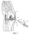
- FIG. 1is a perspective view of a human knee showing the skin removed and illustrating the femur, tibia, articular cartilage covering ends of the femur and tibia, and menisci;
- FIG. 2is a perspective view of a buffer of the present invention comprising an outer sack and an inner ring;
- FIG. 3is a perspective view of the inner ring of the buffer of the present invention.
- FIG. 4is a perspective view of the buffer of the present invention received within a sleeve designed for arthroscopically inserting the buffer into a patient's knee.
- FIG. 1a buffer 10 for a human joint constructed in accordance with embodiments of the invention is illustrated.
- the buffer 10is configured to be inserted into any human joint that is a bearing surface, such as the glenohumeral joint (shoulder joint) or the knee joint.
- glenohumeral jointshoulder joint
- knee jointknee joint
- Embodiments of the present inventionwill be discussed with respect to the knee joint, although it is to be understood that the invention is equally applicable to other joints.
- the buffer 10is configured to be inserted into a patient's knee in the space between the patient's femur and tibia and is intended to supplement any remaining articular cartilage 12 in the joint, or, in the instances where there is no remaining articular cartilage 12 , provide a complete buffer between the femur and tibia.
- the buffer 10is further sized so that it does not interfere with the menisci of the patient's knee, as illustrated in FIG. 1 .
- the buffer 10 of an embodiment of the present inventioncomprises an outer sack 14 enclosing an inner ring 16 and further configured to receive a friction reducing fluid 18 , as illustrated in FIGS. 1 and 2 .
- the buffer 10may be arthroscopically inserted into the patient's knee using a sleeve 20 , illustrated in FIG. 4 , especially designed for stable securement of the buffer 10 during the arthroscopic procedure.
- the outer sack 14 of the buffer 10is generally circular in shape, although the sack 14 may also be generally oval in shape while being inserted into the knee through the sleeve 20 .
- the sack 14is preferably approximately 10-50 mm in size, and more preferably approximately 30-60 mm, and most preferably approximately 35-45 mm.
- the space between the femur and tibia with an average amount of articular cartilagecan be opened to 10-15 mm at arthroscopy but contract to 0 mm when the stress on the ligament is relaxed.
- the sack 14 having the inner ring 16 located thereinhas a height that is preferably at least the distance between the femur and tibia, i.e., approximately 10 mm, and is flexible.
- the sack's heightis slightly larger than the distance between the femur and tibia, such that the height of the sack 14 is approximately the same height as the inner ring.
- the buffer, including the inner ringare manufactured in various sizes to accommodate differently-sized knee joints.
- the sack 14is preferably formed of a material that will be accepted by a human body and that is durable. Because of the pressure that the sack 14 will receive due to the contraction and movement of the femur and tibia, the sack 14 is further preferably formed of a pliable, resilient material. The material is also impermeable, such that the friction reducing fluid 18 does not escape from the sack 14 .
- An exemplary materialincludes polyethylene, although any material having the above-described properties will suffice.
- the material forming the sack 14may be translucent, as illustrated in FIG. 2 , or opaque.
- the sack 14includes a one-way valve 22 that is fluidly connected to a hollow fluid line 24 , as illustrated in FIGS. 1 and 2 .
- the hollow fluid line 24allows for passage of the fluid 18 therethrough.
- the friction reducing fluid 18described in more detail below, is inserted into the sack 14 after the buffer 10 is positioned inside the patient's knee.
- the fluid 18is inserted into the sack 14 via the fluid line 24 , which will be of a length sufficiently long to allow insertion of the fluid 18 into the sack 14 via the sleeve 20 or other instrument, as also described in more detail below.
- the valve 22preferably includes threads 23 (not shown) After the fluid 18 is inserted into the sack 14 ; the fluid line 24 is preferably unscrewed.
- the line 24may be cut proximal to the sack 14 or otherwise formed so that it can be removed from the sack 14 .
- the one-way valve 22allows receipt of the friction reducing fluid 18 via the filler line 24 and after the buffer 10 is inserted into the patient's knee.
- the valve 22is one-way, however, so that it does not allow for escape of the fluid 18 via the valve 22 .
- the friction reducing fluid 18is any fluid that is accepted by a human body and that assists in allowing ease of movement of the inner ring 16 within the outer sack 14 .
- Exemplary friction reducing fluids 18include the patients own synovial fluid found in synovial joints, such as the knee joint, and artificial fluids, such as SYNVISC®, manufactured by Genzyme Corporation.
- the inner ring 16is generally circular in shape, although the ring 16 may also be generally oval in shape. During insertion, the inner ring is flexed to be oval in shape.
- the inner ring 16is preferably approximately 20-60 mm in circumference, and more preferably approximately 25-55 mm, and most preferably approximately 27.5-32.5 mm.
- the inner ring 16is manufactured therefore to fit within the sack 14 .
- the inner ring 16 and sack 14are manufactured as a unit, such that the inner ring 16 is located within the sack 14 and sold as single unit. As noted above, the sack and inner ring may be manufactured in various sizes to fit each particular patient.
- the inner ring 16is preferably solid and formed of a resiliently rigid material, such that the ring 16 can withstand, with little or no deformation along a longitudinal axis A, a relatively high degree of loading pressure occurring from placement between the femur and tibia, yet can also be flexed or otherwise deformed along a transverse axis B for ease of placement within the patient's knee.
- the ring 16can be compressed side to side along axis B for placement in the sleeve.
- the ring's 16 resiliently rigid materialallows it to return to its generally circular shape once the pressure along the axis B is removed and the buffer is placed between the tibia and femur.
- the ring 16preferably comprises an outer surface 26 having a top edge 28 and a bottom edge 30 , an inner surface 32 having a top edge 34 and a bottom edge 36 , a top surface 38 , and a bottom surface 40 .
- the outer, inner, top, and bottom surfaces 26 , 32 , 38 , 40are preferably integrally formed and together define a hollow interior 42 of the ring 16 .
- the top and bottom surfaces 38 , 40are angled inwardly with respect to each other such that the top surface 38 extends downwardly from the top edge 28 of the outer surface 26 and to the top edge 34 of the inner surface 32 , and similarly, the bottom surface 40 extends upwardly (not shown) from the bottom edge 30 of the outer surface 26 and up to the bottom edge 36 of the inner surface 32 .
- top and bottom surfaces 38 , 40form the ring 16 that is generally concave when viewed from a top of the ring 16 and flat when viewed from a bottom of the ring 16 .
- Thisfurther provides a ring wherein a height of the ring along an outer diameter is larger than a height of the ring along an inner diameter, as illustrated in FIG. 3 .
- the concavity of the ring 16 and the orientation of the top and bottom surfaces 38 , 40forms a generally V-shaped cross-section of the ring 16 when cut along the longitudinal axis A.
- the concavityassists with placement and retention of the buffer 10 between the patient's femur and tibia in the patient's knee.
- the buffer 10is partially held in place within the patient's knee and between the femur and tibia by the load-bearing pressure that naturally occurs from the muscles, ligaments, and bones in the patient's knee and the weight of the body.
- the concave top and bottom surfaces 38 , 40 of the inner ring 16assist in locating the buffer 10 between the femur and tibia.
- the sleeve 20 used to insert the buffer 10 into the kneecomprises a generally ovoid, hollow body 44 , a stylus or telescoping plunger 46 (not necessarily drawn to scale with respect to length in FIG. 4 ) having proximal and distal ends 48 , 50 and at least partially housed within the body 44 , and the fluid line 24 having proximal and distal ends 54 , 56 , partially housed within the body 44 and 46 , and operable to allow the passage of fluid therethrough.
- the body 44 of the sleeve 20is generally ovoid to accommodate the shape of the buffer 10 upon insertion, as described in more detail below.
- the fluid line 24is passed through the plunger 46 and is screwed onto and fluidly connected with the valve 22 of the sack 14 at the proximal end 54 of the line 24 and with a fluid source (not shown) at the distal end 56 of the line 24 .
- the plunger 46includes a curved neck 58 located at the proximal end 48 and operable to support the buffer 10 during placement in the patient's knee.
- the buffer 10is preferably flexed along the transverse axis B into a generally ovoid shape so as to be partially inserted into the hollow body 44 of the sleeve 20 , as illustrated in FIG. 4 .
- the buffer 10is inserted into the body 44 such that the valve 22 of the buffer 10 is screwed into fluid communication with the fluid line 24 . Additionally, during insertion the buffer is supported by the curved neck 58 of the plunger 46 .
- the buffer 10is preferably arthroscopically inserted into the patient's knee using the sleeve 20 .
- the buffer 10is flexed into the generally ovoid shape illustrated in FIG. 1 and then positioned within the sleeve 20 . Flexing the buffer 10 into the ovoid shape facilitates insertion into the knee joint.
- the buffer 10is held into place during insertion by being supported by the neck 58 .
- the neck 58could include further structure (not shown) for supporting and holding the buffer 10 at least partially in the sleeve 20 during insertion, such as a U-shaped arm that receives the buffer 10 .
- the buffer 10is inserted between the tibia and femur of the knee joint.
- the buffer 10can be inserted on either side of the knee, as illustrated in FIG. 1 .
- the buffer 10is flexed into the ovoid shape. Once the buffer 10 is released from the sleeve 20 , it springs back into its generally circular shape, as illustrated in FIG. 1 . Once inserted, the friction reducing fluid 18 is then inserted into the buffer 10 via the fluid line 24 , which is fluidly connected between the one way valve 22 of the buffer 10 and the sleeve 20 . Alternatively, the fluid 18 may be inserted into the buffer 10 prior to placement between the femur and tibia and/or prior to release from the sleeve 20 .
- the fluid line 24is preferably physically separated from the buffer, so as to not be an irritant to the patient, by either cutting the line 24 proximal to the sack 14 or otherwise forming the line 24 so that it can be removed from the sack 14 .
- the rigidity of the inner ring 16 of the buffer 10acts to keep the femur and tibia separated, such that the two bones are not contacting each other. Additionally, as noted above, once inserted, the load-bearing pressure of the femur and tibia assist in locating and holding the buffer in place between the bones.
- the concavity of the top and bottom surfaces 38 , 40 of the ring 16further assists in holding the buffer 10 in place, especially when there is no load-bearing pressure exhibited between the femur and tibia.
- the friction reducing fluid 18then assists in movement of the femur and tibia against the buffer 10 and in particular, the inner ring 16 of the buffer 10 .
- the buffer 10is advantageously independently held within the knee joint and does not have to be sutured to any muscle, ligament, or tendon. Moreover, the buffer 10 can be used with any amount of articular cartilage 12 and menisci, such that it is not limited to being only used with very little cartilage. Although the buffer of embodiments of the present invention are preferably permanent, it is to be understood that with time, the buffer 10 may become sufficiently worn so as to require replacement. However, such will likely not be the norm, and the buffer 10 is expected to last many years, depending on the activity level, weight, and age of the patient and other common degradation factors.
- the sleeve 20could not include the fluid line 24 , such that the friction reducing fluid 18 is inserted via a separate line and a syringe 62 , as illustrated in FIG. 1 .
Landscapes
- Health & Medical Sciences (AREA)
- Orthopedic Medicine & Surgery (AREA)
- Transplantation (AREA)
- Heart & Thoracic Surgery (AREA)
- Life Sciences & Earth Sciences (AREA)
- Oral & Maxillofacial Surgery (AREA)
- Engineering & Computer Science (AREA)
- Biomedical Technology (AREA)
- Veterinary Medicine (AREA)
- Vascular Medicine (AREA)
- Cardiology (AREA)
- Animal Behavior & Ethology (AREA)
- General Health & Medical Sciences (AREA)
- Public Health (AREA)
- Physical Education & Sports Medicine (AREA)
- Rheumatology (AREA)
- Prostheses (AREA)
Abstract
Description
1. Field
Embodiments of the present invention relate to apparatus and methods of providing a buffer for a human joint so as to prevent painful bone on bone contact. More particularly, embodiments of the present invention present a buffer for insertion between the femur and tibia in the human knee, so as to protect worn or damaged articular cartilage or exposed bone and to allow the articular surfaces remaining on the femur and tibia to continue to move against each other less painfully.
2. Description of the Related Art
The human knee joint is one of the most complex joints of the body and is also highly susceptible to damage because it is a weight bearing joint. The knee joint itself is comprised of the femur (thigh bone), the tibia (shin bone), the patella (kneecap), articular cartilage, and menisci, which are a type of crescent-shaped cartilage that lies between the femur and tibia. The menisci are located in the medial and lateral articulations of the knee and sometimes act as shock-absorbing pads. The knee is also compromised of tissues that are muscle, ligament, the lining tissue (synovium), and the synovial fluid which is secreted by the synovium.
The ends of the femur and tibia are coated with articular cartilage, which is smooth and hard, so as to provide the femur, tibia, and patella with a slick surface during normal movement. The articular cartilage has a very low coefficient of friction and can also receive large compressive loads, which makes it vital to ensure ease of movement of the knee joint and prevent bone on bone contact between the femur and tibia. Normal articular cartilage is about 50 times slicker than ice.
Over time, the articular cartilage on the femur and tibia, and in any other human joint, wears and degenerates, such that it thins or in some joints is completely lost. Upon wear of the articular cartilage, the slick, low friction surfaces from the cartilage are lost, and the ends of the femur and tibia banes are exposed. Without any protecting articular cartilage, the femur and tibia contact each. This bone on bone contact is painful, and is often the end result of osteoarthritis. Additionally, bones can also become hard and sclerotic over time with associated loss of articular cartilage, which can further increases the pain.
Many methods have been developed to either replace worn cartilage or otherwise minimize the pain associated with the loss of the articular cartilage. The methods have all had varying degrees of success but are often accompanied by very extensive and invasive surgery. All invasive methods are costly, often requiring implanting nonbiologic parts within the knee, or, in some instances, human cadaver parts. These methods of treatment also require lengthy rehabilitation, which often times leaves the patient in considerable pain.
One method of treatment that has been used is implantation of cadaver menisci. This method has had only limited success and multiple failures. A second method is chondroplasty, or removal of and thinning out the existing damaged cartilage. This method is used to smooth the cartilage to reduce the friction between the femur and tibia, and remove the flaps of cartilage that have delaminated from the bone. The success of this procedure is limited by the amount of cartilage remaining, and doctors guard against removal of too much of the articular cartilage so as to prevent exposure of the subchondral bone. For older patients or patients with traumatic arthritis of their knees, chondroplasty has only limited application because of the lack of healthy articular cartilage.
If the articular cartilage loss is small, an osteochondral autograft transplant (known as an OATS procedure) can be performed. The OATS procedure requires removing a dowel shaped portion of bone and replacing it with a commensurate dowel shaped portion of articular cartilage from another area of the knee, another joint, or even a cadaver. The OATS procedure is relatively invasive, has a fairly lengthy rehabilitation time, and has also only had limited success.
An even further alternative to repairing articular cartilage damage is growing the patient's own cartilage in tissue cultures and placing the newly grown cartilage in the areas of cartilage loss. This is an expensive and often unsuccessful method of treatment.
In the most extreme of cases of arthritis, the knee joint may be artificially resurfaced or even replaced. In artificial joint replacement, the ends of the femur and tibia are capped with plastic or metal pieces that are cemented to the ends of the bone. Alternatively, the ends of the femur and tibia can be replaced with a biologic ingrowth coating of the metal used, which removes the need for the cement. This procedure is presently the standard approach to treating severe osteoarthritis of the knee; however, the risks from this procedure are numerous, and this is particularly unfortunate for patients who can ill afford a major complication from this extensive surgery. In places where these artificial joints have been inserted, wear eventually occurs in the polyethylene surface between the metal caps, which can lead to bone destruction just from the particles of the polyethylene. Moreover, this procedure is not only quite invasive but requires a lengthy rehabilitation time. Thus, for these reasons, many doctors delay as long as possible this invasive procedure in many patients.
An even further method of treatment is arthrosporic debridement, which is much less invasive but almost always unsuccessful in limiting the pain from the damaged joint surface, unless most of the pain is from a torn cartilage or loose body in the joint that can be removed arthroscopically.
The problems associated with each of the above procedures are highly dependant on the age and medical condition of the patient. For older patients, their ability and desire to engage in an invasive procedure that requires lengthy rehabilitation is often limited. Moreover, for older patients who are not necessarily engaging in many activities or who do not require a long-term solution to adjust their pain and discomfort, having an invasive, complicated procedure performed is not ideal.
Accordingly, there is a need for a less risky and improved apparatus and method for alleviating and addressing pain resulting from a loss of articular cartilage. There is a need for a new apparatus and method of treatment of lost cartilage that extends beyond attempting to fix or replace damaged cartilage, but instead provides an apparatus and method of treatment that is minimally invasive, relatively inexpensive requires relatively short rehabilitation time, and is suitable for older patients. This invention solves many of the above-described problems and provides a distinct advantage in the art of medical treatment for prevention of bone on bone contact due to the loss of articular cartilage. More particularly this invention provides a new apparatus and method of treatment to address the pain and discomfort associated with the loss of articular cartilage by interposing a thin but slick barrier between the tibia and femur. This invention provides a buffer between the femur and tibia in the human knee that does not require suturing or other permanent securement to muscles, ligaments, or tendons in the knee.
The present invention solves the above-described problems and provides a distinct advance in the art of medical treatments for prevention of bone-on-bone contact due to the loss of articular cartilage. More particularly, the present invention provides a new method of treatment to address the pain and discomfort associated with the loss of articular cartilage. In particular, embodiments of the present invention provide a buffer independently held between a human's femur and tibia and that does not require suturing or other permanent securement to any muscle, ligament, or tendon in the knee.
The buffer of embodiments of the present invention comprises an outer sack formed of a pliable material and having a one-way valve; a generally circularly-shaped, rigid inner ring within the outer sack, the inner ring having a top surface and a bottom surface angled inwardly with respect to each other, such that the ring is generally concave; and a friction reducing fluid received within the outer sack via the one-way valve.
The buffer is inserted into the knee joint using a specially designed sleeve comprising a body and a stylus or plunger. The body is generally an ovoid hollow tube into which is inserted the buffer. The stylus or plunger includes a fluid line comprising a tube through which a fluid can be pumped. The fluid line and buffer, when received within the sleeve, are in fluid communication, such that the fluid can be inserted into the outer sack of the buffer once the buffer is positioned in the knee joint.
To insert the buffer into the sleeve, the buffer is compressed into the sleeve, and the stylus is screwed or otherwise secured to the rigid inner ring of the buffer. The stylus is then unscrewed from the buffer once the device is positioned in the knee joint and after the fluid is placed into the buffer via a one way valve in the buffer.
A method of inserting the buffer into the knee comprises the steps of providing the buffer, providing a sleeve having a stylus operable to securely receive the buffer during insertion, and injecting fluid into the outer sack during insertion via the one way valve located in the buffer. To insert the buffer in the sleeve, the buffer is compressed side to side, placed into the sleeve, and removably secured to the stylus. After being placed into the knee, the buffer is then injected with fluid to help the surfaces of the buffer move over each other by reducing friction between them. Once the buffer is injected with this fluid, the stylus is unscrewed form the rigid inner ring, the fluid is held in place by the one way valve, the sleeve is removed, the skin is closed, and the operation is complete.
Embodiments of the present invention are described in detail below with reference to the attached drawing figures, wherein:
The drawing figures do not limit the present invention to the specific embodiments disclosed and described herein. The drawings are not necessarily to scale, emphasis instead being placed upon clearly illustrating the principles of the invention.
Turning now to the drawing figures, and particularlyFIG. 1 , abuffer 10 for a human joint constructed in accordance with embodiments of the invention is illustrated. Thebuffer 10 is configured to be inserted into any human joint that is a bearing surface, such as the glenohumeral joint (shoulder joint) or the knee joint. Embodiments of the present invention will be discussed with respect to the knee joint, although it is to be understood that the invention is equally applicable to other joints.
Thebuffer 10 is configured to be inserted into a patient's knee in the space between the patient's femur and tibia and is intended to supplement any remainingarticular cartilage 12 in the joint, or, in the instances where there is no remainingarticular cartilage 12, provide a complete buffer between the femur and tibia. Thebuffer 10 is further sized so that it does not interfere with the menisci of the patient's knee, as illustrated inFIG. 1 .
Thebuffer 10 of an embodiment of the present invention comprises anouter sack 14 enclosing aninner ring 16 and further configured to receive afriction reducing fluid 18, as illustrated inFIGS. 1 and 2 . As described in more detail below, thebuffer 10 may be arthroscopically inserted into the patient's knee using asleeve 20, illustrated inFIG. 4 , especially designed for stable securement of thebuffer 10 during the arthroscopic procedure.
Theouter sack 14 of thebuffer 10 is generally circular in shape, although thesack 14 may also be generally oval in shape while being inserted into the knee through thesleeve 20. Thesack 14 is preferably approximately 10-50 mm in size, and more preferably approximately 30-60 mm, and most preferably approximately 35-45 mm. In an average patient, the space between the femur and tibia with an average amount of articular cartilage can be opened to 10-15 mm at arthroscopy but contract to 0 mm when the stress on the ligament is relaxed. Thesack 14 having theinner ring 16 located therein has a height that is preferably at least the distance between the femur and tibia, i.e., approximately 10 mm, and is flexible. More preferably, the sack's height is slightly larger than the distance between the femur and tibia, such that the height of thesack 14 is approximately the same height as the inner ring. In embodiments of the invention, the buffer, including the inner ring, are manufactured in various sizes to accommodate differently-sized knee joints.
Thesack 14 is preferably formed of a material that will be accepted by a human body and that is durable. Because of the pressure that thesack 14 will receive due to the contraction and movement of the femur and tibia, thesack 14 is further preferably formed of a pliable, resilient material. The material is also impermeable, such that thefriction reducing fluid 18 does not escape from thesack 14. An exemplary material includes polyethylene, although any material having the above-described properties will suffice. The material forming thesack 14 may be translucent, as illustrated inFIG. 2 , or opaque.
Thesack 14 includes a one-way valve 22 that is fluidly connected to ahollow fluid line 24, as illustrated inFIGS. 1 and 2 . Thehollow fluid line 24 allows for passage of the fluid18 therethrough. Thefriction reducing fluid 18, described in more detail below, is inserted into thesack 14 after thebuffer 10 is positioned inside the patient's knee. The fluid18 is inserted into thesack 14 via thefluid line 24, which will be of a length sufficiently long to allow insertion of the fluid18 into thesack 14 via thesleeve 20 or other instrument, as also described in more detail below. Thevalve 22 preferably includes threads23 (not shown) After the fluid18 is inserted into thesack 14; thefluid line 24 is preferably unscrewed. Alternatively, theline 24 may be cut proximal to thesack 14 or otherwise formed so that it can be removed from thesack 14. The one-way valve 22 allows receipt of thefriction reducing fluid 18 via thefiller line 24 and after thebuffer 10 is inserted into the patient's knee. Thevalve 22 is one-way, however, so that it does not allow for escape of the fluid18 via thevalve 22.
Thefriction reducing fluid 18 is any fluid that is accepted by a human body and that assists in allowing ease of movement of theinner ring 16 within theouter sack 14. Exemplaryfriction reducing fluids 18 include the patients own synovial fluid found in synovial joints, such as the knee joint, and artificial fluids, such as SYNVISC®, manufactured by Genzyme Corporation.
Theinner ring 16 is generally circular in shape, although thering 16 may also be generally oval in shape. During insertion, the inner ring is flexed to be oval in shape. Theinner ring 16 is preferably approximately 20-60 mm in circumference, and more preferably approximately 25-55 mm, and most preferably approximately 27.5-32.5 mm. Theinner ring 16 is manufactured therefore to fit within thesack 14. In preferred form, theinner ring 16 and sack14 are manufactured as a unit, such that theinner ring 16 is located within thesack 14 and sold as single unit. As noted above, the sack and inner ring may be manufactured in various sizes to fit each particular patient.
Theinner ring 16 is preferably solid and formed of a resiliently rigid material, such that thering 16 can withstand, with little or no deformation along a longitudinal axis A, a relatively high degree of loading pressure occurring from placement between the femur and tibia, yet can also be flexed or otherwise deformed along a transverse axis B for ease of placement within the patient's knee. In particular, during insertion in the knee joint, thering 16 can be compressed side to side along axis B for placement in the sleeve. However, the ring's16 resiliently rigid material allows it to return to its generally circular shape once the pressure along the axis B is removed and the buffer is placed between the tibia and femur.
Thering 16 preferably comprises anouter surface 26 having atop edge 28 and abottom edge 30, aninner surface 32 having atop edge 34 and abottom edge 36, atop surface 38, and abottom surface 40. The outer, inner, top, andbottom surfaces hollow interior 42 of thering 16. The top andbottom surfaces top surface 38 extends downwardly from thetop edge 28 of theouter surface 26 and to thetop edge 34 of theinner surface 32, and similarly, thebottom surface 40 extends upwardly (not shown) from thebottom edge 30 of theouter surface 26 and up to thebottom edge 36 of theinner surface 32. In this manner, the top andbottom surfaces ring 16 that is generally concave when viewed from a top of thering 16 and flat when viewed from a bottom of thering 16. This further provides a ring wherein a height of the ring along an outer diameter is larger than a height of the ring along an inner diameter, as illustrated inFIG. 3 .
The concavity of thering 16 and the orientation of the top andbottom surfaces ring 16 when cut along the longitudinal axis A. The concavity assists with placement and retention of thebuffer 10 between the patient's femur and tibia in the patient's knee. In particular, thebuffer 10 is partially held in place within the patient's knee and between the femur and tibia by the load-bearing pressure that naturally occurs from the muscles, ligaments, and bones in the patient's knee and the weight of the body. However, to insure that thebuffer 10 does not drift outside its proper placement when the knee is not exhibiting load-bearing pressure, the concave top andbottom surfaces inner ring 16 assist in locating thebuffer 10 between the femur and tibia.
As illustrated inFIG. 4 , thesleeve 20 used to insert thebuffer 10 into the knee comprises a generally ovoid,hollow body 44, a stylus or telescoping plunger46 (not necessarily drawn to scale with respect to length inFIG. 4 ) having proximal and distal ends48,50 and at least partially housed within thebody 44, and thefluid line 24 having proximal and distal ends54,56, partially housed within thebody body 44 of thesleeve 20 is generally ovoid to accommodate the shape of thebuffer 10 upon insertion, as described in more detail below. Thefluid line 24 is passed through theplunger 46 and is screwed onto and fluidly connected with thevalve 22 of thesack 14 at theproximal end 54 of theline 24 and with a fluid source (not shown) at thedistal end 56 of theline 24.
Theplunger 46 includes a curved neck58 located at theproximal end 48 and operable to support thebuffer 10 during placement in the patient's knee. Thebuffer 10 is preferably flexed along the transverse axis B into a generally ovoid shape so as to be partially inserted into thehollow body 44 of thesleeve 20, as illustrated inFIG. 4 . Thebuffer 10 is inserted into thebody 44 such that thevalve 22 of thebuffer 10 is screwed into fluid communication with thefluid line 24. Additionally, during insertion the buffer is supported by the curved neck58 of theplunger 46.
Thebuffer 10 is preferably arthroscopically inserted into the patient's knee using thesleeve 20. Thebuffer 10 is flexed into the generally ovoid shape illustrated inFIG. 1 and then positioned within thesleeve 20. Flexing thebuffer 10 into the ovoid shape facilitates insertion into the knee joint. Thebuffer 10 is held into place during insertion by being supported by the neck58. Alternatively, the neck58 could include further structure (not shown) for supporting and holding thebuffer 10 at least partially in thesleeve 20 during insertion, such as a U-shaped arm that receives thebuffer 10.
As discussed briefly above and as illustrated inFIG. 1 , thebuffer 10 is inserted between the tibia and femur of the knee joint. Thebuffer 10 can be inserted on either side of the knee, as illustrated inFIG. 1 .
As noted above, upon insertion, thebuffer 10 is flexed into the ovoid shape. Once thebuffer 10 is released from thesleeve 20, it springs back into its generally circular shape, as illustrated inFIG. 1 . Once inserted, thefriction reducing fluid 18 is then inserted into thebuffer 10 via thefluid line 24, which is fluidly connected between the oneway valve 22 of thebuffer 10 and thesleeve 20. Alternatively, the fluid18 may be inserted into thebuffer 10 prior to placement between the femur and tibia and/or prior to release from thesleeve 20. As noted above, after the fluid is inserted into thesack 14, thefluid line 24 is preferably physically separated from the buffer, so as to not be an irritant to the patient, by either cutting theline 24 proximal to thesack 14 or otherwise forming theline 24 so that it can be removed from thesack 14.
Once inserted, the rigidity of theinner ring 16 of thebuffer 10 acts to keep the femur and tibia separated, such that the two bones are not contacting each other. Additionally, as noted above, once inserted, the load-bearing pressure of the femur and tibia assist in locating and holding the buffer in place between the bones. The concavity of the top andbottom surfaces ring 16 further assists in holding thebuffer 10 in place, especially when there is no load-bearing pressure exhibited between the femur and tibia. Thefriction reducing fluid 18 then assists in movement of the femur and tibia against thebuffer 10 and in particular, theinner ring 16 of thebuffer 10.
Thebuffer 10 is advantageously independently held within the knee joint and does not have to be sutured to any muscle, ligament, or tendon. Moreover, thebuffer 10 can be used with any amount ofarticular cartilage 12 and menisci, such that it is not limited to being only used with very little cartilage. Although the buffer of embodiments of the present invention are preferably permanent, it is to be understood that with time, thebuffer 10 may become sufficiently worn so as to require replacement. However, such will likely not be the norm, and thebuffer 10 is expected to last many years, depending on the activity level, weight, and age of the patient and other common degradation factors.
Although the invention has been described with reference to the preferred embodiment illustrated in the attached drawing figures, it is noted that equivalents may be employed and substitutions made herein without departing from the scope of the invention as recited in the claims. For example, thesleeve 20 could not include thefluid line 24, such that thefriction reducing fluid 18 is inserted via a separate line and asyringe 62, as illustrated inFIG. 1 .
Claims (15)
1. An apparatus comprising:
an outer sack;
a generally circular or oval-shaped inner ring received within the outer sack, the inner ring having an outer surface having a top end and a bottom end, an inner surface having a top end and a bottom end, a top surface, and a bottom surface,
wherein the top and bottom surfaces are angled inwardly with respect to each other, such that the top surface extends downwardly from the top end of the outer surface and to the top end of the inner surface, and the bottom surface extends upwardly from the bottom end of the outer surface and to the bottom end of the inner surface, such that the ring is generally concave; and
a friction reducing fluid received within the outer sack,
said apparatus configured for insertion between two bones of a human body so as to at least partially prevent the bones from contacting each other.
2. The apparatus ofclaim 1 , wherein the apparatus is configured for insertion between a human's femur and tibia so as to at least partially prevent the femur and tibia from contacting each other.
3. The apparatus ofclaim 1 , wherein the outer sack is generally circularly shaped.
4. The apparatus ofclaim 1 , wherein the outer sack is formed of a pliable, resilient material.
5. The apparatus ofclaim 3 , wherein the outer sack is formed of polyethylene.
6. The apparatus ofclaim 1 , wherein the outer sack includes a one-way valve for receipt of the friction reducing fluid into the sack.
7. The apparatus ofclaim 1 , wherein the inner ring is formed of a resiliently rigid material.
8. The buffer ofclaim 1 , wherein the inner surface is smaller in height than the outer surface.
9. The buffer ofclaim 1 , wherein the ring has a generally V-shaped cross section when cut along a plane orthogonal to the plane of the ring.
10. A buffer for placement between a human's femur and tibia, the buffer comprising:
an outer sack formed of a pliable material and having a one-way valve;
a generally circularly shaped inner ring received within the outer sack, the inner ring
including
a generally vertically-oriented outer surface along the circumference of the ring, said outer surface having a top end and a bottom end,
a generally horizontally-oriented top surface extending downwardly from the top end of the outer surface, and
a generally horizontally-oriented bottom surface extending upwardly from the bottom end of the outer surface,
wherein the top surface and the bottom surface are angled inwardly with respect to each other, such that the ring is generally concave; and
a friction reducing fluid received within the outer sack via the one-way valve,
said apparatus configured for insertion between the femur and tibia so as to at least partially prevent the bones from contacting each other.
11. The buffer ofclaim 10 , wherein the buffer is inserted between the femur and tibia using a sleeve.
12. The buffer ofclaim 11 , wherein the sleeve comprises a generally ovoid, hollow body, a telescoping plunger having proximal and distal ends, and a fluid line having proximal and distal ends.
13. The buffer ofclaim 12 , wherein the buffer flexes along a transverse axis for placement in the sleeve.
14. The apparatus ofclaim 10 , wherein the ring further includes an inner surface positioned radially inward from the outer surface and coupled to the top surface and the bottom surface, the inner surface being smaller in height than the outer surface.
15. The apparatus ofclaim 10 , wherein the ring has a generally V-shaped cross section when cut along a plane orthogonal to the plane of the ring.
Priority Applications (3)
| Application Number | Priority Date | Filing Date | Title |
|---|---|---|---|
| US12/133,211US7976578B2 (en) | 2008-06-04 | 2008-06-04 | Buffer for a human joint and method of arthroscopically inserting |
| EP09007304AEP2130517A1 (en) | 2008-06-04 | 2009-06-02 | Buffer for a human joint and method of arthroscopically inserting |
| US13/179,869US8764829B2 (en) | 2008-06-04 | 2011-07-11 | Buffer for a human joint and method of arthroscopically inserting |
Applications Claiming Priority (1)
| Application Number | Priority Date | Filing Date | Title |
|---|---|---|---|
| US12/133,211US7976578B2 (en) | 2008-06-04 | 2008-06-04 | Buffer for a human joint and method of arthroscopically inserting |
Related Child Applications (1)
| Application Number | Title | Priority Date | Filing Date |
|---|---|---|---|
| US13/179,869ContinuationUS8764829B2 (en) | 2008-06-04 | 2011-07-11 | Buffer for a human joint and method of arthroscopically inserting |
Publications (2)
| Publication Number | Publication Date |
|---|---|
| US20090306778A1 US20090306778A1 (en) | 2009-12-10 |
| US7976578B2true US7976578B2 (en) | 2011-07-12 |
Family
ID=40940417
Family Applications (2)
| Application Number | Title | Priority Date | Filing Date |
|---|---|---|---|
| US12/133,211Active2029-02-14US7976578B2 (en) | 2008-06-04 | 2008-06-04 | Buffer for a human joint and method of arthroscopically inserting |
| US13/179,869Active2028-07-04US8764829B2 (en) | 2008-06-04 | 2011-07-11 | Buffer for a human joint and method of arthroscopically inserting |
Family Applications After (1)
| Application Number | Title | Priority Date | Filing Date |
|---|---|---|---|
| US13/179,869Active2028-07-04US8764829B2 (en) | 2008-06-04 | 2011-07-11 | Buffer for a human joint and method of arthroscopically inserting |
Country Status (2)
| Country | Link |
|---|---|
| US (2) | US7976578B2 (en) |
| EP (1) | EP2130517A1 (en) |
Cited By (13)
| Publication number | Priority date | Publication date | Assignee | Title |
|---|---|---|---|---|
| US20110270393A1 (en)* | 2008-06-04 | 2011-11-03 | James Marvel | Buffer for a human joint and method of arthroscopically inserting |
| US20110320005A1 (en)* | 2003-06-27 | 2011-12-29 | Rydell Mark A | System and Method for Ankle Arthroplasty |
| US20120022649A1 (en)* | 2009-09-11 | 2012-01-26 | Articulinx, Inc. | Disc-shaped orthopedic devices |
| US20140180415A1 (en)* | 2012-12-26 | 2014-06-26 | Scott A. Koss | Apparatus, kit, and method for percutaneous intervertebral disc restoration |
| US20140316526A1 (en)* | 2011-09-01 | 2014-10-23 | R. Thomas Grotz | Resilient interpositional arthroplasty device |
| US10004605B2 (en) | 2010-01-22 | 2018-06-26 | Iorthopedics, Inc. | Resilient knee implant and methods |
| USD833613S1 (en) | 2011-01-19 | 2018-11-13 | Iorthopedics, Inc. | Resilient knee implant |
| US20190076260A1 (en)* | 2014-10-02 | 2019-03-14 | Seth McCullen | Anatomically Designed Meniscus Implantable Devices |
| US10307258B2 (en) | 2010-01-22 | 2019-06-04 | Iorthopedics, Inc. | Resilient interpositional arthroplasty device |
| US10449053B2 (en) | 2012-10-02 | 2019-10-22 | Seth McCullen | Implantable devices for musculoskeletal repair and regeneration |
| US10828168B2 (en) | 2017-05-10 | 2020-11-10 | Howmedica Osteonics Corp. | Patient specific composite knee replacement |
| US11419733B2 (en) | 2018-01-12 | 2022-08-23 | Percheron Spine, Llc | Spinal disc implant and device and method for percutaneous delivery of the spinal disc implant |
| US11452606B2 (en) | 2017-05-02 | 2022-09-27 | Orthonika Limited | Composite joint implant |
Families Citing this family (25)
| Publication number | Priority date | Publication date | Assignee | Title |
|---|---|---|---|---|
| PL2124831T3 (en) | 2007-03-15 | 2017-03-31 | Ortho-Space Ltd. | Prosthetic devices |
| US20100198354A1 (en) | 2007-08-01 | 2010-08-05 | Jeffrey Halbrecht | Method and system for patella tendon realignment |
| US8114156B2 (en)* | 2008-05-30 | 2012-02-14 | Edwin Burton Hatch | Flexibly compliant ceramic prosthetic meniscus for the replacement of damaged cartilage in orthopedic surgical repair or reconstruction of hip, knee, ankle, shoulder, elbow, wrist and other anatomical joints |
| US9808345B2 (en) | 2008-07-24 | 2017-11-07 | Iorthopedics, Inc. | Resilient arthroplasty device |
| US9549820B2 (en)* | 2009-06-25 | 2017-01-24 | Zimmer, Inc. | Glenoid implant with synthetic labrum |
| US9861408B2 (en) | 2009-08-27 | 2018-01-09 | The Foundry, Llc | Method and apparatus for treating canine cruciate ligament disease |
| US9278004B2 (en) | 2009-08-27 | 2016-03-08 | Cotera, Inc. | Method and apparatus for altering biomechanics of the articular joints |
| US10349980B2 (en) | 2009-08-27 | 2019-07-16 | The Foundry, Llc | Method and apparatus for altering biomechanics of the shoulder |
| CA2771332C (en) | 2009-08-27 | 2020-11-10 | Cotera, Inc. | Method and apparatus for force redistribution in articular joints |
| US9668868B2 (en) | 2009-08-27 | 2017-06-06 | Cotera, Inc. | Apparatus and methods for treatment of patellofemoral conditions |
| GB2476124A (en)* | 2009-12-08 | 2011-06-15 | R Thomas Grotz | Inflatable arthroplasty implant |
| GB201014824D0 (en)* | 2010-09-07 | 2010-10-20 | Goodfellow John | Unicondylar meniscal bearing knee replacement |
| US9289307B2 (en)* | 2011-10-18 | 2016-03-22 | Ortho-Space Ltd. | Prosthetic devices and methods for using same |
| US9468466B1 (en) | 2012-08-24 | 2016-10-18 | Cotera, Inc. | Method and apparatus for altering biomechanics of the spine |
| WO2017046647A1 (en) | 2015-09-18 | 2017-03-23 | Ortho-Space Ltd. | Intramedullary fixated subacromial spacers |
| US9943414B2 (en)* | 2015-12-30 | 2018-04-17 | Wasas, Llc. | System and method for non-binding allograft subtalar joint implant |
| EP3573806A4 (en) | 2017-01-30 | 2019-12-11 | Ortho-Space Ltd. | Processing machine and methods for processing dip-molded articles |
| US10744313B2 (en)* | 2017-07-27 | 2020-08-18 | University Of Utah Research Foundation | Therapeutic delivery device |
| US10912867B2 (en)* | 2017-11-15 | 2021-02-09 | De Novo Orthopedics Inc. | Bioinductive patch |
| GB201917592D0 (en)* | 2019-12-02 | 2020-01-15 | Aurora Medical Ltd | Implant |
| EP4059476A4 (en)* | 2019-12-27 | 2022-11-23 | Shanghai Endophix Co., Ltd. | ROTATOR CUFF BALLOON |
| US11896476B2 (en) | 2020-01-02 | 2024-02-13 | Zkr Orthopedics, Inc. | Patella tendon realignment implant with changeable shape |
| WO2021231253A1 (en) | 2020-05-11 | 2021-11-18 | Zkr Orthopedics, Inc. | Adjustable patellar tendon realignment implant |
| WO2021234966A1 (en)* | 2020-05-22 | 2021-11-25 | 株式会社Surfs Med | Implant and treatment method |
| US20250120752A1 (en)* | 2023-10-13 | 2025-04-17 | Cheng-Lun SOO | Intraarticular Spacer For Stabilizing An Ilium And A Sacrum |
Citations (125)
| Publication number | Priority date | Publication date | Assignee | Title |
|---|---|---|---|---|
| US4344193A (en)* | 1980-11-28 | 1982-08-17 | Kenny Charles H | Meniscus prosthesis |
| US4502161A (en) | 1981-09-21 | 1985-03-05 | Wall W H | Prosthetic meniscus for the repair of joints |
| US4863477A (en)* | 1987-05-12 | 1989-09-05 | Monson Gary L | Synthetic intervertebral disc prosthesis |
| US4880429A (en)* | 1987-07-20 | 1989-11-14 | Stone Kevin R | Prosthetic meniscus |
| US4919667A (en)* | 1988-12-02 | 1990-04-24 | Stryker Corporation | Implant |
| US4932969A (en)* | 1987-01-08 | 1990-06-12 | Sulzer Brothers Limited | Joint endoprosthesis |
| US5092894A (en)* | 1990-02-13 | 1992-03-03 | Kenny Charles H | Stabilized meniscus prosthesis |
| US5158574A (en) | 1987-07-20 | 1992-10-27 | Regen Corporation | Prosthetic meniscus |
| US5171281A (en)* | 1988-08-18 | 1992-12-15 | University Of Medicine & Dentistry Of New Jersey | Functional and biocompatible intervertebral disc spacer containing elastomeric material of varying hardness |
| US5344459A (en)* | 1991-12-03 | 1994-09-06 | Swartz Stephen J | Arthroscopically implantable prosthesis |
| US5545229A (en)* | 1988-08-18 | 1996-08-13 | University Of Medicine And Dentistry Of Nj | Functional and biocompatible intervertebral disc spacer containing elastomeric material of varying hardness |
| US5549679A (en)* | 1994-05-20 | 1996-08-27 | Kuslich; Stephen D. | Expandable fabric implant for stabilizing the spinal motion segment |
| US5645597A (en)* | 1995-12-29 | 1997-07-08 | Krapiva; Pavel I. | Disc replacement method and apparatus |
| US5800549A (en)* | 1997-04-30 | 1998-09-01 | Howmedica Inc. | Method and apparatus for injecting an elastic spinal implant |
| US5824093A (en)* | 1994-10-17 | 1998-10-20 | Raymedica, Inc. | Prosthetic spinal disc nucleus |
| US6022376A (en)* | 1997-06-06 | 2000-02-08 | Raymedica, Inc. | Percutaneous prosthetic spinal disc nucleus and method of manufacture |
| US6046379A (en)* | 1995-06-07 | 2000-04-04 | Stone; Kevin R. | Meniscal xenografts |
| US6132465A (en)* | 1998-06-04 | 2000-10-17 | Raymedica, Inc. | Tapered prosthetic spinal disc nucleus |
| US20020035400A1 (en)* | 2000-08-08 | 2002-03-21 | Vincent Bryan | Implantable joint prosthesis |
| WO2002039889A2 (en) | 2000-11-14 | 2002-05-23 | S Gabbay | Implantable orthopedic support apparatus and method for making same |
| US6395032B1 (en)* | 1998-12-11 | 2002-05-28 | Dimso (Distribution Medicale Du Sud-Ouest) | Intervertebral disc prosthesis with liquid chamber |
| US6419704B1 (en)* | 1999-10-08 | 2002-07-16 | Bret Ferree | Artificial intervertebral disc replacement methods and apparatus |
| US6425920B1 (en)* | 1999-10-13 | 2002-07-30 | James S. Hamada | Spinal fusion implant |
| US6436146B1 (en)* | 1997-12-10 | 2002-08-20 | Bioprofile | Implant for treating ailments of a joint or a bone |
| US20020147497A1 (en)* | 2001-04-06 | 2002-10-10 | Integrated Vascular Systems, Inc. | Methods for treating spinal discs |
| US6482234B1 (en)* | 2000-04-26 | 2002-11-19 | Pearl Technology Holdings, Llc | Prosthetic spinal disc |
| US20020183848A1 (en)* | 1999-04-05 | 2002-12-05 | Raymedica, Inc. | Prosthetic spinal disc nucleus having a shape change characteristic |
| US20030009224A1 (en)* | 2001-07-03 | 2003-01-09 | Axiomed Inc. | Artificial disc |
| US6527804B1 (en)* | 1998-12-11 | 2003-03-04 | Dimso (Distribution Medicale Du Sud-Quest) | Intervertebral disk prosthesis |
| US20030093152A1 (en)* | 1999-12-17 | 2003-05-15 | Pedersen Walther Batsberg | Prosthetic device |
| US6582466B1 (en)* | 1998-12-11 | 2003-06-24 | Stryker Spine | Intervertebral disc prosthesis with reduced friction |
| US20030195628A1 (en)* | 1994-05-06 | 2003-10-16 | Qi-Bin Bao | Method of making an intervertebral disc prosthesis |
| US6712853B2 (en)* | 2000-12-15 | 2004-03-30 | Spineology, Inc. | Annulus-reinforcing band |
| US6733533B1 (en)* | 2002-11-19 | 2004-05-11 | Zimmer Technology, Inc. | Artificial spinal disc |
| US20040148026A1 (en)* | 1998-08-20 | 2004-07-29 | Bonutti Peter M. | Joint spacer with compartment for orthobiologic material |
| US20040193273A1 (en)* | 2003-03-31 | 2004-09-30 | Shih-Shing Huang | Vividly simulated prosthetic intervertebral disc |
| US20040215342A1 (en)* | 2003-04-23 | 2004-10-28 | Loubert Suddaby | Inflatable intervertebral disc replacement prosthesis |
| US20040243250A1 (en)* | 1998-03-06 | 2004-12-02 | Stone Kevin R. | Soft tissue xenografts |
| US20050049604A1 (en)* | 2002-05-16 | 2005-03-03 | Singer Deron J. | Device for treating intervertebral disc herniations |
| US20050090901A1 (en)* | 2001-12-05 | 2005-04-28 | Armin Studer | Intervertebral disk prosthesis or nucleus replacement prosthesis |
| US20050113923A1 (en)* | 2003-10-03 | 2005-05-26 | David Acker | Prosthetic spinal disc nucleus |
| US6923831B2 (en)* | 1999-05-10 | 2005-08-02 | Barry M. Fell | Surgically implantable knee prosthesis having attachment apertures |
| US20050177240A1 (en)* | 2004-02-06 | 2005-08-11 | Jason Blain | Vertebral facet joint prosthesis and method of fixation |
| US20050197702A1 (en)* | 2002-08-15 | 2005-09-08 | Coppes Justin K. | Intervertebral disc implant |
| US20050234549A1 (en)* | 2004-04-20 | 2005-10-20 | Kladakis Stephanie M | Meniscal repair scaffold |
| US20050251259A1 (en)* | 2003-07-29 | 2005-11-10 | Loubert Suddaby | Inflatable nuclear prosthesis |
| US6969404B2 (en)* | 1999-10-08 | 2005-11-29 | Ferree Bret A | Annulus fibrosis augmentation methods and apparatus |
| US6994730B2 (en) | 2003-01-31 | 2006-02-07 | Howmedica Osteonics Corp. | Meniscal and tibial implants |
| US7004971B2 (en)* | 2002-12-31 | 2006-02-28 | Depuy Acromed, Inc. | Annular nucleus pulposus replacement |
| US20060047341A1 (en)* | 2004-08-24 | 2006-03-02 | Trieu Hai H | Spinal disc implants with reservoirs for delivery of therapeutic agents |
| US20060052874A1 (en)* | 2004-09-09 | 2006-03-09 | Johnson Wesley M | Prostheses for spine discs having fusion capability |
| US20060149370A1 (en) | 2004-11-23 | 2006-07-06 | Reinhold Schmieding | Method and apparatus for arthroscopic joint resurfacing |
| WO2006097932A2 (en) | 2005-03-17 | 2006-09-21 | Active Implants Corporation | Implant devices |
| US20060241758A1 (en)* | 2005-04-20 | 2006-10-26 | Sdgi Holdings, Inc. | Facet spacers |
| US20060241766A1 (en)* | 2005-04-20 | 2006-10-26 | Sdgi Holdings, Inc. | Method and apparatus for preventing articulation in an artificial joint |
| US20060241765A1 (en)* | 2003-04-03 | 2006-10-26 | Burn Peter J | Load bearing intervertebral disk |
| US20060247780A1 (en)* | 2005-04-27 | 2006-11-02 | Bert Jeffrey K | Expandable artificial disc and associated methods and instrumentation |
| US20060293751A1 (en)* | 2001-06-29 | 2006-12-28 | Lotz Jeffrey C | Biodegradable/bioactive nucleus pulposus implant and method for treating degenerated intervertebral discs |
| US20070027547A1 (en)* | 2003-06-27 | 2007-02-01 | Advanced Bio Surfaces, Inc. | System and method for ankle arthroplasty |
| US20070050033A1 (en)* | 2005-09-01 | 2007-03-01 | Reo Michael L | Prosthetic intervertebral discs |
| US20070050032A1 (en)* | 2005-09-01 | 2007-03-01 | Spinal Kinetics, Inc. | Prosthetic intervertebral discs |
| US20070100450A1 (en)* | 2005-11-02 | 2007-05-03 | Zimmer Technology, Inc. | Joint spacer implant |
| US20070118218A1 (en)* | 2005-11-22 | 2007-05-24 | Hooper David M | Facet joint implant and procedure |
| US20070168042A1 (en)* | 2006-01-13 | 2007-07-19 | Hudgins Robert G | Devices and methods for disc replacement |
| US20070173940A1 (en)* | 2006-01-18 | 2007-07-26 | Zimmer Spine, Inc. | Vertebral fusion device and method |
| US7250060B2 (en)* | 2004-01-27 | 2007-07-31 | Sdgi Holdings, Inc. | Hybrid intervertebral disc system |
| US20070179606A1 (en)* | 2003-07-15 | 2007-08-02 | Huyghe Jacques M R | Prosthesis made of a fibre-reinforced hydrogel, method of manufacturing the prosthesis and use thereof. |
| US7252685B2 (en)* | 2003-06-05 | 2007-08-07 | Sdgi Holdings, Inc. | Fusion implant and method of making same |
| US20070233258A1 (en)* | 2006-02-28 | 2007-10-04 | Zimmer Spine, Inc. | Vertebroplasty- device and method |
| US20070250060A1 (en)* | 2006-04-24 | 2007-10-25 | Sdgi Holdings, Inc. | Expandable device for insertion between anatomical structures and a procedure utilizing same |
| US7291169B2 (en)* | 2005-04-15 | 2007-11-06 | Zimmer Technology, Inc. | Cartilage implant |
| US7297161B2 (en)* | 1999-05-10 | 2007-11-20 | Fell Barry M | Surgically implantable knee prosthesis |
| US20070293947A1 (en)* | 2006-06-19 | 2007-12-20 | Mansmann Kevin A | Multi-part implants for combined repair of hyaline and meniscal cartilage in joints |
| US20080015703A1 (en)* | 2004-06-30 | 2008-01-17 | Depuy Spine, Inc. | C-shaped disc prosthesis |
| US20080046082A1 (en)* | 2003-07-17 | 2008-02-21 | Lee Casey K | Intervertebral disc and nucleus prosthesis |
| US20080051896A1 (en)* | 2006-08-25 | 2008-02-28 | Loubert Suddaby | Expandable Spinous Process Distractor |
| US20080071374A1 (en)* | 1995-09-04 | 2008-03-20 | Active Implants Corporation | Method and apparatus for computerized surgery |
| US20080086210A1 (en)* | 2006-10-09 | 2008-04-10 | Active Implants Corporation | Meniscus Prosthetic Device |
| US20080086133A1 (en)* | 2003-05-16 | 2008-04-10 | Spineology | Expandable porous mesh bag device and methods of use for reduction, filling, fixation and supporting of bone |
| US20080132934A1 (en)* | 1994-01-26 | 2008-06-05 | Kyphon, Inc. | Systems and methods treating a vertebral body related applications |
| US20080154371A1 (en)* | 1999-04-02 | 2008-06-26 | Fell Barry M | Proportioned surgically implantable knee prosthesis |
| US20080195210A1 (en)* | 2005-03-01 | 2008-08-14 | Columna Pty Ltd | Intervertebral Disc Restoration |
| US20080208341A1 (en)* | 2006-12-29 | 2008-08-28 | Providence Medical Technology, Inc. | Cervical distraction method |
| US20080243249A1 (en)* | 2007-03-30 | 2008-10-02 | Kohm Andrew C | Devices for multipoint emplacement in a body part and methods of use of such devices |
| US20080300687A1 (en)* | 2007-05-30 | 2008-12-04 | Kwan-Ku Lin | Medical implantation device for spine |
| US20090012615A1 (en)* | 2006-01-13 | 2009-01-08 | Fell Barry M | Surgically implantable prosthesis with active component |
| US7476250B1 (en)* | 1999-04-06 | 2009-01-13 | Mansmann Kevin A | Semi-permeable membranes to assist in cartilage repair |
| US20090024219A1 (en)* | 2004-10-04 | 2009-01-22 | Archus Orthopedics, Inc. | Polymeric joint complex and methods of use |
| US20090030399A1 (en)* | 2007-07-23 | 2009-01-29 | Kamshad Raiszadeh | Drug Delivery Device and Method |
| US7485145B2 (en)* | 2004-02-23 | 2009-02-03 | Alphatec Spine, Incorporated | Artificial intervertebral disc assembly |
| US20090036926A1 (en)* | 2007-07-31 | 2009-02-05 | Zimmer Spine, Inc. | Method for stabilizing a facet joint |
| US7491235B2 (en)* | 1999-05-10 | 2009-02-17 | Fell Barry M | Surgically implantable knee prosthesis |
| US20090076605A1 (en)* | 2007-09-17 | 2009-03-19 | Linares Medical Devices, Llc | Artificial joint support between first and second bones |
| US20090082870A1 (en)* | 2007-09-20 | 2009-03-26 | Osman Said G | Transpedicular, extrapedicular and transcorporeal partial disc replacement |
| US20090112323A1 (en)* | 2007-10-29 | 2009-04-30 | Zimmer Spine, Inc. | Minimally invasive interbody device and method |
| US20090112221A1 (en)* | 2007-10-25 | 2009-04-30 | Disc Dynamics, Inc. | System and method for measuring the shape of internal body cavities |
| US20090118833A1 (en)* | 2007-11-05 | 2009-05-07 | Zimmer Spine, Inc. | In-situ curable interspinous process spacer |
| US20090125108A1 (en)* | 2007-11-08 | 2009-05-14 | Linares Medical Devices, Llc | Artificial knee implant including liquid ballast supporting / rotating surfaces and incorporating flexible multi-material and natural lubricant retaining matrix applied to a joint surface |
| US20090157119A1 (en)* | 2007-10-29 | 2009-06-18 | Horace Winston Hale | Orthopedic fixation mechanism |
| US20090182377A1 (en)* | 2004-11-22 | 2009-07-16 | Petersen David A | Implant device used in minimally invasive facet joint hemi-arthroplasty |
| US20090222093A1 (en)* | 2008-02-28 | 2009-09-03 | Warsaw Orthopedic, Inc. | Nucleus Implant and Method of Installing Same |
| US20090226068A1 (en)* | 2008-03-05 | 2009-09-10 | Conformis, Inc. | Implants for Altering Wear Patterns of Articular Surfaces |
| US20090259314A1 (en)* | 2008-04-09 | 2009-10-15 | Active Implants Corporation | Meniscus prosthetic device selection and implantation methods |
| US7611653B1 (en)* | 2008-04-09 | 2009-11-03 | Active Implants Corporation | Manufacturing and material processing for prosthetic devices |
| US20090276045A1 (en)* | 2001-05-25 | 2009-11-05 | Conformis, Inc. | Devices and Methods for Treatment of Facet and Other Joints |
| US7618461B2 (en)* | 2000-08-30 | 2009-11-17 | Warsaw Orthopedic, Inc. | Composite intervertebral disc implants and methods for forming the same |
| US7618457B2 (en)* | 2005-08-10 | 2009-11-17 | Zimmer Spine, Inc. | Devices and methods for disc nucleus replacement |
| US20090299476A1 (en)* | 2006-05-19 | 2009-12-03 | Ashish Diwan | Tissue prosthesis |
| US20100023126A1 (en)* | 2008-07-24 | 2010-01-28 | Grotz R Thomas | Resilient arthroplasty device |
| US20100070032A1 (en)* | 2005-12-09 | 2010-03-18 | Park Kee B | Surgical implant |
| US7713303B2 (en)* | 2002-09-18 | 2010-05-11 | Warsaw Orthopedic, Inc. | Collagen-based materials and methods for augmenting intervertebral discs |
| US7717956B2 (en)* | 2001-05-25 | 2010-05-18 | Conformis, Inc. | Joint arthroplasty devices formed in situ |
| US7744630B2 (en)* | 2005-11-15 | 2010-06-29 | Zimmer Spine, Inc. | Facet repair and stabilization |
| US20100168859A1 (en)* | 2001-11-19 | 2010-07-01 | Douglas Wardlaw | Intervertebral Disc Prosthesis |
| US7771476B2 (en)* | 2006-12-21 | 2010-08-10 | Warsaw Orthopedic Inc. | Curable orthopedic implant devices configured to harden after placement in vivo by application of a cure-initiating energy before insertion |
| US20100204794A1 (en)* | 2007-06-06 | 2010-08-12 | Peter Jarzem | Prosthetic vertebral body |
| US20100228257A1 (en)* | 2000-01-14 | 2010-09-09 | Bonutti Peter M | Joint replacement component |
| US20100262240A1 (en)* | 2007-11-16 | 2010-10-14 | Kris Chavatte | Porous containment device and associated method for stabilization of vertebral compression fractures |
| US20100268343A1 (en)* | 2009-04-16 | 2010-10-21 | Warsaw Orthopedic, Inc. | Vertebral endplate connection implant and method |
| US20100280615A1 (en)* | 2003-04-14 | 2010-11-04 | Daniel Baumgartner | Intervertebral implant |
| US20100292798A1 (en)* | 2007-10-19 | 2010-11-18 | Gianluca Maestretti | Hemi-prosthesis |
| US20100318190A1 (en)* | 2004-06-29 | 2010-12-16 | Spine Wave, Inc. | Methods for Injecting a Curable Biomaterial into an Intervertebral Space |
| US20110004308A1 (en)* | 2009-06-17 | 2011-01-06 | Marino James F | Expanding intervertebral device and methods of use |
| US20110022089A1 (en)* | 2009-07-24 | 2011-01-27 | Zyga Technology, Inc | Systems and methods for facet joint treatment |
| US7883527B2 (en)* | 1999-09-20 | 2011-02-08 | Nuvasive, Inc. | Annulotomy closure device and related methods |
Family Cites Families (97)
| Publication number | Priority date | Publication date | Assignee | Title |
|---|---|---|---|---|
| US4166292A (en)* | 1977-09-08 | 1979-09-04 | Carbomedics, Inc. | Stress reinforced artificial joint prostheses |
| US4919668A (en)* | 1988-08-08 | 1990-04-24 | Rosenbaum Robert S | Temporomandibular joint disc implant |
| US6248131B1 (en)* | 1994-05-06 | 2001-06-19 | Advanced Bio Surfaces, Inc. | Articulating joint repair |
| US5674296A (en)* | 1994-11-14 | 1997-10-07 | Spinal Dynamics Corporation | Human spinal disc prosthesis |
| US5593445A (en)* | 1995-03-24 | 1997-01-14 | Waits; C. Thomas | Bi-axial prosthetic joint |
| US20020143402A1 (en)* | 1995-09-04 | 2002-10-03 | Limber Ltd. | Hip joint prostheses |
| US20090222103A1 (en)* | 2001-05-25 | 2009-09-03 | Conformis, Inc. | Articular Implants Providing Lower Adjacent Cartilage Wear |
| US5893889A (en)* | 1997-06-20 | 1999-04-13 | Harrington; Michael | Artificial disc |
| US6332893B1 (en)* | 1997-12-17 | 2001-12-25 | Myocor, Inc. | Valve to myocardium tension members device and method |
| WO2000013619A1 (en)* | 1998-09-04 | 2000-03-16 | Spinal Dynamics Corporation | Peanut spectacle multi discoid thoraco-lumbar disc prosthesis |
| FR2787017B1 (en)* | 1998-12-11 | 2001-04-27 | Dimso Sa | INTERVERTEBRAL DISC PROSTHESIS WITH IMPROVED MECHANICAL BEHAVIOR |
| US6206927B1 (en)* | 1999-04-02 | 2001-03-27 | Barry M. Fell | Surgically implantable knee prothesis |
| US20110224801A1 (en)* | 1999-04-06 | 2011-09-15 | Mansmann Kevin A | Semi-permeable membranes for use in surgical repairs |
| US7331994B2 (en)* | 1999-05-17 | 2008-02-19 | Vanderbilt University | Intervertebral disc replacement prosthesis |
| US20040059416A1 (en)* | 1999-06-22 | 2004-03-25 | Murray Martha M. | Biologic replacement for fibrin clot |
| US7060100B2 (en)* | 1999-10-08 | 2006-06-13 | Ferree Bret A | Artificial disc and joint replacements with modular cushioning components |
| US6797002B2 (en)* | 2000-02-02 | 2004-09-28 | Paul A. Spence | Heart valve repair apparatus and methods |
| FR2805985B1 (en)* | 2000-03-10 | 2003-02-07 | Eurosurgical | INTERVERTEBRAL DISK PROSTHESIS |
| US6629997B2 (en)* | 2000-03-27 | 2003-10-07 | Kevin A. Mansmann | Meniscus-type implant with hydrogel surface reinforced by three-dimensional mesh |
| US20020045942A1 (en)* | 2000-10-16 | 2002-04-18 | Ham Michael J. | Procedure for repairing damaged discs |
| EP1328220B1 (en)* | 2000-10-24 | 2011-01-05 | CryoLife, Inc. | Bioprosthetic filler and methods, particularly for the in situ formation of vertebral disc bioprosthetics |
| US20060089721A1 (en)* | 2001-01-17 | 2006-04-27 | Muhanna Nabil L | Intervertebral disc prosthesis and methods of implantation |
| US6632235B2 (en)* | 2001-04-19 | 2003-10-14 | Synthes (U.S.A.) | Inflatable device and method for reducing fractures in bone and in treating the spine |
| FR2824261B1 (en)* | 2001-05-04 | 2004-05-28 | Ldr Medical | INTERVERTEBRAL DISC PROSTHESIS AND IMPLEMENTATION METHOD AND TOOLS |
| US20050027307A1 (en)* | 2001-07-16 | 2005-02-03 | Schwartz Herbert Eugene | Unitary surgical device and method |
| DE50114038D1 (en)* | 2001-08-24 | 2008-07-31 | Zimmer Gmbh | Artificial disc |
| US20040093082A1 (en)* | 2002-04-19 | 2004-05-13 | Ferree Bret A. | Mobile-bearing artificial disc replacement |
| US7179294B2 (en)* | 2002-04-25 | 2007-02-20 | Warsaw Orthopedic, Inc. | Articular disc prosthesis and method for implanting the same |
| EP1542626B1 (en)* | 2002-08-15 | 2012-09-26 | Synthes GmbH | Controlled artificial intervertebral disc implant |
| CN100553590C (en)* | 2002-10-01 | 2009-10-28 | 安普尔医药公司 | The device of finishing semilunar valve annulus |
| US7404824B1 (en)* | 2002-11-15 | 2008-07-29 | Advanced Cardiovascular Systems, Inc. | Valve aptation assist device |
| EP1613364B1 (en)* | 2003-02-26 | 2013-01-16 | Zimmer Orthobiologics, Inc. | Preparation for repairing cartilage tissue, especially articular cartilage defects |
| US7824444B2 (en)* | 2003-03-20 | 2010-11-02 | Spineco, Inc. | Expandable spherical spinal implant |
| AU2004232009B2 (en)* | 2003-04-18 | 2009-08-06 | Ascension Orthopedics, Inc. | Interpositional biarticular disk implant |
| US7067123B2 (en)* | 2003-04-29 | 2006-06-27 | Musculoskeletal Transplant Foundation | Glue for cartilage repair |
| US20050222687A1 (en)* | 2004-04-02 | 2005-10-06 | Gordana Vunjak-Novakovic | Cartilage implant assembly and method for implantation |
| US20050010297A1 (en)* | 2003-05-08 | 2005-01-13 | Kuros Biosurgery Ag | Balloon technologies for tissue repair |
| US6966931B2 (en)* | 2003-05-21 | 2005-11-22 | Tain-Yew Shi | Artificial intervertebral disc with reliable maneuverability |
| WO2004105655A1 (en)* | 2003-06-02 | 2004-12-09 | Impliant Ltd. | Spinal disc prosthesis |
| US6984246B2 (en)* | 2003-06-06 | 2006-01-10 | Tain-Yew Shi | Artificial intervertebral disc flexibly oriented by spring-reinforced bellows |
| US7819880B2 (en)* | 2003-06-30 | 2010-10-26 | Depuy Products, Inc. | Implant delivery instrument |
| US8052751B2 (en)* | 2003-07-02 | 2011-11-08 | Flexcor, Inc. | Annuloplasty rings for repairing cardiac valves |
| US7713304B2 (en)* | 2003-07-31 | 2010-05-11 | Globus Medical, Inc. | Transforaminal prosthetic spinal disc replacement |
| US7909869B2 (en)* | 2003-08-05 | 2011-03-22 | Flexuspine, Inc. | Artificial spinal unit assemblies |
| US20050038509A1 (en)* | 2003-08-14 | 2005-02-17 | Ashe Kassem Ali | Valve prosthesis including a prosthetic leaflet |
| DE20315960U1 (en)* | 2003-10-13 | 2004-02-12 | Aesculap Ag & Co. Kg | Cartilage replacement implant |
| DE10361772B4 (en)* | 2003-12-31 | 2006-10-12 | Henning Kloss | Intervertebral disc implant |
| US20050171605A1 (en)* | 2004-02-02 | 2005-08-04 | Cervitech, Inc. | Cervical prosthesis and instrument set |
| US8206439B2 (en)* | 2004-02-23 | 2012-06-26 | International Heart Institute Of Montana Foundation | Internal prosthesis for reconstruction of cardiac geometry |
| ATE387165T1 (en)* | 2004-03-08 | 2008-03-15 | Impliant Ltd | SPINAL PROSTHESIS |
| US8172904B2 (en)* | 2004-06-30 | 2012-05-08 | Synergy Disc Replacement, Inc. | Artificial spinal disc |
| US20090088846A1 (en)* | 2007-04-17 | 2009-04-02 | David Myung | Hydrogel arthroplasty device |
| US20060129240A1 (en)* | 2004-12-10 | 2006-06-15 | Joe Lessar | Implants based on engineered composite materials having enhanced imaging and wear resistance |
| US8034080B2 (en)* | 2005-02-17 | 2011-10-11 | Kyphon Sarl | Percutaneous spinal implants and methods |
| US8038698B2 (en)* | 2005-02-17 | 2011-10-18 | Kphon Sarl | Percutaneous spinal implants and methods |
| US20060195183A1 (en)* | 2005-02-18 | 2006-08-31 | The Cleveland Clinic Foundation | Apparatus and methods for replacing a cardiac valve |
| US7182783B2 (en)* | 2005-04-25 | 2007-02-27 | Sdgi Holdings, Inc. | Selectively expandable composite structures for spinal arthroplasty |
| AU2006243713A1 (en)* | 2005-05-02 | 2006-11-09 | Kinetic Spine Technologies Inc. | Artificial intervertebral disc |
| US7628800B2 (en)* | 2005-06-03 | 2009-12-08 | Warsaw Orthopedic, Inc. | Formed in place corpectomy device |
| WO2007016540A2 (en)* | 2005-07-29 | 2007-02-08 | Arthrosurface, Inc. | System and method for articular surface repair |
| NZ566184A (en)* | 2005-08-26 | 2010-07-30 | Synthes Gmbh | Hydrogel balloon prosthesis for nucleus pulposus |
| FR2891135B1 (en)* | 2005-09-23 | 2008-09-12 | Ldr Medical Sarl | INTERVERTEBRAL DISC PROSTHESIS |
| US8202320B2 (en)* | 2005-10-31 | 2012-06-19 | Depuy Spine, Inc. | Intervertebral disc prosthesis |
| US8506633B2 (en)* | 2005-12-27 | 2013-08-13 | Warsaw Orthopedic, Inc. | Rehydration and restoration of intervertebral discs with polyelectrolytes |
| US8801790B2 (en)* | 2005-12-27 | 2014-08-12 | Warsaw Orthopedic, Inc. | Intervertebral disc augmentation and rehydration with superabsorbent polymers |
| US20070179607A1 (en)* | 2006-01-31 | 2007-08-02 | Zimmer Technology, Inc. | Cartilage resurfacing implant |
| US7708777B2 (en)* | 2006-02-03 | 2010-05-04 | Depuy Spine, Inc. | Modular intervertebral disc replacements |
| US8147517B2 (en)* | 2006-05-23 | 2012-04-03 | Warsaw Orthopedic, Inc. | Systems and methods for adjusting properties of a spinal implant |
| US8449622B2 (en)* | 2006-09-11 | 2013-05-28 | Warsaw Orthopedic, Inc. | Multi-phase osteochondral implantable device |
| US20080097606A1 (en)* | 2006-10-19 | 2008-04-24 | Cragg Andrew H | Knee joint prosthesis and hyaluronate compositions for treatment of osteoarthritis |
| DE102006051393B4 (en)* | 2006-10-27 | 2008-08-28 | Krehl, Frieder W., Dr. | A joint endoprosthesis |
| US20080125863A1 (en)* | 2006-11-28 | 2008-05-29 | Mckay William F | Implant designs and methods of improving cartilage repair |
| US8057547B2 (en)* | 2007-06-12 | 2011-11-15 | Kinetic Spine Technologies Inc. | Articulating intervertebral disc prosthesis |
| US20090043398A1 (en)* | 2007-08-09 | 2009-02-12 | Zimmer, Inc. | Method of producing gradient articles by centrifugation molding or casting |
| WO2009094585A2 (en)* | 2008-01-24 | 2009-07-30 | Coherex Medical, Inc. | Methods and apparatus for reducing valve prolaspe |
| US20090222098A1 (en)* | 2008-02-28 | 2009-09-03 | Warsaw Orthopedics, Inc. | Spinal nucleus replacement with varying modulus |
| US20090222099A1 (en)* | 2008-02-28 | 2009-09-03 | Warsaw Orthopedics, Inc. | Self Centering Nucleus Implant |
| US8262725B2 (en)* | 2008-04-16 | 2012-09-11 | Cardiovascular Technologies, Llc | Transvalvular intraannular band for valve repair |
| US7976578B2 (en)* | 2008-06-04 | 2011-07-12 | James Marvel | Buffer for a human joint and method of arthroscopically inserting |
| US8814937B2 (en)* | 2008-09-18 | 2014-08-26 | Peter L. Mayer | Intervertebral disc prosthesis, method for assembling, method for implanting prosthesis, and method for explanting |
| SI2403436T1 (en)* | 2008-11-24 | 2021-09-30 | Implantica Patent Ltd. | Foam cushion for insertion in a knee joint |
| US20100145454A1 (en)* | 2008-12-09 | 2010-06-10 | Zimmer Spine, Inc. | Intervertebral disc nucleus replacement prosthesis |
| BRPI0923322A2 (en)* | 2008-12-22 | 2016-01-12 | Synthes Gmbh | expandable vertebral body replacement method and device. |
| US8636803B2 (en)* | 2009-04-07 | 2014-01-28 | Spinal Stabilization Technologies, Llc | Percutaneous implantable nuclear prosthesis |
| US20100268341A1 (en)* | 2009-04-16 | 2010-10-21 | WARSAW ORTHOPEDIC, INC., An Indian Corporation | Minimally invasive expandable vertebral implant and method |
| JP2013504389A (en)* | 2009-09-11 | 2013-02-07 | アーティキュリンクス, インコーポレイテッド | Disc-shaped orthopedic device |
| US8062375B2 (en)* | 2009-10-15 | 2011-11-22 | Globus Medical, Inc. | Expandable fusion device and method of installation thereof |
| US20110098816A1 (en)* | 2009-10-28 | 2011-04-28 | Warsaw Orthopedic, Inc. | Sacro-iliac joint implant system and method |
| US9326799B2 (en)* | 2009-12-07 | 2016-05-03 | Globus Medical, Inc. | Methods and apparatus for treating vertebral fractures |
| GB2476124A (en)* | 2009-12-08 | 2011-06-15 | R Thomas Grotz | Inflatable arthroplasty implant |
| US8147526B2 (en)* | 2010-02-26 | 2012-04-03 | Kyphon Sarl | Interspinous process spacer diagnostic parallel balloon catheter and methods of use |
| US8353964B2 (en)* | 2010-11-04 | 2013-01-15 | Carpenter Clyde T | Anatomic total disc replacement |
| EP2654584A1 (en)* | 2010-12-22 | 2013-10-30 | Illuminoss Medical, Inc. | Systems and methods for treating conditions and diseases of the spine |
| EP2661239B1 (en)* | 2011-01-04 | 2019-04-10 | The Cleveland Clinic Foundation | Apparatus for treating a regurgitant heart valve |
| CN103491901B (en)* | 2011-04-04 | 2016-04-20 | 特拉维夫医学中心医学研究,基础设施及健康服务基金 | Device and method for heart valve repair |
| US20130282121A1 (en)* | 2012-03-22 | 2013-10-24 | Ann Prewett | Spinal facet augmentation implant and method |
| US20130289725A1 (en)* | 2012-04-25 | 2013-10-31 | Kyphon Sarl | System and Device for Providing Nutrition to Intervertebral Tissue and Method of Use |
- 2008
- 2008-06-04USUS12/133,211patent/US7976578B2/enactiveActive
- 2009
- 2009-06-02EPEP09007304Apatent/EP2130517A1/ennot_activeWithdrawn
- 2011
- 2011-07-11USUS13/179,869patent/US8764829B2/enactiveActive
Patent Citations (138)
| Publication number | Priority date | Publication date | Assignee | Title |
|---|---|---|---|---|
| US4344193A (en)* | 1980-11-28 | 1982-08-17 | Kenny Charles H | Meniscus prosthesis |
| US4502161A (en) | 1981-09-21 | 1985-03-05 | Wall W H | Prosthetic meniscus for the repair of joints |
| US4502161B1 (en) | 1981-09-21 | 1989-07-25 | ||
| US4932969A (en)* | 1987-01-08 | 1990-06-12 | Sulzer Brothers Limited | Joint endoprosthesis |
| US4863477A (en)* | 1987-05-12 | 1989-09-05 | Monson Gary L | Synthetic intervertebral disc prosthesis |
| US4880429A (en)* | 1987-07-20 | 1989-11-14 | Stone Kevin R | Prosthetic meniscus |
| US5158574A (en) | 1987-07-20 | 1992-10-27 | Regen Corporation | Prosthetic meniscus |
| US5171281A (en)* | 1988-08-18 | 1992-12-15 | University Of Medicine & Dentistry Of New Jersey | Functional and biocompatible intervertebral disc spacer containing elastomeric material of varying hardness |
| US5545229A (en)* | 1988-08-18 | 1996-08-13 | University Of Medicine And Dentistry Of Nj | Functional and biocompatible intervertebral disc spacer containing elastomeric material of varying hardness |
| US4919667A (en)* | 1988-12-02 | 1990-04-24 | Stryker Corporation | Implant |
| US5092894A (en)* | 1990-02-13 | 1992-03-03 | Kenny Charles H | Stabilized meniscus prosthesis |
| US5344459A (en)* | 1991-12-03 | 1994-09-06 | Swartz Stephen J | Arthroscopically implantable prosthesis |
| US20080132934A1 (en)* | 1994-01-26 | 2008-06-05 | Kyphon, Inc. | Systems and methods treating a vertebral body related applications |
| US7713301B2 (en)* | 1994-05-06 | 2010-05-11 | Disc Dynamics, Inc. | Intervertebral disc prosthesis |
| US20030195628A1 (en)* | 1994-05-06 | 2003-10-16 | Qi-Bin Bao | Method of making an intervertebral disc prosthesis |
| US7077865B2 (en)* | 1994-05-06 | 2006-07-18 | Disc Dynamics, Inc. | Method of making an intervertebral disc prosthesis |
| US5549679A (en)* | 1994-05-20 | 1996-08-27 | Kuslich; Stephen D. | Expandable fabric implant for stabilizing the spinal motion segment |
| US5571189A (en)* | 1994-05-20 | 1996-11-05 | Kuslich; Stephen D. | Expandable fabric implant for stabilizing the spinal motion segment |
| US5824093A (en)* | 1994-10-17 | 1998-10-20 | Raymedica, Inc. | Prosthetic spinal disc nucleus |
| US6046379A (en)* | 1995-06-07 | 2000-04-04 | Stone; Kevin R. | Meniscal xenografts |
| US20080071374A1 (en)* | 1995-09-04 | 2008-03-20 | Active Implants Corporation | Method and apparatus for computerized surgery |
| US5645597A (en)* | 1995-12-29 | 1997-07-08 | Krapiva; Pavel I. | Disc replacement method and apparatus |
| US5800549A (en)* | 1997-04-30 | 1998-09-01 | Howmedica Inc. | Method and apparatus for injecting an elastic spinal implant |
| US6022376A (en)* | 1997-06-06 | 2000-02-08 | Raymedica, Inc. | Percutaneous prosthetic spinal disc nucleus and method of manufacture |
| US6436146B1 (en)* | 1997-12-10 | 2002-08-20 | Bioprofile | Implant for treating ailments of a joint or a bone |
| US20040243250A1 (en)* | 1998-03-06 | 2004-12-02 | Stone Kevin R. | Soft tissue xenografts |
| US6132465A (en)* | 1998-06-04 | 2000-10-17 | Raymedica, Inc. | Tapered prosthetic spinal disc nucleus |
| US7001385B2 (en)* | 1998-08-20 | 2006-02-21 | Bonutti Ip, Llc | Joint spacer with compartment for orthobiologic material |
| US20040148026A1 (en)* | 1998-08-20 | 2004-07-29 | Bonutti Peter M. | Joint spacer with compartment for orthobiologic material |
| US6582466B1 (en)* | 1998-12-11 | 2003-06-24 | Stryker Spine | Intervertebral disc prosthesis with reduced friction |
| US6527804B1 (en)* | 1998-12-11 | 2003-03-04 | Dimso (Distribution Medicale Du Sud-Quest) | Intervertebral disk prosthesis |
| US6395032B1 (en)* | 1998-12-11 | 2002-05-28 | Dimso (Distribution Medicale Du Sud-Ouest) | Intervertebral disc prosthesis with liquid chamber |
| US20080154371A1 (en)* | 1999-04-02 | 2008-06-26 | Fell Barry M | Proportioned surgically implantable knee prosthesis |
| US20020183848A1 (en)* | 1999-04-05 | 2002-12-05 | Raymedica, Inc. | Prosthetic spinal disc nucleus having a shape change characteristic |
| US7476250B1 (en)* | 1999-04-06 | 2009-01-13 | Mansmann Kevin A | Semi-permeable membranes to assist in cartilage repair |
| US7491235B2 (en)* | 1999-05-10 | 2009-02-17 | Fell Barry M | Surgically implantable knee prosthesis |
| US6923831B2 (en)* | 1999-05-10 | 2005-08-02 | Barry M. Fell | Surgically implantable knee prosthesis having attachment apertures |
| US7297161B2 (en)* | 1999-05-10 | 2007-11-20 | Fell Barry M | Surgically implantable knee prosthesis |
| US7883527B2 (en)* | 1999-09-20 | 2011-02-08 | Nuvasive, Inc. | Annulotomy closure device and related methods |
| US6419704B1 (en)* | 1999-10-08 | 2002-07-16 | Bret Ferree | Artificial intervertebral disc replacement methods and apparatus |
| US6969404B2 (en)* | 1999-10-08 | 2005-11-29 | Ferree Bret A | Annulus fibrosis augmentation methods and apparatus |
| US6425920B1 (en)* | 1999-10-13 | 2002-07-30 | James S. Hamada | Spinal fusion implant |
| US7244273B2 (en)* | 1999-12-17 | 2007-07-17 | Cartificial A/S | Prosthetic device |
| US20030093152A1 (en)* | 1999-12-17 | 2003-05-15 | Pedersen Walther Batsberg | Prosthetic device |
| US20100228257A1 (en)* | 2000-01-14 | 2010-09-09 | Bonutti Peter M | Joint replacement component |
| US6482234B1 (en)* | 2000-04-26 | 2002-11-19 | Pearl Technology Holdings, Llc | Prosthetic spinal disc |
| US20020035400A1 (en)* | 2000-08-08 | 2002-03-21 | Vincent Bryan | Implantable joint prosthesis |
| US7618461B2 (en)* | 2000-08-30 | 2009-11-17 | Warsaw Orthopedic, Inc. | Composite intervertebral disc implants and methods for forming the same |
| WO2002039889A2 (en) | 2000-11-14 | 2002-05-23 | S Gabbay | Implantable orthopedic support apparatus and method for making same |
| US6679914B1 (en)* | 2000-11-14 | 2004-01-20 | Shlomo Gabbay | Implantable orthopedic support apparatus |
| US6712853B2 (en)* | 2000-12-15 | 2004-03-30 | Spineology, Inc. | Annulus-reinforcing band |
| US20020147496A1 (en)* | 2001-04-06 | 2002-10-10 | Integrated Vascular Systems, Inc. | Apparatus for treating spinal discs |
| US20020147497A1 (en)* | 2001-04-06 | 2002-10-10 | Integrated Vascular Systems, Inc. | Methods for treating spinal discs |
| US20090276045A1 (en)* | 2001-05-25 | 2009-11-05 | Conformis, Inc. | Devices and Methods for Treatment of Facet and Other Joints |
| US7717956B2 (en)* | 2001-05-25 | 2010-05-18 | Conformis, Inc. | Joint arthroplasty devices formed in situ |
| US20060293751A1 (en)* | 2001-06-29 | 2006-12-28 | Lotz Jeffrey C | Biodegradable/bioactive nucleus pulposus implant and method for treating degenerated intervertebral discs |
| US7641691B2 (en)* | 2001-06-29 | 2010-01-05 | The Regents Of The University Of California | Biodegradable/bioactive nucleus pulposus implant and method for treating degenerated intervertebral discs |
| US20030009224A1 (en)* | 2001-07-03 | 2003-01-09 | Axiomed Inc. | Artificial disc |
| US20100168859A1 (en)* | 2001-11-19 | 2010-07-01 | Douglas Wardlaw | Intervertebral Disc Prosthesis |
| US20050090901A1 (en)* | 2001-12-05 | 2005-04-28 | Armin Studer | Intervertebral disk prosthesis or nucleus replacement prosthesis |
| US20050049604A1 (en)* | 2002-05-16 | 2005-03-03 | Singer Deron J. | Device for treating intervertebral disc herniations |
| US20050197702A1 (en)* | 2002-08-15 | 2005-09-08 | Coppes Justin K. | Intervertebral disc implant |
| US7713303B2 (en)* | 2002-09-18 | 2010-05-11 | Warsaw Orthopedic, Inc. | Collagen-based materials and methods for augmenting intervertebral discs |
| US6733533B1 (en)* | 2002-11-19 | 2004-05-11 | Zimmer Technology, Inc. | Artificial spinal disc |
| US7004971B2 (en)* | 2002-12-31 | 2006-02-28 | Depuy Acromed, Inc. | Annular nucleus pulposus replacement |
| US6994730B2 (en) | 2003-01-31 | 2006-02-07 | Howmedica Osteonics Corp. | Meniscal and tibial implants |
| US20040193273A1 (en)* | 2003-03-31 | 2004-09-30 | Shih-Shing Huang | Vividly simulated prosthetic intervertebral disc |
| US20060241765A1 (en)* | 2003-04-03 | 2006-10-26 | Burn Peter J | Load bearing intervertebral disk |
| US20100280615A1 (en)* | 2003-04-14 | 2010-11-04 | Daniel Baumgartner | Intervertebral implant |
| US20040215342A1 (en)* | 2003-04-23 | 2004-10-28 | Loubert Suddaby | Inflatable intervertebral disc replacement prosthesis |
| US20050267580A1 (en)* | 2003-04-23 | 2005-12-01 | Loubert Suddaby | Inflatable intervertebral disc replacement prosthesis |
| US20080086133A1 (en)* | 2003-05-16 | 2008-04-10 | Spineology | Expandable porous mesh bag device and methods of use for reduction, filling, fixation and supporting of bone |
| US7252685B2 (en)* | 2003-06-05 | 2007-08-07 | Sdgi Holdings, Inc. | Fusion implant and method of making same |
| US20070027547A1 (en)* | 2003-06-27 | 2007-02-01 | Advanced Bio Surfaces, Inc. | System and method for ankle arthroplasty |
| US20070179606A1 (en)* | 2003-07-15 | 2007-08-02 | Huyghe Jacques M R | Prosthesis made of a fibre-reinforced hydrogel, method of manufacturing the prosthesis and use thereof. |
| US20080046082A1 (en)* | 2003-07-17 | 2008-02-21 | Lee Casey K | Intervertebral disc and nucleus prosthesis |
| US20050251259A1 (en)* | 2003-07-29 | 2005-11-10 | Loubert Suddaby | Inflatable nuclear prosthesis |
| US20050113923A1 (en)* | 2003-10-03 | 2005-05-26 | David Acker | Prosthetic spinal disc nucleus |
| US7250060B2 (en)* | 2004-01-27 | 2007-07-31 | Sdgi Holdings, Inc. | Hybrid intervertebral disc system |
| US20050177240A1 (en)* | 2004-02-06 | 2005-08-11 | Jason Blain | Vertebral facet joint prosthesis and method of fixation |
| US7485145B2 (en)* | 2004-02-23 | 2009-02-03 | Alphatec Spine, Incorporated | Artificial intervertebral disc assembly |
| US20050234549A1 (en)* | 2004-04-20 | 2005-10-20 | Kladakis Stephanie M | Meniscal repair scaffold |
| US20100318190A1 (en)* | 2004-06-29 | 2010-12-16 | Spine Wave, Inc. | Methods for Injecting a Curable Biomaterial into an Intervertebral Space |
| US20080015703A1 (en)* | 2004-06-30 | 2008-01-17 | Depuy Spine, Inc. | C-shaped disc prosthesis |
| US20060047341A1 (en)* | 2004-08-24 | 2006-03-02 | Trieu Hai H | Spinal disc implants with reservoirs for delivery of therapeutic agents |
| US20060052874A1 (en)* | 2004-09-09 | 2006-03-09 | Johnson Wesley M | Prostheses for spine discs having fusion capability |
| US20090024219A1 (en)* | 2004-10-04 | 2009-01-22 | Archus Orthopedics, Inc. | Polymeric joint complex and methods of use |
| US20090182377A1 (en)* | 2004-11-22 | 2009-07-16 | Petersen David A | Implant device used in minimally invasive facet joint hemi-arthroplasty |
| US20060149370A1 (en) | 2004-11-23 | 2006-07-06 | Reinhold Schmieding | Method and apparatus for arthroscopic joint resurfacing |
| US20080195210A1 (en)* | 2005-03-01 | 2008-08-14 | Columna Pty Ltd | Intervertebral Disc Restoration |
| WO2006097932A2 (en) | 2005-03-17 | 2006-09-21 | Active Implants Corporation | Implant devices |
| US20080051889A1 (en)* | 2005-04-15 | 2008-02-28 | Zimmer, Inc. | Cartilage implant |
| US7291169B2 (en)* | 2005-04-15 | 2007-11-06 | Zimmer Technology, Inc. | Cartilage implant |
| US20060241758A1 (en)* | 2005-04-20 | 2006-10-26 | Sdgi Holdings, Inc. | Facet spacers |
| US20060241766A1 (en)* | 2005-04-20 | 2006-10-26 | Sdgi Holdings, Inc. | Method and apparatus for preventing articulation in an artificial joint |
| US20060247780A1 (en)* | 2005-04-27 | 2006-11-02 | Bert Jeffrey K | Expandable artificial disc and associated methods and instrumentation |
| US7618457B2 (en)* | 2005-08-10 | 2009-11-17 | Zimmer Spine, Inc. | Devices and methods for disc nucleus replacement |
| US20070050032A1 (en)* | 2005-09-01 | 2007-03-01 | Spinal Kinetics, Inc. | Prosthetic intervertebral discs |
| US20070050033A1 (en)* | 2005-09-01 | 2007-03-01 | Reo Michael L | Prosthetic intervertebral discs |
| US20070100450A1 (en)* | 2005-11-02 | 2007-05-03 | Zimmer Technology, Inc. | Joint spacer implant |
| US7744630B2 (en)* | 2005-11-15 | 2010-06-29 | Zimmer Spine, Inc. | Facet repair and stabilization |
| US20070118218A1 (en)* | 2005-11-22 | 2007-05-24 | Hooper David M | Facet joint implant and procedure |
| US20100070032A1 (en)* | 2005-12-09 | 2010-03-18 | Park Kee B | Surgical implant |
| US20070168031A1 (en)* | 2006-01-13 | 2007-07-19 | Zimmer Spine, Inc. | Devices and methods for disc replacement |
| US20090012615A1 (en)* | 2006-01-13 | 2009-01-08 | Fell Barry M | Surgically implantable prosthesis with active component |
| US20070168042A1 (en)* | 2006-01-13 | 2007-07-19 | Hudgins Robert G | Devices and methods for disc replacement |
| US7645301B2 (en)* | 2006-01-13 | 2010-01-12 | Zimmer Spine, Inc. | Devices and methods for disc replacement |
| US20070173940A1 (en)* | 2006-01-18 | 2007-07-26 | Zimmer Spine, Inc. | Vertebral fusion device and method |
| US20070233258A1 (en)* | 2006-02-28 | 2007-10-04 | Zimmer Spine, Inc. | Vertebroplasty- device and method |
| US20070250060A1 (en)* | 2006-04-24 | 2007-10-25 | Sdgi Holdings, Inc. | Expandable device for insertion between anatomical structures and a procedure utilizing same |
| US20090299476A1 (en)* | 2006-05-19 | 2009-12-03 | Ashish Diwan | Tissue prosthesis |
| US20070293947A1 (en)* | 2006-06-19 | 2007-12-20 | Mansmann Kevin A | Multi-part implants for combined repair of hyaline and meniscal cartilage in joints |
| US20080051896A1 (en)* | 2006-08-25 | 2008-02-28 | Loubert Suddaby | Expandable Spinous Process Distractor |
| US20080086210A1 (en)* | 2006-10-09 | 2008-04-10 | Active Implants Corporation | Meniscus Prosthetic Device |
| US7771476B2 (en)* | 2006-12-21 | 2010-08-10 | Warsaw Orthopedic Inc. | Curable orthopedic implant devices configured to harden after placement in vivo by application of a cure-initiating energy before insertion |
| US20080208341A1 (en)* | 2006-12-29 | 2008-08-28 | Providence Medical Technology, Inc. | Cervical distraction method |
| US20080243249A1 (en)* | 2007-03-30 | 2008-10-02 | Kohm Andrew C | Devices for multipoint emplacement in a body part and methods of use of such devices |
| US20080300687A1 (en)* | 2007-05-30 | 2008-12-04 | Kwan-Ku Lin | Medical implantation device for spine |
| US20100204794A1 (en)* | 2007-06-06 | 2010-08-12 | Peter Jarzem | Prosthetic vertebral body |
| US20090030399A1 (en)* | 2007-07-23 | 2009-01-29 | Kamshad Raiszadeh | Drug Delivery Device and Method |
| US20090036926A1 (en)* | 2007-07-31 | 2009-02-05 | Zimmer Spine, Inc. | Method for stabilizing a facet joint |
| US20090076605A1 (en)* | 2007-09-17 | 2009-03-19 | Linares Medical Devices, Llc | Artificial joint support between first and second bones |
| US20090082870A1 (en)* | 2007-09-20 | 2009-03-26 | Osman Said G | Transpedicular, extrapedicular and transcorporeal partial disc replacement |
| US20100292798A1 (en)* | 2007-10-19 | 2010-11-18 | Gianluca Maestretti | Hemi-prosthesis |
| US20090112221A1 (en)* | 2007-10-25 | 2009-04-30 | Disc Dynamics, Inc. | System and method for measuring the shape of internal body cavities |
| US20090157119A1 (en)* | 2007-10-29 | 2009-06-18 | Horace Winston Hale | Orthopedic fixation mechanism |
| US20090112323A1 (en)* | 2007-10-29 | 2009-04-30 | Zimmer Spine, Inc. | Minimally invasive interbody device and method |
| US20090118833A1 (en)* | 2007-11-05 | 2009-05-07 | Zimmer Spine, Inc. | In-situ curable interspinous process spacer |
| US20090125108A1 (en)* | 2007-11-08 | 2009-05-14 | Linares Medical Devices, Llc | Artificial knee implant including liquid ballast supporting / rotating surfaces and incorporating flexible multi-material and natural lubricant retaining matrix applied to a joint surface |
| US20100262240A1 (en)* | 2007-11-16 | 2010-10-14 | Kris Chavatte | Porous containment device and associated method for stabilization of vertebral compression fractures |
| US20090222093A1 (en)* | 2008-02-28 | 2009-09-03 | Warsaw Orthopedic, Inc. | Nucleus Implant and Method of Installing Same |
| US20090226068A1 (en)* | 2008-03-05 | 2009-09-10 | Conformis, Inc. | Implants for Altering Wear Patterns of Articular Surfaces |
| US20090259314A1 (en)* | 2008-04-09 | 2009-10-15 | Active Implants Corporation | Meniscus prosthetic device selection and implantation methods |
| US7611653B1 (en)* | 2008-04-09 | 2009-11-03 | Active Implants Corporation | Manufacturing and material processing for prosthetic devices |
| US20100023126A1 (en)* | 2008-07-24 | 2010-01-28 | Grotz R Thomas | Resilient arthroplasty device |
| US20100268343A1 (en)* | 2009-04-16 | 2010-10-21 | Warsaw Orthopedic, Inc. | Vertebral endplate connection implant and method |
| US20110004308A1 (en)* | 2009-06-17 | 2011-01-06 | Marino James F | Expanding intervertebral device and methods of use |
| US20110022089A1 (en)* | 2009-07-24 | 2011-01-27 | Zyga Technology, Inc | Systems and methods for facet joint treatment |
Non-Patent Citations (10)
| Title |
|---|
| Avery, F. Lincoln, M.D., The Meniscus: Shock Absorber for the Knee, Orthopaedic Associates of Portland, Portland, ME, Copyrighted 2007. |
| Cluett, Jonathan, Meniscus Repair, About.com: Orthopedics, Mar. 25, 2007. |
| Communication pursuant to Article 94(3) EPC dated Aug. 1, 2010, issued by the European Patent Office in European Patent Application No. 09007304.0; Applicant: Marvel, James. |
| European Search Report dated Aug. 27, 2009 issued by the European Patent Office in European Patent Application No. 09007304.0; Applicant: Marvel, James. |
| Knee Arthroscopy, Apr. 3, 2003, The Center for Orthopaedics and Sports Medicine, Marietta, GA, Copyrighted 1999. |
| Knee Arthroscopy, Aug. 2007, American Academy of Orthopaedic Surgeons, Rosemont, IL, Copyrighted 2007. |
| Knee Joint-Anatomy & Function, Apr. 3, 2003, The Center for Orthopaedics and Sports Medicine, Marietta, GA, Copyrighted 1999. |
| Meniscal Repair, Apr. 3, 2003, The Center for Orthopaedics and Sports Medicine, Marietta, GA, Copyrighted 1999. |
| Meniscal Tear, Aug. 2007, American Academy of Orthopaedic Surgeons, Rosemont, IL, Copyrighted 2007. |
| Torn Cartilage (Meniscus), Apr. 3, 2003, The Center for Orthopaedics and Sports Medicine, Marietta, GA, Copyrighted 1999. |
Cited By (27)
| Publication number | Priority date | Publication date | Assignee | Title |
|---|---|---|---|---|
| US9204971B2 (en)* | 2003-06-27 | 2015-12-08 | Memometal Technologies | System and method for ankle arthroplasty |
| US20110320005A1 (en)* | 2003-06-27 | 2011-12-29 | Rydell Mark A | System and Method for Ankle Arthroplasty |
| US20110270393A1 (en)* | 2008-06-04 | 2011-11-03 | James Marvel | Buffer for a human joint and method of arthroscopically inserting |
| US8764829B2 (en)* | 2008-06-04 | 2014-07-01 | James Marvel | Buffer for a human joint and method of arthroscopically inserting |
| US20120022649A1 (en)* | 2009-09-11 | 2012-01-26 | Articulinx, Inc. | Disc-shaped orthopedic devices |
| US8292955B2 (en)* | 2009-09-11 | 2012-10-23 | Articulinx, Inc. | Disc-shaped orthopedic devices |
| US8764830B2 (en) | 2009-09-11 | 2014-07-01 | Articulinx, Inc. | Disc-shaped orthopedic devices |
| US10617527B2 (en) | 2010-01-22 | 2020-04-14 | Iorthopedics, Inc. | Resilient knee implant and methods |
| US10307258B2 (en) | 2010-01-22 | 2019-06-04 | Iorthopedics, Inc. | Resilient interpositional arthroplasty device |
| US10307257B2 (en) | 2010-01-22 | 2019-06-04 | Iorthopedics, Inc. | Resilient knee implant and methods |
| US10004605B2 (en) | 2010-01-22 | 2018-06-26 | Iorthopedics, Inc. | Resilient knee implant and methods |
| USD833613S1 (en) | 2011-01-19 | 2018-11-13 | Iorthopedics, Inc. | Resilient knee implant |
| US10045851B2 (en) | 2011-09-01 | 2018-08-14 | Iorthopedics, Inc. | Resilient interpositional arthroplasty device |
| US20140316526A1 (en)* | 2011-09-01 | 2014-10-23 | R. Thomas Grotz | Resilient interpositional arthroplasty device |
| US9757241B2 (en)* | 2011-09-01 | 2017-09-12 | R. Thomas Grotz | Resilient interpositional arthroplasty device |
| US11382758B2 (en) | 2012-10-02 | 2022-07-12 | Seth McCullen | Implantable devices for musculoskeletal repair and regeneration |
| US10449053B2 (en) | 2012-10-02 | 2019-10-22 | Seth McCullen | Implantable devices for musculoskeletal repair and regeneration |
| US10285818B2 (en)* | 2012-12-26 | 2019-05-14 | Symbiomedik, Llc | Apparatus, kit, and method for percutaneous intervertebral disc restoration |
| US20140180415A1 (en)* | 2012-12-26 | 2014-06-26 | Scott A. Koss | Apparatus, kit, and method for percutaneous intervertebral disc restoration |
| US10743998B2 (en)* | 2014-10-02 | 2020-08-18 | Seth McCullen | Anatomically designed meniscus implantable devices |
| US20190076260A1 (en)* | 2014-10-02 | 2019-03-14 | Seth McCullen | Anatomically Designed Meniscus Implantable Devices |
| US11890199B2 (en) | 2014-10-02 | 2024-02-06 | Orthonika Limited | Anatomically designed meniscus implantable devices |
| US11452606B2 (en) | 2017-05-02 | 2022-09-27 | Orthonika Limited | Composite joint implant |
| US10828168B2 (en) | 2017-05-10 | 2020-11-10 | Howmedica Osteonics Corp. | Patient specific composite knee replacement |
| US11419733B2 (en) | 2018-01-12 | 2022-08-23 | Percheron Spine, Llc | Spinal disc implant and device and method for percutaneous delivery of the spinal disc implant |
| US11957597B2 (en) | 2018-01-12 | 2024-04-16 | Percheron Spine, Llc | Spinal disc implant and device and method for percutaneous delivery of the spinal disc implant |
| US12318303B2 (en) | 2018-01-12 | 2025-06-03 | Percheron Spine, Llc | Spinal disc implant and device and method for percutaneous delivery of the spinal disc implant |
Also Published As
| Publication number | Publication date |
|---|---|
| US20110270393A1 (en) | 2011-11-03 |
| US8764829B2 (en) | 2014-07-01 |
| EP2130517A1 (en) | 2009-12-09 |
| US20090306778A1 (en) | 2009-12-10 |
Similar Documents
| Publication | Publication Date | Title |
|---|---|---|
| US7976578B2 (en) | Buffer for a human joint and method of arthroscopically inserting | |
| US10092405B2 (en) | Method of treating a patient's joint using a resilient arthroplasty device | |
| US11399950B2 (en) | Implant for a bone joint | |
| US9532804B2 (en) | Implantation approach and instrumentality for an energy absorbing system | |
| US8961613B2 (en) | Low friction resurfacing implant | |
| US20110082548A1 (en) | Low friction resurfacing implant | |
| US20160058548A1 (en) | Resilient medically inflatable interpositional arthroplasty device | |
| CN101854886A (en) | Prosthetic devices and methods of use | |
| US9968457B2 (en) | Hip joint device and method | |
| US20120116532A1 (en) | Hip joint device and method | |
| US11547579B2 (en) | Systems and methods for sizing and introduction of soft-tissue allografts | |
| US20220079766A1 (en) | Hip joint device and method | |
| RU140147U1 (en) | BEAM ENDOPROTHESIS |
Legal Events
| Date | Code | Title | Description |
|---|---|---|---|
| STCF | Information on status: patent grant | Free format text:PATENTED CASE | |
| REMI | Maintenance fee reminder mailed | ||
| FPAY | Fee payment | Year of fee payment:4 | |
| SULP | Surcharge for late payment | ||
| MAFP | Maintenance fee payment | Free format text:PAYMENT OF MAINTENANCE FEE, 8TH YR, SMALL ENTITY (ORIGINAL EVENT CODE: M2552); ENTITY STATUS OF PATENT OWNER: SMALL ENTITY Year of fee payment:8 | |
| MAFP | Maintenance fee payment | Free format text:PAYMENT OF MAINTENANCE FEE, 12TH YR, SMALL ENTITY (ORIGINAL EVENT CODE: M2553); ENTITY STATUS OF PATENT OWNER: SMALL ENTITY Year of fee payment:12 |