US7976517B2 - Fluid management flow implants of improved occlusion resistance - Google Patents
Fluid management flow implants of improved occlusion resistanceDownload PDFInfo
- Publication number
- US7976517B2 US7976517B2US10/955,776US95577604AUS7976517B2US 7976517 B2US7976517 B2US 7976517B2US 95577604 AUS95577604 AUS 95577604AUS 7976517 B2US7976517 B2US 7976517B2
- Authority
- US
- United States
- Prior art keywords
- catheter
- drug
- inhibitors
- agents
- distal end
- Prior art date
- Legal status (The legal status is an assumption and is not a legal conclusion. Google has not performed a legal analysis and makes no representation as to the accuracy of the status listed.)
- Expired - Lifetime, expires
Links
Images


Classifications
- A—HUMAN NECESSITIES
- A61—MEDICAL OR VETERINARY SCIENCE; HYGIENE
- A61L—METHODS OR APPARATUS FOR STERILISING MATERIALS OR OBJECTS IN GENERAL; DISINFECTION, STERILISATION OR DEODORISATION OF AIR; CHEMICAL ASPECTS OF BANDAGES, DRESSINGS, ABSORBENT PADS OR SURGICAL ARTICLES; MATERIALS FOR BANDAGES, DRESSINGS, ABSORBENT PADS OR SURGICAL ARTICLES
- A61L31/00—Materials for other surgical articles, e.g. stents, stent-grafts, shunts, surgical drapes, guide wires, materials for adhesion prevention, occluding devices, surgical gloves, tissue fixation devices
- A61L31/14—Materials characterised by their function or physical properties, e.g. injectable or lubricating compositions, shape-memory materials, surface modified materials
- A61L31/16—Biologically active materials, e.g. therapeutic substances
- A—HUMAN NECESSITIES
- A61—MEDICAL OR VETERINARY SCIENCE; HYGIENE
- A61L—METHODS OR APPARATUS FOR STERILISING MATERIALS OR OBJECTS IN GENERAL; DISINFECTION, STERILISATION OR DEODORISATION OF AIR; CHEMICAL ASPECTS OF BANDAGES, DRESSINGS, ABSORBENT PADS OR SURGICAL ARTICLES; MATERIALS FOR BANDAGES, DRESSINGS, ABSORBENT PADS OR SURGICAL ARTICLES
- A61L29/00—Materials for catheters, medical tubing, cannulae, or endoscopes or for coating catheters
- A61L29/14—Materials characterised by their function or physical properties, e.g. lubricating compositions
- A61L29/16—Biologically active materials, e.g. therapeutic substances
- A—HUMAN NECESSITIES
- A61—MEDICAL OR VETERINARY SCIENCE; HYGIENE
- A61M—DEVICES FOR INTRODUCING MEDIA INTO, OR ONTO, THE BODY; DEVICES FOR TRANSDUCING BODY MEDIA OR FOR TAKING MEDIA FROM THE BODY; DEVICES FOR PRODUCING OR ENDING SLEEP OR STUPOR
- A61M25/00—Catheters; Hollow probes
- A61M25/0067—Catheters; Hollow probes characterised by the distal end, e.g. tips
- A61M25/0068—Static characteristics of the catheter tip, e.g. shape, atraumatic tip, curved tip or tip structure
- A61M25/007—Side holes, e.g. their profiles or arrangements; Provisions to keep side holes unblocked
- A—HUMAN NECESSITIES
- A61—MEDICAL OR VETERINARY SCIENCE; HYGIENE
- A61L—METHODS OR APPARATUS FOR STERILISING MATERIALS OR OBJECTS IN GENERAL; DISINFECTION, STERILISATION OR DEODORISATION OF AIR; CHEMICAL ASPECTS OF BANDAGES, DRESSINGS, ABSORBENT PADS OR SURGICAL ARTICLES; MATERIALS FOR BANDAGES, DRESSINGS, ABSORBENT PADS OR SURGICAL ARTICLES
- A61L2300/00—Biologically active materials used in bandages, wound dressings, absorbent pads or medical devices
- A61L2300/40—Biologically active materials used in bandages, wound dressings, absorbent pads or medical devices characterised by a specific therapeutic activity or mode of action
- A61L2300/404—Biocides, antimicrobial agents, antiseptic agents
- A—HUMAN NECESSITIES
- A61—MEDICAL OR VETERINARY SCIENCE; HYGIENE
- A61M—DEVICES FOR INTRODUCING MEDIA INTO, OR ONTO, THE BODY; DEVICES FOR TRANSDUCING BODY MEDIA OR FOR TAKING MEDIA FROM THE BODY; DEVICES FOR PRODUCING OR ENDING SLEEP OR STUPOR
- A61M25/00—Catheters; Hollow probes
- A61M25/0043—Catheters; Hollow probes characterised by structural features
- A61M2025/0056—Catheters; Hollow probes characterised by structural features provided with an antibacterial agent, e.g. by coating, residing in the polymer matrix or releasing an agent out of a reservoir
- A—HUMAN NECESSITIES
- A61—MEDICAL OR VETERINARY SCIENCE; HYGIENE
- A61M—DEVICES FOR INTRODUCING MEDIA INTO, OR ONTO, THE BODY; DEVICES FOR TRANSDUCING BODY MEDIA OR FOR TAKING MEDIA FROM THE BODY; DEVICES FOR PRODUCING OR ENDING SLEEP OR STUPOR
- A61M25/00—Catheters; Hollow probes
- A61M25/0043—Catheters; Hollow probes characterised by structural features
- A61M2025/0057—Catheters delivering medicament other than through a conventional lumen, e.g. porous walls or hydrogel coatings
- A—HUMAN NECESSITIES
- A61—MEDICAL OR VETERINARY SCIENCE; HYGIENE
- A61M—DEVICES FOR INTRODUCING MEDIA INTO, OR ONTO, THE BODY; DEVICES FOR TRANSDUCING BODY MEDIA OR FOR TAKING MEDIA FROM THE BODY; DEVICES FOR PRODUCING OR ENDING SLEEP OR STUPOR
- A61M25/00—Catheters; Hollow probes
- A61M25/0067—Catheters; Hollow probes characterised by the distal end, e.g. tips
- A61M25/0068—Static characteristics of the catheter tip, e.g. shape, atraumatic tip, curved tip or tip structure
- A61M2025/0073—Tip designed for influencing the flow or the flow velocity of the fluid, e.g. inserts for twisted or vortex flow
- A—HUMAN NECESSITIES
- A61—MEDICAL OR VETERINARY SCIENCE; HYGIENE
- A61M—DEVICES FOR INTRODUCING MEDIA INTO, OR ONTO, THE BODY; DEVICES FOR TRANSDUCING BODY MEDIA OR FOR TAKING MEDIA FROM THE BODY; DEVICES FOR PRODUCING OR ENDING SLEEP OR STUPOR
- A61M27/00—Drainage appliance for wounds or the like, i.e. wound drains, implanted drains
- A61M27/002—Implant devices for drainage of body fluids from one part of the body to another
- A61M27/006—Cerebrospinal drainage; Accessories therefor, e.g. valves
Definitions
- the present inventionrelates to fluid management flow devices such as a catheter device and methods useful with such devices, and in particular hydrocephalus shunts containing an antibiotic and/or drug to minimize the risk of blockage or obstruction inside of the catheter while improving protection against colonization of gram-positive bacteria and/or tissue proliferation when the devices are combined with uniform fluid flow enhancing tips.
- Hydrocephalusis a neurological condition that is caused by the abnormal accumulation of cerebrospinal fluid (CSF) within the ventricles, or cavities, of the brain.
- CSFcerebrospinal fluid
- CSFis a clear, colorless fluid that is primarily produced by the choroid plexus and surrounds the brain and spinal cord.
- CSFconstantly circulates through the ventricular system of the brain and is ultimately absorbed into the bloodstream.
- CSFaids in the protection of the brain and spinal cord. Because CSF keeps the brain and spinal cord buoyant, it acts as a protective cushion or “shock absorber” to prevent injuries to the central nervous system.
- Hydrocephaluswhich affects children and adults, arises when the normal drainage of CSF in the brain is blocked in some way.
- Such blockagecan be caused by a number of factors, including, for example, genetic predisposition, intraventricular or intracranial hemorrhage, infections such as meningitis, head trauma, or the like. Blockage of the flow of CSF consequently creates an imbalance between the amount of CSF produced by the choroid plexus and the rate at which CSF is absorbed into the bloodstream, thereby increasing pressure on the brain, which causes the ventricles to enlarge.
- backflushingis a process that uses the CSF present in the shunt system to remove the obstructing matter. This process can be ineffective, however, due to the small size of the pores of the ventricular catheter and due to the small amount of flushing liquid available in the shunt system.
- Other shunt systemshave been designed to include a mechanism for flushing the shunt system.
- some shunt systemsinclude a pumping device within the system which causes fluid in the system to flow with considerable pressure and velocity, thereby flushing the system.
- using a built-in mechanism to flush the shunt systemcan also fail to remove the obstruction due to factors such as the size of the pores and the degree and extent to which the pores have been clogged.
- Occluded ventricular catheterscan also be repaired by cauterizing the catheter to remove blocking tissue, thereby reopening existing pores that have become occluded.
- new porescan be created in the catheter. These repairs, however, may be incapable of removing obstructions from the ventricular catheter depending on the location of the clogged pores.
- the extent of tissue growth into and around the cathetercan also preclude the creation of additional pores, for example, in situations where the tissue growth covers a substantial portion of the ventricular catheter.
- Another disadvantage of creating new apertures to repair an occluded ventricular catheteris that this method fails to prevent or reduce the risk of repeated obstructions.
- occlusionis more often treated by replacing the catheter. Although this can be accomplished by simply removing the obstructed catheter from the ventricle, the growth of the choroid plexus and other tissues around the catheter and into the pores can hinder removal and replacement of the catheter. Care must be exercised to avoid damage to the choroid plexus, which can cause severe injury to the patient, such as, for example, hemorrhaging. Not only do these procedures pose a significant risk of injury to the patient, they can also be very costly, especially when shunt obstruction is a recurring problem
- FIG. 2 of Lindramatically demonstrates the flow distribution improvement when catheter hole geometry is modified.
- the problem addressed by Linrelates to obstructing agents such as blood clots, cell clusters and normal tissue as causing occlusion of the catheter at its proximal end.
- obstructing agentssuch as blood clots, cell clusters and normal tissue as causing occlusion of the catheter at its proximal end.
- antimicrobial or drug based implantable medical devicessuch as catheters or shunts in an attempt to alleviate occlusion of the catheter lumen caused by biofilm formation through bacterial colonization or occlusion by tissue proliferation.
- fluid management flow implantssuch as shunts and catheter shunt systems that minimize or eliminate the risk of blockage or obstruction in the implant and reduces the possibility of bacterial biofilm or tissue occlusion within the lumens and inner surfaces of the implants.
- FIG. 1 adepicts the first step in staphylococcal biofilm formation that of adhesion of staphylococcal cells to a surface.
- FIG. 1 bdepicts the second step in staphylococcal biofilm formation, that of multiplication of cells and production of a slime matrix.
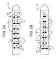
- FIGS. 2 a and 2 bdepict a comparison showing the perceived benefits of antibiotic or drug release for a catheter with uniform fluid flow distribution through catheter holes compared with non-uniform fluid flow distribution.
- FIG. 3depicts an embodiment of a fluid flow enhancing distribution tip.
- the present inventionis directed toward fluid management flow implants such as catheter drainage devices, preferably hydrocephalus shunts, which contain antibiotics to prevent or reduce the risk of infection and slime formation in the interior surfaces of the catheter and/or a drug to prevent or minimize tissue growth, combined with a flow distribution enhancing tip.
- This combination devicewill potentially minimize the risk of blockage or obstruction of the lumens and inner surfaces of the implants due to either biofilm formation or tissue in-growth and allow a greater chance of uninterrupted fluid flow which will in turn lessen the likelihood for costly revision surgery or procedures.
- FIGS. 1 a and 1 bdescribe the above described two step model of biofilm formation.
- FIG. 1 ashows the first step in biofilm formation which is the adherence of the bacterial cells 2 to a surface 1 .
- the second stepis the imbedding of the cells 2 into a thick slime matrix (biofilm) 3 .
- the flow distribution enhancing tipmay be any type of tip that enables uniform flow patterns within the medical device.
- tip designsthat help promote uniform flow distribution within the catheter are contemplated.
- the terms “uniform flow pattern” or “uniform flow distribution”are intended to describe a tip which improves fluid flow over tips not so designed.
- FIGS. 2 a and 2 bdepict the perceived benefit of a flow enhancing tip used in combination with antimicrobial agents and/or drugs compared with antimicrobial agents and/or drugs not combined with a flow enhancing tip.
- tip 10is shown with apertures 12 of varying cross-sectional area. As one proceeds from the distal end to the proximal end of tip 10 , apertures 12 decrease in cross-sectional area. This aperture geometry helps to promote uniform flow which in turn is expected to promote uniform release of antimicrobial agents or drugs 14 .
- conventional tip 10is shown with apertures 12 of constant cross-sectional area. Fluid flow entering through apertures 12 will not produce a uniform flow with tip 10 and therefore release of antimicrobial agents or drugs 14 is not expected to be uniform.
- Lininfra, discloses theoretical and experimental data showing that more than 80% of total fluid mass flows into the two most proximal holes of a hydrocephalus shunt.
- an example of a suitable tip geometrycomprises a tip with a hole pattern of varying hole size where the largest hole is at the distal end of the catheter tip and the smallest hole in the pattern is closest to the shunt valve.
- Most preferredis a whole geometry as depicted in FIG. 3 wherein the size of the holes progressively increase in cross-sectional area from the most proximal inlet hole to the most distal inlet hole.
- the flow enhancing tipmay further comprise a porous device that is incorporated into or onto the tip to reduce the likelihood of blockage by tissue ingrowth.
- the devicemay also be used to dialyze the fluid surrounding the catheter. It is envisioned that the pores would be less than 5 ⁇ m in their largest dimension, and preferably less than 1 ⁇ m, to prevent tissue structures and a supporting blood supply from growing into the luminal space.
- the devicemay be attached to the outside surface of the catheter, or it may be inserted into the lumen. Alternatively, the device may be integrated into the catheter material in such a way as to produce a composite structure.
- the porous devicemay have pore sizes of subnano-, nano- or microporosity to selectively exclude blood vessels, cells, biological debris or molecules of a specific size from the lumen of the catheter.
- the purpose of the porous aspect of the deviceis also to prevent catheter obstruction due to tissue ingrowth.
- the devicemay also be used to dialyze the fluid surrounding the catheter.
- the porous devicemay be attached to the inside and/or outside surfaces of all or part of the catheter.
- the devicemay also be incorporated into the catheter material on may comprise a sleeve which fits over a catheter tip.
- the pore sizeis ideally less than 1 ⁇ m to prevent cellular migration into the lumen of the catheter and the development of tissue structures and a supporting blood supply.
- the porous device described in this inventionmay also be used to prevent blockage at the proximal or distal end of a hydrocephalus catheter, or at the outlet of a drug delivery catheter, or at the end of another fluid management catheter.
- the pore size of the devicemay also be chosen such that only molecules of a specific size range are allowed to pass into the catheter.
- the porous devicemay be fabricated from metal, ceramic, a selected bulk polymer or a polymer film.
- the poresmay be created by manufacturing processes including but not limited to laser drilling, chemical etching, controlled sintering, or incorporating leachable additives or pore-forming agents.
- the fluid discharge from the devices of this inventionmay be to selected areas inside or outside of the human body.
- Typical selected discharge areas inside the human bodyinclude the peritoneum, the right atrium of the heart, the pleural cavity, and the bladder.
- the common selected discharge areas outside the human bodyinclude fluid collection chambers such as drainage bags.
- antimicrobial agentsare intended to encompass those agents that prevent or minimize bacterial colonization and are intended to include but not be limited to antibiotics, antiseptics and disinfectants.
- antibioticsexamples include tetracyclines (e.g., minocycline), rifamycins (e.g., rifampin), macrolides (e.g., erythromycin), penicillins (e.g., nafcillin), cephalosporins (e.g., cefazolin), other beta-lactam antibiotics (e.g., imipenem, aztreonam), aminoglycosides (e.g., gentamicin), chloramphenicol, sufonamides (e.g., sulfamethoxazole), glycopeptides (e.g., vancomycin), quinolones (e.g., ciprofloxacin), fusidic acid, trimethoprim, metronidazole, clindamycin, mupirocin, polyenes (e.g., amphotericin B), azoles (e.g., fluconazo
- antibioticsexamples include minocycline, rifampin, erythromycin, nafcillin, cefazolin, imipenem, aztreonam, gentamicin, sulfamethoxazole, vancomycin, ciprofloxacin, trimethoprim, metronidazole, clindamycin, teicoplanin, mupirocin, azithromycin, clarithromycin, ofloxacin, lomefloxacin, norfloxacin, nalidixic acid, sparfloxacin, pefloxacin, amifloxacin, enoxacin, fleroxacin, temafloxacin, tosufloxacin, clinafloxacin, sulbactam, clavulanic acid, amphotericin B, fluconazole, itraconazole, ketoconazole, and nystatin.
- antiseptics and disinfectantsexamples include hexachlorophene, cationic bisiguanides (e.g., chlorhexidine, cyclohexidine) iodine and iodophores (e.g., povidone-iodine), para-chloro-meta-xylenol, triclosan, furan medical preparations (e.g., nitrofurantoin, nitrofurazone), methenamine, aldehydes (glutaraldehyde, formaldehyde) and alcohols.
- cationic bisiguanidese.g., chlorhexidine, cyclohexidine
- iodine and iodophorese.g., povidone-iodine
- para-chloro-meta-xylenole.g., nitrofurazone
- furan medical preparationse.g., nitrofurantoin, nitrofurazone
- the most preferred antimicrobialsare rifampin and clindamycin hydrochloride. Together they provide superior penetration and persistent antimicrobial activity in devices treated.
- the antimicrobial activitycovers most strains of gram-positive bacteria causing the majority of infections in medical devices such as hydrocephalus shunts.
- drugsare intended to encompass drugs that prevent or minimize tissue growth whether the drugs are cytostatic drugs or cytotoxic drugs.
- Non-limitative examples of drugsinclude therapeutic and pharmaceutic agents including: anti-proliferative/antimitotic agents including natural products such as vinca alkaloids (e.g., vinblastine, vincristine, and vinorelbine), paclitaxel, epidipodophyllotoxins (e.g., etoposide, teniposide), antibiotics (dactinomycin (actinomycin D) daunorubicin, doxorubicin and idarubicin), anthracyclines, mitoxantrone, bleomycins, plicamycin (mithramycin) and mitomycin, enzymes (L-asparaginase which systemically metabolizes L-asparagine and deprives cells which do not have the capacity to synthesize their own asparagine); antiplatelet agents such as G(GP) 11 b /111 a inhibitors and vitronectin receptor antagonists; anti-proliferative/antimitotic alky
- a preferred cytostatic drugis sirolimus (rapamycin) particularly in combination with mycophenolic acid.
- a preferred cytotoxic drugis paclitaxel.
- Non-limiting examples of fluid flow control devices and systemsinclude catheters, shunts, hydrocephalus shunts, central nervous catheters, dialysis grafts, and ear drainage tubes.
- impregnationis preferred when dealing with medical devices made of polymeric materials such as silicone elastomers.
- the antimicrobial agent and/or drugmay also be coated on the inside and/or outside surfaces of all or part of the implant.
- the drugmay be incorporated into the catheter material such that it diffuses from the inside and/or outside surfaces of the tip of the catheter in the region where the fluid drainage holes are located.
- a porous or other type of sleeve, made from a material that contains the drug(s)may be placed over the outside and/or into the inside lumen of the proximal tip of the catheter in the region where the fluid drainage holes are located.
- the impregnation processcan be altered to leave an antimicrobial agent and/or drug on the surface.
- a top-coat that can be used to modulate the elution profile from either the surface or the bulk of the catheter and/or localize the effect of the drugis also being explored.
- the top-coatcan range from a monolayer to a thick layer of synthetic polymer or protein, carbohydrate, or glycoprotein.
- the coatingscan comprise combinations of the previous classes of molecules.
- grafted moleculesconsisting of combinations of synthetic and natural polymers can be used in the form of dendrimers, star, comb or block copolymers.
- the top-coatcan contain drug or could be drug free. Both hydrophilic and or hydrophobic synthetic polymers could be used.
- polyethylene oxide based polymer systemshave been widely used as coatings as have fluorinated polymers and copolymers. Layered systems could provide special benefits. Heparin-based polymer systems as well as other sulfated proteoglycan systems (such as chondroitin sulfate) have also been widely used as coatings. Topcoats consisting of laminated layers of these constituents are also contemplated. Such topcoats could be used to reduce the rate of drug elution or provide an immediate burst of particular drugs.
- topcoatsare also described here. These systems can consist of thicker topcoat layers in the vicinity of drainage orifices or have different materials printed in layers onto different points along the surface of the catheter tip. In addition, different drugs or different concentrations of drugs can be laid down at different points along the surface of the catheter tip. The goal would be to produce local effects at the orifices in the catheter tip and may be advantageous where very expensive drugs or polymer materials are being used.
- Antiomicrobial agents or drugscan be both physically entrapped as well as covalently grafted in the topcoat layers.
- Covalently grafted drugswould either inhibit cell attachment by interfering with cell membrane function or would be slowly released by cleavage of labile linkages. Cleavage could either be by chemical or proteolytic mechanisms.
- antimicrobial agent(s) and/or drug(s)are impregnated into the bulk of the catheter either by compounding-in the drug when the catheter is molded (if the drug is stable to this process) or by impregnating the catheter with drug post-molding. Impregnation can be accomplished by using a solvent or co-solvent system to swell the polymer and diffuse-in the antimicrobial agents/drugs, followed by evaporation of the solvents to entrap the antimicrobial agent/drugs. Impregnation by supercritical fluids or supercritical fluid-organic co-solvent fluids is also described to reduce the quantity of organic solvent needed.
- the advantage hereis primarily ecological (reduced toxic pollutants), but also unique drug-polymer microstructures and release-profiles are possible.
- an antimicrobial agent/drug loading profilethat varies through the thickness of the coating can be achieved. This type of process can provide higher surface concentrations of the antimicrobial agents/drugs.
- antimicrobial agents/drugscan also be included in a sprayed-on coating or dip-coated topcoat. Surface variable coatings can be achieved by masking the implants such as catheters in a spraying process or by selectively spraying only certain areas. Selective material layers can be added by sequentially building up different layers. Finally, coatings can be applied or modified using chemical vapor deposition or plasma coating processes. This can also be desirable for preventing delamination of laminated coatings.
Landscapes
- Health & Medical Sciences (AREA)
- Life Sciences & Earth Sciences (AREA)
- Veterinary Medicine (AREA)
- Engineering & Computer Science (AREA)
- Biomedical Technology (AREA)
- Public Health (AREA)
- General Health & Medical Sciences (AREA)
- Animal Behavior & Ethology (AREA)
- Epidemiology (AREA)
- Heart & Thoracic Surgery (AREA)
- Molecular Biology (AREA)
- Medicinal Chemistry (AREA)
- Chemical & Material Sciences (AREA)
- Vascular Medicine (AREA)
- Surgery (AREA)
- Biophysics (AREA)
- Pulmonology (AREA)
- Anesthesiology (AREA)
- Hematology (AREA)
- Materials For Medical Uses (AREA)
- External Artificial Organs (AREA)
Abstract
Description
- a) providing an implant comprising an antimicrobial or drug-eluting catheter having a proximal and distal end;
- b) providing a flow distribution enhancing tip at the distal end of the catheter;
- c) inserting the distal end of the catheter into an area to be drained;
- d) placing the proximal end of the catheter in a selected area inside or outside of the human body; and
- e) draining fluid from the area to be drained to the selected area through the catheter.
- a) an antimicrobial or drug-eluting device comprising a proximal and distal end; and
- b) a flow distribution enhancing tip at the distal end of the device.
Claims (22)
Priority Applications (9)
| Application Number | Priority Date | Filing Date | Title |
|---|---|---|---|
| US10/955,776US7976517B2 (en) | 2004-09-30 | 2004-09-30 | Fluid management flow implants of improved occlusion resistance |
| AU2005204329AAU2005204329B2 (en) | 2004-09-30 | 2005-08-29 | Fluid management flow implants of improved occlusion resistance |
| AT05256107TATE485846T1 (en) | 2004-09-30 | 2005-09-29 | FLUID TREATMENT IMPLANTS WITH IMPROVED OCCLUSION RESISTANCE |
| DE602005024352TDE602005024352D1 (en) | 2004-09-30 | 2005-09-29 | Implants for liquid treatment with improved closure resistance |
| ES05256107TES2353221T3 (en) | 2004-09-30 | 2005-09-29 | IMPLANTS OF FLOW MANAGEMENT FLOW OF RESISTANCE TO IMPROVED OCLUSION. |
| CA2521884ACA2521884C (en) | 2004-09-30 | 2005-09-29 | Fluid management flow implants of improved occlusion resistance |
| JP2005284791AJP5701469B2 (en) | 2004-09-30 | 2005-09-29 | Fluid management flow implant with improved occlusion resistance |
| EP05256107AEP1649880B1 (en) | 2004-09-30 | 2005-09-29 | Fluid management flow implants of improved occlusion resistance |
| US11/931,024US8221392B2 (en) | 2004-09-30 | 2007-10-31 | Fluid management flow implants of improved occlusion resistance |
Applications Claiming Priority (1)
| Application Number | Priority Date | Filing Date | Title |
|---|---|---|---|
| US10/955,776US7976517B2 (en) | 2004-09-30 | 2004-09-30 | Fluid management flow implants of improved occlusion resistance |
Related Child Applications (1)
| Application Number | Title | Priority Date | Filing Date |
|---|---|---|---|
| US11/931,024ContinuationUS8221392B2 (en) | 2004-09-30 | 2007-10-31 | Fluid management flow implants of improved occlusion resistance |
Publications (2)
| Publication Number | Publication Date |
|---|---|
| US20060074388A1 US20060074388A1 (en) | 2006-04-06 |
| US7976517B2true US7976517B2 (en) | 2011-07-12 |
Family
ID=35794941
Family Applications (2)
| Application Number | Title | Priority Date | Filing Date |
|---|---|---|---|
| US10/955,776Expired - LifetimeUS7976517B2 (en) | 2004-09-30 | 2004-09-30 | Fluid management flow implants of improved occlusion resistance |
| US11/931,024Expired - Fee RelatedUS8221392B2 (en) | 2004-09-30 | 2007-10-31 | Fluid management flow implants of improved occlusion resistance |
Family Applications After (1)
| Application Number | Title | Priority Date | Filing Date |
|---|---|---|---|
| US11/931,024Expired - Fee RelatedUS8221392B2 (en) | 2004-09-30 | 2007-10-31 | Fluid management flow implants of improved occlusion resistance |
Country Status (8)
| Country | Link |
|---|---|
| US (2) | US7976517B2 (en) |
| EP (1) | EP1649880B1 (en) |
| JP (1) | JP5701469B2 (en) |
| AT (1) | ATE485846T1 (en) |
| AU (1) | AU2005204329B2 (en) |
| CA (1) | CA2521884C (en) |
| DE (1) | DE602005024352D1 (en) |
| ES (1) | ES2353221T3 (en) |
Cited By (8)
| Publication number | Priority date | Publication date | Assignee | Title |
|---|---|---|---|---|
| US20110282264A1 (en)* | 2002-03-26 | 2011-11-17 | Medtronic Ps Medical, Inc. | Method of draining cerebrospinal fluid |
| US20140302113A1 (en)* | 2011-09-30 | 2014-10-09 | Sparkmed Research, Llc | Systems, devices, and methods for embedding drug molecules into medical catheters or tubes |
| US9227043B2 (en) | 2011-11-18 | 2016-01-05 | Washington University | Catheter assembly for use with shunt systems and method of using same |
| US10058685B2 (en) | 2010-06-18 | 2018-08-28 | Technion Research & Development Foundation Ltd. | Self cleaning shunt |
| US10518069B2 (en) | 2016-10-28 | 2019-12-31 | Integra LifeSciences Switzerland Sarl | Implantable valve assembly with extended lifespan |
| US11045632B2 (en) | 2017-04-24 | 2021-06-29 | Longeviti Neuro Solutions Llc | Cerebral spinal fluid shunt plug |
| US11439798B2 (en) | 2017-04-24 | 2022-09-13 | Longeviti Neuro Solutions Llc | Cerebral spinal fluid shunt plug |
| US12156962B2 (en) | 2020-05-11 | 2024-12-03 | Minnetronix Neuro, Inc. | Filtering cassettes and filtering systems |
Families Citing this family (53)
| Publication number | Priority date | Publication date | Assignee | Title |
|---|---|---|---|---|
| US20030216710A1 (en)* | 2002-03-26 | 2003-11-20 | Hurt Robert F. | Catheter |
| CA2881343A1 (en)* | 2004-12-08 | 2006-06-15 | Shire Regenerative Medicine, Inc. | Methods and compositions for enhancing vascular access |
| US8002730B2 (en) | 2005-04-29 | 2011-08-23 | Medtronic, Inc. | Anti-thrombogenic venous shunt system and method |
| US20080082036A1 (en)* | 2006-04-25 | 2008-04-03 | Medtronic, Inc. | Cerebrospinal fluid shunt having long term anti-occlusion agent delivery |
| JP2010005282A (en)* | 2008-06-30 | 2010-01-14 | Sumitomo Bakelite Co Ltd | Medical drain tube |
| TW201014624A (en)* | 2008-09-18 | 2010-04-16 | Kci Licensing Inc | A system and method for delivering reduced pressure to subcutaneous tissue |
| US20100106161A1 (en)* | 2008-10-23 | 2010-04-29 | Marwan Tabbara | Surgical methods, devices, and kits |
| US9072296B2 (en) | 2009-03-26 | 2015-07-07 | Organic Medical Ventures, L.L.C. | Transdermal venous access locking solutions |
| US9427498B2 (en) | 2009-03-26 | 2016-08-30 | Organic Medical Ventures, L.L.C. | Syringe treated with transdermal venous access locking solutions and method of treating the syringe |
| US20100249747A1 (en)* | 2009-03-26 | 2010-09-30 | Organic Medical Ventures, L.L.C. | Transdermal venous access locking solution |
| US9433209B2 (en) | 2009-03-26 | 2016-09-06 | Organic Medical Ventures, L.L.C. | Transdermal venous access locking solutions |
| US8092443B2 (en)* | 2009-03-30 | 2012-01-10 | Medtronic, Inc. | Element for implantation with medical device |
| EP2504025A4 (en)* | 2009-11-23 | 2013-05-01 | Stephen F Olmstead | COMPOSITIONS AND METHODS COMPRISING SERRATIA PEPTIDASE FOR THE INHIBITION AND TREATMENT OF BIOFILMS RELATED TO CERTAIN PATHOLOGIES |
| WO2011126898A2 (en)* | 2010-03-30 | 2011-10-13 | Sparkmed Research, Llc | Drug releasing medical catheters, tubes, and devices |
| US8702678B2 (en)* | 2011-08-03 | 2014-04-22 | Venous Therapy, Inc. | Assemblies, systems, and methods for infusing therapeutic agents into the body |
| NZ621994A (en) | 2011-08-31 | 2016-05-27 | Organic Medical Ventures L L C | Transdermal venous access locking solutions |
| US20130102999A1 (en)* | 2011-10-25 | 2013-04-25 | Anthony M. Looper | Pleurodesis catheter with coated and shielded tip |
| CA3094655C (en) | 2013-01-22 | 2023-01-24 | Anuncia, Inc. | Systems and methods for shunting fluid |
| US9682218B2 (en) | 2013-12-23 | 2017-06-20 | Carefusion 2200, Inc. | Pleurodesis device and method |
| ES2519940B1 (en)* | 2014-03-06 | 2015-10-22 | Universidad Miguel Hernández De Elche | Catheter for hydrocephalus treatment |
| AU2015247353B2 (en) | 2014-04-18 | 2020-07-09 | Anuncia, Inc | Systems and methods for shunting fluid |
| US10918827B2 (en) | 2015-07-20 | 2021-02-16 | Strataca Systems Limited | Catheter device and method for inducing negative pressure in a patient's bladder |
| US12064567B2 (en) | 2015-07-20 | 2024-08-20 | Roivios Limited | Percutaneous urinary catheter |
| US10512713B2 (en) | 2015-07-20 | 2019-12-24 | Strataca Systems Limited | Method of removing excess fluid from a patient with hemodilution |
| HUE049050T2 (en) | 2015-07-20 | 2020-08-28 | Strataca Systems Ltd | Ureteral and bladder catheters |
| US10926062B2 (en)* | 2015-07-20 | 2021-02-23 | Strataca Systems Limited | Ureteral and bladder catheters and methods of inducing negative pressure to increase renal perfusion |
| US10493232B2 (en) | 2015-07-20 | 2019-12-03 | Strataca Systems Limited | Ureteral catheters, bladder catheters, systems, kits and methods for inducing negative pressure to increase renal function |
| US11040180B2 (en) | 2015-07-20 | 2021-06-22 | Strataca Systems Limited | Systems, kits and methods for inducing negative pressure to increase renal function |
| US11541205B2 (en) | 2015-07-20 | 2023-01-03 | Roivios Limited | Coated urinary catheter or ureteral stent and method |
| US10765834B2 (en) | 2015-07-20 | 2020-09-08 | Strataca Systems Limited | Ureteral and bladder catheters and methods of inducing negative pressure to increase renal perfusion |
| US11040172B2 (en) | 2015-07-20 | 2021-06-22 | Strataca Systems Limited | Ureteral and bladder catheters and methods of inducing negative pressure to increase renal perfusion |
| US11229771B2 (en) | 2015-07-20 | 2022-01-25 | Roivios Limited | Percutaneous ureteral catheter |
| JP2019530539A (en)* | 2016-10-13 | 2019-10-24 | アルキオーネ・ライフサイエンシズ・インコーポレイテッドAlcyone Lifesciences, Inc. | Shunt flasher and related methods |
| US11284939B2 (en)* | 2017-07-06 | 2022-03-29 | Biosense Webster (Israel) Ltd. | Irrigated catheter with improved ablation tip electrode fluid distribution |
| CN107320839B (en)* | 2017-07-26 | 2023-03-17 | 常州瑞神安医疗器械有限公司 | In-vivo drainage device and method |
| JP2020531159A (en) | 2017-08-25 | 2020-11-05 | ストラタカ システムズ リミテッド | Indwelling pump to facilitate removal of urine from the urinary tract |
| JP2020531157A (en)* | 2017-08-25 | 2020-11-05 | ストラタカ システムズ リミテッド | Ureter and bladder catheters and methods that induce negative pressure and increase renal perfusion |
| JP2021514801A (en)* | 2018-05-03 | 2021-06-17 | マイクロベンション インコーポレイテッドMicrovention, Inc. | Treatment of hydrocephalus |
| KR20220011123A (en) | 2019-04-11 | 2022-01-27 | 엔클리어 테라피스, 인크. | Cerebrospinal fluid improvement method and device and system therefor |
| CN110201174B (en)* | 2019-05-14 | 2021-08-03 | 青岛大学附属医院 | A pharmaceutical composition capable of resisting chronic infection and biofilm bacteria and use thereof |
| CN114173852B (en)* | 2019-07-24 | 2025-04-01 | W.L.戈尔及同仁股份有限公司 | Implantable catheter |
| CN114828931A (en)* | 2019-07-25 | 2022-07-29 | 米奈特朗尼克斯神经有限公司 | System for cerebrospinal fluid treatment |
| US20230001165A1 (en)* | 2019-11-20 | 2023-01-05 | Massachusetts Institute Of Technology | Cerebrospinal fluid space draining catheters |
| US12311131B2 (en)* | 2020-02-24 | 2025-05-27 | Becton, Dickinson And Company | Tubular instrument to reduce vein trauma and related devices and methods |
| EP4221801A4 (en) | 2020-09-29 | 2024-11-06 | Enclear Therapies, Inc. | METHOD AND SYSTEM FOR MANAGING SUBARACHNOID FLUID |
| JP7029005B1 (en) | 2021-02-02 | 2022-03-02 | 厚夫 森 | catheter |
| CN113082309B (en)* | 2021-04-02 | 2025-08-01 | 西安市中心医院 | A tubulose negative pressure drainage ware for orthopedics |
| WO2023039541A1 (en)* | 2021-09-09 | 2023-03-16 | Wayne State University | Solid state shunt valve with active outflow regulator, ventricular catheters, and other embodiments |
| ES2952142B2 (en)* | 2022-03-21 | 2024-03-12 | Univ Miguel Hernandez De Elche | VENTRICULAR CATHETER FOR APPLICATION IN HYDROCEPHALUS TREATMENTS |
| WO2024047081A1 (en) | 2022-09-02 | 2024-03-07 | Universiteit Gent | Catheter for delivery of therapeutic liquid formulation |
| CN115814176A (en)* | 2022-12-13 | 2023-03-21 | 湛江市事达实业有限公司 | Flow guide pipe and disinfection method thereof |
| WO2024151968A1 (en)* | 2023-01-13 | 2024-07-18 | Georgia Tech Research Corporation | Concentric tube fluidic valve system |
| CN119909241A (en)* | 2023-10-31 | 2025-05-02 | 武汉联影智融医疗科技有限公司 | Drainage catheters and drainage systems |
Citations (31)
| Publication number | Priority date | Publication date | Assignee | Title |
|---|---|---|---|---|
| US3601128A (en) | 1968-12-26 | 1971-08-24 | Salomon Hakim | Ventriculoatrial shunt accumulator |
| US3669116A (en) | 1970-07-06 | 1972-06-13 | Heyer Schulte Corp | Drainage catheter with anticlogging means |
| US3690323A (en) | 1970-12-01 | 1972-09-12 | Us Army | Device for draining ventricular fluid in cases of hydrocephalus |
| US3894541A (en) | 1974-02-27 | 1975-07-15 | El Shafei Ismail Lotfy | Method of treating hydrocephalus |
| US4182343A (en) | 1976-10-05 | 1980-01-08 | President of Tokyo Medical and Dental University | Double coeliac drainage tube made of silicone |
| US4377169A (en) | 1981-06-10 | 1983-03-22 | Banks Bruce A | Ion beam sputter-etched ventricular catheter for hydrocephalus shunt |
| US4655745A (en) | 1985-07-29 | 1987-04-07 | Corbett Joseph E | Ventricular catheter |
| US4767400A (en) | 1987-10-27 | 1988-08-30 | Cordis Corporation | Porous ventricular catheter |
| US4917686A (en) | 1985-12-16 | 1990-04-17 | Colorado Biomedical, Inc. | Antimicrobial device and method |
| US4950232A (en) | 1987-08-11 | 1990-08-21 | Surelab Superior Research Laboratories | Cerebrospinal fluid shunt system |
| US5180387A (en) | 1987-09-17 | 1993-01-19 | Neurodynamics, Inc. | Angled hole ventricular catheter with non-circular bore |
| WO1994007549A1 (en) | 1992-09-30 | 1994-04-14 | Target Therapeutics, Inc. | Catheter with atraumatic drug delivery tip |
| US5405316A (en) | 1993-11-17 | 1995-04-11 | Magram; Gary | Cerebrospinal fluid shunt |
| US5451215A (en) | 1990-09-17 | 1995-09-19 | Wolter; Dietmar | Suction drain for the aspiration of discharges |
| US5531673A (en) | 1995-05-26 | 1996-07-02 | Helenowski; Tomasz K. | Ventricular catheter |
| US5579774A (en) | 1994-03-07 | 1996-12-03 | Camino Neurocare, Inc. | Method and apparatus for monitoring local cerebral physiology |
| US6110155A (en) | 1996-04-30 | 2000-08-29 | Medtronic, Inc. | Anti-inflammatory-agent-loaded catheter and method for preventing tissue fibrosis |
| WO2001005210A2 (en)* | 1999-07-19 | 2001-01-25 | I-Flow Corporation | Catheter for uniform delivery of medication |
| US6325815B1 (en) | 1999-09-21 | 2001-12-04 | Microvena Corporation | Temporary vascular filter |
| US20020001609A1 (en) | 2000-03-10 | 2002-01-03 | Macropore, Inc. | Resorbable barrier micro-membranes for attenuation of scar tissue during healing |
| US20020111601A1 (en)* | 2001-02-12 | 2002-08-15 | Medtronic, Inc | Drug delivery device |
| US20020188246A1 (en) | 2001-06-12 | 2002-12-12 | Hayner George M. | Combination ureteral infusion catheter/drainage stent |
| US20030009132A1 (en) | 2001-04-13 | 2003-01-09 | Tricardia Llc | Syringe system |
| US6533763B1 (en) | 1999-12-06 | 2003-03-18 | James A. Schneiter | Harmonic flow catheter |
| US20030073647A1 (en)* | 2001-08-28 | 2003-04-17 | Chao Robert S. | Crystaline clindamycin free base |
| US20030216710A1 (en) | 2002-03-26 | 2003-11-20 | Hurt Robert F. | Catheter |
| US20040054351A1 (en) | 1999-07-19 | 2004-03-18 | Deniega Jose Castillo | Catheter for uniform delivery of medication |
| WO2004073768A2 (en) | 2003-02-18 | 2004-09-02 | Medtronic, Inc. | Occlusion resistant hydrocephalic shunt |
| US20040253185A1 (en)* | 2003-06-12 | 2004-12-16 | Atrium Medical Corp. | Medicated ink |
| US20050153379A1 (en)* | 2003-12-22 | 2005-07-14 | David Hoon | Method and apparatus for in vivo collection of circulating biological components |
| EP1364628B1 (en) | 2002-05-20 | 2007-03-21 | Cordis Corporation | Coated medical devices |
Family Cites Families (1)
| Publication number | Priority date | Publication date | Assignee | Title |
|---|---|---|---|---|
| WO1987003495A1 (en) | 1985-12-16 | 1987-06-18 | Denver Surgical Developments, Inc. | Antimicrobial catheter and method |
- 2004
- 2004-09-30USUS10/955,776patent/US7976517B2/ennot_activeExpired - Lifetime
- 2005
- 2005-08-29AUAU2005204329Apatent/AU2005204329B2/ennot_activeCeased
- 2005-09-29ESES05256107Tpatent/ES2353221T3/enactiveActive
- 2005-09-29ATAT05256107Tpatent/ATE485846T1/ennot_activeIP Right Cessation
- 2005-09-29DEDE602005024352Tpatent/DE602005024352D1/ennot_activeExpired - Lifetime
- 2005-09-29JPJP2005284791Apatent/JP5701469B2/ennot_activeExpired - Fee Related
- 2005-09-29CACA2521884Apatent/CA2521884C/ennot_activeExpired - Lifetime
- 2005-09-29EPEP05256107Apatent/EP1649880B1/ennot_activeExpired - Lifetime
- 2007
- 2007-10-31USUS11/931,024patent/US8221392B2/ennot_activeExpired - Fee Related
Patent Citations (37)
| Publication number | Priority date | Publication date | Assignee | Title |
|---|---|---|---|---|
| US3601128A (en) | 1968-12-26 | 1971-08-24 | Salomon Hakim | Ventriculoatrial shunt accumulator |
| US3669116A (en) | 1970-07-06 | 1972-06-13 | Heyer Schulte Corp | Drainage catheter with anticlogging means |
| US3690323A (en) | 1970-12-01 | 1972-09-12 | Us Army | Device for draining ventricular fluid in cases of hydrocephalus |
| US3894541A (en) | 1974-02-27 | 1975-07-15 | El Shafei Ismail Lotfy | Method of treating hydrocephalus |
| US4182343A (en) | 1976-10-05 | 1980-01-08 | President of Tokyo Medical and Dental University | Double coeliac drainage tube made of silicone |
| US4377169A (en) | 1981-06-10 | 1983-03-22 | Banks Bruce A | Ion beam sputter-etched ventricular catheter for hydrocephalus shunt |
| US4655745A (en) | 1985-07-29 | 1987-04-07 | Corbett Joseph E | Ventricular catheter |
| US4917686A (en) | 1985-12-16 | 1990-04-17 | Colorado Biomedical, Inc. | Antimicrobial device and method |
| US4950232A (en) | 1987-08-11 | 1990-08-21 | Surelab Superior Research Laboratories | Cerebrospinal fluid shunt system |
| US5180387A (en) | 1987-09-17 | 1993-01-19 | Neurodynamics, Inc. | Angled hole ventricular catheter with non-circular bore |
| US4767400A (en) | 1987-10-27 | 1988-08-30 | Cordis Corporation | Porous ventricular catheter |
| US5451215A (en) | 1990-09-17 | 1995-09-19 | Wolter; Dietmar | Suction drain for the aspiration of discharges |
| WO1994007549A1 (en) | 1992-09-30 | 1994-04-14 | Target Therapeutics, Inc. | Catheter with atraumatic drug delivery tip |
| US5405316A (en) | 1993-11-17 | 1995-04-11 | Magram; Gary | Cerebrospinal fluid shunt |
| US5579774A (en) | 1994-03-07 | 1996-12-03 | Camino Neurocare, Inc. | Method and apparatus for monitoring local cerebral physiology |
| US5531673A (en) | 1995-05-26 | 1996-07-02 | Helenowski; Tomasz K. | Ventricular catheter |
| US6110155A (en) | 1996-04-30 | 2000-08-29 | Medtronic, Inc. | Anti-inflammatory-agent-loaded catheter and method for preventing tissue fibrosis |
| US20020082547A1 (en) | 1999-07-19 | 2002-06-27 | Deniega Jose Castillo | Catheter for uniform delivery of medication |
| WO2001005210A2 (en)* | 1999-07-19 | 2001-01-25 | I-Flow Corporation | Catheter for uniform delivery of medication |
| US20040054351A1 (en) | 1999-07-19 | 2004-03-18 | Deniega Jose Castillo | Catheter for uniform delivery of medication |
| US6350253B1 (en) | 1999-07-19 | 2002-02-26 | I-Flow Corporation | Catheter for uniform delivery of medication |
| US6626885B2 (en) | 1999-07-19 | 2003-09-30 | I-Flow Corporation | Method of fluid delivery and catheters for use with same |
| US6325815B1 (en) | 1999-09-21 | 2001-12-04 | Microvena Corporation | Temporary vascular filter |
| US20020032460A1 (en) | 1999-09-21 | 2002-03-14 | Kusleika Richard S. | Temporary vascular filter |
| US6533763B1 (en) | 1999-12-06 | 2003-03-18 | James A. Schneiter | Harmonic flow catheter |
| US20020001609A1 (en) | 2000-03-10 | 2002-01-03 | Macropore, Inc. | Resorbable barrier micro-membranes for attenuation of scar tissue during healing |
| US20020111601A1 (en)* | 2001-02-12 | 2002-08-15 | Medtronic, Inc | Drug delivery device |
| US20030009132A1 (en) | 2001-04-13 | 2003-01-09 | Tricardia Llc | Syringe system |
| US6524268B2 (en) | 2001-06-12 | 2003-02-25 | George M. Hayner | Combination ureteral infusion catheter/drainage stent |
| US20020188246A1 (en) | 2001-06-12 | 2002-12-12 | Hayner George M. | Combination ureteral infusion catheter/drainage stent |
| US20030073647A1 (en)* | 2001-08-28 | 2003-04-17 | Chao Robert S. | Crystaline clindamycin free base |
| US20030216710A1 (en) | 2002-03-26 | 2003-11-20 | Hurt Robert F. | Catheter |
| EP1364628B1 (en) | 2002-05-20 | 2007-03-21 | Cordis Corporation | Coated medical devices |
| WO2004073768A2 (en) | 2003-02-18 | 2004-09-02 | Medtronic, Inc. | Occlusion resistant hydrocephalic shunt |
| WO2004073768A3 (en) | 2003-02-18 | 2005-04-21 | Medtronic Inc | Occlusion resistant hydrocephalic shunt |
| US20040253185A1 (en)* | 2003-06-12 | 2004-12-16 | Atrium Medical Corp. | Medicated ink |
| US20050153379A1 (en)* | 2003-12-22 | 2005-07-14 | David Hoon | Method and apparatus for in vivo collection of circulating biological components |
Non-Patent Citations (20)
| Title |
|---|
| Bayston & Penny, Dev. Med. Child Neurol, 1972, 14 Suppl 27 25-28. |
| Codman Product Brochure, "Codman Bactiseal Antimicrobial Impregnated Catheter System" Codman & Shurtleff, 2002.* |
| Codman Product Brochure, "Codman Bactiseal EVD Catheter", Codman & Shurtleff, Inc., 2004. |
| Collins et al., J. Neurosurg, 48:609-613, 1978. |
| Del Bigio and Bruni, J. Neurosurgery, 64:932-940, 1986. |
| Del Bigio and Fedoroff, J. Biomed Mater Res, 26:979-987, 1992. |
| Del Bigio, Neurosurgery, 42:319-326, 1998. |
| Drake et al.., The Shunt Book, 1995, Shunt Complications, Chapt. 5 pp. 123-192. |
| Drake et al.., The Shunt Book, 1995. Cerebrospinal Fluid Shunt Components, Chapt. 4, pp. 71-119. |
| Edvinsson et al., Acta Physiol Scand, 82:527-531, 1971. |
| EP Search Report dated Aug. 16, 2007 re EP05256107.3. |
| Gotz et al., ASM Press, Washington DC, 2000, pp. 55-88. |
| Jaeger et al., Brain Res, 551:163-170, 1991. |
| Kestle et al., Pediatric Neurosurgery, 33:230-236, 2000. |
| Kossovsky and Snow. J. Biomed Mater Res. 23(A1):73-86, 1989. |
| Lin et al., J. Neurosurg. vol. 99, Aug. 2003, pp. 426-431. |
| Lundberg, et al., J. Neurosurg, 1999, 90: 101-108. |
| Raftopoulos et al., Acta Neurochir (Wien), 177-180, 1994. |
| Sainte-Rose et al., Pediatric Neurosurgery, 17:2-9, 1991. |
| Takahashi et al., Neurol Med Chir. 38:399-404, 1998. |
Cited By (12)
| Publication number | Priority date | Publication date | Assignee | Title |
|---|---|---|---|---|
| US20110282264A1 (en)* | 2002-03-26 | 2011-11-17 | Medtronic Ps Medical, Inc. | Method of draining cerebrospinal fluid |
| US9694166B2 (en)* | 2002-03-26 | 2017-07-04 | Medtronics Ps Medical, Inc. | Method of draining cerebrospinal fluid |
| US10058685B2 (en) | 2010-06-18 | 2018-08-28 | Technion Research & Development Foundation Ltd. | Self cleaning shunt |
| US20140302113A1 (en)* | 2011-09-30 | 2014-10-09 | Sparkmed Research, Llc | Systems, devices, and methods for embedding drug molecules into medical catheters or tubes |
| US9227043B2 (en) | 2011-11-18 | 2016-01-05 | Washington University | Catheter assembly for use with shunt systems and method of using same |
| US10773060B2 (en) | 2011-11-18 | 2020-09-15 | Washington University | Method of using a catheter assembly |
| US11944768B2 (en) | 2011-11-18 | 2024-04-02 | Washington University | Catheter tip for a catheter assembly |
| US10518069B2 (en) | 2016-10-28 | 2019-12-31 | Integra LifeSciences Switzerland Sarl | Implantable valve assembly with extended lifespan |
| US11045632B2 (en) | 2017-04-24 | 2021-06-29 | Longeviti Neuro Solutions Llc | Cerebral spinal fluid shunt plug |
| US11439798B2 (en) | 2017-04-24 | 2022-09-13 | Longeviti Neuro Solutions Llc | Cerebral spinal fluid shunt plug |
| US12171966B2 (en) | 2017-04-24 | 2024-12-24 | Longeviti Neuro Solutions Llc | Cerebral spinal fluid shunt plug |
| US12156962B2 (en) | 2020-05-11 | 2024-12-03 | Minnetronix Neuro, Inc. | Filtering cassettes and filtering systems |
Also Published As
| Publication number | Publication date |
|---|---|
| AU2005204329A1 (en) | 2006-04-13 |
| CA2521884C (en) | 2015-01-13 |
| US8221392B2 (en) | 2012-07-17 |
| ES2353221T3 (en) | 2011-02-28 |
| DE602005024352D1 (en) | 2010-12-09 |
| US20080214982A1 (en) | 2008-09-04 |
| CA2521884A1 (en) | 2006-03-30 |
| ATE485846T1 (en) | 2010-11-15 |
| JP2006110339A (en) | 2006-04-27 |
| EP1649880A2 (en) | 2006-04-26 |
| EP1649880B1 (en) | 2010-10-27 |
| JP5701469B2 (en) | 2015-04-15 |
| AU2005204329B2 (en) | 2011-07-21 |
| EP1649880A3 (en) | 2007-09-26 |
| US20060074388A1 (en) | 2006-04-06 |
Similar Documents
| Publication | Publication Date | Title |
|---|---|---|
| US7976517B2 (en) | Fluid management flow implants of improved occlusion resistance | |
| US11623073B2 (en) | Cerebrospinal fluid shunt having long term anti-occlusion agent delivery | |
| EP1596895B1 (en) | Occlusion resistant hydrocephalic shunt | |
| US20040236309A1 (en) | Mesh ventricular catheter with antithrombogenic coating | |
| CA2794005C (en) | Adjusting drug loading in polymeric materials | |
| US20080269175A1 (en) | Increased drug loading capacity of polymeric material |
Legal Events
| Date | Code | Title | Description |
|---|---|---|---|
| AS | Assignment | Owner name:CODMAN & SHURTLEFF, INC., MASSACHUSETTS Free format text:ASSIGNMENT OF ASSIGNORS INTEREST;ASSIGNORS:DEXTRADEUR, ALAN;MAUGE, CHRISTOPHE;REEL/FRAME:016137/0524;SIGNING DATES FROM 20041202 TO 20041203 Owner name:CODMAN & SHURTLEFF, INC., MASSACHUSETTS Free format text:ASSIGNMENT OF ASSIGNORS INTEREST;ASSIGNORS:DEXTRADEUR, ALAN;MAUGE, CHRISTOPHE;SIGNING DATES FROM 20041202 TO 20041203;REEL/FRAME:016137/0524 | |
| STCF | Information on status: patent grant | Free format text:PATENTED CASE | |
| FPAY | Fee payment | Year of fee payment:4 | |
| AS | Assignment | Owner name:DEPUY SPINE, LLC, MASSACHUSETTS Free format text:ASSIGNMENT OF ASSIGNORS INTEREST;ASSIGNOR:CODMAN & SHURTLEFF, INC.;REEL/FRAME:045058/0627 Effective date:20121230 | |
| AS | Assignment | Owner name:HAND INNOVATIONS LLC, FLORIDA Free format text:ASSIGNMENT OF ASSIGNORS INTEREST;ASSIGNOR:DEPUY SPINE, LLC;REEL/FRAME:045074/0943 Effective date:20121230 Owner name:DEPUY SYNTHES PRODUCTS, LLC, MASSACHUSETTS Free format text:CHANGE OF NAME;ASSIGNOR:HAND INNOVATIONS LLC;REEL/FRAME:045476/0712 Effective date:20121231 Owner name:DEPUY SYNTHES PRODUCTS, INC., INDIANA Free format text:CHANGE OF NAME;ASSIGNOR:DEPUY SYNTHES PRODUCTS, LLC;REEL/FRAME:045479/0763 Effective date:20141219 | |
| AS | Assignment | Owner name:INTEGRA LIFESCIENCES SWITZERLAND SARL, SWITZERLAND Free format text:ASSIGNMENT OF ASSIGNORS INTEREST;ASSIGNOR:DEPUY SYNTHES PRODUCTS, INC.;REEL/FRAME:045569/0299 Effective date:20171002 | |
| MAFP | Maintenance fee payment | Free format text:PAYMENT OF MAINTENANCE FEE, 8TH YEAR, LARGE ENTITY (ORIGINAL EVENT CODE: M1552); ENTITY STATUS OF PATENT OWNER: LARGE ENTITY Year of fee payment:8 | |
| MAFP | Maintenance fee payment | Free format text:PAYMENT OF MAINTENANCE FEE, 12TH YEAR, LARGE ENTITY (ORIGINAL EVENT CODE: M1553); ENTITY STATUS OF PATENT OWNER: LARGE ENTITY Year of fee payment:12 |